Optineurin Is Required for CYLD-Dependent Inhibition of TNFalpha-Induced NF-kappaB Activation
11
Optineurin Is Required for CYLD-Dependent Inhibition of TNFa-Induced NF-kB Activation Ananthamurthy Nagabhushana, Megha Bansal, Ghanshyam Swarup* Centre for Cellular and Molecular Biology, Council of Scientific and Industrial Research, Hyderabad, India Abstract The nuclear factor kappa B (NF-kB) regulates genes that function in diverse cellular processes like inflammation, immunity and cell survival. The activation of NF-kB is tightly controlled and the deubiquitinase CYLD has emerged as a key negative regulator of NF-kB signalling. Optineurin, mutated in certain glaucomas and amyotrophic lateral sclerosis, is also a negative regulator of NF-kB activation. It competes with NEMO (NF-kB essential modulator) for binding to ubiquitinated RIP (receptor interacting protein) to prevent NF-kB activation. Recently we identified CYLD as optineurin-interacting protein. Here we have analysed the functional significance of interaction of optineurin with CYLD. Our results show that a glaucoma- associated mutant of optineurin, H486R, is altered in its interaction with CYLD. Unlike wild-type optineurin, the H486R mutant did not inhibit tumour necrosis factor a (TNFa)-induced NF-kB activation. CYLD mediated inhibition of TNFa- induced NF-kB activation was abrogated by expression of the H486R mutant. Upon knockdown of optineurin, CYLD was unable to inhibit TNFa-induced NF-kB activation and showed drastically reduced interaction with ubiquitinated RIP. The level of ubiquitinated RIP was increased in optineurin knockdown cells. Deubiquitination of RIP by over-expressed CYLD was abrogated in optineurin knockdown cells. These results suggest that optineurin regulates NF-kB activation by mediating interaction of CYLD with ubiquitinated RIP thus facilitating deubiquitination of RIP. Citation: Nagabhushana A, Bansal M, Swarup G (2011) Optineurin Is Required for CYLD-Dependent Inhibition of TNFa-Induced NF-kB Activation. PLoS ONE 6(3): e17477. doi:10.1371/journal.pone.0017477 Editor: Neeraj Vij, Johns Hopkins School of Medicine, United States of America Received October 29, 2010; Accepted February 3, 2011; Published March 7, 2011 Copyright: ß 2011 Nagabhushana et al. This is an open-access article distributed under the terms of the Creative Commons Attribution License, which permits unrestricted use, distribution, and reproduction in any medium, provided the original author and source are credited. Funding: This work was supported in part by grant BT/PR10130/BRB/10/614/2008 from the Department of Biotechnology, Government of India, to GS. AN is recipient of Senior Research Fellowship of UGC, India. MB is recipient of Research Fellowship of CSIR, India. No additional external funding received for this study. The funders had no role in study design, data collection and analysis, decision to publish, or preparation of the manuscript. Competing Interests: The authors have declared that no competing interests exist. * E-mail: [email protected] Introduction Nuclear factor-kB (NF-kB) plays a key role in the expression of many genes involved in regulating immune response, apoptosis, cell cycle and its deregulation is involved in the pathogenesis of many diseases [1,2]. In unstimulated cells, NF-kB is sequestered in the cytoplasm through its association with the inhibitory IkB proteins. During activation of NF-kB by the cytokine tumor necrosis factor (TNF)a, signalling intermediates like TRADD (TNF receptor associated death domain), TRAF2 (TNF receptor associated factor) and RIP (receptor interacting protein) are recruited to the TNF receptor (TNFR1). This results in the activation of IkB kinase complex (IKK) consisting of the catalytic IKKa and b subunits and the regulatory subunit IKK-c/NEMO (NF-kB essential modulator). IKK activation involves conjugation of Lys63-linked polyubiquitin chains to NEMO and its upstream regulators like RIP [3]. RIP has emerged as a central adaptor in the pathways leading to IKK and NF-kB activation and also cell death. Following TNFa stimulation RIP is recruited to TNFR1 signalling complex and is rapidly ubiquitinated with Lys63-linked polyubiquitin chains. NEMO binds to polyubiquitinated RIP through its ubiquitin binding domain (UBD) resulting in the activation of catalytic subunits of IKK. The recognition and association of ubiquitinated RIP with NEMO is essential for IKK activation [4–6]. The activated catalytic subunits of IKK then phosphorylate IkB triggering its ubiquitination and degradation leading to nuclear translocation and activation of NF-kB. Given its role in diverse cellular processes, the activation of NF-kB is governed by several positive and negative regulators. With the increasing role of ubiquitination, deubiquitinases like CYLD and A20 have emerged as key negative regulators of NF-kB activation [3,7– 12]. CYLD was originally identified as a tumor suppressor gene mutated in familial cylindromas [13]. It is the first deubiquitinase shown to inhibit IKK activation [7–9]. CYLD specifically catalyses cleavage of Lys63-linked polyubiquitin chains from its target proteins like RIP, NEMO and TRAFs to prevent NF-kB activation [7– 10,14,15]. Though CYLD targets multiple NF-kB signalling molecules, the mechanism by which CYLD recognises its substrate RIP to regulate NF-kB activation is not completely understood. Optineurin was recently identified as a negative regulator of NF-kB signalling whose expression is governed by NF-kB [16–18]. It is a multifunctional protein involved in membrane trafficking, signal transduction, anti-viral responses and gene expression [16,18–26]. The C-terminal region of optineurin has a novel bipartite UBD which shows homology with NEMO and ABIN1 [16,27]. This UBD of Optineurin, like NEMO, preferentially binds to Lys63-linked ubiquitin chains and does not show significant binding to Lys48-linked polyubiquitin chains [16]. It was suggested that optineurin binds to polyubiquitinated RIP through its UBD to prevent association of NEMO with RIP, thus inhibiting NF-kB activation [16]. Optineurin was identified as a gene mutated in certain glaucomas, a group of neurodegenerative eye diseases that cause blindness, and recently in familial amyotrophic lateral sclerosis [28,29]. However, the nature of PLoS ONE | www.plosone.org 1 March 2011 | Volume 6 | Issue 3 | e17477
Transcript of Optineurin Is Required for CYLD-Dependent Inhibition of TNFalpha-Induced NF-kappaB Activation
Optineurin Is Required for CYLD-Dependent Inhibition ofTNFa-Induced NF-kB ActivationAnanthamurthy Nagabhushana, Megha Bansal, Ghanshyam Swarup*
Centre for Cellular and Molecular Biology, Council of Scientific and Industrial Research, Hyderabad, India
Abstract
The nuclear factor kappa B (NF-kB) regulates genes that function in diverse cellular processes like inflammation, immunityand cell survival. The activation of NF-kB is tightly controlled and the deubiquitinase CYLD has emerged as a key negativeregulator of NF-kB signalling. Optineurin, mutated in certain glaucomas and amyotrophic lateral sclerosis, is also a negativeregulator of NF-kB activation. It competes with NEMO (NF-kB essential modulator) for binding to ubiquitinated RIP (receptorinteracting protein) to prevent NF-kB activation. Recently we identified CYLD as optineurin-interacting protein. Here wehave analysed the functional significance of interaction of optineurin with CYLD. Our results show that a glaucoma-associated mutant of optineurin, H486R, is altered in its interaction with CYLD. Unlike wild-type optineurin, the H486Rmutant did not inhibit tumour necrosis factor a (TNFa)-induced NF-kB activation. CYLD mediated inhibition of TNFa-induced NF-kB activation was abrogated by expression of the H486R mutant. Upon knockdown of optineurin, CYLD wasunable to inhibit TNFa-induced NF-kB activation and showed drastically reduced interaction with ubiquitinated RIP. Thelevel of ubiquitinated RIP was increased in optineurin knockdown cells. Deubiquitination of RIP by over-expressed CYLD wasabrogated in optineurin knockdown cells. These results suggest that optineurin regulates NF-kB activation by mediatinginteraction of CYLD with ubiquitinated RIP thus facilitating deubiquitination of RIP.
Citation: Nagabhushana A, Bansal M, Swarup G (2011) Optineurin Is Required for CYLD-Dependent Inhibition of TNFa-Induced NF-kB Activation. PLoS ONE 6(3):e17477. doi:10.1371/journal.pone.0017477
Editor: Neeraj Vij, Johns Hopkins School of Medicine, United States of America
Received October 29, 2010; Accepted February 3, 2011; Published March 7, 2011
Copyright: � 2011 Nagabhushana et al. This is an open-access article distributed under the terms of the Creative Commons Attribution License, which permitsunrestricted use, distribution, and reproduction in any medium, provided the original author and source are credited.
Funding: This work was supported in part by grant BT/PR10130/BRB/10/614/2008 from the Department of Biotechnology, Government of India, to GS. AN isrecipient of Senior Research Fellowship of UGC, India. MB is recipient of Research Fellowship of CSIR, India. No additional external funding received for this study.The funders had no role in study design, data collection and analysis, decision to publish, or preparation of the manuscript.
Competing Interests: The authors have declared that no competing interests exist.
* E-mail: [email protected]
Introduction
Nuclear factor-kB (NF-kB) plays a key role in the expression of
many genes involved in regulating immune response, apoptosis,
cell cycle and its deregulation is involved in the pathogenesis of
many diseases [1,2]. In unstimulated cells, NF-kB is sequestered in
the cytoplasm through its association with the inhibitory IkB
proteins. During activation of NF-kB by the cytokine tumor
necrosis factor (TNF)a, signalling intermediates like TRADD
(TNF receptor associated death domain), TRAF2 (TNF receptor
associated factor) and RIP (receptor interacting protein) are
recruited to the TNF receptor (TNFR1). This results in the
activation of IkB kinase complex (IKK) consisting of the catalytic
IKKa and b subunits and the regulatory subunit IKK-c/NEMO
(NF-kB essential modulator). IKK activation involves conjugation
of Lys63-linked polyubiquitin chains to NEMO and its upstream
regulators like RIP [3]. RIP has emerged as a central adaptor in
the pathways leading to IKK and NF-kB activation and also cell
death. Following TNFa stimulation RIP is recruited to TNFR1
signalling complex and is rapidly ubiquitinated with Lys63-linked
polyubiquitin chains. NEMO binds to polyubiquitinated RIP
through its ubiquitin binding domain (UBD) resulting in the
activation of catalytic subunits of IKK. The recognition and
association of ubiquitinated RIP with NEMO is essential for IKK
activation [4–6]. The activated catalytic subunits of IKK then
phosphorylate IkB triggering its ubiquitination and degradation
leading to nuclear translocation and activation of NF-kB.
Given its role in diverse cellular processes, the activation of NF-kB
is governed by several positive and negative regulators. With the
increasing role of ubiquitination, deubiquitinases like CYLD and A20
have emerged as key negative regulators of NF-kB activation [3,7–
12]. CYLD was originally identified as a tumor suppressor gene
mutated in familial cylindromas [13]. It is the first deubiquitinase
shown to inhibit IKK activation [7–9]. CYLD specifically catalyses
cleavage of Lys63-linked polyubiquitin chains from its target proteins
like RIP, NEMO and TRAFs to prevent NF-kB activation [7–
10,14,15]. Though CYLD targets multiple NF-kB signalling
molecules, the mechanism by which CYLD recognises its substrate
RIP to regulate NF-kB activation is not completely understood.
Optineurin was recently identified as a negative regulator of
NF-kB signalling whose expression is governed by NF-kB [16–18].
It is a multifunctional protein involved in membrane trafficking,
signal transduction, anti-viral responses and gene expression
[16,18–26]. The C-terminal region of optineurin has a novel
bipartite UBD which shows homology with NEMO and ABIN1
[16,27]. This UBD of Optineurin, like NEMO, preferentially
binds to Lys63-linked ubiquitin chains and does not show
significant binding to Lys48-linked polyubiquitin chains [16]. It
was suggested that optineurin binds to polyubiquitinated RIP
through its UBD to prevent association of NEMO with RIP, thus
inhibiting NF-kB activation [16]. Optineurin was identified as a
gene mutated in certain glaucomas, a group of neurodegenerative
eye diseases that cause blindness, and recently in familial
amyotrophic lateral sclerosis [28,29]. However, the nature of
PLoS ONE | www.plosone.org 1 March 2011 | Volume 6 | Issue 3 | e17477
functional defects caused by mutations in optineurin is beginning
to be understood only now [21,29–31].
Recently we have identified CYLD as an interacting protein of
optineurin in a yeast-two hybrid screen. This was reported briefly
in a review without showing any data [32]. However the
functional significance of this interaction is not known. Since
optineurin interacts with CYLD, the role of optineurin in the
regulation of NF-kB signalling is likely to be complex. Here we
have analysed the role of optineurin-CYLD interaction in the
regulation of TNFa-induced NF-kB activation. A glaucoma-
associated mutant of optineurin (H486R) shows reduced binding
to CYLD. This mutant, unlike wild type optineurin, does not
inhibit TNFa-induced NF-kB activation. Optineurin is essential
both for inhibition of TNFa-induced NF-kB activation by CYLD
and its association with RIP. In addition we show that optineurin
is required for CYLD mediated deubiquitnation of RIP. Our
results thus show that the interaction of optineurin with CYLD is
important for the regulation of TNFa-induced NF-kB activity.
Results
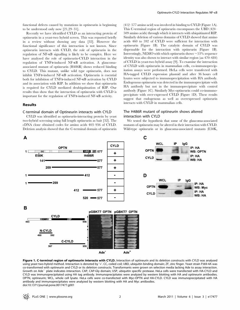
C-terminal domain of Optineurin interacts with CYLDCYLD was identified as optineurin-interacting protein by yeast
two-hybrid screening using full length optineurin as bait [32]. The
cDNA clone obtained codes for amino acids 403–956 of CYLD.
Deletion analysis showed that the C-terminal domain of optineurin
(412–577 amino acid) was involved in binding to CYLD (Figure 1A).
This C-terminal region of optineurin encompasses the UBD (424–
509 amino acids) through which it interacts with ubiquitinated RIP.
Similarly deletion of various domains of CYLD showed that amino
acids 460 to 592 of CYLD were sufficient for interaction with
optineurin (Figure 1B). The catalytic domain of CYLD was
dispensable for the interaction with optineurin (Figure 1B).
Interestingly, NEMO with which optineurin shows ,53% sequence
identity was also shown to interact with similar region (aa 470–684)
of CYLD in yeast-two hybrid assay [9]. To examine the interaction
of CYLD with optineurin in mammalian cells, co-immunoprecip-
itation assays were performed. HeLa cells were transfected with
HA-tagged CYLD expression plasmid and after 36 hours cell
lysates were subjected to immunoprecipitation with HA antibody.
Endogenous optineurin was detected in the immunoprecipitate with
HA antibody but not in the immunoprecipitate with control
antibody (Figure 1C). Similarly Myc-optineurin could co-immuno-
precipitate with over-expressed CYLD (Figure 1D). These results
suggest that endogenous as well as overexpressed optineurin
interacts with CYLD in mammalian cells.
The H486R mutant of optineurin shows alteredinteraction with CYLD
We tested the hypothesis that some of the glaucoma-associated
mutants of optineurin may be altered in their interaction with CYLD.
Wild-type optineurin or its glaucoma-associated mutants (E50K,
Figure 1. C-terminal region of optineurin interacts with CYLD. Interaction of optineurin and its deletion constructs with CYLD was analysedusing yeast two-hybrid method. Interaction is denoted by ‘+’. CC, coiled coil; UBD, ubiquitin binding domain; ZF, zinc finger. Yeast strain PJ69-4A wasco-transformed with optineurin and CYLD or its deletion constructs. Transformants were grown on selection media lacking Ade to assay interaction.Growth on Ade2 plate indicates interaction. CAP, CAP-Gly domain; USP, ubiquitin specific protease. HeLa cells were transfected with HA-CYLD andCYLD was immunoprecipitated using HA tag antbody. Immunoprecipitates were analyzed by western blotting with HA and optineurin antibodies.OPTN, optineurin; WCL, whole cell lysate. HeLa cells were co-transfected with Myc-OPTN and HA-CYLD. CYLD was immunoprecipitated with HAantibody and immunoprecipitates were analyzed by western blotting with HA and Myc antibodies.doi:10.1371/journal.pone.0017477.g001
Optineurin-CYLD Interaction Regulates NF-kB
PLoS ONE | www.plosone.org 2 March 2011 | Volume 6 | Issue 3 | e17477
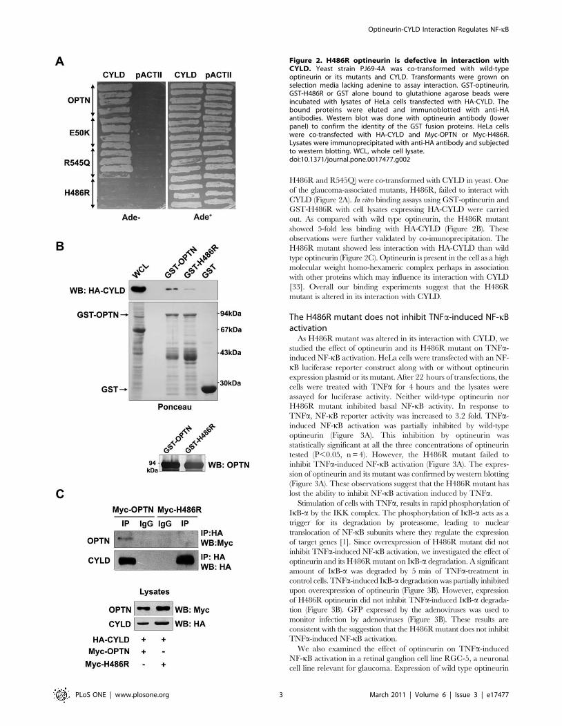
H486R and R545Q) were co-transformed with CYLD in yeast. One
of the glaucoma-associated mutants, H486R, failed to interact with
CYLD (Figure 2A). In vitro binding assays using GST-optineurin and
GST-H486R with cell lysates expressing HA-CYLD were carried
out. As compared with wild type optineurin, the H486R mutant
showed 5-fold less binding with HA-CYLD (Figure 2B). These
observations were further validated by co-imunoprecipitation. The
H486R mutant showed less interaction with HA-CYLD than wild
type optineurin (Figure 2C). Optineurin is present in the cell as a high
molecular weight homo-hexameric complex perhaps in association
with other proteins which may influence its interaction with CYLD
[33]. Overall our binding experiments suggest that the H486R
mutant is altered in its interaction with CYLD.
The H486R mutant does not inhibit TNFa-induced NF-kBactivation
As H486R mutant was altered in its interaction with CYLD, we
studied the effect of optineurin and its H486R mutant on TNFa-
induced NF-kB activation. HeLa cells were transfected with an NF-
kB luciferase reporter construct along with or without optineurin
expression plasmid or its mutant. After 22 hours of transfections, the
cells were treated with TNFa for 4 hours and the lysates were
assayed for luciferase activity. Neither wild-type optineurin nor
H486R mutant inhibited basal NF-kB activity. In response to
TNFa, NF-kB reporter activity was increased to 3.2 fold. TNFa-
induced NF-kB activation was partially inhibited by wild-type
optineurin (Figure 3A). This inhibition by optineurin was
statistically significant at all the three concentrations of optineurin
tested (P,0.05, n = 4). However, the H486R mutant failed to
inhibit TNFa-induced NF-kB activation (Figure 3A). The expres-
sion of optineurin and its mutant was confirmed by western blotting
(Figure 3A). These observations suggest that the H486R mutant has
lost the ability to inhibit NF-kB activation induced by TNFa.
Stimulation of cells with TNFa, results in rapid phosphorylation of
IkB-a by the IKK complex. The phosphorylation of IkB-a acts as a
trigger for its degradation by proteasome, leading to nuclear
translocation of NF-kB subunits where they regulate the expression
of target genes [1]. Since overexpression of H486R mutant did not
inhibit TNFa-induced NF-kB activation, we investigated the effect of
optineurin and its H486R mutant on IkB-a degradation. A significant
amount of IkB-a was degraded by 5 min of TNFa-treatment in
control cells. TNFa-induced IkB-a degradation was partially inhibited
upon overexpression of optineurin (Figure 3B). However, expression
of H486R optineurin did not inhibit TNFa-induced IkB-a degrada-
tion (Figure 3B). GFP expressed by the adenoviruses was used to
monitor infection by adenoviruses (Figure 3B). These results are
consistent with the suggestion that the H486R mutant does not inhibit
TNFa-induced NF-kB activation.
We also examined the effect of optineurin on TNFa-induced
NF-kB activation in a retinal ganglion cell line RGC-5, a neuronal
cell line relevant for glaucoma. Expression of wild type optineurin
Figure 2. H486R optineurin is defective in interaction withCYLD. Yeast strain PJ69-4A was co-transformed with wild-typeoptineurin or its mutants and CYLD. Transformants were grown onselection media lacking adenine to assay interaction. GST-optineurin,GST-H486R or GST alone bound to glutathione agarose beads wereincubated with lysates of HeLa cells transfected with HA-CYLD. Thebound proteins were eluted and immunoblotted with anti-HAantibodies. Western blot was done with optineurin antibody (lowerpanel) to confirm the identity of the GST fusion proteins. HeLa cellswere co-transfected with HA-CYLD and Myc-OPTN or Myc-H486R.Lysates were immunoprecipitated with anti-HA antibody and subjectedto western blotting. WCL, whole cell lysate.doi:10.1371/journal.pone.0017477.g002
Optineurin-CYLD Interaction Regulates NF-kB
PLoS ONE | www.plosone.org 3 March 2011 | Volume 6 | Issue 3 | e17477
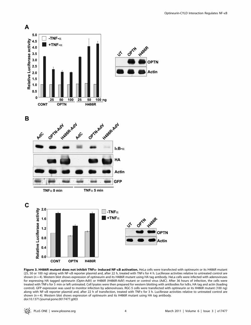
Figure 3. H486R mutant does not inhibit TNFa- induced NF-kB activation. HeLa cells were transfected with optineurin or its H486R mutant(25, 50 or 100 ng) along with NF-kB reporter plasmid and, after 22 h, treated with TNFa for 4 h. Luciferase activities relative to untreated control areshown (n = 4). Western blot shows expression of optineurin and its H486R mutant using HA tag antibody. HeLa cells were infected with adenovirusesfor expressing HA tagged optineurin (Optn-AdV) or H486R (H486R-AdV) mutant or control virus (AdC). After 36 hours of infection, the cells weretreated with TNFa for 5 min or left untreated. Cell lysates were then prepared for western blotting with antibodies for IkBa, HA tag and actin (loadingcontrol). GFP expression was used to monitor infection by adenoviruses. RGC-5 cells were transfected with optineurin or its H486R mutant (100 ng)along with NF-kB reporter plasmid and, after 22 h of transfection, treated with TNFa for 3 h. Luciferase activities relative to untreated control areshown (n = 4). Western blot shows expression of optineurin and its H486R mutant using HA tag antibody.doi:10.1371/journal.pone.0017477.g003
Optineurin-CYLD Interaction Regulates NF-kB
PLoS ONE | www.plosone.org 4 March 2011 | Volume 6 | Issue 3 | e17477
in RGC-5 cell resulted in significant inhibition (P,0.05, n = 4) of
TNFa-induced NF-kB activity whereas the H486R mutant
showed no significant inhibition (Figure 3C).
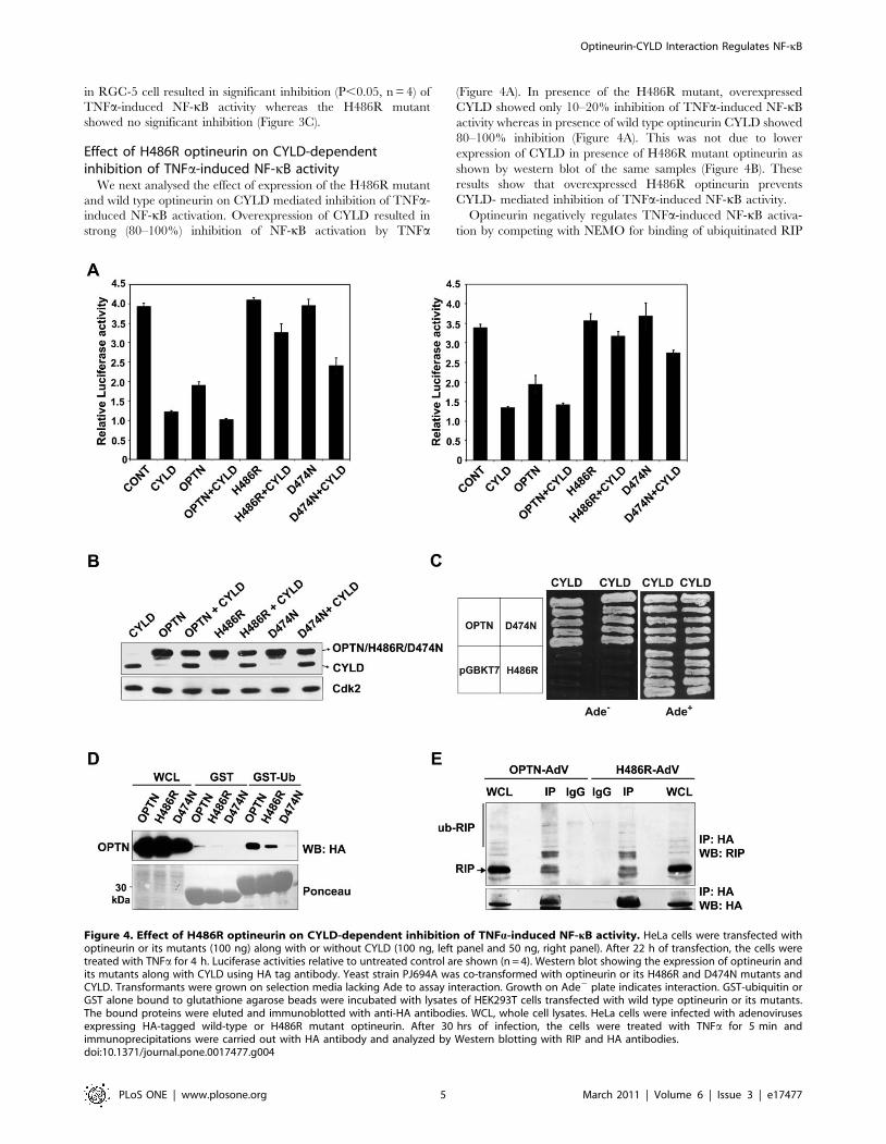
Effect of H486R optineurin on CYLD-dependentinhibition of TNFa-induced NF-kB activity
We next analysed the effect of expression of the H486R mutant
and wild type optineurin on CYLD mediated inhibition of TNFa-
induced NF-kB activation. Overexpression of CYLD resulted in
strong (80–100%) inhibition of NF-kB activation by TNFa
(Figure 4A). In presence of the H486R mutant, overexpressed
CYLD showed only 10–20% inhibition of TNFa-induced NF-kB
activity whereas in presence of wild type optineurin CYLD showed
80–100% inhibition (Figure 4A). This was not due to lower
expression of CYLD in presence of H486R mutant optineurin as
shown by western blot of the same samples (Figure 4B). These
results show that overexpressed H486R optineurin prevents
CYLD- mediated inhibition of TNFa-induced NF-kB activity.
Optineurin negatively regulates TNFa-induced NF-kB activa-
tion by competing with NEMO for binding of ubiquitinated RIP
Figure 4. Effect of H486R optineurin on CYLD-dependent inhibition of TNFa-induced NF-kB activity. HeLa cells were transfected withoptineurin or its mutants (100 ng) along with or without CYLD (100 ng, left panel and 50 ng, right panel). After 22 h of transfection, the cells weretreated with TNFa for 4 h. Luciferase activities relative to untreated control are shown (n = 4). Western blot showing the expression of optineurin andits mutants along with CYLD using HA tag antibody. Yeast strain PJ694A was co-transformed with optineurin or its H486R and D474N mutants andCYLD. Transformants were grown on selection media lacking Ade to assay interaction. Growth on Ade2 plate indicates interaction. GST-ubiquitin orGST alone bound to glutathione agarose beads were incubated with lysates of HEK293T cells transfected with wild type optineurin or its mutants.The bound proteins were eluted and immunoblotted with anti-HA antibodies. WCL, whole cell lysates. HeLa cells were infected with adenovirusesexpressing HA-tagged wild-type or H486R mutant optineurin. After 30 hrs of infection, the cells were treated with TNFa for 5 min andimmunoprecipitations were carried out with HA antibody and analyzed by Western blotting with RIP and HA antibodies.doi:10.1371/journal.pone.0017477.g004
Optineurin-CYLD Interaction Regulates NF-kB
PLoS ONE | www.plosone.org 5 March 2011 | Volume 6 | Issue 3 | e17477
through its UBD. Inactivation of UBD of optineurin by a point
mutation (D474N) results in loss of inhibition of NF-kB activation
[16]. The D474N mutant, however, was not defective in its
interaction with CYLD as revealed by yeast-two hybrid assays
(Figure 4C). Since the H486R mutation is also present in the
UBD, it is possible that this mutant may be defective in ubiquitin
binding. Compared to wild-type optineurin, the H486R mutant
showed reduced binding to GST-ubiquitin whereas the D474N
mutant did not show any binding (Figure 4D). The ability of
H486R mutant optineurin to bind ubiquitinated RIP was tested by
co-immunoprecipitation. As compared to wild-type optineurin, the
H486R mutant showed some reduction in binding to ubiquiti-
nated RIP in TNFa- treated cells (Figure 4E). These results suggest
that the loss of interaction with CYLD is the major reason for the
reduced inhibition of TNFa-induced NF-kB activation by the
H486R mutant. However, the reduced binding to ubiquitin may
also contribute towards its effect on CYLD-dependent inhibition
of NF-kB activation. In presence of the D474N mutant,
overexpressed CYLD showed partial inhibition of TNFa-induced
NF-kB activation (Figure 4A). Thus as compared to the H486R
mutant, the D474N mutant is less effective in preventing CYLD
mediated inhibition of NF-kB activation. This may be due to the
fact that while H486R is defective in binding to CYLD and also
shows reduced binding to ubiquitin, the D474N is defective in only
ubiquitin binding. Since D474N mutant does not bind ubiquiti-
nated RIP [16] this suggests that the interaction of UBD of
optineurin with ubiquitinated proteins (possibly RIP) also plays an
essential role in CYLD mediated inhibition of TNFa- induced NF-
kB activation.
Optineurin is essential for CYLD mediated inhibition ofNF-kB activation
We next examined the requirement of optineurin in CYLD
mediated abrogation of TNFa-induced NF-kB activation. HeLa
cells were infected with adenoviruses expressing shRNA against
optineurin or with control adenoviruses. After 48 h of infection,
these cells were transfected with CYLD along with NF-kB
luciferase reporter and treated with TNFa for 4 h after 22 h of
transfection. As reported earlier, optineurin knockdown enhanced
both basal as well as TNFa-induced NF-kB activity [16,18].
Overexpression of CYLD resulted in strong inhibition of both
basal and TNFa-induced NF-kB activity in control cells
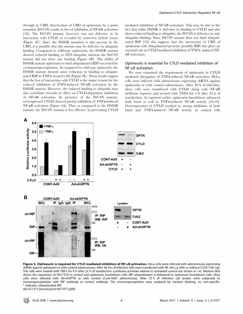
Figure 5. Optineurin is required for CYLD mediated inhibition of NF-kB activation. HeLa cells were infected with adenoviruses expressingshRNA against optineurin or with control adenoviruses. After 48 hrs of infection cells were transfected with NF-kB-Luc with or without CYLD (100 ng).The cells were treated with TNFa for 4 h after 22 h of transfection. Luciferase activities relative to untreated control are shown (n = 4). Western blotshows the expression of HA-CYLD in control and optineurin knockdown cells. RIP ubiquitination is enhanced in optineurin knockdown cells. HeLacells were infected with Ad-shOPTN or with control (Cont-AdV) adenoviruses. After 72 h of infection cell lysates were subjected toimmunoprecipitation with RIP antibody or control antibody. The immunoprecipitates were analyzed by western blotting. ns, non-specific.* indicates ubiquitinated RIP.doi:10.1371/journal.pone.0017477.g005
Optineurin-CYLD Interaction Regulates NF-kB
PLoS ONE | www.plosone.org 6 March 2011 | Volume 6 | Issue 3 | e17477
(Figure 5A). However, upon knockdown of optineurin, overex-
pression of CYLD resulted in only a marginal inhibition of basal
and TNFa-induced NF-kB activity (Figure 5A). These results show
that optineurin is essential for CYLD mediated inhibition of basal
and TNFa- induced NF-kB activity.
The mechanism by which knockdown of optineurin causes
increase in basal NF-kB activity is not clear. The finding that the
increased NF-kB activity is not inhibited by over-expressed CYLD
prompted us to investigate whether loss of optineurin affects levels
of ubiquitinated RIP. RIP was immunoprecipitated from opti-
neurin knockdown and control cells in presence of N-ethylmalei-
mide (to prevent deubiquitination) and the immunoprecipitates
were analysed by western blot using RIP and ubiquitin antibody.
The level of ubiquitinated RIP was increased in optineurin
knockdown cells (Figure 5B). These results suggest that in
optineurin knockdown cells enhanced basal NF-kB activity is
likely to be due to increased level of ubiquitinated RIP.
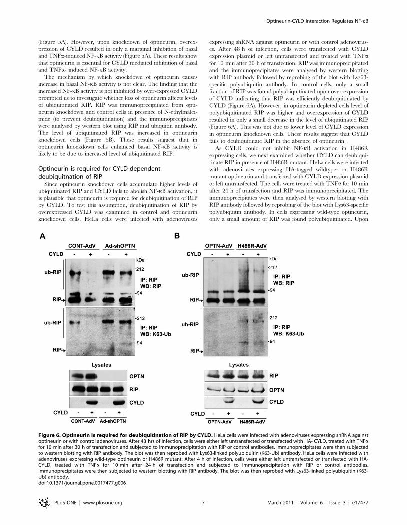
Optineurin is required for CYLD-dependentdeubiquitnation of RIP
Since optineurin knockdown cells accumulate higher levels of
ubiquitinated RIP and CYLD fails to abolish NF-kB activation, it
is plausible that optineurin is required for deubiquitination of RIP
by CYLD. To test this assumption, deubiquitination of RIP by
overexpressed CYLD was examined in control and optineurin
knockdown cells. HeLa cells were infected with adenoviruses
expressing shRNA against optineurin or with control adenovirus-
es. After 48 h of infection, cells were transfected with CYLD
expression plasmid or left untransfected and treated with TNFafor 10 min after 30 h of transfection. RIP was immunoprecipitated
and the immunoprecipitates were analysed by western blotting
with RIP antibody followed by reprobing of the blot with Lys63-
specific polyubiquitin antibody. In control cells, only a small
fraction of RIP was found polyubiquitinated upon over-expression
of CYLD indicating that RIP was efficiently deubiquitinated by
CYLD (Figure 6A). However, in optineurin depleted cells level of
polyubiquitinated RIP was higher and overexpression of CYLD
resulted in only a small decrease in the level of ubiquitinated RIP
(Figure 6A). This was not due to lower level of CYLD expression
in optineurin knockdown cells. These results suggest that CYLD
fails to deubiquitinate RIP in the absence of optineurin.
As CYLD could not inhibit NF-kB activation in H486R
expressing cells, we next examined whether CYLD can deubiqui-
tinate RIP in presence of H486R mutant. HeLa cells were infected
with adenoviruses expressing HA-tagged wildtype- or H486R
mutant optineurin and transfected with CYLD expression plasmid
or left untransfected. The cells were treated with TNFa for 10 min
after 24 h of transfection and RIP was immunoprecipitated. The
immunoprecipitates were then analysed by western blotting with
RIP antibody followed by reprobing of the blot with Lys63-specific
polyubiquitin antibody. In cells expressing wild-type optineurin,
only a small amount of RIP was found polyubiquitinated. Upon
Figure 6. Optineurin is required for deubiquitination of RIP by CYLD. HeLa cells were infected with adenoviruses expressing shRNA againstoptineurin or with control adenoviruses. After 48 hrs of infection, cells were either left untransfected or transfected with HA- CYLD, treated with TNFafor 10 min after 30 h of transfection and subjected to immunoprecipitation with RIP or control antibodies. Immunoprecipitates were then subjectedto western blotting with RIP antibody. The blot was then reprobed with Lys63-linked polyubiquitin (K63-Ub) antibody. HeLa cells were infected withadenoviruses expressing wild-type optineurin or H486R mutant. After 4 h of infection, cells were either left untransfected or transfected with HA-CYLD, treated with TNFa for 10 min after 24 h of transfection and subjected to immunoprecipitation with RIP or control antibodies.Immunoprecipitates were then subjected to western blotting with RIP antibody. The blot was then reprobed with Lys63-linked polyubiquitin (K63-Ub) antibody.doi:10.1371/journal.pone.0017477.g006
Optineurin-CYLD Interaction Regulates NF-kB
PLoS ONE | www.plosone.org 7 March 2011 | Volume 6 | Issue 3 | e17477
co-expression of CYLD in these cells, we were not able to detect
ubiquitinated RIP (Figure 6B). In cells expressing the H486R
mutant, level of polyubiquitinated RIP was higher compared to
wild-type optineurin expressing cells and co-expression of CYLD
resulted in very small decrease in the level of ubiquitinated RIP, as
detected by Lys63-specific polyubiquitin antibody (Figure 6B).
This was not due to lower expression of either the H486R mutant
or CYLD (Figure 6B). These observations suggest that the H486R
mutant prevents CYLD mediated deubiquitination of RIP.
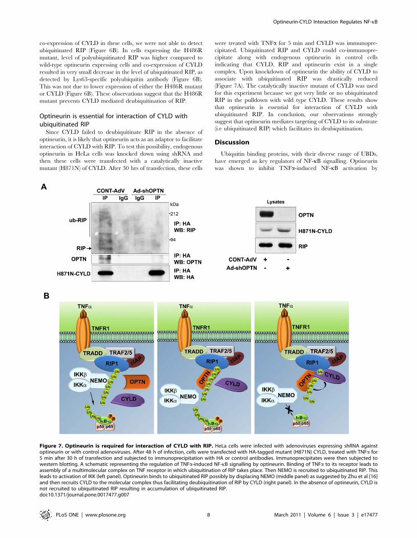
Optineurin is essential for interaction of CYLD withubiquitinated RIP
Since CYLD failed to deubiquitinate RIP in the absence of
optineurin, it is likely that optineurin acts as an adaptor to facilitate
interaction of CYLD with RIP. To test this possibility, endogenous
optineurin in HeLa cells was knocked down using shRNA and
then these cells were transfected with a catalytically inactive
mutant (H871N) of CYLD. After 30 hrs of transfection, these cells
were treated with TNFa for 5 min and CYLD was immunopre-
cipitated. Ubiquitinated RIP and CYLD could co-immunopre-
cipitate along with endogenous optineurin in control cells
indicating that CYLD, RIP and optineurin exist in a single
complex. Upon knockdown of optineurin the ability of CYLD to
associate with ubiquitinated RIP was drastically reduced
(Figure 7A). The catalytically inactive mutant of CYLD was used
for this experiment because we got very little or no ubiquitinated
RIP in the pulldown with wild type CYLD. These results show
that optineurin is essential for interaction of CYLD with
ubiquitinated RIP. In conclusion, our observations strongly
suggest that optineurin mediates targeting of CYLD to its substrate
(i.e ubiquitinated RIP) which facilitates its deubiquitination.
Discussion
Ubiquitin binding proteins, with their diverse range of UBDs,
have emerged as key regulators of NF-kB signalling. Optineurin
was shown to inhibit TNFa-induced NF-kB activation by
Figure 7. Optineurin is required for interaction of CYLD with RIP. HeLa cells were infected with adenoviruses expressing shRNA againstoptineurin or with control adenoviruses. After 48 h of infection, cells were transfected with HA-tagged mutant (H871N) CYLD, treated with TNFa for5 min after 30 h of transfection and subjected to immunoprecipitation with HA or control antibodies. Immunoprecipitates were then subjected towestern blotting. A schematic representing the regulation of TNFa-induced NF-kB signalling by optineurin. Binding of TNFa to its receptor leads toassembly of a multimolecular complex on TNF receptor in which ubiquitination of RIP takes place. Then NEMO is recruited to ubiquitinated RIP. Thisleads to activation of IKK (left panel). Optineurin binds to ubiquitinated RIP possibly by displacing NEMO (middle panel) as suggested by Zhu et al [16]and then recruits CYLD to the molecular complex thus facilitating deubiquitination of RIP by CYLD (right panel). In the absence of optineurin, CYLD isnot recruited to ubiquitinated RIP resulting in accumulation of ubiquitinated RIP.doi:10.1371/journal.pone.0017477.g007
Optineurin-CYLD Interaction Regulates NF-kB
PLoS ONE | www.plosone.org 8 March 2011 | Volume 6 | Issue 3 | e17477
competing with NEMO for the binding of ubiquitinated RIP [16].
However, our results suggest that the regulation of NF-kB
activation by optineurin is more complex and binding of
ubiquitinated RIP to UBD of optineurin is only one of the steps
in this complex regulation. Although ubiquitinated RIP is known
to be a substrate of CYLD, the mechanism by which CYLD is
recruited to RIP to deubiquitinate it, is not clear [14]. The results
presented in this manuscript suggest that optineurin mediates
interaction of deubiquitinase CYLD with polyubiquitinated RIP
and this interaction is essential for deubiquitination of RIP by
CYLD. Thus an important function of optineurin in the regulation
of NF-kB signalling is to act as an adaptor protein bringing CYLD
and its substrate RIP together to facilitate deubiquitination of
ubiquitinated RIP by CYLD.
CYLD targets multiple players of the NF-kB signalling pathway.
How CYLD recognises and is targeted to a specific substrate like
RIP is not well understood. CYLD interacts directly with some of
its substrates like TRAF2 and NEMO [7–9]. Emerging evidence
suggests that CYLD might indirectly associate with some of its
substrates through intermediary adaptor proteins. It has been
shown that p62/sequestosome1 binds to TRAF6 through its UBD
and recruits CYLD to TRAF6 to regulate its ubiquitination
[34,35]. An adaptor protein should be able to specifically bind to
substrates polyubiquitinated with Lys63-linked ubiquitin chains
since CYLD specifically deconjugates Lys63-linked polyubiqitin
chains. In addition, such an adaptor protein should also interact
with CYLD. Since optineurin interacts with Lys63-linked poly-
ubiquitin chains (as in ubiquitinated RIP) through its UBD and
also with CYLD, it is ideally suited to act as an adaptor for CYLD
to recognise its substrates. The ABIN proteins with their
homologous UBD similarly act as adaptors for the recruitment
of A20 to its targets like NEMO [36]. TNFa stimulus triggers
assembly of Lys63-linked polyubiquitin chains on RIP which
initially binds to NEMO to activate IKK complex. Optineurin
binds to polyubiquitinated RIP through its UBD and recruits
CYLD; this may prevent association of NEMO with RIP
(Figure 7B). Close proximity of binding sites of both CYLD and
RIP on optineurin provides support for the adaptor function of
optineurin in facilitating interaction of RIP and CYLD. These
assumptions are strengthened by our observations which show that
both inhibition of NF-kB activation by CYLD and association of
CYLD with RIP are dependent on optineurin. Consistent with its
role as an adaptor facilitating the association of a deubiquitinase
with its substrate, depletion of optineurin impaired deubiquitina-
tion of RIP by CYLD. Whether optineurin plays a role in
recruiting CYLD to its other targets is yet to be investigated.
Knockdown of endogenous optineurin increases basal level of
NF-kB activity in unstimulated cells [16,18] but the mechanism of
this regulation was not clear. CYLD deficiency results in the
accumulation of its ubiquitinated targets like RIP due to
constitutive ubiquitination [10,14]. Hence it has been suggested
that CYLD is a constitutively active deubiquitinase that prevents
spontaneous ubiquitination of its targets like RIP, thus maintaining
low basal NF-kB activity. Our results show that optineurin
depleted cells accumulate higher levels of ubiquitinated RIP. This
elevated level of RIP ubiquitination is likely to be due to inability
of CYLD to associate with and deubiquitinate RIP in the absence
of optineurin leading to enhanced NF-kB activation (Figure 7B).
The elevated level of ubiquitinated RIP in optineurin depleted
cells differs/is not consistent with the model of Zhu et al [16].
However, these observations are consistent with our proposed
model of optineurin as an adaptor protein, as depicted in
Figure 7B. The main feature of this model is that optineurin
mediates binding of CYLD to ubiquitinated RIP. This model
incorporates the findings of Zhu et al [16] that optineurin binds to
ubiquitinated RIP by displacing NEMO. In fact our work/model
can also be viewed as an extension of the work of Zhu et al [16].
Some of the data presented in this study are different from those
published by Zhu et al [16]. There are quantitative differences in
the inhibition of NF-kB activation by optineurin. The major
difference is that in our study we did not see inhibition of basal
NF-kB activity by overexpressed optineurin in HeLa cells. This
observation has been reported earlier by us [18]. Zhu et al have
used HaLa S3 cells and this difference could be due to differences
in the cell lines used. However, we and Zhu et al have shown that
knockdown of optineurin increases basal NF-kB activity in various
cells though the mechanism by which this occurs was not known.
We have now shown that this increase in basal activity is possibly
due to accumulation of ubiquitinated RIP in optineurin knock-
down cells.
Though mutations in optineurin have been associated with
glaucoma and more recently with amyotrophic lateral sclerosis
[28,29,37,38], the functional defects caused by these mutations are
not completely understood. The H486R mutation is associated
with certain types of glaucomas [37,38]. Our observations with
H486R mutant show that this mutant is defective in its interaction
with CYLD. In accordance with the role of optineurin in CYLD
mediated inhibition of NF-kB activation, this mutant is unable to
abrogate NF-kB activation. As the H486R mutation is located
within the UBD, this mutant showed some loss in ubiquitin
binding and in binding to ubiquitinated RIP. Thus the inability of
the H486R mutant to inhibit NF-kB activation is mainly due to its
altered interaction with CYLD although reduced binding to
ubiquitin may also contribute to some extent. This assumption is
strengthened by our results which show that overexpressed CYLD
fails to deubiquitinate RIP and inhibit TNFa-induced NF-kB
activation in presence of H486R mutant.
How does the H486R mutant cause glaucoma? Glaucoma is a
neurodegenerative disease in which loss of vision occurs due to
death of retinal ganglion cells in the optic nerve head. Several
mechanisms are implicated in retinal ganglion cell death in
glaucoma including direct effect on retinal ganglion cells,
activation of glial cells to produce cytotoxic molecules like TNFa,
alteration in trabecular meshwork, autoimmunity etc [39,40].
Since overexpression of H486R mutant does not induce retinal
ganglion cell death unlike the E50K mutant [41], it is plausible
that indirect effects on other cells might contribute to H486R
induced glaucomatous neurodegeneration. NF-kB is activated in
glaucomatous trabecular meshwork cells and is also associated
with autoimmune responses [42–44]. Our results showing
defective NF-kB regulation by H486R mutant optineurin thus
provide a basis for exploring its role in indirect mechanisms of
glaucomatous degeneration.
In conclusion, our studies show that optineurin acts as an
adaptor protein to bring together CYLD and its substrate RIP.
The interaction of CYLD with optineurin is essential for negative
regulation of TNFa-induced NF-kB activation. A defect in this
interaction, as observed in the glaucoma associated H486R
mutant results in deregulation of NF-kB. These findings may
have relevance to the pathogenesis of glaucoma, directly or
indirectly, as deregulation of NF-kB activity has been implicated in
glaucomas.
Materials and Methods
Cell culture and transfectionsCell lines were maintained at 37uC in a CO2 incubator in
Dulbecco’s modified Eagle’s medium supplemented with 10%
Optineurin-CYLD Interaction Regulates NF-kB
PLoS ONE | www.plosone.org 9 March 2011 | Volume 6 | Issue 3 | e17477

foetal bovine serum. Transfections were carried out using
Lipofectamine Plus reagent (Invitrogen, San Diego, CA, USA)
according to the manufacturer’s instructions. All the plasmids for
transfection were prepared by using Qiagen columns (Hilden,
Germany). Human TNFa (Sigma, St. Louis, MO, USA or
Calbiochem) was added wherever indicated at a final concentra-
tion of 10–20 ng/ml.
Expression vectors and antibodiesHuman optineurin expression plasmid and its mutants cloned in
various vectors have been described by us previously [21,41].
Deletion constructs of optineurin were generated by PCR. HA-
tagged CYLD and GFP-CYLD were generated by cloning CYLD
c-DNA obtained in yeast-two hybrid screen in pCDNA3.1-HA or
pEGFP-C2, respectively. The catalytically inactive H871N mutant
of CYLD was generated by site directed mutagenesis. Rabbit
polyclonal optineurin antibody (ab23666) was from Abcam, mouse
monoclonal RIP antibody (Cat no 610459) was from BD
Biosciences and mouse monoclonal Lys63-sepcific anti-ubiquitin
was from Miilipore (Cat no 05-1313). IkB-a (sc-371), actin and
Cdk2 antibodies were from Santa Cruz Biotechnology (Santa
Cruz, CA, USA).
Generation of adenoviral vectorsThe adenoviral shRNA expression vectors (Ad-shOPTN1 & 2)
for targeting human optineurin, were generated by using pAdEasy
system and have been described by us [18,21]. The shRNAs
generated by these adenoviruses target two different regions of
optineurin mRNA [18]. As a control, an adenovirus expressing
shRNA of unrelated sequence of the same length was used.
Adenoviral vectors for expressing optineurin and its mutants with
HA tag were prepared as described [21,45]. These adenoviruses
also express GFP for monitoring of infection. For overexpression
experiments, an adenovirus expressing GFP was used as a control.
Yeast two-hybrid assayYeast two-hybrid assay was performed as described previously
[46]. Briefly, yeast strain PJ69-4A was co-transformed with
required plasmids by lithium-acetate method. Optineurin, its
mutants and deletion constructs were cloned in pGBKT7 (GAL4
DNA binding domain) while CYLD was cloned in pACTII (GAL4
activation domain) (Clonetech). The transformants were selected
by growth in minimal media (Trp2, Leu2). Yeast colonies
obtained on Trp2, Leu2 plates were patched onto selection
plants (Trp2, Leu2 Ade2) and X-Gal (Trp2, Leu2, X-Gal+) plates
to assay activation of reporter genes and hence interaction.
Growth on Ade2 plate or colour on X-Gal plate indicated
interaction.
Immunoprecipitation and GST pull downImmunoprecipitations were carried out essentially as described
[21,46]. Briefly, cells were lysed in lysis buffer (25 mM Tris
pH 7.4, 1% Triton X-100, 150 mM NaCl, 0.1% BSA, 1 mM
PMSF, 10 mM N-ethylmaleimide, 2 mM sodium vanadate,
25 mM NaF and protease inhibitor cocktail (Roche)) and
immunoprecipitations were carried out with 2 mg of appropriate
antibodies overnight at 4uC (2.5 h at 4uC for RIP). The
immunoprecipitated proteins were washed 3 times with lysis
buffer, eluted by boiling in SDS sample buffer and resolved in 8–
10% SDS-PAGE. The proteins were transferred to nitrocellulose
membrane for western blot analysis as described [18].
For GST pull down assays, GST and GST-fusion proteins were
expressed in E. coli and were conjugated to sepharose beads as
described [21,47]. These beads were incubated for 6–8 hours with
lysates of HeLa cells transiently transfected with indicated
plasmids. Bound proteins were eluted by boiling in SDS sample
buffer and subjected to immunoblotting.
Reporter assaysNF-kB reporter assays were performed as described previously
by using a reporter plasmid NF-kB-Luc containing five tandem
NF-kB binding sites upstream of a luciferase gene [18]. HeLa cells
grown in 24 well dishes were transfected with 25 ng of the NF-kB-
luc plasmid, 50 ng of b-galactosidase expression plasmid along
with the required amount of other plasmids [18]. Relative
luciferase activities were calculated after normalizing with b-
galactosidase enzyme activities. TNFa was used at a concentration
of 20 ng/ml.
Statistical analysisGraphs represent average 6 SD values. Statistical differences
were calculated using student’s T-test. When significant differences
were observed, P values for pair wise comparisons were calculated
by using two-tailed T-test. P value less than 0.05 was considered
significant.
Acknowledgments
We thank Miss Asha Kumari for technical assistance.
Author Contributions
Conceived and designed the experiments: AN GS. Performed the
experiments: AN MB. Analyzed the data: AN GS. Wrote the paper: AN
GS.
References
1. Hayden MS, Ghosh S (2004) Signaling to NF-kB. Genes Dev 18: 2195–2224.
2. Courtois G, Gilmore TD (2006) Mutations in the NF-kB signaling pathway:
implications for human disease. Oncogene 25: 6831–6843.
3. Skaug B, Jiang X, Chen ZJ (2009) The role of ubiquitin in NF-kappaB
regulatory pathways. Annu Rev Biochem 78: 769–796.
4. Ea CK, Deng L, Xia ZP, Pineda G, Chen ZJ (2006) Activation of IKK by
TNFalpha requires site-specific ubiquitination of RIP1 and polyubiquitin
binding by NEMO. Mol Cell 22: 245–257.
5. Li H, Kobayashi M, Blonska M, You Y, Lin X (2006) Ubiquitination of RIP is
required for tumor necrosis factor alpha-induced NF-kB activation. J Biol Chem
281: 13636–13643.
6. Wu C, Conze DB, Li T, Srinivasula SM, Ashwell JD (2006) Sensing of Lys 63-
linked polyubiquitination by NEMO is a key event in NF-kB activation Nat. Cell
Biol 8: 398–406.
7. Trompouki E, Hatzivassiliou E, Tsichritzis T, Farmer H, Ashworth A, et al.
(2003) CYLD is a deubiquitinating enzyme that negatively regulates NF-kB
activation by TNFR family members. Nature 424: 793–796.
8. Brummelkamp TR, Nijman SMB, Dirac AMG, Bernards R (2003) Loss of the
cylindromatosis tumour suppressor inhibits apoptosis by activating NF-kB.
Nature 424: 797–801.
9. Kovalenko A, Chable-Bessia C, Cantarella G, Israel A, Wallach D, et al. (2003)
The tumour suppressor CYLD negatively regulates NF-kB signalling by
deubiquitination. Nature 424: 801–805.
10. Sun SC (2010) CYLD: a tumor suppressor deubiquitinase regulating NF-kappaB
activation and diverse biological processes. Cell Death Differ 17: 25–34.
11. Wertz IE, O’Rourke KM, Zhou H, Eby M, Aravind L, et al. (2004) De-
ubiquitination and ubiquitin ligase domains of A20 downregulate NF-kB
signalling. Nature 430: 694–699.
12. Evans PC, Ovaa H, Hamon M, Kilshaw PJ, Hamm S, et al. (2004) Zinc-finger
protein A20, a regulator of inflammation and cell survival, has de-ubiquitinating
activity. Biochem J 378: 727–734.
13. Bignell GR, Warren W, Seal S, Takahashi M, Rapley E, et al. (2000)
Identification of the familial cylindromatosis tumour-suppressor gene. Nat Genet
25: 160–165.
Optineurin-CYLD Interaction Regulates NF-kB
PLoS ONE | www.plosone.org 10 March 2011 | Volume 6 | Issue 3 | e17477
14. Wright A, Reiley WW, Chang M, Jin W, Lee AJ, et al. (2007) Regulation of
early wave of germ cell apoptosis and spermatogenesis by deubiquitinatingenzyme CYLD. Dev Cell 13: 705–716.
15. Massoumi R (2010) Ubiquitin chain cleavage: CYLD at work. Trends Biochem
Sci 35: 392–399.16. Zhu G, Wu CJ, Zhao Y, Ashwell JD (2007) Optineurin negatively regulates
TNFa-induced NF-kB activation by competing with NEMO for ubiquitinatedRIP. Curr Biol 17: 1438–1443.
17. Mrowka R, Bluthgen N, Fahling M (2008) Seed-based systematic discovery of
specific transcription factor target genes. FEBS Journal 275: 3178–3192.18. Sudhakar C, Nagabhushana A, Jain N, Swarup G (2009) NF-kB Mediates
Tumor Necrosis Factor a-Induced Expression of Optineurin, a NegativeRegulator of NF-kB. PLoS ONE 4: e5114.
19. Sahlender DA, Roberts RC, Arden SD, Spudich G, Taylor MJ, et al. (2005)Optineurin links myosin VI to the Golgi complex and is involved in Golgi
organization and exocytosis. J Cell Biol 169: 285–295.
20. del Toro D, Alberch J, Lazaro-Dieguez F, Martın-Ibanez R, Xifro X, et al.(2009) Mutant huntingtin impairs post-Golgi trafficking to lysosomes by
delocalizing optineurin/Rab8 complex from the Golgi apparatus. Mol BiolCell 20: 1478–1492.
21. Nagabhushana A, Chalasani ML, Jain N, Radha V, Rangaraj N, et al. (2010)
Regulation of endocytic trafficking of transferrin receptor by optineurin and itsimpairment by a glaucoma-associated mutant. BMC Cell Biol 19: 11:4.
22. Anborgh PH, Godin C, Pampillo M, Dhami GK, Dale LB, et al. (2005)Inhibition of metabotropic glutamate receptor signaling by the Huntingtin-
binding protein optineurin. J Biol Chem 280: 4840–4848.23. Journo C, Filipe J, About F, Chevalier SA, Afonso PV, et al. (2009) NRP/
Optineurin Cooperates with TAX1BP1 to potentiate the activation of NF-
kappaB by human T-lymphotropic virus type 1 tax protein. PLoS Pathog 5:e1000521.
24. Mankouri J, Fragkoudis R, Richards KH, Wetherill LF, Harris M, et al. (2010)Optineurin negatively regulates the induction of IFN beta in response to RNA
virus infection. PLoS Pathog 6: e1000778.
25. Park B, Tibudan M, Samaraweera M, Shen X, Yue BYJT (2007) Interactionbetween two glaucoma genes, optineurin and myocilin. Genes Cells 12:
969–979.26. Weisschuh N, Alavi MV, Bonin M, Wissinger B (2007) Identification of genes
that are linked with optineurin expression using a combined RNAi–microarrayapproach. Exp Eye Res 85: 450–461.
27. Laplantine E, Fontan E, Chiaravalli J, Lopez T, Lakisic G, et al. (2009) NEMO
specifically recognizes K63-linked poly-ubiquitin chains through a new bipartiteubiquitin-binding domain. EMBO J 28: 2885–2895.
28. Rezaie T, Child A, Hitchings R, Brice G, Miller L, et al. (2002) Adult-onsetprimary open-angle glaucoma caused by mutations in optineurin. Science 295:
1077–1079.
29. Maruyama H, Morino H, Ito H, Izumi Y, Kato H, et al. (2010) Mutations ofoptineurin in amyotrophic lateral sclerosis. Nature 13: 223–226.
30. Chi ZL, Akahori M, Obazawa M, Minami M, Noda T, et al. (2010)Overexpression of optineurin E50K disrupts Rab8 interaction and leads to a
progressive retinal degeneration in mice. Hum Mol Genet 19: 2606–2615.
31. Park B, Ying H, Shen X, Park JS, Qiu Y, et al. (2010) Impairment of protein
trafficking upon overexpression and mutation of optineurin. PLoS One 5:e11547.
32. Chalasani ML, Swarup G, Balasubramanian D (2009) Optineurin and its
mutants: molecules associated with some forms of glaucoma. Ophthalmic Res42: 176–184.
33. Ying H, Shen X, Park B, Yue BY (2010) Posttranslational modifications,localization, and protein interactions of optineurin, the product of a glaucoma
gene. PLoS One 5: e9168.
34. Jin W, Chang M, Paul EM, Babu G, Lee AJ, et al. (2008) Deubiquitinatingenzyme CYLD negatively regulates RANK signaling and osteoclastogenesis in
mice. J Clin Invest 118: 1858–1866.35. Wooten MW, Geetha T, Babu JR, Seibenhener ML, Peng J, et al. (2008)
Essential role of sequestosome 1/p62 in regulating accumulation of Lys63-ubiquitinated proteins. J Biol Chem 283: 6783–6789.
36. Mauro C, Pacifico F, Lavorgna A, Mellone S, Iannetti A, et al. (2006) ABIN-1
binds to NEMO/IKKgamma and co-operates with A20 in inhibiting NF-kappaB. J Biol Chem 281: 18482–18488.
37. Willoughby CE, Chann LL, Herd S, Billingsley G, Noordeh N, et al. (2004)Defining the pathogenicity of optineurin in juvenile open-angle glaucoma. Invest
Ophthalmol Vis Sci 45: 3122–3130.
38. Leung YF, Fan BJ, Lam DS, Lee WS, Tam PO, et al. (2003) Differentoptineurin mutation pattern in primary open-angle glaucoma. Invest Ophthal-
mol Vis Sci 44: 3880–3884.39. Wax MB, Tezel G (2002) Neurobiology of Glaucomatous Optic Neuropathy
Diverse Cellular Events in Neurodegeneration and Neuroprotection. MolecularNeurobiology 26: 45–55.
40. Vrabec JP, Levin LA (2007) The neurobiology of cell death in glaucoma. Eye 21:
S11–14.41. Chalasani ML, Radha V, Gupta V, Agarwal N, Balasubramanian D, et al.
(2007) A glaucoma-associated mutant of optineurin selectively induces death ofretinal ganglion cells which is inhibited by antioxidants. Invest Ophthalmol Vis
Sci 48: 1607–1614.
42. Wang N, Chintala SK, Fini ME, Schuman JS (2001) Activation of a tissue-specific stress response in the aqueous outflow pathway of the eye defines the
glaucoma disease phenotype. Nature Med 7: 304–309.43. Grus FH, Joachim SC, Wuenschig D, Rieck J, Pfeiffer N (2008) Autoimmunity
and glaucoma. J Glaucoma 17: 79–84.44. Takahashi Y, Katai N, Murata T, Taniguchi SI, Hayashi T (2007) Development
of spontaneous optic neuropathy in NF-kappaBetap50-deficient mice: require-
ment for NF-kappaBetap50 in ganglion cell survival. Neuropathol ApplNeurobiol 33: 692–705.
45. Jain N, Sudhakar C, Swarup G (2007) Tumor necrosis factor a-induced caspase-1 gene expression: Role of p73. FEBS Journal 274: 4396–4407.
46. Gupta V, Swarup G (2006) Evidence for a role of transmembrane protein p25 in
localization of protein tyrosine phosphatase TC48 to the ER. J Cell Sci 119:1703–1714.
47. Kumar Y, Radha V, Swarup G (2010) Interaction with Sug1 enables Ipafubiquitination leading to caspase 8 activation and cell death. Biochem J 427:
91–104.
Optineurin-CYLD Interaction Regulates NF-kB
PLoS ONE | www.plosone.org 11 March 2011 | Volume 6 | Issue 3 | e17477