Optical Waveguide Lightmode Spectroscopic Techniques for Investigating Membrane-Bound Ion Channel...
11
Optical Waveguide Lightmode Spectroscopic Techniques for Investigating Membrane-Bound Ion Channel Activities Inna Sze ´ ka ´ cs 1 *, No ´ ra Kasza ´s 2 , Pa ´ l Gro ´f 2 , Katalin Erde ´ lyi 3 , Istva ´ n Szendro ˝ 3 , Bala ´ zs Mihalik 4 ,A ´ gnes Pataki 4 , Ferenc A. Antoni 4 , Emilia Madara ´ sz 1 1 Institute of Experimental Medicine, Hungarian Academy of Sciences, Budapest, Hungary, 2 Semmelweis University, Department of Biophysics and Radiation Biology, Budapest, Hungary, 3 MicroVacuum Ltd., Budapest, Hungary, 4 Egis Pharmaceuticals PLC, Budapest, Hungary Abstract Optical waveguide lightmode spectroscopic (OWLS) techniques were probed for monitoring ion permeation through channels incorporated into artificial lipid environment. A novel sensor set-up was developed by depositing liposomes or cell-derived membrane fragments onto hydrophilic polytetrafluoroethylene (PTFE) membrane. The fibrous material of PTFE membrane could entrap lipoid vesicles and the water-filled pores provided environment for the hydrophilic domains of lipid-embedded proteins. The sensor surface was kept clean from the lipid holder PTFE membrane by a water- and ion- permeable polyethylene terephthalate (PET) mesh. The sensor set-up was tested with egg yolk lecithin liposomes containing gramicidin ion channels and with cell-derived membrane fragments enriched in GABA-gated anion channels. The method allowed monitoring the move of Na + and organic cations through gramicidin channels and detecting the Cl – - channel functions of the (a 5 b 2 c 2 ) GABA A receptor in the presence or absence of GABA and the competitive GABA-blocker bicuculline. Citation: Sze ´ka ´cs I, Kasza ´s N, Gro ´ f P, Erde ´ lyi K, Szendro ˝ I, et al. (2013) Optical Waveguide Lightmode Spectroscopic Techniques for Investigating Membrane- Bound Ion Channel Activities. PLoS ONE 8(12): e81398. doi:10.1371/journal.pone.0081398 Editor: Maria A. Deli, Biological Research Centre of the Hungarian Academy of Sciences, Hungary Received May 30, 2013; Accepted October 13, 2013; Published December 10, 2013 Copyright: ß 2013 Sze ´ka ´cs et al. This is an open-access article distributed under the terms of the Creative Commons Attribution License, which permits unrestricted use, distribution, and reproduction in any medium, provided the original author and source are credited. Funding: This work was supported by the EU FP7 ASMENA Project (Grant No.: CP-FP-214666-2) and the grant of Hungarian Scientific Research Fund (OTKA K 106191). N.K. and P.G. were partially supported by TA ´ MOP-4.2.1/B-09/1/KMR-2010-0001 and OMFB-00380/2007. The funders (EU FP7 ASMENA Project, Hungarian Scientific Research Fund (OTKA), TA ´ MOP-4.2.1/B-09 and OMFB-00380) had no role in study design, data collection and analysis, decision to publish, or preparation of the manuscript. Competing Interests: The scientific contribution of commercial companies, MicroVacuum Ltd. and Egis Pharmaceuticals PLC, does not alter the authors’ adherence to all the PLOS ONE policies on sharing data and materials. * E-mail: [email protected] Introduction Besides routine monitoring food quality, environmental pollu- tion and health safety conditions, biosensors gain increasing significance in drug development, mainly in preclinical screening of novel pharmaceuticals. Biosensors, based on surface plasmon resonance [1] or optical waveguide lightmode spectroscopy (OWLS) [2], detect optical changes in a narrow field of evanescent light over the sensor surface. These label-free techniques provide real time information on molecular interactions including antigen– antibody or water-soluble receptor–ligand reactions. Many potent drug candidates, however, target membrane-embedded or mem- brane-associated proteins, which require appropriate lipid envi- ronment for preserving active conformation or assembling into functional molecular complexes. Several forms of artificial lipid environments have been built on sensor surfaces [3–8], including planar or supported lipid mono- and bilayers, and single or multi- layers of liposomes. Besides remarkable achievements, application of artificial lipid layers in sensor technology faces several difficulties. Incomplete continuity and mechanical vulnerability of the lipid layer(s) were shown to restrict reproducibility and decrease the life-time of lipid-functionalized sensors. In order to monitoring ion channel functions, we aimed to produce an optical sensor set-up, which can provide a stable lipid-environment for lipophilic parts and water-filled spaces for the hydrophilic chains of channel proteins, without impairing the sensitivity of optical detection by OWLS methods. The principle of OWLS detection [9,10] is that linearly polarized laser light is coupled into a thin planar waveguide layer by an optical diffraction grating [11,12]. The angle of light incidence resulting in maximum coupling (incoupling angle) depends on the refractive indices of both the sensor chip and the material on the sensor surface. Varying the angle of incidence of the laser light, the incoupling angle can be determined with high accuracy, and therefore, the refractive index, thickness and coverage (or mass) of the material on the sensor surface can be calculated with high precision. OWLS signals provide information on optical changes in a small volume above the sensor corresponding to the penetration depth of the evanescent light into the sensor surface covering medium. In principle, simultaneous opening or closing of ion channels can be detected by measuring changes in the refractive index caused by the drifts of the ionic composition of the sensor covering fluid layer. For this end, the thin detection layer should be separated from the larger volume of bulk electrolyte inside the cuvette, and ion permeation should be restricted to migration through ion channels located in the separating layer. Lipid layers with built-in ion channels can serve both, as boundaries between PLOS ONE | www.plosone.org 1 December 2013 | Volume 8 | Issue 12 | e81398
-
Upload
independent -
Category
Documents
-
view
4 -
download
0
Transcript of Optical Waveguide Lightmode Spectroscopic Techniques for Investigating Membrane-Bound Ion Channel...
Optical Waveguide Lightmode Spectroscopic Techniquesfor Investigating Membrane-Bound Ion ChannelActivitiesInna Szekacs1*, Nora Kaszas2, Pal Grof2, Katalin Erdelyi3, Istvan Szendro3, Balazs Mihalik4, Agnes Pataki4,
Ferenc A. Antoni4, Emilia Madarasz1
1 Institute of Experimental Medicine, Hungarian Academy of Sciences, Budapest, Hungary, 2 Semmelweis University, Department of Biophysics and Radiation Biology,
Budapest, Hungary, 3 MicroVacuum Ltd., Budapest, Hungary, 4 Egis Pharmaceuticals PLC, Budapest, Hungary
Abstract
Optical waveguide lightmode spectroscopic (OWLS) techniques were probed for monitoring ion permeation throughchannels incorporated into artificial lipid environment. A novel sensor set-up was developed by depositing liposomes orcell-derived membrane fragments onto hydrophilic polytetrafluoroethylene (PTFE) membrane. The fibrous material of PTFEmembrane could entrap lipoid vesicles and the water-filled pores provided environment for the hydrophilic domains oflipid-embedded proteins. The sensor surface was kept clean from the lipid holder PTFE membrane by a water- and ion-permeable polyethylene terephthalate (PET) mesh. The sensor set-up was tested with egg yolk lecithin liposomescontaining gramicidin ion channels and with cell-derived membrane fragments enriched in GABA-gated anion channels.The method allowed monitoring the move of Na+ and organic cations through gramicidin channels and detecting the Cl–-channel functions of the (a5b2c2) GABAA receptor in the presence or absence of GABA and the competitive GABA-blockerbicuculline.
Citation: Szekacs I, Kaszas N, Grof P, Erdelyi K, Szendro I, et al. (2013) Optical Waveguide Lightmode Spectroscopic Techniques for Investigating Membrane-Bound Ion Channel Activities. PLoS ONE 8(12): e81398. doi:10.1371/journal.pone.0081398
Editor: Maria A. Deli, Biological Research Centre of the Hungarian Academy of Sciences, Hungary
Received May 30, 2013; Accepted October 13, 2013; Published December 10, 2013
Copyright: � 2013 Szekacs et al. This is an open-access article distributed under the terms of the Creative Commons Attribution License, which permitsunrestricted use, distribution, and reproduction in any medium, provided the original author and source are credited.
Funding: This work was supported by the EU FP7 ASMENA Project (Grant No.: CP-FP-214666-2) and the grant of Hungarian Scientific Research Fund (OTKA K106191). N.K. and P.G. were partially supported by TAMOP-4.2.1/B-09/1/KMR-2010-0001 and OMFB-00380/2007. The funders (EU FP7 ASMENA Project, HungarianScientific Research Fund (OTKA), TAMOP-4.2.1/B-09 and OMFB-00380) had no role in study design, data collection and analysis, decision to publish, or preparationof the manuscript.
Competing Interests: The scientific contribution of commercial companies, MicroVacuum Ltd. and Egis Pharmaceuticals PLC, does not alter the authors’adherence to all the PLOS ONE policies on sharing data and materials.
* E-mail: [email protected]
Introduction
Besides routine monitoring food quality, environmental pollu-
tion and health safety conditions, biosensors gain increasing
significance in drug development, mainly in preclinical screening
of novel pharmaceuticals. Biosensors, based on surface plasmon
resonance [1] or optical waveguide lightmode spectroscopy
(OWLS) [2], detect optical changes in a narrow field of evanescent
light over the sensor surface. These label-free techniques provide
real time information on molecular interactions including antigen–
antibody or water-soluble receptor–ligand reactions. Many potent
drug candidates, however, target membrane-embedded or mem-
brane-associated proteins, which require appropriate lipid envi-
ronment for preserving active conformation or assembling into
functional molecular complexes. Several forms of artificial lipid
environments have been built on sensor surfaces [3–8], including
planar or supported lipid mono- and bilayers, and single or multi-
layers of liposomes. Besides remarkable achievements, application
of artificial lipid layers in sensor technology faces several
difficulties. Incomplete continuity and mechanical vulnerability
of the lipid layer(s) were shown to restrict reproducibility and
decrease the life-time of lipid-functionalized sensors. In order to
monitoring ion channel functions, we aimed to produce an optical
sensor set-up, which can provide a stable lipid-environment for
lipophilic parts and water-filled spaces for the hydrophilic chains of
channel proteins, without impairing the sensitivity of optical
detection by OWLS methods.
The principle of OWLS detection [9,10] is that linearly
polarized laser light is coupled into a thin planar waveguide layer
by an optical diffraction grating [11,12]. The angle of light
incidence resulting in maximum coupling (incoupling angle)
depends on the refractive indices of both the sensor chip and the
material on the sensor surface. Varying the angle of incidence of
the laser light, the incoupling angle can be determined with high
accuracy, and therefore, the refractive index, thickness and
coverage (or mass) of the material on the sensor surface can be
calculated with high precision. OWLS signals provide information
on optical changes in a small volume above the sensor
corresponding to the penetration depth of the evanescent light
into the sensor surface covering medium.
In principle, simultaneous opening or closing of ion channels
can be detected by measuring changes in the refractive index
caused by the drifts of the ionic composition of the sensor covering
fluid layer. For this end, the thin detection layer should be
separated from the larger volume of bulk electrolyte inside the
cuvette, and ion permeation should be restricted to migration
through ion channels located in the separating layer. Lipid layers
with built-in ion channels can serve both, as boundaries between
PLOS ONE | www.plosone.org 1 December 2013 | Volume 8 | Issue 12 | e81398
electrolyte-filled compartments and as selective ion transducers. In
such a two-compartment model, a relatively slow drift in the ionic
composition will be detected by OWLS assays, rather than the
kinetics of trans-channel ion movement. In reality, such assays are
often corrupted if the separating lipid layer is leaky, while
producing continuous (non-leaky) supported lipid layer(s) with
inbuilt ion channels is not an easy task [8,13]. Genuine particulate
two-compartment models are provided by liposomes and bio-
membrane-derived vesicles. By optical recording, however, the
move of ions through the membrane of vesicles can be hardly
separated from ion migration in the free solution, if lipid vesicles
are included in the optical detection field.
The two-compartment sensing model can be improved if lipid
layer(s) or vesicles are kept at a distance from the detection field. A
‘‘spacer’’ can be inserted for serving two purposes: (i) the spacer
should support the formation of lipid membranes or attachment of
lipid vesicles, thus, beside the ‘‘distance-keeping holder’’ function,
it should serve as an ion-barrier except at sites of built-in ion
channels. For this end, a good ‘‘spacer’’ should also provide
environment for assembling functional ion channels in the spacer-
supported lipid layer(s). (ii) The sensor-facing, bottom side of the
spacer should hold out lipid material/vesicles from the detection
field, while allowing passive migration of ions to the sensor surface.
Multiple layers of polyelectrolytes [14,15] can serve as spacers, and
were shown to support the formation and long-term stability of
continuous artificial lipid layers. The highly charged ionic
environment, however, does not favor the formation of functional
assembly/conformation of membrane-bound proteins. Teflon
membranes, on the other hand, were reported to support the
formation of artificial lipid bilayers with built-in functional ion
channels [16].
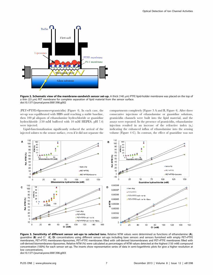
We propose a novel membrane-sandwich sensor-design for
OWLS assays by inserting commercially available filter mem-
branes into the measuring cuvette. A relatively thick polytetraflu-
oroethylene (PTFE) membrane can be filled with lipids or lipid
vesicles containing the ion channels to be investigated. The lipid-
filled membrane creates a water and ion resistant insulating layer,
which can sufficiently reduce the bulk permeation of electrolytes to
the sensing area. The fibrous PTFE membrane with large virtual
pore sizes can entrap bigger membrane fragments or intact/
ruptured GUV, SUV liposomes, but can not completely stop the
escape of small-size lipoid material. Deposition of traces of lipid
material onto the sensor surface, however, can be prevented by
inserting a further thin, water- and ion-permeable polyethylene
terephthalate (PET) membrane. This highly hydrophilic, small
pore-size membrane keeps the optical detection layer free from
lipids but provides direct electrolyte contact with the fluid layer
covering the sensor surface. The ionic composition and, conse-
quently, the refractive index of the small-volume ‘‘sensing’’ fluid
will change if enhanced amount of ions arrive in response to ion
channel opening in the lipid-filled layer.
Fibrous PTFE membranes saturated with liposomes provided
sufficient compartment insulation and supported the self-assembly
of gramicidin channels. To extend the assays for pharmacologi-
cally important membrane-embedded ion channels, an important
neurological/psychiatric drug target, a GABA-gated Cl2/HCO32
channel [17] was to be investigated. Instead of artificial
reconstruction of the multi-unit receptor assembly, biomembranes
were prepared from HEK293GABAa5b2c2 cells expressing the a5, b2
and c2 receptor subunits, and were filled into PTFE membranes.
The sensor set-up allowed detecting the move of Cl2 ions through
channels opened by GABA and reduction of ion flux in response
to the specific channel blocker, bicuculline. The data indicated
that the main pharmacological characteristics of the multi-unit
protein channel complex were preserved and gave confidence for
further development of compartmentalized ‘‘membrane-sand-
wich’’ OWLS sensor set-ups for in vitro pharmacological studies.
Materials and Methods
MaterialsChemicals of reagent grade were obtained from Sigma-Aldrich
(Hungary), unless stated otherwise, and were used without further
purification. Solutions were made with deionized distilled water
(18.2 MV.cm at 25uC) and were filtered through 0.22 mm
MillexHGP filter (Millipore, Hungary). Ethanolamine (Loba
Chemie, Germany), methylamine and guanidine were used in
hydrochloride forms. Cl–-channels were investigated using artifi-
cial cerebro-spinal fluid (ACSF) (mM: 145 NaCl, 3 KCl, 1 MgCl2,
2 CaCl2, 10 D-glucose, 10 HEPES; pH 7.4) or Cl–-free ACSF
(mM: 140 Na-acetate, 5 KH2PO4, 0.8 MgSO4, 1.8 Ca-acetate,
10 D-glucose, 10 HEPES; pH 7.4).
Polyethylene terephthalate (PET) membrane (RoTracH; thick-
ness 23 mm, with regular pores of 50 nm diameter) was kindly
donated by Oxyphen AG (Switzerland). Polytetrafluoroethylene
(PTFE) membrane (LCR; thickness 140 mm, virtual pore diameter
450 nm) was obtained from Millipore (Hungary).
Human Embryonic Kidney (HEK293) cell line, expressing a5,
b2 and c2 subunits of human GABAA receptors was established by
researchers of EGIS Pharmaceutical Inc. (Hungary). Complete
Mini Protease Inhibitor Cocktail Tablets were purchased from
Roche (Hungary).
OWLS AssaysDeposited mass, refractive indices, effective refractive indices
and the thickness of deposited material-layer on the sensor surface
were determined using an OWLS 110 instrument with OW 2400
grating coupler sensors and BioSense 2.6 software (MicroVacuum
Ltd, Budapest, Hungary). OW 2400 grating coupler sensors
consist of a 12 mm68 mm glass substrate covered with a thin
(175 nm) SiO2-TiO2 waveguide layer (refractive index:
nf = 1.7760.03) with an 12 mm62 mm optical grating (2400
lines/mm) (MicroVacuum Ltd, Budapest, Hungary) [12]. The
optical grating incouples light of a He-Ne laser at a given
resonance angle into the waveguide layer in which the light is
propagated by total internal reflection, generating an evanescent
field extending 100–200 nm from the surface. In OWLS assays,
incoupling of the incident laser beam occurs at two well-defined
angles of incidence: one for transverse electric (TE) and one for
transverse magnetic (TM) mode. Rotating the cuvette with 67
degrees, four characteristic photocurrent peaks (one TE and one
TM peak on both the positive and negative sides) can be detected
at the incoupling angles aTE and aTM. The smallest angle-step of
the rotation is 261024 degree, and the angular resolution of the
measuring device is 461024 degree. The resolution is further
smoothed to 161024 degree by peak-fitting function, which
determines the degree-value at the centroid of the peak-delineated
area. The accuracy of photocurrent detection is 61.526 pA. With
these parameters, a real sensitivity of d(NTM)/d(nc),0.128 could
be achieved.
The glass sensor chips were placed on the sensor holder
(MicroVacuum, Hungary) and tightened to its sealing O ring. The
sensor holder formed a flow cell above the glass sensor with a
volume of 12.1 ml. The sensor holder is made from biocompatible
PEEK material, the O ring is made from Kalrez and the tubings
are made from Teflon. The inlet tubing was connected to a sample
injection system (MicroVacuum, Hungary) furnished with a
Rheodyne Model 9725 injector valve.
Optical Detection of Ion Channel Activities
PLOS ONE | www.plosone.org 2 December 2013 | Volume 8 | Issue 12 | e81398
All assays were carried out at continuous flow of a ‘‘base-buffer’’
selected according to the experimental design (see in the text), at a
rate of 23 ml/min, at 22uC (60.1uC) temperature. Each experi-
ment was run with continuous recording of NTE and NTM
OWLS signals. The base line resulted by running through the
‘‘base-buffer’’ was recorded, and optical changes were related to
this base-line. Experimental solutions (test-solutions) were applied
when the base-line was stabilized or returned after a previous
treatment. Test-solutions (see in the text) were injected in 100 ml
aliquots into the continuous buffer-stream through the injector
valve. To accelerate detection velocity (10 data points/min), NTM
and NTE data were collected from a range of 60.2 degree around
the incoupling angles.
Determination of Optical Refractive Indices of SolutionsOW 2400 sensors inside the OWLS sensor holder were perfused
with deionized water until the stabilization of the baseline. The
refractive index and the thickness of the waveguide film (nf and df
respectively) were determined by the self-calibrating protocol of
the instrument. Samples of solutions were injected into the cuvette
and incoupling angles (aTE and aTM) were measured at
l= 632.8 nm wavelength. The effective refractive indices (NTE
and NTM), and the air-related refractive indices for each covering
medium (nc; Table 1) were determined by BioSense 2.6 software.
Preparation of LisosomesLiposomes were prepared from egg yolk lecithin (composition:
70% phosphatidylcholine, 10% phosphatidylethanolamine and
20% other lipids including neutral lipids; Avanti Polar Lipids,
USA) according to Moscho et al (1996) [18] (see Part S1.1 in File
S1) in HEPES-buffered saline (HBS; composition in mM: 10
HEPES, 150 NaCl; pH 7.4) or in normal or Cl–-free artificial
cerebro-spinal fluid (ACSF). The composition of ACSF in mM:
145 NaCl, 3 KCl, 1 MgCl2, 2 CaCl2, 10 D-glucose, 10 HEPES;
pH 7.4. The composition of Cl–-free ACSF in mM: 140 Na-
acetate, 5 KH2PO4, 0.8 MgSO4, 1.8 Ca-acetate, 10 D-glucose, 10
HEPES; pH 7.4.
Liposomes were occasionally labeled with Texas RedH DHPE
(1,2-dihexadecanoyl-sn-glycero-3-phosphoethanolamine, triethy-
lammonium salt; Invitrogen) and/or 1,2-dioleoyl-sn-glycero-3-
phosphoethanolamine-N-biotinyl sodium salt (DOPE-biotin;
Avanti Polar Lipids, USA). Texas Red labeled liposomes were
used to check the lipid coverage of the holder membrane by Zeiss
Axiovert 200M fluorescence microscope (see Part S1.2 in File S1).
Preparation of Membrane-fractions fromHEK293GABAAa5b2c2 Cells
HEK293 cells expressing human GABAA receptors with a5 b2
and c2 subunit composition (see Part S1.3 in File S1) were grown
in Dulbecco’s modified Eagle medium, supplemented with 10%
fetal bovine serum, 200 mg/ml zeocin and 3 mg/ml puromycin (as
selection antibiotics), at 37uC, with 5% CO2. For membrane
preparation, cells were washed off from the culture surface with
1 mM EDTA-PBS (pH 7.4). The cell suspension was distributed
to 108 cells/15 ml tubes and spun down at 200 g for 10 min. The
cell-pellets were frozen and either stored or directly used for
membrane preparation. Frozen pellet of 108 cells was resuspended
in 10-fold volume (200 ml) of ice-cold buffered saline containing
protease inhibitors (applied according to manufacturer’s instruc-
tion). The cells were disrupted by 3 freezing-thawing cycles (dry ice
for 2 min, 37uC water bath for 5 min), then the suspension was
spun at 1100 g for 10 min at 4uC for removing larger cell debris
and nuclei. The supernatant was centrifuged at 21000 g for
20 min at 4uC to sediment mitochondria. The 21000 g superna-
tant containing fragments of mixed cellular membranes was used
in the OWLS assays.
Functionalization of Sensor Surfaces with Multi-liposomeLayers
Reactive amino groups were formed on the surface of sensor
chips by treating with 10% (3-aminopropyl)triethoxysilane [19].
Biotin was coupled by incubating with 1 mg/ml NHS-biotin in
phosphate buffer (mM: 358 Na2HPO4, 87 KH2PO4; pH 7.5) for
12 h at 4uC. After washing with deionized water, biotinylated
sensors were inserted into the OWLS cuvette and washed with
buffered saline until the stabilization of the baseline. NeutrAvidin
(40 mg/ml) in buffered saline was injected into the cuvette, and
washed out after 20 min incubation.
NeutrAvidin-functionalized sensor chips inside the OWLS
cuvette were perfused with HBS. After stabilization of the
baseline, consecutive layers of liposomes were bound to the
surface according to the Membrane Protein Analysis Kit (Layerlab
AB, Sweden) protocol. Briefly, 1.3 mM of biotin-ssDNA (15-base,
single-stranded DNA with covalently bound biotin at one end)
were injected onto the sensor surface. Meantime, liposomes were
incubated (40 min; room temperature) with 1.3 mM Chol-dsDNA
1, (composed of one short and one long strand, with a cholesterol
anchor at the paired termini of the chains), to gain approximately
four (cholesterol anchored) DNA tags per liposome. The free
floating single-stranded end of the long chain had a complemen-
tary sequence to the biotin-ssDNA bound on the biosensor surface.
Chol-dsDNA-tagged liposomes were hybridized onto the biotin-
ssDNA functionalized sensor surface. Multiple layers of liposomes
were built by using another cholesterol-modified DNA (Chol-
dsDNA 2) containing a single-stranded end identical in sequence
to the biotin-ssDNA. Upon injection, Chol-dsDNA 2 tags were
inserted into the membrane of liposomes on the sensor surface and
a new layer of liposomes was formed by adding liposomes
containing Chol-dsDNA 1 [21,22]. Reagent excess was washed
out by HBS prior to each injection.
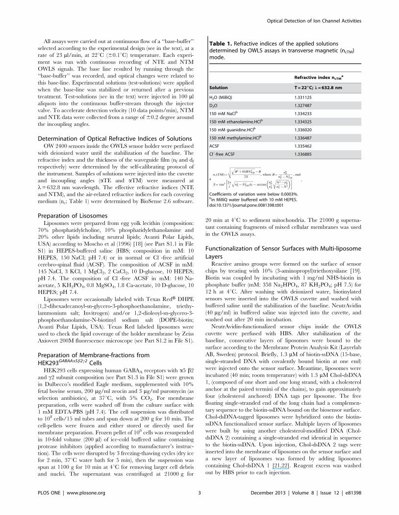
Table 1. Refractive indices of the applied solutionsdetermined by OWLS assays in transverse magnetic (ncTM)mode.
Refractive index ncTMa
Solution T = 226C; l = 632.8 nm
H2O (MilliQ) 1.331125
D2O 1.327487
150 mM NaClb 1.334233
150 mM ethanolamine.HClb 1.334325
150 mM guanidine.HClb 1.336020
150 mM methylamine.HClb 1.336487
ACSF 1.335462
Cl–-free ACSF 1.336885
a
nc(TM)~
ffiffiffiffiffiffiffiffiffiffiffiffiffiffiffiffiffiffiffiffiffiffiffiffiffiffiffiffiffiffiffiffiffiffiffiffiffiffiffiffiffiffiffiffiffiffiffiffiffiffiffiffiffiffiffiffiffiffiffiffiffiffiffiffiffiffiffiffiffiffiffiB2z4SBN2
TM
q{B
2S
vuut, where B~
n4F
n2F {N2
TM
, and
S~ tan2 2p
l
ffiffiffiffiffiffiffiffiffiffiffiffiffiffiffiffiffiffiffiffiffiffiffiffiffiffin2
F {N2TM dF
q{ arctan
n2F
n2S
ffiffiffiffiffiffiffiffiffiffiffiffiffiffiffiffiffiN2{n2
S
n2F {N2
s !" # .
Coefficients of variation were below 0.0003%.bin MilliQ water buffered with 10 mM HEPES.doi:10.1371/journal.pone.0081398.t001
Optical Detection of Ion Channel Activities
PLOS ONE | www.plosone.org 3 December 2013 | Volume 8 | Issue 12 | e81398

Insertion of Commercial Membranes into the OWLSCuvette
Commercially available filter (PTFE as ‘‘holder’’ and PET as
‘‘separating’’) membranes were cut to fit into the OWLS cuvette.
A piece of PET separating membrane was placed on a sensor
surface with carefully preventing ingress of air bubbles. Fibrous
PTFE holder membrane pieces were soaked in the appropriate
assay buffer or were functionalized with 20 mg/ml of NeutrAvidin.
A piece of the PTFE membrane was layered above the PET
separating membrane. The membranes were fastened with the
flow-tight Kalrez O-ring of the OWLS sensor holder.
The inserted membranes occupied 2.1 ml from the total 12.1 ml
cuvette volume. Proper insertion was checked after filling the
PTFE membrane with lipid material (see below) by testing with a
test-buffer with different refractive index in comparison to the
running buffer. Incomplete insertion of the membranes or failure
in lipid entrapment resulted in instant equilibration between the
sensing volume and the running buffer. Such sensor set-ups were
not (and could not be) used for assays.
Application of Liposomes and Cell-derived MembraneFractions onto the Holder Membrane
Egg yolk lecithine liposomes. A 100 ml aliquot of egg yolk
lecithine liposomes corresponding 125 mg lecithin content was
injected onto the PTFE membrane, which was already fitted on
the sensor inside of the OWLS cuvette and was previously washed
with the appropriate (HBS or ACSF, Cl–-free ACSF) assay-buffer.
The inlet and outlet tubing was closed, and the suspension was let
to sediment for at least 2 h. After sedimentation, the cuvette was
streamed through with the assay buffer until stable NTM and
NTE values against time were recorded by OWLS.
Cell-derived membrane fraction. A 100 ml aliquot of the
21000 g supernatant containing fragments of mixed cellular
membranes of 56107 cells was injected into the OWLS cuvette,
as above. After sedimentation, the cuvette was washed with Cl–-
free ACSF until stable NTM and NTE values were measured.
Mixed liposome-biomembrane fraction. Mixed solutions
of egg yolk liposomes and cell-derived membrane fractions were
prepared by incubating 50 ml aliquot of the cell membrane
fraction (derived from 2.56107 cells) with equal volume of
liposomes (corresponding 62 mg lecithin) at room temperature
for 2 h. 100 ml of mixed liposome-cell membrane suspension was
injected into the OWLS cuvette as above, and was washed with
Cl–-free ACSF until stable NTM and NTE values were measured.
Application of Channel Building, Opening and BlockingCompounds
Gramicidin D stock solution was prepared in absolute ethanol at a
concentration of 2 mg/ml, was further diluted with the base buffer
(HBS) to contain gramicidin: lecithine = 1:40 (1/40 molar ratio).
Gramicidin solution was injected into the OWLS cuvette, which
contained settled liposomes already assayed without gramicidin.
After transfusion with 56 cuvette-volume (60 ml) of gramicidin-
containing buffer, the flow was stopped and liposomes were
incubated with the gramicidin solutions for 30 min, at 22uC. After
the incubation, the gramicidin solution was washed out, and the
OWLS assay was continued by recording the baseline and then
the effects of consecutively injected solutions of channel perme-
ating/blocking compounds.
GABA (c-amino-butyric acid) and bicuculline were dissolved in
(Cl–-containing) ACSF at a final concentration of 100 mM and
were applied on sensor set-ups carrying mixed (liposome and cell-
derived) lipid-membrane fractions equilibrated with ACSF. The
test-solutions were injected into the stream of ACSF consecutively:
100 ml of Cl–-containing ACSF, 100 ml of Cl–-containing ACSF
with 100 mM GABA, and 100 ml of Cl–-containing ACSF with
100 mM GABA and 100 mM bicuculline. Each injection was
started after the return of the original NTM and NTE baseline.
Determination of Assay-sensitivitySensitivity of the assays was characterized by determining the
smallest detectable compound (ethanolamine, guanidine or Cl2)
concentration, which resulted in measurable shift of the effective
refractive indices (NTE and NTM). Concentration-series of
compounds from 0 to 150 mM were prepared in the appropriate
assay-buffers, and 100 ml aliquots were injected into a continuous
assay-buffer flow under permanent NTM and NTE. Any effect
was regarded detectable, if the evoked NTM and NTE changes
exceeded the background (NTE and NTM at 0 compound
concentration) with a three-fold value of the standard deviation of
the background average (limit of detection; LOD, [20]). LOD
values were determined for bare sensors and also for sensors
furnished with empty PTFE and PET membranes or with
membranes filled with liposomes and/or cell-derived biomem-
branes. For comparing data obtained on individual sensors,
relative values of effective refractive indices (NTM or NTE) were
calculated as percentages of the effective refractive indices
measured at the highest analyte concentration (100%) in a given
assay.
Data EvaluationAverages and standard deviations were calculated from data
obtained from n$4 independent series of identical experiments. In
order to compare the results obtained on individual sensor-chips,
either the absolute values of nc changes (Dnc) or the relative values
of effective refractive indices (NTM or NTE) (in percentages) were
calculated. Significances were calculated by student t-test. Results
with p,0.005 were accepted as significant.
Results
Various OWLS sensor-arrangements were probed in order to
find techniques for assaying ion permeation through self-assem-
bling gramicidin cation channels and a multi-subunit anion
channel, the GABAA receptor. As optical grating coupler sensors
respond to refractive index changes, pairs of buffer solutions were
prepared by replacing one of their ionic constituents for a
compound with different refractive index but comparable trans-
membrane permeation capability (Table 1). Besides proposing a
novel membrane-sandwich sensor set-up, we present and discuss
some less successful sensor-designs (see also Part S1.4 in File S1).
Assays on Multilayers of Coupled LiposomesAs it was shown by Branden and colleagues [21,22], several
properties of trans-membrane transport proteins embedded into
liposome membranes can be investigated with evanescent optical
(SPR) detection if concentration-drifts of slowly permeating
analytes are monitored in the sensing volume. As an approach,
we assembled multiple layers of interconnected liposomes [21,22]
on NeutrAvidin-functionalized sensor surfaces. Consecutive layers
of liposomes were bound by DNA hybridization (Membrane
Protein Analysis Kit). Through two subsequent hybridization
steps, a thick ($200 nm) layer of liposomes was built on the sensor
surface (Figure 1 A), apparently exceeding the thickness of the
optical detection field. Liposomes were clearly seen on sensors at
the end of long-term OWLS assays by fluorescence microscopy on
sensors carrying Texas Red labeled liposomes (see Part S1.5 in File
Optical Detection of Ion Channel Activities
PLOS ONE | www.plosone.org 4 December 2013 | Volume 8 | Issue 12 | e81398

S1). The observations indicated that interconnected liposome
multilayers could be established and preserved on the sensor
surface.
The interconnected layers of liposomes hydrated in HBS were
probed to study the cation exchange through the lipid membranes,
with or without inbuilt gramicidin channels. For Na+-free
solutions, Na+ was replaced by small organic cations of ethanol-
amine, guanidine, and occasionally methylamine. Ethanolamine
and methylamine are known to pass through gramicidin channels
by different kinetics due to their different size and molecular
shape, but do not block the channel [23]. In contrast, guanidine
was shown to block the channels in artificial membranes by
interacting with gramicidin protein residues and causing flicker
blocks when passing through the channel [23].
Once the liposomes were assembled and the baseline in HBS
was stabilized, 100 ml of Na+-free solutions of ethanolamine,
methylamine or guanidine (150 mM buffered with 10 mM
HEPES) were introduced into the continuous HBS flow, through
the injector system. Each injection was followed by washing with
HBS, and next test-aliquots were injected when NTE and NTM
values at HBS washing returned to the base-line (Figure 1 B). After
three injection cycles, gramicidin was incorporated into the
liposome membranes and the assays with Na+-free solutions were
repeated (Figure 1 C), in three consecutive series.
Injection of Na+-free ethanolamine or guanidine solutions
(Figure1 B) caused an immediate increase in the NTM values
indicating the arrival of a fraction of organic material (with higher
refractive indices) to the sensor surface via free diffusion through
the extra-liposome space. The initial rise in NTM increased in the
presence of gramicidin (Figure 1 C), suggesting that a proportion
of organic ions migrated through the channels and invaded intra-
liposome volumes adjacent to the sensor surface. The subsequent
decrease in NTM was reasoned as a mixed effect of rapid diffusion
of Na+ ions out from the intra- and inter-liposome spaces in
response to Na+-free perfusion, and a slower diffusion of organic
compounds into the liposomes. In the presence of gramicidin,
ethanolamine and methylamine molecules compensated the NTM
decrease by entering liposomes, and thus, conquering a larger
volume in the detection field. The assumption was supported by
the effect of guanidine, which, by blocking gramicidin channels,
was excluded from the liposomes and could partially block also the
outmigration of Na+ from liposomes. As an additional effect,
shrinkage of the liposomes in response to unbalanced inward and
outward ion migration [22] can not be excluded.
While the recorded data including the robustness and
reproducibility of the multi-liposome layer and the clear-cut
differences between the exchange of ethanolamine and guanidine
indicated that such approaches might be used for defined
purposes, the time-resolution of OWLS detection did not allow
analyzing the kinetics of permeation. Moreover, detection of
concentration changes was hindered by summarized recording of
optical changes from the entire detection field. With this sensor
set-up, changes in the intra- or extra-liposome compartments
could not be distinguished. The observations indicated that for
getting interpretable OWLS data on transmembrane ion perme-
ation, the different compartments should be either theoretically
distinguished or physically separated.
Assays with Membrane-sandwich Sensor Set-upsAs a next approach, we excluded liposomes from the optical
detection field by inserting a ‘‘spacer’’ between the sensor surface
and the lipid-containing layer. We probed commercially available
membrane filters as both, lipid holding supports and compartment
separating sheets. Two different filter-membranes were used: (i) a
fibrous PTFE membrane was applied as a lipid-holder layer and
(ii) a PET membrane with uniform, straight cylindrical capillary
pores was inserted to protect the sensor surface from lipid material.
The PET membrane was fitted onto the sensor area and the lipid-
holder PTFE membrane, with or without functionalization, was
layered above it (Figure 2).
Layering the empty ‘‘holder’’ and ‘‘separator’’ membranes onto
the sensor reduced the photocurrent peaks, but did not displace
significantly or widen the incoupling peaks. Changes in the
amplitude of the photocurrent peaks do not disturb the OWLS
assays as long as the peak-position can be precisely determined.
The small changes in the peak-positions, however, indicated that
the separator PET membrane occupied a small fraction of the
sensing volume and thus, physically decreased it.
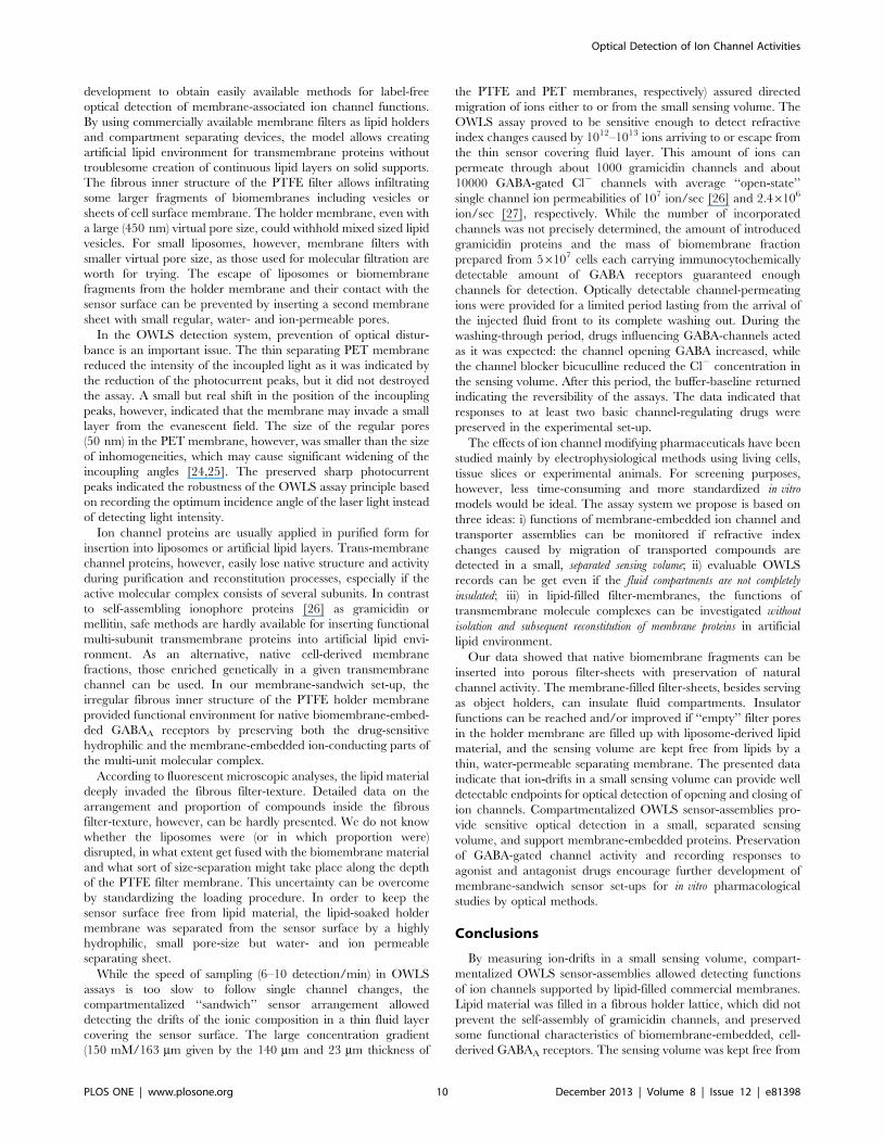
To characterize the sensitivity of the membrane-compartmen-
talized sensor set-up, NTM values were measured in response to
solutions with different concentrations of selected ions on bare
sensors and on sensor set-ups furnished with empty and lipid-filled
membranes (Figure 3). NTM in a base-buffer and in base-buffer
with different concentrations of a test compound were compared
to NTM measured at the highest applied concentration (150 mM)
of a test compound (100%). The relative values of effective
refractive index-changes were comparable among the different
sensor set-ups (Figure 3).
As OWLS detects refractive index changes in the range of
1026 nc values, very small alterations in the composition of the
sensor-covering fluid layer generate measurable signals. To
distinguish signals from noise, NTM values for each running
buffer were carefully analyzed and the average NTM with
standard deviations were determined for each sensor set-up in
each base-buffer. NTM-changes to test solutions were accepted as
specific optical responses, if the NTM value in test-solution
exceeded the NTM in base-buffer with a three-fold value of the
standard deviation [20].
On bare chips, the compound concentration on the sensor
surface was regarded equal to the concentration of the bulk
solution in the OWLS cuvette, so the detection limit could be
determined by measuring the effective refractive index (NTM) as a
function of the concentration of the injected fluid (Figure 3). The
assays with bare sensors detected 0.72 mM concentration of
ethanolamine, 0.75 mM concentration of guanidine and 2.91 mM
of Cl2 (LOD values) corresponding to 1.3861012 ethanolamine
molecules, 1.4461012 guanidine molecules or 5.5961012 of Cl2
ions in the sensing volume (V = width6length6height of evanes-
cent field = (20006800060.2) mm3 = 3.2 nl). After introducing the
PET and PTFE membranes, the detection limits increased, and
the apparent sensitivity was further reduced if the PTFE holder
membrane was filled with lipid material (Table 2). The data,
however, does not mean necessarily that the sensor sensitivity was
impaired; rather, it may indicate the rate of compound retardation
by the inserted membranes. As we could not clear up this point,
the smallest detectable concentrations recorded in empty
PET+PTFE filter containing set-ups were regarded as the LOD
in the assays with membrane-sandwich set-ups.
All assays were run under permanent buffer perfusion at a rate
of 23 ml/min, with injecting of 100 ml volumes of analyte
solutions into the stream. Accordingly, the analytes were
streamed through the cuvette in 4.35 min. The total washing-
through period, however was expanded presumably by fluid-
mixing and retardation resulting in a total washing-through
period of about 10 min. After this period, the buffer-base line
returned indicating the reversibility of the assays, regardless of the
injected analyte.
Optical Detection of Ion Channel Activities
PLOS ONE | www.plosone.org 5 December 2013 | Volume 8 | Issue 12 | e81398

Detection of Cation Migration through GramicidinChannels
The membrane-sandwich sensor set-up was probed for detect-
ing exchange of Na+ for organic cations ethanolamine and
guanidine, first through empty filter layers (PET+PTFE), then
through membranes filled with liposomes (more exactly, liposome-
derived lipid material) (PET+PTFE+liposomes), and finally
through supported lipid material with inbuilt gramicidin channels
Figure 1. OWLS recordings of deposition of multiple liposome layers and permeability for small organic cations. (A) Biotinylatedsensors were treated with (1) NeutrAvidin; (2) biotin-ssDNA; (3) Chol-dsDNA1-tagged liposomes; (4) Chol-dsDNA2. The insert shows the increasingthickness (dA) of the deposited material on the surface reaching the detectable maximum (,200 nm) after the second injection of liposomes. (B, C)Changes of the effective refractive indices (NTM) in a representative series of experiments with cross-linked multilayer of liposomes without (B) orwith (C) gramicidin channels after injections of ethanolamine (5), methylamine (6) or guanidine (7) solutions.doi:10.1371/journal.pone.0081398.g001
Optical Detection of Ion Channel Activities
PLOS ONE | www.plosone.org 6 December 2013 | Volume 8 | Issue 12 | e81398
(PET+PTFE+liposomes+gramicidin) (Figure 4). In each case, the
set-up was equilibrated with HBS until reaching a stable baseline,
then 100 ml aliquots of ethanolamine hydrochloride or guanidine
hydrochloride (150 mM buffered with 10 mM HEPES, pH 7.4)
were injected.
Lipid-functionalization significantly reduced the arrival of the
injected solutes to the sensor surface, even if it did not separate the
compartments completely (Figure 3 A and B, Figure 4). After three
consecutive injections of ethanolamine or guanidine solutions,
gramicidin channels were built into the lipid material, and the
assays were repeated. In the presence of gramicidin, ethanolamine
injection resulted in an increase of the refractive index (nc)
indicating the enhanced influx of ethanolamine into the sensing
volume (Figure 4 C). In contrast, the effect of guanidine was not
Figure 2. Schematic view of the membrane-sandwich sensor set-up. A thick (140 mm) PTFE lipid-holder membrane was placed on the top ofa thin (23 mm) PET membrane for complete separation of lipid material from the sensor surface.doi:10.1371/journal.pone.0081398.g002
Figure 3. Sensitivity of different sensor set-ups to selected ions. Relative NTM values were determined as functions of ethanolamine (A),guanidine (B) and Cl2 (C, D) concentrations using different sensor set-ups including bare sensors and sensors furnished with empty PET+PTFEmembranes, PET+PTFE membranes+liposomes, PET+PTFE membranes filled with cell-derived biomembranes and PET+PTFE membranes filled withcell-derived biomembranes+liposomes. Relative NTM (%) were calculated as percentages of NTM values detected at the highest (150 mM) compoundconcentration (100%) for each sensor set-up. The inserts show representative series of data in semi-logarithmic plots for give a higher resolution atlow concentrations.doi:10.1371/journal.pone.0081398.g003
Optical Detection of Ion Channel Activities
PLOS ONE | www.plosone.org 7 December 2013 | Volume 8 | Issue 12 | e81398

influenced by the presence of gramicidin channels (Figure 4 D) in
agreement with the known channel-blocking effect of guanidine.
The membrane-sandwich set-up was used also to study the
outward diffusion of ethanolamine from and the inward move of
Na+ into liposomes hydrated in ethanolamine-Tris buffer (see Part
S1.6 in File S1). The reproducible acceleration of the ethanol-
amine clearance in the presence of gramicidin verified the
feasibility of the set-up, and let us to probe it with more realistic
biomembrane vesicles and pharmacologically important channels.
Assays on Cell-derived Membranes Carrying GABAA
Anion ChannelsCellular membrane fractions were isolated from HEK293 cells
expressing human GABAA receptors with a5, b2 and c2 subunit
composition. A crude membrane fraction (21000 g supernatant)
containing fragments of mixed cellular membranes, was deposited
onto the PTFE holder membrane alone or – in order to increase
the lipid-saturation of the holder membrane – together with
liposomes.
Table 2. The minimum amount of detectable material in assays with various sensor set-ups.
Concentration in the injected buffer [mM] Number of molecules/ions in the sensing volume (3.2 nl)
Sensor ethanolamine guanidine Cl2 ethanolamine guanidine Cl2
bare 0.72 0.75 2.91 1.3861012 1.4461012 5.5961012
PET+PTFE 0.82 0.89 9.64 1.5761012 1.7561012 1.8561013
PET+PTFE+lipid 4.9 3.52 19.78 9.4161012 6.7661012 3.861013
doi:10.1371/journal.pone.0081398.t002
Figure 4. Detection of cation migration through gramicidin channels in membrane-sandwich sensor set-ups. Refractive indices (ncTM)of the sensor-covering fluid layer were measured in the transverse magnetic (TM) mode in sensor set-ups containing empty or lipid-filled filtermembranes (A, B) after injecting ethanolamine (A) or guanidine (B) solutions, and after the incorporation of gramicidin (C, D). The inserts showaverages and standard deviations of ncTM changes, calculated from three independent experiments. Significant (***p,0.001; paired t-test) differenceswere found in response to filling with liposomes regardless of the chemical nature of the analyte (A, B). Incorporation of gramicidin, while resulted inenhanced permeation of ethanolamine (C), did not cause detectable changes in the move of guanidine (D).doi:10.1371/journal.pone.0081398.g004
Optical Detection of Ion Channel Activities
PLOS ONE | www.plosone.org 8 December 2013 | Volume 8 | Issue 12 | e81398

Different membrane-sandwich assemblies were assayed includ-
ing empty PTFE and PET filter membranes, filter membranes
filled with GABA-receptor containing cell-derived membranes and
filter membranes filled with the cell-derived membranes together
with liposome-derived lipids. The ion-barrier functions of different
membrane-sandwich set-ups were monitored without adding any
channel influencing compounds. The assemblies were perfused
with Cl–-free ACSF until reaching a stable baseline, then 100 ml
ACSF containing different concentrations of Cl2 was streaming
through. Significantly less Cl2 ions arrived to the sensing volume if
cell-derived membranes were added to the filters and even less Cl2
ions were detected if liposomes were added to biomembranes on
the holder membrane.
In Cl–-containing ACSF, the decrease in NTM indicated the
improper insulation of fluid-compartments (Figure 5 A). Injecting
Cl–-containing ACSF together with GABA (100 mM), however,
further decreased the refractive indices indicating an enhanced
influx of Cl2 ions into the sensing volume in comparison to the
effect of Cl–-ACSF alone (Figure 5 A and C). The Cl–-permeation
was markedly reduced, if the solution contained also bicuculline, a
potent GABA-channel inhibitor (Figure 5 A, B and C).
The data demonstrated that GABA channels preserved their
basic function and drug-sensitivity in the membrane-sandwich.
The finding indicates that the sensor set-up might provide an
optical assay tool for studying lipid-associated, multi-subunit
channels with pharmacological interest.
Discussion
OWLS techniques detect changes in the refractive indices,
caused by compositional and/or conformational changes in a
narrow (200 nm) layer above the sensor surface. Changes in the
functionalization of the sensor surface, including motion, shrink-
age, swelling, fusion or detachment of lipid layers/liposomes,
however, can also generate large changes in refractive indices,
imposing serious limitations in recording molecular events.
Moreover, due to the superposition of distinct optical events, a
single data will be obtained at each detected time-point, regardless
of the origin of the optical changes. With the presented data we
wish to demonstrate that functionality and drug-sensitivity of
trans-membrane ion channels can be investigated by OWLS
assays, if concentration-drifts are monitored in a small sensing
volume on the sensor surface. The optical data obtained with
sensor-attached poly-liposome arrays demonstrated that for this
purpose, the sensing fluid-space has to be separated from both
intra-liposome and free buffer spaces. While coupled poly-
liposomes were used to investigate membrane embedded channels
with evanescent optical detection methods [22], our data obtained
with this system led us to develop compartmentalized OWLS
cuvettes, which allowed sufficient separation of the sensing volume
from the rest of the assay system.
The proposed simple membrane-sandwich sensor set-up seems
to overcome several shortcomings and might be worth for further
Figure 5. Demonstration of Cl–-channel functions of cell-derived GABA receptors in the membrane-sandwich sensor set-up. OWLSrecordings were made in Cl–-free ACSF as running buffer with injection of Cl–-containing ACSF (ACSF) with or without GABA and the channel blockerbicuculline (ACSF+GABA and ACSF+GABA+Bic, respectively). (A) Changes of the effective refractive index (NTM) values in a representative OWLSassay. (B) Changes in the refractive index (ncTM) in response to the GABA-channel blocker bicuculline are shown from a representative experiment. (C)Summary of refractive index (ncTM) changes (DncTM) in response to transfusion with Cl–-containing buffer (ACSF), in the presence of the agonist GABA(ACSF+GABA) or in the presence of both GABA and the channel-blocker bicucullin (ACSF+GABA+bicucullin). DncTM values were calculated from dataof 4 independent series of experiments (n$4); averages and standard deviations are presented.doi:10.1371/journal.pone.0081398.g005
Optical Detection of Ion Channel Activities
PLOS ONE | www.plosone.org 9 December 2013 | Volume 8 | Issue 12 | e81398
development to obtain easily available methods for label-free
optical detection of membrane-associated ion channel functions.
By using commercially available membrane filters as lipid holders
and compartment separating devices, the model allows creating
artificial lipid environment for transmembrane proteins without
troublesome creation of continuous lipid layers on solid supports.
The fibrous inner structure of the PTFE filter allows infiltrating
some larger fragments of biomembranes including vesicles or
sheets of cell surface membrane. The holder membrane, even with
a large (450 nm) virtual pore size, could withhold mixed sized lipid
vesicles. For small liposomes, however, membrane filters with
smaller virtual pore size, as those used for molecular filtration are
worth for trying. The escape of liposomes or biomembrane
fragments from the holder membrane and their contact with the
sensor surface can be prevented by inserting a second membrane
sheet with small regular, water- and ion-permeable pores.
In the OWLS detection system, prevention of optical distur-
bance is an important issue. The thin separating PET membrane
reduced the intensity of the incoupled light as it was indicated by
the reduction of the photocurrent peaks, but it did not destroyed
the assay. A small but real shift in the position of the incoupling
peaks, however, indicated that the membrane may invade a small
layer from the evanescent field. The size of the regular pores
(50 nm) in the PET membrane, however, was smaller than the size
of inhomogeneities, which may cause significant widening of the
incoupling angles [24,25]. The preserved sharp photocurrent
peaks indicated the robustness of the OWLS assay principle based
on recording the optimum incidence angle of the laser light instead
of detecting light intensity.
Ion channel proteins are usually applied in purified form for
insertion into liposomes or artificial lipid layers. Trans-membrane
channel proteins, however, easily lose native structure and activity
during purification and reconstitution processes, especially if the
active molecular complex consists of several subunits. In contrast
to self-assembling ionophore proteins [26] as gramicidin or
mellitin, safe methods are hardly available for inserting functional
multi-subunit transmembrane proteins into artificial lipid envi-
ronment. As an alternative, native cell-derived membrane
fractions, those enriched genetically in a given transmembrane
channel can be used. In our membrane-sandwich set-up, the
irregular fibrous inner structure of the PTFE holder membrane
provided functional environment for native biomembrane-embed-
ded GABAA receptors by preserving both the drug-sensitive
hydrophilic and the membrane-embedded ion-conducting parts of
the multi-unit molecular complex.
According to fluorescent microscopic analyses, the lipid material
deeply invaded the fibrous filter-texture. Detailed data on the
arrangement and proportion of compounds inside the fibrous
filter-texture, however, can be hardly presented. We do not know
whether the liposomes were (or in which proportion were)
disrupted, in what extent get fused with the biomembrane material
and what sort of size-separation might take place along the depth
of the PTFE filter membrane. This uncertainty can be overcome
by standardizing the loading procedure. In order to keep the
sensor surface free from lipid material, the lipid-soaked holder
membrane was separated from the sensor surface by a highly
hydrophilic, small pore-size but water- and ion permeable
separating sheet.
While the speed of sampling (6–10 detection/min) in OWLS
assays is too slow to follow single channel changes, the
compartmentalized ‘‘sandwich’’ sensor arrangement allowed
detecting the drifts of the ionic composition in a thin fluid layer
covering the sensor surface. The large concentration gradient
(150 mM/163 mm given by the 140 mm and 23 mm thickness of
the PTFE and PET membranes, respectively) assured directed
migration of ions either to or from the small sensing volume. The
OWLS assay proved to be sensitive enough to detect refractive
index changes caused by 1012–1013 ions arriving to or escape from
the thin sensor covering fluid layer. This amount of ions can
permeate through about 1000 gramicidin channels and about
10000 GABA-gated Cl2 channels with average ‘‘open-state’’
single channel ion permeabilities of 107 ion/sec [26] and 2.46106
ion/sec [27], respectively. While the number of incorporated
channels was not precisely determined, the amount of introduced
gramicidin proteins and the mass of biomembrane fraction
prepared from 56107 cells each carrying immunocytochemically
detectable amount of GABA receptors guaranteed enough
channels for detection. Optically detectable channel-permeating
ions were provided for a limited period lasting from the arrival of
the injected fluid front to its complete washing out. During the
washing-through period, drugs influencing GABA-channels acted
as it was expected: the channel opening GABA increased, while
the channel blocker bicuculline reduced the Cl2 concentration in
the sensing volume. After this period, the buffer-baseline returned
indicating the reversibility of the assays. The data indicated that
responses to at least two basic channel-regulating drugs were
preserved in the experimental set-up.
The effects of ion channel modifying pharmaceuticals have been
studied mainly by electrophysiological methods using living cells,
tissue slices or experimental animals. For screening purposes,
however, less time-consuming and more standardized in vitro
models would be ideal. The assay system we propose is based on
three ideas: i) functions of membrane-embedded ion channel and
transporter assemblies can be monitored if refractive index
changes caused by migration of transported compounds are
detected in a small, separated sensing volume; ii) evaluable OWLS
records can be get even if the fluid compartments are not completely
insulated; iii) in lipid-filled filter-membranes, the functions of
transmembrane molecule complexes can be investigated without
isolation and subsequent reconstitution of membrane proteins in artificial
lipid environment.
Our data showed that native biomembrane fragments can be
inserted into porous filter-sheets with preservation of natural
channel activity. The membrane-filled filter-sheets, besides serving
as object holders, can insulate fluid compartments. Insulator
functions can be reached and/or improved if ‘‘empty’’ filter pores
in the holder membrane are filled up with liposome-derived lipid
material, and the sensing volume are kept free from lipids by a
thin, water-permeable separating membrane. The presented data
indicate that ion-drifts in a small sensing volume can provide well
detectable endpoints for optical detection of opening and closing of
ion channels. Compartmentalized OWLS sensor-assemblies pro-
vide sensitive optical detection in a small, separated sensing
volume, and support membrane-embedded proteins. Preservation
of GABA-gated channel activity and recording responses to
agonist and antagonist drugs encourage further development of
membrane-sandwich sensor set-ups for in vitro pharmacological
studies by optical methods.
Conclusions
By measuring ion-drifts in a small sensing volume, compart-
mentalized OWLS sensor-assemblies allowed detecting functions
of ion channels supported by lipid-filled commercial membranes.
Lipid material was filled in a fibrous holder lattice, which did not
prevent the self-assembly of gramicidin channels, and preserved
some functional characteristics of biomembrane-embedded, cell-
derived GABAA receptors. The sensing volume was kept free from
Optical Detection of Ion Channel Activities
PLOS ONE | www.plosone.org 10 December 2013 | Volume 8 | Issue 12 | e81398

lipid material by inserting a thin water- and ion-permeable
separator membrane. The compartmentalized sensor assembly
provided:
i) satisfactory ion-insulation, which was modulated by insertion
and/or opening of ion channels;
ii) an easily fabricated environment for functional assembly of
membrane-associated ion channels even with large molecular
weight and multi-unit composition;
iii) a simple principle for developing sensor set-ups for sensitive
optical detection of functional responses of biomembrane-
embedded drug-targets.
Supporting Information
File S1 Combined Supporting Information S1. S1.1.
Preparation of liposomes. S1.2. Lipid coverage of the holder
membrane. S1.3. Expression of GABAAa5b2c2 receptor by
HEK293 cells. S1.4. Assays on H2O/D2O exchange using
liposomes immobilized directly on the sensor surface. S1.5. Texas
Red labeled DNA-cross-linked liposomes on the sensor surface.
S1.6. Time-course of washing out of ethanolamine from liposomes
prepared in Tris-buffered ethanolamine.
(DOC)
Acknowledgments
The authors thank Oxyphen GmbH (Zurich, Switzerland) for samples of
RoTrack membranes, for Layerlab AB (Sweden) for Membrane Protein
Analysis Kits. The authors wish to thank the Nikon Microscopy Center at
IEM, Nikon Austria GmbH and Auro-Science Consulting Ltd for kindly
providing microscopy support.
Author Contributions
Conceived and designed the experiments: EM I. Szekacs I. Szendro.
Performed the experiments: I. Szekacs NK. Analyzed the data: I. Szekacs
EM PG KE. Contributed reagents/materials/analysis tools: NK PG KE I.
Szendro BM AP FAA. Wrote the paper: I. Szekacs EM.
References
1. Homola J (2006) Surface plasmon resonance based sensors. Springer-Verlag,
Berlin-Heidelberg-New York. 251 p.
2. Voros J, Ramsden JJ, Csucs G, Szendro I, De Paul SM, at al. (2002) Optical
grating coupler biosensors. Biomaterials 23: 3699–3710.
3. Bally M, Bailey K, Sugihara K, Grieshaber D, Voros J, at al. (2010) Liposome
and bilayer arrays towards biosensing applications. Small 6: 2481–2497.
4. Heimburg T (2010) Lipid ion channels. Biophys Chem 150: 2–22.
5. Majd S, Yusko EC, Billeh YN, Macrae MX, Yang J, at al. (2010) Applications of
biological pores in nanomedicine, sensing, and nanoelectronics. Curr Opin
Biotechnol 21: 439–476.
6. Mashaghi A, Swann M, Popplewell J, Textor M, Reimhult E (2008) Optical
anisotropy of supported lipid structures probed by waveguide spectroscopy and
its application to study of supported lipid bilayer formation kinetics. Anal Chem
80: 3666–3676.
7. Baumann MK, Swann MJ, Textor M, Reimhult E (2011) Pleckstrin homology-
phospholipase C-d1 interaction with phophatidylinositol 4,5-biphosphate
containing supported lipid bilayers monitored in situ with dual polarization
interferometry. Anal Chem 83: 6267–6274.
8. Steller L, Kreir M, Salzer R (2012) Natural and artificial ion channels for
biosensing platforms. Anal Bioanal Chem 402: 209–230.
9. Tiefenthaler K (1992) Integrated optical couplers as chemical waveguide sensors.
Adv Biosens 2: 261–289.
10. Ramsden JJ (1993) Review of new experimental techniques for investigating
random sequential adsorption. J Stat Phys 73: 853–877.
11. Erdelyi K, Frutos AG, Ramsden JJ, Szendro I, Voirin G (2007) In: Marks RS,
Cullen DC, Karube I, Lowe CR, Weetall HH, editors. Handbook of biosensors
and biochips. Wiley, Chichester. pp. 569–586.
12. MicroVacuum website. Available: http//www.owls-sensors.com. Accessed 2013
Oct 25.
13. Sackmann E (1996) Supported membranes: scientific and practical applications.
Science 271: 43–48.
14. Kugler R, Knoll W (2002) Polyelectrolyte-supported lipid membranes.
Bioelectrochemistry 56: 175–178.
15. Sugihara K, Voros J, Zambelli T (2010) A gigaseal obtained with a self-assembled long-lifetime lipid bilayer on a single polyelectrolyte multilayer-filled
nanopore. ACS Nano 4: 5047–5054.
16. Phung T, Zhang Y, Dunlop J, Dalziel J (2011) Bilayer lipid membranessupported on Teflon filters: a functional environment for ion channels. Biosens
Bioelectron 26: 3127–35.17. Sigel E, Steinmann ME (2012) Structure, function and modulation of GABAA
receptors. J Biol Chem Doi: 10.1074/jbc.R112.386664.
18. Moscho A, Orwar O, Chiu DT, Modi BP, Zare RN (1996) Rapid preparation ofgiant unilamellar vesicles. Proc Natl Acad Sci USA 93: 11443–11447.
19. Trummer N, Adanyi N, Varadi M, Szendro I (2001) Modification of the surfaceof integrated optical wave-guide sensors for immunosensor applications.
Fresenius J Anal Chem 371: 21–24.20. Nic M, Jirat J, Kosata B (2006–) ‘‘Limit of detection’’. IUPAC Compendium of
Chemical Terminology (Online ed.) Doi:10.1351/goldbook.L03540.
21. Branden M, Dahlin S, Hook F (2008) Label-free measurements of moleculartransport across liposome membranes using evanescent-wave sensing. Chem-
physchem 9: 2480–2485.22. Branden M, Tabaei SR, Fischer G, Neutze R, Hook F (2010) Refractive-index-
based screening of membrane-protein-mediated transfer across biological
membranes. Biophys J 99: 124–133.23. Hemsley G, Busath D (1991) Small iminium ions block gramicidin channel in
lipid bilayers. Biophys J 59: 901–907.24. Horvath R, Voros J, Graf R, Fricsovszky G, Textor M, et al. (2001) Effect of
patterns and inhomogeneities on the surface of waveguides used for opticalwaveguide lightmode spectroscopy applications. Appl Phys B: Lasers Opt 72:
441–447.
25. Cottier K, Horvath R (2008) Imageless microscopy of surface patterns usingoptical waveguides. Appl Phys B: Lasers Opt 91: 319–327.
26. Kelkar DA, Chattopadhyay A (2007) The gramicidin ion channel: a modelmembrane protein. Biochim Biophys Acta 1768: 2011–2025.
27. Birnir B, Eghbali M, Cox GB, Gage PW (2001) GABA concentration sets the
conductance of delayed GABAA channels in outside-out patches from rathippocampal neurons. J. Membrane Biol 181: 171–183.
Optical Detection of Ion Channel Activities
PLOS ONE | www.plosone.org 11 December 2013 | Volume 8 | Issue 12 | e81398