Oocyte–Granulosa Cell Heterologous Gap Junctions Are Required for the Coordination of Nuclear and...
13
Oocyte–Granulosa Cell Heterologous Gap Junctions Are Required for the Coordination of Nuclear and Cytoplasmic Meiotic Competence Mary Jo Carabatsos,* Caterina Sellitto,² Daniel A. Goodenough,² and David F. Albertini* ,1 *Sackler School of Graduate Biomedical Sciences, Program in Cell, Molecular, and Developmental Biology, Department of Anatomy and Cellular Biology, Tufts University, Boston, Massachusetts 02111; and ²Department of Cellular Biology, Harvard Medical School, Boston, Massachusetts 02115 Homologous gap junctions are generally recognized as a means of coordinating cellular behavior under developmental and homeostatic conditions. In the mammalian ovary, heterologous gap junctions between the oocyte and the granulosa cells have been widely implicated in the regulation of meiotic maturation late in oogenesis. However, the role of oocyte– granulosa cell gap junctions at earlier stages of oogenesis is poorly understood. Stage-specific defects in both oocyte and follicle development have been identified in juvenile mice deficient in heterologous oocyte– granulosa cell gap junctions due to targeted deletion of Gja4, the gene encoding connexin-37. Follicle development arrests at the type 4 preantral stage and although oocytes commence growth, oocyte growth ceases at a diameter of 52 mm (74.3% of control size). Analysis of cell cycle and cytoskeletal markers indicates that oocytes arrest in a G 2 state based on uniform decondensed GV chromatin, interphase microtubule arrays, and nonphosphorylated cytoplasmic centrosomes. Functional assays of meiotic competence confirm that oocytes from connexin-37-deficient mice are unable to enter M phase (initiate meiotic maturation) unless treated with the phosphatase inhibitor okadaic acid (OA). Unlike growing oocytes from heterozygous control animals, OA-treated oocytes from connexin-37-deficient mice respond acutely and progress rapidly to the circular bivalent stage of meiosis I and upon removal from OA rapidly revert to an interphase state. In contrast, OA-treated control incompetent oocytes are slow to respond, exhibit a lower proportion of chromosomal bivalent stage oocytes, but remain in and progress into meiotic M phase upon removal from OA. This study demonstrates that heterologous gap-junctional communication is required for the completion of oocyte growth and the acquisition of cytoplasmic meiotic competence. © 2000 Academic Press INTRODUCTION The process of ovarian follicle development involves coordination of growth and differentiation of the female gamete with growth and differentiation of companion so- matic cells (Hirshfield, 1991; Gougeon, 1996). Ovarian primordial follicles remain in a resting state until they are recruited into the growing follicle pool. During the early stages of preantral follicle development somatic granulosa cells undergo both shape and proliferative changes. Con- comitantly, the oocyte enlarges as it accumulates or- ganelles and maternal mRNAs, acquires the ability to resume and complete meiosis, and secretes a specialized extracellular matrix called the zona pellucida (Wassarman and Albertini, 1994; Gosden et al., 1997). While antral follicles are dependent on extraovarian factors, such as the pituitary-derived gonadotropin follicle-stimulating hor- mone, preantral follicle growth has been shown to be independent of hormonal factors (Erickson and Ryan, 1976; Halpin et al., 1986; Kumar et al., 1997). Local modalities of communication mediated by gap junctions and/or autocrine/paracrine factors are likely to be important deter- minants of preantral follicle development. Gap junctions are collections of intercellular channels that permit direct exchange of ions and metabolites be- tween adjacent cells (reviewed in Simon and Goodenough, 1998). These intercellular channels are composed of con- 1 To whom correspondence should be addressed at the Depart- ment of Anatomy and Cellular Biology, Tufts University School of Medicine, 136 Harrison Avenue, Boston MA 02111. E-mail: [email protected]. Developmental Biology 226, 167–179 (2000) doi:10.1006/dbio.2000.9863, available online at http://www.idealibrary.com on 0012-1606/00 $35.00 Copyright © 2000 by Academic Press All rights of reproduction in any form reserved. 167
Transcript of Oocyte–Granulosa Cell Heterologous Gap Junctions Are Required for the Coordination of Nuclear and...

o
Hhhgftac
Developmental Biology 226, 167–179 (2000)doi:10.1006/dbio.2000.9863, available online at http://www.idealibrary.com on
Oocyte–Granulosa Cell Heterologous Gap JunctionsAre Required for the Coordination of Nuclear andCytoplasmic Meiotic Competence
Mary Jo Carabatsos,* Caterina Sellitto,† Daniel A. Goodenough,†and David F. Albertini*,1
*Sackler School of Graduate Biomedical Sciences, Program in Cell, Molecular,and Developmental Biology, Department of Anatomy and Cellular Biology,Tufts University, Boston, Massachusetts 02111; and †Departmentf Cellular Biology, Harvard Medical School, Boston, Massachusetts 02115
omologous gap junctions are generally recognized as a means of coordinating cellular behavior under developmental andomeostatic conditions. In the mammalian ovary, heterologous gap junctions between the oocyte and the granulosa cellsave been widely implicated in the regulation of meiotic maturation late in oogenesis. However, the role of oocyte–ranulosa cell gap junctions at earlier stages of oogenesis is poorly understood. Stage-specific defects in both oocyte andollicle development have been identified in juvenile mice deficient in heterologous oocyte–granulosa cell gap junctions dueo targeted deletion of Gja4, the gene encoding connexin-37. Follicle development arrests at the type 4 preantral stage andlthough oocytes commence growth, oocyte growth ceases at a diameter of 52 mm (74.3% of control size). Analysis of cellycle and cytoskeletal markers indicates that oocytes arrest in a G2 state based on uniform decondensed GV chromatin,
interphase microtubule arrays, and nonphosphorylated cytoplasmic centrosomes. Functional assays of meiotic competenceconfirm that oocytes from connexin-37-deficient mice are unable to enter M phase (initiate meiotic maturation) unlesstreated with the phosphatase inhibitor okadaic acid (OA). Unlike growing oocytes from heterozygous control animals,OA-treated oocytes from connexin-37-deficient mice respond acutely and progress rapidly to the circular bivalent stage ofmeiosis I and upon removal from OA rapidly revert to an interphase state. In contrast, OA-treated control incompetentoocytes are slow to respond, exhibit a lower proportion of chromosomal bivalent stage oocytes, but remain in and progressinto meiotic M phase upon removal from OA. This study demonstrates that heterologous gap-junctional communicationis required for the completion of oocyte growth and the acquisition of cytoplasmic meiotic competence. © 2000 Academic Press
greafpmiHcam
tt
INTRODUCTION
The process of ovarian follicle development involvescoordination of growth and differentiation of the femalegamete with growth and differentiation of companion so-matic cells (Hirshfield, 1991; Gougeon, 1996). Ovarianprimordial follicles remain in a resting state until they arerecruited into the growing follicle pool. During the earlystages of preantral follicle development somatic granulosacells undergo both shape and proliferative changes. Con-comitantly, the oocyte enlarges as it accumulates or-
1 To whom correspondence should be addressed at the Depart-ment of Anatomy and Cellular Biology, Tufts University School of
1Medicine, 136 Harrison Avenue, Boston MA 02111. E-mail:[email protected].
0012-1606/00 $35.00Copyright © 2000 by Academic PressAll rights of reproduction in any form reserved.
anelles and maternal mRNAs, acquires the ability toesume and complete meiosis, and secretes a specializedxtracellular matrix called the zona pellucida (Wassarmannd Albertini, 1994; Gosden et al., 1997). While antralollicles are dependent on extraovarian factors, such as theituitary-derived gonadotropin follicle-stimulating hor-one, preantral follicle growth has been shown to be
ndependent of hormonal factors (Erickson and Ryan, 1976;alpin et al., 1986; Kumar et al., 1997). Local modalities of
ommunication mediated by gap junctions and/orutocrine/paracrine factors are likely to be important deter-inants of preantral follicle development.Gap junctions are collections of intercellular channels
hat permit direct exchange of ions and metabolites be-ween adjacent cells (reviewed in Simon and Goodenough,
998). These intercellular channels are composed of con-167

1tadsBgomsIbc1Voa
cdsgi1iporctl
igbGrolin
oejc
tdfalopdp
cmcadsiatemtf
wah
ADo
168 Carabatsos et al.
nexins, a multigene family of integral membrane proteins(Goodenough et al., 1996). Homologous gap junctions arefound joining adjacent granulosa cells while heterologousgap junctions are found between the oocyte and the granu-losa cells (Albertini et al., 1975; Anderson and Albertini,976; Valdimarsson et al., 1993). Heterologous gap junc-ions have been shown to support metabolic cooperationnd prevent premature meiotic resumption in oocytes byirect transfer of metabolic substrates and meiosis-arrestingignals, respectively (Gilula et al., 1978; Dekel et al., 1981;ornslaeger and Schultz, 1985). Since oocyte–granulosa cellap junctions are present in preantral follicles before theocyte is capable of resuming meiosis, gap junctions mayediate additional functional requirements during earlier
tages of differentiation (Anderson and Albertini, 1976).ndeed, metabolic cooperation via gap junctions providesiosynthetic substrates to oocytes as they hypertrophy andomplete growth in the preantral follicle (Biggers et al.,967; Donahue and Stern, 1968; Eppig, 1979; Haghighat andan Winkle, 1990). The extent to which and when heterol-gous gap junctions participate in other aspects of oocytend follicle development is not known.It has been proposed that paracrine factors regulate oo-
yte and follicle differentiation during preantral follicleevelopment. The granulosa cell paracrine factor kit ligand/tem cell factor (KL/SCF) has been suggested to promoteerm cell survival, oocyte growth, and follicle recruitmentn cell culture systems (Godin et al., 1991; Packer et al.,994; Parrott and Skinner, 1999). Other soluble factorsndependent of KL/SCF secreted by granulosa cells fromreantral follicles have also been implicated in promotingocyte growth, although the exact identity of these factorsemains unknown (Ceccione et al., 1996). Conversely, para-rine factors secreted by the oocyte that regulate prolifera-ion and differentiation of granulosa cells throughout fol-icle development have been identified (Eppig et al., 1997;
Gosden et al., 1997; Joyce et al., 1999). The oocyte-derivedfactor growth differentiation factor-9 (GDF-9)2 has beenmplicated in regulating granulosa cell proliferation andene expression in both preantral and antral follicles (Cara-atsos et al., 1998; Elvin et al., 1999). To date, KL andDF-9 are the only paracrine factors known to play a direct
ole in preantral follicle development, but it is likely thatther ligand-receptor-mediated signaling pathways are uti-ized. However, the extent to which paracrine factors actndependent of oocyte–granulosa cell gap junction commu-ication has yet to be explored.Connexin-37 (Cx37) is expressed by oocytes at all stages
f folliculogenesis. Targeted deletion of Gja4 (the genencoding Cx37) results in a loss of oocyte–granulosa gapunction intercellular communication, as assayed by inter-ellular transfer of neurobiotin, and results in female infer-
2 Abbreviations used: Cx37, connexin-37; GV, germinal vesicle;GVBD, germinal vesicle breakdown; GDF-9, growth differentiation
mfactor-9; KL, kit ligand; MPF, maturation-promoting factor;MPM-2, mitotic phase marker; OA, okadaic acid.
Copyright © 2000 by Academic Press. All right
ility (Simon et al., 1997). In these animals, while primor-ial follicles continue to be recruited into the growing pool,ollicle development fails at the preantral–antral transitionnd subsequently these follicles transform into corporautea-like structures. Thus, this animal model provides anpportunity to define the functions of gap junctions duringreantral follicle development particularly as related to theifferentiation pathway of gametes or somatic cells com-romised in their ability to communicate with each other.The growth properties of the oocyte and granulosa cell
ompartments have been characterized in Cx372/2 juvenileice. An assessment of cell cycle markers of meiotic
ompetence acquisition reveals unique changes in nuclearnd cytoplasmic structures which are normally observeduring oogenesis. These studies suggest that oocyte–omatic cell gap junctions are required for (1) specific stepsn nuclear and cytoplasmic meiotic competence acquisitionnd (2) the progression of ovarian follicle development tohe antral stage. Moreover, this work shows that earliervents of preantral follicle development (follicle recruit-ent, initiation of oocyte growth, granulosa cell prolifera-
ion) are either independent of or less dependent on theormation of heterologous gap junctions.
METHODS
In Vivo Measurement of Ovarian FolliclesThe Cx37 gene is encoded by a single exon and Cx372/2 animals
ere generated by targeted deletion (Simon et al., 1997). Oocytesnd follicles for this study were isolated from 3-week-old unprimedomozygous (Cx372/2) mice, and samples from heterozygous
(Cx371/2) unprimed animals of the same age were used as controls.nimals were genotyped using PCR amplification from tail biopsyNA as described previously (Simon et al., 1997). All resultsbtained from Cx371/2 mice were consistent with follicle proper-
ties described previously for unprimed 3-week-old wild-type con-trol and other transgenic mouse strains (Carabatsos et al., 1998).
The classification scheme of Peters and Pedersen was used tocharacterize follicle growth kinetics (Pedersen, 1969; Peters, 1969).Specifically, granulosa cell number and the number of granulosacell layers were determined in cross sections of follicles visualizedin fresh frozen cryosections stained with toluidine blue. Follicleswere also examined for the presence of an antral cavity. Follicleanalysis was performed by staging a minimum of 200 follicles/group from ovarian sections of 3-week-old Cx371/2 and Cx372/2
mice.The analysis of in vivo follicle growth was further examined
by direct measurement of follicles isolated from 3-week-oldunprimed Cx371/2 or Cx372/2 mice. Ovaries were teased apartusing 27-gauge needles in Eagle’s MEM with Hanks’ salts andHepes (Life Technologies, Gibco BRL, Gaithersburg, MD)supplemented with 100 iu ml21 penicillin, 100 mg ml21 strepto-mycin, and 0.3% bovine serum albumin (collection medium).Diameters of freshly isolated follicles and their enclosed oocyteswere determined using a Zeiss dissecting stereomicroscopecoupled to a Hitachi CCD camera and Sony PVM-100 monitorwith a final stage magnification of 3003 as measured with acalibrated micrometer (American Optical). The follicle size
diameter examined in this study (60 – 400 mm) was established byeasuring the longest diameter for each follicle and its enclosed
s of reproduction in any form reserved.

f
sasmmTm
Ec
cmc
o
3
169Gap Junctions during Meiotic Competence Acquisition
oocyte; follicles were spherical over the size range examined.Oocyte diameter was determined between paired points at thevitellus. Follicle and oocyte growth curves were generated frommeasurement of at least 25 follicles in each follicle size groupobtained from four different animals from each genotype(Cx371/2 and Cx372/2). Data are plotted as mean oocyte andollicle diameters over a size range given in increments of 10 mm.
Graphs were generated using KaleidaGraph version 3.0.2 forMacintosh.
Isolation and Culture of Mouse Oocytes to AssessOocyte Differentiation Properties
Oocytes were obtained from Cx371/2 and Cx372/2 juvenile mice(15–21 days of age). Oocytes from each group were isolated bymanual dissection using 271⁄2-gauge needles, pipetted to removeurrounding granulosa cells, and selected based on the presence ofn intact germinal vesicle (GV) using a stereo dissecting micro-cope. Oocytes were fixed immediately or cultured to assesseiotic competence. Oocytes were cultured in normal maturationedium (NMM) for 14 h in Eagle’s MEM with Earle’s salts (Lifeechnologies, Gibco BRL), supplemented with 2 mM glutamine, 23M pyruvate, 100 iu ml21 penicillin, 100 mg ml21 streptomycin,
and 0.3% bovine serum albumin (BSA) in a humidified atmosphereof 5% CO2 at 37°C (Schroeder and Eppig, 1984). Oocytes were fixedand extracted for 30 min at 37°C in a microtubule stabilizing buffercontaining 2% formaldehyde, 0.5% Triton X-100, 1 mM taxol, 10units ml21 aprotinin, and 50% deuterium oxide (Herman et al.,1983; Messinger and Albertini, 1991). Samples were washed andstored at 4°C in a phosphate buffered saline (PBS) blocking solutioncontaining 0.2% sodium azide, 2% BSA, 2% powdered milk, 2%normal goat serum, 0.1 M glycine, and 0.01% Triton X-100 untilprocessed for fluorescence microscopy.
Full-grown meiotically competent oocytes were obtained fromequine chorionic gonadotropin-stimulated 21-day-old CF-1 mice byfollicle puncture. Cumulus-enclosed oocytes were cultured for 14 hin NMM and fixed as described above.
Experimental Manipulation of Meiotic Competencein Cultured Oocytes
The protein phosphatase inhibitor okadaic acid (OA) has beenused to manipulate oocyte meiotic cell cycle states in full-grown(Alexandre et al., 1991; Schwartz and Schultz, 1991) and growingmouse oocytes (Gavin et al., 1991; Chesnel and Eppig, 1996).
xperiments with oocytes from Cx371/2 and Cx372/2 mice indi-ated that a 6-h exposure to 1 mM OA resulted in a high degree of
cell cycle resumption in oocytes from Cx372/2 animals comparedto controls (Cx371/2). This prompted a closer evaluation of alteredmeiotic competence properties in oocytes from Cx37-deficientmice.
In preliminary experiments less than 20% of incompetent oo-cytes from Cx371/2 mice underwent GVBD in response to OA,whereas greater than 95% of incompetent oocytes from Cx37-deficient mice showed OA responsiveness (data not shown). Aprolonged culture period was used to enhance OA meiotic resump-tion in incompetent oocytes from Cx371/2 mice since extendedulture period has been shown to enhance OA responsiveness aseasured by GVBD (Chesnel and Eppig, 1996). Incompetent oo-
ytes from 14- to 16-day-old Cx371/2 mice were precultured for48 h and oocytes with an intact GV were collected for OA
treatment and constitute the first experimental group. Incompe-tent oocytes from 14- to 16-day-old Cx372/2 mice required noCopyright © 2000 by Academic Press. All right
preculture to show OA responsiveness and constitute the secondgroup.
In order to evaluate the full meiotic potential of oocytes follow-ing OA-induced meiotic resumption, oocytes were allowed torecover from OA treatment. Incompetent oocytes from 21-day-oldCx371/2 (following a 48-h preculture, see above) or incompetentoocytes from Cx372/2 mice were exposed to varying concentrationsf OA (0 nM, 250 nM, 500 nM, 1 mM) for 6 h. Control oocytes were
cultured in the presence of 0.02% DMSO as a vehicle controlgroup. Oocytes were either fixed (see above) following 6-h OAexposure or washed in NMM and cultured for an additional 12 hprior to fixation. In preliminary experiments, maximum recoverywas observed at 250 nM OA and this dose was used for subsequentstudies on the reversibility of OA.
Processing of Oocytes for Fluorescence Microscopy
Fluorescence microscopy was used to assess markers of oocytedifferentiation and meiotic cell cycle state that include germinalvesicle chromatin patterns, cytoplasmic microtubule organization,centrosome positioning, and MPM-2 phosphorylation (Mattsonand Albertini, 1990; Messinger and Albertini, 1991; Wickramas-inghe et al., 1991). Histone-3 phosphorylation was also used as anM-phase marker to monitor chromatin condensation (Paulson andTaylor, 1982; Wei et al., 1999; Hendzel et al., 1997). The progres-sion of cultured oocytes through meiosis was monitored by evalu-ating chromatin configuration, spindle organization/location, andthe presence or absence of polar bodies. Primary and secondaryantibodies were diluted in blocking solution (see above) and oo-cytes were incubated with respective antibodies for 1 h at 37°C,followed by three washes in PBS blocking solution. Oocytes weretriple-labeled for fluorescence microscopy using a combination ofprimary and secondary antibodies. For microtubule organization,oocytes were incubated in a mixture of monoclonal anti-a-tubulinand anti-b-tubulin at a 1:100 final dilution (Sigma Biosciences, St.Louis, MO). MPM-2 and histone-3 phosphorylation was examinedusing a monoclonal anti-MPM-2 and polyclonal anti-phosphohistone-3, respectively (Upstate Biotechnology, LakePlacid, NY). Centrosomes were monitored using polyclonal anti-pericentrin, an integral protein of pericentriolar material (Dicten-berg et al., 1998). Oocytes were then incubated in a 1:50 dilution ofaffinity-purified fluoresceinated donkey anti-mouse IgG and/or a1:100 dilution of affinity-purified Texas red donkey anti-rabbit IgG(ImmunoResearch Laboratories, Inc., West Grove, PA). Oocyteswere mounted in 50% glycerol/PBS containing sodium azide as anantifading reagent (25 mg ml21; Bock et al., 1985) and Hoechst3258 (1 mg ml21; Polysciences, Inc., Warrington, PA) to label
chromatin. Labeled oocytes were photographed using a Zeiss IM-35equipped with fluorescein (Zeiss 487709), Texas red (Zeiss 487714),and Hoechst (Zeiss 487702) selective filter sets and a 50-W mercuryarc lamp using a 403 and 633 Neofluor objective lens. Images wererecorded on Kodak Tri-X-pan film, using uniform exposure time,and processed with Acufine developer (Acufine, Inc., Chicago, IL)for 4.5 min at 25°C.
RESULTS
Stage-Specific Growth Arrest in Oogenesis andFolliculogenesis in Cx37-Deficient Mice
Follicle growth properties in ovarian sections from
3-week-old Cx371/2 and Cx372/2 mice were assessed usings of reproduction in any form reserved.

4cd
gfo
gTi1ouf1
5
g
(
pom
ogftfbo
ith pg esen
d4o
170 Carabatsos et al.
granulosa cell number, the number of granulosa cell layers,and the presence of an antral cavity. Follicles were classi-fied as type 3a, 3b, 4, 5, or 6–8 (antral) (Pedersen, 1969;Peters, 1969). Two hundred seven follicles were examinedfrom Cx371/2 animals and 202 follicles from Cx372/2 mice,and the percentage in each follicle category was determined(Table 1). Ovarian cryosections from Cx371/2 mice con-tained a relatively uniform distribution of follicle typeswith 42% of follicles falling into either the late preantral(type 5) or the antral (type 6–8) category. In contrast, ovariesfrom Cx37-deficient mice contained few or no follicles inthe late preantral (type 5) or antral stage (type 6–8), respec-tively. A higher percentage in each of the earlier preantralfollicle stages relative to control (type 3a, (Cx371/2) 14% vs(Cx372/2) 23%; type 3b, 16.4% vs 34.7%; type 4, 27.5% vs0.6%) was observed (Table 1). This analysis indicated alear block in follicle development in 3-week-old Cx37-eficient mice at the midpreantral stage of development.To further examine the relationship between follicle
rowth and oocyte growth, measurements of oocyte andollicle size were made on intact follicles isolated fromvaries of 3-week-old Cx371/2 and Cx372/2 mice. Oocyte
and follicle growth profiles in Cx371/2 mice were linearover a follicle diameter range of 50–200 mm (Fig. 1). Oocytegrowth stopped at a follicle diameter of 200 mm and folliclerowth continued in the absence of further oocyte growth.his was consistent with follicle growth properties reported
n other juvenile control transgenic mice (Carabatsos et al.,998). A similar pattern of oocyte and follicle growth wasbserved in follicles isolated from Cx37-deficient animalsp to a follicle diameter of 150 mm, the point at whichollicle growth ceased. Oocytes contained within follicles of40–150 mm diameter (stage of follicle arrest) from Cx372/2
mice were slightly smaller than oocytes in follicles of140–150 mm from Cx371/2 animals, with an average of
2.1 6 4.74 mm (Cx372/2, n 5 53) versus 59.9 6 4.56 mm(Cx371/2, n 5 83). However, a finite arrest point in oocyterowth was observed in oocytes from Cx372/2 mice (52.1
mm) compared to full-grown oocytes from Cx371/2 animalsapproximately 70 mm).
Oocyte growth arrest, therefore, was coincident with theoint of follicle arrest (150 mm) consistent with in vivo databtained from the analysis of ovarian sections. This docu-
TABLE 1Follicle Classification in Ovarian Cryosections from 3-Week-Old C
Genotype
Number of fol
N Type 3a Type
1/2 207 29 (14) 34 (162/2 202 47 (23.3) 70 (34
Note. Data are given as total of follicles observed at each stage, wranulosa cell number, number of granulosa cell layers, and the pr
entation of a restricted growth capacity for both the
Copyright © 2000 by Academic Press. All right
ocyte and the follicle from Cx37-deficient animals sug-ests that the coordination of the processes of oogenesis andolliculogenesis as a function of growth has been main-ained in the absence of heterologous gap junctions. There-ore, we next examined markers of oocyte differentiation toetter define the consequences of gap junction deficienciesn early aspects of oogenesis.
Compromised Expression of DifferentiationMarkers in Oocytes from Cx37-Deficient Mice
Although oocyte growth as a parameter of oocytedevelopment appeared normal until the time of folliclearrest, evaluating meiotic competence acquisition testedthe possibility that later aspects of oocyte development
1/2 and Cx372/2 Mice
observed at each stage (percentage)
Type 4 Type 5Type 6–8(antral)
57 (27.5) 50 (24.2) 37 (17.8)82 (40.6) 3 (1.5) 0
ercentage of N given in parentheses. Classifications were based once of an antral cavity (Pedersen and Peters, 1968; Peters, 1969).
FIG. 1. Relationship between oocyte and follicle growth in fol-licles isolated from 3-week-old Cx371/2 and Cx372/2 animals.Cx371/2 animals (F) exhibit linear oocyte growth over the folliclerange of 60–200 mm diameter, with oocytes reaching a maximumiameter of 73 mm and follicles reaching a maximum diameter of00 mm. Oocytes from Cx372/2 animals (h) exhibit linear growthver a follicle size range of 50–150 mm diameter. Note that oocytes
x37
licles
3b
.4)
.7)
attain a maximum diameter of 55 mm and follicles cease growth at150 mm in Cx37-deficient juvenile mice.
s of reproduction in any form reserved.

sacjcwps(1
3oTosns3
mif
(O
tc
csp
–IV aNum
171Gap Junctions during Meiotic Competence Acquisition
were altered. Meiotic competence acquisition includescell-cycle-dependent modifications in germinal vesiclechromatin, cytoplasmic microtubule organization, pro-tein phosphorylation, and centrosome organization (Al-bertini, 1992; Wickramasinghe and Albertini, 1992).These markers of oocyte differentiation occur normallyin mice at the end of the oocyte growth phase coincidentwith antrum formation (Mattson and Albertini, 1990;Wickramasinghe et al., 1991) and are aberrant in mousetrains known to exhibit defects in meiotic competencecquisition (Albertini and Eppig, 1995). Based on theseriteria and functional assays, it has been shown inuvenile mice that oocytes with stage I and II GV-hromatin patterns tend to be meiotically incompetent,hereas oocytes with stage III and IV GV-chromatinatterns tend to be meiotically competent since theypontaneously resume meiosis when placed in cultureMattson and Albertini, 1990; Wickramasinghe et al.,991).In 3-week-old, unprimed Cx371/2 animals, there was a
:1 ratio of oocytes with stage I/II germinal vesicles toocytes with stage III/IV germinal vesicles as shown inable 2 (time 0). Although the percentage of stage III/IVocytes was lower than expected, this finding was con-istent with previous findings using unstimulated juve-ile mice (Wickramasinghe and Albertini, 1992; Carabat-os et al., 1998). In contrast, oocytes isolated from-week-old unprimed Cx372/2 mice had uniformly staged
I/II GV-chromatin patterns (Table 2, time 0) with anequal ratio of stage I to stage II forms (data not shown). Invitro maturation was used to assess meiotic competenceexpression (Table 2, 14 h) and oocytes were evaluatedbased on chromatin and microtubule organization. Fol-lowing a 14-h culture period, 52.8% of oocytes fromCx371/2 animals resumed meiosis, with 26.8% of theseproceeding to metaphase of meiosis I and 26% to meta-phase of meiosis II (Table 2, 14 h). In contrast, a smallpercentage of oocytes from Cx372/2 mice (2.2%) resumed
eiosis in culture with the vast majority (86.3%) retain-ng a germinal vesicle (Table 2, 14 h). A subset of oocytes
TABLE 2Germinal Vesicle and Meiotic Competence Properties in Oocytes
GenotypeCulture period
(h) NGV(I/II)
2/1 0 181 72.4 (131)2/2 0 122 99.1 (121)2/1 14 98 41.0 (52)2/2 14 95 86.3 (82)
Note. Data are given as the percentage of the total number of oocoocyte number evaluated in each group; GV chromatin patterns Imeiosis I; MII, oocytes that progressed to metaphase of meiosis II.
rom Cx372/2 animals showed signs of degeneration
Copyright © 2000 by Academic Press. All right
12%) following a 14-h culture period (Table 2, 14 h,ther).To further characterize the degree of oocyte differen-
iation attained in the absence of Cx37 expression, oo-ytes from 3-week-old unprimed Cx371/2 and Cx372/2
mice were evaluated for microtubule and centrosomeorganization in relation to germinal vesicle morphology.Growing mouse oocytes contain unrimmed nucleoli (I/II)(Fig. 2A), a dense microtubule network (Fig. 2B), and asolitary centrosome often located in the oocyte cortex(Fig. 2C). Two categories of growing oocytes with distinctmicrotubule patterns and centrosome localization werenoted in oocytes from Cx37-deficient mice. The majorityof growing oocytes from Cx372/2 mice had unrimmednucleoli (Fig. 2D) and displayed a bilaminar microtubulecomplex with one complex encasing the GV and theother subtending the cortex (Fig. 2E); consistently, oo-cytes with bilaminar microtubule arrays had a jux-tanuclear centrosome (Fig. 2F). Only a small fraction ofoocytes from Cx372/2 mice had a dense microtubuleomplex associated with a cortically localized centro-ome similar to oocytes with stage II GV-chromatinatterns in Cx371/2 mice and previously observed in
growing oocytes from control mice (Figs. 2A–2C) (Wick-ramasinghe et al., 1991). MPM-2 phosphorylation wasobserved in germinal vesicles of all incompetent oocytesexamined (data not shown) as reported previously (Wick-ramasinghe and Albertini, 1992). However, MPM-2 phos-phorylation of cytoplasmic centrosomes was never ob-served. Thus, oocytes from juvenile Cx37-deficient miceare arrested in the growth phase of oogenesis based ongerminal vesicle chromatin and phosphorylation patternsand their inability to resume meiosis in culture. More-over, changes in microtubule and centrosome organiza-tion observed in oocytes from Cx37-deficient mice maydefine a novel transition point during early stages ofoogenesis. To further define the meiotic cell cycle statusin growing oocytes, experiments were designed to evalu-ate cell cycle markers in full-grown oocytes undergoingspontaneous meiotic resumption or in incompetent oo-
21-Day-Old Cx371/2 and Cx372/2 Mice
Percentage of oocytes in meiotic stages
GV(III/IV) MI MII Other
24.3 (44) 1.7 (3) 0 00 0 0 03.1 (4) 26.8 (34) 26.0 (33) 3.1 (4)0 1.1 (1) 1.1 (1) 12.0 (11)
examined in each group. Culture period, hours in culture; N, totals described in text; MI, oocytes that progressed to metaphase ofber of oocytes analyzed is given in parentheses.
from
ytes
cytes induced to undergo meiotic resumption.
s of reproduction in any form reserved.

mpwPps3
C
C
r cenm C
172 Carabatsos et al.
Coordination of Histone-3 Phosphorylation andChromatin Condensation in Full-Grown andGrowing Mouse Oocytes
We next examined the ability of oocytes undergoingOA-induced meiotic resumption to exhibit normal M-phasemarkers. Histone-3 phosphorylation is a useful marker formonitoring chromosome condensation in mitotic (Hendzelet al., 1997) and meiotic cells (Wei et al., 1999). Moreover,the major phosphorylation site on histone-3 is Ser-10 anddephosphorylation at this site is likely inhibited during OAexposure (Chadee et al., 1999). To establish the temporalregulation of histone-3 phosphorylation and chromosomecondensation in the mammalian oocyte, we comparedhistone-3 phosphorylation during spontaneous meioticmaturation in full-grown oocytes collected from 21-day-oldprimed CF-1 wild-type mice and OA-induced meiotic re-sumption in incompetent oocytes from 14- to 16-day-oldCx371/2 and Cx372/2 mice (Fig. 3). During spontaneous
eiotic maturation in full-grown mouse oocytes, histone-3hosphorylation first appeared associated with chromatinhen intact bivalents were observed (Figs. 3C and 3c).hosphohistone-3 reactivity was not observed in oocytes inrophase arrest (GV stage I–IV; Figs. 3A and 3a) nor earliertages of chromatin condensation such as diakinesis (Figs.
FIG. 2. Hoechst (A, D), tubulin (B, E), and pericentrin (C, F) patternx371/2 mice contain unrimmed nucleoli (I/II) (A), a dense microtu
(C). Growing oocytes from 14- to 16-day-old Cx372/2 mice containencircling the GV and subtending the cortex (E) with a juxtanucleacortically localized centrosome as observed in growing oocytes fro
B and 3b). In contrast, incompetent oocytes from both
Copyright © 2000 by Academic Press. All right
x371/2 and Cx372/2 mice (14–16 days of age) undergoingOA-induced meiotic resumption exhibited histone-3 phos-phorylation at all stages of chromatin condensation (Figs.3d–3f), including GV stages in which little condensationwas observed (Figs. 3d and 3e). Finally, in oocytes exhibitingcomplete chromatin compaction, phosphohistone-3 reac-tivity was retained (Figs. 3F and 3f). These data show thatphosphorylation of histone-3 during normal meiotic matu-ration in full-grown mouse oocytes is delayed relative to theonset of chromatin condensation, whereas, during OA-induced meiotic resumption in incompetent oocytes, phos-phorylation of histone-3 is evident prior to the earliestdetectable stages of chromatin condensation.
Enhanced Meiotic Resumption in Response to OAin Oocytes from Cx37-Deficient Mice
Previous reports have demonstrated that growing oocytesundergo GVBD in response to OA but the extent to whichthese oocytes progress through or maintain M phase re-mains unclear (Gavin et al., 1991, 1992; Chesnel and Eppig,1996). Oocytes from 14- to 16-day-old Cx371/2 animals werecultured for 48 h, a time point shown previously (Chesneland Eppig, 1996) and in our experiments (data not shown) toincrease the percentage of incompetent oocytes that un-
growing mouse oocytes from 14- to 16-day-old mice. Oocytes fromnetwork (B), and a solitary centrosome located in the oocyte corteximmed nucleoli (D) with either a bilaminar microtubule complextrosome (F) or a dense microtubule network in association with a
x371/2 mice (A–C) (scale bar, 20 mm).
s inbule
unr
dergo GVBD in response to OA treatment. Oocytes that
s of reproduction in any form reserved.

sc(
173Gap Junctions during Meiotic Competence Acquisition
FIG. 3. Hoechst (A–F) and correlative phosphohistone-3 (a–f) patterns in full-grown mouse oocytes from 21-day-old primed CF-1 miceundergoing spontaneous meiotic maturation (A–C; a–c) or incompetent oocytes from 21-day-old Cx37-deficient mice undergoing okadaicacid-induced chromatin condensation. Chromatin patterns in full-grown oocytes at the GV stage and early diakinesis exhibit rimmedgerminal vesicles (A) and nucleolar chromatin condensation (B), respectively. By prometaphase, chromosomal bivalents (C) have acquiredphosphohistone-3 reactivity (c) that is clearly absent at earlier stages (a, b). Incompetent oocytes from Cx37-deficient animals exhibitchromatin dispersed throughout the germinal vesicle with heterochromatic foci associated with the nucleolus (D). Histone-3 reactivity isassociated with all stages of OA-induced chromatin condensation in growing oocytes from Cx372/2 animals (d–f), including germinal vesicletage oocytes in which chromatin condensation is not yet apparent (D, d), early stages of chromatin condensation (E, e), and the compacted
hromatin mass (F, f, arrowhead). Note that granulosa cells that are still attached to the oocyte in (F, f) also show signs of M-phase entryf) (scale bar, 10 mm).
umrtotvnctgs
9m2(owifOr
rb
d is
174 Carabatsos et al.
retained an intact germinal vesicle after 48 h of culturewere selected for further study, and all non-GV-containingoocytes were discarded. Meiotically incompetent oocytesfrom 14- to 16-day-old Cx371/2 mice (I/II) and freshlyisolated GV-stage oocytes from aged-matched Cx372/2 ani-mals were exposed for 6 h to 1 mM OA and were categorized
sing chromatin staining patterns to assess the degree ofeiotic progression in both groups of oocytes. In addition to
ecognizable intact stage I and II GV chromatin patterns,hree progressive states of chromatin condensation werebserved and categorized as follows: (1) oocytes that re-ained a demarcated germinal vesicle with chromatin inarying degrees of condensation and restricted to theucleolar region (GVBD; Fig. 3B), (2) oocytes with intacthromosomal bivalents (CB; Fig. 3C), and (3) oocytes con-aining a centrally located mass of chromatin that lacked aerminal vesicle boundary and nucleoli (CC; Fig. 3F). Asummarized in Table 3, oocytes from both Cx371/2 and
Cx372/2 mice exhibited OA-induced meiotic resumption in0% of the samples examined. Oocytes from Cx371/2 ani-als displayed primarily GVBD progression (53.1%), with
7.4% reaching the CB stage and a smaller percentage15.9%) with a mass of CC. The majority of OA-treatedocytes from Cx37-deficient mice displayed CC (53.2%),hereas a smaller percentage (19.5 and 15%) were observed
n either GVBD or CB stages, respectively. Thus, oocytesrom Cx37-deficient mice exhibit heightened sensitivity toA-induced meiotic resumption based on their ability to
espond to OA without prolonged preculture.
Progression and Stability of OA-Induced M Phasein Growing Oocytes
Although the finding that OA induces M-phase entry ingrowing oocytes (from 14- to 16-day-old mice) has been welldocumented, whether these oocytes progress through andsustain M phase has not been determined. To more com-pletely address the viability of OA-treated oocytes andstability of M phase following OA exposure, GV-containingincompetent oocytes from 3-week-old Cx371/2 2/2
TABLE 3Chromatin Patterns of Growing Mouse Oocytes from 14- to 16-Da
Genotype OA N GV
2/1a — 55 100 (52/1a 1 113 3.5 (2/2 — 199 98.5 (192/2 1 220 12.3 (2
Note. Data are given as percentage of the total of oocytes examinechromatin patterns (I–IV, described in text); GVBD, GVs retained abivalents; CC, a compact aggregate of chromatin. Number analyze
a Oocytes cultured for 48 h before OA exposure (see Methods).
and Cx37mice were exposed to 250 nM OA for 6 h and allowed to
Copyright © 2000 by Academic Press. All right
ecover in maturation medium for 12 h. Following recovery,oth Cx371/2 and Cx372/2 oocytes exhibited interphase and
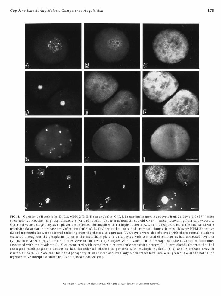
M-phase chromatin patterns with interphase forms beingseen predominantly (Fig. 4). Oocytes with interphase nucleidisplayed decondensed chromatin (Figs. 4A, 4J, 1, and 4J, 2),whereas M-phase chromosomes appeared as condensedchromosomal bivalents (Figs. 4G and 4J, 3). The distinctionbetween interphase and M phase was further documentedby analysis of microtubule organization and MPM-2 orhistone-3 phosphorylation patterns. Oocytes with germinalvesicle chromatin patterns (Figs. 4A and 4J, 1) displayedinterphase microtubule arrays (Figs. 4C and 4L, 1), restora-tion of the nuclear MPM-2 epitope (Fig. 4B), and loss ofphosphohistone-3 epitope (Fig. 4K, 1). Oocytes that con-tained a condensed chromatin aggregate (Fig. 4D) had mi-crotubule arrays radiating from chromatin (Fig. 4F) and lowlevels of MPM-2 phosphorylation in the cytoplasm (Fig.4H). M-phase oocytes with chromosomal bivalents wereeither organized on a metaphase plate of the meiotic spindle(Fig. 4J, 3) or scattered throughout the cytoplasm (Fig. 4G).No localized MPM-2 reactivity was observed in the cyto-plasm of these oocytes (Fig. 4H), although normal meioticspindles typically display MPM-2 reactivity at spindle poles(Messinger and Albertini, 1991). Histone-3 phosphorylationwas observed when chromosomal bivalents were evident(Fig. 4K, 3). Interestingly, oocytes with chromosomesaligned at the metaphase plate (Fig. 4J, 3) contained spindle-associated microtubules and non-spindle-associatedmicrotubule-organizing centers (Fig. 4L, 3, arrowhead), ahallmark of stable metaphase progression (Maro et al.,1985; Messinger and Albertini, 1991). Oocytes were alsoobserved that contained decondensed chromatin and mul-tiple nucleoli, patterns consistent with the appearance ofchromatin in pronuclei after parthenogenetic activation(Fig. 4J, 2). These oocytes displayed interphase microtubulearrays (Fig. 4L, 2) and were phosphohistone-3 negative (Fig.4K, 2), confirming restoration of an interphase state.
The above criteria were used to determine the stabilityand progress through M phase in incompetent oocytesinduced to undergo meiotic resumption and removed from
d Mice Following a 6-h Exposure to Okadaic Acid (1 mM)
Percentage of the total number of oocytes examined
GVBD CB CC
0 0 053.1 (60) 27.4 (31) 15.9 (18)1.5 (3) 0 0
19.5 (43) 15 (33) 53.2 (117)
total oocyte number evaluated in each group; GV, germinal vesiclerical shape with extended chromosomal fibers; CB, chromosomal
given in parentheses.
y-Ol
5)4)6)7)
d. N,sphe
OA (Table 4). Greater than 70% of the oocytes had resumed
s of reproduction in any form reserved.
175Gap Junctions during Meiotic Competence Acquisition
FIG. 4. Correlative Hoechst (A, D, G,), MPM-2 (B, E, H), and tubulin (C, F, I, L) patterns in growing oocytes from 21-day-old Cx371/2 miceor correlative Hoechst (J), phosphohistone-3 (K), and tubulin (L) patterns from 21-day-old Cx372/2 mice, recovering from OA exposure.Germinal vesicle stage oocytes displayed decondensed chromatin with multiple nucleoli (A, J, 1), the reappearance of the nuclear MPM-2reactivity (B), and an interphase array of microtubules (C, L, 1). Oocytes that contained a compact chromatin mass (D) were MPM-2 negative(E) and microtubules were observed radiating from the chromatin aggregate (F). Oocytes were also observed with chromosomal bivalentsscattered throughout the cytoplasm (G) or at the metaphase plate (J, 3). Oocytes with scattered chromosomes had decreased levels ofcytoplasmic MPM-2 (H) and microtubules were not observed (I). Oocytes with bivalents at the metaphase plate (J, 3) had microtubulesassociated with the bivalents (L, 3) or associated with cytoplasmic microtubule-organizing centers (L, 3, arrowhead). Oocytes that hadundergone parthenogenetic activation had decondensed chromatin patterns with multiple nucleoli (J, 2) and interphase array of
microtubules (L, 2). Note that histone-3 phosphorylation (K) was observed only when intact bivalents were present (K, 3) and not in therepresentative interphase states (K, 1 and 2) (scale bar, 20 mm).Copyright © 2000 by Academic Press. All rights of reproduction in any form reserved.

vcbcCgphaiiOt
crtqs
aotr
prccttdfi(1(1G
e cytber o
176 Carabatsos et al.
meiosis at 6 h, the time point at which OA was removed.The majority of growing oocytes from Cx371/2 mice re-erted to a stage I and II germinal vesicle (70%), whileomparable fractions displayed either aligned chromosomalivalents (15%) or chromosomes scattered throughout theytoplasm (15%, Table 4). The majority of oocytes fromx372/2 mice reverted to an interphase state with eithererminal vesicle (68.2%) or pronuclear (16.4%) chromatinatterns. A small percentage of oocytes from Cx372/2 micead meiotic spindles with aligned chromosomes (4.5%) andbout 10% showed compacted chromatin. These resultsllustrate that while growing mouse oocytes have the abil-ty to condense chromatin and enter M phase in response toA, the vast majority are unable to sustain or progress
hrough M phase upon OA removal.
DISCUSSION
Bidirectional communication between granulosa cellsand oocytes in the ovarian follicle has long been viewed asan essential feature for the coordination of oogenesis withfolliculogenesis. Using mice in which the gene encodingCx37 has been experimentally deleted, it has been possibleto assess the role of gap junction communication in follicleand oocyte development. Our results demonstrate thatoocyte growth (hypertrophy) is gap junction dependent,further reinforcing the view that metabolic coupling pro-vides a means for substrate transfer in the biosyntheticallyactive growing oocyte. Interestingly, while the completionof oocyte growth is impaired, oocytes in Cx37-deficientmice initiate and undergo a modest degree of growth (up toa diameter of 52 mm), which reinforces the notion thatommunication pathways other than gap junctions mayegulate the earliest stages of folliculogenesis. Moreover,his work shows that continuation of follicle growth re-uires a signal dependent on Cx37 beyond the preantraltage.
The bidirectional nature of gap junction communicationnd its role in the coordination of folliculogenesis andogenesis have been illustrated in two novel findings fromhis work. First, follicle development was uniformly ar-
TABLE 4Chromatin Patterns in Growing Mouse Oocytes from 21-Day-Old
Genotype N
Perc
GV (I/II) C
2/1 40 70 (28) 02/2 110 68.2 (75) 10.1
Note. Data are given as the percentage of total oocytes examinedand II germinal vesicle patterns (see text); CC, a mass of compactespindle; Scattered, chromosomal bivalents scattered throughout thnuclei, phenotypic of parthenogenetically activated oocytes. Num
ested at the type 4 preantral stage, indicating that later
Copyright © 2000 by Academic Press. All right
roliferative and differentiative events in folliculogenesisequire gap junction-dependent signaling between the oo-yte and the granulosa cells. The observation that granulosaells undergo cell shape changes and proliferation followinghe activation of primordial follicles supports the idea thathe initiation of preantral follicle development may notepend on gap junctions per se, consistent with recentndings implicating paracrine factors at these early stages
Packer et al., 1994; Dong et al., 1996; Parrott and Skinner,999). Second, from an analysis of cell cycle markersMessinger and Albertini, 1991; Wickramasinghe et al.,991) and the use of OA to experimentally manipulate the
2/M cell cycle state (Gavin et al., 1991, 1992; Chesnel andEppig, 1996; deVanterey et al., 1996), a critical transitionpoint in oogenesis has been identified at which somatic cellinput via gap junctions coordinates nuclear and cytoplas-mic maturation and the completion of meiotic competenceacquisition.
It has been generally recognized that growing mouseoocytes lack the ability to enter M phase under in vitroconditions that support meiotic maturation of full-grownoocytes (Wassarman and Albertini, 1994). A number ofstudies have established that mouse oocytes, unlike thoseof other species, acquire meiotic competence around thetime of antrum formation and that competence is acquiredin two steps. In the first step, oocytes exhibit the ability toundergo GVBD and progress to metaphase I. Once oocytesare full grown, the second step allows them to progressthrough meiosis I and arrest at metaphase of meiosis II,when they are designated fully competent oocytes (Erick-son and Ryan, 1976; Wickramasinghe et al., 1991; Albertini,1992; Wickramasinghe and Albertini, 1992). A similar pro-cess is observed in domestic species as well (Mermillod etal., 1999). While fully competent oocytes acquire and retainmarkers of M-phase progression during development of theantral follicle, they are prevented from resuming meiosisdue to cell cycle-arresting factors transferred by somaticcells to the oocyte through gap junctions (Bornslaeger andSchultz, 1985). In addition to their important role in antralfollicles, the present work calls attention to the involve-ment of heterologous gap junctions during the criticalpreantral–antral transition of the follicle. While the conse-
Recovering from OA Exposure (250 nM)
e of the total number of oocytes examined
Spindle Scattered Pronuclei
15 (6) 15 (6) 04.5 (5) 0 16.4 (18)
total number of oocytes evaluated in each group; GV (I/II), stage Iromatin; Spindle, chromosomal bivalents aligned on a metaphaseoplasm; Pronuclei contain decondensed chromatin with multiplef oocytes examined is given in parentheses.
Mice
entag
C
(12)
. N,d ch
quences of abrogated Cx37-based communication on granu-
s of reproduction in any form reserved.

iaegMindipm
Md
sr1uempotatiyGifatccorttrptCrhptc
oictaTfataadtcFqsicct
177Gap Junctions during Meiotic Competence Acquisition
losa cells remain to be more fully established, our studieson oocyte meiotic resumption and M-phase stability shedlight on both the chronology and the control of this aspectof oogenesis. As discussed below the phenotypic character-istics of oocytes from Cx37-deficient mice, based on expres-sion of cell cycle markers and recovery from OA-inducedM-phase entry, suggest that the coordination of nuclear andcytoplasmic structural alterations is due to both gapjunction-dependent (cytoplasmic) and gap junction-independent mechanisms.
Previous studies have shown that the majority of growingmouse oocytes are unable to enter M phase in response toOA. However, if such incompetent oocytes are freed ofsomatic cells and cultured for several days (Chesnel andEppig, 1996) or if microinjected with mRNA encodingeither p34cdc2 or cyclin B (deVantery et al., 1997), OAresponsiveness increases. These experiments suggest thatan increase in MPF (p34cdc2/cyclinB), due to prolongedculture or by microinjection, enhances OA-induced meioticresumption. Oocytes from Cx37-deficient mice show aheightened sensitivity to OA immediately upon removalfrom the follicle, differing from control growing mouseoocytes that require preculture. By sampling chromatinpatterns at 0, 3, and 6 h after OA treatment, a progressionfrom GVBD, to bivalents, and finally to compacted chroma-tin was established (data not shown). Moreover, as shown inTable 3, both a higher incidence of GVBD and a fasterM-phase progression, as indicated by the larger fraction ofoocytes with compacted chromatin masses, were observedin oocytes from Cx37-deficient animals compared to het-erozygous controls. The OA response in oocytes fromCx37-deficient mice did not compromise oocyte viabilitysince normal microtubule arrays were reestablished uponOA removal and no signs of oocyte degeneration wereobserved up to 20 h in culture. Furthermore, recoveryfollowing OA exposure revealed an inability to sustain Mphase in oocytes from Cx37-deficient mice as evidenced byrestoration of decondensed chromatin and interphase mi-crotubule arrays, whereas oocytes from heterozygous mice(30%) were able to maintain M phase. The reversal ofOA-induced M phase in conjunction with microtubuleprofiles clearly indicated that either factors necessary tosustain M phase (p34cdc2, cyclin B, and cdc25) or the signal-ng mechanisms to sustain an appropriate level of MPFctivation were lacking. Given strong evidence for thexpression of cyclin B and p34cdc2 early during oocyterowth (Mitra and Schultz, 1996) and the finding thatAP-kinase activation is sufficient to elicit M-phase entry
n growing mouse oocytes (Chesnel and Eppig, 1996), it isot surprising that OA treatment of oocytes from Cx37-eficient mice readily enter M phase. However, the inabil-ty to sustain M phase suggests that some component of theathway is lacking in these oocytes and may require so-atic cell input.Among markers previously utilized to document-phase progression in mouse oocytes are chromatin con-
ensation, microtubule organization, and MPM-2 centro- d
Copyright © 2000 by Academic Press. All right
ome phosphorylation (Mattson and Albertini, 1990; Wick-amasinghe et al., 1991; Wickramasinghe and Albertini,992). The present studies have extended the baseline andtility of cell cycle markers in establishing the temporalxpression of histone-3 phosphorylation during oocyteaturation in the mouse. We show that histone-3 phos-
horylation of chromatin was not detectable in full-grownocytes undergoing spontaneous maturation in vitro untilhe circular bivalent stage, a time when both microtubulessembly and centrosome position becomes restricted tohe perinuclear region (Mattson and Albertini, 1990; Mess-nger and Albertini, 1991). Moreover, centrosome phosphor-lation, a reversible and developmentally important sign of
2/M and meiotic competence acquisition (Wickramas-nghe and Albertini, 1992), was never observed in oocytesrom Cx372/2 mice prior to or following OA exposure. Thebsence of centrosome phosphorylation before or after OA-reatment is consistent with the notion of “immatureytoplasm” and suggests that the factors responsible forhanges in centrosome phosphorylation and microtubulerganization seen normally during oocyte developmentequire external somatic cell input mediated by gap junc-ions. In contrast, nuclear maturation up to and includinghe formation of chromosomal bivalents was initiated as aesult of OA exposure. It is of further interest that histone-3hosphorylation occurred precociously on chromatin prioro the resolution of bivalents in OA-treated oocytes fromx37 null mice and that upon removal of OA, chromatin
apidly decondensed and interphase nuclei lackingistone-3 phosphorylation reappeared. This suggests that ahosphatase is active at the time of meiotic resumptionhat prevents the phosphorylation of histone-3 until theircular bivalent stage.Thus, this work has shown that the ability of growing
ocytes to engage in early nuclear M-phase events is anntrinsic gap junction-independent event of oogenesis. Inontrast, the acquisition of cytoplasmic meiotic compe-ence is necessary to support complete nuclear maturationnd requires heterologous gap junction communication.hese results demonstrate the plasticity of GV chromatin
rom growing oocytes based on their ability to progress intond revert from meiotic M phase. Given the rapid reforma-ion of stage II GVs, which have decondensed chromatinnd reformed both nucleoli and the nuclear envelope, it ispparent that factors necessary to maintain interphase areominant upon removal of OA. This observation supportshe notion of cytoplasmic immaturity to sustain M phase,onsistent with cell cycle marker analysis described above.urther support of the idea that meiotic competence re-uires acquisition of cytoplasmic maturity derives from thetudies of Bao et al. (2000). Transplantation of nuclei fromncompetent growing mouse oocytes into enucleated oo-yte cytoplasts fully endows these nuclei with the ability toomplete meiotic maturation and upon in vitro fertiliza-ion, these oocytes develop to the blastocyst stage.
The detailed characterization of oocytes from Cx37-
eficient mice provided in this report has revealed key stepss of reproduction in any form reserved.

A
A
A
B
B
B
B
C
C
C
C
D
d
d
D
G
178 Carabatsos et al.
in preantral follicular development and oogenesis which areregulated by the supporting somatic cell compartment ofthe ovarian follicle. The Cx37 deficiency has resulted in aunique asynchrony of nuclear and cytoplasmic maturationin the oocyte. One explanation for this unique asynchronycould be ascribed to nutritional deficiencies resulting froma loss of gap-junctional transfer of essential metabolites,compromising patterns of transcription and protein synthe-sis at a stage with a critical metabolic requirement. Alter-natively, it is also possible that the granulosa cells generatea precisely timed surge of a gap junction-permeable (,1000Da) signal that triggers a regulated cascade in the ooplasmrequired to move the oocyte past a novel preantral/antraltransition point. Methods to culture ovarian follicles affordthe opportunity to restore connexin proteins by intraoocyteinjections of mRNAs to rescue the mutant phenotype.These experiments would permit an understanding of therequirements for a specific member of the connexin familyand possibly shed light on the nature of the transmittedsignals.
ACKNOWLEDGMENTS
This work was supported by NIH Grants HD20068 (D.A.),GM18974 (D.A.G.), and GM37751 (David Paul). M.J.C. is supportedin part by the NIH Training Program in Developmental BiologyHD07403. We thank Dr. Stephen Doxsey for the anti-pericentrinantibody. We also thank Catherine Combelles and Elizabeth Be-necchi for discussions and assistance in the generation of themanuscript.
REFERENCES
Albertini, D. F. (1992). Regulation of meiotic maturation in themammalian oocyte: Interplay between exogenous cues and themicrotubule cytoskeleton. BioEssays 14, 97–103.
Albertini, D. F., and Eppig, J. J. (1995). Unusual cytoskeletal andchromatin configurations in mouse oocytes that are atypical inmeiotic progression. Dev. Genet. 16, 13–19.lbertini, D. F., Fawcett, D. W., and Olds, P. J. (1975). Morphologi-cal variations in gap junctions of ovarian granulosa cells. TissueCell Res. 7, 389–405.lexandre, H., Cauwenberge, A. V., Tsukitant, Y., and Mulnard, J.(1991). Pleiotropic effect of okadaic acid on maturing mouseoocytes. Development 112, 971–980.nderson, E., and Albertini, D. F. (1976). Gap junctions betweenthe oocyte and companion follicle cells in the mammalian ovary.J. Cell Biol. 71, 680–686.
ao, S., Obata, Y., Carroll, J., Domeki, I., and Kono, T. (2000).Epigenetic modifications necessary for normal development areestablished during oocyte growth in mice. Biol. Reprod. 62,616–621.
iggers, J. D., Whittingham, D. G., and Donahue, R. P. (1967). Thepattern of energy metabolism in the mouse oocyte and zygote.Proc. Natl. Acad. Sci. 58, 560–567.
ock, G., Hilchenbach, M., Schausenstein, K., and Wick, G. (1985).Photometric analysis of antifading reagents for immunofluores-
cence with laser and conventional illumination sources. J. His-tochem. Cytochem. 33, 699–705.Copyright © 2000 by Academic Press. All right
ornslaeger, E., and Schultz, R. (1985). Regulation of mouse oocytematuration: Effect of elevating cumulus cell cAMP on oocytecAMP levels. Biol. Reprod. 33, 698–704.arabatsos, M. J., Elvin, J., Matzuk, M. M., and Albertini, D. F.(1998). Characterization of oocyte and follicle development ingrowth differentiation factor-9-deficient mice. Dev. Biol. 204,373–384.eccione, S., Rossi, G., Defelici, M., and Colonna, R. (1996).Mammalian oocyte growth in vitro is stimulated by solublefactor(s) produced by preantral granulosa cells and by Sertolicells. Mol. Reprod. Dev. 44, 540–546.hadee, D. N., Hendzel, M. J., Tylipski, C. P., and Allis, C. D.(1999). Increased Ser-10 phosphorylation of histone H3 inmitogen-stimulated and oncogene-transformed mouse fibro-blasts. J. Biol. Chem. 274, 24914–24920.hesnel, F., and Eppig, J. J. (1996). Induction of precocious germinalvesicle breakdown (GVB) by GVB-incompetent mouse oocytes:Possible role of mitogen activated protein kinase rather thanp34cdc2 kinase. Biol. Reprod. 52, 895–902.ekel, N., Lawrence, T., Gilula, N., and Beers, W. (1981). Modula-tion of cell-to-cell communication in the cumulus–oocyte com-plex and the regulation of oocyte maturation by LH. Dev. Biol.80, 356–362.
eVanterey, C., Gavin, A.-C., Vassalli, J.-D., and Schorderet-Slatkine, S. (1996). An accumulation of p34cdc2 at the end ofmouse oocyte growth correlates with the acquisition of meioticcompetence. Dev. Biol. 174, 335–344.
eVantery, C., Stutz, A., Vassalli, J. D., and Schorderet-Slatkine, S.(1997). Acquisition of meiotic competence in growing mouseoocytes is controlled at both translational and posttranslationallevels. Dev. Biol. 187, 43–54.ictenberg, J. B., Zimmerman, W., Sparks, C. A., Young, A., Vidair,C., Zheng, Y., Carrington, W., Fay, F. S., and Doxsey, S. J. (1998).Pericentrin and g-tubulin form a protein complex and are orga-nized into a novel lattice at the centrosome. J. Cell Biol. 141,163–174.
Donahue, R. P., and Stern, S. (1968). Follicular cell support ofoocyte maturation: Production of pyruvate in vitro. J. Reprod.Fertil. 17, 395–398.
Dong, J., Albertini, D. F., Nishimori, K., Kumar, T. R., Lu, N., andMatzuk, M. M. M. (1996). Growth differentiation factor-9 isrequired during early ovarian folliculogenesis. Nature 383, 531–535.
Elvin, J. A., Yan, C., Wang, P., Nishimori, K., and Matzuk, M. M.(1999). Molecular characterization of the follicle defects in thegrowth differentiation factor-9 deficient ovary. Mol. Endocrinol.13, 1018–1034.
Eppig, J. J. (1979). A comparison between oocyte growth in cocul-ture with granulosa cells and oocytes with granulosa cell–oocytejunctional contact maintained in vitro. J. Exp. Zool. 209, 345–353.
Eppig, J. J., Chesnel, F., Hirao, Y., O’Brien, M. J., Pendola, F. L.,Watanabe, S., and Wigglesworth, K. (1997). Oocyte control ofgranulosa cell development: How and why. Hum. Reprod. 12,127–132.
Erickson, G. F., and Ryan, K. J. (1976). Spontaneous maturation ofoocytes isolated from ovaries of immature hypophysectomizedrats. J. Exp. Zool. 195, 153–158.avin, A.-C., Tsukitani, Y., and Schorderet-Slatkine, S. (1991).Induction of M-phase entry of prophase-blocked mouse oocytes
through microinjection of okadaic acid, a specific phosphataseinhibitor. Exp. Cell Res. 192, 75–81.s of reproduction in any form reserved.
P
S
S
S
S
V
W
W
W
W
179Gap Junctions during Meiotic Competence Acquisition
Gavin, A.-C., Vassalli, J. D., Cavadore, J.-C., and Schorderet-Slatkine, S. (1992). Okadaic acid and p13suc1 modulate the reini-tiation of meiosis in mouse oocytes. Mol. Reprod. Dev. 33,287–296.
Gilula, N., Epstein, M., and Beers, W. (1978). Cell-to-cell commu-nication and ovulation: A study of the cumulus cell–oocytecomplex. J. Cell Biol. 78, 58–75.
Godin, I., Deed, R., Cooke, J., Zsebo, K., Dexter, M., and Wylie,C. C. (1991). Effects of the steel gene product on mouse primor-dial germ cells in culture. Nature (London) 352, 807–809.
Goodenough, D. A., Goliger, J. S., and Paul, D. L. (1996). Connex-ins, connexons, and intercellular communication. Ann. Rev.Biochem. 65, 475–502.
Gosden, R., Krapez, J., and Briggs, D. (1997). Growth and develop-ment of the mammalian oocyte. BioEssays 19, 875–882.
Gougeon, A. (1996). Regulation of ovarian follicular developmentin primates: Facts and hypothesis. Endocr. Rev. 17, 121–155.
Haghighat, N., and Van Winkle, L. J. (1990). Developmental changein follicular cell-enhanced amino acid uptake into mouse oocytesthat depends on intact gap junctions and transport system Gly. J.Exp. Zool. 253, 71–82.
Halpin, D. M. G., Jones, A., Fink, G., and Charlton, H. M. (1986).Postnatal ovarian follicle development in hypogonadal (hpg) andnormal mice and associated changes in the hypothalamic–pituitary ovarian axis. J. Reprod. Fertil. 77, 287–296.
Hendzel, M. J., Wei, Y., Mancini, M. A., VanHooser, A., Ranalli, T.,Brinkley, B. R., Bazett-Jones, D. P., and Allis, C. D. (1997).Mitosis specific phosphorylation of histone H3 initiates primar-ily within pericentromeric heterochromatin during G2 andspreads in an ordered fashion coincident with mitotic chromo-some condensation. Chromosoma 106, 348–360.
Herman, B., Langevin, M. A., and Albertini, D. F. (1983). The effectsof taxol in the organization of the cytoskeleton in culturedovarian granulosa cells. Eur. J. Cell Biol. 31, 34–45.
Hirshfield, A. N. (1991). Development of follicles in the mamma-lian ovary. Int. Rev. Cytol. 124, 43–101.
Joyce, I. M., Pendola, F. L., Wigglesworth, K., and Eppig, J. J. (1999).Oocyte regulation of kit ligand expression in mouse ovarianfollicles. Dev. Biol. 214, 342–353.
Kumar, T. R., Wang, Y., Lu, N., and Matzuk, M. M. M. (1997).Follicle stimulating hormone is required for ovarian folliclematuration but not male fertility. Nat. Genet. 15, 201–204.
Maro, B., Howlett, S. K., and Webb, M. (1985). Non-spindle micro-tubule organizing centers in metaphase-II arrested mouse oo-cytes. J. Cell Biol. 101, 1665–1672.
Mattson, B. A., and Albertini, D. F. (1990). Oogenesis: Chromatinand microtubule dynamics during meiotic prophase. Mol. Re-prod. Dev. 25, 374–383.
Mermillod, P., Oussaid, B., and Cognie, Y. (1999). Aspects offollicular and oocyte maturation that affect the developmentalpotential of embryos. J. Reprod. Fertil. 54(Suppl.), 449–460.
Messinger, S. M., and Albertini, D. F. (1991). Centrosome andmicrotubule dynamics during meiotic progression in the mouseoocyte. J. Cell Sci. 100, 289–298.
Mitra, J., and Schultz, R. M. (1996). Regulation of the acquisition ofmeiotic competence in the mouse: Changes in the subcellular
Copyright © 2000 by Academic Press. All right
localization of cdc2, cyclin B1, cdc25C and wee1, and in theconcentration of these proteins and their transcripts. J. Cell Sci.109, 2407–2415.
Packer, A., Hsu, Y., Besmer, P., and Bachvarova, R. (1994). Theligand of the c-kit receptor promotes oocyte growth. Dev. Biol.161, 194–205.
Parrott, J. A., and Skinner, M. K. (1999). Kit ligand/stem cell factorinduces primordial follicle development and initiates folliculo-genesis. Endocrinology 140, 4262–4271.
Paulson, J. R., and Taylor, S. S. (1982). Phosphorylation of histones1 and 3 and nonhistone high mobility group 14 by an endogenouskinase in HeLa metaphase chromosomes. J. Biol. Chem. 257,6064–6072.
Pedersen, T. (1969). Follicle growth in the immature mouse ovary.Acta Endocrinol. 62, 117–132.
Pedersen, T., and Peters, H. (1968). Proposal for a classification ofoocytes and follicles in the mouse ovary. J. Reprod. Fertil. 17,555–557.
eters, H. (1969). The development of the mouse ovary from birthto maturity. Acta Endocrinol. 62, 98–116.
chroeder, A. C., and Eppig, J. J. (1984). The developmental capacityof mouse oocytes that matured spontaneously in vitro is normal.Dev. Biol. 102, 493–497.
chwartz, D., and Schultz, R. M. (1991). Stimulatory effect ofokadaic acid, an inhibitor of protein phosphatases, on nuclearenvelope breakdown and protein phosphorylation in mouseoocytes and one-cell embryos. Dev. Biol. 145, 119–127.
imon, A. M., and Goodenough, D. A. (1998). Diverse functions ofvertebrate gap junctions. Trends Cell Biol. 8, 477–483.
imon, A. M., Goodenough, D. A., Li, E., and Paul, D. L. (1997).Female infertility in mice lacking connexin 37. Nature 385,525–529.
Tsafiri, A., and Channing, C. P. (1975). An inhibitory influence ofgranulosa cells and follicular fluid upon oocyte meiosis in vitro.Endocrinology 96, 922–927.
aldimarsson, G., DeSousa, P. A., and Kidder, G. M. (1993).Coexpression of gap junction proteins in the cumulus–oocytecomplex. Mol. Reprod. Dev. 36, 7–15.assarman, P. M., and Albertini, D. F. (1994). The mammalianovum. In “The Physiology of Reproduction” (E. Knobil and J. D.Neill, Eds.), pp. 79–122. Raven Press, New York.ei, Y., Yu, L., Bown, J., Gorovsky, M. A., and Allis, C. D. (1999).Phosphorylation of histone H3 is required for proper chromo-some condensation and segregation. Cell 97, 99–109.ickramasinghe, D., and Albertini, D. F. (1992). Centrosomephosphorylation and the developmental expression of meioticcompetence in mouse oocytes. Dev. Biol. 152, 62–74.ickramasinghe, D., Ebert, K. M., and Albertini, D. F. (1991).Meiotic competence acquisition is associated with the appear-ance of M-phase characteristics in growing mouse oocytes. Dev.Biol. 143, 162–172.
Received for publication March 31, 2000Revised June 6, 2000
Accepted June 27, 2000Published online September 20, 2000
s of reproduction in any form reserved.