Restraint Stress Impairs Oocyte Developmental Potential in Mice
Upload
independentCategory
view
3download
0
04 (2007) 317–325www.elsevier.com/locate/ydbio
Developmental Biology 3
Oocyte CD9 is enriched on the microvillar membrane and required fornormal microvillar shape and distribution
Kathryn E. Runge a, James E. Evans a, Zhi-Yong He a, Surabhi Gupta a, Kent L. McDonald b,Henning Stahlberg a, Paul Primakoff c,⁎, Diana G. Myles a
a Section of Molecular and Cellular Biology, University of California Davis, Davis, CA 95616, USAb Electron Microscope Laboratory, University of California, Berkeley, CA 94720, USA
c Department of Cell Biology and Human Anatomy, School of Medicine, University of California Davis, Davis, CA 95616, USA
Received for publication 25 October 2006; revised 15 December 2006; accepted 18 December 2006Available online 23 December 2006
Abstract
Microvilli are found on the surface of many cell types, including the mammalian oocyte, where they are thought to act in initial contact ofsperm and oocyte plasma membranes. CD9 is currently the only oocyte protein known to be required for sperm–oocyte fusion. We found CD9 islocalized to the oocyte microvillar membrane using transmission electron microscopy (TEM). Scanning electron microscopy (SEM) showed thatCD9 null oocytes, which are unable to fuse with sperm, have an altered length, thickness and density of their microvilli. One aspect of this changein morphology was quantified using TEM by measuring the radius of curvature at the microvillar tips. A small radius of curvature is thought topromote fusibility and the radius of curvature of microvillar tips on CD9 wild-type oocytes was found to be half that of the CD9 null oocytes. Wefound that oocyte CD9 co-immunoprecipitates with two Ig superfamily cis partners, EWI-2 and EWI-F, which could have a role in linking CD9 tothe oocyte microvillar actin core. We also examined latrunculin B-treated oocytes, which are known to have reduced fusion ability, and foundaltered microvillar morphology by SEM and TEM. Our data suggest that microvilli may participate in sperm–oocyte fusion. Microvilli could actas a platform to concentrate adhesion/fusion proteins and/or provide a membrane protrusion with a low radius of curvature. They may also have adynamic interaction with the sperm that serves to capture the sperm cell and bring it into close contact with the oocyte plasma membrane.© 2006 Elsevier Inc. All rights reserved.
Keywords: Fertilization; Microvilli; CD9; Tetraspanin
Introduction
The surface of the mammalian oocyte can be divided intotwo regions. The larger region is covered with a densepopulation of microvilli, while the smaller region lacksmicrovilli. Sperm binding and fusion have been reported to berestricted to the microvilli-rich region of the oocyte, indicatingthe importance of this domain of the plasma membrane forsperm–oocyte fusion (Yanagimachi, 1978).
Microvilli are a surface feature of many cell types. They serve adiverse set of functions, from increasing absorptive surface area inthe epithelial brush border to the mechanosensitive reception ofhair cells of the inner ear (Heintzelman and Mooseker, 1992;
* Corresponding author.E-mail address: [email protected] (P. Primakoff).
0012-1606/$ - see front matter © 2006 Elsevier Inc. All rights reserved.doi:10.1016/j.ydbio.2006.12.041
Roberts et al., 1988). Relevant to this study, microvilli have alsobeen shown to participate in cell–cell adhesion during tethering ofleukocytes. For successful leukocyte tethering to the surface ofendothelial cells, the leukocyte adhesion protein L-selectin must berestricted to microvillar membranes (Ivetic et al., 2004).
Images of sperm–oocyte fusion, from both TEM and SEMstudies, have implicated oocyte microvilli in the fusion process.Scanning electron micrographs of mammalian oocytes show thesperm being enveloped by the oocyte microvilli during fusionwhile transmission electron micrographs suggest that the spermfuses first with the microvilli and not with the planar membraneregion between microvilli (Shalgi and Phillips, 1980; Yanagimachiand Noda, 1970).
In addition to microvilli, other actin-based protrusions are foundon the surface of cells. In non-mammalian fertilization, such actin-based protrusions have been shown to play a role by localizing

318 K.E. Runge et al. / Developmental Biology 304 (2007) 317–325
adhesion molecules to the acrosomal process of sea urchin spermand of fusion molecules to the fertilization tubule of the mt+Chlamydomonas gamete (Longo et al., 1994; Misamore et al.,2003; Monroy, 1985).
We have used two approaches to investigate the role of oocytemicrovilli in mouse sperm–oocyte fusion. First we asked if CD9,an oocyte surface protein required for gamete fusion, was localizedto the microvillar membrane. It has been shown by immuno-fluorescence that expression of CD9 is localized to the microvilli-rich region of the oocyte (Kaji et al., 2000). Here we studied thedistribution of CD9 within the microvilli-rich region. We alsoasked if oocyte CD9 is associated with the Ig super-family (IgSF)proteins EWI-2 and EWI-F. These two IgSF molecules may serveas linkers between surface proteins and ezrin, radixin and moesin(ERM proteins), which in turn bind to the actin core of microvilli(Sala-Valdes et al., 2006).
We also investigated oocytes that were impaired in their abilityto fuse with sperm to determine if they had an altered microvillardistribution or microvillar shape. We examined oocytes from CD9knockoutmicewhich have lost their ability to fusewith sperm (Kajiet al., 2000; Le Naour et al., 2000; Miyado et al., 2000). We alsoexamined oocytes treatedwith latrunculinBwith the aimof directlyaffectingmicrovillar structure. LatrunculinB inhibitsmicrofilamentpolymerization by sequesteringmonomeric actin and latrunculinB-treated oocytes had previously been shown to have reduced fusionability (McAvey et al., 2002).
In this study we found that CD9 is primarily localized to themembrane of the microvilli and sparse in the planar regionbetween the microvilli. CD9 on oocytes associates with two CD9cis partners, EWI-2 and EWI-F, which have a role of linking CD9to ERM proteins and hence the microvillar actin core. We alsodocumented an alteration in the structure and number of microvillion fusion-deficient oocytes. Our results are consistent with CD9localization and normal microvillar size, shape and density on theegg membrane being required for sperm–egg fusion.
Materials and methods
Gamete isolation
Wild-type ICR or C57BL/6 (Charles River, Wilmington, MA) female mice,6–8-week-old, were used for oocyte collection. Oocytes lacking CD9 werecollected from CD9 null mice (a gift from Dr. Claude Boucheix, INSERM,Villejuif, France). To obtain zona pellucida-free oocytes, females were super-ovulated, oocytes were collected and adherent cumulus cells were released asdescribed previously (Yuan et al., 1997). To loosen the zona pellucida (ZP), theoocytes were treated with 30 μg/ml chymotrypsin (Sigma-Aldrich, St. Louis,MO) for 3 min at 37 °C and 5% CO2 in medium M199 (Invitrogen, Carlsbad,CA) containing 3.5 mM sodium pyruvate (Sigma-Aldrich), and 1000 IU ofpenicillin–streptomycin (Invitrogen) (M199*). The zonae pellucidae were thenremoved mechanically using a narrow bore pipette. The oocytes were washedthrough three 100-μl drops of fresh M199* at which point they were ready forpreparation for imaging. Wild-type ICR mice were used in many experiments asthey were comparable to wild-type C57BL/6 in microvillar structure andovulated a greater number of oocytes.
Scanning electron microscopy (SEM)
Oocytes from wild-type and CD9 knockout mice were isolated as above andfixed in 2.5% glutaraldehyde (Sigma-Aldrich) and 1× Brinkley Buffer 80 (BRB80)
(Desai et al., 1999) in M199*. After fixation overnight at 4 °C, the oocytes weretransferred into double distilled water and dehydrated through a graded series ofacetones. Following washing with 100% ethanol, the oocytes were dried in aSamdri 780A critical point drying apparatus and were transferred onto a stub withcarbon tape, and coated with gold in a Denton Vacuum Desk II sputter coater. AHitachi S3500N scanning electron microscope was used for imaging.
SEM of treated oocytes
ZP-free oocytes were retrieved as described above and incubated with 4 μg/ml latrunculin B (VWR, Brisbane CA) or 0.5% DMSO in M199* for 1 h at37 °C in 5% CO2. They were then washed through four 100 μl drops of M199*and prepared as described above for imaging by SEM.
Transmission electron microscopy (TEM)
Cells were fixed as for SEM, washed through four 100 μl drops of doubledistilled water, then postfixed in 1% OsO4 (Ted Pella, Redding CA) for 1 h.After postfixation, oocytes were stained overnight in 1% aqueous uranyl acetate(UA). Samples were dehydrated through a graded series of acetones, infiltratedwith 50% eponate araldite resin (Ted Pella) in acetone, infused three times inpure resin and polymerized 48 h at 60 °C. 70 nm sections were prepared andstained with lead citrate and UA. Sections were examined on a Philips 410electron microscope or Philips CM120. Images were captured by the DigitalMicrograph imaging system.
Immunoelectron microscopy on oocytes
After isolation and ZP removal, oocytes were fixed in 4% paraformaldehyde,and labeled using a monoclonal antibody against CD9 (KMC8) (1:200 in M199*;BD Pharmingen, Palo Alto, CA) for 1 h, washed through four 100 μl drops ofM199* and then labeled with a 10 nm gold-conjugated anti-rat IgG secondaryantibody (Ted Pella #15771) for 30 min and washed through four 100 μl drops ofM199*. Samples were then prepared for TEM as described above. Some sampleswere first labeled with primary and secondary antibodies, followed by fixation.
Electron tomography
Immunolabeled samples were cut into 100–200 nm thick sections byultramicrotomy and were subsequently visualized on a JEM-2100F fieldemission gun transmission electron microscope operating at 200 keV. Imageswere recorded on a 2048×2048 pixel Tietz Video Image Processing CCDcamera with a nominal magnification of 24,000× and a defocus of ∼2 microns.Two perpendicular tilt-series (in-plane) of the same section were acquired in onedegree increments from −60 to +60 degrees. Data from two orthogonal tilt-series were aligned utilizing the 10 nm gold particles as fiducial markers toprecisely determine the exact tilt angle, skew and magnification of each 2-Dprojection image with the IMOD software package (Mastronarde, 1997). Thealigned tilt-series were then combined to generate a 3-D reconstruction of thevolume using a weighted back-projection algorithm and filtered for bettervisualization using a bilateral denoising algorithm (Jiang et al., 2003). Thetomogram was then analyzed and modeled as surface renderings to localize theouter-membrane and the immunolabeled gold (Kremer et al., 1996).
Calculations of membrane length, molecular distribution andmicrovillar curvature
Measurements of microvillar and planar membrane length were taken fromTEM images using Scion Image. Counts of gold particles were taken from theseimages and the number of gold particles recorded as either being on themeasured planar membrane or measured microvillar membrane. Radius ofcurvature at the tips of microvilli was determined by analyzing these sameimages using PhotoShop. Radius of curvature was determined to be the radius ofa circle which fits the curve at the tip of the microvillus. Only microvilli withdistinct tips were used. Twenty-five microvilli were measured for both CD9wild-type and CD9 null oocytes.

Fig. 1. Sperm bind to the region of the oocyte with a high concentration ofmicrovilli. (A) Wild-type oocyte showing microvilli-free (*) and microvilli-richregions. (B) Wild-type oocyte after incubation with sperm for 25 min. Sperm arenot bound to microvilli free region. Scale bars, 20 μm.
319K.E. Runge et al. / Developmental Biology 304 (2007) 317–325
Production of CD9 partners EWI-2 and EWI-F and antisera to theseproteins
Mouse EWI-2 cDNA was amplified by PCR from mouse heart cDNA(Clonetech). The primers used for this reaction were EWI-2F (AAGATC-TATGGGCGTCCCTAGCCCCACG) and EWI-2R (aagagctcccactctaggt-gatggctgg). The coding region for the EWI-2 extracellular domain (EWI-2EC)was then amplified from EWI-2 full-length cDNA by two primers: EWI-2F andEWI-2ec (aactcgagcacaaatagggtatccacagc). EWI-2EC was placed in a baculo-virus express system transfer vector (termed pFB-MH, a C-terminal myc-Histags donor vector derived from pFastBac1, constructed in our lab). Recombinantbaculovirus containing EWI-2EC coding sequence was obtained by followingthe baculovirus system protocol (Invitrogen). Recombinant EWI-2EC waspurified from infected Sf9 cell supernatant using a Ni-CAM resin column(Sigma). SDS-PAGE, followed by silver staining of 100 ng of purified protein,showed a single band migrating as expected for EWI-2EC and no contaminatingbands. Antisera against the EWI-2 extracellular domain were raised by injectingpurified EWI-2EC into rabbits. For immunofluorescent detection of EWI-2 onthe oocyte surface, a 1:100 dilution of antiserum was used, followed by anti-rabbit IgG conjugated to AlexaFluor 488 (Molecular Probes). Control oocyteswere incubated with a 1:100 dilution of pre-immune serum.
The entire extracellular portion of mouse EWI-F cDNA (bases 1–2490) wasPCR amplified using primers EWIF-F: 5′-CCTCAGACCGAGCATGGGGCG-3′ and EWIF-R: 5′-tcagcacgtccatcttcacagtta-3′. The PCR product was clonedinto pcDNA3.1/V5-His-TOPO vector (Invitrogen) following the manufacturer'sinstructions. The resulting plasmid was transfected into CHO cells usingLipofectamine 2000 (Invitrogen). Soluble recombinant EWI-F protein waspurified from the spent media using Ni+2-agarose beads (Qiagen). The proteinwas eluted with 250 mM imidazole (Sigma) and concentrated using centriprepYM-10 (Millipore). Silver staining of 10ug of purified protein per lane in SDS-PAGE revealed only one trace contaminant. The concentrated protein wasdialyzed extensively against PBS and used as an immunogen to generateantisera in guinea pigs. For immunofluorescent detection of EWI-F on theoocyte surface, a 1:50 dilution of antiserum was used, followed by anti-guineapig IgG conjugated to AlexaFluor 488 (Molecular Probes). Control oocytes wereincubated with a 1:50 dilution of pre-immune serum.
Immunoprecipitation of CD9
Cumulus-free, zona intact oocytes were collected from 100 female ICR miceas described above in M199* containing 0.4% polyvinylalcohol (PVA). Alloocytes were pooled into 1 ml of PBS+0.4% PVA on a culture dish and washedthree times through 1 ml of PBS/0.4% PVA on culture dishes. The oocytes werethen transferred to 200 μl of PBS lacking PVA, then again to 100 μl and finallyto 50 μl. Since the oocytes are a precious resource, they were used in twosequential experiments. After release of GPI-anchored proteins (for an unrelatedstudy), oocytes were washed and lysed in 100 μl HBS+1% Brij97 for 30 min at4 °C in the presence of protease inhibitors. Insoluble material was removed bycentrifugation at 14,000 rpm for 10 min at 4 °C and the lysate was stored at−80 °C. This complete procedure was performed seven times and over 14,000oocytes were obtained, lysed and the lysate frozen.
Lysates were thawed on ice, pooled and incubated for 3 h at 4 °C with 20 μlagarose bead slurry coupled to KMC8 mAb (Pharmingen). The beads wereremoved by centrifugation at 5000 rpm for 5 min at 4 °C and stored on ice. Thepooled lysates were incubated for anoth er 3 h at 4 °C with an additional 20 μl ofKMC8-coupled beads, which were then removed by centrifugation and pooled withthe previous beads. The pooled beads were washed with 200 μl HBS+1% Brij97five times and resuspended in 20 μl of 1× non-reducing SDS sample buffer.
Analysis of the immunoprecipitate
The immunoprecipitate in 20 μl of SDS-PAGE sample buffer was heated at96 °C for 10 min. Separation of CD9 and its partners was carried out by SDS-PAGE (4–20% gel) under non-reducing conditions. The dye front was run 4 cmdown the gel after which the gel was stained using Colloidal Coomassie Blue for5 h at room temperature. Destaining was carried out overnight at roomtemperature. The sample lane was excised and cut into 5 pieces (<1 cm2). The
pieces were placed into sterile 1.5 ml centrifuge tubes and washed twice with1 ml of 50% acetonitrile. The samples were stored at −20 °C. Proteolyticdigestion and mass spectrometric analysis of the samples were performed at theHarvard Microchemistry Facility by microcapillary reverse-phase HPLC nano-electrospray tandem mass spectrometry (μLC/MS/MS) on a Finnigan LCQDECA XP Plus quadrupole ion trap mass spectrometer. Interpretation of theresults was provided by the Harvard Microchemistry Facility and wasadditionally evaluated by a manual protein BLAST search of the NCBI proteindatabase.
Results
CD9 is expressed on mouse oocyte microvilli
The mature mouse oocyte plasma membrane is divided intotwo large subdomains. The region over the metaphase arrestedchromosomes has a relatively smooth surface. The rest of theoocyte is covered with microvilli and is the “microvilli-richregion” where sperm bind and fuse (Fig. 1). CD9 has beenfound by immunofluorescence to be enriched on the microvilli-rich domain of the oocyte (Kaji et al., 2000). To determine therelationship of CD9 to the microvilli, we examined wild-typeoocytes labeled with a monoclonal antibody to CD9 (KMC8)and a colloidal gold conjugated secondary antibody. Themajority of CD9 molecules were congregated on the microvilli

320 K.E. Runge et al. / Developmental Biology 304 (2007) 317–325
and not on the planar membrane regions in between themicrovilli. This was true whether the cells were labeled beforefixation (Fig. 2A) or after fixation with paraformaldehyde (Fig.2B). No gold particles were bound to wild-type oocytes labeledwith only the gold-conjugated secondary antibody and noKMC8 (Fig. 2C). Also CD9 null oocytes labeled with KMC8and gold-conjugated secondary antibody exhibited no goldparticles (data not shown).
However, as individual 2-D electron micrographs are projec-tions of the entire section, it was difficult in some cases to assign agold particle to either membrane. Therefore, we utilized electrontomography to obtain a more complete representation of thelocalization of CD9 on the oocyte surface. This technique allowedfor the visualization of a 3-D volume of each microvillus and cleardetermination of CD9 distribution along the entire microvillarmembrane. This was achieved by acquiring dual-axis tilt series forseveral 100–200 nm thick sections of oocyte microvilli. These datasets were aligned and back-projected to reconstruct 3-Dtomograms which could be modeled and analyzed. The tomogramreproduced in Fig. 2D shows immunolabeled molecules spreadalong the microvillus with a tendency to localize at the tip.
We then quantified this distribution with TEM bymeasuring the lengths of the planar membranes and of
Fig. 2. Localization of CD9 to individual microvilli. (A) Electron micrograph of wilElectron micrograph of wild-type oocyte labeled for CD9 with paraformaldehyde prewith only gold-conjugated secondary antibody (scale bar, 0.2 μm). (D) RepresentativOocyte plasma membrane is depicted in magenta and CD9molecules as yellow sphereplanar membrane versus microvillar membrane. Each blue diamond represents a regmembrane.
microvillar membranes from the images, and counting thebound gold particles. The number of gold particles per linearmicron of membrane was calculated from these measurements(Fig. 2E). Calculations were done using oocytes prefixed inparaformaldehyde before antibody labeling to prevent anti-body-induced changes in CD9 distribution. The concentrationof CD9 molecules on microvillar membrane was found to bein average ∼4 times greater than that on the planar membrane(Fig. 2E).
Deletion of CD9 results in altered microvillar morphology
To determine if deleting CD9 altered microvillar morphol-ogy, we examined oocytes from CD9 knockout mice by bothSEM and TEM. SEM images showed that on CD9 KO oocytesthe distribution of microvilli was quite dense compared towild-type oocytes. Fig. 3 shows the microvilli of a representa-tive wild-type oocyte (Fig. 3A) and a representative CD9 nulloocyte (Fig. 3B). A qualitative, visual assessment of thedensity of the microvilli from images of these two types ofoocytes is shown in Table 1 and it indicates that CD9 knockoutoocytes show denser arrays of microvilli than wild-typeoocytes.
d-type oocyte labeled live for CD9 with no prefixation (scale bar, 0.5 μm). (B)fixation (scale bar, 0.2 μm). (C) Electron micrograph of wild-type oocyte labelede model generated from a 3-D tomogram of wild-type oocyte labeled for CD9.s. (E) Distribution plot of number of gold particles per linear micron of sectionedion of microvillar membrane and each red triangle represents a region of planar
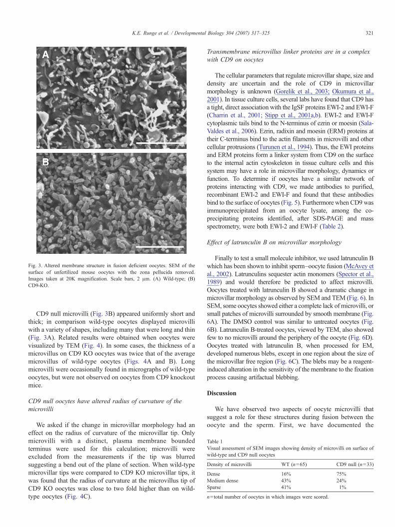
Fig. 3. Altered membrane structure in fusion deficient oocytes. SEM of thesurface of unfertilized mouse oocytes with the zona pellucida removed.Images taken at 20K magnification. Scale bars, 2 μm. (A) Wild-type; (B)CD9-KO.
Table 1Visual assessment of SEM images showing density of microvilli on surface ofwild-type and CD9 null oocytes
Density of microvilli WT (n=65) CD9 null (n=33)
Dense 16% 75%Medium dense 43% 24%Sparse 41% 1%
n=total number of oocytes in which images were scored.
321K.E. Runge et al. / Developmental Biology 304 (2007) 317–325
CD9 null microvilli (Fig. 3B) appeared uniformly short andthick; in comparison wild-type oocytes displayed microvilliwith a variety of shapes, including many that were long and thin(Fig. 3A). Related results were obtained when oocytes werevisualized by TEM (Fig. 4). In some cases, the thickness of amicrovillus on CD9 KO oocytes was twice that of the averagemicrovillus of wild-type oocytes (Figs. 4A and B). Longmicrovilli were occasionally found in micrographs of wild-typeoocytes, but were not observed on oocytes from CD9 knockoutmice.
CD9 null oocytes have altered radius of curvature of themicrovilli
We asked if the change in microvillar morphology had aneffect on the radius of curvature of the microvillar tip. Onlymicrovilli with a distinct, plasma membrane boundedterminus were used for this calculation; microvilli wereexcluded from the measurements if the tip was blurredsuggesting a bend out of the plane of section. When wild-typemicrovillar tips were compared to CD9 KO microvillar tips, itwas found that the radius of curvature at the microvillus tip ofCD9 KO oocytes was close to two fold higher than on wild-type oocytes (Fig. 4C).
Transmembrane microvillus linker proteins are in a complexwith CD9 on oocytes
The cellular parameters that regulate microvillar shape, size anddensity are uncertain and the role of CD9 in microvillarmorphology is unknown (Gorelik et al., 2003; Okumura et al.,2001). In tissue culture cells, several labs have found that CD9 hasa tight, direct association with the IgSF proteins EWI-2 and EWI-F(Charrin et al., 2001; Stipp et al., 2001a,b). EWI-2 and EWI-Fcytoplasmic tails bind to the N-terminus of ezrin or moesin (Sala-Valdes et al., 2006). Ezrin, radixin and moesin (ERM) proteins attheir C-terminus bind to the actin filaments in microvilli and othercellular protrusions (Turunen et al., 1994). Thus, the EWI proteinsand ERM proteins form a linker system from CD9 on the surfaceto the internal actin cytoskeleton in tissue culture cells and thissystem may have a role in microvillar morphology, dynamics orfunction. To determine if oocytes have a similar network ofproteins interacting with CD9, we made antibodies to purified,recombinant EWI-2 and EWI-F and found that these antibodiesbind to the surface of oocytes (Fig. 5). Furthermore when CD9wasimmunoprecipitated from an oocyte lysate, among the co-precipitating proteins identified, after SDS-PAGE and massspectrometry, were both EWI-2 and EWI-F (Table 2).
Effect of latrunculin B on microvillar morphology
Finally to test a small molecule inhibitor, we used latrunculin Bwhich has been shown to inhibit sperm–oocyte fusion (McAvey etal., 2002). Latrunculins sequester actin monomers (Spector et al.,1989) and would therefore be predicted to affect microvilli.Oocytes treated with latrunculin B showed a dramatic change inmicrovillar morphology as observed by SEM and TEM (Fig. 6). InSEM, some oocytes showed either a complete lack of microvilli, orsmall patches of microvilli surrounded by smooth membrane (Fig.6A). The DMSO control was similar to untreated oocytes (Fig.6B). Latrunculin B-treated oocytes, viewed by TEM, also showedfew to no microvilli around the periphery of the oocyte (Fig. 6D).Oocytes treated with latrunculin B, when processed for EM,developed numerous blebs, except in one region about the size ofthe microvillar free region (Fig. 6C). The blebs may be a reagent-induced alteration in the sensitivity of the membrane to the fixationprocess causing artifactual blebbing.
Discussion
We have observed two aspects of oocyte microvilli thatsuggest a role for these structures during fusion between theoocyte and the sperm. First, we have documented the

Fig. 4. Increase in radius of curvature of microvillar tips in CD9-KO oocytes. (A–B) TEM images of microvilli of unfertilized mouse oocytes with the zona pellucidaremoved (scale bars, 0.5 μm). (A) Wild-type; (B) CD9 KO; (C) Measurements of curvature were taken at the tips of microvilli represented in cross-sectional TEMimages of wild-type, and CD9 knockout oocytes, like those in panels A and B. Data represent the mean value±SE of microvillar tips from n=25 wild-type and n=25CD9 KO oocytes, *p<0.05.
Fig. 5. Immunofluorescent staining of oocytes with anti-EWI-F and anti-EWI-2 antibodies. (A–B) Anti-EWI-F antiserum staining and phase contrast of same oocytes.(C–D) Pre-immune serum staining and phase contrast of same oocytes. (E–F) Anti-EWI-2 antiserum staining and phase contrast of same oocytes. (G–H) Pre-immuneserum staining and phase contrast of same oocytes. Clear surface staining is observed on each oocyte using either anti-EWI-F or anti-EWI-2 antibodies. Staining withthe control, pre-immune antisera is negative except for one weakly stained oocyte in panel G.
322 K.E. Runge et al. / Developmental Biology 304 (2007) 317–325

Table 2Identification by mass spectrometry of CD9 and two other proteins that co-precipitate with CD9 in an oocyte-lysate
Peptide fragment sequence Closest protein match
LDTVGSDAYR EWI-F (prostaglandin F2 receptornegative regulator)VLADALVVGPSSRPPPGLSLR
VDGVVLEKKTPDTSLLASHMLAREGEPFELRLVAQLDTEGIGSKGPGYEDR EWI-2 (Ig super family receptor PGRL)SDMAVEAGAPYAERELQEFYKDTTYQK CD9SKDEPQRETLK
323K.E. Runge et al. / Developmental Biology 304 (2007) 317–325
localization of CD9 to the microvillar membrane. Additionally,we found a correlation between the loss of fusion ability ofoocytes and alteration in the morphology of their microvilli.
The finding that CD9, an oocyte protein required for gametefusion, is concentrated on the oocyte microvilli has severalpotential implications. One possibility is that the microvilliconcentrate CD9 molecules in a site that initially contacts thesperm (Yanagimachi et al., 1973). In other cell types,concentration of cell–cell adhesion molecules has been foundon microvilli. L-selectin on lymphocytes and l-gicerin onfibroblasts are both involved in cell adhesion and are localizedto microvillar structures (Ivetic et al., 2004; Okumura et al.,2001). It has also been shown in Chlamydomonas and the seaurchin that important fertilization molecules are localized to themembrane of actin-based protrusions where fusion occurs.
Fig. 6. Oocytes treated with latrunculin B lose most of the microvilli on the plasma mDMSO (solvent) (scale bars, 2 μm). Arrows show rare remaining microvilli after latruoocyte, showing distribution of blebs (scale bar, 20 μm). (D) TEM of latrunculin B-treimage of treated oocytes while a few remain in the SEM image (A) with severe ble
Chlamydomonas concentrates the fusion-related molecule Fus1to the tip surface of its mt+ fertilization tubule (Misamore et al.,2003) and the sea urchin sperm–oocyte adhesion protein bindinis concentrated on the surface of the sperm acrosomal process(Longo et al., 1994). Similar to these examples, localization ofCD9 within the microvillar membrane subdomain increasesCD9 surface density, which could control the adhesive/fusogenic properties of the membrane.
Another possibility is that localization of the tetraspaninCD9 reflects a participation of the tetraspanin web in theformation or maintenance of the microvillar structure. Thetetraspanin web is a network of interacting proteins on the cellsurface organized by various tetraspanins, including CD9(Hemler, 2003; Levy and Shoham, 2005; Rubinstein et al.,1996). The activities of microvilli depend on a functionalattachment of surface proteins to the internal actin cytoskeletonin the microvillus (Bartles, 2000; Fath and Burgess, 1995).While CD9 is known to associate with various other plasmamembrane proteins on a CD9-expressing cell, among the moststable CD9 partners are the novel IgSF proteins EWI-2 andEWI-F that belong to a small IgSF subfamily (Charrin et al.,2001; Stipp et al., 2001a,b). According to current findings,EWI-2 and EWI-F bind directly to ERM (ezrin, radixin andmoesin) proteins through their cytoplasmic tails. The EWIproteins can also bind indirectly to ERM by lateral associationwith their tetraspanin partner CD81 which binds to ERM (Sala-Valdes et al., 2006). Another recent report shows that CD9 incell–cell contact sites is laterally associated with I-CAM-1 or V-
embrane. SEM images of oocytes treated with: (A) latrunculin B and (B) 0.5%nculin B treatment. (C) Lower magnification SEM view of latrunculin B-treatedated oocyte (scale bar, 1 μm). All microvilli are apparently absent from the TEMbbing of membrane.

324 K.E. Runge et al. / Developmental Biology 304 (2007) 317–325
CAM-1, each of which is an IgSF protein that can intracellularlybind to ERM proteins (Barreiro et al., 2005). ERM proteins inturn bind to the actin filaments of the microvillar core (Turunenet al., 1994). These studies in tissue culture cells define anetwork of proteins that attach CD9 on the cell surface to theactin cytoskeleton, in a CD9-IgSF-ERM-actin linkage. Usingboth immunofluorescence microscopy and mass spectrometry,we found that EWI-2 and EWI-F are present in oocytes and co-precipitate with CD9, suggesting a functional connection in theoocyte microvilli. In a recent review (Chen and Olson, 2005), itis proposed that cell–cell fusion in general will requireappropriate deployment of the actin cytoskeleton and ourfindings on mouse gamete fusion are consistent with this idea.The detailed role of a possible oocyte CD9-EWI-ERM-actinnetwork remains to be studied.
The observation that the oocytes impaired in fusion (CD9null oocytes or oocytes treated with latrunculin B) (Kaji et al.,2000; Le Naour et al., 2000; McAvey et al., 2002; Miyado et al.,2000) also show alterations in microvillar structure, implies thatthe microvilli may have an important structural role in fusion.Several potential mechanisms involved in a structural role ofmicrovilli can be considered.
First, microvilli on the oocyte surface may provide aprotrusion which maximizes the efficiency of sperm fusion. Ithas been noted that, in several cases of cell fusion (gametes orsomatic cells), a membrane projection with a small radius ofcurvature at the tip participates in fusion (Monroy, 1985; Wilsonand Snell, 1998). It is possible that the small radius of curvature,present at the tips of the oocyte microvilli, might decrease therepulsive force caused by the hydration barrier as well aselectrostatic repulsion of the negatively charged membranes(Lee et al., 2005; McMahon and Gallop, 2005; Monroy, 1985;Ostrowski et al., 2004; Zimmerberg and Kozlov, 2006). When acomparison is made between the efficiency of fusion of largeunilamellar vesicles (large radius of curvature) and smallunilamellar vesicles (small radius of curvature), the smallvesicles fuse under conditions where the large will not (Lentzet al., 1992). A small radius of curvature is found in themembrane of the sea urchin sperm acrosomal process that is thefirst membrane region to interact and fusewith the oocyte plasmamembrane (Longo et al., 1994; Schatten and Schatten, 1983).This is also true for the acrosomal processes of the sea cucumber,horseshoe crab and starfish (Detmers, 1985; Kyozuka andOsanai, 1988; Tilney and Inoue, 1987) as well as the fertilizationtubule of the mt+ gamete in Chlamydomonas (Goodenough etal., 1982).
An alternate possibility is that dynamic microvilli are neededfor the sperm to interact with the oocyte for fusion andsubsequent engulfment. Microvilli have been shown to be“dynamic” in epithelial cells using scanning ion conductancemicroscopy to demonstrate that the microvilli have formationstates, steady states and retraction states (Gorelik et al., 2003).The highest number of microvilli is at the steady state length,but many shorter microvilli are found, representing thetransition stages of formation and retraction. We observed thatmicrovilli on the surface of a wild-type mouse oocyte are ofvarying sizes and shapes, suggesting a dynamic state.
Furthermore, initial sperm–oocyte contact appears to triggerlocalized simultaneous growth of nearby microvilli. Growth ofoocyte microvilli in response to initial sperm contact has beenseen in at least human, hamster and sea urchin oocytes(Bronson, 1998; Cline et al., 1983; Shalgi and Phillips, 1980).In CD9 null oocytes, we found microvilli to be largely of auniform, short and thick shape and densely arrayed on thesurface. This appearance suggests that CD9 null oocytes maylack dynamic microvilli. Dynamic microvilli could be respon-sible for the correct positioning of the fusing membranes inrelation to each other, or they could be required for the tightapposition of the two membranes that will eventually lead tolipid bilayer confluence. Dynamic microvilli may also act inpart to physically trap the sperm.
One question that arises from our findings is whether theobserved changes in microvillar structure or dynamics could besufficient to cause the dramatic inability of certain types ofoocytes to fuse with sperm. In the case of CD9, loss of thisprotein could block gamete fusion solely because of the effecton microvilli. However, we have previously found that CD9may associate with unidentified oocyte surface proteins (Zhu etal., 2002) and/or a sperm surface ligand (Ellerman et al., 2003)and these interactions may be required for gamete fusion. Thethree possibilities are not mutually exclusive and each mayeventually prove to be correct.
Acknowledgments
We are grateful to Grete Adamson for advice and assistancein the sectioning of samples for the TEM, to Rick Harris forinstruction and advice on SEM, TEM, and quantitativetechniques and to Jowell Go for research on SEM protocols.We thank Dr. Jen Alfieri, Dr. Diego Ellerman, Dr. Katie Stein,Dr. Guo-Zhang Zhu, Arlan Martin and Jowell Go who gavetheir time and skill for oocyte collection for the experimentsidentifying CD9 partners. We are very grateful for the workdone by Dr. Bill Lane and the Harvard University Micro-chemistry Facility in determining the identities of CD9 co-precipitating proteins. Supported by NIH grants HD-16580 andU54-29125.
References
Barreiro, O., Yanez-Mo, M., Sala-Valdes, M., Gutierrez-Lopez, M.D., Ovalle,S., Higginbottom, A., Monk, P.N., Cabanas, C., Sanchez-Madrid, F., 2005.Endothelial tetraspanin microdomains regulate leukocyte firm adhesionduring extravasation. Blood 105, 2852–2861.
Bartles, J.R., 2000. Parallel actin bundles and their multiple actin-bundlingproteins. Curr. Opin. Cell Biol. 12, 72–78.
Bronson, R., 1998. Is the oocyte a non-professional phagocyte? Hum. Reprod.Updat. 4, 763–775.
Charrin, S., Le Naour, F., Oualid, M., Billard, M., Faure, G., Hanash, S.M.,Boucheix, C., Rubinstein, E., 2001. The major CD9 and CD81 molecularpartner. Identification and characterization of the complexes. J. Biol. Chem.276, 14329–14337.
Chen, E.H., Olson, E.N., 2005. Unveiling the mechanisms of cell–cell fusion.Science 308, 369–373.
Cline, C.A., Schatten, H., Balczon, R., Schatten, G., 1983. Actin-mediatedsurface motility during sea urchin fertilization. Cell Motil. 3, 513–524.

325K.E. Runge et al. / Developmental Biology 304 (2007) 317–325
Desai, A., Verma, S., Mitchison, T.J., Walczak, C.E., 1999. Kin I kinesins aremicrotubule-destabilizing enzymes. Cell 96, 69–78.
Detmers, P.A., 1985. Elongation of cytoplasmic processes during gametic mating:models for actin-based motility. Can J. Biochem. Cell Biol. 63, 599–607.
Ellerman, D.A., Ha, C., Primakoff, P., Myles, D.G., Dveksler, G.S., 2003. Directbinding of the ligand PSG17 to CD9 requires a CD9 site essential for sperm–egg fusion. Mol. Biol. Cell 14, 5098–5103.
Fath, K.R., Burgess, D.R., 1995. Microvillus assembly. Not actin alone. Curr.Biol. 5, 591–593.
Goodenough, U.W., Detmers, P.A., Hwang, C., 1982. Activation for cell fusionin Chlamydomonas: analysis of wild-type gametes and nonfusing mutants.J. Cell Biol. 92, 378–386.
Gorelik, J., Shevchuk, A.I., Frolenkov, G.I., Diakonov, I.A., Lab, M.J., Kros,C.J., Richardson, G.P., Vodyanoy, I., Edwards, C.R., Klenerman, D.,Korchev, Y.E., 2003. Dynamic assembly of surface structures in livingcells. Proc. Natl. Acad. Sci. U. S. A. 100, 5819–5822.
Heintzelman, M.B., Mooseker, M.S., 1992. Assembly of the intestinal brushborder cytoskeleton. Curr. Top. Dev. Biol. 26, 93–122.
Hemler, M.E., 2003. Tetraspanin proteins mediate cellular penetration, invasion,and fusion events and define a novel type of membrane microdomain. Annu.Rev. Cell Dev. Biol. 19, 397–422.
Ivetic, A., Florey, O., Deka, J., Haskard, D.O., Ager, A., Ridley, A.J., 2004.Mutagenesis of the ezrin–radixin–moesin binding domain of L-selectin tailaffects shedding, microvillar positioning, and leukocyte tethering. J. Biol.Chem. 279, 33263–33272.
Jiang, W., Baker, M.L., Wu, Q., Bajaj, C., Chiu, W., 2003. Applications of abilateral denoising filter in biological electron microscopy. J. Struct. Biol.144, 114–122.
Kaji, K., Oda, S., Shikano, T., Ohnuki, T., Uematsu, Y., Sakagami, J., Tada, N.,Miyazaki, S., Kudo, A., 2000. The gamete fusion process is defective ineggs of Cd9-deficient mice. Nat. Genet. 24, 279–282.
Kremer, J.R., Mastronarde, D.N., McIntosh, J.R., 1996. Computer visualiza-tion of three-dimensional image data using IMOD. J. Struct. Biol. 116,71–76.
Kyozuka, K., Osanai, K., 1988. Fertilization cone formation in starfish oocytes:the role of the egg cortex actin microfilaments in sperm incorporation.Gamete Res. 20, 275–285.
Le Naour, F., Rubinstein, E., Jasmin, C., Prenant, M., Boucheix, C., 2000. Severelyreduced female fertility in CD9-deficient mice. Science 287, 319–321.
Lee, M.C., Orci, L., Hamamoto, S., Futai, E., Ravazzola, M., Schekman, R.,2005. Sar1p N-terminal helix initiates membrane curvature and completesthe fission of a COPII vesicle. Cell 122, 605–617.
Lentz, B.R., McIntyre, G.F., Parks, D.J., Yates, J.C., Massenburg, D., 1992.Bilayer curvature and certain amphipaths promote poly(ethylene glycol)-induced fusion of dipalmitoylphosphatidylcholine unilamellar vesicles.Biochemistry 31, 2643–2653.
Levy, S., Shoham, T., 2005. The tetraspanin web modulates immune-signallingcomplexes. Nat. Rev., Immunol. 5, 136–148.
Longo, F.J., Cook, S., McCulloh, D.H., Ivonnet, P.I., Chambers, E.L., 1994.Stages leading to and following fusion of sperm and egg plasma membranes.Zygote 2, 317–331.
Mastronarde, D.N., 1997. Dual-axis tomography: an approach with alignmentmethods that preserve resolution. J. Struct. Biol. 120, 343–352.
McAvey, B.A., Wortzman, G.B., Williams, C.J., Evans, J.P., 2002. Involvementof calcium signaling and the actin cytoskeleton in the membrane block topolyspermy in mouse eggs. Biol. Reprod. 67, 1342–1352.
McMahon, H.T., Gallop, J.L., 2005. Membrane curvature and mechanisms ofdynamic cell membrane remodelling. Nature 438, 590–596.
Misamore, M.J., Gupta, S., Snell, W.J., 2003. The Chlamydomonas Fus1protein is present on the mating type plus fusion organelle and required for acritical membrane adhesion event during fusion with minus gametes. Mol.Biol. Cell 14, 2530–2542.
Miyado, K., Yamada, G., Yamada, S., Hasuwa, H., Nakamura, Y., Ryu, F.,Suzuki, K., Kosai, K., Inoue, K., Ogura, A., Okabe, M., Mekada, E., 2000.Requirement of CD9 on the egg plasma membrane for fertilization. Science287, 321–324.
Monroy, A., 1985. Processes controlling sperm–egg fusion. Eur. J. Biochem.152, 51–56.
Okumura, S., Muraoka, O., Tsukamoto, Y., Tanaka, H., Kohama, K., Miki, N.,Taira, E., 2001. Involvement of gicerin in the extension of microvilli. Exp.Cell Res. 271, 269–276.
Ostrowski, S.G., Van Bell, C.T., Winograd, N., Ewing, A.G., 2004. Massspectrometric imaging of highly curved membranes during Tetrahymenamating. Science. 305, 71–73.
Roberts, W.M., Howard, J., Hudspeth, A.J., 1988. Hair cells: transduction,tuning, and transmission in the inner ear. Annu. Rev. Cell Biol. 4, 63–92.
Rubinstein, E., Le Naour, F., Lagaudriere-Gesbert, C., Billard, M., Conjeaud,H., Boucheix, C., 1996. CD9, CD63, CD81, and CD82 are components of asurface tetraspan network connected to HLA-DR and VLA integrins. Eur. J.Immunol. 26, 2657–2665.
Sala-Valdes, M., Ursa, A., Charrin, S., Rubinstein, E., Hemler, M.E., Sanchez-Madrid, F., Yanez-Mo, M., 2006. EWI-2 and EWI-F link the tetraspanin webto the actin cytoskeleton through their direct association with ERM proteins.J. Biol. Chem.
Schatten, G., Schatten, H., 1983. Fertilization and early development of seaurchins. Scan Electron Microsc. 1403–1413.
Shalgi, R., Phillips, D.M., 1980. Mechanics of in vitro fertilization in thehamster. Biol. Reprod. 23, 433–444.
Spector, I., Shochet, N.R., Blasberger, D., Kashman, Y., 1989. Latrunculins-novel marine macrolides that disrupt microfilament organization and affectcell growth: I. Comparison with cytochalasin D. Cell Motil. Cytoskeleton13, 127–144.
Stipp, C.S., Kolesnikova, T.V., Hemler, M.E., 2001a. EWI-2 is a major CD9 andCD81 partner and member of a novel Ig protein subfamily. J. Biol. Chem.276, 40545–40554.
Stipp, C.S., Orlicky, D., Hemler, M.E., 2001b. FPRP, a major, highlystoichiometric, highly specific CD81- and CD9-associated protein. J. Biol.Chem. 276, 4853–4862.
Tilney, L.G., Inoue, S., 1987. Flagellar gyration and midpiece rotation duringextension of the acrosomal process of Thyone sperm: how and why thisoccurs. J. Cell Biol. 104, 407–415.
Turunen, O., Wahlstrom, T., Vaheri, A., 1994. Ezrin has a COOH-terminal actin-binding site that is conserved in the ezrin protein family. J. Cell Biol. 126,1445–1453.
Wilson, N.F., Snell, W.J., 1998. Microvilli and cell–cell fusion duringfertilization. Trends Cell Biol. 8, 93–96.
Yanagimachi, R., 1978. Sperm–egg association in animals. Curr. Top. Dev. Biol.12, 83–105.
Yanagimachi, R., Noda, Y.D., 1970. Ultrastructural changes in the hamstersperm head during fertilization. J. Ultrastruct. Res. 31, 465–485.
Yanagimachi, R., Nicolson, G.L., Noda, Y.D., Fujimoto, M., 1973. Electronmicroscopic observations of the distribution of acidic anionic residues onhamster spermatozoa and eggs before and during fertilization. J. Ultrastruct.Res. 43, 344–353.
Yuan, R., Primakoff, P., Myles, D.G., 1997. A role for the disintegrin domain ofcyritestin, a sperm surface protein belonging to the ADAM family, in mousesperm–egg plasma membrane adhesion and fusion. J. Cell Biol. 137,105–112.
Zhu, G.Z., Miller, B.J., Boucheix, C., Rubinstein, E., Liu, C.C., Hynes, R.O.,Myles, D.G., Primakoff, P., 2002. Residues SFQ (173–175) in the largeextracellular loop of CD9 are required for gamete fusion. Development 129,1995–2002.
Zimmerberg, J., Kozlov, M.M., 2006. How proteins produce cellular membranecurvature. Nat. Rev., Mol. Cell Biol. 7, 9–19.
Copyright © 2022 FDOKUMEN