Ontogenetic, macroevolutionary and morphofunctional patterns in archosaur skulls: a morphometric...
369
ONTOGENETIC, MACROEVOLUTIONARY AND MORPHOFUNCTIONAL PATTERNS IN ARCHOSAUR SKULLS: A MORPHOMETRIC APPROACH Dissertation der Fakultät für Geowissenschaften der Ludwig-Maximilians- Universität München zur Erlangung des Doktorgrades in den Naturwissenschaften (Dr. rer. nat.) von Dipl.-Biol. Christian Foth geb. am 12.11.1984 in Rostock 22. Juli 2013
Transcript of Ontogenetic, macroevolutionary and morphofunctional patterns in archosaur skulls: a morphometric...
ONTOGENETIC, MACROEVOLUTIONARY AND
MORPHOFUNCTIONAL PATTERNS IN ARCHOSAUR SKULLS: A
MORPHOMETRIC APPROACH
Dissertation der Fakultät für Geowissenschaften der Ludwig-Maximilians-
Universität München zur Erlangung des Doktorgrades in den
Naturwissenschaften (Dr. rer. nat.)
von Dipl.-Biol. Christian Foth
geb. am 12.11.1984 in Rostock
22. Juli 2013
II
Supervisor and 1st reviewer: PD Dr. Oliver W. M. Rauhut
Bayerische Staatssammlung für Paläontologie und Geologie, Department of Earth and
Environmental Sciences, Ludwig-Maximilians-Universität München, Richard-Wagner-
Str. 10, D-80333 München, Germany
2nd reviewer: Prof. Dr. Johannes Müller
Museum für Naturkunde, Leibniz-Institut für Evolutions- und Biodiversitätsforschung
an der Humboldt-Universität zu Berlin, Invalidenstraße 43, D-10115 Berlin, Germany
Date of thesis defense: 13.11.2013
Contents
III
Contents
Abbreviations VI
Anatomical abbreviations for the skull VI
Institutional abbreviations VI
Technical abbreviations VII
Abstract of the thesis VIII
Kurzfassung der Dissertation XII
Acknowledgements XVI
Chapter 1: Introduction and summery of the thesis 1
Introduction 2
Characteristics and diversity of archosaur skulls 3
Previous work on archosaur skull diversity, ecology and function 4
Objective of the thesis 7
Introduction to results of Chapter 2 to Chapter 6 8
Ontogenetic and heterochronic patterns in archosaur skulls 23
Conclusions 50
Chapter 2: The good, the bad, and the ugly: the influence of skull
reconstructions and intraspecific variability in studies of cranial
morphometrics in theropods and basal saurischians
52
Abstract 53
Introduction 54
Material and methods 55
Institutional abbreviations 59
Results 59
Discussion 64
Conclusions 72
Acknowledgements 73
Chapter 3: Intraspecific variation in the skull morphology of the black
caiman Melanosuchus niger (Alligatoridae, Caimaninae)
74
Abstract 75
Introduction 76
Contents
IV
Material and methods 79
Results 86
Discussion 91
Conclusions 98
Acknowledgements 99
Chapter 4: Do different disparity proxies converge on a common signal?
Insights from the cranial morphometrics and evolutionary history of
Pterosauria (Diapsida: Archosauria)
101
Abstract 102
Introduction 103
Methods 107
Results 115
Discussion 122
Conclusions 127
Acknowledgements 128
Chapter 5: Macroevolutionary and morphofunctional patterns in theropod
skulls: a morphometric approach
129
Abstract 130
Introduction 131
Material and Methods 134
Results 144
Discussion 153
Conclusions 163
Acknowledgements 166
Chapter 6: An exceptionally preserved juvenile megalosauroid theropod
dinosaur with filamentous integument from the Late Jurassic of Germany
167
Abstract 168
Introduction 169
Systematic paleontology 169
Holotype 170
Etymology 170
Type locality and horizon 170
Diagnosis 170
Contents
V
Description and Comparisons 170
Discussion 179
Acknowledgements 184
References 186
Curriculum vitae 247
Eidesstattliche Versicherung 253
Appendix S1
Supplementary information of Chapter 1 S2
Supplementary information of Chapter 2 S17
Supplementary information of Chapter 3 S27
Supplementary information of Chapter 4 S34
Supplementary information of Chapter 5 S49
Supplementary information of Chapter 6 S68
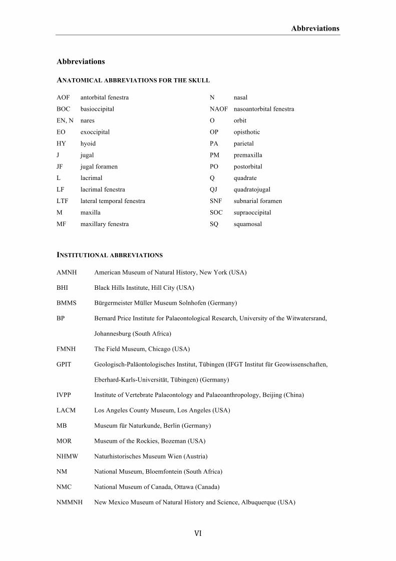
Abbreviations
VI
Abbreviations
ANATOMICAL ABBREVIATIONS FOR THE SKULL
AOF antorbital fenestra
BOC basioccipital
EN, N nares
EO exoccipital
HY hyoid
J jugal
JF jugal foramen
L lacrimal
LF lacrimal fenestra
LTF lateral temporal fenestra
M maxilla
MF maxillary fenestra
N nasal
NAOF nasoantorbital fenestra
O orbit
OP opisthotic
PA parietal
PM premaxilla
PO postorbital
Q quadrate
QJ quadratojugal
SNF subnarial foramen
SOC supraoccipital
SQ squamosal
INSTITUTIONAL ABBREVIATIONS
AMNH American Museum of Natural History, New York (USA)
BHI Black Hills Institute, Hill City (USA)
BMMS Bürgermeister Müller Museum Solnhofen (Germany)
BP Bernard Price Institute for Palaeontological Research, University of the Witwatersrand,
Johannesburg (South Africa)
FMNH The Field Museum, Chicago (USA)
GPIT Geologisch-Paläontologisches Institut, Tübingen (IFGT Institut für Geowissenschaften,
Eberhard-Karls-Universität, Tübingen) (Germany)
IVPP Institute of Vertebrate Palaeontology and Palaeoanthropology, Beijing (China)
LACM Los Angeles County Museum, Los Angeles (USA)
MB Museum für Naturkunde, Berlin (Germany)
MOR Museum of the Rockies, Bozeman (USA)
NHMW Naturhistorisches Museum Wien (Austria)
NM National Museum, Bloemfontein (South Africa)
NMC National Museum of Canada, Ottawa (Canada)
NMMNH New Mexico Museum of Natural History and Science, Albuquerque (USA)

Abbreviations
VII
NCSM North Carolina Museum of Natural Sciences, Raleigh (USA)
PIN Paleontological Institute, Russian Academy of Sciences, Moscow (Russia)
PVSJ Museo de Ciencias Naturales, Universidad Nacional de San Juan, San Juan (Argentina)
QMNS Qatar Museum of Nature and Science (Qatar)
SMA Sauriermuseum, Aathal (Switzerland)
SMNS Staatliches Museum für Naturkunde Stuttgart, Stuttgart (Germany)
SMF Senckenberg Naturmuseum Frankfurt (Germany)
TMP Royal Tyrrell Museum of Palaeontology, Drumheller (Canada)
TTU Texas Tech University, Lubbock (USA)
ULBRA Museu de Ciências Naturais, Universidade Luterana do Brasil, Canoas (Brazil)
USNM National Museum of Natural History (= formerly United States National Museum),
Smithsonian Institution, Washington, D.C. (USA)
UUVP Utah Museum of Natural History, Salt Lake City (USA)
ZFMK Zoologisches Forschungsmuseum Alexander Koenig, Bonn (Germany)
ZMH Zoologisches Museum Hamburg (Germany)
ZPAL Institute of Palaeobiology, Polish Academy of Sciences, Warsaw (Poland)
ZSM Zoologische Staatssammlung München (Germany)
TECHNICAL ABBREVIATIONS
AMS average maximum stress
CVA Canonical Variate Analyses
FEA Finite Element Analysis
GPA Generalized Procrustes Analyses
NPMANOVA non-parametric multivariate ANOVA
PC principal component
PCA Principal Component Analysis
PCO Principal Coordinates Analysis
PIC phylogenetic independent contrast
SSI skull strength indicator
2B-PLS two-block partial least squares analysis
UPGMA Unweighted Pair Group Method with Arithmetic mean
UV ultraviolet
Abstract/Kurzfassung
VIII
Abstract of the thesis
The Archosauria represent the most successful clade within tetrapods, having a large
diversity in terms of species, diet spectra, body plans and locomotion styles. This is also
true for the skull morphology, which shows a wide variety in shape and size, as well as
in the common formation of beaks, crests, domes or horns. Archosaur skulls have been
studied intensively in terms of their morphology, ontogeny, function, ecology and
behavior in the past, but most of these studies have largely been restricted to case
studies of single species or only a small number of taxa. The aim of the current thesis is
to obtain better and comprehensive insight into skull shape diversity of archosaurs by
using a two-dimensional geometric morphometric approach, with a special focus on
ontogenetic and macroevolutionary patterns and their relation to function and ecology.
Skull shape variation was quantified for Crocodylomorpha (including an ontogenetic
series of the recent caimanine alligatorid Melanosuchus niger), Pterosauria,
Sauropodomorpha and Theropoda. The material used for the analyses consists of skull
reconstructions published in the scientific literature and photographs of skull material.
The most important results of the thesis are summarized as follows:
• The use of different skull reconstructions of the same specimen from the
scientific literature has no significant influence on the results of morphometric
analyses. However, the results could be potentially falsified by the use of
reconstructions based on highly incomplete, strongly deformed or pathologic
specimens.
• In some cases the degree of intraspecific variation of one species can be as great
as the interspecific variation of closely related species with similar ecological
Abstract/Kurzfassung
IX
niches. Thus, species with great intraspecific diversity could have an impact on
the results of morphometric analyses.
• The skull shape of Archosauria is strongly correlated with function. A closer
examination within theropod skulls reveals that the shape of the postrostrum is
probably more affected by functional constraints than the snout, but the greatest
correlation to the function was found in the orbital shape. The latter result
supports previous studies on the biomechanics of theropod skulls. A comparison
of the ontogenetic bite force performance with the cranial growth in the
alligatorid Melanosuchus and biomechanical studies on crocodile skulls reveals
that ontogenetic shape changes, especially in the orbital and postorbital region,
are functional constrained.
• Both ontogenetic and interspecific skull shape variation in archosaurs is
correlated to diet preferences and feeding behaviour. A comparison between
carnivorous and non-carnivorous (i.e. omnivorous and herbivorous) theropods
reveals that both ecological groups occupy large areas within the morphospace
without showing a significant overlap. Furthermore, small-bodied theropods
tend to have a larger diet spectrum, suggesting that diet preferences within
theropods are probably size related.
• The distribution of taxa within the morphospace of Crocodylomorpha,
Pterosauria, Sauropodomorpha and Theropoda is strongly correlated with the
phylogenetic interrelationship of these clades: Closely related taxa appear closer
to one another within the morphospace than more distantly related taxa. This
result indicates that skull shape in archosaurs is further constrained by
phylogeny.
Abstract/Kurzfassung
X
• When inferred from geometric morphometric data, disparity results proved to be
similar to those based on limb measurements and discrete characters from
phylogenetic analyses. This results justifies the use of geometric morphometric
data as a further and equally useful proxy for addressing disparity.
• Early archosaur hatchlings share features of the skull shape, including short,
pointed snouts, enlarged orbits and large postorbital regions. However,
ontogenetic shape changes are only congruous in terms of a relative increase of
the snout length and a relative decrease of the orbit size. The degree of these
changes is not uniform, so that adult specimens of different species can vary
substantially in snout length or orbit shape. Furthermore, archosaurs show a
huge variability of changes in the snout depth, the length of the postorbital
region as well as the relative size of the antorbital fenestra and the lateral
temporal fenestra during ontogeny. This variability in ontogenetic trajectories
probably causes the large skull shape diversity found in archosaurs.
• Due to the great variability in ontogenetic trajectories, cranial evolution of
archosaurs is strongly affected by heterochronic events. Skull shape evolution of
Crocodylomorpha, Sauropodomorpha, basal theropods, Tyrannosauroidea as
well as derived Oviraptoridae, Dromaeosauridae and Troodontidae was probably
influenced by peramorphosis. However, within Crocodylia the short skull of
Osteolaemus might result from a paedomorphic event. This is also likely for the
short-snouted basal theropods Daemonosaurus and Limusaurus. The great
similarity in the skull shapes of the juvenile megalosaurid Sciurumimus and
basal coelurosaurs reveals that the skull shapes of the latter might be also caused
by paedomorphosis. Further paedomorphic trends are suspected for the skull
Abstract/Kurzfassung
XI
evolution of basal Maniraptora and Avialae. The heterochronic events found
seem to correlate with body size evolution.
Abstract/Kurzfassung
XII
Kurzfassung der Dissertation
Die Archosaurier repräsentieren die erfolgreichste Gruppe unter den Tetrapoden, die
durch eine große Diversität an Arten, Nahrungspektren, Bewegungsformen und im
Körperbau gekennzeichnet ist. Dies gilt auch für die Schädelmorphologie, die durch
eine große Variation in Größe und Formen sowie der häufigen Ausbildung von
Schnäbeln, Hörnern, Hauben und Kämmen gekennzeichnet ist. Die Schädel der
Archosaurier wurden in der Vergangenheit intensiv hinsichtlich ihrer Morphologie,
Ontogenese, Funktion, Ökologie und Verhalten untersucht, jedoch beschränken sich die
meisten Arbeiten auf Fallstudien zu einzelnen Arten bzw. einer kleinen Auswahl von
Taxa. In der vorliegenden Arbeit soll die Diversität der Schädelmorphologie innerhalb
der Archosaurier mit Hilfe von zwei-dimensionaler geometrischen Morphometrie auf
breiterer Ebene untersucht werden. Dabei sollen sowohl ontogenetische als auch
makroevolutive Muster und ihre Beziehung zu Funktion und Ökologie näher betrachtet
werden. Eine Quantifizierung der Schädelform erfolgte für Crocodylomorpha (inklusive
einer ontogenetischen Serie des rezenten Alligatoriden Melanosuchus niger),
Pterosauria, Sauropodomorpha und Theropoda. Als Grundlage dienten publizierte
Schädelrekonstruktionen aus der wissenschaftlichen Literatur sowie Fotos von
Schädelmaterial. Die wichtigsten Ergebnisse der Arbeit sind wie folgt
zusammengefasst:
• Die Verwendung von verschiedenen Schädelrekonstruktionen desselben
Individuums aus der wissenschaftlichen Literatur hat keinen signifikanten
Einfluss auf die Ergebnisse von morphometrischen Analysen. Die Ergebnisse
können jedoch durch die Verwendung von Rekonstruktionen verfälscht werden,
Abstract/Kurzfassung
XIII
die auf unvollständigem, stark verformtem oder pathologisch verändertem
Material basieren.
• In einigen Fällen kann das Maß der innerartlichen Variation vergleichbar sein
mit der zwischenartlichen Variation von nah-verwandten Arten mit ähnlichen
ökologischen Nischen. Daher können Arten mit großer innerartlichen Variation
die Ergebnisse von morphometrischen Analysen beeinträchtigen.
• Die Schädelform der Archosaurier korreliert stark mit der Funktion. Eine
detaillierte Untersuchung an Theropoden-Schädeln zeigt, dass die Form des
Hinterhaupts stärker durch Funktion beeinflusst wird als die Form der Schnauze.
Die größte Korrelation zwischen Form und Funktion findet sich in der
Augenhöhle, was die Ergebnisse früherer Arbeiten zur Biomechanik von
Theropoden-Schädeln unterstützt. Ein Vergleich der ontogenetischen
Beißkraftleistung mit dem Schädelwachstum bei Melanosuchus mit
biomechanischen Studien an Krokodilschädeln zeigt, dass ontogenetische
Veränderungen der Schädelform, speziell im Augen- und Hinterhauptsbereich,
funktional beeinträchtigt sind.
• Sowohl ontogenetische als auch interspezifische Variation der Schädelform
korrelieren bei Archosauriern mit Nahrungspräferenzen und Fressverhalten. Ein
Vergleich zwischen karnivoren und nicht-karnivoren (d.h. omnivoren und
herbivoren) Theropoden zeigt, dass beide ökologischen Gruppen große Bereiche
im „Morphospace“ einnehmen, jedoch nicht signifikant miteinander überlappen.
Kleinere Theropoden besitzen hier ein breiteres Nahrungspektrum, so dass
Präferenzen in der Nahrung wahrscheinlich größenabhängig sind.
• Die Verteilung der Taxa im „Morphospace“ der Crocodylomorpha, Pterosauria,
Sauropodomorpha und Theropoda korreliert stark mit dem phylogenetischen
Abstract/Kurzfassung
XIV
Verwandtschaft dieser Gruppen, d.h. das näher verwandte Taxa im
„Morphospace“ näher beieinander liegen als entfernt verwandte Taxa. Dieses
Ergebnis zeigt weiterhin, dass die Schädelform der Archosaurier auch durch die
phylogenetische Verwandtschaft beeinträchtigt ist.
• Übereinstimmungen in den Ergebnissen von Disparitätsanalysen basierend auf
geometrischer Morphometrie mit solchen basierend auf Längenmessungen und
diskreten Merkmalen aus phylogenetischen Analysen zeigen, dass Disparität
über mehrere Proxies gemessen werden kann, inklusive geometrisch
morphometrischer Daten.
• Die Schädelformen von verschiedenen Archosaurier-Schlüpflingen ähneln
einander durch das Vorhandensein einer kurzen Schnauze, großen Augenhöhlen
und einer vergrößerten Hinterhauptsregion. Allgemeine ontogenetische
Veränderungen betreffen die relative Verlängerung der Schnauze und die
relative Verkleinerung der Augenhöhle. Diese Veränderungen sind allerdings
nicht einheitlich in ihrer Intensität, so dass ausgewachsene Individuen
verschiedener Arten sich deutlich in der Länge der Schnauze und der Form der
Augen unterscheiden können. Des Weiteren besitzen Archosaurier ein große
ontogenetische Variabilität hinsichtlich der Höhe der Schnauze, der Länge des
Hinterhaupts sowie der relativen Größe des Antorbitalfensters und des lateralen
Temporalfensters. Die große Variabilität der ontogenetischen Trajektorien ist
wahrscheinlich für die große Diversität an Schädelformen innerhalb der
Archosaurier verantwortlich.
• Aufgrund der großen Variabilität ontogenetischer Trajektorien ist die
Schädelevolution der Archosaurier sehr stark durch heterochronische Ereignisse
geprägt. Die Schädelevolution von Crocodylomorpha, Sauropodomorpha,
Abstract/Kurzfassung
XV
basalen Theropoden, Tyrannosauroidea sowie abgeleiteten Oviraptoridae,
Dromaeosauridae und Troodontidae ist wahrscheinlich durch Peramorphose
beeinflusst. Innerhalb der Crocodylia resultiert der kurze Schädel von
Osteolaemus wahrscheinlich aus einer Pädomorphose. Das ist wahrscheinlich
auch der Fall für die beiden kurzschnauzigen basalen Theropoden
Daemonosaurus und Limusaurus. Die große Übereinstimmung der Schädelform
des juvenilen Megalosauriden Sciurumimus mit dem von basalen
Coelurosauriern könnte ebenfalls ein Hinweis sein, dass die Schädelform der
basalen Coelurosaurier das Resultat einer Pädomorphose ist. Weitere
pädomorphe Ereignisse könnten in der Schädelevolution der Maniraptora und
Avialae aufgetreten sein. Die heterochronischen Ereignisse scheinen in enger
Beziehung zur Evolution der Körpergröße zu stehen.
Acknowledgements
XVI
Acknowledgements
Finally, it is done and I feel really happy that I can start soon to enjoy the joy of regular
unemployment as a high profile academic. But before I accept my fate, I would like to
thank my supervisor Oliver Rauhut, who supported my scientific work over the last
three years and gave me the possibility to work on one of the coolest dinosaur fossils
ever found on this planet, in the history of man. Furthermore, I would like to thank
Oliver for giving me an all-inclusive freedom in terms of working, and for making jokes
worse than I could ever do. However, without his tireless dedication to hold me on the
carpet, I would now probably stand on a floor made of oak wood or cobblestones. I
further thank Adriana López-Arbarello and Richard Butler for their support, many
careful advices and fruitful discussions. Especially Richard helped me a lot with
correcting drafts, introducing statistical methods and developing own projects. Probably,
he also thinks that my jokes are at least as bad as Oliver’s, but this is just a single
opinion. Furthermore, I would like to thank Johannes Müller for his willingness to
review my thesis.
Another thanks goes to my co-authors Paula Bona, Julia Desojo, Steve Brusatte,
Helmut Tischlinger, Mark Norell as well as Richard and Oliver. The quality of the
single chapters presented in this thesis benefited substantially from your scientific input.
I further thank Roger Benson, Steve Brusatte, Manubo Sakamoto, Michel Laurin, Mike
Benton, Andrew Farke, Xu Xing, Gareth Dyke and Abby Drake for constructive and
helpful reviews and comments on the single chapters. Another thanks goes to Martín
Ezcurra, Roland Sookias, Christine Böhmer, Kerstin Schröder, Felix Quade, Melissa
Tallman, Serjoscha Evers and Johannes Knebel for pleasurable discussions. Christine
Böhmer and Melissa Tallman introduced me into geometric morphometric methods and
Acknowledgements
XVII
Roland Sookias spent hours of hours with proof-reading manuscripts on crocodylian
skulls. Poor wretch!
During my PhD studies I had the possibility to visit a lot of wonderful
zoological and palaeontological collections all over the world. These visits allowed me
to study some of the most interesting dinosaur fossils ever found and to learn a lot about
reptilian anatomy. Thus, I would like to thank all curators and collection managers who
hosted me during my stays: Frank Glaw, Jakob Hallermann, Gunther Köhler, Linda
Acker, Dennis Rödder, Ursula Bott, Mark Norell, Carl Mehling, Lindsay Zanno,
Fernando Novas, Alejandro Kramarz, Eduardo Ruigómez, Hans-Jakob Siber, Ben Pabst,
Thomas Bollinger, Martina Kölbl-Ebert, Xu Xing and Corwin Sullivan. Martin Dobritz
is thanked for access to the X-ray facilities in the Klinikum rechts der Isar.
“Money, it's a gas, grab that cash with both hands and make a stash.“ (Roger
Waters). In this sense, the current thesis was generously funded by the Deutsche
Forschungsgemeinschaft (DFG project RA-1012/-12-1).
Finally, I would like to thank my parents, my brother and his wife, my
grandparents and all my friends for being there and supporting me during my ontogeny
so far. Furthermore, I thank the sweat reek of this evil plant for helping my neurons to
feel happy and contended in times of media disinformation, collective cognitive
dissonance and political corruption. But whatever, nothing helps more than lying in the
arms of my only true love Julika. You keep me burning!
1
CHAPTER 1
Introduction and summery of the thesis
Keywords:
Archosauria; Saurischia; Crocodylomorpha; Pterosauria; Theropoda; Melanosuchus;
ontogeny; skull shape; sexual dimorphism; feeding ecology; biomechanics; disparity;
macroevolution; heterochrony; geometric morphometrics
Author contributions:
Research design: Christian Foth
Data collection: Christian Foth
Data analyses: Christian Foth
Preparation of figures and tables: Christian Foth
Wrote chapter: Christian Foth
Chapter 1: Introduction and summary
2
Introduction and summary of the thesis
Christian Foth
INTRODUCTION
Within reptiles, the clade Archosauria Cope, 1869 is defined as the monophyletic group
composed of recent crocodylians and birds, and all fossil taxa that share their last
common ancestor (Gauthier & Padian 1985). Within recent tetrapods, archosaurs
represents the most successful clade with approximately 10 000 living species described
(Westheide & Rieger 2004). Based on the fossil record, the origin of Archosauria goes
back at least to the Early Triassic (Nesbitt et al. 2011), whereas stem-line
representatives were already present in the Late Permian (Tatarinov 1960; Borsuk-
Białynicka & Evans 2009). The two main clades of Archosauria are the Pseudosuchia
Zittel, 1887-1890, which are defined as clade including recent crocodylians and all other
archosaurs closer to crocodylians than to birds (Gauthier & Padian 1985), and
Ornithodira Gauthier, 1986, which are defined as the least inclusive clade containing the
pterosaur Pterodactylus and the songbird Passer (Nesbitt 2011). During their
cosmopolitan Mesozoic radiation, archosaurs became extremely diverse in terms of
number of species, diet spectra and body plans, including different forms and manners
of locomotion (Weishampel et al. 2004; Brusatte et al. 2008; Nesbitt 2011), and
mastered not only terrestrial (most archosaur groups), but also aquatic and semi-aquatic
(e.g. Phytosauria, Neosuchia) as well as aerial and arboreal habitats (e.g. Pterosauria,
Avialae).
Chapter 1: Introduction and summary
3
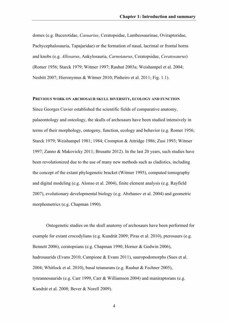
CHARACTERISTICS AND DIVERSITY OF ARCHOSAUR SKULLS
The skulls of recent archosaurs possess several characteristics unique amongst tetrapods
including e.g. the presence of an antorbital fenestra, a laterosphenoid, a mandibular
fenestra, a strongly pneumatized ear and braincase region, thecodont teeth as well as the
absence of a parietal foramen, a supratemporal, a postparietal, a tabular, a postfrontal,
an epipterygoid and palatal teeth (Mickoleit 2004). However, these characteristics do
not represent ‘true’ apomorphic characters of the clade Archosauria. The
pneumatization of the middle ear and the braincase as well as the reduction of the
postfrontal and epipterygoid for example are characters evolved independently within
the stem-line of both crocodylians and birds (Gower & Weber 1998; Gower 2002;
Rauhut et al. 2003a; Holliday & Witmer 2008, 2009; Nesbitt et al. 2011), whereas the
other character mentioned above were already evolved within the stem-line of
archosaurs (Nesbitt 2011). Skull characters found as apomorphic for Archosauria are
palatal processes of the maxilla meeting at the midline, an elongated and tubular
cochlear recess, an external foramen for abducens nerves within the prootic, an
antorbital fossa presented on the lacrimal, the dorsal process of the maxilla and the
dorsolateral margin of the posterior process of the maxilla as well as probably the
presence of foramina for entrance of cerebral branches of internal carotid artery into the
braincase positioned on the anterolateral surface of the parasphenoid (Nesbitt 2011).
Despite these uniting characteristics archosaurs show a high diversity of shape,
which includes the convergent formation of keratinous beaks (e.g. Hadrosauria,
Ornithomimosauria, Oviraptoridae, Neornithes, Shuvosauridae, Tapajaridae), the
reduction of the antorbital fenestra (e.g. Ankylosauria, Ceratopidae, Crocodylia,
Hadrosauridae), the formation of premaxillary, nasal, frontal or postorbital crests and
Chapter 1: Introduction and summary
4
domes (e.g. Bucerotidae, Casuarius, Ceratopsidae, Lambeosaurinae, Oviraptoridae,
Pachycephalosauria, Tapajaridae) or the formation of nasal, lacrimal or frontal horns
and knobs (e.g. Allosarus, Ankylosauria, Carnotaurus, Ceratopsidae, Ceratosaurus)
(Romer 1956; Starck 1979; Witmer 1997; Rauhut 2003a; Weishampel et al. 2004;
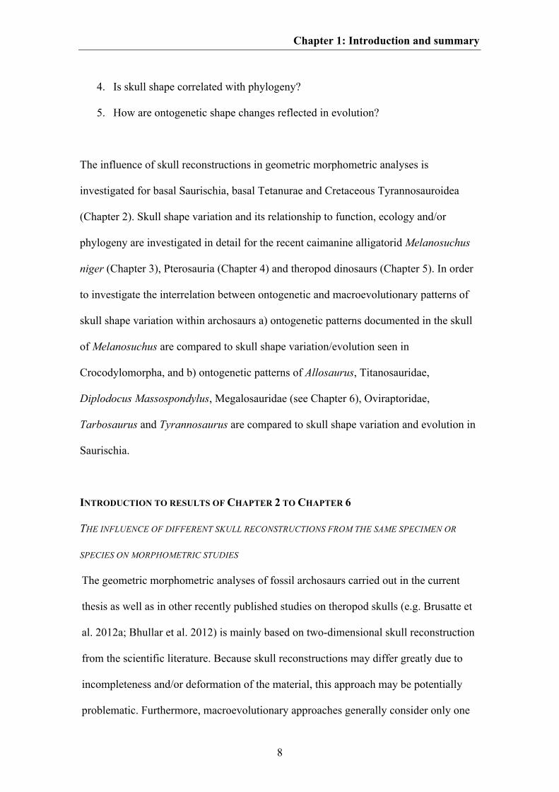
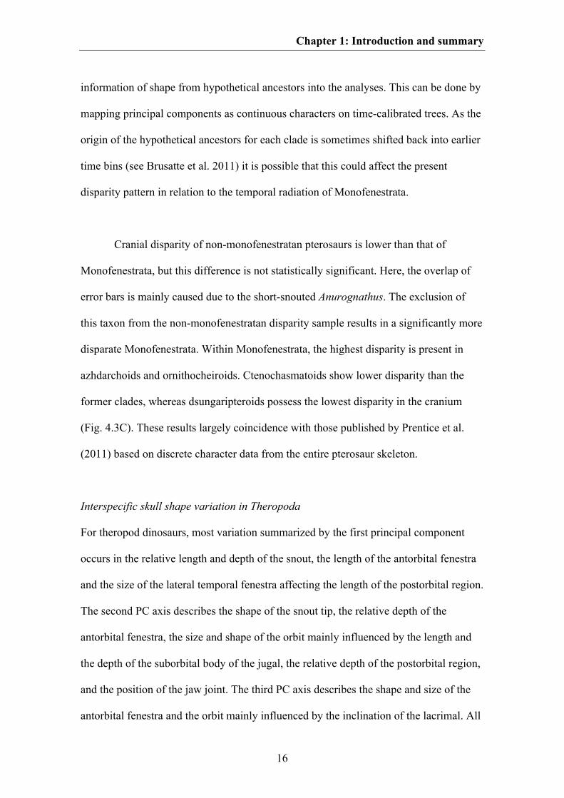
Nesbitt 2007; Hieronymus & Witmer 2010; Pinheiro et al. 2011; Fig. 1.1).
PREVIOUS WORK ON ARCHOSAUR SKULL DIVERSITY, ECOLOGY AND FUNCTION
Since Georges Cuvier established the scientific fields of comparative anatomy,
palaeontology and osteology, the skulls of archosaurs have been studied intensively in
terms of their morphology, ontogeny, function, ecology and behavior (e.g. Romer 1956;
Starck 1979; Weishampel 1981; 1984; Crompton & Attridge 1986; Zusi 1993; Witmer
1997; Zanno & Makovicky 2011; Brusatte 2012). In the last 20 years, such studies have
been revolutionized due to the use of many new methods such as cladistics, including
the concept of the extant phylogenetic bracket (Witmer 1995), computed tomography
and digital modeling (e.g. Alonso et al. 2004), finite element analysis (e.g. Rayfield
2007), evolutionary developmental biology (e.g. Abzhanov et al. 2004) and geometric
morphometrics (e.g. Chapman 1990).
Ontogenetic studies on the skull anatomy of archosaurs have been performed for
example for extant crocodylians (e.g. Kundrát 2009; Piras et al. 2010), pterosaurs (e.g.
Bennett 2006), ceratopsians (e.g. Chapman 1990; Horner & Godwin 2006),
hadrosaurids (Evans 2010; Campione & Evans 2011), sauropodomorphs (Sues et al.
2004; Whitlock et al. 2010), basal tetanurans (e.g. Rauhut & Fechner 2005),
tynrannosaurids (e.g. Carr 1999, Carr & Williamson 2004) and maniraptorans (e.g.
Kundrát et al. 2008; Bever & Norell 2009).
Chapter 1: Introduction and summary
5
Fig. 1.1. A. A typical archosaur skull represented by Tyrannosaurus (modified after Carr & Williamson
2004). B-G. Examples of cranial diversity in archosaurs. B. The pterosaur Anhanguera (modified after
Maisey 1991). C. The crocodylomorph Domicosuchus (modified after Nesbitt 2011). D. The avialian
Archaeopteryx (modified after Rauhut in press). E. The sauropod Diplodocus (modified after Wilson &
Sereno 1998). F. The ceratopsid Styracosaurus (modified after Ryan et al. 2007). G. The oviraptorid
Conchoraptor (after Osmólska et al. 2004). AOF, antorbital fenestra; BOC, basioccipital; EN, nares; EO,
exoccipital; J, jugal; JF, jugal foramen; L, lacrimal; LF, lacrimal fenestra; LTF, lateral temporal fenestra;
Chapter 1: Introduction and summary
6
M, maxilla; MF, maxillary fenestra; N, nasal; O, orbit; OP, opisthotic; PA, parietal; PM, premaxilla; PO,
postorbital; Q, quadrate; QJ, quadratojugal; SNF, subnarial foramen; SOC, supraoccipital; SQ,
squamosal.
As mentioned, great advances have been made with respect to cranial function in
archosaurs. These studies have drawn from classical morphological and experimental
approaches as well as geometric studies (e.g. Weishampel 1984; Henderson 2002;
Henderson & Weishampel 2002; Erickson et al. 2003, 2012; Holliday & Witmer 2008;
Lautenschlager 2013) and, increasingly, from finite element methods (e.g. Rayfield et
al. 2001; Rayfield 2004, 2005, 2011; Witzel & Preuschoft 2005; McHenry 2006; Pierce
et al. 2008; Young et al. 2010; Witzel et al. 2011).
However, most of these studies have largely been restricted to case studies of
single species or only a small number of taxa. Compared to the great number of large
phylogenetic datasets existing for archosaurs (e.g. Wilson 2002; Mayr & Clarke 2003;
Rauhut 2003a; Butler et al. 2007; Brusatte et al. 2010a; Prieto-Márquez 2010; Nesbitt
2011; Carrano et al. 2012; Turner et al. 2012; Pol et al. in press), broad-scale studies
investigating the relationships between cranial diversity, ontogenetic modifications,
functional constraints, and evolutionary processes are still rather rare. One means to
integrate these concepts into one large scheme is via the use of geometric
morphometrics. This method quantifies shape variation of objects in a multivariate
morphospace. This morphospace can be compared thereafter with phylogenetic
relationships, ecological and functional proxies or analysed to assess morphological
disparity, ontogenetic or biogeographical patterns (e.g. Zelditch et al. 2004). Geometric
Chapter 1: Introduction and summary
7
morphometric studies regarding cranial shape of archosaurs have been carried out to
date for Crocodylomorpha (e.g. Pierce et al. 2008; Piras et al. 2009, 2010; Young et al.
2010), ornithischian dinosaurs (Chapman et al. 1981, Chapman 1990, Chapman &
Brett-Surman 1990, Goodwin 1990), sauropodomorphs (Young & Larvan 2010) and
non-avian theropod dinosaurs and extant birds (e.g. Chapman 1990; Mazzetta et al.
1998; Marugán-Lobón & Buscalioni 2004, 2006; Kulemeyer et al. 2009; Brusatte et al.
2012a; Bhullar et al. 2012). The relationship between shape and function was
specifically investigated by, for example, Pierce et al. (2008), Young et al. (2010) and
Brusatte et al. (2012a), whereas Piras et al. (2010) and Bhullar et al. (2012) for example
examined the relationship between shape and ontogeny.
OBJECTIVE OF THE THESIS
As mentioned above, archosaurs possess enormous skull diversity, but only a small
numbers of studies to date have investigated the correlation of shape diversity with
function, ecology, ontogeny and evolution. Thus, the aim of this thesis is to obtain
better insight into the ontogenetic and macroevolutionary patterns of archosaur skulls,
and their relation to function and ecology by using two-dimensional geometric
morphometrics and further statistical methods. The material used for the analyses
consists of a) skull reconstructions published in the scientific literature, and b)
photographs of skull material. The following main questions will be addressed:
1. How large is the impact of differing skull reconstructions from the same
specimen or the same species on the results of morphometric studies?
2. What are the main patterns of shape variation in archosaur skulls?
3. How is skull shape influenced by functional constrains and feeding ecology?
Chapter 1: Introduction and summary
8
4. Is skull shape correlated with phylogeny?
5. How are ontogenetic shape changes reflected in evolution?
The influence of skull reconstructions in geometric morphometric analyses is
investigated for basal Saurischia, basal Tetanurae and Cretaceous Tyrannosauroidea
(Chapter 2). Skull shape variation and its relationship to function, ecology and/or
phylogeny are investigated in detail for the recent caimanine alligatorid Melanosuchus
niger (Chapter 3), Pterosauria (Chapter 4) and theropod dinosaurs (Chapter 5). In order
to investigate the interrelation between ontogenetic and macroevolutionary patterns of
skull shape variation within archosaurs a) ontogenetic patterns documented in the skull
of Melanosuchus are compared to skull shape variation/evolution seen in
Crocodylomorpha, and b) ontogenetic patterns of Allosaurus, Titanosauridae,
Diplodocus Massospondylus, Megalosauridae (see Chapter 6), Oviraptoridae,
Tarbosaurus and Tyrannosaurus are compared to skull shape variation and evolution in
Saurischia.
INTRODUCTION TO RESULTS OF CHAPTER 2 TO CHAPTER 6
THE INFLUENCE OF DIFFERENT SKULL RECONSTRUCTIONS FROM THE SAME SPECIMEN OR
SPECIES ON MORPHOMETRIC STUDIES
The geometric morphometric analyses of fossil archosaurs carried out in the current
thesis as well as in other recently published studies on theropod skulls (e.g. Brusatte et
al. 2012a; Bhullar et al. 2012) is mainly based on two-dimensional skull reconstruction
from the scientific literature. Because skull reconstructions may differ greatly due to
incompleteness and/or deformation of the material, this approach may be potentially
problematic. Furthermore, macroevolutionary approaches generally consider only one
Chapter 1: Introduction and summary
9
representative specimen per species, meaning that the influence of intraspecific
variation is ignored. To test the influence of different skull reconstructions of the same
specimen in morphometric analyses, three datasets for basal Saurischia, basal Tetanurae
and Late Cretaceous Tyrannosauroidea were created (see Chapter 2). The degree of
shape variation (as a measure of disparity) was estimated for skull reconstructions
based on the same specimen and compared to shape variation occurring in skull
reconstructions based on different specimens of the same species and skulls of closely
related species, in order to examine whether this potential source of variation may be
comparable to taxonomically or even phylogenetically significant variation.
The results indicate that the effects of shape variation between different skull
reconstructions based on the same specimen are negligible in geometric morphometric
studies. Thus, if the skull reconstruction is based on rather complete, little deformed
material, the impact of the author’s drawing ability and style should not affect results of
morphometric analyses. However, some skull regions are somewhat more problematic
for the plotting of landmarks than most (e.g. the ventral contact between jugal and
quadratojugal, the contact between the premaxilla and nasal on the dorsal margin of the
skull, the most anterior point of the lacrimal along the dorsal margin of the antorbital
fenestra and the contact between postorbital and squamosal on the dorsal margin of the
lateral temporal fenestra) and their morphology should be verified with photographs or
first hand observations. In contrast, skull shape variation found between different
specimens and species is higher compared to shape variation between different skull
reconstructions of the same specimen, because they further contain intraspecific or
interspecific variation. Interestingly, for closely related species with similar ecological
niche, the degree of interspecific variation can partly overlap with that of intraspecific
Chapter 1: Introduction and summary
10
variation. Such overlap is well documented in recent animals and can be found at both
the morphological and molecular level (e.g. Czechura & Wombey 1982; Lockwood
2005; Meyer 2005, Meier 2006, 2008). Thus, in some cases it could be possible that
species with great intraspecific diversity potentially influence the results of
morphometric analyses. Furthermore, the results could be affected by those species
with unresolved taxonomy.
MAIN PATTERNS OF SHAPE VARIATION IN ARCHOSAUR SKULLS
Besides functional constraints (which will be discussed separately), skull shape in
vertebrates is influenced by intraspecific variation (e.g. ontogenetic variation, sexual
dimorphism) (e.g. Emerson & Bramble 1993; O’Higgins & Collard 2002; Bruner et al.
2005) and evolutionary processes (e.g. natural selection, heterochrony, genetic drift,
adaptive radiation) (e.g. Rieppel 1993; Burns et al. 2002; Abzhanov et al. 2006; Smith
2011; Bhullar 2012), which can be captured in both intraspecific and interspecific
morphospaces. In the current thesis an intraspecific morphospace was estimated for the
recent caimanine alligatorid Melanosuchus niger as an example (see Chapter 3),
whereas an interspecific morphospace was estimated for both Pterosauria (see Chapter
4) and Theropoda (see Chapter 5).
Intraspecific skull shape variation in Melanosuchus niger
The skull shape of Melanosuchus varies mainly in terms of the relative length of the
snout, the depth of the tip of the rostrum, the relative size of the subnarial gap between
premaxilla and maxilla, the shape of the ventral margin of the maxilla, the relative size
and position of the orbit, the relative shape of the jugal region, the overall depth of the
orbital and postorbital region, the relative length and width of the skull roof table and
Chapter 1: Introduction and summary
11
the position of the jaw angle in both an anterolateral-posteromedial direction and an
anteroventral-posterodorsal direction (Fig. 3.2, 3.3). This variation, which is
summarized by the first principal component, describes over 70 % of total skull shape
variation.
For both dorsal and lateral views the first PC axis is strongly correlated with
centroid size (Fig. 3.4, Table 3.1), indicating that the shape variation mentioned above
contains information of allometric variation related to ontogenetic growth. Thus, based
on the relationship found, it is possible to reconstruct major shape changes in skull
shape of Melanosuchus during ontogeny. The skulls of early juvenile individuals
possess a very short snout, which is wide in dorsal view, but dorsoventrally pointed in
lateral view. The ventral margin of the snout is straight and no subnarial gap is present.
The orbit is very large and the jugal region is slender in both dorsal and lateral view.
The postorbital region is elongated in an anteroposterior direction, and the broad skull
roof table is posteriorly inclined in lateral view. The posterior end of the skull is
relatively narrow and the jaw joint lies substantially anterior to the posterior end of the
skull roof table. During ontogeny the snout becomes relatively elongated. In dorsal
view, the snout becomes more slender, but deeper in lateral view, and the snout tip
becomes blunter. Between premaxilla and maxilla a subnarial gap is formed, and the
ventral margin of the maxilla becomes anteriorly convex in lateral view. The relative
size of the orbit decreases, whereas the jugal region becomes broader and deeper. Due
to the overall decrease in size of the orbit, the postrostrum becomes generally flattened
in lateral view resulting in a horizontally aligned skull roof table. The postorbital region
becomes shorter, but expands posterolaterally, and the jaw joint moves substantially
posteriorly to the posterior end of the skull roof table (Fig. 3.2, 3.3). Most crocodylian
Chapter 1: Introduction and summary
12
taxa share similar ontogenetic patterns in skull shape change, including, for example,
relative elongation of the snout, decrease of the relative orbit size and increase of the
interorbital width, as well as relative decrease in length of the postorbital skull roof
(Table 3.2). The only exception is the long-snouted Gavialis gangeticus, in which skull
growth is almost isometric (excepting the orbits) during ontogeny (see Piras et al.
2010).
Skull shape variation was further tested to attempt to detect presence of cranial
sexual dimorphism in adult individuals of Melanosuchus. The results show that distinct
sexual dimorphism is present, which is mainly size-related. This relationship to size is
not surprising as males grow to about 30 % larger than females. Size-related sexual
dimorphism is also described for other crocodylians (e.g. Webb & Messel 1978; Hall &
Portier 1994; Verdade 2000, 2003; Platt et al. 2009), and caused by a generally faster
and longer growth in males (e.g. Chabreck & Joanen 1979; Rootes et al. 1991;
Wilkinson & Rhodes 1997). Statistical support for sexual dimorphism remained after
excluding the effects of allometry with help of an pooled within-group regression from
the dataset, indicating that differences between females and males may be not only size-
related. Non-size related sexual dimorphism has only been described so far for the
crocodylid Crocodylus porosus (Webb & Messel 1978), the gavialid Gavialis
gangeticus (Hall & Portier 1994) and the alligatorid Caiman latirostris (Verdade 2000).
However, differences in the sample size of males and females as well as the large
numbers of landmarks and semi-landmarks compared to the sample size could lead to
false positive signals in the statistical test. Therefore, the current result should be
verified with larger sample sizes, different landmark configurations as well as for other
crocodylian taxa.
Chapter 1: Introduction and summary
13
Due to the diversity of crests and horns present (see above), cranial sexual
dimorphism has been hypothesized for a large number of pterosaurs (e.g. Bennett 1992;
Lü et al. 2011) and dinosaurs, e.g. Ceratopsia (e.g. Kurzanov 1972; Chapman 1990;
Lehman 1990, 1998; Chapman et al. 1997), basal Sauropodomorpha (e.g. Gow et al.
1990), Pachycephalosauria (e.g. Chapman et al. 1997), Hadrosauria (e.g. Chapman et al.
1997), non-avian Theropoda (e.g. Colbert 1989, 1990; Carpenter 1990) and recent birds
(e.g. Selander 1966). However, such interpretations in fossil taxa have been treated with
great caution due to generally low sample sizes of single individuals for each species,
making statistical verification problematic (see Molnar 1990; Padian & Horner 2011a,
in press). Differences seen in cranial shape could be alternatively related to intraspecific
variation (e.g. allometric shape variation due to size differences between different
specimens) (e.g. Ryan et al. 2001), taphonomic deformation (e.g. Forster 1990) or even
taxonomic misidentification (e.g. Evans & Reisz 2007). A further difficulty is that the
determination of sex within extinct archosaurs usually cannot be based on single
osteological characters (e.g. Erickson et al. 2005; Prieto-Márquez 2007). A possible
reliable character for sex determination was found for ornithodirans in the form of
presence of medullary bone in the long bones of several dinosaurs and one pterosaur
species (Schweitzer et al. 2005; Lee & Werning 2008; Chinsamy et al. 2009, 2013;
Hübner 2012). Within recent archosaurs this bone structure is only documented for
female birds during their reproductive periods (e.g. Miller & Bowman 1981; Dacke et
al. 1993), but not in crocodylians (Schweitzer et al. 2007). However, as medullary bone
is only formed during reproductive periods, sex determination based on this character is
seasonally restricted and thus unsuitable for broad-scale sexing of extinct ornithodirans.
Chapter 1: Introduction and summary
14
The best-supported example of sexual dimorphism in the fossil record of
Archosauria may be in the pterosaur Darwinopterus. Here, a female specimen was
identified by the preservation of an egg in the pelvic region (Lü et al. 2011). In contrast
to other specimens referred to this taxon (see Lü et al. 2010a), this particular female
specimen lacks a sagittal crest on the head. If preservation artefacts can be ruled out and
the taxonomic classification is correct, the interpretation of the presence of sexual
dimorphism in the crest morphology of Darwinopterus hypothesized by Lü et al. (2011)
could be valid. However, the expression of cranial sexual dimorphism in archosaurs
must be viewed as an open question, may with such dimorphism varying from species
to species and potentially being expressed in the form of size differences, soft tissue,
colour patterns and/or behaviour rather than in the form of osteological structures (e.g.
Cooper & Vitt 1993; Sampson 1997). Alternatively, it is possible that some species used
cranial ornaments for species recognition (see Padian & Horner 2011b). For some
recent birds it is further documented that both males and females develop ornamental
structures, which are selected for via mate choice in both sexes (Jones & Hunter 1993,
1999; Amundsen 2000; Kraaijeveld et al. 2004). In contrast to common sexual selection
this so-called mutual sexual selection does not result in sexually dimorphic display
structures. Thus, it is further possible that the development of cranial ornaments in some
extinct archosaurs resulted from mutual sexual selection (see Hone et al. 2012) and thus
that such ornamentation was not sexually dimorphic.
Interspecific skull shape variation in Pterosauria
The majority of shape variation in pterosaur skulls occurs in the relative length of the
snout, the relative size of the orbit and postorbital region, the relative size and shape of
the naris-antorbital fenestra region and the position of the jaw joint relative to the orbit
Chapter 1: Introduction and summary
15
(Fig. 4.1B). This shape variation is summarized by the first principal component, which
describes over 50 % of total shape variation. The most extreme species affected by this
shape variation are the short-snouted Anurognathus and the long-snouted Pterodaustro.
However, the chosen landmark configuration does not capture the total shape of cranial
crests known in pterosaurs. Thus, it is likely that the variation summarized by the first
principal component is overestimated, and that the distribution of taxa within the
morphospace should in fact be more widely spread. Variation related to crest formation
is partly summarized by the second, third and fourth principal components. Here, shape
variation captured by PC 2 includes a large frontal crest as present in pteranodontids,
that of PC 3 includes the large rostral crests present in tapajarids, and that of PC 4
includes the premaxillary crest present in ornithocheirids (Fig. 4.1B).
Based on the data generated by the Principal Component Analysis, temporal and
taxonomic disparity was estimated. Despite small sample sizes for each time bin as well
as for particular taxonomic groups, some trends can be observed. Over time, cranial
shape disparity increased within pterosaurs from the Late Triassic to the Early
Cretaceous, but then declined in the Late Cretaceous. Due to small sample sizes, this
result was confirmed by rarefaction analysis, which reduces the error for sample size
differences (Fig. 4.3A, B). This temporal pattern of cranial disparity is consistent with
previous studies based on limb measurements and discrete character data (Dyke et al.
2006, 2009; Prentice et al. 2011; Butler et al. 2012). Thus, if this pattern is correct, the
disparity peak occurs relatively late in pterosaur evolution compared to other animal
groups (see Erwin 2007), probably with the radiation of Monofenestrata. To test the
results and to get a better insight into temporal disparity in pterosaurs it would be
advisable to correct the measures of disparity used in the current study by adding
Chapter 1: Introduction and summary
16
information of shape from hypothetical ancestors into the analyses. This can be done by
mapping principal components as continuous characters on time-calibrated trees. As the
origin of the hypothetical ancestors for each clade is sometimes shifted back into earlier
time bins (see Brusatte et al. 2011) it is possible that this could affect the present
disparity pattern in relation to the temporal radiation of Monofenestrata.
Cranial disparity of non-monofenestratan pterosaurs is lower than that of
Monofenestrata, but this difference is not statistically significant. Here, the overlap of
error bars is mainly caused due to the short-snouted Anurognathus. The exclusion of
this taxon from the non-monofenestratan disparity sample results in a significantly more
disparate Monofenestrata. Within Monofenestrata, the highest disparity is present in
azhdarchoids and ornithocheiroids. Ctenochasmatoids show lower disparity than the
former clades, whereas dsungaripteroids possess the lowest disparity in the cranium
(Fig. 4.3C). These results largely coincidence with those published by Prentice et al.
(2011) based on discrete character data from the entire pterosaur skeleton.
Interspecific skull shape variation in Theropoda
For theropod dinosaurs, most variation summarized by the first principal component
occurs in the relative length and depth of the snout, the length of the antorbital fenestra
and the size of the lateral temporal fenestra affecting the length of the postorbital region.
The second PC axis describes the shape of the snout tip, the relative depth of the
antorbital fenestra, the size and shape of the orbit mainly influenced by the length and
the depth of the suborbital body of the jugal, the relative depth of the postorbital region,
and the position of the jaw joint. The third PC axis describes the shape and size of the
antorbital fenestra and the orbit mainly influenced by the inclination of the lacrimal. All
Chapter 1: Introduction and summary
17
three PC axes are correlated with centroid size, and thus contain allometric shape
information. Interestingly, the length of the snout is inversely related to the length of the
postorbital region (see Marugán-Lobón & Buscalioni 2003), whereas the depths of both
skull regions seems to be more variable and unrelated to one another.
The skulls of Plateosaurus (outgroup), Herrerrasaurus, Eoraptor,
Compsognathus and Erlikosaurus resemble most the centroid shape for the whole
morphospace (Fig. 5.2). By contrast, the most aberrant skulls within theropods occur in
Oviraptorosauria, which possess an extremely short and deep snout and enlarged, round
orbit as well as a huge lateral temporal fenestra. Based on Brusatte et al. (2012a), this
group also possesses the highest within-group variation in skull shape. Another extreme
in cranial shape is the abelisaurid Carnotaurus, which possesses a very short and deep
snout, but with extremely short, oval orbits (Fig. 5.2). Extremes of cranial morphology
are also represented by spinosaurids and Gallimimus, which both possess low skulls
with elongated snouts, and short postorbital regions. Within basal birds, the most
aberrant skull was found in Confuciusornis. Brusatte et al. (2012a) found the toothless
ceratosaur Limusaurus to be yet more extreme, but in the current analyses this taxon
plotted close to the centre of the morphospace. Compared to other ceratosaurs in which
the skull is well known (e.g. Ceratosaurus, several Abelisauridae), the skull shape of
Limusaurus is divergent. However, incomplete skull material from the small-bodied
noasaurid Masiakasaurus shows that some representatives of the group also possess
skulls with low, elongated snouts and enlarged, round orbits (Carrano et al. 2011),
indicating a huge skull shape diversity within ceratosaurs. This is supported by disparity
analyses, performed by Brusatte et al. (2012a), which show that Ceratosauria possess
higher within-group variation in skull shape than, for example, basal Tetanurae,
Chapter 1: Introduction and summary
18
Tyrannosauroidea or Dromaeosauridae.
THE INFLUENCE OF FUNCTIONAL CONSTRAINTS AND FEEDING ECOLOGY ON SKULL SHAPE
Besides evolutionary processes, the shape of a biological structure is further influenced
by functional constrains, in which function is understood as mechanical role or physical
role, i.e. how a phenotypic feature is used (see Bock & Wahlert 1965; Lauder 1995).
Here, a strong correlation between functional loading and shape of a biological structure
implies that a particular structure is selected for ‘optimal’ shape, defined as maximal
strength with minimal material (e.g. Witzel et al. 2011). In the current thesis, the
relationship between skull shape and function was tested for Melanosuchus (see Chapter
3) and theropods dinosaurs (see Chapter 5).
Ontogenetic shape variation vs. function and feeding ecology in Melanosuchus niger
To test the relationship between form and function in Melanosuchus, bite forces were
used as a functional proxy. The bite force for each skull was computed with help of an
equation originally estimated for Alligator mississippiensis (Erickson et al. 2003). These
values were tested against skull shape variation using regression and two-block Partial
Least Square analysis (2B-PLS) (see Rohlf & Corti 2000). Overall skull shape variation
was found to be significantly correlated with bite forces (Table 3.1). This correlation is
primarily influenced by shape captured by the first principal component. As previously
stated, this component contains information on ontogenetic shape variation implying
that these changes could be primarily functionally constrained. However, in adult
crocodylians most mechanical stress during biting is concentrated in the posterior half
of the skull, especially in the jugal region and around the orbits (Pierce et al. 2008).
Thus, especially the shape changes observed in the orbital and postorbital region of
Chapter 1: Introduction and summary
19
Melanosuchus skull ontogeny (e.g. flattening of the skull, expansion of the jugal depth
and the lateral expansion of the postorbital region) can be seen as adaptions for
generating higher bite forces and for minimizing mechanical stress (e.g. Schumacher
1973; Busbey 1989; Bona & Desojo 2011). By contrast, shape variation seen in the
rostrum of crocodylians is highly variable and seems to be less strongly related to
function and rather to prey selection and food processing (McHenry et al. 2006; Pierce
et al. 2008; Erickson et al. 2012). Therefore, it is likely that ontogenetic changes of the
rostral shape of Melanosuchus are correlated with changes of culinary preferences
during life. The enormous increase in skull size seen during ontogeny will necessarily
result in a change of dietary spectrum, which is well documented for other crocodylians
(e.g. Cott 1961; Webb & Messel 1978; Hutton 1987; Webb et al. 1991; Cleuren & de
Vree 2000; Horna et al. 2001, 2003). Here, the short and pointed snout seen in early
juveniles of Melanosuchus is well adapted for hunting small invertebrates. The
elongation and dorsoventral expansion of the snout seen during ontogeny and the
formation of a subnarial gap, which go hand in hand with the postorbital adaptions to
generate higher bite force, facilitate consumption of larger fish, birds and mammals.
Interspecific shape variation vs. function and feeding ecology in Theropoda
Skull shape variation for theropods was tested against two functional proxies, a) the
skull strength indicator (SSI) based on beam models of different theropod skulls (see
Henderson 2002) and b) the average maximum stress (AMS) based on finite element
models (see Rayfield 2011) with help of 2B-PLS and a regression test wherein the
functional and shape parameters were mapped on an informal supertree phylogeny
(Butler & Goswami 2008) and transformed into phylogenetic independent contrasts
(PICs) (see Felsenstein 1985). The relationship between skull shape and feeding
Chapter 1: Introduction and summary
20
ecology was tested using 2B-PLS and NPMANOVA (see Hammer & Harper 2006).
Skull shape in theropods is significantly correlated with both functional proxies
used, in which the SSI correlates best with the second and third PC axes, whereas the
AMS correlates with the first and third PC axes (Table 5.2). All three PC axes contain
information on allometric shape variation, which could be related to function. By
excluding allometric shape variation from skull shape variation using non-allometric
residuals, shape and function are no longer correlated. As in crocodylians (see Pierce et
al. 2008; Erickson et al. 2012), the postorbital region in theropods seems to be more
strongly related to function than the rostrum (Table 5.1), a conclusion that is also
supported by finite element models (e.g. Rayfield 2011). These congruent results for
crocodylians and theropod dinosaurs may indicate that the postorbital region of
archosaur skulls is generally more important for understanding skull biomechanics than
the snout. The strongest correlation to function within theropod skulls however was
found for the orbital shape (Table 5.1), which tends to change from rounded to oval (in
concert with a relative decrease of orbit size in relation to the whole skull) with the
increase of mechanical stress. These findings support previous results from Henderson
(2002).
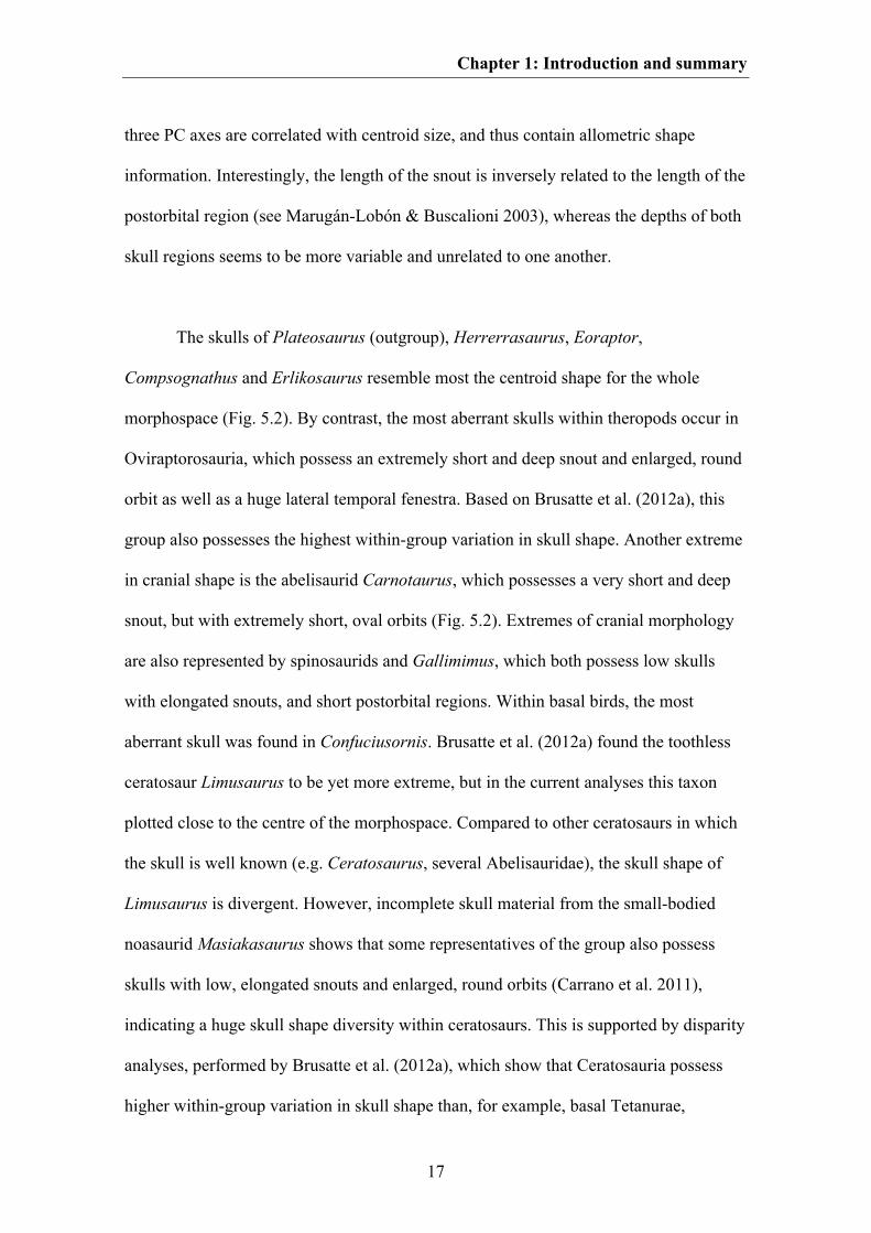
As in crocodylians, the shape of the rostrum may be more related to feeding
ecology than to function (Table 5.1). However, the ecological proxy used for feeding
ecology correlates still better with the shape variation seen in the postorbital region.
This may be because the ecological proxy also contains information on biting behaviour
(i.e. information related to function), which was found to have a stronger effect on the
posterior than the anterior part of the skull (see above). Furthermore, dietary preferences
Chapter 1: Introduction and summary
21
are related to tooth morphology (e.g. Smith 1993), which was not taken into account in
the geometric morphometric analyses. However, non-carnivorous theropods occupy a
large area within the morphospace (Fig. 5.5), indicated by huge skull shape diversity
(see also Brusatte et al. 2012a). Both carnivorous and non-carnivorous theropods are
well separated within the morphospace with only a small area of overlap. In contrast,
herbivorous and omnivorous theropods could not be distinguished from each other
based on the morphometric data (Fig. 5.5, Table 5.3). Based on the Procrustes
consensus shapes, carnivorous theropods tend to have a skull with a deep rostrum due to
maxillary shape, a large antorbital fenestra, a deep suborbital region, and a relatively
small, oval orbit, whereas non-carnivorous theropods (excluding aberrant oviraptorids)
tend to have a tapering rostrum with a small antorbital fenestra, a shallow jugal region,
an enlarged, round orbit, a shortened postorbital region, and a jaw joint significantly
anterior to the quadrate head (by including oviraptorids the rostrum of the non-
carnivorous theropods becomes shorter and deeper, and the postorbital region longer)
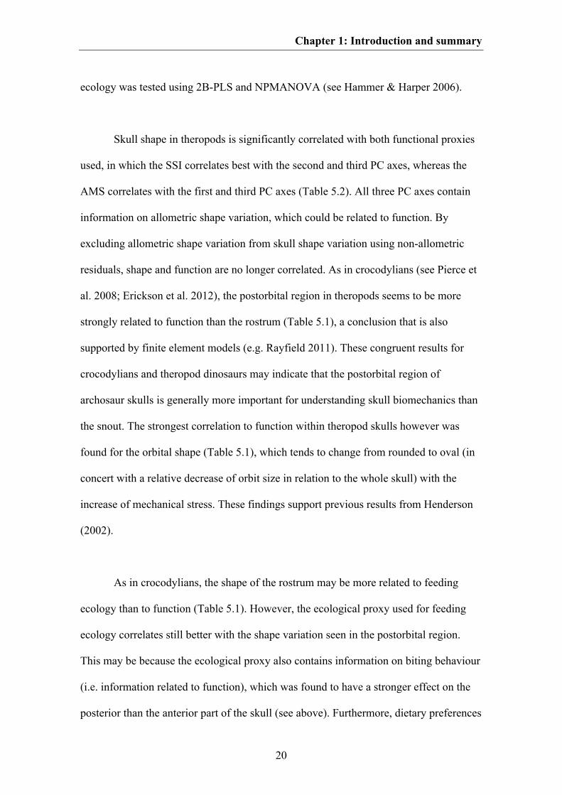
(Fig. 1.2). Thus, the differences in the consensus skull shapes of carnivorous and non-
carnivorous theropods correspond to the allometric trends seen in theropod skulls,
indicating that feeding ecology in theropods may also be influenced by body and/or
relative skull size, in which small-bodied theropods tend to adapt to a broader dietary
spectrum. In this context, the indistinguishability of skull shape of omnivorous and
herbivorous theropods may results from a gradual transition between both feeding
strategies (see Zanno & Makovicky 2011).
Chapter 1: Introduction and summary
22
Fig. 1.2. A. Consensus shape of carnivorous and non-carnivorous theropods. B. Allometric trend from
small-bodied to large-bodied theropods.
THE RELATIONSHIP BETWEEN SKULL SHAPE AND PHYLOGENY
Due to its complexity, the vertebrate skull provides a large number of potential
characters for use in phylogenetic analyses. The number of skull characters (including
tooth characters) used for phylogenetic analyses investigating the interrelationship of
several archosaur groups varies between c. 40 to over 90 % of total number of
characters (e.g. Rauhut 2003a; Butler et al. 2007; Lü et al. 2010a; Sereno & Brusatte
2009; Brusatte et al. 2010a; Prieto-Márquez 2010; Brochu 2011; Nesbitt 2011). Due to
its huge impact on phylogenetic analyses one can assume that the shape of archosaur
skulls is determinated by phylogeny. By mapping phylogenetic hypotheses within the
morphospace (see Stone 2003; Klingenberg & Gidaszewski 2010) this relationship can

Chapter 1: Introduction and summary
23
be tested with help of a permutation test. If a strong phylogenetic signal is present,
closely related taxa should appear closer to one another within morphospace than more
distantly related taxa, in which the tree length of the original topology should be shorter
than 95 % of all randomly generated trees (see Laurin 2004; Klingenberg &
Gidaszewski 2010). As the geometric morphometric data are treated as continuous
characters and optimized with help of e.g. square change parsimony (Maddison 1991)
this approach can be further used to estimate the skull shape of hypothetical ancestors
for each node of the phylogeny, and to comprehend skull shape variation during
evolution (see below for Crocodylomorpha and Saurischia, see Chapter 4 for
Pterosauria). In this thesis, the relationship between shape and phylogeny was tested for
both Pterosauria (Chapter 4) and Theropoda (Chapter 5).
For both groups skull shape is significantly correlated with phylogeny, i.e.
closely related taxa are more similar in skull shape to each other than more distantly
related taxa. A similar result was also found for theropod dinosaurs by Brusatte et al.
(2012a). According to these authors, phylogeny is even the primary determinant for
skull shape variation seen in theropods, but other macroevolutionary analysis using
different samples and landmark configurations found that skull shape evolution in
theropods was further influenced by function and diet (see Chapter 5 for discussion) and
heterochronic events (e.g. Bhullar et al. 2012, see below).
ONTOGENETIC AND HETEROCHRONIC PATTERNS IN ARCHOSAUR SKULLS
One of the key processes in evolution is heterochrony, which is defined as an
evolutionary change of a phenotype due to a change in the timing of developmental
processes (Wiesemüller et al. 2003; Futuyama 2007). Thus, heterochronic events could
Chapter 1: Introduction and summary
24
lead to significant evolutionary changes in body plans within short periods of time.
Evidence for heterochronic events occurring in evolutionary history could be potentially
detected with the help of comparative ontogenetic studies between different taxa, taking
into account their phylogenetic relationships. Here, skull shape variation of both
Crocodylomorpha and saurischian dinosaurs are studied in lateral view in terms of
heterochrony by combining interspecific and ontogenetic skull shape variation into one
analysis for each group respectively. The data were analysed with help of Principal
Component Analysis (PCA), regression methods, WARD cluster and character
mapping, in which ontogenetic patterns in skull shape were compared to evolutionary
patterns. A more detailed summary of material and methods is given in the
supplementary information of the current chapter.
ONTOGENETIC AND HETEROCHRONIC PATTERNS IN CROCODYLOMORPHA
The ontogenetic patterns used as reference for heterochronic events within
Crocodylomorpha are mainly based on Melanosuchus (see above, Chapter 3). However,
comparison with other recent crocodylians shows that some ontogenetic patterns seem
to be relatively similar, e.g. the relative increase of snout length and depth, relative
decrease of the orbit size and relative decrease in the length of the postorbital region
(Table 3.2). Additionally, based on the current dataset, the lateral temporal fenestra
decreases in its relative size during ontogeny.
Within the PCA plot the ontogenetic series of Melanosuchus clusters closely
together with the crocodyline Osteolaemus and the alligatorid Alligator. All three taxa
differ from the other crocodylomorphs in their possession of a relatively flat skull, a
strongly convex ventral margin of the snout, and a relatively large orbit, which is
Chapter 1: Introduction and summary
25
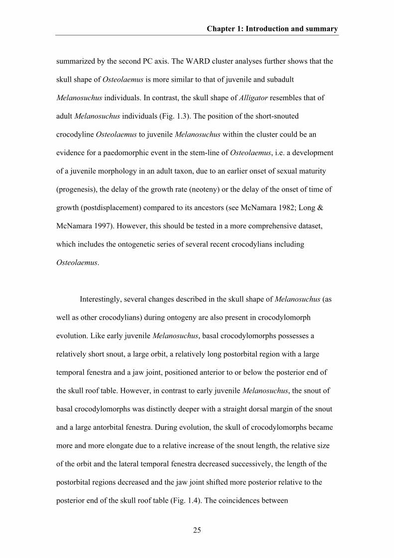
summarized by the second PC axis. The WARD cluster analyses further shows that the
skull shape of Osteolaemus is more similar to that of juvenile and subadult
Melanosuchus individuals. In contrast, the skull shape of Alligator resembles that of
adult Melanosuchus individuals (Fig. 1.3). The position of the short-snouted
crocodyline Osteolaemus to juvenile Melanosuchus within the cluster could be an
evidence for a paedomorphic event in the stem-line of Osteolaemus, i.e. a development
of a juvenile morphology in an adult taxon, due to an earlier onset of sexual maturity
(progenesis), the delay of the growth rate (neoteny) or the delay of the onset of time of
growth (postdisplacement) compared to its ancestors (see McNamara 1982; Long &
McNamara 1997). However, this should be tested in a more comprehensive dataset,
which includes the ontogenetic series of several recent crocodylians including
Osteolaemus.
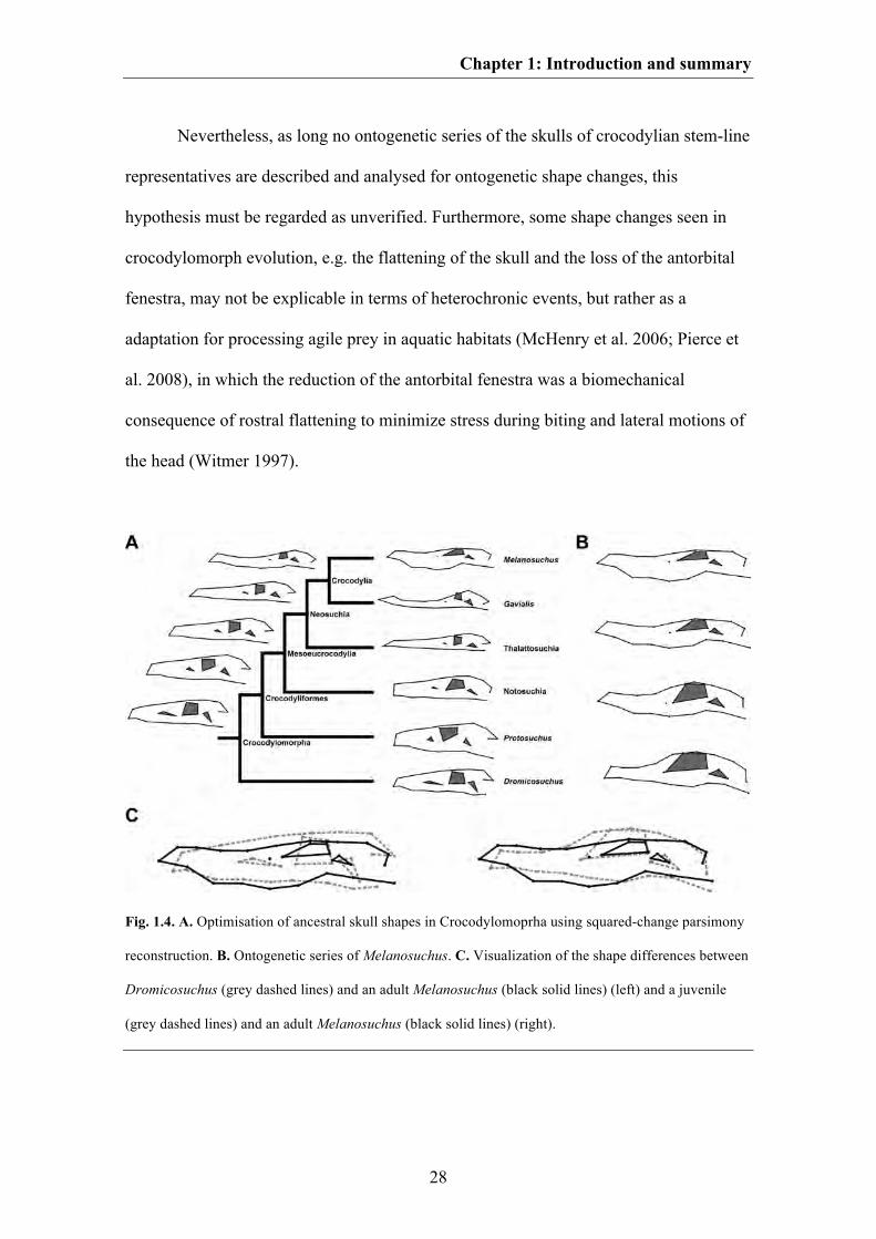
Interestingly, several changes described in the skull shape of Melanosuchus (as
well as other crocodylians) during ontogeny are also present in crocodylomorph
evolution. Like early juvenile Melanosuchus, basal crocodylomorphs possesses a
relatively short snout, a large orbit, a relatively long postorbital region with a large
temporal fenestra and a jaw joint, positioned anterior to or below the posterior end of
the skull roof table. However, in contrast to early juvenile Melanosuchus, the snout of
basal crocodylomorphs was distinctly deeper with a straight dorsal margin of the snout
and a large antorbital fenestra. During evolution, the skull of crocodylomorphs became
more and more elongate due to a relative increase of the snout length, the relative size
of the orbit and the lateral temporal fenestra decreased successively, the length of the
postorbital regions decreased and the jaw joint shifted more posterior relative to the
posterior end of the skull roof table (Fig. 1.4). The coincidences between
Chapter 1: Introduction and summary
26
crocodylomorph evolution and Melanosuchus ontogeny previously described could be
an evidence for a peramorphic trend within crocodylomorph evolution, i.e. the
evolvement of a more “developed” taxon due to a delayed onset of sexual maturity
(hypermorphis), an increase of growth rate (acceleration) or an early onset of growth
(predisplacement) compared to its ancestor (see McNamara 1982; Long & McNamara
1997).
Unfortunately, no study on the body size evolution within Crocodylomorpha has
been conducted to test this hypothesis in detail. Based on the fossil record, basal
crocodylomorphs tend to be relatively small (e.g. Colbert & Mook 1951; Walker 1990;
Wu & Chatterjee 1993; Clark et al. 2004), whereas some neosuchians reached
enormous body size (e.g. Erickson & Brochu 1999; Sereno et al. 2001; Sereno &
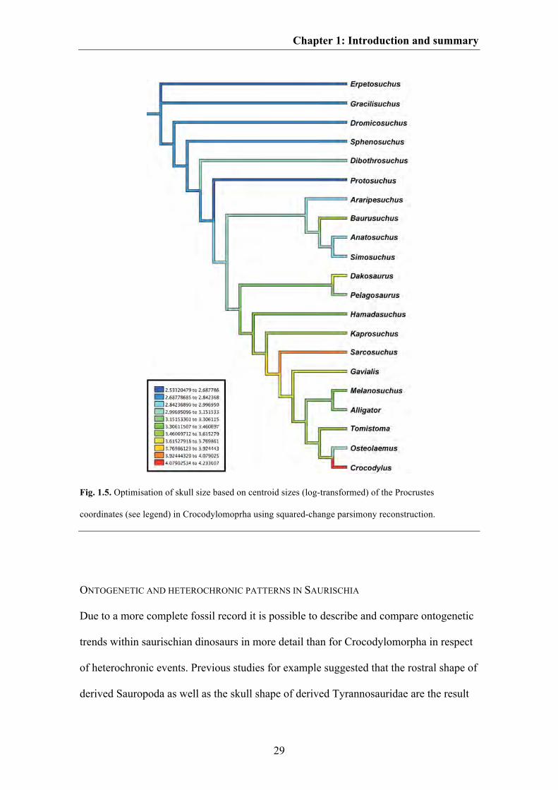
Larsson 2009; Brochu & Storrs 2012). After mapping the centroid size of the aligned
skulls onto the phylogenetic tree used in the current study, it can be seen that skull size
increased successively from the hypothetical ancestor of Crocodylomorpha to that of
Crocodylia (Fig. 1.5), supporting the hypothesized peramorphic trend within
crocodylomorph evolution. Nevertheless, because of missing data on the bone histology
of crocodylomorphs, it is currently not well understood, which growth strategy led to
this supposed peramorphosis. Small basal crocodylomorphs like Terrestrisuchus for
instance grew relatively fast (de Ricqlès et al. 2003), whereas recent crocodylians
possess a more ‘reptile’-like slowed growth pattern (e.g. Hutton 1987). However,
histological data for the giant crocodylian Deinosuchus reveal that peramorphic events
within crown-crocodylians could be caused at least by hypermorphosis, resulting from a
prolongation of growth with juvenile growth rates (see Erickson & Brochu 1999).
Chapter 1: Introduction and summary
27
Fig. 1.3. A. Two-dimensional morphospace of the crocodylomorph skull shape based on the first two PC
axes. The ontogenetic series of Melanosuchus is shown with black dots. B. Ward cluster showing the
similarity in the skull shape of Crocodylomorpha (including an ontogenetic series of Melanosuchus)
based on the Procrustes coordinates. The asterisk shows the cluster containing the ontogenetic series of
Melanosuchus.
Chapter 1: Introduction and summary
28
Nevertheless, as long no ontogenetic series of the skulls of crocodylian stem-line
representatives are described and analysed for ontogenetic shape changes, this
hypothesis must be regarded as unverified. Furthermore, some shape changes seen in
crocodylomorph evolution, e.g. the flattening of the skull and the loss of the antorbital
fenestra, may not be explicable in terms of heterochronic events, but rather as a
adaptation for processing agile prey in aquatic habitats (McHenry et al. 2006; Pierce et
al. 2008), in which the reduction of the antorbital fenestra was a biomechanical
consequence of rostral flattening to minimize stress during biting and lateral motions of
the head (Witmer 1997).
Fig. 1.4. A. Optimisation of ancestral skull shapes in Crocodylomoprha using squared-change parsimony
reconstruction. B. Ontogenetic series of Melanosuchus. C. Visualization of the shape differences between
Dromicosuchus (grey dashed lines) and an adult Melanosuchus (black solid lines) (left) and a juvenile
(grey dashed lines) and an adult Melanosuchus (black solid lines) (right).
Chapter 1: Introduction and summary
29
Fig. 1.5. Optimisation of skull size based on centroid sizes (log-transformed) of the Procrustes
coordinates (see legend) in Crocodylomoprha using squared-change parsimony reconstruction.
ONTOGENETIC AND HETEROCHRONIC PATTERNS IN SAURISCHIA
Due to a more complete fossil record it is possible to describe and compare ontogenetic
trends within saurischian dinosaurs in more detail than for Crocodylomorpha in respect
of heterochronic events. Previous studies for example suggested that the rostral shape of
derived Sauropoda as well as the skull shape of derived Tyrannosauridae are the result
Chapter 1: Introduction and summary
30
of peramorphic events (Long & McNamara 1997), whereas the skull shape of birds may
have been caused by paedomorphosis (Bhullar et al. 2012). The current study, which is
based on a broad-scale sample of both Sauropodomorpha and Theropoda, attempts to
detect more examples of heterochronic events within saurischian evolution, especially
in more basal members of both saurischian clades. In this context, a comparison of the
skull shape of the juvenile megalosaurid Sciurumimus (see Chapter 6) with that of basal
coelurosaurs such as Compsognathus and Dilong could be of interest due to similarities
such as an elongate skull with a triangular shape and a tapering snout, a large round
orbit, a slender jugal or a jaw joint straight below the quadrate head.
For this study, ontogenetic series of basal sauropodomorphs (Massospondylus),
sauropods (Diplodocus and Titanosauridae), basal tetanurans (Allosaurus and
Megalosauridae), tyrannosaurids (Tyrannosaurus and Tarbosaurus) and oviraptorids
were analysed. Furthermore, the skulls of the juvenile theropods Juravenator and
Scipionyx were included into the dataset. The ontogenetic series for titanosaurids based
on the reconstruction of embryonic sauropod skulls found in Patagonia (Chiappe et al.
1998) and the skull of Antarctosaurus. The ontogenetic series for megalosaurids is
based on the skull of Sciurumimus and that of Dubreuillosaurus, whereas the
ontogenetic series of oviraptorids is based on the skull of Yulong and the consensus
shape of Citipati, Conchoraptor and Nemegtomaia. A more detailed discussion on
taxonomic validity of some ontogenetic series used in the current analysis is given in
the supplementary information of Chapter 1. The samples for Allosaurus,
Tyrannosaurus, Tarbosaurus, oviraptorids and Diplodocus represent rather late
ontogenetic series as the juveniles sampled represent late juvenile and subadult
individuals. All ontogenetic shape changes presented are shown in Fig. 1.6.

Chapter 1: Introduction and summary
31
Fig. 1.6. Visualization of ontogenetic shape changes within different saurischian taxa. A. The basal
sauropodomorph Massospondylus. B. The sauropod Diplodocus. C. A hypothetical titanosaurid sauropod.
D. A hypothetical megalosaurid theropod. E. The basal tetanurans theropod Allosaurus. F. The
tyrannosaurid Tarbosaurus. G. The tyrannosaurid Tyrannosaurus. H. An hypothetical oviraptorid.
Juvenile skull shapes are shown in grey dashed lines and adult skull shapes are shown in black solid lines.
Chapter 1: Introduction and summary
32
Ontogenetic patterns in Sauropodomorpha
Early juveniles of Massospondylus possess a short, tapering snout with a straight ventral
margin, an anteroposteriorly compressed antorbital fenestra, an enlarged orbit and
relatively deep postorbital region. Similar skulls shapes are also present from juvenile
specimens of the basal sauropodomorph Mussasaurus (Pol & Powell 2007). During
ontogeny the skull of Massospondylus become more elongate due to a relative increase
of the overall snout length and a decrease of the orbit. The maxilla expands ventrally
forming a convex margin. The relative size of antorbital fenestra increased slightly in
anteroposterior direction. Furthermore, the depth of the postorbital region decrease
relative in size.
The skull of the juvenile Diplodocus possesses a short snout with a straight
ventral margin, a small antorbital fenestra, a relatively large orbit and a relatively deep
orbital and postorbital region. During ontogeny, the snout becomes more elongate due
to an anterior expansion of the maxilla. This change goes hand in hand with a bowing of
the ventral margin of the anterior part of the maxilla in a dorsal direction. Furthermore,
the relative size of the antorbital fenestra increases in an anteroposterior direction,
whereas both the relative size of the orbit and the depth of the postrostral region
decrease. The skull shape of early juvenile titanosaurids resembles that of a juvenile
Massospondylus in terms of presence of a short snout with a small antorbital fenestra
and a large orbit. In contrast to the Massospondylus hatchlings, the ventral margin of the
maxilla of early juvenile titanosaurids is inverted Z-shaped. During ontogeny, both the
premaxilla and the anterior part of the maxilla expand ventrally, and thereby the ventral
margin of the maxilla becomes smoother to form an S-shape. The antorbital fenestra
increases in dorsal and posterior directions. The orbit decreases in relative length, but
Chapter 1: Introduction and summary
33
increases in relative depth. This process goes hand in hand with an increase of the
orbital depth. The postorbital region decreases in relative length, but increases in its
relative depth due a ventral expansion of the jugal and quadratojugal. Despite the
decrease of the postorbital length, the lateral temporal fenestra shows a relative increase
in its length.
Ontogenetic patterns in Theropoda
The skull of the juvenile megalosaurid possesses a relatively long snout, a large and
rounded antorbital fenestra, and an anteriorly inclined lacrimal. The orbital region is
deeper than the rostral and postorbital region. Furthermore, the jaw joint lies ventral to
the squamosal body. During ontogeny, the snout becomes more elongated due to
increase of the premaxilla length. Furthermore, the snout tip becomes deeper due to a
ventral expansion of the premaxilla and the anterior part of the maxilla. The antorbital
fenestra increases in dorsal and posterior direction and the lacrimal becomes more
vertically orientated. This process goes hand in hand with a relative decrease of the
orbital length leading to an oval orbital shape. In the adult specimen the orbital depth is
as deep as the depth of the snout and the postorbital. The postorbital region increases in
its relative length, which occurs in concert with an increase of the depth of the lateral
temporal fenestra and a shift of the jaw joint posterior to the squamosal body.
The skull of a juvenile Allosaurus resembles that of a juvenile megalosaurid
possessing an elongated snout and a rounded orbit. In contrast to the megalosaurid
juvenile, the snout is relatively deeper and the lacrimal is already vertically orientated.
This difference could be the result of the later ontogenetic stage of the juvenile
specimen sampled for Allosaurus. The discovery of a single maxilla identified as that of
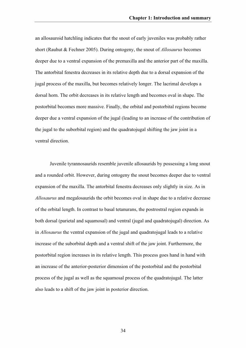
Chapter 1: Introduction and summary
34
an allosauroid hatchling indicates that the snout of early juveniles was probably rather
short (Rauhut & Fechner 2005). During ontogeny, the snout of Allosaurus becomes
deeper due to a ventral expansion of the premaxilla and the anterior part of the maxilla.
The antorbital fenestra decreases in its relative depth due to a dorsal expansion of the
jugal process of the maxilla, but becomes relatively longer. The lacrimal develops a
dorsal horn. The orbit decreases in its relative length and becomes oval in shape. The
postorbital becomes more massive. Finally, the orbital and postorbital regions become
deeper due a ventral expansion of the jugal (leading to an increase of the contribution of
the jugal to the suborbital region) and the quadratojugal shifting the jaw joint in a
ventral direction.
Juvenile tyrannosaurids resemble juvenile allosaurids by possessing a long snout
and a rounded orbit. However, during ontogeny the snout becomes deeper due to ventral
expansion of the maxilla. The antorbital fenestra decreases only slightly in size. As in
Allosaurus and megalosaurids the orbit becomes oval in shape due to a relative decrease
of the orbital length. In contrast to basal tetanurans, the postrostral region expands in
both dorsal (parietal and squamosal) and ventral (jugal and quadratojugal) direction. As
in Allosaurus the ventral expansion of the jugal and quadratojugal leads to a relative
increase of the suborbital depth and a ventral shift of the jaw joint. Furthermore, the
postorbital region increases in its relative length. This process goes hand in hand with
an increase of the anterior-posterior dimension of the postorbital and the postorbital
process of the jugal as well as the squamosal process of the quadratojugal. The latter
also leads to a shift of the jaw joint in posterior direction.
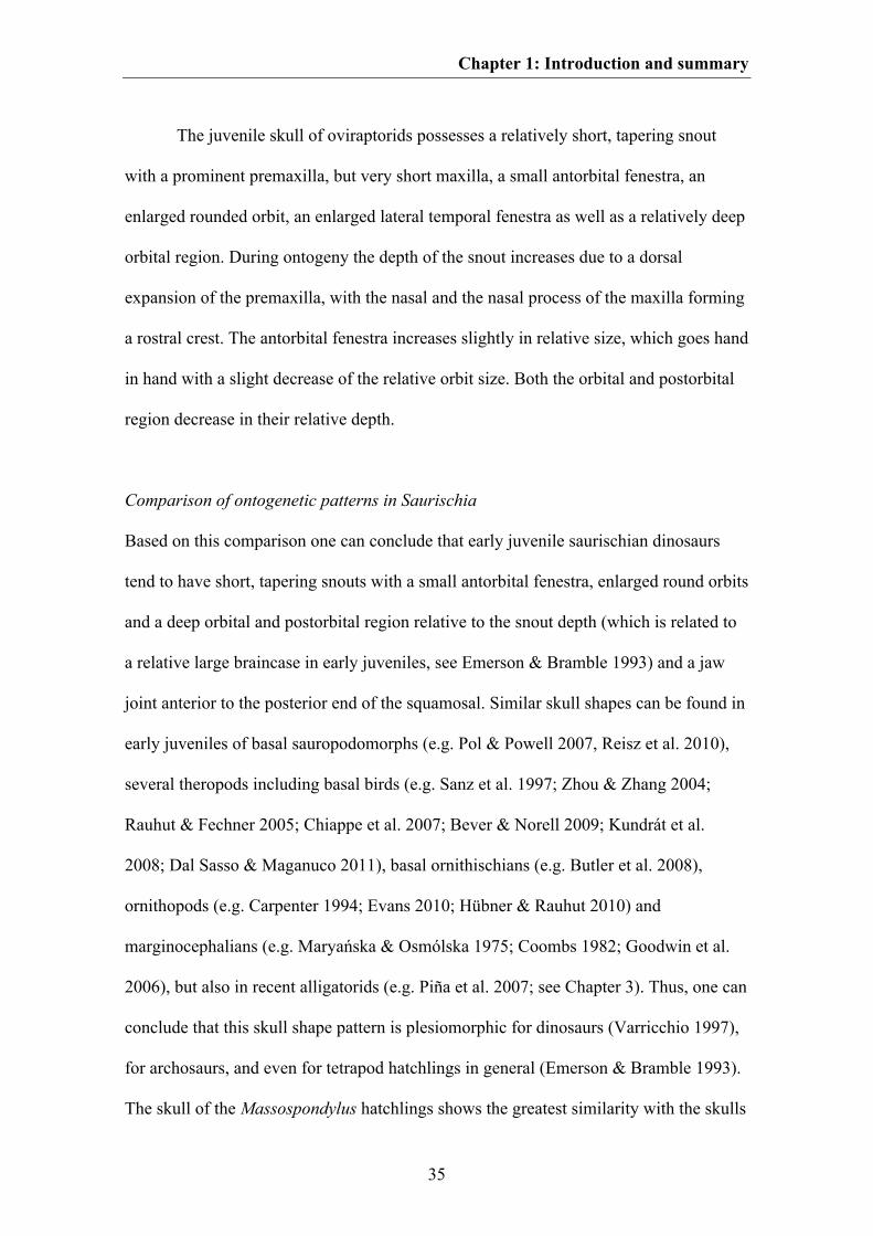
Chapter 1: Introduction and summary
35
The juvenile skull of oviraptorids possesses a relatively short, tapering snout
with a prominent premaxilla, but very short maxilla, a small antorbital fenestra, an
enlarged rounded orbit, an enlarged lateral temporal fenestra as well as a relatively deep
orbital region. During ontogeny the depth of the snout increases due to a dorsal
expansion of the premaxilla, with the nasal and the nasal process of the maxilla forming
a rostral crest. The antorbital fenestra increases slightly in relative size, which goes hand
in hand with a slight decrease of the relative orbit size. Both the orbital and postorbital
region decrease in their relative depth.
Comparison of ontogenetic patterns in Saurischia
Based on this comparison one can conclude that early juvenile saurischian dinosaurs
tend to have short, tapering snouts with a small antorbital fenestra, enlarged round orbits
and a deep orbital and postorbital region relative to the snout depth (which is related to
a relative large braincase in early juveniles, see Emerson & Bramble 1993) and a jaw
joint anterior to the posterior end of the squamosal. Similar skull shapes can be found in
early juveniles of basal sauropodomorphs (e.g. Pol & Powell 2007, Reisz et al. 2010),
several theropods including basal birds (e.g. Sanz et al. 1997; Zhou & Zhang 2004;
Rauhut & Fechner 2005; Chiappe et al. 2007; Bever & Norell 2009; Kundrát et al.
2008; Dal Sasso & Maganuco 2011), basal ornithischians (e.g. Butler et al. 2008),
ornithopods (e.g. Carpenter 1994; Evans 2010; Hübner & Rauhut 2010) and
marginocephalians (e.g. Maryańska & Osmólska 1975; Coombs 1982; Goodwin et al.
2006), but also in recent alligatorids (e.g. Piña et al. 2007; see Chapter 3). Thus, one can
conclude that this skull shape pattern is plesiomorphic for dinosaurs (Varricchio 1997),
for archosaurs, and even for tetrapod hatchlings in general (Emerson & Bramble 1993).
The skull of the Massospondylus hatchlings shows the greatest similarity with the skulls
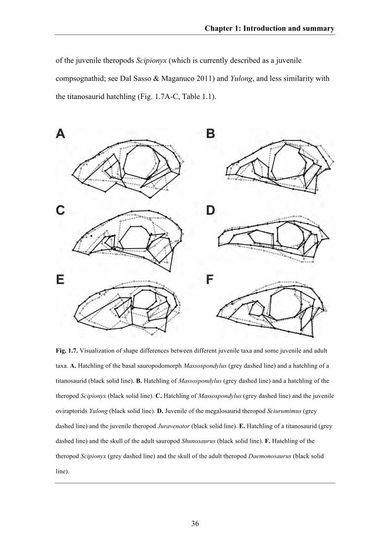
Chapter 1: Introduction and summary
36
of the juvenile theropods Scipionyx (which is currently described as a juvenile
compsognathid; see Dal Sasso & Maganuco 2011) and Yulong, and less similarity with
the titanosaurid hatchling (Fig. 1.7A-C, Table 1.1).
Fig. 1.7. Visualization of shape differences between different juvenile taxa and some juvenile and adult
taxa. A. Hatchling of the basal sauropodomorph Massospondylus (grey dashed line) and a hatchling of a
titanosaurid (black solid line). B. Hatchling of Massospondylus (grey dashed line) and a hatchling of the
theropod Scipionyx (black solid line). C. Hatchling of Massospondylus (grey dashed line) and the juvenile
oviraptorids Yulong (black solid line). D. Juvenile of the megalosaurid theropod Sciurumimus (grey
dashed line) and the juvenile theropod Juravenator (black solid line). E. Hatchling of a titanosaurid (grey
dashed line) and the skull of the adult sauropod Shunosaurus (black solid line). F. Hatchling of the
theropod Scipionyx (grey dashed line) and the skull of the adult theropod Daemonosaurus (black solid
line).
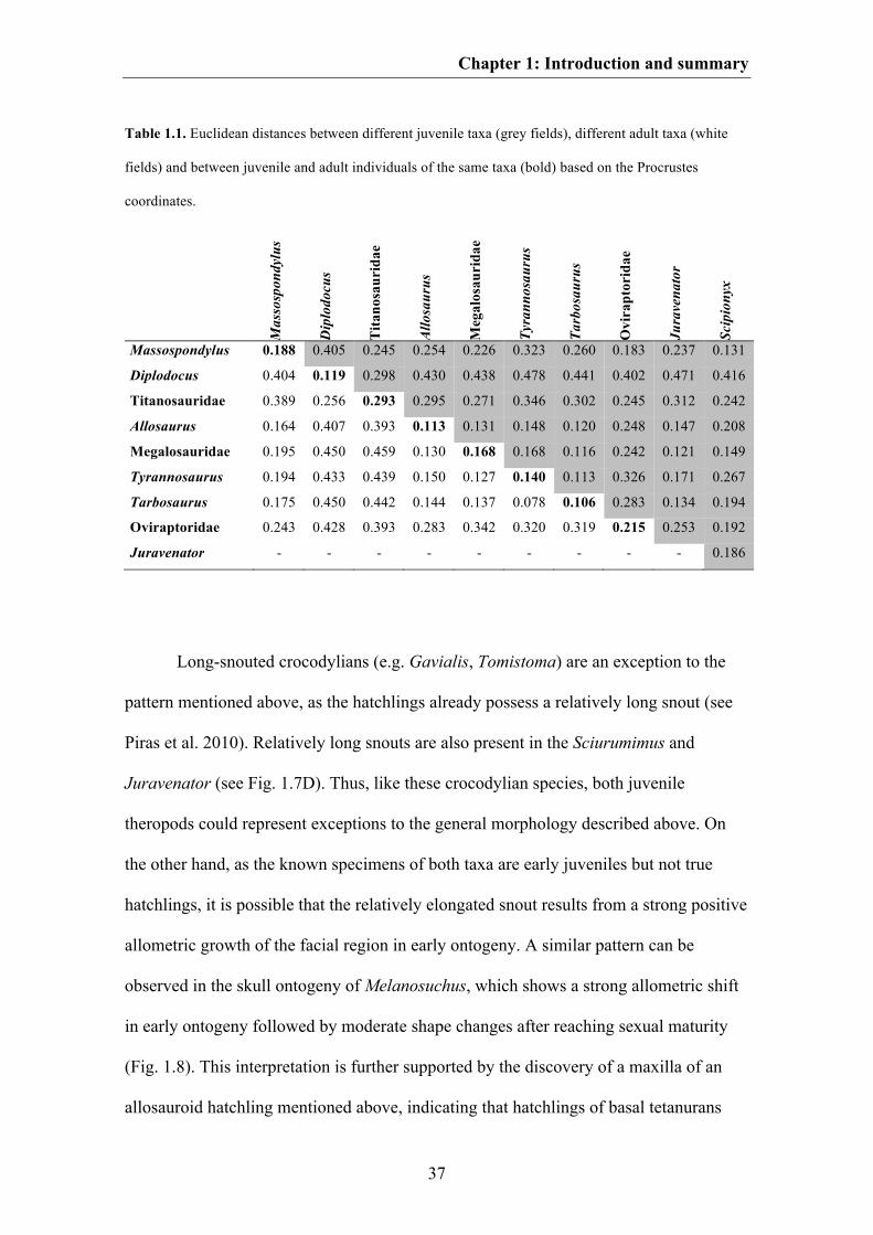
Chapter 1: Introduction and summary
37
Table 1.1. Euclidean distances between different juvenile taxa (grey fields), different adult taxa (white
fields) and between juvenile and adult individuals of the same taxa (bold) based on the Procrustes
coordinates.
Mas
sosp
ondy
lus
Dip
lodo
cus
Tita
nosa
urid
ae
Allo
saur
us
Meg
alos
auri
dae
Tyra
nnos
auru
s
Tarb
osau
rus
Ovi
rapt
orid
ae
Jura
vena
tor
Scip
iony
x
Massospondylus 0.188 0.405 0.245 0.254 0.226 0.323 0.260 0.183 0.237 0.131
Diplodocus 0.404 0.119 0.298 0.430 0.438 0.478 0.441 0.402 0.471 0.416
Titanosauridae 0.389 0.256 0.293 0.295 0.271 0.346 0.302 0.245 0.312 0.242
Allosaurus 0.164 0.407 0.393 0.113 0.131 0.148 0.120 0.248 0.147 0.208
Megalosauridae 0.195 0.450 0.459 0.130 0.168 0.168 0.116 0.242 0.121 0.149
Tyrannosaurus 0.194 0.433 0.439 0.150 0.127 0.140 0.113 0.326 0.171 0.267
Tarbosaurus 0.175 0.450 0.442 0.144 0.137 0.078 0.106 0.283 0.134 0.194
Oviraptoridae 0.243 0.428 0.393 0.283 0.342 0.320 0.319 0.215 0.253 0.192
Juravenator - - - - - - - - - 0.186
Long-snouted crocodylians (e.g. Gavialis, Tomistoma) are an exception to the
pattern mentioned above, as the hatchlings already possess a relatively long snout (see
Piras et al. 2010). Relatively long snouts are also present in the Sciurumimus and
Juravenator (see Fig. 1.7D). Thus, like these crocodylian species, both juvenile
theropods could represent exceptions to the general morphology described above. On
the other hand, as the known specimens of both taxa are early juveniles but not true
hatchlings, it is possible that the relatively elongated snout results from a strong positive
allometric growth of the facial region in early ontogeny. A similar pattern can be
observed in the skull ontogeny of Melanosuchus, which shows a strong allometric shift
in early ontogeny followed by moderate shape changes after reaching sexual maturity
(Fig. 1.8). This interpretation is further supported by the discovery of a maxilla of an
allosauroid hatchling mentioned above, indicating that hatchlings of basal tetanurans
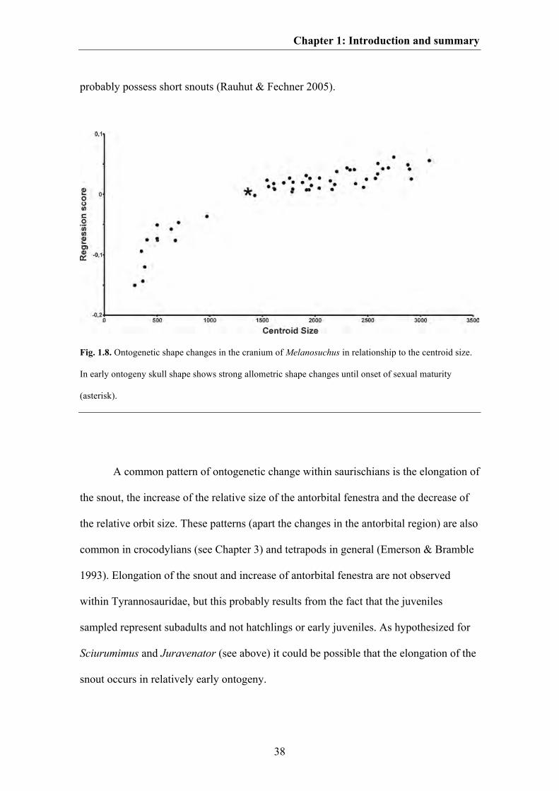
Chapter 1: Introduction and summary
38
probably possess short snouts (Rauhut & Fechner 2005).
Fig. 1.8. Ontogenetic shape changes in the cranium of Melanosuchus in relationship to the centroid size.
In early ontogeny skull shape shows strong allometric shape changes until onset of sexual maturity
(asterisk).
A common pattern of ontogenetic change within saurischians is the elongation of
the snout, the increase of the relative size of the antorbital fenestra and the decrease of
the relative orbit size. These patterns (apart the changes in the antorbital region) are also
common in crocodylians (see Chapter 3) and tetrapods in general (Emerson & Bramble
1993). Elongation of the snout and increase of antorbital fenestra are not observed
within Tyrannosauridae, but this probably results from the fact that the juveniles
sampled represent subadults and not hatchlings or early juveniles. As hypothesized for
Sciurumimus and Juravenator (see above) it could be possible that the elongation of the
snout occurs in relatively early ontogeny.
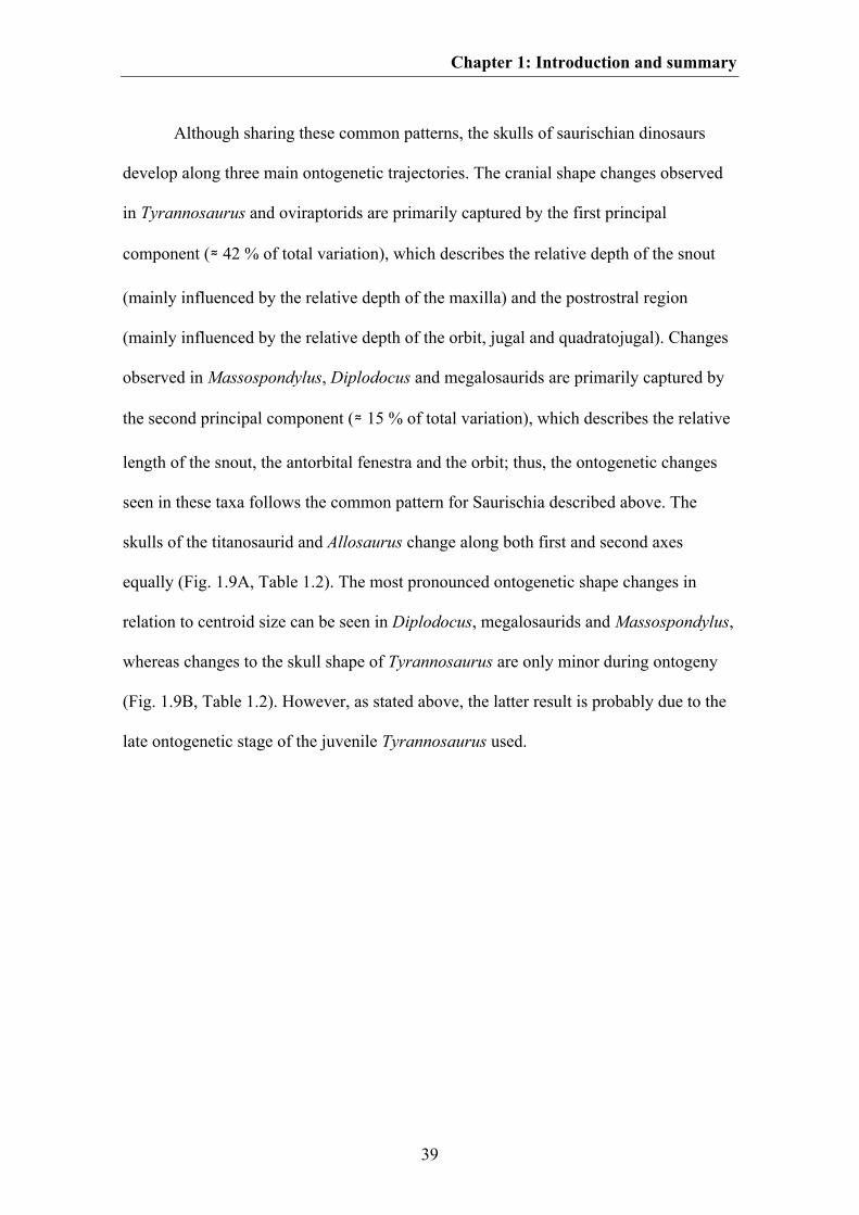
Chapter 1: Introduction and summary
39
Although sharing these common patterns, the skulls of saurischian dinosaurs
develop along three main ontogenetic trajectories. The cranial shape changes observed
in Tyrannosaurus and oviraptorids are primarily captured by the first principal
component (≈ 42 % of total variation), which describes the relative depth of the snout
(mainly influenced by the relative depth of the maxilla) and the postrostral region
(mainly influenced by the relative depth of the orbit, jugal and quadratojugal). Changes
observed in Massospondylus, Diplodocus and megalosaurids are primarily captured by
the second principal component (≈ 15 % of total variation), which describes the relative
length of the snout, the antorbital fenestra and the orbit; thus, the ontogenetic changes
seen in these taxa follows the common pattern for Saurischia described above. The
skulls of the titanosaurid and Allosaurus change along both first and second axes
equally (Fig. 1.9A, Table 1.2). The most pronounced ontogenetic shape changes in
relation to centroid size can be seen in Diplodocus, megalosaurids and Massospondylus,
whereas changes to the skull shape of Tyrannosaurus are only minor during ontogeny
(Fig. 1.9B, Table 1.2). However, as stated above, the latter result is probably due to the
late ontogenetic stage of the juvenile Tyrannosaurus used.
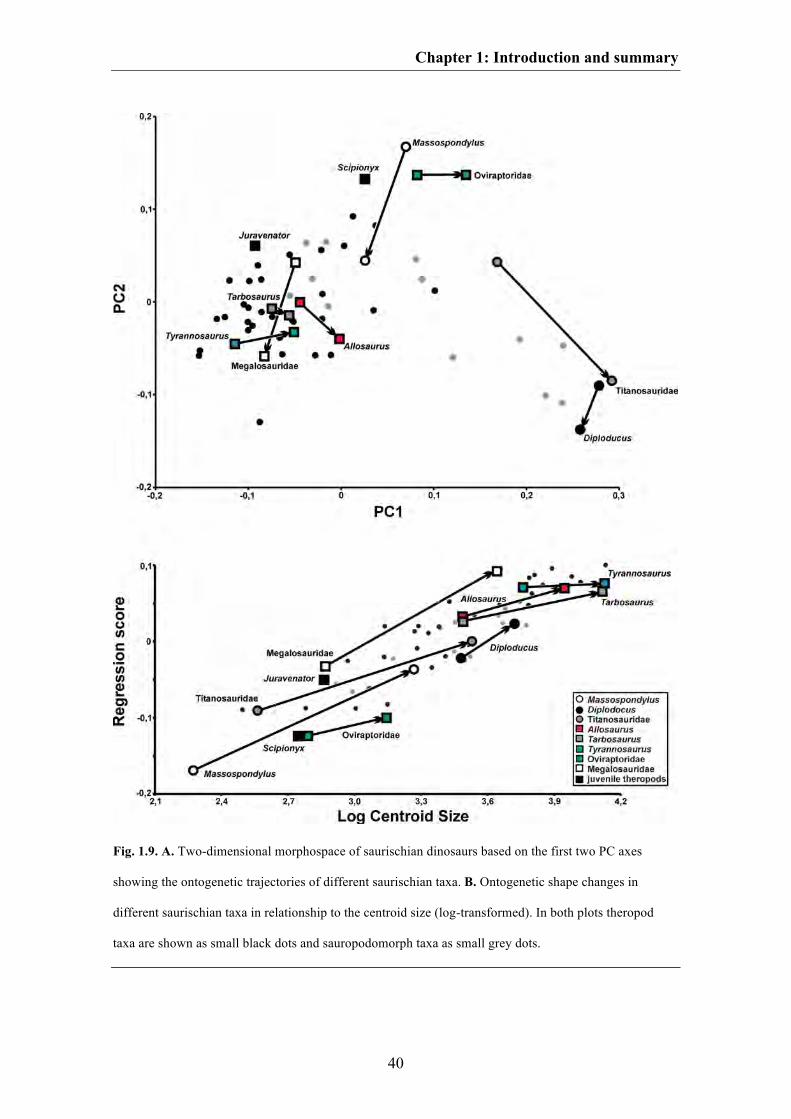
Chapter 1: Introduction and summary
40
Fig. 1.9. A. Two-dimensional morphospace of saurischian dinosaurs based on the first two PC axes
showing the ontogenetic trajectories of different saurischian taxa. B. Ontogenetic shape changes in
different saurischian taxa in relationship to the centroid size (log-transformed). In both plots theropod
taxa are shown as small black dots and sauropodomorph taxa as small grey dots.
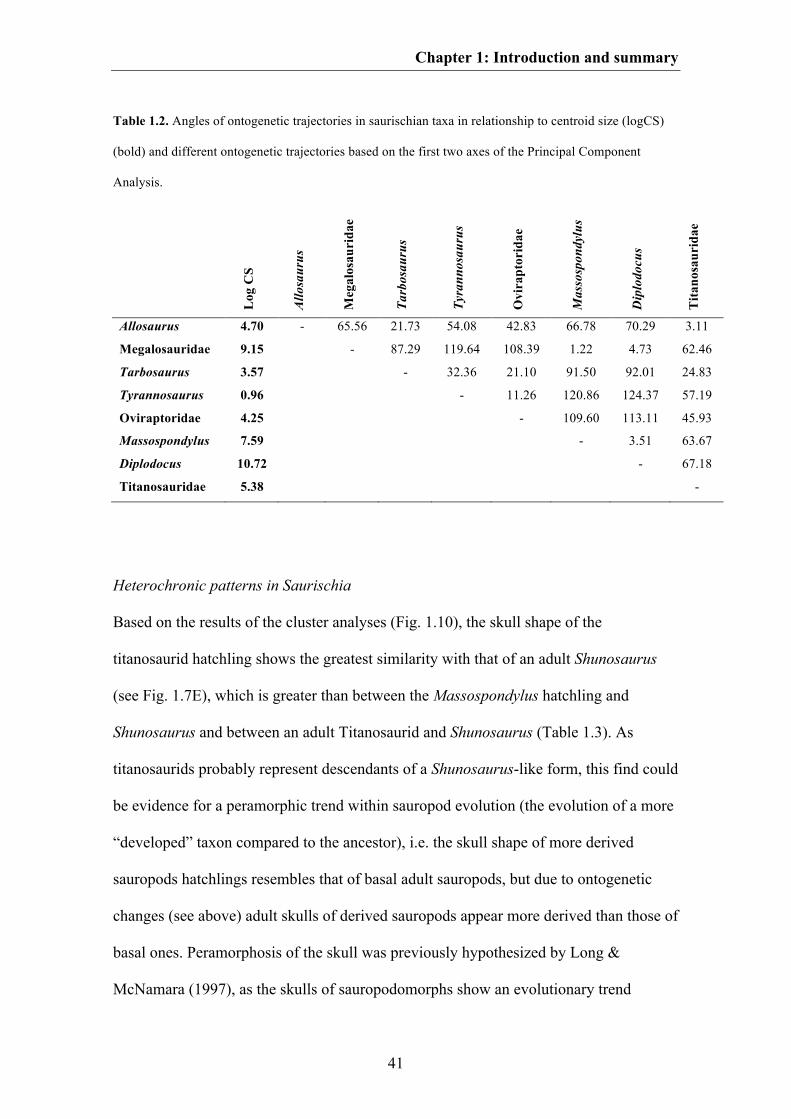
Chapter 1: Introduction and summary
41
Table 1.2. Angles of ontogenetic trajectories in saurischian taxa in relationship to centroid size (logCS)
(bold) and different ontogenetic trajectories based on the first two axes of the Principal Component
Analysis.
Log
CS
Allo
saur
us
Meg
alos
auri
dae
Tarb
osau
rus
Tyra
nnos
auru
s
Ovi
rapt
orid
ae
Mas
sosp
ondy
lus
Dip
lodo
cus
Tita
nosa
urid
ae
Allosaurus 4.70 - 65.56 21.73 54.08 42.83 66.78 70.29 3.11
Megalosauridae 9.15
- 87.29 119.64 108.39 1.22 4.73 62.46
Tarbosaurus 3.57
- 32.36 21.10 91.50 92.01 24.83
Tyrannosaurus 0.96
- 11.26 120.86 124.37 57.19
Oviraptoridae 4.25
- 109.60 113.11 45.93
Massospondylus 7.59
- 3.51 63.67
Diplodocus 10.72
- 67.18
Titanosauridae 5.38
-
Heterochronic patterns in Saurischia
Based on the results of the cluster analyses (Fig. 1.10), the skull shape of the
titanosaurid hatchling shows the greatest similarity with that of an adult Shunosaurus
(see Fig. 1.7E), which is greater than between the Massospondylus hatchling and
Shunosaurus and between an adult Titanosaurid and Shunosaurus (Table 1.3). As
titanosaurids probably represent descendants of a Shunosaurus-like form, this find could
be evidence for a peramorphic trend within sauropod evolution (the evolution of a more
“developed” taxon compared to the ancestor), i.e. the skull shape of more derived
sauropods hatchlings resembles that of basal adult sauropods, but due to ontogenetic
changes (see above) adult skulls of derived sauropods appear more derived than those of
basal ones. Peramorphosis of the skull was previously hypothesized by Long &
McNamara (1997), as the skulls of sauropodomorphs show an evolutionary trend
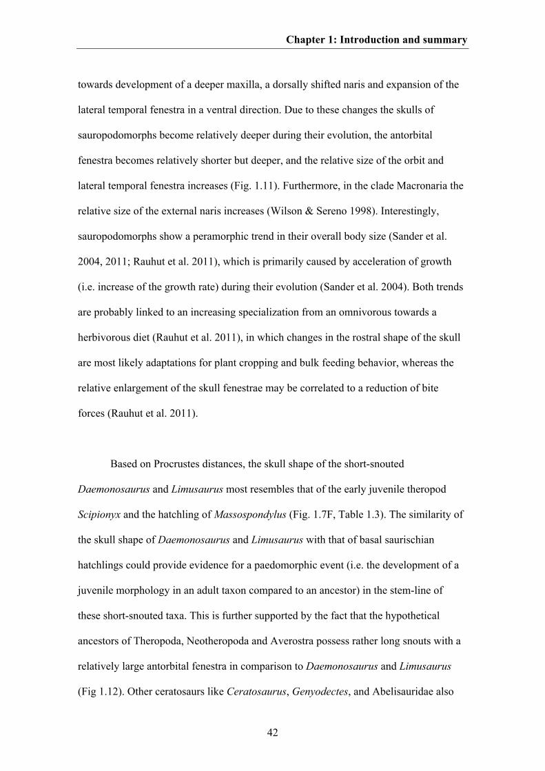
Chapter 1: Introduction and summary
42
towards development of a deeper maxilla, a dorsally shifted naris and expansion of the
lateral temporal fenestra in a ventral direction. Due to these changes the skulls of
sauropodomorphs become relatively deeper during their evolution, the antorbital
fenestra becomes relatively shorter but deeper, and the relative size of the orbit and
lateral temporal fenestra increases (Fig. 1.11). Furthermore, in the clade Macronaria the
relative size of the external naris increases (Wilson & Sereno 1998). Interestingly,
sauropodomorphs show a peramorphic trend in their overall body size (Sander et al.
2004, 2011; Rauhut et al. 2011), which is primarily caused by acceleration of growth
(i.e. increase of the growth rate) during their evolution (Sander et al. 2004). Both trends
are probably linked to an increasing specialization from an omnivorous towards a
herbivorous diet (Rauhut et al. 2011), in which changes in the rostral shape of the skull
are most likely adaptations for plant cropping and bulk feeding behavior, whereas the
relative enlargement of the skull fenestrae may be correlated to a reduction of bite
forces (Rauhut et al. 2011).
Based on Procrustes distances, the skull shape of the short-snouted
Daemonosaurus and Limusaurus most resembles that of the early juvenile theropod
Scipionyx and the hatchling of Massospondylus (Fig. 1.7F, Table 1.3). The similarity of
the skull shape of Daemonosaurus and Limusaurus with that of basal saurischian
hatchlings could provide evidence for a paedomorphic event (i.e. the development of a
juvenile morphology in an adult taxon compared to an ancestor) in the stem-line of
these short-snouted taxa. This is further supported by the fact that the hypothetical
ancestors of Theropoda, Neotheropoda and Averostra possess rather long snouts with a
relatively large antorbital fenestra in comparison to Daemonosaurus and Limusaurus
(Fig 1.12). Other ceratosaurs like Ceratosaurus, Genyodectes, and Abelisauridae also
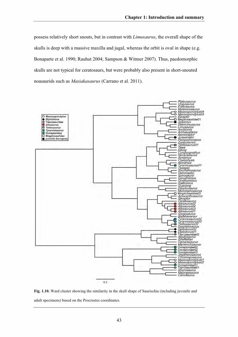
Chapter 1: Introduction and summary
43
possess relatively short snouts, but in contrast with Limusaurus, the overall shape of the
skulls is deep with a massive maxilla and jugal, whereas the orbit is oval in shape (e.g.
Bonaparte et al. 1990; Rauhut 2004; Sampson & Witmer 2007). Thus, paedomorphic
skulls are not typical for ceratosaurs, but were probably also present in short-snouted
noasaurids such as Masiakasaurus (Carrano et al. 2011).
Fig. 1.10. Ward cluster showing the similarity in the skull shape of Saurischia (including juvenile and
adult specimens) based on the Procrustes coordinates.
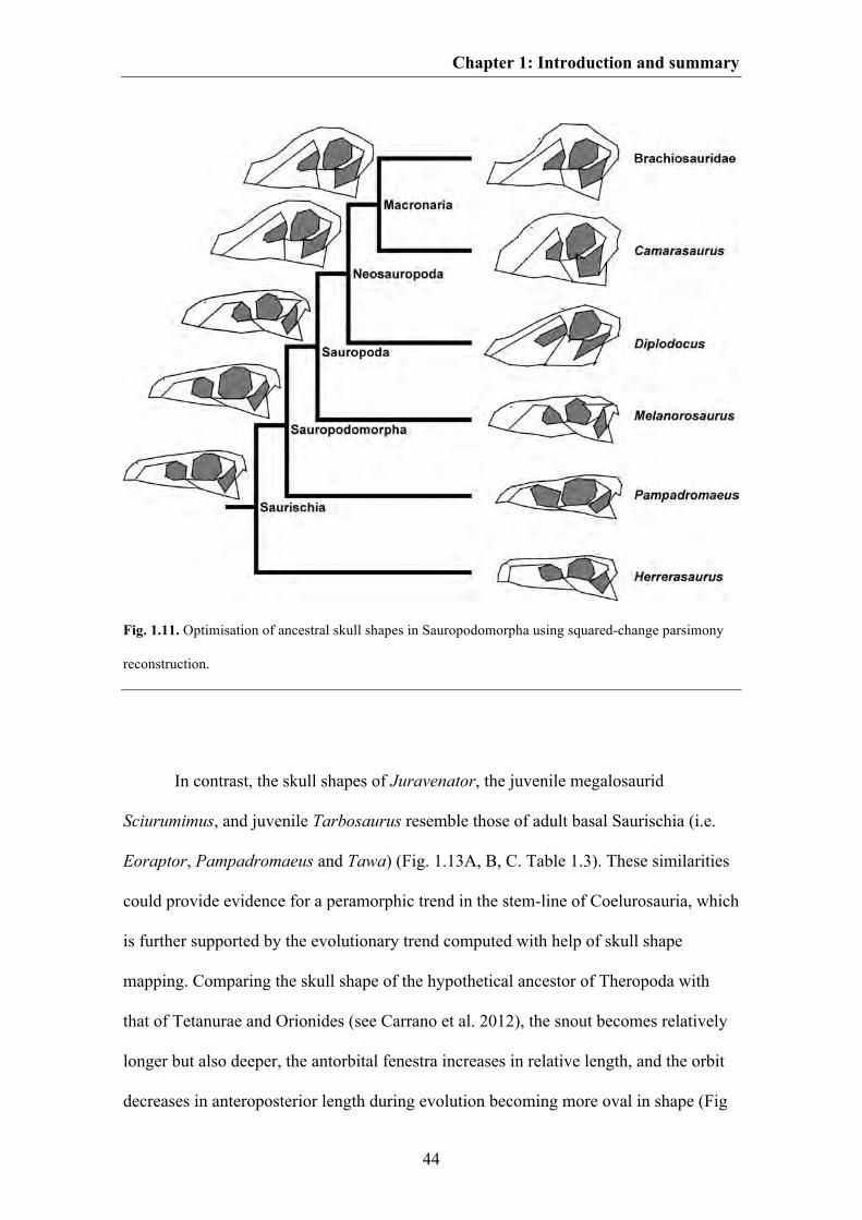
Chapter 1: Introduction and summary
44
Fig. 1.11. Optimisation of ancestral skull shapes in Sauropodomorpha using squared-change parsimony
reconstruction.
In contrast, the skull shapes of Juravenator, the juvenile megalosaurid
Sciurumimus, and juvenile Tarbosaurus resemble those of adult basal Saurischia (i.e.
Eoraptor, Pampadromaeus and Tawa) (Fig. 1.13A, B, C. Table 1.3). These similarities
could provide evidence for a peramorphic trend in the stem-line of Coelurosauria, which
is further supported by the evolutionary trend computed with help of skull shape
mapping. Comparing the skull shape of the hypothetical ancestor of Theropoda with
that of Tetanurae and Orionides (see Carrano et al. 2012), the snout becomes relatively
longer but also deeper, the antorbital fenestra increases in relative length, and the orbit
decreases in anteroposterior length during evolution becoming more oval in shape (Fig
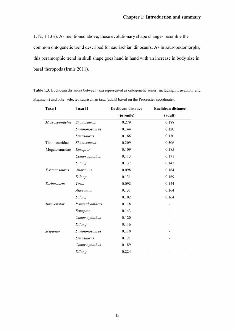
Chapter 1: Introduction and summary
45
1.12, 1.13E). As mentioned above, these evolutionary shape changes resemble the
common ontogenetic trend described for saurischian dinosaurs. As in sauropodomorphs,
this peramorphic trend in skull shape goes hand in hand with an increase in body size in
basal theropods (Irmis 2011).
Table 1.3. Euclidean distances between taxa represented as ontogenetic series (including Juravenator and
Scipionyx) and other selected saurischian taxa (adult) based on the Procrustes coordinates.
Taxa I Taxa II Euclidean distance
(juvenile)
Euclidean distance
(adult)
Massospondylus Shunosaurus 0.279 0.188
Daemonosaurus 0.144 0.120
Limusaurus 0.166 0.130
Titanosauridae Shunosaurus 0.209 0.306
Megalosauridae Eoraptor 0.109 0.185
Compsognathus 0.113 0.171
Dilong 0.137 0.142
Tyrannosaurus Alioramus 0.098 0.164
Dilong 0.131 0.169
Tarbosaurus Tawa 0.092 0.144
Alioramus 0.131 0.164
Dilong 0.102 0.164
Juravenator Pampadromaeus 0.118 -
Eoraptor 0.143 -
Compsognathus 0.120 -
Dilong 0.116 -
Scipionyx Daemonosaurus 0.110 -
Limusaurus 0.121 -
Compsognathus 0.189 -
Dilong 0.224 -
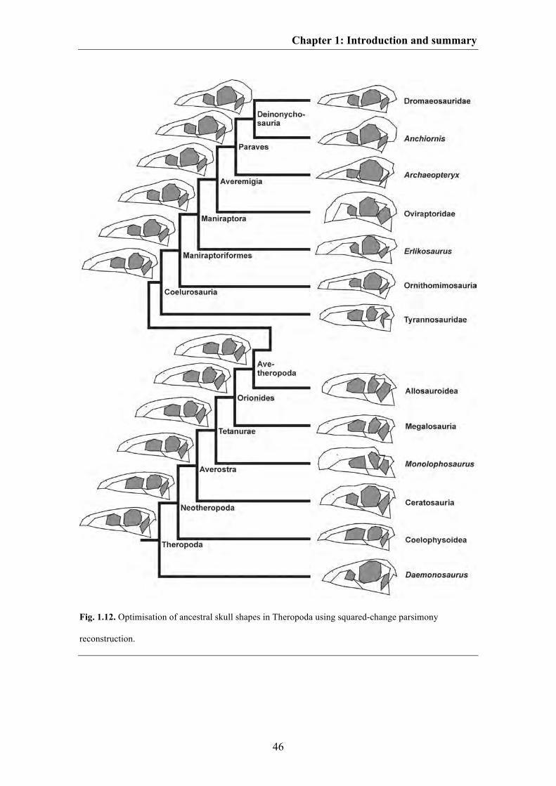
Chapter 1: Introduction and summary
46
Fig. 1.12. Optimisation of ancestral skull shapes in Theropoda using squared-change parsimony
reconstruction.
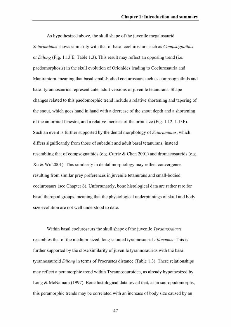
Chapter 1: Introduction and summary
47
As hypothesized above, the skull shape of the juvenile megalosaurid
Sciurumimus shows similarity with that of basal coelurosaurs such as Compsognathus
or Dilong (Fig. 1.13.E, Table 1.3). This result may reflect an opposing trend (i.e.
paedomorphosis) in the skull evolution of Orionides leading to Coelurosauria and
Maniraptora, meaning that basal small-bodied coelurosaurs such as compsognathids and
basal tyrannosaurids represent cute, adult versions of juvenile tetanurans. Shape
changes related to this paedomorphic trend include a relative shortening and tapering of
the snout, which goes hand in hand with a decrease of the snout depth and a shortening
of the antorbital fenestra, and a relative increase of the orbit size (Fig. 1.12, 1.13F).
Such an event is further supported by the dental morphology of Sciurumimus, which
differs significantly from those of subadult and adult basal tetanurans, instead
resembling that of compsognathids (e.g. Currie & Chen 2001) and dromaeosaurids (e.g.
Xu & Wu 2001). This similarity in dental morphology may reflect convergence
resulting from similar prey preferences in juvenile tetanurans and small-bodied
coelurosaurs (see Chapter 6). Unfortunately, bone histological data are rather rare for
basal theropod groups, meaning that the physiological underpinnings of skull and body
size evolution are not well understood to date.
Within basal coelurosaurs the skull shape of the juvenile Tyrannosaurus
resembles that of the medium-sized, long-snouted tyrannosaurid Alioramus. This is
further supported by the close similarity of juvenile tyrannosaurids with the basal
tyrannosauroid Dilong in terms of Procrustes distance (Table 1.3). These relationships
may reflect a peramorphic trend within Tyrannosauroidea, as already hypothesized by
Long & McNamara (1997). Bone histological data reveal that, as in sauropodomorphs,
this peramorphic trends may be correlated with an increase of body size caused by an
Chapter 1: Introduction and summary
48
acceleration of growth (Erickson et al. 2004a).
Fig. 1.13. A-C. Visualization of shape differences between different juvenile theropod taax and basal
Saurischia. A. The juvenile megalosaurid theropod Sciurumimus (grey dashed line) and Eoraptor (black
solid line). B. The juvenile theropod Juravenator (grey dashed line) and Pampadromaeus (black solid
line). C. The juvenile Tarbosaurus (grey dashed line) and Tawa (black solid line). D. Visualization of the
shape difference between Sciurumimus (grey dashed line) and the basal tyrannosauroid Dilong. E.
Evolutionary shape changes from the hypothetical ancestor of Theropoda (grey dashed lines) to that of
Orionides (black solid lines). F. Evolutionary shape changes from the hypothetical ancestor of Orionides
(grey dashed lines) to that of Maniraptoriformes (black solid lines).
Within Maniraptora, Bhullar et al. (2012) found evidence for a paedomorphic
event on the stem-line of Avialae. The hypothetical ancestors of Dromaeosauridae,
Troodontidae and Avialae possess skulls with short, tapering snouts and relatively
enlarged orbital and postorbital regions, which is characteristic for juvenile archosaurs
Chapter 1: Introduction and summary
49
(see above). This paedomorphic event is correlated with a decrease of body size within
Maniraptora (Turner et al. 2007; Benson et al. 2012). Furthermore, Bhullar et al. (2012)
hypothesized that the more theropod-typical skull shape seen in derived
Dromaeosauridae and Troodontidae results from two separate peramorphic events
happening within these groups leading to convergent elongation of the snout. This result
is supported by the skull shape mapping of the current analyses (Fig. 1.12). As in
sauropodomorphs and basal theropods, the peramorphic events in Dromaeosauridae and
Troodontidae go hand in hand with a convergent body size increase within both groups
(Turner et al. 2007). Additional to the described heterochronic events within
Maniraptora, the skull shape mapping shows that the skulls of derived oviraptorids are
also the results of a peramorphic event, which is expressed by a dorsal expansion of the
premaxilla and a relative increase in size of the lateral temporal fenestra. However, the
skulls of basal Oviraptorosauria such as Caudipteryx and Similicaudipteryx possess a
more triangular skull shape with a short tapering snout (Zhou et al. 2000; Xu et al.
2010) resembling the skull shape of the juvenile oviraptorid Yulong (Lü et al. in press)
and basal Paraves (Wellnhofer 2008; Zhang et al. 2008; Hu et al. 2009; Bhullar et al.
2012; Godefroit et al. 2013, in press). This indicates that the peramorphic deepening of
the snout may not be plesiomorphic for Oviraptorosauria, but occurred within the stem-
line of Oviraptoridae. All heterochronic events found within Saurischia are summarized
in Fig. 1.14.
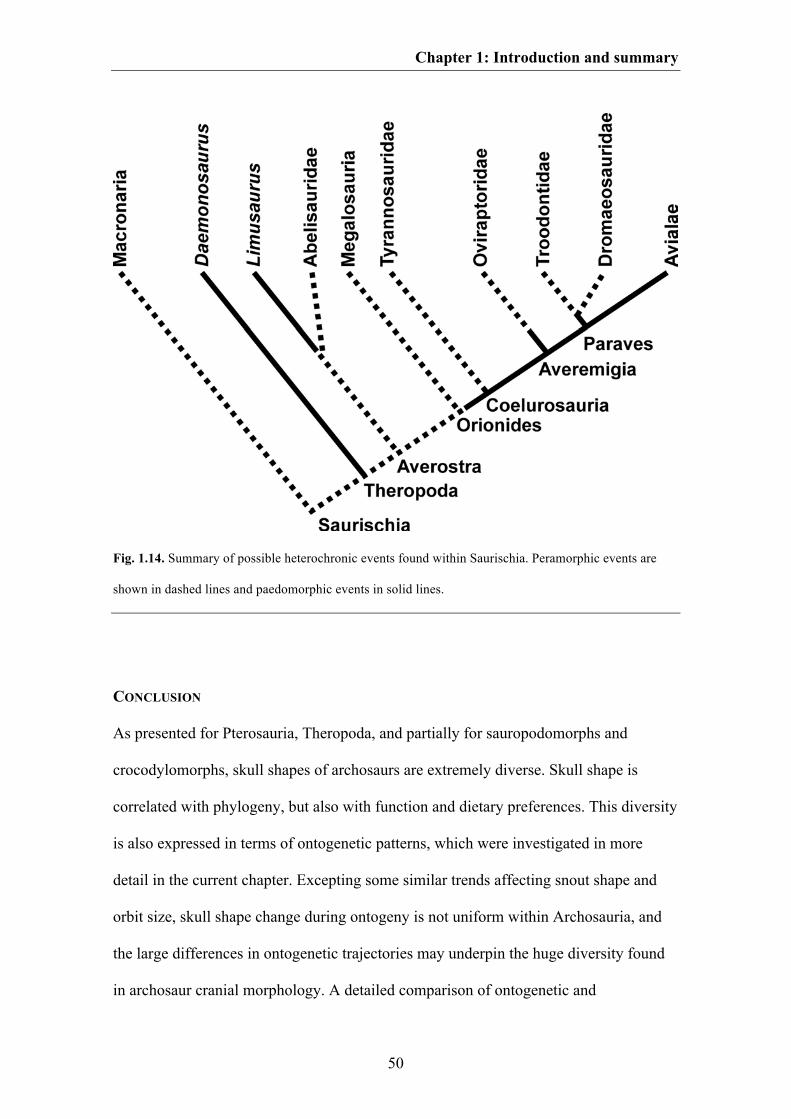
Chapter 1: Introduction and summary
50
Fig. 1.14. Summary of possible heterochronic events found within Saurischia. Peramorphic events are
shown in dashed lines and paedomorphic events in solid lines.
CONCLUSION
As presented for Pterosauria, Theropoda, and partially for sauropodomorphs and
crocodylomorphs, skull shapes of archosaurs are extremely diverse. Skull shape is
correlated with phylogeny, but also with function and dietary preferences. This diversity
is also expressed in terms of ontogenetic patterns, which were investigated in more
detail in the current chapter. Excepting some similar trends affecting snout shape and
orbit size, skull shape change during ontogeny is not uniform within Archosauria, and
the large differences in ontogenetic trajectories may underpin the huge diversity found
in archosaur cranial morphology. A detailed comparison of ontogenetic and
Chapter 1: Introduction and summary
51
evolutionary skulls shape changes in Crocodylomorpha and Saurischia reveal that
archosaur skull evolution is strongly affected by heterochronic events. Evidence for
peramorphosis was found in the stem-line of Crocodylomorpha, within
Sauropodomorpha, basal theropods, Tyrannosauroidea as well as within
Dromaeosauridae, Troodontidae and within the stem-line of Oviraptoridae. In contrast,
paedomorphic events may have occurred in the crocodylian Osteolaemus, in the basal
theropods Daemonosaurus and Limusaurus as well as in basal Coelurosaurs including
basal Maniraptora and Avialae. These heterochronic events appear to be correlated with
body size evolution, but this must be tested in more detail in future analyses. Due to the
close correlation between ontogeny and dietary preferences, heterochronic events often
go hand in hand with evolutionary changes in diet and feeding behaviour, as
exemplarily documented in the juvenile megalosaurid Sciurumimus. Thus, juveniles of
basal tetanurans are of special interest to understand the evolution but also the ecology
of basal coelurosaurs.
52
CHAPTER 2
The good, the bad, and the ugly: the influence of skull reconstructions and intraspecific
variability in studies of cranial morphometrics in theropods and basal saurischians
Keywords:
Theropoda; Saurischia; morphospace; intraspecific variation; geometric morphometrics;
skull reconstructions
The chapter was accepted as:
Foth C1,2 & Rauhut OWM1,2,3. In press. The good, the bad, and the ugly: the influence
of skull reconstructions and intraspecific variability in studies of cranial morphometrics
in theropods and basal saurischians. PloS ONE.
1Bayerische Staatssammlung für Paläontologie und Geologie, Richard-Wagner-Str. 10, D-80333 München, Germany, 2Department of Earth and Environmental Sciences, Ludwig-Maximilians-University, Richard-Wagner-Str. 10, D-80333 München, Germany, 3GeoBioCenter, Ludwig-Maximilians-University; Richard-Wagner-Str. 10, D-80333 München, Germany
Received 22 February 2013; revised 28 June 2013; accepted 09 July 2013
Author contribution:
Research design: Christian Foth, Oliver Rauhut
Data collection: Christian Foth
Data analyses: Christian Foth
Preparation of figures and tables: Christian Foth
Wrote paper: Christian Foth, Oliver Rauhut
*All figures of Chapter 2 are modified after Foth & Rauhut (in press)
Chapter 2: Variation in skull reconstructions
53
The good, the bad, and the ugly: the influence of skull reconstructions and
intraspecific variability in studies of cranial morphometrics in theropods and basal
saurischians
Christian Foth & Oliver W. M. Rauhut
ABSTRACT
Several studies investigating macroevolutionary skull shape variation in fossil reptiles
were published recently, often using skull reconstructions taken from the scientific
literature. However, this approach could be potentially problematic, because skull
reconstructions might differ notably due to incompleteness and/or deformation of the
material. Furthermore, the influence of intraspecific variation has usually not been
explored in these studies. Both points could influence the results of morphometric
analyses by affecting the relative position of species to each other within the
morphospace. The aim of the current study is to investigate the variation in
morphometric data between skull reconstructions based on the same specimen, and to
compare the results to shape variation occurring in skull reconstructions based on
different specimens of the same species (intraspecific variation) and skulls of closely
related species (intraspecific variation). Based on the current results, shape variation of
different skull reconstructions based on the same specimen seems to have generally
little influence on the results of a geometric morphometric analysis, although it cannot
be excluded that some erroneous reconstructions of poorly preserved specimens might
cause problems occasionally. In contrast, for different specimens of the same species
the variation is generally higher than between different reconstructions based on the
same specimen. For closely related species, at least with similar ecological preferences
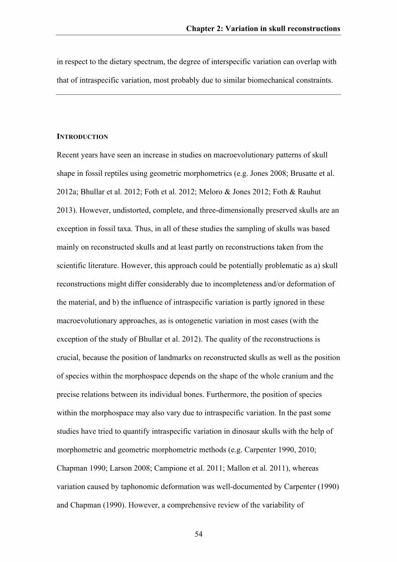
Chapter 2: Variation in skull reconstructions
54
in respect to the dietary spectrum, the degree of interspecific variation can overlap with
that of intraspecific variation, most probably due to similar biomechanical constraints.
INTRODUCTION
Recent years have seen an increase in studies on macroevolutionary patterns of skull
shape in fossil reptiles using geometric morphometrics (e.g. Jones 2008; Brusatte et al.
2012a; Bhullar et al. 2012; Foth et al. 2012; Meloro & Jones 2012; Foth & Rauhut
2013). However, undistorted, complete, and three-dimensionally preserved skulls are an
exception in fossil taxa. Thus, in all of these studies the sampling of skulls was based
mainly on reconstructed skulls and at least partly on reconstructions taken from the
scientific literature. However, this approach could be potentially problematic as a) skull
reconstructions might differ considerably due to incompleteness and/or deformation of
the material, and b) the influence of intraspecific variation is partly ignored in these
macroevolutionary approaches, as is ontogenetic variation in most cases (with the
exception of the study of Bhullar et al. 2012). The quality of the reconstructions is
crucial, because the position of landmarks on reconstructed skulls as well as the position
of species within the morphospace depends on the shape of the whole cranium and the
precise relations between its individual bones. Furthermore, the position of species
within the morphospace may also vary due to intraspecific variation. In the past some
studies have tried to quantify intraspecific variation in dinosaur skulls with the help of
morphometric and geometric morphometric methods (e.g. Carpenter 1990, 2010;
Chapman 1990; Larson 2008; Campione et al. 2011; Mallon et al. 2011), whereas
variation caused by taphonomic deformation was well-documented by Carpenter (1990)
and Chapman (1990). However, a comprehensive review of the variability of
Chapter 2: Variation in skull reconstructions
55
morphometric data due to differential reconstructions or as a result of intraspecific
variation for any dinosaur lineage has not been published yet.
The aim of the current study is to investigate the variation in morphometric data
between skull reconstructions based on the same specimen with the help of geometric
morphometric methods. We furthermore analysed which skull regions might
particularly be affected by high variation within these reconstructions. The results are
compared to shape variation occurring in skull reconstructions based on different
specimens of the same species and skulls of closely related species, in order to
investigate whether this potential source of variation in geometric morphometric data
might be comparable to taxonomically or even phylogenetically significant variation.
MATERIAL AND METHODS
Three different datasets for basal Saurischia, basal Tetanurae, and Tyrannosauroidea
were created, by collecting skull reconstructions in lateral view (Table S2.1, see
supplementary information of Chapter 2). The taxon sample was, of course, limited to
taxa for which several skulls are known and for which various reconstructions based on
the same specimen could be found in the literature. All datasets include a) skull
reconstructions based on the same specimen, b) skull reconstructions of different
specimens of the same species and c) skull reconstruction of closely related species.
Plateosaurus and Allosaurus were treated as each being represented by a single species,
following Weishampel & Chapman (1990), Moser (2003) and Carpenter (2010). The
specimen FMNH PR308, which was originally described as Gorgosaurus (Russell
1970), is placed in Daspletosaurus, following Carr (1999).
Chapter 2: Variation in skull reconstructions
56
Fig. 2.1. Position of landmarks used in the study and variation of skull regions. A. Landmarks used in the
study plotted on a skull reconstruction of Tyrannosaurus specimen of AMNH 5027 (modified after Carr
& Williamson 2004). The green landmarks show skull regions that show most variation between different
reconstructions based on the same specimen in both the original and the randomized dataset. The blue
landmark LM 18 shows additional variation found in the original dataset. B. Skull regions with distinct
variation between reconstructions based on different specimens (intraspecific variation). Red landmarks
show variation found in both the original and the randomized dataset, blue landmarks show variation
found in the randomized dataset. C. Skull regions with distinct variation between reconstructions based
on different, closely related species (interspecific variation). Red landmarks show variation found in both
the original and the randomized dataset, blue landmarks show variation found in the randomized dataset.
Chapter 2: Variation in skull reconstructions
57
The skull shape of all species/specimens was captured by 22 homologous
landmarks, which are figured in Fig. 2.1 and listed in Table S2.2 (see supplementary
information of Chapter 2), using the program tpsDig (Rohlf 2005). This program
outputs a tps (thin plate spline) file with two-dimensional landmark coordinates and
scale (size) data for each specimen. The tps file was loaded into MorphoJ (Klingenberg
2011) and superimposed using Generalized Procrustes Analyses GPA, which align
landmarks from all specimens by minimizing non-shape variation like size, location,
orientation and rotation (Zelditch et al. 2004).
Afterwards, the datasets were divided into different subgroups containing the
Procrustes coordinates of a) single specimens, b) different specimens of the same
species and c) different, closely related species, respectively. To estimate the degree of
variation of skull shape within single specimens, species and between different species a
method was used that was originally developed for estimating the methodological error
for plotting landmarks on specimens by hand (Singleton 2002). On the basis of the
Procrustes coordinates the mean Procrustes distances to the respective consensus
coordinates of each landmark were calculated. Then, the relation of these distances to
the mean distance of the consensus landmarks to the centroid of the consensus shape
was calculated as a percentage of the former from the latter. A further tps file was
created for each dataset including a single skull reconstruction of only one specimen (n
= 10) to calculate the methodological error of plotting landmarks on the skull
reconstruction as mentioned above. The mean error for plotting landmarks (= 0.364 %)
was computed and subtracted from the percentage errors for individual landmarks.
Afterwards, the median of the percentage error of each landmark and its 25th and 75th
percentiles (interquartile range) were computed in PAST 2.17b (Hammer et al. 2001)
Chapter 2: Variation in skull reconstructions
58
and compared between the different subgroups. Using this method for the purpose
mentioned above, the results do not represent methodological errors, but a measure for
morphological variation of overall shape (disparity, see Foote 1991; Wills et al. 1994).
If the median is more than 5.0 % skull shape, variation within a sample was considered
as significant. Thus, skull reconstructions from sample with significant variation could
potentially affect the results of a geometric morphometric analyses and should be
treated with caution. To verify the results, Procrustes coordinates were additionally used
to calculate the Euclidian distances for every sample within each group (Lockwood et
al. 2005). As in the previous case, the median Euclidian distance and its 25th and 75th
percentiles were calculated. Furthermore, we wanted to know, which skull regions are
particularly affected by significant shape variation within reconstructions of the same
specimen, the same species and closely related taxa, respectively. For this, the median
and its 25th and 75th percentiles were calculated for each landmark within the different
subgroups mentioned above.
Due to the generally small numbers of skull reconstructions for most samples,
we tested the robustness of the ‘original’ results in relation to sample size by computing
random samples in the program R (R Development Core Team 2011) with a standard
number of ten ‘hypothetical reconstructions’ per sample on the basis of the Procrustes
coordinates of the original data. The function used computed ten normal pseudorandom
variates based on the mean and the standard deviation of all Procrustes coordinates
related to a corresponding landmark within the original sample (Braun & Murdoch
2008). Afterwards, all methods described above were repeated with randomized
samples and compared to the original data. If both kinds of data produce similar results

Chapter 2: Variation in skull reconstructions
59
one can conclude that the results of the original data are robust in relation to sample
size.
INSTITUTIONAL ABBREVIATIONS
AMNH, American Museum of Natural History, New York; MB, Museum für
Naturkunde, Berlin; MOR, Museum of the Rockies, Bozeman; FMNH, The Field
Museum, Chicago; MOR, Museum of the Rockies, Bozeman; NMC, National Museum
of Canada, Ottawa; PIN, Paleontological Institute, Russian Academy of Sciences,
Moscow; SMNS, Staatliches Museum für Naturkunde Stuttgart, Stuttgart; USNM,
National Museum of Natural History (= formerly United States National Museum),
Smithsonian Institution, Washington, D.C.; UUVP, Utah Museum of Natural History,
Salt Lake City.
RESULTS
Both the values of the median of landmark variation (median of variation) and
Euclidean distances show generally similar distributions between the single samples of
the three subgroups. This is also true for the comparison between original and
randomized data. However, for the Euclidean distances the interquartile range of the
randomized data is usually smaller than for the original data (for all samples with more
than two reconstructions) with exception of Gorgosaurus, Tarbosaurus and the
Daspletosaurus specimen FMNH PR308. In contrast, the range of interquartiles are
comparable for both kinds of data with the exception of Eoraptor, Massospondylus and
Tarbosaurus (here the interquartile range of the randomized data is slightly bigger than
in the original data) as well as Plateosaurus, Acrocanthosaurus and the Tyrannosaurus
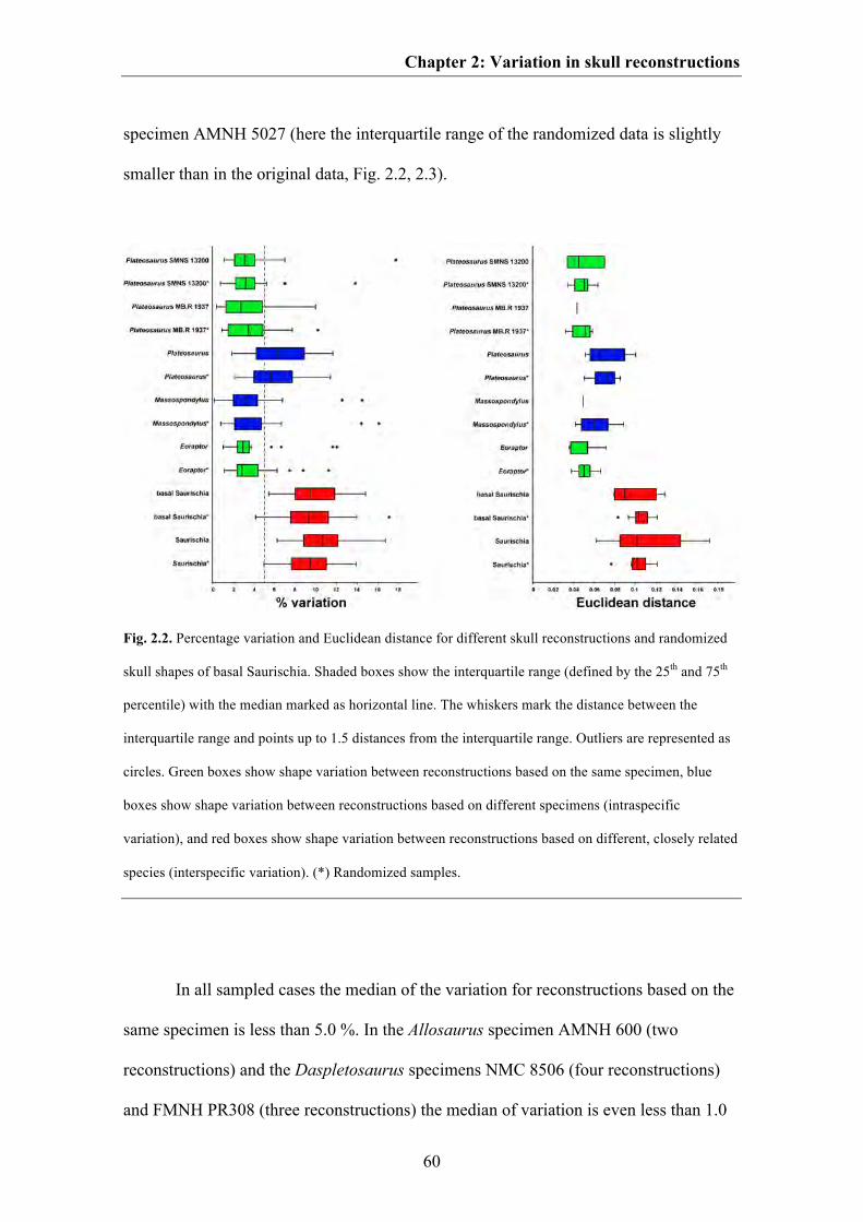
Chapter 2: Variation in skull reconstructions
60
specimen AMNH 5027 (here the interquartile range of the randomized data is slightly
smaller than in the original data, Fig. 2.2, 2.3).
Fig. 2.2. Percentage variation and Euclidean distance for different skull reconstructions and randomized
skull shapes of basal Saurischia. Shaded boxes show the interquartile range (defined by the 25th and 75th
percentile) with the median marked as horizontal line. The whiskers mark the distance between the
interquartile range and points up to 1.5 distances from the interquartile range. Outliers are represented as
circles. Green boxes show shape variation between reconstructions based on the same specimen, blue
boxes show shape variation between reconstructions based on different specimens (intraspecific
variation), and red boxes show shape variation between reconstructions based on different, closely related
species (interspecific variation). (*) Randomized samples.
In all sampled cases the median of the variation for reconstructions based on the
same specimen is less than 5.0 %. In the Allosaurus specimen AMNH 600 (two
reconstructions) and the Daspletosaurus specimens NMC 8506 (four reconstructions)
and FMNH PR308 (three reconstructions) the median of variation is even less than 1.0
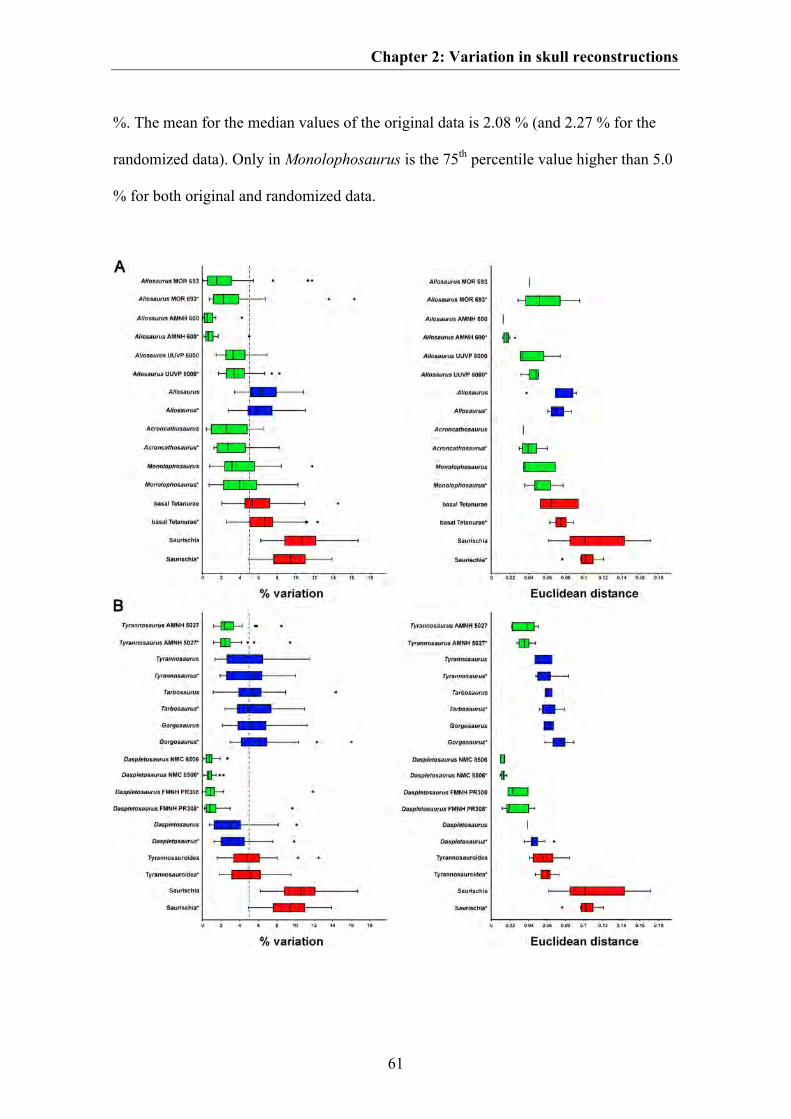
Chapter 2: Variation in skull reconstructions
61
%. The mean for the median values of the original data is 2.08 % (and 2.27 % for the
randomized data). Only in Monolophosaurus is the 75th percentile value higher than 5.0
% for both original and randomized data.
Chapter 2: Variation in skull reconstructions
62
Fig. 2.3. Percentage variation and Euclidean distance for different skull reconstructions and randomized
skull shapes of basal Tetanurae and Tyrannosauroidea. A. basal Tetanurae. B. Tyrannosauroidea. Shaded
boxes show the interquartile range (defined by the 25th and 75th percentile) with the median marked as
horizontal line. The whiskers mark the distance between the interquartile range and points up to 1.5
distances from the interquartile range. Outliers are represented as circles. Green boxes show shape
variation between reconstructions based on the same specimen, blue boxes show shape variation between
reconstructions based on different specimens (intraspecific variation), and red boxes show shape variation
between reconstructions based on different, closely related species (interspecific variation). (*)
Randomized samples.
The mean of the median values for skull reconstructions based on different
specimens of the same species is 4.74 % for the original data (and 4.78 % for the
randomized data), in which the median of the variation of the original data is less than
5.0 % for Daspletosaurus, Massospondylus and Tyrannosaurus. Here, the 75th
percentile value is less than 5.0 % in the former two genera as well. Thus, the median of
the variation of Daspletosaurus and Massospondylus strongly overlaps with that of
reconstructions based on the same specimen for most taxa. In contrast, the median of
the variation of the original data of Allosaurus, Plateosaurus, Tarbosaurus and
Gorgosaurus is more than 5.0 %, but only for Allosaurus is the 25th percentile value
higher than 5.0 %. In contrast, the median of the variation in the randomized datasets is
less than 5.0 % for Tarbosaurus, but more than 5.0 % in Allosaurus, Plateosaurus,
Gorgosaurus and Tyrannosaurus (Fig. 2.2, 2.3).
The mean of the median values for reconstructions of skulls of closely related
taxa is 6.48 % for the original data (and 6.76 % for the randomized data). For the
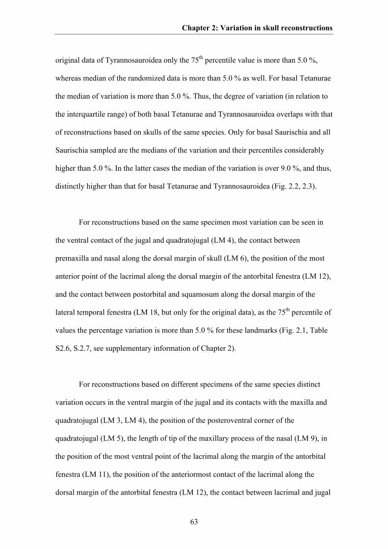
Chapter 2: Variation in skull reconstructions
63
original data of Tyrannosauroidea only the 75th percentile value is more than 5.0 %,
whereas median of the randomized data is more than 5.0 % as well. For basal Tetanurae
the median of variation is more than 5.0 %. Thus, the degree of variation (in relation to
the interquartile range) of both basal Tetanurae and Tyrannosauroidea overlaps with that
of reconstructions based on skulls of the same species. Only for basal Saurischia and all
Saurischia sampled are the medians of the variation and their percentiles considerably
higher than 5.0 %. In the latter cases the median of the variation is over 9.0 %, and thus,
distinctly higher than that for basal Tetanurae and Tyrannosauroidea (Fig. 2.2, 2.3).
For reconstructions based on the same specimen most variation can be seen in
the ventral contact of the jugal and quadratojugal (LM 4), the contact between
premaxilla and nasal along the dorsal margin of skull (LM 6), the position of the most
anterior point of the lacrimal along the dorsal margin of the antorbital fenestra (LM 12),
and the contact between postorbital and squamosum along the dorsal margin of the
lateral temporal fenestra (LM 18, but only for the original data), as the 75th percentile of
values the percentage variation is more than 5.0 % for these landmarks (Fig. 2.1, Table
S2.6, S.2.7, see supplementary information of Chapter 2).
For reconstructions based on different specimens of the same species distinct
variation occurs in the ventral margin of the jugal and its contacts with the maxilla and
quadratojugal (LM 3, LM 4), the position of the posteroventral corner of the
quadratojugal (LM 5), the length of tip of the maxillary process of the nasal (LM 9), in
the position of the most ventral point of the lacrimal along the margin of the antorbital
fenestra (LM 11), the position of the anteriormost contact of the lacrimal along the
dorsal margin of the antorbital fenestra (LM 12), the contact between lacrimal and jugal
Chapter 2: Variation in skull reconstructions
64
on the orbital margin (LM 14), the position of the anteroventral tip of the ventral
process of the squamosal on the margin of the lateral temporal fenestra (LM 17), and in
the dorsal contact between postorbital and squamosal (LM 19). For the randomized data
the contact between frontal and postorbital on the dorsal margin of the orbit (LM 22)
was found to be significant as well (Fig. 2.1, Table S2.6, S2.7, see supplementary
information of Chapter 2).
In comparison, for skull reconstructions of closely related taxa, distinct
landmark variation affects almost the entire skull, with the exception of the length of the
anterior process of the maxillary body (LM 8), the position of the anteriormost point of
the antorbital fenestra (LM 10), the contact of the jugal with both the squamosal and the
quadratojugal on the margin of the lateral temporal fenestra (LM 15, LM 16). For the
randomized data all landmarks except of LM 10 showed significant variation (Fig. 2.1,
Table S2.6, S2.7, see supplementary information of Chapter 2).
DISCUSSION
Based on the results presented above, we can conclude that the shape variation of skull
reconstruction (in relation to the median of variation and the interquartile range) based
on the same specimen seems usually to be negligible in geometric morphometric studies
(only in Monolophosaurus the 75th percentile is more than 5.0 %). The general
consistency of the results between original and randomized data supports this result in
spite of the small sample sizes of the original data. However, taxa for which only a
single specimen and maybe even only a single reconstruction exist could introduce
considerable error in geometric morphometric studies, if the particular specimen is
incomplete or strongly taphonomically deformed. In Allosaurus, for example, the skull
Chapter 2: Variation in skull reconstructions
65
reconstructed by Gilmore (1920) has figured prominently in both the scientific and the
popular literature for a long time, until newly found, better preserved and complete
specimens showed that this reconstruction, based on a disarticulated, partially deformed,
and pathological skull, does not represent the “typical” skull shape of this taxon (Fig.
2.4).
Shape variation in reconstructions might be influenced, for instance, by the
talent of the artists, their anatomical knowledge and their tendency to idealize
structures, which are e.g., taphonomically deformed, damaged or missing (meaning to
attempt a complete de-deformation of the skull). Differences in the skull shape of the
holotype of Monolophosaurus or the Plateosaurus specimens MB.R 1937 and SNMS
13200 are probably partially caused by the latter factor, because Zhao & Currie (1993),
Rauhut (2003a) and Yates (2003) idealized such deformations more completely than
Galton (2001) (Fig. 2.4) or Brusatte et al. (2010b) (e.g. Brusatte et al. figured the
disarticulation between jugal and postorbital on the right side of the skull). Furthermore,
it might be important if the artist saw the specimen first hand, reconstructed the skull on
the basis of photographs or simply redrew the skull from previously published
reconstructions (as is the case e.g. with the reconstruction of Monolophosaurus in
Rauhut 2003a). In order to minimize this source of error, a scientist analysing shape
changes would be wise to not only take the reconstructed skull from the literature, but
also look closely at the available data on the original material and how the skull was
reconstructed from it.
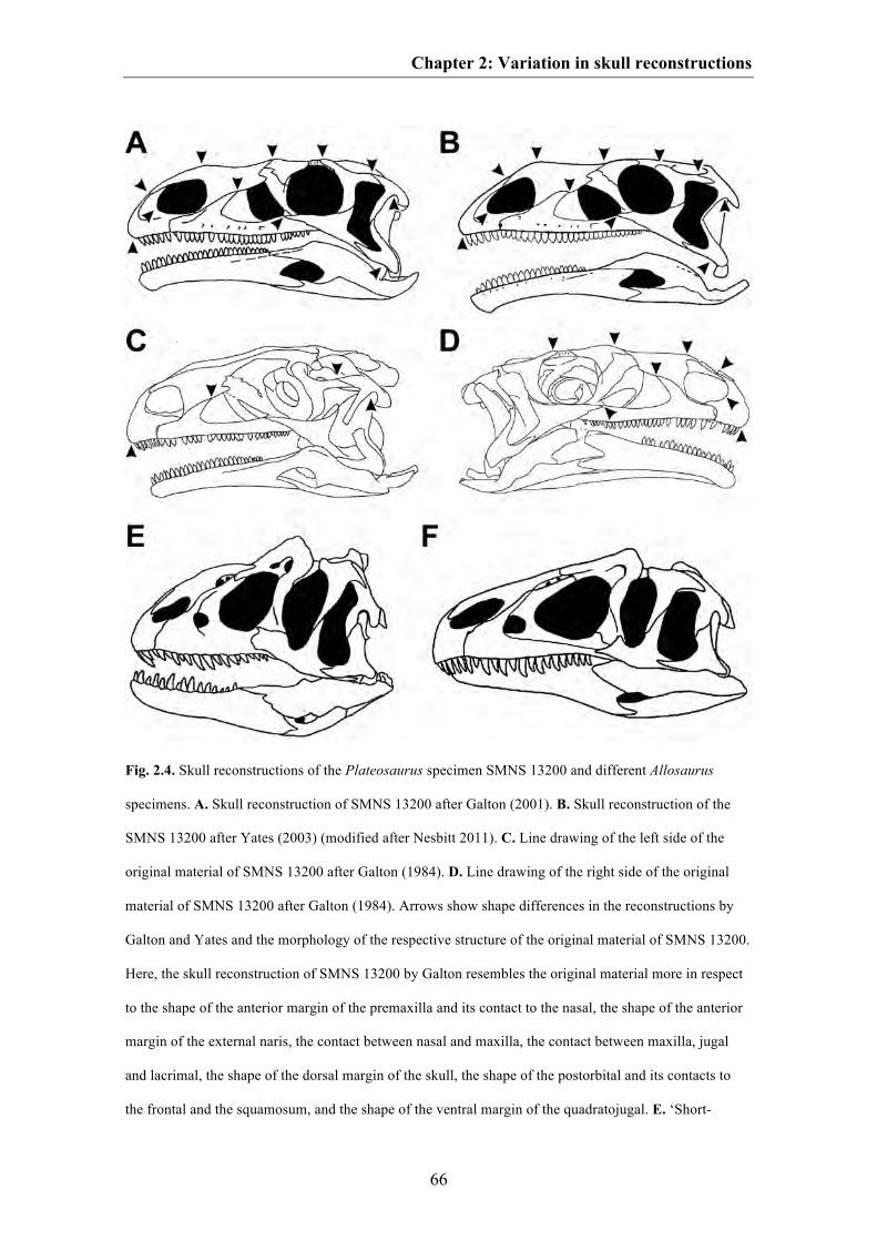
Chapter 2: Variation in skull reconstructions
66
Fig. 2.4. Skull reconstructions of the Plateosaurus specimen SMNS 13200 and different Allosaurus
specimens. A. Skull reconstruction of SMNS 13200 after Galton (2001). B. Skull reconstruction of the
SMNS 13200 after Yates (2003) (modified after Nesbitt 2011). C. Line drawing of the left side of the
original material of SMNS 13200 after Galton (1984). D. Line drawing of the right side of the original
material of SMNS 13200 after Galton (1984). Arrows show shape differences in the reconstructions by
Galton and Yates and the morphology of the respective structure of the original material of SMNS 13200.
Here, the skull reconstruction of SMNS 13200 by Galton resembles the original material more in respect
to the shape of the anterior margin of the premaxilla and its contact to the nasal, the shape of the anterior
margin of the external naris, the contact between nasal and maxilla, the contact between maxilla, jugal
and lacrimal, the shape of the dorsal margin of the skull, the shape of the postorbital and its contacts to
the frontal and the squamosum, and the shape of the ventral margin of the quadratojugal. E. ‘Short-
Chapter 2: Variation in skull reconstructions
67
snouted’ Allosaurus specimen USNM 4735 described by Gilmore (1920), which was based on a
disarticulated, partially deformed, and pathological skull (modified after Henderson 2000). F. ‘Typical’
Allosaurus skull based on MOR 693 (modified after Rauhut 2003a).
Within different reconstructions based on the same specimens the skull regions
described by landmark 4, 6, 12 and 18 (i.e. the ventral contact of the jugal and
quadratojugal, the contact premaxilla and nasal along the dorsal margin of skull, the
position of the most anterior point of the lacrimal along the dorsal margin of the
antorbital fenestra, and the contact between postorbital and squamosum along the dorsal
margin of the lateral temporal fenestra) are more variable than other landmarks,
although their variability is still less than that between landmarks in reconstructions of
different specimens. Thus, these particular skull regions may contain a potential
methodological error for plotting landmarks on dinosaur skulls and maybe also other
reptiles, and should be verified carefully by photo material or first-hand observations.
The variation of skull reconstructions (in relation to the mean of the median
values) based on different specimens of the same species is expected to be higher than
that of different reconstructions of the same specimen as variation is further caused by
intraspecific variation. However, the differences are not significant due to the strong
overlap of the percentiles between both groups and also vary from species to species.
For instance, the intraspecific skull variation found in Massospondylus is relatively low,
challenging Gow et al. (1990), who hypothesized that the shape variation seen in the
skulls of two Massospondylus specimens might be caused by sexual dimorphism. Based
on the results of both the original and randomized samples this hypothesis cannot be
Chapter 2: Variation in skull reconstructions
68
supported statistically. The variation might rather reflect allometric shape variation as
both specimens slightly differ in skull size (Hinić 2002). Cranial sexual dimorphism
was also hypothesized for Allosaurus (Molnar 2005), but also cannot be verified
statistically.
On the other hand, the current results support previous studies on Allosaurus and
Plateosaurus, which show a large intraspecific variation within these taxa (Weishampel
& Chapman 1990; Smith 1998; Carpenter 2010). However, some of the variation
presented in those studies reflects also ontogenetic variation, making a direct
comparison of the studies difficult as this type of variation has only minor impact on the
current results due to selective sampling of adult or nearly adult specimens.
Some of the variation found in the current results may also result from
taphonomic deformation (e.g. the disarticulated contact of quadratojugal and
squamosum in the holotype skull PIN 551-1 of Tarbosaurus, which is pictured in the
reconstruction of Maleev 1974). Taphonomic deformation was also hypothesized as the
major reason for the huge ‘morphological variation’ seen in the southern Germany
Plateosaurus material (Moser 2003), and its influence on skull shape is well-
documented for a Plateosaurus by Chapman (Chapman 1990). Furthermore, some
variation in Allosaurus and Plateosaurus could be also explained by their controversial
taxonomic status. As mentioned in the material and method section, reconstructed skulls
of both genera were treated as belonging to one species, but some authors argued that
there are at least two species for each genus (e.g. Chure 2000; Galton 2001; Yates 2003;
Loewen 2009; Prieto-Márquez & Norell 2011). If the latter case is true, the variation is
Chapter 2: Variation in skull reconstructions
69
partially covered by interspecific variation, and thus the actual intraspecific variation
might be overestimated.
To minimize the ‘error’ of intraspecific variation in macroevolutionary
approaches, taxa for which there are several good quality reconstructions of different
specimens should be tested for intraspecific variation. This can be done in a separate
small dataset with the same landmark configuration used in the macroevolutionary
study by calculating the Procrustes coordinates for each specimen and estimating the
respective Euclidean distances to the consensus shape of the small dataset.
Subsequently, the specimen with the smallest distance to the consensus shape might be
used for the study.
The examples of interspecific variation (in relation to the median of variation)
presented in this study show all significant variation, except for the original sample of
Tyrannosauroidea. However, the latter exception could be the result of a small sample
size (n = 5). Interestingly, the interspecific shape variation (in relation to the
interquartile range) of basal Tetanurae and Tyrannosauroidea strongly overlaps with the
shape variation of the intraspecific variation of Allosaurus, Tyrannosaurus,
Tarbosaurus and Gorgosaurus. The estimated intraspecific variation is even slightly
higher than the estimated interspecific variation of the respective groups. At first glance
this result is surprising, as one would expect that interspecific variation should be larger
than intraspecific variation, as seen in basal Saurischia. Methodically, the overlap could
be a false signal resulting from small sample sizes (see Molnar 1990). However, the
differences between the numbers of reconstructions used for a single species and for
different, closely related species are rather small, making this explanation rather
Chapter 2: Variation in skull reconstructions
70
unlikely. Furthermore, because the results of the randomized data are similar to that of
the original one, the sample size does not seem to influence the current result
significantly. However, it is possible that the chosen landmark configuration does not
capture skull regions that underlie strong interspecific variation in basal Tetanurae or
Tyrannosauroidea, like the dorsal margin of the nasal (e.g. Monolophosaurus) or the
dorsal margin of the lacrimal horn (e.g. Allosaurus). Furthermore, semi-landmark
analysis of overall skull shape, in combination with a landmark-based analysis, might
capture variations in skull shape more completely and thus yield different results. Thus,
it is possible that the present analyses underestimate the actual interspecific variation
between those taxa. Furthermore, it is to be expected that interspecific skull variability
increases with increasing the sample size of taxa analysed, as it is indeed demonstrated
by the higher variation seen in the data set for basal saurischians or saurischians as a
whole. By expanding the data set to species with more derived skull morphologies (e.g.
long-snouted spinosaurids for basal Tetanurae), an increase of the interspecific variation
even in rather closely related forms would also be expected. This is supported by
several studies on crustacean, pterosaur and coelurosaur diversity for instance, which all
show that disparity of larger taxonomic clades is higher than in the respective internal
subclades (see Wills 1998; Prentice et al. 2011; Brusatte et al. 2012b; Foth et al. 2012).
On the other hand, an overlap of intraspecific and interspecific variation in closely
related taxa has also been demonstrated for instance in the cranial shape of recent
Hominoidea (Lockwood et al. 2005), the osteology of skinks (Czechura & Wombey
1982) or in molecular sequences of different bilaterian clades (e.g. Meyer et al. 2005;
Meier et al. 2006, 2008), and the phenomenon is therefore neither restricted to theropod
dinosaurs, nor to skull shape.
Chapter 2: Variation in skull reconstructions
71
In comparison with this rather small variation seen in closely related theropod
taxa, basal Saurischia in total possess a very large interspecific variation. One reason for
this could be the inclusion of Eoraptor, the taxonomic position of which is still debated
(e.g. Martinez & Alcober 2009; Martinez et al. 2011; Nesbitt 2011). However,
excluding Eoraptor from the data set does not change the result (median of variation =
9.66 %). Thus, the large variation seen in the skull shape might be due to diverging
dietary preferences in basal saurischians, towards carnivory in many basal theropods,
with omnivory and finally herbivory in sauropodomorphs (Barrett 2000; Galton &
Upchurch 2004; Langer et al. 2009; Barrett et al. 2011). Indeed, this change in diet
might lead to the evolutionary trend from slender and elongate skulls to short and broad
skulls seen in the early evolution of Sauropodomorpha (Rauhut et al. 2011). A similar
pattern regarding diet preferences was also found in theropods by Brusatte et al. (2012a)
and Foth & Rauhut (2013), who have shown that both carnivorous and non-carnivorous
taxa occupy large areas within the morphospace, but non-carnivorous taxa tend to
develop more diverse, sometimes aberrant skull morphologies (e.g. Oviraptorosauria).
In contrast, large-bodied carnivorous theropods tend to cluster closely together within
morphospace (Brusatte et al. 2012a; Foth & Rauhut 2013), and show a smaller disparity
in skull shape in comparison to smaller theropods with a broad dietary spectrum
(Brusatte et al. 2012a). This might be due to a constrained biomechanical adaptation for
high bite forces (Erickson et al. 1996; Henderson 2002; Sakamoto 2010), including an
oval orbit, a deep jugal body and a short postorbital region (Henderson 2002; Foth &
Rauhut 2013).
Chapter 2: Variation in skull reconstructions
72
CONCLUSION
The median of variation of different skull reconstructions based on the same specimen
seems to have generally little influence on the results of a geometric morphometric
analysis of skull shape in theropods and basal saurischians. Shape differences seem to
be mainly influenced by the talent of the artists, their anatomical knowledge, and their
tendency to idealize structures that are damaged, missing or taphonomically deformed.
In general, it is advisable to verify reconstructions used on the basis of the original
material or photographs thereof. For different specimens of the same species the
variation (in relation to the mean of the median values) is generally higher than in the
previous example, indicating that intraspecific variation cannot be neglected, although
this apparent variation might in some cases be overestimated due to uncertain
taxonomy. For closely related species, at least with similar ecological preferences, the
degree of interspecific variation (in relation to the median of variation and its
percentiles) overlaps with that of intraspecific variation. This probably reflects
considerable constraints in the skulls of theropods with similar feeding strategies. As
would be expected, variation in morphometric data might increase with increased
phylogenetic and/or ecological sampling, but this have to be tested in future studies in
more detail. Given the nature of fossil data, our analysis is necessarily based on rather
small sample sizes, and more investigations of the relation between intraspecific and
interspecific variation in geometric morphometric data in recent animals, for which
higher sample sizes are available, would be desirable.
Chapter 2: Variation in skull reconstructions
73
ACKNOWLEDGMENTS
We would like to thank Martin Schwentner (University of Rostock), Serjoscha Evers
and Richard Butler (both Ludwig-Maximillians-Univeristät, München) for discussion
and two anonymous reviewers for critical comments, which helped to improve the
manuscript. Furthermore, we want to thank Hans-Jakob Siber and Thomas Bollinger
(Sauriermuseum Aathal) and Raimund Albersdörfer for access to Allosaurus material.
74
CHAPTER 3
Intraspecific variation in the skull morphology of the black caiman Melanosuchus niger
(Alligatoridae, Caimaninae)
Keywords:
Melanosuchus; Crocodylia; ontogeny; sexual dimorphism; geometric morphometrics
The chapter was accepted as:
Foth C1, Bona P2,3 & Desojo JB2,4. In press. Intraspecific variation in the skull
morphology of the black caiman Melanosuchus niger (Alligatoridae, Caimaninae).
Acta Zoologica.
1Bayerische Staatssammlung für Paläontologie und Geologie, Richard-Wagner-Str. 10, D-80333 München, Germany 2CONICET, Consejo Nacional de Investigaciones Científicas y Técnicas 3División Paleontología Vertebrados, Museo de La Plata. Paseo del Bosque s/n. 1900, La Plata, Buenos Aires. Argentina 4Sección Paleontología de Vertebrados, Museo Argentino de Ciencias Naturales "Bernardino Rivadavia". Av. Angel Gallardo 470 C1405DRJ, Capital Federal. Argentina
Received 28 January 2013; revised 04 July 2013; accepted 08 July 2013
Author contribution:
Research design: Christian Foth, Paula Bona, Julia Desojo
Data collection: Christian Foth, Julia Desjo
Data analyses: Christian Foth
Preparation of figures and tables: Christian Foth
Wrote paper: Christian Foth, Paula Bona, Julia Desojo
*All figures of Chapter 3 are modified after Foth et al. (in press)
Chapter 3: Skull shape of Melanosuchus
75
Intraspecific variation in the skull morphology of the black caiman Melanosuchus
niger (Alligatoridae, Caimaninae)
Christian Foth, Paula Bona & Julia B. Desojo
ABSTRACT
Melanosuchus niger is a caimanine alligatorid widely distributed in the northern region
of South America. This species has been the focus of several ecological, genetic and
morphological studies. However, morphological studies have generally been limited to
examination of interspecific variation among extant species of South American
crocodylians. Here we present the first study of intraspecific variation in the skull of
Melanosuchus niger using a two-dimensional geometric morphometric approach. The
crania of 52 sexed individuals varying in size were analyzed to quantify shape variation
and to assign observed shape changes to different types of intraspecific variation, i.e.
ontogenetic variation and sexual dimorphism. Most of the variation in this species is
ontogenetic variation in snout length, skull depth, orbit size and the width of the
postorbital region. These changes are correlated with bite force performance and
probably dietary changes. However, a comparison with previous functional studies
reveal that functional adaptions during ontogeny seems to be primarily restricted to the
postrostral region, whereas rostral shape changes are more related to dietary shifts.
Furthermore, the skulls of Melanosuchus niger exhibit a sexual dimorphism, which is
primarily size related. The presence of non-size related sexual dimorphism has to be
tested in future examinations.
Chapter 3: Skull shape of Melanosuchus
76
INTRODUCTION
Knowledge of vertebrate morphology and its intraspecific variation (e.g. ontogenetic
variation, sexually dimorphic variation, polymorphisms) is crucial for accurate
systematic, taxonomic, evolutionary, ecological, physiological and functional
hypothesis of the different groups (e.g. Sudhaus & Rehfeld 1992; Wiesenmüller et al.
2003; Carpenter 2010; Porro et al. 2011). Among extant crocodylians, previous studies
of skull anatomy and intraspecific variation (including ontogenetic variation), evolution
and functional morphology include those of Mook (1921), Kälin (1933), Medem
(1963), Iordansky (1973), Dodson (1975), Busbey (1989), Hall & Portier (1994),
Monteiro & Soares (1997), Brochu (1999, 2001), Verdade (2000), Erickson et al. (2003,
2012), McHenry et al. (2006), Piña et al. (2007), Wu et al. (2006), Sadleir & Makovicky
(2008), Platt et al. (2009) and Bona & Desojo (2011). Nevertheless, lack of
morphological studies and data, especially for osteology, is common to all extant South
American crocodylians. The main problem is that crocodylian skeletons are rare in
South American herpetological collections. This is unfortunate because detailed
knowledge of the osteology of extant species and its morphological variation become
further relevant when reconstructing the evolutionary history of a group, especially
given that most of fossil specimens are preserved only by skeletons (and generally as
fragments).
Extant South American Alligatoridae are grouped in three genera of Caimaninae,
Paleosuchus Cuvier 1807, Caiman Spix 1825 and Melanosuchus Spix 1825. Certain
taxonomic controversies among caimanines are related to Melanosuchus. This genus is
represented by two species: the extinct Melanosuchus fisheri Medina, 1976 from the
Upper Miocene (Urumaco Formation) of Venezuela (Sánchez-Villagra & Aguilera
Chapter 3: Skull shape of Melanosuchus
77
2006) and the extant Melanosuchus niger Spix, 1825, in which the taxonomic status of
the synonym Melanosuchus fisheri has been questioned (e.g. Brochu 1999). The
presence of prominent rostral ridges on the skull, shared with the extant Caiman
latirostris Daudin, 1802 and the extinct Caiman cf. lutescens (Langston, 1965) from the
Miocene of South America, supports a sister-group relationship of Melanosuchus niger
with these two species in morphological cladistic analyses (Norell 1988; Poe 1997;
Brochu 1999, 2003, 2010, 2011; Bona 2007; Aguilera et al. 2006). This result has
generated differing taxonomic proposals with regard to the putative paraphyly of the
genus Caiman (see Norell 1988; Poe 1997).
Melanosuchus niger is particularly interesting among Alligatoridae, because is
one of the largest extant members of the group, with adult males sometimes exceeding 6
m in length (Cott 1926; Brazaitis 1974). It has been the focus of ecological (e.g. Otte
1974; Medem 1981; Plotkin et al. 1983; Herron 1991; Pacheco 1994; Villamarín-Jurado
& Suárez 2007; Marioni et al. 2008; Horna et al. 2001) and genetic works (e.g. Farias et
al. 2004; de Thoisy et al. 2006; Vasconcelos et al. 2008), as well as a limited number of
morphological studies (Mook 1921; Kälin 1933; Medem 1963). Although there have
been some qualitative studies on differently sized specimens of Melanosuchus niger
(e.g. Mook 1921), its general intraspecific morphological variation is poorly
understood. The purpose of the present study is to quantify, describe and interpret the
intraspecific variation in the skull of Melanosuchus niger using a geometric
morphometric approach. Geometric morphometrics is widely regarded as a powerful
tool for taxonomic identification and functional interpretations (Rohlf & Marcus 1993;
Zelditch et al. 2004), and has great potential to characterize developmental and genetic
effects on morphological shape (see Klingenberg 2010). This method quantifies
Chapter 3: Skull shape of Melanosuchus
78
differences in shape between objects from coordinates of homologous landmark
locations, after the effects of non-shape variation (position, size and rotation) are
mathematically held constant (Adams et al. 2004; Zelditch et al. 2004). Geometric
landmark based analysis captures and retain more information about shape than
traditional morphometric measurements (e.g. linear distances, ratios and measurements
of angles), which often fail to capture the full geometry of the original object (Rohlf &
Marcus 1993; Rohlf 2000; Hammer & Harper 2006). Geometric morphometrics has
been used successfully to document intraspecific variation and to test specific
taxonomic and ontogenetic biological hypotheses (e.g. Richtsmeier et al. 1993;
O’Higgins & Collard 2002; Bookstein et al. 2003; Elewa 2004; Zelditch et al. 2004).
However, only a few analyses of extant crocodylian skulls using a geometric
morphometric approach have been previously conducted. Monteiro et al. (1997) studied
ontogenetic changes in three Caiman species, and Pierce et al. (2008) described the
cranial morphospace of extant crocodylians and its correlation with functional
morphology based on finite element modeling (FEM). Piras et al. (2009) investigated
the influence of phylogeny and ecological factors (climate change) on the skull shape of
Alligatoroidea and Crocodyloidea, and Piras et al. (2010) compared allometric
trajectories in different crocodylian taxa to test phylogenetic hypotheses about the
relationships of gavials (Gavialis gangeticus) and false gavials (Tomistoma schlegelii).
In the context of quantifying skull shape variation within Melanosuchus niger,
we want to classify ontogenetic variation and sexual dimorphism, and to test whether
skull shape is correlated with bite force performance (which is used as a functional
proxy) in this species. The results are compared with published data for other
Chapter 3: Skull shape of Melanosuchus
79
crocodylian species to identify probable key patterns in intraspecific variation in cranial
shape and how these patterns might be related to ecology and function.
MATERIAL AND METHODS
SPECIMEN SAMPLING
The crania of 52 individuals of Melanosuchus niger (Table S3.1, see supplementary
information of Chapter 3) were analyzed using a two-dimensional geometric
morphometric approach. Most of the specimens (n = 40) are deposited in the
Zoologische Staatssammlung, Munich (Germany), which possesses one of oldest and
largest collections of extant crocodylian skulls in the world. These specimens were
collected mainly on Marajó Island (NE Brazil) between 1906 and 1925 during
expeditions made by the Zoologische Staatssammlung. Additional material (n = 12) was
examined in the Senckenberg Naturmuseum Frankfurt (Germany), Zoologisches
Forschungsmuseum Alexander Koenig, Bonn (Germany), Zoologisches Museum
Hamburg (Germany) and Naturhistorisches Museum Wien (Austria). The length of the
skulls varies from approximately 5 to 50 cm (Table S3.1, see supplementary
information of Chapter 3). All specimens, for which historical notes are available,
represent wild individuals. This is crucial as skull shape can vary between wild and
captive crocodylians (Erickson et al. 2004b), which could influence the results of shape
analyses.
Determination of the sex of each specimen was based on original collection data.
Unfortunately, most specimens were collected almost 100 years ago, so we were not
able to ascertain how sex was originally determined (e.g. direct inspection of the cloaca,
see Chabreck 1963; Ziegler & Olbort 2007) and no historical notes exist describing this
Chapter 3: Skull shape of Melanosuchus
80
procedure (Frank Glaw, personal communication). Therefore, it is possible that the
dataset includes some misidentifications of sex, especially between small males and
females within the same size range. However, the skull of the largest male is
approximately 14 cm (i.e. about 30 %) longer than that of the largest female (see Table
S3.1, see supplementary information of Chapter 3), which represents a percentile size
difference between the largest females and males similar to that documented for other
crocodylian species (Chabreck & Joanen 1979; Wilkinson & Rhodes 1997, Platt et al.
2009). Based on this observation some specimens were classified as males by one of the
authors (CF) based on their larger size (see Table S3.1, see supplementary information
of Chapter 3).
Unfortunately, there is no information on the sex of the smallest specimens, and
little is known about the reproduction biology of Melanosuchus niger. According to
Herron (1991) it reaches sexual maturity at total body length of c. 2 m. Assuming
isometric growth between body length and skull length (see Webb & Messel 1978;
Verdade 2000; Wu et al. 2006) and a similar relation between body length at maturity
and maximum body length as in Alligator mississippiensis (see Chabreck & Joanen
1979; Wilkinson & Rhodes 1997), the skull length of a Melanosuchus niger individual
reaching sexual maturity is approximately 22 to 26 cm. Based on this estimation,
specimens with skull lengths less than 22 cm were treated as immature juveniles in all
analyses. Furthermore, the age and the place of capture of each specimen investigated in
this study are unknown. Thus, it is not possible to test geographical variation in
Melanosuchus niger with the current dataset.
Chapter 3: Skull shape of Melanosuchus
81
GEOMETRIC MORPHOMETRICS
The majority of specimens were macerated skulls. Skulls were photographed in dorsal
and lateral views. Because macerated skull material of juvenile Melanosuchus niger is
rare, we additionally X-rayed the skulls of alcohol-preserved specimens in dorsal and
lateral views (Table S3.1, see supplementary information of Chapter 3). The X-ray data
were outputted as digital images. Skull shape was captured using eight (lateral view)
and nine (dorsal view) homologous landmarks (Fig. 3.1, see supplementary information
of Chapter 3 for full description of landmarks), which were digitized onto
photographs/X-ray images using the program tpsDig2 (Rohlf 2005). We used landmarks
of types 1 (good evidence for anatomical homology, such as points where two bone
sutures meet) and 2 (good evidence for geometric homology, such as points of maximal
curvature or extremities) following the terminology of Bookstein (1991). Because the
detection of some bone sutures and the determination of the shape of the lateral
temporal fenestra was difficult or unfeasible in the X-ray images, we additionally
captured the outer shape of the skull and the shape of the orbit with help of six (dorsal
view) and 13 (lateral view) semi-landmarks. For dorsal view we used a unilateral
configuration for the right skull side, because a mirroring of the landmark and semi-
landmarks would not add more information (see Young et al. 2010), but in contrast
would inflate the degrees of freedom in the statistical analyses (see Pierce et al. 2008).
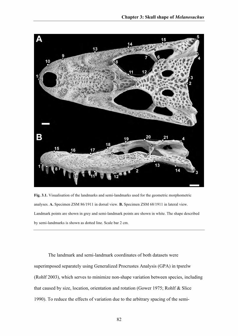
Chapter 3: Skull shape of Melanosuchus
82
Fig. 3.1. Visualisation of the landmarks and semi-landmarks used for the geometric morphometric
analyses. A. Specimen ZSM 86/1911 in dorsal view. B. Specimen ZSM 68/1911 in lateral view.
Landmark points are shown in grey and semi-landmark points are shown in white. The shape described
by semi-landmarks is shown as dotted line. Scale bar 2 cm.
The landmark and semi-landmark coordinates of both datasets were
superimposed separately using Generalized Procrustes Analysis (GPA) in tpsrelw
(Rohlf 2003), which serves to minimize non-shape variation between species, including
that caused by size, location, orientation and rotation (Gower 1975; Rohlf & Slice
1990). To reduce the effects of variation due to the arbitrary spacing of the semi-
Chapter 3: Skull shape of Melanosuchus
83
landmarks over the sampled curves, semi-landmarks were slid along their tangent to
align with the perpendicular of corresponding semi-landmarks, minimizing the
Procrustes distance. Thus, semi-landmarks capture primarily information about the
bowing of the sampled curve (Bookstein 1997; Zelditch et al. 2004).
Before starting the analyses, the percentage error for each landmark and semi-
landmark was computed for two specimens (one represented by a photograph and the
other by a X-ray image; with n = 10 repetitions), each in dorsal and lateral view after
the method of Singleton (2002) (Table S3.2, see supplementary information of Chapter
3). The methodological error for plotting landmarks by hand varies between 0.08 and
1.27 % and should have no significant impact on the shape analyses. Afterwards, the
superimposed landmarks and semi-landmark data of each dataset were imported into
MorphoJ 1.05d (Klingenberg 2011) and subjected to Principal Component Analysis
(PCA) by generating a covariance matrix. This procedure assimilates data from all
Procrustes coordinates and reduces them into a few dimensions with significant
variance in a way that preserves as much variance as possible (see Hammer & Harper
2006).
ONTOGENETIC PATTERNS
In order to assign the observed shape changes to different types of intraspecific
variation and to visualize ontogenetic changes in skull shape, we performed a
multivariate regression in MorphoJ on the Procrustes coordinates as well as the scores
of the first two PC axes against log-transformed centroid size. If an ontogenetic signal is
present then a statistical correlation between size and shape should be detectable. The
degree of correlation was estimated as a percentage of total shape variation, with a
Chapter 3: Skull shape of Melanosuchus
84
corresponding p value computed by a permutation test with 10 000 permutations and a
null hypothesis of independence (see Drake & Klingenberg 2008). Additionally, we
tested the correlation between shape and size by performing a two-block partial least
squares (2B-PLS) in MorphoJ using the Procrustes coordinates and centroid size. This
method explores the pattern of covariation between two sets of variables by
constructing pairs of variables that are linear combinations of the variables within each
of the two sets, and it accounts for as much as possible of the covariation between the
original datasets. In contrast to linear regression models (which casts one set of
variables as dependent on the other), 2B-PLS treats the two sets of variables
symmetrically in an attempt to find relationships between them without assuming that
one is the cause on the other (Rohlf & Corti 2000; Zelditch et al. 2004). The strength of
the correlation is given by the RV coefficient (see Robert & Escoufier 1976) and a p
value generated by 10 000 permutations. In order to test if the results could be falsified
by use of two different sources of samples (i.e. photographs and X-ray images) we
repeated the analyses excluding all specimens represented by X-ray images and
compared the results to the previous analyses.
SEXUAL DIMORPHISM
To determine whether skull shape is related to sex we created a second dataset including
only mature males and females and performed a new generalized Procrustes fit. We
assessed the statistical significance of differences between males and females based on
the Procrustes coordinates using NPMANOVA (nonparametric multivariate analysis of
variance) with 10 000 permutations and Euclidian distances using the software PAST
2.09 (Hammer et al. 2001). This approach tests for significant differences in the
distribution of groups within the morphospace (Anderson 2001). One of the strengths of
Chapter 3: Skull shape of Melanosuchus
85
this approach is that it does not assume or require normality from the multivariate data.
The test generates F and p values, with a significant difference between the sexes
indicated when the F value is high and the p value less than 0.05. Due to size
differences between males and females it is possible that apparent sexual dimorphism is
largely due to size. In order to detect evidence for non-size related sexual dimorphism
we repeated the analysis mentioned above with the non-allometric residuals, which were
separated from the Procrustes coordinates by a multivariate, pooled within-group
regression against log-transformed centroid size. The resulting residuals contain the
non-allometric component of shape variation within the dataset (Drake 2011). If a
significant difference remains in the NPMANOVA, one can conclude that males and
females show sexual dimorphism in cranial shape that is not related to size.
SHAPE VS. FUNCTION
In order to test if function is correlated with shape variation in the cranium we follow
Erickson et al. (2003), who propose an allometric relationship between bite force and
skull length. We used the bite force performance of Alligator mississippiensis in relation
to skull length as functional proxy. The bite force was estimated for every specimen and
log transformed. We performed a 2B-PLS in MorphoJ to determine the degree of
covariation of bite force with cranial shape. Furthermore, we performed regression
analyses between bite force and Procrustes coordinates. Both, the 2B-PLS and the
regression analyses were performed as explained above (see Ontogenetic and allometric
patterns section). To test if bite force is only correlated with allometric shape changes
we additionally performed all analyses with non-allometric residuals. As described
above, we excluded all specimens represented by X-ray images in further dataset to
verify if the results are falsified by the usage of two different sources of samples.

Chapter 3: Skull shape of Melanosuchus
86
RESULTS
In dorsal view the first two axes accounts for over 80 % of total variance (PC 1: 71.07
% and PC 2: 9.90 %). The first PC axis mainly accounts for the relative length of the
snout, the relative size and position of the orbit influencing the relative width of the
jugal region and the interorbital width, the relative length and width of the skull roof
table and the position of the jaw angle in anterolateral-posteromedial direction. The
second PC axis is primarily associated the shape of the snout tip, the position of the
orbit influencing the length of the snout and the postorbital region inversely and the
overall width of the postrostrum influencing the relative width of the occipital region
and the position of the jaw joint in anteromedial-posterolateral direction (Fig. 3.2).
In lateral view the first two PC axes accounts for almost 80 % of total variance
(PC 1: 74.96 % and PC 2: 5.97 %). The first PC accounts for mainly the depth of the tip
of the rostrum, the relative size of the subnarial gap, the shape and length of the ventral
margin of the maxilla, the relative size of the orbit influencing the overall depth of the
jugal and the orbital and postorbital region and the relative position of the jaw joint in
anteroventral-posterodorsal direction. The second PC accounts for the shape and the
relative length of the premaxilla, the relative length of the maxilla and the length of the
postorbital region (Fig. 3.3).
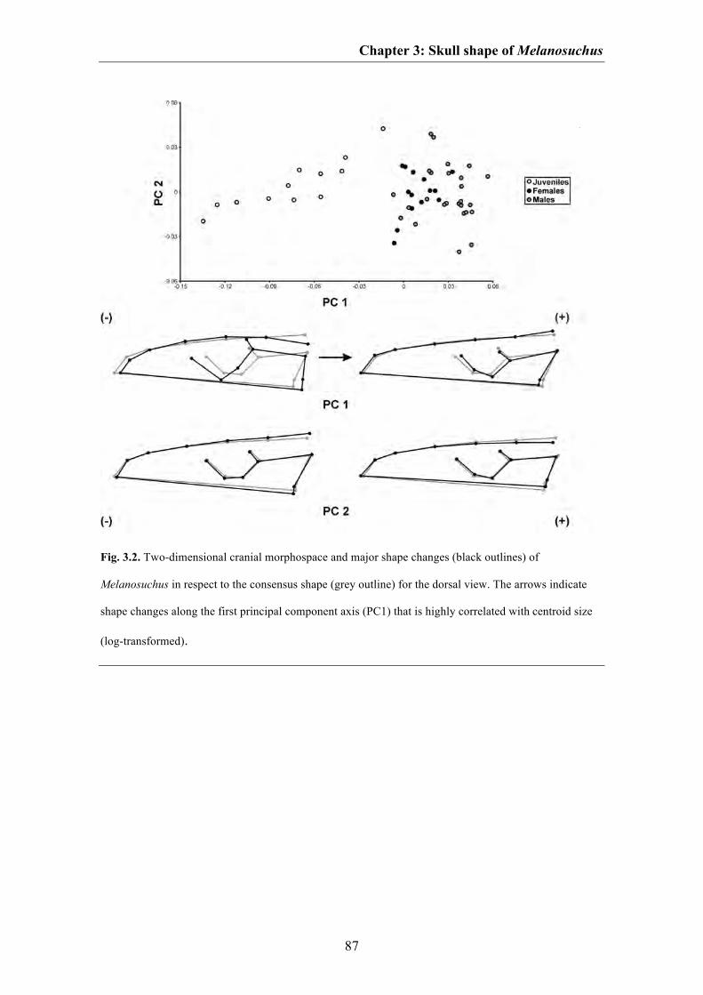
Chapter 3: Skull shape of Melanosuchus
87
Fig. 3.2. Two-dimensional cranial morphospace and major shape changes (black outlines) of
Melanosuchus in respect to the consensus shape (grey outline) for the dorsal view. The arrows indicate
shape changes along the first principal component axis (PC1) that is highly correlated with centroid size
(log-transformed).
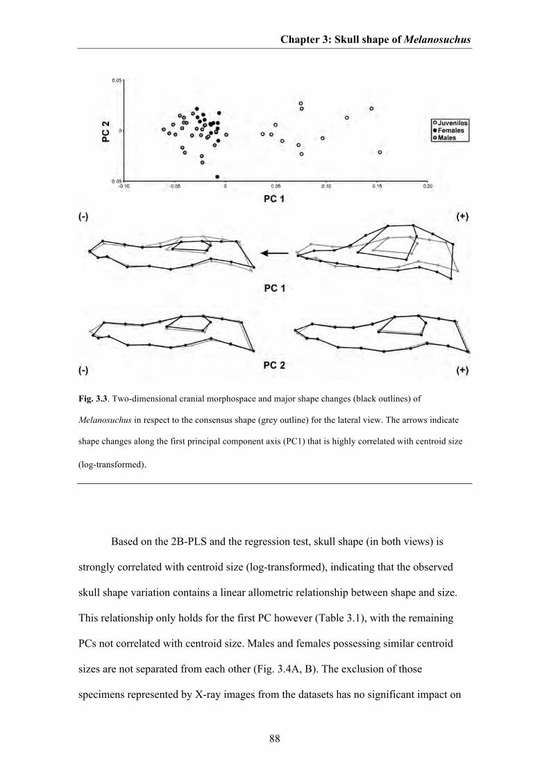
Chapter 3: Skull shape of Melanosuchus
88
Fig. 3.3. Two-dimensional cranial morphospace and major shape changes (black outlines) of
Melanosuchus in respect to the consensus shape (grey outline) for the lateral view. The arrows indicate
shape changes along the first principal component axis (PC1) that is highly correlated with centroid size
(log-transformed).
Based on the 2B-PLS and the regression test, skull shape (in both views) is
strongly correlated with centroid size (log-transformed), indicating that the observed
skull shape variation contains a linear allometric relationship between shape and size.
This relationship only holds for the first PC however (Table 3.1), with the remaining
PCs not correlated with centroid size. Males and females possessing similar centroid
sizes are not separated from each other (Fig. 3.4A, B). The exclusion of those
specimens represented by X-ray images from the datasets has no significant impact on
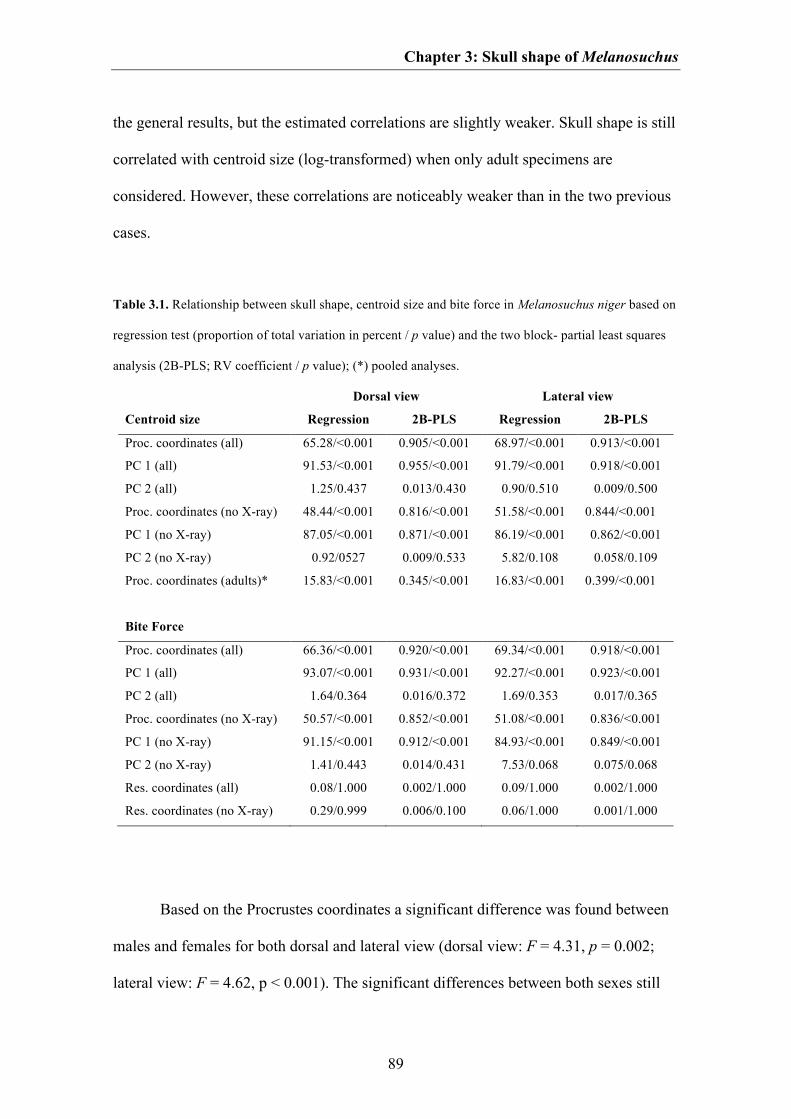
Chapter 3: Skull shape of Melanosuchus
89
the general results, but the estimated correlations are slightly weaker. Skull shape is still
correlated with centroid size (log-transformed) when only adult specimens are
considered. However, these correlations are noticeably weaker than in the two previous
cases.
Table 3.1. Relationship between skull shape, centroid size and bite force in Melanosuchus niger based on
regression test (proportion of total variation in percent / p value) and the two block- partial least squares
analysis (2B-PLS; RV coefficient / p value); (*) pooled analyses.
Dorsal view Lateral view
Centroid size Regression 2B-PLS Regression 2B-PLS
Proc. coordinates (all) 65.28/<0.001 0.905/<0.001 68.97/<0.001 0.913/<0.001
PC 1 (all) 91.53/<0.001 0.955/<0.001 91.79/<0.001 0.918/<0.001
PC 2 (all) 1.25/0.437 0.013/0.430 0.90/0.510 0.009/0.500
Proc. coordinates (no X-ray) 48.44/<0.001 0.816/<0.001 51.58/<0.001 0.844/<0.001
PC 1 (no X-ray) 87.05/<0.001 0.871/<0.001 86.19/<0.001 0.862/<0.001
PC 2 (no X-ray) 0.92/0527 0.009/0.533 5.82/0.108 0.058/0.109
Proc. coordinates (adults)* 15.83/<0.001 0.345/<0.001 16.83/<0.001 0.399/<0.001
Bite Force
Proc. coordinates (all) 66.36/<0.001 0.920/<0.001 69.34/<0.001 0.918/<0.001
PC 1 (all) 93.07/<0.001 0.931/<0.001 92.27/<0.001 0.923/<0.001
PC 2 (all) 1.64/0.364 0.016/0.372 1.69/0.353 0.017/0.365
Proc. coordinates (no X-ray) 50.57/<0.001 0.852/<0.001 51.08/<0.001 0.836/<0.001
PC 1 (no X-ray) 91.15/<0.001 0.912/<0.001 84.93/<0.001 0.849/<0.001
PC 2 (no X-ray) 1.41/0.443 0.014/0.431 7.53/0.068 0.075/0.068
Res. coordinates (all) 0.08/1.000 0.002/1.000 0.09/1.000 0.002/1.000
Res. coordinates (no X-ray) 0.29/0.999 0.006/0.100 0.06/1.000 0.001/1.000
Based on the Procrustes coordinates a significant difference was found between
males and females for both dorsal and lateral view (dorsal view: F = 4.31, p = 0.002;
lateral view: F = 4.62, p < 0.001). The significant differences between both sexes still
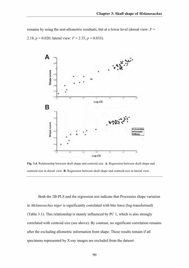
Chapter 3: Skull shape of Melanosuchus
90
remains by using the non-allometric residuals, but at a lower level (dorsal view: F =
2.18, p = 0.020; lateral view: F = 2.33, p = 0.033).
Fig. 3.4. Relationship between skull shape and centroid size. A. Regression between skull shape and
centroid size in dorsal view. B. Regression between skull shape and centroid size in lateral view.
Both the 2B-PLS and the regression test indicate that Procrustes shape variation
in Melanosuchus niger is significantly correlated with bite force (log-transformed)
(Table 3.1). This relationship is mainly influenced by PC 1, which is also strongly
correlated with centroid size (see above). By contrast, no significant correlation remains
after the excluding allometric information from shape. These results remain if all
specimens represented by X-ray images are excluded from the dataset.

Chapter 3: Skull shape of Melanosuchus
91
DISCUSSION
ONTOGENETIC PATTERNS
The correlation between overall shape and centroid size (log-transformed) in
Melanosuchus niger may represent an ontogenetic pattern, which is characterized by the
shape change captured by the first PC. Skulls of young juveniles have a very short
snout, which is wide in dorsal view, but dorsoventrally pointed, and the ventral margin
is straight. The orbit is very large, the jugal region is slender in both dorsal and lateral
view and the postorbital region is elongated in anterior-posterior direction. The broad
skull roof table is posteriorly inclined in lateral view. The posterior end of the skull is
relatively narrow and the jaw joint lies substantially anterior to the posterior end of the
skull roof table. During ontogeny the snout becomes longer due to the elongation of the
maxilla, but also narrower (in dorsal view) and deeper (in lateral view). The tip of the
snout becomes blunter and a subnarial gap is developed between premaxilla and
maxilla. The ventral margin of the maxilla becomes anteriorly convex in lateral view.
The postrostrum becomes flattened and the relative size of the orbit decreases. In
contrast, the jugal region becomes broader and deeper. The postorbital region becomes
shorter, but expands posterolaterally. Due to the overall flattening of the postrostrum the
skull roof table becomes straight in lateral view and the jaw joint moves substantially
posterior to this.
As was mentioned in the introduction, the numbers of studies investigating the
cranial shape variation of crocodylians using geometric morphometric is rare. This is
especially true for ontogenetic studies (see Monteiro et al. 1997; Piras et al. 2010). On
the other hand, in the past ontogenetic variation of crocodylian skulls was studied
multiple times by traditional morphometrics (e.g. Dodson 1975; Webb & Messel 1978;
Chapter 3: Skull shape of Melanosuchus
92
Hall & Portier 1994; Monteiro & Soares 1997; Wu et al. 2006). As no standardized
samplings and analyses do exist so far, we compared the ontogenetic shape variation of
Melanosuchus niger with that of other crocodylian species taken from literature (Table
3.2). However, due to differences in the sampling of skull measurements, landmark
configurations and statistical analyses in the studies mentioned above, the current
comparison is limited to whether certain skull regions growth allometric (positive or
negative) or isometric during ontogeny.
Melanosuchus niger shares a relative increase of snout length with all extant
crocodylians except Tomistoma schlegelii and Gavialis gangeticus (Piras et al. 2010). A
relative increase of the width of the snout during ontogeny occurs also in Alligator
sinensis (Wu et al. 2006), Crocodylus acutus, Mecistops cataphractus and Tomistoma
schlegelii (Piras et al. 2010), whereas Caiman crocodilus and Caiman yacare (Monteiro
& Soares 1997) show a relative narrowing of the rostral width. In Alligator
mississippiensis the posterior part of the snout grows isometrically, but the snout tip
increases in width (Dodson 1975), whereas in Crocodylus novaeguineae snout width
decreases from early juveniles to small adults, but increases again in later ontogenetic
stages (Hall & Portier 1994). In Caiman latirostris snout width grows isometrically
(Monteiro & Soares 1997). A relative ontogenetic increase of the depth of the snout
occurs in Crocodylus acutus, Mecistops cataphractus and Tomistoma schlegelii. The
relative decrease of the orbit size is the only common ontogenetic pattern that is present
in all taxa used for comparison. A further common ontogenetic pattern in crocodylians
is the relative increase of the interorbital width, which is only absent in Alligator
sinensis. Another common pattern is the relative decrease in length of the postorbital
skull roof, which is probably related to the relative increase of the snout length
Chapter 3: Skull shape of Melanosuchus
93
mentioned above (see Marugán-Lobón & Buscalioni 2003). However, in Tomistoma
schlegelii and Gavialis gangeticus the postorbital skull roof grows almost isometrically.
Finally, most taxa show a relative increase of the posterior width of the postorbital
region during ontogeny with the exception of Alligator mississippiensis, Alligator
sinensis, Caiman crocodilus, Caiman yacare and Gavialis gangeticus, which show
isometric growth.
Based on this simplified comparison, most crocodylian taxa share similar
ontogenetic patterns in skull shape and some ontogenetic trajectories seem to be
relatively constrained (e.g. the relative increase of the snout length together with relative
decrease of the postorbital length, the relative decrease of the orbit size together with
the relative increase of the interorbital width). The only exception is the long-snouted
Gavialis gangeticus, which shows an almost isometric growth of the skull during
ontogeny (see also Piras et al. 2010). However, the current comparison of ontogenetic
trends in crocodylian skulls is very limited, as the available ontogenetic studies on
crocodylian skulls are not standardized in sampling and methods. Thus, with the data on
hand it is not possible to compare how strong certain skull regions growth relative to
others and how ontogenetic patterns in the cranium differ within different taxa in detail.
To improve our understanding of cranial ontogeny in crocodylians also with respect to
heterochronic events within their evolution, it would be worthwhile to investigate
ontogenetic shape variation in the future with broader taxon and specimen sampling and
with standardized methods.
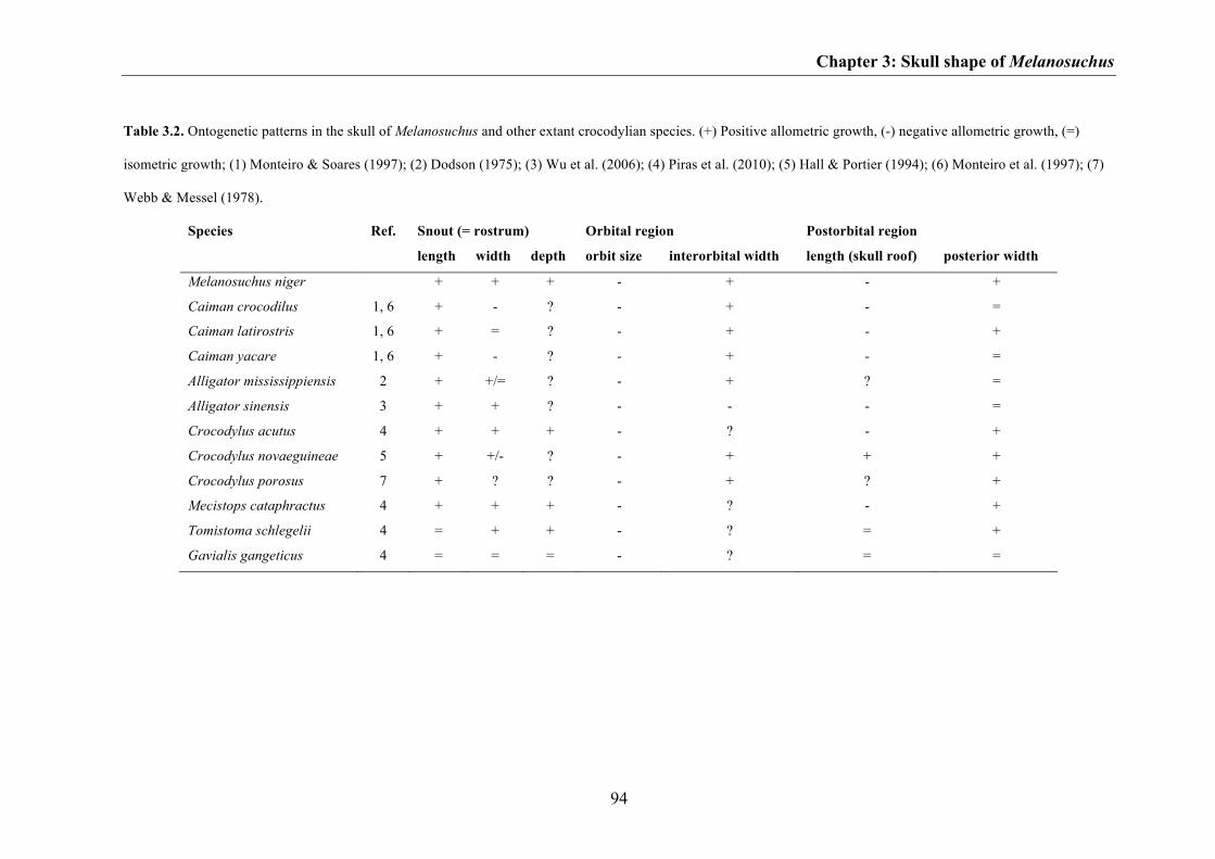
Chapter 3: Skull shape of Melanosuchus
94
Table 3.2. Ontogenetic patterns in the skull of Melanosuchus and other extant crocodylian species. (+) Positive allometric growth, (-) negative allometric growth, (=)
isometric growth; (1) Monteiro & Soares (1997); (2) Dodson (1975); (3) Wu et al. (2006); (4) Piras et al. (2010); (5) Hall & Portier (1994); (6) Monteiro et al. (1997); (7)
Webb & Messel (1978).
Species Ref. Snout (= rostrum) Orbital region Postorbital region
length width depth orbit size interorbital width length (skull roof) posterior width
Melanosuchus niger + + + - + - +
Caiman crocodilus 1, 6 + - ? - + - =
Caiman latirostris 1, 6 + = ? - + - +
Caiman yacare 1, 6 + - ? - + - =
Alligator mississippiensis 2 + +/= ? - + ? =
Alligator sinensis 3 + + ? - - - =
Crocodylus acutus 4 + + + - ? - +
Crocodylus novaeguineae 5 + +/- ? - + + +
Crocodylus porosus 7 + ? ? - + ? +
Mecistops cataphractus 4 + + + - ? - +
Tomistoma schlegelii 4 = + + - ? = +
Gavialis gangeticus 4 = = = - ? = =
Chapter 3: Skull shape of Melanosuchus
95
SEXUAL DIMORPHISM
Sexual dimorphism in crocodylians is described for both Caimaninae (Verdade 2000,
2003) and Crocodylinae (Hall & Portier 1994; Platt et al. 2009) and mainly size related.
The larger size in male crocodylians has been found to be the result of generally faster
and longer growth (e.g. Chabreck & Joanen 1979; Rootes et al. 1991; Wilkinson &
Rhodes 1997), probably resulting from different selective pressures faced by females
and males (see Shine 1989; Platt et al. 2009). Female growth trajectories probably slow
upon reaching sexual maturity as energy is shifted from growth to reproduction (see
Andrews 1982), whereas males are subject to sexual selection favouring large body size
in male-to-male interactions (see Anderson & Vitt 1990; Cooper & Vitt 1993). A size
related sexual dimorphism is also present in Melanosuchus niger, in which the skull
length of the largest males is about 30 % longer than that of the largest females.
In contrast, non-size related sexual variation seems to be less common in crocodylians.
It is only documented for the shape of the external naris in Gavialis gangeticus (Hall &
Portier 1994) and Caiman latirostris (Verdade 2000). Furthermore, Webb & Messel
(1978) describe a non-size related sexual dimorphism in the interorbital width and width
of the skull roof table in Crocodylus porosus. Thus, non-size related sexual dimorphism
documented in these crocodylian taxa is only restricted to certain skull regions. Based
on the results of the NPMANOVA the overall skull shape of males and females in
Melanosuchus niger shows a non-size related sexual dimorphism. As mentioned in the
Material and methods section, no information is known about how sex was determined
for the single specimens and misidentification cannot be ruled out. However, a wrong
identification of sex would rather result in a non-significant signal. Nevertheless, at this
stage the current findings should be still seen with caution, as the sample size of females

Chapter 3: Skull shape of Melanosuchus
96
(n = 14) is distinctively smaller than that of males (n = 26). Furthermore, it is possible
that the large number of landmarks and semi-landmarks compared to the sample size of
both males and females could lead to wrong positive signals due an overestimation of
the true degrees of freedom (Zelditch et al. 2004). Thus, the current findings should be
tested in future examinations in greater detail with larger datasets for Melanosuchus
niger, different landmark configurations (also with a three-dimensional approach) as
well as for other crocodylians.
SHAPE VS. FUNCTION
That allometric skull shape variation in Melanosuchus niger is significantly correlated
with bite force performance (log-transformed) is not surprising because the functional
proxy used in this study is correlated with skull size (see Erickson et al. 2003, 2004b,
2012). Deleting allometric information from shape leads to a non-significant signal,
showing that this correlation seems to be primarily related to allometric shape changes
caused by ontogenetic growth (see above). However, in recent crocodylians, stress
distributions during biting do not distribute over the whole skull uniformly, but are
largely concentrated in the postrostrum, peaking around the orbits and the temporal
fenestrae during bilateral and unilateral biting, and laterally at the level of the jugal
during lateral loading to the snout (Pierce et al. 2008). Thus, the strong correlation
found between bite force performance and ontogenetic shape variation is probably an
artefact of the allometric dependency of both parameters. Based on the biomechanical
results of Pierce et al. (2008) only the shape changes seen in the postrostral region
(including the expansion of the jugal, the relative decrease of the orbit size and the
posterolateral expansion of the postorbital region) seems to be functionally related. In
this context, the posterolateral expansion of the postorbital region might further
Chapter 3: Skull shape of Melanosuchus
97
correlate with the increase of the muscle system in the postorbital region to achieve
higher bite performance (see Schumacher 1973; van Drongelen & Dullemeijer 1982;
Busbey 1989; Erickson et al. 2003, 2012; Bona & Desojo 2011).
Based on the results of recent studies on cranial function in several crocodylian
species with different snout morphologies, snout shape is not strongly correlated with
function but instead with prey selection (McHenry et al. 2006; Pierce et al. 2008;
Erickson et al. 2012). Thus, it is likely that the shape changes seen in the snout of
Melanosuchus niger (including the relative increase of the snout length and depth, the
formation of a subnarial gap and the shape changes of the ventral margin of the maxilla)
are rather related to changes in diet preferences and feeding behavior through ontogeny.
Hatchlings and small Melanosuchus niger possessing skulls with a short pointed snout
feed predominantly on aquatic and shoreline invertebrates (e.g. insects, beetles and
snails) (Da Silveira & Magnusson 1999; Horna et al. 2001, 2003), whereas adults
possessing deep and elongated snouts with a subnarial gap feed on medium-sized prey,
such as capybaras (Hydrochoerus hydrochaeris), white-lipped peccaries (Tayassu
pecari), long-whiskered catfishes (Pimelodidae) and piranhas (Trutnau 1994; Horna et
al. 2001). Similar dietary shifts from small invertebrates to medium-sized vertebrates
during ontogeny are also documented for other crocodylian species (Cott 1961; Webb &
Messel 1978; Hutton 1987; Webb et al. 1991; Cleuren & de Vree 2000). Especially, the
development of a convex shape of the anterior part of the ventral margin of the maxilla
could be related to the development of a prominent upper caniniform tooth, which is
used primarily for seizing larger prey (Erickson et al. 2012). In this context, the
ontogenetic shape changes in the jugal and postorbital region (see above) provide the
mechanical background for handling larger prey.
Chapter 3: Skull shape of Melanosuchus
98
Finally, the usage of two different sample sources (i.e. photographs and X-ray images)
affected only the selection of landmarks in order to capture skull shape, as many skull
structures (e.g. bone sutures or the lateral temporal fenestra) were not visible in the X-
ray images. However, the similar results regarding the relation between shape, centroid
size and bite forces for both datasets with and without X-ray images (Table 3.1) as well
as the minor error for plotting landmarks on both sample sources (Table S3.2, see
supplementary information of Chapter 3) indicate that the usage of photographs and X-
ray images together did not falsify the current results.
CONCLUSIONS
The present study represents the first assessment of intraspecific variation of the skull of
the caimanine crocodylian Melanosuchus niger using a geometric morphometric
approach. Skull shape variation is concentrated in the width and height of the
postorbital region, the length and depth of the snout, the size of the orbit, and the
relative position of jaw joint. Similar patterns can be observed in the cranial ontogeny
of other crocodylian taxa, but due to the lack of broad-scale examinations with
standardized landmark configurations and statistical methods the results of the current
comparison is limited regarding the quality of change. The ontogenetic shape changes in
Melanosuchus niger seems to be correlated with increased bite force performance, but
are primarily restricted to the postrostral region. In contrast, shape variation seen in the
snout is probably rather related to the changes in diet through ontogeny. Based on the
current results the skull shape of females and males differ on a significant level, even
when allometric shape variation is reduced. However, the presence of a non-size related
sexual dimorphism in Melanosuchus niger should be seen as preliminary result due to
the small sample sizes in relation to the number of landmarks and semi-landmarks and
Chapter 3: Skull shape of Melanosuchus
99
differences of sample size between males and females, and thus, has to be tested in
future examinations in more detail. Because knowledge of intraspecific variation is
important for the systematics of extant, but also extinct taxa, it would be worthwhile
investigating ontogenetic patterns in different crocodylian taxa with help of
standardized methods (see e.g. Piras et al. 2010) to capture broad-scale patterns of
intraspecific variation and specific trajectories in crocodylian ontogeny more precisely.
This may in turn allow us to resolve the taxonomic status of problematic extinct species
such as Melanosuchus fisheri.
ACKNOWLEDGEMENTS
We thank Frank Glaw (Zoologische Staatssammlung München) who allowed two of us
(JBD, CF) to study the specimens under his care, and for providing helpful information
concerning historical collection data. We further thanks Martin Dobritz (Institut für
Radiologie, Klinikum rechts der Isar, Technische Universität München), Jakob
Hallermann (Zoologisches Museum Hamburg, Universität Hamburg), Heinz Grillitsch
and Richard Gemel (Naturhistorisches Museum Wien), Gunther Köhler and Linda
Acker (Senckenberg Naturmuseum Frankfurt) and Dennis Rödder and Ursula Bott
(Zoologisches Forschungsmuseum Alexander Koenig, Bonn) for additional photo
material, access to collections and technical support for the production of the X-ray
images. We further thank Richard Butler, Roland Sookias (GeoBio-Center, Ludwig-
Maximilians-University, München), Oliver Rauhut (Bayerische Staatssammlung für
Paläontologie und Geologie, München), Melissa Tallman (Department of Biomedical
Sciences, Grand Valley State University, Allendale), David Gower (Life Sciences
Department, Natural History Museum, London) and Abby Drake (Department of
Biology, Dana Science Center, Skidmore College, Saratoga Springs) for comments and
Chapter 3: Skull shape of Melanosuchus
100
suggestions on an early version of the manuscript. This research was partially funded by
the Agencia Nacional de Promoción Científica y Técnica PICT 2010 N°207, Alexander
von Humboldt Foundation (to JBD) and Deutsche Forschungsgemeinschaft (Project RA
1012/12-1) (to CF).
101
CHAPTER 4
Do different disparity proxies converge on a common signal? Insights from the cranial
morphometrics and evolutionary history of Pterosauria (Diapsida: Archosauria)
Keywords:
Disparity; macroevolution; skull shape; Pterosauria; Monofenestrata; geometric
morphometrics; phylogeny; phylogenetic comparative methods; Mesozoic
The chapter was published as:
Foth C1,2, Brusatte SL3,4 & Butler RJ2,5. 2012. Do different disparity proxies converge
on a common signal? Insights from the cranial morphometrics and evolutionary history
of Pterosauria (Diapsida: Archosauria). Journal of Evolutionary Biology 25: 904-915.
1Bayerische Staatssammlung für Paläontologie und Geologie, Richard-Wagner-Str. 10, D-80333 München, Germany 2Department of Earth and Environmental Sciences, Ludwig-Maximilians-University, Richard-Wagner-Str. 10, D-80333 München, Germany 3Division of Paleontology, American Museum of Natural History, Central Park West at 79th Street, New York, NY 10024, USA 4Department of Earth and Environmental Sciences, Columbia University, New York, NY, USA 5GeoBioCenter, Ludwig-Maximilians-University; Richard-Wagner-Str. 10, D-80333 München, Germany
Received 11 January 2012; revised 26 January 2012; accepted 27 January 2012
Author contribution:
Research design: Christian Foth, Stephen Brusatte, Richard Butler
Data collection: Christian Foth, Richard Butler
Data analyses: Christian Foth, Stephen Brusatte
Preparation of figures and tables: Christian Foth
Wrote paper: Christian Foth, Stephen Brusatte, Richard Butler
*All figures of Chapter 4 are modified after Foth et al. (2012)
Chapter 4: Pterosaur skull morphometrics and disparity
102
Do different disparity proxies converge on a common signal? Insights from the
cranial morphometrics and evolutionary history of Pterosauria (Diapsida:
Archosauria)
Christian Foth, Stephen L. Brusatte & Richard J. Butler
ABSTRACT
Disparity, or morphological diversity, is often quantified by evolutionary biologists
investigating the macroevolutionary history of clades over geological timescales.
Disparity is typically quantified using proxies for morphology, such as measurements,
discrete anatomical characters, or geometric morphometrics. If different proxies
produce differing results, then the accurate quantification of disparity in deep time may
be problematic. However, despite this, few studies have attempted to examine disparity
of a single clade using multiple morphological proxies. Here, as a case study for this
question, we examine the disparity of the volant Mesozoic fossil reptile clade
Pterosauria, an intensively studied group that achieved substantial morphological,
ecological, and taxonomic diversity during their 145+ million year evolutionary history.
We characterise broadscale patterns of cranial morphological disparity for pterosaurs
for the first time using landmark-based geometric morphometrics, and make
comparisons to calculations of pterosaur disparity based on alternative metrics.
Landmark-based disparity calculations suggest that monofenestratan pterosaurs were
more diverse cranially than basal non-monofenestratan pterosaurs (at least when the
aberrant anurognathids are excluded), and that peak cranial disparity may have occurred
in the Early Cretaceous, relatively late in pterosaur evolution. Significantly, our cranial
disparity results are broadly congruent with those based on whole skeleton discrete
Chapter 4: Pterosaur skull morphometrics and disparity
103
character and limb proportion datasets, indicating that these divergent approaches
document a consistent pattern of pterosaur morphological evolution. Therefore,
pterosaurs provide an exemplar case demonstrating that different proxies for
morphological form can converge on the same disparity signal, which is encouraging
because often only one such proxy is available for extinct clades represented by fossils.
Furthermore, mapping phylogeny into cranial morphospace demonstrates that pterosaur
cranial morphology is significantly correlated with, and potentially constrained by,
phylogenetic relationships.
INTRODUCTION
Evolutionary biologists often calculate measures of morphological disparity to help
understand the macroevolutionary history of clades over long time scales (Gould 1991;
Wills et al. 1994; Erwin 2007). Disparity is essentially a morphological equivalent of
taxonomic diversity: it measures the variety of anatomical features expressed by a
group. By tracking disparity over time and among taxa, biologists can assess the pace at
which clades developed novel morphologies, determine how morphological variability
was affected by evolutionary radiations and mass extinctions, and test whether certain
groups were more morphologically variable than others (perhaps due to key innovations
or differential niche exploitation). Disparity studies also allow taxa to be plotted into a
morphospace: a visual representation of the range of morphological form expressed by
the species in question.
Studies of disparity are predicated on a straightforward but difficult issue: how
best to quantify the morphology of organisms using manageable proxies. Three general

Chapter 4: Pterosaur skull morphometrics and disparity
104
proxies are often used, each of which is compiled for every species in an analysis so
that morphology can be measured equivalently: measurements (e.g. Foote 1993;
Lefebvre et al. 2006; Dyke et al. 2009), geometric morphometric landmarks (e.g. Foote
1993; Smith & Lieberman 1999; Friedman 2010; Brusatte et al. 2012a), and discrete
characters (e.g. Foote 1994; Wills et al. 1994; Lupia 1999; Brusatte et al. 2008; Ruta
2009; Butler et al. 2012). Because the accuracy of disparity studies hinges on the choice
of proxies, it is desirable that different proxies converge on the same signals. For
instance, if discrete characters indicate that there was a rapid rise in disparity during the
early history of a group, it is hoped that measurements or geometric landmarks would
show the same result. Surprisingly, this has not been widely tested with empirical case
studies. Villier & Eble (2004) used multiple proxies to quantify the disparity of
echinoids, which generally gave consistent results. It is currently unknown, however,
whether measurements, landmarks, and discrete characters give concordant or
discordant results in any vertebrate clades, which recently have become more frequent
subjects of disparity study than non-vertebrate groups (e.g. Brusatte et al. 2008; Ruta
2009; Friedman 2010; Prentice et al. 2011; Butler et al. 2012).
Pterosaurs, the familiar Mesozoic flying reptiles, are an excellent case study for
assessing this question. Pterosaurs were the earliest vertebrates to evolve active flapping
flight (Padian 1983, 1985). The oldest pterosaur fossils date from the Late Triassic (c.
210 Ma), and the clade formed an important component of terrestrial vertebrate
diversity for nearly 150 million years, prior to their extinction at the end of the
Cretaceous (65.5 Ma) (Wellnhofer 1991; Unwin 2006; Butler et al. 2009a). During their
evolutionary history pterosaurs evolved considerable taxonomic (> 140 species
currently recognised: Barrett et al. 2008; Butler et al. 2009a, 2011a) and morphological
Chapter 4: Pterosaur skull morphometrics and disparity
105
diversity (Butler et al. 2011a, 2012; Prentice et al. 2011). Body size estimates range
from a mere 5-35 g for the frogmouth-like aerial insectivore Anurognathus up to 259–
544 kg for the giant stork-like Quetzalcoatlus, whose wingspan is estimated at 10–12 m
(Witton 2008; Henderson 2010; Witton & Habib 2010). Ecologically, skeletal
morphology has been used to suggest a diverse range of feeding strategies within the
clade, including piscivory, filter-feeding, insectivory, molluscivory, frugivory, aerial
predation, vulture-like scavenging, and stork-like stalking and scavenging (e.g.
Bonaparte 1970; Wellnhofer 1991; Õsi et al. 2005; Unwin 2006; Bennett 2007; Witton
& Naish 2008; Lü et al. 2010a). In short, pterosaurs were a remarkable radiation of
extinct reptiles, and understanding their evolutionary history promises to unlock more
general insights into the development of novel body plans, locomotion styles (flight)
and ecological behaviours over deep time.
Recent years have seen an explosion of scientific interest in pterosaurs, with the
discovery of spectacular new fossils and fossil assemblages (e.g. Wang et al. 2005; Lü
et al. 2010a, 2011), new insights into palaeobiology (e.g. Wilkinson et al. 2006; Butler
et al. 2009b; Claessens et al. 2009; Palmer & Dyke 2010), the development of new
phylogenetic hypotheses (e.g. Andres & Ji 2008; Andres 2010; Andres et al. 2010; Lü et
al. 2010a) and synthetic work focusing on the macroevolutionary history and diversity
of the group (e.g. Dyke et al. 2006, 2009; McGowan & Dyke 2007; Butler et al. 2009a,
2011a, 2012; Prentice et al. 2011). A particularly vigorous research program has
developed concerning the temporal and phylogenetic patterns of morphological and
taxonomic diversity amongst pterosaurs, and the impact of variable fossil record quality
on our understanding of these patterns (Dyke et al. 2009; Butler et al. 2009a, 2011a,
2012; Andres 2009; Benton et al. 2011; Prentice et al. 2011). Among the most
Chapter 4: Pterosaur skull morphometrics and disparity
106
interesting topics of debate is whether the morphological diversity of pterosaurs peaked
early or late in the Mesozoic history of the group, and whether different clades of
pterosaurs were more or less morphologically diverse than others, perhaps due to the
development of novel diets and ecologies (e.g. Dyke et al. 2006, 2009; Prentice et al.
2011; Butler et al. 2011a, 2012). Previous authors have assessed these questions by
using both discrete characters (Prentice et al. 2011; Butler et al. 2011a, 2012) and
measurements (Dyke et al. 2009) to quantify disparity.
Here, we characterise broadscale patterns of cranial morphological diversity for
pterosaurs for the first time using geometric morphometrics, a common technique (but
one infrequently used to study Mesozoic reptile clades) that models a series of
specimens using homologous landmarks, allows these specimens to be plotted into a
morphospace, and then utilises multivariate statistics to tease out major patterns of
shape variation (O’Higgins 2000; Zelditch et al. 2004). The purposes of this project are
two-fold. First, the availability of cranial morphometric data allows us to assess whether
disparity results based on this proxy (both temporal and clade-by-clade comparisons)
are congruent with published results based on measurements and discrete characters.
Second, we use cranial morphometrics to assess large-scale patterns in pterosaur
morphological evolution. Our focus is on a handful of explicit questions. Did pterosaurs
exhibit constant or variable disparity across their history? If the latter is true, did
disparity peak early or late in the history of pterosaur evolution? Did certain pterosaur
subgroups have significantly higher disparity than others? Is pterosaur cranial
morphology significantly correlated with phylogeny, as would be indicative of
phylogenetic constraint in pterosaur cranial shape evolution? Were there any major
trends in cranial shape across pterosaur phylogeny? Taken together, the various
Chapter 4: Pterosaur skull morphometrics and disparity
107
quantitative analyses have broad implications for our understanding of pterosaur
evolutionary history and large-scale patterns of pterosaur cranial evolution during the
Mesozoic.
METHODS
TAXON SELECTION
The taxonomy of pterosaurs used here is based primarily on Unwin (2006). Because the
great majority of preserved pterosaur skull material is crushed or incomplete, we
focused our analyses on cranial reconstructions in lateral view (supplemented by some
photographs of complete, undistorted skulls). Unfortunately, such reconstructions and
complete skulls are available for only a small fraction (~20%) of the known pterosaur
species (see supplementary information of Chapter 4 for details of cranial
reconstructions/photographs used). However, useable reconstructions were available for
at least one species from nearly all significant ‘family’-level clades of pterosaurs, with
the exception of the Early Cretaceous Istiodactylidae and the late Early-early Late
Cretaceous Lonchodectidae. Thus, it is likely that we can successfully capture
broadscale phylogenetic and temporal patterns of cranial morphological diversity across
Pterosauria, even if it is not possible for us capture detailed variation within ‘family’-
level clades.
GEOMETRIC MORPHOMETRICS
We analysed morphological variation in the skull (excluding the lower jaw) of
pterosaurs using two-dimensional geometric morphometrics. The advantage of this
approach is that it has high statistical power to detect shape differences, because
landmark coordinates capture more information about shape than can be obtained from
Chapter 4: Pterosaur skull morphometrics and disparity
108
traditional morphometric measurements (linear distances, ratios and angular measures),
which are generally insufficient to capture the whole geometry of the original object
(Rohlf 2000; Hammer & Harper 2006). Geometric morphometric approaches have been
reviewed and discussed by many authors (e.g. Bookstein 1991; Elewa 2004; Zelditch et
al. 2004), and the methods we use here are similar to those used in the recent
morphometric study of Brusatte et al. (2012a).
We encapsulated the cranial geometry of 31 pterosaur species using 21
homologous landmarks (Fig. 4.1A, see supplementary information of Chapter 4 for full
description), which were plotted onto published cranial reconstructions and photographs
in lateral view (one image per species) using the program tpsDig2 (Rohlf 2005). We
used landmarks of types 2 (good evidence for geometric homology, such as points of
maximal curvature or extremities) and 3 (points constructed between two clearly
homologous landmarks, which mainly define the shape of the skull or skull openings
rather than the position of exact homologous points), following the terminology of
Bookstein (1991). The usage of Type 3 landmarks is necessary because some
reconstructions do not show the patterns of articulation for individual bones (because of
poor preservation and crushing on the fossil specimens), and because in many species a
number of elements are fused or reduced (see supplementary information of Chapter 4).
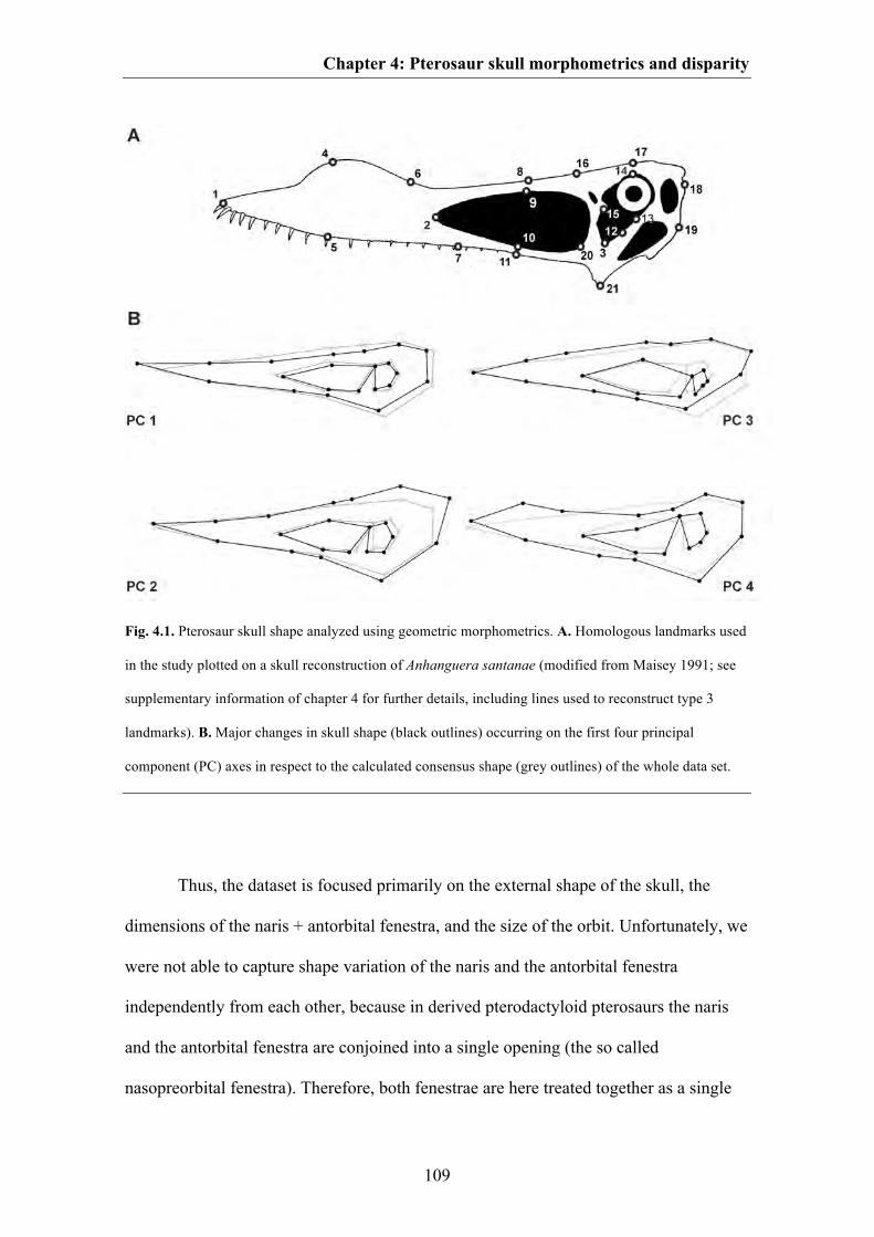
Chapter 4: Pterosaur skull morphometrics and disparity
109
Fig. 4.1. Pterosaur skull shape analyzed using geometric morphometrics. A. Homologous landmarks used
in the study plotted on a skull reconstruction of Anhanguera santanae (modified from Maisey 1991; see
supplementary information of chapter 4 for further details, including lines used to reconstruct type 3
landmarks). B. Major changes in skull shape (black outlines) occurring on the first four principal
component (PC) axes in respect to the calculated consensus shape (grey outlines) of the whole data set.
Thus, the dataset is focused primarily on the external shape of the skull, the
dimensions of the naris + antorbital fenestra, and the size of the orbit. Unfortunately, we
were not able to capture shape variation of the naris and the antorbital fenestra
independently from each other, because in derived pterodactyloid pterosaurs the naris
and the antorbital fenestra are conjoined into a single opening (the so called
nasopreorbital fenestra). Therefore, both fenestrae are here treated together as a single
Chapter 4: Pterosaur skull morphometrics and disparity
110
unit. Furthermore, our landmarks do not, in general, capture overall variation in the
form of cranial crests (although some small degree of variation in these structures is
likely incorporated due to the usage of type 3 landmarks), which are difficult to describe
by homologous landmarks, often incompletely preserved, and are liable to swamp the
dataset with characters related more to extravagant variation in display structures (see
e.g. Bennett 2003; Martill & Naish 2006; Elgin et al. 2008; Tomkins et al. 2010; Hone
et al. 2012) than to features related to underlying skull architecture and function (e.g.
feeding).
The landmark coordinates were superimposed using Generalized Procrustes
Analyses (GPA) in MorphoJ (Klingenberg 2011) which serves to minimise non-shape
variation between species, such as that caused by size, location, orientation, and rotation
(Zelditch et al. 2004). Next, the “corrected” landmarks were converted into a covariance
matrix and subjected to Principal Component Analyses (PCA), also using MorphoJ.
This procedure gathers together data from all landmarks and reduces it into a set of PC
scores that summarise the skull shape of each taxon and describe maximal shape
variation in a morphospace (Hammer & Harper 2006).
DISPARITY ANALYSES
Morphological disparity measures the anatomical diversity (variety) exhibited by a
group of organisms (Foote 1993; Wills et al. 1994; Ciampaglio et al. 2001). Disparity
calculations require a measure of morphological form for each organism being assessed,
and as a proxy for pterosaur skull shape we used PC scores from the morphometric
PCA. We examined temporal trends in cranial disparity by binning pterosaur taxa into
four broad temporal bins (Late Triassic-Early Jurassic, Middle Jurassic-Late Jurassic,
Chapter 4: Pterosaur skull morphometrics and disparity
111
Early Cretaceous, Late Cretaceous; see supplementary information of Chapter 4). Our
assignment of species to temporal bins was based upon information taken from the
Paleobiology Database. Large temporal bins were necessary because using finer-scale
(shorter) bins would, in most cases, render sample sizes too small for meaningful
statistical comparison. Coarse bins of similar (and sometimes identical) length have
been used in previous studies of pterosaur morphological and taxonomic diversity (e.g.
Dyke et al. 2006, 2009; Prentice et al. 2011; but see Butler et al. 2012).
Additionally, we examined taxonomic variation in disparity by measuring
disparity for seven different groupings: non-monofenestratan pterosaurs
(‘rhamphorhynchoids’); non-monofenestratan pterosaurs excluding the aberrant
Anurognathidae (which plot as outliers in morphospace and which have a highly
unusual cranial morphology with an extremely short snout and an enlarged orbit. In that
respect, anurognathids differs substantially from all other pterosaurs: e.g. Bennett
2007); Monofenestrata; Dsungaripteroidea; Azhdarchoidea; Ctenochasmatoidea;
Ornithocheiroidea (see supplementary information of Chapter 4). The major clades of
monofenestratan pterosaurs are all inferred to have originated at approximately the
same time (by the Late Jurassic), and have traditionally been given equivalent rank-
based names. Assignment of species to taxonomic groupings was based primarily on
Unwin (2006), and we note that the first two of our bins are paraphyletic (they exclude
monofenestratan pterosaurs). As an alternative, we also grouped monofenestratan
pterosaurs in several different ways according to the topology of Andres & Ji (2008):
Archaeopterodactyloidea; Ornithocheiroidea; Pteranodontoidea + Nyctosaurus; clade
consisting of Zhejiangopterus + Tapejaridae + Dsungaripteridae + Azhdarchidae (see
supplementary information of Chapter 4).

Chapter 4: Pterosaur skull morphometrics and disparity
112
Four disparity metrics were calculated for each temporal or taxonomic grouping:
the sum and product of the ranges and variances on the first six PC axes of the 31-taxon
morphometric dataset, which together comprise more than 90% of total variance. The
program RARE was used to perform these calculations (Wills 1998). Range measures
summarise the total spread of morphospace occupied by the taxa in question, whereas
variance denotes mean dissimilarity among the taxa (roughly equivalent to their spread
in morphospace). Range metrics can be strongly biased by sample size differences,
including those caused by uneven fossil sampling over time, but variance metrics are
more robust (Wills et al. 1994; Butler et al. 2012). Statistical significance of disparity
comparisons was assessed by the overlap or non-overlap of 95% confidence intervals,
generated by bootstrapping, and rarefaction was used to assess whether disparity
differences between groups are robust to sample size differences (both were also
performed in RARE).
PHYLOGENETIC COMPARATIVE METHODS
An interesting question that has not previously been quantitatively addressed is whether
trends in pterosaur morphological evolution are strongly related to (and perhaps
constrained by) phylogeny. In the current case, the main question is: is pterosaur skull
shape significantly correlated with phylogeny? A strong phylogenetic signal means that
closely related species tend to fall out closer in morphospace than more distantly related
species (Klingenberg & Gidaszewski 2010). One way to assess the degree of this
correlation is to map the phylogeny into the morphospace. This requires an ancestral
state reconstruction of the morphometric data (PC scores) for each internal node on the
tree, which is accomplished in MorphoJ using squared change parsimony (Maddison
1991; Klingenberg 2011). This algorithm collates the sum of square changes of the PC
Chapter 4: Pterosaur skull morphometrics and disparity
113
scores (which are continuous characters) along all branches of the tree and calculates
the most parsimonious ancestral states for each node by minimising the total sum of
squared change across the phylogeny. The optimal configuration is mathematically
described by a tree length (= squared length) value, which can be used to determine the
strength of the correlation between shape and phylogeny (if there is a strong correlation
then the tree length value should be small).
The statistical significance of the tree length is calculated by a permutation test
performed in MorphoJ in which the topology of an input phylogeny (assumed to be the
“true” phylogeny of the group in question) including branch lengths is held constant and
the PC scores for each taxon are randomly permuted across the tree 10 000 times
(Laurin 2004; Klingenberg & Gidaszewski 2010). If the tree length of the “true”
phylogeny is less than that in at least 95% of the randomly generated trees then there is
said to be statistically significant correlation at a 5% threshold between phylogeny and
PC scores (skull shape).
For our pterosaur dataset, we calculated the tree length and its significance value
by using two different topologies: a) a pruned consensus tree (which includes all species
used in this study) from Lü et al. (2010a) and b) an informal pterosaur supertree (sensu
Butler & Goswami 2008), based upon Andres & Ji (2008) and Andres et al. (2010).
Because the phylogenetic position of Feilongus is uncertain (e.g. Wang et al. 2005; Lü
& Ji 2006; Andres & Ji 2008) we placed this taxon at the base of Ornithocheiroidea for
the Lü et al. topology (Lü & Ji 2006; see supplementary information of Chapter 4).
Because Raeticodactylus, Darwinopterus and Shenzhoupterus are not included in the
phylogenies of Andres & Ji (2008) and Andres et al. (2010), we placed Raeticodactylus
Chapter 4: Pterosaur skull morphometrics and disparity
114
in a polytomy with Eudimorphodon, Darwinopterus as sister taxon to Pterodactyloidea,
and Shenzhoupterus in a polytomy with Tapejarinae and Thalassodrominae (after Lü et
al. 2010a).
For both trees branch lengths were assigned based upon first appearances of
species, and zero branch lengths were adjusted by sharing out the time equally between
branches (see Ruta et al. 2006; Brusatte et al. 2008; Brusatte 2011). An arbitrary length
of 10 million years was added to the root. The assignment of branch lengths was
performed in the program R (R Development Core Team 2011) using the APE package
(version 2.7-2; Paradis et al. 2004) and a function written by Graeme Lloyd (see
http://www.graemetlloyd.com/methdpf.html).
To better understand and visualise how skull shape changed during pterosaur
evolution, which may reveal major evolutionary trends, we further mapped the PC
scores as continuous characters onto the time calibrated topology from Lü et al. (2010a)
using squared change parsimony in Mesquite 2.72 (Maddison & Maddison 2009). In the
supplementary information of this chapter an equivalent description is given for the
supertree topology.
Chapter 4: Pterosaur skull morphometrics and disparity
115
RESULTS
MORPHOSPACE OCCUPATION AND MAJOR SHAPE CHANGES
The whole data set is summarised by 30 PC axes. The majority of shape variation in
pterosaur skulls is summarised by the first four PC axes (51.7 %. 15.3 %, 13.2 % and
5.1 % of total variance), which together describe over 85 % of total variance. The main
shape changes of the first four principal component axes (PC) are shown in Fig. 4.1B.
The first PC axis describes the relative length of the snout, the relative size of the orbit
and postorbital region, the size and dorsoventral depth of the naris–antorbital fenestra
region and the position of the jaw joint relative to the orbit. The second PC axis
describes the relative position of the orbit, the depth of the anterior part of the skull roof
posterodorsal to the orbit and the position of the jaw joint relative to the orbit along the
dorsoventral axis. The third PC axis describes the relative size and location of the orbit,
the depth of the skull roof and dorsal part of the snout in the naris-antorbital fenestra
region and the relative size of the jaw joint region (i.e. the distance between the ventral
margin of the orbit and the jaw joint), including the position of the jaw joint relative to
the orbit along the anteroposterior axis. Finally, the fourth PC axis describes the depth
of the tip of the snout, the overall shape of the naris-antorbital fenestra region
(triangular to trapezoidal), the inflection of the snout and the location of the jaw joint
along the dorsoventral axis. The cranial morphospace of the first four PC axes described
above is visualised in Fig. 4.2.
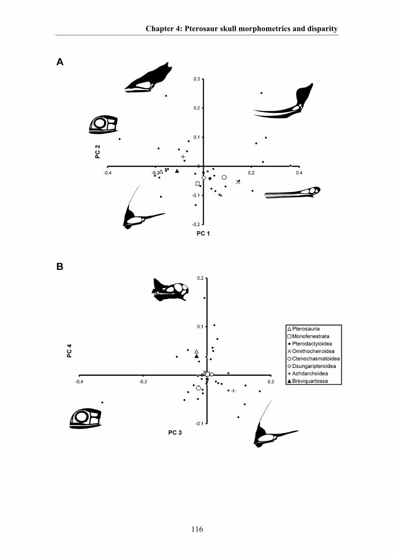
Chapter 4: Pterosaur skull morphometrics and disparity
116
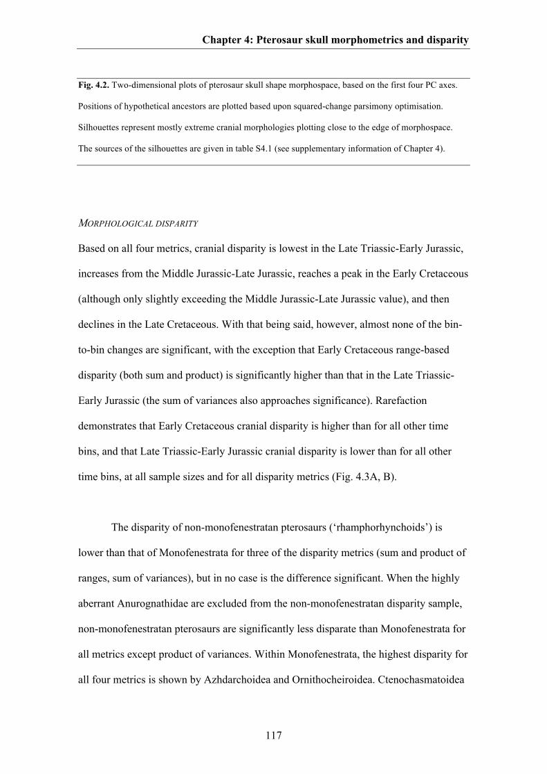
Chapter 4: Pterosaur skull morphometrics and disparity
117
Fig. 4.2. Two-dimensional plots of pterosaur skull shape morphospace, based on the first four PC axes.
Positions of hypothetical ancestors are plotted based upon squared-change parsimony optimisation.
Silhouettes represent mostly extreme cranial morphologies plotting close to the edge of morphospace.
The sources of the silhouettes are given in table S4.1 (see supplementary information of Chapter 4).
MORPHOLOGICAL DISPARITY
Based on all four metrics, cranial disparity is lowest in the Late Triassic-Early Jurassic,
increases from the Middle Jurassic-Late Jurassic, reaches a peak in the Early Cretaceous
(although only slightly exceeding the Middle Jurassic-Late Jurassic value), and then
declines in the Late Cretaceous. With that being said, however, almost none of the bin-
to-bin changes are significant, with the exception that Early Cretaceous range-based
disparity (both sum and product) is significantly higher than that in the Late Triassic-
Early Jurassic (the sum of variances also approaches significance). Rarefaction
demonstrates that Early Cretaceous cranial disparity is higher than for all other time
bins, and that Late Triassic-Early Jurassic cranial disparity is lower than for all other
time bins, at all sample sizes and for all disparity metrics (Fig. 4.3A, B).
The disparity of non-monofenestratan pterosaurs (‘rhamphorhynchoids’) is
lower than that of Monofenestrata for three of the disparity metrics (sum and product of
ranges, sum of variances), but in no case is the difference significant. When the highly
aberrant Anurognathidae are excluded from the non-monofenestratan disparity sample,
non-monofenestratan pterosaurs are significantly less disparate than Monofenestrata for
all metrics except product of variances. Within Monofenestrata, the highest disparity for
all four metrics is shown by Azhdarchoidea and Ornithocheiroidea. Ctenochasmatoidea

Chapter 4: Pterosaur skull morphometrics and disparity
118
exhibit lower disparity for all four metrics than these two aforementioned clades, but the
difference is not significant. Dsungaripteroidea has the lowest disparity of the
monofenestratan clades for all four metrics, and is significantly lower than the disparity
of Azhdarchoidea and Ornithocheiroidea for both range metrics and sum of variances
(but marginally non-significant for product of variances). On no metric is
Dsungaripteroidea significantly less disparate than Ctenochasmatoidea (Fig. 4.3C).
When the monofenestratan clades of Andres & Ji (2008) were used,
Ornithocheiroidea was found to be significantly more disparate than
Archaeopterodactyloidea for three metrics (all except sum of variances), whereas the
disparity values of the two major subclades within Ornithocheiroidea (Pteranodontoidea
+ Nyctosaurus; Tapejaridae + Dsungaripteridae + Azhdarchidae) are not significantly
different from one another (see Fig. S4.6, supplementary information of Chapter 4).
PHYLOGENETIC COMPARATIVE METHODS AND EVOLUTION OF SKULL SHAPE IN PTEROSAURS
For all phylogenies that we tested, the permutation test in MorphoJ demonstrates a
significant correlation between skull shape and phylogeny (p<0.0001). The tree lengths
of the time-calibrated phylogenies are 0.642 for the Lü et al. (2010a) topology and
0.626 for the supertree.
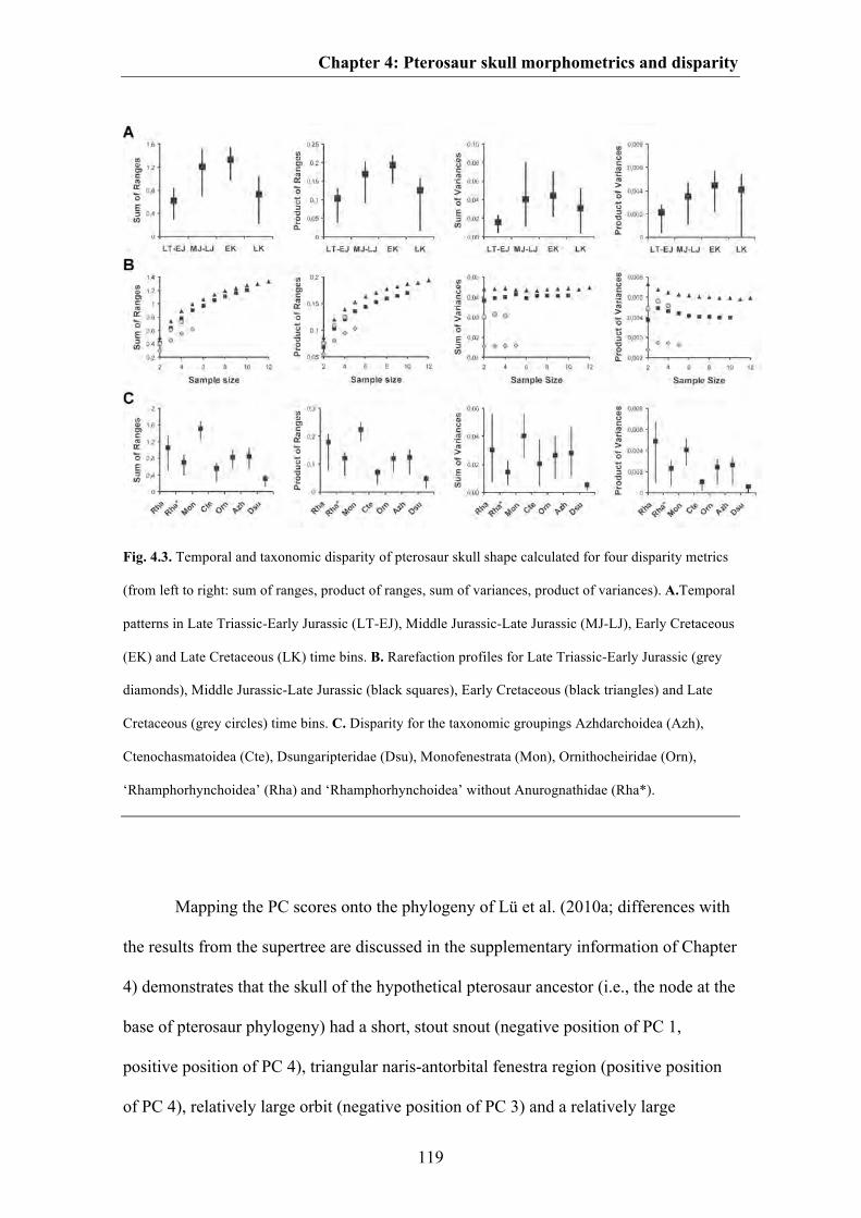
Chapter 4: Pterosaur skull morphometrics and disparity
119
Fig. 4.3. Temporal and taxonomic disparity of pterosaur skull shape calculated for four disparity metrics
(from left to right: sum of ranges, product of ranges, sum of variances, product of variances). A.Temporal
patterns in Late Triassic-Early Jurassic (LT-EJ), Middle Jurassic-Late Jurassic (MJ-LJ), Early Cretaceous
(EK) and Late Cretaceous (LK) time bins. B. Rarefaction profiles for Late Triassic-Early Jurassic (grey
diamonds), Middle Jurassic-Late Jurassic (black squares), Early Cretaceous (black triangles) and Late
Cretaceous (grey circles) time bins. C. Disparity for the taxonomic groupings Azhdarchoidea (Azh),
Ctenochasmatoidea (Cte), Dsungaripteridae (Dsu), Monofenestrata (Mon), Ornithocheiridae (Orn),
‘Rhamphorhynchoidea’ (Rha) and ‘Rhamphorhynchoidea’ without Anurognathidae (Rha*).
Mapping the PC scores onto the phylogeny of Lü et al. (2010a; differences with
the results from the supertree are discussed in the supplementary information of Chapter
4) demonstrates that the skull of the hypothetical pterosaur ancestor (i.e., the node at the
base of pterosaur phylogeny) had a short, stout snout (negative position of PC 1,
positive position of PC 4), triangular naris-antorbital fenestra region (positive position
of PC 4), relatively large orbit (negative position of PC 3) and a relatively large

Chapter 4: Pterosaur skull morphometrics and disparity
120
postorbital region. In the hypothetical ancestor of the clade Breviquartossa the tip of the
snout is more pointed. Compared to the hypothetical pterosaur ancestor, the
hypothetical ancestor of Monofenestrata and Pterodactyloidea had a more elongated
skull with a long snout (more positive position of PC 1), a more trapezoidal naris-
antorbital fenestra region (negative position of PC 4) and a relatively smaller orbit and
postorbital region (positive position of PC 1). In the hypothetical ancestor of
Ornithocheiroidea the snout became more elongated (positive position of PC 1) than
that of Monofenestrata and Pterodactyloidea. Compared to the hypothetical ancestor of
Pterodactyloidea and Ornithocheiroidea, the hypothetical ancestor of Pteranodontidae
showed an expansion of the dorsal skull roof (positive position of PC 2), whereas in the
hypothetical ancestor of Ornithocheiridae the premaxillary region was dorsally
expanded (positive position of PC 4). The skull of the hypothetical ancestor of
Ctenochasmatoidea did not differ substantially from that of the hypothetical ancestor of
Pterodactyloidea. This is also true for the hypothetical ancestor of Dsungaripteroidea,
whereas the hypothetical ancestor of Dsungaripteridae had a skull with an enlarged
naris-antorbital fenestra region, a decreased orbit size, a deep snout (positive position of
PC 1, positive position of PC 3) with a pointed tip and a small postorbital region
(negative position of PC 2). Compared to that of the hypothetical ancestor of
Pterodactyloidea, the skull of the hypothetical ancestor of Azhdarchoidea was
extremely deep with an enlarged naris-antorbital fenestra, a decreased orbit size
(positive position of PC 3), a shortened snout length (negative position of PC 1) and an
enlarged postorbital region (positive position of PC 2) (Fig. 4.4).
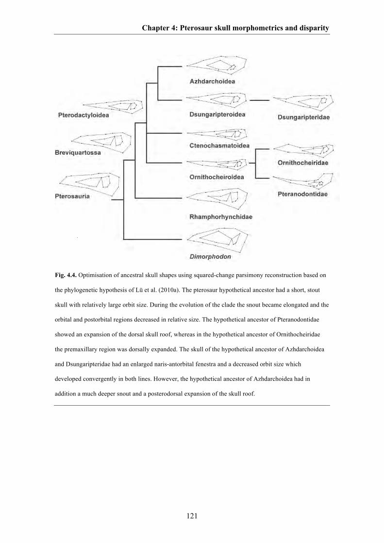
Chapter 4: Pterosaur skull morphometrics and disparity
121
Fig. 4.4. Optimisation of ancestral skull shapes using squared-change parsimony reconstruction based on
the phylogenetic hypothesis of Lü et al. (2010a). The pterosaur hypothetical ancestor had a short, stout
skull with relatively large orbit size. During the evolution of the clade the snout became elongated and the
orbital and postorbital regions decreased in relative size. The hypothetical ancestor of Pteranodontidae
showed an expansion of the dorsal skull roof, whereas in the hypothetical ancestor of Ornithocheiridae
the premaxillary region was dorsally expanded. The skull of the hypothetical ancestor of Azhdarchoidea
and Dsungaripteridae had an enlarged naris-antorbital fenestra and a decreased orbit size which
developed convergently in both lines. However, the hypothetical ancestor of Azhdarchoidea had in
addition a much deeper snout and a posterodorsal expansion of the skull roof.
Chapter 4: Pterosaur skull morphometrics and disparity
122
DISCUSSION
MAJOR PATTERNS IN PTEROSAUR CRANIAL SHAPE
Most pterosaurs bunch fairly close together in the cranial morphospace, in the vicinity
of the origin. The taxa that are most broadly separated are: Anurognathus due to its
extremely short rostrum, large orbit and postorbital region; Pterodaustro due its
extreme elongated rostrum; Thalassodromeus probably due to its enlarged premaxillary
crest (which may be partly captured with our landmark construction approach); and
pteranodontids probably due to their enlarged frontal crests (which may be partly
captured as well: see Fig. 4.2A). The general bunching of the majority of taxa means
that skull shape as captured by the chosen landmarks is, in general terms, fairly uniform
among pterosaurs. It seems, therefore, that despite their long evolutionary history and
great diversity of body size, pterosaurs exhibited a restricted range of skull shape. Most
variation occurs in the length of the snout and size of the postorbital region (PC 1). The
former is probably related with feeding ecology (Witton & Naish 2008; Ősi 2010). A
comparison of the different PC axes demonstrates further that variation in the length of
the snout (PC 1) is greater than variation in the depth of the snout (PC 3). Within the
postorbital region, variation in the shape and size of the skull roof (PC 2) is probably
not strongly linked to the position of the jaw joint (PC 3), as the two are summarised by
different PC axes. The depth of the snout tip (PC 4) seems to be independent from
variation in snout length (PC1) and the depth of the snout (PC 3) in general.
It is important to remember that because we did not include landmarks relative
to entire shape of the cranial crests (see the diversity of crest types in e.g. Nyctosaurus,
Tapejara, Tupuxuara, Pteranodontidae), it is likely that a morphospace based on a
dataset including such landmarks would appear quite different. In particular, because
Chapter 4: Pterosaur skull morphometrics and disparity
123
there is such a diversity of crest morphologies among pterosaurs we suspect that the
first PC axes (and perhaps the first several PC axes) would largely characterise features
relating to the crest. Furthermore, it is likely that there would be a greater spread of taxa
in morphospace (i.e., taxa would not be so closely bunched together, which would
indicate more total morphological diversity). As we are primarily focused on the shape
of the snout and postorbital regions (regions that are more likely to have mechanical
significance), we leave landmark or outlined-based analyses of pterosaur crest shape to
future studies. In particular, such studies may provide evidence relevant to long-
standing debate over the function of cranial crests in pterosaurs, especially the question
of whether they may have provided aerodynamic utility during flight (Frey et al. 2003;
Elgin et al. 2008) or were mostly, or entirely, display structures (Bennett 2003; Martill
& Naish 2006; Elgin et al. 2008; Tomkins et al. 2010; Hone et al. 2012).
TEMPORAL AND TAXONOMIC DISPARITY
The peak disparity observed for all four metrics in the Early Cretaceous is consistent
with other studies that also suggest peak disparity in this time interval, based on
measurements and discrete character data (Dyke et al. 2009; Prentice et al. 2011; Butler
et al. 2012). However, although the Early Cretaceous pterosaur sample is significantly
or nearly significantly more disparate than that of the Late Triassic-Early Jurassic for
three of the disparity metrics, it is not significantly different to that of the Middle
Jurassic-Late Jurassic or the Late Cretaceous. Therefore, there is currently not enough
evidence to conclusively identify true variation in cranial disparity through time (a
problem that may be caused by the low sample size of our study, as there are over 100
additional pterosaurs that cannot be included because they are not known from adequate
skull material). With that said, rarefaction results do support the existence of a disparity
Chapter 4: Pterosaur skull morphometrics and disparity
124
peak during the Early Cretaceous, occurring at all sample sizes and in all metrics,
including variance-based metrics, which have been demonstrated to be less susceptible
that range-based metrics to sampling biases (e.g. Wills et al. 1994; Butler et al. 2012). If
accurate, this disparity peak coincides with the major Early Cretaceous radiation of
monofenestratan pterosaurs, and is unusual in occurring relatively late in pterosaur
evolutionary history (most clades show disparity peaks early in their evolutionary
history e.g. see Erwin 2007).
Our taxonomic disparity results are similar to those obtained by Prentice et al.
(2011) using a discrete character dataset that includes information from the entire
pterosaur skeleton. Our results follow those of Prentice et al. (2011) in recovering
Monofenestrata (approximately equivalent to Pterodactyloidea of their analysis) as
significantly more disparate than non-monofenestratan pterosaurs
(=“Rhamphorhynchoidea” in their analysis), but only when the highly divergent
Anurognathidae are excluded from the non-monofenestratan sample. Because this result
is recovered using sum of variances it seems plausible that it is not simply related to
differences in sample sizes (Wills et al. 1994; Butler et al. 2012), despite the fact that
our sampling of non-monofenestratans is substantially smaller than that of
monofenestratans. Prentice et al. (2011) also found disparity to be lower for
Dsungaripteroidea than other pterosaur ‘superfamilies’, but not significantly so.
Moreover, our results also recover higher disparity for Azhdarchoidea and
Ornithocheiroidea compared to Ctenochasmatoidea (although not significantly so, so it
is possible that these differences relate to taxonomic sampling) when the controversial
taxon Feilongus is assigned to Ornithocheiroidea. Prentice et al. (2011) found no
significant difference in disparity between Ornithocheiroidea and Ctenochasmatoidea
Chapter 4: Pterosaur skull morphometrics and disparity
125
when Feilongus was assigned to Ctenochasmatoidea, but a significant difference
(higher disparity in Ornithocheiroidea) when it was assigned to Ornithocheiroidea.
DIFFERENT DISPARITY PROXIES: A COMMON THEME
As summarised above, our disparity results (both temporal and taxonomic) based upon
cranial morphometrics broadly agree with those based upon discrete characters from the
entire skeleton (Prentice et al. 2011; Butler et al. 2012) and on limb measurements and
ratios (Dyke et al. 2009). This is an encouraging finding, as it demonstrates that
different proxies for morphological form in pterosaurs, a group with complex three-
dimensional skeletons that are difficult to represent in disparity studies, converge on the
same disparity signal. A similar finding was reported by Villier & Eble (2004) in a
study of echinoid morphological evolution, as several proxies for morphological form
(e.g. discrete characters, landmark morphometrics, traditional measurement
morphometrics) produced generally congruent temporal disparity curves for the clade.
Whether this is also true of disparity studies of other groups deserves further study. At
the very least, the echinoid and pterosaur data show that, for exemplar non-vertebrate
(echinodermatan) and vertebrate clades, disparity can be consistently measured based
on several different proxies. This justifies the use of discrete characters [including those
generated for phylogenetic analyses, as were the characters used by Prentice et al.
(2011) and Butler et al. (2012)] when morphometric data may not be available, or vice
versa.
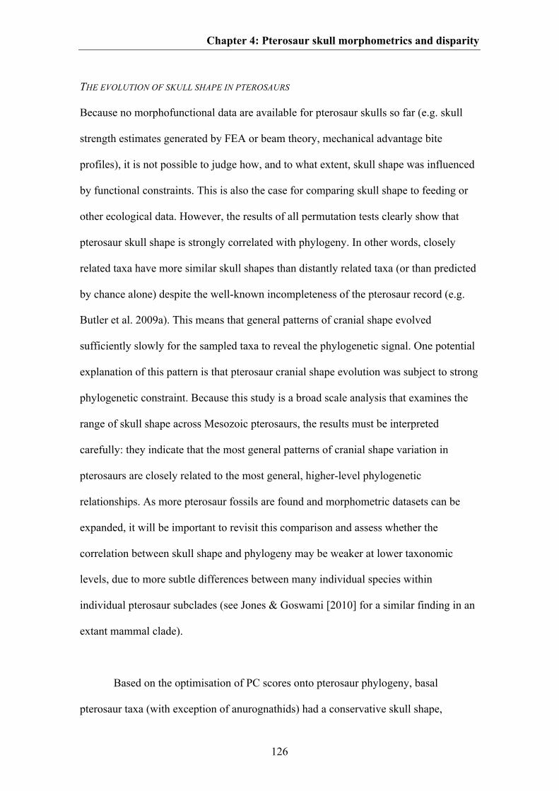
Chapter 4: Pterosaur skull morphometrics and disparity
126
THE EVOLUTION OF SKULL SHAPE IN PTEROSAURS
Because no morphofunctional data are available for pterosaur skulls so far (e.g. skull
strength estimates generated by FEA or beam theory, mechanical advantage bite
profiles), it is not possible to judge how, and to what extent, skull shape was influenced
by functional constraints. This is also the case for comparing skull shape to feeding or
other ecological data. However, the results of all permutation tests clearly show that
pterosaur skull shape is strongly correlated with phylogeny. In other words, closely
related taxa have more similar skull shapes than distantly related taxa (or than predicted
by chance alone) despite the well-known incompleteness of the pterosaur record (e.g.
Butler et al. 2009a). This means that general patterns of cranial shape evolved
sufficiently slowly for the sampled taxa to reveal the phylogenetic signal. One potential
explanation of this pattern is that pterosaur cranial shape evolution was subject to strong
phylogenetic constraint. Because this study is a broad scale analysis that examines the
range of skull shape across Mesozoic pterosaurs, the results must be interpreted
carefully: they indicate that the most general patterns of cranial shape variation in
pterosaurs are closely related to the most general, higher-level phylogenetic
relationships. As more pterosaur fossils are found and morphometric datasets can be
expanded, it will be important to revisit this comparison and assess whether the
correlation between skull shape and phylogeny may be weaker at lower taxonomic
levels, due to more subtle differences between many individual species within
individual pterosaur subclades (see Jones & Goswami [2010] for a similar finding in an
extant mammal clade).
Based on the optimisation of PC scores onto pterosaur phylogeny, basal
pterosaur taxa (with exception of anurognathids) had a conservative skull shape,
Chapter 4: Pterosaur skull morphometrics and disparity
127
highlighted by a short snout, a triangular nares-antorbital fenestra region, a large orbit
and a large postorbital region. However, in the clade Monofenestrata snouts became
extremely elongated relative to the rest of the skull, leading to a relatively small orbit
and postorbital region. The shape of the nares-antorbital fenestra region thus became
trapezoidal. We interpret this shift of shape as related to the fusion of the nares and the
antorbital fenestra into a single large opening, which occurred in this clade. Because
these events apparently occur simultaneously, it is possible that the fusion of the
preorbital skull openings could be functionally linked to elongation of the snout.
However, due to the lack of FEA and other biomechanical data it is not possible to test
this hypothesis at present. The skull shape of the hypothetical ancestors of
Ornithocheiroidea, Dsungaripteroidea and Ctenochasmatoidea more-or-less resembles
that of the hypothetical ancestor of Pterodactyloidea, whereas the ancestors of
Dsungaripteridae, Pteranodontidae and Azhdarchoidea show more variation in shape
compared to the hypothetical ancestor of Pterodactyloidea due to their development of
different kinds of cranial crests.
CONCLUSIONS
In summary, our morphometric analysis of pterosaur skull shape (31 taxa, 21
landmarks) identifies the greatest sources of cranial variation within the clade and
documents morphological changes occurring during pterosaur evolution. Pterosaur skull
shape was apparently subject to strong and statistically significant phylogenetic
constraint, with closely related taxa having more similar skull shapes than distantly
related taxa (or than predicted by chance alone). Cranial disparity of Pterosauria may
have reached a peak in the Early Cretaceous, which occurs relatively late in the clade’s
evolutionary history. With the exclusion of the highly aberrant anurognathids, basal
Chapter 4: Pterosaur skull morphometrics and disparity
128
non-monofenestratan pterosaurs appear to have been less disparate cranially than
monofenestratan pterosaurs, consistent with their apparently more restricted range of
ecological adaptations and body sizes. From a broader evolutionary and methodological
standpoint, one of the most salient results of our study is that temporal and taxonomic
patterns in cranial shape disparity closely match disparity patterns generated from
discrete characters and limb measurements. Therefore, based on a well-studied
exemplar clade (pterosaurs), different sources of morphological data give broadly
congruent disparity results. This is a promising finding because often only one such data
source is available for extinct clades represented by fossils, which are often poorly and
incompletely preserved.
ACKNOWLEDGEMENTS
We thank Gareth Dyke and Michel Laurin for their helpful reviews and Brian Andres,
Roger Benson and Oliver Rauhut for discussion. RJB was funded by the DFG Emmy
Noether Programme (BU 2587/3-1), CF by the DFG (RA 1012/12-1) and SJB by a NSF
Graduate Research Fellowship, Doctoral Dissertation Improvement Grant (DEB
1110357) and the American Museum of Natural History and Columbia University.
129
CHAPTER 5
Macroevolutionary and morphofunctional patterns in theropod skulls: a morphometric
approach
Keywords:
Theropoda; geometric morphometrics; Mesozoic; macroevolution; feeding ecology;
biomechanics; avian evolution
The chapter was published as:
Foth C1,2 & Rauhut OWM1,2,3. 2013. Macroevolutionary and morphofunctional patterns
in theropod skulls: a morphometric approach. Acta Palaeontologica Polonica 58: 1-16.
1Bayerische Staatssammlung für Paläontologie und Geologie, Richard-Wagner-Str. 10, D-80333 München, Germany 2Department of Earth and Environmental Sciences, Ludwig-Maximilians-University, Richard-Wagner-Str. 10, D-80333 Münchrn, Germany 3GeoBioCenter, Ludwig-Maximilians-University; Richard-Wagner-Str. 10, D-80333 München, Germany
Received 16 December 2011, accepted 19 April 2012, available online 20 April 2012.
Author contribution:
Research design: Christian Foth, Oliver Rauhut
Data collection: Christian Foth
Data analyses: Christian Foth
Preparation of figures and tables: Christian Foth
Wrote paper: Christian Foth, Oliver Rauhut
*All figures of Chapter 5 are modified after Foth & Rauhut (2013)
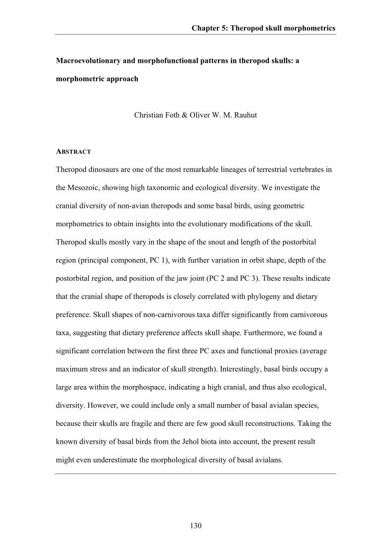
Chapter 5: Theropod skull morphometrics
130
Macroevolutionary and morphofunctional patterns in theropod skulls: a
morphometric approach
Christian Foth & Oliver W. M. Rauhut
ABSTRACT
Theropod dinosaurs are one of the most remarkable lineages of terrestrial vertebrates in
the Mesozoic, showing high taxonomic and ecological diversity. We investigate the
cranial diversity of non-avian theropods and some basal birds, using geometric
morphometrics to obtain insights into the evolutionary modifications of the skull.
Theropod skulls mostly vary in the shape of the snout and length of the postorbital
region (principal component, PC 1), with further variation in orbit shape, depth of the
postorbital region, and position of the jaw joint (PC 2 and PC 3). These results indicate
that the cranial shape of theropods is closely correlated with phylogeny and dietary
preference. Skull shapes of non-carnivorous taxa differ significantly from carnivorous
taxa, suggesting that dietary preference affects skull shape. Furthermore, we found a
significant correlation between the first three PC axes and functional proxies (average
maximum stress and an indicator of skull strength). Interestingly, basal birds occupy a
large area within the morphospace, indicating a high cranial, and thus also ecological,
diversity. However, we could include only a small number of basal avialan species,
because their skulls are fragile and there are few good skull reconstructions. Taking the
known diversity of basal birds from the Jehol biota into account, the present result
might even underestimate the morphological diversity of basal avialans.
Chapter 5: Theropod skull morphometrics
131
INTRODUCTION
Theropod dinosaurs were one of the most remarkable lineages of terrestrial vertebrates
in the Mesozoic Era. They attained a high level of taxonomic and ecological diversity
(Weishampel et al. 2004), and represent the only dinosaur clade that survived the mass
extinction event at the end of the Cretaceous, in the form of birds (Dingus & Rowe
1997). Mesozoic theropod species occupied the mass spectrum from a few hundred
grams to more than six tonnes (Christiansen & Fariña 2004; Turner et al. 2007), and
showed a huge diversity in skull morphologies (Fig. 5.1; Weishampel et al. 2004) and
feeding strategies (Barrett 2005; Zanno & Makovicky 2011). Numerous papers have
been published on the phylogenetic relationships of non-avian theropods and basal birds
(e.g. Gauthier 1986; Sereno 1999; Clark et al. 2002a; Rauhut 2003a; Smith et al. 2007;
Choiniere et al. 2010). These analyses largely agree in the general interrelationships of
major groups, but the phylogenetic position and validity of several clades (e.g.
Alvarezsauridae, Ceratosauria, Compsognathidae, Therizinosauridae) and the detailed
positions of many species are still controversial (see Rauhut 2003a; Choiniere et al.
2010; Zanno 2010; Xu et al. 2011). In contrast to this rather high number of
phylogenetic analyses, relatively few studies have investigated the morphofunctional
evolution of theropod character complexes, or have addressed macroevolutionary
questions, such as the importance of heterochrony or biomechanical constraints in
theropod evolution. Those studies that have addressed such questions have
overwhelmingly concentrated on the evolution of the limbs (e.g. Gatesy 1990; Wagner
& Gauthier 1999; Middleton & Gatesy 2000; Hutchinson 2001a, b; Dececchi & Larsson
2011), growth patterns as indicated by bone histology (e.g. Erickson et al. 2001, 2009;
Padian et al. 2001), body size, breathing and physiology (e.g. Schweitzer & Marshall
2001; Tuner et al. 2007; Benson et al. 2012) or, most recently, the evolution of feathers
Chapter 5: Theropod skull morphometrics
132
(e.g. Xu & Guo 2009) and the variety of diets (Barrett et al. 2011; Zanno & Makovicky
2011). However, apart from the works of Rayfield (2005, 2011), Barrett (2005), Barrett
& Rayfield (2006), Sakamoto (2010), Zanno & Makovicky (2011), and Brusatte et al.
(2012a), the relationships between cranial diversity, functional constraints, diet, and
evolutionary processes have received surprisingly little attention so far.
Fig. 5.1. Diversity of skull shapes in theropod dinosaurs. A. basal theropod Herrerasaurus; B.
coelophysid Syntarsus; C. generalized spinosaurid; D. abelisaurid Carnotaurus; E. allosaurid Allosaurus;
F. ornithomimosaur Ornithomimus; G. unnamed oviraptorid (originally referred to Oviraptor); H.
dromaeosaurid Velociraptor; I. basal bird (avialian) Archaeopteryx. Scale bars represent 10 mm (B, I), 50
mm (a, f-h) and 100 (c-e). Modified from Rauhut (2003a).
In recent years, geometric morphometrics has been used increasingly in
palaeontology. The most comprehensive study focusing on the morphometrics of
archosaurian skulls is Marugán-Lobón & Buscalioni (2003), though this investigation
Chapter 5: Theropod skull morphometrics
133
was based on simple distance measurements for three homologous units of the skull
(braincase, orbit and rostrum). Geometric morphometric studies have often been carried
out for ornithischian dinosaurs (Chapman et al. 1981; Chapman 1990; Chapman &
Brett-Surman 1990; Goodwin 1990; Dodson 1993), where they were mainly used for
taxonomic purposes. Further studies deal with geometric morphometrics of the skulls of
sauropods (Young & Larvan 2010) and of single or small numbers of taxa of theropods,
such as Allosaurus and Tyrannosaurus (Chapman 1990), and Carnotaurus and
Ceratosaurus (Mazzetta et al. 2000), as well as with isolated theropod teeth (D’Amore
2009). More comprehensive analyses were published by Marugán-Lobón & Buscalioni
(2004, 2006) for extant birds. Recently, Brusatte et al. (2012a) investigated the cranial
diversity of non-avian theropods, using two datasets (small data set: 26 taxa and 24
landmarks; large data set: 36 taxa and 13 landmarks). These authors concluded that
cranial shape of theropods is highly correlated with phylogeny, but only weakly with
functional biting behaviour and thus that phylogeny was the major determinant of
theropod skull shape. Their result challenges previous studies, which suggested marked
functional constraints on the evolution of theropod skull shape (Henderson 2002;
Rayfield 2005).
The goal of this study, which was initiated in parallel with that of Brusatte et al.
(2012a), is therefore to evaluate theropod cranial diversity and its relation to phylogeny,
ecology and function, using geometric morphometrics. We used an independent
subsample of taxa and a different combination of landmarks, as well as different proxies
for cranial function. Thus, this study helps to test the results obtained by Brusatte et al.
(2012a), using a different set of data. Further, we investigated how shape variation of
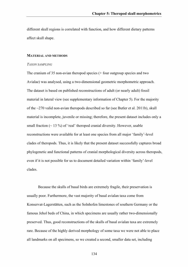
Chapter 5: Theropod skull morphometrics
134
different skull regions is correlated with function, and how different dietary patterns
affect skull shape.
MATERIAL AND METHODS
TAXON SAMPLING
The cranium of 35 non-avian theropod species (+ four outgroup species and two
Avialae) was analysed, using a two-dimensional geometric morphometric approach.
The dataset is based on published reconstructions of adult (or nearly adult) fossil
material in lateral view (see supplementary information of Chapter 5). For the majority
of the ~270 valid non-avian theropods described so far (see Butler et al. 2011b), skull
material is incomplete, juvenile or missing; therefore, the present dataset includes only a
small fraction (~ 13 %) of ‘real’ theropod cranial diversity. However, usable
reconstructions were available for at least one species from all major ‘family’-level
clades of theropods. Thus, it is likely that the present dataset successfully captures broad
phylogenetic and functional patterns of cranial morphological diversity across theropods,
even if it is not possible for us to document detailed variation within ‘family’-level
clades.
Because the skulls of basal birds are extremely fragile, their preservation is
usually poor. Furthermore, the vast majority of basal avialan taxa come from
Konservat-Lagerstätten, such as the Solnhofen limestones of southern Germany or the
famous Jehol beds of China, in which specimens are usually rather two-dimensionally
preserved. Thus, good reconstructions of the skulls of basal avialan taxa are extremely
rare. Because of the highly derived morphology of some taxa we were not able to place
all landmarks on all specimens, so we created a second, smaller data set, including
Chapter 5: Theropod skull morphometrics
135
Pengornis and Shenquiornis. In addition to the paravian taxa from the larger data set,
we included only a few non-paravian coelurosaurs (Compsognathus, Dilong,
Ornithomimus, Erlikosaurus, Shuvuuia and Conchoraptor) as outgroup taxa. We were
able to include the alvarezsaurid Shuvuuia only in the small dataset because of its bird-
like skull shape, which includes the loss of the postorbital process of the jugal. All taxa
are listed in table S5.1 (see supplementary information of Chapter 5).
GEOMETRIC MORPHOMETRICS
Geometric morphometric approaches are commonly used to quantify and study
interspecific or intraspecific shape variation across a number of specimens based on
outline or landmark data that capture the shape of the specimens in question (Adams et
al. 2004; Zelditch et al. 2004). The advantage of this approach is that landmark
coordinates capture more information about shape than can be obtained from traditional
morphometric measurements (linear distances, ratios and angular measures), which are
often insufficient to capture the whole geometry of the original object (Hammer &
Harper 2006). Geometric morphometric approaches have been reviewed and discussed
by many authors including Bookstein (1991), Elewa (2004), Zelditch et al. (2004), and
Adam et al. (2004).
In the large data set, cranial geometry was captured using 20 homologous
landmarks, which were plotted on the reconstructed skulls using the program tpsDig2
(Rohlf 2005), which outputs a tps (thin plate spline) file with two-dimensional landmark
coordinates and scale (size) data for each specimen. The chosen landmarks are of type 1
(good evidence for anatomical homology, such as points where two bone sutures meet)
and type 2 (good evidence for geometric homology, such as points of maximal
Chapter 5: Theropod skull morphometrics
136
curvature or extremities) in the terminology of Bookstein (1991). The landmark dataset
includes the outer shape of the whole cranium (excluding nasal crests or horns on the
skull roof), maxilla, antorbital fenestra, orbit, lateral temporal fenestra, jugal,
quadratojugal, postorbital and the posterior part of the skull roof (parietal and
squamosal). For comparison, the dataset shares eleven landmarks (55 %) with the 26-
taxon dataset of Brusatte et al. (2012a) and only five (25 %) with the 36-taxon data set.
In the smaller dataset we used only 15 landmarks, owing to the fusion or loss of various
skull elements in some taxa (Fig. 5.2, see supplementary information of Chapter 5 for
the anatomical description of the landmarks).
Fig. 5.2. Position of the landmarks on theropod skulls. A. 20 landmarks used for the large dataset plotted
on the skull of Ceratosaurus (modified from Sampson & Witmer 2007). B. 15 landmarks used for the
small dataset plotted on the skull of Anchiornis (modified from Hu et al. 2009). The description of
landmark positions is given in the supplementary information of Chapter 5.
Chapter 5: Theropod skull morphometrics
137
The landmark coordinates were superimposed using Generalized Procrustes
Analyses (GPA) in tpsRelW (Rohlf 2003) and PAST 2.09 (Hammer et al. 2001), which
serves to minimize non-shape variation between species, such as that caused by size,
location, orientation, and rotation (Zelditch et al. 2004). Afterwards, the Procrustes
coordinates were subjected to Principal Component Analysis (PCA) using PAST and
tpsRelW. This procedure assimilates data from all landmarks and reduces it to a set of
PC scores that summarize the skull shape of each taxon and describe maximal shape
variation in a morphospace (Hammer & Harper 2006).
CHARACTER EVOLUTION
One main question is how far is theropod skull shape correlated with phylogeny? This is
important for reconstructing skull shape changes using character-mapping approaches.
If a strong phylogenetic signal is present, closely related taxa should occur closer to one
another in morphospace than more distantly related taxa (Klingenberg & Gidaszewski,
2010). The degree of this correlation can be calculated by mapping the phylogeny into
the morphospace. This requires an ancestral state reconstruction of the morphometric
data for each internal node on the tree using squared change parsimony (Maddison 1991;
Klingenberg 2011). This algorithm collates the sum of squared changes of continuous
characters (here PC scores) along all branches of the tree and calculates parsimonious
ancestral states by minimizing the total sum of squared change across the phylogeny.
The most optimal configuration of ancestral PC scores is mathematically described by a
squared length value, which can be used to determine the strength of the correlation
between shape and phylogeny (see below).
Chapter 5: Theropod skull morphometrics
138
For this approach an informal supertree (sensu Butler & Goswami 2008) was
created. The tree is based on several of the most recent phylogenetic analyses of
theropods: basal theropods, including coelophysoids (Sues et al. 2011), Ceratosauria
(sensu Rauhut 2003a; Smith et al. 2007; Xu et al. 2009a), basal tetanurans (Benson et al.
2010), Coelurosauria (Hu et al. 2009), Tyrannosauroidea (Brusatte et al. 2010a), and
Dromaeosauridae (Csiki et al. 2010). In general, most trees show strong similarities
with regard to the higher-level relationships of theropod dinosaurs, but may disagree on
the positions of individual lineages. Euparkeria, Lesothosaurus, Massospondylus and
Plateosaurus were taken as outgroup taxa (Fig. S5.3, see supplementary information of
Chapter 5; the topology showing the interrelationships of the coelurosaurian taxa used
in the small dataset is shown in Fig. S5.4). As discussed by Hunt & Carrano (2010),
models of phenotypic evolution require information about time. To include this
information, branch lengths were scaled to the present topology, using stratigraphic ages
of taxa obtained from Weishampel et al. (2004) or from original literature (Table S5.1,
see supplementary information of Chapter 5).
In the next step, the original landmark data from the tps file and supertree were
imported into MorphoJ (Klingenberg 2011). The landmark data were superimposed
(GPA) and converted into a covariance matrix and subjected to Principal Component
Analysis (PCA). Subsequently, the tree was mapped into the morphospace (see above).
Furthermore, a permutation test was performed in MorphoJ in which the topology is
held constant and the PC scores for each taxon are randomly permuted across the tree
10 000 times (Laurin 2004; Klingenberg & Gidaszewski 2010). For this approach both
the tps file and the supertree were imported into MorphoJ. If the squared length of the
Chapter 5: Theropod skull morphometrics
139
supertree is less than occurs in at least 95 % of the randomly generated trees then the
phylogenetic signal may be deemed significant.
For a more detailed description of cranial shape changes through time, a similar
character mapping approach was performed using the software package Mesquite 2.72
(Maddison & Maddison 2009). For this approach, a Nexus file containing the PC scores
taken from PAST was produced. The data were used as continuous characters and
mapped on the supertree using squared change parsimony (see above). The shape
changes along the tree were visualised by plotting the ancestral state values in the
morphospace within the visualization window of tpsRelW.
SHAPE VS. FUNCTION
Another main question is whether cranial shape is correlated with skull function? As
proxies for function we used the skull strength indicator (SSI; after Henderson 2002)
and the average maximum stress (after Rayfield 2011) (Table S5.2, see supplementary
information of Chapter 5), which are both size-related parameters. Originally,
Henderson (2002) calculated the skull strength at the longitudinal position of the orbital
midpoint by treating the skull as a cantilevered beam, with the posterior region held
immobile while a vertical force was applied at a point on the ventral edge of the snout.
In the first step, we tested the correlation between SSI and shape using only those taxa
that were also used by Henderson (2002). However, because SSI is strongly correlated
with skull depth in the orbital region (Fig. S5.1, Table S5.2, see supplementary
information of Chapter 5), this distance was measured in order to estimate SSI for all
taxa included in this study. As a second proxy, we used the average maximum stress
(AMS). The estimation of ASM is based on a two-dimensional finite element approach
Chapter 5: Theropod skull morphometrics
140
by calculating the value of maximum stress per element, which was carried out for the
crania of various allosauroids and megalosauroids (Rayfield 2011). For those taxa,
AMS shows a significant correlation with the skull length (see Rayfield 2011). We
therefore used this relationship to estimate AMS for all taxa in this study (Fig. S5.2,
Table S5.2, see supplementary information of Chapter 5).
The estimated values of SSI and AMS were logarithmically transformed to
normalize for data distribution (Freckleton et al. 2002). To evaluate the correlation
between shape and function we performed a two-block partial least squares analysis
(2B-PLS; see Rohlf & Corti 2000) in MorphoJ using the Procrustes coordinates from
the GPA (shape) and both functional proxies (functional coefficients). This method
explores the pattern of covariation between two sets of variables by constructing pairs
of variables that are linear combinations of the variables within each of the two data sets,
and accounts for as much as possible of the covariation between the two original data
sets. The strength of correlation is given by the RV coefficient and a p value, generated
by 10 0000 permutations. Additionally, we divided the landmark dataset into several
modules (preorbital region, postorbital region, antorbital fenestra, orbit, and lateral
temporal fenestra; see supplementary information of Chapter 5), and reran the analyses.
Using this approach, we were able to test how the shape variation of specific skull
regions and skull openings is correlated with functional proxies. If different skull
regions show different degrees of correlation with the functional proxies it is also
possible that shape variations that occur in these regions are independent of each other.
To test this we performed 2B-PLS for the preorbital and postorbital regions, as well as
PLS in one configuration. The difference between both approaches is that the former is
based on a separate Procrustes fit, testing the covariation between the shapes of the parts
Chapter 5: Theropod skull morphometrics
141
of each considered separately, whereas the latter is based on a joint Procrustes fit,
testing the covariation between parts within the context of the structure as a whole (see
Klingenberg 2009).
We performed phylogenetic independent contrast (PICs) analyses on the first
three PC axes and both functional proxies including the small data set, which was based
on the original data from Henderson (2002). First, the correlation of SSI and AMS with
phylogeny was tested, by loading both proxies into Mesquite as continuous characters
and mapping them on the supertree using squared change parsimony. Then, a
permutation test was performed as outlined above. Assuming that a correlation of shape
and function with phylogeny is present, and the terminal scores of both factors are non-
independent, the scores have to be transformed into PICs (Felsenstein 1985). This was
done using the PDTREE package for Mesquite (Midford et al. 2005). This procedure
considers the relationships of species to each other and calculates contrasts that are
statistically independent by assuming that character evolution can be modelled as a
random walk (brownian motion model) and that characters change at a uniform rate per
unit branch length across all branches. To produce ‘standardised’ branch lengths, the
original branch lengths were loge-transformed prior to analysis, as recommended by
Garland et al. (1992). To test if the data fulfil these assumptions the absolute values of
the standardized PICs were plotted against: a) their standard deviations (Garland et al.
1992); b) their estimated nodal values (ancestral PIC values); and c) the corrected age of
their base nodes (Purvis & Rambaut 1995). Finally, the estimated nodal values were
plotted against the corrected node ages. The assumption is justified if no significant
correlation is present in all plots (Garland et al. 1992; Purvis & Rambaut 1995; Midford
et al. 2005). Finally, if all quantities fulfil the four criteria, the resulting contrasts of PC
Chapter 5: Theropod skull morphometrics
142
scores were plotted against the contrasts of SSI and AMS. If any signal is present
between shape and function then a statistical correlation should be detectable. This
relationship was evaluated by calculating the coefficient of determination (R²) and the
corresponding p value.
Because both functional parameters were originally size related, we tested if the
shape variation (after the landmarks were superimposed) is correlated with centroid size
(log-transformed) as a proxy for total size, which is defined as the square root of the
sum of squared differences between landmark coordinates and centroid coordinates for
any dimension. Because original size was previously removed from the data by
performing GPA, a significant correlation between centroid size and shape variation
could indicate an allometric trend (see e.g. Piras et al. 2011). The test was performed for
all Procrustes coordinates and for the first three PC axes.
SHAPE VS. ECOLOGY
To test if feeding ecology is correlated with skull shape we categorized taxa based on
dietary preference and feeding style, using the following characters: character 1,
carnivorous vs. non-carnivorous; character 2, carnivorous vs. omnivorous vs.
herbivorous; and character 3, weak biting vs. medium biting vs. strong biting. The
subdivision of the second character is based on Barrett & Rayfield (2006),
supplemented by data from Zanno & Makovicky (2011). In addition, Euparkeria,
Daemonosaurus and Zupaysaurus were coded as carnivorous, Eoraptor, Archaeopteryx,
Anchiornis, Confuciusornis and oviraptorids as omnivorous and Limusaurus as
herbivorous. The subdivision of the third character is based on Sakamoto (2010) (Table
S5.2, see supplementary information of Chapter 5). The characters were originally
Chapter 5: Theropod skull morphometrics
143
coded as discrete characters. To test the correlation between shape and diet, we
performed 2B-PLS in MorphoJ. Scorings of characters 2 and 3 (see above) were
transformed into covariates via a Principal Coordinates Analysis (PCO) with Euclidean
distances and the transformation exponent c = 2. This analysis was repeated for the
preorbital and postorbital data sets, to test whether dietary patterns have an influence on
the shape variation of different skull regions, and for the small data set, which was
mainly focused on skull variation in basal birds.
To test whether taxa with different dietary preferences (carnivorous vs. non-
carnivorous and carnivory vs. omnivory vs. herbivory) occupied different regions
within the morphospace, we additionally performed a NPMANOVA test
(nonparametric multivariate analysis of variance) in PAST with 10 000 permutations,
Euclidean distances and a Canonical Variate Analyses (CVA), using the first six PC
axes (large data set), which describe over 75 % of the total variance in theropod cranial
shape (all outgroup taxa were excluded from the analysis, to avoid false-positive or
negative signals). The NPMANOVA estimates whether the distribution of the three
groups shows significant differences in morphospace (see Anderson 2001). One of the
strengths of this approach is that it does not assume or require normality of the
multivariate data. The test computes an F statistic and a p value, pointing to a
significant difference between dietary preferences if the F value is high and p value less
than 0.05. The p values were Bonferroni corrected, which set the significance level
lower than the overall significance to avoid false positive signals in a dataset comparing
more than two groups (Hammer & Harper 2006). In a second approach we excluded the
oviraptorid taxa from the data set, to evaluate the influence of their aberrant skull
morphology on previous results.

Chapter 5: Theropod skull morphometrics
144
SHAPE VS. PHYLOGENY, FUNCTION AND ECOLOGY
Finally, as already addressed by Brusatte et al. (2012a), we wanted to evaluate whether
skull shape in theropods is better explained by function, ecology or phylogeny. To
evaluate this, we calculated various agglomerative, hierarchical cluster topologies based
on the Procrustes coordinates, the SSI, ASM, SSI and ASM, as well as feeding ecology.
In the latter case we combined the covariates from the PCO (see above) with SSI and
ASM values, which therefore represent a diet-function cluster. The cluster analyses
(UPGMA and Ward’s method) were performed in PAST. In general, these kinds of
cluster algorithms search for the two most similar objects and join them into a cluster.
Then, the next most similar object is identified and joined and this continues until all
objects are joined into one supercluster (Hammer & Harper 2006). In UPGMA, the
clusters are joined based on the average distance between all members in the two groups,
and in Ward's method the clusters are joined such that increase in within-group variance
is minimized (Hammer 2009). Subsequently, we loaded all topologies into MorphoJ,
mapped them onto the tree, and performed a permutation test. By comparing the
squared length and the p value with that of the supertree topology, we were able to
estimate which of the parameters best explained skull shape, based on a parsimony
criterion. All topologies are shown in the supplementary information of Chapter 5 (Fig.
S5.5-S5.14).
RESULTS
MORPHOSPACE AND EVOLUTIONARY TRENDS IN SKULL SHAPE
The whole dataset (large data set) is summarized by 40 PC axes. Most shape variation
in theropod skulls (including outgroup taxa) is captured by the first three PC axes
(34.4%, 17.1% and 11.4% of total variance), describing over 60% of total variance in all.
Chapter 5: Theropod skull morphometrics
145
The first PC describes the relative length and depth of the snout (premaxilla and maxilla)
and the anteroposterior dimensions of the antorbital fenestra and lateral temporal
fenestra. The second PC describes the angle of the premaxillary body, the relative
length and depth of the anterior extension of the jugal, the dorsoventral dimension of the
antorbital fenestra, the anteroposterior dimension of the orbit, as indicated by the length
of the suborbital body of the jugal, the depth of the postorbital region, and the position
of the jaw joint. The third PC describes the relative length of the posterior extension of
the maxilla, the inclination and depth of the lacrimal and the influence that this has on
the relative size and shape of the antorbital fenestra and orbit, and the relative height of
the quadratojugal. Most taxa plotted on the positive side of the first PC axis and are
equally distributed along the second PC axis. Oviraptorids plot far from the other taxa
because of high negative PC 1 scores, reflecting a shortened snout but enlarged lateral
temporal fenestrae (Fig. 5.3).
The permutation test indicates that theropod cranial form is significantly
correlated with phylogeny (tree length weighted by branch lengths = 0.78293449, p <
0.0001). Based on the ancestral state reconstruction, PC 1 remains almost constant from
the hypothetical ancestor of basal archosauriforms to that of basal deinonychosaurs,
indicating that this component was very uniform and close to the consensus shape (Fig.
5.4A). A significant deviation in this value is seen in the hypothetical ancestor of
Neotheropoda, with a rapid shift to extreme snout elongation in the hypothetical
ancestor of Coelophysidae. Similar deviations, and thus trends of snout elongation, can
be seen in the hypothetical ancestors of Spinosauridae, Tyrannosauroidea,
Ornithomimosauria, and Dromaeosauridae. Opposite deviations are present in the
hypothetical ancestors of ceratosaurs and oviraptorids, which have markedly shorter
Chapter 5: Theropod skull morphometrics
146
snouts; this trend is especially pronounced in oviraptorids. In contrast, according to this
component (PC 1) the skull shape of the hypothetical ancestor of basal birds does not
differ greatly from that of basal deinonychosaurs (Fig. 5.4A). Principal Component 2
(Fig. 5.4B) shows a continuous decrease from the hypothetical ancestor of
Archosauriformes to that of Theropoda, thus demonstrating a trend of relative increase
of the anteroposterior length of the orbit (in relation to orbit height) and decrease of the
dorsoventral depth of the infratemporal fenestra. From the hypothetical ancestor of
theropods to that of neotetanurans the value is relatively constant, but decreases again
from this point to the hypothetical ancestor of deinonychosaurs. However, this value
shows a number of deviations along the main phylogenetic axis of theropods. Whereas
the hypothetical ancestors of Ceratosauria, Tyrannosauroidea, Ornithomimosauria and
Avialae show a further decrease of PC 2, values increase in the hypothetical ancestor of
Abelisauridae, Allosauroidea, Megalosauroidea, and Tyrannosauridae, indicating a shift
to a skull shape with a deep temporal fenestra, a dorsoventrally elongated orbit and a
deep suborbital body of the jugal. In all of these taxa, these changes are related to a
marked increase in body size (Fig. 5.4B). The third PC (Fig. 5.4C) shows a continuous
increase from the hypothetical ancestor of archosauriforms to that of coelurosaurs,
indicating a shift in the orientation of the lacrimal, resulting in an increase of the
relative size of the antorbital fenestra and a decrease of the orbit. From the hypothetical
ancestor of coelurosaurians to that of deinonychosaurs the component decreases only
slightly. However, with the exceptions of the hypothetical ancestors of coelophysids,
basal ceratosaurs and avialans, all other groups show a further increase in their
respective lines. The skull shape of the hypothetical ancestor of Avialae fits with that of
basal Deinonychosauria (Fig. 5.4C).

Chapter 5: Theropod skull morphometrics
147
Fig. 5.3. Two-dimensional morphospace of the theropod skull shape (large data set) with distribution of
the different theropod clades, based on the first two PC axes. The most derived taxa with respect to the
first two components are visualized with relative warps. Shape changes along PC 1 reflect changes in the
length and height of the snout and the anteroposterior dimensions of the lateral temporal fenestra.
Changes along PC 2 reflect the height of the postorbital region, the size of the orbit and the position of the
jaw joint.
The morphospace of the second, smaller dataset is summarized with 13 PC axes.
Here, the first three PC axes describe more than 70% of total variation (PC 1 = 34.6%,
PC 2 = 21.9%, PC 3 = 14.1%). The paravian taxa are well separated from other
coelurosaurian taxa, in which the close paravian outgroup taxa Velociraptor and
Sinornithosaurus plot in the same morphospace as the more basal coelurosaurs
Compsognathus, Dilong and Ornithomimus. This is also true for the alvarezsaurid
Shuvuuia. However, Erlikosaurus and Anchiornis lie closer to Archaeopteryx.
Confuciusornis represents the greatest outlier within Avialae, whereas the skull shape of
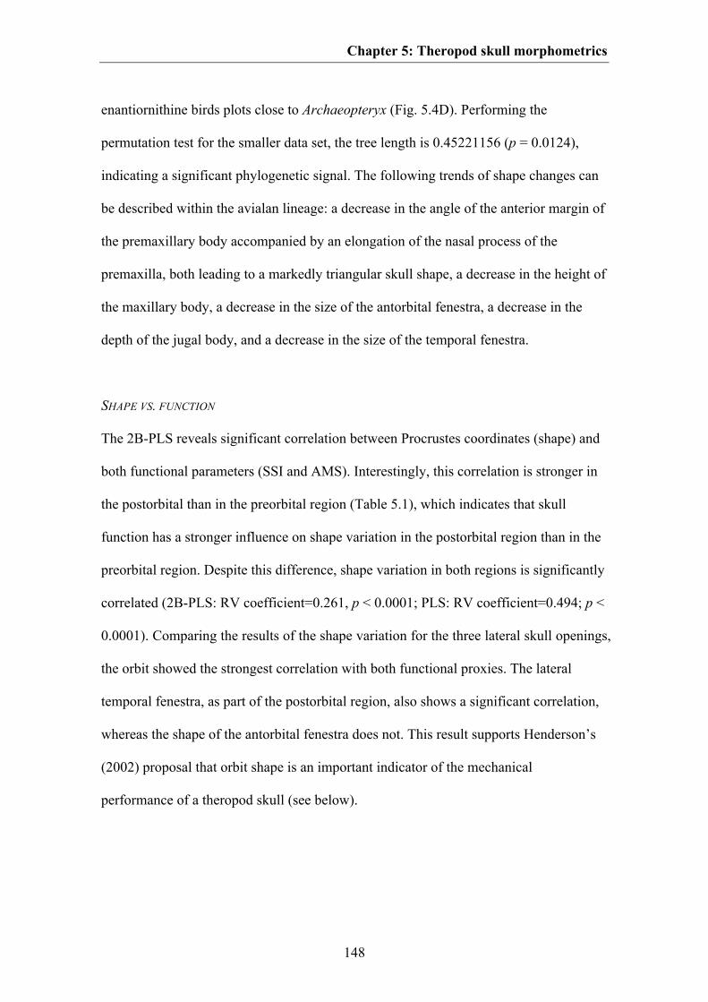
Chapter 5: Theropod skull morphometrics
148
enantiornithine birds plots close to Archaeopteryx (Fig. 5.4D). Performing the
permutation test for the smaller data set, the tree length is 0.45221156 (p = 0.0124),
indicating a significant phylogenetic signal. The following trends of shape changes can
be described within the avialan lineage: a decrease in the angle of the anterior margin of
the premaxillary body accompanied by an elongation of the nasal process of the
premaxilla, both leading to a markedly triangular skull shape, a decrease in the height of
the maxillary body, a decrease in the size of the antorbital fenestra, a decrease in the
depth of the jugal body, and a decrease in the size of the temporal fenestra.
SHAPE VS. FUNCTION
The 2B-PLS reveals significant correlation between Procrustes coordinates (shape) and
both functional parameters (SSI and AMS). Interestingly, this correlation is stronger in
the postorbital than in the preorbital region (Table 5.1), which indicates that skull
function has a stronger influence on shape variation in the postorbital region than in the
preorbital region. Despite this difference, shape variation in both regions is significantly
correlated (2B-PLS: RV coefficient=0.261, p < 0.0001; PLS: RV coefficient=0.494; p <
0.0001). Comparing the results of the shape variation for the three lateral skull openings,
the orbit showed the strongest correlation with both functional proxies. The lateral
temporal fenestra, as part of the postorbital region, also shows a significant correlation,
whereas the shape of the antorbital fenestra does not. This result supports Henderson’s
(2002) proposal that orbit shape is an important indicator of the mechanical
performance of a theropod skull (see below).
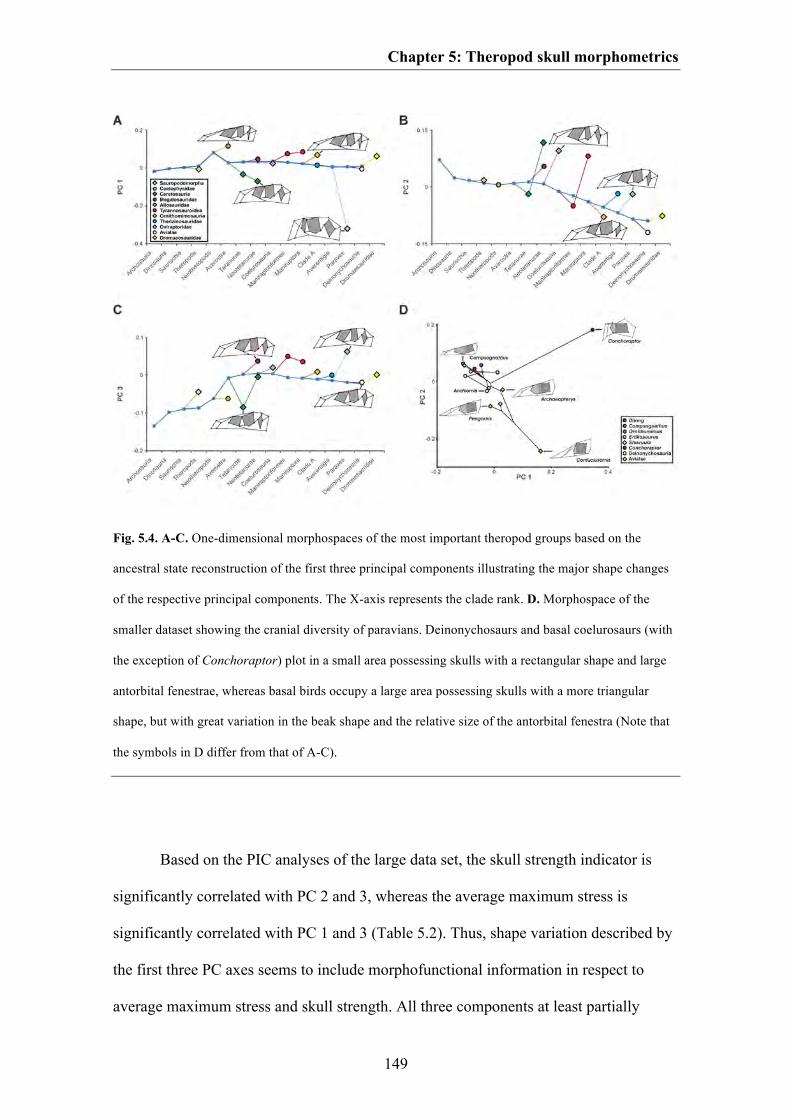
Chapter 5: Theropod skull morphometrics
149
Fig. 5.4. A-C. One-dimensional morphospaces of the most important theropod groups based on the
ancestral state reconstruction of the first three principal components illustrating the major shape changes
of the respective principal components. The X-axis represents the clade rank. D. Morphospace of the
smaller dataset showing the cranial diversity of paravians. Deinonychosaurs and basal coelurosaurs (with
the exception of Conchoraptor) plot in a small area possessing skulls with a rectangular shape and large
antorbital fenestrae, whereas basal birds occupy a large area possessing skulls with a more triangular
shape, but with great variation in the beak shape and the relative size of the antorbital fenestra (Note that
the symbols in D differ from that of A-C).
Based on the PIC analyses of the large data set, the skull strength indicator is
significantly correlated with PC 2 and 3, whereas the average maximum stress is
significantly correlated with PC 1 and 3 (Table 5.2). Thus, shape variation described by
the first three PC axes seems to include morphofunctional information in respect to
average maximum stress and skull strength. All three components at least partially
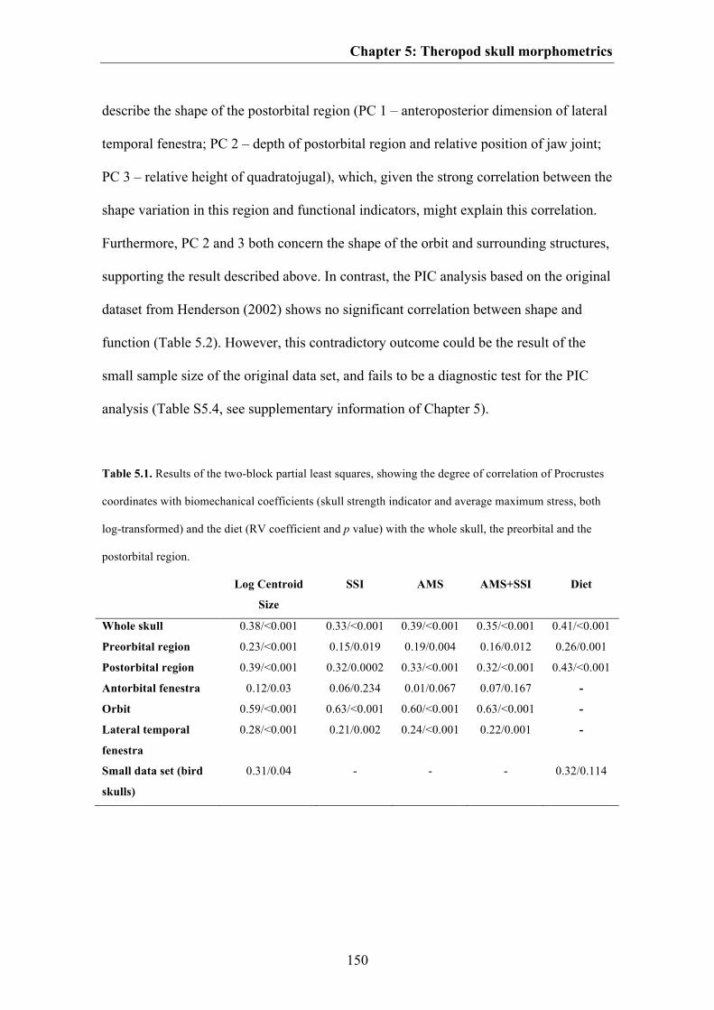
Chapter 5: Theropod skull morphometrics
150
describe the shape of the postorbital region (PC 1 – anteroposterior dimension of lateral
temporal fenestra; PC 2 – depth of postorbital region and relative position of jaw joint;
PC 3 – relative height of quadratojugal), which, given the strong correlation between the
shape variation in this region and functional indicators, might explain this correlation.
Furthermore, PC 2 and 3 both concern the shape of the orbit and surrounding structures,
supporting the result described above. In contrast, the PIC analysis based on the original
dataset from Henderson (2002) shows no significant correlation between shape and
function (Table 5.2). However, this contradictory outcome could be the result of the
small sample size of the original data set, and fails to be a diagnostic test for the PIC
analysis (Table S5.4, see supplementary information of Chapter 5).
Table 5.1. Results of the two-block partial least squares, showing the degree of correlation of Procrustes
coordinates with biomechanical coefficients (skull strength indicator and average maximum stress, both
log-transformed) and the diet (RV coefficient and p value) with the whole skull, the preorbital and the
postorbital region.
Log Centroid
Size
SSI AMS AMS+SSI Diet
Whole skull 0.38/<0.001 0.33/<0.001 0.39/<0.001 0.35/<0.001 0.41/<0.001
Preorbital region 0.23/<0.001 0.15/0.019 0.19/0.004 0.16/0.012 0.26/0.001
Postorbital region 0.39/<0.001 0.32/0.0002 0.33/<0.001 0.32/<0.001 0.43/<0.001
Antorbital fenestra 0.12/0.03 0.06/0.234 0.01/0.067 0.07/0.167 -
Orbit 0.59/<0.001 0.63/<0.001 0.60/<0.001 0.63/<0.001 -
Lateral temporal
fenestra
0.28/<0.001 0.21/0.002 0.24/<0.001 0.22/0.001 -
Small data set (bird
skulls)
0.31/0.04 - - - 0.32/0.114
Chapter 5: Theropod skull morphometrics
151
The overall skull shape represented by the Procrustes coordinates is strongly
correlated with centroid size in the large data set. Furthermore, all three PC axes show a
significant correlation, with the second axis showing the best fit (Table S5.3, see
supplementary information of Chapter 5). This result differs from that of Brusatte et al.
(2012a), where only the second PC axis showed a significant relationship with centroid
size. This indicates that all three PC axes still retain some information on size. A
correlation between centroid size and Procrustes coordinates is also found for the small
data set, thus also indicating a correlation between skull shape and skull size. However,
in contrast to the results for the large data set, there is no significant correlation of
centroid size with each of the first three PC axes.
Table 5.2. Results of the PIC analyses, showing the degree of correlation between PC scores and
biomechanical coefficients (R² and p value). SSI* represents the small dataset which includes only the
original data from Henderson et al. (2002).
R² p value
PC 1 contrasts vs. SSI contrasts 0.011 0.521
PC 2 contrasts vs. SSI contrasts 0.327 <0.001
PC 3 contrasts vs. SSI contrasts 0.341 <0.001
PC 1 contrasts vs. AMS contrasts 0.141 0.015
PC2 contrasts vs. AMS contrasts 0.082 0.070
PC 3 contrasts vs. AMS contrasts 0.154 0.011
PC 1 contrasts vs. SSI* contrasts 0.203 0.122
PC 2 contrasts vs. SSI* contrasts 0.296 0.054
PC 3 contrasts vs. SSI* contrasts 0.069 0.386
SHAPE VS. ECOLOGY
The 2B-PLS analysis reveals a significant correlation between overall skull shape and
dietary parameters. As was the case for the functional proxies, this correlation is
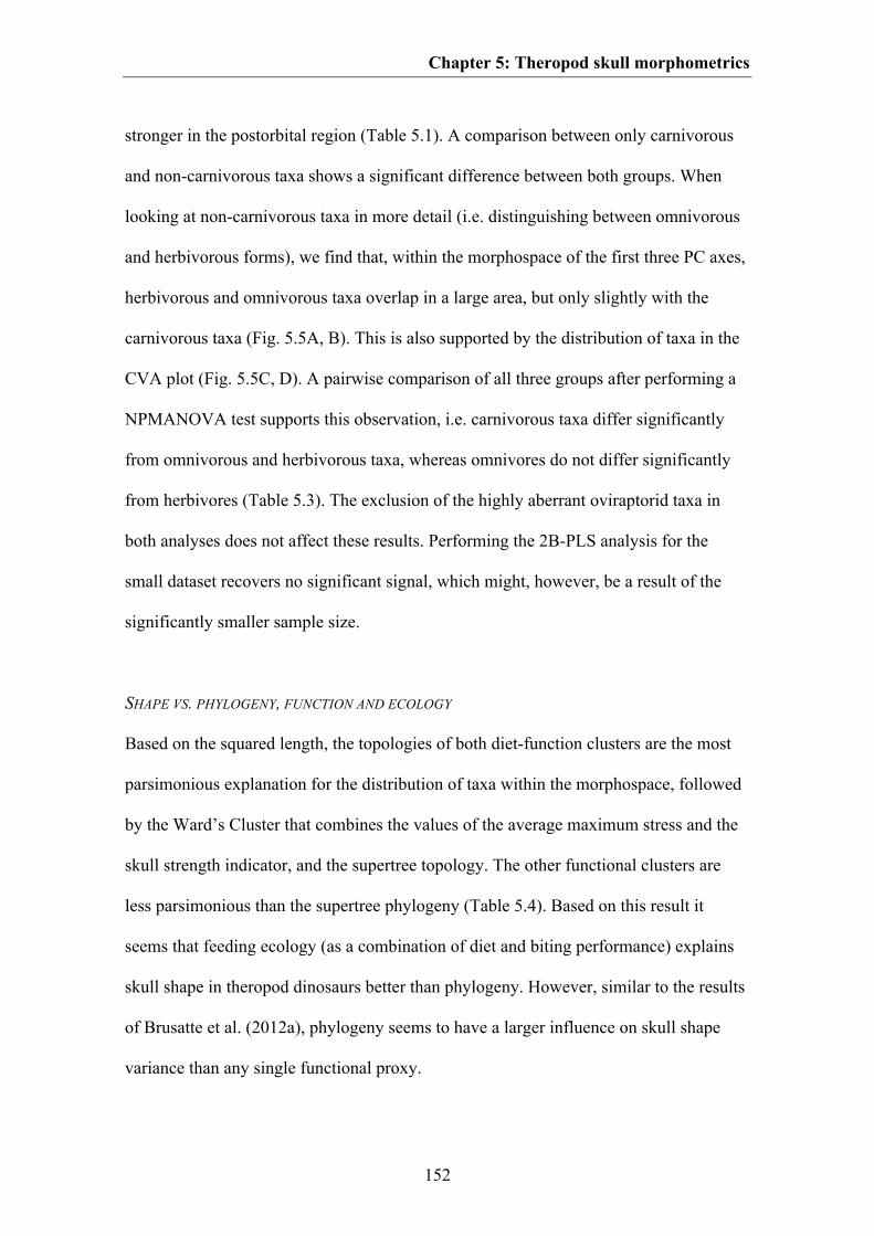
Chapter 5: Theropod skull morphometrics
152
stronger in the postorbital region (Table 5.1). A comparison between only carnivorous
and non-carnivorous taxa shows a significant difference between both groups. When
looking at non-carnivorous taxa in more detail (i.e. distinguishing between omnivorous
and herbivorous forms), we find that, within the morphospace of the first three PC axes,
herbivorous and omnivorous taxa overlap in a large area, but only slightly with the
carnivorous taxa (Fig. 5.5A, B). This is also supported by the distribution of taxa in the
CVA plot (Fig. 5.5C, D). A pairwise comparison of all three groups after performing a
NPMANOVA test supports this observation, i.e. carnivorous taxa differ significantly
from omnivorous and herbivorous taxa, whereas omnivores do not differ significantly
from herbivores (Table 5.3). The exclusion of the highly aberrant oviraptorid taxa in
both analyses does not affect these results. Performing the 2B-PLS analysis for the
small dataset recovers no significant signal, which might, however, be a result of the
significantly smaller sample size.
SHAPE VS. PHYLOGENY, FUNCTION AND ECOLOGY
Based on the squared length, the topologies of both diet-function clusters are the most
parsimonious explanation for the distribution of taxa within the morphospace, followed
by the Ward’s Cluster that combines the values of the average maximum stress and the
skull strength indicator, and the supertree topology. The other functional clusters are
less parsimonious than the supertree phylogeny (Table 5.4). Based on this result it
seems that feeding ecology (as a combination of diet and biting performance) explains
skull shape in theropod dinosaurs better than phylogeny. However, similar to the results
of Brusatte et al. (2012a), phylogeny seems to have a larger influence on skull shape
variance than any single functional proxy.
Chapter 5: Theropod skull morphometrics
153
Fig. 5.5. Two-dimensional morphospace of the theropod skull shape and CVA plot with distribution of
the carnivorous (black triangle), omnivorous (grey squares) and herbivorous (white diamonds) taxa (non-
theropod taxa are not shown). A. PC 1 vs. PC 2. B. PC 2 vs. PC 3. In both diagrams herbivorous and
omnivorous taxa overlap one another strongly, whereas carnivorous taxa overlap only marginally with
herbivorous forms, but moderately with omnivorous forms. C. CVA plot with oviraptorids. D. CVA plot
with oviraptorids excluded.
DISCUSSION
MAJOR PATTERNS IN THEROPOD CRANIAL MORPHOSPACE
Analysing theropod cranial diversity via geometric morphometrics helps to quantify
variation in shape. The data show that snout length and the length of the postorbital
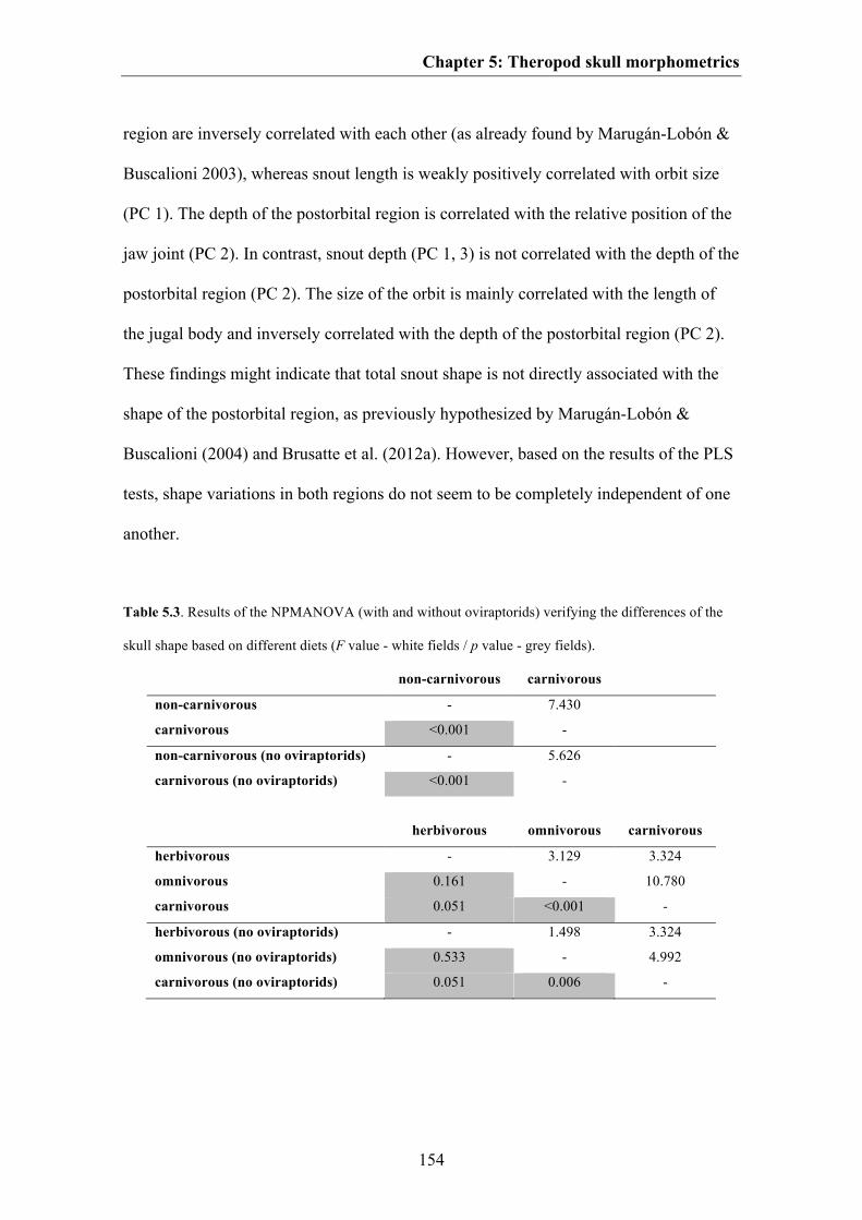
Chapter 5: Theropod skull morphometrics
154
region are inversely correlated with each other (as already found by Marugán-Lobón &
Buscalioni 2003), whereas snout length is weakly positively correlated with orbit size
(PC 1). The depth of the postorbital region is correlated with the relative position of the
jaw joint (PC 2). In contrast, snout depth (PC 1, 3) is not correlated with the depth of the
postorbital region (PC 2). The size of the orbit is mainly correlated with the length of
the jugal body and inversely correlated with the depth of the postorbital region (PC 2).
These findings might indicate that total snout shape is not directly associated with the
shape of the postorbital region, as previously hypothesized by Marugán-Lobón &
Buscalioni (2004) and Brusatte et al. (2012a). However, based on the results of the PLS
tests, shape variations in both regions do not seem to be completely independent of one
another.
Table 5.3. Results of the NPMANOVA (with and without oviraptorids) verifying the differences of the
skull shape based on different diets (F value - white fields / p value - grey fields).
non-carnivorous carnivorous
non-carnivorous - 7.430
carnivorous <0.001 -
non-carnivorous (no oviraptorids) - 5.626
carnivorous (no oviraptorids) <0.001 -
herbivorous omnivorous carnivorous
herbivorous - 3.129 3.324
omnivorous 0.161 - 10.780
carnivorous 0.051 <0.001 -
herbivorous (no oviraptorids) - 1.498 3.324
omnivorous (no oviraptorids) 0.533 - 4.992
carnivorous (no oviraptorids) 0.051 0.006 -
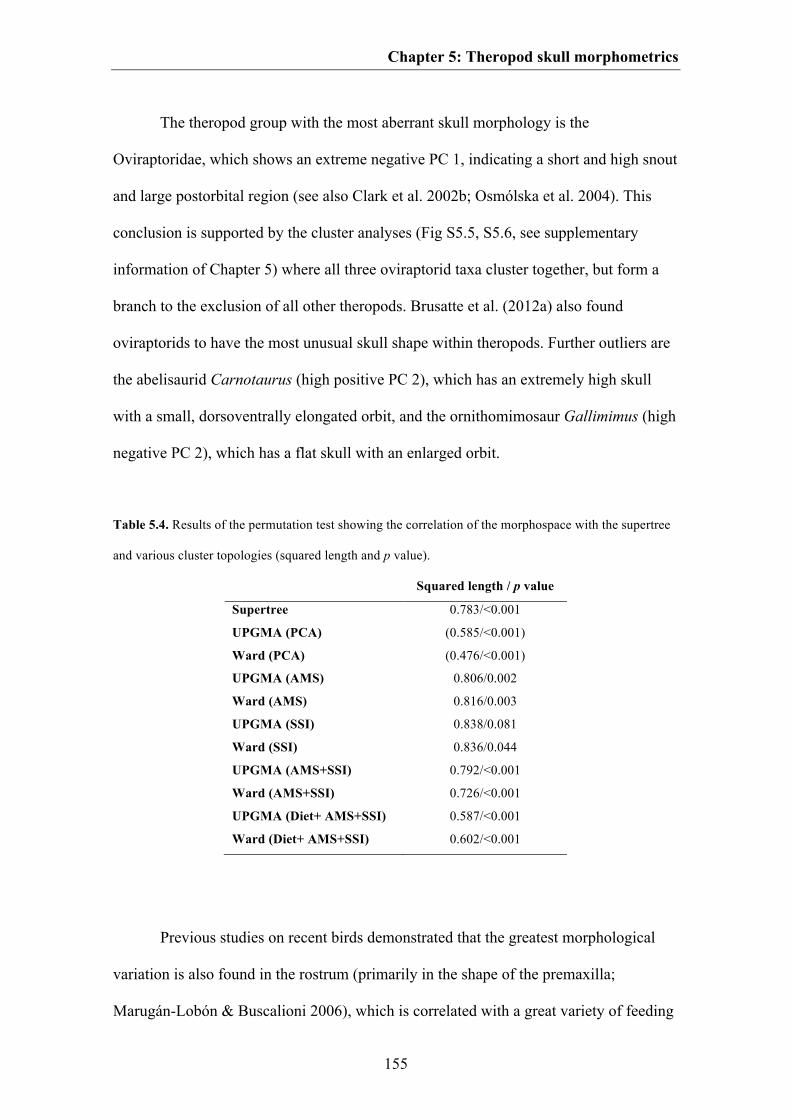
Chapter 5: Theropod skull morphometrics
155
The theropod group with the most aberrant skull morphology is the
Oviraptoridae, which shows an extreme negative PC 1, indicating a short and high snout
and large postorbital region (see also Clark et al. 2002b; Osmólska et al. 2004). This
conclusion is supported by the cluster analyses (Fig S5.5, S5.6, see supplementary
information of Chapter 5) where all three oviraptorid taxa cluster together, but form a
branch to the exclusion of all other theropods. Brusatte et al. (2012a) also found
oviraptorids to have the most unusual skull shape within theropods. Further outliers are
the abelisaurid Carnotaurus (high positive PC 2), which has an extremely high skull
with a small, dorsoventrally elongated orbit, and the ornithomimosaur Gallimimus (high
negative PC 2), which has a flat skull with an enlarged orbit.
Table 5.4. Results of the permutation test showing the correlation of the morphospace with the supertree
and various cluster topologies (squared length and p value).
Squared length / p value
Supertree 0.783/<0.001
UPGMA (PCA) (0.585/<0.001)
Ward (PCA) (0.476/<0.001)
UPGMA (AMS) 0.806/0.002
Ward (AMS) 0.816/0.003
UPGMA (SSI) 0.838/0.081
Ward (SSI) 0.836/0.044
UPGMA (AMS+SSI) 0.792/<0.001
Ward (AMS+SSI) 0.726/<0.001
UPGMA (Diet+ AMS+SSI) 0.587/<0.001
Ward (Diet+ AMS+SSI) 0.602/<0.001
Previous studies on recent birds demonstrated that the greatest morphological
variation is also found in the rostrum (primarily in the shape of the premaxilla;
Marugán-Lobón & Buscalioni 2006), which is correlated with a great variety of feeding
Chapter 5: Theropod skull morphometrics
156
strategies (Zusi 1993; Smith 1993). In birds, the length of the rostrum is independent
from that of the orbital and postorbital region, whereas the orientation of the rostrum
has an influence on the shape of the posterior skull parts (Marugán-Lobón & Buscalioni
2006). However, this morphological variation seems to be controlled by only a small set
of signal molecules (β-catenin, BMP, calmudulin, Dkk3, TGFβIIr; Abzhanov et al. 2004,
2006; Wu et al. 2004, 2006; Mallarino et al. 2011). At this stage, it is of course
speculative to hypothesize similar gene regulatory networks in theropods. However, by
investigating the molecular control of the development of the rostrum shape in different
bird and crocodile groups, it might be possible to create an extant phylogenetic bracket
(sensu Witmer 1995) for cranial development in Archosauria. If it is possible to
correlate different expression patterns of signal molecules with the morphological
variation of the rostrum in birds and crocodiles in a mathematical way (see Johnson &
O’Higgins 1996; Campàs et al. 2010), it might be possible to use geometric
morphometric methods to investigate the genetic control of cranial diversity in fossil
archosaurs (see also Klingenberg 2010).
SHAPE VS. PHYLOGENY
The skull shape of theropods is significantly correlated with higher-level phylogeny in
both data sets. A similar result was found by Brusatte et al. (2012a). However, the
correlation in the smaller dataset was weaker at a lower taxonomic level (see Jones &
Goswami 2010), which could be the result of incomplete sampling.
A problem with this correlation is certainly the highly incomplete sampling of
theropod phylogeny, because of incompleteness of the fossil record, most importantly
the lack of good cranial material for most taxa. Thus, only 13% of known global
Chapter 5: Theropod skull morphometrics
157
theropod diversity could be included in the analyses. Cranial data from Megalosauridae
(e.g. Eustreptospondylus, Torvosaurus), Alvarezsauroidea (included in the small data
set), basal Oviraptorosauria (e.g. Caudipteryx, Incisivosaurus), basal
Therizinosauroidea (e.g. Beipiaosaurus) and some basal birds are missing in the current
analysis. Furthermore, many data are derived from skull reconstructions, the accuracy of
which might be questionable. This problem is exemplified by the spinosaurid skull used,
which is based on a combination of information on skull morphology from three
different taxa (see supplementary information of Chapter 5 and Rauhut 2003a for
details). Additionally, there are large time gaps, especially in the Early and Middle
Jurassic as well as in the late Early Cretaceous to early Late Cretaceous, which might
influence the results of the squared changed parsimony and PIC analyses. The time gaps
mainly concern the basal radiation of the clades Averostra, Abelisauridae,
Tyrannosauridae, Ornithomimosauria, Therizinosauridae, Oviraptoridae, and
Dromaeosauridae.
Interestingly, the skull shape of basal birds does not differ significantly from that
of basal deinonychosaurians, which are represented by Anchiornis. On the one hand this
might mean that the skulls of basal birds and troodontids were very similar, from close
relationship or similar diet preference. Alternatively, this observation might reflect
phylogenetic uncertainty, and it is possible that Anchiornis was instead a basal member
of Avialae, as originally described (see Xu et al. 2009b). This has to be tested in future
phylogenetic analyses, as is the case with the currently published hypothesis that
Anchiornis forms a clade with Archaeopteryx at the base of Deinonychosauria, outside
of Avialae (Xu et al. 2011), in which case the similarities in shape between these two
taxa might represent a low-level taxonomic signal. In contrast, the skull of the
Chapter 5: Theropod skull morphometrics
158
alvarezsaurid Shuvuuia plots within the coelurosaurs, close to Compsognathus, but far
away from the avialan taxa. This is surprising, because the skull of Shuvuuia is
extremely bird-like and possesses several characters otherwise unknown in non-avian
theropods, which was one of the factors that led to the original identification of this
animal as a basal avialian (see e.g. Chiappe et al. 2002). However, in contrast to basal
birds, Shuvuuia possesses an enlarged antorbital fenestra and an extremely elongated
maxillary body, which is similar to basal coelurosaurs. Thus, the different positions of
Shuvuuia and basal birds within the morphospace are mainly based upon the shape of
the snout. However, the shape analysis might lend further support to the hypothesis that
alvarezsaurids are more basal coelurosaurs outside Paraves (Clark et al. 2002a; Novas &
Pol 2002; Choiniere et al. 2010). Within Avialae, the skull shapes of Archaeopteryx,
Confuciusornis and Enantiornithes differ greatly from each other. Mapping the
phylogeny onto the morphospace further demonstrates that the stem species of Avialae,
Pygostylia and Enantiornithes are well separated from each other, indicating a rapid
diversification of skull morphology, probably in connection with the phylogenetic and
ecological radiation of this group in the Early Cretaceous (Zhou & Zhang 2003; You et
al. 2006; Li et al. 2010; O’Connor & Chiappe 2011). This result is further supported by
the cluster analyses carried out for the large dataset, which includes only two avialan
species. However, here both Archaeopteryx and Confuciusornis plot in very different
positions in the morphological clusters, indicating strong dissimilarities in cranial shape
(Fig. S5.5, S5.6, see supplementary information of Chapter 5). Taking the skull
morphology of the long-headed Zhongjianornis and Longipterygidae or the short-
headed Sapeornithidae into account, the actual morphospace of basal birds is probably
much greater than estimated.

Chapter 5: Theropod skull morphometrics
159
SHAPE VS. FUNCTION
As mentioned above, the study presented by Brusatte et al. (2012a) attempted to
correlate geometric morphometric data of the theropod cranium with biting performance,
based on a mechanical advantage approach (see Sakamoto 2010). These authors found a
significant, but weak correlation between shape and function and concluded that
phylogeny was a more important determinant of skull shape than function. By
comparing the RV coefficients of the 2B-PLS presented in both studies, the correlation
between shape and function is about three times higher in the current data set, but the
results of the PIC analyses are almost the same in both studies. The average maximum
stress explains only about 6.6% and the skull strength indicator about 9.5% of total
cranial shape variation in theropods. However, an interesting result of the present study
is that the shape of the postorbital region is better correlated with function than that of
the preorbital region, and that the shape of the orbit shows the strongest correlation,
whereas the shape of the antorbital fenestra shows no significant correlation. These
results are also supported by the correlation of the skull strength indicator with the
second and third PC axes (see PIC analysis), which include aspects that describe the
shape and depth of the orbital and postorbital regions (e.g. shape and size of the orbit,
depth of the suborbital body of the jugal, the position of the jaw joint). These results
support previous morphofunctional studies that demonstrated that the orbital and
postorbital regions of theropod skulls seem to be more important for an understanding
of skull biomechanics than the snout. According to Henderson (2002) there is an inverse
relationship between the size of the theropod orbit and the resistance of the skull to
bending in the sagittal plane, and the narrowness of the orbit shows a positive
correlation with skull strength. Henderson (2002) interpreted the correspondence
between orbit size and shape to relate to increases in the amount of bone comprising the
Chapter 5: Theropod skull morphometrics
160
skull, and so its strength. He furthermore proposed that the shape of the orbit in
theropods with strong skulls is governed by the requirements of the posterior half of the
skull to resist muscle-generated forces associated with prey capture and/or
dismemberment. Furthermore, based on a finite element approach, Rayfield (2005)
demonstrated that high tensile and shear stresses especially affect the jugal bone, the
shape of which in turn influences the anteroposterior dimension of the orbit. According
to Rayfield (2011), the postorbital part of the skull (especially the squamosal, quadrate
and quadratojugal) experiences most of the Von Mises stress associated with biting in
theropods. It is thus to be expected that these stresses result in stronger mechanical
constraints on the postorbital region of the skull than on the preorbital region, which is
confirmed by the findings presented here. Furthermore, since overall stress acting on the
skull (for which the functional data used here are a proxy) is necessarily correlated with
skull size, it is not surprising that the shape of the postorbital region is also correlated
with centroid size (as proxy for skull size). This is also in accordance with the finding of
Rauhut (2007) that phylogenetic characters in the braincases of theropods seem to be
influenced by size. Thus, the results of biomechanical studies (e.g. Rayfield 2011),
phylogenetic data (Rauhut 2007) and morphometric analyses (present study) converge
on the result that the postorbital region of the skull is particularly strongly influenced by
biomechanical forces and related aspects of size. It might be worth noting, however, that
the correlation between functional proxies and the preorbital region, found in both the
2B-PLS and in the PIC analysis (average maximum stress vs. PC 1), indicates that snout
shape is also functionally constrained, but to a weaker degree.
The lack of correlation between the shape of the antorbital fenestra and the
proxies for cranial mechanics used here might be surprising at first glance. Witmer
Chapter 5: Theropod skull morphometrics
161
(1997) suggested that the size and shape of the antorbital fenestra were largely
determined by biomechanical factors, in that bone was resorbed opportunistically by
pneumatic diverticula if it was not biomechanically necessary. Thus, our results seem to
contradict this hypothesis. However, it is necessary to keep in mind that the shape of the
antorbital fenestra is only captured by three landmarks and that the proxies for cranial
function used here are proxies for the overall stress acting on the skull, not for the stress
distribution within the cranium. Thus, although the overall stress experienced by the
skull during biting might be high, this does not contradict the idea that particular parts
of the cranium experience stresses that are below the threshold for the formation of
bone, as has been demonstrated by Finite Element Structural Synthesis for other
dinosaurs (Witzel & Preuschoft 2005; Witzel et al. 2011). In this respect, the hypothesis
that the antorbital fenestra is associated with a region of low stress might even be
supported by the weaker correlation between the shape of the preorbital region with the
functional proxies for overall stress in the cranium, since the latter indicates that there
might be less overall stress acting on this part of the skull (which, in turn, is in
accordance with the results obtained by Rayfield [2011]).
It must be emphasised that the current approach is much more simple than that
used by Brusatte et al. (2012a), and both functional parameters used here are necessarily
strongly correlated with size, since larger taxa are expected to experience higher total
stresses than smaller taxa (see Henderson 2002; Rayfield 2011). In contrast, Brusatte et
al. (2012a) used a metric of functional biting profiles, which are more complex and
independent from size. However, the impact of both the functional parameters (AMS
and SSI) on skull shape is rather small, as previously hypothesized by Brusatte et al.
(2012a). Nevertheless, it would be worthwhile to test the results further by using other
Chapter 5: Theropod skull morphometrics
162
functional parameters or metrics (e.g. finite element analyses) or by using a different
landmark combination, different skull views, or a three-dimensional approach (see also
Brusatte et al. 2012a).
SHAPE VS. ECOLOGY
As is the case with phylogeny and function, dietary patterns are also correlated with
skull shape variation. Interestingly, we found a higher correlation for the postorbital
region than for the preorbital region. This might be regarded as a surprising result,
because one might expect that dietary preferences would have an equal or even stronger
influence on the shape of the snout, the main organ used to obtain and process food.
However, the result probably reflects the generalised subdivision in diet preference
(carnivory vs. omnivory vs. herbivory) and that the dietary patterns also contain
information on biting behaviour (see character 3), and thus biomechanical aspects (see
above). Furthermore, different tooth morphologies, which are not captured in the
current approach, might better reflect more specific dietary patterns than overall shape
of snout (see also Smith 1993; Barrett et al. 2011; Zanno & Makovicky 2011).
Carnivorous, omnivorous and herbivorous theropods occupy large areas of
morphospace. Based on disparity analyses, Brusatte et al. (2012a) demonstrated that
non-carnivorous theropods (i.e. herbivores and omnivores) display a higher cranial
disparity than carnivores. This result was largely influenced by the aberrant skull shape
of oviraptorid dinosaurs, and their exclusion significantly reduced the difference in
disparity between carnivorous and non-carnivorous forms. However, the current results
indicate that both omnivorous and herbivorous taxa overlap strongly in morphospace,
but only slightly with carnivorous theropod taxa (Fig. 5.5, Table 5.3). The exclusion of

Chapter 5: Theropod skull morphometrics
163
oviraptorids does not affect this result. This might indicate that changes between
omnivory and herbivory had only small effects on skull shape, whereas changes
between carnivory and non-carnivory (i.e. omnivory or herbivory) affect skull shape
more strongly. Alternatively, this result might also indicate that the classification of
omnivorous and herbivorous taxa on the basis of skull shape in fossil taxa is rather poor,
particularly because among extant vertebrates the boundary between herbivory and
omnivory is gradual (Zanno & Makovicky 2011). Further information should be
incorporated, such as pelvis morphology, fossilized gut contents and coprolites, or the
presence of gastroliths (Zhou et al. 2004; Barrett & Rayfield 2006; Zanno & Makovicky
2011), i.e. evidence that is independent from cranial morphology. Subdividing the taxa
into carnivorous and non-carnivorous forms leads to a significant difference as well,
supporting the assumption that a change between carnivory and non-carnivory had a
marked effect on cranial shape. However, the large area of morphospace occupied by
non-carnivorous taxa indicates further that an omnivorous or herbivorous diet was an
important resource for many small-bodied theropods and may have been a fundamental
driver of theropod evolution, especially within the coelurosaurian (Zanno & Makovicky
2011; Brusatte et al. 2012a) and avialan clades (O’Connor & Chiappe 2011).
CONCLUSIONS
This study investigates the cranial shape variation of various non-avian theropod
dinosaurs and some basal birds in a broad scale approach, using two-dimensional
geometric morphometrics. The results indicate that most variation in the theropod
cranium occurs in the shape of the snout (PC 1), the shape and size of the orbit and the
shape of the postorbital region (PC 2 and PC 3). Interestingly, especially in the first
principal component, there is surprisingly little change in the ancestral node

Chapter 5: Theropod skull morphometrics
164
reconstructions from basal archosauriforms all the way to birds. This might indicate that,
in respect to snout shape, there is a generalist archosauriform condition, from which
different clades deviate when specializing for certain ecological niches. Oviraptorids
had the most aberrant skull shape in the theropod data set, but we further found that the
skull shapes of abelisaurids and spinosaurids, for instance, differ greatly from those of
other large bodied predators, with the former being characterized by an unusually deep
and short skull and the latter by the other extreme, a low and long skull. Skull shape is
strongly correlated with phylogeny, but also feeding ecology. Interestingly, the skull
shape of non-carnivorous taxa differs significantly from that of carnivorous forms,
which might indicate that a change between both diet preferences strongly affected skull
shape. In sum, non-carnivorous taxa occupy large areas of morphospace, indicating that
a diverse diet might have been a fundamental driver of the evolution of the
morphological diversity of theropod skulls. We further found that skull shape is also
correlated with dietary patterns, average maximum stress and skull strength, indicating
that the cranium of theropods (especially the shapes of the orbital and postorbital
regions) was constrained by ecology and function, especially biomechanics.
Using a different subsample of taxa (including some outgroup taxa and basal
birds) and landmarks, we were able to support most of the results found by Brusatte et
al. (2012a) in their independently conducted study of cranial shape in theropods. These
include major shape changes along the first two PC axes, i.e. the relative length of pre-
and postorbital regions (PC 1), and the depth of the postorbital region and the size and
shape of the orbit (PC 2), leading to a very similar distribution of taxa within the
morphospace. We also found that skull shape is significantly correlated with phylogeny,
but also with function. Comparing the squared lengths of the permutation tests, the
Chapter 5: Theropod skull morphometrics
165
functional clusters of the average maximum stress and the skull strength indicator taken
separately are less parsimonious, which might indicate that phylogeny has a stronger
influence on skull shape than function, as already hypothesized by Brusatte et al.
(2012a). However, a single Ward cluster, which includes both functional proxies, was
found to be more parsimonious, challenging the previous result, whereas the best match
was found for those clusters that include dietary patterns and both functional proxies.
Nevertheless, given the consensus in several important points between Brusatte et al.
(2012a) and the current study, the other results mentioned above can be considered as
strongly supported.
Based on the current results, we prefer not to speculate about which factor had
the largest influence on theropod skull shape, as this might vary within different groups
and also depends on the different proxies used. Further investigations into the
relationship between function and cranial shape are needed, preferably using more
specific data not only on overall stress, but also on stress distribution within the cranium,
such as data from finite element analyses. To further test the current results it would
also be worthwhile to use other subsamples of landmarks, incorporating data from other
views of the skull, broadening the taxonomic sample, and, as far as possible, using
three-dimensional data. Furthermore, it would also be interesting to evaluate the
variation of skull shape of one species based on different reconstructions or different
specimens, or differences between closely related species, and how this might influence
the results of the PCA.

Chapter 5: Theropod skull morphometrics
166
ACKNOWLEDGEMENTS
We thank Roger Benson (University of Cambridge, UK), Steve Brusatte (American
Museum of Natural History, New York, USA) and Manabu Sakamoto (University of
Bristol, UK) for their critical and helpful reviews, Richard Butler (Ludwig Maximilians
University, München, Germany) for proofreading and for discussion, and Michael
Benton (University of Bristol, UK) for the final correction of the manuscript. Christine
Böhmer (Die Bayerische Staatssammlung für Paläontologie und Geologie, München,
Germany) is thanked for help with the introduction to the morphometric software. The
study was funded by the German Research Foundation (DFG) under project RA
1012/12-1.
167
CHAPTER 6
An exceptionally preserved juvenile megalosauroid theropod dinosaur with filamentous
integument from the Late Jurassic of Germany
Keywords:
feather evolution; Megalosauridae; Theropoda; Upper Jurassic
The chapter was published as:
Rauhut OWM1,2,3, Foth C1,2, Tischlinger3 & Norell MA4. 2012. An exceptionally
preserved juvenile megalosauroid theropod dinosaur with filamentous integument from
the Late Jurassic of Germany. Proceedings of the National Academy of Sciences, U.S.A.
109: 11746-11751.
1Bayerische Staatssammlung für Paläontologie und Geologie, Richard-Wagner-Str. 10, D-80333 München, Germany 2Department of Earth and Environmental Sciences, Ludwig-Maximilians-University, Richard-Wagner-Str. 10, D-80333 München, Germany 3GeoBioCenter, Ludwig-Maximilians-University; Richard-Wagner-Str. 10, D-80333 München, Germany 4Tannenweg 16, 85134 Stammham, Germany. 5Division of Paleontology, American Museum of Natural History, Central Park West at 79th Street, New York, NY 10024, USA
Received 27 February 2012, accepted 29 May 2012.
Author contribution:
Research design: Oliver Rauhut, Christian Foth, Mark Norell
Data collection: Oliver Rauhut, Christian Foth, Helmut Tischlinger
Data analyses: Oliver Rauhut, Christian Foth
Preparation of figures and tables: Oliver Rauhut
Wrote paper: Oliver Rauhut, Christian Foth, Mark Norell
*All figures of Chapter 6 are modified after Rauhut et al. (2012)
Chapter 6: An exceptionally preserved juvenile megalosauroid theropod
168
An exceptionally preserved juvenile megalosauroid theropod dinosaur with
filamentous integument from the Late Jurassic of Germany
Oliver W. M. Rauhut, Christian Foth, Helmut Tischlinger & Mark A. Norell
ABSTRACT
Recent discoveries in Asia have greatly increased our understanding of the evolution of
dinosaurs integumentary structures, revealing a previously unexpected diversity of
‘protofeathers’ and feathers. However, all theropod dinosaurs with preserved feathers
reported so far are coelurosaurs. Evidence for filaments or feathers in non-
coelurosaurian theropods is circumstantial and debated. Here we report an exceptionally
preserved skeleton of a juvenile megalosauroid, Sciurumimus albersdoerferi n. gen. n.
sp., from the Late Jurassic of Germany, which preserves a filamentous plumage at the
tail base and on parts of the body. These structures are identical to the type 1 feathers
that have been reported in some ornithischians, the basal tyrannosaur Dilong, the basal
therizinosauroid Beipiaosaurus, and, probably in the basal coelurosaur Sinosauropteryx.
The new taxon represents the phylogenetically most basal theropod that preserves direct
evidence for feathers and helps to close the gap between feathers reported in
coelurosaurian theropods and filaments in ornithischian dinosaurs, further supporting
the homology of these structures. The specimen of Sciurumimus is the most complete
megalosauroid yet discovered and helps clarifying significant anatomical details of this
important basal theropod clade, such as the complete absence of the fourth digit of the
manus. The probably early post-hatchling individual furthermore shows marked
Chapter 6: An exceptionally preserved juvenile megalosauroid theropod
169
similarity with basal coelurosaurian theropods in the dentition, indicating that
coelurosaur occurrences based on isolated teeth should be used with caution.
INTRODUCTION
The discovery of Archaeopteryx in 1861 in Late Jurassic rocks in Southern Germany
provided the first evidence of derived, avialan maniraptoran theropods with feathers
(Wellnhofer 2008). These remains long remained the only skeletal specimens with
preserved feathers from the Mesozoic. In recent years, however, Mesozoic birds that
preserve feathers and even non-avilan theropods with feathery body coverings have
been found and are now phylogenetically and temporally widespread (Xu & Guo 2009).
Nearly all of these come from the Middle Jurassic to Early Cretaceous of eastern Asia
and all are coelurosaurs. Thus, there is a considerable phylogenetic gap between these
animals and some basal ornithischians and pterosaurs, in which mono-filamentous
integumentary structures have been reported (Mayr et al. 2002; Unwin 2006; Zheng et
al. 2009). The specimen reported here is significantly more basal in the evolutionary
tree of theropods and thus represents the phylogenetically most basal theropod yet
discovered with direct fossil evidence of a filamentous body covering. It is furthermore
noteworthy in that it represents the most complete basal tetanuran theropod known to
date and one of very few complete early juvenile theropods known.
SYSTEMATIC PALEONTOLOGY
Dinosauria Owen, 1842; Theropoda Marsh, 1881; Tetanurae Gauthier, 1986;
Megalosauroidea (Fitzinger, 1843); Sciurumimus albersdoerferi gen. et sp. nov.
Chapter 6: An exceptionally preserved juvenile megalosauroid theropod
170
HOLOTYPE
BMMS (Bürgermeister Müller Museum Solnhofen) BK 11, a complete and exquisitely
preserved skeleton of a juvenile individual preserved on a single slab.
ETYMOLOGY
Genus name from the scientific name of the tree squirrels, Sciurus, and mimos (Greek),
meaning mimic, in reference to the bushy tail of the animal. The species epithet honours
Raimund Albersdörfer, who made the specimen available for study.
TYPE LOCALITY AND HORIZON
Rygol quarry, near Painten, Bavaria, Germany. Thin-bedded to laminated micritic
limestones that are equivalent to the upper part of the Rögling Formation (Zeiss 1977),
Upper Kimmeridgian, Beckeri zone, Ulmense subzone, rebouletianum horizon (Link &
Fürsich 2001; Schweigert 2007).
DIAGNOSIS
Megalosauroid theropod with the following apomorphic characters: Axial neural spine
symmetrically “hatchet-shaped” in lateral view; posterior dorsal neural spines with
rectangular edge anteriorly and lobe-shaped dorsal expansion posteriorly; anterior
margin of ilium with semioval anterior process in its dorsal half.
DESCRIPTION AND COMPARISONS
The specimen is preserved in complete articulation, lying on its right side (Fig. 6.1A).
The skull (Fig. 6.1B) is relatively large, longer than the cervical vertebral series and 156
% of the femur length. It is subtriangular in outline and slightly more than two times
Chapter 6: An exceptionally preserved juvenile megalosauroid theropod
171
longer than high. The nares are large, its length being approximately 13 % of the skull
length. The orbit is the largest skull opening and encloses a complete scleral ring. The
premaxillary body is considerably longer than high and meets a long anterior process of
the maxilla below the naris. However, the latter bone is excluded from the narial margin
by a robust posterior subnarial process of the premaxilla that contacts the nasal. The
maxilla has a marked kink in the anterior margin of the ascending process dorsally and a
large maxillary fenestra, which is closed medially (Fig. 6.1B, 6.2A), as in other
megalosaurids (Benson 2008a, 2010). A small premaxillary fenestra seems to be present
under the overhanging anterior rim of the antorbital fossa. The maxillary antorbital
fossa anterior to the antorbital fenestra accounts for approximately 23 % of the total
length of the antorbital fossa, and resembles the condition in other basal tetanurans, but
is unlike the situation in coelurosaurs, where it typically accounts for 40 % or more
(Rauhut 2003a). The lacrimal has a long, thin anterior process, which is laterally forms
a large lacrimal antorbital fossa, which is continuous between the dorsal and ventral part
of the vertical strut, in contrast to most theropods, but as in Torvosaurus (Britt 1991).
The jugal seems to be excluded from the antorbital fenestra by a contact between the
maxilla and lacrimal. The postorbital is slender and “T”-shaped, with the ventral process
ending above the ventral margin of the orbit. The infratemporal fenestra was obviously
high and narrow, though its borders are only partially preserved. In along the fronto-
parietal suture, a triangular area has been reconstructed as bone, occupying almost the
same position and area as the open frontoparietal gap in the hatchling theropod
Scipionyx (Dal Sasso & Maganuco 2011). Although partially obscured by
reconstruction, the borders of the bones towards this gap do not show clear signs of
breakage, making the interpretation of this area as a similar structure probable.
However, a similar reconstructed area is present within the frontal and most probably
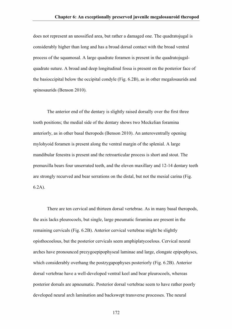
Chapter 6: An exceptionally preserved juvenile megalosauroid theropod
172
does not represent an unossified area, but rather a damaged one. The quadratojugal is
considerably higher than long and has a broad dorsal contact with the broad ventral
process of the squamosal. A large quadrate foramen is present in the quadratojugal-
quadrate suture. A broad and deep longitudinal fossa is present on the posterior face of
the basioccipital below the occipital condyle (Fig. 6.2B), as in other megalosaurids and
spinosaurids (Benson 2010).
The anterior end of the dentary is slightly raised dorsally over the first three
tooth positions; the medial side of the dentary shows two Meckelian foramina
anteriorly, as in other basal theropods (Benson 2010). An anteroventrally opening
mylohyoid foramen is present along the ventral margin of the splenial. A large
mandibular fenestra is present and the retroarticular process is short and stout. The
premaxilla bears four unserrated teeth, and the eleven maxillary and 12-14 dentary teeth
are strongly recurved and bear serrations on the distal, but not the mesial carina (Fig.
6.2A).
There are ten cervical and thirteen dorsal vertebrae. As in many basal theropods,
the axis lacks pleurocoels, but single, large pneumatic foramina are present in the
remaining cervicals (Fig. 6.2B). Anterior cervical vertebrae might be slightly
opisthocoelous, but the posterior cervicals seem amphiplatycoelous. Cervical neural
arches have pronounced prezygoepipophyseal laminae and large, elongate epipophyses,
which considerably overhang the postzygapophyses posteriorly (Fig. 6.2B). Anterior
dorsal vertebrae have a well-developed ventral keel and bear pleurocoels, whereas
posterior dorsals are apneumatic. Posterior dorsal vertebrae seem to have rather poorly
developed neural arch lamination and backswept transverse processes. The neural
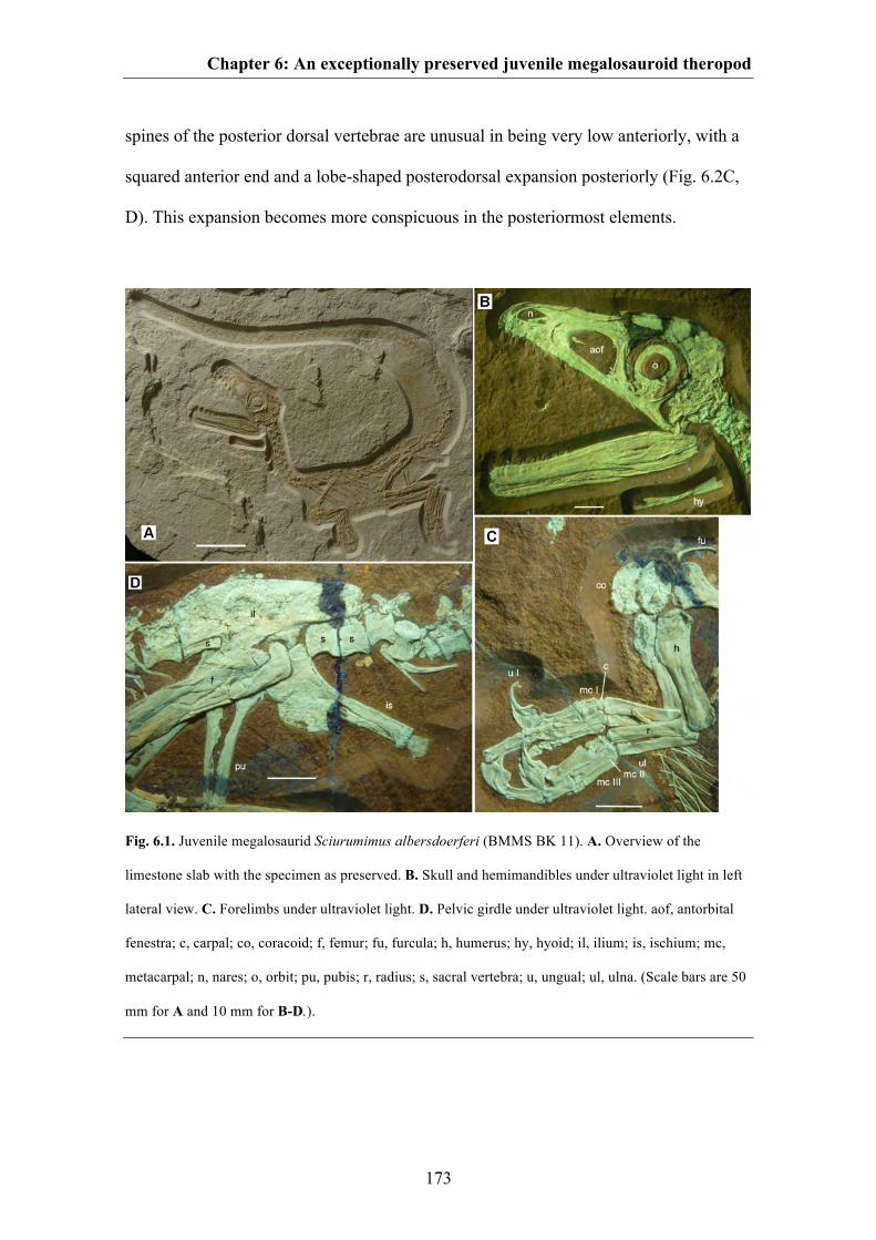
Chapter 6: An exceptionally preserved juvenile megalosauroid theropod
173
spines of the posterior dorsal vertebrae are unusual in being very low anteriorly, with a
squared anterior end and a lobe-shaped posterodorsal expansion posteriorly (Fig. 6.2C,
D). This expansion becomes more conspicuous in the posteriormost elements.
Fig. 6.1. Juvenile megalosaurid Sciurumimus albersdoerferi (BMMS BK 11). A. Overview of the
limestone slab with the specimen as preserved. B. Skull and hemimandibles under ultraviolet light in left
lateral view. C. Forelimbs under ultraviolet light. D. Pelvic girdle under ultraviolet light. aof, antorbital
fenestra; c, carpal; co, coracoid; f, femur; fu, furcula; h, humerus; hy, hyoid; il, ilium; is, ischium; mc,
metacarpal; n, nares; o, orbit; pu, pubis; r, radius; s, sacral vertebra; u, ungual; ul, ulna. (Scale bars are 50
mm for A and 10 mm for B-D.).
Chapter 6: An exceptionally preserved juvenile megalosauroid theropod
174
Fig. 6.2. Anatomical details of Sciurumimus albersdoerferi. A. Dentition of left premaxilla and anterior
end of left maxilla. B. Disarticulated occiput, atlas, axis, and anterior cervical vertebrae. C-D.
Posteriormost dorsal vertebrae and anterior part of ilium, photograph C and interpretative drawing D. All
photographs under UV light. ans, axial neural spine; boc, basioccipital; dv, dorsal vertebrae; epi,
epipophyses; il, ilium; lfe, left femur; m, maxilla; m1, m5, first and fifth maxillary tooth, respectively; p1,
p4, first and fourth premaxillary tooth, respectively; pm, premaxilla; prel, prezygoepipophyseal lamina; r,
rib; rfe, right femur; sv, sacral vertebra; tp, transverse process. (Scale bars are 5 mm for A and 10 mm for
B-D).
The sacrum consists of five vertebrae; the posterior ones being considerably
shorter than the anterior sacrals. A total of 59 caudal vertebrae are preserved, with a few
elements probably missing. Anterior caudal vertebrae lack ventral grooves or keels and
have rather low, simple, posterodorsally directed neural spines. The exact position of
the transition point cannot be established, but transverse processes are certainly absent
Chapter 6: An exceptionally preserved juvenile megalosauroid theropod
175
posterior to caudal twenty. Posterior caudals are elongate in shape and have short,
bowed pre- and postzygapophyses, unlike the strongly elongate prezygapophyses in
allosauroids and coelurosaurs. Chevrons are present in at least 36 vertebrae; they are
simple rod-like structures in lateral view, without ventral anterior or posterior
expansion. Slender gastralia are present, with the medial elements being longer and
more robust than the lateral elements.
The scapula is more than ten times longer than wide at its narrowest point,
unlike the broader scapula in basal theropods, including megalosauroids, but rather
comparable to the situation in derived allosauroids and coelurosaurs (Rauhut 2003a;
Benson 2010). It has a slight distal expansion that gradually arises from the shaft. The
acromion process is only moderately and gradually expanded relative to the width of the
shaft. The coracoid is oval in shape and shorter than high and lacks a subglenoid process
and a biceps tubercle, as it is the case in megalosaurids and spinosaurids. The left ramus
of a small, slender furcula is exposed.
The forelimbs (Fig. 6.1C) are short and robust, as it is the case in some other
megalosaurids (Benson 2010), with a long manus accounting for ~ 45% of the length of
the forelimb. The humerus is short and robust, with a triangular internal tuberosity and a
well-developed deltopectoral crest. Radius and ulna are considerably shorter than the
humerus and the ulna is anteroposteriorly expanded proximally to form a concave facet
for the humerus anteriorly and a small, but stout olecranon process posteriorly. The ulna
is slightly more slender than the radius. A poorly ossified carpal is present and covers
the proximal end of metacarpal I. The manus has three digits, with metacarpal I being
less than half the length of metacarpal II and metacarpal III being shorter and
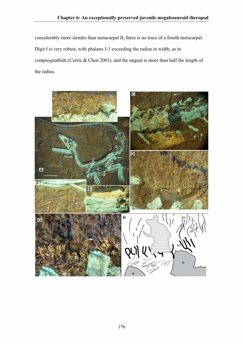
Chapter 6: An exceptionally preserved juvenile megalosauroid theropod
176
considerably more slender than metacarpal II; there is no trace of a fourth metacarpal.
Digit I is very robust, with phalanx I-1 exceeding the radius in width, as in
compsognathids (Currie & Chen 2001), and the ungual is more than half the length of
the radius.
Chapter 6: An exceptionally preserved juvenile megalosauroid theropod
177
Fig. 6.3. Soft tissue preservation in Sciurumimus. A. Overview of skeleton under ultraviolet light, with
position of magnifications B-F indicated. B. Fine filaments above the scapular region of the dorsal
vertebral column. C. Anterior mid-caudal section, with long dorsal filaments (upper white arrow),
preserved skin (yellow patch), and fine filaments at the ventral lateral tail flank (lower white arrows). D.
Long filaments, anchored in the skin, at the dorsal tail base. E. Small section of possibly fossilized muscle
tissue along the posterior edge of the tibia. F. Small, fine filaments ventral to the gastralia in the
abdominal area (arrows point to individual filaments). G-H. Magnification of soft tissues dorsal to the 9th
and 10th caudal vertebra G and interpretative drawing H, showing possible follicles. Greenish white
structures are bone, fine greenish lines above the vertebrae are preserved filaments, and yellow parts
represent skin structures. Arrow points to a filament entering one of the vertical skin structures that might
represent follicles. Abbreviation: col, collagen fibres in the skin; fil, filaments; fo? possible follicles; tp,
transverse process. All photographs under ultraviolet light. (Scale bar in A is 50 mm.).
The ilium is elongate, with a gently curved dorsal margin and an undulate
posterior end (Fig. 6.1D). There is no ventral hook anteriorly, but the anterior end has
an unusual anterior “lip” dorsally (Fig. 6.2C, D). The medial brevis shelf is not exposed
in lateral view. The pubic peduncle is anteroposteriorly longer than the ischial peduncle,
as in other tetanurans (Rauhut 2003a). The pubis is slender, longer than the ischium and
the shaft is straight, with a moderately expanded distal boot. The ischium is slightly
expanded anteriorly distally and the large, hatchet-shaped obturator process is not offset
from the pubic peduncle. The femur is stout and has a wing-like lesser trochanter that
reaches approximately half the height of the slender greater trochanter (Fig. 6.1D, 6.2C,
D). Tibia and fibula are slightly longer than the femur and the fibula is distally
expanded. The metatarsus is slender, metatarsals II and IV are of subequal length, and
metarsal V is transversely flat and anteriorly flexed. Metatarsal I is elongate and splint-
Chapter 6: An exceptionally preserved juvenile megalosauroid theropod
178
like, rather than short and triangular as is the case in most other tetanurans (Rauhut
2003a). In the foot, pedal ungual II is slightly larger than the other unguals.
Soft tissues are preserved in several areas of the skeleton (Fig. 6.3), and most
seem to represent integumentary structures, with the possible exception of a short
section of fossilized tissue along the posterior edge of the tibia, which might represent
muscle tissue (Fig. 6.3E). The best soft tissue preservation is found on the tail, which
preserves large patches of skin especially on the ventral, but also on the dorsal side, and
very fine, long, hair like filaments that correspond to type 1 feathers (Xu & Guo 2009)
dorsally in the anterior mid-section (Fig. 6.3C, D). The skin is smooth and does not
show clear signs of scales, in contrast to the situation in Juravenator (Chiappe &
Göhlich 2010). The feathers seem to be anchored in the skin and form a thick covering
on the dorsal side of the tail and reach more than two and a half times the height of their
respective caudal vertebrae. Shorter filaments are preserved on the ventral tail flank
(Fig. 6.3D), above the mid-dorsal vertebrae (Fig. 6.3B), and in a small patch on the
ventral part of the body (Fig. 6.3F).
The protofeathers are probably monofilaments, since no branching patterns are
visible in the well-preserved, long filaments above the tail; apparent branching patterns
in a few places are probably the result of compaction of these structures (Foth 2012).
Due to the preservation, it cannot be established if these structures were hollow like the
filaments found in other dinosaurs (Currie & Chen 2001; Mayr et al. 2002). The
thickness of these filaments is approximately 0.2 mm in the long filaments in the dorsal
tail region, and less in the shorter filaments at the tail flank, the back and the belly of the
Chapter 6: An exceptionally preserved juvenile megalosauroid theropod
179
animal, comparable to the size of the filamentous protofeathers found in
Sinosauropteryx (Currie & Chen 2001).
DISCUSSION
To establish the phylogenetic position of Sciurumimus, we carried out several analyses,
using three large recently published matrices (see supplementary information of Chapter
6). Sciurumimus was consistently found to be a basal tetanuran, and recovered as a basal
megalosaurid within Megalosauroidea in the most detailed analysis of basal tetanuran
interrelationships yet published (Benson et al. 2010; Fig. 6.4). Synapomorphies of
megalosauroids and more restricted ingroups present in the new taxon include an
elongate anterior process of the maxillary body, a medially closed maxillary fenestra, a
very slender anterior process of the lacrimal, lateral blade of lacrimal does not overhang
antorbital fenestra, presence of a deep fossa ventral to the basioccipital condyle, a
splenial foramen that opens anteroventrally, a slightly dorsally expanded anterior end of
the dentary, a pronounced ventral keel in the anterior dorsal vertebrae, absence of a
posteroventral process of the coracoid, and an enlarged manual ungual I.
Interestingly, the inclusion of Sciurumimus, without changes to any other
codings, resulted in the recovery of a monophyletic Carnosauria that includes
Megalosauroidea and Allosauroidea and represents the sister group to Coelurosauria.
This is in contrast to the vast majority of recent analysis, which depict the former two
clades as successive sister taxa to coelurosaurs. Although this result should certainly be
seen with caution, given the early ontogenetic stage of Sciurumimus, this rather severe
change to the phylogeny by simple inclusion of an additional taxon highlights our still
incomplete understanding of basal tetanuran evolution.
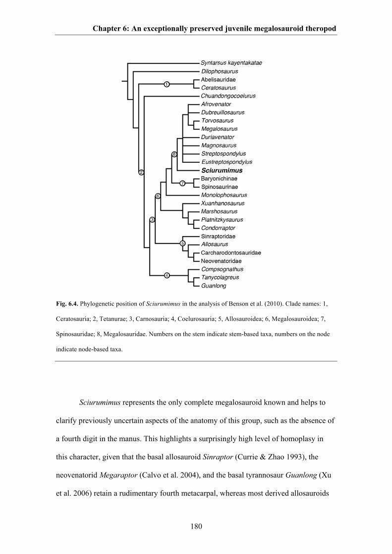
Chapter 6: An exceptionally preserved juvenile megalosauroid theropod
180
Fig. 6.4. Phylogenetic position of Sciurumimus in the analysis of Benson et al. (2010). Clade names: 1,
Ceratosauria; 2, Tetanurae; 3, Carnosauria; 4, Coelurosauria; 5, Allosauroidea; 6, Megalosauroidea; 7,
Spinosauridae; 8, Megalosauridae. Numbers on the stem indicate stem-based taxa, numbers on the node
indicate node-based taxa.
Sciurumimus represents the only complete megalosauroid known and helps to
clarify previously uncertain aspects of the anatomy of this group, such as the absence of
a fourth digit in the manus. This highlights a surprisingly high level of homoplasy in
this character, given that the basal allosauroid Sinraptor (Currie & Zhao 1993), the
neovenatorid Megaraptor (Calvo et al. 2004), and the basal tyrannosaur Guanlong (Xu
et al. 2006) retain a rudimentary fourth metacarpal, whereas most derived allosauroids
Chapter 6: An exceptionally preserved juvenile megalosauroid theropod
181
(Gilmore 1920; Currie & Carpenter 2000) and also coelurosaurs (e.g. Currie & Chen
2001) have only three metacarpals. This suggests that the fourth digit was either reduced
several times independently, or a reduction of this structure at the base of tetanurans
was reversed in some taxa, possibly atavistically.
Several characters indicate that the Sciurumimus albersdoerferi type specimen
represents a very young, probably early posthatchling individual, including the body
proportions, with a very large skull and rather short hindlimbs, lack of fusion in the
skeleton (unfused neurocentral sutures in all of the vertebral column, unfused sacral
vertebrae, lack of fusion between elements of the braincase) (Brochu 1996), coarsely
striated bone surface texture in all skeletal elements (Tumarkin-Deratzian et al. 2006),
and a very regular pattern of tooth development in the maxilla, possibly indicating that
no teeth had been replaced (Dal Sasso & Signore 1998). This differs from the condition
in perinates of more derived coelurosaurs where there is considerable heterogeneity in
among the teeth (Bever & Norell 2009).
The dentition of Sciurumimus differs significantly from those of subadult or
adult basal tetanurans in the slender and unserrated premaxillary teeth and strongly
recurved maxillary teeth with distal serrations only. Given the rather uniform tooth
morphology in most basal tetanurans (at least in respect to general morphology, such as
tooth shape and presence and extent of serrations), these features are here regarded as
juvenile characters. Thus, these differences support the assertion that juveniles of large
theropod species fed on different prey items than adults (Farlow 1976). Conversely, this
dentition is remarkably similar to that of basal coelurosaurs, which commonly have
slender, more rounded premaxillary teeth that lack serrations (Stromer 1934; Rauhut
Chapter 6: An exceptionally preserved juvenile megalosauroid theropod
182
2003a) and often have strongly recurved lateral teeth, frequently without mesial
serrations in at least some teeth ( Stromer 1934; Currie Chen 2001; Norell et al. 2006).
This indicates that the dentition as seen in compsognathids (Stromer 1934; Currie &
Chen 2001; Peyer 2006) and dromaeosaurids (Xu & Wu 2001; Norell et al. 2006) might
have evolved by heterochronic processes, or it reflects convergence due to similar prey
preferences. It also implies that the common practice of referring small, strongly
recurved lateral teeth with reduced or no mesial serrations to dromaeosaurids or
coelurosaurs in general (Maganuco et al. 2005; Knoll & Ruiz-Omeñaca 2009; van der
Lubbe et al. 2009) should be done with caution, and coelurosaur occurrences based on
these tooth characters alone are of no use for inferring biogeographic or evolutionary
patterns.
Sciurumimus is comparable in size to and basically indistinguishable in
proportions from the juvenile basal coelurosaur Juravenator (Göhlich & Chiappe 2006;
Butler & Upchurch 2007; Chiappe & Göhlich 2010) (see supplementary information of
Chapter 6). However, these taxa differ significantly in anatomical details (see
supplementary information of Chapter 6). Thus, if this observation is indicative of the
condition in early posthatchling theropods in general, these seem to have been
remarkably similar in proportions, and differences in allometric growth might account
for the different body plans seen in adult theropods (Carr 1999; Erickson et al. 2004a).
However, data on juvenile theropods is still very limited, and more information is
needed to test this hypothesis.
The presence of type 1 feathers along the dorsal side of the tail, the ventral tail
flank and parts of the body in Sciurumimus show that the entire body of this animal was
Chapter 6: An exceptionally preserved juvenile megalosauroid theropod
183
plumaged, as it is the case in compsognathids (Xu & Guo 2009). As a megalosaurid,
Sciurumimus is the most basal theropod taxon yet reported with such integumentary
structures, and demonstrates that at least juveniles of basal tetanurans had protofeathers.
Sciurumimus thus helps to bridge the considerable gap between basal ornithischians, for
which monofilaments have been reported (Zheng et al. 2009) and coelurosaurs, where
protofeathers [morphotype 1 (Prum 1999)] or feathers seem to be generally present
(Norell & Xu 2005; Xu & Guo 2009; Chiappe & Göhlich 2010). As in tyrannosauroids
(Xu & Guo 2009), the preservation of scaly skin in adult basal tetanurans (Glut 2003) is
therefore no argument against the presence of feathers in this group in general, neither
should the presence of scales in other dinosaur clades (Xu & Guo 2009) be taken as
such. Large adult dinosaurs might have secondarily lost feathers, as in the case of hair
loss in several groups of large mammals today. Furthermore, the joint presence of scales
and filaments in some taxa (Mayr et al. 2002; Chiappe & Göhlich 2010) indicates that
the apparent lack of filaments in animals that preserve impressions of scaly skin in more
coarse-grained sediments could also be due to taphonomic processes. Given that
filaments in ornithischian dinosaurs (Mayr et al. 2002; Zheng et al. 2009) are
morphologically indistinguishable from protofeathers found in tetanurans and basal
coelurosaurs, a filamentous body covering obviously represents the plesiomorphic state
for dinosaurs in general, or, if one assumes that the hair-like structures of pterosaurs
(Unwin 2006) are homologous structures, for ornithodiran archosaurs (Brusatte et al.
2010c).
In the anterior mid-section of the tail of Sciurumimus, the feathers seem to be
anchored in the skin and are associated with dorsoventrally elongate skin structures
(Fig. 6.3). Whereas collagen fibres in avian skin are usually oriented parallel to the body
Chapter 6: An exceptionally preserved juvenile megalosauroid theropod
184
surface, these structures are perpendicular to the long axis of the body, and several of
them show an elongate-cup-shaped outline (Fig. 6.3). The only comparable structures in
the avian skin are the follicles associated with the feathers (Lucas & Stettenheim 1972),
so we tentatively suggest that these structures might represent follicles. Thus, whereas
several recent papers have argued that the origin of follicles was linked with the
evolution of a rachis or barb ridges (Sawyer & Knapp 2003; Alibardi & Toni 2008; Xu
& Guo 2009), Sciurumimus might present evidence for the hypothesis that follicles were
associated with the origin of feathers (Prum 1999). Furthermore, there is a meshwork of
thin, elongate soft tissue structures below this outer layer (Fig. 6.3). These structures
most probably represent collagen fibres within the stratum compactum of the dermis,
which is characterized by a high density of collagen bundles in birds (Lucas &
Stettenheim 1972). The fibres are clearly different from the filaments in their orientation
and their luminescence under filtered UV light, and thus provide evidence against the
interpretation of similarly arranged and oriented filaments in Chinese theropods as
decaying collagen fibres (Feduccia et al. 2005; Lingham-Soliar et al. 2007).
ACKNOWLEDGEMENTS
We especially thank Raimund Albersdörfer, who financed the excavations during which
the specimen was found and made it available for study. Special thanks are also due to
Birgit Albersdörfer, the owner of the specimen. Joseph Schels and Wolfgang Häckel
found and excavated the specimen, and the very delicate preparation was carried out by
Jürgen Geppert, Wolfgang Häckel und Stefan Selzer. Wolfgang Rygol and the
Kalkwerk Rygol GmbH & CoKG courteously permitted the excavations. Martin Röper
and Monika Rothgaenger introduced Raimund Albersdörfer to the fossiliferous layers at
Painten. Martina Köbl-Ebert is thanked for access to Juravenator for comparison, and
Chapter 6: An exceptionally preserved juvenile megalosauroid theropod
185
Adriana López-Arbarello and Richard Butler helped with many useful discussions. The
paper greatly benefited from critical revisions by Xu Xing and Roger Benson. This
research was funded by the Volkswagen Foundation under grant I/84 640 (OWMR) and
the American Museum of Natural History (MAN).
References
186
References
Abzhanov A, Protas M, Grant BR, Grant PR & Tabin CJ. 2004. Bmp4 and
morphological variation of beaks in Darwin’s finches. Science 305: 1462-1465.
Abzhanov A, Kuo WP, Hartmann C, Grant BR, Grant PR & Tabin CJ. 2006. The
calmodulin pathway and evolution of elongated beak morphology in Darwin’s
finches. Nature 442: 563-567.
Adams DC, Rohlf FJ & Sclice DE. 2004. Geometric morphometrics: ten years of
progress following the 'revolution'. Italian Journal of Zoology 71: 5-16.
Aguilera OA, Riff D & Bocquentin-Villanueva J. 2006. A new giant Purusaurus
(Crocodyliformes, Alligatoridae) from the Upper Miocene Urumaco Formation,
Venezuela. Journal of Systematic Palaeontology 4: 221-232.
Alibardi L & Sawyer RH. 2006. Cell structure of developing downfeathers in the
zebrafinch with emphasis on barb ridge morphogenesis. Journal of Anatomy 208:
621-642.
Alibardi L & Toni M. 2008. Cytochemical and molecular charcteristics of the process of
cornification during feather morphogenesis. Progress in Histochemistry and
Cytochemistry 43: 1-69.
Allain R. 2002. Discovery of megalosaur (Dinosauria, Theropoda) in the middle
Bathonian of Normandy (France) and its implications for the phylogeny of basal
Tetanurae. Journal of Vertebrate Paleontology 22: 548-563.
Alonso PD, Milner AC, Ketcham RA, Cookson MJ, Rowe TB. 2004. The avian nature
of the brain and inner ear of Archaeopteryx. Nature 430: 666-669.
Amundsen T. 2000. Why are female birds ornamented? Trends in Ecology and
Evolution 15: 149-155.
References
187
Anderson BG, Barrick RE, Droser ML & Stadtman KL. 1999. Hadrosaur skin
impressions from the Upper Cretaceous Neslen Formation, Book Cliffs, Utah:
Morphology and paleoenviromental context. Vertebrate Paleontology in Utah 99:
295-301.
Anderson MJ. 2001. A new method for non-parametric multivariate analysis of variance.
Austral Ecology 26:32-46.
Anderson RA & Vitt LJ. 1990. Sexual selection versus alternative causes of sexual
dimorphism in teiid lizards. Oecologia 84: 145-157.
Andres B. 2009. The quality of the pterosaur fossil record. Anniversary Meeting Society
of Vertebrate Paleontology, Abstracts 2009: 55A.
Andres B. 2010. Systematics of the Pterosauria. PhD thesis, Yale University, New
Haven.
Andres B & Ji Q. 2008. A new pterosaur from the Liaoning Province of China, the
phylogeny of Pterodactyloidea, and convergence in their cervical vertebrae.
Palaeontology. 51: 453-469.
Andres B, Clark J & Xu X. 2010. A new rhamphorhynchid pterosaur from the Upper
Jurassic of Xinjiang, China, and the phylogenetic relationships of basal pterosaurs.
Journal of Vertebrate Paleontology 30: 163-187.
Andrews RM. 1982. Patterns of growth in reptiles. In: Gans C & Pough FH (eds).
Biology of the Reptilia. Academic Press, New York: 273-320.
Arratia G & Tischlinger H. 2010. The first record of Late Jurassic crossognathiform
fishes from Europe and their phylogenetic importance for teleostean phylogeny.
Fossil Record 13: 317-341.
Averianov AO. 2004. New data on Cretaceous flying reptiles (Pterosauria) from Russia,
Kazakhstan and Kyrgeyztan. Paleontological Journal 38: 426-436.
References
188
Bakhurina NN & Unwin DM. 1995. A preliminary report on the evidence for “hair” in
Sordes pilosus, an Upper Jurassic pterosaur from middle Asia. In: Sun A & Wang Y
(eds). Sixth symposium on mesozoic terrestrial ecosystems and biota. China Ocean
Press, Beijing: 79-82.
Barrett PM. 2000. Prosauropod dinosaurs and iguanas: speculations on the diets of
extinct reptiles. In: Sues H-D (ed). Evolution of herbivory in terrestrial vertebrates.
Cambridge University Press, Cambridge: 42-78.
Barrett PM. 2005. The diet of ostrich dinosaurs (Theropoda: Ornithomimosauria).
Palaeontology 48: 347-358.
Barrett PM & Rayfield EJ. 2006. Ecological and evolutionary implications of dinosaur
feeding behaviour. Trends in Ecology and Evolution 21: 217-224.
Barrett PM, Butler RJ, Edwards NP & Milner AR. 2008. Pterosaur distribution in time
and space: an atlas. Zitteliana B 28: 61-107.
Barrett, PM, Butler RJ & Nesbitt SJ. 2011. The roles of herbivory and omnivory in
early dinosaur evolution. Earth and Environmental Science Transactions of the
Royal Society of Edinburgh 101: 383-396.
Barsbold R & Osmólska H. 1999. The skull of Velociraptor (Theropoda) from the Late
Cretaceous of Mongolia. Acta Palaeontologica Polonica 44: 189-219.
Bell PR. 2012. Standardized terminology and potential taxonomic utility for
hadrosaurid skin impressions: a case study for Saurolophus from Canada and
Mongolia. PLoS ONE 7: e31295.
Bennett SC. 1992. Sexual dimorphism of Pteranodon and other pterosaurs, with
comments on cranial crests. Journal of Vertebrate Paleontology 12: 422-434.
Bennett SC. 2003. New crested specimens of the Late Cretaceous pterosaur
Nyctosaurus. Paläontologische Zeitschrift 77: 61-75.

References
189
Bennett SC. 2006. Juvenile specimens of the pterosaur Germanodactylus cristatus, with
a review on the genus. Journal of Vertebrate Paleontology 26: 872-878.
Bennett SC. 2007. A second specimen of the pterosaur Anurognathus ammoni.
Paläontologische Zeitschrift 81: 376-398.
Benson RBJ. 2008a. A redescription of 'Megalosaurus' hesperis (Dinosauria,
Theropoda) from the Inferior Oolite (Bajocian, Middle Jurassic) of Dorset, United
Kingdom. Zootaxa 1931: 57-67.
Benson RBJ. 2008b. New information on Stokesosaurus, a tyrannosauroid (Dinosauria:
Theropoda) from North America and the United Kingdom. Journal of Vertebrate
Paleontology 28: 732-750.
Benson RBJ. 2010. A description of Megalosaurus bucklandii (Dinosauria: Theropoda)
from the Bathonian of the UK and the relationships of Middle Jurassic theropods.
Zoological Journal of the Linnean Society 158: 882-935.
Benson RBJ, Carrano MT & Brusatte SL. 2010. A new clade of archaic large-bodied
predatory dinosaurs (Theropoda: Allosauroidea) that survived to the latest Mesozoic.
Naturwissenschaften 97: 71-78.
Benson RBJ, Butler RJ, Carrano MT & O’Connor PM. 2012. Air-filled postcranial
bones in theropod dinosaurs: physiological implications and the “reptile”–bird
transition. Biological Reviews 87: 168-193.
Benton MJ & Walker AD. 2002. Erpetosuchus, a crocodile-like basal archosaur from
the Late Triassic of Elgin, Scotland. Zoological Journal of the Linnean Society 136:
25-47.
Benton MJ, Dunhill AM, Lloyd GT & Marx FG. 2011. Assessing the quality of the
fossil record: insights from vertebrates. In: McGowan AJ & Smith AB (eds).
References
190
Comparing the geological and fossil records: implications for biodiversity.
Geological Society, London, Special Publication 358: 63-94.
Bever GS & Norell MA. 2009. The perinate skull of Byronosaurus (Troodontidae) with
observations on the cranial ontogeny of paravian theropods. American Museum
Novitates 3657: 1-51.
Bhullar B-A, Marugán-Lobón J, Racimo F, Bever GS, Rowe T, Norell MA &
Abzhanov A. 2012. Birds have paedomorphic dinosaur skulls. Nature 487: 223-226.
Bock WJ & v. Wahlert G. 1965. Adaption and the form-function complex. Evolution
19: 269-299.
Bona P. 2007. Una nueva especie de Eocaiman Simpson (Crocodylia, Alligatoridae) del
Paleoceno Inferior de Patagonia. Ameghiniana 44: 435-445.
Bona P & Desojo JB. 2011. Osteology and cranial musculature of Caiman latirostris
(Crocodylia: Alligatoridae). Journal of Morphology 272: 780-795.
Bonaparte JF. 1970. Pterodaustro guinazui gen. et sp. nov. Pterosaurio de la Formación
Lagarcito, Provincia de San Luis, Argentina y su significado en la geología regional
(Pterodactylidae). Acta Geologica Lilloana 10: 207-226.
Bonaparte JF, Novas FE & Coria RA. 1990. Carnotaurus sastrei Bonaparte, the horned,
lightly built carnosaur from the Middle Cretaceous of Patagonia. Contributions in
Science 416: 1-42.
Bookstein FL. 1991. Morphometric tools for landmark data. Cambridge University
Press, Cambridge.
Bookstein FL. 1997. Landmark methods for forms without landmarks: morphometrics
of group differences in outline shape. Medical Image Analysis 1: 225-243.
References
191
Bookstein FL, Gunz P, Mitteroecker F, Prossinger H, Schaefer K & Seidler H. 2003.
Cranial integration in Homo: singular warps analysis of the midsagittal plane in
ontogeny and evolution. Journal of Human Evolution 44: 167-187.
Borsuk-Białynicka M & Evans SE. 2009. A long-necked archosauromorph from the
Early Triassic of Poland. Palaeontologia Polonica 65: 203-234.
Braun WJ & Murdoch DJ. 2008. A first course in statsitical programming with R.
Cambridge University Press, Cambridge.
Brazaitis P. 1974. The identification of living crocodiles. Zoologica 58: 59-105.
Britt BB. 1991. Theropods of Dry Mesa Quarry (Morrison Formation, Late Jurassic),
Colorado, with emphasis on the osteology of Torvosaurus tanneri. Brigham Young
University Geology Studies 37: 1-72.
Brochu CA. 1996. Closure of neurocentral sutures during crocodilian ontogeny:
implications for maturity assessment in fossil archosaurs. Journal of Vertebrate
Paleontology 16: 49-62.
Brochu CA. 1999. Phylogenetisc, taxonomy, and historical biogeography of
Alligatoroidea. Journal Vertebrate Paleontology 19: 9-100.
Brochu CA. 2001. Crocodylian snouts in space and time: phylogenetic approaches
toward adaptive radiation. American Zoologist 41: 564-585.
Brochu CA. 2003. Phylogenetic approaches toward crocodylian history. Annual Review
of Earth and Planetary Sciences 31: 357-397.
Brochu CA. 2010. A new alligatorid from the lower Eocene Green River Formation of
Wyoming and the origin of caimans. Journal Vertebrate Paleontology 30: 1109-
1126.
References
192
Brochu CA. 2011. Phylogenetic relationships of Necrosuchus ionensis Simpson, 1937
and the early history of caimanines. Zoological Journal of the Linnean Society 163:
S228-S256.
Brochu CA & Storrs GW. 2012. A giant crocodile from the Plio-Pleistocene of Kenya,
the phylogenetic relationships of Neogene African crocodylines, and the antiquity of
Crocodylus in Africa. Journal of Vertebrate Paleontology 32: 587-602.
Brochu CA, Parris DC, Grandstaff BS, Denton RK Jr. & Gallagher WB. 2012. A new
species of Borealosuchus (Crocodyliformes, Eusuchia) from the Late Cretaceous-
early Paleogene of New Jersey. Journal of Vertebrate Paleontology 32: 105-116.
Bruner E, Costantini D, Fanfani A & Dell’Omo G. 2005. Morphological variation and
sexual dimorphism of the cephalic scales in Lacerta bilineata. Acta Zoologica
(Stockholm) 86: 245-254.
Brusatte, S.L. 2011. Calculating the tempo of morphological evolution: rates of discrete
character change in a phylogenetic context. In: Elewa AMT (ed). Computational
paleontology. Springer-Verlag, Heidelberg: 53-74.
Brusatte SL. 2012. Dinosaur paleobiology. Wiley-Blackwell, Oxford.
Brusatte SL, Benton MJ, Ruta M & Lloyd GT. 2008. Superiority, competition, and
opportunism in the evolutionary radiation of dinosaurs. Science 321: 1485-1488.
Brusatte SL, Carr TD, Erickson GM, Bever GS & Norell MA. 2009. A long-snouted,
multihorned tyrannosaurid from the Late Cretaceous of Mongolia. Proceedings of
the National Academy of Sciences, U.S.A. 106: 17261-17266.
Brusatte SL, Norell MA, Carr TD, Erickson GM, Hutchinson JR, Balanoff AM, Bever
GS, Choiniere JN, Makovicky PJ & Xu X. 2010a. Tyrannosaur paleobiology: new
research on ancient exemplar organisms. Science 329: 1481-1485.
References
193
Brusatte SL, Benson RBJ, Currie PJ & Zhao X. 2010b. The skull of Monolophosaurus
jiangi (Dinosauria: Theropoda) and its implications for early theropod phylogeny and
evolution. Zoological Journal of the Linnean Society 158: 573-607.
Brusatte SL, Nesbitt SJ, Irmis RB, Butler RJ, Benton MJ & Norell MA. 2010c. The
origin and early radiation of dinosaurs. Earth-Science Reviews 101: 68-100.
Brusatte SL, Montanari S, Yi H, Norell MA (2011) Phylogenetic corrections for
morphological disparity analysis: new methodology and case studies. Paleobiology
37: 1-22.
Brusatte SL, Sakamoto M, Montanari S & Harcourt Smith WEH. 2012a. The evolution
of cranial form and function in theropod dinosaurs: insights from geometric
morphometrics. Journal of Evolutionary Biology 25: 365-377.
Brusatte SL, Butler RJ, Prieto-Márquez A & Norell MA. 2012b. Dinosaur
morphological diversity and the end-Cretaceous extinction. Nature Communications
3 (804): 1-8. doi:10.1038/ncomms1815.
Burnham DA. 2004. New information on Bambiraptor feinbergi (Theropoda:
Dromaeosauridae) from the Late Cretaceous of Montana. In: Currie PJ, Koppelhus
EB, Shugar MA & Wright JL (eds). Feathered dragons. Indiana University Press,
Bloomington: 67-111.
Burns KJ, Hackett SJ & Klein NK. 2002. Phylogenetic relationships and morphological
diversity in Darwin’s finches and their relatives. Evolution 56: 1240-1252.
Busbey AB. 1989. Form and function of the feeding apparatus of Alligator
mississippiensis. Journal of Morphology 202: 99-127.
Butler RJ & Goswami A. 2008. Body size evolution in Mesozoic birds: little evidence
for Cope’s rule. Journal of Evolutionary Biology 21: 1673-1682.
References
194
Butler RJ & Upchurch P. 2007. Highly incomplete taxa and the phylogenetic
relationships of the theropod dinosaur Juravenator starki. Journal of Vertebrate
Paleontology 27: 253-256.
Butler RJ, Upchurch P & Norman DB. 2007. The phylogeny of the ornithischian
dinosaurs. Journal of Systematic Palaeontology 6: 1-40.
Butler RJ, Porro LB & Norman DB. 2008. A juvenil skull of the primitive ornithischian
dinosaur Heterodontosaurus tucki from the “Stormberg” of southern Africa. Journal
of Vertebrate Paleontology 28: 702-711.
Butler RJ, Barrett PM, Nowbath S & Upchurch P. 2009a. Estimating the effects of the
rock record on pterosaur diversity patterns: implications for hypotheses of
bird/pterosaur competitive replacement. Paleobiology 35: 432-446.
Butler RJ, Barrett, PM & Gower DJ. 2009b. Postcranial skeletal pneumaticity and air-
sacs in the earliest pterosaurs. Biology Letters 5: 557-560.
Butler RJ, Barrett PM, Benson RBJ, Brusatte SL & Andres B. 2011a. The taxonomic
diversity and morphological disparity of pterosaurs: untangling sampling biases, the
impact of Lagerstätten, and diversification trajectories. Anniversary Meeting Society
of Vertebrate Paleontology, Abstracts 2011: 81A.
Butler RJ, Benson RBJ, Carrano MT, Mannion PD & Upchurch P. 2011b. Sea-level,
dinosaur diversity, and sampling biases: investigating the “common cause”
hypothesis in the terrestrial realm. Proceedings of the Royal Society of London B 278:
1165-1170.
Butler RJ, Brusatte SL, Andres B. & Benson RBJ. 2012. How do rock record biases
affect studies of disparity in deep time? A case study of the Pterosauria (Reptilia:
Archosauria). Evolution 66: 147-162.
References
195
Cabreira SF, Schultz CL, Bittencourt JS, Soares MB, Fortier DC, Silva LR & Langer
MC. 2011. New stem-sauropodomorph (Dinosauria, Saurischia) from the Triassic of
Brazil. Naturwissenschaften 98: 1035-1040.
Calvo JO, Porfiri JD, Veralli C, Novas FE & Poblete F. 2004. Phylogenetic status of
Megaraptor namunhuaiquii Novas based on a new specimen from Neuquén,
Patagonia, Argentina. Ameghiniana 41: 565-575.
Campàs O, Mallarino R, Herrel A, Abzhanov A & Brenner MP. 2010. Scaling and
shear transformations capture beak shape variation in Darwin’s finches. Proceedings
of the National Academy of Sciences, U.S.A. 107: 3356-3360.
Campione NE & Evans DC. 2011. Cranial growth and variation in edmontosaurs
(Dinosauria: Hadrosauridae): implications for Latest Cretaceous megaherbivore
diversity in North America. PLoS ONE 6: e25186.
Carballido JL, Rauhut OWM, Pol D & Salgado L. 2011. Osteology and phylogenetic
relationships of Tehuelchesaurus benitezii (Dinosauria, Sauropoda) from the Upper
Jurassic of Patagonia. Zoological Journal of the Linnean Society 163: 605-662.
Carpenter K. 1990. Variation in Tyrannosaurus rex. In: Carpenter K & Currie PJ (eds).
Dinosaur systematics: approaches and perspectives. Cambridge University Press,
Cambridge: 141-145.
Carpenter K. 1992. Tyrannosaurids (Dinosauria) of Asia and North America. In: Mateer
NJ & Chen P (eds). Aspects of nonmarine Cretaceous geology. China Ocean Press,
Beijing: 250-268.
Carpenter K. 1994. Baby Dryosaurus from the Upper Jurassic Morrison Formation of
Dinosaur National Monument. In: Carpernter K, Hirsch KF & Horner JR (eds).
Dinosaur eggs and babiers. Cambridge University Press, Cambridge: 288-297.

References
196
Carpenter K. 2010. Variation in a population of Theropoda (Dinosauria): Allosaurus
from the Cleveland-Lloyd Quarry (Upper Jurassic), Utah, USA. Paleontological
Research 14: 250-259.
Carr TD. 1999. Craniofacial ontogeny in Tyrannosauridae (Dinosauria, Coelurosauria).
Journal of Vertebrate Paleontology 19: 497-520.
Carr TD & Williamson TE. 2004. Diversity of late Maastrichtian Tyrannosauridae
(Dinosauria: Theropoda) from western North America. Zoological Journal of the
Linnean Society 142: 479-523.
Carr TD & Williamson TE. 2010. Bistahieversor sealeyi, gen. et sp. nov., a new
tyrannosauroid from New Mexico and the origin of deep snouts in Tyrannosauroidea.
Journal of Vertebrate Paleontology 30: 1-16.
Carr TD, Williamson TE, Britt BB & Stadtman K. 2011. Evidence for high taxonomic
and morphologic tyrannosauroid diversity in the Late Cretaceous (Late Campanian)
of the American Southwest and a new short-skulled tyrannosaurid from the
Kaiparowits formation of Utah. Naturwissenschaften 98: 241-246.
Carrano MT, Loewen MA & Sertich JJW. 2011. New materials of Masiakasaurus
knopfleri Sampson, Carrano, and Forster, 2001, and implications for the morphology
of the Noasauridae (Theropoda: Ceratosauria). Smithsonian Contributions of
Paleobiology 95: 1-53.
Carrano MT, Benson RBJ & Sampson SD. 2012. The phylogeny of Tetanurae
(Dinosauria: Theropoda). Journal of Systematic Palaeontology 10: 211-300.
Carvalho IS, Campos ACA & Nobre PH. 2005. Baurusuchus salgadoensis, a new
Crocodylomorpha from the Bauru Basin (Cretaceous), Brazil. Gondwana Research
8: 11-30.
References
197
Chabreck RH. 1963. Methods of capturing, marking and sexing alligators. Southeastern
Association of Game and Fish Commissioners 17: 47-50.
Chabreck RH & Joanen T. 1979. Growth rates of American Alligators in Louisiana
Herpetologica 35: 51-57.
Chapman RE 1990. Shape analysis in the study of dinosaur morphology. In: Carpenter
K & Currie PJ (eds). Dinosaur systematics: approaches and perspectives. Cambridge
University Press, Cambridge: 21-42.
Chapman RE & Brett-Surman MK. 1990. Morphometric observations on hadrosaurid
ornithopods. In: Carpenter K & Currie PJ (eds). Dinosaur systematics: approaches
and perspectives. Cambridge University Press, Cambridge: 163-177.
Chapman RE, Galton PM, Sepkoski Jr JJ & Wall WP. 1981. A morphometric study of
the cranium of the pachycephalosaurid dinosaur Stegoceras. Journal of Paleontology
55: 608-618.
Chapman RE, Weishampel DB, Hunt G & Rasskin-Gutman D. 1997. Sexual
dimorphism in dinosaurs. In: Wohlberg DL, Stumps E & Rosenberg GD (eds).
Dinofest International. Academy of Natural Sciences, Philadelphia: 83-93.
Chiappe LM & Göhlich UB. 2010. Anatomy of Juravenator starki (Theropoda:
Coelurosauria) from the Late Jurassic of Germany. Neues Jahrbuch für Geologie und
Paläontologie - Abhandlungen 258: 257-296.
Chiappe LM, Coria RA, Dingus L, Jackson F, Chinsamy A & Fox M. 1998. Sauropod
dinosaur embryos from the Late Cretaceous of Patagonia. Nature 396: 258-261.
Chiappe LM, Ji S, Ji Q & Norell MA. 1999. Anatomy and systematics of the
Confuciusornithidae (Theropoda: Aves) from the Late Mesozoic of northeastern
China. Bulletin of the American Museum of Natural History 242: 1-89.
References
198
Chiappe LM, Kellner AWA, Rivarola D, Davila S & Fox M. 2000. Cranial morphology
of Pterodaustro guinazui (Pterosauria: Pterodactyloidea) from the Lower Cretaceous
of Argentina. Contributions in Science 483: 1-19.
Chiappe LM, Salgado L & Coria RA. 2001. Embryonic skulls of titanosaur sauropod
dinosaurs. Science 293: 2444-2446.
Chiappe LM, Norell MA & Clark JM. 2002. The Cretaceous, short-armed
Alvarezsauridae: Mononykus and its kin. In: Chiappe LM & Witmer LM (eds).
Mesozoic birds: above the heads of dinosaurs. University of California Press,
Berkeley: 87-120.
Chiappe LM, Ji S & Ji Q. 2007. Juvenile birds from the Early Cretaceous of China:
implications for enantiornithine ontogeny. American Museum Novitates 3594: 1-49.
Chinsamy A, Codorniú L & Chiappe LM. 2009. Palaeobiological implications of the
bone histology of Pterodaustro guinazui. The Anatomical Record 292: 1462-1477.
Chinsamy A, Chiappe LM, Marugán-Lobón J, Gao C & Zhang F. 2013. Gender
identification of the Mesozoic bird Confuciusornis sanctus. Nature Communications
4 (1381): 1-5. doi:10.1038/ncomms2377.
Choiniere JN, Xu X, Clark JM, Forster CA, Guo Y & Han F. 2010. A basal
alvarezsauroid theropod from the Early Jurassic of Xinjiang, China. Science 327:
571-574.
Christiansen P & Fariña RA. 2004. Mass prediction in theropod dinosaurs. Historical
Biology 16: 85-92.
Chure DJ. 2000. A new species of Allosaurus from the Morrison Formation of Dinosaur
National Monument (UT-CO) and a revision of the theropod family Allosauridae.
PhD thesis, Columbia University, New York.
References
199
Chure D, Britt BB, Whitlock JA & Wilson JA. 2010. First complete sauropod dinosaur
skull from the Cretaceous of the Americas and the evolution of sauropod dentition.
Naturwissenschaften 97: 379-391.
Ciampaglio CN, Kemp M & McShea DW. 2001. Detecting changes in morphospace
occupation patterns in the fossil record: characterization and analysis of measures of
disparity. Paleobiology 27: 695-715.
Clark JM, Norell MA & Makovicky PJ. 2002a. Cladistic approaches to the relationships
of birds to other theropod dinosaurs. In: Chiappe LM & Witmer LM (eds). Mesozoic
birds: above the heads of dinosaurs. University of California Press, Berkeley: 31-61.
Clark JM, Norell MA & Rowe T. 2002b. Cranial anatomy of Citipati osmolskae
(Theropoda, Oviraptorosauria), and a reinterpretation of the holotype of Oviraptor
philoceratops. American Museum Novitates 3364: 1-24.
Clark JM, Xu X, Forster CA & Wang Y. 2004. A Middle Jurassic “sphenosuchian”
from China and the origin of the crocodylian skull. Nature 430: 1021-1024.
Claessens LPAM, O'Connor PM & Unwin DM. 2009. Respiratory evolution facilitated
the origin of pterosaur flight and aerial gigantism. PLoS ONE 4: e4497.
Cleuren J & de Vree F. 2000. Feeding in crocodilians. In: Schwenk K (ed). Feeding:
form, function and evolution in tetrapod vertebrates. Academic Press, New York:
337-358.
Colbert EH. 1989. The Triassic dinosaur Coelophysis. Museum of Northern Arizona
Bulletin 57: 1-160.
Colbert EH. 1990. Variation in Coelophysis bauri. In: Carpenter K & Currie PJ (eds).
Dinosaur systematics: approaches and perspectives. Cambridge University Press,
Cambridge: 81-90.
References
200
Colbert EH & Mook CC. 1951. The ancestral crocodilian Protosuchus. Bulletin of the
American Museum of Natural History 97: 143-182.
Collyer ML & Adams DC. 2007. Analysis of two-state multivariate phenotypic change
in ecological studies. Ecology 88: 683-692.
Cooper WE. & Vitt LJ. 1993. Female mate choice of large male broad-headed skinks.
Animal Behaviour 45: 683-693.
Coombs WP Jr. 1982. Juvenile specimens of the ornithischian dinosaur Psittacosaurus.
Palaeontology 25: 89-107.
Coria RA & Chiappe LM. 2007. Embryonic skin from Late Cretaceous sauropods
(Dinosauria) of Auca Mahuevo, Patagonia, Argentina. Journal of Paleontology 81:
1528-1532.
Cott HB. 1926. Observations on the life habits of some batrachians and reptiles from the
lower Amazon, and note on some mammals from Marajó. Proceedings of the
Zoological Society of London 29: 211-256.
Cott HB. 1961. Scientific results of an inquiry into the ecology and economic status of
the Nile crocodile (Crocodilus niloticus) in Uganda and northern Rhodesia.
Transactions of the Zoological Society of London 29 : 211-356.
Crompton AW & Attridge J. 1986. Masticatory apparatus of the larger herbivores
during Late Triassic and Early Jurassic times. In: Padian K (ed). The beginning of the
age of dinosaurs. Cambridge University Press, Cambridge: 223-236.
Crowe R & Niswander L. 1998. Disruption of scale development by Delta-1
misexpression. Developmental Biology 195: 70-74.
Csiki Z, Vremir M, Brusatte SL & Norell MA. 2010. An aberrant island-dwelling
theropod dinosaur from the Late Cretaceous of Romania. Proceedings of the
National Academy of Sciences, U.S.A. 107: 15357-15361.
References
201
Currie PJ & Zhao X. 1993. A new carnosaur (Dinosauria, Theropoda) from the Jurassic
of Xinjiang, People's Republic of China. Canadian Journal of Earth Sciences 30:
2037-2081.
Currie PJ & Carpenter K. 2000. A new specimen of Acrocanthosaurus atokensis
(Theropoda, Dinosauria) from the Lower Cretaceous Antlers Formation (Lower
Cretaceous, Aptian) of Oklahoma, USA. Geodiversitas 22: 207-246.
Currie PJ & Chen P. 2001. Anatomy of Sinosauropteryx prima from Liaoning,
northeastern China. Canadian Journal of Earth Sciences 38: 1705-1727.
Czechura GV & Wombey J. 1982. Three new striped skinks (Ctenotus, Lacertilia,
Scincidae) from Queensland. Queensland Museum Memoirs 20: 639-645.
Dacke CG, Arkle S, Cook DJ, Wormstone IM, Jones S, Zaidi M & Bascal ZA. 1993.
Medullary bone and avian calcium regulation. Journal of Experimental Biology 184:
63-88.
Dal Sasso C & Signore M. 1998. Exceptional soft-tissue preservation in a theropod
dinosaur from Italy. Nature 392: 383-387.
Dal Sasso C & Maganuco S. 2011. Scipionyx samniticus (Theropoda: Compsognathidae)
from the Lower Cretaceous of Italy. Memorie della Società Italiana di Scienze
Naturali e del Museo Civico di Storia Naturale di Milano 37: 1-281.
D'Amore DC. 2009. A functional explanation for denticulation in theropod dinosaur
teeth. The Anatomical Record 292: 1297-1314.
Da Silveira R & Magnusson WE. 1999. Diets of Spectacled and black caiman in the
Anavilhanas Archipelago, Central Amazonia, Brazil. Journal of Herpetology 33:
181-192.
References
202
Dececchi TA & Larsson HCE. 2011. Assessing arboreal adaptations of bird antecedents:
testing the ecological setting of the origin of the avian flight stroke. PLoS ONE 6:
e22292.
de Ricqlès AJ, Padian K & Horner JR. 2003. On the bone histology of some Triassic
pseudosuchian archosaurs and related taxa. Annales de Paléontologie 89: 67-101.
de Thoisy B, Hrbek T, Farias IP, Vasconcelos WR & Lavergne A. 2006. Genetic
structure, population dynamics, and conservation of black caiman (Melanosuchus
niger). Biological Conservation 133: 474-482.
Dhouailly D. 2009. A new scenario for the evolutionary origin of hair, feather, and
avian scales. Journal of Anatomy 214: 587-606.
Dingus L & Rowe T. 1997. The mistaken extinction: dinosaur evolution and the origin
of birds. Freeman, New York.
Dodson P. 1975. Functional and ecological significance of relative growth in Alligator.
Zoology 1975: 315-355.
Dodson P. 1993. Comparative craniology of the Ceratopsia. American Journal of
Science 293: 200-234.
Drake AG. 2011. Dispelling dog dogma: an investigation of heterochrony in dogs using
3D geometric morphometric analysis of skull shape. Evolution & Development 13:
204-213.
Drake AG & Klingenberg CP. 2008. The pace of morphological change: historical
transformation of skull shape in St Bernard dogs. Proceedings of the Royal Society B
275: 71-76.
Dyke GJ, Nudds RL & Rayner JM. 2006. Limb disparity and wing shape in pterosaurs.
Journal of Evolutionary Biolology 19: 1339-1342.
References
203
Dyke GJ, McGowan AJ, Nudds RL & Smith D. 2009. The shape of pterosaur evolution:
evidence from the fossil record. Journal of Evolutionary Biolology 22: 890-898.
Eddy DR & Clarke JA. 2011. New information on the cranial anatomy of
Acrocanthosaurus atokensis and its implications for the phylogeny of Allosauroidea
(Dinosauria: Theropoda). PLoS ONE 6: e17932.
Elewa AMT. 2004. Morphometrics: applications in biology and paleontology.
Springer-Verlag, Heidelberg.
Elgin RA, Grau CA, Palmer C, Hone DWE, Greenwell D & Benton MJ. 2008.
Aerodynamic characters of the cranial crest in Pteranodon. Zitteliana B 28: 167-174.
Emerson SB & Bramble DM. 1993. Scaling, allometry, and skull design. In: Hanken J
& Hall BK (eds). The skull. Vol. 3. Patterns of structural and systematic diversity.
University of Chicago Press, Chicago: 384-421.
Erickson GM & Brochu CA. 1999. How the “terror crocodile” grew so big. Nature 398:
205-206.
Erickson GM, van Kirk SD, Su J, Levenston ME, Caler WE, Carter DR. 1996. Bite-
force estimation for Tyrannosaurus rex from tooth-marked bones. Nature 382: 706-
708.
Erickson GM, Rogers KC & Yerby SA. 2001. Dinosaur growth patterns and rapid avian
growth rates. Nature 412: 429-432.
Erickson GM, Lappin AK & Vliet KA. 2003. The ontogeny of bite-force performance
in American alligator (Alligator mississippiensis). Journal of Zoology, London 260:
317-327.
Erickson GM, Makovicky PJ, Currie PJ, Norell MA, Yerby SA & Brochu CA. 2004a.
Gigantism and comparative life-history parameters of tyrannosaurid dinosaurs.
Nature 430: 772-775.
References
204
Erickson GM, Lappin AK, Parker T & Vliet KA. 2004b. Comparison of bite-force
performance between long-term captive and wild American alligators (Alligator
mississippiensis). Journal of Zoology 262: 21-28.
Erickson GM, Lappin AK & Larson P. 2005. Androgynous rex - The utility of chevrons
for determining the sex of crocodilians and non-avian dinosaurs. Zoology 108: 277-
286.
Erickson GM, Rauhut OWM, Zhou Z, Turner AH, Inouye BD, Hu D & Norell MA.
2009. Was dinosaurian physiology inherited by birds? Reconciling slow growth in
Archaeopteryx. PLoS ONE 4: e7390.
Erickson GM, Gignac PM, Steppan SJ, Lappin AK, Villet KA, Brueggen JD, Inouye
BD, Kledzik D & Webb GJW. 2012. Insights into the ecology and evolutionary
success of crocodilians revealed through bite-force and tooth-pressure
experimentation. PLoS ONE 7: e31781.
Erwin DH. 2007. Disparity: morphological pattern and developmental context.
Palaeontology 50: 57-73.
Evans DC. 2010. Cranial anatomy and systematics of Hypacrosaurus altispinus, and a
comparative analysis of skull growth in lambeosaurine hadrosaurids (Dinosauria:
Ornithischia). Zoological Journal of the Linnean Society 159: 398-434.
Evans DC & Reisz RR. 2007. Anatomy and relationships of Lambeosaurus
magnicristatus, a crested hadrosaurid dinosaur (Ornithischia) from the Dinosaur Park
Formation, Alberta. Journal of Vertebrate Paleontology 27: 373-393.
Ezcurra MD. 2007. The cranial anatomy of the coelophysoid theropod Zupaysaurus
rougieri from the Upper Triassic of Argentina. Historical Biology 19: 185-202.
Ezcurra MD & Novas FE. 2007. Phylogenetic relationships of the Triassic theropod
Zupaysaurus rougieri from NWArgentina. Historical Biology 19: 35-72.
References
205
Fanti F, Currie PJ & Badamgarav D. 2012. New specimens of Nemegtomaia from the
Baruungoyot and Nemegt Formations (Late Cretaceous) of Mongolia. PLoS ONE 7:
e31330.
Farias IP, Silveira R, de Thoisy B, Monjeló LA, Thorbjarnarson JB & Hrbek T. 2004.
Genetic diversity and population structure of Amazonian crocodilians. Animal
Conservation 7: 265-272.
Farlow JO. 1976. Speculations about the diet and foraging behavior of large carnivorous
dinosaurs. American Midland Naturalist 95: 186-191.
Fastovsky DE & Weishampel DB. 2005. The evolution and extinction of the dinosaurs.
University Press, Cambridge.
Feduccia A, Lingham-Soliar T & Hinchliffe JR. 2005. Do feathered dinosaurs exist?
Testing the hypothesis on neontological and paleontological evidence. Journal of
Morphology 266: 125-166.
Felsenstein J. 1985. Phylogenies and the comparative method. The American Naturalist
125: 1-15.
Foote M. 1991. Morphological and taxonomic diversity in a clade’s history: the blastoid
record and stochastic simulations. Contributions from the Museum of Paleontology,
the University of Michigan 28: 101-140.
Foote M. 1993. Discordance and concordance between morphological and taxonomic
diversity. Paleobiology 20: 185-204.
Foote M. 1994. Morphological disparity in Ordovician-Devonian crinoids and the early
saturation of morphological space. Paleobiology 20: 320-344.
Forster CA. 1990. The cranial morphology and systematics of Triceratops, with a
preliminary analysis of ceratopsian phylogeny. PhD thesis, University of
Pennsylvania, Philadelphia.
References
206
Foth C. 2012. On the identification of feather structures in stem-line representatives of
birds: evidence from fossils and actuopalaeontology. Paläontologische Zeitschrift 86:
91-102.
Foth C & Rauhut OWM. 2013. Macroevolutionary and morphofunctional patterns in
theropod skulls: a morphometric approach. Acta Palaeontologica Polonica 58: 1-16.
Foth C & Rauhut OWM. In press. The good, the bad, and the ugly: the influence of
skull reconstructions and intraspecific variability in studies of cranial morphometrics
in theropods and basal saurischians. PloS ONE.
Foth C, Brusatte SL & Butler RJ. 2012. Do different disparity proxies converge on a
common signal? Insights from the cranial morphometrics and evolutionary history of
Pterosauria (Diapsida: Archosauria). Journal of Evolutionary Biology 25: 904-915.
Foth C, Bona P & Desojo JB. In press. Intraspecific variation in the skull morphology
of the black caiman Melanosuchus niger (Alligatoridae, Caimaninae). Acta
Zoologica.
Freckleton RP, Harvey PH & Pagel M. 2002. Phylogenetic analysis and comparative
data: a test and review of the evidence. The American Naturalist 160: 712-726.
Frey E, Tischlinger H, Buchy M-C & Martill DM. 2003. New specimens of Pterosauria
(Reptilia) with soft parts with implications for pterosaurian anatomy and locomotion.
In: Buffetaut E & Mazin J-M (eds). Evolution and palaeobiology of pterosaurs. The
Geological Society, London: 233-266.
Friedman M. 2010. Explosive morphological diversification of spiny-finned teleost
fishes in the aftermath of the end-Cretaceous extinction. Proceedings of the Royal
Society of London B 277: 1675-1683.
Futuyma DJ. 2007. Evolution. Spektrum Akademischer Verlag, München.
References
207
Gallina PA & Apesteguíia S. 2011. Cranial anatomy and phylogenetic position of the
titanosaurian sauropod Bonitasaura salgadoi. Acta Palaeontologica Polonica 56: 45-
60.
Galton PM. 1984. Cranial anatomy of the prosauropod dinosaur Plateosaurus from the
Knollenmergel (Middle Keuper, Upper Triassic) of Germany. Geologica et
Palaeontologica 18: 139-171.
Galton PM. 1985. Cranial anatomy of the prosauropod dinosaur Plateosaurus from the
Knollenmergel (Middle Keuper, Upper Triassic) of Germany. Geologica et
Palaeontologica 19: 119-159.
Galton PM. 2001. The prosauropod dinosaur Plateosaurus Meyer, 1837 (Saurischia:
Sauropodomorpha; Upper Triassic). II. Notes on the referred species. Revue de
Paléobiologie 20: 435-502.
Galton PM & Jensen JA. 1979. A new large theropod dinosaur from the Upper Jurassic
of Colorado. Brigham Young University Geology Studies 26: 1-12.
Galton PM & Upchurch P. 2004. Prosauropoda. In: Weishampel DB, Dodson P,
Osmólska H (eds). The Dinosauria. University of California Press, Berkeley: 232-
258.
García RA, Salgado L, Coria RA & Chiappe LM. 2010. Osteología embrionaria de
saurópodos titanosaurios de Neuquén (Argentina): aspectos ontogenéticos y
evolutivos. Ameghiniana 47: 409-430.
Garland T, Harvey PH & Ives AR. 1992. Procedures for the analysis of comparative
data using phylogenetically independent contrasts. Systematic Biology 41: 18-32.
Gatesy SM. 1990. Caudofemoral musculature and the evolution of theropod locomotion.
Paleobiology 16: 170-186.

References
208
Gauthier JA. 1986. Saurischian monophyly and the origin of birds. Memoirs of the
California Academy of Science 8: 1-55.
Gauthier JA & Padian K. 1985. Phylogenetic, functional, and aerodynamic analyses of
the origin of birds and their flight. In: Hecht MK, Ostrom JH, Viohl G & Wellnhofer
P (eds). The Beginnings of Birds. Freunde des Jura-Museums Eichstätt, Eichstätt:
185-197.
Gilmore GW. 1920. Osteology of the carnivorous dinosauria in the United States
National Museum, with special reference to the genera Antrodemus (Allosaurus) and
Ceratosaurus. Bulletin - United States National Museum 110: 1-159.
Glut DF. 2003. Dinosaurs. The encyclopedia. Supplement 3. Mcfarland & Co, Jefferson.
Godefroit P, Demuynck H, Dyke G, Hu D, Escuillié F & Claeys P. 2013a. Reduced
plumage and flight ability of a new Jurassic paravian theropod from China. Nature
Communications 4 (1394): 1-6.
Godefroit P, Cau A, Hu D, Escuillié F, Wu W & Dyke G. In press. A Jurassic avialan
dinosaur from China resolves the early phylogenetic history of birds. Nature.
Göhlich UB & Chiappe LM. 2006. A new carnivorous dinosaur from the Late Jurassic
Solnhofen archipelago. Nature 440: 329-332.
Goloboff PA, Farris JS & Nixon KC. 2008. TNT, a free program for phylogenetic
analysis. Cladistics 24: 774-786.
Goodwin MB. 1990. Morphometric landmarks of pachycephalosaurid cranial material
from the Judith River Formation of northcentral Montana. In: Carpenter K & Currie
PJ (eds). Dinosaur systematics: approaches and perspectives. Cambridge University
Press, Cambridge: 189-201.
References
209
Goodwin MB, Clemens WA, Horner JR & Padian K. 2006. The smallest known
Triceratops skull: new observations on ceratopsid cranial anatomy and ontogeny.
Journal of Vertebrate Paleontology 26: 103-112.
Gould SJ. 1991. The disparity of the Burgess Shale arthropod fauna and the limits of
cladistic analysis: why we must strive to quantify morphospace. Paleobiology 17:
411-423.
Gow CE, Kitching JW & Raath MA. 1990. Skulls of the prosauropod dinosaur
Massospondylus carinatus Owen in the collections of the Bernand Price Institute for
Palaeontological Research. Palaeontologia Africana 27: 45-58.
Gower DJ. 2002. Braincase evolution in suchian archosaurs (Reptilia: Diapsida):
evidence from the rauisuchian Batrachotomus kupferzellensis. Zoological Journal of
the Linnean Society 136: 49-76.
Gower DJ & Weber E. 1998. The braincase of Euparkeria, and the evolutionary
relationships of birds and crocodilians. Biological Reviews 73: 367-411.
Gower JC. 1975. Generalized Procrustes analysis. Psychometrika 40: 33-51.
Hall PM & Portier KM. 1994. Cranial morphometry of New Guinea Crocodiles
(Crocodylus novaeguineae): ontogenetic variation in relative growth of the skull and
an assessment of its utility as a predictor of the sex and size of individuals.
Herpetological Monographs 8: 203-225.
Hammer O. 2009. PAST: Paleontological Statistics. Reference manual. 191 pp.
http://folk.uio.no/ohammer/past/
Hammer O & Harper D. 2006. Paleontological data analysis. Blackwell Publishing,
Malden.
Hammer O, Harper DAT & Ryan PD. 2001. PAST: Paleontological statistics software
package for education and data analysis. Palaeontologia Electronica 4: 1-9.
References
210
Harris MP, Fallon JF & Prum RO. 2002. Shh-Bmp2 signaling module and the
evolutionary origin and diversification of feathers. Journal of Experimental Zoology
(MOL DEV EVOL) 294: 160-176.
Henderson DM. 2000. Skull and tooth morphology as indicators of niche partitioning in
sympatric Morrison Formation theropods. Gaia 15: 219-226.
Henderson DM. 2002. The eyes have it: The sizes, shapes, and orientations of theropod
orbits as indicators of skull strength and bite force. Journal of Vertebrate
Paleontology 22: 766-778.
Henderson DM. 2010. Pterosaur body mass estimates from three-dimensional
mathematical slicing. Journal of Vertebrate Paleontology 30: 768-785.
Henderson DM & Weishampel DB. 2002. Convergent evolution of the maxilla-dental-
complex among carnivorous archosaurs. Senckenbergiana lethaea 82: 77-92.
Herron JC. 1991. Growth rates of black caiman Melanosuchus niger and spectacled
caiman Caiman crocodilus and the recruitment of breeders in hunted caiman
populations. Biological Conservation 55: 103-113.
Hieronymus TL & Witmer LM. 2010. Homology and evolution of avian compound
rhamphothecae. The AUK 127: 590-604.
Hinić S. 2002. Cranial osteology of Massospondylus carinatus Owen, 1854 and its
implications for prosauropod phylogeny. Master thesis, University of Toronto,
Toronto.
Holliday CM & Witmer LM. 2008. Cranial kinesis in dinosaurs: intracranial joints,
protractor muscles, and their significance for cranial evolution and function in
diapsids. Journal of Vertebrate Paleontology 28: 1073-1088.
References
211
Holliday CM & Witmer LM. 2009. The epipterygoid of crocodyliforms and its
siginificance for the evolution of the orbitotemporal region of eusuchians. Journal of
Vertebrate Paleontology 29: 715-733.
Holtz TR Jr. 2004. Tyrannosauroidea. In: Weishampel DB, Dodson P & Osmólska H
(eds). The Dinosauria. University of California Press. Berkeley: 111-136.
Holtz TR Jr., Molnar RE & Currie PJ. 2004. Basal Tetanurae. In: Weishampel DB,
Dodson P & Osmólska H (eds). The Dinosauria. University of California Press.
Berkeley: 71-110.
Hone DWE, Tischlinger H, Xu X & Zhang F. 2010. The extent of the preserved feathers
on the four-winged dinosaur Microraptor gui under ultraviolet light. PLoS ONE 5:
e9223.
Hone D, Naish D & Cuthill I. 2012. Does mutual sexual selection explain the evolution
of head crests in pterosaurs and dinosaurs? Lethaia 45: 139-156.
Horna J, Horna V, Cintra R & Vásquez P. 2001. Feeding ecology of black caiman
Melanosuchus niger in a western Amazonian forest: The effects of ontogeny and
seasonality on diet composition. Ecotropica 7: 1-11.
Horna V, Zimmermann R, Cintra R, Vásquez P & Horna J. 2003. Feeding ecology of
the black caiman (Melanosuchus niger) in Manu National Park, Peru. Lyonia 4: 65-
72.
Horner JR & Goodwin MB. 2006. Major cranial changes during Triceratops ontogeny.
Proceedings of the Royal Society B 273: 2757-2761.
Hu D, Hou L, Zhang L & Xu X. 2009. A pre-Archaeopteryx troodontid theropod from
China with long feathers on the metatarsus. Nature 461: 640-643.
Hübner TR. 2012. Bone histology in Dysalotosaurus lettowvorbecki (Ornithischia:
Iguanodontia) - variation, growth, and implications. PLoS ONE 7: e29958.
References
212
Hübner TR & Rauhut OWM. 2010. A juvenile skull of Dysalotosaurus lettowvorbecki
(Ornithischia: Iguanodontia), and implications for cranial ontogeny, phylogeny, and
taxonomy in ornithopod dinosaurs. Zoological Journal of the Linnean Society 160:
366-396.
Hunt G & Carrano MT. 2010. Models and methods for analyzing phenotypic evolution
in lineages and clades. The Paleontological Society Papers 16: 245-269.
Hurum JH & Sabath K. 2003. Giant theropod dinosaurs from Asia and North America:
skulls of Tarbosaurus bataar and Tyrannosaurus rex compared. Acta
Palaeontologica Polonica 48: 161-190.
Hutchinson JR. 2001a. The evolution of pelvic osteology and soft tissues on the line to
extant birds (Neornithes). Zoological Journal of the Linnean Society 131: 123-168.
Hutchinson JR. 2001b. The evolution of femoral osteology and soft tissues on the line
to extant birds (Neornithes). Zoological Journal of the Linnean Society 131: 169-197.
Hutton JM. 1987. Growth and feeding ecology of the Nile crocodile Crocodylus
niloticus at Ngezi, Zimbabwe. Journal of Animal Ecology 56: 25-38.
Iordansky NN. 1973. The skull of the Crocodilia. In: Gans C & Parson TS (eds).
Biology of the Reptilia. Academic Press, New York: 201-262.
Irmis RB. 2007. Axial skeleton ontogeny in the Parasuchia (Archosauria: Pseudosuchia)
and its implications for ontogeneitc determination in archosaurs. Journal of
Vertebrate Paleontology 27: 350-361.
Irmis RB. 2011. Evaluating hypotheses for the early diversification of dinosaurs. Earth
and Environmental Science Transactions of the Royal Society of Edinburgh 101:
397-426.
References
213
Ji S, Ji Q, Lü J & Yuan C. 2007. A new giant compsognathid dinosaur with long
filamentous integuments from Lower Cretaceous of northeastern China. Acta
Geologica Sinica 81: 8-15.
Johnson DR & O'Higgins P. 1996. Is there a link between changes in the vertebral "hox
code" and the shape of vertebrae? A quantitative study of shape change in the
cervical vertebral column of mice. Journal of Theoretical Biology 183: 89-93.
Jones IL & Hunter FM. 1993. Mutual sexual selection in a monogamous seabird.
Nature 362: 238-239.
Jones IL & Hunter FM. 1999. Experimental evidence for mutual inter- and intrasexual
selection favoured a crested auklet ornament. Animal Behaviour 57: 521-528.
Jones KE & Goswami A. 2010. Quantitative analysis of the influence of phylogeny and
ecology on phocid abd otariid pinniped (Mammalia; Carnivora) cranial morphology.
Journal of Zoology 280: 297-308.
Jones M. 2008. Skull shape and feeding strategy in Sphenodon and other
Rhynchocephalia (Diapsida: Lepidosauria). Journal of Morphology 269: 945-966.
Kälin JA. 1933. Beiträge zur vergleichenden Osteologie des Crocodilidenschädels.
Zoologisches Jahrbuch 57: 535-714.
Kellner AWA & Campos D de A. 2002. The function of the cranial crest and jaws of a
unique pterosaur from the Early Cretaceous of Brazil. Science 279: 389-392.
Kellner AWA, Wang X, Tischlinger H, Campos DA, Hone DWE & Meng X. 2010. The
soft tissue of Jeholopterus (Pterosauria, Anurognathidae, Batrachognathinae) and the
structure of the pterosaur wing membrane. Proceedings of the Royal Society B 277:
321-329.
References
214
Kley NJ, Sertich JJW, Turner AH, Krause DW, O’Connor PM & Georgi JA. 2010.
Craniofacial morphology of Simosuchus clarki (Crocodyliformes: Notosuchia) from
the Late Cretaceous of Madagascar. Journal of Vertebrate Paleontology 30: 13-98.
Klingenberg CP. 2009. Morphometric integration and modularity in configurations of
landmarks: tools for evaluating a priori hypotheses. Evolution & Development 11:
405-421.
Klingenberg CP. 2010. Evolution and development of shape: integrating quantitative
approaches. Nature Reviews Genetics 11: 623-635.
Klingenberg CP. 2011. MorphoJ: an integrated software package for geometric
morphometrics. Molecular Ecology Resources 11: 353-357.
Klingenberg CP & Gidaszewski NA. 2010. Testing and quantifying phylogenetic
signals and homoplasy in morphometric data. Systematic Biology 59: 245-261.
Knoll F & Ruiz-Omeñaca JI. 2009. Theropod teeth from the basalmost Cretaceous of
Anoual (Morocco) and their palaeobiogeographical significance. Geological
Magazine 146: 602-616.
Kraaijeveld K, Gregurke J, Hall C, Komdeur J & Mulder R. 2004. Mutual
ornamentation, sexual selection, and social dominance in the black swan. Behavioral
Ecology 15: 380-389.
Kulemeyer C, Asbahr K, Gunz P, Frahnert S, Bairlein F. 2009. Functional morphology
and integration of corvid skulls - a 3D geometric morphometric approach. Frontiers
in Zoology 6: 1-14.
Kundrát M. 2009. Heterochronic shift between early organogenesis and migration of
cephalic neural crest cells in two divergent evolutionary phenotypes of archosaurs:
crocodile and ostrich. Evolution & Development 11: 535-546.
References
215
Kundrát M, Cruickshank ARI, Manning TW & Nudds J. 2008. Embryos of
therizinosauroid theropods from the Upper Cretaceous of China: diagnosis and
analysis of ossification patterns. Acta Zoologica (Stockholm) 89: 231-251.
Kurzanov SM. 1972. Sexual dimorphism in protoceratopsians. Paleontologicheskii
Zhurnal 1: 104-112.
Langer MC. 2004. Basal Saurischia. In: Weishampel DB, Dodson P & Osmólska H
(eds). The Dinosauria. University of California Press, Berkeley: 25-46.
Langer MC, Ezcurra MD, Bittencourt JS & Novas FE. 2009. The origin and early
evolution of dinosaurs. Biological Reviews 84: 1-56.
Langston W. 1965. Fossil crocodilians from Colombia and the Cenozoic history of the
Crocodilia in South America. University of California Publications in Geological
Sciences 52: 1-152.
Larson PL. 1997. The king’s new clothes: a fresh look at Tyrannosaurus rex. In:
Wolberg DL, Stumps E & Rosenberg GD (eds). Dinofest International. Academy of
Natural Sciences, Philadelphia: 65-71.
Larson PL. 2008. Variation and sexual dimorphism in Tyrannosaurus rex. In: Larson P
& Carpenter K (eds). Tyrannosaurus rex, the tyrant king. Indiana University Press,
Bloomington: 103-128.
Larsson HCE & Sues H-D. 2007. Cranial osteology and phylogenetic relationships of
Hamadasuchus rebouli (Crocodyliformes: Mesoeucrocodylia) from the Cretaceous
of Morocco. Zoological Journal of the Linnean Society 149: 533-567.
Lauder GV. 1995. On the inferende of function from structure. In: Thomason JJ (ed).
Functional Morphology in Vertebrate Paleontology. Cambridge University Press,
Cambridge: 1-18.
References
216
Laurin M. 2004. The evolution of body size, Cope’s rule and the origin of amniotes.
Systematic Biology 53: 594-622.
Lautenschlager S. 2013. Cranial myology and bite force performance of Erlikosaurus
andrewsi: a novel approach for digital muscle reconstructions. Journal of Anatomy
222: 260-272.
Leal LA, Azevedo SAK, Kellner AWA & Da Rosa AAS. 2004. A new early dinosaur
(Sauropodomorpha) from the Caturrita Formation (Late Triassic), Paraná Basin,
Brazil. Zootaxa 690: 1-24.
Lee AH & Werning S. 2008. Sexual maturity in growing dinosaurs does not fit reptilian
growth models. Proceedings of the National Academy of Sciences, U.S.A. 105: 582-
587.
Lefebvre B, Eble GJ, Navarro N. & David B. 2004. Diversification of atypical
Paleozoic echinoderms: a quantitative survey of patterns of stylophoran disparity,
diversity, and geography. Paleobiology 32: 483-510.
Lehman TM. 1990. The ceratopsian subfamily Chasmosaurinae: sexual dimorphism and
systematics. In: Carpenter K & Currie PJ (eds). Dinosaur systematics: approaches
and perspectives. Cambridge University Press, Cambridge: 211-229.
Lehman TM. 1998. A gigantic skull and skeleton of the horned dinosaur Pentaceratops
sternbergi from New Mexico. Journal of Paleontology 72: 894-906.
Li D, Sullivan C, Zhou Z & Zhang F. 2010. Basal birds from China: a brief review.
Chinese Birds 1: 83-96.
Lingham-Soliar T, Feduccia A & Wang X. 2007. A new Chinese specimen indicates
that “protofeathers” in the Early Cretaceous theropod dinosaur Sinosauropteryx are
degraded collagen fibres. Proceedings of the Royal Society B 274: 1823-1829.
References
217
Link E & Fürsich FT. 2001. Hochauflösende Feinstratigraphie und Mikrofaziesanalyse
der Oberjura Plattenkalke von Painten, Südliche Frankenalb. Archaeopteryx 19: 71-
88.
Lockwood CA, Kimbel WH & Lynch JM. 2005. Variation in early hominin temporal
bone morphology and its implications for species diversity. Transactions of the
Royal Society of South Africa 60: 1-5.
Loewen MA. 2009. Variation in the Late Jurassic theropod dinosaur Allosaurus:
ontogenetic, functional, and taxonomic implications. University of Utah, Salt Lake
Citiy.
Long JA & McNamara KJ. 1997. Heterochrony: the key to dinosaur evolution. In:
Wolberg DL, Stumps E & Rosenberg GD (eds). Dinofest International. Academy of
Natural Sciences, Philadelphia: 113-123.
Lü J & Ji Q. 2006. Preliminary results of a phylogenetic analysis of the pterosaurs from
western Liaoning and surrounding areas. Journal of the Paleontological Society of
Korea 22: 239-261.
Lü J, Unwin DM, Xu L & Zhang X. 2008. A new azhdarchoid pterosaur from the
Lower Cretaceous of China and its implications for pterosaur phylogeny and
evolution. Naturwissenschaften 95: 891-897.
Lü J, Unwin DM, Jin X, Liu Y & Ji Q. 2010a. Evidence for modular evolution in a
long-tailed pterosaur with a pterodactyloid skull. Proceedings of the Royal Society of
London B 277: 383-389.
Lü J, Kobayashi Y, Li T & Zhong S. 2010b. A new basal sauropod dinosaur from the
Lufeng Basin, Yunnan Province, southwestern China. Acta Geologica Sinica 84:
1336-1342.
References
218
Lü J, Unwin DM, Deeming DC, Ji X, Liu J & Ji Q. 2011. An egg-adult association,
gender, and reproduction in pterosaurs. Science 331: 321-324.
Lü J, Currie PJ, Li X, Zhang X, Pu H, Jia S. In press. Chicken-sized oviraptorid
dinosaurs from central China and their ontogenetic implications.
Naturwissenschaften.
Lucas AM & Stettenheim PR. 1972. Avian anatomy. Integument, Part I & II. U.S.
Government Printing Office, Washington.
Lupia R. 1999. Discordant morphological disparity and taxonomic diversity during the
Cretaceous angiosperm radiation: North American pollen record. Paleobiology 25: 1-
28.
Makovicky PJ, Kobayashi Y & Currie PJ. 2004. Ornithomimosauria. In: Weishampel
DB, Dodson P & Osmólska H (eds). The Dinosauria. University of California Press,
Berkeley: 137-150.
Maddison WP. 1991. Squared-change parsimony reconstructions of ancestral states for
continuous-valued characters on a phylogenetic tree. Systematic Zoology 40: 304-314.
Maddison WP & Maddison DR. 2009. Mesquite: a modular system of evolutionary
analysis. Version 2.72. http://mesquiteproject.org/
Madsen JH. 1976. Allosaurus fragilis: a revised osteology. Utah Geological and
Mineralogical Survey Bulletin 109: 3-163.
Madsen JH & Welles SP. 2000. Ceratosaurus (Dinosauria, Theropoda), a revised
osteology. Utah Geology Survey Miscellaneous Publication 00-2: 1-80.
Maganuco S, Cau A & Pasini G. 2005 First description of theropod remains from the
Middle Jurassic (Bathonian) of Madagascar. Atti della Società Italiana di Scienze
Naturali e del Museo Civico di Storia Naturale in Milano 146: 165-202.
Maisey JG. 1991. Santana Fossils. TFH Publications, Neptune City, New Jersey.
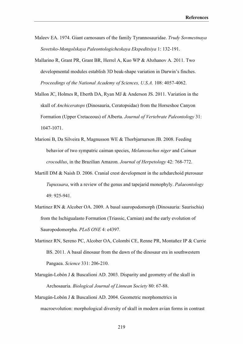
References
219
Maleev EA. 1974. Giant carnosaurs of the family Tyrannosauridae. Trudy Sovmestnaya
Sovetsko-Mongolskaya Paleontologicheskaya Ekspeditsiya 1: 132-191.
Mallarino R, Grant PR, Grant BR, Herrel A, Kuo WP & Abzhanov A. 2011. Two
developmental modules establish 3D beak-shape variation in Darwin’s finches.
Proceedings of the National Academy of Sciences, U.S.A. 108: 4057-4062.
Mallon JC, Holmes R, Eberth DA, Ryan MJ & Anderson JS. 2011. Variation in the
skull of Anchiceratops (Dinosauria, Ceratopsidae) from the Horseshoe Canyon
Formation (Upper Cretaceous) of Alberta. Journal of Vertebrate Paleontology 31:
1047-1071.
Marioni B, Da Silveira R, Magnusson WE & Thorbjarnarson JB. 2008. Feeding
behavior of two sympatric caiman species, Melanosuchus niger and Caiman
crocodilus, in the Brazilian Amazon. Journal of Herpetology 42: 768-772.
Martill DM & Naish D. 2006. Cranial crest development in the azhdarchoid pterosaur
Tupuxuara, with a review of the genus and tapejarid monophyly. Palaeontology
49: 925-941.
Martinez RN & Alcober OA. 2009. A basal sauropodomorph (Dinosauria: Saurischia)
from the Ischigualasto Formation (Triassic, Carnian) and the early evolution of
Sauropodomorpha. PLoS ONE 4: e4397.
Martinez RN, Sereno PC, Alcober OA, Colombi CE, Renne PR, Montañez IP & Currie
BS. 2011. A basal dinosaur from the dawn of the dinosaur era in southwestern
Pangaea. Science 331: 206-210.
Marugán-Lobón J & Buscalioni AD. 2003. Disparity and geometry of the skull in
Archosauria. Biological Journal of Linnean Society 80: 67-88.
Marugán-Lobón J & Buscalioni AD. 2004. Geometric morphometrics in
macroevolution: morphological diversity of skull in modern avian forms in contrast
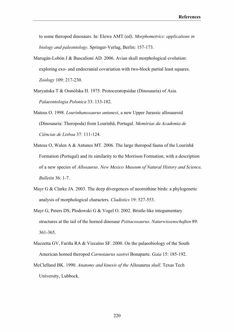
References
220
to some theropod dinosaurs. In: Elewa AMT (ed). Morphometrics: applications in
biology and paleontology. Springer-Verlag, Berlin: 157-173.
Marugán-Lobón J & Buscalioni AD. 2006. Avian skull morphological evolution:
exploring exo- and endocranial covariation with two-block partial least squares.
Zoology 109: 217-230.
Maryańska T & Osmólska H. 1975. Protoceratopsidae (Dinosauria) of Asia.
Palaeontologia Polonica 33: 133-182.
Mateus O. 1998. Lourinhanosaurus antunesi, a new Upper Jurassic allosauroid
(Dinosauria: Theropoda) from Lourinhã, Portugal. Memórias da Academia de
Ciências de Lisboa 37: 111-124.
Mateus O, Walen A & Antunes MT. 2006. The large theropod fauna of the Lourinhã
Formation (Portugal) and its similarity to the Morrison Formation, with a description
of a new species of Allosaurus. New Mexico Museum of Natural History and Science,
Bulletin 36: 1-7.
Mayr G & Clarke JA. 2003. The deep divergences of neornithine birds: a phylogenetic
analysis of morphological characters. Cladistics 19: 527-553.
Mayr G, Peters DS, Plodowski G & Vogel O. 2002. Bristle-like integumentary
structures at the tail of the horned dinosaur Psittacosaurus. Naturwissenschaften 89:
361-365.
Mazzetta GV, Fariña RA & Vizcaíno SF. 2000. On the palaeobiology of the South
American horned theropod Carnotaurus sastrei Bonaparte. Gaia 15: 185-192.
McClelland BK. 1990. Anatomy and kinesis of the Allosaurus skull. Texas Tech
University, Lubbock.
References
221
McGowan AJ & Dyke GJ. 2007. A morphospace-based test for competitive exclusion
among flying vertebrates: did birds, bats and pterosaurs get in each other’s space?
Journal of Evolutionary Biology 20: 1230-1236.
McHenry CR, Clausen PD, Daniel WJT, Meers MB & Pendharkar A. 2006.
Biomechanics of the rostrum in crocodilians: a comparative analysis using finite-
element modeling. The Anatomical Record 288: 827-849.
McNamara KJ. 1982. Heterochrony and phylogenetic trends. Paleobiology 8: 130-142.
Meloro C & Jones M. 2012. Tooth and cranial disparity in the fossil relatives of
Sphenodon (Rhynchocephalia) dispute the persistent “living fossil” label. Journal of
Evolutionary Biology 25: 2194-2209.
Medem F. 1963. Osteología craneal, distribución geográfica y ecología de
Melanosuchus niger (Spix), (Crocodylia, Alligatoridae). Ibidem 12: 5-19.
Medem F. 1981. Los Crocodylia de Sur América. Vol. I. Los Crocodylia de Colombia.
Colciencias, Bogotá.
Meier R, Shiyang K, Vaidya G & Ng PKL. 2006. DNA barcoding and taxonomy in
Diptera: a tale of high intraspecific variability and low identification success.
Systematic Biology 55: 715-728.
Meier R, Zhang G & Ali F. 2008. The use of mean instead of smallest interspecific
distances exaggerates the size of the “barcoding gap” and leads to misidentification.
Systematic Biology 57: 809-813.
Meyer CP & Paulay G. 2005. DNA barcoding: error rates based on comprehensive
sampling. PloS Biology 3: e422.
Mickoleit G. 2004. Phylogenetische Systematik der Wirbeltiere. Verlag Dr. Friedrich
Pfeil, München.
References
222
Middleton KM & Gatesy SM. 2000. Theropod forelimb design and evolution.
Zoological Journal of the Linnean Society 128: 149-187.
Midford PE, Garland Jr T & Maddison WP. 2005. PDAP Package of Mesquite. Version
1.07. http://mesquiteproject.org/pdap_mesquite/
Miller SC & Bowman BM. 1981. Medullary bone osteogenesis following estrogen
administration to mature male Japanese quail. Developmental Biology 87: 52-63.
Molnar RE. 1990. Variation in theory and in theropods. In: Carpenter K & Currie PJ
(eds). Dinosaur systematics: approaches and perspectives. Cambridge University
Press, Cambridge: 71-79.
Molnar RE. 2005. Sexual selection and sexual dimorphism in theropods. In: Carpenter
K (ed). The carnivorous dinosaurs. Indiana University Press, Bloomington: 284-312.
Molnar RE, Kurzanov SM, Dong Z. 1990. Carnosauria. In: Weishampel DB, Dodson P,
& Osmólska H (eds). The Dinosauria. University of California Press, Berkeley 169-
209.
Monteiro LR & Soares M. 1997. Allometric analysis of the ontogenetic variation and
evolution of the skull in Caiman Spix, 1825 (Crocodylia: Alligatoridae).
Herpetologica, 53: 62-69.
Monteiro LR, Cavalcanti MJ & Sommer HSJ. 1997. Comparative ontogenetic shape
changes in the skull of Caiman species (Crocodylia, Alligatoridae). Journal of
Morphology 321: 53-62.
Mook CC. 1921. Notes on the postcranial skeleton in the Crocodilia. Bulletin of the
American Museum of Natural History 44: 67-100.
Moser M. 2003. Plateosaurus engelhardti Meyer, 1937 (Dinosauria: Sauropodomorpha)
aus dem Feuerletten (Mittelkeuper; Obertrias) von Bayern. Zitteliana B 24: 3-186.
References
223
Nesbitt SJ. 2007. The anatomy of Effigia okeeffeae (Archosauria, Suchia), theropod-like
convergence, and the distribution of related taxa. Bulletin of the American Museum
of Natural History 302: 1-84.
Nesbitt SJ. 2011. The early evolution of archosaurs: relationships and the origin of
major clades. Bulletin of the American Museum of Natural History 352: 1-292.
Nesbitt SJ & Butler RJ. 2013. Redescription of the archosaur Parringtonia gracilis from
the Middle Triassic Manda beds of Tanzania, and the antiquity of Erpetosuchidae.
Geological Magazine 150: 225-238.
Norell MA. 1988. Cladistics approaches to evolution and paleobiology as applied to
the phylogeny of alligatorids. Yale University, New Haven.
Norell MA & Xu X. 2005. Feathered dinosaurs. Annual Review of Earth and Planetary
Sciences 33: 277-299.
Norell MA, Clark JM, Turner AH, Makovicky PJ, Barsbold R & Rowe T. 2006. A new
dromaeosaurid theropod from Ukhaa Tolgod (Ömnögov, Mongolia). American
Museum Novitates 3545: 1-51.
Norman DB, Witmer LM & Weishampel DB. 2004. Basal Ornithischia. In:
Weishampel DB, Dodson P & Osmólska H (eds). The Dinosauria. University of
California Press, Berkeley: 325-334.
Novas FE & Pol D. 2002. Alvarezsaurid relationships reconsidered. In: Chiappe LM &
Witmer LM (eds). Mesozoic birds: above the heads of dinosaurs. University of
California Press, Berkeley: 121-125.
O'Connor JK & Chiappe LM. 2011. A revision of enantiornithine (Aves:
Ornithothoraces) skull morphology. Journal of Systematic Palaeontology 9: 135-157.
O'Higgins P. 2000. The study of morphological variation in the hominid fossil record:
biology, landmarks and geometry. Journal of Anatomy 197: 103-120.
References
224
O’Higgins P & Collard M. 2002. Sexual dimorphism and facial growth in papionin
monkeys. Journal of Zoology 257: 255-272.
Osborn HF. 1903. The skull of Creosaurus. Bulletin of the American Museum of
Natural History 19: 697-701.
Osborn HF. 1912. Crania of Tyrannosaurus and Allosaurus. Memoirs of the American
Museum of Natural History 1: 1-30.
Ősi A. 2010. Feeding-related characters in basal pterosaurs: implications for jaw
mechanism, dental function and diet. Lethaia 44: 136-152.
Ősi A, Weishampel DB & Jianu CM. 2005. First evidence of azhdarchid pterosaurs
from the Late Cretaceous of Hungary. Acta Palaeontologica Polonica 50: 777-787.
Osmólska H, Currie PJ & Barsbold R. 2004. Oviraptorosauria. In: Weishampel DB,
Dodson P & Osmólaka H (eds). The Dinosauria. University of California Press,
Berkeley: 165-183.
Ostrom JH. 1978. The osteology of Compsognathus longipes Wagner. Zitteliana 4: 73-
118.
Otte KC. 1974. Research programme Melanosuchus niger in the Manu National Park.
World Wildlife Yearbook 1973-1974: 257-260.
Ouyang H & Ye Y. 2002. The first mamenchisaurian skeleton with complete skull,
Mamenchisaurus youngi. Sichuan Science and Technology Press. Chengdu.
Pacheco LF. 1994. Estimating crocodilian abundance in forest lagoons. In: Group,
Proceedings of the 12th Working Meeting of the IUCN-SSC Crocodile Specialist.
Crocodiles. IUCN, Gland: 241-244.
Padian K. 1983. A functional analysis of flying and walking in pterosaurs. Paleobiology
9: 218-239.

References
225
Padian K. 1985. The origins and aerodynamics of flight in extinct vertebrates.
Palaeontology 28: 413-433.
Padian K 2008a. The Early Jurassic pterosaur Dorygnathus banthensis (Theorori, 1830).
Special Papers in Palaeontology 80: 1-64.
Padian K 2008b. The early Jurassic pterosaur Campylognathoides Strand, 1928. Special
Papers in Palaeontology 80: 65-107.
Padian K & Horner JR. 2011a. The definition of sexual selection and its implications
for dinosaurian biology. Journal of Zoology 283: 23-27.
Padian K & Horner JR. 2011b. The evolution of “bizarre structures” in dinosaurs:
biomechanics, sexual selection, social selection or species recognition? Journal of
Zoology 283: 3-17.
Padian K & Horner JR. In press. Misconceptions of sexual selection and species
recognition: a response to Knell et al. and to Mendelson and Shaw. Trends in
Ecology and Evolution.
Padian K, de Ricqlès A de & Horner JR. 2001. Dinosaurian growth rates and bird
origins. Nature 412: 405-408.
Palmer C & Dyke GJ. 2010. Biomechanics of the unique pterosaur pteroid. Proceedings
of the Royal Society B 277: 1121-1127.
Paradis E, Claude J & Strimmer K. 2004. APE: analyses of phylogenetics and evolution
in R language. Bioinformatics 20: 289-290. http://ape.mpl.ird.fr/
Paul GS. 2002. Dinosaurs of the air: the evolution and loss of flight in dinosaurs and
birds. John Hopkins University Press, Baltimore.
Paul GS. 2008. The extreme lifestyle and habits of the gigantic tyrannosaurid
superpredators of the Late Cretaceous of North America and Asia. In: Larson P, &
References
226
Carpenter K (eds). Tyrannosaurus rex, the tyrant king. Indiana University Press,
Bloomington: 307-352.
Paul GS. 2010. The Princeton field guide to dinosaurs. Princeton University Press, New
Jersey.
Peyer K. 2006. A reconsideration of Compsognathus from the Upper Tithonian of
Canjuers, southeastern France. Journal of Vertebrate Paleontology 26: 879-896.
Pierce SE & Benton MJ. 2006. Pelagosaurus typus Bronn, 1841 (Mesoeucrocodylia:
Thalattosuchia) from the Upper Lias (Toarcian, Lower Jurassic) of Somerset,
England. Journal of Vertebrate Paleontology 26: 621-635.
Pierce SE, Angielczyk KD & Rayfield EJ. 2008. Patterns of morphospace occupation
and mechanical performance in extant crocodilian skulls: a combined geometric
morphometric and finite element modelling approach. Journal of Morphology 269:
840-864.
Piña C, Larriera A, Siroski P & Verdade LM. 2007. Cranial sexual discrimination in
hatchling broad-snouted caiman (Caiman latirostris). Iheringia, Série Zoologia 97:
17-20.
Pinheiro FL, Fortier DC, Schultz CL, Artur J, Andrade FG de & Bantim RAM. 2011.
New information on Tupandactylus imperator, with comments on the relationships
of Tapejaridae (Pterosauria). Acta Palaeontologica Polonica 56: 567-580.
Piras P, Teresi L, Buscalioni AD & Cubo J. 2009. The shadow of forgotten ancestors
differently constrains the fate of Alligatoroidea and Crocodyloidea. Global Ecology
and Biogeography 18: 30-40.
Piras P, Colangelo P, Adams DC, Buscalioni AD, Cubo J, Kotsakis T, Meloro C & Raia
P. 2010. The Gavialis-Tomistoma debate: the contribution of skull ontogenetic
References
227
allometry and growth trajectories to the study of crocodylian relationships. Evolution
& Development 12: 568-579.
Piras P, Salvi D, Ferrara G, Maiorino L, Delfino M, Pedde L & Kotsakis T. 2011. The
role of post-natal ontogeny in the evolution of phenotypic diversity in Podarcis
lizards. Journal of Evolutionary Biology 24: 2705-2720.
Platt SG, Rainwater TR, Thorbjarnarson JB, Finger AG, Anderson TA & McMurry S. T.
2009. Size estimation, morphometrics, sex ratio, sexual size dimorphism, and
biomass of Morelet’s crocodile in northern Belize. Caribbean Journal of Science 45:
80-93.
Plotkin MJ, Medem F, Mittermeier RA & Constable IA. 1983. Distribution and
conservation of the black caiman (Melanosuchus niger). In: Rhodin A & Mitaya K
(eds). Advances in Herpetology and Evolutionary Biology. Museum of Comparative
Zoology, Cambridge: 695-705.
Poe S. 1997. Data set incongruence and the phylogeny of crocodilians. Systematic
Biology 45: 393-414.
Pol D & Gasparini Z. 2009. Skull anatomy of Dakosaurus andiniensis (Thalattosuchia:
Crocodylomorpha) and the phylogenetic position of Thalattosuchia. Journal of
Systematic Palaeontology 7: 163-197.
Pol D & Powell JE. 2007. Skull anatomy of Mussaurus patagonicus (Dinosauria:
Sauropodomorpha) from the Late Triassic of Patagonia. Historical Biology 19: 125-
144.
Pol D & Rauhut OWM. 2012. A Middle Jurassic abelisaurid from Patagonia and the
early diversification of theropod dinosaurs. Proceedings of the Royal Society B 279:
3170-3175.
References
228
Pol D, Rauhut OWM, Lecuona A, Leardi JM, Xu X, Clark JM. In press. A new fossil
from the Jurassic of Patagonia reveals the early basicranial evolution and the origins
of Crocodyliformes. Biological Reviews.
Porro LB, Holliday CM, Anapol F, Ontiveros LC, Ontiveros LT & Ross CF. 2011. Free
body analysis, beam mechanics, and finite element modeling of the mandible of
Alligator mississippiensis. Journal of Morphology 272: 910-937.
Prentice KC, Ruta M & Benton MJ. 2011. Evolution of morphological disparity in
pterosaurs. Journal of Systematic Palaeontology 9: 337-353.
Prieto-Márquez A, Gignac PM & Joshi S. 2007. Neontological evaluation of pelvic
skeletal attributes purported to reflect sex in extinct non-avian archosaurs. Journal of
Vertebrate Paleontology 27: 603-609.
Prieto-Márquez A. 2010. Global phylogeny of hadrosauridae (Dinosauria: Ornithopoda)
using parsimony and Bayesian methods. Zoological Journal of the Linnean Society
159: 435-502.
Prieto-Márquez A & Norell MA. 2011. Redescription of a nearly complete skull of
Plateosaurus (Dinosauria: Sauropodomorpha) from the Late Triassic of Trossingen
(Germany). American Museum Novitates 3727: 1-58.
Prum RO. 1999. Development and evolutionary origin of feathers. Journal of
Experimental Zoology (MOL DEV EVOL) 285: 291-306.
Prum RO & Brush AH. 2002. The evolutionary origin and diversification of feathers.
The Quarterly Review of Biology 77: 261-295.
Purvis A. & Rambaut A. 1995. Comparative analysis by independent contrasts (CAIC) -
an Apple Macintosh application for analysing comparative data. Computer
Applications in the Biosciences 11: 247-251.
References
229
Rauhut OWM. 2003a. The interrelationships and evolution of basal theropod dinosaurs.
Special Papers in Palaeontology 69: 1-213.
Rauhut OWM. 2003b. A tyrannosauroid dinosaur from the Upper Jurassic of Portugal.
Palaeontology 46: 903-910.
Rauhut OWM. 2004. Provenance and anatomy of Genydectes serus, a large toothed
ceratosaur (Dinosauria: Theropoda) from Patagonia. Journal of Vertebrate
Paleontology 24: 894-902.
Rauhut OWM. 2007. The myth of the conservative character: braincase characters in
theropod phylogenies. Hallesches Jahrbuch für Geowissenschaften, Beiheft 23: 51-
54.
Rauhut OWM. In press. New observations on the skull of Archaeopteryx.
Paläontolologische Zeitschrift.
Rauhut OWM & Fechner R. 2005. Early development of the facial region in a non-
avian theropod dinosaur. Proceedings of the Royal Society B 272: 1179-1183.
Rauhut OWM, Fechner R, Remes K & Reis K. 2011. How to get big in the Mesozoic:
the evolution of the sauropodomorph body plan. In: Klein N, Remes K, Gee CT &
Sander PM (eds). Biology of the sauropod dinosaurs: understanding the life of giants.
Indiana University Press, Bloomington: 119-149.
Rauhut OWM, Foth C, Tischlinger H & Norell MA. 2012. Exceptionally preserved
juvenile megalosauroid theropod dinosaur with filamentous integument from the
Late Jurassic of Germany. Proceedings of the National Academy of Sciences, U.S.A.
109: 11746-11751.
Rayfield EJ. 2004. Cranial mechanics and feeding in Tyrannosaurus rex. Proceedings
of the Royal Society B 271: 1451-1459.
References
230
Rayfield EJ. 2005. Aspects of comparative cranial mechanics in the theropod dinosaurs
Coelophysis, Allosaurus and Tyrannosaurus. Zoological Journal of the Linnean
Society 144: 309-316.
Rayfield EJ. 2007. Finite Element Analysis and understanding the biomechanics and
evolution of living and fossil organisms. Annual Review of Earth and Planetary
Sciences 35: 541-576.
Rayfield EJ. 2011. Structural performance of tetanuran theropod skulls, with emphasis
on the Megalosauridae, Spinosauridae and Charcharodontosauridae. Special Papers
in Palaeontology 86: 241-253.
Rayfield EJ, Norman DB, Horner CC, Horner JR, Smith PM, Thomason JJ & Upchurch
P. 2001. Cranial design and function in a large theropod dinosaur. Nature 409: 1033-
1037.
R Development Core Team. 2011. R: A Language and Environment for Statistical
Computing. R Foundation for Statistical Computing, Vienna. http://www.R-
project.org
Reisz RR, Evans DC, Sues H-D & Scott D. 2010. Embryonic skeletal anatomy of the
sauropodomorph dinosaur Massospondylus from the Lower Jurassic of South Africa.
Journal of Vertebrate Paleontology 30: 1653-1665.
Richtsmeier JT, Corner BD, Grausz HM, Cheverud JM & Danahey SE. 1993. The role
of postnatal growth pattern in the production of facial morphology. Systematic
Biology 42: 307-330.
Rieppel OC. 1993. Patterns of diversity in the reptilian skull. In: Hanken J & Hall BK
(eds). The skull. Vol. 2. Patterns of structural and systematic diversity. University of
Chicago Press, Chicago: 344-390.

References
231
Robert P & Escoufier Y. 1976. A unifying tool for linear multivariate statistical
methods: the RV-coefficient. Applied Statistics 25: 257-265.
Rohlf FJ. 2000. Statistical power comparisons among alternative morphometric
methods. American Journal of Physical Anthropology 111: 463-478.
Rohlf FJ. 2003. tpsRelw, relative warps analysis, version 1.36. Department of Ecology
and Evolution, State University of New York at Stony Brook.
http://life.bio.sunysb.edu/morph/
Rohlf FJ. 2005. tpsDig, digitize landmarks and outlines, version 2.05. Department of
Ecology and Evolution, State University of New York at Stony Brook.
http://life.bio.sunysb.edu/morph/
Rohlf FJ & Corti M. 2000. Use of two-block partial least-squares to study covariation in
shape. Systematic Biology 49: 740-753.
Rohlf FJ & Marcus LF. 1993. A revolution in morphometrics. Trends in Ecology and
Evolution 8: 129-132.
Rohlf FJ & Slice DE. 1990. Extensions of the Procrustes method for the optimal
superimposition of landmarks. Systematic Zoology 39: 40-59.
Romer AS. 1956. Osteology of the reptiles. University of Chicago Press, Chicago.
Rootes WL, Chabreck RH, Wright VL, Brown BW & Hess TJ. 1991. Growth rates of
American alligators in estuarine and palustrine wetlands in Louisiana. Estuaries 14:
489-494.
Russell DA. 1970. Tyrannosaurs from the Late Cretaceous of western Canada. National
Museum of Natural Sciences, Publications in Palaeontology 1: 1-34.
Ruta M. 2009. Patterns of morphological evolution in major groups of Palaeozoic
Temnospondyli (Amphibia: Tetrapoda). Special Papers in Palaeontology 81: 91-120.

References
232
Ruta M, Wagner PJ & Coates MI. 2006. Evolutionary patterns in early tetrapods. I.
Rapid initial diversification followed by decrease in rates of character change.
Proceedings of the Royal Society B 273: 2107-2111.
Ryan MJ, Russell AP, Eberth DA & Currie PJ. 2001. The taphonomy of a Centrosaurus
(Ornithischia: Certopsidae) bone bed from the Dinosaur Park Formation (upper
Campanian), Alberta, Canada, with comments on cranial ontogeny. Palaios 16: 482-
506.
Ryan MJ, Holmes R & Russell AP. 2007. A revision of the late Campanian
centrosaurine ceratopsid genus Styracosaurus from the wester interior of North
America. Journal of Vertebrate Paleontology 27: 944-962.
Sadleir RW & Makovicky PJ. 2008. Cranial shape and correlated characters in
crocodilian evolution. Journal of Evolutionary Biology 21: 1578-1596.
Sadleir RW, Barrett PM & Powell HP. 2008. The anatomy and systematics of
Eustreptospondylus oxoniensis, a theropod dinosaur from the Middle Jurassic of
Oxfordshire, England. Monograph of the Palaeontological Society 627: 1-82.
Sakamoto M. 2010. Jaw biomechanics and the evolution of biting performance in
theropod dinosaurs. Proceedings of the Royal Society B 277: 3327-3333.
Sánchez-Villagra MR & Aguilera OA. 2006. Neogene vertebrates from Urumaco,
Falcón State, Venezuela: Diversity and significance. Journal of Systematic
Palaeontology 4: 213-220.
Sander PM, Klein N, Buffetaut E, Cuny G, Suteethorn V & Le Loeuff J. 2004. Adaptive
radiation in sauropod dinosaurs: bone histology indicates rapid evolution of giant
body size through acceleration. Organisms, Diversity & Evolution 4: 165-173.
Sander PM, Mateus O, Laven T & Knötschke N. 2006. Bone histology indicates insular
dwarfism in a new Late Jurassic sauropod dinosaur. Nature 441: 739-741.
References
233
Sander PM, Klein N, Stein K & Wings O. 2011. Sauropod bone histology and its
implications for sauropod biology. In: Klein N, Remes K, Gee CT & Sander PM
(eds). Biology of the sauropod dinosaurs: understanding the life of giants. Indiana
University Press, Bloomington: 276-302.
Sanz JL, Chiappe LM, Pérez-Moreno BP, Moratalla JJ, Hernández-Carrasquilla F,
Buscalioni, AD, Ortega F, Poyato-Ariza FJ, Rasskin-Gutman D & Martíinez-Delcòs
X. 1997. A nestling bird from the Lower Cretaceous of Spain: implications for avian
skull and neck evolution. Science 276: 1543-1546.
Sampson SD. 1997. Craniofacial ontogeny in centrosaurine dinosaurs (Ornithischia:
Ceratopsidae): taxonomic and behavioral implications. Zoological Journal of the
Linnean Society 121: 293-337.
Sampson SD & Witmer LM. 2007. Craniofacial anatomy of Majungasaurus
crenatissimus (Theropoda: Abelisauridae) from the Late Cretaceous of Madagascar.
Journal of Vertebrate Paleontology 27: 32-102.
Sawyer RH & Knapp LW. 2003. Avian skin development and the evolutionary origin of
feathers. Journal of Experimental Zoology (MOL DEV EVOL) 298B: 57-72.
Schumacher GH. 1973. The head muscles and hyolaryngeal skeleton of turtles and
crocodilians. In: Gans C & Parsons TS (eds). Biology of the Reptilia. Academic Press,
London: 101-199.
Schweigert G. 2007. Ammonite biostratigraphy as a tool for dating Upper Jurassic
lithographic limestones from South Germany - first results and open questions.
Neues Jahrbuch für Geologie und Paläontologie - Abhandlungen 245: 117-125.
Schweigert G, Tischlinger H & Dietl G. 2010. Eine fossile Feder aus dem Nusplinger
Plattenkalk (Oberjura, Schwäbische Alb). Archaeopteryx 28: 31-40.
References
234
Schweitzer MH & Marshall CL. 2001. A molecular model for the evolution of
endothermy in the theropod-bird lineage. Journal of Experimental Zoology (MOL
DEV EVOL) 291: 317-338.
Schweitzer MH, Wittmeyer JL & Horner JR. 2005. Gender-specific reproductive tissue
in ratites and Tyrannosaurus rex. Science 308: 1456-1460.
Schweitzer MH, Elsey RM, Dacke CG, Horner JR & Lamm E-T. 2007. Do egg-laying
crocodilian (Alligator mississippiensis) archosaurs form medullary bone? Bone 40:
1152-1158.
Selander RK. 1966. Sexual dimorphism and differential niche utilization in birds. The
Condor 68: 113-151.
Sereno PC. 1999. The evolution of dinosaurs. Science 284: 2137-2147.
Sereno PC & Brusatte SL. 2009. Comparative assessment of tyrannosaurid
interrelationships. Journal of Systematic Palaeontology 7: 455-470.
Sereno PC & Larsson HCE. 2009. Cretaceous crocodyliforms from the Sahara. ZooKeys
28: 1-143.
Sereno PC, Forster CA, Rogers RR & Monetta AM. 1993. Primitive dinosaur skeleton
from Argentina and the early evolution of Dinosauria. Nature 361: 64-66.
Sereno PC, Larsson HCE, Sidor CA & Gado B. 2001. The giant crocodyliform
Sarcosuchus from the Cretaceous of Africa. Science 294: 1516-1519.
Shine R. 1989. Ecological causes for the evolution of sexual dimorphism: a review of
the evidence. The Quarterly Review of Biology 64: 419-461.
Singleton M. 2002. Patterns of cranial shape variation in the Papionini (Primates:
Cercopithecinae). Journal of Human Evolution 42: 547-578.
Smith DK. 1998. A morphometric analysis of Allosaurus. Journal of Vertebrate
Paleontology 18: 126-142.

References
235
Smith HF. 2011. The role of genetic drift in shaping modern human cranial evolution: a
test using microevolutionary modeling. International Journal of Evolutionary
Biology 2011: 1-11.
Smith KK. 1993. The form of the feeding apparatus in terrestrial vertebrates: Studies of
adaptation and constraint. In: Hanken J & Hall BK (eds). The skull. Vol. 3. Patterns
of structural and systematic diversity. University of Chicago Press, Chicago: 150-
196.
Smith LH & Lieberman BS. 1999. Disparity and constraint in olenelloid trilobites and
the Cambrian radiation. Paleobiology 25: 459-470.
Smith ND, Makovicky PJ, Hammer WR & Currie PJ. 2007. Osteology of
Crylophosaurus ellioti (Dinosauria: Theropoda) from the Early Jurassic of Antarctica
and implications for early theropod evolution. Zoological Journal of the Linnean
Society 151: 377-421.
Smith ND, Makovicky PJ, Agnolín FL, Ezcurra MD, Pais DF & Salisbury SW. 2008. A
Megaraptor-like theropod (Dinosauria: Tetanurae) in Australia: support for faunal
exchange across eastern and western Gondwana in the Mid-Cretaceous. Proceedings
of the Royal Society B 275: 2085-2093.
Soto M & Perea D. 2008. A ceratosaurid (Dinosauria, Theropoda) from the Late
Jurassic-Early Cretaceous of Uruguay. Journal of Vertebrate Paleontology 28: 439-
444.
Starck D. 1979. Vergleichende Anatomie der Wirbeltier auf evolutionsbiologischer
Grundlage. Bd. 2: Das Skeletsystem. Allgemeines, Skeletsubstanzen, Skelet der
Wirbeltiere einschließlich Lokomotionstypen. Springer-Verlag, Berlin.
References
236
Stecher R. 2008. A new Triassic pterosaur from Switzerland (Central Austroalpine,
Grisons), Raeticodactylus filisurensis gen. et sp. nov. Swiss Journal of Geosciences
101: 185-201.
Stein K, Csiki Z, Rogers KC, Weishampel DB, Redelstorff R, Carballido JL & Sander
PM. 2010. Small body size and extreme cortical bone remodeling indicate phyletic
dwarfism in Magyarosaurus dacus (Sauropoda: Titanosauria). Proceedings of the
National Academy of Sciences, U.S.A. 107: 9258-9263.
Stone JR. 2003. Mapping cladograms into morphospaces. Acta Zoologica (Stockholm)
84: 63-68.
Stromer E. 1934. Die Zähne des Compsognathus und Bemerkungen über das Gebiß der
Theropoda. Centralblatt für Mineralogie, Geologie und Paläontologie, B 1934: 74-
85.
Sudhaus W & Rehfeld K. 1992. Einführung in die Phylogenetik und Systematik. Gustav
Fischer Verlag, Stuttgart.
Sues H-D, Reisz RR, Hinić S & Raath MA. 2004. On the skull of Massospondylus
carinatus Owen, 1854 (Dinosauria: Sauropodomorpha) from the Elliot and
Clarens Formations (Lower Jurassic) of South Africa. Annals of Carnegie
Museum 73: 239-257.
Sues H-D, Nesbitt SJ, Berman D & Henrici AC. 2011. A late-surviving basal theropod
dinosaur from the latest Triassic of North America. Proceedings of the Royal
Society B 278: 3459-3464.
Swafford DL. 2003. PAUP*. Phylogenetic analysis using parsimony (* and other
methods). Version 4. Sinauer Associates, Sunderland, Massachusetts.
http://paup.csit.fsu.edu/
References
237
Tatarinov LP. 1960. [Discovery of pseudosuchians in the Upper Permian of the USSR].
Paleontologicheskii Zhurnal 1960: 74-80. [in Russian]
Thompson HS. 1988. Generation of swine. Tales of shame and degradation in the ’80s.
Simon & Schuster, Inc., New York.
Tischlinger H. 2002. Der Eichstätter Archaeopteryx im langwelligen UV-Licht.
Archaeopteryx 20: 21-38.
Tischlinger H. 2005a. Neue Informationen zum Berliner Exemplar von Archaeopteryx
lithographica H.v.Meyer 1861. Archaeopteryx 23: 33-50.
Tischlinger H. 2005b. Ultraviolet light investigations of fossils from the Upper Jurassic
plattenkalks of Southern Frankonia. Zitteliana B 26: 26.
Tischlinger H & Unwin DM. 2004. UV-Untersuchungen des Berliner Exemplars von
Archaeopteryx lithographica H. v. Meyer 1861 und der isolierten Archaeopteryx-
Feder. Archaeopteryx 22: 17-50.
Tischlinger H & Frey E. 2010. Multilayered is not enough! New soft tissue structures in
the Rhamphorhynchus flight membrane. Acta Geoscientia Sinica 31: 64.
Tomkins JL, LeBas NR, Witton MP, Martill DM & Humphries S. 2010. Positive
allometry and the prehistory of sexual selection. American Naturalist 176: 141-148.
Trutnau L. 1994. Krokodile. Westarp Wissenschaften, Magdeburg.
Tsuihiji T, Watabe M, Tsogtbaatar K, Tsubamoto T, Barsbold R, Suzuki S, Lee AH,
Ridgely RC, Kawahara Y & Witmer LM. 2011. Cranial osteology of a juvenile
specimens of Tarbosaurus bataar (Theropoda, Tyrannosauridae) from the Nemegt
Formatipn (Upper Cretaceous) of Bugin Tsav, Mongolia. Journal of Vertebrate
Paleontology 31: 497-517.
Tumarkin-Deratzian AR. 2009. Evaluation of long bone surface textures as ontogenetic
indicators in centrosaurine ceratopsids. The Anatomical Record 292: 1485-1500.
References
238
Tumarkin-Deratzian AR, Vann DR & Dodson P. 2006. Bone surface texture as an
ontogenetic indicator in long bones of the Canada goose Branta canadensis
(Anseriformes: Anatidae). Zoological Journal of the Linnean Society 148: 133-168.
Turner AH, Pol D, Clarke JA, Erickson GM & Norell MA. 2007. A basal
dromaeosaurid and size evolution preceding avian flight. Science 317: 1378-1381.
Turner AH, Makovicky PJ & Norell MA. 2012. A review of dromaeosaurid systematics
and paravian phylogeny. Bulletin of the American Museum of Natural History 371:
1-206.
Tykoski RS. 1998. The osteology of Syntarsus kayentakatae and its implications for
ceratosaurid phylogeny. Master thesis, The University of Texas, Austin.
Tykoski RS. 2005. Anatomy, ontogeny, and phylogeny of Coelophysoid theropods. PhD
thesis, The University of Texas, Austin.
Unwin DM. 2006. The pterosaurs from deep time. Pi Press, New York.
van der Lubbe T, Richter U & Knötschke N. 2009. Velociraptorine dromaeosaurid teeth
from the Kimmeridgian (Late Jurassic) of Germany. Acta Palaeontogica Polonica 54:
401-408.
van Drongelen W & Dullemeijer P. 1982. The feeding apparatus of Caiman crocodilus;
a functional–morphological study. Anatomischer Anzeiger 151: 337-366.
Varricchio DJ. 1997. Growth and Embryology. In: Currie PJ & Padian K (eds).
Encyclopedia of Dinosaurs. Academic Press, San Diego: 282-288.
Vasconcelos WR, Hrbek T, Da Silveira R, de Thoisy B, Marioni B & Farias IP. 2006.
Population genetic analysis of Caiman crocodilus (Linnaeus, 1758) from South
America. Genetics and Molecular Biology 29: 220-230.
References
239
Verdade LM. 2000. Regression equations between body and head measurements in the
broad-snouted Caiman (Caiman latirostris). Revista Brasileira de Biologia 60: 469-
482.
Verdade LM. 2003. Cranial sexual dimorphism in captive adult broad-snouted caiman
(Caiman latirostris). Amphibia-Reptilia 24: 92-99.
Villamarín-Jurado F & Suárez E. 2007. Nesting of the black caiman (Melanosuchus
niger) in northeastern Ecuador. Journal of Herpetology 41: 164-167.
Villier L & Eble GJ. 2004. Assessing the robustness of disparity estimates: the impact
of morphometric scheme, temporal scale, and taxonomic level in spatangoid
echinoids. Paleobiology 30: 652-665.
Wagner GP & Gauthier JA. 1999. 1,2,3 = 2,3,4: A solution to the problem of the
homology of the digits in the avian hand. Proceedings of the National Academy of
Sciences, U.S.A. 96: 5111-5116.
Walker AD. 1990. A revision of Sphenosuchus acutus Haughton, a crocodylomorph
reptile from the Elliot Formation (late Triassic or early Jurassic) of South Africa.
Philosophical Transactions of the Royal Society of London, Series B 330: 1-120.
Wang X, Zhou Z, Zhang F & Xu X. 2002. A nearly completely articulated
rhamphorhynchoid pterosaur with exeptionally well-preserved wing membranes and
“hair” from Inner Mongolia, northeast China. Chinese Science Bulletin 47: 226-230.
Wang X, Kellner AWA, Zhou Z & Campos D de A. 2005. Pterosaur diversity and
faunal turnover in Cretaceous terrestrial ecosystems in China. Nature 437: 875-879.
Webb GJW & Messel H. 1978. Morphometric analysis of Crocodylus porosus from the
north coast of Arnhem Land, northern Australia. Australian Journal of Zoology 26:
1-27.
References
240
Webb GJW, Hollis GJ & Manolis SC. 1991. Feeding, growth, and food conversion rates
of wild juvenile saltwater crocodiles (Crocodylus porosus). Journal of Herpetology
25: 462-473.
Weishampel DB. 1981. Acoustic analyses of potential vocalization in lambeosaurine
dinosaurs (Reptilia: Ornithischia). Paleobiology 7: 252-261.
Weishampel DB. 1984. Evolution of jaw mechanisms in ornithopod dinosaurs.
Advances in Anatomy, Embryology and Cell Biology 87: 1-110.
Weishampel DB & Chapman RE. 1990. Morphometric study of Plateosaurus from
Trossingen (Baden-Württemberg, Federal Republic of Germany). In: Carpenter K &
Currie PJ (eds). Dinosaur systematics: approaches and perspectives. Cambridge
University Press, Cambridge: 43-51.
Weishampel DB, Dodson P & Osmólska H. 2004. The Dinosauria. University of
California Press, Berkeley.
Wellnhofer P. 1970. Die Pterodactyloidea (Pterosauria) der Oberjura-Plattenkalke
Süddeutschlands. Bayerische Akademie der Wissenschaften, Mathematisch-
Naturwissenschaftliche Klasse, Abhhandlungen 141: 1-155.
Wellnhofer P. 1975. Die Rhamphorhynchoidea (Pterosauria) der Oberjura-Plattenkalke
Süddeutschlands. Palaeontographica Abt. A 148: 1-185.
Wellnhofer P. 1978. Pterosauria. Gustav Fischer Verlag, Stuttgart.
Wellnhofer P. 1987. New crested pterosaurs from the Lower Cretaceous of Brazil.
Mitteilungen der Bayerischen Staatssammlung für Paläontologie und historischen
Geologie 27: 175-186.
Wellnhofer P. 1991. The illustrated encyclopedia of pterosaurs. Salamander Books Ltd.,
London.
References
241
Wellnhofer P. 2008. Archaeopteryx. Der Urvogel von Solnhofen. Verlag Dr. Friedrich
Pfeil, München.
Wellnhofer P & Kellner AWA. 1991. The skull of Tapejara wellnhoferi Kellner
(Reptilia, Pterosauria) from the Lower Cretaceous Santana Formation of the Araripe
Basin, Northeastern Brazil. Mitteilungen der Bayerischen Staatssammlung für
Paläontologie und historischen Geologie 31: 89-106.
Westheide W & Rieger R. 2004. Spezielle Zoologie. Teil 2: Wirbel- oder Schädeltiere.
Spektrum Akademischer Verlag, Heidelberg.
Whitlock JA, Wilson JA & Lamanna MC. 2010. Description of a nearly complete
juvenile skull of Diplodocus (Sauropoda: Diplodocoidea) from the Late Jurassic of
North America. Journal of Vertebrate Paleontology 30: 442-457.
Widelitz RB, Jiang T, Lu J & Chuong C. 2000. β-catenin in epithelial morphogenesis:
Conversion of part of avian foot scales into feather buds with a mutated β-Catenin.
Developmental Biology 219: 98-114.
Wiesemüller B, Rothe H & Henke W. 2003. Phylogenetische Systematik. Springer-
Verlag, Berlin.
Wild R. 1984. Flugsaurier aus der Obertrias von Italien. Naturwissenschaften 71: 1-11.
Wilkinson MT, Unwin DM & Ellington CP. 2006. High lift function of the pteroid bone
and forewing of pterosaurs. Proceedings of the Royal Society B 273: 119-126.
Wilkinson PM & Rhodes WE. 1997. Growth rates of American alligators in coastal
South Carolina. Journal of Wildlife Management 61: 397-402.
Wills MA. 1998. Crustacean disparity through the Phanerozoic: comparing
morphological and stratigraphic data. Biological Journal of Linnean Society 65: 455-
500.
References
242
Wills MA, Briggs DEG & Fortey RA. 1994. Disparity as an evolutionary index: a
comparison of Cambrian and Recent arthropods. Paleobiology 20: 93-130.
Wilson JA. 2002. Sauropod dinosaur phylogeny: critique and cladistic analysis.
Zoological Journal of the Linnean Society 136: 217-276.
Wilson JA & Sereno PC. 1998. Early evolution and higher-level phylogeny of sauropod
dinosaurs. Society of Vertebrate Paleontology Memoir 5: 1-68.
Witmer LM. 1995. The Extant Phylogenetic Bracket and the importance of
reconstructing soft tissues in fossils. In: Thomason JJ (ed). Functional morphology in
vertebrate paleontology. Cambridge University Press, Cambridge: 19-33.
Witmer LM. 1997. The evolution of the antorbital cavity of archosaurs: a study in soft-
tissue reconstruction in the fossil record with an analysis of the function of
pneumaticity. Memoirs of the Society of Vertebrate Paleontology 17: 1-73.
Witmer LM. 2009. Fuzzy origins for feathers. Nature 458: 293-295.
Witton MP. 2008. A new approach to determining pterosaur body mass and its
implications for pterosaur flight. Zitteliana B 28: 143-158.
Witton MP & Naish D. 2008. A reappraisal of azhdarchid pterosaur functional
morphology and paleoecology. PLoS ONE 3: e2271.
Witton MP & Habib MB. 2010. On the size and flight diversity of giant pterosaurs, the
use of birds as pterosaur analogues and comments on the pterosaur flightlessness.
PLoS ONE 5: e13982.
Witzel U & Preuschoft H. 2005. Finite-element model construction for the virtual
synthesis of the skulls in vertebrates: case study of Diplodocus. The Anatomical
Record 283A: 391-401.
Witzel U, Mannhardt J, Goessling R, Micheeli P & Preuschoft, H. 2011. Finite element
analyses and virtual syntheses of biological structures and their application to
References
243
sauropod skulls. In: Klein N, Remes K, Gee GT & Sander PM (eds). Biology of the
sauropod dinosaurs: understanding the life of giants. Indiana University Press,
Bloomington: 171-181.
Wu P, Jiang T, Suksaweang S, Widelitz RB & Chuong C. 2004. Molecular shaping of
the beak. Science 305: 1465-1466.
Wu P, Jiang T, Shen J, Widelitz RB & Chuong C. 2006. Morphoregulation of avian
beaks: comparative mapping of growth zone activities and morphological evolution.
Developmental Dynamics 235: 1400-1412.
Wu XB, Xue H, Wu LS, Zhu JL & Wang RP. 2006. Regression analysis between body
and head measurements of Chinese alligators (Alligator sinensis) in captive
population. Animal Biodiversity and Conservation 29: 65-71.
Wu X & Chatterjee S. 1993. Dibothrosuchus elaphros, a crocodylomorph from the
Lower Jurassic of China and the phylogeny of the Sphenosuchina. Journal of
Vertebrate Paleontology 13: 58–89.
Xing L, Peng G & Shu C. 2008. Stegosaurian skin impressions from the Upper Jurassic
Shangshaximiao Formation, Zigong, Sichuan, China: A new observation. Geological
Bulletin of China 27: 1049-1053.
Xu X & Wu X. 2001. Cranial morphology of Sinornithosaurus millenii Xu et al. 1999
(Dinosauria: Theropoda: Dromaeosauridae) from the Yixian Formation of Liaoning,
China. Canadian Journal of Earth Sciences 38: 1739-1752.
Xu X & Guo Y. 2009. The origin and early evolution of feathers: insights from recent
paleontological and neontological data. Vertebrata PalAsiatica 47: 311-329.
Xu X, Norell MA, Kuang X, Wang X, Zhao Q & Jia C. 2004. Basal tyrannosauroids
from China and evidence for protofeathers in tyrannosauroids. Nature 431: 680-684.
References
244
Xu X, Clark JM, Forster CA, Norell MA, Erickson GM, Eberth DA, Jia C & Zhao Q.
2006. A basal tyrannosauroid dinosaur from the Late Jurassic of China. Nature 439:
715-718.
Xu X, Clark JM, Mo J, Choiniere J, Forster CA, Erickson GM, Hone DWE, Sullivan C,
Eberth DA, Nesbitt S, Zhao Q, Hernandez R, Jia C, Han F & Guo Y. 2009a. A
Jurassic ceratosaur from China helps clarify avian digital homologies. Nature 459:
940-944.
Xu X, Zhao Q, Norell MA, Sullivan C, Hone D, Erickson GM, Wang X, Han F & Guo
Y. 2009b. A new feathered maniraptoran dinosaur fossil that fills a morphological
gap in avian origin. Chinese Science Bulletin 54: 430-435.
Xu X, Zheng X & You H. 2010. Exceptional dinosaur fossils show ontogenetic
development of early feathers. Nature 464: 1338-1341.
Xu X, You H, Du K & Han F. 2011. An Archaeopteryx-like theropod from China and
the origin of Avialae. Nature 475: 465-470.
Xu X, Wang K, Zhang K, Ma Q, Xing L ,Sullivan C, Hu D, Cheng S & Wang S. 2012.
A gigantic feathered dinosaur from the Lower Cretaceous of China. Nature 484: 92-
95.
Yates A. 2003. The species taxonomy of the sauropodomorph dinosaurs from the
Löwenstein Formation (Norian, Late Triassic) of Germany. Palaeontology 46: 317-
337.
Yates A. 2007. The first complete skull of the Triassic dinosaur Melanorosaurus
Haughton (Sauropodomorpha: Anchisauria). Special Papers in Palaeontology 77: 9-
55.
References
245
Yates A. 2012. Basal Sauropodomorpha: the “prosauropods.” In: Brett-Surman MK,
Holtz TR Jr & Farlow JO (eds). The complete dinosaur. Indiana University Press,
Bloomington: 425-444.
You H, Lamanna MC, Harris JD, Chiappe LM, O'Connor JK, Ji S, Lü J, Yuan C, Li D,
Zhang X, Lacovara KJ, Dodson P & Ji, Q. 2006. A nearly modern amphibious bird
from the Early Cretaceous of northwestern China. Science 312: 1640-1643.
Young MT & Larvan MD. 2010. Macroevolutionary trends in the skull of
sauropodomorph dinosaurs - the largest terrestrial animals to have ever lived. In:
Elewa AMT (ed). Morphometrics for non-morphometricans Springer-Verlag, Berlin:
259-269.
Young MT, Brusatte SL, Ruta M & Andrade MB. 2010. The evolution of
Metriorhynchoidea (Mesoeucrocodylia, Thalattosuchia): an integrated approach
using geometric morphometrics, analysis of disparity, and biomechanics. Zoological
Journal of the Linnean Society 158: 801-859.
Zanno LE. 2010. A taxonomic and phylogenetic re-evaluation of Therizinosauria
(Dinosauria: Maniraptora). Journal of Systematic Palaeontology 8: 503-543.
Zanno LE & Makovicky PJ. 2011. Herbivorous ecomorphology and specialization
patterns in theropod dinosaur evolution. Proceedings of the National Academy of
Sciences, U.S.A. 108: 232-237.
Zeiss A. 1977. Jurasssic stratigraphy of Franconia. Stuttgarter Beiträge zur Naturkunde
- Serie B 31: 1-32.
Zelditch ML, Swiderski DL, Sheets HD & Fink WL. 2004. Geometric morphometrics
for biologists: a primer. Elsevier Academic Press, San Diego.
Zhao X & Currie PJ. 1993. A large crested theropod from the Jurassic of Xinjiang,
People’s Republic of China. Canadian Journal of Earth Sciences 30: 2027-2036.
References
246
Zhang F, Zhou Z, Xu X, Wang X & Sullivan C. 2008. A bizarre Jurassic maniraptoran
from China with elongate ribbon-like feathers. Nature 455: 1105-1108.
Zheng X, You H, Xu X & Dong Z. 2009. An Early Cretaceous heterodontosaurid
dinosaur with filamentous integumentary structures. Nature 458: 333-336.
Zhong Z. 1996. Cranial anatomy of Shunosaurus and Camarasaurus (Dinosauria:
Sauropoda) and the phylogeny of Sauropoda. PhD thesis, Texas Tech University,
Lubbock.
Zhou Z. 2004. The origin and early evolution of birds: discoveries, disputes, and
perspectives from fossil evidence. Naturwissenschaften 91: 455-471.
Zhou Z & Zhang F. 2003. Anatomy of the primitive bird Sapeornis chaoyangensis from
the Early Cretaceous of Liaoning, China. Canadian Journal of Earth Sciences 40:
731-747.
Zhou Z & Zhang F. 2004. A precocial avian embryo from the Lower Cretaceous of
China. Science 306: 653.
Zhou Z, Wang X, Zhang F & Xu X. 2000. Important features of Caudipteryx - Evidence
from two nearly complete new specimens. Vertebrata PalAsiatica 38: 241-254.
Zhou Z, Clarke J, Zhang Z & Wings O. 2004. Gastroliths in Yanornis: an indication of
the earliest radical diet-switching and gizzard plasticity in the lineage leading to
living birds? Naturwissenschaften 91: 571-574.
Ziegler T & Olbort S. 2007. Genital structures and sex identification in crocodiles.
Crocodile Specialist Group Newsletter 26: 16-17.
Zusi RL. 1993. Patterns of diversity in the avian skull. In: Hanken J & Hall BK (eds).
The skull. Vol. 2. Patterns of structural and systematic diversity. University of
Chicago Press, Chicago: 391-437.
Curriculum vitae
247
Curriculum vitae
PERSONAL DATA
• Name: Christian Foth
• Date of birth: 12th November 1984
• Place of birth: Rostock
• Nationality: German
EDUCATION
PhD thesis: Ontogenetic, macroevolutionary and morphofunctional patterns
in archosaur skulls: a morphometric approach (Supervisor: PD
Dr. Oliver Rauhut, Bayerische Staatssammlung für Paläontologie
und Geologie).
since 01/2011 PhD program Evolution, Ecology and Systematics at the Ludwig-
Maximilians-University of Munich
since 04/2010 PhD study of Palaeontology at the Ludwig-Maximilians-
University of Munich
Diploma thesis: Die Morphologie des Erstlingsgefieders ausgewählter Vogeltaxa
unter Berücksichtigung der Phylogenie (The morphology of
neoptile feathers from subset bird taxa under regard of their
phylogeny), 183 p. (Supervisor/reviewer: Prof. Stefan Richter,
Allgemeine und Spezielle Zoologie, University of Rostock; Dr.
Gerald Mayr, Forschungsinstitut Senckenberg, Frankfurt).
10/2004-12/2009 study of Biology at the University of Rostock, with final degree
06/2004 A level at the Käthe-Kollwitz-Gymnasium, Rostock
Curriculum vitae
248
CAREER HISTORY
011/2011- researcher in the DFG project: Macroevolutionary patterns in the
evolution of the skull in theropod dinosaurs: a morphometric
approach (PD Dr. Oliver Rauhut, Bayerische Staatssammlung für
Paläontologie und Geologie)
08/2011-10/2011 researcher in the DFG project: The early evolution of the
Neopterygii (Dr. Adriana López-Arbarello, Bayerische
Staatssammlung für Paläontologie und Geologie)
03/2011-07/2011 researcher in the DFG project: Middle to Late Jurassic archosaurs
from Chubut Province, Argentinean Patagonia and Jurassic
archosaurs radiation in the southern hemisphere (PD Dr. Oliver
Rauhut, Bayerische Staatssammlung für Paläontologie und
Geologie)
09/2010-02/2011 researcher in the DFG project: Saurischian dinosaurs from the
Elliot Formation, South Africa (PD Dr. Oliver Rauhut,
Bayerische Staatssammlung für Paläontologie und Geologie)
11/2009-07/2010 student helper at the Max Planck Institute for Demographic
Research, Rostock. Working group: Evolutionary
Biodemography (Dr. Alexander Scheuerlein)
2007 volunteer helper for the special exhibition “Evolution der Vögel”
(Evolution of birds) by the Zoological Collection of the
University of Rostock
2005-2008 assistance at zoological practises at the University of Rostock
GRANTS
06/2007-12/2009 scholarship of the Studienstiftung des deutschen Volkes
(“German National Merit Foundation”)
04/2008 Travel grant of the Studienstiftung des deutschen Volkes
(“German National Merit Foundation”) for the field work in
China
Curriculum vitae
249
FIELD WORK
02/2011 Cerro Condor, Chubut Province, Argentina (Dr. Oliver Rauhut,
ten days)
11-12/2010 Cerro Condor, Chubut Province, Argentina (Dr. Oliver Rauhut,
three weeks)
04/2008 Turpan Basin, Xinjiang, China (Dr. Oliver Wings, four weeks)
09/2005 Münchehagen, Germany (Dr. Annette Richter, one week)
RESEARCH INTERESTS
• Archosaur evolution
• Evolution and morphology of vertebrate integuments
• Ontogeny and life history of reptiles
SOFTWARE EXPERIENCES
• Cladistics: TNT, Mesquite
• Geometric morphometrics: Tps, MorphoJ, Morphologika, Landmark
• Statistics: PAST, R (basic knowledge)
• Image processing and 3D reconstruction: Photoshop, GIMP, CorelDraw, Inkscape,
Imaris, VGStudio Max
Curriculum vitae
250
SCIENTIFIC PUBLICATIONS
Foth C, Rauhut OWM. In press. The good, the bad, and the ugly: the influence of skull
reconstructions and intraspecific variability in studies of cranial morphometrics in
theropods and basal saurischians. PLoS ONE.
Foth C, Bona P, Desojo JB. In press. Intraspecific variation in the skull morphology of
the black caiman Melanosuchus niger (Alligatoridae, Caimaninae). Acta Zoologica.
Foth C, Rauhut OWM. 2013. Macroevolutionary and functional-morphological patterns
n theropod skulls: a morphometric approach. Acta Palaeontologica Polonica 58: 1-
16.
Butler RJB, Yates AM, Rauhut OWM, Foth C. 2013. A pathological tail in a basal
sauropodomorph dinosaur from South Africa: Evidence of traumatic amputation?
Journal of Vertebrate Paleontology 33: 224-228.
Rauhut OWM, Foth C, Tischlinger H, Norell MA. 2012. Exceptionally preserved
juvenile megalosauroid theropod dinosaur with filamentous integument from the
Late Jurassic of Germany. Proceedings of the National Academy of Sciences, U.S.A.
109: 11746-11751.
Buchwitz M, Foth C, Kogan I, Voigt S. 2012. On the use of osteoderm features for a
cladistic approach on the internal relationships of the Chroniosuchia (Tetrapoda:
Reptiliomorpha). Palaeontology 55: 623-640.
Foth C, Brusatte SL, Butler, RJ. 2012. Do different disparity proxies converge on a
common signal? Insights from the cranial morphometrics and evolutionary history of
Pterosauria (Diapsida: Archosauria). Journal of Evolutionary Biology 25: 904-915.
Foth C. 2012. On the identification of feather structures in stem-line representatives of
birds: evidence from fossils and actuopalaeontology. Paläontologische Zeitschrift 86:
91-102.
Foth C, Kalbe J, Kautz R. 2011. First evidence of Elasmosauridae (Reptilia:
Sauropterygia) in an erratic boulder of Campanian age originating from southern
Sweden or the adjacent Baltic Sea area. Zitteliana A 51: 285-290
Foth C. 2011. The morphology of neoptile feathers: ancestral state reconstruction and
its phylogenetic implications. Journal of Morphology 272: 387-403.
Foth C. 2008. Konvergente Entstehung des Fluges bei Amniota und ihre Bedeutung
zum Verständnis der Vogelevolution. Sitzungsberichte der Gesellschaft
Naturforschender Freunde zu Berlin 47: 33-66.
Curriculum vitae
251
REVIEWS FOR SCIENTIFIC JOURNALS
• Paläontologische Zeitschrift (2)
• PloS ONE (1)
PUBLIC LECTURES
Foth C. 2009. Dinosaurier. Kinder-Campus, Universität Rostock, 13.05.2009.
Foth C. 2009. Konvergente Evolution des Flugs in der Stammlinie der Vögel. Darwin
Colloquium “Evolution of organisms and environment“, TU Bergakademie
Freiberg, 12.-13.06.2009.
Foth C. 2008. Dinosaurierjagd in China. Vorweisung der Zoologischen Sammlung der
Universität Rostock, 03.12.2008.
Foth C. 2008. Zur Evolution des Fluges bei terrestrischen Wirbeltieren. Vorweisung der
Zoologischen Sammlung der Universität Rostock, 02.07.2008.
Foth C. 2006. Über die Biologie der Tyrannosauroidea. Vorweisung der Zoologischen
Sammlung der Universität Rostock, 08.11.2006.
PUBLISHED ABSTRACTS
Foth C, Bona P, Desojo JB. 2013. Intraspecific variation in the skull morphology of the
black caiman Melanosuchus niger (Alligatoridae, Caimaninae). 10th International
Congress of Vertebrate Morphology, 08-12 July 2013, Barcelona, Anatomical
Record 296, Special Feature: 250.
Rauhut OWM, Foth C. 2013. A new look for an old bird: new observations on the
morphology of Archaeopteryx. 10th International Congress of Vertebrate
Morphology, 08-12 July 2013, Barcelona, Anatomical Record 296, Special Feature:
235.
Foth C, Brusatte S, Butler, R. 2012. Cranial morphometrics, disparity and evolutionary
history of Pterosauria (Diapsida: Archosauria). 72nd Annual Meeting of the Society of
Vertebrate Paleontology, 17-20 October 2012, Raleigh, Journal of Vertebrate
Paleontology, Program and Abstracts, 2012: 96.
Hübner T, Foth C, Heinrich W-D. 2012. Reappraisal of the Upper Jurassic
Dysalotosaurus locality ‘Ig/WJ’ at Tendaguru, based on historic, taphonomic, and
demographic data. Centenary Meeting of the Paläontologische Gesellschaft, 24-29
September 2012, Berlin, Terra Nostra 2012 (3): 83-84.
Curriculum vitae
252
Foth C, Yates AM, Rauhut OWM, Butler RJ. 2012. A pathological tail in a basal
sauropodomorph dinosaur from South Africa. Centenary Meeting of the
Paläontologische Gesellschaft, 24-29 September 2012, Berlin, Terra Nostra 2012
(3): 56-57.
Foth C. 2012. The influence of taphonomy on fossil body plumages and the
identification of feather structures in stem-line representatives of birds. 10th Annual
Meeting of the European Association of Vertebrate Palaeontologists, 19-24 June
2012, Teruel, Fundamental 20: 77-79.
Rauhut OWM, Foth C. 2011. New information on Late Jurassic theropod dinosaurs
from Southern Germany. IV Congress of Latin-American Vertebrate Paleontology,
21-24 September 2011, San Juan, Ameghiniana 48, Suplemento: R120-R121.
Foth C, Scheuerlein A. 2010. When do dinosaurs become mature? - A case study on
Psittacosaurus from the Lower Cretaceous in China. 80. Jahrestagung der
Paläontologischen Gesellschaft, 05-08 October 2010, München, Zitteliana B 29: 37.
Foth C, Kalbe J, Kautz R. 2010. First evidence of an Elasmosauridae (Reptilia:
Sauropterygia) in a Geschiebe from the Campanian (Upper Cretaceous) of South
Scandinavia. 80. Jahrestagung der Paläontologischen Gesellschaft, 05-08 October
2010, München, Zitteliana B 29: 36.
Foth C. 2009. The morphology of neoptile feathers in extant birds - implications for
feather evolution in theropod dinosaurs. 79. Jahrestagung der Paläontologischen
Gesellschaft, 05-07 October 2009, Bonn, Terra Nostra 2009 (3): 34.
PROFESSIONAL ASSOCIATIONS
• Society of Vertebrate Paleontology
• European Association of Vertebrate Palaeontology
• Verein der Freunde und Förderer der Zoologischen Sammlung Rostock
• Deutsche Zoologische Gesellschaft (German Zoological Society)
• Paläontologische Gesellschaft (German Palaeontological Society)
Eidesstattliche Versicherung
253
Appendix
of
Ontogenetic, macroevolutionary and morphofunctional patterns in
archosaur skulls: a morphometric approach
by Christian Foth
Supplementary information of Chapter 1 to Chapter 6
S2
SUPPLEMATARY INFORMATION OF CHAPTER 1
Introduction and summery of the thesis
1. Taxon sampling
2. Description of Landmarks
3. Methods
4. Phylogeny
Supplementary information of Chapter 1
S3
1. TAXON SAMPLING
For both geometric morphometric analyses performed in Chapter 1 the skulls of 21
pseudosuchians (19 Crocodylomorpha and two outgroup taxa) and 54 saurischian taxa
were sampled. The datasets are mainly based on published reconstructions, except the
skulls of the ontogenetic series of Melanosuchus niger. The skulls for the later based on
photo material (see Chapter 3).
Table S1.1. List of crocodylomorph taxa with data of occurences (in million years, Ma). SMF
Senckenberg Naturmuseum Frankfurt (Germany); NHMW Naturhistorisches Museum Wien (Austria);
ZSM Zoologische Staatssammlung München (Germany). *Ontogenetic series of Melanosuchus niger:
SMF 30113; SMF 40172; ZSM 13/1911; ZSM 130/1911; ZSM 64/1911; NHMW 2025 (Table S3.1, see
supplementary information of Chapter 3).
Taxon Systematic affinities Age (Ma) Sources
Erpetosuchus granti Pseudosuchia (outgroup) 220.3 Benton & Walker 2002
Gracilisuchus stipanicicorum Pseudosuchia (outgroup) 241.1 Nesbitt 2011
Dibothrosuchus elaphros basal Crocodylomorpha 193.1 Wu & Chatterjee 1993
Dromicosuchus grallator basal Crocodylomorpha 220.3 Nesbitt 2011
Sphenosuchus acutus basal Crocodylomorpha 195.6 Nesbitt 2011
Protosuchus richardsoni basal Crocodyliformes 199.1 Nesbitt 2011
Anatosuchus minor Notosuchia 112.5 Sereno & Larrson 2009
Araripesuchus wegneri Notosuchia 112.5 Sereno & Larrson 2009
Baurusuchus salgadoensis Notosuchia 88.5 Carvalho et al. 2005
Simosuchus clarki Notosuchia 68.1 Kley et al. 2010
Dakosaurus andiniensis Thalattosuchia 145.5 Pol & Gasparini 2009
Pelagosaurus typus Thalattosuchia 182.5 Pierce & Benton 2006
Hamadasuchus rebouli Neosuchia 102.7 Larsson & Sues 2007
Kaprosuchus saharicus Neosuchia 96.6 Sereno & Larrson 2009
Sarcosuchus imperator Neosuchia 119.5 Sereno et al. 2001
Alligator sinensis Crocodylia Recent Iordansky 1973
Crocodylus thorbjarnarsoni Crocodylia 2.7 Brochu & Storrs 2012
Gavialis gangeticus Crocodylia Recent Iordansky 1973
*Melanosuchus niger Crocodylia Recent see Chapter 3
Osteolaemus tetraspis Crocodylia Recent Iordansky 1973
Tomistoma schlegelii Crocodylia Recent Iordansky 1973
Supplementary information of Chapter 1
S4
Table S1.2. List of saurischian taxa with data of occurences (in million years, Ma). *ontogenetic series
(see also taxonomical comments below); **juvenile specimens.
Taxon Systematic affinities Age (Ma) Sources
Herrerasaurus ischigualastensis basal Saurischia 220.3 Nesbitt 2011
Chuxiongosaurus lufengensis basal Sauropodomorpha 195.6 Lü et al. 2010b
Eoraptor lunensis basal Sauropodomorpha 228.3 Martinez et al. 2011
Jingshanosaurus xinwaensis basal Sauropodomorpha 199.1 Yates 2012
*Massospondylus carinatus basal Sauropodomorpha 195.6 Gow et al. 1990; Reisz et al.
2010
Melanorosaurus readi basal Sauropodomorpha 211.6 Yates 2007
Pampadromaeus barberenai basal Sauropodomorpha 220.3 Cabreira et al. 2011
Plateosaurus engelhardti basal Sauropodomorpha 208.6 Yates 2003
Unaysaurus tolentinoi basal Sauropodomorpha 213.6 Leal et al. 2004
Mamenchisaurus youngi basal Sauropoda 153.4 Ouyang & Ye 2002
Shunosaurus lii basal Sauropoda 158.6 Zhong 1996
Diplodocus longus Neosauropoda 150.6 Wilson & Sereno 1998;
Whitlock et al. 2010
Abydosaurus mcintoshi Macronaria 101.3 Chure et al. 2010
*Antarctosaurus wichmannianus Macronaria 77.1 Gallina & Apesteguía 2011
*Titanosaurid embryo Macronaria c. 81.5 Garcia et al. 2010
Camarasaurus lentus Macronaria 150.6 Wilson & Sereno 1998
Giraffatitan brancai Macronaria 153.4 Wilson & Sereno 1998
Daemonosaurus chauliodus basal Theropoda 203.6 Sues et al. 2011
Tawa hallae basal Theropoda 213.6 Nesbitt 2011
Coelophysis bauri Coelophysoidea 213.6 Nesbitt 2011
Dilophosaurus wetherilli Coelophysoidea 189.8 Rauhut 2003a
Syntarsus kayentakatae Coelophysoidea 189.8 Tykosky 2005
Zupaysaurus rougieri Coelophysoidea 213.6 modified after Ezcurra 2007
Carnotaurus sastrai Ceratosauria 77.1 Rauhut 2003a
Ceratosaurus nasicornis Ceratosauria 153.3 Sampson & Witmer 2007
Limusaurus inextricabilis Ceratosauria 158.5 Xu et al. 2009a
Majungasaurus crenatissimus Ceratosauria 68.1 Sampson & Witmer 2007
Monolophosaurus jiangi basal Tetanurae 163.0 Brusatte et al. 2010b
*Dubreuillosaurus valesdunensis Megalosauridae 166.2 Allain 2002
*Sciurumimus albersdoerferi Megalosauridae 153.3 This study
Spinosauridae Megalosauria 108.1 Rauhut 2003a
Acrocanthosaurus atokensis Allosauroidea 117.2 Eddy & Clarke 2011
*Allosaurus spp. Allosauroidea 153.4 Loewen 2009
Sinraptor dongi Allosauroidea 158.6 Currie & Zhao 1993
Supplementary information of Chapter 1
S5
Alioramus altai Tyrannosauroidea 68.1 Brusatte et al. 2009
Bistahieversor sealeyi Tyrannosauroidea 77.5 Carr & Williamson 2010
Daspletosaurus torosus Tyrannosauroidea 74.5 Holtz 2004
Dilong paradoxus Tyrannosauroidea 126.2 Xu et al. 2004
Gorgosaurus libratus Tyrannosauroidea 77.1 Carr 1999
Guanlong wucaii Tyrannosauroidea 158.5 Xu et al. 2006
*Tarbosaurus baatar Tyrannosauroidea 74.5 Hurum & Sabbath 2003;
Tsuihiji et al. 2011
*Tyrannosaurus rex Tyrannosauroidea 74.5 Carr & Williamson 2004
Gallimimus bullatus Ornithomimosauria 74.5 Makovicky et al. 2004
Garudimimus brevipes Ornithomimosauria 91.6 Makovicky et al. 2004
Ornithomimus velox Ornithomimosauria 74.5 Rauhut 2003a
Compsognathus longipes Compsognathidae 148.2 Peyer 2006
Erlikosaurus andrewsi Therizinosauroidea 91.6 Rauhut 2003a
*Citiapati osmolskae Oviraptorosauria 74.5 Osmólska et al. 2004
*Conchoraptor gracilis Oviraptorosauria 77.1 Osmólska et al. 2004
*Nemegtomaia barsboldi Oviraptorosauria 68.1 Fanti et al. 2012
*Yulong mini Oviraptorosauria 82.6 Lü et al. in press
Anchiornis huxleyi Paraves 159.3 Hu et al. 2009
Archaeopteryx lithographica Paraves 148.2 Rauhut in press
Bambiraptor feinbergi Paraves 78.2 Burnham 2004
Sinornithosaurus millenii Paraves 126.2 Xu & Wu 2001
Tsaagan mangas Paraves 74.5 Turner et al. 2012
Velociraptor mongoliensis Paraves 74.5 Turner et al. 2012
**Juravenator starki 153.3 modified after Chiappe &
Göhlich 2010
**Scipionyx samniticus 110.5 Dal Sasso & Maganuco 2011
TAXONOMICAL COMMENTS TO THE ONTOGENETIC SERIES USED FOR SAURISCHIA
Not all ontogenetic series used in the analyses for heterochronic patterns in saurischians
represent true monospecific series. Due to the incomplete fossil record some series were
created artificially by adding skulls of adult species, which are closely related to known
juvenile species/specimens. To describe the ontogenetic series in titanosaurids we used
Supplementary information of Chapter 1
S6
skull reconstruction of titanosaurid embryos, which where found in the locality Auca
Mahuevo, Argentina (Upper Neuquén Group, Campanian) (Chiappe et al. 2001). For
adult specimen, a skull reconstruction of the titanosaurid Antarctosaurus was used,
which lived about the same time (Gallina & Apesteguía 2011). However, one has to
consider that only the posterior part of the skull of Antarctosaurus is present (Gallina &
Apesteguía 2011). For Megalosauridae, the juvenile specimen is represented by
Sciurumimus from the upper Jurassic of southern Germany (see Chapter 6).
Unfortunately, skull material of adult individuals of basal megalosaurids known so far
(e.g. Afrovenator, Dubreuillosaurus, Eustreptospondylus, Torvosaurus) is usually
incomplete. The most complete skull material is known from Dubreuillosaurus from
Middle Jurassic of France (Allain 2002) and Eustreptospondylus from the Middle
Jurassic of England (Sadleir et al. 2008). However, in comparison the skull material
from Dubreuillosaurus is slightly more complete than that of Eustreptospondylus. For
Oviraptoridae the juvenile species Yulong mini from the Upper Cretaceous of China was
chosen as juvenile representative of an ontogenetic series. For the adult Oviraptoridae, a
consensus shape was calculated a posteriori from Citiapati osmolskae, Conchoraptor
gracilis, Nemegtomaia barsboldi based on the Procrustes coordinates and principal
components.
Both Juravenator and Scipionyx were originally described as juvenile
compsognathids (Göhlich & Chiappe 2006; Dal Sasso & Maganuco 2011). The
discovery of the juvenile megalosaurid Sciurumimus (see Chapter 6) however showed
that juvenile individuals of basal tetanurans resemble basal coelurosaurs in their
morphology (see Chapter 6). Thus, it is likely that the taxonomic ascertainment of
Juravenator and Scipionyx as compsognathids based simply on juvenile characters. This
Supplementary information of Chapter 1
S7
has to be tested in future investigations in more detail. Therefore, no skull material from
adult species was added to these two juveniles.
2. DESCRIPTION OF LANDMARKS
The skull shape of Crocodylomorpha was captured by 15 homologous landmarks (LM)
and 14 homologous semi-landmarks (semi-LM) and that of the Saurischia by 17
homologous landmarks and 32 homologous semi-landmarks. Landmarks and semi-
landmarks were plotted on the skull reconstructions / photos with help of the program
tpsDig2 (Rohlf 2005). The chosen landmarks are of type 1 (points where two bones
meet) and type 2 (points of maximal curvature or extremities) following the
terminology of Bookstein (1991).
CROCODYLOMORPHA
1 anteroventral corner of the premaxilla
2 contact of premaxilla and maxilla along the tooth row
3 contact of maxilla and jugal along the ventral margin of the skull
4 most posterior point of the quadratojugal
5 contact between jugal and postorbital on the posterior margin of the orbit
6 contact between jugal and postorbital on the anterior margin of the lateral
temporal fenestra
7 contact between jugal and quadratojugal on the posteroventral margin of the
lateral temporal fenestra
8 contact between postorbital and postorbital on the dorsal margin of the lateral
temporal fenestra
Supplementary information of Chapter 1
S8
9 contact between postorbital and frontal on the posterodorsal margin of the
orbit
10 contact between prefrontal and frontal on the anterodorsal margin of the orbit
11 contact between squamosal and quadrate on the posterior end of the skull
12 contact between lacrimal and jugal on the anteroventral margin of the orbit
13-21 nine semi-LMs on the dorsal margin of the skull between LM 1 and LM 11
22* contact between maxilla and lacrimal on the dorsal margin of the antorbital
fenestra
23* most ventral point of the lacrimal on the posterior margin of the antorbital
fenestra
24* most anterior point of the antorbital fenestra
25-27 three semi-LMs on the ventral margin of the maxilla between LM 2 and LM 3
28-29 two semi-LMs on the ventral margin of the skull between LM 3 and LM 4
*Landmarks describing the shape of the external antorbital fenestra. In Crocodylia and
Sarcosuchus this skull opening is absent. Thus, for these taxa three landmarks were
plotted at the meeting point of lacrimal, jugal and maxilla.
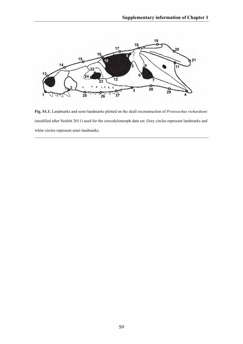
Supplementary information of Chapter 1
S9
Fig. S1.1. Landmarks and semi-landmarks plotted on the skull reconstruction of Protosuchus richardsoni
(modified after Nesbitt 2011) used for the crocodylomorph data set. Grey circles represent landmarks and
white circles represent semi-landmarks.
Supplementary information of Chapter 1
S10
SAURISCHIA
1 anteroventral corner of the premaxilla
2 contact of premaxilla and maxilla along the tooth row
3 contact of maxilla and jugal along the ventral margin of the skull
4 posteroventral corner of the quadratojugal (postorbital region)
5 most ventral point of the lacrimal on the posterior margin of the antorbital
fenestra
6 contact between maxilla and lacrimal on the dorsal margin of the antorbital
fenestra
7 contact between lacrimal and jugal on the anteroventral margin of the orbit
8 contact between jugal and postorbital on the posterior margin of the orbit
9 contact between jugal and postorbital on the anterior margin of the lateral
temporal fenestra
10 contact between jugal and quadratojugal on the posteroventral margin of the
lateral temporal fenestra
11 contact between postorbital and frontal on the posterodorsal margin of the orbit
12 contact between postorbital and squamosal on the dorsal margin of the lateral
temporal fenestra
13 anteroventral tip of the squamosal on the posterior margin of the lateral temporal
fenestra
14 contact of premaxilla and nasal on the dorsal margin of the external naris
15 most anteroventral point of the subnarial process of the nasal on the ventral
margin of the external naris
16-18 three semi-LMs on the ventral margin of the maxilla between LM 2 and LM 3
Supplementary information of Chapter 1
S11
19-20 two semi-LMs on the ventral margin of the skull between LM 3 and LM 4
21 one semi-LM on the posterodorsal margin of the antorbital fenestra between LM
5 and LM 6
22 dorsal contact between postorbital and squamosal
23 anteroventral tip of the squamosal on the posterior margin ventral process
24-26 three semi-LMs on the anteroventral margin of the antorbital fenestra between
LM 6 and LM 5
27-30 four semi-LMs on the anterodorsal margin of the orbit between LM 7 and LM
11
31-45 fifteen semi-LMs on the dorsal margin of the skull between LM 1 and LM 23
46-47 two semi-LMs on the posterior margin of the orbit between LM 11 and LM 8
48 one semi-LM on the anteroventral margin of the lateral temporal fenestra
between LM 9 and LM 10
49 one semi-LM on the posterodorsal margin of the lateral temporal fenestra
between LM 12 and LM 13
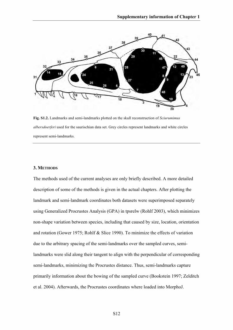
Supplementary information of Chapter 1
S12
Fig. S1.2. Landmarks and semi-landmarks plotted on the skull reconstruction of Sciurumimus
albersdoerferi used for the saurischian data set. Grey circles represent landmarks and white circles
represent semi-landmarks.
3. METHODS
The methods used of the current analyses are only briefly described. A more detailed
description of some of the methods is given in the actual chapters. After plotting the
landmark and semi-landmark coordinates both datasets were superimposed separately
using Generalized Procrustes Analysis (GPA) in tpsrelw (Rohlf 2003), which minimizes
non-shape variation between species, including that caused by size, location, orientation
and rotation (Gower 1975; Rohlf & Slice 1990). To minimize the effects of variation
due to the arbitrary spacing of the semi-landmarks over the sampled curves, semi-
landmarks were slid along their tangent to align with the perpendicular of corresponding
semi-landmarks, minimizing the Procrustes distance. Thus, semi-landmarks capture
primarily information about the bowing of the sampled curve (Bookstein 1997; Zelditch
et al. 2004). Afterwards, the Procrustes coordinates where loaded into MorphoJ.
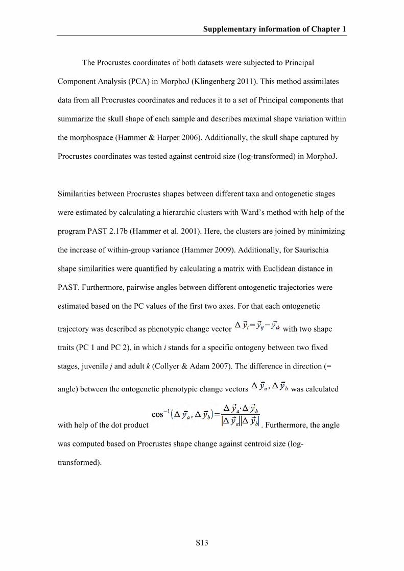
Supplementary information of Chapter 1
S13
The Procrustes coordinates of both datasets were subjected to Principal
Component Analysis (PCA) in MorphoJ (Klingenberg 2011). This method assimilates
data from all Procrustes coordinates and reduces it to a set of Principal components that
summarize the skull shape of each sample and describes maximal shape variation within
the morphospace (Hammer & Harper 2006). Additionally, the skull shape captured by
Procrustes coordinates was tested against centroid size (log-transformed) in MorphoJ.
Similarities between Procrustes shapes between different taxa and ontogenetic stages
were estimated by calculating a hierarchic clusters with Ward’s method with help of the
program PAST 2.17b (Hammer et al. 2001). Here, the clusters are joined by minimizing
the increase of within-group variance (Hammer 2009). Additionally, for Saurischia
shape similarities were quantified by calculating a matrix with Euclidean distance in
PAST. Furthermore, pairwise angles between different ontogenetic trajectories were
estimated based on the PC values of the first two axes. For that each ontogenetic
trajectory was described as phenotypic change vector with two shape
traits (PC 1 and PC 2), in which i stands for a specific ontogeny between two fixed
stages, juvenile j and adult k (Collyer & Adam 2007). The difference in direction (=
angle) between the ontogenetic phenotypic change vectors was calculated
with help of the dot product . Furthermore, the angle
was computed based on Procrustes shape change against centroid size (log-
transformed).
Supplementary information of Chapter 1
S14
To estimate cranial shape changes during evolution all juvenile taxa were
excluded from the data set. For that an informal supertree (Butler & Goswami 2008)
was created for both Crocodylomorpha and Saurischia (see below). For both
phylogenies branch lengths were assigned based on the mid-point age of the existence
of taxa (this information was taken from the Paleobiology Database), and zero branch
lengths were adjusted by sharing out the time equally between branches (see Ruta et al.
2006; Brusatte et al. 2008; Brusatte 2011). An arbitrary length of 10 Ma was added to
the root. The assignment of branch lengths was estimated with help of the software
package R (R Development Core Team, 2011) using the APE package (version 2.7-2;
Paradis et al. 2004) and a function written by Graeme Lloyd (see
http://www.graemetlloyd.com/methdpf.html). Afterwards the trees were loaded into
MorphoJ and Procrustes coordinates were mapped as continuous characters on the
topology (Klingenberg & Gidaszewski 2010). The reconstruction of hypothetical
ancestral shapes based on square change parsimony by minimizing the total sum of
square changes across the phylogeny (Maddison 1991). Afterwards ontogenetic and
evolutionary shape changes were compared. To compare the evolutionary shape
changes in Crocodylomorpha with their body size evolution, centroid size of the
Procrustes coordinates were mapped with square change parsimony on the respective
phylogeny with help of the software package Mesquite 2.72 (Maddison & Maddison
2009).
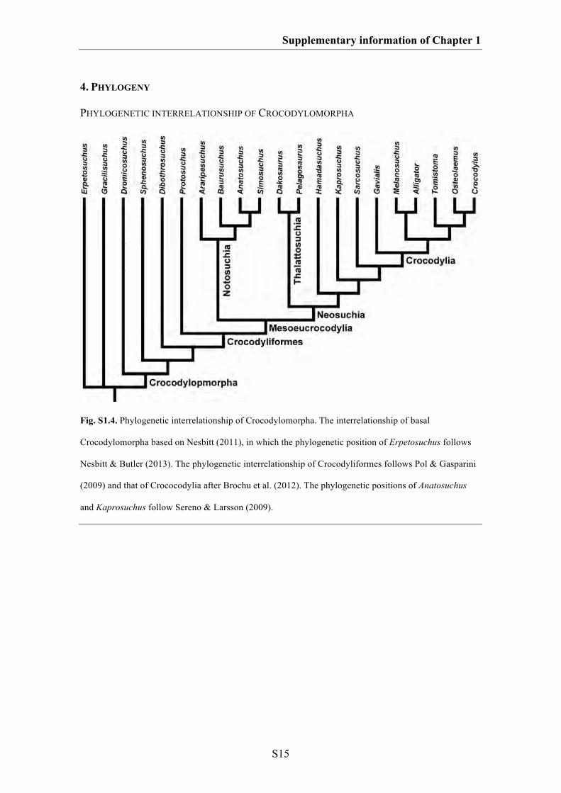
Supplementary information of Chapter 1
S15
4. PHYLOGENY
PHYLOGENETIC INTERRELATIONSHIP OF CROCODYLOMORPHA
Fig. S1.4. Phylogenetic interrelationship of Crocodylomorpha. The interrelationship of basal
Crocodylomorpha based on Nesbitt (2011), in which the phylogenetic position of Erpetosuchus follows
Nesbitt & Butler (2013). The phylogenetic interrelationship of Crocodyliformes follows Pol & Gasparini
(2009) and that of Crococodylia after Brochu et al. (2012). The phylogenetic positions of Anatosuchus
and Kaprosuchus follow Sereno & Larsson (2009).
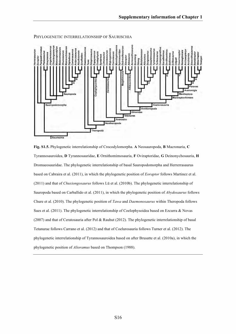
Supplementary information of Chapter 1
S16
PHYLOGENETIC INTERRELATIONSHIP OF SAURISCHIA
Fig. S1.5. Phylogenetic interrelationship of Crocodylomorpha. A Neosauropoda, B Macronaria, C
Tyrannosauroidea, D Tyrannosauridae, E Ornithomimosauria, F Oviraptoridae, G Deinonychosauria, H
Dromaeosauridae. The phylogenetic interrelationship of basal Sauropodomorpha and Herrerrasaurus
based on Cabraira et al. (2011), in which the phylogenetic position of Eoraptor follows Martinez et al.
(2011) and that of Chuxiongosaurus follows Lü et al. (2010b). The phylogenetic interrelationship of
Sauropoda based on Carballido et al. (2011), in which the phylogenetic position of Abydosaurus follows
Chure et al. (2010). The phylogenetic position of Tawa and Daemonosaurus within Theropoda follows
Sues et al. (2011). The phylogenetic interrelationship of Coelophysoidea based on Ezcurra & Novas
(2007) and that of Ceratosauria after Pol & Rauhut (2012). The phylogenetic interrelationship of basal
Tetanurae follows Carrano et al. (2012) and that of Coelurosauria follows Turner et al. (2012). The
phylogenetic interrelationship of Tyrannosauroidea based on after Brusatte et al. (2010a), in which the
phylogenetic position of Alioramus based on Thompson (1988).
S17
SUPPLEMATARY INFORMATION OF CHAPTER 2
The good, the bad, and the ugly: the influence of skull reconstructions and
intraspecific variability in studies of cranial morphometrics in theropods and basal
saurischians
1. Institutional abbreviations
2. Taxon sampling
3. Description of landmarks
4. Skull shape variation
Supplementary information of Chapter 2
S18
1. INSTITUTIONAL ABBREVIATIONS
AMNH, American Museum of Natural History, New York; BHI, Black Hills Institute,
Hill City; BP, Bernard Price Institute for Palaeontological Research, University of the
Witwatersrand, Johannesburg; FMNH, The Field Museum, Chicago; GPIT, Geologisch-
Paläontologisches Institut, Tübingen (IFGT Institut für Geowissenschaften, Eberhard-
Karls-Universität, Tübingen); IVPP, Institute of Vertebrate Palaeontology and
Palaeoanthropology, Beijing; LACM, Los Angeles County Museum, Los Angeles; MB,
Museum für Naturkunde, Berlin; MOR, Museum of the Rockies, Bozeman; NM,
National Museum, Bloemfontein; NMC, National Museum of Canada, Ottawa;
NMMNH, New Mexico Museum of Natural History and Science, Albuquerque; NCSM,
North Carolina Museum of Natural Sciences, Raleigh; PIN, Paleontological Institute,
Russian Academy of Sciences, Moscow; PVSJ, Museo de Ciencias Naturales,
Universidad Nacional de San Juan, San Juan; QMNS, Qatar Museum of Nature and
Science; SMA, Sauriermuseum, Aathal; SMNS, Staatliches Museum für Naturkunde
Stuttgart, Stuttgart; TMP, Royal Tyrrell Museum of Palaeontology, Drumheller; TTU,
Texas Tech University, Lubbock; ULBRA, Museu de Ciências Naturais, Universidade
Luterana do Brasil, Canoas; UUVP, Utah Museum of Natural History, Salt Lake City;
ZPAL, Institute of Palaeobiology, Polish Academy of Sciences, Warsaw.
Supplementary information of Chapter 2
S19
2. TAXON SAMPLING
Table S2.1. List of specimens used in the study
Taxon Collection number Reference
Allosauroidea + basal Tetanurae
Acrocanthosaurus atokensis NCSM 14345 Currie & Carpenter 2000; Eddy & Clarke 2011
Allosaurus spp. TTU P9269 McClelland 1990
AMNH 600 Osborn 1903; Molnar et al. 1990
DINO 11541 Chure 2000
MOR 693 Rauhut 2003a; Foth & Rauhut, this study
UUVP 6000 Madsen 1976; Molnar et al. 1990; Holtz et al. 2004;
Paul 2002; 2008; Fastovsky & Weishampel 2005;
Westheide & Rieger 2009; Foth & Rauhut, this study
SMA 0005 Foth & Rauhut, this study
QMNS-FO-456 Foth & Rauhut, this study
Monolophosaurus jiangi IVPP 84019 Zhao & Currie 1993; Rauhut 2003a; Brusatte et al.
2010b
Sinoraptor dongi IVPP 10600 Currie & Zhao 1993
Sauropodomorpha + basal Saurischia
Eoraptor lunensis PVSJ 512 Sereno et al. 1993; Rauhut 2003a; Langer 2004; Paul
2002; Martinez et al. 2011, Nesbitt 2011
Massospondylus carinatus BP/l/4934 Gow et al. 1990
BP/l/5241 Gow et al. 1990
Melanorosaurus NM QR3314 Yates 2007
Pampadromaeus barberenai ULBRA-PVT016 Cabreira et al. 2011
Plateosaurus engelhardti AMNH 6810 Galton 2001
Composite Wilson & Sereno 1998;
GPIT 1 Galton 2001
MB R. 1937 Galton 2001; Rauhut 2003a
SMNS 12949 Galton 2001
SMNS 13200 Galton 2001, Yates 2003; Nesbitt 2011
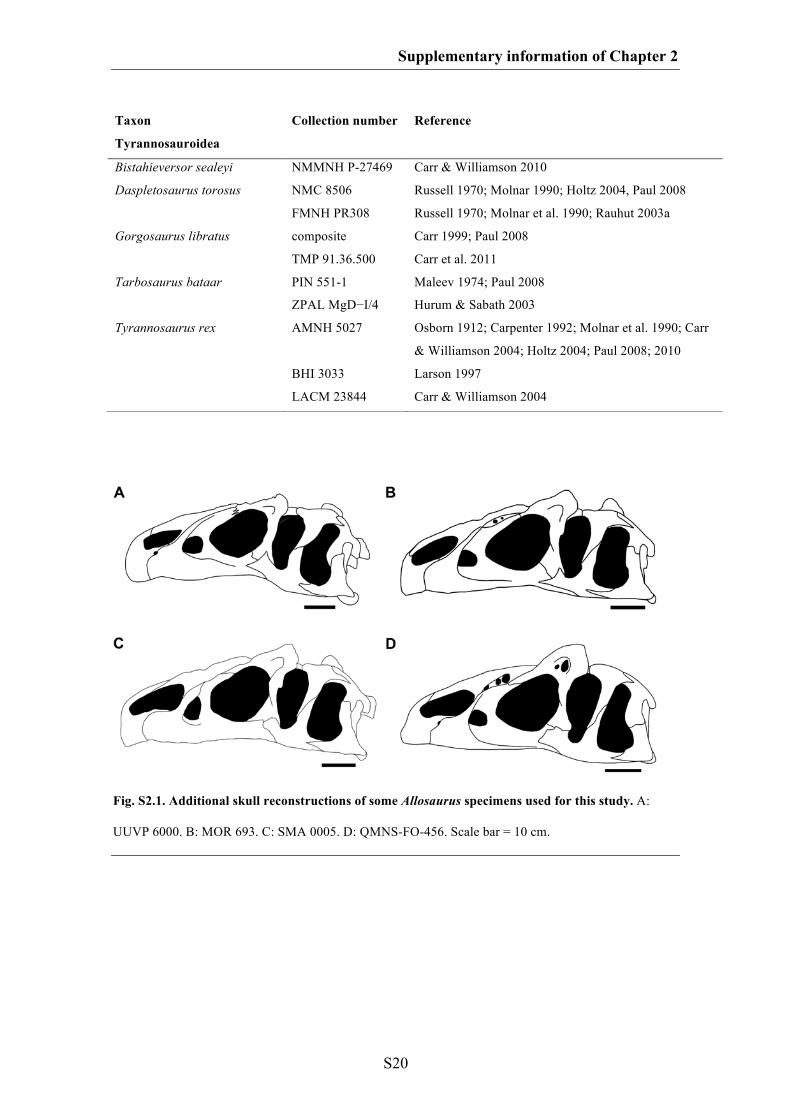
Supplementary information of Chapter 2
S20
Fig. S2.1. Additional skull reconstructions of some Allosaurus specimens used for this study. A:
UUVP 6000. B: MOR 693. C: SMA 0005. D: QMNS-FO-456. Scale bar = 10 cm.
Taxon Collection number Reference
Tyrannosauroidea
Bistahieversor sealeyi NMMNH P-27469 Carr & Williamson 2010
Daspletosaurus torosus NMC 8506 Russell 1970; Molnar 1990; Holtz 2004, Paul 2008
FMNH PR308 Russell 1970; Molnar et al. 1990; Rauhut 2003a
Gorgosaurus libratus composite Carr 1999; Paul 2008
TMP 91.36.500 Carr et al. 2011
Tarbosaurus bataar PIN 551-1 Maleev 1974; Paul 2008
ZPAL MgD−I/4 Hurum & Sabath 2003
Tyrannosaurus rex AMNH 5027 Osborn 1912; Carpenter 1992; Molnar et al. 1990; Carr
& Williamson 2004; Holtz 2004; Paul 2008; 2010
BHI 3033 Larson 1997
LACM 23844 Carr & Williamson 2004
Supplementary information of Chapter 2
S21
3. DESCRIPTION OF LANDMARKS
Table S2.2. Description of landmarks used for the different dataset.
No Landmark
1 anteroventral corner of the premaxilla
2 contact of premaxilla and maxilla along the tooth row
3 contact of maxilla and jugal along the ventral margin of the skull
4 contact between jugal and quadratojugal along the ventral margin of the skull
5 posteroventral corner of the quadratojugal
6 contact of premaxilla and nasal along the dorsal margin of the skull
7 contact of premaxilla and nasal along the dorsal margin of the external naris
8 tip of the maxillary process of the premaxilla
9 tip of the maxillary process of the nasal
10 most-anterior point of the antorbital fenestra
11 ventralmost point of the lacrimal along the margin of the antorbital fenestra
12 anteriormost contact of the lacrimal along the dorsal margin of the antorbital fenestra
13 contact between lacrimal and jugal on the orbital margin
14 contact between postorbital and jugal on the orbital margin
15 contact between postorbital and jugal on the margin of the lateral temporal fenestra
16 contact between jugal and quadratojugal on the margin of the lateral temporal fenestra
17 anteroventral tip of the ventral process of the squamosal on the margin of the lateral temporal
fenestra
18 ventral contact of postorbital and squamosal on the margin of the lateral temporal fenestra
19 dorsal contact between postorbital and squamosal
20 anteriormost point of the jugal
21 posteriormost point of the postorbital
22 contact between frontal and postorbital on the dorsal margin of the orbit
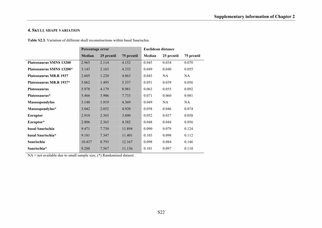
Supplementary information of Chapter 2
S22
4. SKULL SHAPE VARIATION
Table S2.3. Variation of different skull reconstructions within basal Saurischia.
Percentage error
Euclidean distance
Median 25 prcntil 75 prcntil Median 25 prcntil 75 prcntil
Plateosaurus SMNS 13200 2.965 2.114 4.152 0.045 0.034 0.070
Plateosaurus SMNS 13200* 3.143 2.163 4.332 0.049 0.040 0.055
Plateosaurus MB.R 1937 2.605 1.220 4.863 0.043 NA NA
Plateosaurus MB.R 1937* 3.062 1.495 5.337 0.051 0.039 0.056
Plateosaurus 5.978 4.179 8.981 0.063 0.055 0.092
Plateosaurus* 5.466 3.906 7.733 0.071 0.060 0.081
Massospondylus 3.148 1.919 4.369 0.049 NA NA
Massospondylus* 3.042 2.052 4.920 0.058 0.046 0.074
Eoraptor 2.910 2.363 3.600 0.052 0.037 0.058
Eoraptor* 2.806 2.363 4.382 0.048 0.044 0.056
basal Saurischia 9.471 7.730 11.894 0.090 0.079 0.124
basal Saurischia* 9.181 7.347 11.401 0.103 0.098 0.112
Saurischia 10.437 8.793 12.167 0.098 0.084 0.146
Saurischia* 9.288 7.567 11.136 0.101 0.097 0.110
NA = not available due to small sample size, (*) Randomized dataset.
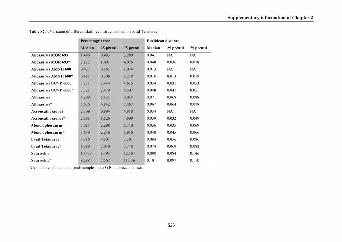
Supplementary information of Chapter 2
S23
Table S2.4. Variation of different skull reconstructions within basal Tetanurae
Percentage error
Euclidean distance
Median 25 prcntil 75 prcntil Median 25 prcntil 75 prcntil
Allosaurus MOR 693 1.466 0.483 3.289 0.041 NA NA
Allosaurus MOR 693* 2.122 1.091 4.076 0.048 0.036 0.078
Allosaurus AMNH 600 0.437 0.161 1.074 0.013 NA NA
Allosaurus AMNH 600* 0.481 0.306 1.218 0.016 0.013 0.019
Allosaurus UUVP 6000 3.271 2.444 4.615 0.034 0.031 0.053
Allosaurus UUVP 6000* 3.321 2.479 4.507 0.048 0.041 0.051
Allosaurus 6.209 5.131 8.014 0.071 0.069 0.088
Allosaurus* 5.636 4.842 7.467 0.067 0.064 0.078
Acroncathosaurus 2.309 0.894 4.818 0.034 NA NA
Acroncathosaurus* 2.595 1.526 4.609 0.039 0.032 0.049
Monolophosaurus 3.057 2.298 5.710 0.036 0.035 0.069
Monolophosaurus* 3.649 2.246 5.916 0.048 0.045 0.066
basal Tetanurae 5.316 4.507 7.301 0.064 0.056 0.086
basal Tetanurae* 6.289 5.040 7.778 0.074 0.069 0.082
Saurischia 10.437 8.793 12.167 0.098 0.084 0.146
Saurischia* 9.288 7.567 11.136 0.101 0.097 0.110
NA = not available due to small sample size, (*) Randomized dataset.
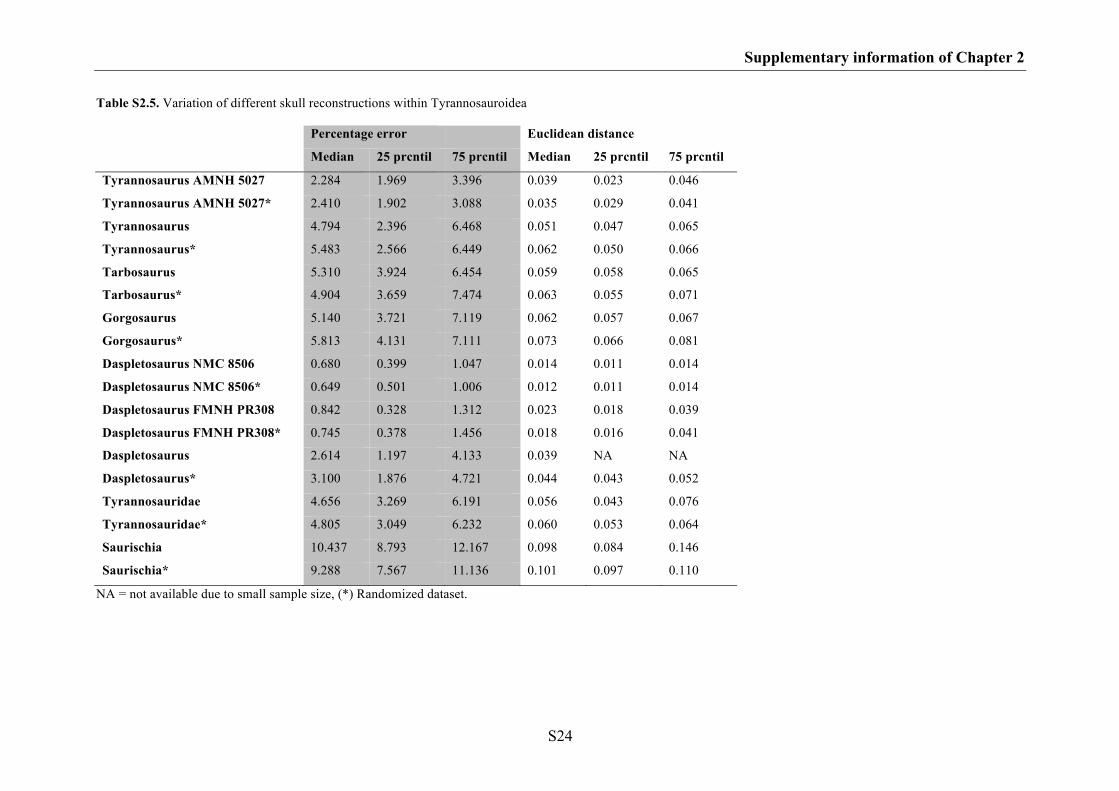
Supplementary information of Chapter 2
S24
Table S2.5. Variation of different skull reconstructions within Tyrannosauroidea
Percentage error
Euclidean distance
Median 25 prcntil 75 prcntil Median 25 prcntil 75 prcntil
Tyrannosaurus AMNH 5027 2.284 1.969 3.396 0.039 0.023 0.046
Tyrannosaurus AMNH 5027* 2.410 1.902 3.088 0.035 0.029 0.041
Tyrannosaurus 4.794 2.396 6.468 0.051 0.047 0.065
Tyrannosaurus* 5.483 2.566 6.449 0.062 0.050 0.066
Tarbosaurus 5.310 3.924 6.454 0.059 0.058 0.065
Tarbosaurus* 4.904 3.659 7.474 0.063 0.055 0.071
Gorgosaurus 5.140 3.721 7.119 0.062 0.057 0.067
Gorgosaurus* 5.813 4.131 7.111 0.073 0.066 0.081
Daspletosaurus NMC 8506 0.680 0.399 1.047 0.014 0.011 0.014
Daspletosaurus NMC 8506* 0.649 0.501 1.006 0.012 0.011 0.014
Daspletosaurus FMNH PR308 0.842 0.328 1.312 0.023 0.018 0.039
Daspletosaurus FMNH PR308* 0.745 0.378 1.456 0.018 0.016 0.041
Daspletosaurus 2.614 1.197 4.133 0.039 NA NA
Daspletosaurus* 3.100 1.876 4.721 0.044 0.043 0.052
Tyrannosauridae 4.656 3.269 6.191 0.056 0.043 0.076
Tyrannosauridae* 4.805 3.049 6.232 0.060 0.053 0.064
Saurischia 10.437 8.793 12.167 0.098 0.084 0.146
Saurischia* 9.288 7.567 11.136 0.101 0.097 0.110
NA = not available due to small sample size, (*) Randomized dataset.
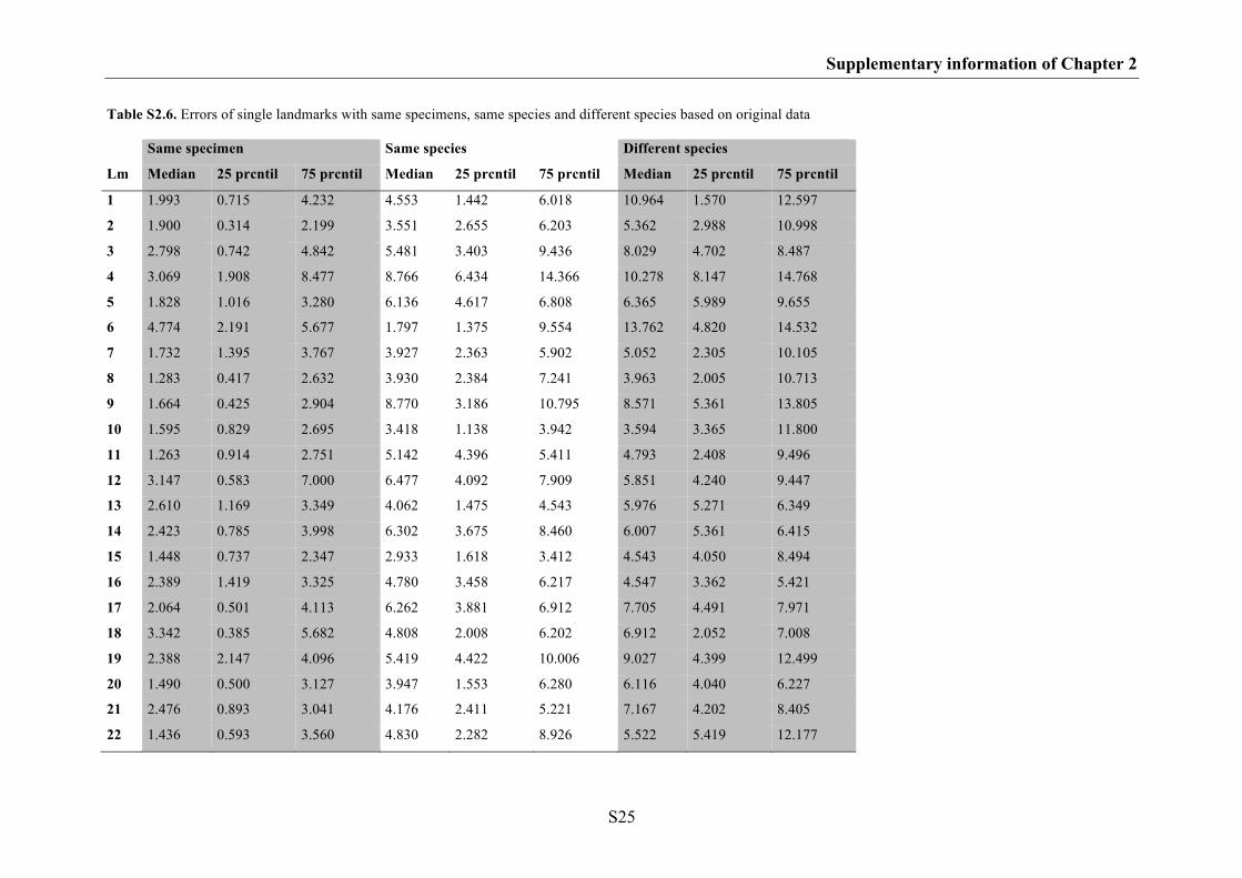
Supplementary information of Chapter 2
S25
Table S2.6. Errors of single landmarks with same specimens, same species and different species based on original data
Same specimen Same species Different species
Lm Median 25 prcntil 75 prcntil Median 25 prcntil 75 prcntil Median 25 prcntil 75 prcntil
1 1.993 0.715 4.232 4.553 1.442 6.018 10.964 1.570 12.597
2 1.900 0.314 2.199 3.551 2.655 6.203 5.362 2.988 10.998
3 2.798 0.742 4.842 5.481 3.403 9.436 8.029 4.702 8.487
4 3.069 1.908 8.477 8.766 6.434 14.366 10.278 8.147 14.768
5 1.828 1.016 3.280 6.136 4.617 6.808 6.365 5.989 9.655
6 4.774 2.191 5.677 1.797 1.375 9.554 13.762 4.820 14.532
7 1.732 1.395 3.767 3.927 2.363 5.902 5.052 2.305 10.105
8 1.283 0.417 2.632 3.930 2.384 7.241 3.963 2.005 10.713
9 1.664 0.425 2.904 8.770 3.186 10.795 8.571 5.361 13.805
10 1.595 0.829 2.695 3.418 1.138 3.942 3.594 3.365 11.800
11 1.263 0.914 2.751 5.142 4.396 5.411 4.793 2.408 9.496
12 3.147 0.583 7.000 6.477 4.092 7.909 5.851 4.240 9.447
13 2.610 1.169 3.349 4.062 1.475 4.543 5.976 5.271 6.349
14 2.423 0.785 3.998 6.302 3.675 8.460 6.007 5.361 6.415
15 1.448 0.737 2.347 2.933 1.618 3.412 4.543 4.050 8.494
16 2.389 1.419 3.325 4.780 3.458 6.217 4.547 3.362 5.421
17 2.064 0.501 4.113 6.262 3.881 6.912 7.705 4.491 7.971
18 3.342 0.385 5.682 4.808 2.008 6.202 6.912 2.052 7.008
19 2.388 2.147 4.096 5.419 4.422 10.006 9.027 4.399 12.499
20 1.490 0.500 3.127 3.947 1.553 6.280 6.116 4.040 6.227
21 2.476 0.893 3.041 4.176 2.411 5.221 7.167 4.202 8.405
22 1.436 0.593 3.560 4.830 2.282 8.926 5.522 5.419 12.177
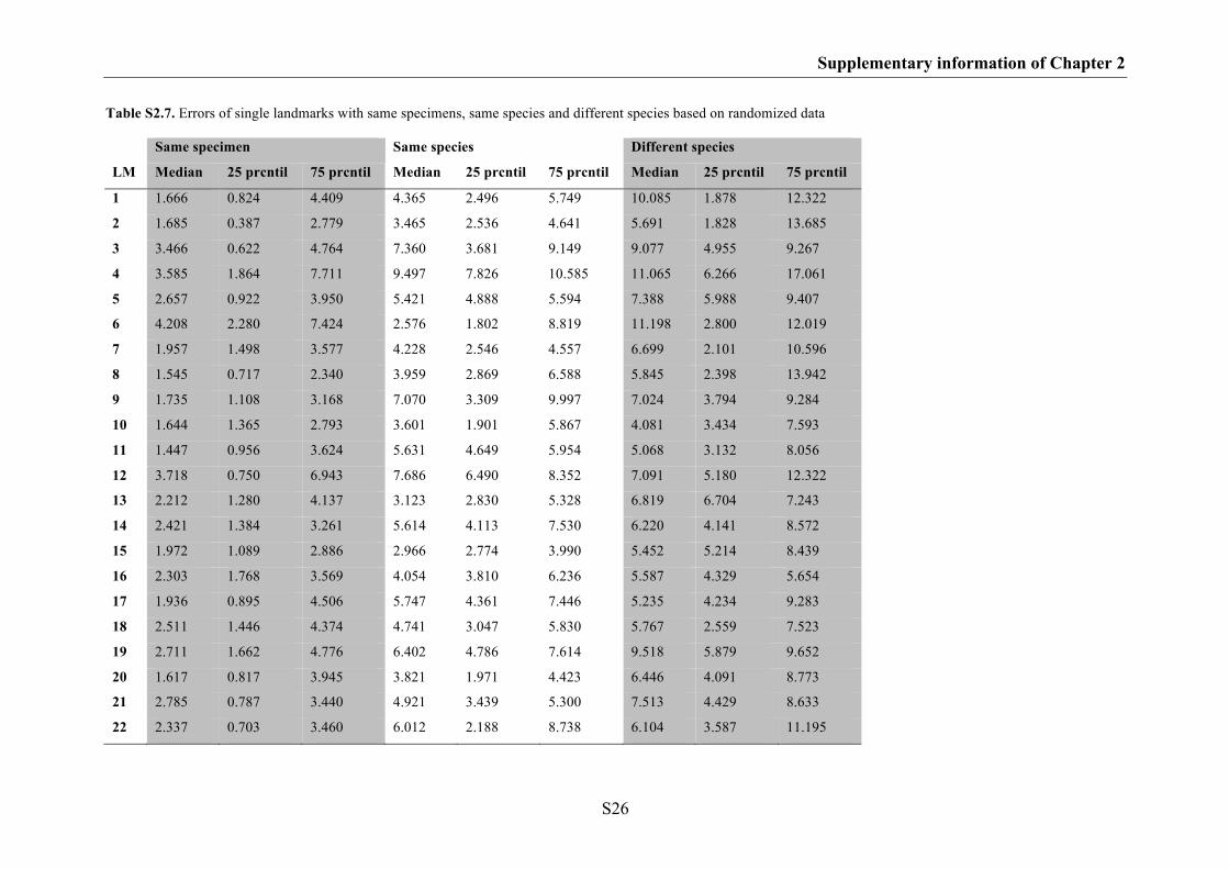
Supplementary information of Chapter 2
S26
Table S2.7. Errors of single landmarks with same specimens, same species and different species based on randomized data
Same specimen Same species Different species
LM Median 25 prcntil 75 prcntil Median 25 prcntil 75 prcntil Median 25 prcntil 75 prcntil
1 1.666 0.824 4.409 4.365 2.496 5.749 10.085 1.878 12.322
2 1.685 0.387 2.779 3.465 2.536 4.641 5.691 1.828 13.685
3 3.466 0.622 4.764 7.360 3.681 9.149 9.077 4.955 9.267
4 3.585 1.864 7.711 9.497 7.826 10.585 11.065 6.266 17.061
5 2.657 0.922 3.950 5.421 4.888 5.594 7.388 5.988 9.407
6 4.208 2.280 7.424 2.576 1.802 8.819 11.198 2.800 12.019
7 1.957 1.498 3.577 4.228 2.546 4.557 6.699 2.101 10.596
8 1.545 0.717 2.340 3.959 2.869 6.588 5.845 2.398 13.942
9 1.735 1.108 3.168 7.070 3.309 9.997 7.024 3.794 9.284
10 1.644 1.365 2.793 3.601 1.901 5.867 4.081 3.434 7.593
11 1.447 0.956 3.624 5.631 4.649 5.954 5.068 3.132 8.056
12 3.718 0.750 6.943 7.686 6.490 8.352 7.091 5.180 12.322
13 2.212 1.280 4.137 3.123 2.830 5.328 6.819 6.704 7.243
14 2.421 1.384 3.261 5.614 4.113 7.530 6.220 4.141 8.572
15 1.972 1.089 2.886 2.966 2.774 3.990 5.452 5.214 8.439
16 2.303 1.768 3.569 4.054 3.810 6.236 5.587 4.329 5.654
17 1.936 0.895 4.506 5.747 4.361 7.446 5.235 4.234 9.283
18 2.511 1.446 4.374 4.741 3.047 5.830 5.767 2.559 7.523
19 2.711 1.662 4.776 6.402 4.786 7.614 9.518 5.879 9.652
20 1.617 0.817 3.945 3.821 1.971 4.423 6.446 4.091 8.773
21 2.785 0.787 3.440 4.921 3.439 5.300 7.513 4.429 8.633
22 2.337 0.703 3.460 6.012 2.188 8.738 6.104 3.587 11.195
S27
SUPPLEMATARY INFORMATION OF CHAPTER 3
Intraspecific variation in the skull morphology of the black caiman Melanosuchus niger
(Alligatoridae, Caimaninae)
1. List of specimens
2. Description of Landmarks
3. Error test after Singleton (2002)

Supplementary information of Chapter 3
S28
1. LIST OF SPECIMENS
Table S3.1. List of specimens of Melanosuchus niger used in the geometric morphometric analysis with
information on sex, skull length SL, bite force, and data sets in which in was included (dorsal, ventral and
lateral views). The bite force BF estimation based on the equation of Erickson et al. (2003): LogBF =
2.75 x LogSL – 0.65; j juvenile, j* juvenile specimens, which were x-rayed; f female, m male, m*
identification of males by one of the authors (CF) based on the large size compared to the largest female.
SMF Senckenberg Naturmuseum Frankfurt (Germany); ZFMK Zoologisches Forschungsmuseum
Alexander Koenig, Bonn (Germany); NHMW Naturhistorisches Museum Wien (Austria); ZMH
Zoologisches Museum Hamburg (Germany); ZSM Zoologische Staatssammlung München (Germany).
Specimen sex SL (cm) logSL (cm) logBF (N)
ZFMK 52355 j* 4.70 0.67 1.20
ZFMK 52353 j* 5.85 0.77 1.46
ZSM 858/1920 j* 6.10 0.79 1.51
ZSM 139/1982 j* 6.20 0.79 1.53
ZSM 2414/2006 j* 7.90 0.90 1.82
ZMH R08660 j* 8.00 0.90 1.83
SMF 30113 j 8.80 0.94 1.95
SMF 30102 j 10.20 1.01 2.12
ZSM 3/1971 j* 11.10 1.05 2.22
SMF 40142 j 13.10 1.12 2.42
SMF 40172 j 13.90 1.14 2.49
ZSM 13/1911 j 16.30 1.21 2.68
ZSM 130/1911 f 26.80 1.43 3.28
ZSM 27/1911 f 29.00 1.46 3.37
ZSM 87/1911 f 29.00 1.46 3.37
ZSM 76/1911 f 29.80 1.47 3.40
ZSM 85/1911 f 31.10 1.49 3.46
ZSM 84/1911 f 31.50 1.50 3.47
ZSM 77/1911 f 32.00 1.51 3.49
ZSM 86/1911 f 33.30 1.52 3.54
ZSM 83/1911 f 34.00 1.53 3.56
ZSM 91/1911 f 34.00 1.53 3.56
ZSM 68/1911 f 35.50 1.55 3.61
ZSM 14/1911 f 36.40 1.56 3.64
ZSM 89/1911 f 36.80 1.57 3.66
ZSM 70/1911 f 38.50 1.59 3.71
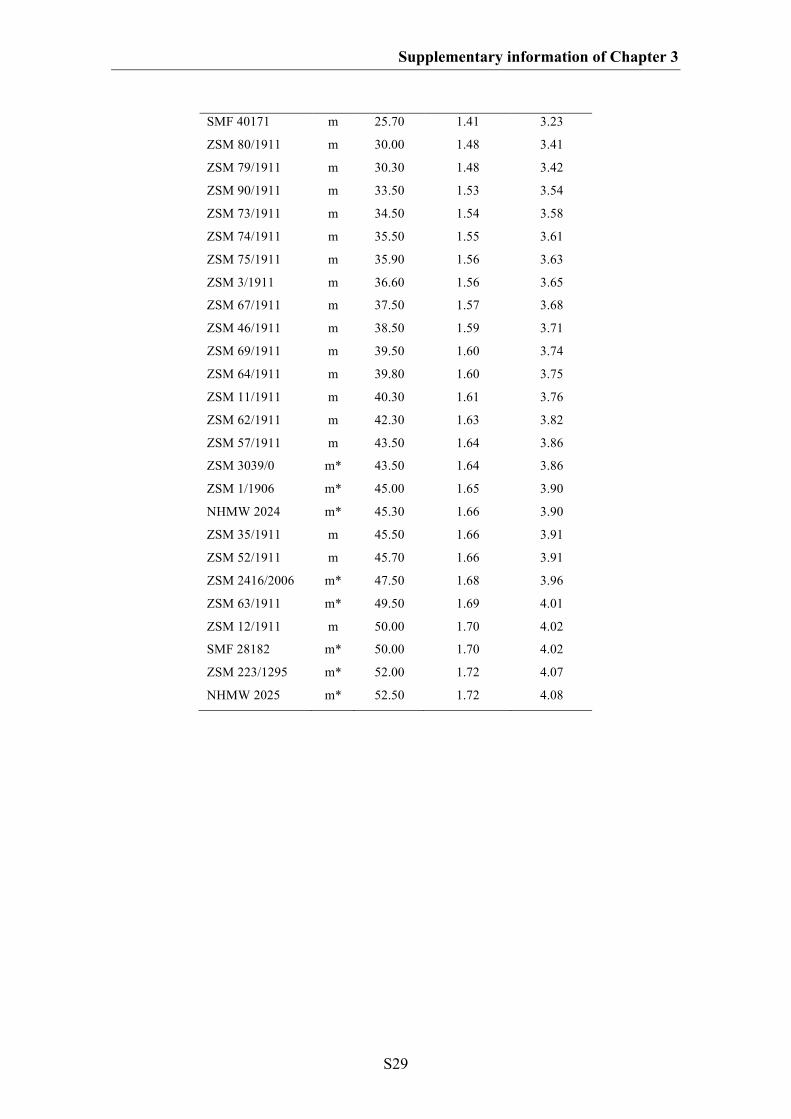
Supplementary information of Chapter 3
S29
SMF 40171 m 25.70 1.41 3.23
ZSM 80/1911 m 30.00 1.48 3.41
ZSM 79/1911 m 30.30 1.48 3.42
ZSM 90/1911 m 33.50 1.53 3.54
ZSM 73/1911 m 34.50 1.54 3.58
ZSM 74/1911 m 35.50 1.55 3.61
ZSM 75/1911 m 35.90 1.56 3.63
ZSM 3/1911 m 36.60 1.56 3.65
ZSM 67/1911 m 37.50 1.57 3.68
ZSM 46/1911 m 38.50 1.59 3.71
ZSM 69/1911 m 39.50 1.60 3.74
ZSM 64/1911 m 39.80 1.60 3.75
ZSM 11/1911 m 40.30 1.61 3.76
ZSM 62/1911 m 42.30 1.63 3.82
ZSM 57/1911 m 43.50 1.64 3.86
ZSM 3039/0 m* 43.50 1.64 3.86
ZSM 1/1906 m* 45.00 1.65 3.90
NHMW 2024 m* 45.30 1.66 3.90
ZSM 35/1911 m 45.50 1.66 3.91
ZSM 52/1911 m 45.70 1.66 3.91
ZSM 2416/2006 m* 47.50 1.68 3.96
ZSM 63/1911 m* 49.50 1.69 4.01
ZSM 12/1911 m 50.00 1.70 4.02
SMF 28182 m* 50.00 1.70 4.02
ZSM 223/1295 m* 52.00 1.72 4.07
NHMW 2025 m* 52.50 1.72 4.08
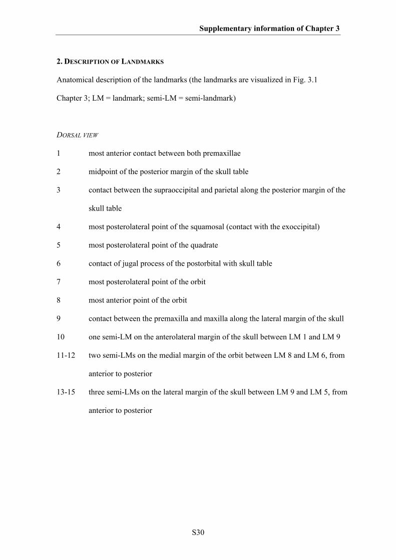
Supplementary information of Chapter 3
S30
2. DESCRIPTION OF LANDMARKS
Anatomical description of the landmarks (the landmarks are visualized in Fig. 3.1
Chapter 3; LM = landmark; semi-LM = semi-landmark)
DORSAL VIEW
1 most anterior contact between both premaxillae
2 midpoint of the posterior margin of the skull table
3 contact between the supraoccipital and parietal along the posterior margin of the
skull table
4 most posterolateral point of the squamosal (contact with the exoccipital)
5 most posterolateral point of the quadrate
6 contact of jugal process of the postorbital with skull table
7 most posterolateral point of the orbit
8 most anterior point of the orbit
9 contact between the premaxilla and maxilla along the lateral margin of the skull
10 one semi-LM on the anterolateral margin of the skull between LM 1 and LM 9
11-12 two semi-LMs on the medial margin of the orbit between LM 8 and LM 6, from
anterior to posterior
13-15 three semi-LMs on the lateral margin of the skull between LM 9 and LM 5, from
anterior to posterior
Supplementary information of Chapter 3
S31
LATERAL VIEW
1 most anteroventral point of the premaxillae
2 contact between the maxilla and jugal along the ventral margin of the skull
3 most posterior point of the quadratojugal at the jaw joint
4 most posterior point of the skull roof surface
5 postorbital foramen
6 most ventral contact between the jugal and postorbital
7 most anterior point of the orbit
8 contact of the premaxilla and maxilla along the margin of the tooth row
9 one semi-LM on the ventral margin of the premaxilla between LM 1 and LM 8
10-12 three semi-LMs on the ventral margin od the maxilla between LM 8 and LM 2, from
anterior to posterior
13-14 two semi-LMs on the ventral margin of the jugal between LM 2 and LM 3, from
anterior to posterior
15-21 seven semi-LMs on the dorsal margin of the skull between LM 1 and LM 4, from
anterior to posterior
Supplementary information of Chapter 3
S32
3. ERROR TEST AFTER SINGLETON (2002)
For the error test, estimating the methodological error of plotting landmarks on the
skulls, Procrustes distances of the Procrustes coordinates to the respective consensus
coordinates of each landmark were calculated. Then, the relation of these distances to
the mean distance of the consensus landmarks to the centroid of the consensus shape
was calculated as a percentage of the former from the latter. Based on the test all
landmarks possess only a small percentage error for plotting landmarks (≈ 0. 08-
1.27 %).
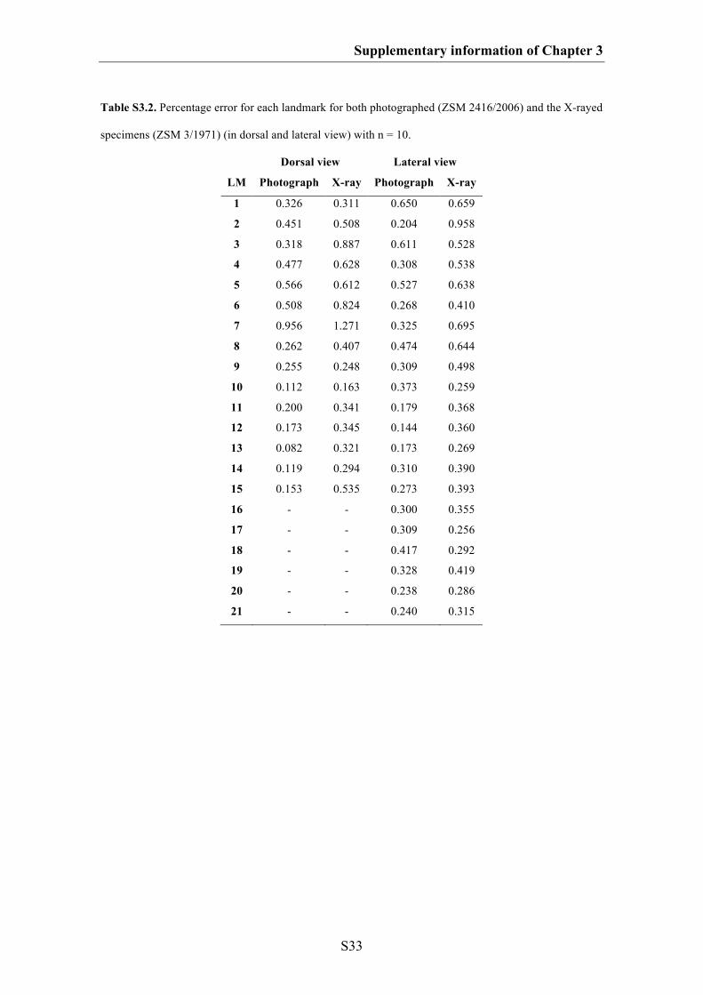
Supplementary information of Chapter 3
S33
Table S3.2. Percentage error for each landmark for both photographed (ZSM 2416/2006) and the X-rayed
specimens (ZSM 3/1971) (in dorsal and lateral view) with n = 10.
Dorsal view Lateral view
LM Photograph X-ray Photograph X-ray
1 0.326 0.311 0.650 0.659
2 0.451 0.508 0.204 0.958
3 0.318 0.887 0.611 0.528
4 0.477 0.628 0.308 0.538
5 0.566 0.612 0.527 0.638
6 0.508 0.824 0.268 0.410
7 0.956 1.271 0.325 0.695
8 0.262 0.407 0.474 0.644
9 0.255 0.248 0.309 0.498
10 0.112 0.163 0.373 0.259
11 0.200 0.341 0.179 0.368
12 0.173 0.345 0.144 0.360
13 0.082 0.321 0.173 0.269
14 0.119 0.294 0.310 0.390
15 0.153 0.535 0.273 0.393
16 - - 0.300 0.355
17 - - 0.309 0.256
18 - - 0.417 0.292
19 - - 0.328 0.419
20 - - 0.238 0.286
21 - - 0.240 0.315
S34
SUPPLEMATARY INFORMATION OF CHAPTER 4
Do different disparity proxies converge on a common signal? Insights from the cranial
morphometrics and evolutionary history of Pterosauria (Diapsida: Archosauria)
1. List of species
2. Description of Landmarks
3. Phylogeny
4. Time
5. Morphospace
6. Skull shape evolution based on the supertree topology
7. Morphological disparity based on supertree topology
*All figures of the supplementary information of Chapter 4 are modified after
Foth et al. (2012)
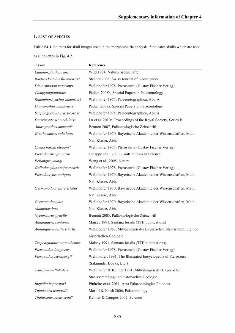
Supplementary information of Chapter 4
S35
1. LIST OF SPECIES
Table S4.1. Sources for skull images used in the morphometric analysis. *indicates skulls which are used
as silhouettes in Fig. 4.2.
Taxon Reference
Eudimorphodon ranzii Wild 1984, Naturwissenschaften
Raeticodactylus filisurensis* Stecher 2008, Swiss Journal of Geosciences
Dimorphodon macronyx Wellnhofer 1978, Pterosauria (Gustav Fischer Verlag)
Campylognathoides Padian 2008b, Special Papers in Palaeontology
Rhamphorhynchus muensteri Wellnhofer 1975, Palaeontographica, Abt. A
Dorygnathus banthensis Padian 2008a, Special Papers in Palaeontology
Scaphognathus crassirostris Wellnhofer 1975, Palaeontographica, Abt. A
Darwinopterus modularis Lü et al. 2010a, Proceedings of the Royal Society, Series B
Anurognathus ammoni* Bennett 2007, Paläontologische Zeitschrift
Gnathosaurus subulatus
Wellnhofer 1970, Bayerische Akademie der Wissenschaften, Math.
Nat. Klasse, Abh.
Ctenochasma elegans* Wellnhofer 1978, Pterosauria (Gustav Fischer Verlag)
Pterodaustro guinazui Chiappe et al. 2000, Contributions in Science
Feilongus youngi Wang et al., 2005, Nature
Gallodactylus canjuersensis Wellnhofer 1978, Pterosauria (Gustav Fischer Verlag)
Pterodactylus antiquus
Wellnhofer 1970, Bayerische Akademie der Wissenschaften, Math.
Nat. Klasse, Abh.
Germanodactylus cristatus
Wellnhofer 1970, Bayerische Akademie der Wissenschaften, Math.
Nat. Klasse, Abh.
Germanodactylus
rhamphastinus
Wellnhofer 1970, Bayerische Akademie der Wissenschaften, Math.
Nat. Klasse, Abh.
Nyctosaurus gracilis Bennett 2003, Paläontologische Zeitschrift
Anhanguera santanae Maisey 1991, Santana fossils (TFH publications)
Anhanguera blittersdorffi
Wellnhofer 1987, Mitteilungen der Bayerischen Staatssammlung und
historischen Geologie
Tropeognathus mesembrinus Maisey 1991, Santana fossils (TFH publications)
Pteranodon longiceps Wellnhofer 1978, Pterosauria (Gustav Fischer Verlag)
Pteranodon sternbergi*
Wellnhofer, 1991, The Illustrated Encyclopedia of Pterosaurs
(Salamnder Books, Ltd.)
Tapejara wellnhoferi
Wellnhofer & Kellner 1991, Mitteilungen der Bayerischen
Staatssammlung und historischen Geologie
Ingridia imperator* Pinheiro et al. 2011, Acta Palaeontologica Polonica
Tupuxuara leonardii Martill & Naish 2006, Palaeontology
Thalassodromeus sethi* Kellner & Campos 2002, Science
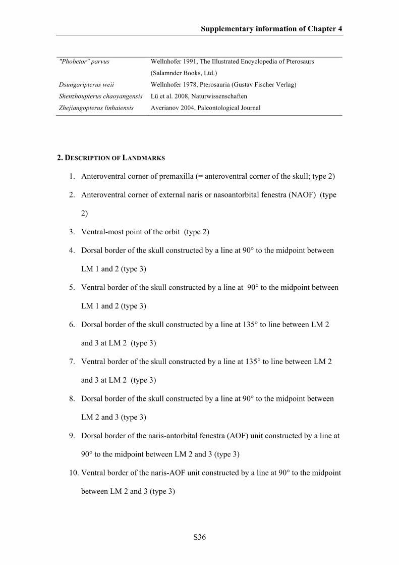
Supplementary information of Chapter 4
S36
"Phobetor" parvus
Wellnhofer 1991, The Illustrated Encyclopedia of Pterosaurs
(Salamnder Books, Ltd.)
Dsungaripterus weii Wellnhofer 1978, Pterosauria (Gustav Fischer Verlag)
Shenzhoupterus chaoyangensis Lü et al. 2008, Naturwissenschaften
Zhejiangopterus linhaiensis Averianov 2004, Paleontological Journal
2. DESCRIPTION OF LANDMARKS
1. Anteroventral corner of premaxilla (= anteroventral corner of the skull; type 2)
2. Anteroventral corner of external naris or nasoantorbital fenestra (NAOF) (type
2)
3. Ventral-most point of the orbit (type 2)
4. Dorsal border of the skull constructed by a line at 90° to the midpoint between
LM 1 and 2 (type 3)
5. Ventral border of the skull constructed by a line at 90° to the midpoint between
LM 1 and 2 (type 3)
6. Dorsal border of the skull constructed by a line at 135° to line between LM 2
and 3 at LM 2 (type 3)
7. Ventral border of the skull constructed by a line at 135° to line between LM 2
and 3 at LM 2 (type 3)
8. Dorsal border of the skull constructed by a line at 90° to the midpoint between
LM 2 and 3 (type 3)
9. Dorsal border of the naris-antorbital fenestra (AOF) unit constructed by a line at
90° to the midpoint between LM 2 and 3 (type 3)
10. Ventral border of the naris-AOF unit constructed by a line at 90° to the midpoint
between LM 2 and 3 (type 3)
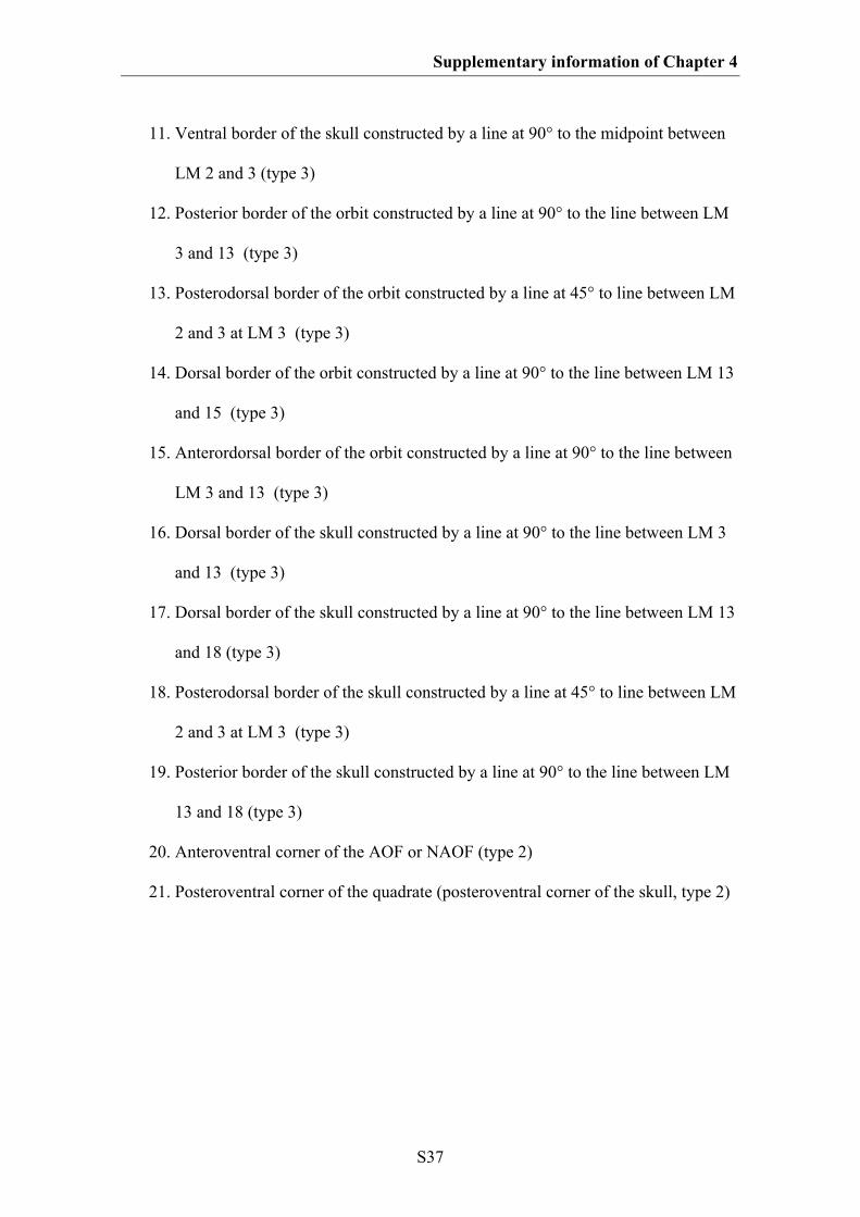
Supplementary information of Chapter 4
S37
11. Ventral border of the skull constructed by a line at 90° to the midpoint between
LM 2 and 3 (type 3)
12. Posterior border of the orbit constructed by a line at 90° to the line between LM
3 and 13 (type 3)
13. Posterodorsal border of the orbit constructed by a line at 45° to line between LM
2 and 3 at LM 3 (type 3)
14. Dorsal border of the orbit constructed by a line at 90° to the line between LM 13
and 15 (type 3)
15. Anterordorsal border of the orbit constructed by a line at 90° to the line between
LM 3 and 13 (type 3)
16. Dorsal border of the skull constructed by a line at 90° to the line between LM 3
and 13 (type 3)
17. Dorsal border of the skull constructed by a line at 90° to the line between LM 13
and 18 (type 3)
18. Posterodorsal border of the skull constructed by a line at 45° to line between LM
2 and 3 at LM 3 (type 3)
19. Posterior border of the skull constructed by a line at 90° to the line between LM
13 and 18 (type 3)
20. Anteroventral corner of the AOF or NAOF (type 2)
21. Posteroventral corner of the quadrate (posteroventral corner of the skull, type 2)
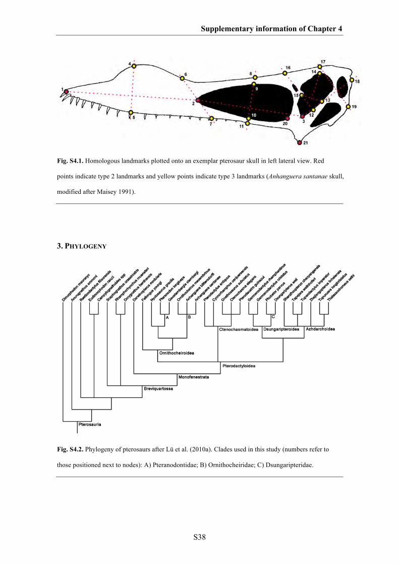
Supplementary information of Chapter 4
S38
Fig. S4.1. Homologous landmarks plotted onto an exemplar pterosaur skull in left lateral view. Red
points indicate type 2 landmarks and yellow points indicate type 3 landmarks (Anhanguera santanae skull,
modified after Maisey 1991).
3. PHYLOGENY
Fig. S4.2. Phylogeny of pterosaurs after Lü et al. (2010a). Clades used in this study (numbers refer to
those positioned next to nodes): A) Pteranodontidae; B) Ornithocheiridae; C) Dsungaripteridae.
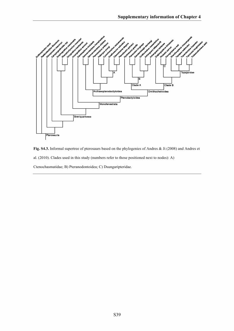
Supplementary information of Chapter 4
S39
Fig. S4.3. Informal supertree of pterosaurs based on the phylogenies of Andres & Ji (2008) and Andres et
al. (2010). Clades used in this study (numbers refer to those positioned next to nodes): A)
Ctenochasmatidae; B) Pteranodontoidea; C) Dsungaripteridae.
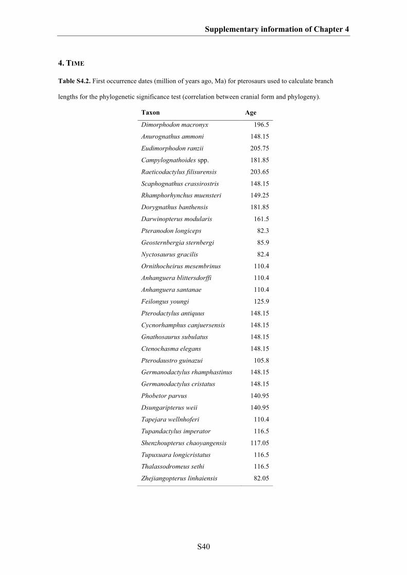
Supplementary information of Chapter 4
S40
4. TIME
Table S4.2. First occurrence dates (million of years ago, Ma) for pterosaurs used to calculate branch
lengths for the phylogenetic significance test (correlation between cranial form and phylogeny).
Taxon Age
Dimorphodon macronyx 196.5
Anurognathus ammoni 148.15
Eudimorphodon ranzii 205.75
Campylognathoides spp. 181.85
Raeticodactylus filisurensis 203.65
Scaphognathus crassirostris 148.15
Rhamphorhynchus muensteri 149.25
Dorygnathus banthensis 181.85
Darwinopterus modularis 161.5
Pteranodon longiceps 82.3
Geosternbergia sternbergi 85.9
Nyctosaurus gracilis 82.4
Ornithocheirus mesembrinus 110.4
Anhanguera blittersdorffi 110.4
Anhanguera santanae 110.4
Feilongus youngi 125.9
Pterodactylus antiquus 148.15
Cycnorhamphus canjuersensis 148.15
Gnathosaurus subulatus 148.15
Ctenochasma elegans 148.15
Pterodaustro guinazui 105.8
Germanodactylus rhamphastinus 148.15
Germanodactylus cristatus 148.15
Phobetor parvus 140.95
Dsungaripterus weii 140.95
Tapejara wellnhoferi 110.4
Tupandactylus imperator 116.5
Shenzhoupterus chaoyangensis 117.05
Tupuxuara longicristatus 116.5
Thalassodromeus sethi 116.5
Zhejiangopterus linhaiensis 82.05
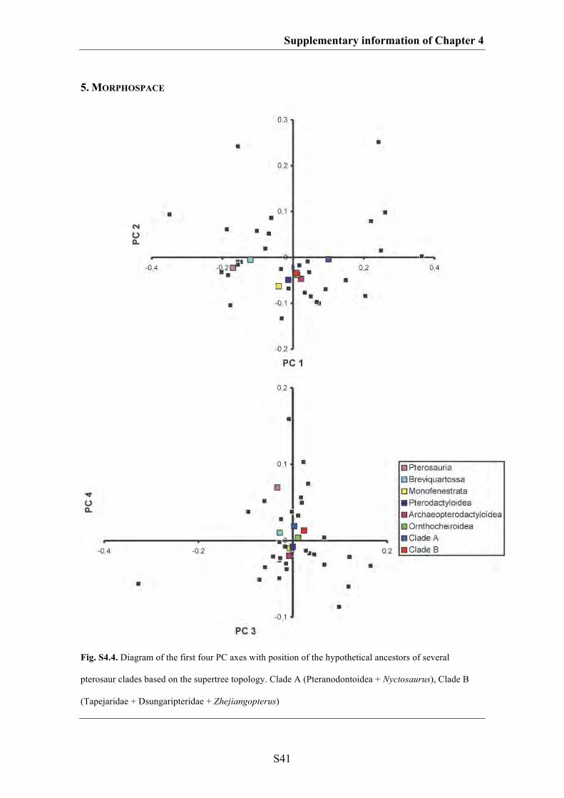
Supplementary information of Chapter 4
S41
5. MORPHOSPACE
Fig. S4.4. Diagram of the first four PC axes with position of the hypothetical ancestors of several
pterosaur clades based on the supertree topology. Clade A (Pteranodontoidea + Nyctosaurus), Clade B
(Tapejaridae + Dsungaripteridae + Zhejiangopterus)
Supplementary information of Chapter 4
S42
6. SKULL SHAPE EVOLUTION BASED ON THE SUPERTREE TOPOLOGY
The results from the ancestral state analysis based on the supertree are similar to those
based on the Lü et al. (2010a) topology (Fig. S4.5). The skull of the hypothetical
ancestor of all pterosaurs had a short, stout snout (negative position of PC 1, positive
position of PC 4), a triangular naris-antorbital fenestra region (positive position of PC 4),
a relatively large orbit (negative position of PC 3) and a relatively large postorbital
region, whereas the hypothetical ancestor of the clade Breviquartossa had a more
pointed tip of the snout. Compared to the basal pterosaur clades, the hypothetical
ancestors of Monofenestrata, Pterodactyloidea, Archaeopterodactyloidea and
Ornithocheiroidea possessed a more elongated skull with a long snout (more positive
position of PC 1), a more trapezoidal naris-antorbital fenestra region (more negative
position of PC 4) and a relatively smaller orbit and postorbital region (more positive
position of PC 1). The skull of the hypothetical ancestor of clade A (Pteranodontoidea +
Nyctosaurus) had a more elongated snout with a shallower antorbital region than the
ancestor of Ornithocheiroidea (more positive position of PC 1, more negative position
of PC 3), whereas the skull of the hypothetical ancestor of clade B (Tapejaridae +
Dsungaripteridae + Zhejiangopterus) had a shortened snout (more negative position of
PC 1) and deeper antorbital region (more positive position of PC 3).
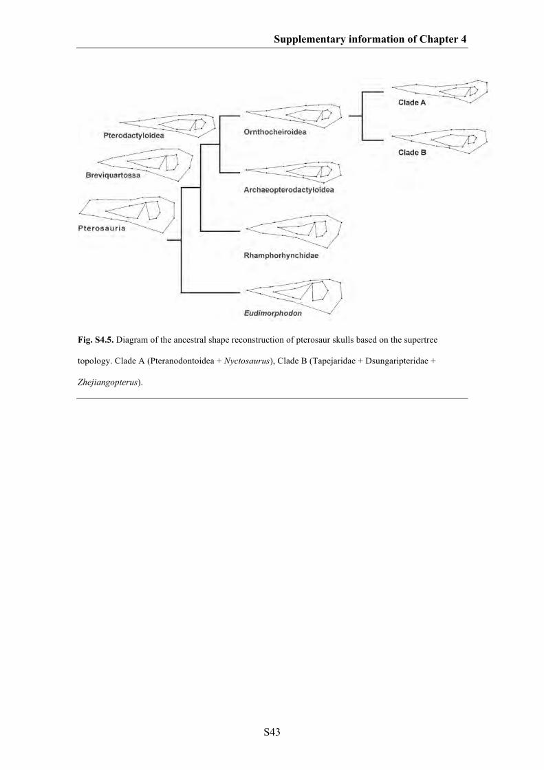
Supplementary information of Chapter 4
S43
Fig. S4.5. Diagram of the ancestral shape reconstruction of pterosaur skulls based on the supertree
topology. Clade A (Pteranodontoidea + Nyctosaurus), Clade B (Tapejaridae + Dsungaripteridae +
Zhejiangopterus).
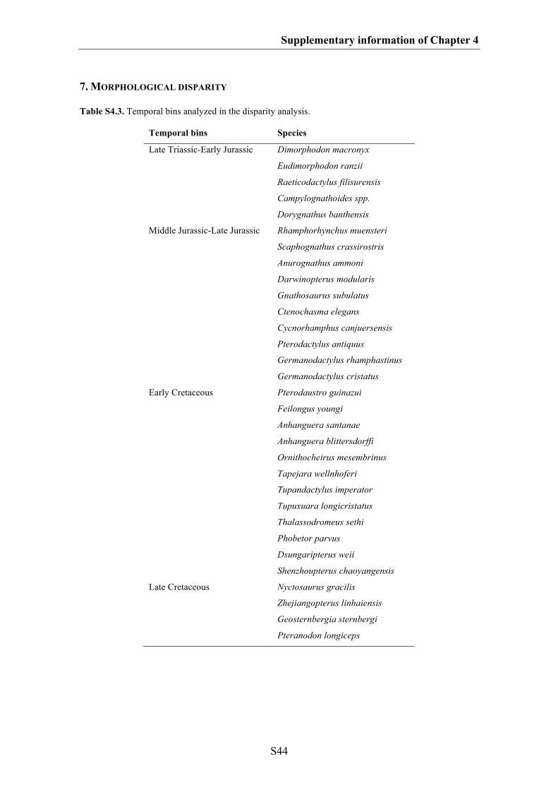
Supplementary information of Chapter 4
S44
7. MORPHOLOGICAL DISPARITY
Table S4.3. Temporal bins analyzed in the disparity analysis.
Temporal bins Species
Late Triassic-Early Jurassic Dimorphodon macronyx
Eudimorphodon ranzii
Raeticodactylus filisurensis
Campylognathoides spp.
Dorygnathus banthensis
Middle Jurassic-Late Jurassic Rhamphorhynchus muensteri
Scaphognathus crassirostris
Anurognathus ammoni
Darwinopterus modularis
Gnathosaurus subulatus
Ctenochasma elegans
Cycnorhamphus canjuersensis
Pterodactylus antiquus
Germanodactylus rhamphastinus
Germanodactylus cristatus
Early Cretaceous Pterodaustro guinazui
Feilongus youngi
Anhanguera santanae
Anhanguera blittersdorffi
Ornithocheirus mesembrinus
Tapejara wellnhoferi
Tupandactylus imperator
Tupuxuara longicristatus
Thalassodromeus sethi
Phobetor parvus
Dsungaripterus weii
Shenzhoupterus chaoyangensis
Late Cretaceous Nyctosaurus gracilis
Zhejiangopterus linhaiensis
Geosternbergia sternbergi
Pteranodon longiceps

Supplementary information of Chapter 4
S45
Table S4.4. Phylogenetic groups within Pterosauria after Lü et al. (2010a) analyzed in the disparity
analysis.
Phylogenetic groups Species
non-monofenestratan pterosaurs Dimorphodon macronyx
Eudimorphodon ranzii
Raeticodactylus filisurensis
Campylognathoides spp.
Dorygnathus banthensis
Rhamphorhynchus muensteri
Scaphognathus crassirostris
Anurognathus ammoni
Monofenestrata Darwinopterus modularis
Gnathosaurus subulatus
Ctenochasma elegans
Cycnorhamphus canjuersensis
Pterodactylus antiquus
Germanodactylus rhamphastinus
Germanodactylus cristatus
Pterodaustro guinazui
Feilongus youngi
Anhanguera santanae
Anhanguera blittersdorffi
Ornithocheirus mesembrinus
Tapejara wellnhoferi
Tupandactylus imperator
Tupuxuara longicristatus
Thalassodromeus sethi
Phobetor parvus
Dsungaripterus weii
Shenzhoupterus chaoyangensis
Nyctosaurus gracilis
Zhejiangopterus linhaiensis
Geosternbergia sternbergi
Pteranodon longiceps
Dsungaripteroidea Phobetor parvus
Dsungaripterus weii
Germanodactylus rhamphastinus
Germanodactylus cristatus
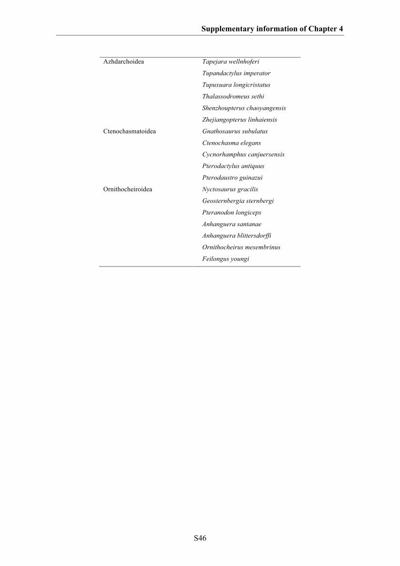
Supplementary information of Chapter 4
S46
Azhdarchoidea Tapejara wellnhoferi
Tupandactylus imperator
Tupuxuara longicristatus
Thalassodromeus sethi
Shenzhoupterus chaoyangensis
Zhejiangopterus linhaiensis
Ctenochasmatoidea Gnathosaurus subulatus
Ctenochasma elegans
Cycnorhamphus canjuersensis
Pterodactylus antiquus
Pterodaustro guinazui
Ornithocheiroidea Nyctosaurus gracilis
Geosternbergia sternbergi
Pteranodon longiceps
Anhanguera santanae
Anhanguera blittersdorffi
Ornithocheirus mesembrinus
Feilongus youngi
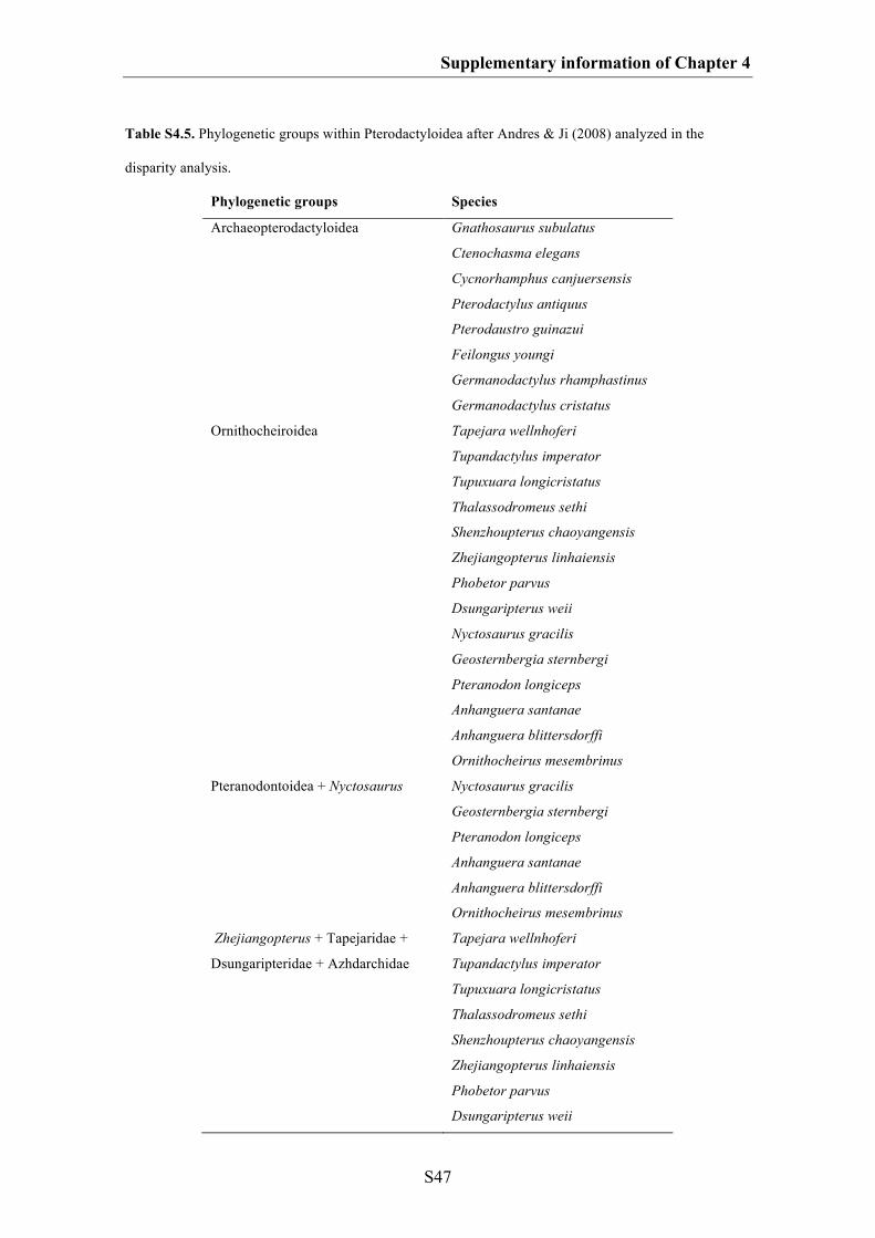
Supplementary information of Chapter 4
S47
Table S4.5. Phylogenetic groups within Pterodactyloidea after Andres & Ji (2008) analyzed in the
disparity analysis.
Phylogenetic groups Species
Archaeopterodactyloidea Gnathosaurus subulatus
Ctenochasma elegans
Cycnorhamphus canjuersensis
Pterodactylus antiquus
Pterodaustro guinazui
Feilongus youngi
Germanodactylus rhamphastinus
Germanodactylus cristatus
Ornithocheiroidea Tapejara wellnhoferi
Tupandactylus imperator
Tupuxuara longicristatus
Thalassodromeus sethi
Shenzhoupterus chaoyangensis
Zhejiangopterus linhaiensis
Phobetor parvus
Dsungaripterus weii
Nyctosaurus gracilis
Geosternbergia sternbergi
Pteranodon longiceps
Anhanguera santanae
Anhanguera blittersdorffi
Ornithocheirus mesembrinus
Pteranodontoidea + Nyctosaurus Nyctosaurus gracilis
Geosternbergia sternbergi
Pteranodon longiceps
Anhanguera santanae
Anhanguera blittersdorffi
Ornithocheirus mesembrinus
Zhejiangopterus + Tapejaridae +
Dsungaripteridae + Azhdarchidae
Tapejara wellnhoferi
Tupandactylus imperator
Tupuxuara longicristatus
Thalassodromeus sethi
Shenzhoupterus chaoyangensis
Zhejiangopterus linhaiensis
Phobetor parvus
Dsungaripterus weii
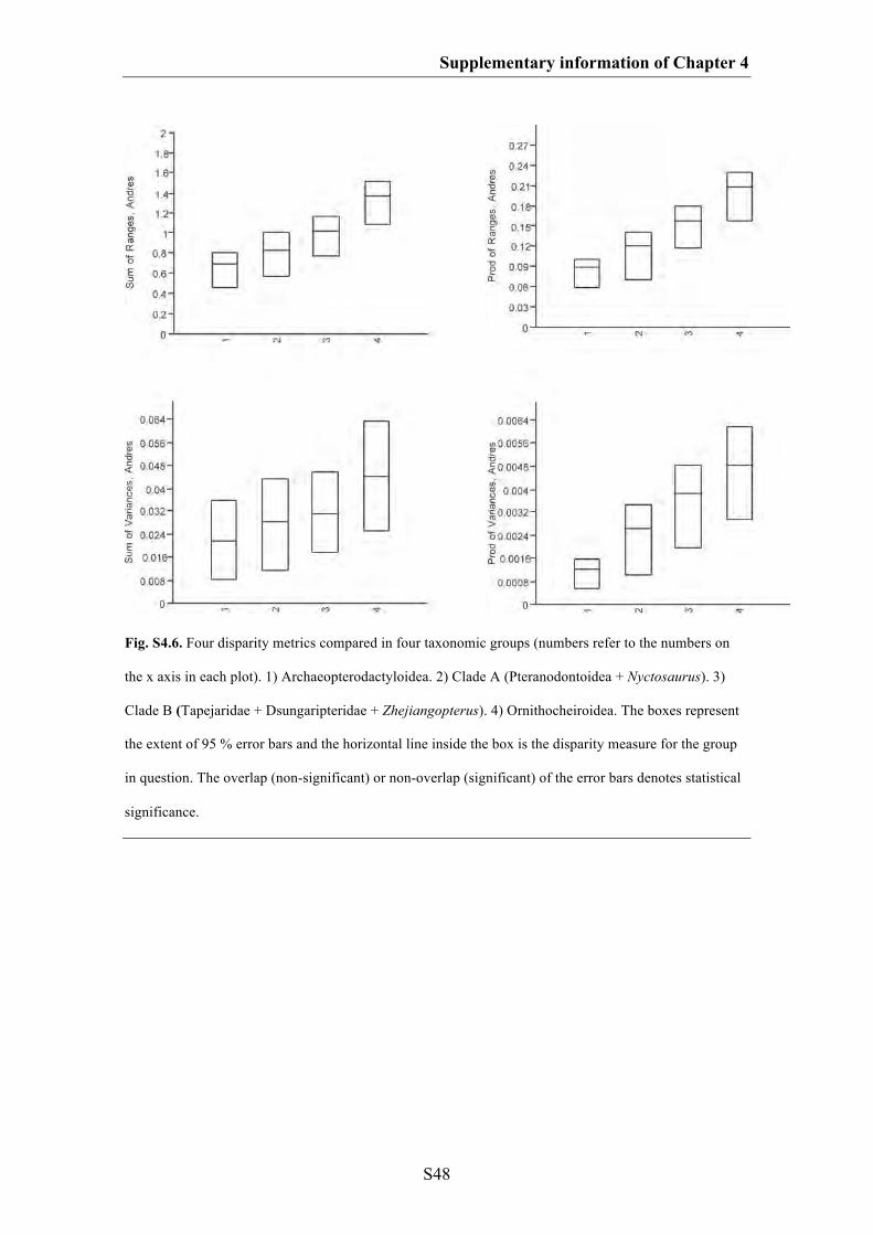
Supplementary information of Chapter 4
S48
Fig. S4.6. Four disparity metrics compared in four taxonomic groups (numbers refer to the numbers on
the x axis in each plot). 1) Archaeopterodactyloidea. 2) Clade A (Pteranodontoidea + Nyctosaurus). 3)
Clade B (Tapejaridae + Dsungaripteridae + Zhejiangopterus). 4) Ornithocheiroidea. The boxes represent
the extent of 95 % error bars and the horizontal line inside the box is the disparity measure for the group
in question. The overlap (non-significant) or non-overlap (significant) of the error bars denotes statistical
significance.
S49
SUPPLEMATARY INFORMATION OF CHAPTER 5
Macroevolutionary and morphofunctional patterns in theropod skulls: a morphometric
approach
1. Taxon sampling
2. Description of landmarks
3. Biomechanic and ecological parameters
4. Phylogeny and cluster topologies
5. Phylogenetic signals of functional proxies (SSI and AMS) and diagnostic test
for PIC analysis
*All figures of the supplementary information of Chapter 5 are modified after
Foth & Rauhut (2013)
Supplementary information of Chapter 5
S50
1. TAXON SAMPLING
Table S5.1. List of taxa used in the present analyses with data of occurrences (in million of years, Ma)
and sources of images.
Taxon Systematic affinities Age (Ma) Sources
Euparkeria Basal archosauriform Anisian (241.5) Rauhut 2003a
Lesothosaurus Ornithischia Hettangian/Sinemurian (202) Norman et al. 2004
Massospondylus Sauropodomorpha Sinemurian (198.5) Gow et al. 1990
Plateosaurus Sauropodomorpha Norian (215.5) Galton 1985
Herrerasaurus Herrerasauridae Carnian (224) Langer 2004
Eoraptor Basal theropod Carnian (224) Langer 2004
Daemonosaurus Basal theropod Rhaetian (208) Sues et al. 2011
Syntarsus
kayentakatae
Coelophysidae
Sinemurian/Pliensbachian
(195)
Tykosky 1998
Coelophysis Coelophysidae Carnian/Norian (221) Colbert 1989
Zupaysaurus Basal neotheropod Norian (215.5) Ezcurra 2007
Limusaurus Ceratosauria Oxfordian (156.5) Xu et al. 2009a
Ceratosaurus
Ceratosauria
Kimmeridgian/Tithonian
(151)
Sampson & Witmer 2007
Carnotaurus
Abelisauridae
Campanian/Maastrichtian
(71.3)
Rauhut 2003a
Majungasaurus Abelisauridae Campanian (77.4) Sampson & Witmer 2007
Monolophosaurus Megalosauroidea Callovian (161.5) Brusatte et al. 2010b
Spinosaurid Megalosauroidea Albian (105.5)* Rauhut 2003a
Sinraptor Allosauroidea Oxfordian (156.5) Currie & Zhao 1993
Acrocanthosaurus Allosauroidea Aptian/Albian (112) Eddy & Clarke 2011
Allosaurus
Allosauroidea
Kimmeridgian/Tithonian
(151)
Rauhut 2003a
Guanlong Tyrannosauroidea Oxfordian (156.5) Xu et al. 2006
Dilong Tyrannosauroidea Barremian (124) Xu et al. 2004
Bistahieversor Tyrannosauroidea Campanian (77.4) Carr & Williamson 2010
Alioramus Tyrannosauridae Maastrichtian (68.15) Brusatte et al. 2009
Daspletosaurus Tyrannosauridae Campanian (77.4) Holtz 2004
Gorgosaurus Tyrannosauridae Campanian (77.4) Rauhut 2003a
Tarbosaurus Tyrannosauridae
Campanian/Maastrichtian
(71.3) Hurum & Sabbath 2003
Tyrannosaurus Tyrannosauridae
Campanian/Maastrichtian
(71.3) Holtz 2004
Compsognathus Compsognathidae Kimmeridgian (152.5) Peyer 2006
Supplementary information of Chapter 5
S51
Garudimimus Ornithomimosauria Cenomanian-Santonian (89) Makovicky et al. 2004
Gallimimus Ornithomimosauria Maastrichtian (68.15) Makovicky et al. 2004
Ornithomimus
Ornithomimosauria
Campanian/Maastrichtian
(71.3)
Rauhut 2003a
Erlikosaurus Therizinosauridae Cenomanian-Santonian (89) Rauhut 2003a
Conchoraptor Oviraptoridae Campanian (77.4) Osmólska et al. 2004
Citipati Oviraptoridae Campanian (77.4) Osmólska et al. 2004
Oviraptor Oviraptoridae Campanian (77.4) Osmólska et al. 2004
Archaeopteryx Avialae Kimmeridgian (152.5) Rauhut 2003a
Confuciusornis Avialae Barremian (124) Chiappe et al. 1999
Pengornis
Avialae
Barremian (124)
O’Connor & Chiappe
2011
Shenquiornis
Avialae
Barremian (124)
O’Connor & Chiappe
2011
Anchiornis Troodontidae Oxfordian (156.5) Hu et al. 2009
Sinornithosaurus Dromaeosauridae Barremian (124) Xu & Wu 2001
Bambiraptor Dromaeosauridae Campanian (77.4) Burnham 2004
Velociraptor
Dromaeosauridae
Campanian (77.4)
Barsbold & Osmólska
1999
Shuvuuia Alvarezsauridae Campanian (77.4) Chiappe et al. 2002
*The reconstruction of a generalized spinosaurid is based on several taxa (the orbital and postorbital
region mainly based on Irritator, and the snout based on Suchomimus, with some elements reconstructed
after Baryonyx; see Rauhut 2003a), which range in age from the Barremian to the Cenomanian, so we
used an intermediate stage between these extremes for the age estimate of this reconstruction.
2. DESCRIPTION OF LANDMARKS
Homologous landmarks plotted on all theropod skulls in lateral view. The landmarks
present in both data sets are bold. Landmark 21 is only present in the smaller data set
(latin). Marked (*) landmarks are identical with Brusatte et al. (2012a).

Supplementary information of Chapter 5
S52
1. anteroventral corner of the premaxilla (this point is reconstructed in
Alioramus and Zupaysaurus due to a missing premaxilla) (preorbital
region)*
2. contact of premaxilla and nasal above the external naris (preorbital
region)*
3. contact of premaxilla and maxilla along the tooth row (preorbital region)*
4. tip of the maxillary process of the premaxilla (preorbital region)
5. anterodorsal contact between lacrimal and nasal (preorbital region)
6. contact between maxilla and jugal along the margin of antorbital fenestra
(in those taxa where the jugal do not reach the antorbital fenestra – the
most anterior point of the jugal is chosen, as in most theropods the contact
between maxilla and jugal along the antorbital fenestra is also the most
anterior point of the jugal) (preorbital region)
7. contact of maxilla and jugal along the ventral margin of the skull
(preorbital region)*
8. contact between lacrimal and jugal on the orbital margin (preorbital region)
9. contact between postorbital and jugal on the orbital margin (postorbital
region)*
10. contact between postorbital and jugal on the margin of the lateral temporal
fenestra (postorbital region)*
11. contact between jugal and quadratojugal on the margin of the lateral
temporal fenestra (postorbital region)*
12. anteroventral tip of the squamosal on the margin of the lateral temporal
fenestra (postorbital region)*
Supplementary information of Chapter 5
S53
13. ventral contact of postorbital and squamosal on the margin of the lateral
temporal fenestra (postorbital region)
14. anterior tip of the postorbital on the orbital margin (postorbital region)
15. most-anterior point of the antorbital fenestra (preorbital region)*
16. dorsal contact between postorbital and squamosal (postorbital region)
17. posteroventral corner of the quadratojugal (postorbital region)*
18. posteroventral tip of the squamosal posterior process (postorbital region)*
19. most-ventral point of the orbit (postorbital region)
20. posterior tip of the lacrimal on the orbital margin (postorbital region)
21. contact of the frontal with the parietal on the skull roof
3. BIOMECHANICAL AND ECOLOGICAL PARAMETERS
The respective skull lengths and depths were estimated with help of the program
tpsDig2 (Rohlf 2005) using the measure mode. The skull length was measured from the
anterior tip of the premaxilla to the posterior end of the quadratojugal. The skull depth
was measured at the height of the orbit. The average maximum stress was estimated
using the data from Rayfield (2011). To estimate the skull strength indicator (SSI) the
original data from Henderson (2002) were used to calculate a regression between skull
depth and SSI (see Fig. S5.1). The estimated average maximum stress based on a
regression of the original data from Rayfield (2011) (see Fig. S5.2).
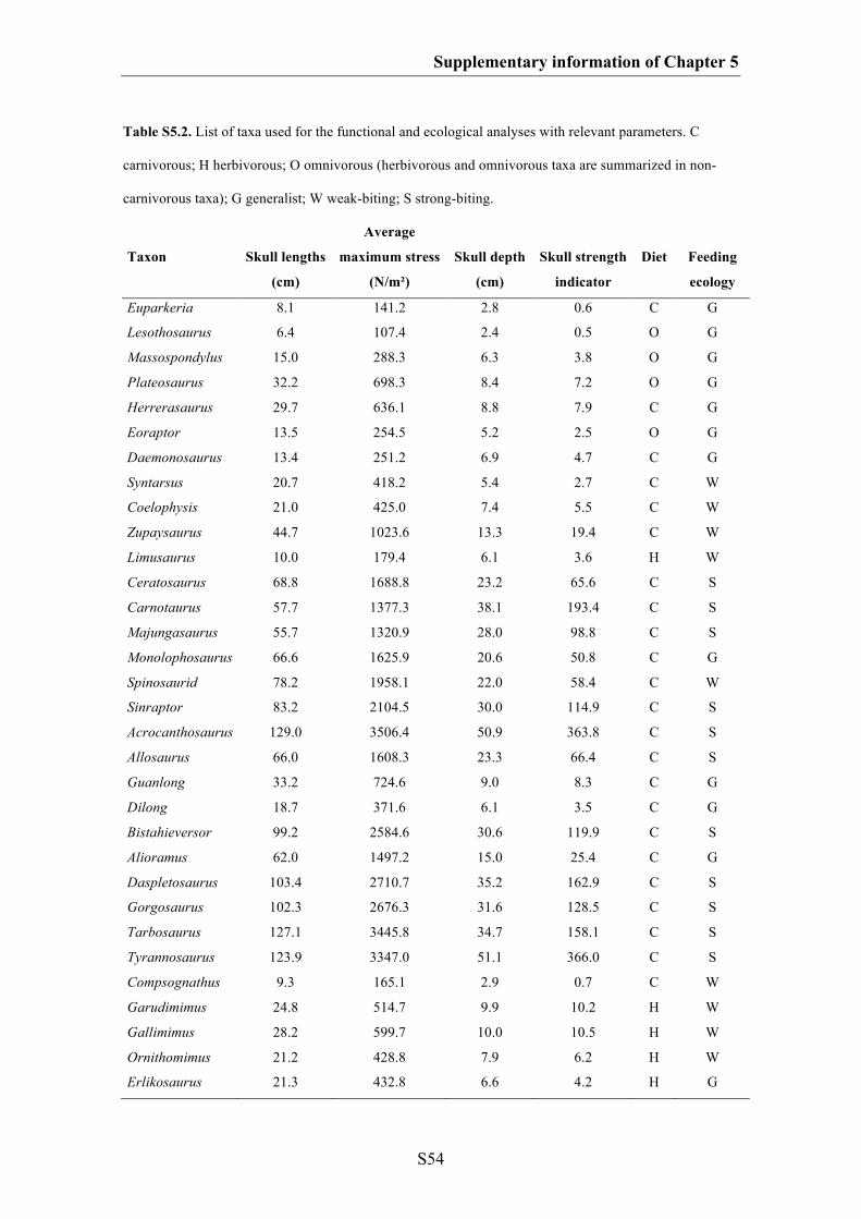
Supplementary information of Chapter 5
S54
Table S5.2. List of taxa used for the functional and ecological analyses with relevant parameters. C
carnivorous; H herbivorous; O omnivorous (herbivorous and omnivorous taxa are summarized in non-
carnivorous taxa); G generalist; W weak-biting; S strong-biting.
Taxon
Skull lengths
(cm)
Average
maximum stress
(N/m²)
Skull depth
(cm)
Skull strength
indicator
Diet
Feeding
ecology
Euparkeria 8.1 141.2 2.8 0.6 C G
Lesothosaurus 6.4 107.4 2.4 0.5 O G
Massospondylus 15.0 288.3 6.3 3.8 O G
Plateosaurus 32.2 698.3 8.4 7.2 O G
Herrerasaurus 29.7 636.1 8.8 7.9 C G
Eoraptor 13.5 254.5 5.2 2.5 O G
Daemonosaurus 13.4 251.2 6.9 4.7 C G
Syntarsus 20.7 418.2 5.4 2.7 C W
Coelophysis 21.0 425.0 7.4 5.5 C W
Zupaysaurus 44.7 1023.6 13.3 19.4 C W
Limusaurus 10.0 179.4 6.1 3.6 H W
Ceratosaurus 68.8 1688.8 23.2 65.6 C S
Carnotaurus 57.7 1377.3 38.1 193.4 C S
Majungasaurus 55.7 1320.9 28.0 98.8 C S
Monolophosaurus 66.6 1625.9 20.6 50.8 C G
Spinosaurid 78.2 1958.1 22.0 58.4 C W
Sinraptor 83.2 2104.5 30.0 114.9 C S
Acrocanthosaurus 129.0 3506.4 50.9 363.8 C S
Allosaurus 66.0 1608.3 23.3 66.4 C S
Guanlong 33.2 724.6 9.0 8.3 C G
Dilong 18.7 371.6 6.1 3.5 C G
Bistahieversor 99.2 2584.6 30.6 119.9 C S
Alioramus 62.0 1497.2 15.0 25.4 C G
Daspletosaurus 103.4 2710.7 35.2 162.9 C S
Gorgosaurus 102.3 2676.3 31.6 128.5 C S
Tarbosaurus 127.1 3445.8 34.7 158.1 C S
Tyrannosaurus 123.9 3347.0 51.1 366.0 C S
Compsognathus 9.3 165.1 2.9 0.7 C W
Garudimimus 24.8 514.7 9.9 10.2 H W
Gallimimus 28.2 599.7 10.0 10.5 H W
Ornithomimus 21.2 428.8 7.9 6.2 H W
Erlikosaurus 21.3 432.8 6.6 4.2 H G

Supplementary information of Chapter 5
S55
Conchoraptor 8.7 152.4 4.1 1.5 O S
Citipati 13.4 252.3 6.3 3.8 O S
Oviraptor 17.1 335.4 7.9 6.2 O S
Archaeopteryx 3.0 44.8 1.2 0.1 O W
Confuciusornis 6.7 133.2 2.8 0.6 O G
Anchiornis 5.4 87.3 3.0 0.8 O W
Sinornithosaurus 13.7 258.5 4.5 1.9 C G
Bambiraptor 11.2 205.3 3.7 1.2 C G
Velociraptor 25.4 530.4 7.3 5.3 C G
Pengornis - - - - O W
Shenquiornis - - - - O W
Shuvuuia - - - - O W
Fig. S5.1. Correlation between skull depth and skull strength indicator (LogSSI = 2.18 LogSD – 1.1602,
R² = 0.963, p value < 0.001) (based on the data set from Henderson 2002).

Supplementary information of Chapter 5
S56
Fig. S5.2. Correlation between skull length and average maximum stress (LogAMS = 1.16 LogSL –
1.0913, R² = 0.725, p value < 0.001) (based on the data set from Rayfield 2011, without a crestless
Monolophosaurus).
Table S5.3. Correlation of specimen centroid size (log transformed) with Procrustes Coordinates and the
first three PC axes.
R² p value
Procrustes Coordinates (large data set) 0.159 <0.001
Procrustes Coordinates (small data set – Paraves) 0.140 <0.001
PC 1 (large data set) 0.166 0.007
PC 1 (small data set – Paraves) 0.083 0.335
PC 2 (large data set) 0.452 <0.001
PC 2 (small data set – Paraves) 0.169 0.147
PC 3 (large data set) 0.163 0.009
PC 3 (small data set – Paraves) 0.139 0.188
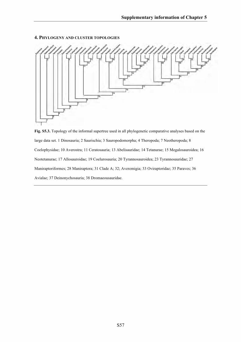
Supplementary information of Chapter 5
S57
4. PHYLOGENY AND CLUSTER TOPOLOGIES
Fig. S5.3. Topology of the informal supertree used in all phylogenetic comparative analyses based on the
large data set. 1 Dinosauria; 2 Saurischia; 3 Sauropodomorpha; 4 Theropoda; 7 Neotheropoda; 8
Coelophysidae; 10 Averostra; 11 Ceratosauria; 13 Abelisauridae; 14 Tetanurae; 15 Megalosauroidea; 16
Neotetanurae; 17 Allosauroidae; 19 Coelurosauria; 20 Tyrannosauroidea; 23 Tyrannosauridae; 27
Maniraptoriformes; 28 Maniraptora; 31 Clade A; 32; Averemigia; 33 Oviraptoridae; 35 Paraves; 36
Avialae; 37 Deinonychosauria; 38 Dromaeousauridae.
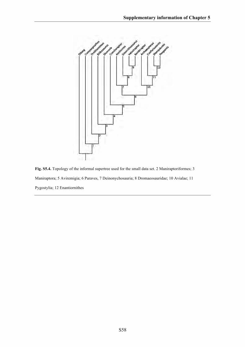
Supplementary information of Chapter 5
S58
Fig. S5.4. Topology of the informal supertree used for the small data set. 2 Maniraptoriformes; 3
Maniraptora; 5 Aviremigia; 6 Paraves, 7 Deinonychosauria; 8 Dromaeosauridae; 10 Avialae; 11
Pygostylia; 12 Enantiornithes
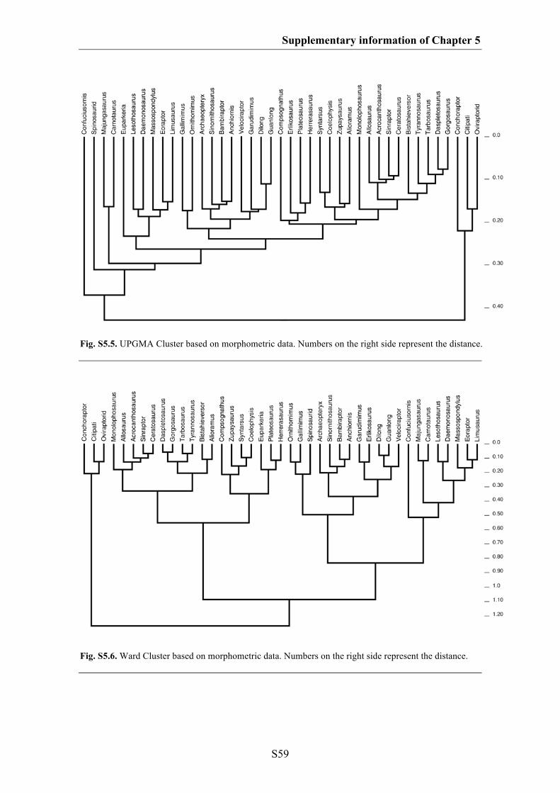
Supplementary information of Chapter 5
S59
Fig. S5.5. UPGMA Cluster based on morphometric data. Numbers on the right side represent the distance.
Fig. S5.6. Ward Cluster based on morphometric data. Numbers on the right side represent the distance.
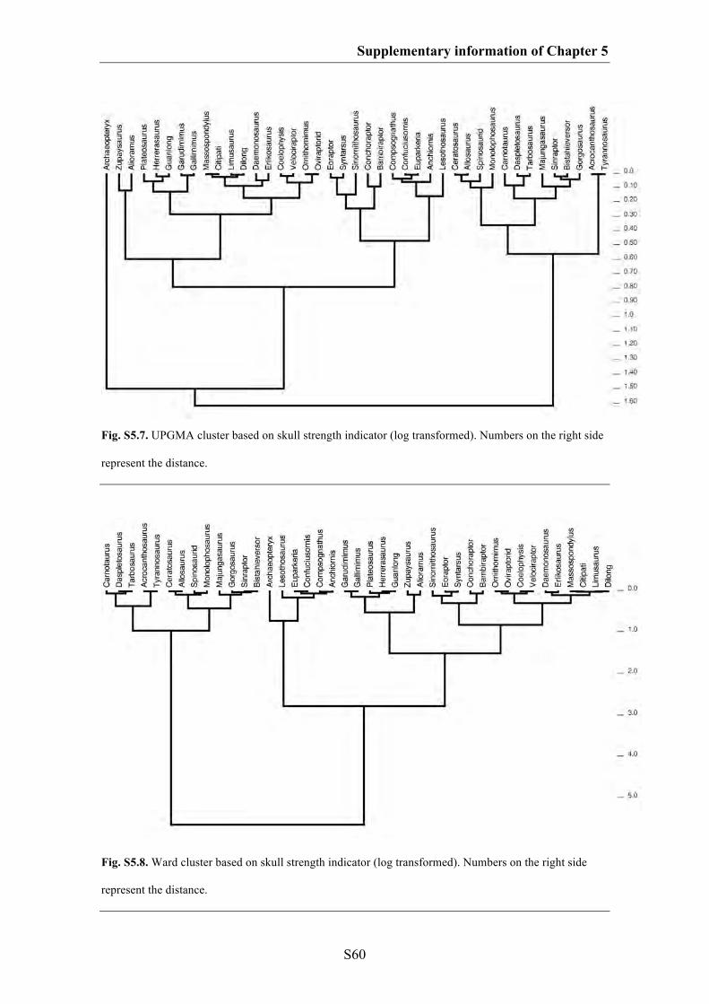
Supplementary information of Chapter 5
S60
Fig. S5.7. UPGMA cluster based on skull strength indicator (log transformed). Numbers on the right side
represent the distance.
Fig. S5.8. Ward cluster based on skull strength indicator (log transformed). Numbers on the right side
represent the distance.
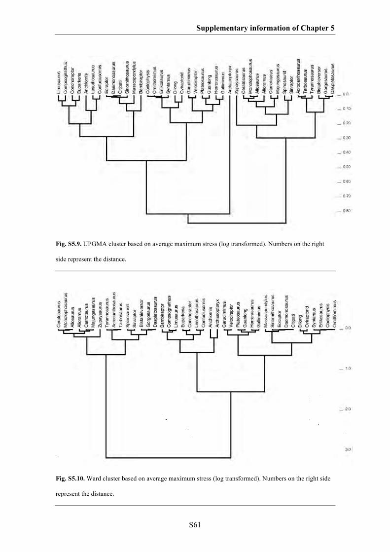
Supplementary information of Chapter 5
S61
Fig. S5.9. UPGMA cluster based on average maximum stress (log transformed). Numbers on the right
side represent the distance.
Fig. S5.10. Ward cluster based on average maximum stress (log transformed). Numbers on the right side
represent the distance.
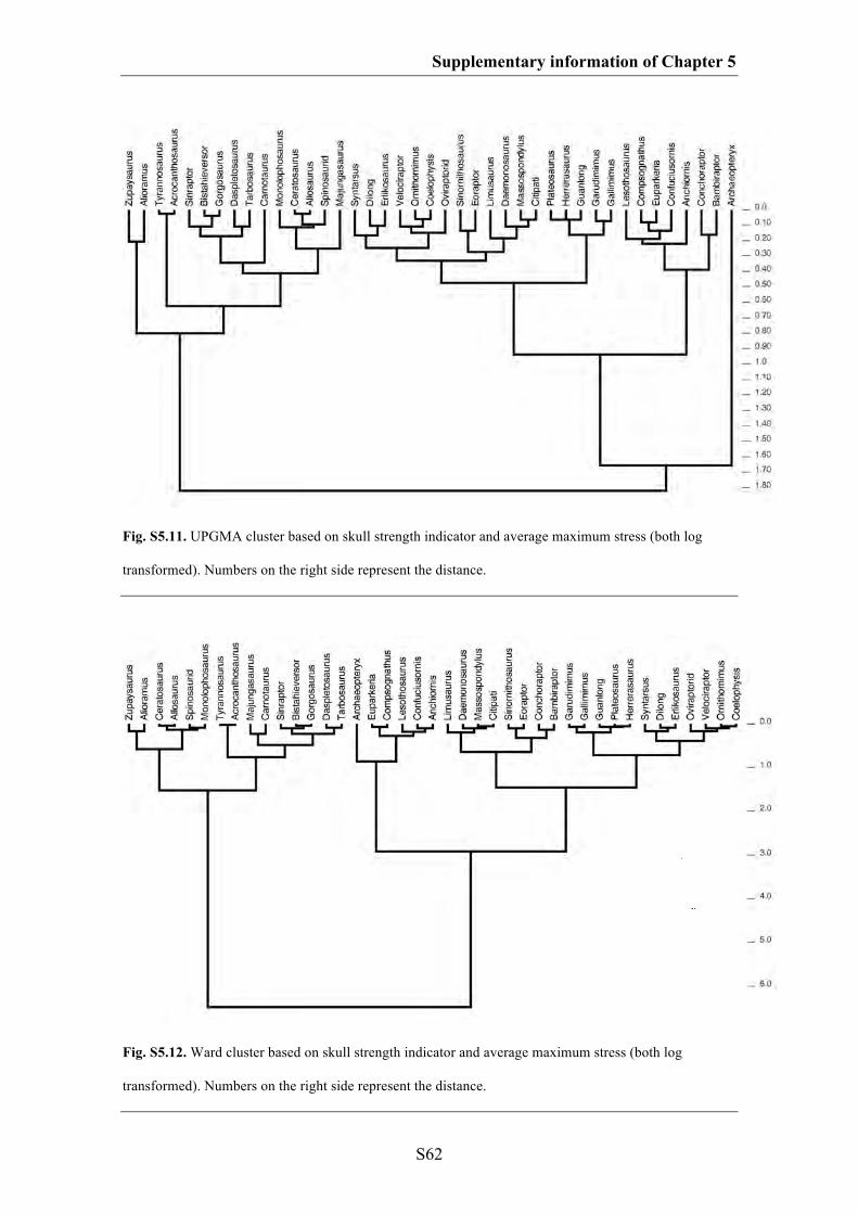
Supplementary information of Chapter 5
S62
Fig. S5.11. UPGMA cluster based on skull strength indicator and average maximum stress (both log
transformed). Numbers on the right side represent the distance.
Fig. S5.12. Ward cluster based on skull strength indicator and average maximum stress (both log
transformed). Numbers on the right side represent the distance.
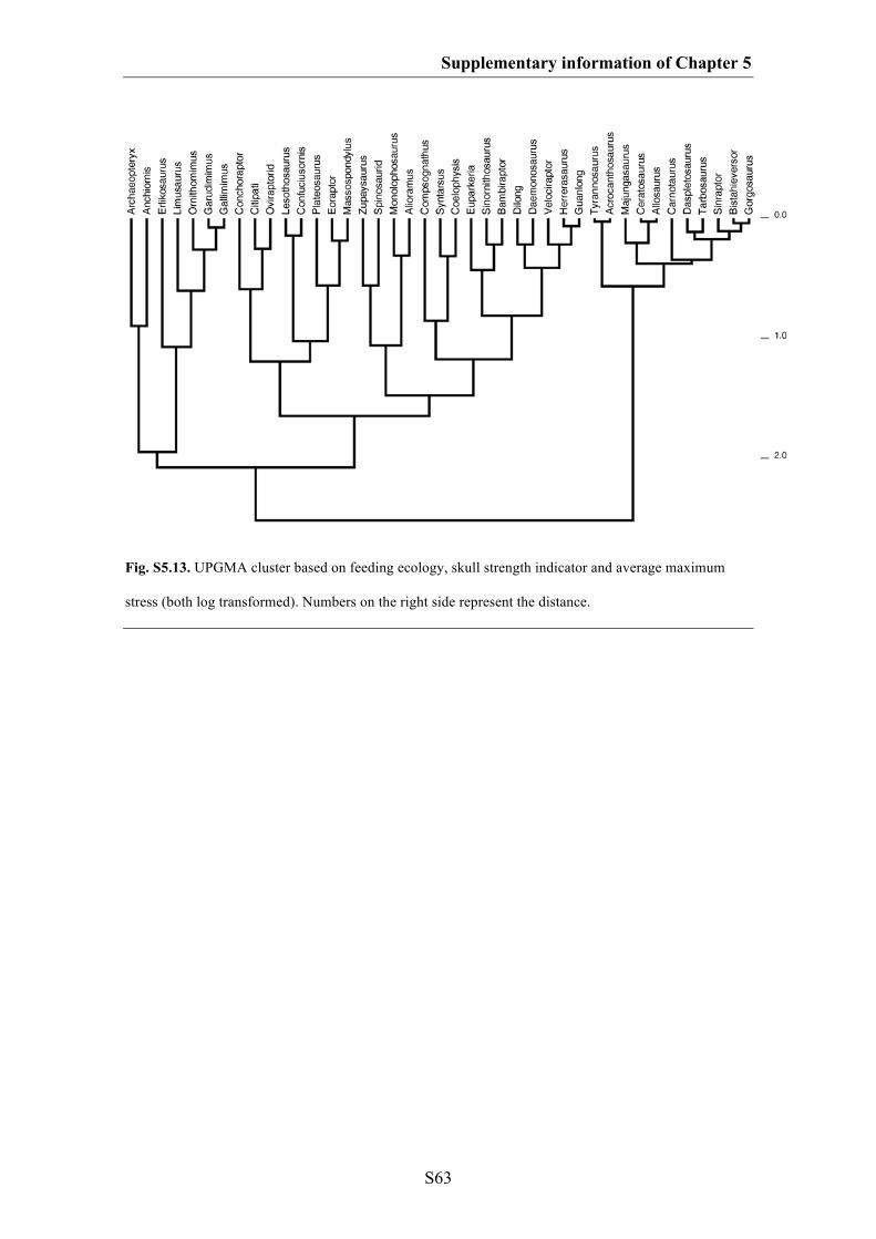
Supplementary information of Chapter 5
S63
Fig. S5.13. UPGMA cluster based on feeding ecology, skull strength indicator and average maximum
stress (both log transformed). Numbers on the right side represent the distance.
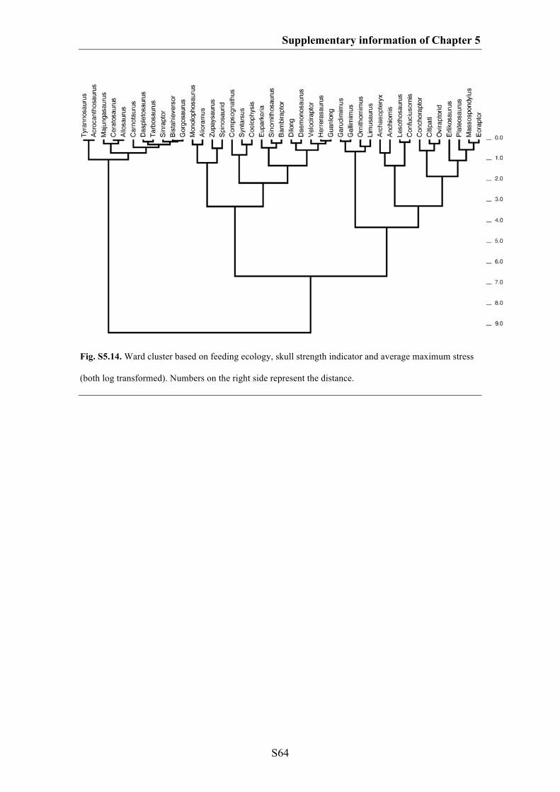
Supplementary information of Chapter 5
S64
Fig. S5.14. Ward cluster based on feeding ecology, skull strength indicator and average maximum stress
(both log transformed). Numbers on the right side represent the distance.
Supplementary information of Chapter 5
S65
5. PHYLOGENETIC SIGNALS OF FUNCTIONAL PROXIES (SSI AND AMS) AND
DIAGNOSTIC TEST FOR PIC ANALYSIS
Both morphometric data and functional factors (SSI and AMS) show a phylogenetic
signal (Fig. S5.15, Table 5.4). After transforming the scores into PIC values, we first
analysed if they fulfil the four criteria listed in the materials and methods section (Table
S5.4). Here, PC 1 and 2 as well as SSI and AMS show no significant correlations. For
PC 3 a correlation is present for the fourth criterion (estimated node values vs. the
corrected node high). However, as the fourth test indicates primarily evolutionary trends
and it is not strictly diagnostic (Midford et al. 2005), all scores can be modelled as
random walk with a uniform rate of change, and thus, fulfil the assumptions for PIC
analyses. In contrast, the SSI based on Henderson’s (2002) original data posses a
significant correlation with the first criterion (standard deviation). However, this could
be the result of the small sample size.
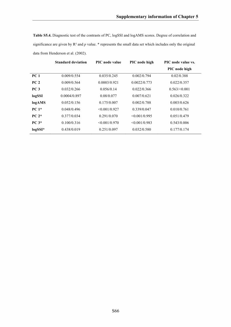
Supplementary information of Chapter 5
S66
Table S5.4. Diagnostic test of the contrasts of PC, logSSI and logAMS scores. Degree of correlation and
significance are given by R² and p value. * represents the small data set which includes only the original
data from Henderson et al. (2002).
Standard deviation PIC node value PIC node high PIC node value vs.
PIC node high
PC 1 0.009/0.554 0.035/0.245 0.002/0.794 0.02/0.388
PC 2 0.009/0.564 0.0003/0.921 0.0022/0.773 0.022/0.357
PC 3 0.032/0.266 0.056/0.14 0.022/0.366 0.563/<0.001
logSSI 0.0004/0.897 0.08/0.077 0.007/0.621 0.026/0.322
logAMS 0.052/0.156 0.175/0.007 0.002/0.788 0.003/0.626
PC 1* 0.048/0.496 <0.001/0.927 0.339/0.047 0.010/0.761
PC 2* 0.377/0.034 0.291/0.070 <0.001/0.995 0.051/0.479
PC 3* 0.100/0.316 <0.001/0.970 <0.001/0.983 0.543/0.006
logSSI* 0.438/0.019 0.251/0.097 0.032/0.580 0.177/0.174
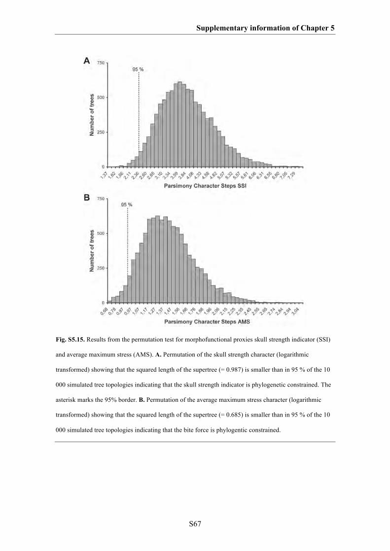
Supplementary information of Chapter 5
S67
Fig. S5.15. Results from the permutation test for morphofunctional proxies skull strength indicator (SSI)
and average maximum stress (AMS). A. Permutation of the skull strength character (logarithmic
transformed) showing that the squared length of the supertree (= 0.987) is smaller than in 95 % of the 10
000 simulated tree topologies indicating that the skull strength indicator is phylogenetic constrained. The
asterisk marks the 95% border. B. Permutation of the average maximum stress character (logarithmic
transformed) showing that the squared length of the supertree (= 0.685) is smaller than in 95 % of the 10
000 simulated tree topologies indicating that the bite force is phylogentic constrained.

S68
SUPPLEMATARY INFORMATION OF CHAPTER 6
Macroevolutionary and morphofunctional patterns in theropod skulls: a morphometric
approach
1. Methods: UV photography
2. Additional information on Sciurumimus
3. Ontogenetic stage of the specimen
4. Phylogenetic analysis
5. Comparison with Juravenator starki
6. Comparisons with other Jurassic theropods
7. Additional discussion of soft tissues
8. Repository of the specimen
*All figures of the supplementary information of Chapter 6 are modified after
Rauhut et al. (2012)
Supplementary information of Chapter 6
S69
1. METHODS: UV PHOTOGRAPHY
Most fossil skeletal remains and sometimes mineralized soft parts from the Upper
Jurassic plattenkalks of southern Germany and from the Middle to Late Mesozoic
localities of Northeastern China are fluorescent under ultraviolet radiation. In most
cases, this fuorescence allows a more precise investigation of morphological details of
skeletal remains as well as soft parts. Delicate skeletal elements and remains of soft
parts are poorly or not discernable in visible light but shine conspicuously under filtered
UV. The technique can be used to differentiate bone sutures from cracks, more clearly
establish outlines of compressed skeletal elements, and to separate bones or soft parts
from the underlying matrix or each other.
During the past 10 years, one of us (Helmut Tischlinger) has considerably
improved ultraviolet investigation techniques and ultraviolet-light photography of
fossils from Solnhofen and Solnhofen-type-Lagerstaetten as well as from the Middle
Jurassic to Early Cretaceous lacustrine deposits of the Jinlingsi and Jehol Group,
Northeastern China, using powerful UV lamps and new photographic documentation
techniques (Tischlinger 2002, 2005a, b; Tischlinger & Unwin 2004; Arratia &
Tischlinger 2010; Tischlinger & Frey 2010; Hone et al. 2010; Kellner et al. 2010;
Schweigert et al. 2010). For our investigations we predominantly use UVA lamps with
a wavelength of 365-366 nanometers.
Sometimes essential details of bones and soft parts are poorly or not visible even
under UV light with the naked eye or even under a microscope, and can exclusively be
demonstrated by ultraviolet-light photography. The application of different filters
allows a selective visualisation of peculiar fine structures. In most cases a selection of
Supplementary information of Chapter 6
S70
different colour correction filters is necessary. Each limestone slab and bone or tissue
will react differently to different light wavelengths and is captured differently with
varying exposures and filters. The right combination is needed to highlight the area of
interest. The optimum filtering and exposure time has to be tested in a series of
experiments (Tischlinger 2002). The number and combination of filters varies greatly
and exposure times vary between 1 second and some minutes, depending on the nature
of the fossil material and the magnification, intensity, and incident angle of the
ultraviolet lamps. Filtering works optimally with analogue photography using slide
films, although digital cameras can be used, too.
2. ADDITIONAL INFORMATION ON SCIURUMIMUS
HISTORY OF FIND AND PREPARATION
The specimen was found during systematic excavations in the Rygol Quarry at Painten,
Bavaria, Germany. First bony elements of the central area of the body appeared after
cleaning on the floor of the excavation area, so the slab with the skeleton was excavated
and brought into the lab for preparation. In the lab, the upper surface (the one exposed
in the quarry) was stabilized with a ceramic glue (Uniflott) and fixed to another slab.
Then, the specimen was mechanically prepared from the underside. Damaged areas
were reconstructed with Keraquick, which is clearly visible under UV light. Loose
bones and sections were glued onto the specimen, but no arrangement or orientation of
bones was changed. The specimen was studied by one of us (Helmut Tischlinger) prior
to preparation, so that there can be no doubt about its authenticity.

Supplementary information of Chapter 6
S71
Table S6.1. Selected measurements of Sciurumimus albersdoerferi
Body part Length
Total length of skeleton 719 mm
Skull length 79 mm
Posterior skull height: c. 32 mm
Length of orbit 19.7 mm
Height of orbit 21.5 mm
Length of mandible 73.2 mm
Length of cervical series 69 mm
Length of dorsal series 102 mm
Length of sacrum 37.25 mm
Length of preserved caudal series 432 mm
Length of humerus 26.8 mm
Length of radius 17 mm
Length of metacarpal II 11 mm
Length of femur 50.6 mm
Length of tibiotarsus 54.2 mm
Length of metatarsal III 32.1 mm
3. ONTOGENETIC STAGE OF THE SPECIMEN
Although no histological sampling is possible in this unique specimen, several lines of
evidence indicate that the holotype is an early juvenile, probably early posthatchling
individual. First, there is no fusion of any skeletal elements in the skeleton. In the
vertebral column, the neurocentral sutures of the cervical, dorsal and at least anterior
caudal vertebrae are open, and the neural arches even have slightly disarticulated from
the centra in at least some elements. The sacral centra are preserved in articulation, but
the posterior two sacrals are displaced ventrally from the anterior end of the sacrum,
demonstrating that there is neither fusion of the sacral vertebrae with each other, nor of
the sacral ribs with the ilium. Although the pattern of neurocentral suture closure varies
within dinosaurs (Irmis 2007), the lack of fusion in all vertebrae, with the possible
Supplementary information of Chapter 6
S72
exception of the distalmost caudals (which are already closed in hatchling crocodiles;
Brochu 1996), clearly indicates that the specimen of Sciurumimus is an immature
individual. This is furthermore supported by disarticulation in other elements that
usually show very tight sutures or even fusion in theropods, such as the basioccipital
and exoccipital, or the distal ischium. Likewise, several skeletal elements, such as the
carpal and distal tarsal bones show poor ossification and several joint surfaces,
including the proximal articular end of the humerus, exhibit strongly porous surfaces,
indicating poorly ossified articular ends.
Another indicator of the early juvenile stage of Sciurumimus is found in the
surface structure of basically all bony elements. Both dermal and enchondral elements
show a coarsely striated surface (Fig. S6.1, S6.2). Such a surface structure corresponds
to bone texture type I of Tumarkin-Deratzian et al. (2006). According to these authors,
in birds, this texture occurs only in individuals of 50 % or less skeletal maturity, and
hatching-year birds only exhibit this type of texture, as it is the case in Sciurumimus.
Bone surface textures were found to be useful as ontogenetic indicator in a number of
fossil amniotes (summary in Tumarkin-Deratzian 2009) and thus this represents an
independent indication of an early ontogenetic stage for the specimen.
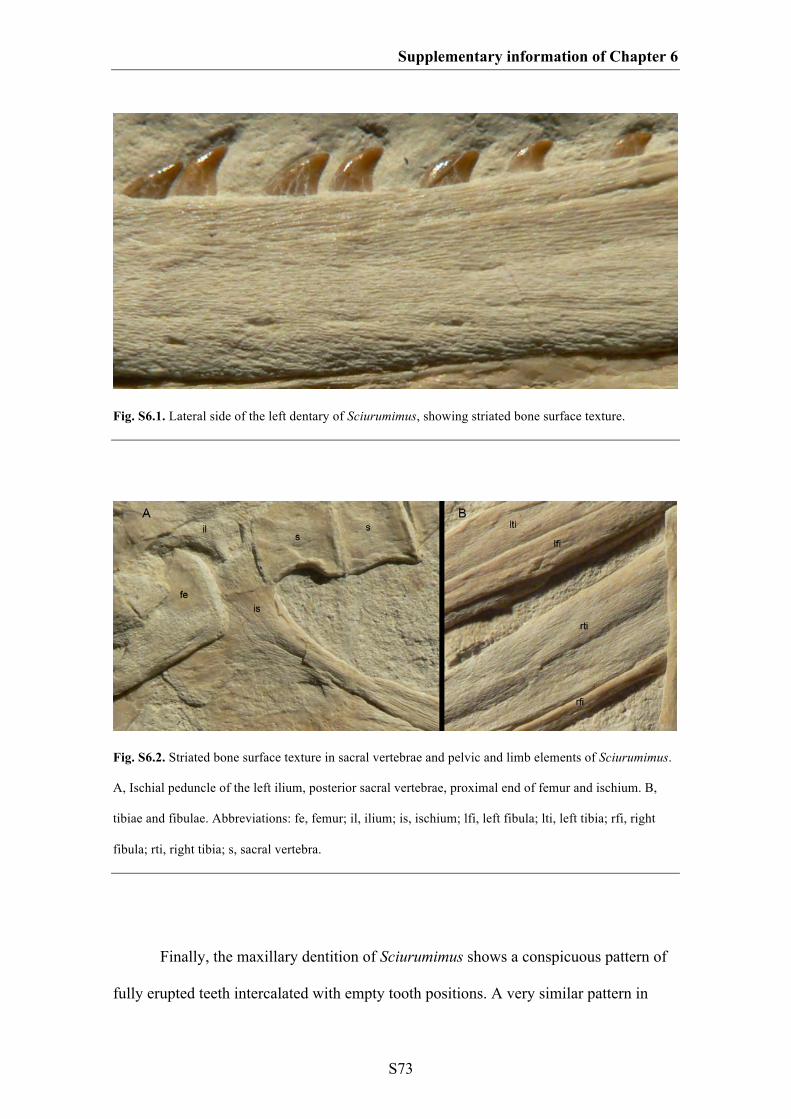
Supplementary information of Chapter 6
S73
Fig. S6.1. Lateral side of the left dentary of Sciurumimus, showing striated bone surface texture.
Fig. S6.2. Striated bone surface texture in sacral vertebrae and pelvic and limb elements of Sciurumimus.
A, Ischial peduncle of the left ilium, posterior sacral vertebrae, proximal end of femur and ischium. B,
tibiae and fibulae. Abbreviations: fe, femur; il, ilium; is, ischium; lfi, left fibula; lti, left tibia; rfi, right
fibula; rti, right tibia; s, sacral vertebra.
Finally, the maxillary dentition of Sciurumimus shows a conspicuous pattern of
fully erupted teeth intercalated with empty tooth positions. A very similar pattern in
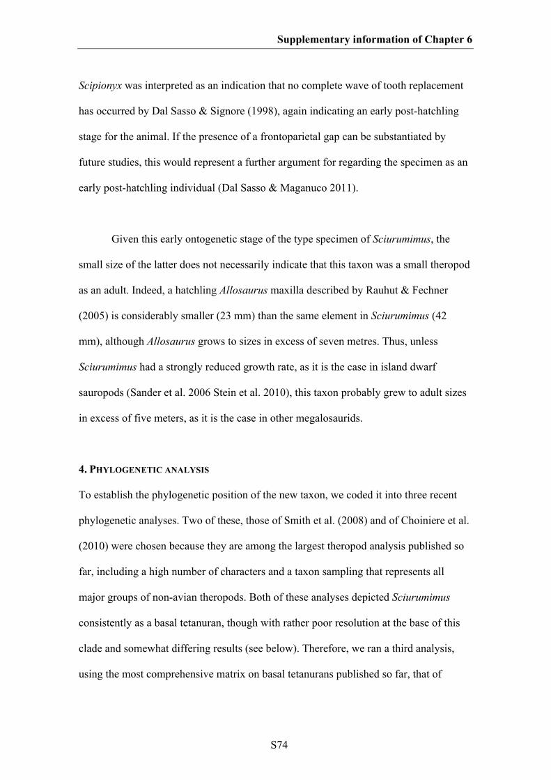
Supplementary information of Chapter 6
S74
Scipionyx was interpreted as an indication that no complete wave of tooth replacement
has occurred by Dal Sasso & Signore (1998), again indicating an early post-hatchling
stage for the animal. If the presence of a frontoparietal gap can be substantiated by
future studies, this would represent a further argument for regarding the specimen as an
early post-hatchling individual (Dal Sasso & Maganuco 2011).
Given this early ontogenetic stage of the type specimen of Sciurumimus, the
small size of the latter does not necessarily indicate that this taxon was a small theropod
as an adult. Indeed, a hatchling Allosaurus maxilla described by Rauhut & Fechner
(2005) is considerably smaller (23 mm) than the same element in Sciurumimus (42
mm), although Allosaurus grows to sizes in excess of seven metres. Thus, unless
Sciurumimus had a strongly reduced growth rate, as it is the case in island dwarf
sauropods (Sander et al. 2006 Stein et al. 2010), this taxon probably grew to adult sizes
in excess of five meters, as it is the case in other megalosaurids.
4. PHYLOGENETIC ANALYSIS
To establish the phylogenetic position of the new taxon, we coded it into three recent
phylogenetic analyses. Two of these, those of Smith et al. (2008) and of Choiniere et al.
(2010) were chosen because they are among the largest theropod analysis published so
far, including a high number of characters and a taxon sampling that represents all
major groups of non-avian theropods. Both of these analyses depicted Sciurumimus
consistently as a basal tetanuran, though with rather poor resolution at the base of this
clade and somewhat differing results (see below). Therefore, we ran a third analysis,
using the most comprehensive matrix on basal tetanurans published so far, that of
Supplementary information of Chapter 6
S75
Benson et al. (2010). The results of the latter analysis were used for the phylogenetic
placement of the new taxon presented in the main manuscript.
Given the juvenile status of the specimen, one important question is, of course,
the possible affect of ontogentically variable characters on its phylogenetic position.
Clearly age-dependant characters, such as fusion of skeletal elements, were coded as
“?” for Sciurumimus in all analysis. Furthermore, in addition to the analyses reported on
below, we ran additional analyses of the three data matrices with all characters we
considered to be potentially variable with ontogeny (characters concerning cranial
ornamentation [crests, rugosities], orbit shape and size, morphometric ratios between
different elements or between different structures within one element, development of
muscle attachments) coded as “?” for Sciurumimus. Although this considerably
increased the amount of missing data in Sciurumimus, the phylogenetic results remained
the same as those reported below.
ANALYSIS BASED ON SMITH ET AL. (2008)
Smith et al. (2008) presented a phylogenetic analysis of six outgroup and 51
neotheropod ingroup taxa, plus one single specimen from the Early Cretaceous of
Australia, coded across 353 morphological characters. This matrix is a slightly
expanded version of the matrix of Smith et al. (2007) and includes a wide array of non-
avian theropods, from coelophysoids to paravians, though with emphasis on non-
coelurosaurian forms (39 of the ingroup OTUs). We coded Sciurumimus in the same
matrix, without changes to other codings, and ran the analysis in PAUP* 4.0 (Swofford
2003), using a heuristic search with TBR branch swapping and random addition
sequence with 100 replicates. The analysis resulted in the recovery of 3 720 equally
Supplementary information of Chapter 6
S76
parsimonious trees with a length of 887 steps. The strict consensus of these trees (Fig.
S6.3) generally agrees with that found by Smith et al. (2008), though with slightly less
resolution within Megalosauroidea (=Spinosauroidea). Sciurumimus was found to be the
sister taxon to Monolophosaurus and Neotetanurae in this analysis. However, only one
additional step is needed to place this taxon within Megalosauroidea, whereas a
placement within Neotetanurae implies at least six additional steps. Tree support is low,
with bootstrap values below 50 for the vast majority of nodes within Theropoda, with
the exception of some coelurosaur clades.
Codings for Sciurumimus in the matrix of Smith et al. (2008)
00200[0/1]0101??100?10?000?21000011???00?00?10?1[1/2]0?01????0001000?00????
00?000?????0121??0??0?100????00000??????????????????0100100?11?1000[0/1]0?11
?0110?1110[0/1]?0?0?0120?00??00101????000?10020????000??0010001000000?????
01[0/1]000000[0/1]0000000?00?011111010110?010010000000??001100?1?[0/1]1?0?0
???00?10?00000011?0?1??201?????10?????????00?????????????1100?0?01??1201?00
00100

Supplementary information of Chapter 6
S77
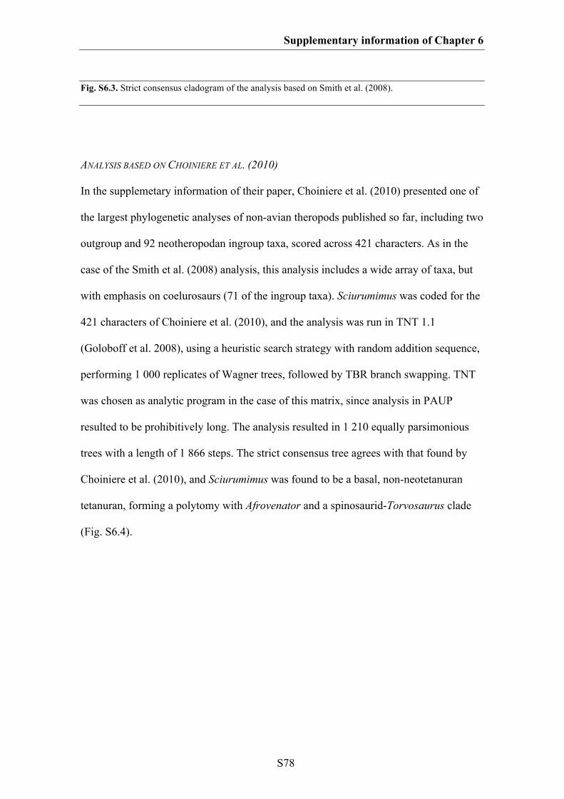
Supplementary information of Chapter 6
S78
Fig. S6.3. Strict consensus cladogram of the analysis based on Smith et al. (2008).
ANALYSIS BASED ON CHOINIERE ET AL. (2010)
In the supplemetary information of their paper, Choiniere et al. (2010) presented one of
the largest phylogenetic analyses of non-avian theropods published so far, including two
outgroup and 92 neotheropodan ingroup taxa, scored across 421 characters. As in the
case of the Smith et al. (2008) analysis, this analysis includes a wide array of taxa, but
with emphasis on coelurosaurs (71 of the ingroup taxa). Sciurumimus was coded for the
421 characters of Choiniere et al. (2010), and the analysis was run in TNT 1.1
(Goloboff et al. 2008), using a heuristic search strategy with random addition sequence,
performing 1 000 replicates of Wagner trees, followed by TBR branch swapping. TNT
was chosen as analytic program in the case of this matrix, since analysis in PAUP
resulted to be prohibitively long. The analysis resulted in 1 210 equally parsimonious
trees with a length of 1 866 steps. The strict consensus tree agrees with that found by
Choiniere et al. (2010), and Sciurumimus was found to be a basal, non-neotetanuran
tetanuran, forming a polytomy with Afrovenator and a spinosaurid-Torvosaurus clade
(Fig. S6.4).
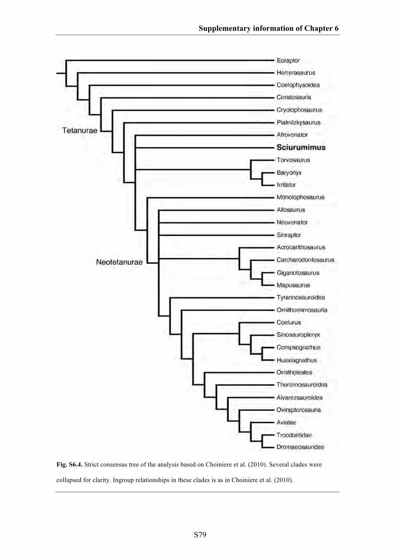
Supplementary information of Chapter 6
S79
Fig. S6.4. Strict consensus tree of the analysis based on Choiniere et al. (2010). Several clades were
collapsed for clarity. Ingroup relationships in these clades is as in Choiniere et al. (2010).
Supplementary information of Chapter 6
S80
Codings for Sciurumimus in the matrix of Choiniere et al. (2010)
10?0[01]00?00?11010??????20001?00?0?0000?000?11100000??1??0010??????000?00
????0100???0?10????????000?????????????000?1?0000??????????0?1000100?01110?
0?011??00??01001?00002000?0??00?0202??[01]0?00111??0?00??00?00011001?23???
?0000000000??0000100001000??1???0000?11000?00001000?0000?000001?1???0111
00010?012112111?1000000100?02?0???0?0?1??1?0?00??01??0??00001000010?1?000
?01??0?000100????1???1?????00??????????????0010??00?0?2000?
ANALYSIS BASED ON BENSON ET AL. (2010)
After establishing that Sciurumimus is a basal, non-coelurosaurian theropod in the
analyses of Smith et al. (2010) and Choiniere et al. (2010), we decided to test its
detailed phylogenetic position in the most extensive phylogenetic analysis of basal
tetanurans published so far, that of Benson et al. (2010). This matrix included four
outgroup and 41 tetanuran ingroup taxa, with emphasis on basal, non-coelurosaurian
taxa [38 of the ingroup taxa, as opposed to 20 in Smith et al. (2008) and 13 in Choiniere
et al. (2010)], scored across 233 characters. We included Sciurumimus in this matrix
and reran the analysis in PAUP* 4.0, using the same settings described above for the
Smith et al. (2008) analysis. The analysis resulted in 7 383 equally parsimonious tress
with a length of 656 steps. The strict consensus of these trees recovered Sciurumimus in
a large polytomy within Megalosauroidea more derived than Monolophosaurus. After
the exclusion of Piveteausaurus, a reduced consensus tree depicts Sciurumimus as the
most basal representative of the Megalosauridae (Fig. 6.4, S6.5). As in the previous
analyses, tree support is rather low, with most clades showing bootstrap values below
50 %.

Supplementary information of Chapter 6
S81
An interesting result of the analysis is that the inclusion of Sciurumimus, without
any other changes to the original matrix of Benson et al. (2010), led to the recovery of
the monophyletic Carnosauria, including the Megalosauroidea and Allosauroidea. This
relationship was also found by Rauhut (2003a), but is in contrast to most recent
analyses, which recovered megalosauroids (or spinosauroids) as outgroup to a
monophyletic Neotetanurae that includes allosauroids and coelurosaurs e.g. Smith et al.
(2008), Choiniere et al. (2010) and Benson et al. (2010). Synapomorphies of carnosaurs
include the presence of a subnarial foramen, the presence of at least weakly developed
enamel wrinkles in the lateral teeth, opisthocoelous cervical vertebrae, a kinked anterior
edge of the anterior caudal neural spines, the presence of an indentation between the
acromion process of the scapula and the coracoid, a biceps tubercle that is developed as
an obliquely oriented ridge, the presence of a broad ridge above the acetabulum on the
ilium, and the presence of a well-developed extensor groove on the anterior side of the
distal femur. However, making Neotetanurae monophyletic, to the exclusion of
megalosauroids, requires only two additional steps. Thus, the interrelationships of basal
tetanurans remain problematic and need additional investigation.
Codings for Sciurumimus in the matrix of Benson et al. (2010)
[0/1]??01?0?1001101?0???????0?0000?010210???010000??000?0?00??11????00101??
1????1??0?110000010020?00101?00?111?0[1/2]?10[0/1]?11??0000000??001001?0020
0000?1????0000[0/1]001011??0001????[0/1]0??00??0?101001?1??11???????01??????
??02???0010?010???00?00?0??
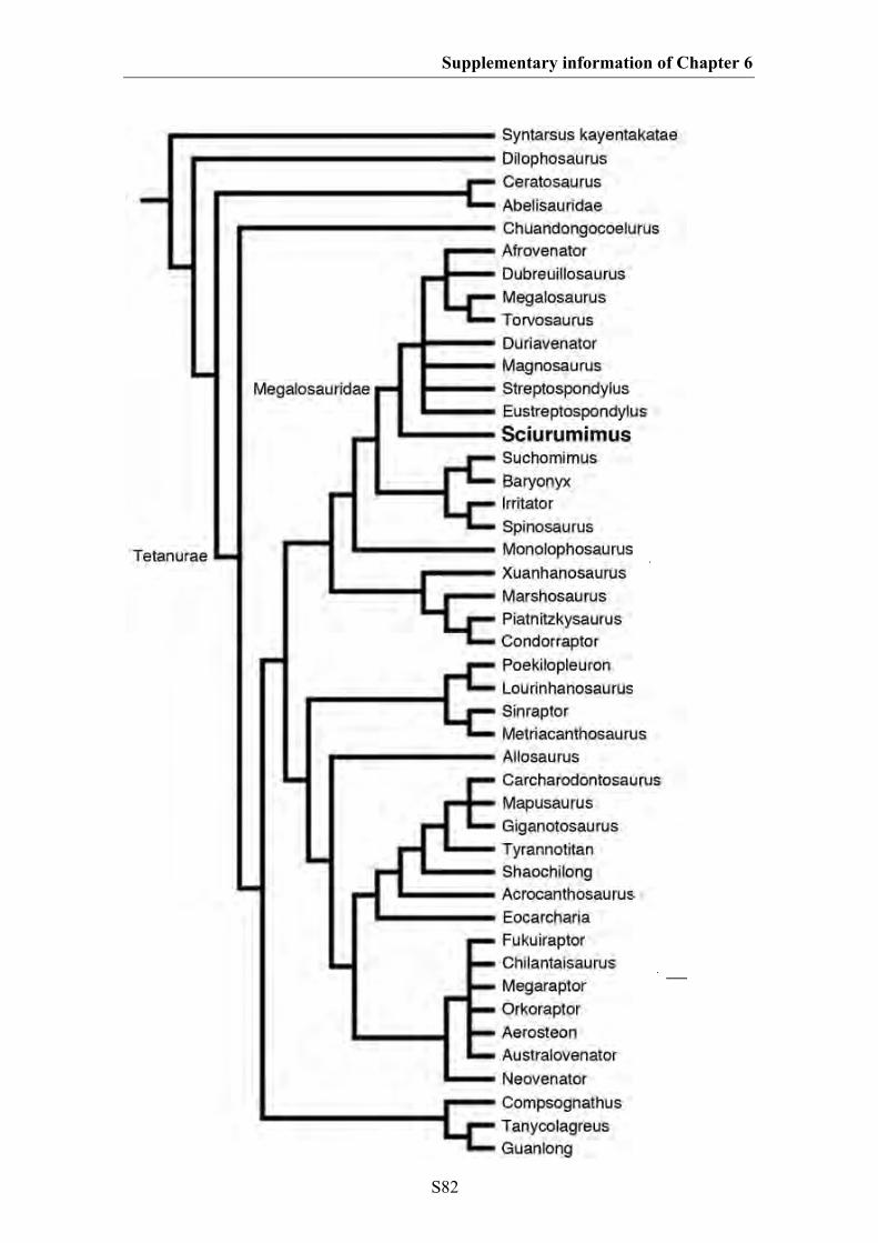
Supplementary information of Chapter 6
S82
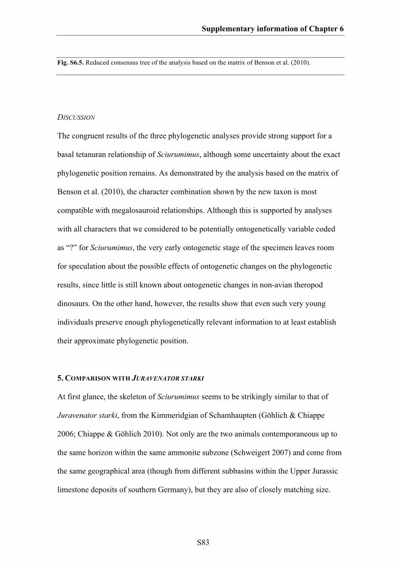
Supplementary information of Chapter 6
S83
Fig. S6.5. Reduced consensus tree of the analysis based on the matrix of Benson et al. (2010).
DISCUSSION
The congruent results of the three phylogenetic analyses provide strong support for a
basal tetanuran relationship of Sciurumimus, although some uncertainty about the exact
phylogenetic position remains. As demonstrated by the analysis based on the matrix of
Benson et al. (2010), the character combination shown by the new taxon is most
compatible with megalosauroid relationships. Although this is supported by analyses
with all characters that we considered to be potentially ontogenetically variable coded
as “?” for Sciurumimus, the very early ontogenetic stage of the specimen leaves room
for speculation about the possible effects of ontogenetic changes on the phylogenetic
results, since little is still known about ontogenetic changes in non-avian theropod
dinosaurs. On the other hand, however, the results show that even such very young
individuals preserve enough phylogenetically relevant information to at least establish
their approximate phylogenetic position.
5. COMPARISON WITH JURAVENATOR STARKI
At first glance, the skeleton of Sciurumimus seems to be strikingly similar to that of
Juravenator starki, from the Kimmeridgian of Schamhaupten (Göhlich & Chiappe
2006; Chiappe & Göhlich 2010). Not only are the two animals contemporaneous up to
the same horizon within the same ammonite subzone (Schweigert 2007) and come from
the same geographical area (though from different subbasins within the Upper Jurassic
limestone deposits of southern Germany), but they are also of closely matching size.
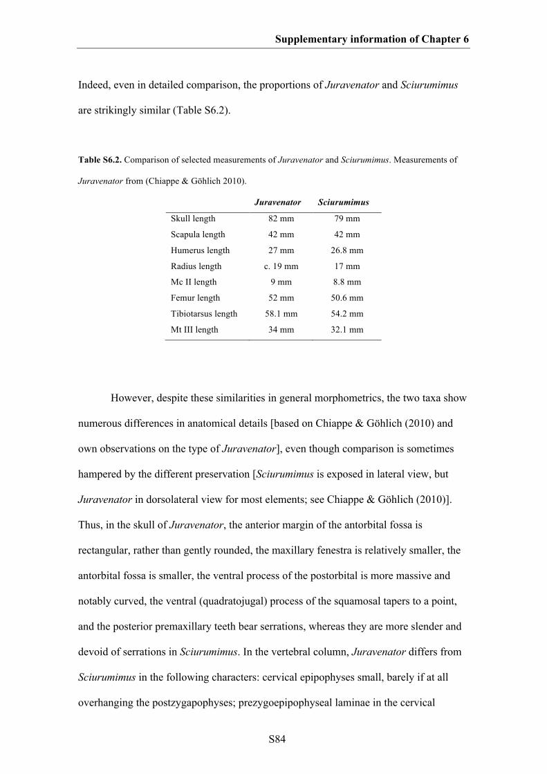
Supplementary information of Chapter 6
S84
Indeed, even in detailed comparison, the proportions of Juravenator and Sciurumimus
are strikingly similar (Table S6.2).
Table S6.2. Comparison of selected measurements of Juravenator and Sciurumimus. Measurements of
Juravenator from (Chiappe & Göhlich 2010).
Juravenator Sciurumimus
Skull length 82 mm 79 mm
Scapula length 42 mm 42 mm
Humerus length 27 mm 26.8 mm
Radius length c. 19 mm 17 mm
Mc II length 9 mm 8.8 mm
Femur length 52 mm 50.6 mm
Tibiotarsus length 58.1 mm 54.2 mm
Mt III length 34 mm 32.1 mm
However, despite these similarities in general morphometrics, the two taxa show
numerous differences in anatomical details [based on Chiappe & Göhlich (2010) and
own observations on the type of Juravenator], even though comparison is sometimes
hampered by the different preservation [Sciurumimus is exposed in lateral view, but
Juravenator in dorsolateral view for most elements; see Chiappe & Göhlich (2010)].
Thus, in the skull of Juravenator, the anterior margin of the antorbital fossa is
rectangular, rather than gently rounded, the maxillary fenestra is relatively smaller, the
antorbital fossa is smaller, the ventral process of the postorbital is more massive and
notably curved, the ventral (quadratojugal) process of the squamosal tapers to a point,
and the posterior premaxillary teeth bear serrations, whereas they are more slender and
devoid of serrations in Sciurumimus. In the vertebral column, Juravenator differs from
Sciurumimus in the following characters: cervical epipophyses small, barely if at all
overhanging the postzygapophyses; prezygoepipophyseal laminae in the cervical
Supplementary information of Chapter 6
S85
vertebrae absent; presence of a posterior pleurocoel in a mid-cervical centrum;
anteriormost dorsal vertebrae distinctly elongate; strongly posteriorly inclined,
triangular neural spines in the anterior caudal vertebrae; relatively more elongate
posterior caudal vertebrae; posterior caudal prezygapophyses more elongate and
anteriorly directed, rather than anterodorsally; distal chevrons skid-like. In the pectoral
girdle and forelimb, the following differences can be established: the scapula is less
slender and has a distinctly curved blade; the supraglenoid fossa is triangular, with an
acutely angled posterior rim; the internal tuberosity of the humerus is confluent with the
proximal humeral articular surface, forming a rectangular edge on the medial side of the
proximal humerus; the ulna lacks a proximal expansion and olecranon process; shaft of
ulna more massive than shaft of radius. In the pelvis and hindlimb, Juravenator differs
from Sciurumimus in the lack of an anterior dorsal lip of the ilium (the presence of
which represents an autapomorphy of Sciurumimus), the relatively smaller pubic
peduncle of the ilium, a more reduced supraacetabular crest, which is confluent
posteriorly with the lateral brevis shelf, a pronounced antitrochanteric lip on the ischial
peduncle of the ilium, a rectangular, rather than undulate posterior end of the
postacetabular blade of the ilium, an obturator process on the ischium [erroneously
identified as pubis by Chiappe & Göhlich (2010)] that is offset from the pubic peduncle,
the lack of a distal expansion of the ischial shaft, the short and triangular metatarsal I, a
metatarsal IV that is distinctly longer than metatarsal II, and the shorter and more robust
metatarsal V. Thus, these numerous differences strongly indicate that the two animals
cannot be referred to the same taxon, despite the similar size and proportions.
Looking at the phylogenetic position of Juravenator led to some interesting
results. To test the position of this taxon, we also coded it in the matrices of Smith et al.
Supplementary information of Chapter 6
S86
2008 and Choiniere et al. (2010), and analysed the matrices under the same parameters
outlined above. When analysed together with Sciurumimus, Juravenator was found to
be the sister taxon to this genus in both analyses, with otherwise no changes in the
phylogenetic position of Sciurumimus (i.e. both taxa were found to be basal, non-
neotetanuran tetanurans). However, when Sciurumimus was removed from the analyses,
Juravenator was found to be a basal coelurosaur in both cases (Fig. S6.6, S6.7).
As it is the case with Sciurumimus, the type of Juravenator is most probably an
early posthatchling individual, since it lacks any fusion of skeletal elements, even lacks
ossified carpal and distal tarsal elements altogether, and shows coarsely striated surface
texture in all skeletal elements (see Chiappe & Göhlich 2010). Several of the characters
shared by Sciurumimus and Juravenator, and interpreted as synapomorphies of these
taxa in the analyses, are probably ontogenetically variable, such as the round orbit,
anterodorsally sloping ventral strut of the lacrimal (related to the size and shape of the
orbit), absence of a posteroventral process in the coracoid, absence of a ventral hook on
the preacetabular blade of the ilium, and poorly developed attachment of the m.
iliofibularis on the fibula (all three muscle attachment areas). Thus, analysis of these
two early juveniles together with otherwise subadult and adult theropods might give
erroneous results, and we consider the phylogenetic position of Juravenator as
uncertain. Juravenator shows a highly unusual character combination (Chiappe &
Göhlich 2010) and further analysis of its affinities is necessary to firmly establish its
phylogenetic position. However, such a detailed reappraisal of Juravenator is beyond
the scope of the current paper.
Supplementary information of Chapter 6
S87
These phylogenetic results furthermore suggest that the frequent referral of early
juvenile theropods, such as Juravenator (Göhlich & Chiappe 2006) and Scipionyx (Dal
Sasso & Maganuco 2011) to the Compsognathidae might simply be due to similarities
between these taxa and the also juvenile type specimen of Compsognathus bavarica,
and thus the phylogenetic status and content of the Compsognathidae need to be
reevaluated.
Codings for Juravenator in the matrix of Smith et al. (2008)
00200[0/1]0[0/1]01???00100??00?21010[0/1]111??20??0?10?1[1/2]0?101[0/1]0?0001
00?000?0?00[0/1]??0000?0?00???????????????????????????????????????0??0?0?????0
????????????01101???0??100100000??0[0/1]01??????0?100?0????00011?00???10010
00?????0101100000000?001?00???111101111??0100100?0000??001002?0000?101???
??????0??0100000????2011??????1????????00??????????2??11?0???????1200?0?????
?
Codings for Juravenator in the matrix of Choiniere et al. (2010)
10?0[01]00?00?11010?1????20101?00?0?0?00000??[12]110000??110?0010?1????000
?00?000??00???????????????0?0?????????????????????????????????0??000?0???0???
???0????0???0???0?000020???0?????01?10????00101???10??????????[01]011?1?????
??000000?????0?0?100?1000??1???0?0??10?01??00?10000???1?0000???????011100?
11?0?2?12111?10000001002000011?0?0?100111?00?????????????10?00?1?000?0?01
??0?0001?00???1?????????00???????????1??0?10?????1?2000?
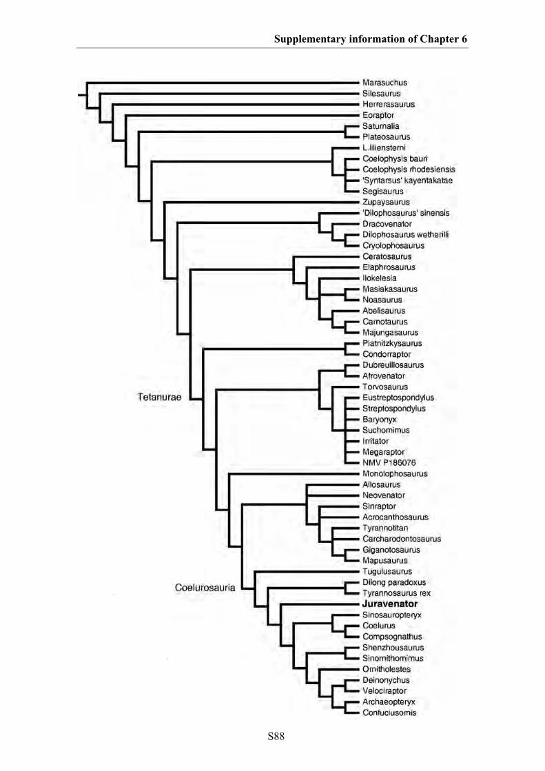
Supplementary information of Chapter 6
S88
Supplementary information of Chapter 6
S89
Fig. S6.6. Phylogenetic analysis of Juravenator, excluding Sciurumimus, based on the matrix of Smith et
al. (2008).

Supplementary information of Chapter 6
S90
Fig. S6.7. Phylogenetic analysis of Juravenator, excluding Sciurumimus, based on the matrix of
Choiniere et al. (2010). Several clades have been collapsed for clarity.
Supplementary information of Chapter 6
S91
6. COMPARISONS WITH OTHER JURASSIC THEROPODS
Given the early juvenile stage of the type specimen of Sciurumimus albersdoerferi, one
might ask whether this animal represents a juvenile of another, known taxon of
theropods from the Late Jurassic. Apart from Juravenator, theropods known from the
Late Jurassic of Europe include the ceratosaur Ceratosaurus (Mateus et al. 2006; Soto
& Perea 2008), the megalosaurid Torvosaurus (Mateus et al. 2006), the allosauroids
Allosaurus europaeus, Lourinhanosaurus and Metriacanthosaurus (Mateus et al. 2006;
Benson et al. 2010), and the coelurosaurs Compsognathus (Ostrom 1978; Peyer 2006),
Aviatyrannis (Rauhut 2003b), Stokesosaurus langhami (Benson 2008b) and
Archaeopteryx (Wellnhofer 2008)
First of all, Sciurumimus differs from all of these taxa, in which comparable
material is preserved, in its apomorphic characters. Numerous differences with
Ceratosaurus further include most tetanuran synapomorphies, such as the presence of a
maxillary fenestra, the presence of only one pleurocoel in the cervical vertebrae, a hand
with only three metacarpals, and the presence of a wing-like lesser trochanter that
reaches at least half the height of the femoral head (Gilmore 1920; Madsen & Welles
2000).
Given that the phylogenetic analysis indicates that Sciurumimus represents a
basal megalosaurid, comparisons with the megalosaurid Torvosaurus might be most
important. However, numerous differences between the two taxa include the number of
premaxillary teeth (three in Torvosaurus, four in Sciurumimus), the offset of the
maxillary fenestra from the anterior rim of the antorbital fossa in Sciurumimus, the lack
of a well-developed prezygoepipophyseal lamina in the cervical vertebrae of
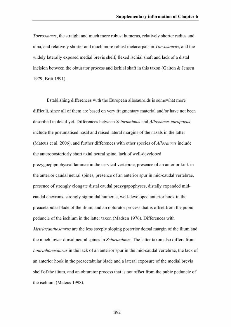
Supplementary information of Chapter 6
S92
Torvosaurus, the straight and much more robust humerus, relatively shorter radius and
ulna, and relatively shorter and much more robust metacarpals in Torvosaurus, and the
widely laterally exposed medial brevis shelf, flexed ischial shaft and lack of a distal
incision between the obturator process and ischial shaft in this taxon (Galton & Jensen
1979; Britt 1991).
Establishing differences with the European allosauroids is somewhat more
difficult, since all of them are based on very fragmentary material and/or have not been
described in detail yet. Differences between Sciurumimus and Allosaurus europaeus
include the pneumatised nasal and raised lateral margins of the nasals in the latter
(Mateus et al. 2006), and further differences with other species of Allosaurus include
the anteroposteriorly short axial neural spine, lack of well-developed
prezygoepipophyseal laminae in the cervical vertebrae, presence of an anterior kink in
the anterior caudal neural spines, presence of an anterior spur in mid-caudal vertebrae,
presence of strongly elongate distal caudal prezygapophyses, distally expanded mid-
caudal chevrons, strongly sigmoidal humerus, well-developed anterior hook in the
preacetabular blade of the ilium, and an obturator process that is offset from the pubic
peduncle of the ischium in the latter taxon (Madsen 1976). Differences with
Metriacanthosaurus are the less steeply sloping posterior dorsal margin of the ilium and
the much lower dorsal neural spines in Sciurumimus. The latter taxon also differs from
Lourinhanosaurus in the lack of an anterior spur in the mid-caudal vertebrae, the lack of
an anterior hook in the preacetabular blade and a lateral exposure of the medial brevis
shelf of the ilium, and an obturator process that is not offset from the pubic peduncle of
the ischium (Mateus 1998).
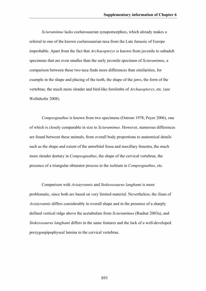
Supplementary information of Chapter 6
S93
Sciurumimus lacks coelurosaurian synapomorphies, which already makes a
referral to one of the known coelurosaurian taxa from the Late Jurassic of Europe
improbable. Apart from the fact that Archaeopteryx is known from juvenile to subadult
specimens that are even smaller than the early juvenile specimen of Sciurumimus, a
comparison between these two taxa finds more differences than similarities, for
example in the shape and placing of the teeth, the shape of the jaws, the form of the
vertebrae, the much more slender and bird-like forelimbs of Archaeopteryx, etc. (see
Wellnhofer 2008).
Compsognathus is known from two specimens (Ostrom 1978; Peyer 2006), one
of which is closely comparable in size to Sciurumimus. However, numerous differences
are found between these animals, from overall body proportions to anatomical details
such as the shape and extent of the antorbital fossa and maxillary fenestra, the much
more slender dentary in Compsognathus, the shape of the cervical vertebrae, the
presence of a triangular obturator process in the ischium in Compsognathus, etc.
Comparison with Aviatyrannis and Stokesosaurus langhami is more
problematic, since both are based on very limited material. Nevertheless, the ilium of
Aviatyrannis differs considerably in overall shape and in the presence of a sharply
defined vertical ridge above the acetabulum from Sciurumimus (Rauhut 2003a), and
Stokesosaurus langhami differs in the same features and the lack of a well-developed
prezygoepipophyseal lamina in the cervical vertebrae.
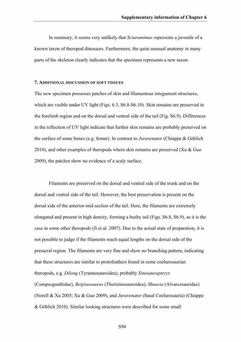
Supplementary information of Chapter 6
S94
In summary, it seems very unlikely that Sciurumimus represents a juvenile of a
known taxon of theropod dinosaurs. Furthermore, the quite unusual anatomy in many
parts of the skeleton clearly indicates that the specimen represents a new taxon.
7. ADDITIONAL DISCUSSION OF SOFT TISSUES
The new specimen possesses patches of skin and filamentous integument structures,
which are visible under UV light (Figs. 6.3, S6.8-S6.10). Skin remains are preserved in
the forelimb region and on the dorsal and ventral side of the tail (Fig. S6.9). Differences
in the reflection of UV light indicate that further skin remains are probably preserved on
the surface of some bones (e.g. femur). In contrast to Juravenator (Chiappe & Göhlich
2010), and other examples of theropods where skin remains are preserved (Xu & Guo
2009), the patches show no evidence of a scaly surface.
Filaments are preserved on the dorsal and ventral side of the trunk and on the
dorsal and ventral side of the tail. However, the best preservation is present on the
dorsal side of the anterior-mid section of the tail. Here, the filaments are extremely
elongated and present in high density, forming a bushy tail (Figs. S6.8, S6.9), as it is the
case in some other theropods (Ji et al. 2007). Due to the actual state of preparation, it is
not possible to judge if the filaments reach equal lengths on the dorsal side of the
presacral region. The filaments are very fine and show no branching pattern, indicating
that these structures are similar to protofeathers found in some coelurosaurian
theropods, e.g. Dilong (Tyrannosauroidea), probably Sinosauropteryx
(Compsognathidae), Beipiaosaurus (Therizinosauroidea), Shuuvia (Alvarezsauridae)
(Norell & Xu 2005; Xu & Guo 2009), and Juravenator (basal Coelurosauria) (Chiappe
& Göhlich 2010). Similar looking structures were described for some small
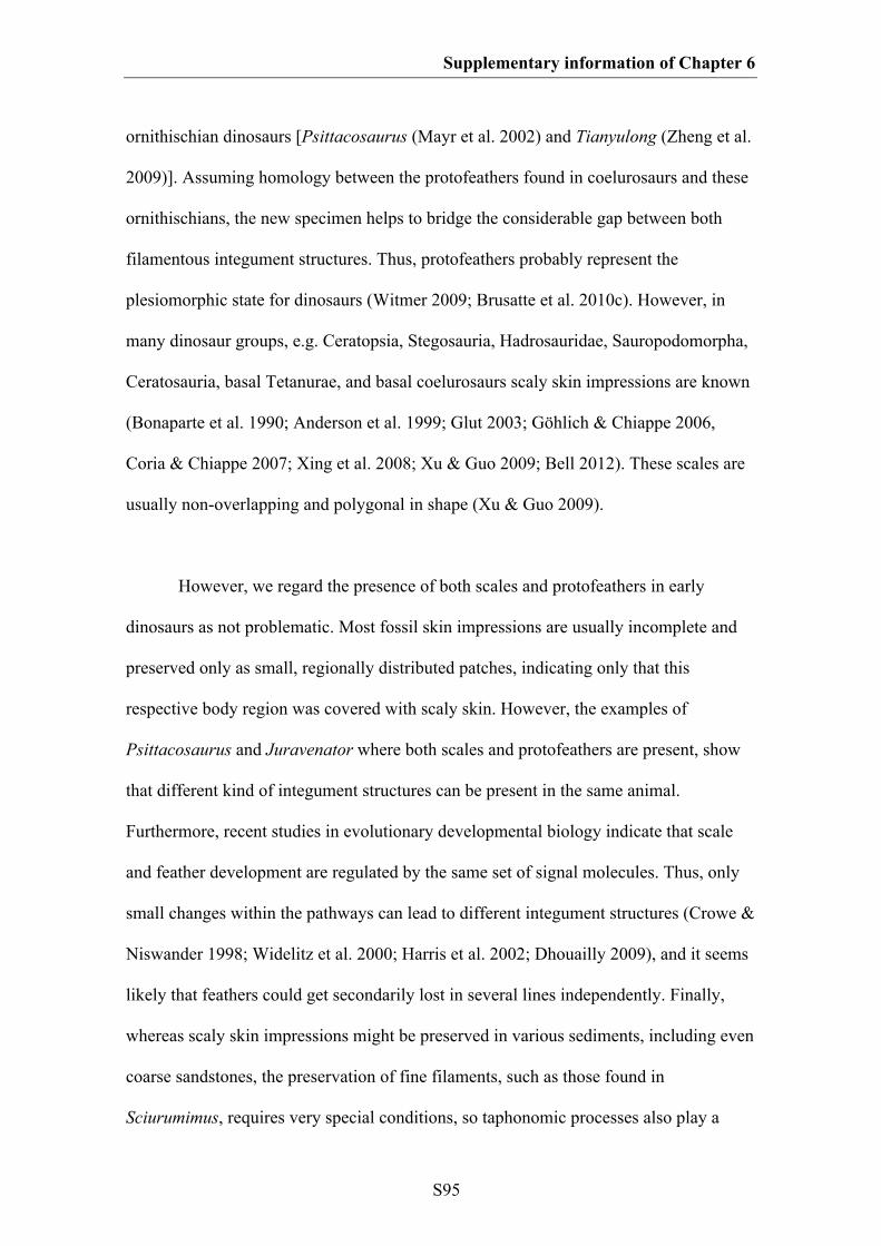
Supplementary information of Chapter 6
S95
ornithischian dinosaurs [Psittacosaurus (Mayr et al. 2002) and Tianyulong (Zheng et al.
2009)]. Assuming homology between the protofeathers found in coelurosaurs and these
ornithischians, the new specimen helps to bridge the considerable gap between both
filamentous integument structures. Thus, protofeathers probably represent the
plesiomorphic state for dinosaurs (Witmer 2009; Brusatte et al. 2010c). However, in
many dinosaur groups, e.g. Ceratopsia, Stegosauria, Hadrosauridae, Sauropodomorpha,
Ceratosauria, basal Tetanurae, and basal coelurosaurs scaly skin impressions are known
(Bonaparte et al. 1990; Anderson et al. 1999; Glut 2003; Göhlich & Chiappe 2006,
Coria & Chiappe 2007; Xing et al. 2008; Xu & Guo 2009; Bell 2012). These scales are
usually non-overlapping and polygonal in shape (Xu & Guo 2009).
However, we regard the presence of both scales and protofeathers in early
dinosaurs as not problematic. Most fossil skin impressions are usually incomplete and
preserved only as small, regionally distributed patches, indicating only that this
respective body region was covered with scaly skin. However, the examples of
Psittacosaurus and Juravenator where both scales and protofeathers are present, show
that different kind of integument structures can be present in the same animal.
Furthermore, recent studies in evolutionary developmental biology indicate that scale
and feather development are regulated by the same set of signal molecules. Thus, only
small changes within the pathways can lead to different integument structures (Crowe &
Niswander 1998; Widelitz et al. 2000; Harris et al. 2002; Dhouailly 2009), and it seems
likely that feathers could get secondarily lost in several lines independently. Finally,
whereas scaly skin impressions might be preserved in various sediments, including even
coarse sandstones, the preservation of fine filaments, such as those found in
Sciurumimus, requires very special conditions, so taphonomic processes also play a
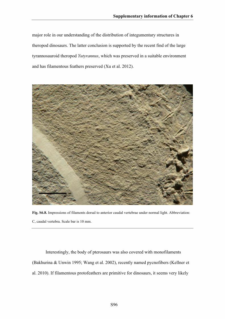
Supplementary information of Chapter 6
S96
major role in our understanding of the distribution of integumentary structures in
theropod dinosaurs. The latter conclusion is supported by the recent find of the large
tyrannosauroid theropod Yutyrannus, which was preserved in a suitable environment
and has filamentous feathers preserved (Xu et al. 2012).
Fig. S6.8. Impressions of filaments dorsal to anterior caudal vertebrae under normal light. Abbreviation:
C, caudal vertebra. Scale bar is 10 mm.
Interestingly, the body of pterosaurs was also covered with monofilaments
(Bakhurina & Unwin 1995; Wang et al. 2002), recently named pycnofibers (Kellner et
al. 2010). If filamentous protofeathers are primitive for dinosaurs, it seems very likely
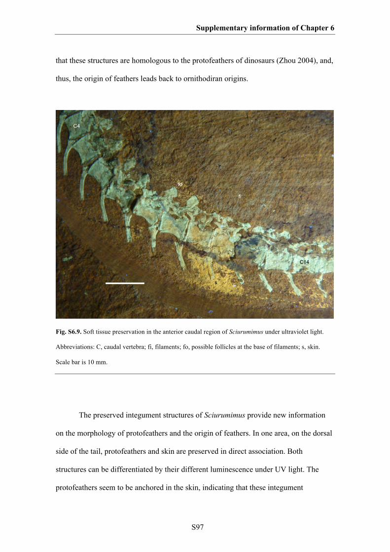
Supplementary information of Chapter 6
S97
that these structures are homologous to the protofeathers of dinosaurs (Zhou 2004), and,
thus, the origin of feathers leads back to ornithodiran origins.
Fig. S6.9. Soft tissue preservation in the anterior caudal region of Sciurumimus under ultraviolet light.
Abbreviations: C, caudal vertebra; fi, filaments; fo, possible follicles at the base of filaments; s, skin.
Scale bar is 10 mm.
The preserved integument structures of Sciurumimus provide new information
on the morphology of protofeathers and the origin of feathers. In one area, on the dorsal
side of the tail, protofeathers and skin are preserved in direct association. Both
structures can be differentiated by their different luminescence under UV light. The
protofeathers seem to be anchored in the skin, indicating that these integument
Supplementary information of Chapter 6
S98
structures might have grown from follicles. Indeed, there are conspicuous,
dorsoventrally elongate skin structures preserved where the filaments reach the skin,
which might represent direct evidence for these follicles. This is interesting, because it
has been suggested that follicle formation was a late event in feather evolution, which
took place with the evolution of vaned feathers (Sawyer & Knapp 2003; Alibardi &
Sawyer 2006; Alibardi & Toni 2008). This scenario was based on the feather
embryogenesis of some recent bird species, where barb ridge formation occurs before
follicle formation. The hypothesis that unbranched protofeathers apparently grow from
a follicle supports the idea that feather evolution is highly correlated with follicle
formation (Prum 1999; Prum & Brush 2002). Further support for this comes from
Psittacosaurus, where the bristles extend under the skin layer (Mayr et al. 2002), also
lending additional support for the homology of ornithischian filaments with theropod
protofeathers and bird feathers.
8. REPOSITORY OF THE SPECIMEN
The holotype specimen of Sciurumimus belongs to the private Painten collection of the
Albersdörfer family, where it bears the collection number 1687. However, the scientific
availability of the specimen is guaranteed by its inclusion in the register of cultural
objects of national importance of Germany (Verzeichnis national wertvollen
Kulturgutes). Under the Act to prevent the exodus of German Cultural Property
(KultSchG; Bundesgesetzblatt I: 1754; 1999), the inclusion of the specimen in this list
prevents it from being sold outside Germany and guarantees that its current repository is
always known and changes of repository have to be announced. Furthermore, the type
specimen of Sciurumimus albersdoerferi is deposited as a permanent loan at the

Supplementary information of Chapter 6
S99
municipal Bürgermeister Müller Museum in Solnhofen, Bavaria, where it is also
available for additional scientific study and bears the specimen number BMMS BK 11.
Fig. S6.10. Short filaments on the ventral tail flank below the 12th and 13th caudal vertebra. Arrows point
to single filaments.