Il capitolo francese della storia italiana dei volgarizzamenti: un primo abbozzo

This article appeared in a journal published by Elsevier. The attachedcopy is furnished to the author for internal non-commercial researchand education use, including for instruction at the authors institution
and sharing with colleagues.
Other uses, including reproduction and distribution, or selling orlicensing copies, or posting to personal, institutional or third party
websites are prohibited.
In most cases authors are permitted to post their version of thearticle (e.g. in Word or Tex form) to their personal website orinstitutional repository. Authors requiring further information
regarding Elsevier’s archiving and manuscript policies areencouraged to visit:
http://www.elsevier.com/copyright

Author's personal copy
- REV IEW ART ICLE -
Primo Vascular System as a New MorphofunctionalIntegrated System
Miroslav Stefanov 1,2,*, Jungdae Kim 2,3
1Department of Animal Morphology, Physiology and Nutrition, Agricultural Faculty, Trakia University,Stara Zagora, Bulgaria2Nano Primo Research Center, Advanced Institute of Convergence Technology, Seoul National University,Republic of Korea3 Pharmacopuncture Medical Research Center, Korean Pharmacopuncture Institute, Seoul, Republic of Korea
Available online Aug 14, 2012
Received: Jun 5, 2012Revised: Jun 13, 2012Accepted: Jul 16, 2012
KEYWORDSBong-Han theory;cancer;morphology;morphological-
functional discovery;physiology;primo vascular system
AbstractThe purpose of this review is to describe the methodology, instruments, and subjectanimals used until now for studies of the meridian (Kyungrak) system and the primovascular system (PVS). The PVS is observed as an anatomical system distributed in cavi-ties, organs, and tissues throughout the body. We analyzed the most important pointsof the PVS based on the results obtained until the present. Our main effort has beendirected to describing the main thesis relating to the morphological structures and theirtopography, the functional mechanisms of the PVS, and possible roles of the PVS in path-ological processes. The substance of the PVS in all its aspects is as a system covering thewhole body and regulating and coordinating the biological processes that are the basis forlife. In conclusion, we suggest that the finding of the PVS represents the discovery ofa new integrated morphological-functional system.
1. Introduction
The morphological architectonics and functions of theprimo vascular system (PVS) are the two aspects for whichcombinations thereof may drastically change our current
understanding of basic biology and its related branchesof medicine, veterinary medicine, and sciences suchas morphology, physiology, biochemistry, biophysics,pathology, etc. When we investigate the new anatomicalsystem, the first surprising finding is that morphological
* Corresponding author. Department of Animal Morphology, Physiology and Nutrition, Agricultural Faculty, Trakia University, Student’sTown, 6000 Stara Zagora, Bulgaria.E-mail: [email protected]
Copyright ª 2012, International Pharmacopuncture Institutehttp://dx.doi.org/10.1016/j.jams.2012.07.001
Available online at www.sciencedirect.com
Journal of Acupuncture and Meridian Studies
journa l homepage: www. jams-kp i .com
J Acupunct Meridian Stud 2012;5(5):193e200

Author's personal copy
science had not discovered this system until now. Thereason lies in the suspicion of Western medicine toward theacupunctural meridian system. The therapeutic effects ofacupuncture have been accepted worldwide [1e3].Western medicine, however, has refused to see the obviouspossibilities of an anatomical substance whose existencecould be the framework for acupuncture treatment, whichhas been used in traditional Eastern medicine for thousandsof years. Modern science cannot explain the long-standingsuccessful existence and functional paths of the curingmodality for traditional Eastern medicine. Another reasonis that current science is convinced of the correctness ofthe fundamental basis of biological science, but cannotexplain many questions concerning the mechanisms of theprocesses of life. The new system was not being pursued inthe right place, and it was not investigated with adequatemethods in the past as it has been recently by scientistsfrom China, Japan, and Russia [4]. It would appear that thePVS only comes to light when numerous appropriatemethods are applied.
We can determine two periods for the discovery of thenew morphological-functional system. The first oneincludes the five papers from Bong-Han Kim from 1962 to1965 [5e9], and we have commented on them in a detailedmanner in another article of this journal [10]. Bong-Han Kimrevealed much, suggested more, but left many detailsunanswered. The second one, which we would call ‘arediscovery of the PVS,’ is from 2002 until now, and it wasmade mainly by the scientists from Seoul National Univer-sity (SNU), Korea. They developed the methods to detectand identify the new anatomical system [11,12]. In 2011,the SNU team was expanded to the Nano Primo ResearchCenter headed by Prof. K. S. Soh with the main purpose offurther investigation into the PVS. He named the newsystem the primo vascular system, and the Bonghan ductsand corpuscles for the channels and the nodes wererenamed as primo vessels (PVs) and primo nodes (PNs),respectively. The SNU group has published about 56 articlesand obtained more than 200 citations concerning thissubject. (These data were obtained from the BioInfoBankLibrary, Warszawa, Poland.)
Today the substance of the PVS in all its aspects is sug-gested to be a system covering the whole body, and regu-lating and coordinating the biological processes that are thebasis for life. In modern biological sciences, the unity oforganisms and the environment and the regulation mecha-nisms involved in the coordination of all body systemsconstitute fundamental and still unsolved problems.
The purpose of this review is to describe present-dayfindings and to analyze the most important points of thePVS as a new discovery. In this paper we use the termi-nology adopted at the International Symposium on thePrimo Vascular System held in 2010 [11].
2. Methodology, instruments, and subjectanimals
2.1. Bong-Han Kim’s work on the meridian system
Bong-Han Kim’s study of the meridian (Kyungrak) systemcan be divided into three parts [6]: a morphological
study, an experimental physiological study, anda biochemical and histochemical study. His morphologicalstudy (the first part) on the Kyungrak system was donethrough anatomical observations on superficial andprofound Bonghan (BH) corpuscles (primo nodes) in theliving body. He found that the superficial BH corpuscleswere distributed in the skin and that the profound BHcorpuscles were positioned in the organism under deepskin. He made numerous descriptions of the corpusclesaccording to their locations, forms, and shapes. Manyhistological studies were performed with microscopes,and the microscopic images were presented in the rangesof tissue and cellular levels corresponding to magnifica-tions of 100w500 times. The morphology of the BH ducts[primo vessels (PVs)] was also studied through anatomicalobservations and histological methods. One of the specialcharacteristics of BH ducts is that they are composed ofmany subductules with endothelial cells with rod-shapednuclei. That observation was made during a phase-contrast microscopic study.
Bong-Han Kim’s findings on the corpuscle and ductsystem naturally induced further experimental physiolog-ical studies (the second part). The duct systems weredistributed throughout the living body, and they ran alongthe outsides and the insides of blood and lymphatic vessels.This may be considered to be one of the distinctive featuresof the Kyungrak system. In his second study, he tried toelucidate the question of the circulation of the BH liquidthat flows through the BH ducts. He used radioactivetracers to identify the circulation and adopted electro-physiological methods for research into the excitability andthe conductivity of the Kyungrak system. He also inventeda dye injection method. However, the methods of dyingbeing kept secret and incomplete descriptions of thescientific protocols were the main reasons for his findingsbeing forgotten for many years. The following relates to themethods and protocols used in the past and the currentmodern methods. In his reports [5e9], Bong-Han Kim makesreferences to a series of methods, such as anatomical,histological (Hematoxylin and Eosin (H&E), Mason’s tri-chrome, Verhoff, silver staining, feulgen reaction, Acridineorange), dosimetry of radioactivity, radio-autographical,histochemical, and a “mysterious” blue staining, butwithout describing any details of the scientific protocols.He mentioned the use of the “mysterious” dye many timeswithout giving any detailed information on the procedurefor using it. He used different types of microscopes, such asstereomicroscopes, as well as phase-contrast, inverted,luminescent, and transmission electron microscopes.
The third part of his studies focused on the biochemicaland histochemical analyses of the corpuscle and ductsystem. Various forms of the phosphorus contents in the BHducts were determined by using the Fiske-Subbarowmethod and the Schmidt-Thannhauser extraction proce-dure. Granules in the BH corpuscles and ducts showedpositive reactions with the Unna-Pappenheim method, aswell as positive Feulgen reactions. As a result of hisexperiments, he noticed that the BH corpuscles and ductscontained more nucleic acids, especially DNA, than anyother tissues. Based on his observations, he concluded thatthe functions of the Kyungrak system were closely relatedto nucleic acids and to DNA in particular.
194 M. Stefanov, J. Kim

Author's personal copy
Bong-Han Kim’s claims could not be reproduced becausethe formula for the staining dye was undisclosed. In 1966,Kellner [13] used histological methods to deny Kim’sclaims. It is difficult for us to be satisfied with theextremeness of Kellner’s position, for rejecting Bong-HanKim’s whole theory, which was created by using the manymethods, described above with only routine histologicalmethods used by Kellner is not possible. Only Fujiwara andYu [14] were able to partially confirm Bong-Han Kim’sfindings.
2.2. SNU group’s work on the primo vascular system
In 2002, Prof. K. S. Soh started an intensive re-investigation into Bong-Han theory by using modernizedtechniques such as fluorescent microscopy, confocalmicroscopy, and electron microscopy. The Progression ofthe rediscovery of the PVS was determined by the devel-opment of methodology and by the team’s creativity. Thefirst key technique developed by the SNU team was theintravenous injection of a 10% dextrose solution to replaceblood with a transparent liquid for observation witha stereomicroscope [15e17], but with low success ratesbecause the PVS could not be clearly distinguished fromfibrin strings (Fig. 1) [12].
In 2008, there was a major step in the methodology. TheSNU group discovered the Trypan blue technique for thespecific visualization of the PVS [18]. This technique issimple, but very effective. The same group had previouslyobtained successful results by using Janus Green B (Fig. 2)[19] and Alcian blue [20,21]. In fact, the SNU group hasapplied all the standard methods used by Bong-Han Kim, aswell as several new ones [22].
During the last decade, a series of conventional andmodern methods and technologies have been utilized.Confocal laser scanning microscopy [23]; various types ofelectron microscopy, such as scanning electron microscopy(SEM), cryo-SEM, focused-ion-beam SEM, and high voltagetransmission electron microscopy (TEM) [24e26]; X-raymicrotomography [27]; atomic force microscopy [28];fluorescent nanoparticle [29e33]; immunohistochemistry[34,35]; proteomic analysis [36]; the ELISA technique forhormone analysis [37,38],; and electrophysiologicalmethods [39] have been employed [12]. Magnetic reso-nance image (MRI) and computed tomography (CT) havebeen tried, but no important results have yet been ob-tained [22].
Bong-Han Kim used primarily rabbits as laboratoryanimals. No data on other animals, including humansubjects, were presented, but he mentioned that the PVShad been observed in various mammalian species, includinghumans, avians, amphibians, fish and invertebrates such ashydra, without suggesting any specific animal species [7].The SNU team performed their investigations with labora-tory animals such as mice, rats and rabbits. A few casestudies have been reported for cows [40], pigs [41], anddogs [42]. As we mentioned in the other review [10], as theanimals are divided from lower to higher, the PVS ofanimals can be divided in a similar way. The developmentof the PVS may be progressive, with the most developedone being in humans.
3. Morphological characteristics of the PVS:actual anatomical systems, cavities, organs,and tissues
In this section, we describe Bong-Han Kim’s findings, thathave been confirmed, as well as the new anatomicalstructures that have been shown to include the PVS. Leeet al. [43] described PVs as freely floating in bovine heartchambers. This is the first report of the PVS in large animalorgans. The authors did not say, but for the first time anorgan obtained from an animal slaughterhouse was used fora PVS investigation. Use of organs after slaughter allowsnew approaches. First, it presents an opportunity for usinglarge animals. Second, animals can be used some hoursafter they have died. Third, the PVS can be visualized afterbleeding has occurred.
The PVS was observed in the caudal vena cava, thehepatic vein, the hepatic portal vein, the femoral vein, theaorta [15] and large lymph vessels along the caudal vena cava[19,27,29,30,44]. The PVs are located inside the vessels, aswell as along the blood and the lymphatic vessel walls. ThePVs inside the lymph vessels are close to the lymph valves
Figure 1 (A) Threadlike structure with fibrin observed bydifferential interference contrast microsope. Scale bar 50㎛;(B) acridine orange stained image of the same sample. Long rod-shaped nuclei can be seen from the threadlike structure [12,J Acupunct Meridian Stud. 2009;2:93e106]. Scale bar 50 ㎛.
Primo vascular system 195

Author's personal copy
[27,29], freely float in the lymph, and pass through thelymph valves. For better observation of the PVs inside thelymph vessels, pressing the sample between two glass platesgives an opportunity to temporally fix the PVs in the center ofthe lumen. It should be noted that the PVs [10] prepared inthis way show good mechanical stability and that the fluidflow in the PVs cannot be stopped by the plate’s pressure.
As shown in Fig. 3, the PVs and the PNs were found in thethird ventricle, the fourth ventricle, and the cerebralaqueduct, along the central canal of the spinal cord [45],and on the arachnoid mater, cerebellum [46] and theperineurium and the epineurium of the sciatic nerve[46,47]. The PVs and the PNs were observed on the surfacesof visceral organs (liver, stomach, small and large intes-tines, bladder, spleen, kidney and omentum), the abdom-inal cavity [18,23e26], the hypodermal layer of the skin,the superficial fascia [21], fat tissue [48] and cancer fascia[49]. However, very rarely were the PVs observed to enterinternal organ tissues [50].
Bong-Han Kim and K. S. Soh both proposed a classifica-tion of the PVS based on six subnetworks: superficial,intravascular, extravascular, organ surface, intraorgan, andneural [12]. The data show that the PVS exists in threemorphological places. We propose to develop this classifi-cation of the topographical locations of the PVS. The firstPVS morphological location is the PVS in internal cavitiesand lumens. Some PVs freely floating internal cavities(brain cavities and heart chambers) and in lumens (blood
and lymphatic vessel’s lumens, cerebral aqueduct, andspinal cord channel). The second PVS morphological loca-tion is the PVS within organ-covering membranes. The PVSappears within organ-covering membranes (serousmembranes: parietal and visceral peritoneum which coverthe abdominal wall and all internal organs; the pia materand the arachnoidea of the brain). The third PVS morpho-logical location is the PVS in connective and fat tissues(hypodermal layer, superficial fascia, epineurium andperineurium, vessel’s adventitia, fat tissue and cancerfascia). We also propose a new classification of the topo-graphical position of the PVS structures to replace Bong-Han Kim’s description [6]. We propose to rename thesuperficial PVs and PNs as “receiving PVS.” The interior-exterior PNs may be given the name “communicatingPVS.” This classification could be developed with “organic”and “extra-organic” parts of the PVS. We propose torename the PVs and the PNs in the organs and on the organsas the “organic PVS.” We propose to rename the PVs andthe PNs that are between the organic and the receivingparts of the PVS as the “extra-organic” parts of the PVS.
There are no data whether the PVS exists in anatomicalstructures such as the head’s organs (tongue, salivaryglands, nasal cavity organs, and sensory organs as eyes,ears, ethmoidal labyrinth, vomeronasal organ), the neck’sorgans (esophagus and trachea), the thoracic cavity organs(lungs, esophagus, trachea, and pericardium), the pelviccavity’s organs (female generative system, urethra,accessory glands and male penis), the glands, the
Figure 2 BHD (Primo vessel) inside a rabbit lymphatic vesselstained with Janus Green B [12, J Acupunct Meridian Stud.2009;2:93e106]. (A) Stereo-microscopic view; (B) cross-sectionimage of H&E stained sample.
Figure 3 PVS found in the brain [12, J Acupunct MeridianStud. 2009; 2(2): 93-106]. (A) Stereomicroscopic image of PVSin an aqueduct and third ventricle of rabbit brain. (B) Stereo-microscopic image of PV node and vessel taken from the brain.
196 M. Stefanov, J. Kim
Author's personal copy
autonomic nervous system, the muscles, and the bones.Further study is necessary to identify the connection of thePVS to these kinds of organs.
4. Functional aspects of the PVS
The PVs are surrounded by a membrane. The membraneconsists of a high concentration of hyaluronic acid [27].Hyaluron is responsible for cell growth and differentiation.Probably, the hyaluron could be involved with repairs ofdamaged PVs. The PVs consist of several sub-channels. Thesubchannels have double coats [10] and a layer of endo-thelial cells. Rod-shaped endothelial nuclei (10e20㎛ ) arehallmarks of the PVS [51e53]. The cells of the PVS showa smooth muscle-like excitability. The excitable cells haveCa-ion channels, which are necessary for cell movement[39]. The sub-vessels have adventitia containing connectivetissue and an amorphous substance [6] as supporting tissue.Also the PVS often lies in connective tissue. Collagen is themain component of connective tissue. Collagen is anabundant molecule with special properties, and data indi-cate that it has properties of interference with photonemission emanating from biomolecular sources. This prop-erty of collagen to interfere with photon-emittingprocesses facilitates the possibility of tuning photon emis-sion throughout an organism, and it is a step towards thehypothesis that metabolism is regulated by a photon field[54]. This supports Soh’s hypothesis [55] regarding the PVSas an optical channel of biophoton emission. Biophotonsmay be the electromagnetic signals that play a key role inthe processes of cell development and differentiation. DNAmay act as a photon store and coherent radiator [55]. Thereis a suggestion that spontaneous ultraweak photon emissionfrom cultured cells is mainly involved in the changes in theploidy number that occur during the proliferative process ofcancer cell lines [56]. This hypothetical light propagationfunction of the PVS may explain the instantaneous effectthat often occurs throughout the entire body with theapplication of needles at acupoints [12,55].
The presence of chromaffin cells at acupoints [37,38]has provided a new view of their function as an endocrinecatecholamine organ [12] besides the currently knownadrenal medulla, postganglionic fibers, and Merkel cells[57]. Fujiwara proposed that the PVS is a hormone path thatis more efficient than conventional blood flow paths.
The primo subvessels and primo nodes carry a liquid. Theliquid was found to be rich in basophilic granules, whichhave been observed as individual granules and have beenseen in groups of two or three, and as granular clusters[58]. There are different proteins [36], stem cell niches [48]or microcells [28,59,60] with much harder membranes thansimilar-sized apoptotic bodies [61], and hormones[37,38,62] in the primo liquid. The flow speed of the liquidwas measured at 0.3 mm/s by injecting an Alcian bluesolution into the PVS on the surface of the liver [63] and inrange of 100e800 um/second when directly measured byusing radioactive tracers [25,64,65], which values aresignificantly higher than those observed in lymphaticvessels [58]. This supports the concept that there are manymorphological features that allow faster flow of the primofluid [10].
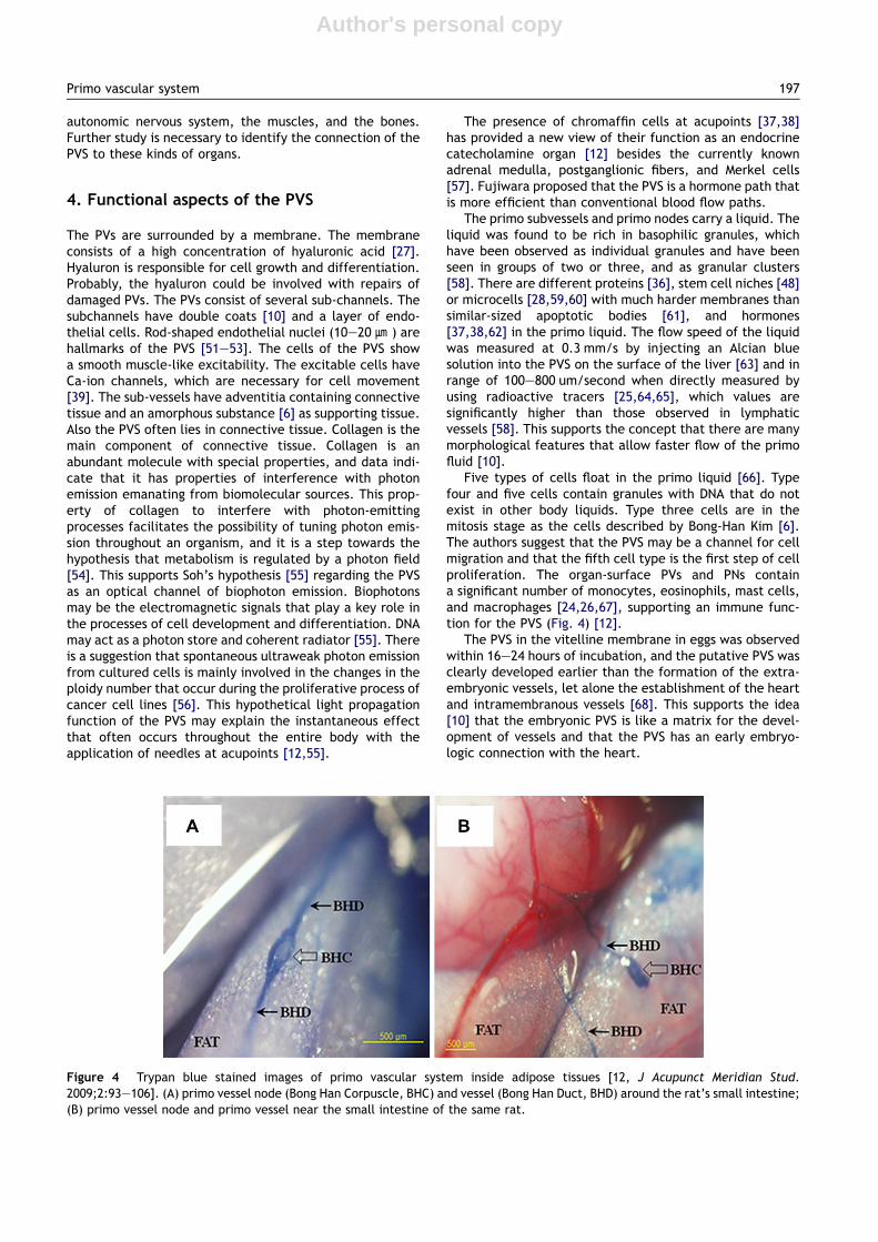
Five types of cells float in the primo liquid [66]. Typefour and five cells contain granules with DNA that do notexist in other body liquids. Type three cells are in themitosis stage as the cells described by Bong-Han Kim [6].The authors suggest that the PVS may be a channel for cellmigration and that the fifth cell type is the first step of cellproliferation. The organ-surface PVs and PNs containa significant number of monocytes, eosinophils, mast cells,and macrophages [24,26,67], supporting an immune func-tion for the PVS (Fig. 4) [12].
The PVS in the vitelline membrane in eggs was observedwithin 16e24 hours of incubation, and the putative PVS wasclearly developed earlier than the formation of the extra-embryonic vessels, let alone the establishment of the heartand intramembranous vessels [68]. This supports the idea[10] that the embryonic PVS is like a matrix for the devel-opment of vessels and that the PVS has an early embryo-logic connection with the heart.
Figure 4 Trypan blue stained images of primo vascular system inside adipose tissues [12, J Acupunct Meridian Stud.2009;2:93e106]. (A) primo vessel node (Bong Han Corpuscle, BHC) and vessel (Bong Han Duct, BHD) around the rat’s small intestine;(B) primo vessel node and primo vessel near the small intestine of the same rat.
Primo vascular system 197

Author's personal copy
5. PVS role in pathological processes
The PVS is found to be connected to tumor tissues growingin internal organs as well as on the fascia of tumor tissue[4,49,69,70]. A direct relation of the PVS to tumor tissueswas found in nude mouse experiments with the injection ofcancer cells. The PVS was observed in the fascia wrappingtumor tissue that grew in the skin and was visualized byusing the Trypan blue method. The PVS was hypothesized toplay a double role: a novel path of metastasis and control oftumor tissue via acupuncture [12]. The PVS may be utilizedas a drug delivery path for cancer [55] and for repair andregeneration of tissues [10,12].
Based on experiments related to internal-organsurfaces, the PVS was conjectured to be a pathway formacrophages in adipose tissue [48]. The PVS was visualizedinside adipose tissues by using in vivo Trypan blue staining.The PVS in adipose tissues may be a source of mesenchymalstem cells, which can be differentiated into adipocytes.Obesity is known to be associated with macrophage accu-mulation in adipose tissue [71]. A molecular study showedthat alterations of adipose tissue and its metabolic endo-crine function led to an increased release of fatty acids,hormones, and pro-inflammatory molecules that contrib-uted to obesity-associated complications. The apparentrelationships between stored fats and the PVS may providea critical clue to identifying a potential mediator for thetreatment of obesity.
Bong-Han Kim showed that a corpuscle and duct systemexisted throughout the whole body, including the brain.Threadlike structures have been observed in the cerebro-spinal fluid of the brain ventricles and the spinal centralcanal of rabbits [45]. The PVS in the central nervoussystem, such as the brain and the spinal cord, suggests thatthe BH system is a potential acupuncture meridian. Fora long time, the brain was believed to be incapable ofregeneration after embryonic development. However, theprocess by which neurons are generated from neural stemand progenitor cells, now known as neurogenesis, is nowknown to occur even in adults. Adult neurogenesis wasfound in the subventricular zone of the lateral ventriclesand the subgranular zone of the dentate gyrus of thehippocampus. Acupuncture has long been used to treatneurologic conditions, with the point ST36 being used fortreating stroke and Alzheimer’s disease in Eastern countries[72]. Further studies are required to elucidate the role ofthe PVS connecting the skin to the brain in terms of thebeneficial effects of acupuncture and its relationship to themeridian system.
6. Concluding remark
We reviewed the methodology, instruments, and subjectanimals used for the studies of the PVS, which is suggestedto be the physical structure of the meridian system foracupuncture treatment in traditional Eastern medicine.The PVS has been observed as an anatomical systemdistributed throughout the entire living body. We analyzedthe most important points of the PVS based on the resultsobtained up to now with regards to the morphological andthe functional aspects thereof. Our main efforts have been
directed to describing the main thesis regarding themorphological structures and their topographies, thefunctional mechanisms of the PVS, and possible roles of thePVs in pathological processes. The substance of the PVS inall aspects may play a critical role as a core structurecovering the whole body and regulating and coordinatingthe biological processes that are the basis for life. Weconclude that the PVS is a new integrated morphological-functional system.
Acknowledgment
We would like to thank Prof. K. S. Soh for the invitation tomake this review of the PVS and for his fruitful discussionand advice. All the figures in the paper are reproduced athis courtesy. We also acknowledge the financial supportfrom a grant from the Traditional Korean Medicine R&DProject, Ministry of Health and Welfare, Korea (B110076).
References
1. Foster JMG, Sweeney BP. The mechanisms of acupunctureanalgesia. Br J Hosp Med. 1987;38:308e312.
2. Son Y, Park H, Kwon O, Jung S, Sin H, Lim S. Antipiretic effectsof acupuncture on the lipopolysaccharide-induced fever andexpression of interleukin-6 and interleukin-1 beta mRNAs in thehypothalamus of rats. Neurosci Lett. 2002;19:45e48.
3. Libert C. A nervous connection. Nature. 2003;421:328e329.4. Yoo JS, Hossein Ayati M, Kim HB, Zhang W, Soh KS. Charac-
terization of the primo-vascular system in the abdominal cavityof lung Cancer mouse model and its differences from thelymphatic system. PLoS ONE. 2010;5(4):1e6.
5. Kim BH. Study on the reality of acupuncture meridian. J Jo SunMed. 1962;9:5e13.
6. Kim BH. On the Kyungrak system. J Acad Med Sci DPR Korea.1963;90:1e41.
7. Kim BH. The Kyungrak system. J Jo Sun Med. 1965;108:1e38.8. Kim BH. Sanal theory. J Jo Sun Med. 1965;108:39e62.9. Kim BH. Sanals and hematopoiesis. J Jo Sun Med. 1965:1e6.10. Stefanov M. Critical review and comments on BH Kim’s work on
the primo vascular system. J Acupunc Meridian Stud. 2012;5:241e247.
11. Soh KS, Kang KA, Harrison D. The Primo Vascular System, itsRole in Cancer and Regeneration. New York: Springer; 2011.
12. Soh KS. Bonghan circulatory system as an extension ofacupuncture meridians. J Acupunct Meridian Stud. 2009;2(2):93e106.
13. Kellner G. Bau und Funktion der Haut. Dtsch Zschr Akup. 1966;15:1e31.
14. Fujiwara S, Yu SB. “Bonghan Theory” morphological studies.Igaki no Ayumi. 1967;60:567e577.
15. Jiang X, Kim HK, Shin HS, Lee BC, Choi C, Soh KS, et al. Methodfor observation intravascular Bonghan ducts. Korean J OrientPrevent Med. 2002;6:162e166.
16. Shin HS, Soh KS. Electrical method to detect a Bonghan ductinside blood vessels. New Phys. 2002;45:376e378.
17. Lee BC, Baik KY, Cho S, Min C, Johng HM, Hahm J, et al.Comparison of intravascular Bonghan ducts from rats and mice.Korean J Orient Prevent Med. 2003;7:47e53.
18. Lee BC, Kim KW, Soh KS. Visualizing the network of Bonghanducts in the omentum and peritoneum by using trypan blue.J Acupunct Meridian Stud. 2009;2:66e70.
19. Lee BC, Yoo JS, Baik KY, Kim KW, Soh KS. Novel threadlikestructures (Bonghan ducts) inside lymphatic vessels of rabbits
198 M. Stefanov, J. Kim
Author's personal copy
visualized with Janus green B staining method. Anat Rec B NewAnat. 2005;286:1e7.
20. Lee C, Seol SK, Lee BC, Hong YK, Je JH, Soh KS. Alcian bluestaining method to visualize Bonghan threads inside largecaliber lymphatic vessels and X-ray microtomography to revealtheir microchannels. Lymphat Res Bio. 2006;4:181e190.
21. Han HJ, Sung B, Ogay V, Soh KS. The flow path of Alcian bluefrom Acupoint BL23 to the surface of abdominal organs. JAcupunct Meridian Stud. 2009;2:182e189.
22. Soh KS. Current stage of research on the primo vascularsystem. In: Soh KS, Kang KA, Harrison D, eds. The PrimoVascular System, Its Role in Cancer and Regeneration. NewYork: Springer; 2011.
23. Shin HS, Johng H, Lee BC, Cho S, Baik KY, Yoo JS, et al. Feulgenreaction study of novel Threadlike structures on the surface ofrabbit livers. Anat Rec B New Anat. 2005;284:35e40.
24. Lee BC, Yoo JS, Ogay V, Kim KW, Dobberstein H, Soh KS, et al.Electronmicroscopic study of novel threadlike structures on thesurfaces of mammalian organs.Micro Res Tech. 2007;70:34e43.
25. Sung B, Kim MS, Lee BC, Yoo JS, Lee SH, Kin YJ, et al.Measurement of flow speed in the channels of novel threadlikestructures on the surface of mammalian organs. Natur-wissenschaften. 2008;95:117e124.
26. Yoo JS, Kim MS, Sung B, Lee BC, Soh KS, Lee SH, et al. Cribri-form structure with channels in the acupuncture meridian-likesystem on the organ surfaces of rabbits. Acup Electrother Res.2007;32:130e132.
27. Lee C, Seol SK, Lee BC, Hong YK, Je JH, Soh KS. Alcian bluestaining method to visualize Bonghan threads inside largecaliber lymphatic vessels and X-ray microtomography to revealtheir microchannels. Lymphat Res Biol. 2006;4:181e190.
28. Kwon JH, Baik KY, Lee BC, Soh KS, Lee NJ, Kang CJ. Scanningprobe microscopy study of microcells from the organ surfaceBonghan corpuscle. Appl Phys Let. 2007;90(173903):1e3.
29. Johng HM, Yoo JS, Yoon TJ, Shin HS, Lee BC, Lee C, et al. Use ofmagnetic nanoparticles to visualize threadlike structuresinside lymphatic vessels of rats. Evid Based ComplementAlternat Med. 2007;4:77e82.
30. Yoo TJ, Johng HM, Yoon TJ, Shin HS, Lee BC, Lee C, et al.In vivo fluorescence imaging of threadlike tissues (Bonghanducts) inside lymphatic vessels with nanoparticles. Curr ApplPhys. 2007;4:342e348.
31. Lee BC, Ogay V, Kim KW, Lee Y, Lee JK, Soh KS. Acupuncturemuscle channel in the subcutaneous layer of rat skin. J Acu-punct Meridian Stud. 2008;1:13e19.
32. Lim JK. Visualization of Primo Vascular System in Brain andSpinal Cord with Fluorescent Nano Particles. Ph. D. Thesis,Seoul National University, 2011.
33. Lim JK, Jung JH, Lee S, Su Z, Qiang Z, Cha JM, et al. Estimationthe density of fluorescent nanoparticles in the primo vessels inthe fourth ventricle and the spinal cord of a rat. J Biomed Opt.2011;16:116010.
34. Soh KS, Hong S, Hong JY, Lee BC, Yoo JS. Immunohistochemicalcharacterization of intravascular Bonghan duct. Microcircula-tion. 2006;13:166.
35. Kim MS, Hong JY, Hong S, Lee BC, Nam CH, Woo HJ, et al. Bong-Han corpuscles as possible stem cell niches on the organ-surfaces. J Kor Pharmacopunct Inst. 2008;11:5e12.
36. Lee SJ, Lee BC, Nam CH, Lee WC, Jhang SU, Park HS, et al.Proteomic analysis for tissues and liquid from Bonghan ducts onrabbit intestinal surfaces. J Acupunct Meridian Stud. 2008;1:97e109.
37. Kim JD, Ogay V, Lee BC, Kim MS, Lim I, Woo HJ, et al. Cate-cholamine producing novel endocrine organ: Bonghan system.Med Acupunct. 2008;1:83e90.
38. Ogay V, Kim KM, Seok HJ, Choi CJ, Soh KS. Catecholamine-storing cells at acupuncture points of rabbits. J AcupunctMeridian Stud. 2008;1:83e90.
39. Park SH. Bioelectrical Study of Bonghan System. Ph.D. Thesis,Seoul National University, 2009.
40. Lee BC, Kim HB, Sung B, Kim KW, Sohn J, Son B, et al. Structureof the Sinus in the Primo Vessel Inside the Bovine CardiacChambers. In: Soh KS, et al., eds. The Primo Vascular System:Its Role in Cancer and Regeneration. New York: Springer; 2011:57e62.
41. Hossein Ayati MH, Yu-Ying T, Tao H, Yu-Qing Z, Yong-zhe C,Wei-Bo Z, et al. Finding a Novel Threadlike Structure on theIntra-Abdominal Organ surface of Small Pigs by Using In-VivoTripan Blue Staining. In: Soh KS, et al., eds. The PrimoVascular System: Its Role in Cancer and Regeneration. NewYork: Springer; 2011:63e70.
42. Jia Z, Soh KS, Zhou Q, Bo D, Wenhui Y. Observation of the PrimoVascular System on the Fascia of Dogs. In: Soh KS, et al., eds.The Primo Vascular System: Its Role in Cancer and Regenera-tion. New York: Springer; 2011:71e76.
43. Lee BC, Kim HB, Sung B, Kim KW, Sohn J, Son B, et al. Networkof endocardial vessels. Cardiology. 2011;118:1e7.
44. Lee BC, Soh KS. Contrast enchancing optical method to observea Bonghan duct floating inside a lymph vessel of a rabbit.Lymphology. 2008;41:178e185.
45. Lee BC, Kim SK, Soh KS. Novel anatomic structures in the brainand spinal cord of rabbit that may belong to the Bonghansystem of potential acupuncture meridians. J AcupunctMeridian Stud. 2008;1:29e35.
46. Lee BC, Eom KH, Soh KS. Primo vessels and primo nodes in ratbrain, spine and sciatic nerve. J Acupunct Meridian Stud. 2010;3:111e115.
47. Jia ZF, Lee BC, Eom KH, Cha JM, Lee JK, Su ZD, et al.Fluorescent nanoparticles for observing primo vascular systemalong sciatic nerve. J Acupunct Meridian Stud. 2010;3:150e155.
48. Lee BC, Bae KH, Jhon GJ, Soh KS. Bonghan System as mesen-chimal stem cell niches and pathways of macrophages inadipose tissues. J Acupunct Meridian Stud. 2009;2:79e82.
49. Yoo JS, Ayati MH, Kim HB, Zhang WB, Soh KS. Characterizationof the primo vascular system in the abdominal cavity of lungcancer mouse model and its differences from the lymphaticsystem. PLoS ONE. 2010;5:e9940.
50. Han HJ, Ogay V, Park SJ, Lee BC, Kim KW, Lee YW, et al. Primovessels as new flow paths for intratesticular injected dye inrats. J Acupunct Meridian Stud. 2010;3:81e88.
51. Lee BC, Baik KY, Johg HM, Nam TJ, Lee J, Sung B, et al. Acri-dine orange staining method to reveal the characteristicfeatures of an intravascular threadlike structure. Anat Rec BNew Anat. 2004;278:27e30.
52. Baik KY, Lee J, Lee BC, Johg HM, Nam TJ, Sung B, et al.Acupuncture meridian and intravascular Bonghan duct. KeyEng Mater. 2005;277:125e129.
53. Baik KY, Lee BC, Johg HM, Nam TJ, Sung B, Soh KS. Longthreadlike structure inside the blood vessels of rats. TheNewest Med. 2004;47:18e22.
54. Wijk E, Groeneveld M, Greef J, Wijk R. Unusual optical prop-erties of collagen and implication for the primo-vascularsystem. In: Soh KS, Kang KA, Harrison D, eds. The PrimoVascular system, Its Role in Cancer and Regeneration. NewYork: Springer; 2011:235e241.
55. Soh KS. Bonghan duct and acupuncture meridian as opticalchannel of biophoton. J Korean Physic Soc. 2004;45:1196e1198.
56. Kierszenbaum AL. Histology and Cell biology: an Introductionto Pathology. St. Louis, MO: Mosby; 2002. p. 516.
57. Kim J, Kim Y, Lee YJ, Kobayashi M, Tsutsumi Y, Kondo R, et al.Spontaneous ultraweak photon emission during the cell pop-ulation of culture HeLa cell line. J Health Sci. 2007;53:481e485.
58. Vodyanov V. Characterization of primo nodes and vessels byhigh resolution light microscopy. In: Soh KS, Kang KA,
Primo vascular system 199

Author's personal copy
Harrison D, eds. The Primo Vascular System, Its Role in Cancerand Regeneration. New York: Springer; 2011:83e94.
59. Ogay V, Baik KY, Lee BC, Soh KS. Characterization of DNA-containing granules flowing through the meridian-like systemon the internal organs of rabbits. Acupunct Electrother Res.2006;31:13e31.
60. Baik KY, Ogay V, Jeoung SC, Soh KS. Visualization of Bonghanmicrocells by electron and atomic force microscopy. J Acu-punct Meridian Stud. 2009;2:124e129.
61. Baik KY. Fluorescence Imaging of Bonghan Duct with Nano-particles and Study of Sanal Membrane with Atomic ForceMicroscope. Ph.D. Thesis, Seoul National University, 2008.
62. Yoo JS, Choi K, Baik KY, Soo CD, Soh KS. Liquid-phasemicroextraction method in capillary electrophoresis todetect adrenaline in Bonghan lipid. J Int Soc Life. 2005;23:292e295.
63. Lee CH, Yoo JS, Kim HH, Kwon J, Soh KS. Flow of Nanoparticlesinside organs-surface Bonghan ducts. Proc 23rd Sym Kor SocJungshin Sci. 2005;23:129e134.
64. Daras JC, Albaredo P, deVernejoul P. Nuclear medicine inves-tigationes of transmission of acupuncture information. Acu-punct Med. 1993;11:22e28.
65. Zhang WB, Tian YY, Li H, Jh Tian, Luo MF, Xu FL, et al. Adiscovery of low hydraulic resistance channel along meridians.J Acupunct Meridian Stud. 2008;1:20e28.
66. Sung B, Kim MS, Lee BC, Ahn SH, Hwang SY, Soh KS. A cyto-logical observation of the fluid in the primo-nodes and vesselson the surface of mammalian internal organs. Biologia. 2010;65:914e918.
67. Ogay V, Bae KH, Jhon GJ, Soh KS. Comparison of the charac-teristic features of Bonghan duct, blood and lymphatic capil-laries. J Acupunct Meridian Stud. 2009;2:107e117.
68. Lee SY, Lee BC, Soh KS, Jhon G. Development of the putativeprimo vascular system before the formation of vitelline vesselsin chick embryos. In: Soh KS, Kang KA, Harrison D, eds. ThePrimo Vascular System, Its Role in Cancer and Regeneration.New York: Springer; 2001:77e82.
69. Yoo JS, Kim HB, Ogay V, Lee BC, Ahn S, Soh KS. Bonghan ductsas possible metastasis-path of cancer. J Acupunct MeridianStud. 2009;2:118e123.
70. Yoo JS, Kim HB, Won N, Bang J, Kim S, Ahn S, et al. Evidence foran additional metastatic route: in vivo imaging of cancer cellsin the primo-vascular system around tumors and organs. MolImaging Biol. 2011;13:471e480.
71. Weisberg S, McCann D, Desai M, Rosenbaum M, Leibel R,Ferrante A. Obesity is associated with macrophage accumula-tion in adipose tissue. J Clin Invest. 2003;112:1796e1808.
72. Nam MH, Yin CS, Soh KS, Choi SH. Adult neurogenesis andacupuncture stimulation at ST36. J Acupunct Meridian Stud.2011;4:153e158.
200 M. Stefanov, J. Kim
Copyright © 2022 FDOKUMEN