Reforms in Examination and Education System: “India has examination system, not education system”
Upload
khangminh22Category
view
2download
0
I N T E G U M E N T A R Y S Y S T E M
MODULE 4: INTEGUMENTARY SYSTEM
O U T L I N E
5.1 Structure and Function of the Integument 119
5.1a Integument Structure 1195.1b Integument Functions 120
5.2 Epidermis 121
5.2a Epidermal Strata 1215.2b Variations in the Epidermis 122
5.3 Dermis 125
5.3a Papillary Layer of the Dermis 1265.3b Reticular Layer of the Dermis 1265.3c Stretch Marks, Wrinkles, and Lines of Cleavage 1265.3d Innervation and Blood Supply 127
5.4 Subcutaneous Layer (Hypodermis) 128
5.5 Epidermal Accessory Organs 129
5.5a Nails 1295.5b Hair 1305.5c Exocrine Glands of the Skin 133
5.6 Integument Repair and Regeneration 136
5.7 Aging of the Integument 138
5.7a Skin Cancer 139
5.8 Development of the Integumentary System 140
5.8a Integument Development 1405.8b Nail Development 1405.8c Hair Development 1405.8d Sebaceous and Sweat Gland Development 1405.8e Mammary Gland Development 141
5Integumentary System
mck78097_ch05_118-145.indd 118mck78097_ch05_118-145.indd 118 2/11/11 3:02 PM2/11/11 3:02 PM
Epidermis
Dermis
Subcutaneouslayer
Sweat pore
Epidermal ridge
Sebaceous (oil) gland
Sweat gland duct
Arrector pili muscle
Vein
Hair follicle
Sensory nerve fiber
Sensoryreceptors
Hair shaft
Artery
Apocrine sweat gland
Merocrine sweat gland
Adipose connective tissue
Areolarconnective tissue
Dermal papilla
Papillarylayer
Reticularlayer
Chapter Five Integumentary System 119
T he integument (in-teg u -ment; integumentum = a covering) is the skin that covers your body. Skin is also known as the
cutaneous (ku -ta ne -u s) membrane, or cutaneous layer. The integu-mentary (in-teg-u -men ta -re ) system consists of the skin and its derivatives—nails, hair, sweat glands, and sebaceous glands. We are most conscious of this highly visible and overexamined body system, because it characterizes our self-image and reflects our emotions. Our skin is a vulnerable barrier to the outside world; it is subjected to trauma, harmful chemicals, pollutants, microbes, and damaging sunlight. Still, it usually remains strong and pliable, is easily cleaned, is self-renewing, and serves as a visual indicator of our physiology and health. Changes in the color of the skin may reflect body disorders or anomalies; skin changes or lesions some-times indicate systemic infections or diseases. The scientific study and treatment of the integumentary system is called dermatology (der-ma -tol o -je ; derma = skin, logos = study).
5.1 Structure and Function of the IntegumentLearning Objectives: 1. Describe the general structure of the integument. 2. Identify the varied functions of the integument.
The integument, or skin, is the body’s largest organ. Although the skin is not as complex as most other organs, it does consist of different tissue types that collectively perform specific activities. Its surface is covered by an epithelium that protects underlying body layers. The connective tissues that underlie the epithelium contain blood vessels, which provide nutrients to the epithelial cells and give strength and resilience to the skin. Smooth muscle controls blood vessel diameter and hair position for these integumentary structures. Finally, nervous tissue sup-ports and monitors sensory receptors in the skin, which provide information about touch, pressure, temperature, and pain.
5.1a Integument Structure
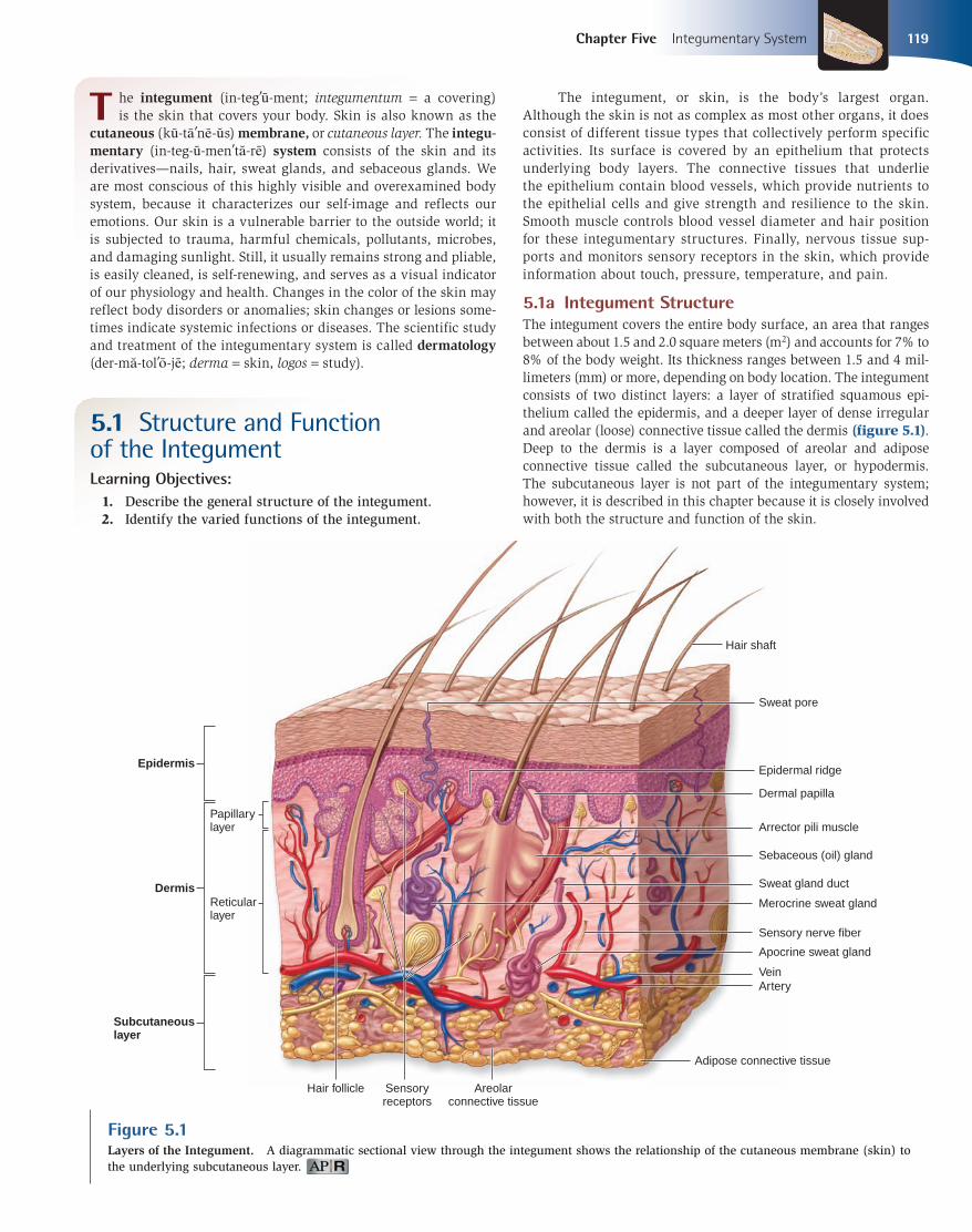
The integument covers the entire body surface, an area that ranges between about 1.5 and 2.0 square meters (m2) and accounts for 7% to 8% of the body weight. Its thickness ranges between 1.5 and 4 mil-limeters (mm) or more, depending on body location. The integument consists of two distinct layers: a layer of stratified squamous epi-thelium called the epidermis, and a deeper layer of dense irregular and areolar (loose) connective tissue called the dermis (figure 5.1). Deep to the dermis is a layer composed of areolar and adipose connective tissue called the subcutaneous layer, or hypodermis. The subcutaneous layer is not part of the integumentary system; however, it is described in this chapter because it is closely involved with both the structure and function of the skin.
Figure 5.1 Layers of the Integument. A diagrammatic sectional view through the integument shows the relationship of the cutaneous membrane (skin) to the underlying subcutaneous layer.
mck78097_ch05_118-145.indd 119mck78097_ch05_118-145.indd 119 2/11/11 3:02 PM2/11/11 3:02 PM
120 Chapter Five Integumentary System
The integument meets the mucous membranes within the nostrils, lips, anus, urethral opening, and vaginal opening. At these sites, the transition is seamless, and the epithelial defenses remain intact and functional.
5.1b Integument Functions
The integument is more than just a wrapping around the body. It serves many varied functions, including protection, prevention of water loss, temperature regulation, metabolic regulation, immune defense, sensory reception, and excretion.
Protection
The skin acts as a physical barrier that protects the entire body from physical injury, trauma, bumps, and scrapes. It also offers protection against harmful chemicals, toxins, microbes, and exces-sive heat or cold. Paradoxically, it can absorb certain chemicals and drugs (such as estrogen from a birth control patch or nicotine from a nicotine patch). Thus, the skin is said to be selectively permeablebecause some materials are able to pass through it, while others are effectively blocked. The epidermis is designed to withstand stresses and regenerate itself continuously throughout a person’s lifetime. The skin also protects deeper tissues from solar radiation, especially ultraviolet rays. When exposed to the sun, the melano-cytes become more active and produce more melanin, thus giving the skin a darker, tanned look. Even when you get a sunburn, the deeper tissues (muscles and internal organs) remain unaffected.
Prevention of Water Loss
The epidermis is water resistant and helps prevent unnecessary water loss. (If the skin were not water resistant, each time you took a bath you would swell up like a sponge as your skin absorbed water!) Water cannot easily enter or exit the skin, unless it is specifically secreted by the sweat glands. The skin also prevents the water within the body cells and in the extracellular fluid (the fluid outside of cells) from “leaking out.” When the skin is severely burned, a primary danger is dehydration, because the individual has lost the protective skin barrier, and water can escape from body tissues. Although the integument is water resistant, it is not entirely waterproof. Some interstitial fluids slowly escape through the epidermis to the surface, where they evaporate into the surround-ing air, a process called transepidermal water loss (TEWL).Approximately 500 milliliters (mL) (approximately 1 pint) of water is lost daily by evaporation of moisture from the skin or from respiratory passageways during breathing. Insensible per-spiration is the release of water vapor from sweat glands under “normal” circumstances when we are not sweating. In contrast, sensible perspiration is visible sweating. On most parts of the skin, water vapor released from sweat glands during insensible perspiration mixes with sebaceous secretions (sebum) to produce a thin, slightly acidic film (pH 4–6) over the surface of the epider-mis. This film helps slow down TEWL by forming an oily barrier over the surface of the skin. The acidic nature of the barrier also prevents the invasion of certain bacteria.
Temperature Regulation
Body temperature is influenced by vast capillary networks and sweat glands in the dermis. When the body is too warm and needs to dissipate heat, the diameter of the blood vessels in the dermis enlarges to permit more blood flow through the dermis, and sweat glands release fluid onto the skin surface. As relatively more blood
flows through these dermal vessels, the warmth from the blood dissipates through the skin, and the body cools off by evapora-tion of the sweat. Conversely, when the body is cold and needs to conserve heat, the blood vessels in the dermis constrict to reduce blood flow. In an effort to conserve heat, more blood is shunted to deeper body tissues, and relatively less blood flows in the dermal blood vessels.
Metabolic Regulation
Vitamin D3 is a cholesterol derivative synthesized from cholecal-ciferol (ko le -kal-sif er-ol), which is produced by some epidermal cells when they are exposed to ultraviolet radiation. Calcitriol (kal-si-trı ol) is synthesized from the cholecalciferol by some endocrine cells in the kidney. Calcitriol, the active form of vitamin D3, is a hormone that promotes calcium and phosphorus absorption from ingested materials across the wall of the small intestine. Thus, the synthesis of vitamin D3 is important in regulating the levels of cal-cium and phosphate in the blood. As little as 15 minutes of sunlight a day will provide your body with its daily vitamin D requirement!
Immune Defense
The epidermis contains a small population of immune cells. These immune cells (derived from a type of white blood cell), called epidermal dendritic (den-drit ik) cells, or Langerhans cells, play an important role in initiating an immune response by phagocytizing pathogens that have penetrated the epidermis and also against epidermal cancer cells.
Sensory Reception
The skin contains numerous sensory receptors. These receptors are associated with nerve endings that detect heat, cold, touch, pressure, texture, and vibration. For example, tactile cells (or Merkel cells) are large, specialized epithelial cells that stimulate specific sensory nerve endings when they are distorted by fine touch or pressure. Because your skin is responsible for perceiving many stimuli, it needs different sensory receptor types to detect, distinguish, and interpret these stimuli.
Excretion by Means of Secretion
Skin exhibits an excretory function when it secretes substances from the body during sweating. Sweating, or sensible perspiration, occurs when the body needs to cool itself off. Notice that sweat sometimes feels “gritty” because of the waste products being secreted onto the skin surface. These substances include water, salts, and urea, a nitrogen-containing waste product of body cells. In addition, the skin contains sebaceous glands that secrete an oily material called sebum, which lubricates the skin surface and hair.
WHAT DID YOU LEARN?
●1 What are the two major layers of the integument and the components of each?
●2 What is the relationship between exposure to sunlight and the body’s need for vitamin D?
WHAT DO YOU THINK?
●1 During the Industrial Revolution, as children spent little time outdoors and most of their time working in factories, increasing numbers of them developed a bone disorder called rickets. Rickets is caused by inadequate vitamin D. Based on your knowledge of skin function, why do you think these children developed rickets?
WW
mck78097_ch05_118-145.indd 120mck78097_ch05_118-145.indd 120 2/11/11 3:02 PM2/11/11 3:02 PM
(a)
Stratum corneum
Tactile cell
Sensory nerve ending
Melanocyte
Epidermaldendritic cell
Living keratinocyte
Dead keratinocytes
Basement membrane
Stratum basale
Stratum spinosum
Stratum lucidum
Dermis
Stratumgranulosum
(b)
Chapter Five Integumentary System 121
5.2 EpidermisLearning Objectives: 1. Describe the structure, composition and arrangement, and
functions of the epidermal strata. 2. Identify the epidermal variations in thickness, color, and
markings.
The epithelium of the integument is called the epidermis (ep-i-derm is; epi = on, derma = skin). The epidermis is a keratinized, stratified squamous epithelium. Like other epithelia, the epidermis is avascular, and it acquires its nutrients through diffusion from the underlying dermis.
5.2a Epidermal Strata
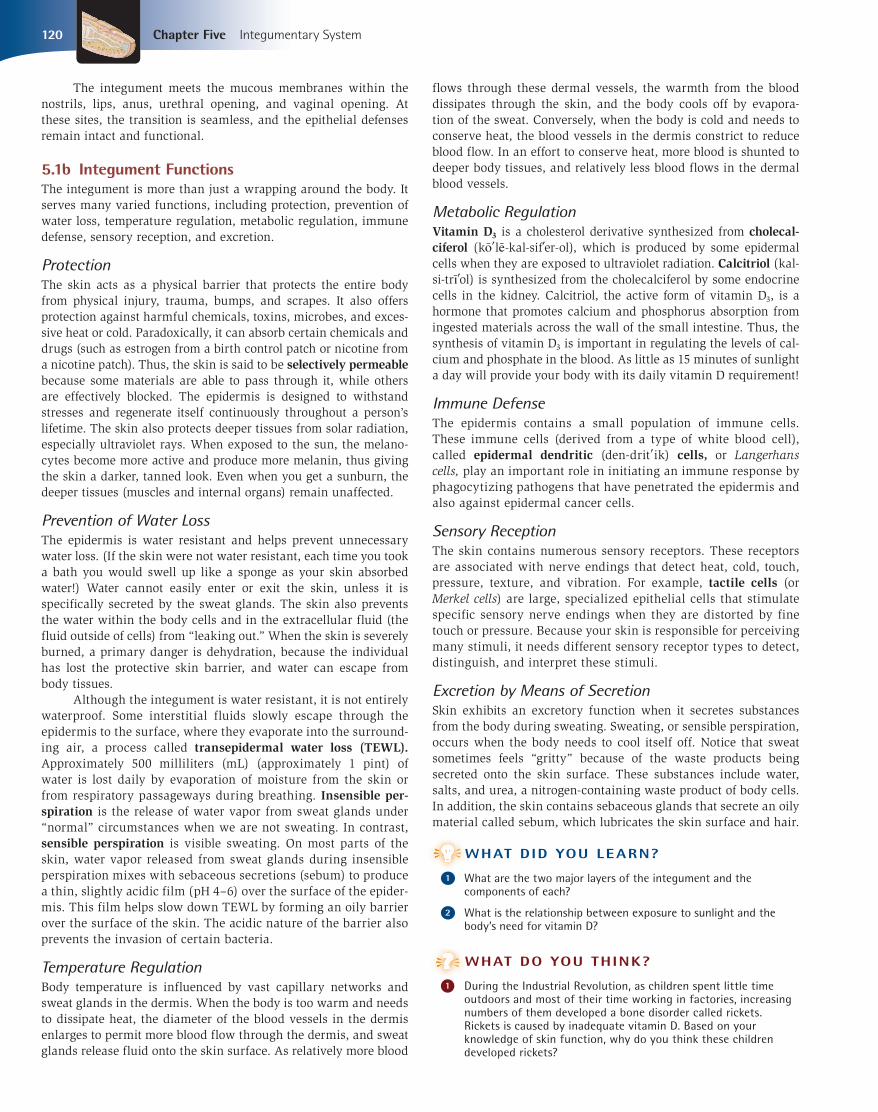
Careful examination of the epidermis, from the basement mem-brane to its surface, reveals several layers, or strata. From deep to superficial, these layers are the stratum basale, the stratum spinosum, the stratum granulosum, the stratum lucidum (found in thick skin only), and the stratum corneum (figure 5.2). The first three strata listed are composed of living keratinocytes, and last two strata contain dead keratinocytes.
Stratum Basale
The deepest epidermal layer is the stratum basale (strat u m bah-sa le ) (also known as the stratum germinativum or basal layer). This single layer of cells ranges from cuboidal to low columnar in appear-ance. It is tightly attached to an underlying basement membrane that separates the epidermis from the connective tissue of the adjacent dermis. Three types of cells occupy the stratum basale (figure 5.2b):
1. Keratinocytes (ke-rat i-no -sı t; keras = horn) are the most abundant cell type in the epidermis and are found throughout all epidermal strata. The stratum basale is dominated by large keratinocyte stem cells, which divide to provide both replacement stem cells and new keratinocytes that replace the dead keratinocytes shed from the surface. Their name is derived from their role in the synthesis of the protein keratin (ker a -tin)
in the epidermal cells of the skin. Keratin is a family of fibrous structural proteins that are both tough and insoluble. Fibrous keratin molecules can twist and intertwine around each other to form helical intermediate filaments of the cytoskeleton (see chapter 2). The keratins found in epidermal cells of the skin are called cytokeratins. Their structure in these cells gives skin its strength and makes the epidermis almost waterproof.
2. Melanocytes (mel a -no -sı t; melano = black) have long, branching cytoplasmic processes and are scattered among the keratinocytes of the stratum basale. These processes transfer pigment granules, called melanosomes (mel a -no -so mes), by phagocytosis or exocytosis, into the keratinocytes within the basal layer and sometimes within more superficial layers. This pigment (black, brown, or yellow-brown) accumulates around the nucleus of the keratinocyte and shields the DNA within the nucleus from ultraviolet radiation. The darker tones of the skin result from melanin being produced by the melanocytes and from the darkening of melanin already present upon exposure to ultraviolet light.
3. Tactile cells are few in number and found scattered among the cells within the stratum basale. Tactile cells are sensitive to touch, and when compressed, they release chemicals that stimulate sensory nerve endings, providing information about objects touching the skin.
Stratum Spinosum
Several layers of polygonal keratinocytes form the stratum spino-sum (spı -no su m), or spiny layer. Each time a keratinocyte stem cell in the stratum basale divides, the daughter cell that will differenti-ate into the new epidermal cell is pushed toward the external sur-face from the stratum basale. Once this new cell enters the stratum spinosum, the cell begins to differentiate into a nondividing, highly specialized keratinocyte. Sometimes the deepest cells in this layer still undergo mitosis to help replace epidermal cells that exfoliate from the epidermal surface. The nondividing keratinocytes in the stratum spinosum attach to their neighbors by many intercellular junctions called desmosomes (described in chapter 4). The process of preparing epidermal tissue for observation on a microscope slide
Figure 5.2Epidermal Strata. (a) Photomicrograph and (b) diagram compare the order and relationships of the epidermal strata in thick skin.
mck78097_ch05_118-145.indd 121mck78097_ch05_118-145.indd 121 2/11/11 3:02 PM2/11/11 3:02 PM

122 Chapter Five Integumentary System
shrinks the cytoplasm of the cells in the stratum spinosum. Because the cytoskeletal elements and desmosomes remain intact, the shrunken stratum spinosum cells resemble miniature porcupines attached to their neighbors. These bridges between neighboring cells provide a spiny appearance, explaining the name of the layer. In addition to the keratinocytes, the stratum spinosum also contains the fourth epidermal cell type, the epidermal dendritic cells (figure 5.2b). Epidermal dendritic cells are immune cells that help fight infection in the epidermis. These cells are often present but not easily identifiable in both the stratum spinosum and the more superficial stratum granulosum. Their phagocytic activity initiates an immune response to protect the body against patho-gens that have penetrated the superficial layers of the epidermis as well as against epidermal cancer cells.
Stratum Granulosum
The stratum granulosum (gran-u -lo sum), or granular layer, consists of three to five layers of keratinocytes superficial to the stratum spi-nosum. Within this stratum begins a process called keratinization(ker a -tin-i-za shu n), by which the keratinocytes fill up with the protein keratin. Several significant events occur during keratinization. As the cells pass through the stratum granulosum and true keratin filaments (intermediate filaments of the cytoskeleton) begin to develop, the cells become thinner and flatter. Their membranes thicken and become less permeable. The nucleus and all organelles disintegrate, and the cells start to die. Subsequently, the dehydrated material left within the cells forms a tightly interlocked layer of keratin fibers sandwiched between thickened phospholipid membranes. Keratinization is not complete until the cells reach the more superficial epidermal layers. A fully keratinized cell is dead (because it has neither a nucleus nor organelles), but it is strong because it contains keratin.
Stratum Lucidum
The stratum lucidum (lu sı -dum), or clear layer, is a thin, translu-cent region about two to three cell layers thick that is superficial to the stratum granulosum. This stratum is found only in thick skin, such as the palms of the hands and the soles of the feet. Cells occu-pying this layer appear pale and featureless, and have indistinct boundaries. The keratinocytes within this layer are flattened and filled with the protein eleidin (e -le ı -din), an intermediate product in the process of keratin maturation.
Stratum Corneum
The stratum corneum (ko r ne -u m; corneus = horny, or hornlike layer), is the most superficial layer of the epidermis. It is the stra-tum you see when you look at your skin. The stratum corneum consists of about 20 to 30 layers of dead, scaly, interlocking kera-tinized cells called corneocytes (ko r ne -o -sı t). The dead cells are anucleate (lacking a nucleus) and tightly packed together. A keratinized (or cornified) epithelium contains large amounts of keratin. After keratinocytes are formed from stem cells within the stratum basale, they change in structure and in their relationship to their neighbors as they move through the different strata until they eventually reach the stratum corneum and are sloughed off from its external surface. Migration of the keratinocyte to the stratum corneum from the stratum basale occurs during the first 2 weeks of the keratinocyte’s life. The dead, keratinized cells usually remain for an additional 2 weeks in the exposed stratum corneum layer, providing a barrier for cells deeper in the epidermis before they are shed, washed away, or removed by abrasion. Overall, keratinocytes are present for about 1 month following their formation.
The normally dry stratum corneum presents a thickened surface unsuitable for the growth of many microorganisms. Additionally, some secretions onto the surface of the epidermis from exocrine glands help prevent the growth of microorganisms on the epidermis, thus supporting its barrier function.
5.2b Variations in the Epidermis
The epidermis exhibits variations among different body regions within a single individual, as well as differences between individuals. The epidermis varies in thickness, coloration, and skin markings.
Thick Skin Versus Thin Skin
Over most of the body, the skin ranges from 1 mm to 2 mm in thickness. Skin is classified as either thick or thin based on the
Study Tip!In your anatomy lab, you may be asked to identify a specific epider-
mal stratum. Answer the following questions to help identify these strata.
1. Is the epidermal stratum near the free surface of the epithelium or closer to the basal surface? Remember, the stratum corneum forms the free surface, while the stratum basale forms the deepest epidermal layer.
2. What is the shape of the cells? The stratum basale contains cells that are cuboidal to low columnar in shape; the stratum spinosum contains polygonal cells; and the stratum lucidum and stratum corneum contain squamous cells.
3. Do the keratinocytes have a nucleus, or are they anucleate (lacking a nucleus)? When the keratinocytes are still alive (as in the strata basale, spinosum, and granulosum), you will be able to see nuclei in the keratinocytes. The stratum lucidum and stratum corneum layers contain anucleate keratinocytes.
4. How many layers of cells are in the stratum? The stratum basale has only one layer of cells, and the stratum corneum contains 20 to 30 layers of cells. The other layers contain about 2 to 5 layers of cells.
5. Does the cytoplasm of the cells contain visible dark granules? If the answer is yes, you likely are looking at the stratum granulosum.
Transdermal Administration of DrugsSome drugs may be administered through the skin, a process called transdermal administration. Drugs that are soluble either in oils or lipid-soluble carriers may be administered transdermally by affixing a patch containing the drug to the skin surface. These drugs slowly penetrate the epidermis and are absorbed into the blood vessels of the dermis. Transdermal patches are especially useful because they release a continual, slow amount of the drug over a relatively long period of time. The epidermal barrier requires that the concentration of the drug in the patch be relatively high. There are transdermal patches that contain nicotine (to help people quit smoking), estrogen (for hormone replacement therapy [HRT] or birth control), or nitroglycerin (to prevent heart attack). These patches are advantageous because the patient is not required to ingest daily medication.
CLINICAL VIEW
mck78097_ch05_118-145.indd 122mck78097_ch05_118-145.indd 122 2/11/11 3:02 PM2/11/11 3:02 PM

Epidermis
Epidermis
Dermis
(a) Thick skin (b) Thin skin
Stratum basaleStratum spinosum
Stratum granulosum
Stratum basale
Stratum spinosum
Stratum granulosum
Stratum lucidum
Stratum corneum
Stratum corneum
Dermal papillae
LM 40x LM 100x
Vesicle filledwith melanin
Basementmembrane
Melanin pigment
Melanin pigmentin keratinocyte
Melanocyte
(a)
Stratum basale with melanin pigment
(b)
Epidermis
Dermis
LM 124x
Chapter Five Integumentary System 123
number of strata in the epidermis and the relative thickness of the epidermis, rather than the thickness of the entire integument (figure 5.3). Thick skin is found on the palms of the hands, the soles of the feet, and corresponding surfaces of the fingers and toes. All five epidermal strata occur in thick skin. Thick skin ranges between 400 and 600 micrometers (μm) thick. Thick skin contains sweat glands, but no hair follicles or sebaceous glands. Thin skin covers most of the body. The epidermis lacks the stratum lucidum, so it has only four layers. Thin skin contains the fol-lowing accessories: hair follicles, sebaceous glands, and sweat glands. The epidermis of thin skin is only 75 μm to 150 μm thick.
WHAT DO YOU THINK?
●2 Why does thick skin lack hair follicles and sebaceous glands? Think about the body locations of thick skin and how the presence of hair follicles and sebaceous glands might interfere with the job of thick skin in those areas.
Skin Color
Normal skin color results from a combination of hemoglobin, mela-nin, and carotene. Hemoglobin (he -mo -glo bin; haima = blood) is an oxygen-binding protein present within red blood cells. Upon binding oxygen, hemoglobin exhibits a bright red color, giving blood vessels in the dermis a bright reddish tint that is most easily observed in the skin of lightly pigmented individuals. Melanin (mel a -nin) is a pigment produced and stored in cells called melanocytes (figure 5.4; see figure 5.2b). This pigment is synthesized from the amino acid tyrosine, and its production requires the enzyme tyrosinase. There are two types of melanin, eumelanin and pheomelanin, and they occur in various ratios of yellow, reddish, tan, brown, and black shades. Melanin is transferred in membrane-bound vesicles from melanocytes to kera-tinocytes in the stratum basale and stratum spinosum via phago-cytosis or exocytosis. The keratinocytes that receive the melanin are displaced toward the stratum corneum, and thus melanocyte activity affects the color of the entire epidermis.
Figure 5.3Thick Skin and Thin Skin. The stratified squamous epithelium of the epidermis varies in thickness, depending upon the region of the body in which it is located. (a) Thick skin contains all five epidermal strata and covers the soles of the feet and the palms of the hands. (b) Thin skin covers most body surfaces; it lacks a stratum lucidum.
Figure 5.4Production of Melanin by Melanocytes. Melanin gives a yellow to tan to brown color to the skin. (a) Vesicles in melanocytes transport the melanin pigment to the keratinocytes, where the pigment surrounds the nucleus. (b) Melanin is incorporated into the cells of the stratum basale.
mck78097_ch05_118-145.indd 123mck78097_ch05_118-145.indd 123 2/11/11 3:02 PM2/11/11 3:02 PM
124 Chapter Five Integumentary System
All people have about the same number of melanocytes. However, melanocyte activity and the color of the melanin produced by these cells varies among individuals and races, resulting in different skin tones. Darker-skinned individuals have melanocytes that produce relatively more melanin than do those of lighter-skinned individuals. Further, these more active mela-nocytes tend to package and send melanin to cells in the more superficial epidermal layers, such as the stratum granulosum. The amount of melanin in the skin is determined by both hered-ity and light exposure. Melanin pigment surrounds the kerati-nocyte nucleus, where it absorbs ultraviolet (UV) radiation in sunlight, thus preventing damage to nuclear DNA. Exposure to
UV light both darkens melanin already present and stimulates melanocytes to make more melanin.
Carotene (kar o -te n) is a yellow-orange pigment that is acquired in the body by eating various yellow-orange vegetables, such as carrots, corn, and squash. Normally, carotene accumu-lates inside keratinocytes of the stratum corneum and within the subcutaneous fat. In the body, carotene is converted into vitamin A, which has an important function in normal vision. Additionally, carotene has been implicated in reducing the number of potentially dangerous molecules formed during normal meta-bolic activity and in improving immune cell number and activity.
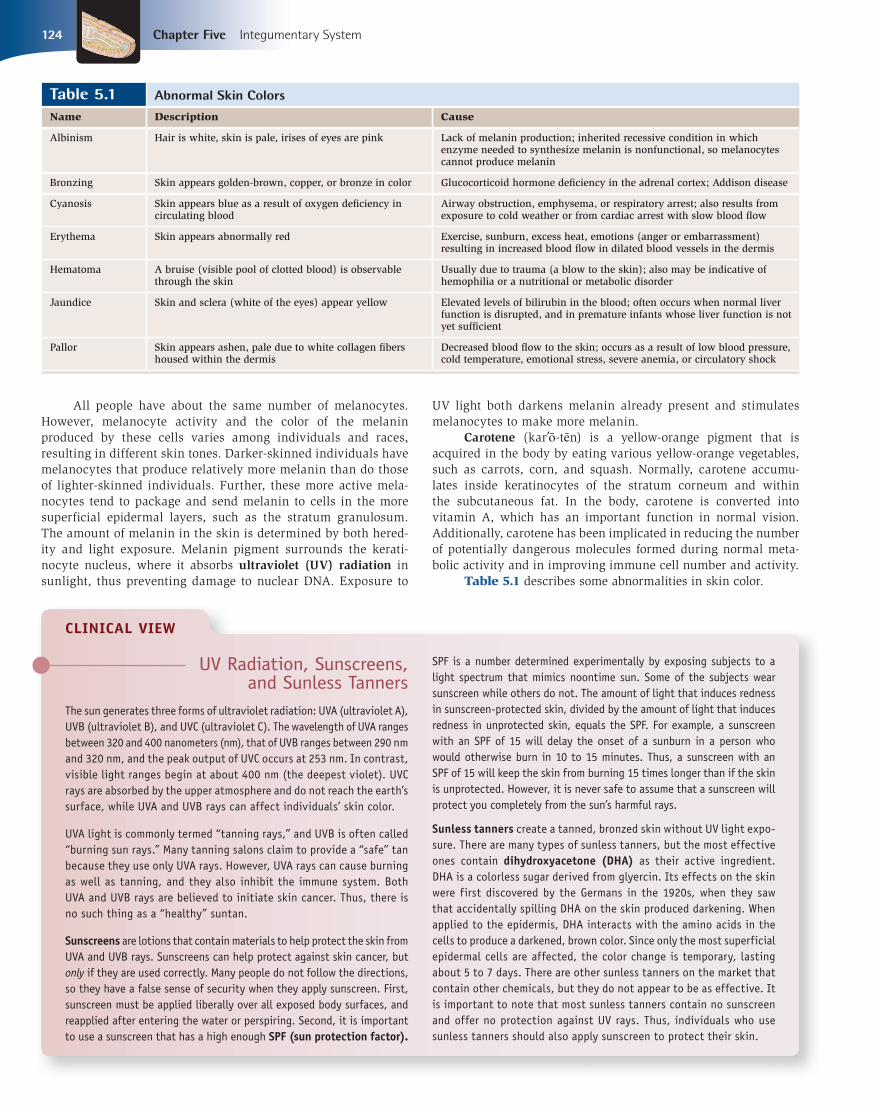
Table 5.1 describes some abnormalities in skin color.
Table 5.1 Abnormal Skin Colors
Name Description Cause
Albinism Hair is white, skin is pale, irises of eyes are pink Lack of melanin production; inherited recessive condition in which enzyme needed to synthesize melanin is nonfunctional, so melanocytes cannot produce melanin
Bronzing Skin appears golden-brown, copper, or bronze in color Glucocorticoid hormone defi ciency in the adrenal cortex; Addison disease
Cyanosis Skin appears blue as a result of oxygen defi ciency in circulating blood
Airway obstruction, emphysema, or respiratory arrest; also results from exposure to cold weather or from cardiac arrest with slow blood fl ow
Erythema Skin appears abnormally red Exercise, sunburn, excess heat, emotions (anger or embarrassment) resulting in increased blood fl ow in dilated blood vessels in the dermis
Hematoma A bruise (visible pool of clotted blood) is observable through the skin
Usually due to trauma (a blow to the skin); also may be indicative of hemophilia or a nutritional or metabolic disorder
Jaundice Skin and sclera (white of the eyes) appear yellow Elevated levels of bilirubin in the blood; often occurs when normal liver function is disrupted, and in premature infants whose liver function is not yet suffi cient
Pallor Skin appears ashen, pale due to white collagen fi bers housed within the dermis
Decreased blood fl ow to the skin; occurs as a result of low blood pressure, cold temperature, emotional stress, severe anemia, or circulatory shock
UV Radiation, Sunscreens, and Sunless Tanners
The sun generates three forms of ultraviolet radiation: UVA (ultraviolet A), UVB (ultraviolet B), and UVC (ultraviolet C). The wavelength of UVA ranges between 320 and 400 nanometers (nm), that of UVB ranges between 290 nm and 320 nm, and the peak output of UVC occurs at 253 nm. In contrast, visible light ranges begin at about 400 nm (the deepest violet). UVC rays are absorbed by the upper atmosphere and do not reach the earth’s surface, while UVA and UVB rays can affect individuals’ skin color.
UVA light is commonly termed “tanning rays,” and UVB is often called “burning sun rays.” Many tanning salons claim to provide a “safe” tan because they use only UVA rays. However, UVA rays can cause burning as well as tanning, and they also inhibit the immune system. Both UVA and UVB rays are believed to initiate skin cancer. Thus, there is no such thing as a “healthy” suntan.
Sunscreens are lotions that contain materials to help protect the skin from UVA and UVB rays. Sunscreens can help protect against skin cancer, but only if they are used correctly. Many people do not follow the directions, so they have a false sense of security when they apply sunscreen. First, sunscreen must be applied liberally over all exposed body surfaces, and reapplied after entering the water or perspiring. Second, it is important to use a sunscreen that has a high enough SPF (sun protection factor).
SPF is a number determined experimentally by exposing subjects to a light spectrum that mimics noontime sun. Some of the subjects wear sunscreen while others do not. The amount of light that induces redness in sunscreen-protected skin, divided by the amount of light that induces redness in unprotected skin, equals the SPF. For example, a sunscreen with an SPF of 15 will delay the onset of a sunburn in a person who would otherwise burn in 10 to 15 minutes. Thus, a sunscreen with an SPF of 15 will keep the skin from burning 15 times longer than if the skin is unprotected. However, it is never safe to assume that a sunscreen will protect you completely from the sun’s harmful rays.
Sunless tanners create a tanned, bronzed skin without UV light expo-sure. There are many types of sunless tanners, but the most effective ones contain dihydroxyacetone (DHA) as their active ingredient. DHA is a colorless sugar derived from glyercin. Its effects on the skin were first discovered by the Germans in the 1920s, when they saw that accidentally spilling DHA on the skin produced darkening. When applied to the epidermis, DHA interacts with the amino acids in the cells to produce a darkened, brown color. Since only the most superficial epidermal cells are affected, the color change is temporary, lasting about 5 to 7 days. There are other sunless tanners on the market that contain other chemicals, but they do not appear to be as effective. It is important to note that most sunless tanners contain no sunscreen and offer no protection against UV rays. Thus, individuals who use sunless tanners should also apply sunscreen to protect their skin.
CLINICAL VIEW
mck78097_ch05_118-145.indd 124mck78097_ch05_118-145.indd 124 2/11/11 3:02 PM2/11/11 3:02 PM

Arch Whorl
CombinationLoop
Chapter Five Integumentary System 125
Skin Markings
A nevus (ne vu s; pl., ne vı ; naevus = mole, birthmark), commonly called a mole is a harmless, localized overgrowth of melanin-forming cells. Almost everyone is born with a few nevi, and some people have as many as 20 or more. On very rare occasions, a nevus may become malignant, typically as a consequence of excessive UV light exposure. Thus, nevi should be monitored for changes that may suggest malignancy. Freckles are yellowish or brown spots that represent localized areas of excessive melanocyte activity, not an increase in melanocyte numbers. A freckle’s degree of pigmentation varies and depends on both sun exposure and heredity. A hemangioma (he-man je -o ma ; angio = vessel, oma =tumor) is a congenital anomaly that results in skin discoloration due to blood vessels that proliferate and form a benign tumor. Capillary hemangiomas or “strawberry-colored birthmarks,” appear in the skin as bright red to deep purple nodules that usually disappear in childhood. Cavernous hemangiomas, some-times called “port-wine stains,” involve larger dermal blood ves-sels and may last a lifetime. The contours of the skin surface follow ridge patterns, vary-ing from small, conical pegs (in thin skin) to complex arches and whorls (in thick skin) called friction ridges. Friction ridges are found on the fingers, palms, soles, and toes (figure 5.5). These ridges are formed from large folds and valleys of both the dermis and epidermis. With the help of moisture from merocrine sweat glands, friction ridges increase friction so that objects do not slip easily from our hands and our feet do not slip on the floor when we walk. Friction ridges can leave noticeable prints on touched surfaces, commonly called “fingerprints.” Because each individual has a unique pattern of friction ridges, fingerprints have become a valuable identification tool for law enforcement. Medical applications are possible as well (see Clinical View: “Dermatoglyphics”).
WHAT DO YOU THINK?
●3 Why are people’s attempts to change their recognizable fingerprints usually not successful?
WHAT DID YOU LEARN?
●3 Why is the stratum spinosum important in maintaining the integrity of the skin?
●4 Briefly describe the process of keratinization. Where does it begin? Why is it important?
●5 What normal skin accessories are not present in thick skin?
●6 How do melanocytes help protect the skin?
5.3 DermisLearning Objectives: 1. Describe the organization and function of the layers of the
dermis. 2. Identify nerve and blood supply to the dermis.
The dermis (der mis) lies deep to the epidermis and ranges in thickness from 0.5 mm to 3.0 mm. The dermis consists of two types of connective tissue: areolar and dense irregular. These con-nective tissue layers of the integument are composed of cells of the connective tissue proper and primarily of collagen fibers, although both elastic and reticular fibers are also present. Other components of the dermis are blood vessels, sweat glands, sebaceous glands, hair follicles, nail roots, sensory nerve endings, and smooth muscle tissue. There are two major regions of the dermis: a superficial pap-illary layer and a deeper reticular layer (figure 5.6).
WW
Figure 5.5Friction Ridges of Thick Skin. Friction ridges form fingerprints, palm prints, and toe prints. Shown here are four basic fingerprint patterns.
DermatoglyphicsThe study of friction ridge patterns is known as dermatoglyph-ics (derma = skin, qlyph = carving). Friction ridge patterns are well formed by the fourth month of fetal development, and are a unique identifier because no two individuals share the same set of fingerprints. Even identical twins have different fingerprints. Biological anthropologists and other scientists have studied dermatoglyphics among different populations. They have found gender differences in dermatoglyphic patterns. For example, males tend to have relatively more whorls in their fingerprint patterns, whereas females tend to have relatively more arches. Some regional populations also exhibit characteristic dermatoglyphic patterns.
Specific dermatoglyphic patterns have been noted with some medical conditions. For example, individuals with Down syndrome (trisomy 21) tend to have a single palmar crease, known as a simian crease. Other dermatoglyphic patterns have been associated with schizophrenia, Alzheimer disease, rubella, some forms of cancer, and heart disease. Early research indicates fingerprint patterns can serve as early indicators of some conditions since the patterns do not change after birth. Researchers hope to eventually be able to use an individual’s dermatoglyphic pattern to help diagnose disease.
CLINICAL VIEW
mck78097_ch05_118-145.indd 125mck78097_ch05_118-145.indd 125 2/11/11 3:02 PM2/11/11 3:02 PM
Epidermis
Dermis
Subcutaneouslayer
Epidermal ridges
Blood vessels
Dermal papillae
Papillarylayer
Reticularlayer
126 Chapter Five Integumentary System
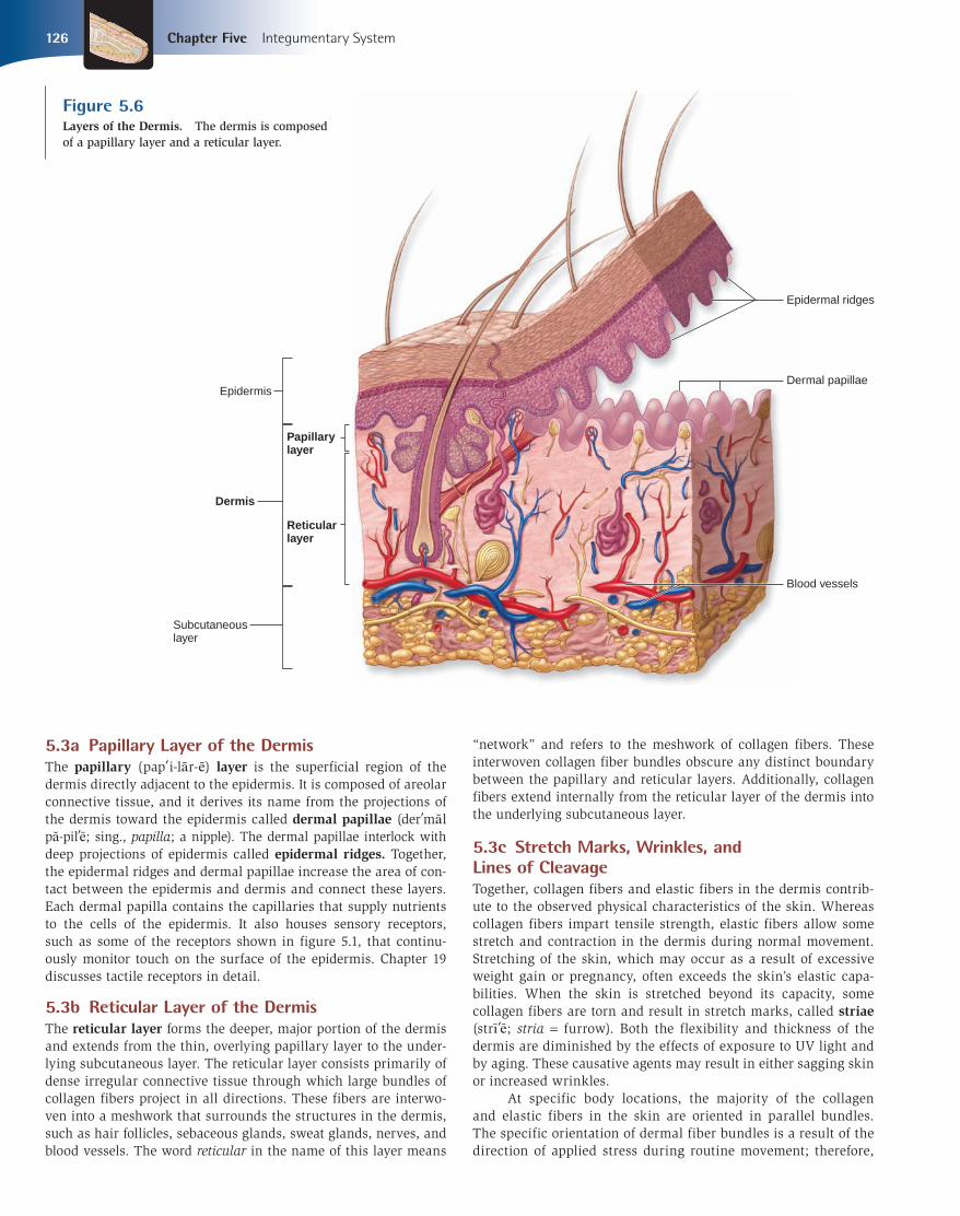
5.3a Papillary Layer of the Dermis
The papillary (pap i-la r-e ) layer is the superficial region of the dermis directly adjacent to the epidermis. It is composed of areolar connective tissue, and it derives its name from the projections of the dermis toward the epidermis called dermal papillae (der ma l pa -pil e ; sing., papilla; a nipple). The dermal papillae interlock withdeep projections of epidermis called epidermal ridges. Together, the epidermal ridges and dermal papillae increase the area of con-tact between the epidermis and dermis and connect these layers. Each dermal papilla contains the capillaries that supply nutrients to the cells of the epidermis. It also houses sensory receptors, such as some of the receptors shown in figure 5.1, that continu-ously monitor touch on the surface of the epidermis. Chapter 19 discusses tactile receptors in detail.
5.3b Reticular Layer of the Dermis
The reticular layer forms the deeper, major portion of the dermis and extends from the thin, overlying papillary layer to the under-lying subcutaneous layer. The reticular layer consists primarily of dense irregular connective tissue through which large bundles of collagen fibers project in all directions. These fibers are interwo-ven into a meshwork that surrounds the structures in the dermis, such as hair follicles, sebaceous glands, sweat glands, nerves, and blood vessels. The word reticular in the name of this layer means
“network” and refers to the meshwork of collagen fibers. These interwoven collagen fiber bundles obscure any distinct boundary between the papillary and reticular layers. Additionally, collagen fibers extend internally from the reticular layer of the dermis into the underlying subcutaneous layer.
5.3c Stretch Marks, Wrinkles, and
Lines of Cleavage
Together, collagen fibers and elastic fibers in the dermis contrib-ute to the observed physical characteristics of the skin. Whereas collagen fibers impart tensile strength, elastic fibers allow some stretch and contraction in the dermis during normal movement. Stretching of the skin, which may occur as a result of excessive weight gain or pregnancy, often exceeds the skin’s elastic capa-bilities. When the skin is stretched beyond its capacity, some collagen fibers are torn and result in stretch marks, called striae(strı e ; stria = furrow). Both the flexibility and thickness of the dermis are diminished by the effects of exposure to UV light and by aging. These causative agents may result in either sagging skin or increased wrinkles. At specific body locations, the majority of the collagen and elastic fibers in the skin are oriented in parallel bundles. The specific orientation of dermal fiber bundles is a result of the direction of applied stress during routine movement; therefore,
Figure 5.6Layers of the Dermis. The dermis is composed of a papillary layer and a reticular layer.
mck78097_ch05_118-145.indd 126mck78097_ch05_118-145.indd 126 2/11/11 3:02 PM2/11/11 3:02 PM

An incision perpendicular to cleavage lines may gape and delay healing.
An incision parallel to cleavage lines is more likely to heal quickly and not gape open.
Chapter Five Integumentary System 127
the alignment of the bundles functions to resist stress. Lines of cleavage (or tension lines) in the skin identify the predominant orientation of collagen fiber bundles (figure 5.7). These are clin-ically and surgically significant because any procedure resulting in a cut at right angles to a cleavage line is usually pulled open due to the recoil from cut elastic fibers. This often results in slow healing and increased scarring. In contrast, a cut parallel to a cleavage line usually remains closed, resulting in faster healing. Therefore, surgical procedures should be planned to allow for these lines of cleavage, thus ensuring rapid healing and prevent-ing scarring.
5.3d Innervation and Blood Supply
Nerve fibers are extensively dispersed throughout the dermis, a property called innervation. Nerve fibers in the skin monitor sen-sory receptors in the dermis and epidermis, and they also control both blood flow and gland secretion rates. Tactile corpuscles and tactile cells perceive touch sensations and work with a variety of other sensory nerve endings in the skin. This rich innervation allows us to be very aware of our surroundings and to differenti-ate among the different kinds of sensory signals from receptors in the skin. Recall that all epithelia, including the epidermis, are avascu-lar. Therefore, blood vessels within the dermis must supply nutrients to the living cells in the epidermis as well as to all structures in the dermis. The largest of these blood vessels lie along the border between the reticular layer of the dermis and the subcutaneous layer. Smaller vessels branch into the dermis to supply the hair
follicles, sweat glands, sensory receptors, and other structures housed there. The smallest arterial vessels connect to capillary loops within the dermal papillae. Eventually, these capillaries drain into small vessels, forming a vessel network that merges into small veins draining the dermis. Dermal blood vessels have an important role in regulating body temperature and blood pressure. Vasoconstriction (va so ; vas = a vessel) means that the diameters of the vessels narrow, so relatively less blood can travel through them. Therefore, relatively more blood must travel in blood vessels that are deeper internal to the skin. The net effect of vasoconstriction of the dermal blood vessels is a shunting of blood away from the periphery of the body. If the body is cold, the dermal blood vessels vasoconstrict to conserve heat in the blood. This is why we are paler when we are exposed to cold temperatures. Conversely, vasodilation of the dermal blood vessels means that the diameter of the vessels increases, so relatively more blood can travel through them. As more blood is shunted to these super-ficial blood vessels, the heat from the blood may be more easily dissipated through the skin. If the body is too warm, the dermal blood vessels vasodilate so more blood can travel close to the sur-face and excess heat can be lost. This additional blood flow in the dermis gives a more reddish or pinkish hue to the skin. Thus, your face may become flushed when you exercise because your dermal blood vessels are dilated in an attempt to release the excess heat you generated while working out. Because blood volume typically remains constant, any increase in circulation to the skin results in a decrease in circulation to other organs.
Figure 5.7Lines of Cleavage. Lines of cleavage (tension lines) partition the skin and indicate the predominant direction of underlying collagen fibers in the reticular layer of the dermis.
mck78097_ch05_118-145.indd 127mck78097_ch05_118-145.indd 127 2/11/11 3:02 PM2/11/11 3:02 PM

128 Chapter Five Integumentary System
WHAT DID YOU LEARN?
●7 Briefly describe the structure of epidermal ridges and dermal papillae. What is the importance of each, and how do they interact?
●8 What is indicated by the lines of cleavage in the skin, and why are they medically important?
●9 Why must the circulation to the skin be closely regulated?
5.4 Subcutaneous Layer (Hypodermis)Learning Objective: 1. Identify and describe the structure and function of the
subcutaneous layer.
Deep to the integument is the subcutaneous (su b-ku -ta ne -u s; sub = beneath; cutis = skin) layer, also called the hypodermis or superficial fascia. It is not considered a part of the integument. This layer consists of both areolar connective tissue and adipose connec-tive tissue (see figure 5.1). In some locations of the body, adipose
WW connective tissue predominates, and the subcutaneous layer is called subcutaneous fat. The connective tissue fibers of the reticular layer are extensively interwoven with those of the subcutaneous layer to stabilize the position of the skin and bind it to the underlying tissues. The subcutaneous layer pads and protects the body and its parts, acts as an energy reservoir, and provides thermal insulation. Drugs are often injected into the subcutaneous layer be-cause its excessive vascular network promotes rapid absorption. Normally, the subcutaneous layer is thicker in women than in men, and its regional distribution also differs between the sexes. Adult males accumulate subcutaneous fat primarily at the neck, upper arms, abdomen, along the lower back, and over the buttocks, whereas adult females accumulate subcutaneous fat primarily in the breasts, buttocks, hips, and thighs.
Table 5.2 reviews the layers of the integument and the sub-cutaneous layer.
WHAT DID YOU LEARN?
●10 What are some functions of the subcutaneous layer?
WW
Table 5.2 Layers of the Integument and the Subcutaneous Layer
Layer Specifi c Sublayers Structure
INTEGUMENT: EPIDERMIS
Stratum corneum Most superfi cial layer of epidermis; 20–30 layers of dead, fl attened, anucleate, keratin-fi lled keratinocytes called corneocytes
Stratum lucidum 2–3 layers of anucleate, dead cells; seen only in thick skin (e.g., palms, soles)
Stratum granulosum 3–5 layers of keratinocytes with distinct granules in the cytoplasm: keratinization begins in this layer
Stratum spinosum Several layers of keratinocytes attached to neighbors by desmosomes; epidermal dendritic cells present
Stratum basale Deepest, single layer of cuboidal to low columnar cells in contact with basement membrane; mitosis occurs here; contains keratinocytes, melanocytes, and tactile cells
INTEGUMENT: DERMIS
Papillary layer More superfi cial layer of dermis; composed of areolar connective tissue; contains dermal papillae
Reticular layer Deeper layer of dermis; dense irregular connective tissue surrounding blood vessels, hair follicles, nerves, sweat glands, and sebaceous glands
SUBCUTANEOUS LAYER
Not considered part of the integument; deep to dermis; composed of areolar connective tissue and adipose connective tissue plus hair follicles and merocrine sweat glands often project into the subcutaneous layer
mck78097_ch05_118-145.indd 128mck78097_ch05_118-145.indd 128 2/11/11 3:02 PM2/11/11 3:02 PM

(a) Onychomycosis (b) Yellow nail syndrome (c) Spoon nails (d) Beau’s lines
Chapter Five Integumentary System 129
5.5 Epidermal Accessory OrgansLearning Objectives: 1. Describe the structure and functions of nails. 2. Identify the components of a hair and a hair follicle. 3. Describe the growth, distribution, and replacement of
hairs. 4. Describe how hair changes throughout life. 5. Identify and describe the characteristics of sweat glands,
sebaceous glands, and other glands found in the skin.
Nails, hair, and sweat and sebaceous glands are derived from epidermis and are considered accessory organs, or append-ages, of the integument. These structures originate from the invagination of the epidermis during embryological develop-ment; they are located in the dermis and may project through
the epidermis to the surface. Both nails and hair are composed primarily of dead, keratinized cells.
5.5a Nails
Nails are scalelike modifications of the epidermis that form on the dorsal tips of the fingers and toes. They protect the exposed distal tips and prevent damage or distortion when the fingers or toes are subjected to mechanical stress—for example, dur-ing jumping, kicking, catching, or grasping. Nails are derived from the same type of cells that produce the stratum corneum layer of the epidermis. The cells that form the nails are densely packed and filled with parallel fibers of hard keratin. Each nail has a pinkish nail body and a distal whitish free edge (figure 5.8a). Most of the nail body appears pink because of the blood flowing in the underlying capillaries. In contrast, the free edge of the nail appears white because there are no
Nail DisordersChanges in the shape, structure, or appearance of the nails may indicate the existence of a disease that affects metabolism throughout the body. In fact, the state of a person’s fingernails and toenails can be indica-tive of his or her overall health. Nails are subject to various disorders.
Brittle nails are prone to vertical splitting and separation of the nail plate layers at the free edge. Overexposure to water or to certain household chemicals can cause brittle nails, because these substances dry out the nails. Keeping the nails moisturized and limiting exposure to water and chemicals can alleviate brittle nails.
An ingrown nail occurs when the edge of a nail digs into the skin around it. This condition is first characterized by pain and inflammation. Any nail may be affected, but the great toenail is the most common site. Some ingrown toenails, if left untreated, can cause infection. The most common causes of ingrown nails are too-tight shoes and improperly trimmed nails (e.g., cutting the nails too short or cutting them in a rounded fashion, instead of straight across).
Onychomycosis (on i-ko-mı-ko sis; onych = nail, mykes = fungus, osis = condition) is also known as a fungal infection. Fungal infections account for about half of all nail disorders. These infections occur in nails con-stantly exposed to warmth and moisture, such as toenails in overly warm shoes or fingernails on hands that are constantly in warm water (e.g., washing dishes). The fungus starts to grow under the nail and eventually causes a yellowish discoloration, thickened nail, and brittle, cracked edges (figure a). Fungus infections can result in permanent damage to the nail or spread of the infection. Treatment involves taking oral fungal
medications for long periods of time (a minimum of 6 to 12 weeks, and in some cases up to a year) to eradicate the fungal infection.
Bacterial and viral infections can also affect the nails. To treat a bacterial infection, oral antibiotics are administered.
Yellow nail syndrome occurs when growth and thickening of the nail slows or stops completely. As nail growth slows, the nails become yellowish or sometimes greenish (figure b). Yellow nail syndrome is often, but not always, an outward sign of respiratory disease, such as chronic bronchitis.
In spoon nails, or koilonychia (koy-lo-nik e-a; koilos = hollow), nails are malformed so that the outer surfaces are concave instead of convex (figure c). Spoon nails frequently are a sign of iron deficiency. Treating the iron deficiency should alleviate the condition.
Beau’s lines run horizontally across the nail and indicate a temporary interference with nail growth at the time this portion of the nail was formed (figure d). Severe illness or injury can cause Beau’s lines. Beau’s lines may also be seen in individuals suffering from chronic malnutrition.
Vertical ridging of the nails is common and usually does not indicate any serious medical problem. The condition occurs more frequently as we get older.
In the condition called half-and-half, a transverse line forms on the nail to partition it into a distal brown or pink region and a proximal dull white region. Half-and-half is the result of uremia, excess nitrogen waste in the blood.
Hapalonychia (hap -a; lo-nik e-a; hapalos = soft) is a condition in which the free edge of the nail bends and breaks as a result of nail thinning.
CLINICAL VIEW
mck78097_ch05_118-145.indd 129mck78097_ch05_118-145.indd 129 2/11/11 3:02 PM2/11/11 3:02 PM

Nail groove
Nail fold
Nail plate
Nail root
Nail body
Nail bed
Lunula Eponychium (cuticle)
Nail matrix
Free edge
Hyponychium
Phalanx (finger bone)
Epidermis
Dermis
(a)
(b)
130 Chapter Five Integumentary System
underlying capillaries. The lunula (loo noo-la ; luna = moon) is the whitish semilunar area of the proximal end of the nail body. It appears whitish because a thickened underlying stratum basale obscures the underlying blood vessels. Along the lateral and proximal borders of the nail, portions of skin called nail foldsoverlap the nail so that the nail is recessed internal to the level of the surrounding epithelium and is bounded by a nail groove. The eponychium (ep-o-nik e-um; epi = upon, onyx = nail), also known as the cuticle, is a narrow band of epidermis that extends from the margin of the nail wall onto the nail body. The nail body covers a layer of epidermis called the nail bed, which contains only the deeper, living cell layers of the epidermis (figure 5.8b). The nail root is the proximal part of the nail embedded in the skin. At the nail root, the nail bed thick-ens to form the nail matrix, which is the actively growing part of the nail. The hyponychium (hı -po-nik e-um; hypo = below) is a region of thickened stratum corneum over which the free nail edge projects. Together, the nail root, the nail body, and the free edge make up the nail plate.
5.5b Hair
Hair is found almost everywhere on the body except the palms of the hands, the sides and soles of the feet, the lips, the sides of the fingers and toes, and portions of the external genitalia. Most
of the hairs on the human body are on the general body surface rather than the head. The general structure of hair and its rela-tionship to the integument are shown in figure 5.9.
Hair Type and Distribution
A single hair is also called a pilus. It has the shape of a slender filament, and is composed of keratinized cells growing from hair follicles that extend deep into the dermis, often project-ing into the underlying subcutaneous layer. Differences in hair density are due primarily to differences in its texture and pigmentation. During our lives, we produce three kinds of hair: lanugo, vellus, and terminal hair. Lanugo (la -noo go ) is a fine, unpig-mented, downy hair that first appears on the fetus in the last trimester of development. At birth, most of the lanugo has been replaced by similarly fine, unpigmented or lightly pigmented hair called vellus (vel u s; vellus = fleece). Vellus is the primary human hair and is found on most of the body. Terminal hairis usually coarser, pigmented, and longer than vellus. It grows on the scalp, and is also the hair of eyebrows and eyelashes. At puberty, terminal hair replaces vellus in the axillary (ak sil-a r-e; underarm) and pubic regions. Additionally, it forms the beard on the faces of males, as well as on their arms, legs, and trunk.
Hair Structure and Follicles
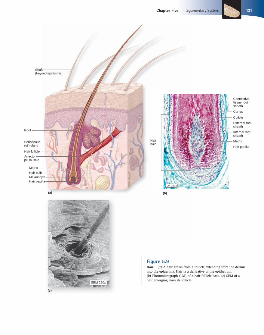
There are three recognizable zones along the length of a hair (figure 5.9a):
1. The hair bulb consists of epithelial cells and is a swelling at the base where the hair originates in the dermis. The epithelium at the base of the bulb surrounds a small hair papilla, which is composed of a small amount of connective tissue containing tiny blood vessels and nerves.
2. The root is the hair within the follicle internal to the skin surface.
3. The shaft is that portion of the hair that extends beyond the skin surface.
The root and shaft consist of dead epithelial cells, while the hair bulb contains living epithelial cells. Thus, it doesn’t hurt to get a haircut because the hairstylist is cutting dead cells. In constrast, it hurts to pull a hair out by its root, because you are disturbing the live portion of the hair. Hair production involves a specialized type of keratiniza-tion that occurs in the hair matrix. Basal epithelial cells near the center of the hair matrix divide, producing daughter cells that are gradually pushed toward the surface. The medulla, not found in all hair types, is a remnant of the soft core of the matrix. It is composed of loosely arranged cells and air spaces, and contains flexible, soft keratin. Several layers of flattened cells closer to the outer surface of the developing hair form the relatively hard cor-tex. Hair stiffness results from the hardness of the intercellular cements (extracellular matrix) around the cells of the cortex and intracellular matrix around keratin fibers. When the matrix in each region is hard, the hair is stiff; when the matrices are more fluid and softer, the hair loses its body and becomes more pliable and, sometimes, limp. Multiple cell layers around the cortex form the cuticle (ku ti-kl), which coats the hair. The free edges of cuticle cells are directed externally. The hair root extends from the hair bulb to the region where the hair shaft is completely mature. The entire hair root lies
Figure 5.8Structure of a Fingernail. Nails, the hard derivative of the stratum corneum, protect sensitive fingertips. (a) Surface view of a fingernail. (b) Sagittal section showing the internal details of a fingernail.
mck78097_ch05_118-145.indd 130mck78097_ch05_118-145.indd 130 2/11/11 3:03 PM2/11/11 3:03 PM
LM 100x
Hair bulb
Matrix
Melanocyte
Sebaceous (oil) gland
Hair papilla
Shaft(beyond epidermis)
Root
Hair follicle
Arrectorpili muscle
(a)
(c)
(b)
SEM 260x
Connective tissue root sheath
External root sheath
Internal root sheath
Cuticle
Cortex
Hair papilla
MatrixHairbulb
Chapter Five Integumentary System 131
Figure 5.9Hair. (a) A hair grows from a follicle extending from the dermis into the epidermis. Hair is a derivative of the epithelium. (b) Photomicrograph (LM) of a hair follicle base. (c) SEM of a hair emerging from its follicle.
mck78097_ch05_118-145.indd 131mck78097_ch05_118-145.indd 131 2/14/11 4:22 PM2/14/11 4:22 PM

132 Chapter Five Integumentary System
internal to the skin, and the hair shaft extends from the hair root to the exposed tip the hair. The hair shaft’s size, shape, and color can be highly variable. The hair follicle (fol i-kl; folliculus = a small sac) is an oblique tube that surrounds the root hair. The follicle always extends into the dermis and sometimes into the subcutaneous layer. The cells of the follicle walls are organized into two prin-cipal concentric layers: an outer connective tissue root sheath, which originates from the dermis, and an inner epithelial tissue root sheath, which originates from the epidermis (figure 5.9b). The epithelial tissue root sheath is composed of two parts: an internal root sheath and an external root sheath. The internal root sheath is produced by peripheral cells of the matrix. It surrounds both the hair and the deep part of the shaft. This layer does not extend the entire length of the follicle because its cells are quickly destroyed. The external root sheath extends between the skin sur-face and the hair matrix. In general, it contains the same epidermal cell layers as the skin surface. However, all of the cells resemble those of the stratum basale where this sheath joins the hair matrix. Extending from the dermal papillae to the mid-region of the hair follicles are thin ribbons of smooth muscle that are col-lectively called the arrector pili (a -rek to r pı lı ; rectus = to raise up, pilus = hair) muscles. The arrector pili muscles are usually stimulated in response to an emotional state, such as fear or rage, or exposure to cold temperatures. Upon stimulation, the arrector pili muscles contract, pulling on the follicles and elevating the hairs, to produce “goose bumps.”
Functions of Hair
The millions of hairs on the surface of the human body have important functions, including:
■ Protection. The hair on the head protects the scalp from sunburn and injury. Hairs within the nostrils protect the respiratory system by preventing inhalation of large foreign particles, while those within the external ear canal protect the ear from insects and foreign particles. Eyebrows and eyelashes protect the eyes.
■ Heat retention. Hair on the head prevents the loss of conducted heat from the scalp to the surrounding air. Individuals who have lost their scalp hair lose much more heat than those who have a full head of hair. The scalp is the only place on the body where the hair is thick enough to retain heat.
■ Facial expression. The hairs of the eyebrows function primarily to enhance facial expression.
■ Sensory reception. Hairs have associated touch receptors (hair root plexuses) that detect light touch.
■ Visual identification. Hair characteristics are important in determining species, age, and sex, and in identifying individuals.
■ Chemical signal dispersal. Hairs help disperse pheromones, which are chemical signals involved in attracting members of the opposite sex and in sex recognition. After pheromones are secreted by selected sweat glands, such as those in the axillary and pubic regions, they are released onto the hairs in these areas.
Hair Color
Hair color is a result of the synthesis of melanin in the matrix adjacent to the papillae. Variations in hair color reflect geneti-cally determined differences in the structure of the melanin.
Additionally, environmental and hormonal factors may influence the color of the hair. As people age, pigment production decreases, and thus hair becomes lighter in color. Gray hair results from the gradual reduction of melanin production within the hair follicle, while white hair signifies the lack of pigment entirely. Usually, hair color changes gradually.
Hair Growth Cycle
There are three stages of the hair growth cycle: anagen, catagen, and telogen:
1. The anagen phase is the active phase of growth where living cells of the hair bulb are rapidly growing, dividing, and transforming into hair. It is the longest part of the growth cycle and lasts from about 18 months to as much as 7 years, depending on the genetics of the person (e.g., people who can grow long hair have the longest anagen phase of the cycle). On the scalp, during the anagen phase, each hair strand grows about one-third of a millimeter per day, or 0.5 to 1.0 centimeter per month. On a normal scalp, 80–95% of follicles are in anagen phase.
2. The catagen phase is a brief regression period where cell division ceases. The follicle undergoes involution and shrinks toward the scalp surface. This very short phase lasts for about 3 to 4 weeks.
3. The telogen phase is the resting phase and is usually the phase where the hair is shed (these hairs are the ones we find in our comb or brush). After 3 to 4 months in the telogen phase, the cells of the hair bulb are stimulated (stimulus unknown) to start regrowing, and the follicle reenters the anagen phase.
The hair growth rate and the duration of the hair growth cycle vary; however, the scalp normally loses between 10 and 100 hairs per day. Thus, this amount of hair loss shouldn't cause noticeable thinning of the scalp hair. Continuous losses that exceed 100 hairs per day often indicate a health problem. Sometimes hair loss may be temporary as a result of one or more of the following factors: exposure to drugs, dietary fac-tors, radiation, high fever, or stress. Thinning of the hair, called alopecia (al-o -pe she -a ; alopekia = a disease like fox mange), can occur in both sexes, usually as a result of aging. In diffuse hair loss, a condition that is both dramatic and distressing, hair is shed from all parts of the scalp. Women primarily suffer from this condition, which may be due to hormones, drugs, or iron deficiency. In males, the condition called male pattern baldness causes loss of hair first from only the crown region of the scalp rather than uniformly. It is caused by a combination of genetic and hormonal influences. At puberty, the testes begin secreting large quantities of male sex hormones (primarily testosterone). As one effect of sex hormone production, males develop a typical pattern of underarm hair, facial hair, and chest hair. The relevant gene for male pattern baldness has two alleles, one for uniform hair growth and one for baldness. The baldness allele is dominant in males and is expressed only in the presence of a high level of testosterone. In men who are either heterozygous or homozygous for the baldness allele, testoster-one causes the terminal hair of the scalp to be replaced by thinner vellus, beginning on the top of the head and later at the sides. In females, the baldness allele is recessive. This is a sex-influenced trait, in which an allele is dominant in one sex (males) and reces-sive in the other (females). Changes in the level of the sex hormones
mck78097_ch05_118-145.indd 132mck78097_ch05_118-145.indd 132 2/11/11 3:03 PM2/11/11 3:03 PM

Apocrinesweat glandduct
Sebaceousglands
Hair follicle
Merocrinesweat glandduct
(a)
Merocrine sweat gland
Sweatgland duct
Sebaceousgland
Apocrine sweat gland
Arrector pilimuscle
Sweatpore
Hair follicle
(b) Merocrine sweat glands
(d) Sebaceous glands
(c) Apocrine sweat glands
LM 100x
LM 100x
LM 40x
Chapter Five Integumentary System 133
circulating in the blood can affect hair development on the scalp, causing a shift from terminal hair to vellus production.
5.5c Exocrine Glands of the Skin
The skin houses two types of exocrine glands: sweat (sudo-riferous) glands and sebaceous glands (figure 5.10a). Sweat glands produce a watery solution that performs several specific functions. Sebaceous glands produce an oily material that coats hair shafts and the epidermal surface (see chapter 4). Table 5.3compares the types of glands found in the skin.
Sweat Glands
The two types of sweat glands in the skin are merocrine sweat glands and apocrine sweat glands. These sweat glands have a coiled, tubular secretory portion located either in the reticular layer of the dermis, or in the subcutaneous layer. A sweat gland ductcarries the secretion to the surface of the epidermis (in a merocrine gland) or into a hair follicle (in an apocrine gland). The opening of the sweat gland duct on the epidermal surface is an indented region called a sweat pore.
Both types of sweat glands contain myoepithelial cells. These specialized cells are sandwiched between the secretory gland cells and the underlying basement membrane. In response to sympathetic nervous system stimulation, myoepithelial cells con-tract to squeeze the gland, causing it to discharge its accumulated secretions into the duct.
WHAT DO YOU THINK?
●4 The sympathetic nervous system is the part of the nervous system that can be activated when we are frightened or nervous. What would you expect to happen to sweat gland production and secretion when we are experiencing these emotions?
Merocrine Sweat Glands Merocrine sweat glands are simple, coiled, tubular glands that release their secretion onto the surface of the skin. They are the most numerous and widely distributed sweat glands in the body. The adult integument contains between 3 and 4 million merocrine sweat glands. The palms of the hands, the soles of the feet, and the forehead have the highest numbers of these glands; some estimates suggest that the palm of each
Figure 5.10 Exocrine Glands of the Skin. (a) The integument contains sweat glands and sebaceous glands. (b) Merocrine sweat glands have a duct with a narrow lumen that opens onto the skin surface through a pore. (c) Apocrine sweat glands exhibit a duct with a large lumen to convey secretion products into a hair follicle. (d) The cells of sebaceous glands are destroyed during the release of their oily secretion into the follicle.
mck78097_ch05_118-145.indd 133mck78097_ch05_118-145.indd 133 2/11/11 3:03 PM2/11/11 3:03 PM
134 Chapter Five Integumentary System
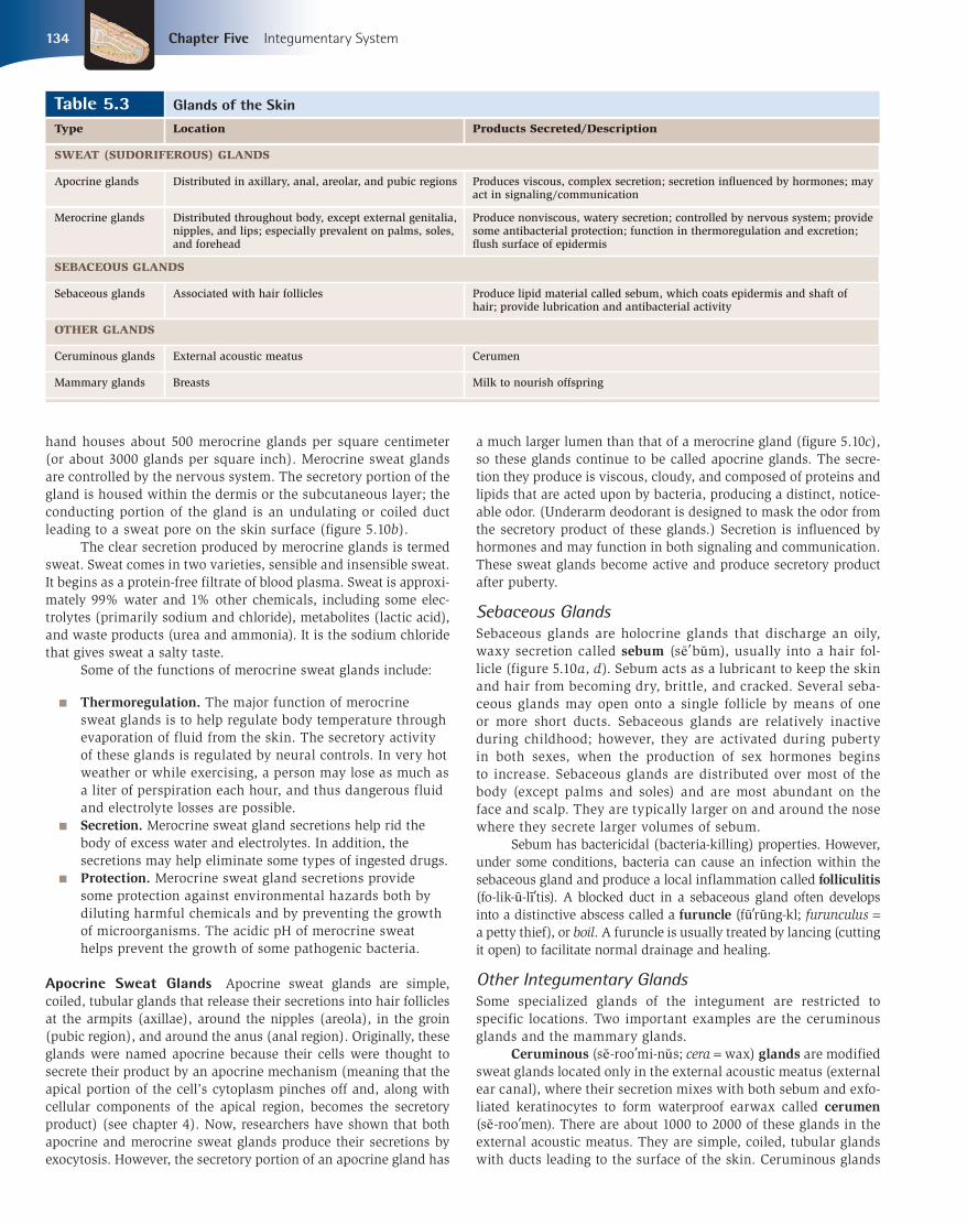
Table 5.3 Glands of the Skin
Type Location Products Secreted/Description
SWEAT (SUDORIFEROUS) GLANDS
Apocrine glands Distributed in axillary, anal, areolar, and pubic regions Produces viscous, complex secretion; secretion infl uenced by hormones; may act in signaling/communication
Merocrine glands Distributed throughout body, except external genitalia, nipples, and lips; especially prevalent on palms, soles, and forehead
Produce nonviscous, watery secretion; controlled by nervous system; provide some antibacterial protection; function in thermoregulation and excretion; fl ush surface of epidermis
SEBACEOUS GLANDS
Sebaceous glands Associated with hair follicles Produce lipid material called sebum, which coats epidermis and shaft of hair; provide lubrication and antibacterial activity
OTHER GLANDS
Ceruminous glands External acoustic meatus Cerumen
Mammary glands Breasts Milk to nourish offspring
hand houses about 500 merocrine glands per square centimeter (or about 3000 glands per square inch). Merocrine sweat glands are controlled by the nervous system. The secretory portion of the gland is housed within the dermis or the subcutaneous layer; the conducting portion of the gland is an undulating or coiled duct leading to a sweat pore on the skin surface (figure 5.10b). The clear secretion produced by merocrine glands is termed sweat. Sweat comes in two varieties, sensible and insensible sweat. It begins as a protein-free filtrate of blood plasma. Sweat is approxi-mately 99% water and 1% other chemicals, including some elec-trolytes (primarily sodium and chloride), metabolites (lactic acid), and waste products (urea and ammonia). It is the sodium chloride that gives sweat a salty taste. Some of the functions of merocrine sweat glands include:
■ Thermoregulation. The major function of merocrine sweat glands is to help regulate body temperature through evaporation of fluid from the skin. The secretory activity of these glands is regulated by neural controls. In very hot weather or while exercising, a person may lose as much as a liter of perspiration each hour, and thus dangerous fluid and electrolyte losses are possible.
■ Secretion. Merocrine sweat gland secretions help rid the body of excess water and electrolytes. In addition, the secretions may help eliminate some types of ingested drugs.
■ Protection. Merocrine sweat gland secretions provide some protection against environmental hazards both by diluting harmful chemicals and by preventing the growth of microorganisms. The acidic pH of merocrine sweat helps prevent the growth of some pathogenic bacteria.
Apocrine Sweat Glands Apocrine sweat glands are simple, coiled, tubular glands that release their secretions into hair follicles at the armpits (axillae), around the nipples (areola), in the groin (pubic region), and around the anus (anal region). Originally, these glands were named apocrine because their cells were thought to secrete their product by an apocrine mechanism (meaning that the apical portion of the cell’s cytoplasm pinches off and, along with cellular components of the apical region, becomes the secretory product) (see chapter 4). Now, researchers have shown that both apocrine and merocrine sweat glands produce their secretions by exocytosis. However, the secretory portion of an apocrine gland has
a much larger lumen than that of a merocrine gland ( figure 5.10c), so these glands continue to be called apocrine glands. The secre-tion they produce is viscous, cloudy, and composed of proteins and lipids that are acted upon by bacteria, producing a distinct, notice-able odor. (Underarm deodorant is designed to mask the odor from the secretory product of these glands.) Secretion is influenced by hormones and may function in both signaling and communication. These sweat glands become active and produce secretory product after puberty.
Sebaceous Glands
Sebaceous glands are holocrine glands that discharge an oily, waxy secretion called sebum (se bu m), usually into a hair fol-licle (figure 5.10a, d). Sebum acts as a lubricant to keep the skin and hair from becoming dry, brittle, and cracked. Several seba-ceous glands may open onto a single follicle by means of one or more short ducts. Sebaceous glands are relatively inactive during childhood; however, they are activated during puberty in both sexes, when the production of sex hormones begins to increase. Sebaceous glands are distributed over most of the body (except palms and soles) and are most abundant on the face and scalp. They are typically larger on and around the nose where they secrete larger volumes of sebum. Sebum has bactericidal (bacteria-killing) properties. However, under some conditions, bacteria can cause an infection within the sebaceous gland and produce a local inflammation called folliculitis(fo-lik-u -lı tis). A blocked duct in a sebaceous gland often develops into a distinctive abscess called a furuncle (fu ru ng-kl; furunculus = a petty thief), or boil. A furuncle is usually treated by lancing (cutting it open) to facilitate normal drainage and healing.
Other Integumentary Glands
Some specialized glands of the integument are restricted to specific locations. Two important examples are the ceruminous glands and the mammary glands. Ceruminous (se -roo mi-nu s; cera = wax) glands are modified sweat glands located only in the external acoustic meatus (external ear canal), where their secretion mixes with both sebum and exfo-liated keratinocytes to form waterproof earwax called cerumen (se -roo men). There are about 1000 to 2000 of these glands in the external acoustic meatus. They are simple, coiled, tubular glands with ducts leading to the surface of the skin. Ceruminous glands
mck78097_ch05_118-145.indd 134mck78097_ch05_118-145.indd 134 2/11/11 3:03 PM2/11/11 3:03 PM
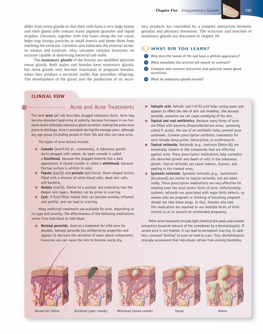
Blackhead (open comedo)Normal hair follicle Whitehead (closed comedo) Papule Nodule
Chapter Five Integumentary System 135
differ from sweat glands in that their coils have a very large lumen and their gland cells contain many pigment granules and liquid droplets. Cerumen, together with tiny hairs along the ear canal, helps trap foreign particles or small insects and keeps them from reaching the eardrum. Cerumen also lubricates the external acous-tic meatus and eardrum. Also, cerumen contains lysozyme, an enzyme capable of destroying bacterial cell walls. The mammary glands of the breasts are modified apocrine sweat glands. Both males and females have mammary glands, but these glands only become functional in pregnant females, when they produce a secretion (milk) that nourishes offspring. The development of the gland and the production of its secre-
tory products are controlled by a complex interaction between gonadal and pituitary hormones. The structure and function of mammary glands are discussed in chapter 28.
WHAT DID YOU LEARN?
●11 Why does the lunula of the nail have a whitish appearance?
●12 What stimulates the arrector pili muscle to contract?
●13 Compare and contrast merocrine and apocrine sweat gland secretions.
●14 What do sebaceous glands secrete?
WW
Acne and Acne TreatmentsThe term acne (ak -ne) describes plugged sebaceous ducts. Acne may become abundant beginning at puberty, because increases in sex hor-mone levels stimulate sebaceous gland secretion, making the pores more prone to blockage. Acne is prevalent during the teenage years, although any age group (including people in their 30s and 40s) can have acne.
The types of acne lesions include:
■ Comedo (kom e-do; pl., comedones). A sebaceous gland’s ducts plugged with sebum. An open comedo is called a blackhead, because the plugged material has a dark appearance. A closed comedo is called a whitehead, because the top surface is whitish in color.
■ Papule (pap ul) and pustule (pus chool). Dome-shaped lesions filled with a mixture of white blood cells, dead skin cells, and bacteria.
■ Nodule (nod ul). Similar to a pustule, but extending into the deeper skin layers. Nodules can be prone to scarring.
■ Cyst. A fluid-filled nodule that can become severely inflamed and painful, and can lead to scarring.
Many medicinal treatments are available for acne, depending on its type and severity. The effectiveness of the following medications varies from individual to individual:
■ Benzoyl peroxide. Used as a treatment for mild acne for decades, benzoyl peroxide has antibacterial properties and appears to decrease the secretion of some sebum components. Excessive use can cause the skin to become overly dry.
■ Salicylic acid. Salicylic (sal-i-sil ik) acid helps unclog pores and appears to affect the rate of skin cell shedding. Like benzoyl peroxide, excessive use can cause overdrying of the skin.
■ Topical and oral antibiotics. Because many forms of acne are filled with bacteria (Propionibacterium acnes, commonly called P. acnes ), the use of an antibiotic helps prevent acne outbreaks. Common prescription antibiotic treatments for acne include doxycycline, tetracycline, or erythromycin.
■ Topical retinoids. Retinoids (e.g., tretinoin [Retin-A]) are essentially vitamin A–like compounds that are effective against acne. These prescription medications help normalize the abnormal growth and death of cells in the sebaceous glands. Topical retinoids can cause redness, dryness, and peeling in the treated areas.
■ Systemic retinoids. Systemic retinoids (e.g., isotretinoin [Accutane]) are similar to topical retinoids, but are taken orally. These prescription medications are very effective for treating even the most severe forms of acne. Unfortunately, systemic retinoids are associated with major birth defects, so women who are pregnant or thinking of becoming pregnant should not take these drugs. In fact, females who take this medication are required to use multiple forms of birth control so as to prevent an unintended pregnancy.
Other acne treatments include light chemical skin peels and comedo extraction (surgical removal of the comedones by a dermatologist). If severe acne is not treated, it can lead to permanent scarring. In addi-tion, constant “picking” at acne can lead to scars. Thus, dermatologists strongly recommend that individuals refrain from picking blemishes.
CLINICAL VIEW
mck78097_ch05_118-145.indd 135mck78097_ch05_118-145.indd 135 2/11/11 3:03 PM2/11/11 3:03 PM

Epidermisof skin
Wound
Dermisof skin
Fibroblast
Scar tissue(fibrosis)
Regeneratedstratum basale(epidermis)
Granulation tissue
Macrophages
Fibroblast
Regrowth ofblood vessel
Blood clot
Macrophages
Fibroblast
Neutrophils
Scab
Cut blood vessels bleed into the wound.1 Blood clot forms, and leukocytes clean wound.2
Blood vessels regrow, and granulation tissue forms.3 Stratum basale regenerates, and connective tissue fibrosis occurs.4
Blood clot
Leukocyte
136 Chapter Five Integumentary System
Figure 5.11Stages in Wound Healing. Cut blood vessels in tissue initiate the multistep process of wound healing.
5.6 Integument Repair and RegenerationLearning Objectives: 1. Identify how burns affect the integument. 2. Describe the treatment of burns.
The components of the integumentary system exhibit a tremendous ability to respond to stresses, trauma, and damage. Repetitive mechanical stresses applied to the integument stimulate mitotic activity in the stem cells of the stratum basale, resulting in thickening of the epidermis and improved ability to withstand stress. For example, walking about without shoes causes the soles of the feet to thicken, thus providing more protection for the underlying tissues.
Damaged tissues are normally repaired in one of two ways. Regeneration replaces damaged or dead cells with the same cell type and restores organ function. When regeneration is not possible because part of the organ is too severely damaged or its cells lack the capacity to divide, the body fills in the gap with scar tissue. This process, known as fibrosis, effectively binds the “broken” parts back together. The replacement scar tissue is produced by fibro-blasts and composed primarily of collagen fibers. Although fibrosis restores some structure it does not restore function. Fibrosis is the repair response in tissues subjected to severe injuries or burns. Both regeneration and fibrosis may occur in the healing of damaged skin. Figure 5.11 illustrates the following stages in wound healing:
1. Cut blood vessels initiate bleeding into the wound. The blood brings clotting proteins, platelets, numerous white
mck78097_ch05_118-145.indd 136mck78097_ch05_118-145.indd 136 2/11/11 3:03 PM2/11/11 3:03 PM

In Depth
(a) First-degree burn (b) Second-degree burn (c) Third-degree burn
Chapter Five Integumentary System 137
blood cells, and antibodies to the site. The clotting proteins and platelets stop the bleeding, while the white blood cells and antibodies clean the wound and fight any infection that may have been introduced.
2. A blood clot forms, temporarily patching the edges of the wound together and acting as a barrier to prevent the entry of pathogens into the body. Internal to the clot, macrophages and neutrophils (two types of leukocytes) clean the wound of cellular debris.
3. The cut blood vessels regenerate and grow in the wound. A soft mass deep in the wound becomes granulation (gran u-la shun) tissue, a vascular connective tissue that initially forms in a healing wound. Macrophages within the wound begin to remove the clotted blood. Fibroblasts produce new collagen in the region.
4. Regeneration of the epidermis occurs due to division of stratum basale cells at the edge of the wound. These new
epithelial cells migrate over the wound, creeping internally to the now superficial remains of the clot (the scab). The connective tissue is replaced by fibrosis.
The skin repair and regeneration process is not rapid. The wider and deeper the surface affected, the longer it takes for skin to be repaired. Additionally, the area under repair is usually more susceptible to complications due to fluid loss and infection. As the severity of damage increases, the repair and regeneration ability of the skin is strained, and a return to its original condition becomes much less likely. Some integumentary system components are not repaired following damage; these include hair follicles, exocrine glands, nerve cells, and muscle fibers.
WHAT DID YOU LEARN?
●15 What is the source of new epidermal cells and new dermal cells in the repair of the integument?
WW
CLINICAL VIEW:
BurnsBurns are a major cause of accidental death, primarily as a result of their effects on the skin. They are usually caused by heat, radiation, harmful chemicals, sunlight, or electrical shock. The immediate threat to life results primarily from fluid loss, infection, and the effects of burned, dead tissue. Burns are classified based on the depth of tissue involvement. First- and second-degree burns are called partial-thickness burns; third-degree burns are called full-thickness burns.
First-degree burns involve only the epidermis and are characterized by redness, pain, and slight edema (swelling) (figure a). An example is a mild sunburn. Treatment involves immersing the burned area in cool water or applying cool, wet compresses; this is sometimes followed by covering the burn with sterile, nonadhesive bandages. The healing time averages about 3 to 5 days, and typically no scarring results.
Second-degree burns involve the epidermis and part of the dermis. The skin appears red, tan, or white and is blistered and painful (figure b). Examples include very severe sunburns (characterized by blisters) or scalding from hot liquids or chemicals. Treatment is similar to that for first-degree burns. Care must be taken not to break the blisters because breaking would increase the risk of infection. In addition, ointments should not be applied to the blisters because ointments could cause heat to be retained in the burned area. Elevating burned limbs is recom-mended to prevent swelling. Healing takes approximately 2 to 4 weeks, and slight scarring may occur.
Third-degree burns involve the epidermis, dermis, and subcutaneous layer, which often are destroyed (figure c). Third-degree burns are usu-ally caused by contact with corrosive chemicals or fire, or by prolonged contact with extremely hot water. Dehydration is a major concern with a third-degree burn, because the entire portion of skin has been lost, and water cannot be retained in the area. Third-degree burn victims must be aggressively treated for dehydration, or they may die. In addition, patients are typically given antibiotics because the risk of infection is very great. Treatment may vary slightly, depending upon what caused the burn. Most third-degree burns require hospitalization.
With third-degree burns, regeneration of the integument may occur from the edge only, due to the absence of dermis. Skin grafting is usually needed for patients with third-degree burns, since the entire dermis and its vasculature are destroyed and regeneration is limited. A skin graft is a piece of skin transplanted from one part of the body to another to cover a destroyed area. Skin grafts help prevent infec-tion and dehydration in the affected area, and they also help minimize abnormal connective tissue fibrosis and disfigurement.
The first step in determining the severity of burns involves a careful assessment of the body surface area (TBSA). Physicians calculate the body surface area that has been burned by using the rule of nines. For example, head and neck = 9%, each arm = 9%, anterior thorax = 18%, posterior thorax = 18%, each leg = 18%, and the perineum = 1%. The amount of surface area involved affects the treatment plan.
mck78097_ch05_118-145.indd 137mck78097_ch05_118-145.indd 137 2/11/11 3:03 PM2/11/11 3:03 PM
138 Chapter Five Integumentary System
5.7 Aging of the IntegumentLearning Objectives: 1. Describe the changes that occur in the skin during aging. 2. Identify the warning signs and characteristics of skin
cancer.
Although some people develop acne when they enter puberty, most skin problems do not become obvious until an individual reaches middle age. Eventually, all components of the integumentary system are affected by age in the following ways:
■ As an individual ages, the skin repair processes take longer to complete because of the reduced number and activity of stem cells. Skin repair and regeneration that take 3 weeks in a healthy young person often take twice that time for a person in his or her 70s. Additionally, the reduced stem cell activity in the epidermis results in thinner skin that is less likely to protect against mechanical trauma.
■ Collagen fibers in the dermis decrease in number and organization, and elastic fibers lose their elasticity. Also years of making particular facial expressions (e.g., squinting, smiling) produce crease lines in the integument. As a result, the skin forms wrinkles and becomes less resilient.
CLINICAL VIEW
Treatments for Aging Skin As a result of the aging process and exposure to UV radiation, skin begins to lose its resiliency and elasticity due to excessive cross-linking of collagen molecules in the dermis. Combined with years of repeated facial movements (e.g., frowning, squinting, and smiling), this contributes to wrinkle formation that accompanies skin aging. There are several techniques used to rid the skin of fine lines and even deep wrinkles. These techniques vary from noninvasive treatments (facial massage and microdermabrasian, glycolic peel , cosmetics) to cosmetic surgery. The most common medical procedures are laser resurfacing, fat injections, collagen injections, hyaluronic acid injections, and facial implants.
LASER RESURFACING This procedure uses a focused laser (acronym for light amplification by stimulated emission of radiation) to send out short impulses (each about a millisecond) of high-energy light into the epidermis and dermis. In the skin, water and other substances absorb the light energy. As the light energy is absorbed, heat is generated within the tissues. The heat energy vaporizes thin sections of the skin tissues.
Laser resurfacing is usually very precise and causes little damage to the surrounding skin and tissues. The procedure is most effective for improving skin texture and appearance, including wrinkles around the eyes and mouth caused by aging and long-term sun exposure. It is also effective on mild and moderate acne scars but is not effective on severe scarring. Tattoos can also be obliterated using laser treatments. These treatments can produce improvements to deep wrinkles caused by the repeated movements of muscles of the forehead or along the sides of the nose, but these wrinkles usually reappear after months or years. There are few side effects of laser resurfacing treatments. The most common are redness, swelling, crusting, itching, and tenderness. People with dark skin can experience pigment changes (lightening of the skin).
FAT INJECTIONS In this procedure, also known as fat transfer, fat is liposuctioned from the abdominal area of the patient's body and then the fat is purified. Using fine needles, the fat is injected into the dermis and subcutane-ous layer under the wrinkle or area to be augmented. This procedure is primarily used to add fullness to the face (e.g., baggy skin under the eyes, lines around the mouth), lips, hands, and the buttocks. When injecting fat into the dermis and subcutaneous layer of the face, the fullness achieved will often provide some lifting and smoothing to the facial structure. Most of the injected fat cells survive in their new place permanently; some cells are lost in the procedure so doctors will
usually inject a slightly greater volume than necessary knowing that after a few weeks the final result will be more to the patient's liking.
COLLAGEN INJECTIONS This procedure and its results are similar to fat injections. Collagen injections are used to correct imperfections of the face by smoothing skin, plumping lips, and filling wrinkles and scars. With collagen injections, rather than using a person’s own body as the source, most often the collagen is harvested from the skin of cattle (in some cases, the collagen is collected from human skin and grown in tissue culture media). Bovine collagen can cause severe allergic reactions in people with rheumatoid arthritis, scleroderma, systemic or discoid lupus ery-thematosus, and other connective tissue autoimmune diseases. Prior to the treatment, multiple skin tests with the collagen to be used are often required for those individuals who have had any of these diseases. Collagen injections are not permanent. It usually takes a few to several months for the collagen to be absorbed by the body.
HYALURONIC (HA) INJECTIONS Hyaluronic (HA) injections are popular alternatives to collagen injec-tions. They perform similar to collagen, but HA injections have fewer side effects. HA is a natural constituent of the dermis and subcutaneous layer. The HA used in the injections is produced by bacterial fermenta-tion and, after production, is stabilized into a clear gel form (the HA in the gel is similar but not identical to HA in human skin). The gel is injected into the dermis or subcutaneous layer beneath wrinkles, which adds volume and lift, resulting in smoother wrinkles and less prominent folds. In addition to treating wrinkles, HA injections also work well for lip augmentation and/or enhancement and facial contour-ing of the cheeks and mouth. HA injections are not permanent—thus their effects last about 3 to 6 months.
FACIAL IMPLANTS Facial implants are usually solid, sculpted materials that are inserted through small incisions in the lip, mouth, or chin. Solid silicone is the most common implant material, but there are other implants made out of Teflon or related materials. Sometimes natural substances, such as bone, can also be carved and reshaped to create implants that can be used to change the shape of the facial skeleton. Implants are designed to enhance the physical structure of the face. The implants commonly used are chin implants to increase the size and/or shape of the chin in people who have a small or recessed chin; jaw implants to increase the width of the lower third of the face in people with a "weak" or sloping jaw; and cheek implants to increase the projection and add volume to the areas around the cheekbones in people with recessed or flat regions. Because they will stretch the skin, strategically placed implants can hide many wrinkles in the face.
mck78097_ch05_118-145.indd 138mck78097_ch05_118-145.indd 138 2/11/11 3:03 PM2/11/11 3:03 PM

Chapter Five Integumentary System 139
Table 5.4 Skin Cancer
BASAL CELL CARCINOMA
■ Most common type of skin cancer ■ Least dangerous type because it seldom metastasizes ■ Originates in stratum basale ■ First appears as small, shiny elevation that enlarges and develops central depression with pearly edge ■ Usually occurs on face ■ Treated by surgical removal of lesion
SQUAMOUS CELL CARCINOMA
■ Arises from keratinocytes of stratum spinosum ■ Lesions usually appear on scalp, ears, lower lip, or back of hand ■ Early lesions are raised, reddened, scaly; later lesions form concave ulcers with elevated edges ■ Treated by early detection and surgical removal of lesion ■ May metastasize to other parts of the body
MALIGNANT MELANOMA
■ Most deadly type of skin cancer due to aggressive growth and metastasis ■ Arises from melanocytes, usually in a preexisting mole ■ Individuals at increased risk include those who have had severe sunburns, especially as children. ■ Characterized by change in mole diameter, color, shape of border, and symmetry ■ Survival rates improved by early detection and surgical removal of lesion. ■ Advanced cases (metastasis of disease) are difficult to cure and are treated with chemotherapy, interferon
therapy, and radiation therapy.
The usual signs of melanoma may be easily remembered using the ABCD rule. Report any of the following changes in a birthmark or mole to your physician:A = Asymmetry: One-half of a mole or birthmark does not match the other.B = Border: Edges are notched, irregular, blurred, or ragged.C = Color: Color is not uniform; differing shades (usually brown or black and sometimes patches of white, blue, or
red) may be observed.D = Diameter: Affected area is larger than 6 mm (about 1/4 inch) or is growing larger.
■ The skin’s immune responsiveness is diminished by a decrease in the number and efficiency of epidermal dendritic cells. This decreased immune response may be related to the appearance of longer dendritic processes and the decrease in molecules on their surface that recognize pathogens.
■ Skin becomes drier and sometimes scaly because decreased sebaceous gland activity diminishes the amounts of natural skin lubricants.
■ A decrease in melanocytes causes altered skin pigmentation. As a result, hair becomes gray or white, and sensitivity to sun exposure increases. Often, exposure to the sun or other forms of UV light leads to an increase in skin pigmentation in certain body areas. The resulting flat, brown or black spots are called liver spots, although they are unrelated to the liver.
■ Sweat production diminishes as a result of decreased sweat gland activity.
■ The dermal blood vessels lose some permeability as a result of decreased elasticity. Blood supply to the dermis is reduced as the extent of blood vessel distribution decreases. These vascular changes, along with glandular changes associated with aging, lead to impaired thermoregulation.
■ Hair follicles either produce thinner hairs or stop production entirely.
■ Integumentary production of vitamin D3 decreases. If vitamin D3 levels diminish significantly, the body is unable to absorb calcium and phosphorus from the digestive tract. The resulting declines in calcium and phosphorus concentrations affect muscle activity and bone density.
■ Chronic overexposure to UV rays can damage the DNA in epidermal cells and accelerate aging.
5.7a Skin Cancer
Chronic overexposure to UV rays can damage the DNA in epidermal cells and accelerate aging. This overexposure is the predominant factor in the development of nearly all skin cancers. Skin cancer is the most common type of cancer. It occurs most frequently on the head and neck regions, followed by other regions commonly exposed to the sun. Fair-skinned individuals, especially those who experienced severe sunburns as children, are most at risk for skin cancer. However, skin cancer can arise in anyone of any age. Individuals should use sunscreen regularly and avoid prolonged exposure to the sun (see Clinical View earlier in this chapter). An individual should regularly and thoroughly inspect his or her skin for any changes, such as an increase in the number or size of moles or the appearance of new skin lesions. In addition, an examina-tion by a dermatologist should be part of everyone’s routine health checkup. Table 5.4 describes the three main types of skin cancer.
mck78097_ch05_118-145.indd 139mck78097_ch05_118-145.indd 139 2/11/11 3:03 PM2/11/11 3:03 PM

Periderm
Basal layerMesenchyme
Melanoblast
Developingepidermis
Epidermis
Vernix caseosa
Developingdermis
7 – 8 weeks 11 – 12 weeks Birth
Dermis
Melanocyte
Periderm
140 Chapter Five Integumentary System
WHAT DID YOU LEARN?
●16 Why do skin repair processes take longer as we age?
●17 What is the cause of increased sensitivity to the sun as we age?
WHAT DO YOU THINK?
●5 If two people enjoy being in the sun, which one of them would you expect to get more wrinkles—the individual who has used sunscreen all his or her life, or the individual who has never used sunscreen? Why?
5.8 Development of the Integumentary SystemLearning Objectives: 1. Describe the development of the integument from surface
ectoderm and mesoderm. 2. Describe the development of epidermal derivatives.
The structures of the integumentary system are derived from the ectodermal and mesodermal germ layers. The ectoderm is the origin of the epidermis, while the mesoderm gives rise to the dermis.
5.8a Integument Development
By the end of the seventh week of development, the surface ecto-derm is composed of a simple cuboidal epithelium. These epithe-lial cells divide, grow, and form a layer of squamous epithelium that flattens and becomes a covering layer called the peridermand an underlying basal layer (figure 5.12). The basal layer will form the stratum basale and all other epidermal layers. By the eleventh week of development, the cells of the basal layer form an intermediate layer of skin, and by the twenty-first week, the stratum corneum forms. Also by this time, the friction ridges have formed. During the fetal period, the periderm is eventu-ally sloughed off, and these sloughed off cells mix with sebum secreted by the sebaceous glands, producing a waterproof coating called the vernix caseosa. The vernix caseosa protects the skin of the fetus from damage by amniotic fluid. Although keratinocytes are formed from epidermal cells, melanocytes originate from specialized neural crest cells called melanoblasts, which arise from the ectoderm that also forms nervous tissue. Melanoblasts migrate to the future epidermis. Melanoblasts differentiate into melanocytes about 40 to 50 days after fertilization and thereafter begin to produce the pigment melanin.
WW The dermis is derived from mesoderm. During the embry-onic period, this mesoderm becomes mesenchyme, and it occu-pies a zone internal to the ectoderm. At about 11 weeks, the mesenchymal cells begin to form the components of the dermis. The formation of collagen and elastic fibers causes folding at the boundary of the overlying epidermis and dermis, resulting in the formation of dermal papillae. Blood vessels start to form in the dermis, and by the end of the first trimester, the primary vascular pattern in the dermis is present.
5.8b Nail Development
Fingernails and toenails start to form in the tenth week of devel-opment. These nails form from thickened ridges of epithelium called nail fields at the tip of each digit. The nail fields are sur-rounded by folds of epidermis called nail folds. The proximal nail fold grows over the nail field and becomes keratinized, forming the nail plate. The fingernails reach the tips of the fingers by 32 weeks, while the toenails become fully formed by about 36 weeks. Infants born prematurely may not have fully formed fingernails and toenails.
5.8c Hair Development
Hair development is illustrated in figure 5.13a. Hair follicles must be present before hair can form. Hair follicles begin to appear between 9 and 12 weeks of development as pockets of cells called hair buds that invade the dermis from the overlying stratum basale of the epidermis. These buds differentiate into a hair bulb, hair papillae, sebaceous glands, and other structures associated with hair follicles. The hair papilla is formed from dif-ferentiating mesenchymal cells located around the epithelial cells of the hair bulb. Eventually, hair grows due to continuous mitotic activity in the epithelial cells of the hair bulb. These hairs do not become easily recognizable in the fetus until about the twentieth week, when they appear as lanugo. These very fine, soft hairs help hold the vernix caseosa on the skin. They are replaced by vellus hairs after birth and scalp hairs become terminal hairs during early childhood.
5.8d Sebaceous and Sweat Gland Development
Both sweat and sebaceous glands develop from the stratum basale of the epidermis (figure 5.13b). These glands originate from epidermis but start to grow and burrow into the underlying dermis. Sweat glands appear at about 20 weeks on the palms and soles and later in other regions. The secretory portion of these glands coils as the gland develops within the dermis. Sebaceous glands typically develop as epidermal outgrowths from the sides
Figure 5.12Integument Development. Skin structure becomes increasingly complex in the period from 7 weeks to birth.
mck78097_ch05_118-145.indd 140mck78097_ch05_118-145.indd 140 2/11/11 3:03 PM2/11/11 3:03 PM

Developingepidermis
Developingdermis
15 weeks12 weeks
Hair bud Hair bulb
Hair
Sebaceousgland
Hair papilla
Developingsebaceous gland
Differentiating mesenchyme(forms hair papilla)
Birth(a) Hair development
Bud of developingsweat gland
Developingepidermis
Developingdermis
15 weeks12 weeks
Pore
Sweatgland
Developingsweat gland
Birth(b) Gland development
Developingepidermis
Developingdermis
16 weeks 28 weeks6 weeks
Primary mammary bud
Mammary pit
Secondary mammary buds
Lactiferousduct
Lactiferous glands
Areola
Chapter Five Integumentary System 141
Figure 5.13Hair and Gland Development. Comparison of the development of the hair and the glands of the skin at 12 weeks, at 15 weeks, and at birth.
Figure 5.14Mammary Gland Development. Development of the mammary glands at 6 weeks, 16 weeks, and 28 weeks.
of a developing hair follicle. Sebaceous glands start to form sebum during the fetal period. As previously described, this sebum mixes with the cells of the sloughed off periderm to form the vernix caseosa.
5.8e Mammary Gland Development
A primary mammary bud first appears during the sixth week of development as an epidermal growth into the underlying dermal layer (figure 5.14). At about 16 weeks (the fourth month) of devel-opment, each primary mammary bud branches to form secondary mammary buds, which branch and elongate. Later in the fetal
period, these mammary buds develop lumina (internal openings) that eventually form lactiferous ducts (milk ducts). The fat and the connective tissue of the mammary gland are formed from the nearby mesenchyme within the dermis. Late in the fetal period, the mammary gland develops an external epidermal depression called the mammary pit. The mammary pit forms the center around which the nipple tissue will grow. The developing milk ducts open up into this pit. At birth, the mammary glands remain underdeveloped. At puberty, female mammary glands grow and differentiate due to increased levels of female sex hormones.
mck78097_ch05_118-145.indd 141mck78097_ch05_118-145.indd 141 2/11/11 3:03 PM2/11/11 3:03 PM

142 Chapter Five Integumentary System
Chapter Summary
Clinical Terms
athlete’s foot A fungal infection of the skin, especially between the toes; causes itching, redness, and peeling. Also called tinea pedis.
blister A thin-walled, fluid-filled sac either internal to or within the epidermis; caused by a burn or by excessive friction.
cold sore Small, fluid-filled blister that is sensitive and painful to the touch; associated with the lips and the mucosa of the oral cavity; caused by herpes simplex type 1 virus, which infects nerve cells that supply the skin. Also called a fever blister.
dandruff Flaking of the epidermis of the scalp, resulting in white or gray scales in the hair.
eczema (ek ze -ma ) Noncontagious skin inflammation often characterized by itchy, red vesicles that may be scaly or crusty.
hives Eruption of reddish, raised areas on the skin, usually accompanied by extreme itching; causes include certain foods, specific drugs, or stress. Also called urticaria.
impetigo (im-pe-tı go ) A contagious, pus-forming bacterial infection of the skin; fluid-filled vesicles form and then rupture, forming a yellow crust.
keloid (ke loyd) Excess scar tissue caused by collagen formation during healing; often painful and tender.
pruritis (proo-rı tus) Irritating, itching condition of the skin that may be caused by infection or exposure to various irritants, such as chemicals, cleaning solutions, or mites.
psoriasis (so -rı a -sis) Chronic inflammatory condition characterized by lesions with dry, silvery scales, usually on the scalp, elbows, and knees.
rosacea (ro-za -she-uh) Chronic skin disorder that results in redness and small, red, pus-filled bumps or pustules, mainly on the face. The cause of rosacea remains unknown but evidence indicates heredity and environmental factors are to blame.
shingles A skin rash characterized by blisters on red base with burning pain. Caused by the varicella-zoster virus, which causes chickenpox. After someone has had chickenpox, the virus remains dormant in the body, associated with parts of the nervous system. Emotional stress, immune deficiency, or cancer can reactivate the virus to cause shingles (see Clinical View in chapter 16 for an expanded description of shingles).
wart A growth of epidermal cells that forms a roughened projection from the surface of the skin; caused by human papillomavirus.
■ The integumentary system consists of the skin (integument) and its derivatives (nails, hair, sweat glands, and sebaceous glands).
5.1 Structure and Function of the
Integument 119
■ The integument is the body’s largest organ.
5.1a Integument Structure 119
■ The integument contains a superficial, stratified squamous epithelium called the epidermis, and a deeper, dense irregular connective tissue layer called the dermis.
■ Deep to the dermis is the subcutaneous layer, or hypodermis, which is not part of the integumentary system.
5.1b Integument Functions 120
■ The integument’s functions include providing mechanical protection and a physical barrier, protecting against water loss and microbial invasion, regulating temperature, aiding metabolism, contributing to immune defense, perceiving sensations, and excreting wastes through secretion.
5.2 Epidermis 121 ■ Four distinct cell types are found within the epidermis: keratinocytes, melanocytes, tactile cells, and epidermal dendritic cells.
5.2a Epidermal Strata 121
■ The stratum basale is a single cell layer of stem cells adjacent to the basement membrane, separating the epidermis from the dermis.
■ The stratum spinosum contains multiple layers of keratinocytes attached together by desmosomes.
■ The stratum granulosum is composed of three to five layers of keratinocytes. The process of keratinization begins here.
■ The stratum lucidum is a thin, translucent layer of anucleate cells superficial to the stratum granulosum; it occurs only in the thick skin of the palms and soles.
■ The stratum corneum has numerous layers of dead, scaly, interlocking keratinized cells.
5.2b Variations in the Epidermis 122
■ Normal skin color is a result of a combination of hemoglobin in the blood of the dermis and variable quantities of the pigments melanin and carotene.
mck78097_ch05_118-145.indd 142mck78097_ch05_118-145.indd 142 2/11/11 3:03 PM2/11/11 3:03 PM

Chapter Five Integumentary System 143
5.3 Dermis 125 ■ Components of the dermis include blood vessels, sweat glands, sebaceous glands, hair follicles, nail roots, sensory nerve endings, and muscular tissue.
5.3a Papillary Layer of the Dermis 126
■ The papillary layer is composed of areolar connective tissue. Epidermal ridges interdigitate with dermal papillae at the boundary between the epidermis and dermis to interlock these layers and increase the area of contact between them.
5.3b Reticular Layer of the Dermis 126
■ The reticular layer lies deep to the papillary layer; it consists of dense irregular connective tissue.
5.3c Stretch Marks, Wrinkles, and Lines of Cleavage 126
■ Skin stretching due to weight gain or pregnancy causes stretch marks, called striae.
■ Lines of cleavage in the skin indicate the predominant direction of the underlying bundles of collagen fibers.
5.3d Innervation and Blood Supply 127
■ Vasoconstriction of dermal blood vessels causes decreased circulation to the skin and a corresponding conservation of heat in the blood. Vasodilation of dermal blood vessels causes increased circulation to the skin and loss of excess heat.
5.4 Subcutaneous Layer
(Hypodermis) 128
■ The subcutaneous layer consists of areolar connective tissue and adipose connective tissue.
5.5 Epidermal Accessory
Organs 129
■ The nails, hair, and sweat and sebaceous glands are epidermal derivatives that are considered accessory organs of the integument.
5.5a Nails 129
■ Nails are formed from stratum corneum; they protect the exposed distal tips of the fingers and toes.
5.5b Hair 130
■ Hairs project beyond the skin surface almost everywhere except over the palms of the hands, the sides and soles of the feet, the lips, the sides of the fingers and toes, and portions of the external genitalia.
■ Hair functions include protection, thermoregulation, facial expression, sensory reception, visual identification, and dispersal of pheromones.
5.5c Exocrine Glands of the Skin 133
■ Merocrine sweat glands produce a thin, watery secretion called sweat (sensible perspiration).
■ Apocrine sweat glands produce a thick secretion that becomes odorous after exposure to bacteria on the skin surface.
■ Sebaceous glands discharge an oily sebum into hair follicles by holocrine secretion.
■ Ceruminous glands housed within the external ear canal are modified sweat glands; they produce a waxy product called cerumen.
■ Mammary glands are modified apocrine sweat glands that produce milk to nourish a newborn infant.
5.6 Integument Repair
and Regeneration 136
■ The skin can regenerate even after considerable damage, including trauma due to burns.
■ Severe damage to the dermis and accessory structures of the skin cannot be repaired. Often, fibrous scar tissue forms, and a graft is required.
5.7 Aging of the
Integument 138
■ Changes to the skin due to aging include slower regeneration and repair, decreased numbers of collagen fibers and melanocytes, diminished immune responsiveness and sweat production, and increased dryness.
5.7a Skin Cancer 139
■ UV rays from the sun pose the greatest risk for this most common type of cancer.
5.8 Development of the
Integumentary System 140
■ The epidermis is derived from the ectoderm, and the dermis is derived from the mesoderm.
5.8a Integument Development 140
■ Surface ectoderm forms a covering called periderm and an underlying basal layer.
5.8b Nail Development 140
■ Nails form from thickened epithelial ridges called nail fields.
5.8c Hair Development 140
■ Hair follicles form from hair buds that differentiate into hair bulbs, hair papillae, and sebaceous glands.
5.8d Sebaceous and Sweat Gland Development 140
■ Both types of glands originate from the stratum basale of the epidermis.
5.8e Mammary Gland Development 141
■ A primary mammary bud develops as an epidermal outgrowth in the underlying dermis: each bud branches to form secondary mammary buds.
mck78097_ch05_118-145.indd 143mck78097_ch05_118-145.indd 143 2/11/11 3:03 PM2/11/11 3:03 PM
144 Chapter Five Integumentary System
Challenge Yourself
______ 1. integument
______ 2. fingernails
______ 3. keratin
______ 4. tactile cells
______ 5. melanocytes
______ 6. keratinocytes
______ 7. epidermal dendritic cell
______ 8. subcutaneous layer
______ 9. reticular layer
______ 10. arrector pili
a. smooth muscle attached to hair follicle
b. most numerous epidermal cell
c. a phagocytic cell (active in immune response)
d. layer deep to dermis
e. formed from stratum corneum
f. receptors for touch
g. composed of epidermis and dermis
h. dense irregular connective tissue
i. fibrous protein in epidermis
j. pigment-forming cells
MatchingMatch each numbered item with the most closely related lettered item.
Multiple ChoiceSelect the best answer from the four choices provided.
______ 1. “ Strawberry-colored birthmarks” are also called a. cavernous hemangiomas. b. freckles. c. capillary hemangiomas. d. erythema.
______ 2. The layer of the epidermis in which cells begin the process of keratinization is the
a. stratum corneum. b. stratum basale. c. stratum lucidum. d. stratum granulosum.
______ 3. The sweat glands that communicate with skin surfaces only in the axillary, areolar, pubic, and anal regions are
a. apocrine glands. b. merocrine glands. c. sebaceous glands. d. All of these are correct.
______ 4. Which of the following is not a function of the integument?
a. acts as a physical barrier b. stores calcium in the dermis c. regulates temperature through
vasoconstriction and vasodilation of dermal blood vessels
d. participates in immune defense
______ 5. Which of the following layers contains areolar connective tissue and dermal papillae?
a. reticular layer b. subcutaneous layer c. papillary layer d. epidermis
______ 6. Melanin is a. an orange-yellow pigment that strengthens the
epidermis. b. a pigment that accumulates inside keratinocytes. c. a protein fiber found in the dermis. d. a pigment that gives the characteristic color to
hemoglobin.
______ 7. The layer of squamous epithelium that forms by the seventh week of development to give rise to the integument is the
a. mesenchyme. b. periderm. c. basal layer. d. sebaceous layer.
______ 8. The cells in a hair follicle that are responsible for forming hair are the
a. papillary cells. b. matrix cells. c. medullary cells. d. cortex cells.
______ 9. Which epidermal cell type is responsible for detecting touch sensations?
a. keratinocyte b. melanocyte c. tactile cell d. epidermal dendritic cell
______ 10. Water loss due to evaporation of interstitial fluid through the surface of the skin is termed
a. latent perspiration. b. sensible perspiration. c. active perspiration. d. insensible perspiration.
mck78097_ch05_118-145.indd 144mck78097_ch05_118-145.indd 144 2/11/11 3:03 PM2/11/11 3:03 PM
Chapter Five Integumentary System 145
Developing Critical Reasoning 1. When you are outside on a cold day, your skin is much paler
than normal. Later, you enter a warm room, and your face becomes flushed. What are the reasons for these changes in color?
2. Teri is a 14-year-old with a bad case of acne. Explain the probable cause of Teri’s skin condition.
3. As a young man, John spent every summer afternoon at the pool for many years. As he approached the age of 50, his skin was quite wrinkled, and he discovered some suspicious growths on his face. He visited a dermatologist, who removed these growths. What were the growths, and what probably caused them?
Content Review 1. What effect does the protein keratin have on both the
appearance and the function of the integument?
2. Describe two ways in which the skin helps regulate body temperature.
3. List the layers of the epidermis from deep to superficial and compare their structure.
4. Identify and distinguish among the three types of hair produced during a person’s lifetime.
5. List and discuss the three zones along the length of a hair.
6. How do apocrine and merocrine sweat glands differ in structure and function?
7. Describe how the skin is involved in vitamin D production.
8. Briefly discuss the origin and function of sebum.
9. Describe the four steps in wound repair of the integument.
10. What are some effects of aging on the integument?
Answers to “What Do You Think?”
1. The children were not getting enough vitamin D in their diet, and they were spending all of their daylight hours indoors. Since the children weren’t exposed to much sunlight, their skin could not synthesize vitamin D from the UV rays of the sun. Without adequate amounts of vitamin D, the children succumbed to rickets.
2. Thick skin is found on the palms of the hands and the soles of the feet. Secretions from sebaceous glands would make these areas slippery, which would interfere with grasping objects and walking. The presence of hair in these areas would interfere with these same functions.
3. Fingerprints are formed from folds of both epidermal and dermal tissue, so in order to physically “change” or remove
fingerprints, you would have to destroy or damage both layers. This would be very painful, and permanent scarring or malformation would likely result.
4. When we are frightened or nervous, the sympathetic nervous system is stimulated, which in turn stimulates the sweat glands to produce and release sweat. This is why our palms and other body regions become sweaty in nervous or frightening situations.
5. The person who has never used sunscreen is more likely to get wrinkles. Constant, unprotected exposure to UV rays can cause wrinkling and increased aging of the skin, and it also increases the risk of developing skin cancer.
www.mhhe.com/mckinley3 Enhance your study with practice tests and activities to assess your understanding. Your instructor may also recommend the interactive eBook, individualized learning tools, and more.
mck78097_ch05_118-145.indd 145mck78097_ch05_118-145.indd 145 2/11/11 3:03 PM2/11/11 3:03 PM
Copyright © 2022 FDOKUMEN