Effects of oleanolic acid on pulmonary morphofunctional and biochemical variables in experimental...
8
Please cite this article in press as: Santos, R.S., et al., Effects of oleanolic acid on pulmonary morphofunctional and biochemical variables in experimental acute lung injury. Respir. Physiol. Neurobiol. (2011), doi:10.1016/j.resp.2011.07.008 ARTICLE IN PRESS G Model RESPNB-1677; No. of Pages 8 Respiratory Physiology & Neurobiology xxx (2011) xxx–xxx Contents lists available at ScienceDirect Respiratory Physiology & Neurobiology j our nal ho me p age: www.elsevier.com/locate/resphysiol Effects of oleanolic acid on pulmonary morphofunctional and biochemical variables in experimental acute lung injury Raquel S. Santos a , Pedro L. Silva a , Gisele P. Oliveira a , Fernanda F. Cruz a , Débora S. Ornellas a,b , Marcelo M. Morales b , Janaina Fernandes c , Manuella Lanzetti d , Samuel S. Valenc ¸ a d , Paolo Pelosi e , Cerli R. Gattass c , Patricia R.M. Rocco a,∗ a Laboratory of Pulmonary Investigation, Carlos Chagas Filho Institute of Biophysics, Federal University of Rio de Janeiro, Rio de Janeiro, RJ, Brazil b Laboratory of Cellular and Molecular Physiology, Carlos Chagas Filho Institute of Biophysics, Federal University of Rio de Janeiro, Rio de Janeiro, RJ, Brazil c Laboratory of Cellular Immunology, Carlos Chagas Filho Institute of Biophysics, Federal University of Rio de Janeiro, Rio de Janeiro, RJ, Brazil d Laboratory of Inflammation, Oxidative Stress and Cancer, Biomedical Institute of Sciences, Federal University of Rio de Janeiro, Rio de Janeiro, RJ, Brazil e Department of Surgical Sciences and Integrated Diagnostics, University of Genoa, Genoa, Italy a r t i c l e i n f o Article history: Accepted 13 July 2011 Keywords: Dexamethasone Cytokines Chemokines Histopathology Lung mechanics Oxidative stress a b s t r a c t We analysed the effects of oleanolic acid (OA) on lung mechanics and histology and its possible mecha- nisms of action in experimental acute lung injury (ALI). BALB/c mice were randomly divided into Control (saline, ip) and ALI (paraquat, 25 mg/kg, ip) groups. At 1 h, both groups were treated with saline (SAL, 50 l ip), OA (10 mg/kg ip), or dexamethasone (DEXA, 1 mg/kg ip). At 24 h, lung static elastance, viscoelas- tic pressure, and alveolar collapse reduced more after OA compared to DEXA administration. Tumour necrosis factor-, macrophage migration inhibitory factor, interleukin-6, interferon-, and transforming growth factor- mRNA expressions in lung tissue diminished similarly after OA or DEXA. Conversely, only OA avoided reactive oxygen species generation and yielded a significant decrease in nitrite concentra- tion. OA and DEXA restored the reduced glutathione/oxidized glutathione ratio and catalase activity while increasing glutathione peroxidase induced by paraquat. In conclusion, OA improved lung morphofunction by modulating the release of inflammatory mediators and oxidative stress. © 2011 Elsevier B.V. All rights reserved. 1. Introduction Lung inflammation is a hallmark of acute lung injury (ALI) and acute respiratory distress syndrome (ARDS). The response of cells to lung inflammation may lead to oxidant/antioxidant imbalance, with production of nitric oxide and superoxide and release of cytotoxic and pro-inflammatory compounds, including proteolytic enzymes, reactive oxygen species (ROS), reactive nitrogen species (RNS) and additional inflammatory cytokines, resulting in cellular dysfunction (Chabot et al., 1998; Tasaka et al., 2008) and inhibition of certain lung proteins. This oxidative injury perpetuates inflam- mation and damages the alveolar-capillary membrane (Lee et al., 2010). Several pharmacological treatments have been tested to mod- ulate the signalling pathways in order to decrease pulmonary inflammation (Calfee and Matthay, 2007) and restore the oxi- ∗ Corresponding author at: Laboratory of Pulmonary Investigation, Carlos Chagas Filho Institute of Biophysics, Federal University of Rio de Janeiro, Centro de Ciências da Saúde, Avenida Carlos Chagas Filho, s/n, Bloco G-014, Ilha do Fundão, 21941-902 Rio de Janeiro, RJ, Brazil. Tel.: +55 21 2562 6530; fax: +55 21 2280 8193. E-mail address: [email protected] (P.R.M. Rocco). dant/antioxidant balance (Chavko et al., 2009). So far, however, an effective pharmacological therapy for ALI/ARDS has not been identified. Recently, natural products derived from plant extracts and their synthetic derivatives have been used to treat a wide range of respiratory diseases due to their anti-inflammatory and antioxidative properties. In this line, oleanolic acid (OA), a triter- penoid compound present in a great variety of plants and food products (Liu, 2005), modulates the production and activity of pro- inflammatory cytokines and enzymatic antioxidant defence, as well as protects from oxidant stress by activating Nrf2 (Reisman et al., 2009; Takada et al., 2010; Wang et al., 2010). Chemical synthesis of oleanolic acid has provided many useful derivatives that are more potent and specific than natural parent structures (Honda et al., 1997). Reddy et al. demonstrated that intermittent administration of a synthetic triterpenoid compound, CDDO-imidazole (CDDO- Im) (1-[2-cyano-3-,12-dioxooleana-1,9(11)-dien-28-oyl] imida- zole, during exposure to hyperoxia confers protection against the development of ALI in mice (Reddy et al., 2009). However, the effects of oleanolic acid derivatives and triterpene derivatives are not necessarily similar to those of their parent molecules (Honda et al., 1998, 1999). Additionally, even though the biological activity of oleanolic acid is lower than that of its derivatives, it is known to be relatively non-toxic (Liu, 1995, 2005). 1569-9048/$ – see front matter © 2011 Elsevier B.V. All rights reserved. doi:10.1016/j.resp.2011.07.008
-
Upload
independent -
Category
Documents
-
view
0 -
download
0
Transcript of Effects of oleanolic acid on pulmonary morphofunctional and biochemical variables in experimental...
G
R
Ev
RMPa
b
c
d
e
a
AA
KDCCHLO
1
atwce(dom2
ui
FdR
1d
ARTICLE IN PRESS Model
ESPNB-1677; No. of Pages 8
Respiratory Physiology & Neurobiology xxx (2011) xxx– xxx
Contents lists available at ScienceDirect
Respiratory Physiology & Neurobiology
j our nal ho me p age: www.elsev ier .com/ locate / resphys io l
ffects of oleanolic acid on pulmonary morphofunctional and biochemicalariables in experimental acute lung injury
aquel S. Santosa, Pedro L. Silvaa, Gisele P. Oliveiraa, Fernanda F. Cruza, Débora S. Ornellasa,b,arcelo M. Moralesb, Janaina Fernandesc, Manuella Lanzettid, Samuel S. Valenc ad,
aolo Pelosie, Cerli R. Gattassc, Patricia R.M. Roccoa,∗
Laboratory of Pulmonary Investigation, Carlos Chagas Filho Institute of Biophysics, Federal University of Rio de Janeiro, Rio de Janeiro, RJ, BrazilLaboratory of Cellular and Molecular Physiology, Carlos Chagas Filho Institute of Biophysics, Federal University of Rio de Janeiro, Rio de Janeiro, RJ, BrazilLaboratory of Cellular Immunology, Carlos Chagas Filho Institute of Biophysics, Federal University of Rio de Janeiro, Rio de Janeiro, RJ, BrazilLaboratory of Inflammation, Oxidative Stress and Cancer, Biomedical Institute of Sciences, Federal University of Rio de Janeiro, Rio de Janeiro, RJ, BrazilDepartment of Surgical Sciences and Integrated Diagnostics, University of Genoa, Genoa, Italy
r t i c l e i n f o
rticle history:ccepted 13 July 2011
eywords:examethasoneytokines
a b s t r a c t
We analysed the effects of oleanolic acid (OA) on lung mechanics and histology and its possible mecha-nisms of action in experimental acute lung injury (ALI). BALB/c mice were randomly divided into Control(saline, ip) and ALI (paraquat, 25 mg/kg, ip) groups. At 1 h, both groups were treated with saline (SAL, 50 �lip), OA (10 mg/kg ip), or dexamethasone (DEXA, 1 mg/kg ip). At 24 h, lung static elastance, viscoelas-tic pressure, and alveolar collapse reduced more after OA compared to DEXA administration. Tumour
hemokinesistopathologyung mechanicsxidative stress
necrosis factor-�, macrophage migration inhibitory factor, interleukin-6, interferon-�, and transforminggrowth factor-� mRNA expressions in lung tissue diminished similarly after OA or DEXA. Conversely, onlyOA avoided reactive oxygen species generation and yielded a significant decrease in nitrite concentra-tion. OA and DEXA restored the reduced glutathione/oxidized glutathione ratio and catalase activity whileincreasing glutathione peroxidase induced by paraquat. In conclusion, OA improved lung morphofunction
e of in
by modulating the releas. Introduction
Lung inflammation is a hallmark of acute lung injury (ALI) andcute respiratory distress syndrome (ARDS). The response of cellso lung inflammation may lead to oxidant/antioxidant imbalance,ith production of nitric oxide and superoxide and release of
ytotoxic and pro-inflammatory compounds, including proteolyticnzymes, reactive oxygen species (ROS), reactive nitrogen speciesRNS) and additional inflammatory cytokines, resulting in cellularysfunction (Chabot et al., 1998; Tasaka et al., 2008) and inhibitionf certain lung proteins. This oxidative injury perpetuates inflam-ation and damages the alveolar-capillary membrane (Lee et al.,
010).
Please cite this article in press as: Santos, R.S., et al., Effects of oleanolic
experimental acute lung injury. Respir. Physiol. Neurobiol. (2011), doi:10.1
Several pharmacological treatments have been tested to mod-late the signalling pathways in order to decrease pulmonary
nflammation (Calfee and Matthay, 2007) and restore the oxi-
∗ Corresponding author at: Laboratory of Pulmonary Investigation, Carlos Chagasilho Institute of Biophysics, Federal University of Rio de Janeiro, Centro de Ciênciasa Saúde, Avenida Carlos Chagas Filho, s/n, Bloco G-014, Ilha do Fundão, 21941-902io de Janeiro, RJ, Brazil. Tel.: +55 21 2562 6530; fax: +55 21 2280 8193.
E-mail address: [email protected] (P.R.M. Rocco).
569-9048/$ – see front matter © 2011 Elsevier B.V. All rights reserved.oi:10.1016/j.resp.2011.07.008
flammatory mediators and oxidative stress.© 2011 Elsevier B.V. All rights reserved.
dant/antioxidant balance (Chavko et al., 2009). So far, however,an effective pharmacological therapy for ALI/ARDS has not beenidentified. Recently, natural products derived from plant extractsand their synthetic derivatives have been used to treat a widerange of respiratory diseases due to their anti-inflammatory andantioxidative properties. In this line, oleanolic acid (OA), a triter-penoid compound present in a great variety of plants and foodproducts (Liu, 2005), modulates the production and activity of pro-inflammatory cytokines and enzymatic antioxidant defence, as wellas protects from oxidant stress by activating Nrf2 (Reisman et al.,2009; Takada et al., 2010; Wang et al., 2010). Chemical synthesis ofoleanolic acid has provided many useful derivatives that are morepotent and specific than natural parent structures (Honda et al.,1997). Reddy et al. demonstrated that intermittent administrationof a synthetic triterpenoid compound, CDDO-imidazole (CDDO-Im) (1-[2-cyano-3-,12-dioxooleana-1,9(11)-dien-28-oyl] imida-zole, during exposure to hyperoxia confers protection against thedevelopment of ALI in mice (Reddy et al., 2009). However, theeffects of oleanolic acid derivatives and triterpene derivatives are
acid on pulmonary morphofunctional and biochemical variables in016/j.resp.2011.07.008
not necessarily similar to those of their parent molecules (Hondaet al., 1998, 1999). Additionally, even though the biological activityof oleanolic acid is lower than that of its derivatives, it is known tobe relatively non-toxic (Liu, 1995, 2005).
ING Model
R
2 ology &
ii
2
CrtRm
2
korrgf[(d(smmowaad
2
wsbtUoioomas
aa(eaId
2
mvas
ARTICLEESPNB-1677; No. of Pages 8
R.S. Santos et al. / Respiratory Physi
We tested the hypothesis that oleanolic acid may curtail thenflammatory process, improving lung morphology and functionn experimental ALI induced by paraquat.
. Materials and methods
This study was approved by the Health Sciences Centre Ethicsommittee at the Federal University of Rio de Janeiro. All animalseceived humane care in compliance with the “Principles of Labora-ory Animal Care” formulated by the National Society for Medicalesearch and the “Guide for the Care and Use of Laboratory Ani-als” prepared by the National Academy of Sciences, USA.
.1. Animal preparation and experimental protocol
One hundred and eight BALB/c male mice (20–25 g) wereept under specific pathogen-free conditions in the Laboratoryf Pulmonary Investigation animal care facility. All animals wereandomly assigned to two groups. In the control group (C), miceeceived saline intraperitoneally (50 �L, ip), while in the ALIroup paraquat (25 mg/kg, ip) was administered. Both groups wereurther treated with saline [ALI-SAL (0.1 mL, ip)], oleanolic acidALI-OA (10 mg/kg, ip)] or dexamethasone [ALI-DEXA (1 mg/kg, ip)]Göcgeldi et al., 2008) 1 h after paraquat or saline injection, in ran-omized order. For the present ALI model, different doses of OA5, 10, and 20 mg/kg animal body weight) were titrated in pilottudies, and the 10 mg/kg dose was chosen based on the lowestortality rate and lung morphofunction impairment. Thirty-sixice (n = 6/each) were used to evaluate lung mechanics and histol-
gy, as well as molecular biology. Forty-two animals (n = 7/each)ere submitted to the same protocol described above to obtain
liquots of bronchoalveolar lavage fluid (BALF). The remaining 30nimals (n = 5/each) were used to evaluate the activity of antioxi-ant enzymes, GSH/GSSG ratio and RNS.
.2. Lung mechanics
24 h after administration of paraquat or saline, the animalsere sedated [diazepam (1 mg/kg, ip), anaesthetised [thiopental
odium (20 mg/kg, ip)], tracheotomised, paralysed (vecuroniumromide, 0.005 mg/kg, iv), and ventilated with a constant flow ven-ilator (Samay VR15; Universidad de la Republica, Montevideo,ruguay) with the following parameters: respiratory frequencyf 100 breaths min−1, tidal volume (VT) of 0.2 mL, and fraction ofnspired oxygen of 0.21. During spontaneous breathing, the levelf anaesthesia was assessed by evaluating the size and positionf the pupil, its response to light, the position of the nictitatingembrane, and the tone of jaw muscles. After muscle relaxation,
dequate depth of anaesthesia was assessed by evaluating pupilize and light reactivity (Correa et al., 2001).
A positive end-expiratory pressure (PEEP) of 2 cmH2O waspplied and the anterior chest wall was surgically removed. After
10-min ventilation period, lung static elastance (Est,L), resistive�P1,L) and viscoelastic (�P2,L) pressures were measured by thend-inflation occlusion method (Bates et al., 1985). All data werenalysed using the ANADAT data analysis software (RHT-InfoData,nc., Montreal, Quebec, Canada). The duration of the lung mechanicsata collection was 30 min per animal.
.3. Histology
A laparotomy was done immediately after determination of lung
Please cite this article in press as: Santos, R.S., et al., Effects of oleanolic
experimental acute lung injury. Respir. Physiol. Neurobiol. (2011), doi:10.1
echanics, and heparin (1000 IU) was injected intravenously in theena cava. The trachea was clamped at end expiration, and thebdominal aorta and vena cava were sectioned, yielding a mas-ive haemorrhage that quickly killed the animals. The right lung
PRESS Neurobiology xxx (2011) xxx– xxx
was fixed with 10% buffered formaldehyde solution and paraf-fin embedded. Four-micrometre-thick slices (3/lung) were cut andstained with haematoxylin-eosin. Lung morphometric analysis wasperformed using an integrating eyepiece with a coherent systemconsisting of a grid with 100 points and 50 lines (known length)coupled to a conventional light microscope (Olympus BX51, Olym-pus Latin America-Inc., Brazil). The volume fraction of the lungoccupied by collapsed alveoli (alveoli with rough or plicate walls)or normal pulmonary areas, and the amount of polymorpho- andmononuclear cells and pulmonary tissue were determined by thepoint-counting technique (Weibel, 1990), made across 10 randomnon-coincident microscopic fields at a magnification of 200× and1000×, respectively.
2.4. Cytokine mRNA expression using ribonuclease protectionassay
Four animals in each group were used for determination ofcytokine mRNA expression by using ribonuclease protection assay(RPA).
The in vitro transcription kit and a customized template set[containing mouse tumour necrosis factor (TNF)-�, interleukin (IL)-6, interferon (IFN)-�, transforming growth factor (TGF)-�1, thehousekeeping gene glyceraldehyde-3-phosphate-dehydrogenase(GAPDH), and L32 (ribosomal RNA)] were used to synthesize aradiolabeled probe set using [�-32P]UTP. Each specific hybridizedproduct migrates according to its size, thereby allowing identifi-cation of individual bands that were assigned to specific mRNAproducts. After RNAse treatment and purification, protected probeswere run on a sequence gel, exposed to X-ray films, and devel-oped. The quantity of each mRNA species in the original RNAsample was determined on the basis of the signal intensity(by optical densitometry) given by the appropriately sized, pro-tected probe fragment band. Density of each cytokine mRNA wasexpressed relative to that of the housekeeping gene GAPDH. Thesevalues were then related to control group (Leite-Junior et al.,2008).
2.5. Evaluation of bronchoalveolar lavage fluid
In 42 additional animals (n = 7/each) reactive oxygen species(ROS) were measured in leukocytes recovered in bronchoalveo-lar lavage fluid with a flow cytometry assay. For this purpose, apolyethylene cannula was inserted into the trachea and a totalvolume of 1.5 mL of buffered saline (PBS) containing 10 mM EDTAwas instilled and aspirated three times. The bronchoalveolar lavagefluid was centrifuged, and the pellet containing leukocytes wasresuspended in PBS. ROS were measured using a fluorescentprobe dissolved in DMSO and re-suspended in PBS to a finalconcentration of 20 �M. Flow cytometry was used to measureintracellular fluorescence. To measure ROS generation, H2DCF-DA(2,7-dichlorodihydrofluorescein diacetate from molecular probes)was used. The fluorescence was measured at the fluorescent (FL)1channel and the results were expressed as the mean of fluorescenceintensity (MFI) (Ka et al., 2003).
2.6. Lung tissue analyses
2.6.1. Protein contentIn the last set of animals, lungs were homogenized (Homog-
enizer Nova Tecnica mod NT 136, Piracicaba, Brazil) in 1.0 mLpotassium phosphate buffer (pH 7.5), centrifuged at 3000 × g (cen-
acid on pulmonary morphofunctional and biochemical variables in016/j.resp.2011.07.008
trifuge FANEM mod 243 M, Sao Paulo, Brazil) for 10 min, andsupernatants were collected for biochemical analysis. Protein con-centration was estimated by Bradford’s protocol, using bovineserum albumin as a standard (Bradford, 1976).
IN PRESSG Model
R
logy & Neurobiology xxx (2011) xxx– xxx 3
2
mpsncat
2(
5ndvTTaowgpG
2
Cpmp
2
lmocopacap
3
tA
*
**
60
L-1)
*A
20
40
(cm
H2O
.mL
*****
0
20
Est,L
(
C SAL DEXA OA
ΔP1ΔP2*
3ΔP1,L
ALI
C SAL DEX A OA
BΔP2
*
*
(cm
H2O
)
2
ΔP2,L
*
ΔP,
L (
0
1*
*** **
0
ALI
C SAL DEXA OA
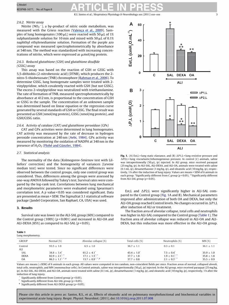
Fig. 1. (A) Est,L = lung static elastance, and (B) �P1,L = lung resistive pressure and�P2,L = lung viscoelastic/inhomogeneous pressure. In control (C) animals, salinewas intraperitoneally (50 �L, ip) injected. In ALI group, mice received paraquat(25 mg/kg, ip). In ALI-SAL, ALI-DEXA, and ALI-OA, animals were treated with saline(0.1 mL, ip), dexamethasone (1 mg/kg, ip), and oleanolic acid (10 mg/kg, ip), respec-tively, 1 h after the induction of lung injury. Values are means + SEM of 6 animals in
TL
Vtii
ARTICLEESPNB-1677; No. of Pages 8
R.S. Santos et al. / Respiratory Physio
.6.2. Nitrite assayNitrite (NO2
−), a by-product of nitric oxide metabolism, waseasured with the Griess reaction (Valenc a et al., 2009). Sam-
les of lung homogenates (100 �L) were reacted with 50 �L of 1%ulphanilamide solution for 10 min and mixed with 50 �L of 0.1%aphthyl ethylenediamine solution. Formation of the purple azoompound was measured spectrophotometrically by absorbancet 540 nm. The method was standardized with increasing concen-rations of nitrite, which were expressed as �mol/mg protein.
.6.3. Reduced glutathione (GSH) and glutathione disulfideGSSG) assay
This assay was based on the reaction of GSH or GSSG with,5-dithiobis-(2-nitrobenzoic acid) (DTNB), which produces the 2-itro-5-thiobenzoate (TNB) chromophore (Rahman et al., 2006). Toetermine GSSG, lung homogenate samples were treated with 2-inylpyridine, which covalently reacted with GSH (but not GSSG).he excess 2-vinylpyridine was neutralized with triethanolamine.he rate of formation of TNB, measured spectrophotometrically bybsorbance at 412 nm, is proportional to the concentration of GSHr GSSG in the sample. The concentration of an unknown sampleas determined based on linear equation or the regression curve
enerated by several standards of GSH or GSSG. The final result wasresented as GSH (nmol/mg protein), GSSG (nmol/mg protein), andSH/GSSG ratio.
.6.4. Activity of catalase (CAT) and glutathione peroxidase (GPx)CAT and GPx activities were determined in lung homogenates.
AT activity was measured by the rate of decrease in hydrogeneroxide concentration at 240 nm (Aebi, 1984). GPx activity waseasured by monitoring the oxidation of NADPH at 340 nm in the
resence of H2O2 (Flohé and Günzler, 1984).
.7. Statistical analysis
The normality of the data (Kolmogorov-Smirnov test with Lil-iefors’ correction) and the homogeneity of variances (Levene
edian test) were tested. Since no significant differences werebserved between the control groups, only one control group wasonsidered. Thus, differences among the groups were assessed byne-way ANOVA followed by Tukey’s test. Survival rates were com-ared by the log-rank test. Correlations between lung mechanicalnd morphometric parameters were evaluated using Spearman’sorrelation test. A p value < 0.05 was considered significant. Datare presented as mean + SEM. The SigmaStat 3.1 statistical softwareackage (Jandel Corporation, San Raphael, CA, USA) was used.
. Results
Please cite this article in press as: Santos, R.S., et al., Effects of oleanolic
experimental acute lung injury. Respir. Physiol. Neurobiol. (2011), doi:10.1
Survival rate was lower in the ALI-SAL group (60%) compared tohe Control group (100%) (p < 0.001) and increased in ALI-OA andLI-DEXA (85%) as compared to ALI-SAL (p < 0.05).
able 1ung morphometry.
GROUP Normal (%) Alveolar collapse (%)
Control 93.5 ± 1.0 6.5 ± 1.0
ALISAL 38.8 ± 3.4* 61.2 ± 4.1*
DEXA 82.9 ± 2.1*,** 17.1 ± 1.5*,**
OA 86.3 ± 1.1*,** ,# 13.7 ± 0.9*,** ,#
alues are means (±SEM) of 6 animals in each group. All values were computed in ten raotal cells, neutrophils, and MN (mononuclear cells). In Control animals, saline was intrapp). In ALI-SAL, ALI-DEXA, and ALI-OA, animals were treated with saline (0.1 mL, ip), dexamnduction of lung injury.
* Significantly different from Control group (p < 0.05).** Significantly different from ALI-SAL group (p < 0.05).# Significantly different from ALI-DEXA group (p < 0.05).
each group. *Significantly different from C group (p < 0.05). **Significantly differentfrom ALI-SAL group (p < 0.05).
Est,L and �P2,L were significantly higher in ALI-SAL com-pared to the Control group (Fig. 1A and B). Mechanical parametersimproved after administration of both OA and DEXA, but only theALI-OA group reached Control levels. No changes occurred in �P1,Lafter induction of ALI or treatment.
The fraction area of alveolar collapse, total cells and neutrophils
acid on pulmonary morphofunctional and biochemical variables in016/j.resp.2011.07.008
was higher in ALI-SAL compared to the Control group (Table 1). Thefraction area of alveolar collapse was reduced in ALI-OA and ALI-DEXA, but this reduction was more effective in the ALI-OA group.
Total cells (%) Neutrophils (%) MN (%)
36.7 ± 1.1 0.3 ± 0.1 36.1 ± 1.1
43.4 ± 1.0* 7.5 ± 0.4* 35.9 ± 1.037.7 ± 1.8 1.9 ± 0.1*,** 35.8 ± 1.837.7 ± 0.8 2.1 ± 0.3*,** 35.5 ± 0.9
ndom, non-coincident fields per mice. Fraction areas of normal, collapsed alveoli,eritoneally (50 �L, ip) injected. In the ALI group, mice received paraquat (25 mg/kg,ethasone (1 mg/kg, ip), and oleanolic acid (10 mg/kg, ip), respectively, 1 h after the
ARTICLE IN PRESSG Model
RESPNB-1677; No. of Pages 8
4 R.S. Santos et al. / Respiratory Physiology & Neurobiology xxx (2011) xxx– xxx
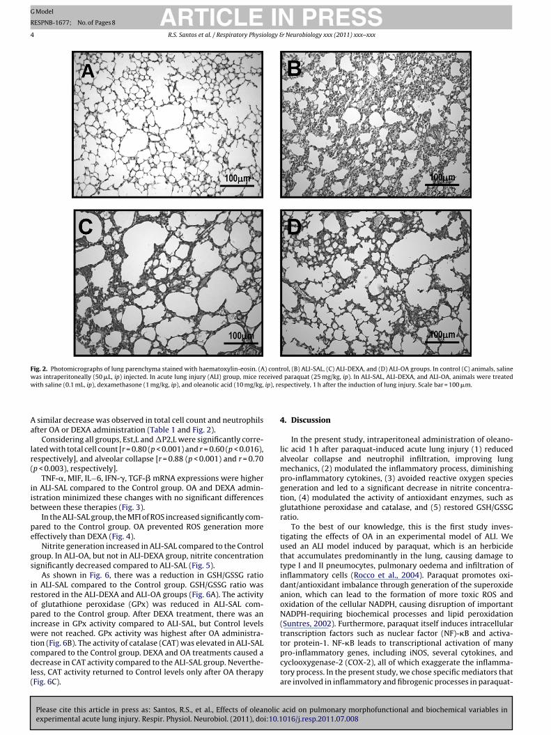
Fig. 2. Photomicrographs of lung parenchyma stained with haematoxylin-eosin. (A) control, (B) ALI-SAL, (C) ALI-DEXA, and (D) ALI-OA groups. In control (C) animals, salinewas intraperitoneally (50 �L, ip) injected. In acute lung injury (ALI) group, mice received paraquat (25 mg/kg, ip). In ALI-SAL, ALI-DEXA, and ALI-OA, animals were treatedw ip), re
Aa
lr(
iib
pe
gs
iropiwtcdl(
ith saline (0.1 mL, ip), dexamethasone (1 mg/kg, ip), and oleanolic acid (10 mg/kg,
similar decrease was observed in total cell count and neutrophilsfter OA or DEXA administration (Table 1 and Fig. 2).
Considering all groups, Est,L and �P2,L were significantly corre-ated with total cell count [r = 0.80 (p < 0.001) and r = 0.60 (p < 0.016),espectively], and alveolar collapse [r = 0.88 (p < 0.001) and r = 0.70p < 0.003), respectively].
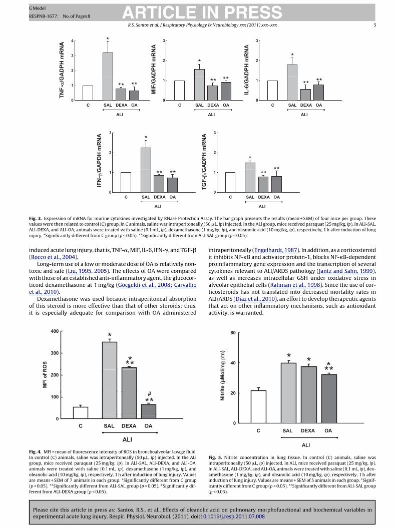
TNF-�, MIF, IL−6, IFN-�, TGF-� mRNA expressions were highern ALI-SAL compared to the Control group. OA and DEXA admin-stration minimized these changes with no significant differencesetween these therapies (Fig. 3).
In the ALI-SAL group, the MFI of ROS increased significantly com-ared to the Control group. OA prevented ROS generation moreffectively than DEXA (Fig. 4).
Nitrite generation increased in ALI-SAL compared to the Controlroup. In ALI-OA, but not in ALI-DEXA group, nitrite concentrationignificantly decreased compared to ALI-SAL (Fig. 5).
As shown in Fig. 6, there was a reduction in GSH/GSSG ration ALI-SAL compared to the Control group. GSH/GSSG ratio wasestored in the ALI-DEXA and ALI-OA groups (Fig. 6A). The activityf glutathione peroxidase (GPx) was reduced in ALI-SAL com-ared to the Control group. After DEXA treatment, there was an
ncrease in GPx activity compared to ALI-SAL, but Control levelsere not reached. GPx activity was highest after OA administra-
ion (Fig. 6B). The activity of catalase (CAT) was elevated in ALI-SAL
Please cite this article in press as: Santos, R.S., et al., Effects of oleanolic
experimental acute lung injury. Respir. Physiol. Neurobiol. (2011), doi:10.1
ompared to the Control group. DEXA and OA treatments caused aecrease in CAT activity compared to the ALI-SAL group. Neverthe-
ess, CAT activity returned to Control levels only after OA therapyFig. 6C).
spectively, 1 h after the induction of lung injury. Scale bar = 100 �m.
4. Discussion
In the present study, intraperitoneal administration of oleano-lic acid 1 h after paraquat-induced acute lung injury (1) reducedalveolar collapse and neutrophil infiltration, improving lungmechanics, (2) modulated the inflammatory process, diminishingpro-inflammatory cytokines, (3) avoided reactive oxygen speciesgeneration and led to a significant decrease in nitrite concentra-tion, (4) modulated the activity of antioxidant enzymes, such asglutathione peroxidase and catalase, and (5) restored GSH/GSSGratio.
To the best of our knowledge, this is the first study inves-tigating the effects of OA in an experimental model of ALI. Weused an ALI model induced by paraquat, which is an herbicidethat accumulates predominantly in the lung, causing damage totype I and II pneumocytes, pulmonary oedema and infiltration ofinflammatory cells (Rocco et al., 2004). Paraquat promotes oxi-dant/antioxidant imbalance through generation of the superoxideanion, which can lead to the formation of more toxic ROS andoxidation of the cellular NADPH, causing disruption of importantNADPH-requiring biochemical processes and lipid peroxidation(Suntres, 2002). Furthermore, paraquat itself induces intracellulartranscription factors such as nuclear factor (NF)-�B and activa-tor protein-1. NF-�B leads to transcriptional activation of many
acid on pulmonary morphofunctional and biochemical variables in016/j.resp.2011.07.008
pro-inflammatory genes, including iNOS, several cytokines, andcyclooxygenase-2 (COX-2), all of which exaggerate the inflamma-tory process. In the present study, we chose specific mediators thatare involved in inflammatory and fibrogenic processes in paraquat-
ARTICLE IN PRESSG Model
RESPNB-1677; No. of Pages 8
R.S. Santos et al. / Respiratory Physiology & Neurobiology xxx (2011) xxx– xxx 5
*4 *A *
3
A *3
A
**
2
3AD
PH m
RN
A**
2 *
ADPH
mR
NA
**
2*
ADPH
mR
NA
0
1
C SAL DEXA OA
TNF-
α/G
****
0
1
C SAL DEX A OA
MIF
/GA
** **
0
1
C SAL DEXA OA
IL-6
/GA
** **
ALI ALI ALI
*3
*NA *
3
NA
**
2
GA
PDH
mR
N
**
2
*
GAD
PH m
RN
**
0
1
C SAL DEX A OA
** **
IFN
- γ/G
0
1
C SAL DEXA OATG
F-β
/G **
ALI ALI
Fig. 3. Expression of mRNA for murine cytokines investigated by RNase Protection Assay. The bar graph presents the results (mean + SEM) of four mice per group. Thesev lly (50A e (1 mi ALI-S
i(
twte
oi
FIgaoa(f
alues were then related to control (C) group. In C animals, saline was intraperitoneaLI-DEXA, and ALI-OA, animals were treated with saline (0.1 mL, ip), dexamethason
njury. *Significantly different from C group (p < 0.05). **Significantly different from
nduced acute lung injury, that is, TNF-�, MIF, IL-6, IFN-�, and TGF-�Rocco et al., 2004).
Long-term use of a low or moderate dose of OA is relatively non-oxic and safe (Liu, 1995, 2005). The effects of OA were comparedith those of an established anti-inflammatory agent, the glucocor-
icoid dexamethasone at 1 mg/kg (Göcgeldi et al., 2008; Carvalho
Please cite this article in press as: Santos, R.S., et al., Effects of oleanolic
experimental acute lung injury. Respir. Physiol. Neurobiol. (2011), doi:10.1
t al., 2010).Dexamethasone was used because intraperitoneal absorption
f this steroid is more effective than that of other steroids; thus,t is especially adequate for comparison with OA administered
*
**
400*
300
f RO
S ***200
MFI
of
#100 **#
0
ALI
OADEXASALC
ALI
ig. 4. MFI = mean of fluorescence intensity of ROS in bronchoalveolar lavage fluid.n control (C) animals, saline was intraperitoneally (50 �L, ip) injected. In the ALIroup, mice received paraquat (25 mg/kg, ip). In ALI-SAL, ALI-DEXA, and ALI-OA,nimals were treated with saline (0.1 mL, ip), dexamethasone (1 mg/kg, ip), andleanolic acid (10 mg/kg, ip), respectively, 1 h after induction of lung injury. Valuesre means + SEM of 7 animals in each group. *Significantly different from C groupp < 0.05). **Significantly different from ALI-SAL group (p < 0.05). #Significantly dif-erent from ALI-DEXA group (p < 0.05).
�L, ip) injected. In the ALI group, mice received paraquat (25 mg/kg, ip). In ALI-SAL,g/kg, ip), and oleanolic acid (10 mg/kg, ip), respectively, 1 h after induction of lung
AL group (p < 0.05).
intraperitoneally (Engelhardt, 1987). In addition, as a corticosteroidit inhibits NF-�B and activator protein-1, blocks NF-�B-dependentproinflammatory gene expression and the transcription of severalcytokines relevant to ALI/ARDS pathology (Jantz and Sahn, 1999),as well as increases intracellular GSH under oxidative stress inalveolar epithelial cells (Rahman et al., 1998). Since the use of cor-ticosteroids has not translated into decreased mortality rates in
acid on pulmonary morphofunctional and biochemical variables in016/j.resp.2011.07.008
ALI/ARDS (Diaz et al., 2010), an effort to develop therapeutic agentsthat act on other inflammatory mechanisms, such as antioxidantactivity, is warranted.
*
**
0
20
40
60
*
ALI
C SAL DEXA OA
*
Nitr
ite (µ
Mol
/mg
p tn)
***
Fig. 5. Nitrite concentration in lung tissue. In control (C) animals, saline wasintraperitoneally (50 �L, ip) injected. In ALI, mice received paraquat (25 mg/kg, ip).In ALI-SAL, ALI-DEXA, and ALI-OA, animals were treated with saline (0.1 mL, ip), dex-amethasone (1 mg/kg, ip), and oleanolic acid (10 mg/kg, ip), respectively, 1 h afterinduction of lung injury. Values are means + SEM of 5 animals in each group. *Signif-icantly different from C group (p < 0.05). **Significantly different from ALI-SAL group(p < 0.05).

ARTICLE IN PRESSG Model
RESPNB-1677; No. of Pages 8
6 R.S. Santos et al. / Respiratory Physiology & Neurobiology xxx (2011) xxx– xxx
*
**
2
G
*
**
2
***#BA
1*
GSH
/GSS
G
μM
1 ****
0OADEXASALC
GPx
(
0OADEXASALC
ALI ALI
*8
)
C
***
*
**
4
6 *
***
2
4
**
Cat
alas
e (U **
ALI
0OADEXASALC
C
Fig. 6. (A) GSH/GSSG ratio. Antioxidant enzyme activities. (B) Glutathione peroxidase. (C) Catalase. In control (C) animals, saline was intraperitoneally (50 �L, ip) injected. InA animaa eans* ALI-D
bR((btai
G2wiCdboehtehe
pGc
LI group, mice received paraquat (25 mg/kg, ip). In ALI-SAL, ALI-DEXA, and ALI-OA,cid (10 mg/kg, ip), respectively, 1 h after the induction of lung injury. Values are m*Significantly different from ALI-SAL group (p < 0.05). #Significantly different from
In the present study, OA acted on the inflammatory processy reducing generation of pro-inflammatory cytokines (Fig. 3),OS, and nitrite, as well as by upregulating antioxidant enzymesFigs. 4–6). Anti-inflammatory effects of OA have been reportedNataraju et al., 2009; Martín et al., 2010) and associated with inhi-ition of NF-�B (Takada et al., 2010). This, in turn, has been observedo yield a reduction in inflammatory cytokines and apoptotic cells,s well as nitrite overproduction, with subsequent maintenance ofntracellular GSH level (Abdel-Zaher et al., 2007).
Additionally, recent studies have suggested that OA modulatesSH, CAT and GPx activities (Ovesná et al., 2004; Tsai and Yin,008; Wang et al., 2010) and exhibits potent scavenging behaviour,ith a quenching effect on superoxide anion radicals, prevent-
ng redox imbalance and formation of oxidant radicals (Yin andhan, 2007). It has been proposed that OA may play an antioxi-ant role through inhibition of the release of high mobility groupox-1 protein (HMGB1) (Kawahara et al., 2009) and the activationf Nrf2, a transcriptional factor that induces antioxidant-responselements (Reisman et al., 2009; Wang et al., 2010). A recent studyas reported that the targeting of Nrf2 with oleanolic acid deriva-ive may provide an effective therapy to limit the potential adverseffects of hyperoxia (Reddy et al., 2009). However, so far, no studyas analysed the impact of oleanolic acid in paraquat inducedxperimental acute lung injury.
Please cite this article in press as: Santos, R.S., et al., Effects of oleanolic
experimental acute lung injury. Respir. Physiol. Neurobiol. (2011), doi:10.1
Therefore, the protective effects of OA against ROS in the presentaraquat-induced ALI could be associated with a restoration ofSH/GSSG ratio. GSH is a nonprotein thiol that may provide intra-ellular protection against the oxidative action of paraquat (Tasaka
ls were treated with saline (0.1 mL, ip), dexamethasone (1 mg/kg, ip), and oleanolic + SEM of 5 animals in each group. *Significantly different from C group (p < 0.05).EXA group (p < 0.05).
et al., 2008), and also modulate the activity of catalase and GPx(Fig. 6). Furthermore, OA may protect against oxidative stressthrough iNOS inhibition (Suh et al., 1998), preventing the increasein nitrite, since excessive production of nitric oxide contributes tothe pathogenesis of ALI (Lange et al., 2010).
Lung viscoelastic/inhomogeneous pressure and static elastanceincreased in the ALI-SAL group (Fig. 1A and B) due to alveolarcollapse, oedema, and inflammatory cell infiltration (Table 1 andFig. 2). In the present model, morphofunctional changes werereduced by both DEXA and OA, but these beneficial effects weremore intense after OA administration. The mechanical improve-ment in the OA group was related to a greater reduction in theinflammatory process, which led to less alveolar collapse andoedema. OA and DEXA reduced inflammatory cytokines to a similardegree. However, OA was more effective than DEXA in modulatingoxidative stress and regulating the release of nitrite and antioxidantenzymes, such as catalase and glutathione peroxidase. This advan-tage may be related to the ability of OA to activate nuclear factorE2-related factor 2 (Nrf2) and MAP kinases (JNK and ERK) (Wanget al., 2010), while the main role of DEXA is to downregulate NF-�Band AP1 (Meduri et al., 2009).
This study has some limitations that need to be addressed: (1)a specific experimental model of paraquat induced ALI was used.Therefore, the present results may not be extended to other exper-
acid on pulmonary morphofunctional and biochemical variables in016/j.resp.2011.07.008
imental models of ALI, (2) animals were mechanically ventilatedin air, and thus we cannot rule out that the increase in inflamma-tory mediators in ALI-SAL may be related, at least in part, to hypoxiaresulting from a greater amount of atelectasis, and/or that different
ING Model
R
logy &
rcbeaimTr(auOtbnli
tiiaa
A
BhLEt
FDStSN(
R
A
AB
B
C
C
C
C
C
D
ARTICLEESPNB-1677; No. of Pages 8
R.S. Santos et al. / Respiratory Physio
esults could have been obtained with higher FiO2, (3) OA was notompared with a ROS inhibitor but with dexamethasone which haseen used in the clinical setting. Thus, we cannot rule out differentffects with other types of steroids, different doses and routes ofdministration, (4) a single intraperitoneal dose of OA was admin-stered, and consequently, we cannot exclude the possibility that
ultiple doses or continuous infusion could yield better results.he methods to quantify OA in plasma, and the optimal range andoute of OA administration in humans are currently being definedSong et al., 2006; Ji et al., 2009). Even though OA might be safelydministered in humans, the optimal oral or intravenous dosagender different clinical conditions remains to be determined, (5)A was given 1 h after the induction of lung injury, and therefore,
he effect of OA at a later phase of ALI is unknown, and (6) OA,ut not its derivatives, was used in the current study, thus we can-ot exclude that different results could be obtained, and (7) only a
imited number of cytokines were investigated, mainly related tonflammatory and fibrogenic processes in paraquat- induced ALI.
In conclusion, intraperitoneal injection of oleanolic acid 1 h afterhe induction of paraquat-induced acute lung injury modulated thenflammatory and oxidative processes, preventing lung mechan-cal and histological changes. Thus, oleanolic acid, a drug withnti-inflammatory and anti-oxidative properties, may be a usefuldjunct therapy for acute lung injury.
cknowledgements
The authors would like to express their gratitude to Mr. Andreenedito da Silva for animal care, Miss Thaiana Borges de Sousa forer skilful technical assistance during the experiments, Mrs. Anaucia Neves da Silva for her help with microscopy, and Mrs. Moiralizabeth Schöttler and Claudia Buchweitz for assistance in editinghe manuscript.
Supported by: The Centres of Excellence Program (PRONEX-APERJ), The Brazilian Council for Scientific and Technologicalevelopment (CNPq), The Carlos Chagas Filho Rio de Janeirotate Research Supporting Foundation (FAPERJ), Coordination forhe Improvement of Higher Education Personnel (CAPES), Theão Paulo State Research Support Foundation (FAPESP), and Theational Institute of Science and Technology of Drugs and Medicine
INCT-INOFAR).
eferences
bdel-Zaher, A.O., Abdel-Rahman, M.M., Hafez, M.M., Omran, F.M., 2007. Role ofnitric oxide and reduced glutathione in the protective effects of aminoguanidine,gadolinium chloride and oleanolic acid against acetaminophen-induced hepaticand renal damage. Toxicology 234, 124–134.
ebi, H., 1984. Catalase in vitro. Methods Enzymol. 105, 121–126.ates, J.H., Decramer, M., Chartrand, D., Zin, W.A., Boddener, A., Milic-Emili, J., 1985.
Volume time profile during relaxed expiration in the normal dog. J. Appl. Physiol.59, 732–737.
radford, M.M., 1976. A rapid and sensitive method for the quantitation of micro-gram quantities of protein utilizing the principle of protein–dye binding. Anal.Biochem. 72, 248–254.
alfee, C.S., Matthay, M.A., 2007. Nonventilatory treatments for acute lung injuryand ARDS. Chest 131, 913–920.
arvalho, G.M., Oliveira, V.R., Soares, R.M., Azevedo, S.M., Lima, L.M., Barreiro, E.J.,Valenc a, S.S., Saldiva, P.H., Faffe, D.S., Zin, W.A., 2010. Can LASSBio 596 and dex-amethasone treat acute lung and liver inflammation induced by microcystin-LR?Toxicon 56, 604–612.
habot, F., Mitchell, J.A., Gutteridge, J.M., Evans, T.W., 1998. Reactive oxygen speciesin acute lung injury. Eur. Respir. J. 11, 745–757.
orrea, F.C., Ciminelli, P.B., Falcão, H., Alcântara, B.J., Contador, R.S., Medeiros, A.S.,Zin, W.A., Rocco, P.R., 2001. Respiratory mechanics and lung histology in normalrats anesthetized with sevoflurane. J. Appl. Physiol. 91, 803–810.
Please cite this article in press as: Santos, R.S., et al., Effects of oleanolic
experimental acute lung injury. Respir. Physiol. Neurobiol. (2011), doi:10.1
havko, M., Adeeb, S., Ahlers, S.T., McCarron, R.M., 2009. Attenuation of pulmonaryinflammation after exposure to blast overpressure by N-acetylcysteine amide.Shock 32, 325–331.
iaz, J.V., Brower, R., Calfee, C.S., Matthay, M.A., 2010. Therapeutic strategies forsevere acute lung injury. Crit. Care Med. 38, 1644–1650.
PRESS Neurobiology xxx (2011) xxx– xxx 7
Engelhardt, G., 1987. Effect of corticosteroids on the toxic pulmonary oedemainduced by nitrogen dioxide inhalation in the rat. Arzneimittelforschung 37,519–523.
Flohé, L., Günzler, W.A., 1984. Assays of glutathione peroxidase. Methods Enzymol.105, 114–121.
Göcgeldi, E., Uysal, B., Korkmaz, A., Ogur, R., Reiter, R.J., Kurt, B., Oter, S., Topal, T.,Hasde, M., 2008. Establishing the use of melatonin as an adjuvant therapeuticagainst paraquat-induced lung toxicity in rats. Exp. Biol. Med. 233, 1133–1141.
Honda, T., Finlay, H.J., Cribble, G.W., Suh, N., Sporn, M.B., 1997. New enone derivativesof oleanolic acid and ursolic acid as inhibitors of nitric oxide production in mousemacrophages. Bioorg. Med. Chem. Lett. 7, 1623–1628.
Honda, T., Rounds, B.V., Gribble, G.W., Suh, N., Wang, Y., Sporn, M.B., 1998. Design andsynthesis of 2-cyano-3,12-dioxoolean-1,9-dien-28-oic acid, a novel and highlyactive inhibitor of nitric oxide production in mouse macrophages. Bioorg. Med.Chem. Lett. 8, 2711–2714.
Honda, T., Rounds, B.V., Bore, L., Favaloro Jr., F.G., Gribble, G.W., Suh, N., Wang, Y.,Spor, M.B., 1999. Novel synthetic oleanane triterpenoids: a series of highly activeinhibitors of nitric oxide production in mouse macrophages. Bioorg. Med. Chem.Lett. 9, 3429–3434.
Ji, H.Y., Shin, B.S., Jeong, D.W., Park, E.J., Park, E.S., Yoo, S.D., Lee, H.S., 2009. Inter-species scaling of oleanolic acid in mice, rats, rabbits and dogs and prediction ofhuman pharmacokinetics. Arch. Pharm. Res. 32, 251–257.
Jantz, M.A., Sahn, S.A., 1999. Corticosteroids in acute respiratory failure. Am. J. Respir.Crit. Care Med. 160, 1079–1100.
Ka, H., Park, H.J., Jung, H.J., Choi, J.W., Cho, K.S., Ha, J., Lee, K.T., 2003. Cinnamaldehydeinduces apoptosis by ROS-mediated mitochondrial permeability transition inhuman promyelocytic leukaemia HL-60 cell. Cancer Lett. 196, 143–152.
Kawahara, K., Hashiguchi, T., Masuda, K., Saniabadi, A.R., Kikuchi, K., Tancharoen,S., Ito, T., Miura, N., Morimoto, Y., Biswas, K.K., Nawa, Y., Meng, X., Oyama, Y.,Takenouchi, K., Shrestha, B., Sameshima, H., Shimizu, T., Adachi, T., Adachi, M.,Maruyama, I., 2009. Mechanisms of HMGB1 release inhibition from RAW264.7cells by oleanolic acid in Punus mume sieb. et Zucc. Int. J. Mol. Med. 23, 615–620.
Lange, M., Nakano, Y., Traber, D.L., Hamahata, A., Esechie, A., Jonkam, C., Bansal, K.,Traber, L.D., Enkhbaatar, P., 2010. Role of different nitric oxide synthase iso-forms in a murine model of acute lung injury and sepsis. Biochem. Biophys. Res.Commun. 399, 286–291.
Lee, J.Y., Moon, H., Kim, C.J., 2010. Effects of hydroxy pentacyclic triterpene acidsfrom Forsythia viridissima on asthmatic responses to ovalbumin challenge inconscious guinea pigs. Biol. Pharm. Bull. 33, 230–237.
Leite-Junior, J.H., Garcia, C.S., Souza-Fernandes, A.B., Silva, P.L., Ornellas, D.S.,Larangeira, A.P., Castro-Faria-Neto, H.C., Morales, M.M., Negri, E.M., Capelozzi,V.L., Zin, W.A., Pelosi, P., Bozza, P.T., Rocco, P.R., 2008. Methylprednisoloneimproves lung mechanics and reduces the inflammatory response in pulmonarybut not in extrapulmonary mild acute lung injury in mice. Crit. Care Med. 36,2621–2628.
Liu, J., 1995. Pharmacology of oleanolic acid and ursolic acid. J. Ethnopharmacol. 49,57–68.
Liu, J., 2005. Oleanolic acid and ursolic acid: research perspectives. J. Ethnopharma-col. 100, 92–94.
Martín, R., Carvalho-Tavares, J., Hernández, M., Arnés, M., Ruiz-Gutiérrez, V., Nieto,M.L., 2010. Beneficial actions of oleanolic acid in an experimental model ofmultiple sclerosis: a potential therapeutic role. Biochem. Pharmacol. 79, 198–208.
Meduri, G.U., Annane, D., Chrousos, G.P., Marik, P.E., Sinclair, S.E., 2009. Activationand regulation of systemic inflammation in ARDS: rationale for prolonged glu-cocorticoid therapy. Chest 136, 1631–1643.
Nataraju, A., Saini, D., Ramachandran, S., Benshoff, N., Liu, W., Chapman, W.,Mohanakumar, T., 2009. Oleanolic Acid, a plant triterpenoid, significantlyimproves survival and function of islet allograft. Transplantation 88, 987–994.
Ovesná, Z., Vachálková, A., Horváthová, K., Tóthová, D., 2004. Pentacyclic triter-penoic acids: new chemoprotective compounds. Minireview. Neoplasma 51,327–333.
Rahman, I., Bel, A., Mulier, B., Donaldson, K., MacNee, W., 1998. Differential regula-tion of glutathione by oxidants and dexamethasone in alveolar epithelial cells.Am. J. Physiol. 275, L80–L86.
Rahman, I., Kode, A., Biswas, S.K., 2006. Assay for quantitative determination of glu-tathione and glutathione disulfide levels using enzymatic recycling method. Nat.Protoc. 1, 3159–3165.
Reddy, N.M., Suryanaraya, V., Yates, M.S., Kleeberger, S.R., Hassoun, P.M., Yamamoto,M., Liby, K.T., Sporn, M.B., Kensler, T.W., Reddy, S.P., 2009. The triterpenoidCDDO-imidazolide confers potent protection against hyperoxic acute lunginjury in mice. Am. J. Respir. Crit. Care Med. 180, 867–874.
Reisman, S.A., Aleksunes, L.M., Klaassen, C.D., 2009. Oleanolic acid activates Nrf2and protects from acetaminophen hepatotoxicity via Nrf2-dependent and Nrf2-independent processes. Biochem. Pharmacol. 77, 1273–1282.
Rocco, P.R.M., Facchinetti, L.D., Ferreira, H.C., Negri, E.M., Capelozzi, V.L., Faffe, D.S.,Zin, W.A., 2004. Time course of respiratory mechanics and pulmonary structuralremodelling in acute lung injury. Respir. Physiol. Neurobiol. 143, 49–61.
Song, M., Hang, T.J., Wang, Y., Jiang, L., Wu, X.L., Zhang, Z., Shen, J., Zhang, Y., 2006.Determination of oleanolic acid in human plasma and study of its pharmacoki-netics in Chinese healthy male volunteers by HPLC tandem mass spectrometry.
acid on pulmonary morphofunctional and biochemical variables in016/j.resp.2011.07.008
J. Pharm. Biomed. Anal. 40, 190–196.Suh, N., Honda, T., Finlay, H.J., Barchowsky, A., Williams, C., Benoit, N.E., Xie,
Q.W., Nathan, C., Gribble, G.W., Sporn, M.B., 1998. Novel triterpenoids suppressinducible nitric oxide synthase (iNOS) and inducible cyclooxygenase (COX-2) inmouse macrophages. Cancer Res. 58, 717–723.
ING Model
R
8 ology &
S
T
T
T
ARTICLEESPNB-1677; No. of Pages 8
R.S. Santos et al. / Respiratory Physi
untres, Z.E., 2002. Role of antioxidants in paraquat toxicity. Toxicology 180,65–77.
akada, K., Nakane, T., Masuda, K., Ishii, H., 2010. Ursolic acid and oleanolic acid,members of pentacyclic triterpenoid acids, suppress TNF-�-induced E-selectinexpression by cultured umbilical vein endothelial cells. Phytomedicine 17,1114–1119.
Please cite this article in press as: Santos, R.S., et al., Effects of oleanolic
experimental acute lung injury. Respir. Physiol. Neurobiol. (2011), doi:10.1
asaka, S., Amaya, F., Hashimoto, S., Ishizaka, A., 2008. Roles of oxidants and redoxsignaling in the pathogenesis of acute respiratory distress syndrome. Antioxid.Redox Signal. 10, 739–753.
sai, S.J., Yin, M.C., 2008. Antioxidative and anti-inflammatory protection of oleanolicacid and ursolic acid in PC12 cells. J. Food Sci. 7, H174–H178.
PRESS Neurobiology xxx (2011) xxx– xxx
Valenc a, S.S., Pimenta, W.A., Rueff-Barroso, C.R., Ferreira, T.S., Resende, A.C., Moura,R.S., Porto, L.C., 2009. Involvement of nitric oxide in acute lung inflammationinduced by cigarette smoke in the mouse. Nitric Oxide 20, 175–181.
Wang, X., Ye, X.L., Liu, R., Chen, H.L., Bai, H., Liang, X., Zhang, X.D., Wang, Z., Li, W.L.,Hai, C.X., 2010. Antioxidant activities of oleanolic acid in vitro: possible role ofNrf2 and MAP kinases. Chem. Biol. Interact. 184, 328–337.
acid on pulmonary morphofunctional and biochemical variables in016/j.resp.2011.07.008
Weibel, E.R., 1990. Morphometry: stereological theory and practical methods. In: Gil,J. (Ed.), Models of Lung Disease – Microscopy and Structural Methods. MarcelDekker, NY, pp. 199–247.
Yin, M.C., Chan, K.C., 2007. Nonenzymatic antioxidative and antiglycative effects ofoleanolic acid and ursolic acid. J. Agric. Food Chem. 55, 7177–7181.