On the possibilities of hard X-ray imaging with high spatio-temporal resolution using polychromatic...
24
draft J X Ray Sci Tech / Techn. & Status Rep., Rev1 On the possibilities of hard X-ray imaging with high spatio-temporal resolution using polychromatic synchrotron radiation A. Rack European Synchrotron Radiation Facility, Grenoble, France ∗ F. Garc´ ıa-Moreno Helmholtz Zentrum Berlin f¨ ur Materialien und Energie, Institute of Applied Materials, Germany C. Schmitt and O. Betz Zoologisches Institut, Abteilung Evolutionsbiologie der Invertebraten, Universit¨ at T¨ ubingen, Germany A. Cecilia and A. Ershov Institute for Synchrotron Radiation – ANKA, Karlsruher Institut f¨ ur Technologie, Germany T. Rack Charit´ e – Dept. of Maxillofacial and Facial-Plastic Surgery, Division of Oral Medicine, Radiology and Surgery, Berlin, Germany J. Banhart and S. Zabler Technical University of Berlin, Institute for Materials Science and Technology, Germany (Dated: July 20, 2010) 1
Transcript of On the possibilities of hard X-ray imaging with high spatio-temporal resolution using polychromatic...

draft J X Ray Sci Tech / Techn. & Status Rep., Rev1
On the possibilities of hard X-ray imaging with high
spatio-temporal resolution using polychromatic synchrotron
radiation
A. Rack
European Synchrotron Radiation Facility, Grenoble, France∗
F. Garcıa-Moreno
Helmholtz Zentrum Berlin fur Materialien und Energie,
Institute of Applied Materials, Germany
C. Schmitt and O. Betz
Zoologisches Institut, Abteilung Evolutionsbiologie
der Invertebraten, Universitat Tubingen, Germany
A. Cecilia and A. Ershov
Institute for Synchrotron Radiation – ANKA,
Karlsruher Institut fur Technologie, Germany
T. Rack
Charite – Dept. of Maxillofacial and Facial-Plastic Surgery,
Division of Oral Medicine, Radiology and Surgery, Berlin, Germany
J. Banhart and S. Zabler
Technical University of Berlin, Institute for Materials Science and Technology, Germany
(Dated: July 20, 2010)
1

Abstract
Time-resolved imaging with penetrating radiation has an outstanding scientific value but its
realisation requires a high density of photons as well as corresponding fast X-ray image detection
schemes. Bending magnets and insertion devices of third generation synchrotron light sources
offer a polychromatic photon flux density which is high enough to perform hard X-ray imaging
with a spatio-temporal resolution up to the µm-µs range. Existing indirect X-ray image detectors
commonly used at synchrotron light sources can be adapted for fast image acquisition by employing
CMOS-based digital high speed cameras already available on the market. Selected applications
from life sciences and materials research underline the high potential of this high-speed hard X-ray
microimaging approach.
Keywords: Synchrotron radiation, X-ray imaging, radioscopy, CMOS, cineradiography, microtomography,
scintillator, X-ray phase contrast, X-ray detector, real time experiment, digital radiography
∗Corresponding author’s address: A. Rack, ESRF, BP 220, 38043 Grenoble Cedex, France, tel: +33 (0)476
88 1781, fax: +33 (0)476 88 2785, e-mail: [email protected]
2

I. INTRODUCTION
The insights provided by moving images can be deep but capturing such images is chal-
lenging. The outstanding scientific value of even early motion pictures is demonstrated
by the famous movies showing the collapse of the Tacoma Narrows Bridge (USA) in 1943
[1]. An analysis of this problem of non-linear dynamics would not be possible if only a
few individual photographic images were available [2]. In order to study events which take
place on a time scale shorter than the common video frame rate, high image acquisition
speed is required: already at the beginning of the past century, scientists like Lucien Bull
developed techniques for high-speed visible-light imaging to study, e. g., the biomechanics of
living insects [3]. To achieve comparable imaging frame rates with penetrating radiation is
challenging, mainly due to the limited available photon flux density. In special cases, such
as periodic movements, one can overcome this issue, for example by choosing a stroboscopic
approach (cf., e. g., Kardjilov et al. [4]).
Several so-called third-generation synchrotron light sources have been in operation now
for many years all over the world. The polychromatic hard X-ray radiation provided by
either bending magnets or insertion devices is intense enough to image aperiodic movements
continuously with high time resolution. With the ongoing detector development, microradio-
graphy can be developed further towards microradioscopy, allowing one to visualize internal
structures of opaque objects as they change with time. The ultimate limit of reachable time
resolution is defined by the detector electronics as well as the bunch structure of the electron
current inside the synchrotron’s storage ring. For the latter case, single-shot imaging using
isolated bunches has been reported [5].
A further advantage of using a synchrotron light source is the partial coherence of the
emitted wavefronts at the position of the experiment. This allows one to use interference
effects as a contrast mode, called ’inline X-ray phase contrast’ [6, 7, 8]. This method is
much more sensitive than plain attenuation contrast and therefore relaxes the demands on
the photon flux density. For a further introduction into the techniques of synchrotron-based
microimaging the reader is referred to the literature [9, 10].
In this article, we introduce the basics of the instrumentation needed to perform
synchrotron-based imaging with a high spatio-temporal resolution up to the µm-µs range.
One of the important aspects is that common indirect detectors available can be modified
with minor development effort, by using high speed cameras already available on the market.
3

Equally important is that with simple bending magnet beamlines, i. e. with a moderate pho-
ton flux density, cutting edge investigations can be performed. Several selected applications
from life sciences and materials research, employing frame rates from a few hundred up to
several 10 000 images/s, underline the high potential of this approach and aim at stimu-
lating further developments at other light sources. The possibility of an extension towards
time-resolved three-dimensional imaging (i. e., microtomography) is shown.
II. INDIRECT X-RAY IMAGE DETECTORS
The development of high resolution X-ray imaging detectors for in situ investigations with
a given time resolution dates back to the late 1960s: so-called live topography was developed
to study online the growth of crystals [11]. At that time, an approach based on modified
television cameras was employed where X-ray photons are directly converted into electrons
that are then registered. Presently, this scheme is commonly called ’direct detection’ and
has been further developed using more sophisticated (electronic) concepts. In the middle
of the 1970s, first detectors were reported in combination with live topography that use
a scintillator screen to convert X-rays into visible light (similar to the medical technique
fluoroscopy). The resulting luminescence image is projected via diffraction-limited visible
light optics onto a camera [12]. This frequently called ’indirect detection’ scheme was applied
in the early 1980s in combination with synchrotron radiation sources, again to perform
live topography [13]. Indirect detectors have the advantage of providing high resolutions
as well as containing mostly components that are commercially available. The reachable
spatial resolution is determined by the scintillating screen and the numerical aperture of the
objective used (restricted by Abbe’s theorem) [14, 15]. Further concepts for high resolution
X-ray imaging detectors were developed for astronomical measurements [25].
Also during the 1980s, the development of synchrotron-based X-ray imaging started. It
promised to reach a high performance in terms of both spatial resolution and contrast due to
the available high photon flux density compared to laboratory based sources as well as the
nearly parallel propagation of synchrotron light [16]. One of the aims to be reached was to
transfer the already pioneered microtomography techniques to the synchrotron storage rings
[9, 17, 18, 19, 20, 21, 22]. The most promising approach to reach high spatial resolutions
was the combination of the indirect detection scheme with CCD cameras [23, 24].
4

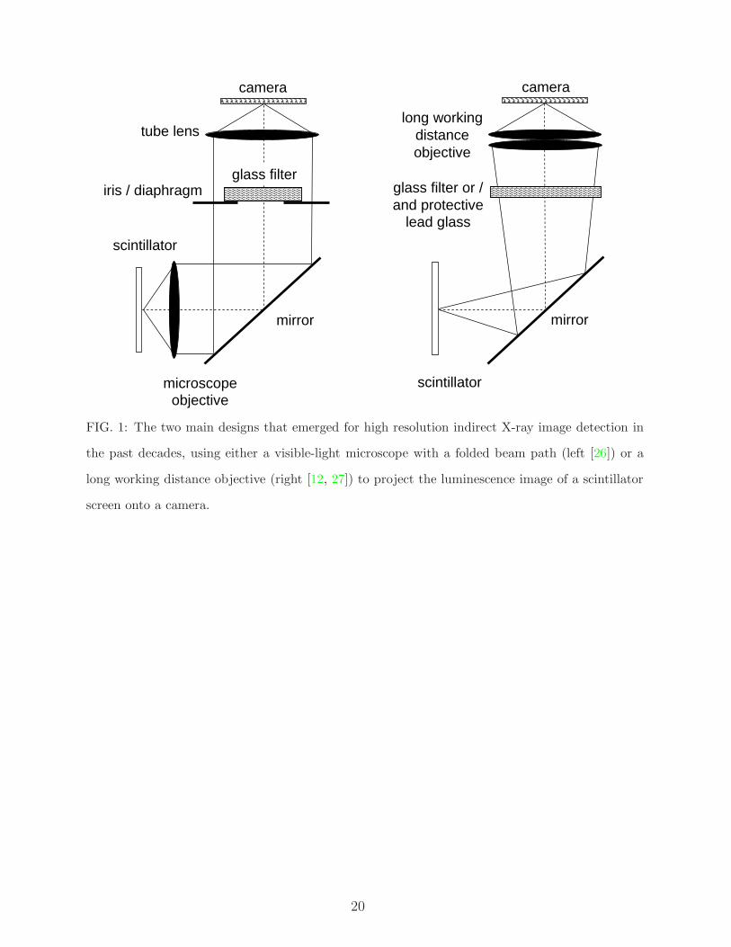
In the 1990s, a design was established that basically uses a visible-light microscope with
a folded beam path to project luminescence images from the scintillator onto the CCD
camera – optimised for spatial resolution, while partially compromising on the efficiency
and the field of view [26] – cf. Fig. 1 (left). Such detectors can reach a resolving power
in the (sub-)micrometre range as standard application when transparent single-crystal thin
film scintillators are employed [15]. For high photon flux densities in combination with a
harder spectrum and corresponding heat loads, the visible light optics directly behind the
scintillator can be damaged and/or deteriorate the resolution, e. g., in case they start to
emit fluorescent X-rays. In order to avoid this, a long working distance objective is used
instead of the visible light microscope. It projects the image through the mirror onto the
camera [27, 28] – cf. Fig. 1 (right). The latter approach is also used in neutron imaging
[4, 29]. For moderate photon energies, so-called inline versions of the optics shown can be
employed [30].
The original application of indirect detectors, live topography (using polychromatic hard
X-ray synchrotron radiation) as well as ex situ topography is of course benefitting from
these developments as well [31, 32]. Further review articles on high-resolution detectors
for synchrotron-based hard X-ray imaging, CCD-based X-ray detection schemes as well as
direct pixel area detectors have been published elsewhere [33, 34, 35].
The data acquisition rate of indirect detector systems can be pushed up the range of
> 104 images/s (i. e. tens of µs/image) when CMOS cameras are used instead of CCDs
[36, 37]. The higher data acquisition speed of CMOS cameras is only compromised by the
limited dynamic range as well as the short exposure times required. To some extent, frame
transfer CCD cameras represent up to now a compromise between CCD and CMOS cameras
[28].
The crucial component of any indirect detector which defines its performance in terms of
efficiency as well as resolution in space and time is the luminescence screen. For this reason,
a lot of effort has been put into developing optimised scintillator material compositions for
synchrotron-based microimaging applications [38, 39, 40, 41, 42, 43, 44].
5
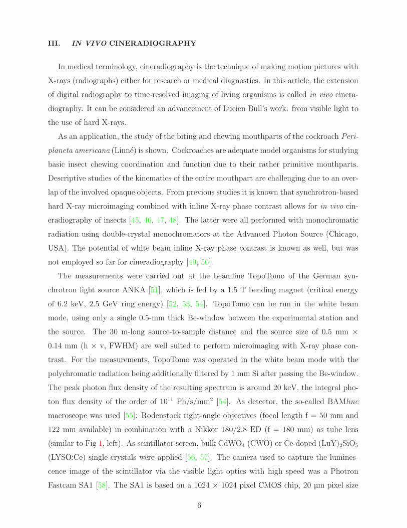
III. IN VIVO CINERADIOGRAPHY
In medical terminology, cineradiography is the technique of making motion pictures with
X-rays (radiographs) either for research or medical diagnostics. In this article, the extension
of digital radiography to time-resolved imaging of living organisms is called in vivo cinera-
diography. It can be considered an advancement of Lucien Bull’s work: from visible light to
the use of hard X-rays.
As an application, the study of the biting and chewing mouthparts of the cockroach Peri-
planeta americana (Linne) is shown. Cockroaches are adequate model organisms for studying
basic insect chewing coordination and function due to their rather primitive mouthparts.
Descriptive studies of the kinematics of the entire mouthpart are challenging due to an over-
lap of the involved opaque objects. From previous studies it is known that synchrotron-based
hard X-ray microimaging combined with inline X-ray phase contrast allows for in vivo cin-
eradiography of insects [45, 46, 47, 48]. The latter were all performed with monochromatic
radiation using double-crystal monochromators at the Advanced Photon Source (Chicago,
USA). The potential of white beam inline X-ray phase contrast is known as well, but was
not employed so far for cineradiography [49, 50].
The measurements were carried out at the beamline TopoTomo of the German syn-
chrotron light source ANKA [51], which is fed by a 1.5 T bending magnet (critical energy
of 6.2 keV, 2.5 GeV ring energy) [52, 53, 54]. TopoTomo can be run in the white beam
mode, using only a single 0.5-mm thick Be-window between the experimental station and
the source. The 30 m-long source-to-sample distance and the source size of 0.5 mm ×
0.14 mm (h × v, FWHM) are well suited to perform microimaging with X-ray phase con-
trast. For the measurements, TopoTomo was operated in the white beam mode with the
polychromatic radiation being additionally filtered by 1 mm Si after passing the Be-window.
The peak photon flux density of the resulting spectrum is around 20 keV, the integral pho-
ton flux density of the order of 1011 Ph/s/mm2 [54]. As detector, the so-called BAMline
macroscope was used [55]: Rodenstock right-angle objectives (focal length f = 50 mm and
122 mm available) in combination with a Nikkor 180/2.8 ED (f = 180 mm) as tube lens
(similar to Fig 1, left). As scintillator screen, bulk CdWO4 (CWO) or Ce-doped (LuY)2SiO5
(LYSO:Ce) single crystals were applied [56, 57]. The camera used to capture the lumines-
cence image of the scintillator via the visible light optics with high speed was a Photron
Fastcam SA1 [58]. The SA1 is based on a 1024 × 1024 pixel CMOS chip, 20 µm pixel size
6

(peak quantum efficiency of 42% at 640 nm). The dynamic range is 10bit (800:1) with a
12bit digitalization. The minimal shutter time is 2 µs and can be triggered with 100 ns time
resolution. Without restricting the region-of-interest (ROI) the camera can acquire 5 400
images per second, with a reduced ROI up to 675 000 images/s (64 × 16 pixel). 32 GB
on-board memory are installed for a fast intermediate data storage. In order to fully exploit
the advantages of inline phase contrast, this X-ray detector was positioned approximately
0.5 m from the specimen.
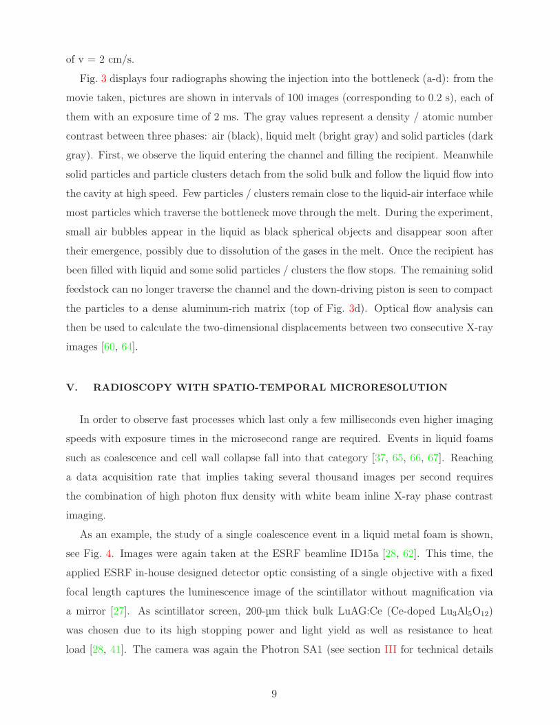
In Fig. 2, example images extracted from a movie showing a feeding cockroach are dis-
played. They were taken with a frame acquisition rate of 125 images/s. The detector was
equipped with a bulk LYSO:Ce crystal. The effective pixel size was around 13.5 µm (result-
ing in a real spatial resolution around 30 µm applying Shannon’s theorem). The full movie
is available online [59]. Due to the hard spectrum employed, the contrast is dominated by
refraction (phase contrast). The time resolution is sufficient to sample one chewing cycle
with about 40 images. Relaxing the demands on spatial resolution and/or signal-to-noise
ratio allows one to reach even higher data acquisition rates. Movies with 250 images/s were
taken during the same experiment as well [60]. In principle, the imaging speed is only lim-
ited by the dose the living specimen can stand. Hence, frame rates up to 1 000 images are
imaginable.
Employing polychromatic radiation is a valuable option in order to perform in vivo cin-
eradiography at synchrotron light sources where the available photon flux density is not
high enough to use monochromatic radiation. Despite the large energy spread, refraction by
means of inline X-ray phase contrast is available. One of the drawbacks is the higher dose
involved.
IV. IN SITU MICRORADIOSCOPY
In materials research, samples can often stand a higher dose than in life sciences. Thus,
higher frame rates or other contrast modes can be applied in microradioscopy. In the pre-
vious chapter, the high imaging speed was accessible despite the moderate available photon
density due to the use of X-ray inline phase contrast that is more sensitive and therefore
less demanding on flux. More photons are needed in order to achieve comparable contrast
and frame rates when absorption information is required.
7

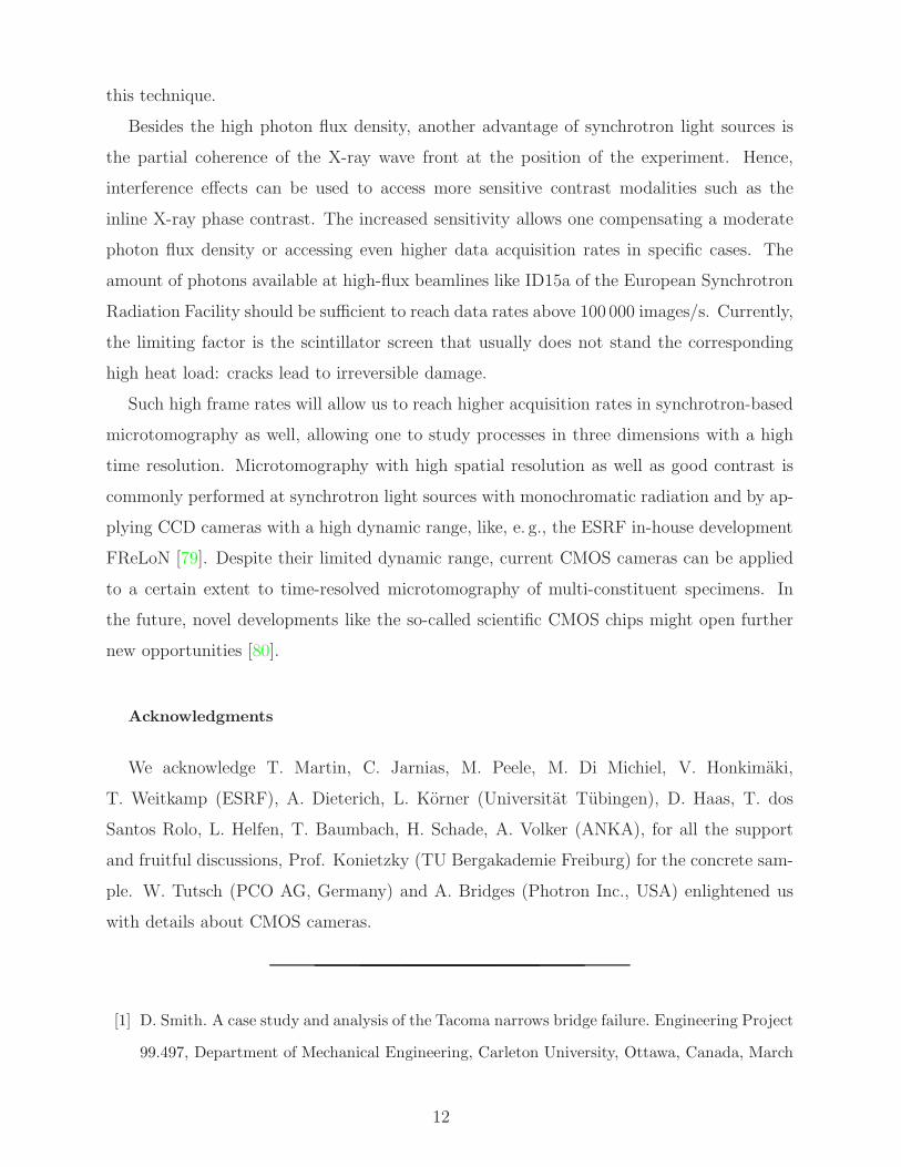
In this section, the visualization of semi-solid metal flow at an acquisition rate of 500 im-
ages/s is shown. Semi-solid casting (SSC) is an experimental production process for light
metals, currently in use for niche-market applications only. It leads to a reduction of un-
wanted shrinkage during solidification due to the lower processing temperatures [61]. The
process holds promise of improved mechanical properties at only slightly higher costs. Until
now, SSC is preferably applied to coarse structures: it suffers from the incomplete under-
standing of the rheological properties of the metallic two-phase mixture. More knowledge
would be necessary for numerical simulations and an optimization of the process. Par-
ticularly for thin cavities of dimensions close to the average particle / cluster size of the
solid-phase, SSC yields only poor results. In situ microradioscopy now provides a tool for
visualizing injection processes in such cavities directly.
Images were taken at the high-flux beamline ID15a of the European Synchrotron Ra-
diation Facility (ESRF) [28]. As X-ray source, the available asymmetric multipole wiggler
(1.84 T, 44 keV critical energy) was chosen. Further details on the beamline are available
online [62]. The white radiation was filtered with approximately 20 mm of silicon, the pho-
ton flux density available for the imaging experiment is estimated to 1015 Ph/s/mm2. The
employed detector system is an ESRF in-house development, similar to the design shown in
Fig 1 (right): a manual zoom objective in combination with a lens projects the scintillator
image through a lead glass and a mirror onto the camera. The system has been successfully
used for high speed imaging earlier [36]. As scintillator, a 100 µm-thick YAG:Ce (Ce-doped
Y3Al5O12) single crystal was chosen [41]. The camera was again the Photron Fastcam SA1
(see previous section for technical details). Images were acquired at 500 frames/s and 5.5 µm
effective pixel size (spatial resolution R > 11 µm). Since the readout time of the CMOS chip
is negligibly short, each frame corresponds to an exposure of 2 ms. The distance between
the sample and the detector was approximately 0.1 m (closest possible distance in order to
reduce phase contrast effects).
An experimental setup for in situ flow monitoring of semi-solid slurry was constructed by
the Helmholtz Zentrum Berlin fur Materialien und Energie GmbH and the Federal Institute
for Materials Research and Testing (BAM), both Germany. A detailed description has been
published elsewhere [63, 64]. After heating the Al-32Ge alloy to 450◦C, a miniaturized
injection process was captured with the high-speed imaging system, see Fig. 3. Semi-solid
slurry was pushed through a 0.4 mm thin bottleneck by a steel piston advancing at a speed
8
of v = 2 cm/s.
Fig. 3 displays four radiographs showing the injection into the bottleneck (a-d): from the
movie taken, pictures are shown in intervals of 100 images (corresponding to 0.2 s), each of
them with an exposure time of 2 ms. The gray values represent a density / atomic number
contrast between three phases: air (black), liquid melt (bright gray) and solid particles (dark
gray). First, we observe the liquid entering the channel and filling the recipient. Meanwhile
solid particles and particle clusters detach from the solid bulk and follow the liquid flow into
the cavity at high speed. Few particles / clusters remain close to the liquid-air interface while
most particles which traverse the bottleneck move through the melt. During the experiment,
small air bubbles appear in the liquid as black spherical objects and disappear soon after
their emergence, possibly due to dissolution of the gases in the melt. Once the recipient has
been filled with liquid and some solid particles / clusters the flow stops. The remaining solid
feedstock can no longer traverse the channel and the down-driving piston is seen to compact
the particles to a dense aluminum-rich matrix (top of Fig. 3d). Optical flow analysis can
then be used to calculate the two-dimensional displacements between two consecutive X-ray
images [60, 64].
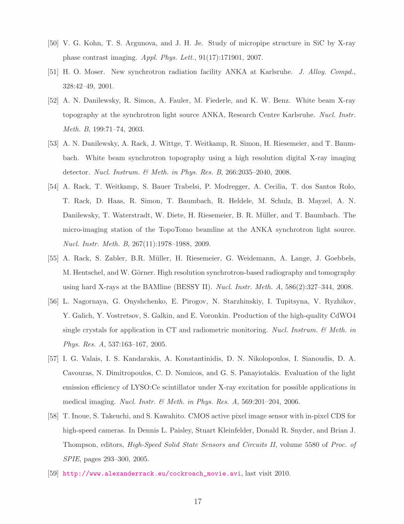
V. RADIOSCOPY WITH SPATIO-TEMPORAL MICRORESOLUTION
In order to observe fast processes which last only a few milliseconds even higher imaging
speeds with exposure times in the microsecond range are required. Events in liquid foams
such as coalescence and cell wall collapse fall into that category [37, 65, 66, 67]. Reaching
a data acquisition rate that implies taking several thousand images per second requires
the combination of high photon flux density with white beam inline X-ray phase contrast
imaging.
As an example, the study of a single coalescence event in a liquid metal foam is shown,
see Fig. 4. Images were again taken at the ESRF beamline ID15a [28, 62]. This time, the
applied ESRF in-house designed detector optic consisting of a single objective with a fixed
focal length captures the luminescence image of the scintillator without magnification via
a mirror [27]. As scintillator screen, 200-µm thick bulk LuAG:Ce (Ce-doped Lu3Al5O12)
was chosen due to its high stopping power and light yield as well as resistance to heat
load [28, 41]. The camera was again the Photron SA1 (see section III for technical details
9

on the camera), operated with a restricted region-of-interest to reach frame rates above
10 000 images/s. The white radiation of the source was filtered by approximately 25 mm
of silicon. The sample-to-detector distance was increased to around 0.5 m in order to fully
exploit phase contrast.
The foaming of AlSi6Cu4 was carried out under 5 bar argon pressure in a furnace at
600◦C as described earlier [36]. The furnace was depressurized during expansion in order
to increase rapidly the foam volume and therefore the growth-induced cell wall rupture rate
during the short time window in which the camera can save images at the high frame rate.
The coalescence event shown in Fig. 4 was captured by taking 40 000 images/s (the complete
movie is available online [69]). The entire merger of two pores happens here within around
3 ms, sampled in time with 120 images. The main features displayed in Fig. 4 are: at 0 µs
two pores and the joint cell wall are still distinguishable, 450 µs later, the cell wall has
ruptured, but still the original shape of the two pores is visible. Then, at 750 µs the new
walls are already straight – defining the so-called rupture time [36]. After 3.4 ms, oscillations
of the new cell walls have stopped. We note that the coalescence event could be resolved due
to the high spatio-temporal resolution used. Nevertheless, the rupture of the cell wall itself
is not resolved, the specific wall between the two pores rather seems to disappear suddenly
between frame 17 (400 µs) and 18 (425 µs).
Time-resolved studies like the one introduced are important to extend the knowledge
about the effective viscosity of liquid metal in a cell wall and the mechanisms behind foam
stability [68, 70, 71]. The rupture time measured allows one to conclude that the collapse
of this single cell wall is dominated by inertia. Hence, modifying, e. g., the viscosity of the
liquid melt should have only minor influence.
VI. TIME-RESOLVED MICROTOMOGRAPHY
In order to extend radiography towards tomography, several hundred projection images
have to be recorded. This naturally reduces the imaging rates by orders of magnitude when
progressing from two to three dimensions. Nevertheless, processes like, e. g., corrosion or
hardening can occur on time-scales which would allow for three-dimensional investigations
using these tomographic imaging rates. First approaches to access higher imaging speeds
for synchrotron-based hard X-ray microtomography by employing radiation with a larger
10

energy spread reach back to the beginning of this century [72]. While with monochromatic
radiation it has been demonstrated that scanning times of a few minutes down to less than
half a minute are applicable [73, 74], the use of white high-energy synchrotron radiation could
allow one to acquire tomographic data sets within only a few seconds [28]. More recently,
successful experiments have been reported which combine, e. g., monochromatic undulator
radiation or polychromatic radiation from so-called super-bends with fast detectors, in order
to study the evolution of two-constituent specimens by taking tomographic data sets within
a few seconds down to less than 1 s [75, 76, 77, 78].
As a feasibility study, a static sample made of concrete (kindly provided by Prof. Koniet-
zky, TU Bergakademie Freiberg, Germany) was scanned with similar parameters as chosen
in paragraph IV: the white beam of the ESRF beamline ID15a was filtered with 25 mm
of silicon. The ESRF in-house designed zoom optic with 1.5x effective magnification was
combined with the Photron SA1 camera (13.3 µm effective pixel size). As scintillator, a
200-µm thick YAG:Ce crystal was chosen. The available rotation stage was operated in a
kind of infinite loop with a rotation speed of 45◦/s. Triggering of the camera by the rotation
stage was not applied. The Photron camera acquired 250 images/s which resulted in a uti-
lization of approximately 80% of the camera’s dynamic range. Thus, an entire tomographic
scan with a 180◦ rotation, sampled with 1000 projection images was acquired in 4 s. The
resulting tomographic reconstruction of one selected slice is shown in Fig. 5. Inside this
multi-constituent specimen, a highly absorbing impurity is discernable next to pores inside
the matrix.
VII. SUMMARY
The polychromatic hard X-ray radiation of a third-generation synchrotron bending mag-
net or insertion device delivers a photon flux density high enough to perform time-resolved
radiography. Depending on the available number of photons as well as the desired signal-
to-noise-ratio and resolution in space and time, imaging rates from a few hundred frames
per second up to several 10 000 frames per second can be reached. Existing indirect X-ray
image detectors can be combined with commercially available CMOS cameras, which allows
for a high data acquisition rate. Selected applications from life sciences (in vivo cineradio-
graphy) and materials research (in situ microradioscopy) demonstrate the high potential of
11
this technique.
Besides the high photon flux density, another advantage of synchrotron light sources is
the partial coherence of the X-ray wave front at the position of the experiment. Hence,
interference effects can be used to access more sensitive contrast modalities such as the
inline X-ray phase contrast. The increased sensitivity allows one compensating a moderate
photon flux density or accessing even higher data acquisition rates in specific cases. The
amount of photons available at high-flux beamlines like ID15a of the European Synchrotron
Radiation Facility should be sufficient to reach data rates above 100 000 images/s. Currently,
the limiting factor is the scintillator screen that usually does not stand the corresponding
high heat load: cracks lead to irreversible damage.
Such high frame rates will allow us to reach higher acquisition rates in synchrotron-based
microtomography as well, allowing one to study processes in three dimensions with a high
time resolution. Microtomography with high spatial resolution as well as good contrast is
commonly performed at synchrotron light sources with monochromatic radiation and by ap-
plying CCD cameras with a high dynamic range, like, e. g., the ESRF in-house development
FReLoN [79]. Despite their limited dynamic range, current CMOS cameras can be applied
to a certain extent to time-resolved microtomography of multi-constituent specimens. In
the future, novel developments like the so-called scientific CMOS chips might open further
new opportunities [80].
Acknowledgments
We acknowledge T. Martin, C. Jarnias, M. Peele, M. Di Michiel, V. Honkimaki,
T. Weitkamp (ESRF), A. Dieterich, L. Korner (Universitat Tubingen), D. Haas, T. dos
Santos Rolo, L. Helfen, T. Baumbach, H. Schade, A. Volker (ANKA), for all the support
and fruitful discussions, Prof. Konietzky (TU Bergakademie Freiburg) for the concrete sam-
ple. W. Tutsch (PCO AG, Germany) and A. Bridges (Photron Inc., USA) enlightened us
with details about CMOS cameras.
[1] D. Smith. A case study and analysis of the Tacoma narrows bridge failure. Engineering Project
99.497, Department of Mechanical Engineering, Carleton University, Ottawa, Canada, March
12

1974.
[2] Y. Billah and B. Scanlan. Resonance, Tacoma narrows bridge failure, and undergraduate
physics textbooks. Am. J. Phys., 59(2):118–124, 1991.
[3] L. Bull. La Cinematographie. Paris: Armand Collin, 1928.
[4] N. Kardjilov, A. Hilger, I. Manke, M. Strobl, W. Treimer, and J. Banhart. Industrial applica-
tions at the new cold neutron radiography and tomography facility of the HMI. Nucl. Instr.
Meth. A, 542:16–21, 2005.
[5] Y. Wang, X. Liu, K.-S. Im, W.-K. Lee, J. Wang, K. Fezzaa, D. L. S. Hung, and J. R.
Winkelman. Ultrafast X-ray study of dense-liquid-jet flow dynamics using structure-tracking
velocimetry. Nat. Phys., 4(4):305–309, 2008.
[6] P. Cloetens, R. Barrett, J. Baruchel, J.-P. Guigay, and M. Schlenker. Phase objects in syn-
chrotron radiation hard X-ray imaging. J. phys. D: Appl. phys., 29(1):133–146, 1996.
[7] K. A. Nugent, T. E. Gureyev, D. F. Cookson, D. Paganin, and Z. Barnea. Quantitative phase
imaging using hard X rays. Phys. Rev. Lett., 77:2961–2964, 1996.
[8] A. Snigirev, I. Snigireva, V. Kohn, and S. Kuznetsov. On the possibilities of X-ray phase
contrast microimaging by coherent high-energy synchrotron radiation. Rev. Sci. Instrum.,
66(12):5486–5492, 1995.
[9] W. Graeff and K. Engelke. Microradiography and microtomography. In S. Ebashi, M. Koch,
and E. Rubenstein, editors, Handbook on Synchrotron Radiation, volume 4, pages 361–406.
North-Holland; Amsterdam, Oxford, New York, Tokyo, 1991.
[10] J. Banhart, editor. Advanced Tomographic Methods in Materials Research and Engineering.
Oxford University Press, 2008.
[11] J.-I. Chikawa and I. Fujimoto. X-ray diffraction topography with a Vidicon television image
system. Appl. Phys. Lett., 13(11):387–389, 1968.
[12] W. Hartmann, G. Markewitz, U. Rettenmaier, and H. J. Queisser. High resolution direct-
display X-ray topography. Appl. Phys. Lett., 27:308–309, 1975.
[13] T. Tuomi, V. Kelha, and M. Blomberg. Real-time video imaging of synchrotron X-ray to-
pographs: Moving multiple diffraction stripes. Nucl. Instr. & Meth., 208:697–700, 1983.
[14] R. K. Swank. Calculation of modulation transfer functions of X-ray fluorescent screens. Appl.
Optics, 12(8):1865–1870, 1973.
[15] A. Koch, C. Raven, P. Spanne, and A. Snigirev. X-ray imaging with submicrometer resolution
13

employing transparent luminescent screens. J. Opt. Soc. Am., 15:1940–1951, 1998.
[16] E. Spiller. Recent developments towards high resolution X-ray imaging. Nucl. Instr. & Meth.,
177(1):187–192, 1980.
[17] J. C. Elliott and S. D. Dover. X-ray microtomography. J. Microsc., 126(2):211–213, 1982.
[18] A. C. Thompson, J. Llacer, L. Campbell Finman, E. B. Hughes, J. N. Otis, S. Wilson, and
H. D. Zeman. Computed tomography using synchrotron radiation. Nucl. Instr. Meth., 222(1–
2):319–323, 1984.
[19] D. K. Bowen, J. C. Elliott, S. R. Stock, and S. D. Dover. X-ray microtomography with
synchrotron radiation. In D. K. Bowen and L. V. Knight, editors, X-ray imaging II, volume
691 of Proc. of SPIE, pages 94–98, 1986.
[20] B. P. Flannery, H. W. Deckmann, W. G. Roberge, and K. L. D’Amico. Three-dimensional
X-ray microtomography. Science, 237(4821):1439–1444, 1987.
[21] P. Spanne and M. L. Rivers. Computerized microtomography using synchrotron radiation
from the NSLS. Nucl. Instrum. & Meth. Phys. Res. B, 24-25:1063–1067, 1987.
[22] J. H. Kinney, Q. C. Johnson, M. C. Nichols, U. Bonse, R. A. Saroyan, R. Nusshardt, and
R. Pahl. X-ray microtomography on beamline X at SSRL. Rev. Sci. Instrum., 60(7):2471–
2474, 1989.
[23] J. H. Kinney, Q. C. Johnson, U. Bonse, R. Nusshardt, and M. C. Nichols. The performance
of CCD array detectors for application in high-resolution tomography. In D. K. Bowen and
L. V. Knight, editors, X-ray imaging II, volume 691 of Proc. of SPIE, pages 43–50, 1986.
[24] U. Bonse, R. Nusshardt, F. Busch, R. Pahl, Q. C. Johnson, J. H. Kinney, R. A. Saroyan, and
M. C. Nichols. Optimization of CCD-based energy-modulated X-ray microtomography. Rev.
Sci. Instrum., 60(7):2478–2481, 1989.
[25] J. P. Henry, E. M. Kellogg, U. G. Briel, S. S. Murray, and L. P. Van Speybroeck. High
resolution X-ray imaging detector for astronomical measurements. In D. K. Bowen and L. V.
Knight, editors, X-ray imaging II, volume 691 of Proc. of SPIE, pages 43–50, 1986.
[26] U. Bonse and F. Busch. X-ray computed microtomography (µCT) using synchrotron radiation
(SR). Prog. Biophys. Molec. Biol., 65:133–169, 1996.
[27] A. Koch. Lens coupled scintillating screen-CCD X-ray area detector with a high quantum
efficiency. Nucl. Instrum. & Meth. in Phys. Res. A, 348:654–658, 1994.
[28] M. Di Michiel, J. M. Merino, D. Fernandez-Carreiras, T. Buslaps, V. Honkimaki, P. Falus,
14

T. Martins, and O. Svensson. Fast microtomography using high energy synchrotron radiation.
Rev. Sci. Instrum., 76:043702–1–043702–7, 2005.
[29] S. Koerner, B. Schillinger, P. Vontobel, and H. Rauch. A neutron tomography facility at a
low power research reactor. Nucl. Instr. Meth. A, 471(1–2):69–74, 2001.
[30] Y. Wang, F. De Carlo, D. C. Mancini, I. McNulty, B. Tieman, J. Bresnahan, I. Foster, J. Insley,
P. Lange, G. von Laszewski, C. Kesselmann, M.-H. Su, and M. Thibaux. A high-throughput X-
ray microtomography system at the Advanced Photon Source. Rev. Sci. Instrum., 72(4):2062–
2068, 2001.
[31] A. Danilewsky, J. Wittge, A. Hess, A. Croll, D. Allen, P. McNally, P. Vagovic, A. Cecilia, Z. Li,
T. Baumbach, E. Gorostegui-Colinas, and M. R. Elizalde. Dislocation generation related to
micro-cracks in Si wafers: High temperature in situ study with white beam X-ray topography.
Nucl. Instr. Meth. B, 268(3–4):399–402, 2010.
[32] A. N. Danilewsky, J. Wittge, A. Rack, T. Weitkamp, R. Simon, and P. McNally. White beam
topography of 300 mm Si-wafers. J. Mater. Sci.: Mater. Electron., 19:269–272, 2008.
[33] H. Graafsma and T. Martin. Detectors for synchrotron tomography. In J. Banhart, editor, Ad-
vanced Tomographic Methods in Materials Research and Engineering, pages 277–302. Oxford
University Press, 2008.
[34] S. M. Gruner, M. W. Tate, and E. F. Eikenberry. Charge-coupled device area X-ray detectors.
Rev. Sci. Instrum., 73:2815–2842, 2002.
[35] M. J. Renzi, M. W. Tate, A. Ercan, S. M. Gruner, E. Fontes, C. F. Powell, A. G. Mac-Phee,
S. Narayanan, J. Wang, Y. Yue, and R. Cuenca. Pixel array detectors for time resolved
radiography. Rev. Sci. Instrum., 73:1621–1624, 2002.
[36] F. Garcıa-Moreno, A. Rack, L. Helfen, T. Baumbach, S. Zabler, N. Babcsan, J. Banhart,
T. Martin, C. Ponchut, and M. Di Michiel. Fast processes in liquid metal foams investigated
by high-speed synchrotron X-ray microradioscopy. Appl. Phys. Lett., 92:134104–1–134104–3,
2008.
[37] A. Rack, F. Garcıa-Moreno, T. Baumbach, and J. Banhart. Synchrotron-based radioscopy em-
ploying spatio-temporal micro-resolution for studying fast phenomena in liquid metal foams.
J. Synchrotron Radiat., 16(3):432–434, 2009.
[38] P. A. Rodnyi. Physical Processes in Inorganic Scintillators. CRC Press, Boca Raton; New
York, 1997.
15

[39] A. Koch, F. Peyrin, P. Heurtier, B. Ferrand, B. Chambaz, W. Ludwig, and M. Couchaud. X-
ray camera for computed microtomography of biological samples with micrometer resolution
using Lu3Al5O12 and Y3Al5O12 scintillators. In J. M. Boone and J. T. Dobbins III, editors,
Physics of Medical Imaging, volume 3659 of Proc. of SPIE, pages 170–179, 1999.
[40] T. Martin and A. Koch. Recent developments in X-ray imaging with micrometer spatial
resolution. J. Synchrotron Rad., 13:180–194, 2006.
[41] J. Tous, M. Horvath, L. Pına, K. Blazek, and B. Sopka. High-resolution application of YAG:Ce
and LuAG:Ce imaging detectors with a CCD X-ray camera. Nucl. Instr. & Meth. Res. A,
591:264–267, 2008.
[42] T. Martin, P.-A. Douissard, M. Couchaud, A. Cecilia, T. Baumbach, K. Dupre, and A. Rack.
LSO-based single crystal film scintillator for synchrotron-based hard X-ray micro-imaging.
IEEE Trans. Nucl. Sci., 56(3):1412–1416, 2009.
[43] A. Cecilia, A. Rack, D. Pelliccia, P.-A. Douissard, T. Martin, M. Couchaud, K. Dupre,
T. Baumbach. Studies of LSO:Tb radio-luminescence properties using white beam hard X-ray
synchrotron irradiation. Rad. Eff. Def. Solids, 164(9):517–522, 2009.
[44] P.-A. Douissard, A. Cecilia, T. Martin, V. Chevalier, M. Couchaud, T. Baumbach, K. Dupre,
M. Kuhbacher, and A. Rack. Novel epitaxially grown LSO-based thin film scintillator for
micro-imaging using hard synchrotron radiation. J. Synchrotron Radiat., 17(5): in print,
2010.
[45] M. W. Westneat, O. Betz, R. W. Blob, K. Fezzaa, W. J. Cooper, and W.-K. Lee. Tracheal
respiration in insects visualized with synchrotron X-ray imaging. Science, 299:558–560, 2003.
[46] J. J. Socha, M. W. Westneat, J. F. Harrison, J. S. Waters, and W.-K. Lee. Real-time phase-
contrast X-ray imaging: a new technique for the study of animal form and function. BMC
Biology, 5(1):6, 2007.
[47] M. W. Westneat, J. J. Socha, and W.-K. Lee. Advances in biological structure, function, and
physiology using synchrotron X-ray imaging. Annu. Rev. Physiol., 70:119–142, 2008.
[48] B. J. Sinclair, A. G. Gibbs, W.-K. Lee, A. Rajamohan, S. P. Roberts, and J. J. Socha. Syn-
chrotron X-ray visualisation of ice formation in insects during lethal and non-lethal freezing.
PLoS ONE, 4(12):e8259, 12 2009.
[49] A. G. Peele, F. De Carlo, P. J. McMahon, B. B. Dhal, and K. A. Nugent. X-ray phase contrast
tomography with a bending magnet source. Rev. Sci. Instrum., 76(8):083707, 2005.
16
[50] V. G. Kohn, T. S. Argunova, and J. H. Je. Study of micropipe structure in SiC by X-ray
phase contrast imaging. Appl. Phys. Lett., 91(17):171901, 2007.
[51] H. O. Moser. New synchrotron radiation facility ANKA at Karlsruhe. J. Alloy. Compd.,
328:42–49, 2001.
[52] A. N. Danilewsky, R. Simon, A. Fauler, M. Fiederle, and K. W. Benz. White beam X-ray
topography at the synchrotron light source ANKA, Research Centre Karlsruhe. Nucl. Instr.
Meth. B, 199:71–74, 2003.
[53] A. N. Danilewsky, A. Rack, J. Wittge, T. Weitkamp, R. Simon, H. Riesemeier, and T. Baum-
bach. White beam synchrotron topography using a high resolution digital X-ray imaging
detector. Nucl. Instrum. & Meth. in Phys. Res. B, 266:2035–2040, 2008.
[54] A. Rack, T. Weitkamp, S. Bauer Trabelsi, P. Modregger, A. Cecilia, T. dos Santos Rolo,
T. Rack, D. Haas, R. Simon, T. Baumbach, R. Heldele, M. Schulz, B. Mayzel, A. N.
Danilewsky, T. Waterstradt, W. Diete, H. Riesemeier, B. R. Muller, and T. Baumbach. The
micro-imaging station of the TopoTomo beamline at the ANKA synchrotron light source.
Nucl. Instr. Meth. B, 267(11):1978–1988, 2009.
[55] A. Rack, S. Zabler, B.R. Muller, H. Riesemeier, G. Weidemann, A. Lange, J. Goebbels,
M. Hentschel, and W. Gorner. High resolution synchrotron-based radiography and tomography
using hard X-rays at the BAMline (BESSY II). Nucl. Instr. Meth. A, 586(2):327–344, 2008.
[56] L. Nagornaya, G. Onyshchenko, E. Pirogov, N. Starzhinskiy, I. Tupitsyna, V. Ryzhikov,
Y. Galich, Y. Vostretsov, S. Galkin, and E. Voronkin. Production of the high-quality CdWO4
single crystals for application in CT and radiometric monitoring. Nucl. Instrum. & Meth. in
Phys. Res. A, 537:163–167, 2005.
[57] I. G. Valais, I. S. Kandarakis, A. Konstantinidis, D. N. Nikolopoulos, I. Sianoudis, D. A.
Cavouras, N. Dimitropoulos, C. D. Nomicos, and G. S. Panayiotakis. Evaluation of the light
emission efficiency of LYSO:Ce scintillator under X-ray excitation for possible applications in
medical imaging. Nucl. Instr. & Meth. in Phys. Res. A, 569:201–204, 2006.
[58] T. Inoue, S. Takeuchi, and S. Kawahito. CMOS active pixel image sensor with in-pixel CDS for
high-speed cameras. In Dennis L. Paisley, Stuart Kleinfelder, Donald R. Snyder, and Brian J.
Thompson, editors, High-Speed Solid State Sensors and Circuits II, volume 5580 of Proc. of
SPIE, pages 293–300, 2005.
[59] http://www.alexanderrack.eu/cockroach_movie.avi, last visit 2010.
17

[60] O. Betz, A. Rack, C. Schmitt, A. Ershov, A. Dieterich, L. Korner, A. Ershov, and T. Baum-
bach. High-speed X-ray cineradiography for analyzing complex kinematics in living insects.
Synchrotron Rad. News, 21(5):34–38, 2008.
[61] M. C. Flemings. Behavior of metal alloys in the semisolid state. Metall. Trans. A, 22(5):957–
981, 1991.
[62] ID15 High Energy Diffraction and Scattering Beamlines.
http://www.esrf.eu/UsersAndScience/Experiments/StructMaterials/ID15, last visit
2010.
[63] S. Zabler, A. Rueda, A. Rack, H. Riesemeier, P. Zaslansky, I. Manke, F. Garcıa-Moreno, and
J. Banhart. Coarsening of grain-refined semi-solid Al-Ge32 alloy: X-ray microtomography
and in situ radiography. Acta Mater., 55(15):5045–5055, 2007.
[64] S. Zabler, A. Rack, A. Rueda, L. Helfen, F. Garcıa-Moreno, and J. Banhart. Direct observation
of particle flow in semi-solid alloys by synchrotron X-ray micro-radioscopy. Physica stat. sol.
A, 207(3):718–723, 2010.
[65] B. M. Weon, J. H. Je, Y. Hwu, and G. Margaritondo. Decreased surface tension of water by
hard-X-ray irradiation. Phys. Rev. Lett., 100(21):217403, May 2008.
[66] J. Banhart, H. Stanzick, L. Helfen, and T. Baumbach. Metal foam evolution studied by
synchrotron radioscopy. Appl. Phys. Lett., 78(8):1152–1154, 2001.
[67] S. Ata, E. S. Davis, D. Dupin, S. P. Armes, and E. J. Wanless. Direct observation of ph-
induced coalescence of latex-stabilized bubbles using high-speed video imaging. Langmuir,
26(11):7865–7874, 2010.
[68] A. Myagotin, L. Helfen, and T. Baumbach. Coalescence measurements for evolving foams
monitored by real-time projection imaging. Meas. Sci. Technol., 20(5):055703, 2009.
[69] http://www.alexanderrack.eu/coalescence_movie.avi, last visit 2010.
[70] A. Rack, H.-M. Helwig, A. Butow, A. Rueda, B. Matijasevic-Lux, L. Helfen, J. Goebbels,
and J. Banhart. Early pore formation in aluminium foams studied by synchrotron-based
microtomography and 3-D image analysis. Acta Mater., 57(16):4809–4821, 2009.
[71] A. Haibel, A. Rack, and J. Banhart. Why are metal foams stable? Appl. Phys. Lett.,
89(15):154102, 2006.
[72] C. Rau, T. Weitkamp, A. A. Snigirev, C. G. Schroer, B. Benner, J. Tuemmler, T. F. Guenzler,
M. Kuhlmann, B. Lengeler, C. E. Krill III, K. Doebrich, D. Michels, and A. Michels. Tomog-
18
raphy with high resolution. In Ulrich Bonse, editor, Developments in X-Ray Tomography III,
volume 4503 of Proc. of SPIE, pages 14–22, 2002.
[73] P. Babin, G. Della Valle, H. Chiron, P. Cloetens, J. Hoszowska, P. Pernot, A. L. Reguerre,
L. Salvo, and R. Dendievel. Fast X-ray tomography analysis of bubble growth and foam
setting during breadmaking. J. Cereal Sci., 43(3):39–397, 2006.
[74] J. Lambert, I. Cantat, R. Delannay, A. Renault, F. Graner, J. A. Glazier, I. Veretennikov, and
P. Cloetens. Extraction of relevant physical parameters from 3D images of foams obtained by
X-ray tomography. Colloids Surf. A, 263(1–3):295–302, 2005.
[75] K. Uesugi, T. Sera, and N. Yagi. Fast tomography using quasi-monochromatic undulator
radiation. J. Synchrotron Radiat., 13(5):403–407, 2006.
[76] H. Takano, K. Yoshida, T. Tsuji, T. Koyama, Y. Tsusaka, and Y. Kagoshima. Fast X-ray
micro-CT for real-time 4D observation. J. Phys.: Conf. Series (XRM2008), 186:012049, 2009.
[77] A. Momose, W. Yashiro, H. Maikusa, and Y. Takeda. High-speed X-ray phase imaging and
X-ray phase tomography with Talbot interferometer and white synchrotron radiation. Opt.
Express, 17(15):12540–12545, 2009.
[78] R. Mokso, F. Marone, and M. Stampanoni. Real time tomography at the Swiss Light Source.
In R. Garrett, I. Gentle, K. Nugent, and S. Wilkins, editors, AIP Conference Proceedings
(SRI09), volume 1234, pages 87–90, 2010.
[79] J.-C. Labiche, O. Mathon, S. Pascarelli, M. A. Newton, G. G. Ferre, C. Curfs, G. Vaughan,
A. Homs, and D. F. Carreiras. The fast readout low noise camera as a versatile X-ray de-
tector for time resolved dispersive extended X-ray absorption fine structure and diffraction
studies of dynamic problems in materials science, chemistry, and catalysis. Rev. Sci. Instrum.,
78:0901301, 2007.
[80] G. Holst. Scientific CMOS image sensors. Laser+Photonics, 5:18–21, 2009.
APPENDIX A: FIGURES
19
mirror
camera
microscopeobjective
scintillator
iris / diaphragm
mirror
camera
scintillator
glass filter or /and protective
lead glass
tube lenslong working
distanceobjective
glass filter
FIG. 1: The two main designs that emerged for high resolution indirect X-ray image detection in
the past decades, using either a visible-light microscope with a folded beam path (left [26]) or a
long working distance objective (right [12, 27]) to project the luminescence image of a scintillator
screen onto a camera.
20

0992 ms / frame 125 1088 ms / frame 137
1200 ms / frame 151 1304 ms / frame 164
1 mm
antennae
maxillarypalps
mandible
food
labial palp
maxilla
labrum
FIG. 2: Sequence of four consecutive in vivo images showing the head of Periplaneta americana
during ingestion (acquired at the TopoTomo beamline of the ANKA light source, Germany). Due
to the lateral perspective the multilayered assembly can easily be understood (from dorsal to
ventral: labrum, mandibles, maxillae and labium). During food intake these mouthparts interact
in a periodic way (see movie sequence deposited online [59]). The small inset pictures in the upper
left quarter illustrate the position of the mandibles (highlighted in yellow) as can be seen in the
dorsal view. The movie was acquired with 125 images/s data acquisition speed. The effective pixel
size is approximately 13.5 µm.
21

500 µm
000 ms / frame 000
200 ms / frame 100
400 ms / frame 200
FIG. 3: Three selected radiographs showing the flow of semi-solid Al-Ge alloys through a channel
with a right-angle turn (dashed line, 500 images/s acquisition speed). Each frame corresponds to
2 ms exposure time (acquired at the beamline ID15a of the ESRF, France). The gray values reflect
a density / atomic number contrast: air (black), liquid melt (bright gray) and solid particles (dark
gray).
22

0000 µs / frame 000 500 µm
0450 µs / frame 018
0750 µs / frame 030
3400 µs / frame 136
FIG. 4: Left column: image sequence taken out of a series of 184 000 (acquired at the beamline
ID15a of the ESRF, France). Sequence shows a coalescence event inside a liquid metal foam (25 µs
exposure time / 40 000 images/s, 20 µm effective pixel size, 192 × 192 pixel ROI); right column:
sketch highlighting major features (see movie sequence deposited online [69]).
23

1 mm
FIG. 5: Tomographic slice of a concrete specimen (kindly provided by Prof. Konietzky, TU
Bergakademie Freiberg, Germany) imaged using polychromatic hard X-ray synchrotron radiation
(acquired at the beamline ID15a of the ESRF, France). 1000 projection images were captured
within 4 s, representing a 180◦ rotation of the specimen (diameter of the reconstructed slice is
approximately 350 pixels). The CMOS-based camera Photron SA1 was used in combination with
an indirect X-ray image detector. Several features are distinguishable within this multi-constituent
specimen: pores as well as a highly absorbing impurities inside the sub-structured matrix.
24