Removing electroencephalographic artifacts: comparison between ICA and PCA

IEEE TRANSACTIONS ON NEURAL SYSTEMS AND REHABILITATION ENGINEERING 1
On the automated removal of artifacts related tohead movement from the EEG
Ian Daly, Martin Billinger, Gernot Muller-Putz, and Reinhold Scherer,
Abstract—Contamination of the electroencephalogram (EEG)by artifacts related to head movement is a major cause of reducedsignal quality. This is a problem in both neuroscience and Brain-computer interfacing (BCI) studies. To attempt to reduce theinfluence, on the EEG, of artifacts related to head movement, anaccelerometer is placed on the head and independent componentanalysis (ICA) is applied to attempt to seperate artifacts whichare statistically related to head movements.
To evaluate the method, EEG and acceleromter measurementsare made from fourteen individuals with Cerebral plasy (CP)attempting to control a sensorimotor rhythm based BCI. Resultsshow that the approach significantly reduces the influence of headmovement related artifacts in the EEG.
Index Terms—EEG, Automated artifact removal, ICA, Headmovement, BCI.
I. INTRODUCTION
THE electroencephalogram (EEG) is a non-invasivemethod for recording bioelectrical brain activity which
may be used for, amongst other things, neuroscience studies.It is a commonly used method because it is relatively cheap,safe, and convient to use with humans, both in clinical andnatural settings, when compared to many other neuroimagingtechnologies ([1]).
The EEG may also be used in the control of brain-computerinterfaces (BCIs). A BCI is a device which allows control ofa computer via the interpretation of brain activity ([2]). Assuch, BCIs may be used as assistive technology devices to helpseverly disabled individuals with communication, navigation,interaction, or entertainment ([3]).
The uses to which BCIs are being applied have expandedconsiderably in recent years. Increasing research efforts havebeen devoted to the use of BCIs for rehabilition, for examplefor individuals with stroke ([4], [5], [6]), and for other groupswho could benefit from the technology, such as individualswith Cerebral Palsy (CP) ([7], [8]). Additionally, there is agrowing interest in the development of BCIs for healthy users,for entertainment and communication ([9]).
EEG studies, are conducted with a very broad range ofsubject groups. Both healthy subjects and patients may beinvolved.
In many of these groups head movement, both voluntarymovement by individuals who do not wish to sit completelystill, and involuntary movement, for example caused by spasmsin the case of individuals with CP, or a number of other
I. Daly, M. Billinger, G. Muller-Putz and R. Scherer are with the Institutefor Knowledge Discovery, Laboratory of Brain-Computer Interfaces, GrazUniversity of Technology, Krenngasse 37/IV, 8010 Graz, Austria.
Manuscript received
conditions, is a problem. Head movement produces musclemovement artifacts as the neck muscles stretch and contractto move the head, contaminating the EEG with electromyo-graphic (EMG) noise and reducing the quality of the EEG sig-nals obtained. Additionally, low frequency harmonics relatedto the, relatively, slow head movements can pollute the EEG[10]. Finally, head movement can, in some cases, cause pullingat the leads and small shifts in electrode positions which resultin other large artifacts in the EEG.
These artifacts share overlapping spectral and spatial char-acteristics with a number of features of the EEG which are ofinterest to neurosicence and which are used for BCI control.For example, the sensorimotor rhythm activity related to motorimagery (a common BCI paradigm) can be contaminated byneck muscle movement related EMG, which affects broadgroups of electrodes over the scalp in a very broad frequencyrange.
Therefore, there is a clear need to reduce the influence ofhead movements and their related artifacts on the EEG. Fur-thermore, it is desirable to do this by reducing the magnitudeof the contamination of the EEG rather then by just removingthe corresponding EEG signals. Reducing the influence of arti-facts, while retaining the corresponding period of EEG, allowsa greater proportion of the EEG to be used for neurosciencestudies or could, potentially, result in faster, more accurate,BCI control.
Current state-of-the-art approaches to removing the influ-ence of head movement artifacts are not as prevelent asattempts to remove electrooculography (EOG) artifacts, orEMG artifacts. In [10] a method is presented for the automaticdetection of movement related artifacts via first extracting alarge number of time/frequency features and then classififyingEEG epochs into normal EEG or head movement artifactcontaminated EEG via linear discriminant analysis (LDA).Such an approach is able to identify some head movementartifacts but does not attempt to reduce or remove theirinfluence from the EEG.
A potential solution to this problem is proposed in thispaper. An accelerometer is placed on the centre of the headand used to record head movements in the x,y, and z dimmen-sions, relative to the starting position and orientation of theaccelerometer. Independent component analysis (ICA) is thenused to identify components in the EEG which are maximallystatistically independent of one another. Components whichare correlated with the accelerometer signals above somethreshold are then flagged for removal and cleaned EEGreconstructed from the remaining components.
The assumption underlying the method is that EMG, and
IEEE TRANSACTIONS ON NEURAL SYSTEMS AND REHABILITATION ENGINEERING 2
other artifacts, generated during head movement are statisti-cally related to periods of head movement. They will, there-fore, be placed into the same component(s) as low frequencysignals which are also statistically related to head movementand which correlate with the accelerometer signals.
Therefore, this work aims to investigate the posibility ofreducing head movement related components in the EEG viaan acceleromter and ICA. This is done from two perspectives.Firstly, from the perspective of neuroscience studies, in whichthe objective is to obtain as clear EEG signals as possible.Secondly, from the perspective of BCI, in which the objectiveis to maximise the users ability to interact with/or operate acomputer.
EEG and accelerometer signals are recorded during at-tempted online control of a sensorimotor rhythm based BCI,by fourteen users with CP, as part of an online BCI study.Individuals with CP represent a good candidate populationfor the proposed artifact reduction method as they could,potentially, benefit significantly from BCIs as assistive tech-nology for communication and control. At the same timea number of individuals with CP experience problems withmuscle tone regulation and spasms which produce inter-mittenthead movements and reduce the quality of their EEG ([11]).
We hypothesise that the proposed method will, (1) reducethe influence of head movement related artifacts on the EEG,(2) improve the quality of the EEG, and (3) improve the the-oretical (offline) BCI control accuracies that may be obtained.
II. METHODS
A total of fourteen individuals with CP attempted to use anonline sensorimotor rhythm BCI while an accelermoter wasattached onto the top of their head. ICA was then appliedoffline to attempt to reduce the influence of head movementrelated artifacts in the EEG.
A. Subjects
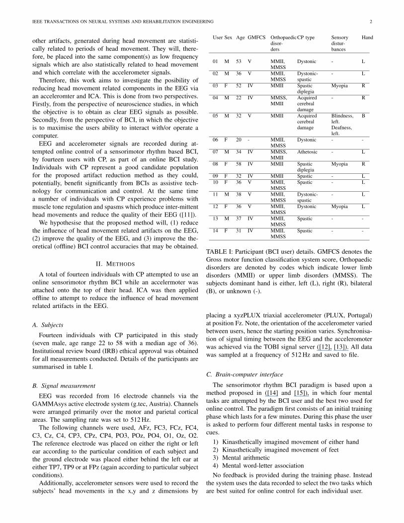
Fourteen individuals with CP participated in this study(seven male, age range 22 to 58 with a median age of 36).Institutional review board (IRB) ethical approval was obtainedfor all measurements conducted. Details of the participants aresummarised in table I.
B. Signal measurement
EEG was recorded from 16 electrode channels via theGAMMAsys active electrode system (g.tec, Austria). Channelswere arranged primarily over the motor and parietal corticalareas. The sampling rate was set to 512 Hz.
The following channels were used, AFz, FC3, FCz, FC4,C3, Cz, C4, CP3, CPz, CP4, PO3, POz, PO4, O1, Oz, O2.The reference electrode was placed on either the right or leftear according to the particular condition of each subject andthe ground electrode was placed either behind the left ear ateither TP7, TP9 or at FPz (again according to particular subjectconditions).
Additionally, accelerometer sensors were used to record thesubjects’ head movements in the x,y and z dimensions by
User Sex Age GMFCS Orthopaedicdisor-ders
CP type Sensorydistur-bances
Hand
01 M 53 V MMII,MMSS
Dystonic - L
02 M 36 V MMII,MMSS
Dystonic-spastic
- L
03 F 52 IV MMII Spasticdiplegia
Myopia R
04 M 22 IV MMSS,MMII
Acquiredcerebraldamage
- R
05 M 32 V MMII Acquiredcerebraldamage
Blindness,left.Deafness,left.
B
06 F 20 - MMII,MMSS
Dystonic - -
07 M 34 IV MMSS,MMII
Athetosic - L
08 F 58 IV MMII Spasticdiplegia
Myopia R
09 F 32 IV MMII Spastic - L10 F 36 V MMII,
MMSSSpastic - L
11 M 38 V MMII,MMSS
Dystonic-spastic
- L
12 F 36 V MMII,MMSS
Dystonic Myopia L
13 M 37 IV MMII,MMSS
Spastic - -
14 F 31 IV MMII,MMSS
Spastic - -
TABLE I: Participant (BCI user) details. GMFCS denotes theGross motor function classification system score, Orthopaedicdisorders are denoted by codes which indicate lower limbdisorders (MMII) or upper limb disorders (MMSS). Thesubjects dominant hand is either, left (L), right (R), bilateral(B), or unknown (-).
placing a xyzPLUX triaxial accelerometer (PLUX, Portugal)at position Fz. Note, the orientation of the accelerometer variedbetween users, hence the starting position varies. Synchronisa-tion of signal timing between the EEG and the acceleromoterwas achieved via the TOBI signal server ([12], [13]). All datawas sampled at a frequency of 512 Hz and saved to file.
C. Brain-computer interface
The sensorimotor rhythm BCI paradigm is based upon amethod proposed in ([14] and [15]), in which four mentaltasks are attempted by the BCI user and the best two used foronline control. The paradigm first consists of an initial trainingphase which lasts for a few minutes. During this phase the useris asked to perform four different mental tasks in response tocues.
1) Kinasthetically imagined movement of either hand2) Kinasthetically imagined movement of feet3) Mental arithmetic4) Mental word-letter associationNo feedback is provided during the training phase. Instead
the system uses the data recorded to select the two tasks whichare best suited for online control for each individual user.

IEEE TRANSACTIONS ON NEURAL SYSTEMS AND REHABILITATION ENGINEERING 3
During the second phase the user is again cued to performone of the two best performing (most discriminative) tasks.This time feedback is provided to the user informing themhow well they are performing at the specified task. The centralaim of this work is to investigate the aritfact removal method,therefore, for further details on the BCI system used the readeris referred to [16].
Performing each task should result in an event-related(de)synchronisation (ERD/S), a power decrease or increase ina specfic EEG frequency band relative to a reference interval.This occurs primarily over the sensorimotor areas and withinthe mu (8 - 13 Hz) and beta (13 - 30 Hz) frequency bands[17]. The timing, spatial, and spectral distributions of theERD/S differs between tasks and allows identification of whichpredefined task a BCI user is attempting.
D. Preprocessing
EEG signals are first pre-processed before application ofthe artifact removal method. This ensures any improvementsin signal quality seen may be atributed to the proposed artifactremoval method, and not to other pre-processing steps. Signalsare band-pass filtered in the range 1 to 40 Hz with a 4th orderButterworth filter.
E. Artifact reduction
Head movement related artifacts are removed from theEEG by first applying SOBI ICA ([18]) to seperate the EEGinto components which are maximally statistically independentfrom one another. SOBI is an extension of the ICA methodbased upon the concept of joint diagonalisation of time laggedcovariance matrices and is chosen because it is known to beeffective at seperating neuronal sources and artifacts in theEEG [19]. Each independant component (IC) derived by SOBIis then bandpass filtered (4th order, Butterworth) between 1- 10 Hz to create filtered ICs (ICf ). This frequency range ischosen to attempt to retain head movement related artifacts,which are observed in the accelerometer signals to reside< 10Hz, while removing low frequency drifts in baseline atbelow 1 Hz. Accelerometer signals are also filtered into thesame range.
Pearsons correlation coefficient is calculated between eachICf and each accelerometer signal (x,y, and z). Thresholdingis applied to remove ICf s which are highly correlated with oneor more of the accelerometer signals. The threshold is set totwo standard deviations above the mean correlation calculatedbetween the ICf and the acceleromter signals. ICf s whichhave greater correlations than this threshold with one or moreof the accelerometer signals are marked for removal. Thischoice of threshold is based upon visual inspection of ICf sfrom a subset of the data. ICf s containing movement artifactswere observed to have correlations with the accelerometersignals greater then 2 standard deviations above the mean.
Finally, the remaining original ICs, corresponding to theICf s not marked for removal, are mulitplied by the mixingmatrix produced by the SOBI ICA algorithm to reconstructcleaned EEG with the component(s) which highly correlatewith the accelerometer signals removed.
F. Validation
Validation of the efficacy of the artifact reduction methodis performed in five ways, (1) analytically, by measuring thechange in the signal quality index (SQI) [20] of the EEG,(2) by visually inspecting the signals before and after artifactremoval, (3) by visually inspecting the ERD/S maps (generatedvia the method described in [21]) during attempted motorimagery and mental task performance, (4) by measuring thestrength of the ERD/S signal in the original and cleaned EEG,and (5) by measuring the change in offline BCI classificationaccuracies achievable before and after cleaning the signals.
The SQI is described in [20]. It is based upon thresholdingvarious measures applied to the EEG such as the amplitude,the power spectrum, and various statistical measures such askurtosis etc. It attempts to determine if a given portion ofEEG is clean based upon whether the thresholds are exceeded.When applied to a long period of EEG the SQI is applied in amoving window of length 1 s, step size 0.25 s. Each windowin which thresholds are exceeded is assigned the value 1 andeach window in which they are not is assigned the value zero.The mean SQI over all windows then gives an approximatevalue of the cleanliness of the EEG. Thus, high mean SQIvalues indicate EEG heavily contaminated by artifacts.
To determine if there is an effect on BCI classificationresults, accuracies are calculated offline for both the originaland cleaned EEG. This ensures that comparisons made aremeaningful as with both datasets the accuracy reflects the bestachievable accuracy via offline analysis. For a given trial oflength 6 s a linear discriminant analysis (LDA) classifier isapplied in a sliding window of length 0.25 s. Band powerfeatures are extracted from the frequency bands 8 - 13 Hzand 13 - 30 Hz. Training and validation is performed within aleave-one-out train and validation scheme for trials recordedduring the training phase (4 class) and during the feedbackphase (2 class).
Note, due to factors such as subject fatigue and poor perfor-mance by subjects in the training phase not all subjects wereable to attempt the feedback phase. Additionally, differentnumbers of trials were attempted by each subject in boththe training and feedback phases. Hence, direct comparionsof accuracies between subjects is not possible.
Therefore, to compare the results Cohen’s kappa coefficientis calculated for every user from both the raw and cleanedEEG at the location of the peak accuracy. A paired t-test isthen used to compare the Cohen’s kappa values between theoriginal EEG and the cleaned EEG.
The strength of the ERD/S is assesed via measuring theproportion of ERD/S values which are deemed to be signifi-cant within time-frequency regions of interest. Significance isdetermined via the method described in [21]. The regions ofinterest are taken from 0 - 6 s relative to cue and in frequencybands 8 - 13 Hz and 13 - 30 Hz. The total number of significanttime-frequency locations of observed ERD/S is divided by thetotal possible number of significant ERD/S locations in eachregion of interest and averaged over sensorimotor channels,FC3, FCz, FC4, C3, Cz, C4, CP3, CPz, and CP4.

IEEE TRANSACTIONS ON NEURAL SYSTEMS AND REHABILITATION ENGINEERING 4
0 10 20 30 40 500
0.02
0.04
0.06
0.08
0.1
0.12
0.14
0.16
0.18
Frequency (Hz)
Pow
er (
dB)
Retained ICsRemoved ICs
Fig. 1: Mean power spectra of all ICs flagged for removalby the method vs. those not flagged for removal (i.e. thoseretained for reconstruction of the cleaned EEG.
III. RESULTS
It is important to first check the assumption that EMGartifacts that occur during head movement, such as EMG,are grouped into the same ICs as the low frequency headmovement induced artifacts. To do this the power spectra ofICs flagged for removal are inspected. Figure 1 illustrates themean power spectra for ICs marked for removal compared tothe mean power spectra for ICs marked as clean for a typicaluser (user 5).
Note, the ICs marked for removal contain broad bandsignals with both large low frequency components and largehigh frequency components. Visual inspection of the timeseries of these ICs reveals the high frequency componentsin the removed ICs correspond to EMG activity and thelow frequency components correlate with the accelerometersignals. Thus, the initial assumption upon which the methodis based, that ICA may group EMG activity into the same ICsas the low frequency head movement related artifacts to whichthey are statistically related, is validated.
Visual inspection of the EEG, before and after application ofthe proposed method, reveals a visually apparent decrease inthe amount of movement related artifacts. Figure 2 illustratesan example of EEG before and after artifact reduction takenfrom user 9. Note, there is a period of head movementfrom 237 s through to 244 s apparent in the accelerometersignals. After reduction of the movement components the headmovement contamination in the EEG is visibly reduced.
The power spectra is also illustrated in Figure 2. Theapplication of the artifact removal method is seem to reducethe power at frequencies above 30 Hz and below 2 Hz bya large amount while making only very small reductions inpower to other spectral regions. As the artifacts are broadband we would expect some reduction at a wide rangeof frequencies. The low frequency reduction is most likelythe removal of signal components correlating with the lowfrequency accelerometer signals, while the high frequency
TABLE II: Peak classification accuracies achieved during thetraining (4 class) and feedback (2 class) phases of sensorimotorBCI operation. All accuracies are found via offline analysison the original EEG and the cleaned EEG. Peak accuraciesare identified in the time range 2 - 6 s to discount short-livedbrain state responses to the cue ([22]). Statistically significantaccuracies (P < 0.05), assesed against the null hypothesisof equal chance classification into each class, are indicatedwith an asterisk (*). The ‘Raw’ column indicates the original,uncleaned EEG, and the ‘No.’ column indicates the numberof trials.
Training kappa Feedback kappa
User Raw Clean No. Raw Clean No.
01 0.551 * 0.469 * 50 0.711 * 0.800 * 46
02 0.325 0.350 41 0.591 0.727 * 23
03 0.430 * 0.418 * 87 0.750 * 0.750 * 9
04 0.433 * 0.367 32 - - -
05 0.366 * 0.341 42 0.809 * 0.667 * 22
06 0.309 0.405 * 32 - - -
07 0.435 * 0.435 * 64 - - -
08 0.538 * 0.410 * 40 1.000 * 0.945 * 56
09 0.341 0.363 * 45 0.778 * 0.722 * 19
10 0.400 * 0.367 32 - - -
11 0.419 * 0.387 * 34 0.606 0.757 * 30
12 0.379 * 0.316 96 0.870 * 0.774 * 32
13 0.375 * 0.350 41 0.727 * 0.772 * 23
14 0.428 * 0.405 * 43 0.700 * 0.750 * 21
reduction is EMG components that are statistically related tohead movements.
Table II lists the peak accuracies achieved during offlineattempts to classify the orginal EEG (pre-processed only)and the cleaned EEG after application of pre-processing andthe artifact reduction method. Results are listed for both thetraining phase (4 classes) and the feedback phase (2 classes)for users who completed enough trials for a classifier to betrained. Peak accuracies are taken from 2 s after the cue to theend of the trial to attempt to avoid cue related classifications.Statistically significant (P < 0.05) results are indicated by anasterisk (*).
Statistical significance is indicated against the null hypoth-esis of equal probability of each class been selected by theclassifier. Because each user performed tasks in a differentorder, became fatigued at different points, and had varyingdegrees of success at training the classifier, different numbersof trials are available for each user.
Comparison of Cohen’s kappa values, calculated at thelocations of peak accuracy, from each user between the orig-inal and cleaned EEG in the training and feedback phasesreveals no significant difference induced in the classificationaccuracies. For both the training and feedback phases thepaired t-test returns p values of p > 0.05.
Figure 3 illustrates an example of the ERD/S spectrum(calculated via the method described in [21]) during the feetmotor imagery task estimated from the original (pre-processedonly) EEG and from the cleaned EEG from user 1. Statistically

IEEE TRANSACTIONS ON NEURAL SYSTEMS AND REHABILITATION ENGINEERING 5
(a) EEG (b) Power spectra
Fig. 2: An example of the effect of head movement artifact reduction in the EEG from user 9. The top-left plot illustrates 16channel original EEG prior to artifact reduction. The centre-left plot illustrates the same signals after artifact reduction and thelower-left plot illustrates the accelerometer signals in the x,y, and z dimmensions. Finally, the plot on the right illustrates thepower spectra over the EEG recorded from user 9 before and after artifact removal.
significant time-frequency regions of ERD are illustrated inred and ERS in blue. Note, after reduction of the influenceof movement related artifacts ERD/S patterns are clearer andmore concentrated in the mu frequency band.
The distribution of ERD/S strengths over all users, in theregion of interest 0 - 6 s, 8 - 13 Hz, before and after artifactremoval, are also illustrated in Figure 3. Note, the mean ERD/Sstrength is increased after application of the artifact removalmethod. This increase is statistically significant as assesed viaa paired t-test (p < 0.05). Note, the ERD/S strength in theregion of interest 0 - 6 s, 13 - 30 Hz exhibited no significantchange between the original and cleaned EEG.
Note, it is also important to compare the baseline signalsfrom which the ERD/S is calculated. The mean power inthe baseline (-1.5 - 0 s relative to cue) over all subjects iscompared between the original EEG and the cleaned EEG.No significant differences in bandpower in the baseline isobserved, as assesed via a paired t-test (p > 0.05).
Changes in the SQI between the original raw EEG and thecleaned EEG are listed in table III. Mean ± the variance of theSQI values calculated over the entire EEG signals are listed foreach user alongisde the P-values for paired t-tests comparingthe means of the distributions of original and cleaned SQIs.The lower the SQI values the cleaner the signals. In all but4 cases there is a significant reduction (improvement) in SQIvalues, and in only 2 cases (users 7 and 12) does the SQI getsignificantly worse after attempted artifact reduction.
TABLE III: Mean signal quality index (SQI) measures ±variances for the original and cleaned EEG for each user. TheP-values for paired T-tests indicate the significance level of thechange in SQI values induced by the artifact removal method.
User Raw EEG Cleaned EEG p
01 0.0105 ± 0.0002 0.0083 ± 0.0002 0.0000
02 0.0181 ± 0.0005 0.0120 ± 0.0003 0.0000
03 0.0885 ± 0.0036 0.0631 ± 0.0026 0.0000
04 0.0105 ± 0.0002 0.0088 ± 0.0002 0.0016
05 0.0130 ± 0.0002 0.0108 ± 0.0002 0.0000
06 0.1566 ± 0.0010 0.1575 ± 0.0009 0.3185
07 0.0588 ± 0.0026 0.0991 ± 0.0039 0.0000
08 0.0088 ± 0.0001 0.0072 ± 0.0001 0.0000
09 0.0492 ± 0.0034 0.0204 ± 0.0003 0.0000
10 0.0080 ± 0.0002 0.0058 ± 0.0002 0.0000
11 0.0297 ± 0.0008 0.0154 ± 0.0004 0.0000
12 0.0499 ± 0.0031 0.0869 ± 0.0036 0.0000
13 0.0024 ± 0.0004 0.0022 ± 0.0003 0.3871
14 0.0524 ± 0.0023 0.0443 ± 0.0025 0.0000
IV. DISCUSSION
The proposed method reduces the influence of head move-ment artifacts in the EEG. This is validated by analyticallymeasuring the signal quality index and the strength of theERD/S before and after artifact removal, which both exhibitsignificant improvements. It is also seen in visual inspections

IEEE TRANSACTIONS ON NEURAL SYSTEMS AND REHABILITATION ENGINEERING 6
(a) Feet imagery, original EEG (b) Feet imagery, cleaned EEG
(c) Mean ERD/S strength
(d) Word-letter association, original EEG (e) Word-letter association, cleaned EEG
Fig. 3: ERD/S strengths. Examples are presented of sensorimotor rhythms relating to the condition feet imagery, in the originalEEG (plot 3a) and the cleaned EEG (plot 3b), and from the word-letter assiciation task, in the original EEG (plot( 3d) andthe cleaned EEG (plot 3e), from user 1. Each plot is split into 9 subplots illustrating the common average referenced (CAR)channels FC3, FCz, FC4, C3, Cz, C4, CP3, CPz, and CP4. Red colours indicate significant (p < 0.01) periods of ERD and bluesignificant periods of ERS relative to the reference period (-1.5 - 0 s, relative to the cue at 0 s). The significance is determinedvia bootstrapping, which is described [21], alongside the colour scales the ERD/S values are reported in. The vertical linedenotes the cue presentation time. Plot 3c illustrates the proportion of significant ERD/S values found in the time-frequencyregion of interest 0 - 6 s, 8-13 Hz in the original and cleaned EEG.
of the raw EEG and the ERD/S sprectra. The BCI classificationaccuracies are not significantly changed by application of themethod.
The magnitude of other artifacts such as the EMG may alsobe seen to be reduced after application of the method. Indeed,the IC(s) removed by the proposed method are observed to bebroad band and contain both low frequency changes, whichcorrelate with the accelermoter signals, and high frequencyEMG which shares a statistical relationship with head move-ment periods and is therefore grouped into the same ICs.
Visual inspection of the EEG before and after applicationof the method reveals a considerable reduction in both highand low frequency components. While inspection of the powerspectra reveals attentuation at a range of frequencies. Inspec-tion of the ERD/S maps before and after artifact removalreveals a reduction in noise and a sharpening of the appearenceof significant ERD/S patterns in the mu frequency band andtime range 0 - 6 s, which is known to be important in ERD/S[17]. Furthermore, this improvement is analytically measuredand seen to be statistically significant (p < 0.05).
There is a significant improvement (reduction) in the SQIin 10 users and a non-significant improvement in 1 other user.In two users (users 7 and 12) there is a significant worsening(increase) in the SQI. However, inspecting the experimentallogs and the raw EEG for these two users reveals that therewere some periods when either the ground electrode or one, ormore, other electrodes came loose during measurements due tolarge muscle spasms. Therefore, rendering portions of the EEGunusable and potentially confounding the attempted seperationof clean EEG and head movement artifacts into different ICs.
It is important to note that the method is not able to removeall types of artifacts, only those which are in some waystatistically related to head movements. Thus, electrode popsare not always removed, neither are blinks. To remove thesetypes of artifacts it would be necessery to couple the proposedmethod with a good eye blink removal method such as thatproposed in [23]. Alternatively, EOG signals could be recordedand, potentially, incorparated into the artifact removal method.However, this is a task for future work.
It is also important to highlight that the method is currently

IEEE TRANSACTIONS ON NEURAL SYSTEMS AND REHABILITATION ENGINEERING 7
applied to the entire signal. Thus, for EEG recorded over 5minutes the ICA method would be applied to the entire signalset. Therefore, to use the method in other applications, suchas BCI, it is necessery to modify the method to be appliedover much shorter time windows. This could be done by, forexample, training the ICA mixing matrix on a long periodof EEG (for example recorded during an initial calibrationsession) and then applying the trained mixing matrix to muchshorted time windows during online BCI operation. However,validation of the efficacy of this approach is a task requiringfurther investigation.
Additionally, it is important to consider different causes ofhead movement. Subjects are, by experimental design, seated,and do not move about the measurement room during thecourse of the experiment. Thus, head movements recorded onthe acceleormeter are a result of subject induced movement.However, this movement could be a result of either neck ortrunk movements. Both movement types have the potential toproduce similar accelerometer signals but are likely to producesome differing artifact patterns. Currently, the two movementtypes are not distinguished by the method, and, as the methodis applied to the whole signal duration, it is probable thata mixture of both movement types is used as a basis forIC removal. Future work will explore alternative possibilitiesfor differentiating these movement types and their affect onartifact contamination in the EEG.
An alternative use for the method could be to use theaccelerometer signals for epoch based rejection. Thus, anEEG epoch is rejected if the accelerometer signals at thecorresponding times exhibit large fluctuations. However, epochbased rejection, while straightforward, entails a potentiallylarge loss of the amount of available EEG. Artifact attentuationapproaches are therefore preferable.
When considering the classification accuracy at the BCItask, there are both increases and decreases in accuracies.Over all subjects there is no significant change in classificationaccuracy induced by the method, as assesed by a paired t-test(p > 0.05). However, users for whom there is a reductionin accuracy need special attention. The feedback accuracy ofuser 5 appears to decrease by a large amount. However, itis important to note that in this user there are only a smallnumber of trials (22) available during the feedback phase, solarge changes in accuracy correspond to only a small numberof trials having differences in classification results induced bythe artifact reduction method.
A number of other users also exhibit small reductionsin classification accuracies after application of the method.This is surprising given the significant increase in ERD/Sstrength and that no significant change in pre-ERD/S baselineis observed. Given that there is not a signifiant change inaccuracy it’s possible that a small number of trials containedcue related artifacts. However, the small number of trialsinvolved means statistical verification of this is difficult.
It is important to highlight the need to reduce the influenceof head movement artifacts, not only for neuroscience studies,but also for use of the EEG in BCI control. Without doingso one cannot be sure that classification results are notconfounded by artifacts.
While the proposed method does not produce perfectly cleanEEG it does significantly improve the signal quality in a largenumber of subjects. Considering the method is only basedupon 16 channel EEG the finding that there is a meaningfulreduction in head movement related artifacts is encouraging.Future work may address how well the method scales todiffering numbers of channels.
The proposed use of an accelerometer to attempt to atten-tuate movement artifacts is the first such attempt we’re awareof. However, it’s worth noting that the Emotiv BCI headset(Emotiv, USA) includes an integrated accelerometer. Thus, ourproposed method could be applicable to the application of thissystem to recording the EEG.
One of the next steps in the use of this method will be toattempt to apply it online during BCI operation to improvethe quality of the EEG prior to classification. Doing so will,hopefully, improve the online performance of BCI systems byreducing the influence of a major source of artifacts in theEEG.
To conclude, artifact reduction in the EEG by the use of anacceleromter to measure head movements coupled with ICA issuccesful at reducing the influence of head movement artifacts.This is measured in five ways, (1) visual inspection of the rawEEG signals reveals a reduction in head movement relatedartifacts, (2) visual inspection of the ERD/S spectra reveals astrengthening of the ERD/S after application of the method,(3) measuring the ERD/S strength reveals a significant increaseafter application of the method, (4) there is no significantchange in BCI classification accuracies after application ofthe method, and (5) there are significant improvements in SQIin 10 of the 14 subjects after application of the method.
Therefore, this approach has been shown to have the poten-tial to be used in improving the quality of EEG signals usedin neurosicence studies and also in the construction of BCIs.In particular the proposed method could be useful for a rangeof BCI user groups who are liable to produce head movementrelated artifacts during BCI operation.
ACKNOWLEDGMENT
This work was supported by the FP7 Framework EU Re-search Project ABC (No. 287774). This paper only reflects theauthors’ views and funding agencies are not liable for any usethat may be made of the information contained herein.
REFERENCES
[1] B.-K. Min, M. J. Marzelli, and S.-S. Yoo, “Neuroimaging-basedapproaches in the brain-computer interface.” Trends in biotechnology,vol. 28, no. 11, pp. 552–60, Nov. 2010. [Online]. Available:http://dx.doi.org/10.1016/j.tibtech.2010.08.002
[2] J. R. Wolpaw, N. Birbaumer, D. J. McFarland, G. Pfurtscheller, and T. M.Vaughan, “Brain-computer interfaces for communication and control.”Clinical neurophysiology, vol. 113, no. 6, pp. 767–791, Jun. 2002.
[3] J. d. R. Millan, R. Rupp, G. Muller-Putz, R. Murray-Smith, C. Giuliemma, M. Tangermann, C. Vidaurre,F. Cincotte, A. Kubler, R. Leeb, C. Neuper, K. Muller,and D. Mattia, “Combining braincomputer interfaces andassistive technologies: state-of-the-art and challenges,” Frontiersin Neuroprosthetics, vol. 4, no. 1, 2011. [Online]. Available:http://www.frontiersin.org/Neuroprosthetics/10.3389/fnins.2010.00161/abstract
IEEE TRANSACTIONS ON NEURAL SYSTEMS AND REHABILITATION ENGINEERING 8
[4] G. Prasad, P. Herman, D. Coyle, S. McDonough, and J. Crosbie,“Using motor imagery based brain-computer interface for post-strokerehabilitation,” in 2009 4th International IEEE/EMBS Conference onNeural Engineering. IEEE, Apr. 2009, pp. 258–262.
[5] K. K. Ang, C. Guan, K. S. G. Chua, A. B. T., C. Kuah, C. Wang, K. S.Phua, Z. Y. Chin, and H. Zhang, “Clinical study of neurorehabilitationin stroke using EEG-based motor imagery brain-computer interface withrobotic feedback,” in Proceedings of the 32nd Annual International IEEEEMBS Conference, 2010.
[6] V. Kaiser, I. Daly, F. Pichiorri, D. Mattia, G. Muller-Putz, and C. Neuper,“On the relationship between electrical brain responses to motor imageryand motor impairment in stroke,” Stroke, 2012.
[7] C. Neuper, G. R. Muller, A. Kubler, N. Birbaumer, and G. Pfurtscheller,“Clinical application of an EEG-based brain-computer interface: a casestudy in a patient with severe motor impairment.” Clinical neurophys-iology : official journal of the International Federation of ClinicalNeurophysiology, vol. 114, no. 3, pp. 399–409, Mar. 2003.
[8] M. J. Mir, “Electrocorticography (ECoG) based Brain computer interfacefor Cerebral palsy patients,” Ph.D. dissertation, Cornell University, 2009.
[9] A. Nijholt, D. P.-O. Bos, and B. Reuderink, “Turning shortcomingsinto challenges: Braincomputer interfaces for games,” EntertainmentComputing, vol. 1, no. 2, pp. 85–94, Apr. 2009.
[10] S. O’ Regan, S. Faul, and W. Marnane, “Automatic detection of EEGartefacts arising from head movements.” Conference proceedings : ...Annual International Conference of the IEEE Engineering in Medicineand Biology Society. IEEE Engineering in Medicine and BiologySociety. Conference, vol. 2010, pp. 6353–6, Jan. 2010. [Online].Available: http://www.ncbi.nlm.nih.gov/pubmed/21096691
[11] F. Miller, Cerebral palsy, F. Miller and E. Browne, Eds. Springer,2005.
[12] G. R. Muller-Putz, C. Breitwieser, F. Cincotti, R. Leeb, M. Schreuder,F. Leotta, M. Tavella, L. Bianchi, A. Kreilinger, A. Ramsay, M. Rohm,M. Sagebaum, L. Tonin, C. Neuper, and J. D. R. Millan, “Tools forBrain-Computer Interaction: A General Concept for a Hybrid BCI.”Frontiers in neuroinformatics, vol. 5, p. 30, Jan. 2011.
[13] C. Breitwieser, I. Daly, C. Neuper, and G. Muller-Putz, “Proposing a Standardized Protocol for Raw BiosignalTransmission.” IEEE transactions on bio-medical engineering,vol. 59, no. 3, pp. 852–859, Dec. 2011. [Online]. Available:http://www.ncbi.nlm.nih.gov/pubmed/22194230
[14] J. Faller, C. Vidaurre, E. V. C. Friedrich, U. Costa, and E. Opisso,“Automatic adaptation to oscillatory EEG activity in spinal cord injuryand stroke patients,” in TOBI workshop, no. 1, 2012, pp. 7–8.
[15] E. V. C. Friedrich, R. Scherer, and C. Neuper, “The effect of distinctmental strategies on classification performance for braincomputer inter-faces,” International Journal of Psychophysiology, vol. 84, pp. 86–94,2012.
[16] J. Faller, C. Vidaurre, T. Solis-Escalante, C. Neurper, and R. Scherer,“Autocalibration and recurrent adaptation: Towards a plug and playonline ERD-BCI,” IEEE Transactions on Neural Systems and Reha-bilitation Engineering, vol. 20, no. 3, pp. 313–319, 2012.
[17] G. Pfurtscheller and F. Lopes da Silva, “Event-related EEG/MEGsynchronization and desynchronization: basic principles,” Clinical Neu-rophysiology, vol. 110, no. 11, pp. 1842–1857, Nov. 1999.
[18] A. Belouchrani, K. Abed-Meraim, J.-F. Cardoso, and E. Moulines, “Ablind source separation technique using second-order statistics,” IEEETransactions on Signal Processing, vol. 45, no. 2, pp. 434–444, 1997.
[19] A. C. Tang, M. T. Sutherland, and C. J. McKinney, “Validationof SOBI components from high-density EEG.” NeuroImage,vol. 25, no. 2, pp. 539–53, Apr. 2005. [Online]. Available:http://dx.doi.org/10.1016/j.neuroimage.2004.11.027
[20] I. Daly, F. Pichiorri, J. Faller, V. Kaiser, A. Kreilinger, R. Scherer, andG. Muller-Putz, “What does clean EEG look like?” in Proceedings ofthe 34th Annual International Conference of the IEEE Engineering inMedicine and Biology Society, 2012.
[21] B. Graimann, J. E. Huggins, S. P. Levine, and G. Pfurtscheller,“Visualization of significant ERD/ERS patterns in multichannelEEG and ECoG data.” Clinical neurophysiology : official journalof the International Federation of Clinical Neurophysiology,vol. 113, no. 1, pp. 43–7, Jan. 2002. [Online]. Available:http://www.ncbi.nlm.nih.gov/pubmed/11801423
[22] G. Pfurtscheller, R. Scherer, G. R. Muller-Putz, and F. H.Lopes da Silva, “Short-lived brain state after cued motorimagery in naive subjects.” The European journal of neuroscience,vol. 28, no. 7, pp. 1419–26, Oct. 2008. [Online]. Available:http://www.ncbi.nlm.nih.gov/pubmed/18973568
[23] A. Schlogl, C. Keinrath, D. Zimmermann, R. Scherer, R. Leeb,and G. Pfurtscheller, “A fully automated correction method of EOGartifacts in EEG recordings.” Clinical neurophysiology : officialjournal of the International Federation of Clinical Neurophysiology,vol. 118, no. 1, pp. 98–104, Jan. 2007. [Online]. Available:http://www.ncbi.nlm.nih.gov/pubmed/17088100
PLACEPHOTOHERE
Ian Daly holds a PhD in Cybernetics and an M.Engin Computer Science from the University of Read-ing, UK and has been a post-doctoral researcher inthe Laboratory of Brain-computer interfaces at TUGraz, Austria since May 2011. His research focuseson Brain Computer Interfaces (BCIs), non-lineardynamics, machine learning, signal processing, bio-signal analysis, meta-heuristic search techniques andfunctional connectivity analysis in the EEG.
PLACEPHOTOHERE
Martin Billinger holds an M.Sc in Electrical En-gineering from the Graz University of Technology,Austria. He is doing his PhD in Computer Siencein the Laboratory of Brain-computer interfaces atTUG since November 2009. His research interestsare Brain-Computer Interfaces, bio-signal process-ing, machine learning, mathematical modeling andeffective connectivity analysis in the EEG.
PLACEPHOTOHERE
Gernot Muller-Putz Biography text here.
PLACEPHOTOHERE
Reinhold Scherer Biography text here.
Copyright © 2022 FDOKUMEN