Radiographic Artifacts - Canvas
15
Radiographic Artifacts NOT ESPECIALLY OLD OR VALUABLE • Chris Ober, DVM, PhD, DACVR • 14 February 2011 Artifacts Defined • Any opacity on the radiograph that does not correspond to an actual anatomic structure • Any misrepresentation of an actual anatomic structure • Anything decreasing radiographic quality Quick CR primer • Cassette exposed by x- rays; storage phosphors excited Quick CR primer • Cassette exposed by x- rays; storage phosphors excited • Cassette put into reader Quick CR primer • Laser shines onto detectors Quick CR primer • Laser shines onto detectors • Excited phosphors give off light photons
-
Upload
khangminh22 -
Category
Documents
-
view
1 -
download
0
Transcript of Radiographic Artifacts - Canvas

Radiographic Artifacts
NOT ESPECIALLY OLD OR VALUABLE
• Chris Ober, DVM, PhD, DACVR
• 14 February 2011
Artifacts Defined
• Any opacity on the radiograph that does not correspond to an actual anatomic structure
• Any misrepresentation of an actual anatomic structure
• Anything decreasing radiographic quality
Quick CR primer
• Cassette exposed by x-rays; storage phosphors excited
Quick CR primer
• Cassette exposed by x-rays; storage phosphors excited
• Cassette put into reader
Quick CR primer
• Laser shines onto detectors
Quick CR primer
• Laser shines onto detectors
• Excited phosphors give off light photons

Quick CR primer
• Laser shines onto detectors
• Excited phosphors give off light photons
• Light photons travel through light guide
Quick CR primer
• Laser shines onto detectors
• Excited phosphors give off light photons
• Light photons travel through light guide
• Image produced by computer
Quick DR primer
• Panel exposed by x-rays, detectors excited
• Data sent directly to computer via cord
• Image created
Sequence: Film-Screen
• Film storage & handling
• Positioning
• Exposure
• Film handling
• Film processing– Manual
– Automatic
• Image archiving
Sequence: CR (With Cassette)
• Cassette storage
• Positioning
• Exposure
• Postexposure
• Cassette reading
• Workstation
Sequence: DR (No Cassette)
• Panel storage
• Positioning
• Exposure
• Image reading & transfer
• Workstation

Another Classification System
• Color– Black
– White
• Distribution– Focal
– Regional or Global
Storage & Pre-Exposure Handling Artifacts
Screen-Film
• Pressure
• Abrasions & Scratches
• Fingerprints
• Static electricity
• Material in cassette
• Fog
• Light leak
CR & DR
• Storage scatter (CR)
Pressure Artifacts –Film Crease Crescents (SF)
• Black & focal
• Cause: rough handling
• Pressure from crease causes activation of crystals
• NOT caused by fingernail (usually)
Pressure Artifacts –Film Crease Crescents (SF)
Abrasions & Scratches (SF)
• Black or white, focal
• Cause: rough handling
• Black: pressure from abrasion activates crystals
• White: emulsion has been removed (scratch)
• Can feel texture in emulsion
Fingerprints (SF)
• Focal
• White: most common– Oil on fingertips blocks
developer
OR– Fixer on fingertips

Fingerprints (SF)
• Focal
• White: most common– Oil on fingertips blocks
developer
OR– Fixer on fingertips
Fingerprints (SF)
• Black: less common– Developer on
fingertips
• Prevent:– Keep fingers dry and
clean
– Touch only film edges
Hogge JP, et al.RadioGraphics 1999.
Static Electricity (SF)
• Black & focal
• Two patterns– Branching
– Smudge / Spot
• Cause: electrical discharge
Static Electricity (SF)
• Prevent:– Moderate humidity
– Avoid friction while handling film
– Antistatic cassette cleaners
Static Electricity (SF)
Hogge JP, et al.RadioGraphics 1999.
Material in Cassette /Dirty Screen (SF)
• White & focal
• Sharp margins (close to film – no penumbra)
• Cause: Visible light photons from screen can’t reach and expose film emulsion

Material in Cassette /Dirty Screen (SF)
• THUS “radiolucent”material can still cause this artifact
• Common causes– Hair
– Dust
Fog (SF)
• Increased blackness, usually regional or global
• Definition: any unwanted film exposure / development
• Decreased image contrast and detail
Fog (SF)
• Causes:– Light exposure
– Heat• (store at < 68 F)
– Humidity • (keep at 30-50%)
– Radiation (including scatter)
– Chemicals
– Old film
Fog (SF)
Hogge JP, et al.RadioGraphics 1999.
Light Leak (SF)
• Black, focal to global
• A form of fog
• Sources:– Storage bin (along 1
edge)
– Cassette not closed (1 edge or corner)
– Overhead light
– Safelight filter crack
Storage Scatter (CR)
• Black & global
• Like fog – unwanted exposure of CR plate
• Causes:– Background radiation
– Scatter from imaging procedure
Jiménez DA, et al.Vet Radiol Ultrasound 2008.

Positioning Artifacts
Screen-Film
• Magnification
• Foreshortening / distortion
• Patient rotation
• Upside-down cassette
• Grid cutoff
CR & DR
• Magnification
• Foreshortening / distortion
• Patient rotation
• Upside-down cassette (CR) or panel (DR)
• Grid cutoff
Magnification
• Object closer to film:– Closer to normal size
– Sharper margins
• Object farther from film:– Magnified
– Less distinct margins
Foreshortening / Distortion
• X-ray beam does not pass perpendicular to long axis of structure
• Differential magnification of structure
Foreshortening / Distortion
Patient Rotation
• A form of distortion• Lesions may be
masked by atypical superimposition on normal anatomy
• Unusual projection of normal anatomy may be incorrectly diagnosed as abnormal
Upside-down Cassette (SF)
• White & focal• Springs & latches on
back of cassette block x-ray beam
• Global lightness• Lead backing absorbs
some x-ray photons (film appears underexposed)

Upside-down Cassette (SF) Upside-down Cassette (SF)
Upside-down Cassette (CR)
• White & multifocal
• Construction of cassette back absorbs x-rays
• Pattern depends on manufacturer
Upside-down Cassette (CR)
Upside-down Panel (DR)
• White & multifocal
• Electronics in the back of the panel absorb x-rays
Grid Cutoff (SF, CR, DR)
• White, regional to global
• Orientation of lead strips in grid requires appropriate geometry relative to x-ray beam
• Wrong geometry lead strips attenuate more x-rays

Laterally DecenteredFocused Grid
Result: Too white acrossentire image
Off-Level Focused Grid
Result: Too white acrossentire image
Upside-Down Focused Grid
Result: OK in the center,Severely too white at periphery
Distance DecenteredFocused Grid
• Similar to upside-down grid– OK in the center,
Markedly too white at periphery
• Often not as severe as upside-down grid
Combined Lateral and Distance Decentering of Grid
Result: OK on one side,Gradually whiter towardother side
Exposure Artifacts
Screen-Film
• Motion
• Double exposure
• Overexposure
• Underexposure
• Material obstructing x-ray beam
CR & DR
• Motion
• Double exposure (CR)
• Overexposure (Saturation)
• Underexposure
• Material obstructing x-ray beam
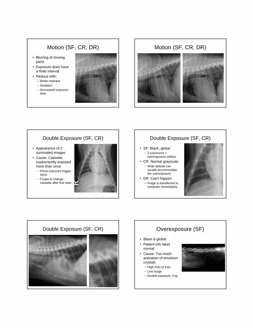
Motion (SF, CR, DR)
• Blurring of moving parts
• Exposure does have a finite interval
• Reduce with:– Better restraint
– Sedation
– Decreased exposure time
Motion (SF, CR, DR)
Double Exposure (SF, CR)
• Appearance of 2 summated images
• Cause: Cassette inadvertently exposed more than once– Press exposure trigger
twice
– Forget to change cassette after first view
Double Exposure (SF, CR)
• SF: Black, global– 2 exposures =
overexposure artifact
• CR: Normal grayscale– Wide latitude can
usually accommodate the overexposure
• DR: Can’t happen– Image is transferred to
computer immediately
Double Exposure (SF, CR) Overexposure (SF)
• Black & global
• Patient info label normal
• Cause: Too much activation of emulsion crystals– High mAs or kVp
– Line surge
– Double exposure, Fog

Saturation (CR, DR)
• Black, may only be apparent in parts of image
• Just like SF overexposure
• High exposure maxes out sensitivity range of detector– Anatomy (esp. thinner
parts) may be “burned through”
Saturation (CR, DR)
• Black, may only be apparent in parts of image
• Just like SF overexposure
• High exposure maxes out sensitivity range of detector– Anatomy (esp. thinner
parts) may be “burned through”
Paradoxical Overexposure, Saturation, & Planking (DR)
Paradoxical Overexposure, Saturation, & Planking (DR)
Underexposure (SF)
• White & global
• Patient info label normal
• Cause: Too little activation of emulsion crystals– Low mAs or kVp
– General x-ray obstruction
– X-ray tube troubles
Underexposure:Quantum Mottle (CR, DR)
• Noisy & global
• Computer algorithms adjust image to remain gray
• Low signal level causes grainy appearance and poor contrast & sharpness
• Cause: As with SF

Underexposure:Quantum Mottle (CR, DR)
Material ObstructingX-Ray Beam (SF, CR, DR)
• White, focal
• Cause: Any radiopaque structure in the path of the x-ray beam– On patient
– In patient
– On table
– On cassette
Material ObstructingX-Ray Beam (SF, CR, DR)
OMG!It’s a fracture fragment!
Material ObstructingX-Ray Beam (SF, CR, DR)
…or gravel on the table.
Material ObstructingX-Ray Beam (SF, CR, DR)
Material ObstructingX-Ray Beam (SF, CR, DR)
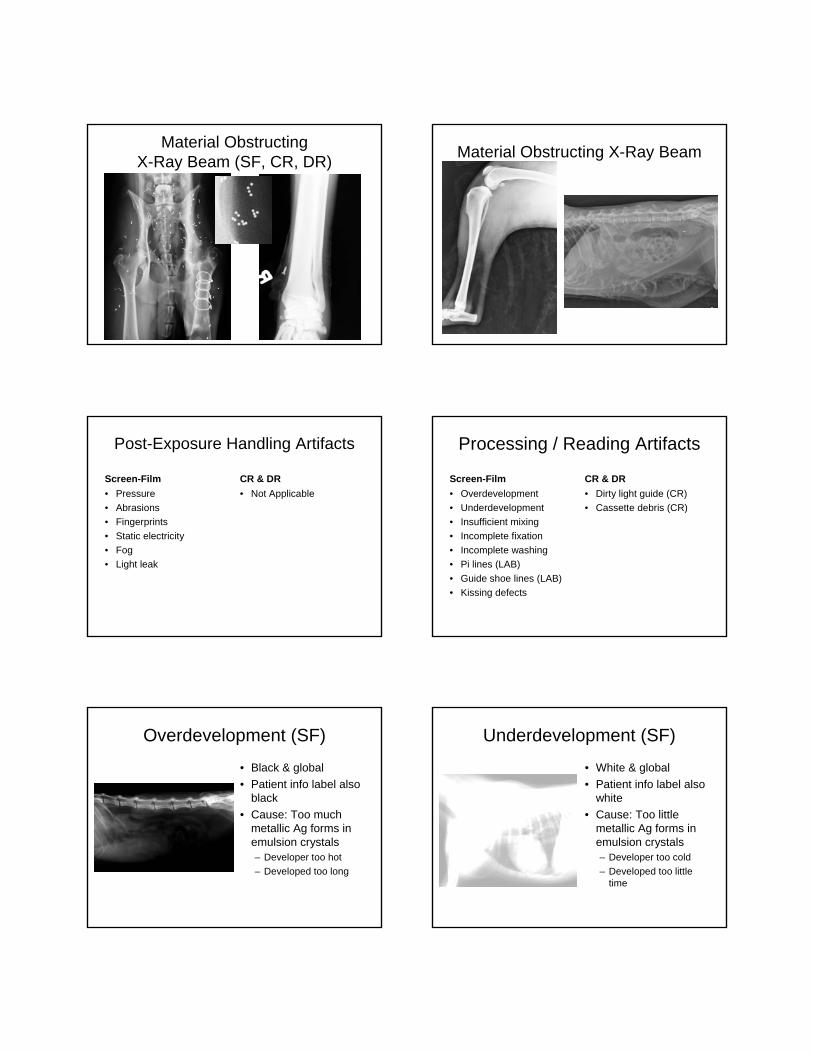
Material ObstructingX-Ray Beam (SF, CR, DR)
Material Obstructing X-Ray Beam
Post-Exposure Handling Artifacts
Screen-Film
• Pressure
• Abrasions
• Fingerprints
• Static electricity
• Fog
• Light leak
CR & DR
• Not Applicable
Processing / Reading Artifacts
Screen-Film
• Overdevelopment
• Underdevelopment
• Insufficient mixing
• Incomplete fixation
• Incomplete washing
• Pi lines (LAB)
• Guide shoe lines (LAB)
• Kissing defects
CR & DR
• Dirty light guide (CR)
• Cassette debris (CR)
Overdevelopment (SF)
• Black & global
• Patient info label also black
• Cause: Too much metallic Ag forms in emulsion crystals– Developer too hot
– Developed too long
Underdevelopment (SF)
• White & global
• Patient info label also white
• Cause: Too little metallic Ag forms in emulsion crystals– Developer too cold
– Developed too little time

Insufficient Mixing (SF) Inadequate Fixation (SF)
• Initially film is cloudy and milky
• Over time appears yellow/brown –Dichroic stain
Incomplete Washing / Rinsing (SF)
• Fixer remains– Cloudy, sticky residue
– Yellow-brown stain
– Sulfur smell
Kissing Defects (SF)
Dirty Light Guide (CR)
• White & focal• Sharp white line• Cause:
– Light emitted from plate reaches light guide
– Dirt blocks path for an entire line of data
– No light along that line = assumption that there was no exposure
Dirty Light Guide (CR)
• White & focal• Sharp white line• Cause:
– Light emitted from plate reaches light guide
– Dirt blocks path for an entire line of data
– No light along that line = assumption that there was no exposure

Dirty Light Guide (CR)
• White & focal• Sharp white line• Cause:
– Light emitted from plate reaches light guide
– Dirt blocks path for an entire line of data
– No light along that line = assumption that there was no exposure
Cassette Debris (CR)
• White & focal
• Sharp margins
• Essentially the same as FS dirty cassette
• EXCEPT…
Cassette Debris (CR)
• Problem is that photons emitted from plate during reading don’t reach photomultiplier tube
• (Contrast with FS, where light photons from screen don’t reach film)
Workstation Artifacts
Screen-Film
• Not Applicable
CR & DR
• Faulty Transfer (DR)
• Clipping
• Planking
• Uberschwinger
Faulty Transfer (CR, DR)
• Appearance: Anything goes, generally distorted anatomy
• Cause: Problems in data transfer to workstation– Memory problems
– Communication errors (e.g. loose cables)
– Power fluctuation
Faulty Transfer (DR)
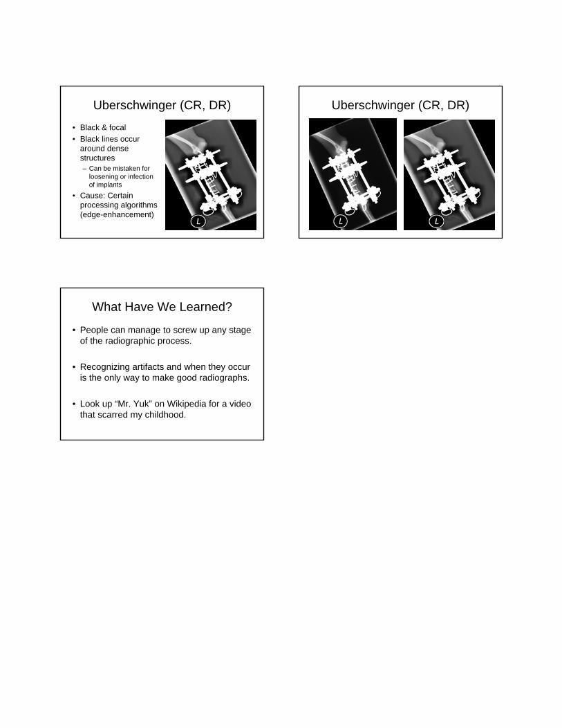
Uberschwinger (CR, DR)
• Black & focal
• Black lines occur around dense structures– Can be mistaken for
loosening or infection of implants
• Cause: Certain processing algorithms (edge-enhancement)
Uberschwinger (CR, DR)
What Have We Learned?
• People can manage to screw up any stage of the radiographic process.
• Recognizing artifacts and when they occur is the only way to make good radiographs.
• Look up “Mr. Yuk” on Wikipedia for a video that scarred my childhood.