Olive Oil and Walnut Breakfasts Reduce the Postprandial Inflammatory Response In Mononuclear Cells...
7
Atherosclerosis 204 (2009) e70–e76 Contents lists available at ScienceDirect Atherosclerosis journal homepage: www.elsevier.com/locate/atherosclerosis Olive oil and walnut breakfasts reduce the postprandial inflammatory response in mononuclear cells compared with a butter breakfast in healthy men Yolanda Jiménez-Gómez a , José López-Miranda a,∗ , Luis M. Blanco-Colio b , Carmen Marín a , Pablo Pérez-Martínez a , Juan Ruano a , Juan A. Paniagua a , Fernando Rodríguez c , Jesús Egido b , Francisco Pérez-Jiménez a a The Lipids and Arteriosclerosis Unit, Reina Sofía University Hospital, University of Córdoba, CIBER de Fisiopatologia de la Obesidad y Nutricion (CIBEROBN), Avenida Menéndez Pidal s/n, 14004 Córdoba, Spain b The Vascular Research Laboratory, Jiménez Díaz Foundation, Autonomous University of Madrid, Spain c The Clinical Analyses Service, Reina Sofía University Hospital, Spain article info Article history: Received 17 August 2007 Received in revised form 4 August 2008 Accepted 9 September 2008 Available online 17 September 2008 Keywords: Breakfast Mononuclear cells Proinflammatory cytokines Fatty acids Postprandial lipemia abstract Background: Inflammation is crucial in all stages of atherosclerosis, and few studies have investigated the effect of dietary fat on markers of inflammation related to this disease during the postprandial period. Objective: To evaluate the chronic effects of dietary fat on the postprandial expression of proinflammatory genes in peripheral blood mononuclear cells (PBMCs) in healthy subjects. Design: 20 healthy men followed three different diets for 4 weeks each, according to a randomized crossover design: Western diet: 15% protein, 47% carbohydrates (CHO), 38% fat (22% saturated fatty acid (SFA)); Mediterranean diet: 15% protein, 47% CHO, 38% fat (24% monounsaturated fatty acid (MUFA)); CHO-rich and n-3 diet: 15% protein, 55% CHO, <30% fat (8% polyunsaturated fatty acid (PUFA)). After 12- h fast, volunteers were given a breakfast with a fat composition similar to that consumed in each of the diets—butter breakfast: 35% SFA; olive oil breakfast: 36% MUFA; walnut breakfast: 16% PUFA, 4% -linolenic acid (LNA). Results: The butter breakfast induced a higher increase in tumor necrosis factor (TNF)- messenger RNA (mRNA) expression than the olive oil or walnut breakfasts (P =0.014) in PBMCs. Moreover, we found a higher postprandial response in the mRNA of interleukin (IL)-6 with the intake of butter and olive oil breakfasts than with the walnut breakfast (P = 0.025) in these cells. However, the effects of the three fatty breakfasts on the plasma concentrations of these proinflammatory parameters showed no significant differences (P = N.S.). Conclusion: Consumption of a butter-enriched meal elicits greater postprandial expression of proinflam- matory cytokine mRNA in PBMCs, compared to the olive oil and walnut breakfasts. © 2008 Elsevier Ireland Ltd. All rights reserved. 1. Introduction Atherosclerosis is the major cause of death in western soci- eties [1] and there is evidence that inflammation plays a central role in all phases of the atherosclerotic process [2]. A first step in this condition is the adhesion of circulating monocytes to the endothelium and its migration to the intima layer [3]. A crucial chemokine responsible for the recruitment of monocytes to inflam- matory lesions in the vasculature is monocyte chemoattractant protein-1 (MCP-1) [4,5]. This chemokine is highly expressed in the macrophage-rich area of the atherosclerotic lesions in human ∗ Corresponding author. Tel.: +34 957 218250; fax: +34 957 218250. E-mail address: [email protected] (J. López-Miranda). [6] and its expression and secretion from vascular cells have been proved to account for the increased monocyte chemotatic activ- ity [7]. Once monocytes have reached the subendothelial space, the modified LDL and various molecules produced by the T lym- phocytes, endothelial cells and smooth muscle cells stimulate the transformation of the monocytes into macrophages, which are important mediators of inflammation. In the final stage, the plaque is ruptured in the shoulder [8], area more vulnerable, which is enriched in T lymphocytes and macrophages. The diet, and particularly its fat content, can modulate the cardiovascular risk factors and the mechanisms related to the for- mation and development of the atheroma plaques [9,10]. However, the influence of the diet on atherosclerosis goes beyond its known effects on the classic cardiovascular risk factors [9]. Fatty acids and other components of the diet modulate the expression of several 0021-9150/$ – see front matter © 2008 Elsevier Ireland Ltd. All rights reserved. doi:10.1016/j.atherosclerosis.2008.09.011
-
Upload
iberoamericana -
Category
Documents
-
view
0 -
download
0
Transcript of Olive Oil and Walnut Breakfasts Reduce the Postprandial Inflammatory Response In Mononuclear Cells...
Oi
YCFa
Ab
c
a
ARRAA
KBMPFP
1
eriecmpt
0d
Atherosclerosis 204 (2009) e70–e76
Contents lists available at ScienceDirect
Atherosclerosis
journa l homepage: www.e lsev ier .com/ locate /a therosc leros is
live oil and walnut breakfasts reduce the postprandial inflammatory responsen mononuclear cells compared with a butter breakfast in healthy men
olanda Jiménez-Gómeza, José López-Mirandaa,∗, Luis M. Blanco-Coliob,armen Marína, Pablo Pérez-Martíneza, Juan Ruanoa, Juan A. Paniaguaa,ernando Rodríguezc, Jesús Egidob, Francisco Pérez-Jiméneza
The Lipids and Arteriosclerosis Unit, Reina Sofía University Hospital, University of Córdoba, CIBER de Fisiopatologia de la Obesidad y Nutricion (CIBEROBN),venida Menéndez Pidal s/n, 14004 Córdoba, SpainThe Vascular Research Laboratory, Jiménez Díaz Foundation, Autonomous University of Madrid, SpainThe Clinical Analyses Service, Reina Sofía University Hospital, Spain
r t i c l e i n f o
rticle history:eceived 17 August 2007eceived in revised form 4 August 2008ccepted 9 September 2008vailable online 17 September 2008
eywords:reakfastononuclear cells
roinflammatory cytokinesatty acidsostprandial lipemia
a b s t r a c t
Background: Inflammation is crucial in all stages of atherosclerosis, and few studies have investigated theeffect of dietary fat on markers of inflammation related to this disease during the postprandial period.Objective: To evaluate the chronic effects of dietary fat on the postprandial expression of proinflammatorygenes in peripheral blood mononuclear cells (PBMCs) in healthy subjects.Design: 20 healthy men followed three different diets for 4 weeks each, according to a randomizedcrossover design: Western diet: 15% protein, 47% carbohydrates (CHO), 38% fat (22% saturated fatty acid(SFA)); Mediterranean diet: 15% protein, 47% CHO, 38% fat (24% monounsaturated fatty acid (MUFA));CHO-rich and n-3 diet: 15% protein, 55% CHO, <30% fat (8% polyunsaturated fatty acid (PUFA)). After 12-h fast, volunteers were given a breakfast with a fat composition similar to that consumed in each of thediets—butter breakfast: 35% SFA; olive oil breakfast: 36% MUFA; walnut breakfast: 16% PUFA, 4% �-linolenicacid (LNA).Results: The butter breakfast induced a higher increase in tumor necrosis factor (TNF)-� messenger RNA
(mRNA) expression than the olive oil or walnut breakfasts (P = 0.014) in PBMCs. Moreover, we found ahigher postprandial response in the mRNA of interleukin (IL)-6 with the intake of butter and olive oilbreakfasts than with the walnut breakfast (P = 0.025) in these cells. However, the effects of the three fattybreakfasts on the plasma concentrations of these proinflammatory parameters showed no significantdifferences (P = N.S.).Conclusion: Consumption of a butter-enriched meal elicits greater postprandial expression of proinflam-PBM
[pitpti
matory cytokine mRNA in
. Introduction
Atherosclerosis is the major cause of death in western soci-ties [1] and there is evidence that inflammation plays a centralole in all phases of the atherosclerotic process [2]. A first stepn this condition is the adhesion of circulating monocytes to thendothelium and its migration to the intima layer [3]. A crucial
hemokine responsible for the recruitment of monocytes to inflam-atory lesions in the vasculature is monocyte chemoattractantrotein-1 (MCP-1) [4,5]. This chemokine is highly expressed inhe macrophage-rich area of the atherosclerotic lesions in human
∗ Corresponding author. Tel.: +34 957 218250; fax: +34 957 218250.E-mail address: [email protected] (J. López-Miranda).
ie
cmteo
021-9150/$ – see front matter © 2008 Elsevier Ireland Ltd. All rights reserved.oi:10.1016/j.atherosclerosis.2008.09.011
Cs, compared to the olive oil and walnut breakfasts.© 2008 Elsevier Ireland Ltd. All rights reserved.
6] and its expression and secretion from vascular cells have beenroved to account for the increased monocyte chemotatic activ-
ty [7]. Once monocytes have reached the subendothelial space,he modified LDL and various molecules produced by the T lym-hocytes, endothelial cells and smooth muscle cells stimulate theransformation of the monocytes into macrophages, which aremportant mediators of inflammation. In the final stage, the plaques ruptured in the shoulder [8], area more vulnerable, which isnriched in T lymphocytes and macrophages.
The diet, and particularly its fat content, can modulate the
ardiovascular risk factors and the mechanisms related to the for-ation and development of the atheroma plaques [9,10]. However,he influence of the diet on atherosclerosis goes beyond its knownffects on the classic cardiovascular risk factors [9]. Fatty acids andther components of the diet modulate the expression of several
erosc
gpi
etDttfitrtcMdtmn�oiotoacpwm
2
2
utsphbetsCt
2
v
dPnowt
tSwdwcscsteaoae
fcicwtTtbfsom
io
2
rwcE
TD
PCFSMP�
C
Y. Jiménez-Gómez et al. / Ath
enes involved in the inflammatory and immune response, such asroinflammatory cytokines, adhesion molecules, chemokines and
nflammatory enzymes [11–13].Changes in postprandial metabolism take place every time we
at a meal and alterations in this state may play an important role inhe development of cardiovascular and associated diseases [14–16].uring postprandial lipemia, there is an increase in circulating
riacylglycerol-rich lipoproteins (TRL), which may be deposited intohe arterial wall and accumulated in atheromatous plaques [16],ormation of highly oxidisable small, dense LDL and a reductionn the concentration of HDL [17]. Furthermore, it has been foundhat during this phase, when triacylglycerols (TG) and glucoseise, the neutrophil count increases with the subsequent produc-ion of proinflammatory cytokines and oxidative stress, with thesehanges possibly contributing to endothelial dysfunction [18,19].oreover, van Oostrom et al. [20] provided evidence that postpran-
ial triglyceridemia is related to the proinflammatory state due tohe high expression of the activation markers in neutrophils and
onocytes. Our group has also shown that butter and walnuts, butot olive oil, elicit postprandial activation of nuclear factor-�B (NF-B) in PBMCs in healthy men [21]. Since human beings spend muchf the day in the postprandial state it is important to understand thenflammatory changes that take place during this period in termsf the type of fat consumed. Our aim was therefore to evaluatehe chronic effect of the type of fat on the postprandial expressionf proinflammatory genes in PBMCs from healthy men. Becausepolipoprotein (apo)E is an important mediator of the clearance ofirculating TRL by receptor [22] and the apoE E2/E3/E4 polymor-hism is implicated in a variable lipid postprandial response [23],e realized the study in subjects with the apoE3/E3 genotype, theost common allele in the population.
. Methods
.1. Study subjects
Twenty male medical students all gave informed consent andnderwent a comprehensive medical history, physical examina-ion and clinical chemistry analysis before enrolment. None of theubjects showed signs of any chronic disease or obesity, and noneracticed unusually high levels of physical activity. The volunteersad normal biochemical parameters. They were selected on theasis of having the apoE3/E3 genotype, in order to avoid the alleleffects of this gene locus on postprandial lipemia [23]. None wasaking medications or vitamins known to affect plasma lipids. Thetudy protocol was approved by the Human Investigation Reviewommittee of the Reina Sofía University Hospital, according to Insti-utional and Good Clinical Practice guidelines.
.2. Study design
Each volunteer in the trial was subjected to three diet inter-ention periods of 4 weeks of duration, in a randomized crossover
sbfpM
able 1iet and breakfast composition (% of energy intake)
Western dietrich in SFA
Mediterranean dietenriched in virgin olive oil
High-CHO envegetal n-3 fa
rotein 15 15 15HO 47 47 55at 38 38 <30FA 22 <10 <10UFA 12 24 12
UFA 4 4 8-LNA 0.4 0.4 2
HO: carbohydrates; SFA: saturated fatty acid; MUFA: monounsaturated fatty acid; PUFA
lerosis 204 (2009) e70–e76 e71
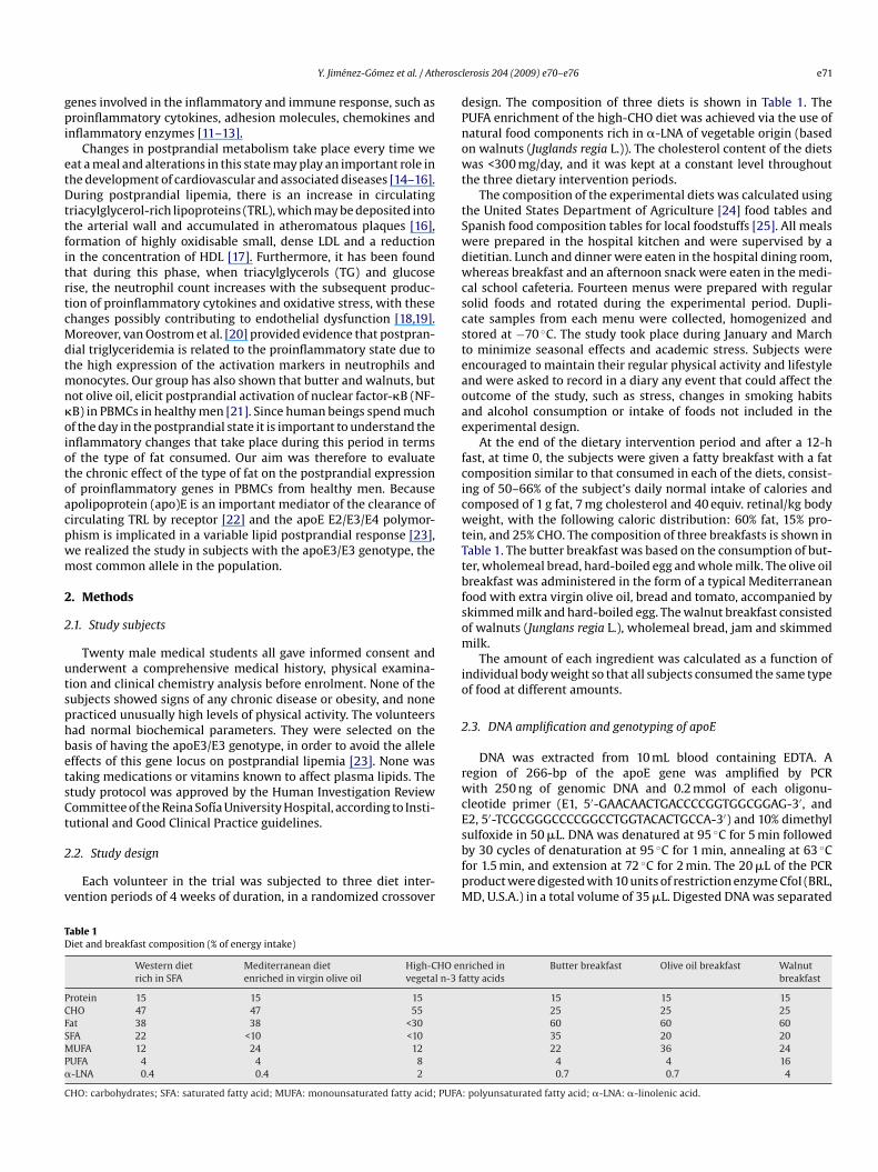
esign. The composition of three diets is shown in Table 1. TheUFA enrichment of the high-CHO diet was achieved via the use ofatural food components rich in �-LNA of vegetable origin (basedn walnuts (Juglands regia L.)). The cholesterol content of the dietsas <300 mg/day, and it was kept at a constant level throughout
he three dietary intervention periods.The composition of the experimental diets was calculated using
he United States Department of Agriculture [24] food tables andpanish food composition tables for local foodstuffs [25]. All mealsere prepared in the hospital kitchen and were supervised by aietitian. Lunch and dinner were eaten in the hospital dining room,hereas breakfast and an afternoon snack were eaten in the medi-
al school cafeteria. Fourteen menus were prepared with regularolid foods and rotated during the experimental period. Dupli-ate samples from each menu were collected, homogenized andtored at −70 ◦C. The study took place during January and Marcho minimize seasonal effects and academic stress. Subjects werencouraged to maintain their regular physical activity and lifestylend were asked to record in a diary any event that could affect theutcome of the study, such as stress, changes in smoking habitsnd alcohol consumption or intake of foods not included in thexperimental design.
At the end of the dietary intervention period and after a 12-hast, at time 0, the subjects were given a fatty breakfast with a fatomposition similar to that consumed in each of the diets, consist-ng of 50–66% of the subject’s daily normal intake of calories andomposed of 1 g fat, 7 mg cholesterol and 40 equiv. retinal/kg bodyeight, with the following caloric distribution: 60% fat, 15% pro-
ein, and 25% CHO. The composition of three breakfasts is shown inable 1. The butter breakfast was based on the consumption of but-er, wholemeal bread, hard-boiled egg and whole milk. The olive oilreakfast was administered in the form of a typical Mediterraneanood with extra virgin olive oil, bread and tomato, accompanied bykimmed milk and hard-boiled egg. The walnut breakfast consistedf walnuts (Junglans regia L.), wholemeal bread, jam and skimmedilk.The amount of each ingredient was calculated as a function of
ndividual body weight so that all subjects consumed the same typef food at different amounts.
.3. DNA amplification and genotyping of apoE
DNA was extracted from 10 mL blood containing EDTA. Aegion of 266-bp of the apoE gene was amplified by PCRith 250 ng of genomic DNA and 0.2 mmol of each oligonu-
leotide primer (E1, 5′-GAACAACTGACCCCGGTGGCGGAG-3′, and2, 5′-TCGCGGGCCCCGGCCTGGTACACTGCCA-3′) and 10% dimethyl
ulfoxide in 50 �L. DNA was denatured at 95 ◦C for 5 min followedy 30 cycles of denaturation at 95 ◦C for 1 min, annealing at 63 ◦Cor 1.5 min, and extension at 72 ◦C for 2 min. The 20 �L of the PCRroduct were digested with 10 units of restriction enzyme CfoI (BRL,D, U.S.A.) in a total volume of 35 �L. Digested DNA was separatedriched intty acids
Butter breakfast Olive oil breakfast Walnutbreakfast
15 15 1525 25 2560 60 6035 20 2022 36 24
4 4 160.7 0.7 4
: polyunsaturated fatty acid; �-LNA: �-linolenic acid.

e erosc
b1
2
1hb1poHcptHi[f
2
aPaccobkcap
2
mim
2
a
RaaAc
2
m(eDtaaURaBswMAta9tgmd
2
(ttmce
TP
T
0
3
G
72 Y. Jiménez-Gómez et al. / Ath
y electrophoresis on an 8% non-denaturing polyacrylamide gel at50 V for 2 h. Bands were visualized by silver staining.
.4. Lipid analysis
Venous blood samples were collected in tubes containingmg/mL EDTA in fasting, at time 0, and every 3 h until the 9thour after the ingestion of the breakfasts. Plasma was obtainedy low-speed centrifugation (1500 × g) for 15 min at 4 ◦C withinh of venipuncture. In order to reduce interassay variation,lasma samples were stored at −80 ◦C and analyzed at the endf the study. Lipid parameters were assessed with a DDPPIIitachi modular analyzer (Roche, Basel, Switzerland), using spe-ific reagents (Boehringer-Mannheim, Mannheim, Germany). Totallasma cholesterol (C) and TG concentrations and lipoprotein frac-ions were measured by colorimetric enzymatic techniques [26,27].DL–C levels were measured using colorimetric assay after precip-
tating the lipoproteins containing apoB with polyethylene glycol28]. LDL–C concentrations were calculated by using the Friedewaldormula based on the C, TG, and HDL–C values [29].
.5. Plasma fatty acid composition
Plasma lipids were first methylated and an aliquot of fattycid methyl esters was analyzed by gas chromatograph (Hewlett-ackard 5890; series II) equipped with a flame ionization detectornd a SP-2380 (Sulpeco, Bellefonte, PA, U.S.A.) fused silica capillaryolumn (60 m in length and with an internal diameter of 0.25 mm)oated with cyanopropylpolysiloxane (0.20 �m film thickness). Theven temperature program was isothermal at 160 ◦C for 8 minefore rising to 220 ◦C at a rate of 2 ◦C/min. The temperature wasept at 220 ◦C for 12 min. Hydrogen was used as carrier gas at aolumn-head pressure of 20 psi. The injector and detector temper-tures were 210 and 250 ◦C, respectively. Sample injections wereerformed in the split mode.
.6. Adhesion molecules immunoassay
Plasma concentration of MCP-1, IL-6 and TNF-� were deter-ined in duplicate with commercially available enzyme-linked
mmunosorbent assay kits (R&D Systems, Inc.) according to theanufactures guidelines.
.7. Isolation of PBMCs
Blood samples were diluted 1:1 in PBS, and cells were sep-rated in 5 mL Ficoll gradient (lymphocyte isolation solution,
stotp
able 2lasma fatty acid composition (percentage relative to the total fatty acids) according to th
ime and breakfast Fatty acid (% relative to the total fatty acids)
14:0 16:0
hButter 0.53 ± 0.18a 21.95 ± 1.40cOlive oil 0.40 ± 0.12b 21.16 ± 1.54Walnuts 0.38 ± 0.16b 20.9 ± 1.04b
hButter 1.40 ± 0.43a 23.10 ± 0.71aOlive oil 0.30 ± 0.15b 20.05 ± 0.84bWalnuts 0.40 ± 0.15b 20.10 ± 1.38b
lobal analysis P valuesTime effect 0.001 0.076Breakfast effect 0.001 0.009Breakfast × Time effect 0.001 0.001
a n = 20. Means in a column with different letters are significantly different, P < 0.05 (AN
lerosis 204 (2009) e70–e76
afer, Zaragoza, Spain) by centrifugation at 2000 × g for 30 mint 4 ◦C. PBMCs were collected, washed twice with cold PBS,nd resuspended in TRIZOL (Tri® Reagent, Sigma, St. Louis, MO).pproximately 95% of the cells were mononuclear cells (flowytometer, data not shown).
.8. Total RNA isolation and real-time RT-PCR
Total cellular RNA from PBMCs was extracted using the trizolethod according to the recommendations of the manufacturer
Tri® Reagent, Sigma, St. Louis, MO). Next, since PCR can detectven a single molecule of DNA, RNA samples were digested withNase I (AMP-D1, Sigma) before RT-PCR. The expression levels of
he TNF-�, IL-6 and MCP-1 genes, and of 18S ribosomal RNA (rRNA)s a housekeeping gene, were measured by real-time RT-PCR using7500 Real-Time PCR System (Applied Biosystems, Foster City, CA,.S.A). RT-PCR was performed in two steps as follows: 2 �g of totalNA underwent random primed reverse transcription for 10 mint 25 ◦C and 2 h at 37 ◦C using the High-Capacity cDNA kit (Appliediosystems) and RNase inhibitor (rRNasin 40 U/�L, Promega, Madi-on, U.S.A.) to synthesize the cDNA. Real-time PCR was realizedith 2 �L of cDNA and 18 �L of reaction mixture (10 �L of 2× Taq-an Universal PCR Master Mix (Applied Biosystems), 1 �L of 20×
ssays-on-DemandTM Gene Expression Assay Mix (Applied Biosys-ems) and 7 �L RNase-free water). After an initial hold of 2 mint 50 ◦C and 10 min at 95 ◦C, the samples were cycled 40 times at5 ◦C for 15 s and 60 ◦C for 60 s. For all quantitative cDNA analysis,he �Ct technique was utilised [30]. The expression of each targetene was normalized to housekeeping gene transcript. All measure-ents were performed in duplicate. Controls consisting of double
istilled H2O were negative in all runs.
.9. Statistical analysis
Statistical analysis used SPSS statistical software, version 11.0SPSS Inc., Chicago, IL). ANOVA for repeated measures was usedo analyze the differences in plasma lipid and lipoprotein concen-rations and plasma fatty acid composition. ANCOVA for repeated
easures, using the basal values of each mRNA and plasma con-entration as covariate, was utilised to study the differences in thexpression and production of proinflammatory cytokines under
tudy. In these analysis, we studied the statistical effects of theime alone or the change in the variable after ingesting fatty foodver the entire lipemic period (represented as P1) and the effect ofhe breakfast (represented as P2), independently of the time in theostprandial study. We also studied the effect of the interactione type of fat consumed during the postprandiala
18:1 18:2 18:3
18.24 ± 2.13b 23.30 ± 2.15b 0.34 ± 0.0720.12 ± 2.70a 22.00 ± 2.90b 0.35 ± 0.08
17.8 ± 2.42b 25.9 ± 1.64a 0.41 ± 0.15
18.35 ± 2.30b 23.10 ± 1.10b 0.38 ± 0.12b27.43 ± 3.98a 21.90 ± 2.28b 0.38 ± 0.04b17.50 ± 1.21b 30.15 ± 1.60a 1.88 ± 0.63a
0.022 0.129 0.0010.001 0.001 0.0010.001 0.002 0.001
OVA for repeated measures). Values are means ± S.D. (all such values).
Y. Jiménez-Gómez et al. / Atherosclerosis 204 (2009) e70–e76 e73
Table 3Plasma lipid and lipoprotein concentrations according to the type of fat consumed during the postprandial phasea
Time and breakfast Lipids and lipoproteins (mmol/L)
Total C TG HDL–C LDL–C
0 hButter 3.85 ± 0.12a 0.78 ± 0.07 1.17 ± 0.05 2.17 ± 0.08aOlive oil 3.63 ± 0.12b 0.78 ± 0.07 1.18 ± 0.04c 1.91 ± 0.07bWalnuts 3.57 ± 0.14b 0.72 ± 0.06 1.12 ± 0.05b 1.96 ± 0.10b
3 hButter 3.62 ± 0.12 1.42 ± 0.12 1.05 ± 0.04 1.76 ± 0.09Olive oil 3.59 ± 0.12 1.59 ± 0.18 1.04 ± 0.04 1.65 ± 0.09Walnuts 3.49 ± 0.12 1.34 ± 0.12 1.04 ± 0.04 1.57 ± 0.13
6 hButter 3.58 ± 0.14 0.88 ± 0.09a 1.06 ± 0.04 1.90 ± 0.09Olive oil 3.53 ± 0.14 0.64 ± 0.04b 1.07 ± 0.04 1.99 ± 0.10Walnuts 3.46 ± 0.13 0.64 ± 0.04b 1.07 ± 0.04 1.92 ± 0.08
9 hButter 3.72 ± 0.14 0.59 ± 0.05 1.14 ± 0.04 2.15 ± 0.10dOlive oil 3.58 ± 0.13 0.53 ± 0.03c 1.12 ± 0.04 2.07 ± 0.10Walnuts 3.55 ± 0.15 0.62 ± 0.04b 1.10 ± 0.04 1.95 ± 0.09b
Global analysis P valuesTime effect 0.001 0.001 0.001 0.001
0.860.00
5 (AN
omawtlce
3
3
daappMwlwatc
3
fthHawi(p
olwt(
3
bptWhoopb6cwtcnlt
Metr
3p
Breakfast effect 0.614Breakfast × Time effect 0.036
a n = 20. Means in a column with different letters are significantly different, P < 0.0
f both factors – breakfast and time – which is indicative of theagnitude of the postprandial response in each meal (represented
s P3). When statistically significant effects were found, an ANOVAas used to identify group differences in each time. A study of
he relation among parameters was carried out using Pearson’sinear correlation coefficient. A probability of less than 0.05 wasonsidered significant. All data presented in the text and tables arexpressed as mean ± S.E.M.
. Experimental results
.1. Plasma fatty acid composition
The data suggest that the type of diet consumed during theietary intervention period and the intake of the fatty breakfastst the end of each period has a direct influence on plasma fattycid composition (Table 2). At the end of the dietary interventioneriod, we observed that the Western diet raised the proportion ofalmitic and myristic fatty acids (P < 0.05). On the other hand, theediterranean diet produced an increase in oleic acid (P < 0.05),hile the high-CHO diet enriched in n-3 fatty acids augmented
inoleic (P < 0.05) and linolenic acids. In the postprandial period,e found an increase (P < 0.05) of palmitic and myristic fatty acids
nd of oleic acid with the butter and olive oil breakfasts, respec-ively. Moreover, we showed that the walnut breakfast increasedoncentrations of linolenic and linoleic fatty acids (P < 0.05).
.2. Diet intake and postprandial lipemia
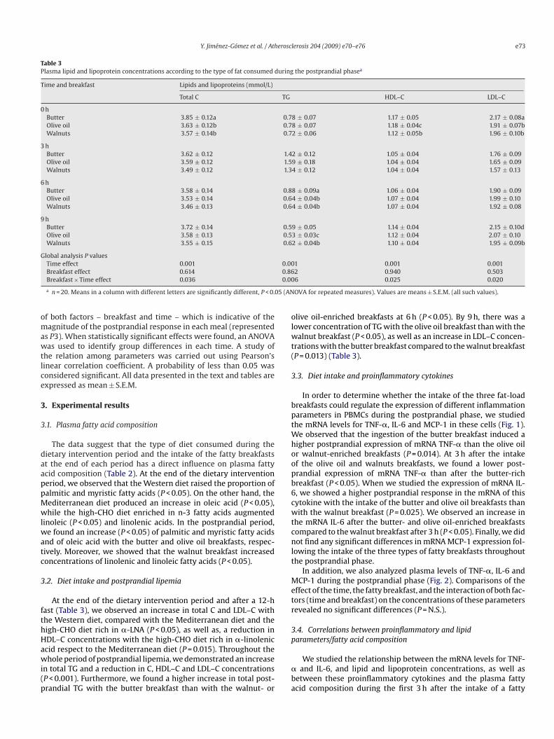
At the end of the dietary intervention period and after a 12-hast (Table 3), we observed an increase in total C and LDL–C withhe Western diet, compared with the Mediterranean diet and theigh-CHO diet rich in �-LNA (P < 0.05), as well as, a reduction inDL–C concentrations with the high-CHO diet rich in �-linolenic
cid respect to the Mediterranean diet (P = 0.015). Throughout thehole period of postprandial lipemia, we demonstrated an increasen total TG and a reduction in C, HDL–C and LDL–C concentrationsP < 0.001). Furthermore, we found a higher increase in total post-randial TG with the butter breakfast than with the walnut- or
�ba
2 0.940 0.5036 0.025 0.020
OVA for repeated measures). Values are means ± S.E.M. (all such values).
live oil-enriched breakfasts at 6 h (P < 0.05). By 9 h, there was aower concentration of TG with the olive oil breakfast than with thealnut breakfast (P < 0.05), as well as an increase in LDL–C concen-
rations with the butter breakfast compared to the walnut breakfastP = 0.013) (Table 3).
.3. Diet intake and proinflammatory cytokines
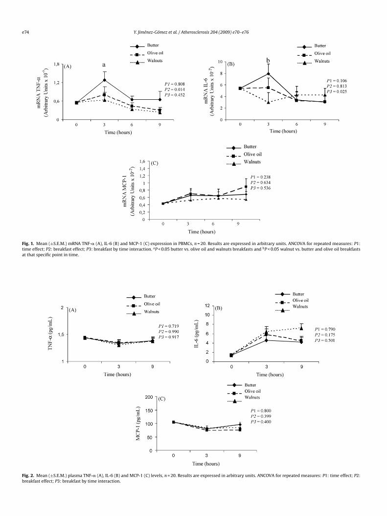
In order to determine whether the intake of the three fat-loadreakfasts could regulate the expression of different inflammationarameters in PBMCs during the postprandial phase, we studiedhe mRNA levels for TNF-�, IL-6 and MCP-1 in these cells (Fig. 1).
e observed that the ingestion of the butter breakfast induced aigher postprandial expression of mRNA TNF-� than the olive oilr walnut-enriched breakfasts (P = 0.014). At 3 h after the intakef the olive oil and walnuts breakfasts, we found a lower post-randial expression of mRNA TNF-� than after the butter-richreakfast (P < 0.05). When we studied the expression of mRNA IL-, we showed a higher postprandial response in the mRNA of thisytokine with the intake of the butter and olive oil breakfasts thanith the walnut breakfast (P = 0.025). We observed an increase in
he mRNA IL-6 after the butter- and olive oil-enriched breakfastsompared to the walnut breakfast after 3 h (P < 0.05). Finally, we didot find any significant differences in mRNA MCP-1 expression fol-
owing the intake of the three types of fatty breakfasts throughouthe postprandial phase.
In addition, we also analyzed plasma levels of TNF-�, IL-6 andCP-1 during the postprandial phase (Fig. 2). Comparisons of the
ffect of the time, the fatty breakfast, and the interaction of both fac-ors (time and breakfast) on the concentrations of these parametersevealed no significant differences (P = N.S.).
.4. Correlations between proinflammatory and lipidarameters/fatty acid composition
We studied the relationship between the mRNA levels for TNF-and IL-6, and lipid and lipoprotein concentrations, as well as
etween these proinflammatory cytokines and the plasma fattycid composition during the first 3 h after the intake of a fatty
e74 Y. Jiménez-Gómez et al. / Atherosclerosis 204 (2009) e70–e76
Fig. 1. Mean (±S.E.M.) mRNA TNF-� (A), IL-6 (B) and MCP-1 (C) expression in PBMCs, n = 20. Results are expressed in arbitrary units. ANCOVA for repeated measures: P1:time effect; P2: breakfast effect; P3: breakfast by time interaction. aP < 0.05 butter vs. olive oil and walnuts breakfasts and bP < 0.05 walnut vs. butter and olive oil breakfastsat that specific point in time.
Fig. 2. Mean (±S.E.M.) plasma TNF-� (A), IL-6 (B) and MCP-1 (C) levels, n = 20. Results are expressed in arbitrary units. ANCOVA for repeated measures: P1: time effect; P2:breakfast effect; P3: breakfast by time interaction.
erosc
bnn
4
pmorb
ewttsdetnoMtaipcpclpcptpbipbdedPth[
cc[mtoisvwttbfsi
ifhopm�dsetecaB[ib
oamortmc
A
2FtNdt((Sd
R
Y. Jiménez-Gómez et al. / Ath
reakfast without considering the type of fat consumed. We didot find correlation among the different studied parameters (datao shown).
. Discussion
Ours results show that a butter-enriched breakfast increasesostprandial expression of mRNA TNF-� in PBMCs from healthyen with apoE3/E3 genotype compared with a breakfast rich in
live oil or walnuts. Moreover, we observed a higher postprandialesponse of mRNA IL-6 in these cells with the butter and olive oilreakfasts compared to the breakfast rich in walnuts.
Few studies have investigated changes in inflammatory mark-rs related to atherosclerosis during the postprandial state [31,32],hich is the normal metabolic condition of the human beings
hroughout the day, and none of them considered the effect ofhe type of fat on such response. Our group has already demon-trated that the consumption of an olive oil-enriched breakfastoes not activate NF-�B in PBMCs as do butter- and walnut-nriched breakfasts [21]. Nevertheless, the chronic effect of theype of dietary fat on the postprandial inflammatory response isot known. MCP-1 regulates the transmigration of monocytes andther mononuclear cells on inflammatory sites [33]. Moreover,CP-1 also recruits monocytes into atherosclerotic lesions and into
he infarct zone after myocardial infarction [34]. However, when wenalyzed this chemokine, we did not observe significant differencesn the expression and plasma levels of MCP-1 throughout the post-randial phase. This result may be explained, at least in part, by theharacteristics of our sample population, which consisted of com-letely healthy young people. On the other hand, TNF-� and IL-6an mediate the systemic effects of inflammation, including fever,oss of appetite, mobilization of protein and fat, and acute phaserotein synthesis. The production of sufficient amounts of theseytokines is clearly beneficial in response to infection, but inap-ropriate quantities or overproduction may be harmful. In ordero alleviate inflammation, therefore, it is important to inhibit theroduction of proinflammatory cytokines. Nutrition strategies maye desirable to manipulate the secretion of these cytokines and,
n this regard, our present study showed a higher expression ofroinflammatory cytokines in PBMCs following a butter-enrichedreakfast than after those rich in olive oil or walnuts. However, weid not observe significant postprandial differences in plasma lev-ls of these proinflammatory cytokines. The fact that we only foundifferences in the expression of TNF-� and IL-6 at mRNA levels inBMCs following the intake of the three breakfasts may be due tohat the synthesis and secretion processes of these proteins do notappen simultaneously, and to the short half-life of both cytokines35,36].
Biologically, TNF-� acts as a trigger that activates a cascade ofytokine production. A number of regulatory agents, including glu-orticoids, acute-phase proteins, eicosanoids and soluble receptors37], limit TNF-� production. Lipids have been shown to be potent
odulators of inflammation since a large number of the modula-ory compounds cited previously are derived from the hydrolysisf membrane phospholipids. Because of this effect of lipids in thenflammation and, to the fact that apoE genotype is know to haveignificant effects on postprandial lipemia [23], we study only indi-idual homozygous for the most common allele, apoE3/E3. Whene investigated whether the effect observed in the proinflamma-
ory cytokines was due to the change in the lipid profile [38] or in
he plasma fatty acid composition, we did not observe a correlationetween these parameters and mRNA for TNF-� and IL-6. There-ore, it is possible that other factors take part in the process. Wepeculated that, as indicated in our previous study [21], an increasen activation of NF-�B may be one of the main mechanisms of the[
lerosis 204 (2009) e70–e76 e75
nflammatory properties of the butter breakfast, since this nuclearactor plays a central role in regulating the cytokine network. Itas also been observed that the modulatory effect of fatty acidsn the synthesis of proinflammatory cytokines may be due to aeroxisome proliferative activated receptor � (PPAR�)-dependentechanism. Desreumaux et al. [39] showed that activation of PPAR-in the colon inhibits mucosal production of IL-1� and TNF-� by
ownregulation of the NF-�B and mitogen-activated protein kinaseignal pathways. Furthermore, we observed a higher mRNA IL-6xpression in PBMCs following the butter and olive oil breakfastshan with the walnut breakfast. A mechanism possibly capable ofxplaining this result is that n-3 polyunsaturated fatty acids inducehanges in both cycloxygenase and lipoxygenase products, such asreduction in the production of prostaglandin E2 and leukotriene4 [40]. Since both metabolites enhance the release of IL-6 in vitro41], a reduction in these eicosanoids could explain the reductionn mRNA IL-6 that we observed in this study with the walnut-richreakfast.
To summarize, this study has shown that breakfasts rich in oliveil or walnuts had an anti-inflammatory effect, which may providen additional beneficial mechanism of both types of fat in the pri-ary and secondary prevention of cardiovascular disease. The final
bjective in the prevention and treatment of coronary atheroscle-osis is to reduce the risk of new heart attacks and mortality dueo cardiovascular failure. Identifying a suitable diet is thus funda-
ental for avoiding the development of this disease and associatedardiovascular events.
cknowledgments
Supported in part by research grants from the MCYT (AGL004/07907, AGL2006-01979/ALI to JL-M and SAF2003-05770 toP-J), the Spanish Ministry of Health (FIS PI041619 to CM, FIS 01/449o JL-M, and CB06/03/0047 (CIBER Fisiopatologia de la Obesidad yutricion is an initiative of ISCIII) to FP-J), Consejería de Salud, Juntae Andalucía (03/75, 04/238 to JL-M, 03/73, 04/191 to FP-J, 05/396o CM), the Diputación Provincial de Córdoba (to FP-J), the CAM08.4/0021.1/2003 to JE) and the Spanish Cardiovascular Networkto JE). We extend our thanks to Canoliva (Antonio Cano e HijosA, Luque, Córdoba), who generously donated the olive oil for theietary experiments, and also to the volunteers.
eferences
[1] Braunwald E. Shattuck lecture—cardiovascular medicine at the turn of themillennium: triumphs, concerns, and opportunities. N Engl J Med 1997;337:1360–9.
[2] Ross R. Atherosclerosis—an inflammatory disease. N Engl J Med 1999;340:115–26.
[3] Ross R. The pathogenesis of atherosclerosis: a perspective for the 1990s. Nature1993;362:801–8.
[4] Gu L, Okada Y, Clinton SK, et al. Absence of CC chemokine receptor-2 reducesatherosclerosis in low density lipoprotein receptor-deficient mice. Mol Cell1998;2:275–81.
[5] De Lemos JA, Morrow DA, Sabatine MS, et al. Association between plasmachemoattractant protein-1 and long-term clinical outcomes in patients withacute coronary syndromes. Circulation 2003;107:690–5.
[6] Yla-Herttuala S, Lipton BA, Rosenfeld ME, et al. Expression of monocytechemottractant protein-1 in macrophage-rich areas of human and rabbitatherosclerosis lesions. Proc Natl Acad Sci USA 1991;88:5252–6.
[7] Okada M, Matsumori A, Ono K, et al. Cyclic stretch upregulates productionof interleukin-8 and monocyte chemotactic and activating factor/monocytechemoattractant protein-1 in human endothelial cells. Atheroscler ThrombVasc Biol 1998;18:894–901.
[8] Fuster V. Elucidation of the role of plaque instability and rupture in acute coro-
nary events. Am J Cardiol 1995;76:24C–33C.[9] Perez-Jimenez F, Lopez-Miranda J, Mata P. Protective effect of dietarymonounsaturated fat on arteriosclerosis: beyond cholesterol. Atherosclerosis2002;163:385–98.
10] Mensink RP, Katan MB. Effect of dietary fatty acids on serum lipids and lipopro-teins. A meta-analysis of 27 trials. Arterioscler Thromb 1992;12:911–9.
e erosc
[
[
[
[
[
[
[
[
[
[
[
[
[
[
[
[
[
[
[
[
[
[
[
[
[
[
[
[
[
[
76 Y. Jiménez-Gómez et al. / Ath
11] Paschos GK, Rallidis LS, Liakos GK, et al. Background diet influences the anti-inflammatory effect of alpha-linolenic acid in dyslipidaemic subjects. Br J Nutr2004;92:649–55.
12] Baer DJ, Judd JT, Clevidence BA, Tracey RP. Dietary fatty acids affect plasmamarkers of inflammation in healthy men fed controlled diets: a randomisedcrossover study. Am J Clin Nutr 2004;79:969–73.
13] Grimble RF. The modulation of immune function by dietary fat. Br J Int Care1994;4:159–67.
14] Patsch W, Esterbauer H, Foger B, Patsch JR. Postprandial lipemia and coronaryrisk. Curr Atheroscler Rep 2000;2:232–42.
15] Parks EF. Recent findings in the study of postprandial lipemia. Curr AtherosclerRep 2001;3:462–70.
16] Zilversmit DB. Atherogenesis: a postprandial phenomenon. Circulation1979;60:473–85.
17] Patsch JR, Prasad S, Gotto Jr AM, Bengtsson-Olivecrona G. Postprandial lipemia.A key for the conversion of high density lipoprotein2 into high density lipopro-tein3 by hepatic lipase. J Clin Invest 1984;74:2017–23.
18] van Oostrom AJ, Sijmonsma TP, Rabelink TJ, Van Asbeck BS, Cabezas MC.Postprandial leukocyte increase in healthy subjects. Metabolism 2003;52:199–202.
19] van Oostrom AJ, Sijmonsma TP, Verseyden C, et al. Postprandial recruit-ment of neutrophils may contribute to endothelial dysfunction. J Lipid Res2003;44:576–83.
20] van Oostrom AJ, Rabelink TJ, Verseyden C, et al. Activation of leukocytesby postprandial lipemia in healthy volunteers. Atherosclerosis 2004;177:175–82.
21] Bellido C, López-Miranda J, Blanco-Colio LM, et al. Butter and walnuts, but notolive oil, elicit postprandial NF-�B activation in peripheral mononuclear bloodcells from healthy male volunteers. Am J Clin Nutr 2004;80:1487–91.
22] Mahley RW. Apolipoprotein E: cholesterol transport protein with expandingrole in cell biology. Science 1988;240:622–30.
23] Boerwinkle E, Brown S, Sharrett AR, Heiss G, Patsch W. Apolipoprotein Epolymorphism influences postprandial retinyl palmitate but not triglycerideconcentrations. Am J Hum Genet 1994;54:341–60.
24] Human Nutrition Information Service, Agriculture Handbook No. 8. Depart-ment of Agriculture Composition of Foods. Washington, DC: US GovernmentPrinting Office, 1987.
25] Varela G. Tablas de composición de alimentos (Spanish) (Food compositiontables). Madrid: Instituto de Nutrición. CSIC; 1980.
26] Bucolo G, David H. Quantitative determination of serum triglycerides by use ofenzymes. Clin Chem 1973;19:476–82.
27] Allain CC, Poon LS, Chang CSG, Richmond W, Fu PC. Enzymatic determinationof total serum cholesterol. Clin Chem 1974;20:470–5.
[
lerosis 204 (2009) e70–e76
28] Brigg CJ, Anderson D, Johnson P, Deegan T. Evaluation of the polyethylene glycolprecipitation method for the estimation of high-density lipoprotein cholesterol.Ann Clin Biochem 1981;18:177–81.
29] Friedewald WT, Levy RI, Fredrickson DS. Estimation of the concentration oflow-density lipoprotein cholesterol in plasma without use of a preparativeultracentrifuge. Clin Chem 1972;18:499–502.
30] Cohen CD, Franch K, Schlondorff D, Kretzler M. Cuantitative gene expressionanalysis in renal biopsies: a novel protocol for a high-throughput multicenterapplication. Kidney Int 2002;61:133–40.
31] Nappo F, Esposito K, Cioffi M, et al. Postprandial endothelial activation in healthysubjects and type 2 diabetic patients: role of fat and carbohydrate meals. J AmColl Cardiol 2002;39:1145–50.
32] Jellema A, Plat J, Mensink RP. Weigh reduction, but not a moderate intake offish oil, lowers concentrations of inflammatory markers and PAI-1 antigen inobese men during the fasting and postprandial state. Eur J Clin Invest 2004;34:766–73.
33] Krsihnaswamy G, Kelley J, Yerra L, Smith JK, Chi DS. Human endothelium as asource of multifunctional cytokines: molecular regulation and possible role inhuman disease. J Interferon Cytokine Res 1999;19:91–104.
34] Kumar AG, Ballantyne CM, Michael LH, et al. Induction of monocyte chemoat-tractant protein-1 in the small veins of the ischemic and reperfused caninemyocardium. Circulation 1997;95:693–700.
35] Futterman LG, Lemberg L. High-sensitivity C-reactive protein is the mosteffective prognostic measurement of acute coronary events. Am J Crit Care2002;11:482–6.
36] Kishimoto T. Interleukin-6; form basic science to medicine-40 years inimmunology. Annu Rev Immunol 2005;23:1–20.
37] Grimble RF. Interaction between nutrients, proinflammatory cytokines andinflammation. Clin Sci 1996;91:121–30.
38] Khan BV, Parthasarathy SS. Modified low-density lipoprotein and its con-stituents augment cytokine-activated vascular cell adhesion-1 gene expressionin human vascular endothelial cells. J Clin Invest 1995;95:1262–70.
39] Desreumaux P, Dubuquoy L, Nutten S, et al. Attenuation of colon inflam-mation through activators of the retinoid × receptor (RXR)/peroxisomeproliferators-activated receptor gamma (PPARgamma) heterodimer. A basis fornew therapeutic strategies. J Exp Med 2001;193:827–38.
40] Chavali SR, Zhong WW, Forse RA. Dietary �-linolenic acid increases TNF, and
decreases IL-6, IL-10 in response to LPS: effects of sesamin on the �-5 desat-uration of �6 and �3 fatty acids in mice. Prostaglandins Leukot Essent FattyAcids 1998;58:185–91.41] Leal-Berumen I, O’Byrne P, Gupta A, Richards CD, Marshal JS. Prostanoidenhancement of interleukin-6 production by rat peritoneal mast cells. JImmunol 1995;154:4759–67.