Healthy chocolate enriched with probiotics: a review - SciELO
Upload
independentCategory
view
0download
0
AEoBAB
Etaap(psasaa
B‡§SpCRdt
a
Journal of the American College of Cardiology Vol. 48, No. 8, 2006© 2006 by the American College of Cardiology Foundation ISSN 0735-1097/06/$32.00P
Diet and Endothelial Function
cute Effects of High-Fat Mealsnriched With Walnuts or Olive Oiln Postprandial Endothelial Functionerenice Cortés, BS,*† Isabel Núñez, MD,‡ Montserrat Cofán, PHD,† Rosa Gilabert, MD, PHD,‡na Pérez-Heras, RD,† Elena Casals, MD, PHD,§ Ramón Deulofeu, PHD,§ Emilio Ros, MD, PHD†arcelona, Spain
OBJECTIVES We sought to investigate whether the addition of walnuts or olive oil to a fatty meal havedifferential effects on postprandial vasoactivity, lipoproteins, markers of oxidation andendothelial activation, and plasma asymmetric dimethylarginine (ADMA).
BACKGROUND Compared with a Mediterranean diet, a walnut diet has been shown to improve endothelialfunction in hypercholesterolemic patients. We hypothesized that walnuts would reversepostprandial endothelial dysfunction associated with consumption of a fatty meal.
METHODS We randomized in a crossover design 12 healthy subjects and 12 patients with hypercholes-terolemia to 2 high-fat meal sequences to which 25 g olive oil or 40 g walnuts had been added.Both test meals contained 80 g fat and 35% saturated fatty acids, and consumption of eachmeal was separated by 1 week. Venipunctures and ultrasound measurements of brachial arteryendothelial function were performed after fasting and 4 h after test meals.
RESULTS In both study groups, flow-mediated dilation (FMD) was worse after the olive oil meal thanafter the walnut meal (p � 0.006, time-period interaction). Fasting, but not postprandial,triglyceride concentrations correlated inversely with FMD (r � �0.324; p � 0.024). Flow-independent dilation and plasma ADMA concentrations were unchanged, and the concen-tration of oxidized low-density lipoproteins decreased (p � 0.051) after either meal. Theplasma concentrations of soluble inflammatory cytokines and adhesion molecules decreased(p � 0.01) independently of meal type, except for E-selectin, which decreased more (p �0.033) after the walnut meal.
CONCLUSIONS Adding walnuts to a high-fat meal acutely improves FMD independently of changes inoxidation, inflammation, or ADMA. Both walnuts and olive oil preserve the protectivephenotype of endothelial cells. (J Am Coll Cardiol 2006;48:1666–71) © 2006 by the
ublished by Elsevier Inc. doi:10.1016/j.jacc.2006.06.057
American College of Cardiology Foundation
rpefmuaApapEAt
�sdphe
ndothelial dysfunction is a critical event in atherogenesishat is implicated both in early disease and in advancedtherosclerosis (1). It is characterized by a decreased bio-vailability of nitric oxide (NO) and increased expression ofroinflammatory cytokines and cellular adhesion molecules2). The predominant mechanism of NO inactivation is aerturbation of the L-arginine–NO pathway by oxidativetress leading to elevations of plasma asymmetric dimethyl-rginine (ADMA), which in turn exacerbates oxidativetress (3). Both a pro-oxidant status and increased ADMAre common features of disease states associated withtherosclerosis, including hypercholesterolemia (3,4).
From the *Departament de Medicina, Universitat Autònoma de Barcelona,arcelona, Spain; and the †Secció d’Ecografia, Centre de Diagnòstic per l’Imatge;Unitat de Lípids, Institut Clínic de Malalties Digestives i Metabòliques; andCentre de Diagnòstic Biològic, Institut d’Investigacions Biomèdiques August Pi iunyer, Hospital Clínic, Barcelona, Spain. Supported by an unrestricted grant androvision of walnuts by the California Walnut Commission (CWC), Sacramento,alifornia, and by grants from the Spanish Ministry of Health (FIS 00/0992,T/C03-01, RT/G03-140). Berenice Cortés was supported by a grant from Fun-ación Carolina, Madrid, Spain. Dr. Ros serves on the Scientific Advisory Board ofhe CWC. The first two authors contributed equally to this work.
mManuscript received March 22, 2006; revised manuscript received May 31, 2006,
ccepted June 6, 2006.
Food intake is an important factor that affects vasculareactivity. Short-term feeding trials have shown theotential of food for improving endothelial function,ither as isolated nutrients, such as n-3 polyunsaturatedatty acids (PUFA), L-arginine, and antioxidant vita-ins, or as healthy food patterns (5). A high-fat meal is
sually followed by transient endothelial dysfunction inssociation with raised triglyceride-rich lipoproteins (6).bnormal vasoactivity after a fatty meal is attenuated byretreatment with antioxidant phytochemicals (7) orddition of antioxidants to the meal (8,9), suggesting thatostprandial oxidative stress plays an important role.levated concentrations of the endogenous NO inhibitorDMA may also contribute to fatty meal-induced endo-
helial dysfunction (3).Walnuts are a rich source of antioxidants, L-arginine, and
-linolenic acid (ALA), a plant n-3 PUFA. Recently wehowed that, compared with a Mediterranean diet, a walnutiet improves endothelial function in hypercholesterolemicatients (10). To test the hypothesis that walnuts also wouldave acute favorable effects on vasoactivity, we examined theffects of adding walnuts or olive oil to a single high-fat
eal on postprandial endothelial function of the brachial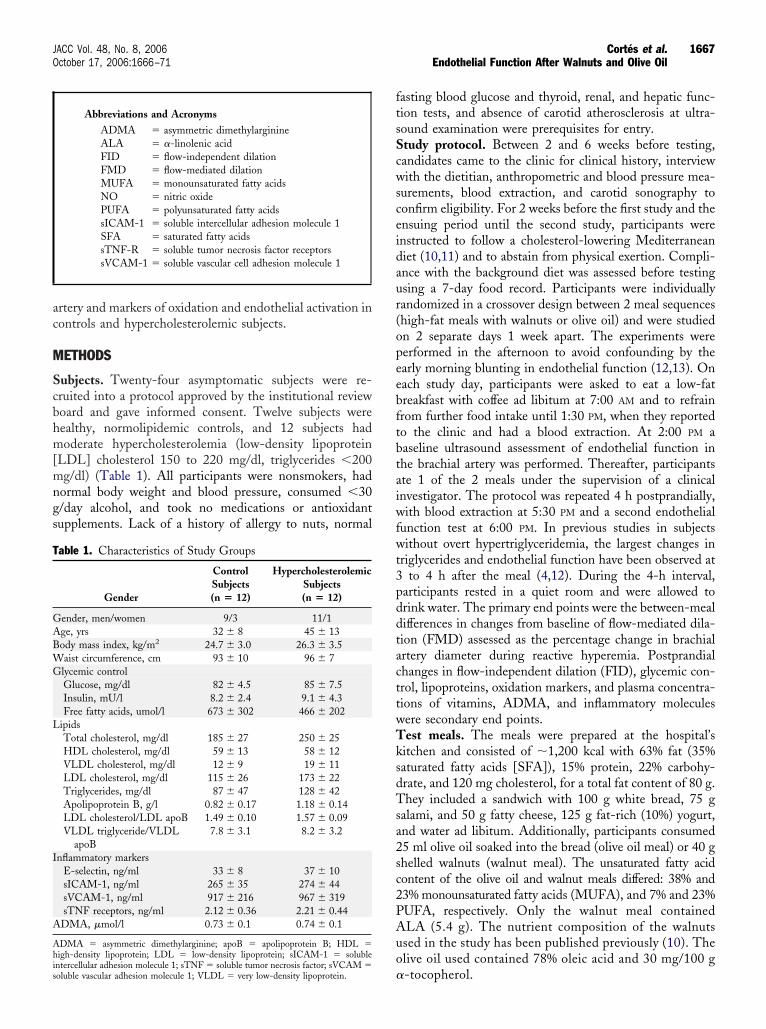
ac
M
Scbhm[mngs
ftsScwsceidaur(opeebftbtaiwfwt3pddtacttwTksdTsa2sc2PAuo
T
GABWG
L
I
A
Ahis
1667JACC Vol. 48, No. 8, 2006 Cortés et al.October 17, 2006:1666–71 Endothelial Function After Walnuts and Olive Oil
rtery and markers of oxidation and endothelial activation inontrols and hypercholesterolemic subjects.
ETHODS
ubjects. Twenty-four asymptomatic subjects were re-ruited into a protocol approved by the institutional reviewoard and gave informed consent. Twelve subjects wereealthy, normolipidemic controls, and 12 subjects hadoderate hypercholesterolemia (low-density lipoprotein
LDL] cholesterol 150 to 220 mg/dl, triglycerides �200g/dl) (Table 1). All participants were nonsmokers, had
ormal body weight and blood pressure, consumed �30/day alcohol, and took no medications or antioxidantupplements. Lack of a history of allergy to nuts, normal
Abbreviations and AcronymsADMA � asymmetric dimethylarginineALA � �-linolenic acidFID � flow-independent dilationFMD � flow-mediated dilationMUFA � monounsaturated fatty acidsNO � nitric oxidePUFA � polyunsaturated fatty acidssICAM-1 � soluble intercellular adhesion molecule 1SFA � saturated fatty acidssTNF-R � soluble tumor necrosis factor receptorssVCAM-1 � soluble vascular cell adhesion molecule 1
able 1. Characteristics of Study Groups
Gender
ControlSubjects(n � 12)
HypercholesterolemicSubjects(n � 12)
ender, men/women 9/3 11/1ge, yrs 32 � 8 45 � 13ody mass index, kg/m2 24.7 � 3.0 26.3 � 3.5aist circumference, cm 93 � 10 96 � 7lycemic controlGlucose, mg/dl 82 � 4.5 85 � 7.5Insulin, mU/l 8.2 � 2.4 9.1 � 4.3Free fatty acids, umol/l 673 � 302 466 � 202
ipidsTotal cholesterol, mg/dl 185 � 27 250 � 25HDL cholesterol, mg/dl 59 � 13 58 � 12VLDL cholesterol, mg/dl 12 � 9 19 � 11LDL cholesterol, mg/dl 115 � 26 173 � 22Triglycerides, mg/dl 87 � 47 128 � 42Apolipoprotein B, g/l 0.82 � 0.17 1.18 � 0.14LDL cholesterol/LDL apoB 1.49 � 0.10 1.57 � 0.09VLDL triglyceride/VLDL
apoB7.8 � 3.1 8.2 � 3.2
nflammatory markersE-selectin, ng/ml 33 � 8 37 � 10sICAM-1, ng/ml 265 � 35 274 � 44sVCAM-1, ng/ml 917 � 216 967 � 319sTNF receptors, ng/ml 2.12 � 0.36 2.21 � 0.44
DMA, �mol/l 0.73 � 0.1 0.74 � 0.1
DMA � asymmetric dimethylarginine; apoB � apolipoprotein B; HDL �igh-density lipoprotein; LDL � low-density lipoprotein; sICAM-1 � soluble
�ntercellular adhesion molecule 1; sTNF � soluble tumor necrosis factor; sVCAM �oluble vascular adhesion molecule 1; VLDL � very low-density lipoprotein.
asting blood glucose and thyroid, renal, and hepatic func-ion tests, and absence of carotid atherosclerosis at ultra-ound examination were prerequisites for entry.tudy protocol. Between 2 and 6 weeks before testing,andidates came to the clinic for clinical history, interviewith the dietitian, anthropometric and blood pressure mea-
urements, blood extraction, and carotid sonography toonfirm eligibility. For 2 weeks before the first study and thensuing period until the second study, participants werenstructed to follow a cholesterol-lowering Mediterraneaniet (10,11) and to abstain from physical exertion. Compli-nce with the background diet was assessed before testingsing a 7-day food record. Participants were individuallyandomized in a crossover design between 2 meal sequenceshigh-fat meals with walnuts or olive oil) and were studiedn 2 separate days 1 week apart. The experiments wereerformed in the afternoon to avoid confounding by thearly morning blunting in endothelial function (12,13). Onach study day, participants were asked to eat a low-fatreakfast with coffee ad libitum at 7:00 AM and to refrainrom further food intake until 1:30 PM, when they reportedo the clinic and had a blood extraction. At 2:00 PM aaseline ultrasound assessment of endothelial function inhe brachial artery was performed. Thereafter, participantste 1 of the 2 meals under the supervision of a clinicalnvestigator. The protocol was repeated 4 h postprandially,ith blood extraction at 5:30 PM and a second endothelial
unction test at 6:00 PM. In previous studies in subjectsithout overt hypertriglyceridemia, the largest changes in
riglycerides and endothelial function have been observed atto 4 h after the meal (4,12). During the 4-h interval,
articipants rested in a quiet room and were allowed torink water. The primary end points were the between-mealifferences in changes from baseline of flow-mediated dila-ion (FMD) assessed as the percentage change in brachialrtery diameter during reactive hyperemia. Postprandialhanges in flow-independent dilation (FID), glycemic con-rol, lipoproteins, oxidation markers, and plasma concentra-ions of vitamins, ADMA, and inflammatory moleculesere secondary end points.est meals. The meals were prepared at the hospital’sitchen and consisted of �1,200 kcal with 63% fat (35%aturated fatty acids [SFA]), 15% protein, 22% carbohy-rate, and 120 mg cholesterol, for a total fat content of 80 g.hey included a sandwich with 100 g white bread, 75 g
alami, and 50 g fatty cheese, 125 g fat-rich (10%) yogurt,nd water ad libitum. Additionally, participants consumed5 ml olive oil soaked into the bread (olive oil meal) or 40 ghelled walnuts (walnut meal). The unsaturated fatty acidontent of the olive oil and walnut meals differed: 38% and3% monounsaturated fatty acids (MUFA), and 7% and 23%UFA, respectively. Only the walnut meal containedLA (5.4 g). The nutrient composition of the walnutssed in the study has been published previously (10). Thelive oil used contained 78% oleic acid and 30 mg/100 g
-tocopherol.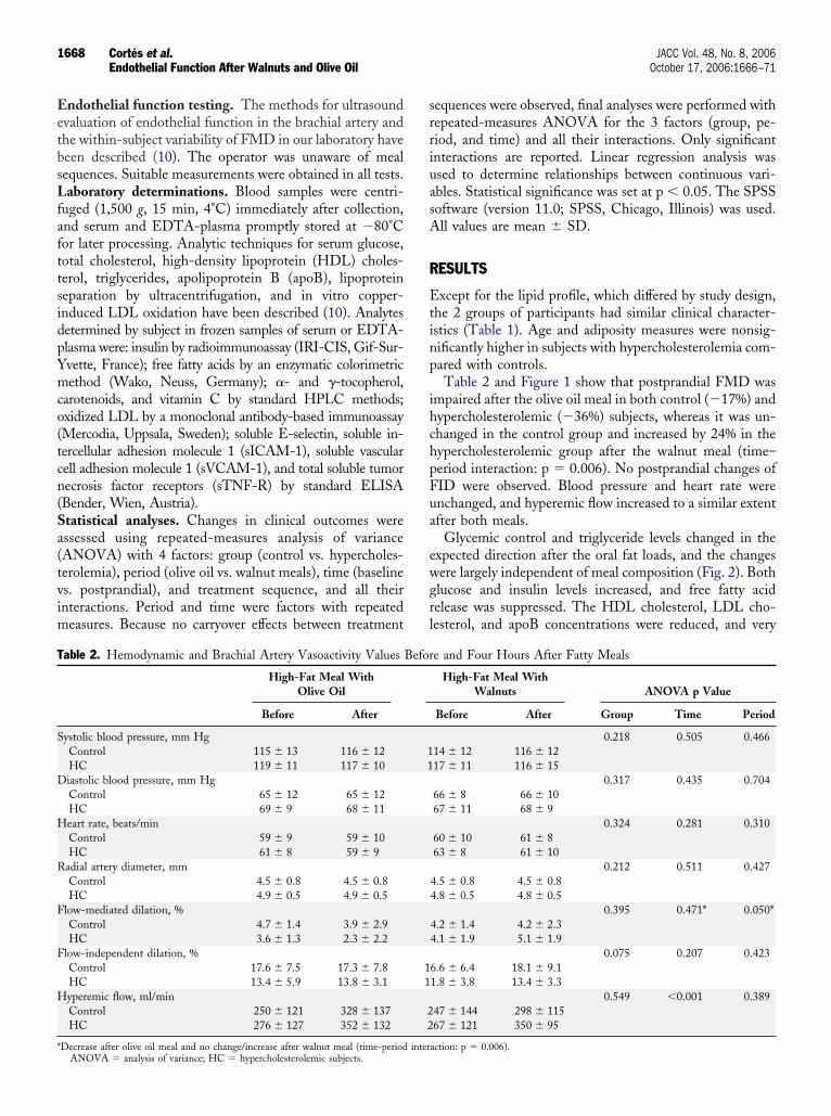
EetbsLfafttsidpYmco(tcn(Sa(tvim
srriuasA
R
Etinp
ihchpFua
ewgrl
T
S
D
H
R
F
F
H
*
1668 Cortés et al. JACC Vol. 48, No. 8, 2006Endothelial Function After Walnuts and Olive Oil October 17, 2006:1666–71
ndothelial function testing. The methods for ultrasoundvaluation of endothelial function in the brachial artery andhe within-subject variability of FMD in our laboratory haveeen described (10). The operator was unaware of mealequences. Suitable measurements were obtained in all tests.aboratory determinations. Blood samples were centri-
uged (1,500 g, 15 min, 4°C) immediately after collection,nd serum and EDTA-plasma promptly stored at �80°Cor later processing. Analytic techniques for serum glucose,otal cholesterol, high-density lipoprotein (HDL) choles-erol, triglycerides, apolipoprotein B (apoB), lipoproteineparation by ultracentrifugation, and in vitro copper-nduced LDL oxidation have been described (10). Analytesetermined by subject in frozen samples of serum or EDTA-lasma were: insulin by radioimmunoassay (IRI-CIS, Gif-Sur-vette, France); free fatty acids by an enzymatic colorimetricethod (Wako, Neuss, Germany); �- and �-tocopherol,
arotenoids, and vitamin C by standard HPLC methods;xidized LDL by a monoclonal antibody-based immunoassayMercodia, Uppsala, Sweden); soluble E-selectin, soluble in-ercellular adhesion molecule 1 (sICAM-1), soluble vascularell adhesion molecule 1 (sVCAM-1), and total soluble tumorecrosis factor receptors (sTNF-R) by standard ELISABender, Wien, Austria).tatistical analyses. Changes in clinical outcomes weressessed using repeated-measures analysis of varianceANOVA) with 4 factors: group (control vs. hypercholes-erolemia), period (olive oil vs. walnut meals), time (baselines. postprandial), and treatment sequence, and all theirnteractions. Period and time were factors with repeated
easures. Because no carryover effects between treatment
able 2. Hemodynamic and Brachial Artery Vasoactivity Values
High-Fat Meal WithOlive Oil
Before After
ystolic blood pressure, mm HgControl 115 � 13 116 � 12HC 119 � 11 117 � 10iastolic blood pressure, mm HgControl 65 � 12 65 � 12HC 69 � 9 68 � 11eart rate, beats/minControl 59 � 9 59 � 10HC 61 � 8 59 � 9
adial artery diameter, mmControl 4.5 � 0.8 4.5 � 0.8HC 4.9 � 0.5 4.9 � 0.5
low-mediated dilation, %Control 4.7 � 1.4 3.9 � 2.9HC 3.6 � 1.3 2.3 � 2.2
low-independent dilation, %Control 17.6 � 7.5 17.3 � 7.8HC 13.4 � 5.9 13.8 � 3.1yperemic flow, ml/minControl 250 � 121 328 � 137HC 276 � 127 352 � 132
Decrease after olive oil meal and no change/increase after walnut meal (time-period interANOVA � analysis of variance; HC � hypercholesterolemic subjects.
equences were observed, final analyses were performed withepeated-measures ANOVA for the 3 factors (group, pe-iod, and time) and all their interactions. Only significantnteractions are reported. Linear regression analysis wassed to determine relationships between continuous vari-bles. Statistical significance was set at p � 0.05. The SPSSoftware (version 11.0; SPSS, Chicago, Illinois) was used.ll values are mean � SD.
ESULTS
xcept for the lipid profile, which differed by study design,he 2 groups of participants had similar clinical character-stics (Table 1). Age and adiposity measures were nonsig-ificantly higher in subjects with hypercholesterolemia com-ared with controls.Table 2 and Figure 1 show that postprandial FMD was
mpaired after the olive oil meal in both control (�17%) andypercholesterolemic (�36%) subjects, whereas it was un-hanged in the control group and increased by 24% in theypercholesterolemic group after the walnut meal (time–eriod interaction: p � 0.006). No postprandial changes ofID were observed. Blood pressure and heart rate werenchanged, and hyperemic flow increased to a similar extentfter both meals.
Glycemic control and triglyceride levels changed in thexpected direction after the oral fat loads, and the changesere largely independent of meal composition (Fig. 2). Bothlucose and insulin levels increased, and free fatty acidelease was suppressed. The HDL cholesterol, LDL cho-esterol, and apoB concentrations were reduced, and very
e and Four Hours After Fatty Meals
High-Fat Meal WithWalnuts ANOVA p Value
Before After Group Time Period
0.218 0.505 0.46614 � 12 116 � 1217 � 11 116 � 15
0.317 0.435 0.70466 � 8 66 � 1067 � 11 68 � 9
0.324 0.281 0.31060 � 10 61 � 863 � 8 61 � 10
0.212 0.511 0.4274.5 � 0.8 4.5 � 0.84.8 � 0.5 4.8 � 0.5
0.395 0.471* 0.050*4.2 � 1.4 4.2 � 2.34.1 � 1.9 5.1 � 1.9
0.075 0.207 0.4236.6 � 6.4 18.1 � 9.11.8 � 3.8 13.4 � 3.3
0.549 �0.001 0.38947 � 144 298 � 11567 � 121 350 � 95
Befor
11
11
22
action: p � 0.006).

lt(h1ViimTea
�s��abcaeLg
c
tddsm0
vpaubp��e
D
Owawtc
tmiospcita
Fac
FwSv
1669JACC Vol. 48, No. 8, 2006 Cortés et al.October 17, 2006:1666–71 Endothelial Function After Walnuts and Olive Oil
ow-density lipoprotein (VLDL) lipids increased substan-ially after either meal in both groups (p � 0.001 for all)data not shown). Postprandial lipemia was enhanced inypercholesterolemic compared with control subjects. Table
shows baseline lipoprotein lipid to apoB ratios. TheLDL triglyceride/apoB ratio, a measure of VLDL size,
ncreased after both meals (p � 0.001). In control subjects,t increased by 85% and 57% after the olive oil and walnuts
eals, respectively (group–period interaction: p � 0.034).he rises in hypercholesterolemic subjects were similar after
ither meal. The meals had little effect on LDL cholesterol/poB ratios, indicating unchanged LDL size (data not shown).
The plasma levels of vitamin C, �-tocopherol, and-tocopherol decreased significantly (p � 0.05) and to aimilar extent in the 2 groups. The changes ranged from2% to �14% and were unrelated to meal type. The-carotene and �-carotene levels were similar before andfter either meal (data not shown). As shown in Table 3,aseline oxidized LDL was higher in hypercholesterolemicompared with control subjects and decreased postprandi-lly in the 2 groups. In the copper-induced oxidizabilityxperiments, opposite effects were observed in lag times ofDL-conjugated diene production depending on studyroup and meal type.
The values of inflammatory cytokines and adhesion mole-
igure 1. Mean flow-mediated dilation before and 4 h after fatty mealsith added olive oil (squares) or walnuts (triangles). Vertical bars indicateE. Time–period interaction: p � 0.006 by repeated-measures analysis ofariance.
ules differed little at baseline between groups (Table 1), andff
heir responses to meals were similar by group, a reason theata are assembled in Figure 3 to show that all valuesecreased to a small extent after either meal. The decrease inoluble E-selectin was more pronounced after the walnuteal than the olive oil meal (time-period interaction: p �
.033). There were no changes in ADMA.In the overall participants, baseline FMD correlated in-
ersely and significantly with fasting triglycerides (r � �0.324;� 0.024) and nonsignificantly with the VLDL triglyceride/
poB ratio (r � �0.262; p � 0.072). Postprandial FMD wasnrelated to triglycerides after either meal. The relationshipetween FMD and VLDL triglyceride/apoB ratios ap-roached statistical significance after the olive oil meal (r �0.355; p � 0.088) but not after the walnut meal (r �0.246; p � 0.247). The FMD changes from baseline after
ither meal were unrelated to changes in any variable.
ISCUSSION
ur main finding was that, in comparison with olive oil,alnuts reverse the impairment of endothelial function
ssociated with eating a fatty meal. The fact that a singlealnut meal positively effects postprandial vasoactivity fur-
her supports the beneficial effects of walnuts on cardiovas-ular risk (10,11).
The results confirm and extend those of an earlier feedingrial in hypercholesterolemic subjects showing that a walnuteal in a background walnut diet was associated with
mproved brachial artery vasoactivity in comparison with anlive oil meal during a Mediterranean diet (10). In thattudy, the benefit on endothelial function was mediated inart through an improved lipid profile. This mechanismannot be implicated in our acute studies, where both mealsnduced postprandial hypertriglyceridemia to a similar ex-ent. However, VLDL triglyceride/apoB ratios increasedfter the olive oil meal in comparison with the walnut
igure 2. Changes from baseline of variables related to glycemic controlnd triglycerides after the test meals in study groups. Vertical bars are 95%onfidence interval. No group, time, or period interactions were observed
or any of the variables by repeated-measures analysis of variance. FFA �ree fatty acids; HC � hypercholesterolemia group.
mrtgevai
bslpdd
cFutem(mFmrAae
eoUdoApwmdTPdf
ticctG
T
O
C
* hangercholes
Ftmr(vsm
1670 Cortés et al. JACC Vol. 48, No. 8, 2006Endothelial Function After Walnuts and Olive Oil October 17, 2006:1666–71
eal and were related to impaired FMD. Because thisatio is an indirect measure of the presence of largeriglyceride-rich VLDL particles that are highly athero-enic (14), our findings may explain in part the differentialffects of the test meals on endothelial function. Abnormalasoactivity after a lipid challenge has been related toccumulation of postprandial triglyceride-rich lipoproteinsn other studies (6,9,15).
That a fatty meal induces endothelial dysfunction haseen shown by most (5–9,13,15,16) but not all (5,16)tudies. Because earlier studies were designed so that base-ine testing was performed in the early morning andostprandial testing was done 3 to 4 h later, a reason for thisiscrepancy is that the physiologic improvement of FMDuring the morning counteracted any changes due to the fat
able 3. Values of LDL Oxidation Analytes Before and Four Ho
High-Fat Meal WithOlive Oil
Before After
xidized LDL, U/lControl 37.7 � 10.1 36.9 � 10.0HC 45.7 � 8.7 44.5 � 7.7onjugated diene formationLag time, min
Control 54.7 � 21.9 62.6 � 36.0HC 41.3 � 23.7 36.5 � 19.0
Vmax, nmol/min/mg proteinControl 16.1 � 3.6 16.6 � 4.6HC 16.0 � 5.2 13.9 � 4.7
Cmax, nmol/mg proteinControl 552 � 194 538 � 247HC 508 � 115 449 � 123
Increase in control subjects and decrease in HC after olive oil meal with opposite cANOVA � analysis of variance; Cmax � maximum concentration; HC � hype
igure 3. Changes from baseline of inflammation and endothelial activa-ion markers and asymmetric dimethylarginine (ADMA) after the testeals in study groups. Vertical bars are 95% confidence interval. *Greater
eduction after the walnut meal compared with the olive oil mealtime–period interaction: p � 0.033 by repeated-measures analysis ofariance). sICAM-1 � soluble intercellular adhesion molecule 1; sTNF �
foluble tumor necrosis factor; sVCAM-1 � soluble vascular cell adhesionolecule 1.
hallenge (12,13). For the same reason, the deterioration ofMD after a fatty meal may be more pronounced thansually reported. This objection may also apply to studies ofhe acute vasomotor effects of unsaturated fats and couldxplain the inconsistent effects on FMD reported aftereals rich in MUFA from olive oil or other sources
5,8,16,17) or the lack of effect of an n-3 PUFA-rich salmoneal (8). Recently, West et al. (17) reported increasedMD in diabetic patients after meals enriched with eitherarine n-3 PUFA or ALA, thus supporting the beneficial
ole of ALA-rich walnuts on endothelial function. BesidesLA, other cardioprotective constituents of walnuts, such
s L-arginine and antioxidants (10), could favorably influ-nce vasoactivity.
A role for oxidative stress in the causation of postprandialndothelial dysfunction is suggested by the protective effectf different antioxidants when added to fatty meals (5,7–9).nexpectedly, oxidized LDL decreased postprandially, in-ependently of group and meal type, and the susceptibilityf LDL to oxidation was unaffected overall (Table 3).lthough the supplemental foods provided vitamin E com-ounds (�-tocopherol in olive oil and �-tocopherol inalnuts), their plasma levels were slightly reduced aftereals. Presumably, partial consumption of circulating antioxi-
ants helped preserve the resistance of LDL to oxidation.hese results provide further evidence that, in spite of a highUFA content, walnut intake does not promote lipid peroxi-ation (10,11). They also suggest that FMD changes after aatty meal are independent of lipoprotein oxidation (18,19).
Besides denoting impaired FMD, endothelial dysfunc-ion comprises a specific state of endothelial activation thats characterized by enhanced expression and release into theirculation of inflammatory cytokines and adhesion mole-ules (1,2). A fatty meal thus activates the endothelium, andhis process is counteracted by antioxidant vitamins (20).iven the positive effects of the walnut meal on FMD, the
fter Fatty Meals
High-Fat Meal WithWalnuts ANOVA p Value
Before After Group Time Period
0.031 0.051 0.7226.9 � 8.5 36.6 � 11.15.8 � 8.0 43.5 � 6.7
0.253* 0.617* 0.773*6.1 � 24.9 43.3 � 31.83.7 � 25.0 52.1 � 25.2
0.539 0.296 0.7787.7 � 5.4 13.8 � 6.94.9 � 4.0 15.3 � 4.8
0.819 0.067 0.27482 � 94 425 � 22550 � 192 435 � 136
s after walnut meal (group-time-period interaction: p � 0.034).terolemic subjects; LDL � low-density lipoprotein; Vmax � maximum velocity.
urs A
34
54
11
45
act that the circulating levels of sTNF-R, soluble

EnadfltiemsafiepswTa
mtSraohhtdtofbwCt(oaeictflaPdpnnc
AWws
RLE
R
1
1
1
1
1
1
1
1
1
1
2
2
2
1671JACC Vol. 48, No. 8, 2006 Cortés et al.October 17, 2006:1666–71 Endothelial Function After Walnuts and Olive Oil
-selectin, sICAM-1, and sVCAM-1 did not increase wasot unexpected. However, a similar lack of endothelialctivation was observed after the olive oil meal, in clearissociation from its effects on FMD. Postprandially, in-ammatory biomarkers decreased independently of mealype, except for soluble E-selectin, an adhesion moleculenvolved in the early steps of monocyte recruitment to thendothelium (21), which decreased more after the walnuteal than after the olive oil meal. We have previously
hown that a walnut diet attenuates endothelial activation,s suggested by decreased sVCAM-1 levels (10). Ourndings are consistent with the evidence of decreasedxpression of adhesion molecules by endothelial cells ex-osed to marine n-3 PUFA or oleic acid (21). A recenttudy (22) has shown that diets enriched in ALA fromalnuts reduce the levels of inflammatory biomarkers.aken together, these findings suggest that ALA shares the
nti-inflammatory effects of marine n-3 PUFA.The plasma ADMA level was unchanged after eithereal (Fig. 3), suggesting that this factor was not involved in
he changes of endothelial function.tudy limitations. Our study is limited by the smallepresentation of women; therefore, the findings may onlypply to men. Hypercholesterolemic subjects were slightlylder than control subjects, but participants in both groupsad no other nonlipid risk factors and showed similaremodynamics and lack of carotid atherosclerosis. Besides,he study was crossover and focused on between-mealifferences, not between-group ones. Another limitation ishat we did not evaluate the effects of fat loads made up onlyf foods rich in SFA. Olive oil alone may impair endothelialunction (8,16), thus it is not completely clear whetheretween-meal differences were due to a beneficial effect ofalnuts or a detrimental effect of olive oil, or both.onclusions. The mechanisms underlying impaired endo-
helial function in the postprandial state are likely multifactorial3,5,6,20). We showed that supplemental walnuts, but notlive oil, counteracted the detrimental changes in FMDssociated with eating a fatty meal. Vasomotor changes, how-ver, did not appear to occur through pro-oxidative,nflammation-sensitive, or ADMA-related mechanisms. Be-ause of an increased delivery of ALA, improved FMD afterhe walnut meal could be mediated by increased membraneuidity of endothelial cells promoting enhanced synthesisnd/or release of NO, as has been postulated for marine n-3UFA (21). Nevertheless, unsaturated fatty acids and antioxi-ants in both olive oil and walnuts appear to preserve therotective phenotype of endothelial cells. Additional work iseeded to prove these contentions and explore basic mecha-isms for the vascular effects of different fats, such as vascularell signaling and pro- or antiatherogenic gene expression.
cknowledgmentse sincerely thank María J. Ortega for excellent assistance
ith sonographic studies and Emili Corbella for expert
tatistical advice.eprint requests and correspondence: Dr. Emilio Ros, Unitat deípids, Hospital Clínic, Villarroel 170, 08036 Barcelona, Spain.-mail: [email protected].
EFERENCES
1. Bonetti PO, Lerman LO, Lerman A. Endothelial dysfunction: amarker of atherosclerotic risk. Arterioscler Thromb Vasc Biol 2003;23:168–75.
2. Hanson GK. Inflammation, atherosclerosis, and coronary artery dis-ease. N Engl J Med 2005;352:1685–95.
3. Cooke JP. Asymmetrical dimethylarginine. The über marker? Circu-lation 2004;109;1813–9.
4. Sydowa K, Münzel T. ADMA and oxidative stress. Atheroscler Suppl2003;4:41–51.
5. West SG. Effect of diet on vascular reactivity: an emerging marker forvascular risk. Curr Atheroscler Rep 2001;3:446–55.
6. de Koning EJP, Rabelink TJ. Endothelial function in the postprandialstate. Atheroscler Suppl 2002;3:11–6.
7. Plotnick GD, Corretti MC, Vogel RA, Hesslink R, Wise JA. Effect ofsupplemental phytonutrients on impairment of the flow-mediatedbrachial artery vasoactivity after a single high-fat meal. J Am CollCardiol 2003;41:1744–9.
8. Vogel RA, Corretti MC, Plotnick GD. The postprandial effect ofcomponents of the Mediterranean diet on endothelial function. J AmColl Cardiol 2000;36:1455–60.
9. Plotnick GD, Corretti MC, Vogel RA. Effect of antioxidant vitaminson the transient impairment of endothelium-dependent vasoactivityfollowing a single high-fat meal. JAMA 1997;278:1682–6.
0. Ros E, Núñez I, Pérez-Heras A, et al. A walnut diet improves endothelialfunction in hypercholesterolemic subjects. A randomized crossover trial.Circulation 2004;109:1609–14.
1. Zambón D, Sabaté J, Muñoz S, et al. Substituting walnuts formonounsaturated fat improves the serum lipid profile of hypercholes-terolemic men and women. A randomized crossover trial. Ann InternMed 2000;132:538–46.
2. Otto ME, Svatikova A, de Mattos Barretto RB, et al. Early morningattenuation of endothelial function in healthy humans. Circulation2004;109:2507–10.
3. Gaenzer H, Sturm W, Neumayr G, et al. Pronounced postprandiallipemia impairs endothelium-dependent dilation of the brachial arteryin men. Cardiovasc Res 2001;52:509–16.
4. Packard CJ, Shepherd J. Lipoprotein heterogeneity and apolipoproteinB metabolism. Arterioscler Thromb Vasc Biol 1997;17:3542–56.
5. Maggi FM, Raselli S, Grigore L, Redaelli L, Fantappie S, CatapanoAL. Lipoprotein remnants and endothelial dysfunction in the post-prandial phase. J Clin Endocrinol Metab 2004;89:2946–50.
6. Sanderson P, Olthof M, Grimble RF, et al. Dietary lipids and vascularfunction: UK Food Standards Agency workshop report. Br J Nutr2004;91:491–500.
7. West SWG, Hecker KD, Mustad VA, et al. Acute effects ofmonounsaturated fatty acids with and without omega-3 fatty acids onvascular reactivity in individuals with type 2 diabetes. Diabetologia2005;48:113–22.
8. Bae JH, Schwemmer M, Lee IK, Park KR, Kim KY, Bassenge E.Postprandial hypertriglyceridemia-induced endothelial dysfunction inhealthy subjects is independent of lipid oxidation. Int J Cardiol2003;87:259–67.
9. Steinberg FM, Guthrie NL, Villablanca AC, Kumar K, MurrayMJ. Soy protein with isoflavones has favorable effects on endo-thelial function that are independent of lipid and antioxidant effectsin healthy postmenopausal women. Am J Clin Nutr 2003;78:123–30.
0. Burdge GC, Calder PC. Plasma cytokine response during the post-prandial period: a potential causal process in vascular disease? Br J Nutr2005;93:3–9.
1. De Caterina R, Liao JK, Libby P. Fatty acid modulation of endothelialactivation. Am J Clin Nutr 2000;71 Suppl:213S–23S.
2. Zhao G, Etherton TD, Martin KR, West SG, Gillies PJ, Kris-Etherton PM. Dietary �-linolenic acid reduces inflammatory and lipid
cardiovascular risk factors in hypercholesterolemic men and women.J Nutr 2004;134:2991–7.Copyright © 2022 FDOKUMEN