Nuclear S100A7 Is Associated with Poor Prognosis in Head and Neck Cancer
10
Nuclear S100A7 Is Associated with Poor Prognosis in Head and Neck Cancer Satyendra Chandra Tripathi 1 , Ajay Matta 2,3 , Jatinder Kaur 1 , Jorg Grigull 4 , Shyam Singh Chauhan 1 , Alok Thakar 5 , Nootan Kumar Shukla 6 , Ritu Duggal 7 , Siddhartha DattaGupta 8 , Ranju Ralhan 2,3,9,10,11,12 * . , K. W. Michael Siu 2,3 * . 1 Department of Biochemistry, All India Institute of Medical Sciences, New Delhi, India, 2 Department of Chemistry, York University, Toronto, Ontario, Canada, 3 Centre for Research in Mass Spectrometry, York University, Toronto, Ontario, Canada, 4 Department of Mathematics and Statistics, York University, Toronto, Ontario, Canada, 5 Department of Otorhinolaryngology, All India Institute of Medical Sciences, New Delhi, India, 6 Department of Surgery, Dr. B. R. A. Institute Rotary Cancer Hospital, All India Institute of Medical Sciences, New Delhi, India, 7 Department of Dental Surgery, All India Institute of Medical Sciences, New Delhi, India, 8 Department of Pathology, All India Institute of Medical Sciences, New Delhi, India, 9 Joseph and Mildred Sonshine Family Centre for Head and Neck Diseases, Mount Sinai Hospital, Toronto, Ontario, Canada, 10 Department of Otolaryngology – Head and Neck Surgery, Mount Sinai Hospital, Toronto, Ontario, Canada, 11 Department of Pathology and Laboratory Medicine, Mount Sinai Hospital, Toronto, Ontario, Canada, 12 Department of Otolaryngology – Head and Neck Surgery, University of Toronto, Toronto, Ontario, Canada Abstract Background: Tissue proteomic analysis of head and neck squamous cell carcinoma (HNSCC) and normal oral mucosa using iTRAQ (isobaric tag for relative and absolute quantitation) labeling and liquid chromatography-mass spectrometry, led to the identification of a panel of biomarkers including S100A7. In the multi-step process of head and neck tumorigenesis, the presence of dysplastic areas in the epithelium is proposed to be associated with a likely progression to cancer; however there are no established biomarkers to predict their potential of malignant transformation. This study aimed to determine the clinical significance of S100A7 overexpression in HNSCC. Methodology: Immunohistochemical analysis of S100A7 expression in HNSCC (100 cases), oral lesions (166 cases) and 100 histologically normal tissues was carried out and correlated with clinicopathological parameters and disease prognosis over 7 years for HNSCC patients. Overexpression of S100A7 protein was significant in oral lesions (squamous cell hyperplasia/ dysplasia) and sustained in HNSCC in comparison with oral normal mucosa (p trend ,0.001). Significant increase in nuclear S100A7 was observed in HNSCC as compared to dysplastic lesions (p = 0.005) and associated with well differentiated squamous cell carcinoma (p = 0.031). Notably, nuclear accumulation of S100A7 also emerged as an independent predictor of reduced disease free survival (p = 0.006, Hazard ratio (HR = 7.6), 95% CI = 1.325.1) in multivariate analysis underscoring its relevance as a poor prognosticator of HNSCC patients. Conclusions: Our study demonstrated nuclear accumulation of S100A7 may serve as predictor of poor prognosis in HNSCC patients. Further, increased nuclear accumulation of S100A7 in HNSCC as compared to dysplastic lesions warrants a large- scale longitudinal study of patients with dysplasia to evaluate its potential as a determinant of increased risk of transformation of oral premalignant lesions. Citation: Tripathi SC, Matta A, Kaur J, Grigull J, Chauhan SS, et al. (2010) Nuclear S100A7 Is Associated with Poor Prognosis in Head and Neck Cancer. PLoS ONE 5(8): e11939. doi:10.1371/journal.pone.0011939 Editor: Torbjorn Ramqvist, Karolinska Institutet, Sweden Received March 12, 2010; Accepted July 5, 2010; Published August 3, 2010 Copyright: ß 2010 Tripathi et al. This is an open-access article distributed under the terms of the Creative Commons Attribution License, which permits unrestricted use, distribution, and reproduction in any medium, provided the original author and source are credited. Funding: SCT is a recipient of a Senior Research Fellowship from Indian Council of Medical Research (ICMR), New Delhi, India. RR gratefully acknowledges support from the Ontario Institute for Cancer Research (OICR), Joseph and Mildred Sonshine Centre for Head and Neck Diseases and Temmy Latner/Dynacare Family Foundation, Canada. KWMS acknowledges infrastructural support from the Canadian Institutes of Health Research (CIHR), Ontario Research and Development Challenge Fund and Applied Biosystems/MDS Analytical Technologies. The funders had no role in study design, data collection and analysis, decision to publish, or preparation of the manuscript. Competing Interests: The authors have declared that no competing interests exist. * E-mail: [email protected] (RR); [email protected] (KWMS) . These authors contributed equally to this work. Introduction Head and neck squamous cell carcinoma (HNSCC) is the sixth most common cancer accounting for over 500,000 new cases annually worldwide that includes sites in the oral cavity, pharynx and larynx [1]. Squamous cell carcinoma of the oral cavity accounts for two-thirds of the HNSCC cases occurring in developing countries. The majority of oral squamous cell carcinomas are preceded by visible changes of the oral mucosa. Leukoplakia is the most commonly encountered oral lesion of the oral cavity. These oral leukoplakia lesions show histological evidence of squamous cell hyperplasia or dysplasia. The oral lesions with histologically confirmed dysplasia are termed as oral premalignant lesions (OPLs); on average, about one percent of oral lesions transform into cancer annually [2–4]. Despite improve- ment in treatment strategies, including surgery, radiotherapy (RT) and/or chemotherapy (CT), the prognosis of OSCC patients remains largely unsatisfactory, due to loco-regional recurrence. PLoS ONE | www.plosone.org 1 August 2010 | Volume 5 | Issue 8 | e11939
-
Upload
independent -
Category
Documents
-
view
3 -
download
0
Transcript of Nuclear S100A7 Is Associated with Poor Prognosis in Head and Neck Cancer
Nuclear S100A7 Is Associated with Poor Prognosis inHead and Neck CancerSatyendra Chandra Tripathi1, Ajay Matta2,3, Jatinder Kaur1, Jorg Grigull4, Shyam Singh Chauhan1, Alok
Thakar5, Nootan Kumar Shukla6, Ritu Duggal7, Siddhartha DattaGupta8, Ranju Ralhan2,3,9,10,11,12*., K. W.
Michael Siu2,3*.
1 Department of Biochemistry, All India Institute of Medical Sciences, New Delhi, India, 2 Department of Chemistry, York University, Toronto, Ontario, Canada, 3 Centre for
Research in Mass Spectrometry, York University, Toronto, Ontario, Canada, 4 Department of Mathematics and Statistics, York University, Toronto, Ontario, Canada,
5 Department of Otorhinolaryngology, All India Institute of Medical Sciences, New Delhi, India, 6 Department of Surgery, Dr. B. R. A. Institute Rotary Cancer Hospital, All
India Institute of Medical Sciences, New Delhi, India, 7 Department of Dental Surgery, All India Institute of Medical Sciences, New Delhi, India, 8 Department of Pathology,
All India Institute of Medical Sciences, New Delhi, India, 9 Joseph and Mildred Sonshine Family Centre for Head and Neck Diseases, Mount Sinai Hospital, Toronto, Ontario,
Canada, 10 Department of Otolaryngology – Head and Neck Surgery, Mount Sinai Hospital, Toronto, Ontario, Canada, 11 Department of Pathology and Laboratory
Medicine, Mount Sinai Hospital, Toronto, Ontario, Canada, 12 Department of Otolaryngology – Head and Neck Surgery, University of Toronto, Toronto, Ontario, Canada
Abstract
Background: Tissue proteomic analysis of head and neck squamous cell carcinoma (HNSCC) and normal oral mucosa usingiTRAQ (isobaric tag for relative and absolute quantitation) labeling and liquid chromatography-mass spectrometry, led tothe identification of a panel of biomarkers including S100A7. In the multi-step process of head and neck tumorigenesis, thepresence of dysplastic areas in the epithelium is proposed to be associated with a likely progression to cancer; howeverthere are no established biomarkers to predict their potential of malignant transformation. This study aimed to determinethe clinical significance of S100A7 overexpression in HNSCC.
Methodology: Immunohistochemical analysis of S100A7 expression in HNSCC (100 cases), oral lesions (166 cases) and 100histologically normal tissues was carried out and correlated with clinicopathological parameters and disease prognosis over7 years for HNSCC patients. Overexpression of S100A7 protein was significant in oral lesions (squamous cell hyperplasia/dysplasia) and sustained in HNSCC in comparison with oral normal mucosa (ptrend,0.001). Significant increase in nuclearS100A7 was observed in HNSCC as compared to dysplastic lesions (p = 0.005) and associated with well differentiatedsquamous cell carcinoma (p = 0.031). Notably, nuclear accumulation of S100A7 also emerged as an independent predictor ofreduced disease free survival (p = 0.006, Hazard ratio (HR = 7.6), 95% CI = 1.325.1) in multivariate analysis underscoring itsrelevance as a poor prognosticator of HNSCC patients.
Conclusions: Our study demonstrated nuclear accumulation of S100A7 may serve as predictor of poor prognosis in HNSCCpatients. Further, increased nuclear accumulation of S100A7 in HNSCC as compared to dysplastic lesions warrants a large-scale longitudinal study of patients with dysplasia to evaluate its potential as a determinant of increased risk oftransformation of oral premalignant lesions.
Citation: Tripathi SC, Matta A, Kaur J, Grigull J, Chauhan SS, et al. (2010) Nuclear S100A7 Is Associated with Poor Prognosis in Head and Neck Cancer. PLoSONE 5(8): e11939. doi:10.1371/journal.pone.0011939
Editor: Torbjorn Ramqvist, Karolinska Institutet, Sweden
Received March 12, 2010; Accepted July 5, 2010; Published August 3, 2010
Copyright: � 2010 Tripathi et al. This is an open-access article distributed under the terms of the Creative Commons Attribution License, which permitsunrestricted use, distribution, and reproduction in any medium, provided the original author and source are credited.
Funding: SCT is a recipient of a Senior Research Fellowship from Indian Council of Medical Research (ICMR), New Delhi, India. RR gratefully acknowledgessupport from the Ontario Institute for Cancer Research (OICR), Joseph and Mildred Sonshine Centre for Head and Neck Diseases and Temmy Latner/DynacareFamily Foundation, Canada. KWMS acknowledges infrastructural support from the Canadian Institutes of Health Research (CIHR), Ontario Research andDevelopment Challenge Fund and Applied Biosystems/MDS Analytical Technologies. The funders had no role in study design, data collection and analysis,decision to publish, or preparation of the manuscript.
Competing Interests: The authors have declared that no competing interests exist.
* E-mail: [email protected] (RR); [email protected] (KWMS)
. These authors contributed equally to this work.
Introduction
Head and neck squamous cell carcinoma (HNSCC) is the sixth
most common cancer accounting for over 500,000 new cases
annually worldwide that includes sites in the oral cavity, pharynx
and larynx [1]. Squamous cell carcinoma of the oral cavity
accounts for two-thirds of the HNSCC cases occurring in
developing countries. The majority of oral squamous cell
carcinomas are preceded by visible changes of the oral mucosa.
Leukoplakia is the most commonly encountered oral lesion of the
oral cavity. These oral leukoplakia lesions show histological
evidence of squamous cell hyperplasia or dysplasia. The oral
lesions with histologically confirmed dysplasia are termed as oral
premalignant lesions (OPLs); on average, about one percent of oral
lesions transform into cancer annually [2–4]. Despite improve-
ment in treatment strategies, including surgery, radiotherapy (RT)
and/or chemotherapy (CT), the prognosis of OSCC patients
remains largely unsatisfactory, due to loco-regional recurrence.
PLoS ONE | www.plosone.org 1 August 2010 | Volume 5 | Issue 8 | e11939

The 5-year survival rate is less than 50%, and the prognosis of
advanced cases has not improved much over the past three
decades [5,6]. At present, the most important prognostic factors
include histological tumor grade, stage, depth of the tumor
invasion, and involvement of regional lymph nodes at the time of
diagnosis. In addition to these clinicopathological parameters,
molecular markers are being intensively sought and verified for
this malignancy. Lack of biomarkers for early detection and risk
assessment is clearly reflected by the fact that more than 50% of all
HNSCC patients have advanced disease at the time of diagnosis
[5].
In our recent study using iTRAQ (isobaric tag for relative and
absolute quantitation) labeling and multidimensional liquid
chromatography/tandem mass spectrometry (LC-MS/MS) for
examining differential protein expressions between HNSCC and
non-malignant tissues, we identified a panel of biomarker
candidates for this malignancy [7]. S100A7/psoriasin was
identified as overexpressed in HNSCC and emerged among the
panel of three best-performing potential biomarkers for distin-
guishing HNSCC from normal oral mucosa [7]. In another
independent study using iTRAQ, we also reported increased
expression of S100A7 protein in oral premalignant lesions
(dysplasia), albeit in only limited number of cases [8].
S100 protein family consists of at least 25 different types of low
molecular-weight proteins (9–13 kDa), which are characterized by
the presence of two calcium-binding sites of the EF-hand type
conformation [9–12]. S100A7 gene is located within the
‘epidermal differentiation complex’ on human chromosome
1q21 [13–16]. S100A7 protein , with a molecular weight of
11.4 kDa, was found to be upregulated in skin lesions of psoriatic
patients [17]. S100A7 is distributed in the cytoplasm of
keratinocytes in normal human epidermis and is present at the
cell periphery in terminally differentiated keratinocytes [18].
Increased S100A7 expression has been reported in several
epithelial malignancies such as, in situ ductal breast carcinoma,
lung, bladder, skin, esophageal and gastric cancer [19–24]. Altered
expression of S100A4 and S100A2 proteins has been associated
with prognosis in HNSCC [10,25–28]. S100A7 overexpression
has also been reported in a small set of HNSCC [29,30]. Although
increased expression of S100A7/psoriasin has been reported in
these studies, the impact of its expression on cancer development,
disease prognosis, and survival of HNSCC patients remains to be
completely determined. In this context our study assumes
importance, because of its retrospective nature, the large set of
patients representing different stages of HNSCC, and the long
term follow-up analysis. We analyzed the expression of S100A7/
psoriasin in HNSCC, oral lesions (with histological evidence of
squamous cell hyperplasia or dysplasia) and non-malignant oral
tissues by immunohistochemistry, determined its correlation with
clinicopathological parameters, and investigated its utility as a
prognostic marker for HNSCC.
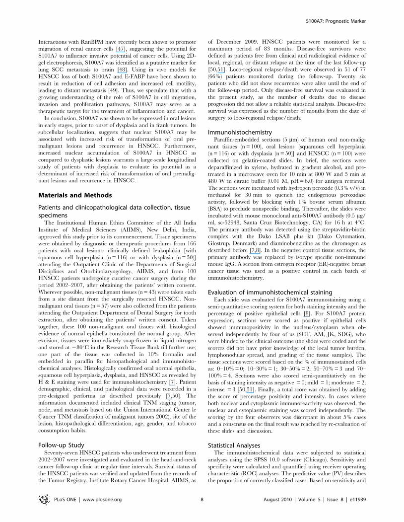
Results
Immunohistochemical analysis of S100A7 expression inoral leukoplakia lesions and cancers
To determine the clinical significance of S100A7 protein in
head-and-neck tumorigenesis, its expression was analyzed in
clinical specimens from HNSCC, oral leukoplakia lesions with
squamous cell hyperplasia or dysplasia, and histologically normal
tissues, using a specific monoclonal antibody by immunohisto-
chemistry. Figure 1A shows the total immunostaining score
distribution of nuclear/cytoplasmic S100A7 expression in oral
normal tissues, oral leukoplakia lesions with squamous cell
hyperplasia or dysplasia and HNSCC. Of the 100 normal tissues
analyzed, 84% did not show detectable S100A7 immunostaining
in nucleus/cytoplasm of the epithelial cells (Figure 1B(i)). In the
remaining normal tissues (16%), moderate cytoplasmic staining
was observed in differentiated epithelial cells in the suprabasal
layer only. Chi square trend analysis showed significant increase in
S100A7 expression (nuclear/cytoplasmic) in tissues obtained from
different stages of head-and-neck tumorigenesis (normal, squa-
mous cell hyperplasia, dysplasia and HNSCC; Table 1,
ptrend,0.001).
Oral leukoplakia lesions (squamous cell hyperplasia/
dysplasia). Of the 166 oral leukoplakia lesions analyzed, 97
cases (58.4%) showed significant increase in cytoplasmic S100A7
immunostaining (p,0.001, Odds ratio (OR) = 7.4, 95%
CI = 3.9213.7). Among these immunopositive cases 62 tissues
showed significant increase in nuclear S100A7 immunostaining
also (p,0.001, OR = 11.3, 95% CI = 4.3229.3) in comparison
with the normal tissues (Table 1). These 166 oral leukoplakia
lesions included 116 squamous cell hyperplasias; 54.3% (63/116)
cases showed significant increase in cytoplasmic S100A7
immunostaining (total score .3, p,0.001, OR = 6.2, 95%
CI = 3.2211.9) relative to the normal tissues (Table 1 and
Figure 1B(ii)). Significant increase in nuclear S100A7
immunostaining was also observed in 40/116 (34.5%) cases
(p,0.001, OR = 10.0, 95% CI = 3.8226.6). Notably, increased
cytoplasmic localization of S100A7 was observed in 68% dysplasia
(34 of 50 cases) (p,0.001, OR = 11.2, 95% CI = 5.0224.8) in
comparison with normal tissues (Table 1 and Figure 1B(iii)).
Similarly, progressive increase in nuclear expression of S100A7
was also observed in 22/50 (44%) dysplasia (p,0.001, OR = 14.9,
95% CI = 5.1243.0). Interestingly, S100A7 overexpression
(cytoplasmic/nuclear) was restricted to parabasal and suprabasal
layers only. None of these tissue sections showed S100A7
expression in proliferating layers in the basement membrane.
Mild membranous S1007 immunostaining (total score ,3) was
observed in 3 squamous cell hyperplasias, but in none of the
dysplasias analyzed.
HNSCC. zWe observed a similar pattern of S100A7
expression in HNSCC as well. Sixty seven out of 100 HNSCC
(67%) showed nuclear localization of S100A7 in tumor cells as
compared to the normal tissues (p,0.001, OR = 38.6, 95%
CI = 14.42103.9). Notably, significant increase in nuclear S100A7
expression was observed in HNSCC (67%) as compared to
dysplasia (44%) (p = 0.005, OR = 2.7, 95% CI = 1.325.4). In
addition to nuclear staining, intense S100A7 staining was also
observed in the cytoplasm of tumor cells in 74 of 100 HNSCC
analyzed (p,0.001, OR = 14.9, 95% CI = 7.4229.9, Table 1 and
Figure 1B (iv)). The clinicopathological parameters of HNSCC
and their correlation with nuclear/cytoplasmic expression of
S100A7 are shown in Table 1. Interestingly, nuclear S100A7
overexpression showed an association with histopathological
differentiation of HNSCC (p = 0.031). None of the HNSCC
tissues showed membranous S100A7 immunostaining. Majority
of the HNSCC tissues analyzed in this S100A7 immuno-
histochemistry study had more than 80% tumor cells in H&E
sections. However, there were five cases that showed dysplastic or
hyperplastic areas tissue adjacent to the tumor and these regions
showed immunostaining similar to that observed in the cases
that had only dysplasia or hyperplasia (Figure 1B (v). No
immunostaining was observed in HNSCC tissue sections used as
negative controls where the primary antibody was replaced by
isotype specific IgG (Figure 1B (vi)), while the positive control (ER-
negative breast cancer) showed S100A7 expression (Figure 1B
(vii)).
S100A7: Prognostic Marker
PLoS ONE | www.plosone.org 2 August 2010 | Volume 5 | Issue 8 | e11939

S100A7: Prognostic Marker
PLoS ONE | www.plosone.org 3 August 2010 | Volume 5 | Issue 8 | e11939

Figure 1. (A) Box-Plot analysis: Box plots showing distribution of total scores based on immunohistochemistry of S100A7 protein inparaffin-embedded sections of oral normal tissues, squamous cell hyperplasia, dysplasia and HNSCC. The vertical axis shows the totalimmunostaining score, obtained as described in the Methods section. (i) Nuclear S100A7 expression in squamous cell hyperplasia (IHC scoring range0–7), dysplasia (range 0–7) and HNSCC (range 0–7) (b) cytoplasmic S100A7 in normal (range 0–7), squamous cell hyperplasia (range 0–7), dysplasia(range 0–7) and HNSCC (range 0–7). (B) Immunohistochemical analysis of S100A7 in head-and-neck tissues. Paraffin-embeddedsections of histologically normal mucosa, squamous cell hyperplasia or with dysplasia, and HNSCC were stained using anti-S100A7monoclonal antibody as described in the Methods section. (i) Normal oral mucosa showing no S100A7 immunostaining; (ii) squamous cellhyperplasia showing nuclear and cytoplasmic S100A7 immunostaining; (iii) dysplasia depicting nuclear and cytoplasmic S100A7 immunostaining inepithelial cells; (iv) HNSCC illustrating both intense cytoplasmic and nuclear staining in tumor cells; (v) HNSCC section with a dysplasia showingS100A7 immunostaining in epithelial cells (original magnification 6100); (vi) HNSCC used as a negative control, showing no S100A7 immunostainingin tumor cells; and (vii) ER-negative breast cancer tissue showing S100A7 immunostaining. Arrows show nuclear and cytoplasmic localization (i-v, viioriginal magnification 6200).doi:10.1371/journal.pone.0011939.g001
Table 1. Analysis of S100A7 protein expression in Oral Lesions and its correlation with clinicopathological parameters.
ClinicopathologicalFeatures
TotalCases
NuclearN
Positivity(%) p-value OR (95% CI)
CytoplasmicN
Positivity(%) p-value OR (95% CI)
NORMAL 100 5 (5) 16 (16)
ORAL LESIONS (OL) 166 62 (37.3) ,0.0015 11.3 (4.3229.3) 97 (58.4) ,0.0016 7.4 (3.9213.7)
SQUAMOUS CELLHYPERPLASIA
116 40 (34.5) ,0.0017 10.0 (3.8226.6) 63 (54.3) ,0.0018 6.2 (3.2211.9)
DYSPLASIA 50 22 (44.0) ,0.0019 14.9 (5.1243.0) 34 (68.0) ,0.00110 11.2 (5.0224.8)
HNSCC 100 67 (67.0) ,0.00111 38.6 (14.42103.9) 74 (74.0) ,0.00112 14.9 (7.4229.9)
Age (Median, 53 yrs)(25–85 yrs)
,53 49 31 (63.3) 0.43 --- 35 (71.4) 0.566 ---
$53 51 36 (70.6) 39 (76.5)
Gender
Male 75 51 (68.0) 0.71 --- 58 (77.4) 0.188 ---
Female 25 16 (64.0) 16 (64.0)
HistologicalDifferentiation
Well 45 35 (77.8) 0.03113 2.6 (1.126.6) 37 (84.1) 0.09 ---
Moderate 49 32 (65.3) 35 (71.4)
Poor 6 0 (0) 2 (33.3)
Tumor Size
T1+ T2 39 23 (59.0) 0.17 --- 24 (61.5) 0.02 2.8 (1.127.1)
T3 + T4 61 44 (72.1) 50 (82.0)
Nodal Status
N0 33 21 (63.6) 0.62 --- 22 (66.7) 0.241 ---
N1–4 67 46 (68.7) 52 (77.6)
*Habits
Non consumer 22 15 (68.2) 0.89 --- 15 (68.2) 0.69 ---
Tobacco consumer 78 52 (66.7) 59 (75.6)
Nuclear staining:5Normal vs. oral lesions.7Normal vs. squamous cell hyperplasia.9Normal vs. dysplasia.11Normal vs. HNSCC.13Well differentiated SCCs vs Moderately and poorly differentiated SCCs; Squamous cell hyperplasia vs dysplasia, p = 0.245; HNSCC vs dysplasia, p = 0.005; N/OL/HNSCC:
ptrend,0.001.Cytoplasmic staining:6Normal vs oral lesions.8Normal vs squamous cell hyperplasia.10Normal vs Dysplasia.12Normal vs. HNSCC; N/OL/HNSCC: ptrend,0.001; Squamous cell hyperplasia vs Dysplasia p = 0.101.*tobacco consumption habits include tobacco chewing and/or smoking of bidi or cigarettes, chewing of betel quid, areca nut or pan masala.doi:10.1371/journal.pone.0011939.t001
S100A7: Prognostic Marker
PLoS ONE | www.plosone.org 4 August 2010 | Volume 5 | Issue 8 | e11939

Evaluation of S100A7 as potential diagnostic marker fororal leukoplakia lesions and HNSCC
Receiver Operating Characteristic (ROC) analysis was used to
determine the potential of S100A7 overexpression to distinguish
squamous cell hyperplasia, dysplasia and HNSCC from normal
oral tissues. The values for area-under-the-curve (AUC) were
0.664, 0.691 and 0.824 for squamous cell hyperplasia (Figure 2a),
dysplasia (Figure 2b), and HNSCC (Figure 2c), respectively
(Table 2). Similarly, ROC analysis was used for determination
of AUC for cytoplasmic S100A7 staining in all these three groups
and area-under-the-curve (AUC) values were 0.650, 0.746 and
0.788 respectively as shown in Table 2 and Figure 2a–2c. The
positive predictive values (PPV) were 88.9, 81.5, and 93.1 for
nuclear immunostaining. Similarly, for cytoplasmic immunostain-
ing positive predictive values (PPV) were 79.8, 68.0, and 82.2 in
the three groups (Table 2).
Evaluation of S100A7 overexpression as prognosticmarker for HNSCC
The estimated predictive power of the marker i.e. the strength
of the statistical association of S100A7 expression with poor
prognosis was assessed by Kaplan-Meier survival analysis.
Kaplan–Meier survival analysis showed significantly reduced
disease-free survival (p = 0.016; median survival 13 months) in
HNSCC patients harboring increased nuclear expression of
S100A7, compared with median disease-free survival of 70
months in the patients showing no nuclear S100A7 immunostain-
ing (Figure 3a). Similarly, reduced disease-free survival of 14
months was observed in HNSCC patients showing intense
cytoplasmic expression of S100A7, compared with patients who
did not show increased cytoplasmic S100A7 (median survival of 70
months, Figure 3b). Cox regression analysis was carried out to
determine the prognostic potential of S100A7 expression (nuclear/
cytoplasmic) for HNSCC in comparison with the other clinical
and pathologic parameters - histological grade, tumor size and
nodal status (Table 3). Nuclear S100A7 expression emerged as the
most significant prognostic marker for HNSCC (p = 0.006,
Hazard’s ratio (HR) = 7.6, 95% CI = 1.3–5.1).
Based on our data, the additional prognostic value that nuclear
S100A7 expression provided for predicting cancer recurrence (PPV)
in HNSCC patients was measured by the ratio: PPVrelapse/HNSCC
(83 months|S100A7)/PPVrelapse/HNSCC (83 months) = 71.4/61.0;
or for excluding (NPV) cancer recurrence in HNSCC patients was
NPVrelapse/HNSCC (83 months|S100A7)/NPVrelapse/HNSCC (83
months) = 57.1/39.0, as shown in Figure 3c and 3d respectively.
Verification of S100A7 overexpression by RT-PCR andWestern blotting
The overexpression of S100A7 in oral lesions was verified by
RT-PCR and Western blot analyses in the same representative
tissue samples as used for immunohistochemical analysis. RT-PCR
analysis demonstrated increased levels of S100A7 transcripts in
squamous cell hyperplasia, dysplasia, and HNSCC in comparison
with normal tissues (Figure 4a). Western blot analysis showed a
single intense band of 11.4 kDa, confirming the increased
expression in squamous cell hyperplasia, dysplasia and HNSCC,
as compared to the normal tissues (Figure 4b).
Discussion
The salient findings of our study are: (i) significant increase in
S100A7 expression (cytoplasmic/nuclear) in squamous cell
Figure 2. Receiver operating characteristic curves of S100A7 (nuclear/cytoplasmic) in (a) normal vs. squamous cell hyperplasia; (b)normal vs. dysplasia; (c) normal vs. HNSCC. Bold line shows ROC analysis for nuclear S100A7. Dashed line shows ROC analysis for cytoplasmicS100A7. Y-axis of the plot shows true-positive fraction and X-axis shows false positive fraction.doi:10.1371/journal.pone.0011939.g002
Table 2. Biomarker Analysis of S100A7 (Nuclear/Cytoplasmic)In Oral Lesions.
S100A7 Sensitivity Specificity PPV AUC
Nuclear Staining
Normal vs. squamous cellhyperplasia
34.5 95.0 88.9 0.664
Normal vs. dysplasia 44.0 95.0 81.5 0.691
Normal vs. HNSCC 67.0 95.0 93.1 0.824
Cytoplasmic Staining
Normal vs. squamous cellhyperplasia
54.3 84.0 79.8 0.650
Normal vs. dysplasia 68.0 84.0 68.0 0.746
Normal vs. HNSCC 74.0 84.0 82.2 0.788
doi:10.1371/journal.pone.0011939.t002
S100A7: Prognostic Marker
PLoS ONE | www.plosone.org 5 August 2010 | Volume 5 | Issue 8 | e11939

hyperplasia, dysplasia and HNSCC in comparison with normal
oral tissues; (ii) cytoplasmic S100A7 expression distinguishes
squamous cell hyperplasia, dysplasia and HNSCC from normal
mucosa with high specificity and PPV; (iii) significant increase in
nuclear S100A7 accumulation in HNSCC as compared to
dysplasia; and (iv) potential of nuclear S100A7 as a marker of
poor prognosis of HNSCC. It is noteworthy that studies on
molecular analysis of oral leukoplakia lesions - with squamous cell
hyperplasia or dysplasia are very limited, often because these
patients do not seek medical attention, due to small lesions that do
not pose any serious clinical problems. Increased S100A7
expression has been reported in dysplastic lesions [29,30].
However, our data suggest S100A7 overexpression as early as in
squamous cell hyperplasia with no histological evidence of
Figure 3. Evaluation of S100A7 overexpression (nuclear/cytoplasmic) as a prognostic marker in HNSCC. Kaplan–Meier estimation ofcumulative proportion of disease-free survival: (a) Median time for disease-free survival (DFS; no recurrence/metastasis) in HNSCC patients showingnuclear immunostaining of S100A7 was 13 months, whereas in patients showing no/faint S100A7 immunostaining in nucleus median DFS was 70months (p = 0.016); (b) In patients showing increased cytoplasmic S100A7 expression the median DFS was 14 months compared with HNSCC thatshowed mild or moderate cytoplasmic immunostaining (median DFS = 70 months, p = 0.055). Time-dependent Positive and Negative PredictiveValues (PPV(t), NPV(t)) of nuclear S100A7 expression. (c) PPV(t) for time to cancer relapse for 49 HNSCC patients with S100A7+ (solid line) and for all 77HNSCC patients with survival data (dashed line); d) NPV(t) for time to cancer relapse for 28 HNSCC patients with S100A72 (solid line).doi:10.1371/journal.pone.0011939.g003
Table 3. Correlation of Overall Survival withClinicopathological Parameters and S100A7 Expression:Multivariate Analysis.
S. No.Clinicopathologicalparameter p-value HR (95% C.I.)
1 Histological differentiation 0.003 6.6 (1.224.1)
2 S100A7 (nuclear) 0.006 7.6 (1.325.1)
doi:10.1371/journal.pone.0011939.t003
S100A7: Prognostic Marker
PLoS ONE | www.plosone.org 6 August 2010 | Volume 5 | Issue 8 | e11939

dysplasia. The onset of squamous cell hyperplasia is often
associated with chronic inflammation and the molecular links
between inflammation and pre-malignancy are being intensively
pursued. In this context, notably earlier studies have reported the
role of S100 proteins in inflammation, supporting our findings
[21,31–33]. S100A7 was identified in oral pre-malignant epithelia
(dysplasia) by microarray analysis and proposed to be a marker for
inflammation [34]. S100A7 has been shown to play a role in
facilitating the host inflammatory cell response, where it is
implicated as a chemotactic factor for lymphocytes and neutro-
phils in skin disease [35]. S100A7 has also been associated with
increased inflammatory cell infiltrates across all types of invasive
breast tumors [24]. In addition, S100A7 has been proposed as an
epidermal response gene to inflammatory cytokines [36,37]. In
breast cancer (DCIS), both oncostatin M (OSM) and interleukin-6
(IL-6) have been proposed to regulate the expression and activity
of S100A7 by regulating PI3K, STAT3 and Erk signaling [38].
These mechanisms may extend to HNSCC as well, since the
involvement of IL-6 and PI3K signaling in HNSCC has been well
documented by our laboratory and others [39,40].
A major clinical challenge faced by oncologists is the lack of
molecular markers to identify patients with oral leukoplakia lesions
that are at high risk of transformation to malignancy. In this
context, the significant increase in S100A7 expression observed in
HNSCCs (67% cases) as compared to the oral leukoplakia lesions
with dysplasia (44%) is an important finding of our study
(p = 0.005, OR = 2.8, 95% CI = 1.425.7), suggesting that nuclear
accumulation of S100A7 may be linked to increased risk of
malignant transformation and might serve as a marker to identify
the high-risk lesions. Nevertheless, this finding warrants confirma-
tion in a longitudinal follow-up study of patients with oral
leukoplakia lesions, to establish a possible link between nuclear
S100A7 expression and risk of cancer development.
In our study, S100A7 was shown to be a prognostic factor for
reduced survival of HNSCC patients. However, in contrast to
these findings, the expression of this protein was associated with
high differentiation. None of the poorly differentiated specimens
(n = 6), expressed S100A7. This finding seems to be contradictory
at a first glance. However, it is worthwhile to note that among
the 100 HNSCC cases analyzed in our study, only 6 poorly
differentiated tumors were analyzed; in comparison 45 well
differentiated tumors were investigated. Thus larger number of
poorly differentiated tumors needs to be analyzed to determine the
correlation with S100A7 expression in a future study. Our study
was also supported by an earlier report that showed association of
nuclear S100A7 expression with well differentiated squamous cell
carcinoma as compared to moderate and poorly differentiated
squamous cell carcinomas [30]. In a parallel study using OSCC
tissues sections, Kesting et al., [29] showed a significant correlation
between increased S100A7 expression and tumor stage (I and II),
well differentiated carcinomas and non-metastatic tumors, thus
supporting our findings. Similar observations have been reported
in bladder, breast and skin cancer [18,29,41–44]. S100A7
overexpression showed association with well differentiated squa-
mous cell carcinoma of the bladder in comparison the less
differentiated tumors. Similarly, S100A7 overexpression has been
shown to be expressed in the superficial, differentiated region of
the epithelium and its expression correlates with the degree of
keratinocyte differentiation [18]. S100A7 expression is relatively
low in normal, benign and atypical hyperplastic proliferative
ductal lesions, high in the pre-invasive ductal carcinoma in situ
(DCIS), but reduced in invasive carcinomas [41–44]. In skin
tumors, it is absent in undifferentiated basalioma and strongly
expressed in carcinoma in situ, as well as in keratoacanthoma and
differentiated squamous cell carcinoma. Taken together, these
findings clearly suggest the role of S100A7 in epithelial
differentiation. In an attempt to explain the role of S100A7,
Zhou et al. [30] demonstrated overexpression of S100A7 protein
resulted in degradation of b-catenin by non-canonical pathway
independent of GSK3b in oral cancer cells [30]. Further,
overexpression of S100A7 inhibited cell proliferation in vitro but
promoted tumor differentiation in an orthotopic xenograft oral
cancer model, supporting our clinical findings. Thus, S100A7
overexpression suppresses tumor growth and invasion by negative
regulation of b-catenin signaling. However, the exact mechanism
explaining this biphasic role of S100A7 expression in proliferation
in early stages but inducing differentiation in advanced stages in
head and neck carcinogenesis remains to be completely under-
stood and warrants further investigation.
Interestingly, S100A7 over expression (cytoplasmic/nuclear) has
been associated with poor patient outcome in ER-negative
invasive breast cancer patients [41,45]. Notably, our study showed
significance of nuclear S100A7 expression as a poor prognosticator
of HNSCC (independent of other clinical and pathological
parameters as revealed by Cox regression model). Further, our
time dependent predictive analysis also revealed poor prognosis of
HNSCC patients showing increased nuclear S100A7 expression.
Taken together, these findings demonstrated the potential of
nuclear S100A7 as a predictive marker for poor prognosis of
HNSCC. Altered expression of several S100 proteins has also been
associated with HNSCC [10,25–28]. Recently, both S100A4 and
S100A2 have been proposed as biomarkers of diagnostic and/or
prognostic relevance [26,46]. S100 family of proteins including
S100A7 have been shown to form both homodimers and
heterodimers interacting with cjun activation domain binding
protein 1 (Jab1), Ran binding protein M (RanBPM), epidermal
fatty acid binding protein (EFABP), and transglutaminase.
Figure 4. Verification of S100A7 expression in tissues. (a) RT-PCRanalysis of S100A7 in oral normal mucosa, squamous cell hyperplasia,dysplasia and HNSCC tissues. For RT-PCR analysis and Western blotanalysis, we used normal (n = 5), hyperplasia (n = 5), dysplasia (n = 5) andHNSCC (n = 5) tissues. Panel shows increased levels of S100A7transcripts in oral lesions -squamous cell hyperplasia (H), dysplasia (D)and HNSCC (T) compared with the oral normal mucosa (N) that showedbasal levels of S100A7 transcripts. b-actin used as a control to normalizethe quantity of RNA used for each RT-PCR reaction is shown in thelower panel. (b) Western blot analysis of S100A7 in oral normal mucosa(N), squamous cell hyperplasia (H), dysplasia (D) and HNSCC tissues.Equal amount of protein lysates from these tissues were electropho-resed on 12% SDS-PAGE and transferred to PVDF membrane. Themembrane was incubated with respective primary and secondaryantibodies as described in the Methods section and the signal detectedby enhanced chemiluminescence method. Panel shows increasedexpression of S100A7 protein in oral lesions - squamous cell hyperplasia(H), dysplasia (D) and HNSCC (T) compared with oral normal mucosa (N).GAPDH was used as loading control.doi:10.1371/journal.pone.0011939.g004
S100A7: Prognostic Marker
PLoS ONE | www.plosone.org 7 August 2010 | Volume 5 | Issue 8 | e11939
Interactions with RanBPM have recently been shown to promote
migration of renal cancer cells [47], suggesting the potential for
S100A7 to influence invasive potential of cancer cells. Using 2D-
gel electrophoresis, S100A7 was identified as a putative marker for
lung SCC metastasis to brain [48]. Using in vivo models for
HNSCC loss of both S100A7 and E-FABP have been shown to
result in reduction of cell adhesion and increased cell motility,
leading to distant metastasis [49]. Thus, we speculate that with a
growing understanding of the role of S100A7 in cell migration,
invasion and proliferation pathways, S100A7 may serve as a
therapeutic target for the treatment of inflammation and cancer.
In conclusion, S100A7 was shown to be expressed in oral lesions
in early stages, prior to onset of dysplasia and in frank tumors. Its
subcellular localization, suggests that nuclear S100A7 may be
associated with increased risk of transformation of oral pre-
malignant lesions and recurrence in HNSCC. Furthermore,
increased nuclear accumulation of S100A7 in HNSCC as
compared to dysplastic lesions warrants a large-scale longitudinal
study of patients with dysplasia to evaluate its potential as a
determinant of increased risk of transformation of oral premalig-
nant lesions and recurrence in HNSCC.
Materials and Methods
Patients and clinicopathological data collection, tissuespecimens
The Institutional Human Ethics Committee of the All India
Institute of Medical Sciences (AIIMS), New Delhi, India,
approved this study prior to its commencement. Tissue specimens
were obtained by diagnostic or therapeutic procedures from 166
patients with oral lesions- clinically defined leukoplakia [with
squamous cell hyperplasia (n = 116) or with dysplasia (n = 50)]
attending the Outpatient Clinic of the Departments of Surgical
Disciplines and Otorhinolaryngology, AIIMS, and from 100
HNSCC patients undergoing curative cancer surgery during the
period 2002–2007, after obtaining the patients’ written consent.
Wherever possible, non-malignant tissues (n = 43) were taken each
from a site distant from the surgically resected HNSCC. Non-
malignant oral tissues (n = 57) were also collected from the patients
attending the Outpatient Department of Dental Surgery for tooth
extraction, after obtaining the patients’ written consent. Taken
together, these 100 non-malignant oral tissues with histological
evidence of normal epithelia constituted the normal group. After
excision, tissues were immediately snap-frozen in liquid nitrogen
and stored at 280uC in the Research Tissue Bank till further use;
one part of the tissue was collected in 10% formalin and
embedded in paraffin for histopathological and immunohisto-
chemical analyses. Histologically confirmed oral normal epithelia,
squamous cell hyperplasia, dysplasia, and HNSCC as revealed by
H & E staining were used for immunohistochemistry [7]. Patient
demographic, clinical, and pathological data were recorded in a
pre-designed performa as described previously [7,50]. The
information documented included clinical TNM staging (tumor,
node, and metastasis based on the Union International Center le
Cancer TNM classification of malignant tumors 2002), site of the
lesion, histopathological differentiation, age, gender, and tobacco
consumption habits.
Follow-up StudySeventy-seven HNSCC patients who underwent treatment from
2002–2007 were investigated and evaluated in the head-and-neck
cancer follow-up clinic at regular time intervals. Survival status of
the HNSCC patients was verified and updated from the records of
the Tumor Registry, Institute Rotary Cancer Hospital, AIIMS, as
of December 2009. HNSCC patients were monitored for a
maximum period of 83 months. Disease-free survivors were
defined as patients free from clinical and radiological evidence of
local, regional, or distant relapse at the time of the last follow-up
[50,51]. Loco-regional relapse/death were observed in 51 of 77
(66%) patients monitored during the follow-up. Twenty six
patients who did not show recurrence were alive until the end of
the follow-up period. Only disease-free survival was evaluated in
the present study, as the number of deaths due to disease
progression did not allow a reliable statistical analysis. Disease-free
survival was expressed as the number of months from the date of
surgery to loco-regional relapse/death.
ImmunohistochemistryParaffin-embedded sections (5 mm) of human oral non-malig-
nant tissues (n = 100), oral lesions [squamous cell hyperplasia
(n = 116) or with dysplasia (n = 50)] and HNSCC (n = 100) were
collected on gelatin-coated slides. In brief, the sections were
deparaffinized in xylene, hydrated in gradient alcohol, and pre-
treated in a microwave oven for 10 min at 800 W and 5 min at
480 W in citrate buffer (0.01 M, pH = 6.0) for antigen retrieval.
The sections were incubated with hydrogen peroxide (0.3% v/v) in
methanol for 30 min to quench the endogenous peroxidase
activity, followed by blocking with 1% bovine serum albumin
(BSA) to preclude nonspecific binding. Thereafter, the slides were
incubated with mouse monoclonal anti-S100A7 antibody (0.5 mg/
ml, sc-52948, Santa Cruz Biotechnology, CA) for 16 h at 4uC.
The primary antibody was detected using the streptavidin-biotin
complex with the Dako LSAB plus kit (Dako Cytomation,
Glostrup, Denmark) and diaminobenzidine as the chromogen as
described before [7,8]. In the negative control tissue sections, the
primary antibody was replaced by isotype specific non-immune
mouse IgG. A section from estrogen receptor (ER)-negative breast
cancer tissue was used as a positive control in each batch of
immunohistochemistry.
Evaluation of immunohistochemical stainingEach slide was evaluated for S100A7 immunostaining using a
semi-quantitative scoring system for both staining intensity and the
percentage of positive epithelial cells [8]. For S100A7 protein
expression, sections were scored as positive if epithelial cells
showed immunopositivity in the nucleus/cytoplasm when ob-
served independently by four of us (SCT, AM, JK, SDG), who
were blinded to the clinical outcome (the slides were coded and the
scorers did not have prior knowledge of the local tumor burden,
lymphonodular spread, and grading of the tissue samples). The
tissue sections were scored based on the % of immunostained cells
as: 0–10% = 0; 10–30% = 1; 30–50% = 2; 50–70% = 3 and 70–
100% = 4. Sections were also scored semi-quantitatively on the
basis of staining intensity as negative = 0; mild = 1; moderate = 2;
intense = 3 [50,51]. Finally, a total score was obtained by adding
the score of percentage positivity and intensity. In cases where
both nuclear and cytoplasmic immunoreactivity was observed, the
nuclear and cytoplasmic staining was scored independently. The
scoring by the four observers was discrepant in about 5% cases
and a consensus on the final result was reached by re-evaluation of
these slides and discussion.
Statistical AnalysesThe immunohistochemical data were subjected to statistical
analyses using the SPSS 10.0 software (Chicago). Sensitivity and
specificity were calculated and quantified using receiver operating
characteristic (ROC) analyses. The predictive value (PV) describes
the proportion of correctly classified cases. Based on sensitivity and
S100A7: Prognostic Marker
PLoS ONE | www.plosone.org 8 August 2010 | Volume 5 | Issue 8 | e11939

specificity values for S100A7, a cutoff $3 was defined as positive
criterion for both cytoplasmic and nuclear S100A7 immunoposi-
tivity for statistical analyses. The relationships between S100A7
protein expression and clinicopathological parameters were tested
using Chi-Square and Fischer’s exact test. Two-sided p values
were calculated and p,0.05 was considered to be significant.
Similarly, positive predictive value (PPV) was calculated for oral
leukoplakia and HNSCC with respect to normal tissues.
The correlation of S100A7 staining with patient survival was
evaluated using life tables constructed from survival data with
Kaplan-Meier plots [50,51]. Multivariate analysis was carried out
using Cox regression model. The systematic and rigorous
assessment of Positive and Negative Predictive Values (PPV and
NPV respectively) for prognostic biomarkers was carried out as
described earlier by us [50]. For the follow-up study of HNSCC,
let T denote the failure time, i.e., the first time recurrence is
diagnosed after surgical removal of the tumor. For these data, the
positive and negative predictive values as functions of time are
defined as follows:
PPVtumor(t) = Prob (T # t AND Recurrence| S100A7 (nuclear)
$3);
NPVtumor(t) = Prob (T . t OR No Recurrence| S100A7
(nuclear) ,3); 0# t #83
These probabilities are estimated from the observed accumu-
lated incidences over the respective time periods.
Verification of S100A7 expression using ReverseTranscription-PCR and Western blotting
Representative frozen tissue specimens of histologically con-
firmed oral normal tissues (n = 5), squamous cell hyperplasia
(n = 5), dysplasia (n = 5), and HNSCC (n = 5) were used for
extraction of total RNA using the TRI reagent (Sigma, MO) as
previously described [7,52]. First-strand cDNA was synthesized
using 2 mg RNA with oligo dT as the primer with MMLV reverse
transcriptase. PCR was carried out using S100A7 specific primers
forward (59-CTTCCTTAGTGCCTGTGACAAAAA-39) and re-
verse (59-AAGGACAGAAACTCAGAAAA ATCAATCT-39) and
PCR product was visualized on agarose gel with UV light.
Western blotting was carried out using whole-cell lysates in same
tissue samples as used for RT-PCR as described earlier [7,8].
Author Contributions
Conceived and designed the experiments: AM RR MS. Performed the
experiments: SCT AM. Analyzed the data: SCT AM JK JG SD.
Contributed reagents/materials/analysis tools: SSC AT NKS RD SD
RR MS. Wrote the paper: SCT AM RR MS.
References
1. Jemal A, Siegel R, Ward E, Hao Y, Xu J, et al. (2009) Cancer statistics, 2009.
CA Cancer J Clin 59: 225–249.
2. Hunter KD, Parkinson EK, Harrison PR (2005) Profiling early head and neck
cancer. Nat Rev Cancer 5: 127–135.
3. Warnakulasuriya KA, Ralhan R (2007) Clinical, pathological, cellular and
molecular lesions caused by oral smokeless tobacco—a review. J Oral Pathol
Med 36: 63–77.
4. Warnakulasuriya S, Johnson NW, van der Waal I (2007) Nomenclature and
classification of potentially malignant disorders of the oral mucosa. J Oral Pathol
Med 36: 575–580.
5. Bettendorf O, Piffko J, Bankfalvi A (2004) Prognostic and predictive factors in
oral squamous cell cancer: important tools for planning individual therapy? Oral
Oncol 40: 110–119.
6. Neville BW, Day TA (2002) Oral cancer and precancerous lesions. CA
Cancer J Clin 52: 195–215.
7. Ralhan R, Desouza LV, Matta A, Chandra Tripathi S, Ghanny S, et al. (2008)
Discovery and verification of head-and-neck cancer biomarkers by differential
protein expression analysis using iTRAQ labeling, multidimensional liquid
chromatography, and tandem mass spectrometry. Mol Cell Proteomics 7:
1162–1173.
8. Ralhan R, Desouza LV, Matta A, Chandra Tripathi S, Ghanny S, et al. (2009)
iTRAQ-multidimensional liquid chromatography and tandem mass spectrom-
etry-based identification of potential biomarkers of oral epithelial dysplasia and
novel networks between inflammation and premalignancy. J Proteome Res 8:
300–309.
9. Santamaria-Kisiel L, Rintala-Dempsey AC, Shaw GS (2006) Calcium-
dependent and -independent interactions of the S100 protein family.
Biochem J 396: 201–214.
10. Mueller A, Schafer BW, Ferrari S, Weibel M, Makek M, et al. (2005) The
calcium-binding protein S100A2 interacts with p53 and modulates its
transcriptional activity. J Biol Chem 280: 29186–29193.
11. Watson PH, Leygue ER, Murphy LC (1998) Psoriasin (S100A7). Int J Biochem
Cell Biol 30: 567–571.
12. Emberley ED, Murphy LC, Watson PH (2004) S100A7 and the progression of
breast cancer. Breast Cancer Res 6: 153–159.
13. Heizmann CW, Fritz G, Schafer BW (2002) S100 proteins: structure, functions
and pathology. Front Biosci 7: d1356–1368.
14. Donato R (2003) Intracellular and extracellular roles of S100 proteins. Microsc
Res Tech 60: 540–551.
15. Donato R (2001) S100: a multigenic family of calcium-modulated proteins of the
EF-hand type with intracellular and extracellular functional roles. Int J Biochem
Cell Biol 33: 637–668.
16. Eckert RL, Broome AM, Ruse M, Robinson N, Ryan D, et al. (2004) S100
proteins in the epidermis. J Invest Dermatol 123: 23–33.
17. Madsen P, Rasmussen HH, Leffers H, Honore B, Dejgaard K, et al. (1991)
Molecular cloning, occurrence, and expression of a novel partially secreted
protein ‘‘soriasin’’that is highly up-regulated in psoriatic skin. J Invest Dermatol
97: 701–712.
18. Broome AM, Ryan D, Eckert RL (2003) S100 protein subcellular localization
during epidermal differentiation and psoriasis. J Histochem Cytochem 51:
675–685.
19. El-Rifai W, Moskaluk CA, Abdrabbo MK, Harper J, Yoshida C, et al. (2002)
Gastric cancers overexpress S100A calcium-binding proteins. Cancer Res 62:
6823–6826.
20. Zhang H, Zhao Q, Chen Y, Wang Y, Gao S, et al. (2008) Selective expression of
S100A7 in lung squamous cell carcinomas and large cell carcinomas but not in
adenocarcinomas and small cell carcinomas. Thorax 63: 352–359.
21. Moubayed N, Weichenthal M, Harder J, Wandel E, Sticherling M, et al. (2007)
Psoriasin (S100A7) is significantly up-regulated in human epithelial skin
tumours. J Cancer Res Clin Oncol 133: 253–261.
22. Ji J, Zhao L, Wang X, Zhou C, Ding F, et al. (2004) Differential expression of
S100 gene family in human esophageal squamous cell carcinoma. J Cancer Res
Clin Oncol 130: 480–486.
23. Celis JE, Rasmussen HH, Vorum H, Madsen P, Honore B, et al. (1996) Bladder
squamous cell carcinomas express psoriasin and externalize it to the urine. J Urol
155: 2105–2112.
24. Al-Haddad S, Zhang Z, Leygue E, Snell L, Huang A, et al. (1999) Psoriasin
(S100A7) expression and invasive breast cancer. Am J Pathol 155: 2057–2066.
25. Sapkota D, Bruland O, Boe OE, Bakeer H, Elgindi OA, et al. (2008) Expression
profile of the S100 gene family members in oral squamous cell carcinomas.
J Oral Pathol Med 37: 607–615.
26. Moriyama-Kita M, Endo Y, Yonemura Y, Heizmann CW, Schafer BW, et al.
(2004) Correlation of S100A4 expression with invasion and metastasis in oral
squamous cell carcinoma. Oral Oncol 40: 496–500.
27. Tsai ST, Jin YT, Tsai WC, Wang ST, Lin YC, et al. (2005) S100A2, a potential
marker for early recurrence in early-stage oral cancer. Oral Oncol 41: 349–357.
28. Suzuki F, Oridate N, Homma A, Nakamaru Y, Nagahashi T, et al. (2005)
S100A2 expression as a predictive marker for late cervical metastasis in stage I
and II invasive squamous cell carcinoma of the oral cavity. Oncol Rep 14:
1493–1498.
29. Kesting MR, Sudhoff H, Hasler RJ, Nieberler M, Pautke C, et al. (2009)
Psoriasin (S100A7) up-regulation in oral squamous cell carcinoma and its
relation to clinicopathologic features. Oral Oncol 45: 731–736.
30. Zhou G, Xie TX, Zhao M, Jasser SA, Younes MN, et al. (2008) Reciprocal
negative regulation between S100A7/psoriasin and beta-catenin signaling plays
an important role in tumor progression of squamous cell carcinoma of oral
cavity. Oncogene 27: 3527–3538.
31. Anderson KS, Wong J, Polyak K, Aronzon D, Enerback C (2009) Detection of
psoriasin/S100A7 in the sera of patients with psoriasis. Br J Dermatol 160:
325–332.
32. Leon R, Murray JI, Cragg G, Farnell B, West NR, et al. (2009) Identification
and characterization of binding sites on S100A7, a participant in cancer and
inflammation pathways. Biochemistry 48: 10591–10600.
33. Webb M, Emberley ED, Lizardo M, Alowami S, Qing G, et al. (2005)
Expression analysis of the mouse S100A7/psoriasin gene in skin inflammation
and mammary tumorigenesis. BMC Cancer 5: 17.
S100A7: Prognostic Marker
PLoS ONE | www.plosone.org 9 August 2010 | Volume 5 | Issue 8 | e11939

34. Banerjee AG, Bhattacharyya I, Vishwanatha JK (2005) Identification of genes
and molecular pathways involved in the progression of premalignant oralepithelia. Mol Cancer Ther 4: 865–875.
35. Jinquan T, Vorum H, Larsen CG, Madsen P, Rasmussen HH, et al. (1996)
Psoriasin: a novel chemotactic protein. J Invest Dermatol 107: 5–10.36. Gazel A, Rosdy M, Bertino B, Tornier C, Sahuc F, et al. (2006) A characteristic
subset of psoriasis-associated genes is induced by oncostatin-M in reconstitutedepidermis. J Invest Dermatol 126: 2647–2657.
37. Liang SC, Tan XY, Luxenberg DP, Karim R, Dunussi-Joannopoulos K, et al.
(2006) Interleukin (IL)-22 and IL-17 are coexpressed by Th17 cells andcooperatively enhance expression of antimicrobial peptides. J Exp Med 203:
2271–2279.38. West NR, Watson PH S100A7 (psoriasin) is induced by the proinflammatory
cytokines oncostatin-M and interleukin-6 in human breast cancer. Oncogene 29:2083–2092.
39. Duffy SA, Taylor JM, Terrell JE, Islam M, Li Y, et al. (2008) Interleukin-6
predicts recurrence and survival among head and neck cancer patients. Cancer113: 750–757.
40. Kaur J, Sawhney M, Dattagupta S, Shukla NK, Srivastava A, et al. (2010)Clinical significance of Phosphatidyl Inositol Synthase overexpression in oral
cancer. BMC Cancer 10: 168.
41. Emberley ED, Niu Y, Njue C, Kliewer EV, Murphy LC, et al. (2003) Psoriasin(S100A7) expression is associated with poor outcome in estrogen receptor-
negative invasive breast cancer. Clin Cancer Res 9: 2627–2631.42. Enerback C, Porter DA, Seth P, Sgroi D, Gaudet J, et al. (2002) Psoriasin
expression in mammary epithelial cells in vitro and in vivo. Cancer Res 62:43–47.
43. Leygue E, Snell L, Hiller T, Dotzlaw H, Hole K, et al. (1996) Differential
expression of psoriasin messenger RNA between in situ and invasive humanbreast carcinoma. Cancer Res 56: 4606–4609.
44. Celis JE, Moreira JM, Gromova I, Cabezon T, Gromov P, et al. (2007)
Characterization of breast precancerous lesions and myoepithelial hyperplasia in
sclerosing adenosis with apocrine metaplasia. Mol Oncol 1: 97–119.
45. Jiang WG, Watkins G, Douglas-Jones A, Mansel RE (2004) Psoriasin is
aberrantly expressed in human breast cancer and is related to clinical outcomes.
Int J Oncol 25: 81–85.
46. Moriyama-Kita M, Endo Y, Yonemura Y, Heizmann CW, Miyamori H, et al.
(2005) S100A4 regulates E-cadherin expression in oral squamous cell carcinoma.
Cancer Lett 230: 211–218.
47. Wang D, Li Z, Messing EM, Wu G (2002) Activation of Ras/Erk pathway by a
novel MET-interacting protein RanBPM. J Biol Chem 277: 36216–36222.
48. Zhang H, Wang Y, Chen Y, Sun S, Li N, et al. (2007) Identification and
validation of S100A7 associated with lung squamous cell carcinoma metastasis to
brain. Lung Cancer 57: 37–45.
49. Uma RS, Naresh KN, D’Cruz AK, Mulherkar R, Borges AM (2007) Metastasis
of squamous cell carcinoma of the oral tongue is associated with down-regulation
of epidermal fatty acid binding protein (E-FABP). Oral Oncol 43: 27–32.
50. Matta A, Tripathi SC, DeSouza LV, Grigull J, Kaur J, et al. (2009)
Heterogeneous ribonucleoprotein K is a marker of oral leukoplakia and
correlates with poor prognosis of squamous cell carcinoma. Int J Cancer 125:
1398–1406.
51. Matta A, DeSouza LV, Shukla NK, Gupta SD, Ralhan R, et al. (2008)
Prognostic significance of head-and-neck cancer biomarkers previously discov-
ered and identified using iTRAQ-labeling and multidimensional liquid
chromatography-tandem mass spectrometry. J Proteome Res 7: 2078–2087.
52. Arora S, Matta A, Shukla NK, Deo SV, Ralhan R (2005) Identification of
differentially expressed genes in oral squamous cell carcinoma. Mol Carcinog 42:
97–108.
S100A7: Prognostic Marker
PLoS ONE | www.plosone.org 10 August 2010 | Volume 5 | Issue 8 | e11939