A Subpopulation of CD163Positive Macrophages Is Classically Activated in Psoriasis

Novel mechanisms of T-cell and dendritic cell activation revealed byprofiling of psoriasis on the 63,100-element oligonucleotide array
Xianghong Zhou,1 James G. Krueger,2 Ming-Chih J. Kao,1 Ed Lee,2
Fenghe Du,3 Alan Menter,4 Wing Hung Wong,1,5 Anne M. Bowcock3,6
1Department of Biostatistics, Harvard University, Boston 02115; 2Laboratory for InvestigativeDermatology, The Rockefeller University, New York, New York 10021; 3Department of Genetics,Washington University School of Medicine, St. Louis 63110; 4Department of Internal Medicine,Division of Dermatology, Baylor University Medical Center, Dallas, Texas 75246; 5Department ofStatistics, Harvard University, Cambridge, Massachusetts 02138; and 6Departments of Pediatricsand Medicine, Washington University School of Medicine, St. Louis, Missouri 63110
Submitted 15 November 2002; accepted in final form 13 January 2003
Zhou, Xianghong, James G. Krueger, Ming-Chih J.Kao, Ed Lee, Fenghe Du, Alan Menter, Wing HungWong, Anne M. Bowcock. Novel mechanisms of T-cell anddendritic cell activation revealed by profiling of psoriasis onthe 63,100-element oligonucleotide array. Physiol Genomics13: 69–78, 2003; 10.1152/physiolgenomics.00157.2002.—Aglobal picture of gene expression in the common immune-mediated skin disease, psoriasis, was obtained by interrogat-ing the full set of Affymetrix GeneChips with psoriatic andcontrol skin samples. We identified 1,338 genes with poten-tial roles in psoriasis pathogenesis/maintenance and re-vealed many perturbed biological processes. A novel methodfor identifying transcription factor binding sites was alsodeveloped and applied to this dataset. Many of the identifiedsites are known to be involved in immune response andproliferation. An in-depth study of immune system genesrevealed the presence of many regulating cytokines and che-mokines within involved skin, and markers of dendritic cell(DC) activation in uninvolved skin. The combination of manyCCR7� T cells, DCs, and regulating chemokines in psoriaticlesions, together with the detection of DC activation markersin nonlesional skin, strongly suggests that the spatial orga-nization of T cells and DCs could sustain chronic T-cellactivation and persistence within focal skin regions.
immune signaling; promoter analysis; chemokines; gene ex-pression
PSORIASIS AFFECTS �2% of the human population of Eu-ropean descent. It is a T-cell, immune-mediated der-matosis characterized by hyperproliferative keratino-cytes producing psoriatic lesions with clinical featuresof erythema, induration, and scaling. Psoriatic arthri-tis is present in over 10% of psoriasis patients. Psori-atic lesions commonly involve the scalp, elbows, knees,and other sites of repetitive trauma. Throat streptococ-cal infections are the most common trigger of the onsetor exacerbation of psoriasis (6, 26). In addition, it canbe triggered by a number of different agents including
drugs, as well as bacterial and fungal infections of skin(22, 25, 32).
Both genetic and environmental triggers have beenproposed to be responsible for the development of pso-riasis. A number of genetic loci have been implicated inits etiology. A major susceptibility locus is the HLAclass I region on chromosome 6p21.3. Other loci locatedon chromosomes 1p35-p34, 1q21, 2p, 3q21, 4qter, 7, 8q,14q31-q32, 15q, 16q, 17q24-q25, 19p13.3, and 20p havealso been proposed on the basis of genome-wide linkagescans (7, 11).
A study of changes in gene expression in psoriasiscomplements genetic findings and may provide mech-anisms for the downstream consequences of geneticalterations and environmental triggers. We recentlydescribed comprehensive studies of gene expressionchanges using the �7,000-element oligonucleotide ar-ray HU6800 (34) and the �12,600-element array U95A(10). In the current study we extended our analysis tothe entire set of �63,100 Affymetrix gene probes on theU95A, B, C, D, and E arrays. To examine the complexpathophysiology that underlies psoriasis, we exhaus-tively surveyed the biological processes that are statis-tically significantly perturbed and performed an in-depth study of genes of the immune system. This led tothe identification of chemokines that have not previ-ously been implicated in this disease and of a mecha-nism for sustaining T-cell activation and chronic in-flammation in psoriatic skin lesions through dendriticcell (DC) effects and direct or indirect effects on sec-ondary lymphoid tissue. Finally, we integrated expres-sion and sequence data to identify putative transcrip-tion factors for coexpressed genes involved in psoriasis.
METHODS
Samples, Arrays, and Selection of Differentially ExpressedGenes
Most of the samples used in this study and target prepa-ration and hybridization are described elsewhere (10). Addi-tional samples used in this study were involved and unin-volved skin from patient PS1 (I1/U1), involved skin frompatient PS22 (I22), and uninvolved skin from patients PS04and PS23 (U4, U23). Normal skin from two additional indi-
Article published online before print. See web site for date ofpublication (http://physiolgenomics.physiology.org).
Address for reprint requests and other correspondence: A. M.Bowcock, 4566 Scott Ave., Washington Univ. School of Medicine, St.Louis, MO 63110 (E-mail: [email protected]).
Physiol Genomics 13: 69–78, 2003;10.1152/physiolgenomics.00157.2002.
1094-8341/03 $5.00 Copyright © 2003 the American Physiological Society 69

viduals was also included: patients N2 (female) and N14(male). The mRNAs of these samples were interrogated withthe Affymetrix U95A–E arrays. GeneChip 3.2 software (Af-fymetrix) was used to scan the images. The expression lev-els of 63,100 probe sets were computed and normalized withthe model-based approach implemented in the dChip soft-ware (29).
Transcripts were defined to be differentially expressed intwo given groups based on the following criteria: 1) �2-foldchange in the means of the expression levels in two groups; 2)t-test P value �0.05; 3) the maximum of groupwise meanexpression levels greater than 200, i.e., the genes are signif-icantly expressed in at least one group; and 4) the sum of thepresence-call fractions of all three groups �0.8. To furthervalidate differential expression of the identified genes, weperformed permutation t-tests (available in the statisticalpackage R at http://www.r-project.org). In addition, weperformed K-means clustering to identify differentially ex-pressed transcripts to discover those that could not be pickedup in the above pairwise comparison analysis due to smallfold changes (�2.0). To select transcripts with significantexpression variability for K-means clustering, we used thefollowing criteria: 1) the ratio of standard deviation over theaverage of expression levels over all samples �0.1, 2) genesare significantly expressed (�1,000) in at least 20% of thesamples, and 3) the difference between the maximumexpression level and the minimum expression level was�500. We were left with 19,269 transcripts, which were thenplaced into 100 clusters with Eisen’s K-means clusteringsoftware (14).
TaqMan RT-PCR
Primers and probes specific to IL1HY1 and IL1H1 weredesigned with PrimerExpress (Perkin-Elmer). Ten nano-grams of RNA of each gene was subjected to a RT-PCRreaction using TaqMan chemistry and a model 7700 se-quence detector (Perkin-Elmer). Gene expression was quan-tified by computing the threshold cycle (Ct) at which ampli-fication became linear, as determined by a program of the7700 sequence detector. Expression of both genes was nor-malized to expression of human acidic ribosomal protein(HARP) mRNA, a housekeeping gene, that was coamplifiedfrom an aliquot of each sample.
Promoter Analysis
To discover transcription factor binding sites (TFBS)among genes under common transcriptional control, we firstgenerated gene sets that were sufficiently homogenous intheir expression patterns. We started with the gene clustersresulting either from the K-means clustering or from thepairwise comparison among sample groups. We refined thoseclusters by a constrained recursive K-means clustering toidentify tight expression clusters. Here, a cluster is definedas a set of genes satisfying the following: 1) the number ofgenes is less than 15 and more than 3 and 2) the homogeneityof expression, defined as the average distance between allgene pairs based on normalized expression vectors, should beless than 3. At each step of the constrained recursive K-means clustering, we separated genes into k clusters. Be-cause an ideal cluster for our purpose should contain 5–10genes, we set k � no. genes/7. Each of the clusters thatsatisfied the given homogeneity threshold, and that con-tained more than � genes and less than � genes (thresholdshere predefined as � � 3 and � � 15), was selected for theinvestigation of enrichment of particular TFBSs. For theremaining genes, we further applied the same algorithm
recursively. Because of the heuristic nature of the algorithm,we applied this recursive scheme multiple times to select alltight expression clusters.
For each tight expression cluster, we scanned 1,100 bp ofthe 5� flanking sequence of each gene (1,000 bp upstream and100 bp downstream of transcription start site) using MatIn-spector (http://www.genomatix.de) to identify TFBS mo-tifs, defined as either individual TFBSs or paired TFBSs withfixed order and with a distance of 20–80 bp. We use thehypergeometric distribution to assess the statistical signifi-cance of the enrichment of all identified motifs against theirrespective occurrences over the upstream sequences of allgenes on the U95A–E arrays. We derived one-tail P valuesfor overrepresentation and select only those motifs with P �0.02. Since TFBS motifs are likely to be conserved betweenthe human and the mouse genomes, the presences of theidentified motifs in the upstream regions of their correspond-ing mouse orthologs were determined if available. We iden-tified mouse orthologs using The Institute for Genomic Re-search (TIGR) Orthologous Gene Alignments (http://www.tigr.org). The upstream sequences for mouse genes wereobtained from the Celera mouse genome (http://www.celera.com).
FACs Analysis
Leukocytes were obtained from explant cultures of split-thickness biopsies of lesional psoriasis skin in patients notundergoing active treatment according to methods describedby Ferenczi et al. (15). Four-color flow cytometry analysis ofT lymphocytes was performed by gating on CD4�, CD8�, orCD3� lymphocytes (PERCP-conjugated antibodies, BDPharmingen). Additional antibodies were FITC-conjugatedCD62L, FITC-CLA (HECA452), APC-CD45RA (BD Pharm-ingen), or PE-CCR7 (R & D Systems).
RESULTS AND DISCUSSION
Summary of Differentially Expressed Genes
RNA from the psoriatic involved/uninvolved skin of16 patients and 8 controls was used to interrogate the63,100-element Affymetrix array U95A–E. We usedtwo methods to select transcripts that were differen-tially expressed among the psoriasis-involved, psoria-sis-uninvolved, and normal skin groups. Our firstmethod for identifying differentially expressed geneswas based on a combination of fold changes (�2.0) andt-tests (P � 0.05 in both the Student’s t-test and apermutation test) and assessed whether the means ofthe gene expression in any two groups were signifi-cantly different. We identified 612 genes that weredifferentially expressed between involved vs. unin-volved samples, 710 between involved vs. normal sam-ples, and 205 between uninvolved vs. normal samples.To select differentially expressed transcripts that couldnot be picked up in the above pairwise comparisonanalysis due to small fold changes (�2.0), we alsoperformed K-means clustering. Out of 100 clusters, weidentified 5 in which the transcripts showed higher (4clusters) or lower (1 cluster) mean expression levels ininvolved skin than in both uninvolved and normal skin.These 5 clusters include 438 genes, of which 179 werenot identified previously with the stringent pairwisemean expression comparison.
70 MICROARRAY PROFILING OF PSORIASIS
Physiol Genomics • VOL 13 • www.physiolgenomics.org

In total, we identified 1,338 genes that were differ-entially expressed among psoriasis-involved, psoriasis-uninvolved, and normal tissues, for which the overlap-ping and non-overlapping expression changes amongthe 3 sample groups are illustrated in Fig. 1. All iden-tified genes and their related information can be foundon our web site (http://hg.wustl.edu). Among them,600 transcripts were differentially expressed in in-volved vs. uninvolved/normal skin. These changes arelikely to be secondary to the initiating events. All 49genes differentially expressed in involved/uninvolvedsamples vs. the controls were upregulated in diseasesamples, and more than half of them have not beendescribed before. The following seven genes showedfold changes of 5 or more when involved samples werecompared with normal samples: FLJ21763, TRIM22,IFI27, S100A7, BAL, FLJ23153, and STAT1. All sevengenes showed progressive upregulation from normal touninvolved skin (�2.0-fold) and from uninvolved toinvolved skin (�2.0-fold).
A breakdown of the three sample group comparisonsaccording to annotated gene function from the Gene-Ontology (GO) database is shown in Fig. 2 and illus-trates the wide range of biological processes in psoria-sis. When involved skin is compared with that fromnormal individuals, strongly perturbed biological pro-cesses (P � 0.05) include the immune and inflamma-tory responses, response to wounding, response to pest/pathogen, cell proliferation, the JAK-STAT signalingcascade, and cell growth and/or maintenance. Severalmetabolic processes linked to immune or inflammatory
responses, such as nitric oxide biosynthesis, argininemetabolism, and leukotriene metabolism, were alsoactivated. A significant portion of the identified genesin the super-category “developmental processes” be-longs to epidermal differentiation or neurogenesis.Epidermal differentiation is the only process consis-tently altered in comparisons of involved vs. normal aswell as uninvolved vs. normal skin. Others that weresignificantly perturbed in only uninvolved vs. normalskin comparisons included melanin biosynthesis, exo-cytosis, and cell organization and biogenesis. Thesedifferences observed in the uninvolved skin of psoriasispatients may have been triggered by circulating cyto-kines or may reflect underlying genetic alterations inpatients. This will only be resolved once genetic riskfactors for psoriasis and their corresponding biochem-ical pathways are identified.
Alterations in Immune Signaling in Psoriasis
Components of immune signaling cascades, such asadhesion receptors, cytokines, chemokines, their recep-tors, and immune-regulated transcription factors, havebeen shown to play fundamental roles in the pathogen-esis of psoriasis. From current GO annotations andlists compiled from the literature, we identified 131genes involved in immune signaling that showed meanexpression fold changes �1.2 with P values �0.05between any 2 of the 3 sample groups. We clusteredthem into groups based on their expression profiles(Fig. 3) to identify those with potentially cooperativeroles.
Interleukin-1 cluster of genes. Our expression studiessuggest complex interactions between interleukin-1(IL-1) gene family products in psoriasis. Prior studieshave described elevated expression of IL1RA, in in-volved skin (27). IL-1 receptor activation is also im-plied by our prior Affymetrix study, in which we ob-served elevated expression of the IL-1 receptor associ-ated kinase, IRAK1 (34). In the current study we alsoobserved increased expression of IRAK2. In the hier-archical clustering tree, one prominent cluster of genesthat were upregulated in involved vs. normal skinincluded three IL-1 receptor antagonists: IL1RN,IL1HY1, and the putative IL-1 receptor antagonistIL1H1 (fold changes of 2.3, 21.4, and 6.2, respectively).Only IL1RN has previously been reported to be up-regulated in psoriasis (23). To confirm these findings,we performed real-time (TaqMan) RT-PCR on bothIL1HY1 and IL1H1 mRNA and obtained mean expres-sion fold changes between involved and normal skin of16.4 and 11.0, respectively. We also observed highconcordance between the microarray data and the RT-PCR measurements across samples, with Pearson’scorrelation coefficient 0.93 for IL1H1 and 0.92 forIL1HY1. This suggests that our microarray study ac-curately gauged differential expression and confirmsthe involvement of these two IL-1 receptor antagonistsin psoriasis. Several IL1R agonists, such as IL-1 andIL-1, were also previously documented to show ex-pression alterations in psoriasis (23). In our study,
Fig. 1. Venn diagram representing the overlapping and non-overlap-ping expression alteration among the 3 skin groups. The diagramshows the number of genes (number of upregulated genes/number ofdownregulated genes) with the indicated expression patterns.
71MICROARRAY PROFILING OF PSORIASIS
Physiol Genomics • VOL 13 • www.physiolgenomics.org
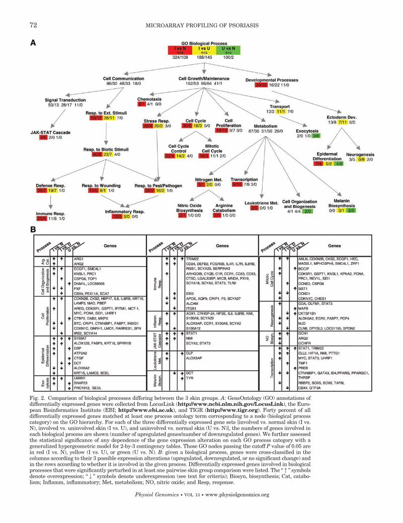
Fig. 2. Comparison of biological processes differing between the 3 skin groups. A: GeneOntology (GO) annotations ofdifferentially expressed genes were collected from LocusLink (http://www.ncbi.nlm.nih.gov/LocusLink), the Euro-pean Bioinformatics Institute (EBI; http://www.ebi.ac.uk), and TIGR (http://www.tigr.org). Forty percent of alldifferentially expressed genes matched at least one process ontology term corresponding to a node (biological processcategory) on the GO hierarchy. For each of the three differentially expressed gene sets [involved vs. normal skin (I vs.N), involved vs. uninvolved skin (I vs. U), and uninvolved vs. normal skin (U vs. N)], the numbers of genes involved ineach biological process are shown (number of upregulated genes/number of downregulated genes). We further assessedthe statistical significance of any dependence of the gene expression alteration on each GO process category with ageneralized hypergeometric model for 2-by-3 contingency tables. Those GO nodes passing the cutoff P value of 0.05 arein red (I vs. N), yellow (I vs. U), or green (U vs. N). B: given a biological process, genes were cross-classified in thecolumns according to their 3 possible expression alterations (upregulated, downregulated, or no significant change) andin the rows according to whether it is involved in the given process. Differentially expressed genes involved in biologicalprocesses that were significantly perturbed in at least one pairwise skin group comparison were listed. The “1” symbolsdenote overexpression; “2” symbols denote underexpression (see text for criteria); Biosyn, biosynthesis; Cat, catabo-lism; Inflamm, inflammatory; Met, metabolism; NO, nitric oxide; and Resp, response.
72 MICROARRAY PROFILING OF PSORIASIS
Physiol Genomics • VOL 13 • www.physiolgenomics.org

Fig. 3. Clustering of expression pro-files of immune signaling molecules;131 immune signaling genes were se-lected that were significantly differen-tially expressed (P � 0.05 and foldchanges � 1.2) in at least one pairwisecomparison among the 3 skin groups.The tissue types are presented on thetop: normal skin (N), psoriasis-unin-volved skin (U), and psoriasis-involvedskin (I). The normalized expressionlevel for each gene (rows) in each sam-ple (columns) is indicated by a colorcode (see the expression index bar atthe bottom). Gene names from the Uni-Gene database as well as common syn-onyms are indicated.
73MICROARRAY PROFILING OF PSORIASIS
Physiol Genomics • VOL 13 • www.physiolgenomics.org

another IL1R agonist, IL-1�, also showed a pattern ofdifferential expression. Most of these IL-1 cytokinesreceptor antagonists were discovered in the last fewyears.
T-cell and DC activation. The middle section of Fig. 3shows genes that were upregulated in involved vs.normal skin. It thus reflects an immune profile ofpsoriatic lesions. It is known that psoriasis lesionscontain marked increases in various classes of infiltrat-ing leukocytes, including macrophages, CD11c� DCs,CD83� DCs (probably derived from activated Langer-hans cells in situ), CD4� T cells, CD8� T cells,CD103� T cells, and neutrophils (27). Each of theseleukocytes subsets can be characterized by the upregu-lation of activation-related proteins and strong expres-sion of lineage-related gene products. We previouslyreported upregulation of IL-8, CD24, CD25, and CD47in involved skin (10, 38). However, upregulation ofmost of the genes shown in Fig. 3 has not been de-scribed before in psoriasis. This upregulation could bedue in part to persistent activation of T lymphocytesand DCs in the skin, or de novo activation of memory Tcells and DCs in the skin. Upregulation of CD47, ageneral lineage marker for blood-derived cells is verystrong across all samples. This probably reflects vari-ous leukocyte subsets infiltrating psoriasis lesions. Tcells in lesions are activated as evidenced by the up-regulation of IL-2R and IL-2R subunits, CD71,CD69, and IL-7R.
Trafficking of leukocytes into peripheral tissues isstrongly regulated by “leukocyte integrins.” Both themajor T-cell integrin CD11a/CD18 and the second leu-kocyte integrin CD11b/CD18 are known to be ex-pressed in high levels on monocytes/DCs and neutro-phils (28). All genes encoding these subunits wereupregulated in involved skin. The upregulation of ad-hesion molecules (E-selectin, P-selectin, and ICAM)would support leukocyte infiltration into inflamed skincompartments, as well as T-cell adhesion to DCs.
CD163, CD32, major histocompatibility complex(MHC) class I, MHC class II, CD83, CD53, and CD24are overexpressed in involved skin. The products ofthese genes are associated with monocytes/DCs andantigen presentation.
CD44 is a receptor for hyaluronic acid. By FACS (BDBiosciences) staining, we have found upregulation ofCD44 on lesional T cells (unpublished), while immuno-staining shows diffuse expression on various cells inpsoriasis lesions. Therefore, we suggest CD44 as thebest candidate for expression of an adhesion moleculethat would promote T-cell or DC retention in inflamedskin. The infiltration of neutrophils is reflected by theoverexpression of IL-8 along with its receptor CXCR2.
The bottom of Fig. 3 shows genes with expressionchanges that differ in uninvolved vs. normal skin. CD4antigen, expressed by CD4� T cells and by dendriticantigen-presenting cells, is increased in uninvolvedskin, as is CD11c, which is known to be strongly ex-pressed by some DCs. While psoriatic lesions exhibit avery large increase in CD11c� DCs (2), we have seenan increase in these cells in the early stages of eruptive
psoriasis lesions (unpublished results). Hence, the up-regulation of CD11c in uninvolved skin in the currentstudy implies either activation of gene expression inendogenous DCs or the beginning of infiltration byexogenous cells. We note that DCs endogenous to theskin can be activated by processing of an antigen or byreaction to cytokines such as TNF. Since we foundputative binding sites for NF�B, STAT1, and IRF-1 inthe promoter of the CD11c gene, its expression may beupregulated in response to increases in circulatingTNF or �-interferon (IFN-�) in patients with activedisease. An increase in DC activation could also ex-plain the increased expression of CD86, a major co-stimulatory molecule that is upregulated by the acti-vation process. Another gene that is elevated is CD103.Its expression is restricted to a group of epithelialhoming CD8� T cells that were first identified in guttissue (12). In a previous study we found increasedCD103� CD8� T cells in psoriatic lesional epidermis(37). Other studies (36) suggest that TGF is a matur-ing or inducing factor for this integrin, probably in thecutaneous microenvironment, and in fact TGF3 clus-ters with these genes that are upregulated in unin-volved vs. normal skin. SCYB14/BRAK is a chemokinethat promotes trafficking of activated monocytes intoskin as part of normal immune surveillance, and isupregulated in uninvolved vs. normal skin. With itsincreased expression, there may be more monocytetrafficking and even some differentiation of monocytesinto macrophages or DCs. In conclusion, gene expres-sion in uninvolved skin suggests a low level of immuneactivation involving both DCs and T cells.
A type-1 bias to activated T cells is indicated by theoverexpression of IFN-�. Orchestrating a wide range ofdiverse cellular programs, IFN-� controls the expres-sions of many genes. Interestingly, the most highlyupregulated transcription factors in involved skin wereTRIM22 (upregulated 12.3-fold) and STAT1 (upregu-lated 5.3-fold), both of which are IFN-� inducible.While STAT1 is a primary response gene to IFN-�, thetranscription regulation of TRIM22 in IFN-� responseremains to be determined.
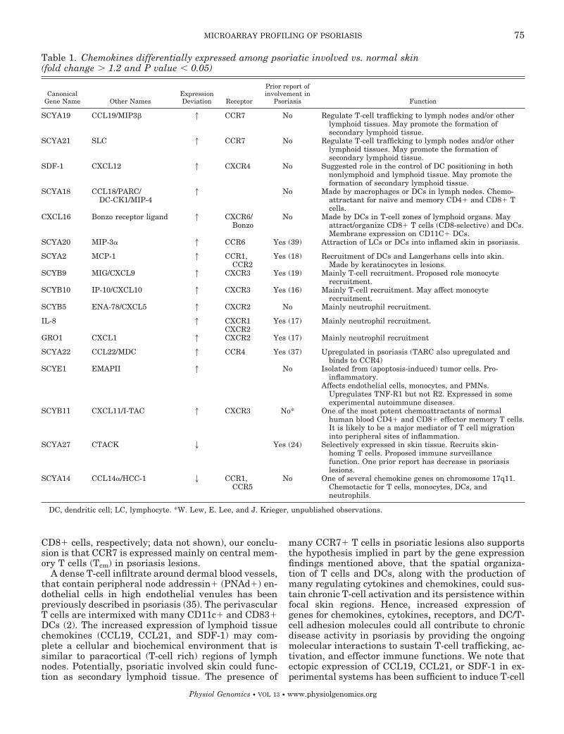
Chemokines in psoriasis. Chemokines play a complexrole in lymphocyte trafficking in psoriasis (27). Table 1lists the 19 chemokines that were determined to bedifferentially expressed in this study. Eleven of thesehave not been previously described in psoriasis.SCYA19/CCL19, SCYA21/CCL21/SLC, and SDF-1/CXCL12 were all upregulated in involved skin. Weinvestigated SCYA2/CCR7, the receptor for SCYA19,with flow cytometry and found evidence for its expres-sion in T cells and DCs of psoriasis lesions (results notshown). The combination of adhesion receptor (L-selec-tin) and chemokine receptor is normally found on node-homing T cells. CLA� and CLA cells both expressCCR7, suggesting entry of cells into psoriasis lesionswhich are not specifically differentiated for skin hom-ing and would otherwise be expected to home to lymphnodes or other “formal” lymphoid tissues. Since thereare very few naı̈ve T cells in involved skin (measuredby CD45RA and CD45RA/CD27 staining of CD4� and
74 MICROARRAY PROFILING OF PSORIASIS
Physiol Genomics • VOL 13 • www.physiolgenomics.org
CD8� cells, respectively; data not shown), our conclu-sion is that CCR7 is expressed mainly on central mem-ory T cells (Tcm) in psoriasis lesions.
A dense T-cell infiltrate around dermal blood vessels,that contain peripheral node addressin� (PNAd�) en-dothelial cells in high endothelial venules has beenpreviously described in psoriasis (35). The perivascularT cells are intermixed with many CD11c� and CD83�DCs (2). The increased expression of lymphoid tissuechemokines (CCL19, CCL21, and SDF-1) may com-plete a cellular and biochemical environment that issimilar to paracortical (T-cell rich) regions of lymphnodes. Potentially, psoriatic involved skin could func-tion as secondary lymphoid tissue. The presence of
many CCR7� T cells in psoriatic lesions also supportsthe hypothesis implied in part by the gene expressionfindings mentioned above, that the spatial organiza-tion of T cells and DCs, along with the production ofmany regulating cytokines and chemokines, could sus-tain chronic T-cell activation and its persistence withinfocal skin regions. Hence, increased expression ofgenes for chemokines, cytokines, receptors, and DC/T-cell adhesion molecules could all contribute to chronicdisease activity in psoriasis by providing the ongoingmolecular interactions to sustain T-cell trafficking, ac-tivation, and effector immune functions. We note thatectopic expression of CCL19, CCL21, or SDF-1 in ex-perimental systems has been sufficient to induce T-cell
Table 1. Chemokines differentially expressed among psoriatic involved vs. normal skin(fold change � 1.2 and P value � 0.05)
CanonicalGene Name Other Names
ExpressionDeviation Receptor
Prior report ofinvolvement in
Psoriasis Function
SCYA19 CCL19/MIP3 1 CCR7 No Regulate T-cell trafficking to lymph nodes and/or otherlymphoid tissues. May promote the formation ofsecondary lymphoid tissue.
SCYA21 SLC 1 CCR7 No Regulate T-cell trafficking to lymph nodes and/or otherlymphoid tissues. May promote the formation ofsecondary lymphoid tissue.
SDF-1 CXCL12 1 CXCR4 No Suggested role in the control of DC positioning in bothnonlymphoid and lymphoid tissue. May promote theformation of secondary lymphoid tissue.
SCYA18 CCL18/PARC/DC-CK1/MIP-4
1 No Made by macrophages or DCs in lymph nodes. Chemo-attractant for naı̈ve and memory CD4� and CD8� Tcells.
CXCL16 Bonzo receptor ligand 1 CXCR6/Bonzo
No Made by DCs in T-cell zones of lymphoid organs. Mayattract/organize CD8� T cells (CD8-selective) and DCs.Membrane expression on CD11C� DCs.
SCYA20 MIP-3 1 CCR6 Yes (39) Attraction of LCs or DCs into inflamed skin in psoriasis.
SCYA2 MCP-1 1 CCR1,CCR2
Yes (18) Recruitment of DCs and Langerhans cells into skin.Made by keratinocytes in lesions.
SCYB9 MIG/CXCL9 1 CXCR3 Yes (19) Mainly T-cell recruitment. Proposed role monocyterecruitment.
SCYB10 IP-10/CXCL10 1 CXCR3 Yes (16) Mainly T-cell recruitment. May affect monocyterecruitment.
SCYB5 ENA-78/CXCL5 1 CXCR2 No Mainly neutrophil recruitment.
IL-8 1 CXCR1 Yes (17) Mainly neutrophil recruitment.CXCR2
GRO1 CXCL1 1 CXCR2 Yes (17) Mainly neutrophil recruitment
SCYA22 CCL22/MDC 1 CCR4 Yes (37) Upregulated in psoriasis (TARC also upregulated andbinds to CCR4)
SCYE1 EMAPII 1 No Isolated from (apoptosis-induced) tumor cells. Pro-inflammatory.
Affects endothelial cells, monocytes, and PMNs.Upregulates TNF-R1 but not R2. Expressed in someexperimental autoimmune diseases.
SCYB11 CXCL11/I-TAC 1 CXCR3 No* One of the most potent chemoattractants of normalhuman blood CD4� and CD8� effector memory T cells.It is likely to be a major mediator of T cell migrationinto peripheral sites of inflammation.
SCYA27 CTACK 2 Yes (24) Selectively expressed in skin tissue. Recruits skin-homing T cells. Proposed immune surveillancefunction. One prior report has decrease in psoriasislesions.
SCYA14 CCL14/HCC-1 2 CCR1,CCR5
No One of several chemokine genes on chromosome 17q11.Chemotactic for T cells, monocytes, DCs, andneutrophils.
DC, dendritic cell; LC, lymphocyte. *W. Lew, E. Lee, and J. Krieger, unpublished observations.
75MICROARRAY PROFILING OF PSORIASIS
Physiol Genomics • VOL 13 • www.physiolgenomics.org

infiltrates and organized lymphoid tissues in otherorgans (30), while the expression of CCL19 and CCL21by endothelial cells in early lesions of experimentalautoimmune encephalomyelitis has been suggested tobe a key inducer of this disease (5).
SCYA18/CCL18, also upregulated in psoriatic skin,could play an additional role in this type of inflamma-tory reaction, since it might recruit naı̈ve T cells toskin-draining lymph nodes. Another upregulated gene,CXCL16, is expressed on the surface of CD11c� DCs.This suggests a link with the increased abundance ofCD11c� DCs in psoriatic tissue. In addition, we notethat CXCL16 selectively recruits of CD8� T cells,which are particularly increased in the epidermis ofinvolved skin.
The IFN-�-regulated chemokines IP-10 and MIGform a tightly upregulated cluster in involved skin inFig. 3. These two chemokines, together with I-TAC, areligands of G protein-coupled receptor 9 (CXCR3). Thesefindings are in concert with observed infiltration ofCXCR3� T cells in psoriatic skin (37). Clustered to-gether with IP-10 and MIG are MDC and MIP-4, bothof which are expressed in normal human keratinocytesactivated by Th1-derived supernatant (4). MIP-3, pro-posed as a major regulator of DC migration into skin(13), was also overexpressed in lesional skin.
Large-scale Promoter Analysis
The expression clustering of psoriasis-related genesover the whole genome enabled us to perform large-scalepromoter analysis of coexpressed genes. We present inTable 2 thirteen such coexpressed gene clusters and theirshared TFBS motifs which are in addition strongly sup-ported by mouse orthologous data.
As an interesting example, we identified the TFBSmotif [IRF2]-[ISRE] in four of seven genes in an ex-pression cluster. The transcription factor IRF2 (inter-feron response factor 2) can be induced by IFN-� (1),and ISRE is the interferon-stimulated response ele-ment. Notably, three of the four genes identified withthis motif are IFN inducible: 2,5-oligoadenylate syn-thetase 1 (OAS1), 2,5-oligoadenylate synthetase 2(OAS2), and interferon-stimulated protein (ISG15). Al-though the function of the remaining gene C1orf29 isunknown, based on its expression pattern and its pro-moter organization, it is likely to also be IFN inducibleand to play a part in inflammation and antiviral re-sponse. In fact, its mouse ortholog is a minor histocom-patibility antigen. The mouse orthologs of all fourgenes contain the same paired [IRF2]-[ISRE] motif.
The TFBS pair [NF-Y]-[Sp1] is identified in the up-stream sequences of four genes in an eight-gene ex-
Table 2. Potential TFBS motifs for genes of tight expression clusters
TFBS Motif Functions of Individual TFs Synexpression Genes
[Ikaros3] hematopoietic differentiation SLC6A14, PBEF*, GLUL, EVA1*[c-Ets-2] proliferation, differentiation, development,
transformation, apoptosisS100A7, S100A8*, S100A9*, KRT6A
[NF�B] inflammation, immune response LDLR*, LAMP3, MYC [NF�B(8)]*, HBP17*[AP1] hematopoietic differentiation, immune
response, apoptosisTGM3*, AKR1B10*, EVA1*, GJB6*, GJB2†, CPT2*
[ROR1] neural development, bone metabolism TNFSF10, RASGRP1*, DNASE1L3*, KPNA2*, MYO1B*, GK[VDR/RXR]-[HSF1] [VDV/RXR]: differentiation and apoptosis
of monocytesWNT5A*-*, GBP2, CASP1
[HSF1]: stress response[PPAR]-[OCT1] [PPAR]: inflammation, lipid metabolism,
epidermal differentiationISG15[*-*], TRIM22[*-*], MX1†-*, FLJ20637†-*
[OCT1]: Constitutive[GATA-1]-[EVI1] [GATA-1]: hematopoietic differentiation,
angiogenesisKYNU[*-*], S100A12, FLJ21343[*-*]
[EVA1]: hematopoietic differentiation[NF1]-[Creb1] [NF1]: proliferation, epidermal
differentiationTRB@ [NF1(9)], FLJ22662*-*, POLB [CREBP1 (33)]*-†
[Creb1]: cAMP response[IRF1]-[E2F] [IRF1]: antiviral response FLJ10134[*-*], FLJ22408, AI968669[*-*]
[E2F]: proliferation[NF-Y]-[Sp1] [NF-Y]: cellular senescence ‡TOP2A [NF-Y(3)][Sp1(41)]*-†, TRA1*-*, EIF5*-*, APG-1†-*
[Sp1]: constitutive, immune response[IRF2]-[ISRE] [IRF2]: immune response OAS1[*-*], OAS2[*-*], ISG15[*-*], Clorf29*-*
[ISRE]: immune response[Ikaros1]-[STAT1] [Ikaros1]: hematopoietic differentiation LOC84518[*-*], AMD1*-†, TGM3[*-*]
[STAT1]: immune response
Transcription factor binding sites (TFBS) were identified by searching for overrepresentation of particular promoter elements or theircombinations in the upstream sequences of genes in the same expression clusters generated based on highly correlated gene expressionprofiles with a constrained recursive K-means clustering approach. The distance between paired TFBSs is constrained between 20-80 bp. Forthose genes marked with *or † , their mouse orthologs can be identified; */†denotes the presence/absence of the same TFBS motif in theupstream sequences of the mouse orthologs. For paired TFBS motifs, *-*denotes that the mouse ortholog contains both of the motifs identifiedin its human counterpart. In addition, [*-*]denotes that the distance in the paired motif in the mouse ortholog is between 20-80 bp and inthe same order. Likewise, for other combinations of * and †. Literature support for specific TFBSs of our predictions is noted with underscore.‡The upstream sequence of the mouse TOP2A is unsatisfactory, due to the existence of large gaps in the Celera mouse genome assembly.
76 MICROARRAY PROFILING OF PSORIASIS
Physiol Genomics • VOL 13 • www.physiolgenomics.org

pression cluster. The four genes, topoisomerase II(TOP2A), tumor rejection antigen 1 (TRA1), eukaryotictranslation initiation factor 5 (EIF5), heat shock pro-tein (APG-1), are all involved in cell proliferation andstress response. The proliferation-dependent expres-sion of topoisomerase II has been experimentallyshown to require both NF-Y (3) and Sp1 (41). Otherstudies have shown that these two transcription fac-tors synergistically stimulate the transcription of somecell-cycle-dependent genes (42). In fact, in an indepen-dent study their binding to adjacent sites has beenshown to be cooperative (40). Our results suggest thatthis cooperativity may be more prevalent than previ-ously known.
As another example, we identified TFBSs for NF�Bin four clustered genes: DC lysosome-associated mem-brane protein (LAMP3), c-myc (MYC), heparin-bindinggrowth factor binding protein (HBP17), and low-den-sity lipoprotein receptor (LDLR). The first three areinvolved in cell proliferation, while the last supportscell proliferation in lymphocytes (31). Except forLAMP3, we were able to obtain their mouse or-thologs, all of which contained NF�B TFBSs in theirupstream sequences. In addition, the induction ofexpression of c-myc by NF�B has been experimen-tally determined (8).
Conclusion
This is the most comprehensive analysis of the hu-man transcriptome to date. We have generated a list of1,338 genes that are potentially psoriasis-related, and60% of these encode newly discovered proteins. Some ofthese genes are likely to be potential targets for newtherapeutic strategies (21).
In the context of the whole transcriptome, we sur-veyed the complete GeneOntology hierarchy and iden-tified a wide range of biological processes that weresignificantly perturbed in psoriasis. The genes involvedin immune signaling pathways were of particular in-terest and suggest de novo activation of T cells and DCsin the skin or novel methods for maintaining theiractivation.
Large-scale gene expression analysis also facilitatedthe discovery of regulatory mechanisms that governthe differential expression of genes. By focusing ontightly coregulated genes and by combining both ex-pression data and mouse orthologous promoter se-quences, we identified several individual and pairedpotential TFBSs in the upstream regions of psoriasis-related genes. Many of these motifs are biologicallyplausible and are related to epidermal differentiation,immune response, or proliferation. An important roleis proposed in psoriasis for STAT1 and TRIM22 tran-scription factors, primary response genes in the IFN-�pathway.
This study also provides an explanation for the effi-cacy of numerous biologic agents currently under eval-uation. For example, efalizumab, a humanized anti-body to CD11a, downregulates and blocks LFA on Tcells (20), and data presented here argue for the up-
regulation and potential importance of the leukocyteintegrins. The therapeutic agent CTLA4Ig binds CD80and CD86 and targets activated DCs. Its therapeuticresponses have been linked to decreased tissue infil-tration of CD11c � DCs, CD83 � DCs, and T cells (2).The impressive response of psoriasis to TNF inhibitorsis likely to be reflected in alterations in the expressionof genes with NF�B sites, possibly combined with GASor ISRE elements.
We thank Cheng Li for assistance with dChip analysis, and IgorLeykin and Haiyan Huang for support in promoter analysis.
The work of X. Zhou and W. H. Wong was supported by NationalInstitutes of Health (NIH) Grant 1R01-HG-02341. J. G. Krueger andE. Lee were supported by NIH Grants RR-100102, AI-49572, andAI-49832. The work of M.-C. J. Kao was supported by the HowardHughes Medical Institute predoctoral fellowship. We thank Li Caofor technical help.
REFERENCES
1. Aberger F, Costa-Pereira AP, Schlaak JF, Williams TM,O’Shaughnessy RF, Hollaus G, Kerr IM, and FrischaufAM. Analysis of gene expression using high-density and IFN-gamma-specific low-density cDNA arrays. Genomics 77: 50–57,2001.
2. Abrams JR, Kelley SL, Hayes E, Kikuchi T, Brown MJ,Kang S, Lebwohl MG, Guzzo CA, Jegasothy BV, LinsleyPS, and Krueger JG. Blockade of T lymphocyte costimulationwith cytotoxic T lymphocyte-associated antigen 4-immunoglobu-lin (CTLA4Ig) reverses the cellular pathology of psoriaticplaques, including the activation of keratinocytes, dendritic cells,and endothelial cells. J Exp Med 192: 681–694, 2000.
3. Adachi N, Nomoto M, Kohno K, and Koyama H. Cell-cycleregulation of the DNA topoisomerase II promoter is mediatedby proximal CCAAT boxes: possible involvement of acetylation.Gene 245: 49–57, 2000.
4. Albanesi C, Scarponi C, Sebastiani S, Cavani A, FedericiM, Sozzani S, and Girolomoni G. A cytokine-to-chemokineaxis between T lymphocytes and keratinocytes can favor Th1 cellaccumulation in chronic inflammatory skin diseases. J LeukocBiol 70: 617–623, 2001.
5. Alt C, Laschinger M, and Engelhardt B. Functional expres-sion of the lymphoid chemokines CCL19 (ELC) and CCL 21(SLC) at the blood-brain barrier suggests their involvement inG-protein-dependent lymphocyte recruitment into the centralnervous system during experimental autoimmune encephalomy-elitis. Eur J Immunol 32: 2133–2144, 2002.
6. Barker JN. The pathophysiology of psoriasis. Lancet 338: 227–230, 1991.
7. Bhalerao J and Bowcock AM. The genetics of psoriasis: acomplex disorder of the skin and immune system. Hum MolGenet 7: 1537–1545, 1998.
8. Bourgarel-Rey V, Vallee S, Rimet O, Champion S, BraguerD, Desobry A, Briand C, and Barra Y. Involvement of nuclearfactor �B in c-Myc induction by tubulin polymerization inhibi-tors. Mol Pharmacol 59: 1165–1170, 2001.
9. Bourke PF, van Leeuwen BH, Campbell HD, and YoungIG. Localization of the inducible enhancer in the mouse inter-leukin-5 gene that is responsive to T-cell receptor stimulation.Blood 85: 2069–2077, 1995.
10. Bowcock AM, Shannon W, Du F, Duncan J, Cao K, After-gut K, Catier J, Fernandez-Vina MA, and Menter A. In-sights into psoriasis and other inflammatory diseases from large-scale gene expression studies. Hum Mol Genet 10: 1793–1805,2001.
11. Capon F, Munro M, Barker J, and Trembath R. Searchingfor the major histocompatibility complex psoriasis susceptibilitygene. J Invest Dermatol 118: 745–751, 2002.
12. Cerf-Bensussan N, Jarry A, Brousse N, Lisowska-Grospi-erre B, Guy-Grand D, and Griscelli C. A monoclonal antibody(HML-1) defining a novel membrane molecule present on humanintestinal lymphocytes. Eur J Immunol 17: 1279–1285, 1987.
77MICROARRAY PROFILING OF PSORIASIS
Physiol Genomics • VOL 13 • www.physiolgenomics.org

13. Dieu-Nosjean MC, Massacrier C, Homey B, Vanbervliet B,Pin JJ, Vicari A, Lebecque S, Dezutter-Dambuyant C,Schmitt D, Zlotnik A, and Caux C. Macrophage inflammatoryprotein 3 is expressed at inflamed epithelial surfaces and is themost potent chemokine known in attracting Langerhans cellprecursors. J Exp Med 192: 705–718, 2000.
14. Eisen MB, Spellman PT, Brown PO, and Botstein D. Clus-ter analysis and display of genome-wide expression patterns.Proc Natl Acad Sci USA 95: 14863–14868, 1998.
15. Ferenczi K, Burack L, Pope M, Krueger JG, and AustinLM. CD69, HLA-DR and the IL-2R identify persistently acti-vated T cells in psoriasis vulgaris lesional skin: blood and skincomparisons by flow cytometry. J Autoimmun 14: 63–78, 2000.
16. Flier J, Boorsma DM, van Beek PJ, Nieboer C, Stoof TJ,Willemze R, and Tensen CP. Differential expression ofCXCR3 targeting chemokines CXCL10, CXCL9, and CXCL11 indifferent types of skin inflammation. J Pathol 194: 398–405,2001.
17. Gillitzer R, Ritter U, Spandau U, Goebeler M, and BrockerEB. Differential expression of GRO- and IL-8 mRNA in psori-asis: a model for neutrophil migration and accumulation in vivo.J Invest Dermatol 107: 778–782, 1996.
18. Gillitzer R, Wolff K, Tong D, Muller C, Yoshimura T, Hart-mann AA, Stingl G, and Berger R. MCP-1 mRNA expressionin basal keratinocytes of psoriatic lesions. J Invest Dermatol 101:127–131, 1993.
19. Goebeler M, Toksoy A, Spandau U, Engelhardt E, BrockerEB, and Gillitzer R. The C-X-C chemokine Mig is highlyexpressed in the papillae of psoriatic lesions. J Pathol 184:89–95, 1998.
20. Gottlieb AB, Krueger JG, Wittkowski K, Dedrick R, Wal-icke PA, and Garovoy M. Psoriasis as a model for T-cell-mediated disease: immunobiologic and clinical effects of treat-ment with multiple doses of efalizumab, an anti-CD11a anti-body. Arch Dermatol 138: 591–600, 2002.
21. Griffiths CE. Immunotherapy for psoriasis: from serendipity toselectivity. Lancet 359: 279–280, 2002.
22. Griffiths TW, Griffiths CE, and Voorhees JJ. Immunopatho-genesis and immunotherapy of psoriasis. Dermatol Clin 13:739–749, 1995.
23. Hammerberg C, Bata-Csorgo Z, Voorhees JJ, and CooperKD. IL-1 and IL-1 receptor antagonist regulation during kera-tinocyte cell cycle and differentiation in normal and psoriaticepidermis. Arch Dermatol Res 290: 367–374, 1998.
24. Homey B, Alenius H, Muller A, Soto H, Bowman EP, YuanW, McEvoy L, Lauerma AI, Assmann T, Bunemann E,Lehto M, Wolff H, Yen D, Marxhausen H, To W, SedgwickJ, Ruzicka T, Lehmann P, and Zlotnik A. CCL27-CCR10interactions regulate T cell-mediated skin inflammation. NatMed 8: 157–165, 2002.
25. Kadunce DP and Krueger GG. Pathogenesis of psoriasis.Dermatol Clin 13: 723–737, 1995.
26. Kavli G, Forde OH, Arnesen E, and Stenvold SE. Psoriasis:familial predisposition and environmental factors. Br Med J(Clin Res Ed) 291: 999–1000, 1985.
27. Krueger JG. The immunologic basis for the treatment of pso-riasis with new biologic agents. J Am Acad Dermatol 46: 1–23;quiz 23–26, 2002.
28. Larson RS and Springer TA. Structure and function of leuko-cyte integrins. Immunol Rev 114: 181–217, 1990.
29. Li C and Wong WH. Model-based analysis of oligonucleotidearrays: expression index computation and outlier detection. ProcNatl Acad Sci USA 98: 31–36, 2001.
30. Luther SA, Bidgol A, Hargreaves DC, Schmidt A, Xu Y,Paniyadi J, Matloubian M, and Cyster JG. Differing activi-ties of homeostatic chemokines CCL19, CCL21, and CXCL12 inlymphocyte and dendritic cell recruitment and lymphoid neogen-esis. J Immunol 169: 424–433, 2002.
31. Martinez-Botas J, Suarez Y, Reshef A, Carrero P, OrtegaH, Gomez-Coronado D, Teruel JL, Leitersdorf E, and La-suncion MA. Impact of different low-density lipoprotein (LDL)receptor mutations on the ability of LDL to support lymphocyteproliferation. Metabolism 48: 834–839, 1999.
32. Menter A and Barker JN. Psoriasis in practice. Lancet 338:231–234, 1991.
33. Narayan S, He F, and Wilson SH. Activation of the humanDNA polymerase beta promoter by a DNA-alkylating agentthrough induced phosphorylation of cAMP response element-binding protein-1. J Biol Chem 271: 18508–18513, 1996.
34. Oestreicher JL, Walters IB, Kikuchi T, Gilleaudeau P,Surette J, Schwertschlag U, Dorner AJ, Krueger JG, andTrepicchio WL. Molecular classification of psoriasis disease-associated genes through pharmacogenomic expression profil-ing. Pharmacogenomics J 1: 272–287, 2001.
35. Paavonen T and Renkonen R. Selective expression of sialyl-Lewis x and Lewis a epitopes, putative ligands for L-selectin, onperipheral lymph-node high endothelial venules. Am J Pathol141: 1259–1264, 1992.
36. Pauls K, Schon M, Kubitza RC, Homey B, Wiesenborn A,Lehmann P, Ruzicka T, Parker CM, and Schon MP. Role ofintegrin E(CD103)7 for tissue-specific epidermal localizationof CD8� T lymphocytes. J Invest Dermatol 117: 569–575, 2001.
37. Rottman JB, Smith TL, Ganley KG, Kikuchi T, andKrueger JG. Potential role of the chemokine receptors CXCR3,CCR4, and the integrin E7 in the pathogenesis of psoriasisvulgaris. Lab Invest 81: 335–347, 2001.
38. Trepicchio WL, Ozawa M, Walters IB, Kikuchi T, Gil-leaudeau P, Bliss JL, Schwertschlag U, Dorner AJ, andKrueger JG. Interleukin-11 therapy selectively downregulatestype I cytokine proinflammatory pathways in psoriasis lesions.J Clin Invest 104: 1527–1537, 1999.
39. Vanbervliet B, Homey B, Durand I, Massacrier C, Ait-Yahia S, de Bouteiller O, Vicari A, and Caux C. Sequentialinvolvement of CCR2 and CCR6 ligands for immature dendriticcell recruitment: possible role at inflamed epithelial surfaces.Eur J Immunol 32: 231–242, 2002.
40. Wright KL, Moore TL, Vilen BJ, Brown AM, and Ting JP.Major histocompatibility complex class II-associated invariantchain gene expression is up-regulated by cooperative interac-tions of Sp1 and NF-Y. J Biol Chem 270: 20978–20986, 1995.
41. Yoon JH, Kim JK, Rha GB, Oh M, Park SH, Seong RH,Hong SH, and Park SD. Sp1 mediates cell proliferation-depen-dent regulation of rat DNA topoisomerase II gene promoter.Biochem J 344: 367–374, 1999.
42. Zwicker J, Lucibello FC, Wolfraim LA, Gross C, Truss M,Engeland K, and Muller R. Cell cycle regulation of the cyclinA, cdc25C and cdc2 genes is based on a common mechanism oftranscriptional repression. EMBO J 14: 4514–4522, 1995.
78 MICROARRAY PROFILING OF PSORIASIS
Physiol Genomics • VOL 13 • www.physiolgenomics.org
Copyright © 2022 FDOKUMEN