A Subpopulation of CD163Positive Macrophages Is Classically Activated in Psoriasis
17
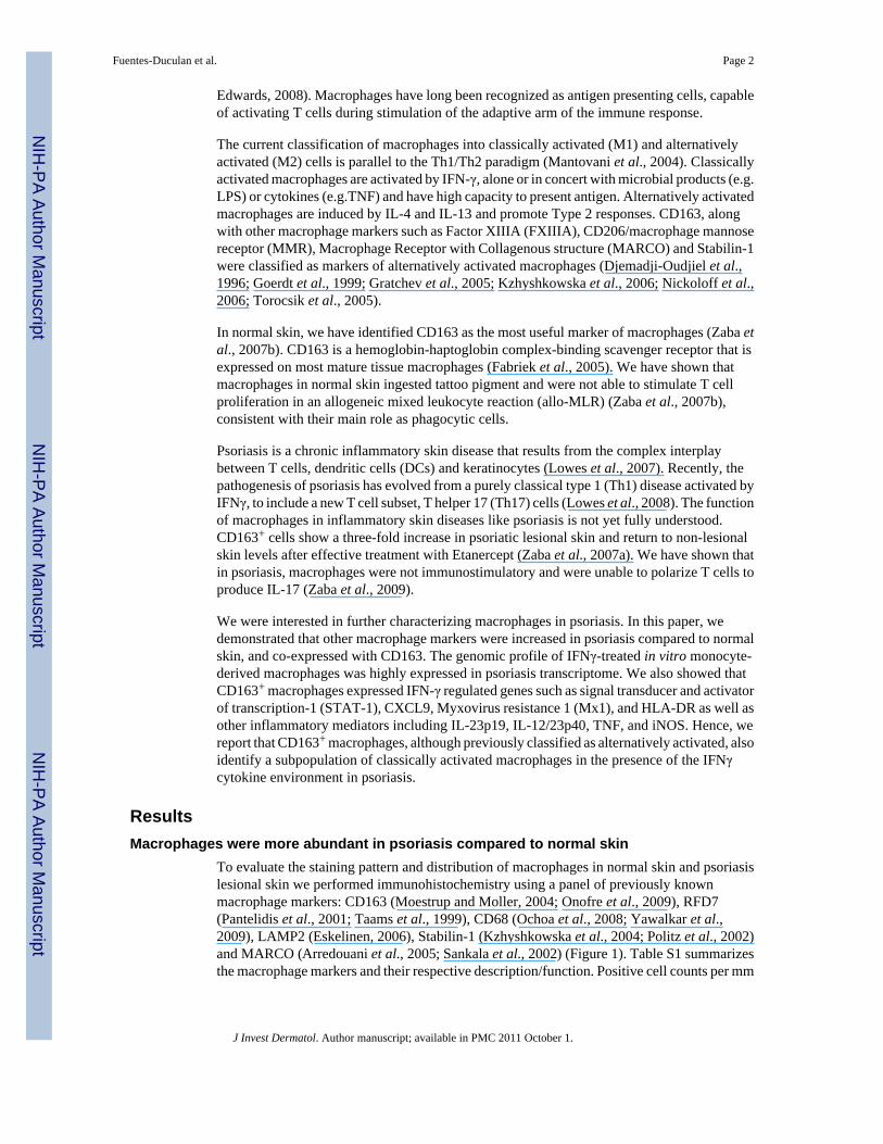
A subpopulation of CD163 positive macrophages is classically activated in psoriasis Judilyn Fuentes-Duculan 1 , Mayte Suárez-Fariñas 1,2 , Lisa C. Zaba 1 , Kristine E. Nograles 1 , Katherine C. Pierson 1 , Hiroshi Mitsui 1 , Cara A. Pensabene 1 , Julia Kzhyshkowska 3 , James G. Krueger 1 , and Michelle A. Lowes 1 1 Laboratory for Investigative Dermatology, The Rockefeller University, 1230 York Avenue, New York, NY, USA 10065 2 Center for Clinical and Translational Science, The Rockefeller University, 1230 York Avenue, New York, NY, USA 10065 3 Department of Dermatology, University Medical Centre Mannheim, Ruprecht-Karls University Heidelberg, Mannheim, Germany Abstract Macrophages are important cells of the innate immune system, and their study is essential to gain greater understanding of the inflammatory nature of psoriasis. We used immunohistochemistry and double-label immunofluorescence to characterize CD163 + macrophages in psoriasis. Dermal macrophages were increased in psoriasis compared to normal skin and were identified by CD163, RFD7, CD68, LAMP2, Stabilin-1, and MARCO. CD163 + macrophages expressed C-lectins CD206/ MMR and CD209/DC-SIGN, as well as co-stimulatory molecules CD86 and CD40. They did not express mature DC markers CD208/DC-LAMP, CD205/DEC205 or CD83. Microarray analysis of in vitro derived macrophages treated with IFNγ showed that many of the genes upregulated in macrophages were found in psoriasis, including STAT1, CXCL9, Mx1 and HLA-DR. CD163 + macrophages produced inflammatory molecules IL-23p19 and IL-12/23p40 as well as TNF and iNOS. These data demonstrate that CD163 is a superior marker of macrophages, and identifies a subpopulation of “classically activated” macrophages in psoriasis. We conclude that macrophages are likely to be contributing to the pathogenic inflammation in psoriasis, a prototypical Th1 and Th17 disease, by releasing key inflammatory products. Keywords Psoriasis; macrophages; myeloid dendritic cell; BDCA-1; Tip-DC; dermis; inflammation; skin Introduction Macrophages are important sentinels of the innate immune system. Their primary role is believed to be as phagocytic cells that participate in tissue homeostasis, and in the clearance of erythrocytes and removal of cellular debris generated during tissue remodeling (Mosser and Corresponding author Michelle A. Lowes The Rockefeller University Laboratory for Investigative Dermatology 1230 York Avenue, Box 178 NY, NY 10065 Ph: 212-327-7576 Fax: 212-327-8353 [email protected] Communicating author Judilyn Fuentes- Duculan The Rockefeller University Laboratory for Investigative Dermatology 1230 York Avenue, Box 178 NY, NY 10065 Ph: 212-327-8027 Fax: 212-327-8353 [email protected]. Conflict of Interest The authors do not have any financial interest related to this work. NIH Public Access Author Manuscript J Invest Dermatol. Author manuscript; available in PMC 2011 October 1. Published in final edited form as: J Invest Dermatol. 2010 October ; 130(10): 2412–2422. doi:10.1038/jid.2010.165. NIH-PA Author Manuscript NIH-PA Author Manuscript NIH-PA Author Manuscript
-
Upload
rockefeller -
Category
Documents
-
view
5 -
download
0
Transcript of A Subpopulation of CD163Positive Macrophages Is Classically Activated in Psoriasis
A subpopulation of CD163 positive macrophages is classicallyactivated in psoriasis
Judilyn Fuentes-Duculan1, Mayte Suárez-Fariñas1,2, Lisa C. Zaba1, Kristine E. Nograles1,Katherine C. Pierson1, Hiroshi Mitsui1, Cara A. Pensabene1, Julia Kzhyshkowska3, JamesG. Krueger1, and Michelle A. Lowes11Laboratory for Investigative Dermatology, The Rockefeller University, 1230 York Avenue, NewYork, NY, USA 100652Center for Clinical and Translational Science, The Rockefeller University, 1230 York Avenue, NewYork, NY, USA 100653Department of Dermatology, University Medical Centre Mannheim, Ruprecht-Karls UniversityHeidelberg, Mannheim, Germany
AbstractMacrophages are important cells of the innate immune system, and their study is essential to gaingreater understanding of the inflammatory nature of psoriasis. We used immunohistochemistry anddouble-label immunofluorescence to characterize CD163+ macrophages in psoriasis. Dermalmacrophages were increased in psoriasis compared to normal skin and were identified by CD163,RFD7, CD68, LAMP2, Stabilin-1, and MARCO. CD163+ macrophages expressed C-lectins CD206/MMR and CD209/DC-SIGN, as well as co-stimulatory molecules CD86 and CD40. They did notexpress mature DC markers CD208/DC-LAMP, CD205/DEC205 or CD83. Microarray analysis ofin vitro derived macrophages treated with IFNγ showed that many of the genes upregulated inmacrophages were found in psoriasis, including STAT1, CXCL9, Mx1 and HLA-DR. CD163+
macrophages produced inflammatory molecules IL-23p19 and IL-12/23p40 as well as TNF andiNOS. These data demonstrate that CD163 is a superior marker of macrophages, and identifies asubpopulation of “classically activated” macrophages in psoriasis. We conclude that macrophagesare likely to be contributing to the pathogenic inflammation in psoriasis, a prototypical Th1 and Th17disease, by releasing key inflammatory products.
KeywordsPsoriasis; macrophages; myeloid dendritic cell; BDCA-1; Tip-DC; dermis; inflammation; skin
IntroductionMacrophages are important sentinels of the innate immune system. Their primary role isbelieved to be as phagocytic cells that participate in tissue homeostasis, and in the clearanceof erythrocytes and removal of cellular debris generated during tissue remodeling (Mosser and
Corresponding author Michelle A. Lowes The Rockefeller University Laboratory for Investigative Dermatology 1230 York Avenue,Box 178 NY, NY 10065 Ph: 212-327-7576 Fax: 212-327-8353 [email protected] Communicating author Judilyn Fuentes-Duculan The Rockefeller University Laboratory for Investigative Dermatology 1230 York Avenue, Box 178 NY, NY 10065 Ph:212-327-8027 Fax: 212-327-8353 [email protected] of Interest The authors do not have any financial interest related to this work.
NIH Public AccessAuthor ManuscriptJ Invest Dermatol. Author manuscript; available in PMC 2011 October 1.
Published in final edited form as:J Invest Dermatol. 2010 October ; 130(10): 2412–2422. doi:10.1038/jid.2010.165.
NIH
-PA Author Manuscript
NIH
-PA Author Manuscript
NIH
-PA Author Manuscript
Edwards, 2008). Macrophages have long been recognized as antigen presenting cells, capableof activating T cells during stimulation of the adaptive arm of the immune response.
The current classification of macrophages into classically activated (M1) and alternativelyactivated (M2) cells is parallel to the Th1/Th2 paradigm (Mantovani et al., 2004). Classicallyactivated macrophages are activated by IFN-γ, alone or in concert with microbial products (e.g.LPS) or cytokines (e.g.TNF) and have high capacity to present antigen. Alternatively activatedmacrophages are induced by IL-4 and IL-13 and promote Type 2 responses. CD163, alongwith other macrophage markers such as Factor XIIIA (FXIIIA), CD206/macrophage mannosereceptor (MMR), Macrophage Receptor with Collagenous structure (MARCO) and Stabilin-1were classified as markers of alternatively activated macrophages (Djemadji-Oudjiel et al.,1996; Goerdt et al., 1999; Gratchev et al., 2005; Kzhyshkowska et al., 2006; Nickoloff et al.,2006; Torocsik et al., 2005).
In normal skin, we have identified CD163 as the most useful marker of macrophages (Zaba etal., 2007b). CD163 is a hemoglobin-haptoglobin complex-binding scavenger receptor that isexpressed on most mature tissue macrophages (Fabriek et al., 2005). We have shown thatmacrophages in normal skin ingested tattoo pigment and were not able to stimulate T cellproliferation in an allogeneic mixed leukocyte reaction (allo-MLR) (Zaba et al., 2007b),consistent with their main role as phagocytic cells.
Psoriasis is a chronic inflammatory skin disease that results from the complex interplaybetween T cells, dendritic cells (DCs) and keratinocytes (Lowes et al., 2007). Recently, thepathogenesis of psoriasis has evolved from a purely classical type 1 (Th1) disease activated byIFNγ, to include a new T cell subset, T helper 17 (Th17) cells (Lowes et al., 2008). The functionof macrophages in inflammatory skin diseases like psoriasis is not yet fully understood.CD163+ cells show a three-fold increase in psoriatic lesional skin and return to non-lesionalskin levels after effective treatment with Etanercept (Zaba et al., 2007a). We have shown thatin psoriasis, macrophages were not immunostimulatory and were unable to polarize T cells toproduce IL-17 (Zaba et al., 2009).
We were interested in further characterizing macrophages in psoriasis. In this paper, wedemonstrated that other macrophage markers were increased in psoriasis compared to normalskin, and co-expressed with CD163. The genomic profile of IFNγ-treated in vitro monocyte-derived macrophages was highly expressed in psoriasis transcriptome. We also showed thatCD163+ macrophages expressed IFN-γ regulated genes such as signal transducer and activatorof transcription-1 (STAT-1), CXCL9, Myxovirus resistance 1 (Mx1), and HLA-DR as well asother inflammatory mediators including IL-23p19, IL-12/23p40, TNF, and iNOS. Hence, wereport that CD163+ macrophages, although previously classified as alternatively activated, alsoidentify a subpopulation of classically activated macrophages in the presence of the IFNγcytokine environment in psoriasis.
ResultsMacrophages were more abundant in psoriasis compared to normal skin
To evaluate the staining pattern and distribution of macrophages in normal skin and psoriasislesional skin we performed immunohistochemistry using a panel of previously knownmacrophage markers: CD163 (Moestrup and Moller, 2004; Onofre et al., 2009), RFD7(Pantelidis et al., 2001; Taams et al., 1999), CD68 (Ochoa et al., 2008; Yawalkar et al.,2009), LAMP2 (Eskelinen, 2006), Stabilin-1 (Kzhyshkowska et al., 2004; Politz et al., 2002)and MARCO (Arredouani et al., 2005; Sankala et al., 2002) (Figure 1). Table S1 summarizesthe macrophage markers and their respective description/function. Positive cell counts per mm
Fuentes-Duculan et al. Page 2
J Invest Dermatol. Author manuscript; available in PMC 2011 October 1.
NIH
-PA Author Manuscript
NIH
-PA Author Manuscript
NIH
-PA Author Manuscript
epidermis surface length (Figure 1), and per square area (Table S1) were all significantlyincreased in psoriasis compared to normal skin (p<0.05).
CD163+ and RFD7+ macrophages were widely distributed over the papillary and reticulardermis of normal and psoriatic skin (Figure 1a, 1b), but CD163+ cells were more abundant.These numbers of CD163+ and RFD7+ macrophages were similar to the numbers ofinflammatory DCs in psoriasis (Zaba et al., 2007a), and thus identify a second major myeloidleukocyte population in lesional skin.
Most of the CD68+ and LAMP2+ macrophages were located in the upper portion of both normaland psoriatic dermis (Figure 1c, 1d). There were some CD68+ cells near the dermo-epidermaljunction (DEJ) particularly in psoriasis. The non-specific reactivity noted in the epidermis withLAMP2 antibody is common with antibodies conjugated to FITC. In both normal and psoriaticskin, Stabilin-1+ macrophages were observed in a perivascular distribution in the dermis(Figure 1e). MARCO+ cells were scattered throughout the dermis of normal and psoriatic skin(Figure 1f). While all these macrophage markers identified many dermal cells in psoriasis, theyhad slightly different staining patterns and distributions, suggesting some heterogeneity ofsurface antigens on macrophages.
CD163+ macrophages co-expressed additional macrophage markers in psoriasisWe further characterized the co-expression of these macrophage markers with CD163 inpsoriatic skin using double-label immunofluorescence (Figure 2). We have previously reportedthat in normal skin FXIIIA+, CD68+ and RFD7+ cells co-localized with CD163 (Zaba et al.,2007b). In psoriatic skin, FXIIIA+ and RFD7+ macrophages almost completely co-expressedCD163 (Figure 2a, 2b). The majority of CD68+ cells co-expressed CD163 in the upper reticulardermis (Figure 2c) but the cells near the DEJ did not co-localize with CD163. We havepreviously demonstrated that these CD68+ cells near the DEJ are CD11c+ (Wang et al.,2006), and therefore this marker identifies both myeloid DCs and macrophages. Over half ofLAMP2+ and Stabilin-1+ macrophages also co-expressed CD163 (Figures 2d, 2e).MARCO+ cells had the least co-expression with CD163 (Figure 2f).
To assess the potential for these markers to be exclusive for macrophages, we performeddouble-label immunofluorescence with CD11c (Figure S1). All these macrophage markerswere co-expressed to some degree with CD11c. However, among all the markers studied,CD163 had the least co-expression with CD11c making it a superior marker of macrophagesin psoriasis. We also showed that CD163+ macrophages and CD11c+ myeloid DCs co-expressed CD14, CD16 and LFA-1 (Figure S2).
CD163+ macrophages also expressed C-lectins and co-stimulatory molecules in psoriasisWe evaluated the expression of C-lectins and co-stimulatory molecules on CD163+
macrophages by double-label immunofluorescence (Figure S3). In normal skin, we havepreviously reported that CD206/MMR and CD209/DC-specific ICAM-3-grabbing non-integrin (DC-SIGN) were more abundant on macrophages than DCs (Zaba et al., 2007b). Wenow demonstrate that in psoriatic skin, almost all CD163+ macrophages were CD206+/MMRand approximately three quarters co-expressed CD-209/DC-SIGN+(Supplemental Figure 3a,3b). It has been previously reported that macrophages constitutively express CD86 (Way etal., 2009) and CD40 (Suttles and Stout, 2009). Here, we demonstrated that in psoriasis lessthan half of CD163+ macrophages expressed CD86 and CD40 co-stimulatory molecules.
CD163+ macrophages did not express “classic” dendritic cell markers in psoriasisIn order to clearly distinguish macrophages from DCs, we performed double-labelimmunofluorescence with CD163 and the well-recognized DC markers CD208/DC-
Fuentes-Duculan et al. Page 3
J Invest Dermatol. Author manuscript; available in PMC 2011 October 1.
NIH
-PA Author Manuscript
NIH
-PA Author Manuscript
NIH
-PA Author Manuscript
lysosomal-associated membrane glycoprotein (DC-LAMP), CD205/DEC-205, and CD83(Figure S4). In psoriatic and normal skin, CD163+ macrophages did not co-express any of theseDC maturation markers. These observations support that there are distinct markers for matureDCs and macrophages.
Genomic signature of in vitro “classically activated” (M1) macrophages was over-expressedin psoriasis
Previous studies have suggested that CD163 is a marker of alternatively activated macrophagesin psoriasis (Djemadji-Oudjiel et al., 1996). However, we were interested to explore whyalternatively activated cells would be present in the Th1 “classical” microenvironment ofpsoriasis. As we were unable to obtain sufficient quantities of macrophages from psoriaticlesional skin to study ex vivo, we turned to in vitro methods to further understand macrophagebiology in chronic cutaneous inflammation. To determine the transcriptional profile associatedwith macrophage polarization to M1 or M2, we cultured macrophages derived from monocytes(n=7) for 7 days and treated them with IFNγ, IL-4, TNF, LPS, and LPS+IFNγ. Martinez etal used a similar experimental approach to generate LPS+IFNγ- and IL-4-treated macrophages(Martinez et al., 2006). However, we included additional conditions (IFNγ alone, LPS alone,and TNF) and also generated lists of differentially expressed genes (DEG) by comparing theeffect of the cytokine on macrophages with control (Table S2). A heatmap of the estimatedfold change of every condition versus control showed a tight relationship between the effectof LPS+IFNγ, IFNγ, LPS, and TNF treatment of macrophages, while IL-4 has a distinct effect(Figure 3a).
We were particularly interested in the genomic profiles of “M1” macrophages induced withIFNγ, compared to “M2” macrophages treated with IL-4 (Figure 3b). We identified 919 DEGsbetween IFNγ-treated macrophages compared to control (FCH>2.0, FDR<0.01). There were585 probes upregulated (in red) in the IFNγ-treated macrophages, and 334 down-regulatedprobes (in green). Results of Ingenuity analysis of up- and down-regulated M1 and M2 genesare provided in Table S3. As expected, Gene Ontology classified upregulated M1 genes withInflammatory Response and Inflammatory Disease. M1 upregulated genes were significant inthe Cannonical Pathways for Communication between Innate and Adaptive Immune Cells,Interferon Signaling, and Role of Macrophages, Fibroblasts and Endothelial Cells inRheumatoid Arthritis.
In psoriasis lesional skin, there were 304 upregulated probes and 192 downregulated probescompared to non-lesional skin, on the same Illumina Human HT-12 microarray chip (FCH>2.0,FDR<0.01). The overlap of these two DEG lists showed that 90/585 (15%) probes wereupregulated in both IFNγ-treated macrophages and psoriasis, and 20/334 (6%) probes weredownregulated in both IFNγ-treated macrophages and psoriasis. In contrast, there were 132probes that were upregulated by IL-4 treated macrophages, and 29 probes down-regulated.Only 6/132 (4.5%) probes overlapped with the psoriasis transcriptome and 2 probes were incommon in the downregulated set.
We generated cytokine-treated macrophage “pathways”, consisting of the genes expressed bythe addition of a given cytokine to in vitro monocyte-derived macrophages. We then used GeneSet Enrichment Analysis (GSEA) to compare these macrophage pathways with the genomicprofile of psoriasis lesional skin. We hypothesized that there should be greater expression of“M1” macrophage genes in psoriasis. The GSEA results indicated that the following gene setswere significantly enriched in psoriasis lesional tissue (compared with non-lesional): LPS-UP,IFNγ-UP, LPS+IFNγ-UP, TNFα-UP, IL-4-DOWN (FDR<0.013 for all pathways) (Table S4).The LPS and IFNγ “pathways” were the top 3, with a normalized enrichment score (NES) >2.3,showing that these pathways were the most enriched in the psoriasis gene set. The LPS
Fuentes-Duculan et al. Page 4
J Invest Dermatol. Author manuscript; available in PMC 2011 October 1.
NIH
-PA Author Manuscript
NIH
-PA Author Manuscript
NIH
-PA Author Manuscript
+IFNγ-DOWN and IFNγ-DOWN gene sets were also enriched in non-lesional tissue(FDR=0.009 and 0.037 respectively).
Table 1 presents the fold change of 20 selected up-regulated genes in common betweenpsoriasis and IFNγ-treated macrophages. CXCL9 (122.69), CXCL10 (81.22), Mx1 (18.97),CCL20 (8.85) and STAT1 (8.54) (FDR <10−6 for all genes) are well known psoriasis genesthat were increased by IFNγ in macrophages. Table S5 lists the fold change for the few genesin common between psoriasis and IL-4-treated and TNF-treated macrophages.
CD163+ macrophages expressed IFN-γ regulated genesIt is well appreciated that there is a dominant “Type 1” IFNγ signature in psoriasis (Kryczeket al., 2008; Lew et al., 2004) and our microarray results suggested that macrophages treatedwith IFNγ were well represented at the genomic level in psoriasis. To verify that macrophagesin psoriasis lesions are indeed responsive to IFNγ, we performed double labelimmunofluorescence with CD163 and IFN-γ regulated genes identified by microarray,STAT-1, CXCL9, Mx1, and HLA-DR (Figure 4). We observed that the majority ofmacrophages expressed STAT-1 (Figure 4a), which is known as one of the most consistenttranscription factor alterations in psoriasis (Lew et al., 2004). By immunohistochemistry,STAT-1 expression in normal skin was minimal, but was abundant and nuclear in psoriaticlesional skin (Figure S5a), indicating activation in psoriasis.
Approximately half of CD163+ macrophages expressed CXCL9 (Figure 4b). This chemokineis considered a key IFN-γ-regulated gene and is thought to bind to CXCR3-bearing activatedT-cells, and may be involved in T-cell trafficking to psoriatic dermis and epidermis (Nograleset al., 2008; Rottman et al., 2001). There was low level CXCL9 expression in normal skin butwas increased in psoriasis by immunohistochemistry (Figure S5b). Some CD163+
macrophages expressed Mx1 (Figure 4c), which is one of the genes induced by IFN-γ inkeratinocytes (Haider et al., 2007). Almost three quarters of CD163+ macrophages expressedHLA-DR (Figure 4d), a molecule induced on many cell types by IFNγ (van den Oord et al.,1995).
CD163+ macrophages expressed products of classically activated macrophagesWe were also interested in the expression of cytokines and chemokines important for psoriasispathogenesis in macrophages, including IL-12 and IL-23 subunits, and CCL20 (Sanmiguel etal., 2009). There was increased gene expression of IL-23p19, IL-12/23p40 and CCL20 in M1macrophages (+IFNγ) and LPS+IFNγ-treated macrophages. CCL20 was also increased inmacrophages treated with TNF and LPS. IL-12p35 was filtered out from the gene lists due tolow expression. We confirmed these findings by real time RT-PCR (Figure 5a). Wedemonstrated that IL-23p19 and IL-12/23p40 were significantly increased in IFNγ-treated andIFN-γ+LPS-treated macrophages compared to control (p<0.001). We attributed this increaseto IFN-γ since macrophages treated with LPS alone did not show a significant increasecompared to control. IL-23p19 was also increased significantly (p<0.05) in TNF-α treatedmacrophages compared to control. CCL20 was significantly increased in IFNγ-treated andIFN-γ+LPS-treated macrophages compared to control (p<0.001), and also in TNF-α and LPStreated macrophages compared to control (p<0.05).
We also confirmed these findings at the protein level showing that CD163+ cells producedIL23p19 and IL12/IL23p40 (Figure 5b, 5c). This has been recently shown by Yawalkar et alusing an antibody that binds both subunits of IL-23 (Yawalkar et al., 2009). Having shownthat macrophages can produce IL-23, we were also interested in whether they could produceother inflammatory products often ascribed to DCs. We have previously shown that myeloidDCs produce TNF and iNOS in psoriasis, and called these cells TNF-α and iNOS-producing
Fuentes-Duculan et al. Page 5
J Invest Dermatol. Author manuscript; available in PMC 2011 October 1.
NIH
-PA Author Manuscript
NIH
-PA Author Manuscript
NIH
-PA Author Manuscript

DCs (Tip-DCs) (Lowes et al., 2005). We demonstrated that some CD163+ cells also had co-expression with iNOS (Figure 5d), and TNF (Figure 5e), and were thus able to produce productsof classically activated macrophages (Mosser and Edwards, 2008).
DiscussionThe role of macrophages in inflammation and tumorigenesis is a field of great interest to manyinvestigators. Primary immunodeficiency of macrophages has been implicated as a cause ofCrohn’s disease, which for many years has been considered a prototypic autoimmune disease(Casanova and Abel, 2009). Although much has been learned about DCs, a related myeloidpopulation, studies on macrophages in human skin have been less common over recent years.
Macrophages and DCs share many properties: they are both cells of the innate arm of theimmune system, of myeloid cell origin, are variably phagocytic, and capable of antigenpresentation to T cells. The two key differences between these cells are that macrophages arenon-migratory and live and die in the tissue in which they develop, and that mature DCs canpresent antigen to both naïve and memory T cells (Bryant and Ploegh, 2004). It has beensurprisingly difficult to find markers to consistently distinguish these two cell populations inhumans. There appear to be certain molecules that are restricted to mature DCs, such as CD205,CD208, and CD83, which we have confirmed in this study. Perhaps these molecules areimportant for antigen presentation to naïve T cells, a specialized DC function. However,immature DCs and macrophages both possess pattern recognition receptors such as C-lectins(Wu et al., 1996) and TLRs (Krutzik et al., 2005) for antigen binding, HLA-Class I and IImolecules, and co-stimulatory molecules for antigen presentation. Differentiatingmacrophages and DCs is not simply academic, as understanding the properties and functionsof these cells during inflammation may yield new cellular or cytokine therapeutic targets.
Our data suggest that the linear classification of macrophages based on surface phenotype maybe over-simplified. It was originally proposed that tissue macrophages undergo local activationin response to various inflammatory and immune stimuli, classified as either “classicallyactivated” or “alternatively activated”, leading to M1 and M2 macrophages, respectively(Gordon, 2003). The functional phenotype of macrophages may evolve in response tomicroenvironmental cues, as suggested by Stout et al (Stout et al., 2005). M1 and M2macrophages can even be re-polarized by Th2 or Th1 cytokines, respectively (Gratchev etal., 2006). Recently, an alternative paradigm for macrophages was proposed to take intoaccount the apparent plasticity of macrophages. In this circular model, three populations ofmacrophages were defined as either wound-healing, classically activated, or regulatorymacrophages (Mosser and Edwards, 2008). This model suggests that macrophages might retaintheir original features and acquire new abilities in their response to environmental changes, forexample during inflammation, cancer, or tissue remodeling. Thus there are macrophages intransit, which may have features of more than one subgroup.
We used in vitro macrophages as a surrogate to demonstrate that many of the genes upregulatedby IFNγ in macrophages were present in psoriasis lesional skin. We also demonstrated thatseveral of these known IFNγ regulated genes (STAT 1, CXCL9, Mx1, HLA-DR) were presentin CD163+ cells, indicating that macrophages in psoriasis were responding to IFNγ in situ. Aninterpretation of our data is that steady state macrophages in the skin are all CD163+, perhapsin anticipation of their expected role as tissue phagocytes, and antigen presenting cells. As theydevelop in the Th1 inflammatory environment of psoriasis, characterized by abundant IFNγ,a subpopulation acquire new markers and properties, but retain their original surface markers.Therefore it may be more useful to consider the function of macrophages by the products theyproduce rather than surface markers.
Fuentes-Duculan et al. Page 6
J Invest Dermatol. Author manuscript; available in PMC 2011 October 1.
NIH
-PA Author Manuscript
NIH
-PA Author Manuscript
NIH
-PA Author Manuscript
Lately, the role of macrophages in psoriatic inflammation has become of more interest as thelink between psoriasis skin disease and the metabolic syndrome has become appreciated. TNFmay play a key role inducing systemic inflammation, and macrophages in the skin and adiposetissue may be crucial to this process (Gisondi et al., 2009; Nijsten and Wakkee, 2009).Macrophages have also recently been implicated in atherosclerosis, as cells that retain lipidsin plaques, and are a promising target for treatment of atherosclerosis (Wilson et al., 2009).Characterization of the markers, capabilities, and functions of macrophages in psoriasis isessential to understanding the systemic manifestations associated with psoriasis.
In conclusion, our study provides new tools that can be used to study macrophages ininflammation, and contributes to the evolving paradigm of macrophage polarization.Furthermore, our results reinforce the important role of IFNγ driving macrophage activationand contributing to the maintenance phase of psoriasis pathogenesis.
Materials and MethodsMaterial and Methods, and statistical analysis are described in greater detail in SupplementalMaterials and Methods (SMM).
Skin samplesSkin punch biopsies (6mm diameter) were obtained from normal volunteers and psoriasispatients under Rockefeller University IRB-approved protocols. Informed consent was obtainedand the study was performed in adherence with the Principles of the Declaration of Helsinki.Biopsies were frozen in OCT (Sakura, Tokyo, Japan) and stored at −80 °C forimmunohistochemistry and immunofluorescence.
Immunohistochemistry and ImmunofluorescenceStandard procedures were used for immunohistochemistry and immunofluorescence aspreviously described (Zaba et al., 2009) and outlined in greater detail in SMM. Normal andpsoriasis lesional skin sections (n=5-10) were stained with macrophage markers CD163,RFD7, LAMP2, CD68, Stabilin, and MARCO (Table S6). Immunofluorescence images wereacquired using either Zeiss Axioplan 2 widefield fluorescence microscope, or upright confocalmicroscope. Methods for counting cells are described in Figure S6.
Macrophage culturesMacrophages were produced from peripheral blood mononuclear cells (PBMCs; n=7) usingM-CSF (50ng/ml) as described in SMM. Macrophages were polarized by adding 20 ng/mlIFN-γ (for M1 polarization), 25ng/ml IL-4 (for M2 polarization), 20ng/ml TNF-α, 1 ug/mlLPS, and 1 ug/ml LPS plus 20ng/ml IFN-γ.
Reverse transcriptase-polymerase chain reaction (RT-PCR)RNA was extracted from cultured macrophages (n=7), and paired non-lesional and lesionalpsoriasis samples (n=5 pairs) using the RNeasy Mini KIT (Qiagen, Valencia, CA, U.S.A.).RT-PCR was performed as previously described (Chamian et al., 2005) and outlined furtherin SMM. The results were normalized to HARP housekeeping gene.
Gene ArrayRNA was amplified and labeled (Message Amp Premier RNA Amplification Kit, Ambion Inc.,TX). A total of 750ng of biotinylated cRNA was hybridized to Illumina Human HT-12 BeadChip (Illumina, Inc. San Diego, CA, USA). Results were analyzed as described in SMM.
Fuentes-Duculan et al. Page 7
J Invest Dermatol. Author manuscript; available in PMC 2011 October 1.
NIH
-PA Author Manuscript
NIH
-PA Author Manuscript
NIH
-PA Author Manuscript

Briefly, to compare gene expression of in vitro cytokine treated macrophages versus control,and lesional versus non-lesional samples of psoriasis tissues, a moderated paired t-test availablein limma package from R/Bioconductor was used. Genes were considered significant if theyhad a FCH > 2, and FDR<0.01. Gene Set Enrichment Analysis (GSEA) approach (Subramanianet al., 2005; Zaba et al., 2009) was used to correlate the response profile of the macrophagepathways in psoriasis lesional versus non-lesional skin. The data discussed in this publicationhave been deposited in NCBI’s Gene Expression Omnibus and are accessible through GEOSeries accession number GSE18686. Ingenuity analysis was performed using M1 and M2macrophage DEGs.
StatisticsCell counts were analyzed by Mann Whitney U test, significance was accepted as p<0.05. RT-PCR data was analyzed using a repeated measures ANOVA, and Dunnett’s MultipleComparison Test was used to compare with control. The p values were designated as p < 0.05(*), p< 0.01 (**) and p<0.001 (***).
Supplementary MaterialRefer to Web version on PubMed Central for supplementary material.
AcknowledgmentsResearch was supported by National Institutes of Health (NIH) grant UL1 RR024143 from the National Center forResearch Resources (NCRR). MSF is partially supported by NIH grant UL1 RR024143 and Milstein Program inMedical Research, LCZ is supported by NIH MSTP grant GM07739; KEN is supported by the Clinical ScholarsProgram at The Rockefeller University; KCP is supported by the Dana Foundation (Human Immunology ConsortiumGrant); MAL is supported by K23 AR052404-01A1 and The Doris Duke Charitable Foundation. We thank Dr LeanneJohnson-Huang for her valuable comments on the manuscript. We appreciated assistance and advice from the Bio-imaging Resource Center (Dr A North) at Rockefeller University. We thank Patricia Gilleaudeau and Mary Whalen-Sullivan for excellent care of our patients.
Abbreviations
LAMP2 Lysosomal Associated Membrane Protein 2
MARCO Macrophage Receptor with Collagenous structure
MMR Macrophage Mannose Receptor
DC-SIGN Dendritic Cell-Specific ICAM-3-Grabbing Non-integrin
DC Dendritic Cell
BDCA Blood Dendritic Cell Antigen
DC-LAMP Dendritic Cell-Lysosomal-Associated Membrane glycoprotein
allo-MLR allogeneic mixed leukocyte reaction
Tip-DC TNF-and-iNOS producing dendritic cell
iNOS inducible nitric oxide synthase
GSEA Gene Set Enrichment Analysis
FDR False Discovery Rate
FCH Fold change
CS Connectivity Score
DEG differentially expressed genes
Fuentes-Duculan et al. Page 8
J Invest Dermatol. Author manuscript; available in PMC 2011 October 1.
NIH
-PA Author Manuscript
NIH
-PA Author Manuscript
NIH
-PA Author Manuscript

DEJ dermo-epidermal junction
ReferencesArredouani MS, Palecanda A, Koziel H, Huang YC, Imrich A, Sulahian TH, et al. MARCO is the major
binding receptor for unopsonized particles and bacteria on human alveolar macrophages. J Immunol2005;175:6058–6064. [PubMed: 16237101]
Bryant P, Ploegh H. Class II MHC peptide loading by the professionals. Curr Opin Immunol 2004;16:96–102. [PubMed: 14734116]
Casanova JL, Abel L. Revisiting Crohn’s disease as a primary immunodeficiency of macrophages. J ExpMed 2009;206:1839–1843. [PubMed: 19687225]
Chamian F, Lowes MA, Lin SL, Lee E, Kikuchi T, Gilleaudeau P, et al. Alefacept reduces infiltrating Tcells, activated dendritic cells, and inflammatory genes in psoriasis vulgaris. Proc Natl Acad Sci U SA 2005;102:2075–2080. [PubMed: 15671179]
Djemadji-Oudjiel N, Goerdt S, Kodelja V, Schmuth M, Orfanos CE. Immunohistochemical identificationof type II alternatively activated dendritic macrophages (RM 3/1+3, MS-1+/−, 25F9-) in psoriaticdermis. Arch Dermatol Res 1996;288:757–764. [PubMed: 8950456]
Eskelinen EL. Roles of LAMP-1 and LAMP-2 in lysosome biogenesis and autophagy. Mol Aspects Med2006;27:495–502. [PubMed: 16973206]
Fabriek BO, Dijkstra CD, van den Berg TK. The macrophage scavenger receptor CD163. Immunobiology2005;210:153–160. [PubMed: 16164022]
Gisondi P, Targher G, Zoppini G, Girolomoni G. Non-alcoholic fatty liver disease in patients with chronicplaque psoriasis. J Hepatol 2009;51:758–764. [PubMed: 19560226]
Goerdt S, Politz O, Schledzewski K, Birk R, Gratchev A, Guillot P, et al. Alternative versus classicalactivation of macrophages. Pathobiology 1999;67:222–226. [PubMed: 10725788]
Gordon S. Alternative activation of macrophages. Nat Rev Immunol 2003;3:23–35. [PubMed: 12511873]Gratchev A, Kzhyshkowska J, Kothe K, Muller-Molinet I, Kannookadan S, Utikal J, et al. Mphi1 and
Mphi2 can be re-polarized by Th2 or Th1 cytokines, respectively, and respond to exogenous dangersignals. Immunobiology 2006;211:473–486. [PubMed: 16920487]
Gratchev A, Kzhyshkowska J, Utikal J, Goerdt S. Interleukin-4 and dexamethasone counterregulateextracellular matrix remodelling and phagocytosis in type-2 macrophages. Scand J Immunol2005;61:10–17. [PubMed: 15644118]
Haider AS, Lowes MA, Suarez-Farinas M, Zaba LC, Cardinale I, Blumenberg M, et al. Cellular GenomicMaps Help Dissect Pathology in Human Skin Disease. J Invest Dermatol 2007;128:606–615.[PubMed: 17928892]
Krutzik SR, Tan B, Li H, Ochoa MT, Liu PT, Sharfstein SE, et al. TLR activation triggers the rapiddifferentiation of monocytes into macrophages and dendritic cells. Nat Med 2005;11:653–660.[PubMed: 15880118]
Kryczek I, Bruce AT, Gudjonsson JE, Johnston A, Aphale A, Vatan L, et al. Induction of IL-17+ T celltrafficking and development by IFN-gamma: mechanism and pathological relevance in psoriasis. JImmunol 2008;181:4733–4741. [PubMed: 18802076]
Kzhyshkowska J, Gratchev A, Goerdt S. Stabilin-1, a homeostatic scavenger receptor with multiplefunctions. J Cell Mol Med 2006;10:635–649. [PubMed: 16989725]
Kzhyshkowska J, Gratchev A, Martens JH, Pervushina O, Mamidi S, Johansson S, et al. Stabilin-1localizes to endosomes and the trans-Golgi network in human macrophages and interacts with GGAadaptors. J Leukoc Biol 2004;76:1151–1161. [PubMed: 15345724]
Lew W, Lee E, Krueger JG. Psoriasis genomics: analysis of proinflammatory (type 1) gene expressionin large plaque (Western) and small plaque (Asian) psoriasis vulgaris. Br J Dermatol 2004;150:668–676. [PubMed: 15099362]
Lowes MA, Bowcock AM, Krueger JG. Pathogenesis and therapy of psoriasis. Nature 2007;445:866–873. [PubMed: 17314973]
Fuentes-Duculan et al. Page 9
J Invest Dermatol. Author manuscript; available in PMC 2011 October 1.
NIH
-PA Author Manuscript
NIH
-PA Author Manuscript
NIH
-PA Author Manuscript
Lowes MA, Chamian F, Abello MV, Fuentes-Duculan J, Lin SL, Nussbaum R, et al. Increase in TNF-alpha and inducible nitric oxide synthase-expressing dendritic cells in psoriasis and reduction withefalizumab (anti-CD11a). Proc Natl Acad Sci U S A 2005;102:19057–19062. [PubMed: 16380428]
Lowes MA, Kikuchi T, Fuentes-Duculan J, Cardinale I, Zaba LC, Haider AS, et al. Psoriasis VulgarisLesions Contain Discrete Populations of Th1 and Th17 T Cells. J Invest Dermatol 2008;128:1207–1211. [PubMed: 18200064]
Mantovani A, Sica A, Sozzani S, Allavena P, Vecchi A, Locati M. The chemokine system in diverseforms of macrophage activation and polarization. Trends Immunol 2004;25:677–686. [PubMed:15530839]
Martinez FO, Gordon S, Locati M, Mantovani A. Transcriptional profiling of the human monocyte-to-macrophage differentiation and polarization: new molecules and patterns of gene expression. JImmunol 2006;177:7303–7311. [PubMed: 17082649]
Moestrup SK, Moller HJ. CD163: a regulated hemoglobin scavenger receptor with a role in the anti-inflammatory response. Ann Med 2004;36:347–354. [PubMed: 15478309]
Mosser DM, Edwards JP. Exploring the full spectrum of macrophage activation. Nat Rev Immunol2008;8:958–969. [PubMed: 19029990]
Nickoloff BJ, Bonish BK, Marble DJ, Schriedel KA, DiPietro LA, Gordon KB, et al. Lessons learnedfrom psoriatic plaques concerning mechanisms of tissue repair, remodeling, and inflammation. JInvestig Dermatol Symp Proc 2006;11:16–29.
Nijsten T, Wakkee M. Complexity of the association between psoriasis and comorbidities. J InvestDermatol 2009;129:1601–1603. [PubMed: 19521405]
Nograles KE, Zaba LC, Guttman-Yassky E, Fuentes-Duculan J, Suarez-Farinas M, Cardinale I, et al.Th17 cytokines interleukin (IL)-17 and IL-22 modulate distinct inflammatory and keratinocyte-response pathways. Br J Dermatol 2008;159:1086–1091. [PubMed: 18684157]
Ochoa MT, Loncaric A, Krutzik SR, Becker TC, Modlin RL. “Dermal Dendritic Cells” Comprise TwoDistinct Populations: CD1(+) Dendritic Cells and CD209(+) Macrophages. J Invest Dermatol. 2008
Onofre G, Kolackova M, Jankovicova K, Krejsek J. Scavenger receptor CD163 and its biologicalfunctions. Acta Medica (Hradec Kralove) 2009;52:57–61. [PubMed: 19777868]
Pantelidis P, McGrath DS, Southcott AM, Black CM, du Bois RM. Tumour necrosis factor-alphaproduction in fibrosing alveolitis is macrophage subset specific. Respir Res 2001;2:365–372.[PubMed: 11737936]
Politz O, Gratchev A, McCourt PA, Schledzewski K, Guillot P, Johansson S, et al. Stabilin-1 and -2constitute a novel family of fasciclin-like hyaluronan receptor homologues. Biochem J2002;362:155–164. [PubMed: 11829752]
Rottman JB, Smith TL, Ganley KG, Kikuchi T, Krueger JG. Potential role of the chemokine receptorsCXCR3, CCR4, and the integrin alphaEbeta7 in the pathogenesis of psoriasis vulgaris. Lab Invest2001;81:335–347. [PubMed: 11310827]
Sankala M, Brannstrom A, Schulthess T, Bergmann U, Morgunova E, Engel J, et al. Characterization ofrecombinant soluble macrophage scavenger receptor MARCO. J Biol Chem 2002;277:33378–33385.[PubMed: 12097327]
Sanmiguel JC, Olaru F, Li J, Mohr E, Jensen LE. Interleukin-1 regulates keratinocyte expression of Tcell targeting chemokines through interleukin-1 receptor associated kinase-1 (IRAK1) dependent andindependent pathways. Cell Signal 2009;21:685–694. [PubMed: 19166933]
Stout RD, Jiang C, Matta B, Tietzel I, Watkins SK, Suttles J. Macrophages sequentially change theirfunctional phenotype in response to changes in microenvironmental influences. J Immunol2005;175:342–349. [PubMed: 15972667]
Subramanian A, Tamayo P, Mootha VK, Mukherjee S, Ebert BL, Gillette MA, et al. Gene set enrichmentanalysis: a knowledge-based approach for interpreting genome-wide expression profiles. Proc NatlAcad Sci U S A 2005;102:15545–15550. [PubMed: 16199517]
Suttles J, Stout RD. Macrophage CD40 signaling: a pivotal regulator of disease protection andpathogenesis. Semin Immunol 2009;21:257–264. [PubMed: 19540774]
Taams LS, Poulter LW, Rustin MH, Akbar AN. Phenotypic analysis of IL-10-treated macrophages usingthe monoclonal antibodies RFD1 and RFD7. Pathobiology 1999;67:249–252. [PubMed: 10725795]
Fuentes-Duculan et al. Page 10
J Invest Dermatol. Author manuscript; available in PMC 2011 October 1.
NIH
-PA Author Manuscript
NIH
-PA Author Manuscript
NIH
-PA Author Manuscript
Torocsik D, Bardos H, Nagy L, Adany R. Identification of factor XIII-A as a marker of alternativemacrophage activation. Cell Mol Life Sci 2005;62:2132–2139. [PubMed: 16132226]
van den Oord JJ, De Ley M, De Wolf-Peeters C. Distribution of interferon-gamma receptors in normaland psoriatic skin. Pathol Res Pract 1995;191:530–534. [PubMed: 7479374]
Wang F, Lee E, Lowes MA, Haider AS, Fuentes-Duculan J, Abello MV, et al. Prominent production ofIL-20 by CD68+/CD11c+ myeloid-derived cells in psoriasis: Gene regulation and cellular effects. JInvest Dermatol 2006;126:1590–1599. [PubMed: 16645593]
Way KJ, Dinh H, Keene MR, White KE, Clanchy FI, Lusby P, et al. The generation and properties ofhuman macrophage populations from hemopoietic stem cells. J Leukoc Biol 2009;85:766–778.[PubMed: 19181863]
Wilson HM, Barker RN, Erwig LP. Macrophages: promising targets for the treatment of atherosclerosis.Curr Vasc Pharmacol 2009;7:234–243. [PubMed: 19356007]
Wu K, Yuan J, Lasky LA. Characterization of a novel member of the macrophage mannose receptor typeC lectin family. J Biol Chem 1996;271:21323–21330. [PubMed: 8702911]
Yawalkar N, Tscharner GG, Hunger RE, Hassan AS. Increased expression of IL-12p70 and IL-23 bymultiple dendritic cell and macrophage subsets in plaque psoriasis. J Dermatol Sci 2009;54:99–105.[PubMed: 19264456]
Zaba LC, Cardinale I, Gilleaudeau P, Sullivan-Whalen M, Farinas MS, Fuentes-Duculan J, et al.Amelioration of epidermal hyperplasia by TNF inhibition is associated with reduced Th17 responses.J Exp Med 2007a;204:3183–3194. [PubMed: 18039949]
Zaba LC, Fuentes-Duculan J, Eungdamrong NJ, Abello MV, Novitskaya I, Pierson KC, et al. PsoriasisIs Characterized by Accumulation of Immunostimulatory and Th1/Th17 Cell-Polarizing MyeloidDendritic Cells. J Invest Dermatol 2009;129:79–88. [PubMed: 18633443]
Zaba LC, Fuentes-Duculan J, Steinman RM, Krueger JG, Lowes MA. Normal human dermis containsdistinct populations of CD11cBDCA-1 dendritic cells and CD163FXIIIA macrophages. J Clin Invest2007b;117:2517–2525. [PubMed: 17786242]
Fuentes-Duculan et al. Page 11
J Invest Dermatol. Author manuscript; available in PMC 2011 October 1.
NIH
-PA Author Manuscript
NIH
-PA Author Manuscript
NIH
-PA Author Manuscript

Figure 1. Macrophage markers were more abundant in psoriasis lesional skin compared to normalskinRepresentative immunohistochemistry staining and cell counts in normal and psoriasis skin of(a) CD163, (b) RFD7, (c) CD68, (d) LAMP2, (e) Stabilin and (f) MARCO, showing asignificantly increased number of positive cells/per mm epidermis surface length in psoriasiscompared to normal skin. Each dot represents a patient. Bar = 100 μm. **p<0.01; ***p<0.001.
Fuentes-Duculan et al. Page 12
J Invest Dermatol. Author manuscript; available in PMC 2011 October 1.
NIH
-PA Author Manuscript
NIH
-PA Author Manuscript
NIH
-PA Author Manuscript
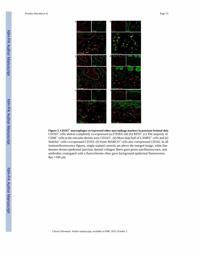
Figure 2. CD163+ macrophages co-expressed other macrophage markers in psoriasis lesional skinCD163+ cells almost completely co-expressed (a) FXIIIA and (b) RFD7. (c) The majority ofCD68+ cells in the reticular dermis were CD163+. (d) More than half of LAMP2+ cells and (e)Stabilin+ cells co-expressed CD163. (f) Some MARCO+ cells also coexpressed CD163. In allimmunofluorescence figures, single-stained controls are above the merged image, white linedenotes dermo-epidermal junction, dermal collagen fibers gave green autofluorescence, andantibodies conjugated with a fluorochrome often gave background epidermal fluorescence.Bar =100 μm.
Fuentes-Duculan et al. Page 13
J Invest Dermatol. Author manuscript; available in PMC 2011 October 1.
NIH
-PA Author Manuscript
NIH
-PA Author Manuscript
NIH
-PA Author Manuscript
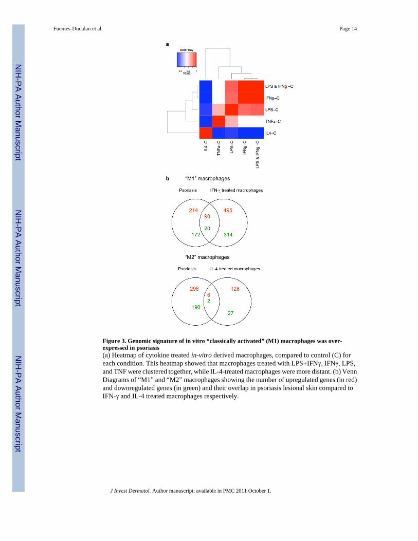
Figure 3. Genomic signature of in vitro “classically activated” (M1) macrophages was over-expressed in psoriasis(a) Heatmap of cytokine treated in-vitro derived macrophages, compared to control (C) foreach condition. This heatmap showed that macrophages treated with LPS+IFNγ, IFNγ, LPS,and TNF were clustered together, while IL-4-treated macrophages were more distant. (b) VennDiagrams of “M1” and “M2” macrophages showing the number of upregulated genes (in red)and downregulated genes (in green) and their overlap in psoriasis lesional skin compared toIFN-γ and IL-4 treated macrophages respectively.
Fuentes-Duculan et al. Page 14
J Invest Dermatol. Author manuscript; available in PMC 2011 October 1.
NIH
-PA Author Manuscript
NIH
-PA Author Manuscript
NIH
-PA Author Manuscript
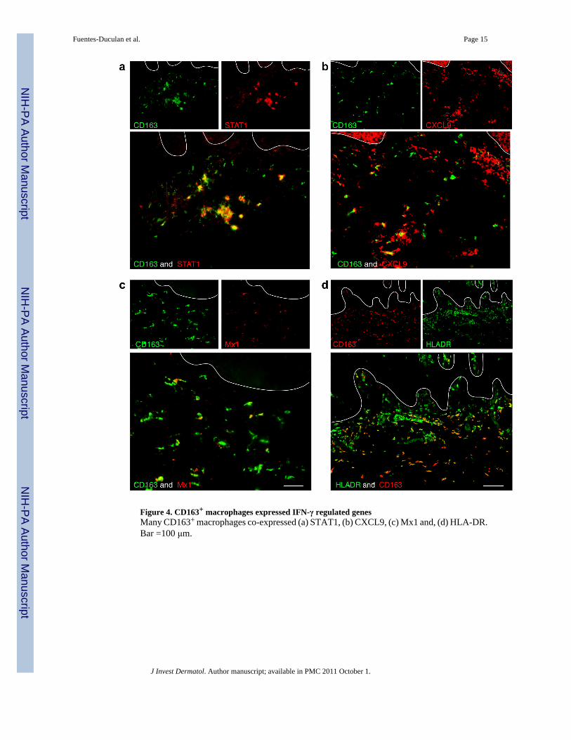
Figure 4. CD163+ macrophages expressed IFN-γ regulated genesMany CD163+ macrophages co-expressed (a) STAT1, (b) CXCL9, (c) Mx1 and, (d) HLA-DR.Bar =100 μm.
Fuentes-Duculan et al. Page 15
J Invest Dermatol. Author manuscript; available in PMC 2011 October 1.
NIH
-PA Author Manuscript
NIH
-PA Author Manuscript
NIH
-PA Author Manuscript

Figure 5. Expression of cytokines in in vitro-derived macrophages and psoriasis by RT-PCR andimmunofluorescence. (a)RT-PCR analysis of mRNA expression of IL-23p19, IL-12/IL-23p40, IL-12p35 and CCL20in cytokine treated (IFNγ, IL-4, TNF-α, LPS and LPS+IFN) in vitro macrophages (n=7)compared to control. mRNA expression normalized to human acidic ribosomal protein(HARP). * p<0.05, ***p<0.001. Some CD163+ macrophages co-expressed products ofclassically activated macrophages, (b) IL-23p19, (c) IL-12/IL-23p40, and inflammatorymediators (d) INOS and (e) TNF-α. Bar =100 μm.
Fuentes-Duculan et al. Page 16
J Invest Dermatol. Author manuscript; available in PMC 2011 October 1.
NIH
-PA Author Manuscript
NIH
-PA Author Manuscript
NIH
-PA Author Manuscript

NIH
-PA Author Manuscript
NIH
-PA Author Manuscript
NIH
-PA Author Manuscript
Fuentes-Duculan et al. Page 17
Table 1
Upregulated genes in IFN-γ-treated macrophages in common with Psoriasis
GeneSymbol Description Fold
Change
CXCL9 Chemokine (C-X-C motif) ligand 9 122.69
CXCL10 Chemokine (C-X-C motif) ligand 10 81.22
ISG 20 Interferon stimulated exonuclease gene 20kDa 58.32
RSAD2 Radical s-adenosyl methionine domain containing 2 25.15
CCL5 Chemokine (C-C motif) ligand 5 23.14
MX1 Myxovirus resistance 1, interferon-inducible protein p78 18.97
CCL8 Chemokine (C-C motif) ligand 8 18.21
IL1b Interleukin 1, beta 9.65
CCL20 Chemokine (C-C motif) ligand 20 8.85
STAT1 Signal transducer and activator of transcription 1 8.54
IL1F9 Interleukin 1 family, member 9 6.44
CCL4L2 Chemokine (C-C motif) ligand 4-Like 2 6.34
CCL4L1 Chemokine (C-C motif) ligand 4-Like 1 4.89
IL7R Interleukin 7 receptor 4.42
TNIP3 TNFAIP3 interacting protein 3 3.06
IL8 Interleukin 8 2.79
CCL3L3 Chemokine (C-C motif) ligand 3-Like 3 2.70
IL1RN Interleukin 1 receptor antagonist 2.16
IL19 Interleukin 19 2.15
S100A12 S100 Calcium Binding protein A12 2.09
J Invest Dermatol. Author manuscript; available in PMC 2011 October 1.