Noninvasive Quantitative Evaluation of the Dentin Layer during Dental Procedures Using Optical...
8
Research Article Noninvasive Quantitative Evaluation of the Dentin Layer during Dental Procedures Using Optical Coherence Tomography Cosmin Sinescu, 1 Meda Lavinia Negrutiu, 1 Adrian Bradu, 2 Virgil-Florin Duma, 3,4,5 and Adrian Gh. Podoleanu 2 1 School of Dental Medicine, Victor Babes University of Medicine and Pharmacy of Timisoara, 2 Eſtimie Murgu Place, 300041 Timisoara, Romania 2 Applied Optics Group, University of Kent, Canterbury CT2 7NH, UK 3 3OM Optomechatronics Group, “Aurel Vlaicu” University of Arad, 77 Revolutiei Avenue, 310130 Arad, Romania 4 Doctoral School, Polytechnics University of Timisoara, 1 Mihai Viteazu Avenue, 300222 Timisoara, Romania 5 Faculty of Physics, West University of Timisoara, 4 Vasile Parvan, 300223 Timisoara, Romania Correspondence should be addressed to Virgil-Florin Duma; [email protected] Received 30 November 2014; Revised 21 April 2015; Accepted 24 April 2015 Academic Editor: Xibo Ma Copyright © 2015 Cosmin Sinescu et al. is is an open access article distributed under the Creative Commons Attribution License, which permits unrestricted use, distribution, and reproduction in any medium, provided the original work is properly cited. A routine cavity preparation of a tooth may lead to opening the pulp chamber. e present study evaluates quantitatively, in real time, for the first time to the best of our knowledge, the drilled cavities during dental procedures. An established noninvasive imaging technique, Optical Coherence Tomography (OCT), is used. e main scope is to prevent accidental openings of the dental pulp chamber. Six teeth with dental cavities have been used in this ex vivo study. e real time assessment of the distances between the bottom of the drilled cavities and the top of the pulp chamber was performed using an own assembled OCT system. e evaluation of the remaining dentin thickness (RDT) allowed for the positioning of the drilling tools in the cavities in relation to the pulp horns. Estimations of the safe and of the critical RDT were made; for the latter, the opening of the pulp chamber becomes unavoidable. Also, by following the fractures that can occur when the extent of the decay is too large, the dentist can decide upon the right therapy to follow, endodontic or conventional filling. e study demonstrates the usefulness of OCT imaging in guiding such evaluations during dental procedures. 1. Introduction e pulp chamber is the cavity situated in the central portion of the tooth [1, 2], containing both nerves and blood vessels. It is placed in both the coronal and the roots part of the tooth. In frontal teeth, the pulp chamber is partially located in the crown, whilst in posterior teeth, in the cervical part of the root. Each chamber has a roof at its incisal or occlusal margin; this roof presents projections that are called pulp horns. Normaly one pulp horn is found in each cusped tooth—molars, premolars, and canines. However, in young incisors three pulp horns can be found, while one of the types of maxillary lateral incisors has only one pulp horn. ese different morphological aspects, as well as the exact locations of the pulp horns, cannot be determined with conventional procedures such as radiography, due to its low resolution. Radiography is also invasive due to its well-known irradiation issue and it can be applied only before dental procedures and not during them. Cavities/dental caries affect the teeth, therefore their drilling and cleaning, followed by fillings with different direct restorative dental materials is necessary. A major issue of such procedures is that a routine cavity preparation may lead to opening the pulp chamber because of its complex morphol- ogy, as mentioned above or due to possible complications of an even simple dental procedure [3]. erefore, a proportion of the samples subjected to cavity preparation may end in opening of the pulp chamber that subsequently requires root canal treatment [4, 5]. is leads to a significant increased frequency of endodontic treatments that have to follow the dental preparation and crown cementation [6, 7]. Hindawi Publishing Corporation Computational and Mathematical Methods in Medicine Article ID 709076
-
Upload
independent -
Category
Documents
-
view
0 -
download
0
Transcript of Noninvasive Quantitative Evaluation of the Dentin Layer during Dental Procedures Using Optical...

Research ArticleNoninvasive Quantitative Evaluation of the Dentin Layer duringDental Procedures Using Optical Coherence Tomography
Cosmin Sinescu1 Meda Lavinia Negrutiu1 Adrian Bradu2
Virgil-Florin Duma345 and Adrian Gh Podoleanu2
1School of Dental Medicine Victor Babes University of Medicine and Pharmacy of Timisoara 2 Eftimie Murgu Place300041 Timisoara Romania2Applied Optics Group University of Kent Canterbury CT2 7NH UK33OM Optomechatronics Group ldquoAurel Vlaicurdquo University of Arad 77 Revolutiei Avenue 310130 Arad Romania4Doctoral School Polytechnics University of Timisoara 1 Mihai Viteazu Avenue 300222 Timisoara Romania5Faculty of Physics West University of Timisoara 4 Vasile Parvan 300223 Timisoara Romania
Correspondence should be addressed to Virgil-Florin Duma dumavirgilosamemberorg
Received 30 November 2014 Revised 21 April 2015 Accepted 24 April 2015
Academic Editor Xibo Ma
Copyright copy 2015 Cosmin Sinescu et alThis is an open access article distributed under theCreative CommonsAttribution Licensewhich permits unrestricted use distribution and reproduction in any medium provided the original work is properly cited
A routine cavity preparation of a tooth may lead to opening the pulp chamber The present study evaluates quantitatively in realtime for the first time to the best of our knowledge the drilled cavities during dental procedures An established noninvasiveimaging technique Optical Coherence Tomography (OCT) is usedThemain scope is to prevent accidental openings of the dentalpulp chamber Six teeth with dental cavities have been used in this ex vivo studyThe real time assessment of the distances betweenthe bottom of the drilled cavities and the top of the pulp chamber was performed using an own assembled OCT system Theevaluation of the remaining dentin thickness (RDT) allowed for the positioning of the drilling tools in the cavities in relation tothe pulp horns Estimations of the safe and of the critical RDT were made for the latter the opening of the pulp chamber becomesunavoidable Also by following the fractures that can occur when the extent of the decay is too large the dentist can decide uponthe right therapy to follow endodontic or conventional filling The study demonstrates the usefulness of OCT imaging in guidingsuch evaluations during dental procedures
1 Introduction
The pulp chamber is the cavity situated in the central portionof the tooth [1 2] containing both nerves and blood vesselsIt is placed in both the coronal and the roots part of the tooth
In frontal teeth the pulp chamber is partially located inthe crown whilst in posterior teeth in the cervical part ofthe root Each chamber has a roof at its incisal or occlusalmargin this roof presents projections that are called pulphorns Normaly one pulp horn is found in each cuspedtoothmdashmolars premolars and canines However in youngincisors three pulp horns can be found while one of the typesof maxillary lateral incisors has only one pulp horn Thesedifferent morphological aspects as well as the exact locationsof the pulp horns cannot be determined with conventionalprocedures such as radiography due to its low resolution
Radiography is also invasive due to its well-known irradiationissue and it can be applied only before dental procedures andnot during them
Cavitiesdental caries affect the teeth therefore theirdrilling and cleaning followed by fillings with different directrestorative dentalmaterials is necessary Amajor issue of suchprocedures is that a routine cavity preparation may lead toopening the pulp chamber because of its complex morphol-ogy as mentioned above or due to possible complications ofan even simple dental procedure [3] Therefore a proportionof the samples subjected to cavity preparation may end inopening of the pulp chamber that subsequently requires rootcanal treatment [4 5] This leads to a significant increasedfrequency of endodontic treatments that have to follow thedental preparation and crown cementation [6 7]
Hindawi Publishing CorporationComputational and Mathematical Methods in MedicineArticle ID 709076
2 Computational and Mathematical Methods in Medicine
The present work represents an approach to test a nonin-vasive real time monitoring method that allows us to avoidsuch situations and to prevent accidental openings of the pulpchamber during dental procedures Of course such openingsare sometimes unavoidable as the depth of a restorationpreparation is often based on the extent of the decay andcannot in such cases be controlled by the dentist Howeverwhen the depth of the decay allows the dentist should beempowered with an instrument to evaluate the RDT Thisshould allow the dentist to accurately position the drillingtools in the cavities in relation to the pulp horns In contrastnowadays the dentist is only able to identify visuallymdashbyseeing the blushing of the pulp through the dentinmdashanRDT that is so small (ie the drilling is so close to thepulp) that an exposure of the pulp chamber already becomesunavoidable Our study aims to prevent such situation whilealso providing an assessment of such an RDT
Training is the next factor in determining the properdepth of cavity preparation Therefore we will show how theinstrument presented here can be also utilized in teachingdemonstrating the impact of cavity preparation in schools inaddition to being put to practical clinical use
In this study in order to achieve these aims we evaluatethe possibility of using an advanced and well-establishedbiomedical imaging technique Optical Coherence Tomog-raphy (OCT) [8ndash10] to improve the safety of the cavitypreparation by avoiding the opening of the pulp chamberOCT is a low coherence interferometry technique that hasadvanced from ophthalmology applications to areas like skindentistry and endoscopy (while also being used in industrialapplications andmaterials studies) In dentistry the microm-eter resolution and millimeter penetration depth of OCThas attracted numerous studies including for imaging thehard and soft tissue of the oral cavity [11] caries and dentaltreatments [12ndash14] demineralization [15]microleakages [16]fractures and defects for both teeth and prosthesis [17ndash19]and dental abfraction and attrition [20] as well as quality ofsealants and adhesives [21ndash23]
To the best of our knowledge despite the number ofOCT studies already performed in dentistry this is the firsttime when OCT is applied in the real time monitoringof RDT during caries excavation and cavity preparationAnother scope of this study is to evaluate how OCT canbe utilized to investigate the morphology of the dental pulpchamber and to precisely localize the pulp horns We alsodemonstrate ex vivo the OCT capabilities to be utilized asa noninvasive technique to guide the dentist during dentalprocedures Thus a quantitative assessment of the RDT isperformed during procedures and estimations are made onthe safety and critical RDT This proves OCT as a valuabletool prior to dental procedures and most important for realtime assessment and treatment of caries
2 Material and Methods
Six extracted teeth with mesial cavities were used for theinvestigation OCT imaging was performed while the dentalprocedures were applied on each of them in order todetermine the RDT in real time as well as localize the pulp
chamber in relationship to the drilled cavity After completionof the dental procedures each sample was sectioned in twohalves in order to evaluate the morphology of the pulpchamber at the end of the procedure to measure directly thefinal RDT and thus to validate its value obtained using theOCT imagesmdashfor each tooth
An own assembled Time Domain (TD) OCT system(Figure 1) described in principle in Hughes and Podoleanu[24] and similar in imaging functionality to that in Sinescu etal [14] was utilized for this studyThe anatomy of the systemis shown in Figure 1(a) It utilizes a pigtailed superlumines-cent diode (SLD) emitting at a wavelength of 1300 nm andwith a spectral bandwidth of 65 nm This produces a longi-tudinalaxial resolution in tissue of around 15micrometersThe low numerical aperture of the interface optics of theOCTsystem allows for a maximum lateral image size of 10mm
TheOCT system is based on a low coherence interferom-eter with dynamic focus where the coherence gate and focusgate are synchronized [24]This procedure securesmaximumsensitivity as well as conservation of lateral resolution overdepth Light from the SLD is split into a sample and areference arm respectively through a first directional coupler(DC1) The sample arm of the interferometer comprises two
microscope objectives (MO1and MO
2) and a flat mirror
(FM) to focus the light on the sample (ie the frontal partof the tooth as shown in Figure 1(b)) by using a dual axistwo-dimensional (2-D) galvanometer scanner (GS) Lightbackscattered by the sample passes a second time throughthe object arm and is guided via DC
1toward the second
directional coupler (DC2) where it interferes with that
coming from the reference arm Polarization controllers (PC1
and PC2) are positioned on each of the interferometer arms
Both output fibres from (DC2) are connected to two-pin
photo-detectors in a balanced photo-detection (BPD) unitThe OCT system is equipped with a 2-D GS which is
comprised of two 1-D GSs with orthogonal axes they providea raster scan of the laser beam on the surface of the sample
Using the translation stage (TS) the optical path inthe reference arm and the one in the sample arm of theinterferometer are scanned together An A-scan (in-depthreflectivity profile) is obtained for each of the positions ofthe laser beam on the frontal surface of the tooth B-scans(ie transversal sections in the sample) are generated asraster scans by collecting many such A-scans from adjacenttransverse positions they provide therefore 2-D sections ofthe dental material in a plane sharing a lateral coordinateand the axial (in-depth) coordinate B-scans are producedby the fast GS driven in this setup with a triangular signalwith a frequency of 500Hz Driving the GS with a triangularsignal allows production of OCT images with less fly-backdistortions than when using a saw-tooth signal [25 26]OCT images such obtained allow visualization of the internaltopography of the tooth
The second slow GS positions the line of pixels generatedby the fast scanner on the surface of the sample with aramp signal with a frequency of 2Hz This can be repeatedat different values of the second lateral coordinate usingbias voltages applied to the two GSs [27] The microscopeobjective in front of the dual axis GS is a ThorLabs scan lens
Computational and Mathematical Methods in Medicine 3
minus
+
To computerBPD
SLD
Reference arm
TS
FM2-D GS
Sample
PC1
PC2
DC2
DC1
MO1
MO2
(a)
TD-OCT system
Translation stage of
the sample
Sample (drilledtooth)
(b)
Figure 1 (a) Anatomy of the Time Domain (TD) Optical Coherence Tomography (OCT) system Components are as follows SLD =superluminescent diode DC = directional couplers PC = polarization controllers MO = microscope objectives FM = flat mirror GS =galvanometer scanner TS = translation stage BPD = balance photo-detector (b) sample placed in front of the in-house system
with a 40mm focal length specially designed to prevent imagedegradation and distortion during scanning The calibrationof the images was performed using a 1951 USAF High-Resolution Target 210158401015840 times 210158401015840 positive system The numericalaperture of this microscope objective determines the lateralsize of the B-scans which can be in this case adjustedcontinuously up to 10mm
As discussed in the next section for this dental applica-tion B-scans of 2 to 3 mm in lateral size were enough to beconsidered in order to image the area of interest in the teethThe wider B-scans along the lateral direction allow initiallocalization of the area of interest at the start of the proce-dure By assembling successive B-scans three-dimensional(3-D) reconstructionsvolumes of dental constructions anddentures are obtained as described in previous works [28]The base of this OCT volume is the scanned surface of thetooth placed in front of the TD-OCT system (Figure 1(b)) theheight of this volume is the penetration depth of the systemwhich is about 1mm in hard tissue As demonstrated bythis research as well as by previous studies this penetrationdepth is sufficient for dentistry applications in this workas shown in the next section it allows for imaging therelevant volumes during cavity preparation the assessment
of the quality and durability of dental constructs can thus beachieved
The working protocol for this study has been approvedby the Ethics Committee of the Victor Babes Universityof Medicine and Pharmacy of Timisoara All experimentswere conducted according to Romanian and EuropeanUnionregulations
3 Results
During the cavity preparation of each sample an OCT 2-DslicesB-scans were generated They revealed in real time thedistances from the bottom of the drilled cavities to the pulpchamberThe 2-D slices were then combined into 3-D recon-structions as shown in Figure 2(a) and further in Figure 3Such 3-D images allow the investigator to perform full-fieldnavigation through the sample with micrometer resolutionwhich is one of the major advantages of the OCT technique
A validation of the OCT imaging procedure is shown inFigures 2(b) and 2(c) where an evaluation of themorphologyof the pulp chamber has been performed This evaluationwas made after the processed tooth was sectioned in halfafter completion of the dental procedure As it can be seen in
4 Computational and Mathematical Methods in Medicine
Drilled cavity
(4) Pulpchamber
(2) Dentin layer
1mm
2mm
(a)
(1)
(2)
(3)
(4)
(b)
(2)
(1)
(3)
(4)
(c)
Figure 2 (a) Tridimensional (3-D)volumetric OCT reconstructions of the drilled cavity obtained in real time during the dentistryproceduremdashillustrating the upper wall of the pulp chamber (b) macroscopic approach on the morphology of this tooth obtained aftersectioning the tooth after the procedure it shows that there is still a lot of dentin left under the drilled cavity to protect the pulp chamber (c)another example for which the drilling already affected the pulp chamber by opening the pulp horns accidentally (the opening is proved byinserting an endodontic needle from the drilled cavity through the pulp horn towards the pulp chamber)mdashas shown in Figure 3(d) Notationsare as follows (1) drilled cavity on the occlusal surface of the tooth (2) ceiling of the pulp chamber (3) pulp horns (difficult to evaluate duringa normal drilling process) (4) pulp chamber
Drilled cavity
Pulpchamber
1mmsim05mm
(a)
Pulpchamber
Drilled cavity1mm
2mm
sim015mmsim03mm
(b)
Pulpchamber
Drilled cavity
1mm
2mm
sim005mm
(c)
Drilled cavity
Pulp chamber Communication between thedrilled cavityand the pulp
chamber
1mm
2mm
sim025mm
(d)
Figure 3 Real time OCT-based evaluations of the remaining dentin thickness (RDT) between the drilled cavity and the pulp chamber (a)measurement of the safety limit of the RDT (b) decrease of the dentin layer towards the critical value of its thickness (ie for which a fracturecannot be avoided) (c) image taken just before the fracture in the dentin is initialized (d) communication between the drilled cavity and thedental pulp chamber demonstrated using the OCT investigation
Computational and Mathematical Methods in Medicine 5
Figure 2(b) from a half of one of the teeth a large portion ofdentin has been left under the drilled cavity in order to pro-tect the pulp chamberThis image is correlated with the OCTimages obtained during the procedures in Figures 2(a) and3(a)
Figure 2(c) shows a tooth where the drilling accidentallyopened the pulp horns thus affecting the pulp chamber Thiscase is illustrated by the OCT image in Figure 3(d)
On the 3-D OCT investigations the RDT between thepulp chamber and drilled cavities was assessed as demon-strated in Figure 3 This shows that the dentist has the possi-bility to evaluate in real time the position of the drilled cavitiesrelated to the pulp horns Thus accidental openings duringthe procedure could be prevented The chance for accidentalopening can further be diminished by imposing a minimumwidth for the remaining dentin layer The evaluations madewith different teeth in this study suggest an RDT safety limitof 05mm as shown by the example in Figure 3(a)
However the depth of a restoration preparation is oftenbased on the extent of the decay and cannot in such cases becontrolled by the dentist Therefore sometimes during thedrilling procedure fractures can occur they can penetratethe remaining layer between the cavity and the pulp chamberSuch occurrences have been documented in real time inFigure 3(b) the average RDT decreased to a critical valueof 03mm and from this value it decreased immediately to015mm only From the work on different teeth involved inthis study a value of 015mm represents an average for whichthe initialization of fractures cannot be avoided anymore Ofcourse a decay that is within 015mm of the pulp is alsoeasily identified by the pulp blushing through the dentinThe dentist needs an OCT system to avoid approaching thecritical RDT value
This process is shown further in Figure 3(c) where thefracture is imminentThe communication between the dentalpulp chamber and the drilled cavity can thus be inducedaccidentally during the drilling procedures as pointed out inFigure 3(d)
This investigation clearly shows that by evaluating thecurrent width of the remaining dental layer (ie the RDT)the dentist can estimate the risk of fracture in real time duringthe drilling process Based on such findings the dentist candecide whether to use further endodontic therapy instead ofa conventional filling procedure
After completion of the dental procedures (combinedwith OCT imaging) the accuracy of the RDT measurementthrough deep dentin cavity with OCT was verified for eachsample Thus each tooth was finally sectioned in two halvesand the RDT was measured directly The values obtainedconfirm the results estimated during the OCT imaging Thefinal RDTvalue obtained usingOCT for each samplewas thusvalidated The validity of the OCT monitoring its capabilityto determine the internal topography of the tooth withoutdistortions was thus demonstrated
4 Discussions and Conclusions
Theknowledge of the internal dental anatomy is fundamentalfor the success of any dental therapy In spite of the fact
that every group of teeth presents similar morphologicalfeatures the individual variations in pulp chamber anatomyare a challenge for every clinician with direct impact on theoutcome of the dental therapy
Besides the issue of the different locations and shapes ofthe pulp chamber at different teeth and individuals thereare several reports on the existence of supplementary pulphorns Their identification based only on retro-alveolar orpanoramic X-rays is however difficult because the real 3-Dstructure of the pulp chamber volume cannot be inferredusing a 2-D projection
Such methods are reviewed below
(i) Cone Beam Computer Tomography (CBCT) relies onradiations and so it is an invasive technique thereforenot suitable for dental routine treatment CBSCTcannot distinguish microanatomical details smallerthan 076mm As consequence CBCT is considereda complementary modality for specific applicationsrather than a replacement for 2-D imaging
(ii) Dental operating microscopy is efficient in all the stepsof the endodontic therapy but provides magnifica-tion and illumination only when a communicationbetween the dental cavity and the pulp chamber hasbeen already establishedThis is a severe limitation forthe commondental therapy and it would not fulfill theaim of this study
(iii) Orascope is a medical endoscope modified for treat-ment in the oral cavity It helps to find cracks on thefloor of the pulp chamber and extra canalsmore easilythan with loupe spectacles Compared with the dentalmicroscope its flexibility facilitates the applicationin the lateral tooth region Additional intraoperativeradiological checks can be avoided and the radiationload for the patients is thus reduced However manymicroendoscopic medical applications require smallworking diameters (ie from 015 to about 1mm)which permit only about 3000 pixels for the imageguide because of space requirements while higherresolutions of 10000 to 50000 pixels would beneeded Moreover this technology offers only surfaceinformation with no details related to the dentinfloor depth and therefore no internal cracks orfractures can be discovered Above these limitationsthe accidental communication between the cavity andthe pulp chamber remains undiscovered
By contrast to such methods OCT represents an alternativenoninvasive technique to these shortcomings OCT exhibitsresolutions better than a few micrometers The penetrationdepth in teeth is only 1mm because the hard tissue of theteeth decreases the backscattered signal strength Howeveras demonstrated even such reduced penetration depth issufficient for guidance of dental procedures related to thedrilled cavity and to the pulp chamber
The results we have obtained proved that OCT canperform quantitative data retrieval of the relation betweenthe drilled cavity floor and the pulp horns Fast OCT realtime acquisitions of over 1 Gigavoxel of data in a single
6 Computational and Mathematical Methods in Medicine
second [29] are now possible with modern spectral domainOCT With such a tool the surgeon can decide during theprocedure the best dental therapy to follow OCT evaluationscan be used in conventional dental cavities preparationusing drilling procedures as well as in the laser treatmentof cavities The latter is a direction of future work Anotherdirection of research opened by this study is to advancefrom the ex vivo study presented to an in vivo study to assistdental procedures performed in the oral cavity [14] OCTunits with handheld scanning probes have been alreadydemonstrated covering a wide range of applications such asEar-Nose-Throat investigations [30] to dental prosthesis [19]The next step would be to perform similar investigationswith hand-held mobile units in the oral cavity
In conclusion the study demonstrated that OCT technol-ogy can be harnessed to assist cavity preparation that can leadto safer and more conservative results of dental proceduresin real time The ex vivo study presented illustrates the utilityof OCT imaging employed in the training process of dentistsperforming dental procedures
It is also expected that such procedures can be extendedto clinical environments for in vivo investigations The realtime OCT monitoring proves promising in the prevention ofaccidental pulp exposure during deep caries excavation
Conflict of Interests
This work is free of conflict of interests
Authorsrsquo Contribution
Cosmin Sinescu and Virgil-Florin Duma have equally con-tributed to this work
Acknowledgments
The authors acknowledge the support of the RomanianNational Authority for Scientific Research through ProjectPN-II-PT-PCCA-2011-32-1682 (http3om-group-optome-chatronicsro) and of COST ACTION MP 1005 C Sinescuacknowledges the support of the CNDI-UEFISCDI ProjectTE 1012010 A Podoleanu acknowledges the support of theNIHR Biomedical Research Centre at Moorfields Eye Hos-pital of the NHS Foundation Trust and of the UCL Instituteof Ophthalmology
References
[1] J K Avery Oral Development and Histology Thieme MedicalPublishers New York NY USA 2nd edition 1994
[2] A R Ten Cate Oral Histology Development Structure andFunction Mosby St Louis Mo USA 5th edition 1998
[3] I A Mjor ldquoPulp-dentin biology in restorative dentistry Part2 initial reactions to preparation of teeth for restorative pro-ceduresrdquo Quintessence International vol 32 no 7 pp 537ndash5512001
[4] A Zollner and P Gaengler ldquoPulp reactions to different prepara-tion techniques on teeth exhibiting periodontal diseaserdquo Journalof Oral Rehabilitation vol 27 no 2 pp 93ndash102 2000
[5] P Lumley N Adams and P Tomson Practical Clinical Endo-dontics Elsevier Health Sciences 2006
[6] J Valderhaug A Jokstad E Ambjoslashrnsen and P W NorheimldquoAssessment of the periapical and clinical status of crownedteeth over 25 yearsrdquo Journal of Dentistry vol 25 no 2 pp 97ndash105 1997
[7] I A V P Poiate E Poiate Jr and R Y Ballester ldquoBiomechanicalanalysis of restored teeth with cast intra-radicular retainerwith and without ferrulerdquo in Finite Element AnalysismdashFromBiomedical Applications to Industrial Developments D MoratalEd InTech 2012
[8] D Huang E A Swanson C P Lin et al ldquoOptical coherencetomographyrdquo Science vol 254 no 5035 pp 1178ndash1181 1991
[9] M Wojtkowski ldquoHigh-speed optical coherence tomographybasics and applicationsrdquoApplied Optics vol 49 no 16 pp D30ndashD61 2010
[10] A G Podoleanu ldquoOptical coherence tomographyrdquo Journal ofMicroscopy vol 247 no 3 pp 209ndash219 2012
[11] F I Feldchtein G V Gelikonov V M Gelikonov et al ldquoIn vivoOCT imaging of hard and soft tissue of the oral cavityrdquo OpticsExpress vol 3 no 6 pp 239ndash250 1998
[12] B T Amaechi A G Podoleanu S M Higham and D AJackson ldquoCorrelation of quantitative light-induced fluorescenceand optical coherence tomography applied for detection andquantification of early dental cariesrdquo Journal of BiomedicalOptics vol 8 no 4 pp 642ndash647 2003
[13] J S Holtzman K Osann J Pharar et al ldquoAbility of opticalcoherence tomography to detect caries beneath commonly useddental sealantsrdquo Lasers in Surgery and Medicine vol 42 no 8pp 752ndash759 2010
[14] C Sinescu M L Negrutiu C Todea et al ldquoQuality assessmentof dental treatments using en-face optical coherence tomog-raphyrdquo Journal of Biomedical Optics vol 13 no 5 Article ID054065 2008
[15] D P PopescuM G SowaMDHewko and L-P Choo-SmithldquoAssessment of early demineralization in teeth using the signalattenuation in optical coherence tomography imagesrdquo Journalof Biomedical Optics vol 13 no 5 Article ID 054053 2008
[16] C Todea C Balabuc C Sinescu et al ldquoEn face optical cohe-rence tomography investigation of apical microleakage afterlaser-assisted endodontic treatmentrdquo Lasers in Medical Sciencevol 25 no 5 pp 629ndash639 2010
[17] Y Nakajima Y ShimadaMMiyashin Y Takagi J Tagami andY Sumi ldquoNoninvasive cross-sectional imaging of incompletecrown fractures (cracks) using swept-source optical coherencetomographyrdquo International Endodontic Journal vol 45 no 10pp 933ndash941 2012
[18] K-J Park H Schneider and R Haak ldquoAssessment of interfacialdefects at composite restorations by swept source optical coher-ence tomographyrdquo Journal of Biomedical Optics vol 18 no 7Article ID 076018 2013
[19] D Demian V F Duma C Sinescu et al ldquoDesign and testingof prototype handheld scanning probes for optical coherencetomographyrdquo Journal of Engineering inMedicine vol 228 p 7432014
[20] C Marcauteanu A Bradu C Sinescu F I Topala M LNegrutiu and A G Podoleanu ldquoQuantitative evaluation ofdental abfraction and attrition using a swept-source opticalcoherence tomography systemrdquo Journal of Biomedical Opticsvol 19 no 2 Article ID 021108 2014
Computational and Mathematical Methods in Medicine 7
[21] A K S Braz C M Aguiar and A S L Gomes ldquoEvaluation ofthe integrity of dental sealants by optical coherence tomogra-phyrdquo Dental Materials vol 27 no 4 pp e60ndashe64 2011
[22] M Rominu A Manescu C Sinescu et al ldquoZirconia enricheddental adhesive a solution for OCT contrast enhancementDemonstrative study by synchrotron radiation microtomogra-phyrdquo Dental Materials vol 30 no 4 pp 417ndash423 2014
[23] R Oancea A Bradu C Sinescu et al ldquoAssessment of the sea-lanttooth interface using optical coherence tomographyrdquo Jour-nal of Adhesion Science and Technology vol 29 no 1 pp 49ndash582015
[24] M Hughes and A G Podoleanu ldquoSimplified dynamic focusmethod for time domain OCTrdquo Electronics Letters vol 45 no12 pp 623ndash624 2009
[25] V-F Duma ldquoOptimal scanning function of a galvanometerscanner for an increased duty cyclerdquo Optical Engineering vol49 no 10 Article ID 103001 2010
[26] V-F Duma K-S Lee P Meemon and J P Rolland ldquoExperi-mental investigations of the scanning functions of galvano-meter-based scanners with applications in OCTrdquo Applied Op-tics vol 50 no 29 pp 5735ndash5749 2011
[27] V F Duma ldquoMathematical functions of a 2-D scanner withoscillating elementsrdquo in Modeling Simulation and Controlof Nonlinear Engineering Dynamical Systems pp 243ndash253Springer Heidelberg Germany 2009
[28] A G Podoleanu and R B Rosen ldquoCombinations of techniquesin imaging the retina with high resolutionrdquo Progress in Retinaland Eye Research vol 27 no 4 pp 464ndash499 2008
[29] T Klein W Wieser C M Eigenwillig B R Biedermann andR Huber ldquoMegahertz OCT for ultrawide-field retinal imagingwith a 1050nm Fourier domain mode-locked laserrdquo OpticsExpress vol 19 no 4 pp 3044ndash3062 2011
[30] R Cernat T S Tatla J Pang et al ldquoDual instrument for in vivoand ex vivo OCT imaging in an ENT departmentrdquo BiomedicalOptics Express vol 3 no 12 pp 3346ndash3356 2012
Submit your manuscripts athttpwwwhindawicom
Stem CellsInternational
Hindawi Publishing Corporationhttpwwwhindawicom Volume 2014
Hindawi Publishing Corporationhttpwwwhindawicom Volume 2014
MEDIATORSINFLAMMATION
of
Hindawi Publishing Corporationhttpwwwhindawicom Volume 2014
Behavioural Neurology
EndocrinologyInternational Journal of
Hindawi Publishing Corporationhttpwwwhindawicom Volume 2014
Hindawi Publishing Corporationhttpwwwhindawicom Volume 2014
Disease Markers
Hindawi Publishing Corporationhttpwwwhindawicom Volume 2014
BioMed Research International
OncologyJournal of
Hindawi Publishing Corporationhttpwwwhindawicom Volume 2014
Hindawi Publishing Corporationhttpwwwhindawicom Volume 2014
Oxidative Medicine and Cellular Longevity
Hindawi Publishing Corporationhttpwwwhindawicom Volume 2014
PPAR Research
The Scientific World JournalHindawi Publishing Corporation httpwwwhindawicom Volume 2014
Immunology ResearchHindawi Publishing Corporationhttpwwwhindawicom Volume 2014
Journal of
ObesityJournal of
Hindawi Publishing Corporationhttpwwwhindawicom Volume 2014
Hindawi Publishing Corporationhttpwwwhindawicom Volume 2014
Computational and Mathematical Methods in Medicine
OphthalmologyJournal of
Hindawi Publishing Corporationhttpwwwhindawicom Volume 2014
Diabetes ResearchJournal of
Hindawi Publishing Corporationhttpwwwhindawicom Volume 2014
Hindawi Publishing Corporationhttpwwwhindawicom Volume 2014
Research and TreatmentAIDS
Hindawi Publishing Corporationhttpwwwhindawicom Volume 2014
Gastroenterology Research and Practice
Hindawi Publishing Corporationhttpwwwhindawicom Volume 2014
Parkinsonrsquos Disease
Evidence-Based Complementary and Alternative Medicine
Volume 2014Hindawi Publishing Corporationhttpwwwhindawicom
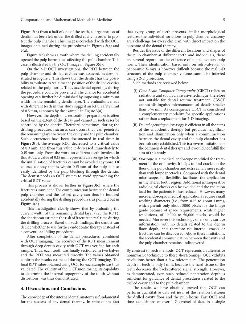