Noninvasive measurement of aortic aneurysm sac tension with vibrometry
9
BASIC RESEARCH STUDIES From the Midwestern Vascular Surgical Society Noninvasive measurement of aortic aneurysm sac tension with vibrometry Geza Mozes, MD, PhD, a Randall R. Kinnick, b Peter Gloviczki, MD, a Russell E. Bruhnke, c Michele Carmo, MD, a Tanya L. Hoskin, MS, d Kevin E. Bennet, c and James F. Greenleaf, PhD, b Rochester, Minn Objective: Currently, the risk of aneurysm sac rupture after endovascular abdominal aortic aneurysm repair (EVAR) is estimated by using a group of anatomic variables. Available techniques for pressure monitoring include either direct measurement using catheter-based techniques or indirect measurement requiring implantation of a pressure sensor during aneurysm repair. None of these methods is without limitations. Radiation pressure, such as that generated by a modulated ultrasound (US) beam, can induce surface vibration at a distance. The velocity of the resulting surface waves depends on the tensile stress of the vibrated surface. By measuring the change in wave velocity, it is possible to detect the change in tensile stress and calculate the pressure through the vibrated surface. We tested this concept in an in vitro aneurysm model. Methods: Rubber tubes and explanted porcine abdominal aortas were used to model an aneurysm sac. The surface of the model was vibrated with an amplitude-modulated US beam. The resulting motion was detected either by reflected laser light or by Doppler US. The phase of the propagating wave was measured to assess changes in velocity with different pressures. Results: Increasing hydrostatic pressure in the rubber model correlated well with the cumulative phase shift (R 2 0.96-0.99; P < .0001). By using a pump to generate dynamic pressure (between 110 and 200 mm Hg), the cumulative phase shift correlated well with the square of the mean pressure (R 2 0.92; P < .0001); however, the correlation with pulse pressure was poor (24-36 mm Hg; r 0.38; P < .02). In the porcine in vitro aortic sac model, the cumulative phase shift detected with both laser (r 0.94-0.99; P < .0001) and Doppler (r 0.96-0.99; P < .0001) correlated well with the aneurysm pressure. Conclusions: Application of vibrometry for noninvasive measurement of aortic aneurysm sac tension is feasible in an in vitro setting. The concept of vibrometry may be used to detect endotension noninvasively after EVAR. Vibrometry may also be used to estimate wall stress in native aneurysms, and it may predict the risk of aneurysm rupture. ( J Vasc Surg 2005;42:963-71.) Clinical Relevance: Vibrometry may offer a technique for completely noninvasive monitoring of aneurysm sac pressure after EVAR. Vibrometry is based on the following principles: radiation pressure, such as that generated by modulated US, can induce surface vibration at a distance; by measuring the change in wave velocity of vibration, it is possible to detect changes in tensile stress and calculate the pressure through the vibrated surface. We tested this concept in an in vitro model and found that application of vibrometry for noninvasive measurement of aortic aneurysm sac tension is feasible. Vibrometry may also be used to estimate wall stress in native aneurysms. The risk of abdominal aortic aneurysm (AAA) rupture is not completely eliminated by endovascular AAA repair (EVAR), and it is reported in different series to occur at a 0.5% to 2% frequency over 3 to 5 years after repair. 1-3 This problem originates from the very nature of endovascular repair, during which the aneurysm sac is not obliterated but only excluded from the systemic circulation. Therefore, the effectiveness of EVAR is dependent on the maintenance of a perfect seal between the device and the aortic or iliac wall. The potential for endoleak at attachment sites due to failure of the device, migration, structural fatigue, and the poten- tial for changes in the geometry of the postrepair vessel caused by remodeling and aging currently necessitates life- long follow-up with imaging studies. The mainstay of current follow-up is computed tomographic (CT) angiog- From the Division of Vascular Surgery, a Ultrasound Research Laboratory, b Division of Engineering, c and Division of Biostatistics, d Mayo Clinic and Mayo Medical School. Competition of interest: none. Presented at the 28th Annual Meeting of the Midwestern Vascular Surgical Society, Omaha, Nebraska, September, 2004. Reprint requests: Peter Gloviczki, MD, Division of Vascular Surgery, Mayo Clinic, 200 First St, SW, Rochester, MN 55905. (e-mail: gloviczki. [email protected]). 0741-5214/$30.00 Copyright © 2005 by The Society for Vascular Surgery. doi:10.1016/j.jvs.2005.07.012 963
-
Upload
independent -
Category
Documents
-
view
0 -
download
0
Transcript of Noninvasive measurement of aortic aneurysm sac tension with vibrometry

BASIC RESEARCH STUDIESFrom the Midwestern Vascular Surgical Society
Noninvasive measurement of aortic aneurysm sactension with vibrometryGeza Mozes, MD, PhD,a Randall R. Kinnick,b Peter Gloviczki, MD,a Russell E. Bruhnke,c
Michele Carmo, MD,a Tanya L. Hoskin, MS,d Kevin E. Bennet,c and James F. Greenleaf, PhD,b
Rochester, Minn
Objective: Currently, the risk of aneurysm sac rupture after endovascular abdominal aortic aneurysm repair (EVAR) isestimated by using a group of anatomic variables. Available techniques for pressure monitoring include either directmeasurement using catheter-based techniques or indirect measurement requiring implantation of a pressure sensorduring aneurysm repair. None of these methods is without limitations. Radiation pressure, such as that generated by amodulated ultrasound (US) beam, can induce surface vibration at a distance. The velocity of the resulting surface wavesdepends on the tensile stress of the vibrated surface. By measuring the change in wave velocity, it is possible to detect thechange in tensile stress and calculate the pressure through the vibrated surface. We tested this concept in an in vitroaneurysm model.Methods: Rubber tubes and explanted porcine abdominal aortas were used to model an aneurysm sac. The surface of themodel was vibrated with an amplitude-modulated US beam. The resulting motion was detected either by reflected laserlight or by Doppler US. The phase of the propagating wave was measured to assess changes in velocity with differentpressures.Results: Increasing hydrostatic pressure in the rubber model correlated well with the cumulative phase shift (R2 �0.96-0.99; P < .0001). By using a pump to generate dynamic pressure (between 110 and 200 mm Hg), the cumulativephase shift correlated well with the square of the mean pressure (R2 � 0.92; P < .0001); however, the correlation withpulse pressure was poor (24-36 mm Hg; r � 0.38; P < .02). In the porcine in vitro aortic sac model, the cumulative phaseshift detected with both laser (r � 0.94-0.99; P < .0001) and Doppler (r � 0.96-0.99; P < .0001) correlated well withthe aneurysm pressure.Conclusions: Application of vibrometry for noninvasive measurement of aortic aneurysm sac tension is feasible in an invitro setting. The concept of vibrometry may be used to detect endotension noninvasively after EVAR. Vibrometry mayalso be used to estimate wall stress in native aneurysms, and it may predict the risk of aneurysm rupture. ( J Vasc Surg2005;42:963-71.)
Clinical Relevance: Vibrometry may offer a technique for completely noninvasive monitoring of aneurysm sac pressureafter EVAR. Vibrometry is based on the following principles: radiation pressure, such as that generated by modulatedUS, can induce surface vibration at a distance; by measuring the change in wave velocity of vibration, it is possible todetect changes in tensile stress and calculate the pressure through the vibrated surface. We tested this concept in an in vitromodel and found that application of vibrometry for noninvasive measurement of aortic aneurysm sac tension is feasible.
Vibrometry may also be used to estimate wall stress in native aneurysms.The risk of abdominal aortic aneurysm (AAA) ruptureis not completely eliminated by endovascular AAA repair
From the Division of Vascular Surgery,a Ultrasound Research Laboratory,b
Division of Engineering,c and Division of Biostatistics,d Mayo Clinic andMayo Medical School.
Competition of interest: none.Presented at the 28th Annual Meeting of the Midwestern Vascular Surgical
Society, Omaha, Nebraska, September, 2004.Reprint requests: Peter Gloviczki, MD, Division of Vascular Surgery, Mayo
Clinic, 200 First St, SW, Rochester, MN 55905. (e-mail: [email protected]).
0741-5214/$30.00Copyright © 2005 by The Society for Vascular Surgery.
doi:10.1016/j.jvs.2005.07.012(EVAR), and it is reported in different series to occur at a0.5% to 2% frequency over 3 to 5 years after repair.1-3 Thisproblem originates from the very nature of endovascularrepair, during which the aneurysm sac is not obliterated butonly excluded from the systemic circulation. Therefore, theeffectiveness of EVAR is dependent on the maintenance ofa perfect seal between the device and the aortic or iliac wall.The potential for endoleak at attachment sites due to failureof the device, migration, structural fatigue, and the poten-tial for changes in the geometry of the postrepair vesselcaused by remodeling and aging currently necessitates life-long follow-up with imaging studies. The mainstay of
current follow-up is computed tomographic (CT) angiog-963
mon
JOURNAL OF VASCULAR SURGERYNovember 2005964 Mozes et al
raphy, supplemented selectively with ultrasonography (US)and catheter angiography, aiming at the detection of en-doleaks, endograft deformation, migration, and enlarge-ment of the aneurysm.4 Unfortunately, aneurysm ruptureafter EVAR without demonstrable endoleak has been re-ported on several occasions.3 Significantly increasing ordecreasing aneurysm size on follow-up images can suggestfailure or success of the repair; however, in many patients,the size of the aneurysm remains unchanged. These pa-tients, if no detectable endoleak is present, may have endo-tension and are exposed to an uncertain risk of aneurysmrupture.5,6 Additionally, follow-up with CT angiographyrequires repeated exposure to ionizing radiation and tolarge volumes of nephrotoxic contrast dye.
Intuitively, the risk of rupture should be dependent onthe pressure in the excluded aneurysm sac. Current at-tempts at measuring pressure in the aneurysm sac requireeither invasive catheterization or the implantation of pres-sure sensors. Pressure sensors can be implanted during the
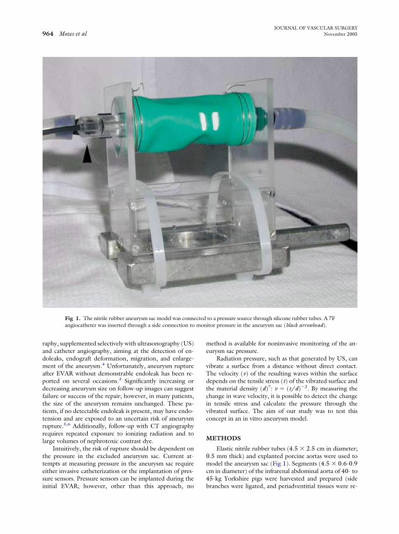
Fig 1. The nitrile rubber aneurysm sac model was conneangiocatheter was inserted through a side connection to
initial EVAR; however, other than this approach, no
method is available for noninvasive monitoring of the an-eurysm sac pressure.
Radiation pressure, such as that generated by US, canvibrate a surface from a distance without direct contact.The velocity (v) of the resulting waves within the surfacedepends on the tensile stress (t) of the vibrated surface andthe material density (d)7: v � (t/d)�2. By measuring thechange in wave velocity, it is possible to detect the changein tensile stress and calculate the pressure through thevibrated surface. The aim of our study was to test thisconcept in an in vitro aneurysm model.
METHODS
Elastic nitrile rubber tubes (4.5 � 2.5 cm in diameter;0.5 mm thick) and explanted porcine aortas were used tomodel the aneurysm sac (Fig 1). Segments (4.5 � 0.6-0.9cm in diameter) of the infrarenal abdominal aorta of 40- to45-kg Yorkshire pigs were harvested and prepared (side
to a pressure source through silicone rubber tubes. A 7Fitor pressure in the aneurysm sac (black arrowhead).
cted
branches were ligated, and periadventitial tissues were re-

JOURNAL OF VASCULAR SURGERYVolume 42, Number 5 Mozes et al 965
moved). Animal care and killing were compliant with thecorresponding institutional policies.
The aneurysm sac model was secured to 20- or 6-mmcannulas proximally and distally and connected to a hydro-static (fluid column or pressure bag) or dynamic (Bio-Console Pump; Bio-Medicus, Minneapolis, Minn) pressuresource with silicone rubber tubes and immersed in a waterbath. The aneurysm sac was perfused with degassed water(nitrile tube) or a 0.9% sodium chloride solution (porcineaorta) at room temperature. Through a side connection ofthe distal cannula, a 7F angiocatheter was introduced intothe lumen of the aneurysm model and connected to adigital pressure monitor. A 45-mm-diameter 3-MHz drivetransducer and a 5-MHz dual-element Doppler transducerwere positioned near the axial center of the aneurysm withtheir focal points spaced 2.5 cm apart on the surface of themodel. The focal lengths of the transducers were 10 and 8cm, respectively. The surface of the aneurysm sac modelwas vibrated with a focused US beam with a centerfrequency of 3 MHz and an amplitude modulated at 500
Fig 2. An amplitude-modulated ultrasound beam (bla(porcine aorta is shown). Continuous-wave Doppler imgenerated wall motion.
Hz. The resulting motion was detected either by a laser
vibrometer (PSV-300; Polytec, Inc, Tustin, Calif) or by aDoppler transducer operating in continuous-wave mode(Fig 2). The phase of the propagating wave was measuredto assess changes in velocity with different pressures (eg, atpressure 1 and 2 [P1 and P2, respectively]; Fig 3). Phasemeasurements were taken in this manner at 10 mm Hgintervals over a range of 50 to 200 mm Hg. The lowestvalue of pressure used in this study was selected on the basisof visual observations of full inflation; this suggests thatadditional increases in pressure would have been associatedwith minimal changes in diameter. This setting allowed usto use pressure as a surrogate measure of wall tensionbecause changes in the radius (r) and wall thickness (t) ofthe aortic model remained in a negligible range, and ac-cording to the law of Laplace (� � �Pr/2t), wall tension(�) was primarily determined by pressure changes (�P).The maximum pressure evaluated was determined by thecapacity of the pressure sources and by the resistance of theexperimental aneurysm model connections to leakage. Wallstrain was not measured directly with a strain gauge because
rowhead) vibrated the surface of the aneurysm model(white arrowhead) was used to detect the phase of the
ck araging
it would have required material properties to calculate

of th
JOURNAL OF VASCULAR SURGERYNovember 2005966 Mozes et al
tension. We did not have the appropriate scanner for mea-suring the diameter changes of the sac with pressure be-cause the variation in diameter was a very small fraction ofthe baseline size of the aneurysm model.
A phase shift was calculated for each 10 mm Hg incre-mental increase in pressure. The wave phase at baseline (50and 110 mm Hg mean pressure depending on the pressuresource and 24 mm Hg pulse pressure) was normalized to0°. The phase-shift values were sequentially added for each10 mm Hg increase in pressure to obtain the cumulativephase-shift curve.
The relationship between cumulative phase shift andpressure was assessed visually, and linear regression modelswere used to describe the relationship between these twovalues for series measurements recorded across a range ofpressures in a particular experiment. A simple linear regres-sion model with only a pressure term was used whenpossible. However, when plots revealed curvature in therelationship, a quadratic term was included in the model.For each relationship assessed in this way, the strength ofthe association is reported as the coefficient of determina-tion (R2). In models that do not include the quadraticterm, the square root of R2 is equal to the correlationcoefficient (r) between cumulative phase shift and pressure.
Fig 3. Measurement of phase shift. The radiation force osurface of the aneurysm. Continuous-wave Doppler imapressures (P1 and P2). The difference between the phase
In all cases, R2 is interpreted as the percentage of variability
in the response (cumulative phase shift) accounted for bythe model. In addition, overall P values are reported foreach model. Correlation coefficients are reported when asimple linear model is deemed appropriate. In experimentsinvolving multiple animals, a separate correlation coeffi-cient was calculated for each animal, and an average corre-lation was calculated across animals. P values �.05 wereconsidered statistically significant. Analyses were performedwith JMP statistical software (version 5.1.2; SAS Institute,Inc, Cary, NC).
RESULTS
In the rubber aneurysm model (12 series of measure-ments), strong relationships were observed between cumu-lative phase shift and pressure. One experiment appliedhydrostatic pressure by a static fluid column (50-110 mmHg). Four series of cumulative phase-shift measurementswere collected across this pressure range with 10 mm Hgincremental increases in pressure (Fig 4). A quadratic re-gression model predicting cumulative phase shift usingpressure and the square of pressure yielded R2 � 0.99. Theoverall P value for this model was �.0001, and both thelinear and quadratic terms were highly significant (P �.0001). Similarly, four series of cumulative phase-shift mea-
mplitude-modulated ultrasound (US) beam vibrated thedetected the phase of the propagating wave at differente wave at P1 and P2 is the phase shift.
f an aging
surements were collected for hydrostatic pressure applied

JOURNAL OF VASCULAR SURGERYVolume 42, Number 5 Mozes et al 967
by a pressure bag across pressures ranging from 110 to 200mm Hg (Fig 5). Again, a quadratic regression model usingpressure and pressure squared explained nearly all of thevariability in cumulative phase shift with R2 � 0.96 and hada highly significant P value for the entire model (P �.0001). Four series of measurements using a pump togenerate dynamic pressure (110-200 mm Hg) in the samerubber aneurysm model resulted in R2 � 0.92 (P � .0001)for the regression model using pressure and the square ofpressure to predict the cumulative phase shift detected bylaser. In a new set of experiments, however, the cumulativephase shift showed only a weak correlation with the pulsepressure (24-36 mm Hg; Fig 6). Specifically, the observedcorrelation coefficient between cumulative phase shift andpulse pressure was only 0.38 (P � .02).
In the porcine in vitro aortic sac model, 12 series ofmeasurements were recorded by using aortas from 5 ani-mals. In this model, the cumulative phase shift detected bylaser showed a strong linear relationship with the dynamicpressure between 50 and 200 mm Hg. The correlationcoefficients between the two measurements for each of thefive animals were 0.99, 0.94, 0.98, 0.97, and 0.97 for anaverage correlation coefficient between cumulative phase
Fig 4. In the nitrile rubber aneurysm model, the squar(50-110 mm Hg) correlated well with the cumulative phlabel different sessions, whereas empty and dark plots wi� 0.99; P � .0001).
shift and dynamic pressure across animals of 0.97 (P �
.0001). In the same porcine model (six series of measure-ments in three animals), the cumulative phase shift detectedby Doppler scan correlated well with the dynamic pressurebetween 50 and 200 mm Hg, with an average correlationcoefficient of 0.98 across the three animals; the three indi-vidual correlation coefficients between the measurementsfor these animals were 0.99, 0.96, and 0.98 (all P � .0001;Fig 7).
DISCUSSION
An “ideal” follow-up study after EVAR should identifypatients at continued risk for aneurysm rupture with highaccuracy; it should be easily reproducible, inexpensive,noninvasive, and safe to apply repeatedly, because theendograft and the aneurysm are both subject to struc-tural changes with time. None of the currently availablefollow-up modalities, either alone or in combination,fulfills these requirements.
AAAs rupture when the tangential wall tension exceedsthe material strength of the aortic wall. In an ideal sphericalobject, according to the law of Laplace, wall tension isproportional to the pressure gradient across the wall andthe diameter of the object, and it is inversely related to wall
he hydrostatic pressure applied by a static fluid columnhift. Different shapes (square and rhombus) of the plotssame shape label different series within one session (R2
e of tase s
th the
thickness (� � �Pr/2t, where � indicates wall tension, �P

JOURNAL OF VASCULAR SURGERYNovember 2005968 Mozes et al
indicates pressure gradient, r indicates radius, and t indi-cates the thickness of the wall).8 No previous attempts weremade to measure aneurysm wall tension directly; rather,aneurysm diameter and pressure measurements were usedas surrogate indicators of wall tension and the risk ofaneurysm rupture.
Aneurysm diameter, although it is the most well estab-lished parameter predicting spontaneous rupture, may notbe as useful after EVAR. According to a recent report,aneurysm sac shrinkage of 5 mm or more occurs in only39%, 60%, and 68% of cases at 1, 2, and 3 years after EVAR,respectively.9 Even more interestingly, the rate of sac en-largement in this study was 3.5% at 1 year, 11% at 2 years,and 21% at 3 years. The high rate of failure to shrink,however, does not correlate well with the low frequencyof rupture after EVAR. Shrinkage may even be the cause ofrupture if structural changes result in the dislodgement ofthe stent graft and induce attachment site failure. Addition-ally, aneurysm shrinkage is associated with aortic wall atro-phy; therefore, if the aneurysm is repressurized because of alate endoleak, rupture may occur rapidly in a small, shrunkenaneurysm sac. Consequently, traditional geometric mea-surements alone cannot serve as the base of a safe
Fig 5. In the nitrile rubber aneurysm model, the squaremm Hg) correlated well with the cumulative phase shidifferent sessions, whereas empty and dark plots with th0.96; P � .0001).
follow-up regimen after EVAR. Complex geometric
analysis, such as the assessment of tortuosity and asym-metry on two-dimensional CT or finite analysis of wallstress on three-dimensional CT angiography, can signif-icantly enhance the accuracy of predicting rupture risk innative AAA.10,11 However, the importance of these in-novations in aneurysm follow-up after EVAR is unclear,because the anatomy is distorted by the endograft andbecause peak stress areas may not overlap with thosedictated by aneurysm geometry.
Pressure, another variable in the law of Laplace (� ��Pr/2t), can be measured to assess the risk of aneurysm sacrupture after EVAR. In particular, pressure can be useful inthe presence of an endoleak and can help in understandingthe significance of different types of endoleaks. Palpationis the simplest and most traditional method of measuringpressure. However, palpation detects the pulse and not themean pressure: a pressure oscillating between 100 and 140mm Hg or 50 and 90 mm Hg can evoke the same feeling,whereas a nonpulsatile pressure at 160 mm Hg may not bedetected at all. Therefore, for any valid observation, directsac pressure measurement is necessary from one of thepossible approaches, such as translumbar aortic puncture,perigraft catheterization, retrograde selective aortic side
hydrostatic pressure applied by a pressure bag (110-200fferent shapes (square and rhombus) of the plots labele shape label different series within one session (R2 �
of theft. Die sam
branch catheterization, or implanted pressure sensors. Re-

r � 0
JOURNAL OF VASCULAR SURGERYVolume 42, Number 5 Mozes et al 969
gardless of the approach preferred, all pressure measure-ments are invasive and carry the potential problem of com-partmentalization. With any catheter-based technique ofpressure measurement, compartmentalization may be due tothe presence of intrasac thrombus. Previous theoretical aneu-rysm models suggested that thrombus in native aneurysmsprotects the wall from rupture and serves as a cushion.12,13
However, clinical observation on the amount of thrombus inruptured aneurysms did not support this idea.
In a recent experiment, pressure in the patent andthrombosed segments of the aneurysm was measured afteraortic cross-clamping during open repair.14 Surprisingly,there was no difference in pressure measured in the two“compartments.” Another, in vitro, experiment using hu-man aneurysm thrombus in a latex aneurysm model led toa similar conclusion.15 Although thrombus may not beas detrimental to pressure measurements as previouslythought, human studies on intrasac pressure measurementsafter EVAR still remain inconclusive. Intuitively, one mayassume that similar considerations should apply to theeffect of thrombus on the tension of the aneurysm wall aswell. One study suggests that in the absence of an endoleak,endovascular repair is associated with a significant reduc-tion in the pulse pressure, but not in the mean pressure of
Fig 6. In the nitrile rubber aneurysm model, cumulaDifferent shapes (square, rhombus, and triangle) of the pthe same shape label different series within one session (
the aneurysm sac.16 In another study, a significant decrease
in the sac pressure was initially observed after successfulendograft repair; however, within 2 days, the pressurestarted to increase and reached a systemic level again with-out the detection of an endoleak.17 Additionally, pressuremeasured in the stream of the endoleak during secondaryinterventions was found to be systemic in most cases.17
Widespread use of pressure measurements is, however,prevented by the invasiveness of the catheter-based ap-proaches. Intrasac pressure measurement with implantedpressure sensors has the advantage of avoiding the need forrepeat catheterization by using wireless charging and trans-ducer techniques.18,19 If sensors are implanted routinely atthe time of repair, this method does not necessitate anysecondary interventions; however, long-term implantationof the sensors may lead to compartmentalization.
In contrast to previous methods, in which variableson the left side of the Laplace equation (aneurysm sizeand intrasac pressure) were measured to estimate walltension and rupture risk, we directly measured changesin wall tension by using vibrometry. The background ofvibrometry in physics is that the velocity (v) of waves withina surface depends on the tensile stress (t) and the materialdensity (d) of the vibrated surface7 [v � (t/d)�2]. Thegeneral concept of using amplitude-modulated US and
phase shift correlated poorly with the pulse pressure.bel different sessions, whereas empty and dark plots with.38; P � .02).
tivelots la
Doppler US for vibrometry has been described earlier by

e ses
JOURNAL OF VASCULAR SURGERYNovember 2005970 Mozes et al
Fatemi and Greenleaf.20 Our hypothesis in this study wasthat vibrometry is a suitable method to assess wall tension inan experimental in vitro aortic aneurysm model. We usedthe phase shift of the propagating wave instead of velocity,because measuring velocity would have required morecomplex calibration without the addition of any informa-tion on the hypothesis tested. We had no other availablemethod of measuring wall tension to serve as a control forvibrometry; therefore, we chose the experimental setting ina way that allowed us to use pressure for controlling walltension. The aneurysm models were prestretched to a pointat which additional changes in pressure were associated withminimal changes in diameter. Therefore, correlation betweenpressure and phase shift would represent well the correlationbetween wall tension and wave velocity or phase shift. In an invitro aorta model, we found that the mean, but not the pulse,intrasac pressure correlated well with the phase shift of thepropagating wave measured by vibrometry.
There are limitations of our study that may point topotential problems and may have to be faced once thedevice is tested in vivo. First, even a prestretched in vitroaneurysm model is far from being an ideal spherical object,and, therefore, changes in the intrasac pressure will notperfectly reflect changes in wall tension. Second, in terms of
Fig 7. In the porcine model, the cumulative phase shpressure. Different shapes (square, rhombus, and triangleplots with the same shape label different series within on
clinical utility, a major concern is the need for calibration;
during our study, we did not measure absolute values, butonly changes in wall tension and in the correspondingintrasac pressure. There are two potential solutions for thisproblem: changes in wall tension can be evaluated beforeand after exclusion during EVAR, or the surface of thenonaneurysmal aorta or the endograft can be used as aninternal reference point. Material characteristics of an en-dograft can be measured before implantation. Therefore,if the tension of this surface is measured in vivo withvibrometry, the transmural pressure (transmural pressure� aortic pressure � intra-sac pressure) can be calculated.Routine cuff blood pressure measurements can be used toapproximate the aortic pressure in the absence of significantsubclavian artery stenosis, and the intrasac pressure caneasily be calculated. The third limitation of our study maybe technical: it is not known how changes in materialdensity and elasticity or the presence of calcification willalter wave propagation and cause deterioration of the sig-nal. Some of these problems may require complex elec-tronic filtering of the wave signals detected to obtain ap-propriate sensitivity with vibrometry. The density of theaortic wall may vary among different patients; however, it isnot expected to change significantly within the same aneu-rysm. Conversely, calcification may interfere with vibrom-
tected by Doppler correlated well with the aneurysmhe plots label different sessions, whereas empty and darksion (r � 0.96-0.99; P � .0001).
ift de) of t
etry and limit the evaluation to uncalcified areas of the

JOURNAL OF VASCULAR SURGERYVolume 42, Number 5 Mozes et al 971
aneurysm wall. Another limitation is that the experimentwas set to examine pressure changes in a limited interval of50 to 200 mm Hg, although we have no reason to thinkthat vibrometry would not work at lower pressures as well.
In conclusion, in vitro application of vibrometry fornoninvasive measurement of aortic aneurysm sac tension isfeasible. Therefore, the concept of vibrometry should beadopted to develop the technology to detect endoten-sion noninvasively or to assess aneurysm sac pressureassociated with different endoleaks after endovascularrepair. Vibrometry may also be used to map wall stress innative aneurysms and, possibly, predict the risk of aneurysmrupture. The role of vibrometry in these areas will ulti-mately need to be tested in clinical experiments.
REFERENCES
1. Ohki T, Veith FJ, Shaw P, Lipsitz E, Suggs WD, Wain RA, et al.Increasing incidence of midterm and long-term complications afterendovascular graft repair of abdominal aortic aneurysms: a note ofcaution based on a 9-year experience. Ann Surg 2001;234:323-34;discussion 334-5.
2. Zarins CK, White RA, Moll FL, Crabtree T, Bloch DA, Hodgson KJ, etal. The AneuRx stent graft: four-year results and worldwide experience2000 [erratum appears in J Vasc Surg 2001;33:1318]. J Vasc Surg2001;33(2 Suppl):S135-45.
3. Harris PL, Vallabhaneni SR, Desgranges P, Becquemin JP, van Mar-rewijk C, Laheij RJ. Incidence and risk factors of late rupture, conver-sion, and death after endovascular repair of infrarenal aortic aneurysms:the EUROSTAR experience. European Collaborators on Stent/grafttechniques for aortic aneurysm repair. J Vasc Surg 2000;32:739-49.
4. Elkouri S, Gloviczki P, McKusick MA, Panneton JM, Andrews J, BowerTC, et al. Perioperative complications and early outcome after endovas-cular and open surgical repair of abdominal aortic aneurysms. J VascSurg 2004;39:497-505.
5. Lin PH, Bush RL, Katzman JB, Zemel G, Puente OA, Katzen BT, et al.Delayed aortic aneurysm enlargement due to endotension after endo-vascular abdominal aortic aneurysm repair. J Vasc Surg 2003;38:840-2.
6. Surowiec SM, Davies MG, Fegley AJ, Tanski WJ, Pamoukian VN,Sternbach Y, et al. Relationship of proximal fixation to postoperativerenal dysfunction in patients with normal serum creatinine concentra-tion. J Vasc Surg 2004;39:804-10.
7. Belohlavek M, Asanuma T, Kinnick RR, Greenleaf JF. Vibro-acoustography: quantification of flow with highly-localized low-
frequency acoustic force. Ultrason Imaging 2001;23:249-56.8. Sumner D. Essential hemodynamic principles. In: Rutherford RB,editor. Vascular surgery. 5th ed. Philadelphia: WB Saunders; 2000. p.73-120.
9. Ouriel K, Clair DG, Greenberg RK, Lyden SP, O’Hara PJ, Sarac TP, etal. Endovascular repair of abdominal aortic aneurysms: device-specificoutcome. J Vasc Surg 2003;37:991-8.
10. Fillinger MF, Racusin J, Baker RK, Cronenwett JL, Teutelink A,Schermerhorn ML, et al. Anatomic characteristics of ruptured abdom-inal aortic aneurysm on conventional CT scans: implications for rupturerisk. J Vasc Surg 2004;39:1243-52.
11. Fillinger MF, Marra SP, Raghavan ML, Kennedy FE. Prediction ofrupture risk in abdominal aortic aneurysm during observation: wallstress versus diameter. J Vasc Surg 2003;37:724-32.
12. Mower WR, Quinones WJ, Gambhir SS. Effect of intraluminal throm-bus on abdominal aortic aneurysm wall stress. J Vasc Surg 1997;26:602-8.
13. Inzoli F, Boschetti F, Zappa M, Longo T, Fumero R. Biomechanicalfactors in abdominal aortic aneurysm rupture. Eur J Vasc Surg 1993;7:667-74.
14. Hans SS, Jareunpoon O, Huang R, Hans B, Bove P, Zelenock GB.Relationship of residual intraluminal to intrathrombotic pressure in aclosed aneurysmal sac. J Vasc Surg 2003;37:949-53.
15. Mehta M, Veith FJ, Ohki T, Lipsitz EC, Cayne NS, Darling RC III.Significance of endotension, endoleak, and aneurysm pulsatility afterendovascular repair. J Vasc Surg 2003;37:842-6.
16. Vallabhaneni SR, Gilling-Smith GL, How TV, Brennan JA, Gould DA,McWilliams RG, et al. Aortic side branch perfusion alone does notaccount for high intra-sac pressure after endovascular repair (EVAR) inthe absence of graft-related endoleak. Eur J Vasc Endovasc Surg 2003;25:354-9.
17. Baum RA, Carpenter JP, Cope C, Golden MA, Velazquez OC, NeschisDG, et al. Aneurysm sac pressure measurements after endovascularrepair of abdominal aortic aneurysms. J Vasc Surg 2001;33:32-41.
18. Milner R, Verhagen HJ, Prinssen M, Blankensteijn JD. Noninvasiveintrasac pressure measurement and the influence of type 2 and type 3endoleaks in an animal model of abdominal aortic aneurysm. Vascular2004;12:99-105.
19. Ellozy SH, Carroccio A, Lookstein RA, Minor ME, Sheahan CM, JutaJ, et al. First experience in human beings with a permanently implant-able intrasac pressure transducer for monitoring endovascular repair ofabdominal aortic aneurysms. J Vasc Surg 2004;40:405-12.
20. Fatemi M, Greenleaf JF. Probing the dynamics of tissue at low frequen-cies with the radiation force of ultrasound. Phys Med Biol 2000;45:1449-64.
Submitted Dec 5, 2005; accepted Jul 2, 2005.