The essentiality of arachidonic acid and docosahexaenoic acid
Upload
independentCategory
view
4download
0
Author's Accepted Manuscript
Non-enzymatic lipid mediators, neuropros-tanes, exert the anti-arrhythmic properties ofdocosahexaenoic acid
Jérôme Roy, Camille Oger, Jérôme Thireau,Julien Roussel, Olivia Mercier-Touzet, DelingerFaure, Edith Pinot, Charlotte Farah, Douglass F.Taber, Jean-Paul Cristol, Jetty C.Y Lee, AlainLacampagne, Jean-Marie Galano, ThierryDurand, Jean-Yves Le Guennec
PII: S0891-5849(15)00177-XDOI: http://dx.doi.org/10.1016/j.freeradbiomed.2015.04.014Reference: FRB12393
To appear in: Free Radical Biology and Medicine
Cite this article as: Jérôme Roy, Camille Oger, Jérôme Thireau, Julien Roussel,Olivia Mercier-Touzet, Delinger Faure, Edith Pinot, Charlotte Farah, Douglass F.Taber, Jean-Paul Cristol, Jetty C.Y Lee, Alain Lacampagne, Jean-Marie Galano,Thierry Durand, Jean-Yves Le Guennec, Non-enzymatic lipid mediators,neuroprostanes, exert the anti-arrhythmic properties of docosahexaenoicacid, Free Radical Biology and Medicine, http://dx.doi.org/10.1016/j.freerad-biomed.2015.04.014
This is a PDF file of an unedited manuscript that has been accepted forpublication. As a service to our customers we are providing this early version ofthe manuscript. The manuscript will undergo copyediting, typesetting, andreview of the resulting galley proof before it is published in its final citable form.Please note that during the production process errors may be discovered whichcould affect the content, and all legal disclaimers that apply to the journalpertain.
www.elsevier.com/locate/freerad-
biomed

1
Non-enzymatic lipid mediators, neuroprostanes, exert the anti-
arrhythmic properties of docosahexaenoic acid
Jérôme Roy1, Camille Oger
2, Jérôme Thireau
1, Julien Roussel
1, Olivia Mercier-Touzet
1,
Delinger Faure1,2
, Edith Pinot2, Charlotte Farah
1, Douglass F. Taber
3, Jean-Paul Cristol
1, Jetty
C.Y Lee4, Alain Lacampagne
1, Jean-Marie Galano
2*, Thierry Durand
2*, Jean-Yves Le
Guennec1*
1 Inserm U1046 - UMR CNRS 9214 Physiologie et Médecine Expérimentale du cœur et des
muscles – PHYMEDEXP, Université de Montpellier, Montpellier, France.
2 Institut des Biomolécules Max Mousseron, CNRS UMR 5247, Université de Montpellier,
ENSCM, Montpellier (France).
3 Department of Chemistry and Biochemistry, University of Delaware, Newark (USA)
4 The University of Hong Kong, School of Biological Sciences (Hong Kong SAR)
* these authors contributed equally to this work.
Corresponding author:
Jean-Yves LE GUENNEC
Inserm 1046 - UMR CNRS 9214 Physiologie et Médecine Expérimentale du Coeur et des
Muscles
CHU Arnaud de Villeneuve, Bâtiment Crastes de Paulet
371 avenue du doyen Gaston Giraud
34295 MONTPELLIER Cedex 5, (FRANCE)
Phone: +33 4 67 41 52 21
Fax: +33 4 67 41 52 42
Email: [email protected]
2
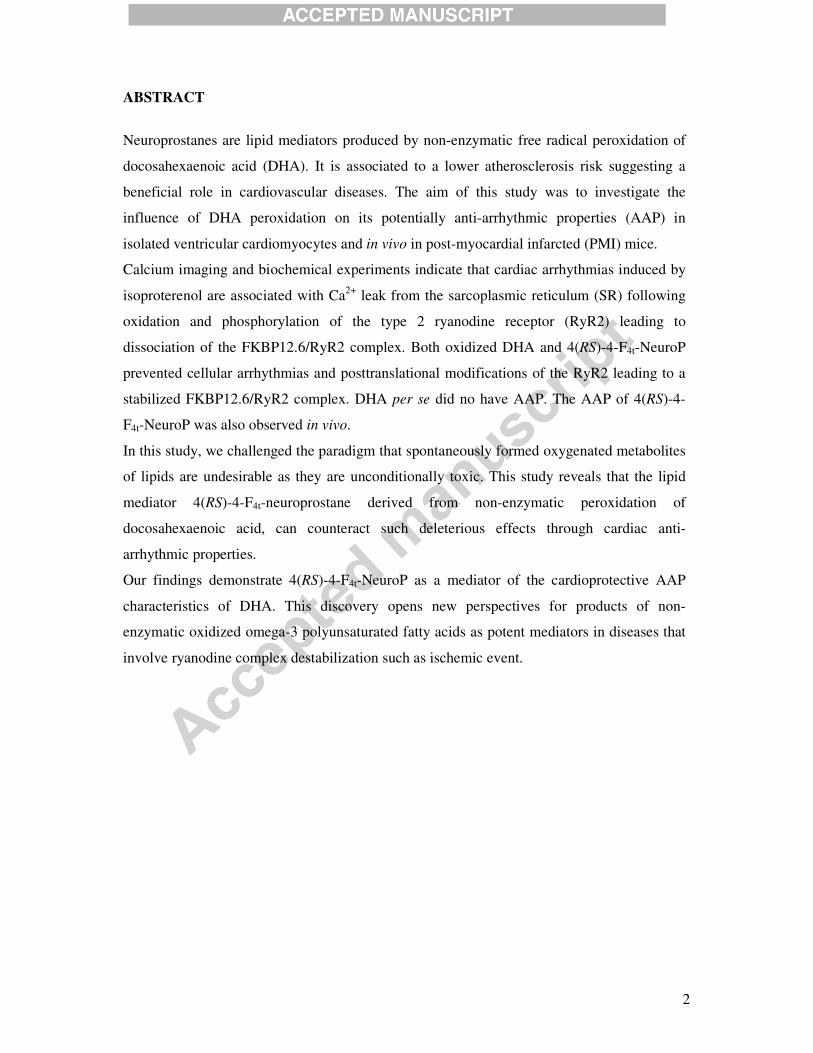
ABSTRACT
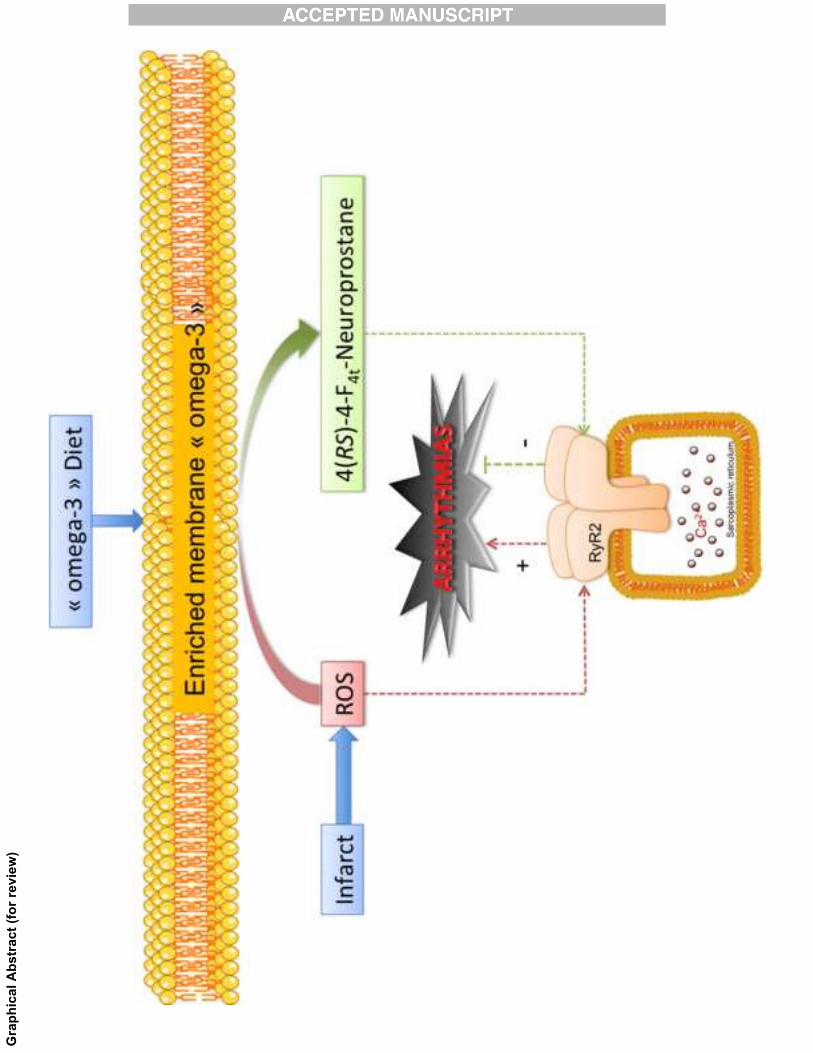
Neuroprostanes are lipid mediators produced by non-enzymatic free radical peroxidation of
docosahexaenoic acid (DHA). It is associated to a lower atherosclerosis risk suggesting a
beneficial role in cardiovascular diseases. The aim of this study was to investigate the
influence of DHA peroxidation on its potentially anti-arrhythmic properties (AAP) in
isolated ventricular cardiomyocytes and in vivo in post-myocardial infarcted (PMI) mice.
Calcium imaging and biochemical experiments indicate that cardiac arrhythmias induced by
isoproterenol are associated with Ca2+
leak from the sarcoplasmic reticulum (SR) following
oxidation and phosphorylation of the type 2 ryanodine receptor (RyR2) leading to
dissociation of the FKBP12.6/RyR2 complex. Both oxidized DHA and 4(RS)-4-F4t-NeuroP
prevented cellular arrhythmias and posttranslational modifications of the RyR2 leading to a
stabilized FKBP12.6/RyR2 complex. DHA per se did no have AAP. The AAP of 4(RS)-4-
F4t-NeuroP was also observed in vivo.
In this study, we challenged the paradigm that spontaneously formed oxygenated metabolites
of lipids are undesirable as they are unconditionally toxic. This study reveals that the lipid
mediator 4(RS)-4-F4t-neuroprostane derived from non-enzymatic peroxidation of
docosahexaenoic acid, can counteract such deleterious effects through cardiac anti-
arrhythmic properties.
Our findings demonstrate 4(RS)-4-F4t-NeuroP as a mediator of the cardioprotective AAP
characteristics of DHA. This discovery opens new perspectives for products of non-
enzymatic oxidized omega-3 polyunsaturated fatty acids as potent mediators in diseases that
involve ryanodine complex destabilization such as ischemic event.

3
Keywords:
Oxidative stress, Neuroprostanes, anti-arrhythmic, cardioprotection, DHA, Ryanodine
receptor, calcium
Abbreviations:
4(RS)-4-F4t-NeuroP, 4(RS)-4-F4t-neuroprostane; �3 PUFAs, omega-3 polyunsaturated fatty
acids; AAP, anti-arrhythmic properties; AoVTI, aortic velocity time integral; COX-2,
cyclooxygenase; CYP450, cytochrome P450; DHA, docosahexaenoic acid; DNP,
carbonylated RyR; ECG, electrocardiograms; EF, ejection fraction; EPA, eicosapentaenoic
acid; ES, extrasystoles; F, fluorescence; FAC, fractional area change; FS, fraction shortening;
FsK, forskolin; GPx, glutathione peroxidase enzymes; H2O2, hydrogen peroxide; HR, heart
rate; ISO, isoproterenol; LOX, lipooxygenase; LV, left ventricular; NE, norepinephrine; PMI,
post-myocardial infracted; QTc, corrected QT interval; ROS, reactive oxygen specie; RyR 2,
type 2 ryanodine receptor; RYR2-p2808, Phosphorylated RyR PKA site serine 2808 ; SCD,
sudden cardiac death; sHE, epoxide hydrolase (diols); S-NO, S-Nitrosylated RyR; SR,
sarcoplasmic reticulum; Vit E, vitamin E
4
INTRODUCTION
The cardioprotective effects of omega-3 polyunsaturated fatty acids (�3 PUFAs) have been
evident since the mid-70s [1]. The intake of fatty fish such as mackerel or tuna is associated
with a lower risk of cardiac arrhythmias including sudden cardiac death (SCD) [2–4] and
arrhythmic coronary heart disease death [5]. Administration of Omacor®
, a mixture of 850
mg of eicosapentaenoic acid (EPA) and docosahexaenoic acid (DHA) the two major
polyunsaturated fatty acids (PUFAs) of fatty fish, decreased the incidence of SCD in
secondary prevention of myocardial infarction [6]. The mechanisms responsible for the anti-
arrhythmic properties (AAP) of PUFAs remain unclear. In dogs, infusion of a DHA emulsion
tended to slow heart rate (HR), shortened the corrected QT interval (QTc) at rest and
significantly prevented ischemia-induced fatal ventricular arrhythmias [7]. These
experiments confirmed previous reports on the prevention of ischemia-induced ventricular
arrhythmias in dogs [8] and marmosets [9] by PUFAs. In humans, a significant slowing of
HR and the likelihood of prolonged QT has been observed [10].
Experimental studies on isolated cardiac cells suggest that �3 PUFAs have direct
cardiac electrophysiological effects [11]. However, DHA is highly prone to peroxidation and
we have shown that non-enzymatic oxygenated products of DHA and not DHA per se are
active on cardiac ionic channels [12]. In agreement, it has been demonstrated in rabbit
ventricular cells that early depolarization induced by H2O2 is inhibited by DHA while
reactive oxygen species (ROS) production is not altered, indicating the resiliency of oxidized
DHA [13].
DHA can be oxidized through two pathways; enzymatically, resulting in the
production of compounds such as resolvins or maresins, or non-enzymatically by ROS
initiation and propagation of free radical reactions, leading to the release of numerous
products, including neuroprostanes (F4-NeuroPs) [14]. Neuroprostanes are recognized as
oxidative stress biomarkers for the DHA-rich brain and are associated to ischemic stroke and
neurodegenerative diseases [15,16]. More recently, it has been proposed that F4-NeuroPs
may play a favorable role as potential bioactive components in identifying atherosclerosis
risk [17].
In the present study, we show through in cellulo and in vivo approaches that non-
enzymatic oxidation of DHA is a prerequisite to obtain ventricular anti-arrhythmic effects. In
particular, one of the F4-NeuroPs isomer, 4(RS)-4-F4t-neuroprostane (4(RS)-4-F4t-NeuroP)
5
appears to be the main anti-arrhythmic metabolite of DHA in preventing deleterious post-
translational modification of RyR2 and thus regulating calcium homeostasis.
MATERIALS AND METHODS
Animal experiments
Male C57Bl/6 mice (Janvier, France) of 7 weeks old were randomly assigned into two main
groups 1) mice with post-myocardial infarction (PMI mice) after left coronary artery ligation
as previously described [18] and 2) sham-operated mice that were submitted to the surgical
procedure but not to the artery ligation. All animal-handling procedures conformed to
European Parliament Directive 2010/63/EU and the institutional animal research committee
council on the protection of animals (CEEA-LR-12096).
In brief, anesthesia was performed for left thoracotomy and cardiac monitoring (2%
isoflurane/O2, Aerrane®, Baxter, France). The artery was ligated 1-2 mm beyond the
emergence from the top of the left atrium, using an 8-0 suture for PMI mice. A subcutaneous
injection of 0.01 mL buprenorphine solution (0.3 mg/mL) for post-operative analgesia was
administered. Shams were subjected to the same surgical procedure but without coronary
artery ligation. The mice were housed in single cages in a room under regulated temperature
and hygroscopic conditions (23±1°C, 45±10% humidity, light-dark schedule of 12h:12h ad
libitum feed).
After 4 weeks, the mice were randomly assigned to the different treatment groups:
Sham and PMI dosed with vehicle (NaCl 0,9%); PMI mice treated with 10 µM DHA (PMI
DHA); PMI treated with 10 µM DHA and 1 µM �-tocopherol (PMI DHA + Vit E); PMI mice
treated with 10 µM DHA and 1 µM hydrogen peroxide (PMI DHA + H2O2) and PMI mice
treated with 1 µM 4(RS)-4-F4t-NeuroP. We chose to work on PMI mice challenged with NE
since it has been shown that the AAP of DHA is secondary to myocardial infarction in human
[6].
Treatments were administrated as intravenous injection (200 µl) of the prepared
solution equivalent to 10 times the concentration to reach the final concentrations matching in
cellulo experiments.
It is known that the activation of the adrenergic nervous system is one factor that may
play a crucial role in the genesis of arrhythmias associated with acute myocardial infarction
[19]. To mimic this activation, all PMI mice were then intra-peritoneally (i.p.) challenged

6
with the �1-adrenergic agonist, norepinephrine (2.5 mg/kg) [20] 20 min after they received
their treatment.
Echocardiography
Doppler echocardiography was performed using a high-resolution ultrasound system (Vevo
2100; VisualSonics, Toronto, Canada) equipped with a 40-MHz transducer. The mice were
anesthetized with 1.5% isoflurane in 100% oxygen and placed on a heating table in a supine
position. Body temperature was monitored through a rectal thermometer to be maintained at
36-38°C and electrocardiograms (ECG) were recorded all along the echocardiographic
procedure with limb electrodes. Ejection (EF%) and shortening (SF%) fractions were
calculated from the left ventricular diameters on M-mode measurements at the level of
papillary muscles in a parasternal short-axis two-dimensional view. To better consider
coronary ligation-induced left ventricular remodeling, EF was also calculated from a B-mode
parasternal long axis view (EF% B-mode) by tracing endocardial end-diastolic and end-
systolic areas to estimate left ventricular volumes, and the endocardial fractional area change
(FAC%) on a parasternal short-axis view at papillary muscle level was calculated. Pulsed-
wave Doppler of the ascending aortic blood flow was recorded permitting measurements of
the velocity time integral (AoVTI). All measurements were quantified and averaged for three
cardiac cycles (Table 1).
Synthesis of 4(RS)-4-F4t-NeuroP
Using the protocol previously reported, we synthesized F4-NeuroPs. The strategy is based on
an easily accessible bicyclic precursor to obtain isoprostanoid derivatives [21], while more
refined strategy were used for the synthesis of the isomers, 4(RS)-4-F4t-NeuroP [22], 10-F4t-
NeuroP [23] and 14(RS)-14-F4t-NeuroP (non yet published). The 13-F4t-NeuroP was
synthesized using another strategy [24].
Fatty acid solution and oxidation
To observe antioxidant or oxidant effect, cells were incubated 20 minutes in Tyrode solution
containing in 10 µM DHA or 10 µM DHA + 1 µM Vit E or 10 µM DHA + 1 µM H2O2. To
prepare these solutions, DHA (stock prepared in ethanol) was added in the Tyrode solution
after Vit E (stock prepared in chloroform) or H2O2 (stock prepared in reverse osmosis water).
Stock solution of 4(RS)-4-F4t-NeuroP was prepared in Tyrode solution and diluted
accordingly for the experimentations.
7
Quantification of 4(RS)-4-F4t-NeuroP
In Tyrode solution (control), 10 µM DHA was incubated with or without 1 µM α-tocopherol
(Vit E) or 1 µM hydrogen peroxide (H2O2) for 20 min in room temperature. The reaction was
terminated with antioxidant butylated hydroxytoluene BHT (0.005%, w/v). The internal
standard C21-15-F2t-IsoP (2.5 ng), synthesized by IBMM (Montpellier, France) laboratory
was added to each sample mix. The sample was further diluted in aqueous sodium acetate
solution (pH 4.6), acidified with 1M HCl and applied to pre-washed (methanol) Bond Elut
Certify II SPE cartridge (Agilent, CA USA). After loading the sample (control, DHA or
DHA+VitE or DHA+H2O2), it was sequentially cleaned with water/methanol (1:1) and
hexane / ethyl acetate (7:3) and then F4-NeuroPs was eluted with ethyl acetate/methanol (9:1).
The eluate was dried under nitrogen and then derivatized in room temperature for 30 min with
10% pentafluorobenzyl bromide and 10% N,N-diisopropylethylamine prepared in acetonitrile
(2:1). Thereafter, it was dried under nitrogen and then derivatized with N,O-
bis(trimethylsilyl)trifluoroacetamide (BSTFA) + trimethylsilylchlorosilane (TMCS) 1% and
N,N-dimethylformamide (2:1) (Sigma Aldrich, USA). After drying the reagents under
nitrogen, samples were re-suspended in decane.
Gas chromatography-mass spectrometry set at negative ion chemical ionization
(TraceGC and DSQ II Mass Spectrometer, Thermo Fisher Scientific, MA USA) was used to
determine 4(RS)-4-F4t-NeuroP [25]. Analytical column FactorFour™ (Varian, USA) fused
silica capillary was used. Helium gas was the carrier gas and the column temperature was
programmed from 140°C to 250°C at 30°C per minute then 250°C to 300°C at 4°C per minute
and remained at this temperature for 10 minutes. The ion source temperature was 200°C and
isobutane (1 mL/min) was used as the reagent gas for NICI. Selected ion monitoring was
performed to monitor ions m/z 593.5 for 4(RS)-4-F4t-NeuroP and at m/z 583.5 for C21-15-F2t-
IsoP internal standard. Quantitation was achieved by relating the peak area of the 4(RS)-4-
F4t-NeuroP with C21-15-F2t-IsoP internal standard peak.
Preparation of cardiomyocytes
Cellular experiments were performed on freshly isolated left ventricular myocytes from the
non-infarcted free wall (excluding the border zone). In brief, after cervical dislocation, the
heart was removed, washed and aorta was cannulated to a modified Langendorff system. The
heart was perfused by retrograde flow rate of 5–10 mL/min at 37°C for 6–8 min with a
modified Tyrode solution composed of 113 mM NaCl, 4.7 mM, KCl, 0.6 mM KH2PO4, 0.6
8
mM Na2HPO4, 1.2 mM MgSO4, 12 mM NaHCO3, 10 mM KHCO3, 10 mM Hepes, (pH 7.4)
and 0.1 g/mL liberase dispase (high research grade, Roche, France).
After enzymatic treatment (4-6 min), a part of the left ventricle was removed and
minced to separate the cells. Isolated myocytes were re-suspended in a sterile enzyme-free
Tyrode solution, and the Ca2+
concentration of the ventricular cell suspension was gradually
increased to 1 mM by the addition of CaCl2 in five sequential steps 100, 100, 300 and 500 µM
with 10 min interval between steps. Finally the cardiomyocytes were kept at room
temperature 22 – 24° C until use. Prior to the treatments, the freshly isolated cardiomyocytes
were then superfused with standard Tyrode solution (121 mM NaCl, 5.0 mM KCl, 1.8 mM
CaCl2, 0.5 mM MgCl2, 0.4 mM NaH2PO4, 24 mM NaHCO3, 0.1 mM EDTA and 5.5 mM
glucose). Cardiomyocytes with obvious sarcolemmal blebs or spontaneous contractions were
not used. Only cardiomyocytes with clear edges were selected and were used within 1-6 h
after isolation.
Inhibition of enzymatic lipid peroxidation of cardiomyocytes
Inhibitors of enzymatic lipid peroxidation, anti-lipoxygenase (1 µM zileuton), anti-
cytochrome P450 (3 µM ketoconazole) and anti-cycloxygenase 2 (1 µM celecoxib) from
Sigma-Aldrich, USA and anti-epoxide hydrolase (10 nM) from Cayman Chemicals USA, and
a combination of 4 inhibitors were tested with and without 10 µM DHA + 1µM H2O2 in the
cellular arrhythmias. We also used glutathione peroxidase enzyme, which reduce lipid
peroxides to alcohols and H2O2. Stock solution of GPx was dissolved in water with 10 mM of
phosphate sodium and 1 mM of dithiothreitol. GPx was either added before the mix DHA +
H2O2 at concentration of 10 units (1 unit oxide 1 µM of DHA per minute) in Tyrode solution
or after the DHA + H2O2 mix. For all the experiments, solutions were prepared freshly from
the stock and diluted with Tyrode medium.
Calcium channeling
The effect of oxidation of �-3 PUFAs on cell shortening and Ca2+
transients of field-
stimulated cardiomyocytes is monitored online using commercial myocyte calcium and
contractility monitoring system (IonOptix®, Milton, MA, USA) connected to a standard
inverted fluorescent microscope. Cells were field-stimulated with 1 ms current pulses
delivered via two platinum electrodes. To monitor intracellular Ca2+
concentration,
cardiomyocytes were loaded with the fluorescent ratiometric Ca2+
indicator Indo-1AM (2 µM,
Invitrogen, France). They were simultaneously illuminated at 365 nm using a xenon arc bulb
9
light. Cytosolic Ca2+
concentration was determined by Indo-1 AM fluorescence which emit at
405 nm and 480 nm concurrently. The ratio of 405nm/480nm indicates the cytosolic Ca2+
concentration.
To observe arrhythmias (ventricular extrasystoles), the cells were bathed with 10 nM
isoproterenol and stimulated with 30 s pacing period (1.0 Hz), followed by 30 s rest period
[20,26]. Confocal imaging was performed using a Zeiss LSM510 confocal microscope (Carl
Zeiss Inc., Oberkochen, Germany) equipped with an argon laser (488 nm) and a 60X, 1.3 NA
oil immersion objective set at axial and radial resolutions of 1.0 and 0.4 µm, respectively.
Ca2+
sparks were recorded in quiescent myocytes incubated with the Ca2+
indicator
Fluo-4-AM (4 µM) (Molecular Probes, OR USA) for 15 min. The dye was excited at 488 nm
and the fluorescence emission was collected through a 505 nm long-pass filter. Myocytes
were field-stimulated at 1 Hz with 1 ms current pulses delivered via two platinum electrodes,
one on each side of the perfusion chamber. During the rest period that follows stimulation,
myocyte were repetitively scanned along the entire length of the cell at 1.5 ms intervals, for a
maximum of 6 s. The laser intensity was reduced to 5% maximum to decrease cell damage
and dye bleaching. Line scan diagrams were constructed by stacking emission lines,
corresponding to excitation scans, in temporal order. An average of the Ca2+
sparks were
determined by the intensity of each sequential scan line and plotting the mean intensity as a
function of time. The SparkMaster® plugin for ImageJ® software was used to detect and
analyze Ca2+
sparks.
Immunoblot
Proteins were extracted from basal left ventricular frozen cells (50 mg) homogenized with a
manual polytron instrument. Cells were then lysed in 600 µL extraction buffer (Tris maleate
10m M, NaF 35 mM, Triton 1%, activated orthovanadate 20 mM, inhibitor cocktail, Roche,
France) for 45 min under rotated agitation. Membrane and cytosolic proteins were collected
from the supernatant after 5 min centrifugation at 10, 000 x g at 4 °C.
For the immunoprecipitation assay, left ventricular (LV) tissues were lysed in 1 mL
buffer containing 10 mM Tris maleate (pH 6.8), 35 mM NaF, triton 1% and protease
inhibitors (Roche 11873580001, France). A concentration of 10 µg anti-RyR2 antibody was
used to immunoprecipitate RyR2 from 500 µg of LV homogenate. The samples were
incubated with anti-RyR antibody in 0.5 ml modified RIPA buffer (Tris HCl 10 mM, pH 7.4,
NaCl 150 mM, Triton 1%, NaF 5 mM and protease inhibitor cocktail) for 2 hours at 4°C. The
10
immune complexes were incubated with protein A/G magnetic beads (Pierce 88802, USA) at
4°C for 2 hours, after which the beads were washed three times with RIPA buffer.
To detect RyR2 protein oxidation, the immune complex was treated with 2,4
dinitrophenylhydrazine (DNPH) and the DNP-derivatized protein samples formed were
detected using on Immunoblot Protein oxidation detection Kit (Millipore S7150, USA).
Proteins were then separated using SDS-PAGE, blotted onto nitrocellulose membranes (0.2
µm, GE Healthcare, France) and incubated overnight at 4°C with primary antibodies: the type
2 ryanodine receptor, RyR2 (1/1000 dilution, Pierce, France) and anti FKBP12.6 (1/1000
dilution, RD System AF 4174, France). Protein levels were expressed relative to GAPDH
(1/60000 dilution, AB8245, ABCAM, France). All immunoblots were developed and
quantified using the Odyssey infrared imaging system (LI-COR Biosystems, USA) using
infrared-labeled anti-mouse and anti-rabbit IgG (1/30000 dilution) secondary antibodies.
Statistical analysis
All data are given as mean ± SEM. Statistical analyses were performed using GraphPad
Prism® (Prism 5 for Mac OS X). One-way ANOVA for multiple comparisons was used,
followed by a parametric t-test with Fishers correction. For paired studies, Wilcoxon signed
rank test is used. Percentage of arrhythmic cells data was analyzed by a χ2-test. A p-value of
0.05 or less was indicated as statistically significant.

11
RESULTS
DHA under oxidative stress conditions reduced cardiac arrhythmias
AAP of DHA was evaluated in models of cardiac arrhythmias. Different conditions of
oxidative status were obtained by preparing DHA solutions in the presence of an antioxidant,
1 µM �-tocopherol [27] or a pro-oxidant, 1 µM H2O2 [28]. Bathing isolated mice ventricular
cardiomyocytes for 20 min in DHA prevented arrhythmias and concurs in part with previous
report [11]. Importantly, AAP of DHA was potentiated by pro-oxidants and conversely
prevented in the presence of �-tocopherol (Figures 1A and 1B). When applied alone, H2O2
and �-tocopherol had no effect (Figure 1B), which further support the role of DHA oxidation
on AAP.
Additionally, we investigated the efficacy of DHA to reduce the trigger of ventricular
extrasystoles (ES) in a post myocardial infarction (PMI) mice model established by coronary
artery ligation and sensitized by norepinephrine (NE) [29]. PMI mice develop calcium
dependent-ES due to increased diastolic calcium level in a context of increased ROS
production [30]. These arrhythmias are potentiated by a norepinephrine challenge. In this
validated model and as described in Table 1, intravenous injection of DHA to the mice
reduced ES by 45%, which further reinforced the AAP of DHA in pro-oxidant conditions
(Figures 1C and 1D).
AAP are not mediated by enzymatic oxidation of DHA
Enzymatic oxidation of DHA can develop endogenously [31], like the conditions used in this
experimental approach. The AAP of DHA was investigated in cardiomyocytes in the presence
of different inhibitors of enzymes that oxidize PUFAs namely cyclooxygenase (COX-2),
lipooxygenase (LOX) and cytochrome P450 (CYP450). Individually or combined, the
inhibitors did not modify the AAP of pro-oxidized DHA (Figure 2A). Our observation infers
that DHA exert a strong AAP through non-enzymatic peroxidation process that generates
metabolites such as F4-neuroprostanes and not through typical enzymatic process that
involves for example resolvins, protectins and maresins. In order to further explore the
chemical entities needed for the observed AAP, we buffered any hydroperoxyl derivatives
potentially formed by our pro-oxidant condition (DHA+H2O2) by a late addition of
glutathione peroxidase enzymes (GPx) (Figure 2B). AAP were still observed in these

12
conditions, indicating these effects are not related to endoperoxide or hydroperoxide
metabolites of DHA. On the contrary, arrhythmia persisted when GPX was added prior to
DHA+H2O2 indicating that H2O2 initiated the formation of the required metabolites of DHA
for AAP (Figure 2B).
Among the F4-neuroprostanes, 4(RS)-4-F4t-NeuroP had the most active AAP
Supplementation of DHA to atherosclerotic LDLR-/-
mice showed that liver F4-NeuroP
concentration is negatively correlated to atherosclerosis risk [17]. The isomer 4(RS)-4-F4t-
NeuroP is the most abundant F4-NeuroPs formed from non-enzymatic DHA peroxidation
[14]. In our in vitro experiments, incubation of DHA (10 µM) with H2O2 (1 µM) in Tyrode
solution for 20 min generated 0.61±0.08 µM of 4(RS)-4-F4t-NeuroP (Figures 3A and B)
whereas DHA with Vit E had no 4(RS)-4-F4t-NeuroP formed and DHA alone had trace
amount (0.03±0.01 µM). Furthermore, we recently discovered that levels of 4(RS)-4-F4t-
NeuroP and another isomer, 10-F4t-neuroprostane (10-F4t-NeuroP) are concentrated in brains
of preterm pigs [32], and in adult rat brain and heart [33].
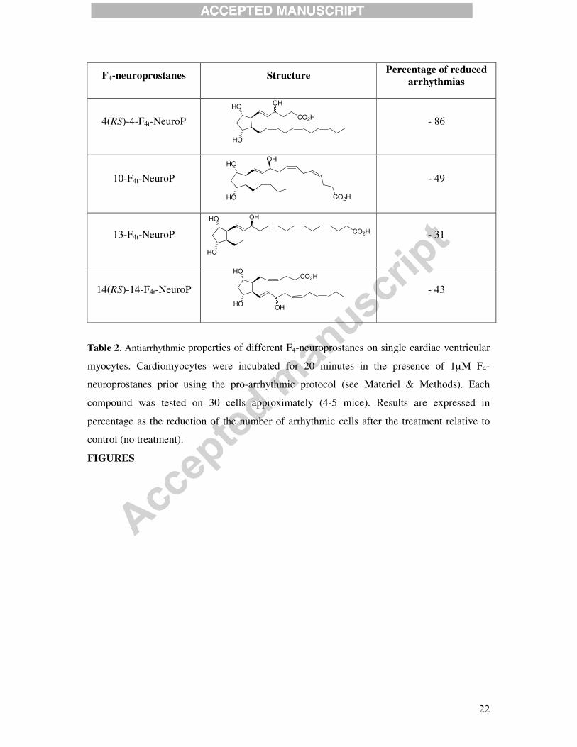
From our findings, the high concentration of 4(RS)-4-F4t-NeuroP in the heart indicates
a potential bioactive role. We compared the anti-arrhythmic effect of 4(RS)-4-F4t-NeuroP with
other F4-neuroprostanes (10-F4t-NeuroP, 13-F4t-NeuroP, 14(RS)-4-F4t-NeuroP) (Table 2). Of
the four F4-neuroprostanes tested, the 4(RS)-4F4t-NeuroP is the most potent (IC50�100 nM)
(Figure 3C).
Despite the IC50�100nM of 4(RS)-4-F4t-NeuroP, we subjected the maximum
concentration (1 µM) to authenticate the AAP in our study. Our in vivo evaluation indicates
AAP of 1 µM 4(RS)-4-F4t-NeuroP was comparable with the positive control 1 µM carvedilol,
which is a referenced anti-arrhythmic drug (Figure 3D). Also, 4(RS)-4-F4t-NeuroP inhibited
arrhythmias produced by the adenylyl cyclase activator, forskolin (Figure 3E) suggesting that
�-blocking properties is not involved in the AAP of 4(RS)-4-F4t-NeuroP. Absence of
bradycardia following 4(RS)-4-F4t-NeuroP consolidates this hypothesis (Table 1). Further, the
AAP of DHA+H2O2 and 4(RS)-4-F4t-NeuroP were similar suggesting that the AAP of DHA is
largely due to the generation of 4(RS)-4-F4t-NeuroP in DHA+H2O2.
4(RS)-4-F4t-NeuroP prevents RyR2 dysfunction

13
In our experimental models, the arrhythmias prevented by 4(RS)-4-F4t-NeuroP are mainly due
to extrasystoles (ES). The mechanisms of triggering ES include alterations of Ca2+
homeostasis that are potentiated by catecholamines; the two phenomena observed after
myocardial infarction [34]. The Ca2+
signaling alteration could be associated with a leaky type
2 ryanodine receptor (RyR2) [35–37]. As a consequence, cytosolic Ca2+
levels in diastole rise
and promote ES [38]. We thus evaluated the leaky behavior of RyR2 by measuring the
frequency of spontaneous Ca2+
sparks by confocal microscopy [39] and the resting cytosolic
Ca2+
concentration by epifluorescence. In our model, Ca2+
sparks events frequency was
increased by isoprenaline (Figures 4A and 4B). This increase was partially prevented by DHA
as also shown in previous reports [40,41]. Interestingly, the effects of DHA on Ca2+
sparks
frequency were blunted by �-tocopherol and enhanced by H2O2. PMI cardiomyocytes also
exhibited an increased frequency of Ca2+
sparks that were significantly reduced by 4(RS)-4-
F4t-NeuroP. The changes in the sparks frequency followed changes in resting Ca2+
concentration (Figure 4C).
At the molecular level, post-translational modifications of RyR2 may account for their
leaky behavior [35–37]. We quantified RyR2 carbonylation, S-nitrosylation, phosphorylation
and the degree of association of RyR2 with FKBP12.6 on the arrhythmic cardiomyocytes
after 10 nM isoproterenol (ISO) treatment with or without 10 µM DHA, 10 µM DHA + 1 µM
Vit E or 10 µM DHA + 1 µM H2O2 (Figure 5). Iso challenge promotes dissociation of
FKBP12.6 from the RyR2 macromolecular complex as previously reported and accounts for
the abnormal SR-Ca2+
leak [35] (Figure 5A). Iso also promotes RyR2 carbonylation, S-
nitrosylation and phosphorylation on serine 2808 (Figure 5B-D). These effects were
prevented by DHA and more so by DHA + H2O2 compared to Iso and DHA + Vit E. This is
supported by the lack of FKBP12.6 dissociation from the RyR2 complex of DHA or DHA +
H2O2 treatment compared to Iso and DHA + Vit E (Figure 5A). Additionally, our
observations indicate that oxidation of DHA is necessary to prevent RyR2 modification as no
effect was found in the presence of Vit E, an antioxidant. In order to verify that the
involvement of such an effect is due to a non-enzymatic oxidized lipid product of DHA, we
repeated the evaluation on the arrhythmic cardiomyocytes with 4(RS)-4-F4t-NeuroP (Figure
5A and 5B). The prevention of RyR2 phosphorylation and dissociation of FKBP12.6 of
NeuroP was comparable to DHA + H2O2. This strongly supports that the effects of oxidized
DHA and 4(RS)-4-F4t-NeuroP in correcting SR Ca2+
leak, normalizing diastolic Ca2+
level and
preventing ES triggering results from the counteraction of RyR2 post-translational
modifications and RyR2 dysfunction.
14
DISCUSSION
In the present study, we clearly demonstrated that oxidized DHA possesses potent AAP in
cellulo and in vivo, and that unmodified DHA per se was inactive. This effect is not mediated
by enzymatic lipid peroxidation but instead by neuroprostanes, the stable end products of the
non-enzymatic lipid peroxidation of DHA, particularly 4(RS)-4-F4t-NeuroP. This metabolite
displayed a unique and unprecedented mode of action in the family of lipid mediators and of
any known endogenous biomolecule, as it stabilized RyR2 and maintained this complex
closed during diastole.
This discovery bridges the missing relationship between cardiac ischemic events and
the AAP of DHA. Indeed, following cardiac ischemic diseases and myocardial infarction
there is a burst of ROS [42] that can be involved in the generation of the arrhythmias, the
origin of SCD [43]. Such an abrupt imbalance of the oxidative status can oxidize proteins
[44], nucleic acids [45] and lipids [46]. These highly reactive compounds play a pivotal role
in the pathogenesis of post-ischemic injury that progresses to SCD [47,48]. Classically, there
is a consensus that overproduction of ROS are mainly deleterious and do not play a role in
normal physiology. For example it has been shown that an isoketal, E2-IsoK, originating from
the oxidation of arachidonic acid can produce adducts with the sodium channel protein
NaV1.5, perturbing its activity in a pro-arrhythmic way [47]. However, the possibility that
non-enzymatic oxygenated metabolites of �3 PUFAs can exert counter effects has been under
investigated.
We have previously reported that DHA needs to be oxidized to influence ionic channel
activities [12] and it is agreed by others that such effects on ionic channels contributes to the
AAP of DHA [49]. We hereby demonstrated that the AAP of DHA are dependent on the
oxidative status of the environment, and that non-enzymatic auto-oxidation of DHA was a
perquisite to AAP. Our assays pinpointed neuroprostane metabolites as the potential
metabolites with APP properties. The main isomer, 4(RS)-4-F4t-NeuroP, showed potent AAP
in cellulo in a dose dependent manner and also in vivo in PMI mice.
At the cellular level, the mechanism of action is unlikely to be due to a �-blocker
effect, but the AAP can instead be explained by a rycal-like effect, in particular, stabilization
of the RyR2 complex with FKBP12.6 [50]. This effect was strongly supported by both
oxidized DHA and 4(RS)-4-F4t-NeuroP in correcting SR Ca2+
leak, normalizing diastolic Ca2+
levels and from the prevention of the dissociation of FKBP12.6 from RyR2. Although we can
not exclude a direct effect of 4(RS)-4-F4t-NeuroP on RyR2, in contrast to rycal effects, we

15
also observed that both oxidized DHA and 4(RS)-4-F4t-NeuroP prevent RyR2 hyperactive S-
nitrosylation, oxidation and phosphorylation, suggesting an upstream mechanism rather than a
direct effect on RyR2 as previously reported [37]. Altogether we showed that 4(RS)-4-F4t-
NeuroP is capable of preventing ES triggering effects from the prevention of RyR2 post-
translational modifications and RyR2 dysfunction by normalizing RyR2 activity in diastole
and thus calcium homeostasis.
Emulsion of �3 PUFAs, injected intravenously has been shown to be anti-arrhythmic
in a canine model of myocardial infarction [7] and similar effects on sustained ventricular
tachycardia have been observed in humans [20,51]. The use of an in vivo ischemic model to
monitor the triggering of ventricular ES clearly confirmed our hypothesis that DHA auto-
oxidatively has to be transformed into neuroprostane for rapid inhibition of arrhythmias.
The pre-requisite for DHA oxidation to elicit any AAP could explain the lack of
elucidation for the beneficial effects of �3 PUFAs other than in ischemic cardiovascular
diseases [2,52,53]. After initial reports establishing that �3 PUFAs have cardioprotective
properties, the beneficial effects were then extended to many if not all cardiovascular
problems, even those that are not ischemic [53,54]. This mindset could be the drawback for
the proper development of therapeutic targets in cardiac ischemia and for the difficulty in
justifying its valuable impact in cardioprotection. Overall, the mechanism of action we
describe here can explain the favorable impact of �3 PUFAs in chronic diseases implicating
RyR2 dysfunction when an unbalanced oxidative status is present.
CONCLUSION
From this work, we conclude that the oxidation of DHA to 4(RS)-4-F4t-NeuroP is
necessary to prevent ischemia-induced arrhythmias. We propose that in oxidative stress
conditions such as ischemic diseases, non-enzymatic oxygenated metabolites of DHA formed
by peroxidation of cardiac membrane lipids, notably 4(RS)-4-F4t-NeuroP, are responsible for
the AAP of DHA by countering the cellular stress by ROS. Importantly, it appears that non-
enzymatically oxygenated metabolites of �3 PUFAs can communicate and exert a
physiological role. This highlights potential beneficial effects of increased ROS production
dependent on the cellular environment.
ACKNOWLEDGEMENTS

16
A part of this work was financially supported by the Université Montpellier I (grants BQR-
2008 and 2011), the Centre National de la Recherche Scientifique (CNRS) for PEPII INSB-
INC, and the Fondation pour la Recherche Médicale (FRM) (DCM20111223047). We are
grateful to Valérie Bultel-Poncé and Alexandre Guy for their assistance in synthesizing the
4(RS)-4-F4t-NeuroP, and Valérie Scheuerman and Patrice Bideaux for performing
biochemistry and prepare the PMI mice respectively.
17
REFERENCES
[1] Bang, H. O.; Dyerberg, J.; Hjøorne, N. The composition of food consumed by Greenland
Eskimos. Acta Med. Scand. 200: 69–73; 1976.
[2] Burr, M. L.; Fehily, A. M.; Gilbert, J. F.; Rogers, S.; Holliday, R. M.; Sweetnam, P. M.;
Elwood, P. C.; Deadman, N. M. Effects of changes in fat, fish, and fibre intakes on death
and myocardial reinfarction: diet and reinfarction trial (DART). Lancet 2: 757–761; 1989.
[3] Siscovick, D. S.; Raghunathan, T. E.; King, I.; Weinmann, S.; Wicklund, K. G.; Albright,
J.; Bovbjerg, V.; Arbogast, P.; Smith, H.; Kushi, L. H. Dietary intake and cell membrane
levels of long-chain n-3 polyunsaturated fatty acids and the risk of primary cardiac arrest.
JAMA 274: 1363–1367; 1995.
[4] Albert, C. M.; Campos, H.; Stampfer, M. J.; Ridker, P. M.; Manson, J. E.; Willett, W. C.;
Ma, J. Blood levels of long-chain n-3 fatty acids and the risk of sudden death. N. Engl. J.
Med. 346: 1113–1118; 2002.
[5] Mozaffarian, D.; Lemaitre, R. N.; Kuller, L. H.; Burke, G. L.; Tracy, R. P.; Siscovick, D.
S.; Cardiovascular Health Study. Cardiac benefits of fish consumption may depend on the
type of fish meal consumed: the Cardiovascular Health Study. Circulation 107: 1372–
1377; 2003.
[6] Dietary supplementation with n-3 polyunsaturated fatty acids and vitamin E after
myocardial infarction: results of the GISSI-Prevenzione trial. Gruppo Italiano per lo
Studio della Sopravvivenza nell’Infarto miocardico. Lancet 354: 447–455; 1999.
[7] Billman, G. E.; Kang, J. X.; Leaf, A. Prevention of sudden cardiac death by dietary pure
omega-3 polyunsaturated fatty acids in dogs. Circulation 99: 2452–2457; 1999.
[8] Billman, G. E.; Hallaq, H.; Leaf, A. Prevention of ischemia-induced ventricular
fibrillation by omega 3 fatty acids. Proc. Natl. Acad. Sci. U. S. A. 91: 4427–4430; 1994.
[9] McLennan, P. L.; Bridle, T. M.; Abeywardena, M. Y.; Charnock, J. S. Dietary lipid
modulation of ventricular fibrillation threshold in the marmoset monkey. Am. Heart J.
123: 1555–1561; 1992.
[10] Mozaffarian, D.; Prineas, R. J.; Stein, P. K.; Siscovick, D. S. Dietary fish and n-3 fatty
acid intake and cardiac electrocardiographic parameters in humans. J. Am. Coll. Cardiol.
48: 478–484; 2006.
[11] Leaf, A.; Kang, J. X.; Xiao, Y.-F. Fish oil fatty acids as cardiovascular drugs. Curr.
Vasc. Pharmacol. 6: 1–12; 2008.
[12] Judé, S.; Bedut, S.; Roger, S.; Pinault, M.; Champeroux, P.; White, E.; Le Guennec, J.-
Y. Peroxidation of docosahexaenoic acid is responsible for its effects on I TO and I SS in
rat ventricular myocytes. Br. J. Pharmacol. 139: 816–822; 2003.
[13] Zhao, Z.; Wen, H.; Fefelova, N.; Allen, C.; Guillaume, N.; Xiao, D.; Huang, C.; Zang,
W.; Gwathmey, J. K.; Xie, L.-H. Docosahexaenoic Acid reduces the incidence of early

18
afterdepolarizations caused by oxidative stress in rabbit ventricular myocytes. Front.
Physiol. 3: 252; 2012.
[14] Roberts, L. J.; Montine, T. J.; Markesbery, W. R.; Tapper, A. R.; Hardy, P.; Chemtob,
S.; Dettbarn, W. D.; Morrow, J. D. Formation of isoprostane-like compounds
(neuroprostanes) in vivo from docosahexaenoic acid. J. Biol. Chem. 273: 13605–13612;
1998.
[15] Roberts, L. J.; Fessel, J. P.; Davies, S. S. The biochemistry of the isoprostane,
neuroprostane, and isofuran Pathways of lipid peroxidation. Brain Pathol. Zurich Switz.
15: 143–148; 2005.
[16] Seet, R. C. S.; Lee, C.-Y. J.; Chan, B. P. L.; Sharma, V. K.; Teoh, H.-L.;
Venketasubramanian, N.; Lim, E. C. H.; Chong, W.-L.; Looi, W.-F.; Huang, S.-H.; Ong,
B. K. C.; Halliwell, B. Oxidative damage in ischemic stroke revealed using multiple
biomarkers. Stroke J. Cereb. Circ. 42: 2326–2329; 2011.
[17] Gladine, C.; Newman, J. W.; Durand, T.; Pedersen, T. L.; Galano, J.-M.; Demougeot,
C.; Berdeaux, O.; Pujos-Guillot, E.; Mazur, A.; Comte, B. Lipid profiling following
intake of the omega 3 fatty acid DHA identifies the peroxidized metabolites F4-
neuroprostanes as the best predictors of atherosclerosis prevention. PloS One 9: e89393;
2014.
[18] Thireau, J.; Karam, S.; Fauconnier, J.; Roberge, S.; Cassan, C.; Cazorla, O.; Aimond,
F.; Lacampagne, A.; Babuty, D.; Richard, S. Functional evidence for an active role of B-
type natriuretic peptide in cardiac remodelling and pro-arrhythmogenicity. Cardiovasc.
Res. 95: 59–68; 2012.
[19] Podrid, P. J.; Fuchs, T.; Candinas, R. Role of the sympathetic nervous system in the
genesis of ventricular arrhythmia. Circulation 82: I103–I113; 1990.
[20] Den Ruijter, H. M.; Berecki, G.; Verkerk, A. O.; Bakker, D.; Baartscheer, A.;
Schumacher, C. A.; Belterman, C. N. W.; de Jonge, N.; Fiolet, J. W. T.; Brouwer, I. A.;
Coronel, R. Acute administration of fish oil inhibits triggered activity in isolated
myocytes from rabbits and patients with heart failure. Circulation 117: 536–544; 2008.
[21] Oger, C.; Brinkmann, Y.; Bouazzaoui, S.; Durand, T.; Galano, J.-M. Stereocontrolled
access to isoprostanes via a bicyclo[3.3.0]octene framework. Org. Lett. 10: 5087–5090;
2008.
[22] Oger, C.; Bultel-Poncé, V.; Guy, A.; Balas, L.; Rossi, J.-C.; Durand, T.; Galano, J.-M.
The handy use of Brown’s P2-Ni catalyst for a skipped diyne deuteration: application to
the synthesis of a [D4]-labeled F4t-neuroprostane. Chem. Weinh. Bergstr. Ger. 16:
13976–13980; 2010.
[23] Guy, A.; Oger, C.; Heppekausen, J.; Signorini, C.; De Felice, C.; Fürstner, A.; Durand,
T.; Galano, J.-M. Oxygenated metabolites of n-3 polyunsaturated fatty acids as potential
oxidative stress biomarkers: total synthesis of 8-F3t-IsoP, 10-F4t-NeuroP and [D4]-10-
F4t-NeuroP. Chem. Weinh. Bergstr. Ger. 20: 6374–6380; 2014.

19
[24] Taber, D. F.; Reddy, P. G.; Arneson, K. O. A potential route to neuroprostanes and
isoprostanes: preparation of the four enantiomerically pure diastereomers of 13-F4t-
neuroP. J. Org. Chem. 73: 3467–3474; 2008.
[25] Mas, E.; Michel, F.; Guy, A.; Bultel, V.; Falquet, Y.; Chardon, P.; Rossi, J.-C.; Cristol,
J. P.; Durand, T. Quantification of urinary F2-isoprostanes with 4(RS)-F4t-neuroprostane
as an internal standard using gas chromatography-mass spectrometry Application to
polytraumatized patients. J. Chromatogr. B Analyt. Technol. Biomed. Life. Sci. 872: 133–
140; 2008.
[26] Berecki, G.; Den Ruijter, H. M.; Verkerk, A. O.; Schumacher, C. A.; Baartscheer, A.;
Bakker, D.; Boukens, B. J.; van Ginneken, A. C. G.; Fiolet, J. W. T.; Opthof, T.; Coronel,
R. Dietary fish oil reduces the incidence of triggered arrhythmias in pig ventricular
myocytes. Heart Rhythm Off. J. Heart Rhythm Soc. 4: 1452–1460; 2007.
[27] Burton, G. W.; Ingold, K. U. Vitamin E as an in vitro and in vivo antioxidant. Ann. N.
Y. Acad. Sci. 570: 7–22; 1989.
[28] Janero, D. R.; Hreniuk, D.; Sharif, H. M. Hydrogen peroxide-induced oxidative stress
to the mammalian heart-muscle cell (cardiomyocyte): lethal peroxidative membrane
injury. J. Cell. Physiol. 149: 347–364; 1991.
[29] Tarnavski, O.; McMullen, J. R.; Schinke, M.; Nie, Q.; Kong, S.; Izumo, S. Mouse
cardiac surgery: comprehensive techniques for the generation of mouse models of human
diseases and their application for genomic studies. Physiol. Genomics 16: 349–360; 2004.
[30] Hori, M.; Nishida, K. Oxidative stress and left ventricular remodelling after
myocardial infarction. Cardiovasc. Res. 81: 457–464; 2009.
[31] Wang, R.; Chai, Q.; Lu, T.; Lee, H.-C. Activation of vascular BK channels by
docosahexaenoic acid is dependent on cytochrome P450 epoxygenase activity.
Cardiovasc. Res. 90: 344–352; 2011.
[32] De La Torre, A.; Lee, Y. Y.; Oger, C.; Sangild, P. T.; Durand, T.; Lee, J. C.-Y.;
Galano, J.-M. Synthesis, discovery, and quantitation of dihomo-isofurans: biomarkers for
in vivo adrenic acid peroxidation. Angew. Chem. Int. Ed Engl. 53: 6249–6252; 2014.
[33] De la Torre, A.; Lee, Y. Y.; Mazzoni, A.; Guy, A.; Bultel-Poncé, V.; Durand, T.;
Oger, C.; Lee, J. C.-Y.; Galano, J.-M. Total syntheses and in vivo quantitation of novel
neurofuran and dihomo-isofuran derived from docosahexaenoic acid and adrenic acid.
Chemistry. 21: 2442–2446; 2015.
[34] Sankaranarayanan, R.; Venetucci, L. Are the anti-arrhythmic effects of omega-3 fatty
acids due to modulation of myocardial calcium handling? Front. Physiol. 3: 373; 2012.
[35] Marx, S. O.; Reiken, S.; Hisamatsu, Y.; Jayaraman, T.; Burkhoff, D.; Rosemblit, N.;
Marks, A. R. PKA phosphorylation dissociates FKBP12.6 from the calcium release
channel (ryanodine receptor): defective regulation in failing hearts. Cell 101: 365–376;
2000.

20
[36] Fauconnier, J.; Thireau, J.; Reiken, S.; Cassan, C.; Richard, S.; Matecki, S.; Marks, A.
R.; Lacampagne, A. Leaky RyR2 trigger ventricular arrhythmias in Duchenne muscular
dystrophy. Proc. Natl. Acad. Sci. U. S. A. 107: 1559–1564; 2010.
[37] Fauconnier, J.; Meli, A. C.; Thireau, J.; Roberge, S.; Shan, J.; Sassi, Y.; Reiken, S. R.;
Rauzier, J.-M.; Marchand, A.; Chauvier, D.; Cassan, C.; Crozier, C.; Bideaux, P.;
Lompré, A.-M.; Jacotot, E.; Marks, A. R.; Lacampagne, A. Ryanodine receptor leak
mediated by caspase-8 activation leads to left ventricular injury after myocardial
ischemia-reperfusion. Proc. Natl. Acad. Sci. U. S. A. 108: 13258–13263; 2011.
[38] Xie, L.-H.; Weiss, J. N. Arrhythmogenic consequences of intracellular calcium waves.
Am. J. Physiol. Heart Circ. Physiol. 297: H997–H1002; 2009.
[39] Cheng, H.; Lederer, W. J.; Cannell, M. B. Calcium sparks: elementary events
underlying excitation-contraction coupling in heart muscle. Science 262: 740–744; 1993.
[40] Negretti, N.; Perez, M. R.; Walker, D.; O’Neill, S. C. Inhibition of sarcoplasmic
reticulum function by polyunsaturated fatty acids in intact, isolated myocytes from rat
ventricular muscle. J. Physiol. 523 Pt 2: 367–375; 2000.
[41] O’Neill, S. C.; Perez, M. R.; Hammond, K. E.; Sheader, E. A.; Negretti, N. Direct and
indirect modulation of rat cardiac sarcoplasmic reticulum function by n-3 polyunsaturated
fatty acids. J. Physiol. 538: 179–184; 2002.
[42] Bolli, R. Causative role of oxyradicals in myocardial stunning: a proven hypothesis. A
brief review of the evidence demonstrating a major role of reactive oxygen species in
several forms of postischemic dysfunction. Basic Res. Cardiol. 93: 156–162; 1998.
[43] Downey, J. M. Free radicals and their involvement during long-term myocardial
ischemia and reperfusion. Annu. Rev. Physiol. 52: 487–504; 1990.
[44] Davies, K. J. Protein damage and degradation by oxygen radicals. I. general aspects. J.
Biol. Chem. 262: 9895–9901; 1987.
[45] Dizdaroglu, M. Oxidative damage to DNA in mammalian chromatin. Mutat. Res. 275:
331–342; 1992.
[46] Burton, K. P.; Morris, A. C.; Massey, K. D.; Buja, L. M.; Hagler, H. K. Free radicals
alter ionic calcium levels and membrane phospholipids in cultured rat ventricular
myocytes. J. Mol. Cell. Cardiol. 22: 1035–1047; 1990.
[47] Fukuda, K.; Davies, S. S.; Nakajima, T.; Ong, B.-H.; Kupershmidt, S.; Fessel, J.;
Amarnath, V.; Anderson, M. E.; Boyden, P. A.; Viswanathan, P. C.; Roberts, L. J.;
Balser, J. R. Oxidative mediated lipid peroxidation recapitulates proarrhythmic effects on
cardiac sodium channels. Circ. Res. 97: 1262–1269; 2005.
[48] Jeong, E.-M.; Liu, M.; Sturdy, M.; Gao, G.; Varghese, S. T.; Sovari, A. A.; Dudley, S.
C. Metabolic stress, reactive oxygen species, and arrhythmia. J. Mol. Cell. Cardiol. 52:
454–463; 2012.
[49] Leaf, A.; Xiao, Y. F.; Kang, J. X.; Billman, G. E. Prevention of sudden cardiac death
by n-3 polyunsaturated fatty acids. Pharmacol. Ther. 98: 355–377; 2003.
21
[50] Andersson, D. C.; Marks, A. R. Fixing ryanodine receptor Ca leak - a novel
therapeutic strategy for contractile failure in heart and skeletal muscle. Drug Discov.
Today Dis. Mech. 7: e151–e157; 2010.
[51] Schrepf, R.; Limmert, T.; Claus Weber, P.; Theisen, K.; Sellmayer, A. Immediate
effects of n-3 fatty acid infusion on the induction of sustained ventricular tachycardia.
Lancet 363: 1441–1442; 2004.
[52] Marchioli, R.; Barzi, F.; Bomba, E.; Chieffo, C.; Di Gregorio, D.; Di Mascio, R.;
Franzosi, M. G.; Geraci, E.; Levantesi, G.; Maggioni, A. P.; Mantini, L.; Marfisi, R. M.;
Mastrogiuseppe, G.; Mininni, N.; Nicolosi, G. L.; Santini, M.; Schweiger, C.; Tavazzi, L.;
Tognoni, G.; Tucci, C.; Valagussa, F.; GISSI-Prevenzione Investigators. Early protection
against sudden death by n-3 polyunsaturated fatty acids after myocardial infarction: time-
course analysis of the results of the Gruppo Italiano per lo Studio della Sopravvivenza
nell’Infarto Miocardico (GISSI)-Prevenzione. Circulation 105: 1897–1903; 2002.
[53] De Lorgeril, M.; Salen, P.; Defaye, P.; Rabaeus, M. Recent findings on the health
effects of omega-3 fatty acids and statins, and their interactions: do statins inhibit omega-
3? BMC Med. 11: 5; 2013.
[54] Von Schacky, C. Omega-3 Index and sudden cardiac death. Nutrients 2: 375–388;
2010.
[55] Lu, P.; Schrag, M. L.; Slaughter, D. E.; Raab, C. E.; Shou, M.; Rodrigues, A. D.
Mechanism-based inhibition of human liver microsomal cytochrome P450 1A2 by
zileuton, a 5-lipoxygenase inhibitor. Drug Metab. Dispos. Biol. Fate Chem. 31: 1352–
1360; 2003.
[56] Peri, K. G.; Almazan, G.; Varma, D. R.; Chemtob, S. A role for protein kinase C alpha
in stimulation of prostaglandin G/H synthase-2 transcription by 14,15-epoxyeicosatrienoic
acid. Biochem. Biophys. Res. Commun. 244: 96–101; 1998.
[57] Brueggemann, L. I.; Mackie, A. R.; Mani, B. K.; Cribbs, L. L.; Byron, K. L.
Differential effects of selective cyclooxygenase-2 inhibitors on vascular smooth muscle
ion channels may account for differences in cardiovascular risk profiles. Mol. Pharmacol.
76: 1053–1061; 2009.
[58] Imig, J. D.; Hammock, B. D. Soluble epoxide hydrolase as a therapeutic target for
cardiovascular diseases. Nat. Rev. Drug Discov. 8: 794–805; 2009.
22
F4-neuroprostanes Structure Percentage of reduced
arrhythmias
4(RS)-4-F4t-NeuroP
HO
HO
CO2H
OH
- 86
10-F4t-NeuroP
HO
HO
OH
CO2H
- 49
13-F4t-NeuroP
OHHO
HO
CO2H
- 31
14(RS)-14-F4t-NeuroP
HO
HO
OH
CO2H
- 43
Table 2. Antiarrhythmic properties of different F4-neuroprostanes on single cardiac ventricular
myocytes. Cardiomyocytes were incubated for 20 minutes in the presence of 1µM F4-
neuroprostanes prior using the pro-arrhythmic protocol (see Materiel & Methods). Each
compound was tested on 30 cells approximately (4-5 mice). Results are expressed in
percentage as the reduction of the number of arrhythmic cells after the treatment relative to
control (no treatment).
FIGURES
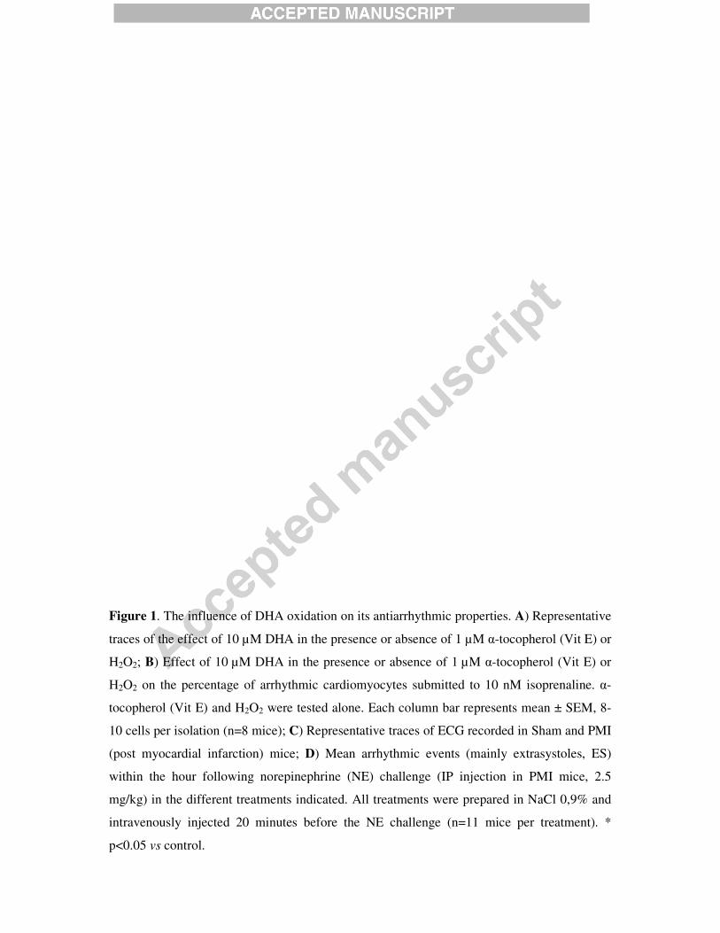
Figure 1. The influence of DHA
traces of the effect of 10 µM DHA in the presence or absence of 1
H2O2; B) Effect of 10 µM DHA in the presence or absence of 1
H2O2 on the percentage of arrhythmic cardiomyocytes subm
tocopherol (Vit E) and H2O2 were tested alone.
10 cells per isolation (n=8 mice);
(post myocardial infarction) mice
within the hour following norepinephrine (NE) chall
mg/kg) in the different treatments indicated. All t
intravenously injected 20 minutes before the NE cha
p<0.05 vs control.
oxidation on its antiarrhythmic properties. A) Representative
µM DHA in the presence or absence of 1 µM �-tocopherol (Vit E) or
µM DHA in the presence or absence of 1 µM �-tocopherol (Vit E) or
on the percentage of arrhythmic cardiomyocytes submitted to 10 nM isoprenaline.
were tested alone. Each column bar represents mean ± SEM, 8
; C) Representative traces of ECG recorded in Sham and PMI
mice; D) Mean arrhythmic events (mainly extrasystoles
within the hour following norepinephrine (NE) challenge (IP injection in PMI mice, 2.5
mg/kg) in the different treatments indicated. All treatments were prepared in Na
intravenously injected 20 minutes before the NE challenge (n=11 mice per treatment). *
) Representative
tocopherol (Vit E) or
tocopherol (Vit E) or
itted to 10 nM isoprenaline. �-
represents mean ± SEM, 8-
Sham and PMI
extrasystoles, ES)
enge (IP injection in PMI mice, 2.5
NaCl 0,9% and
(n=11 mice per treatment). *

Figure 2. AAP of DHA and oxidized DHA in the presence of enzyme inhibito
of different PUFAs enzymatic inhibitors on DHA+H
arrhythmias. Zileuton (1 µM), ketoconazole (3 µM) a
LOX [55], CYP450 [56] and COX
metabolites of soluble epoxide hydrolase (diols) fr
individually or combined (ALL);
induced prevention of cellular arrhythmias. GPx was added before or aft
put in the Tyrode solution. * p<0.05
and oxidized DHA in the presence of enzyme inhibito
of different PUFAs enzymatic inhibitors on DHA+H2O2 induced reduction of cellular
arrhythmias. Zileuton (1 µM), ketoconazole (3 µM) and celecoxib (1 µM) inhibit respectively
and COX-2 [57]. sEH inhibitor (10 nM) prevents the formation of
metabolites of soluble epoxide hydrolase (diols) from EETs [58]. They have been used
individually or combined (ALL); B) Effects of GPx (10 units, see Methods) on DHA+H
f cellular arrhythmias. GPx was added before or after DHA+ H
in the Tyrode solution. * p<0.05 vs control.
and oxidized DHA in the presence of enzyme inhibitors. A) Effects
induced reduction of cellular
nd celecoxib (1 µM) inhibit respectively
. sEH inhibitor (10 nM) prevents the formation of
ey have been used
) Effects of GPx (10 units, see Methods) on DHA+H2O2
er DHA+ H2O2 were
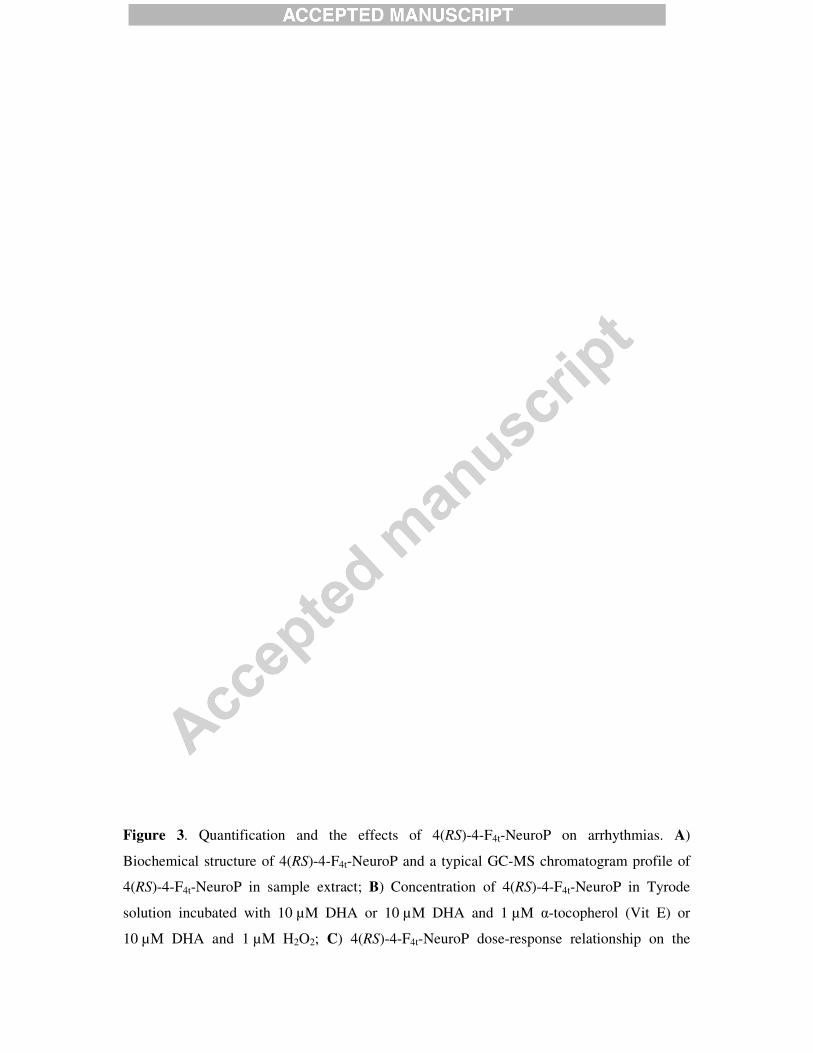
Figure 3. Quantification and
Biochemical structure of 4(RS)-4
4(RS)-4-F4t-NeuroP in sample extract;
solution incubated with 10 µM DHA or 10
10 µM DHA and 1 µM H2O2;
and the effects of 4(RS)-4-F4t-NeuroP on arrhythmias
4-F4t-NeuroP and a typical GC-MS chromatogram profile of
-NeuroP in sample extract; B) Concentration of 4(RS)-4-F4t-NeuroP in Tyrode
µM DHA or 10 µM DHA and 1 µM �-tocopherol (Vit E) or
; C) 4(RS)-4-F4t-NeuroP dose-response relationship on the
arrhythmias. A)
MS chromatogram profile of
-NeuroP in Tyrode
tocopherol (Vit E) or
response relationship on the

percentage of arrhythmic cells; D
following isoproterenol challenge (IP injection in PMI
intravenous injection of 4(RS)-4-
1µM; E) Effects of 4(RS)-4-
arrhythmias. * p<0.05 vs control; ° p<0.05
Figure 4. Calcium cycling of the cardiomyocytes. Concentration of treatments
10 µM DHA, 1 µM �-tocopherol (Vit E), 1
indicates isoprenaline. A) Typical line
from Fluo-4AM loaded cardiomyocytes in different treatments i
calcium sparks measured in A.
measured in Indo-1AM loaded cardiomyocytes, (expressed as the ratio
480 and 405 nm). *p<0.05 vs control; °p<0.05
D) Mean arrhythmic events of extrasystoles within on
lowing isoproterenol challenge (IP injection in PMI mice, 2.5 mg/kg)
-F4t-NeuroP and carvedilol to reach a blood concentrati
-F4t-NeuroP on 10 µM forskolin (Fsk) induced cellular
control; ° p<0.05 vs Fsk.
of the cardiomyocytes. Concentration of treatments
tocopherol (Vit E), 1 µM H2O2 and 1 µM 4(RS)-4-F4t
Typical line-scan confocal images of spontaneous calcium spark
4AM loaded cardiomyocytes in different treatments indicated; B)
A. C) Effects of the different conditions on diastolic calcium
1AM loaded cardiomyocytes, (expressed as the ratio of fluorescence (F) at
control; °p<0.05 vs PMI.
) Mean arrhythmic events of extrasystoles within one hour
mice, 2.5 mg/kg) then by an
-NeuroP and carvedilol to reach a blood concentration of
µM forskolin (Fsk) induced cellular
of the cardiomyocytes. Concentration of treatments indicated are
4t-NeuroP. Iso
scan confocal images of spontaneous calcium spark
Frequency of
t conditions on diastolic calcium
of fluorescence (F) at
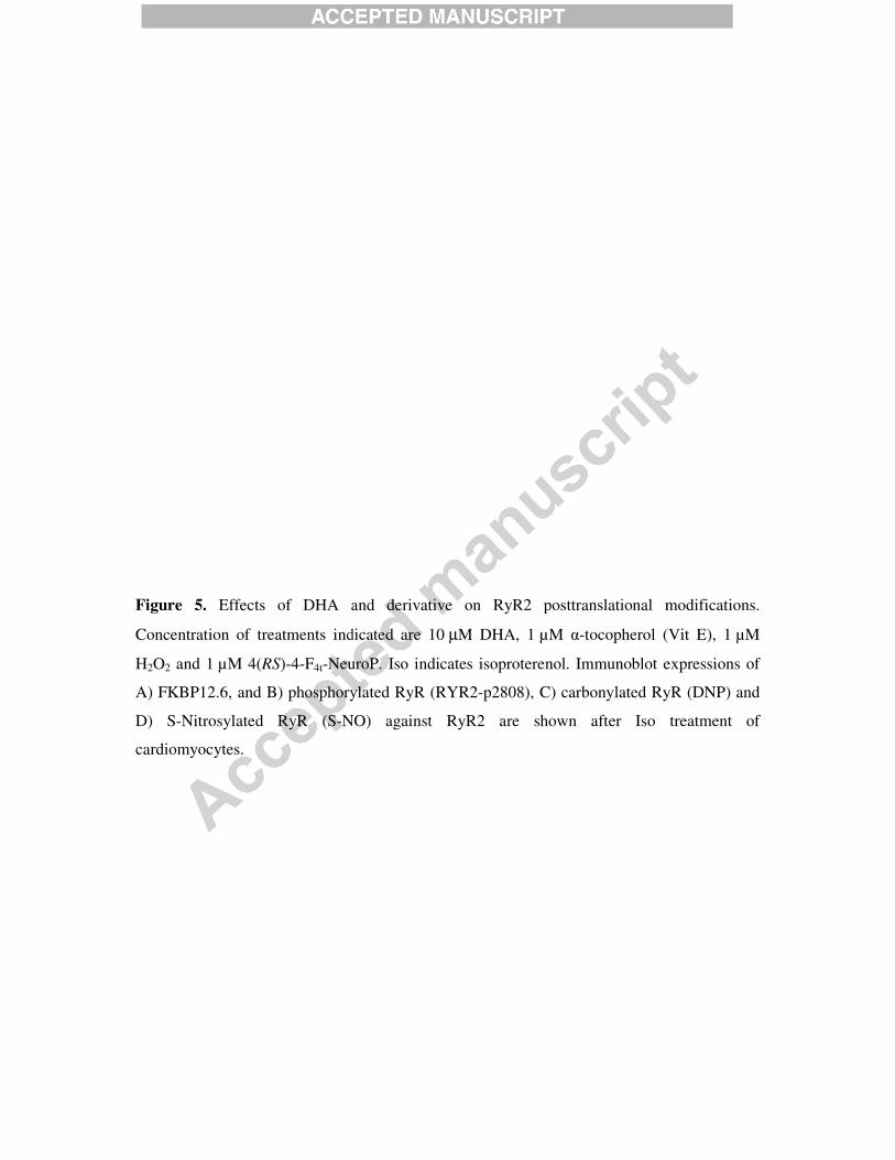
Figure 5. Effects of DHA and derivative on RyR2
Concentration of treatments indicated are 10
H2O2 and 1 µM 4(RS)-4-F4t-NeuroP. Iso
A) FKBP12.6, and B) phosphorylated RyR (RYR2
D) S-Nitrosylated RyR (S-NO)
cardiomyocytes.
Effects of DHA and derivative on RyR2 posttranslational modif
Concentration of treatments indicated are 10 µM DHA, 1 µM �-tocopherol (Vit E), 1
-NeuroP. Iso indicates isoproterenol. Immunoblot expressions of
B) phosphorylated RyR (RYR2-p2808), C) carbonylated RyR
NO) against RyR2 are shown after Iso
posttranslational modifications.
tocopherol (Vit E), 1 µM
indicates isoproterenol. Immunoblot expressions of
C) carbonylated RyR (DNP) and
treatment of
28
Highlights:
- Oxidation of DHA (oxDHA) is a pre-requisite condition for anti-arrhythmic effect.
- 4(RS)-4-F4t-neuroprostane from oxDHA displayed potent anti-arrhythmic properties.
- Anti-arrhythmia effect involves stabilization of the FKPB12.6/RyR2 complex.
- Non-enzymatic metabolites of omega-3 fatty acids have physiological roles.
Gra
ph
ica
l A
bstr
act
(fo
r re
vie
w)

TABLE
SHAM PMI
(n = 6) (n =9)
HR (bpm) 427 ± 29 454 ± 37
EF (%) 60 ± 4 25 ± 12 ***
FS (%) 32 ± 3 12 ± 6 ***
FAC (%) 52 ± 3 19 ± 9 ***
EF (%) B-mode 58 ± 5 12 ± 7 ***
Ao VTI (mm) 49.0 ± 5.8 35.6 ± 4.0 ***
Table 1. Echocardiographic parameters in sham and infarcted mice. HR, heart rate; EF,
ejection fraction; FS, fractional shortening; FAC, fractional area change; AoVTI, aortic
velocity time integral. Data are represented as mean ± SD. *** p<0.001 for SHAM vs. PMI.
Copyright © 2022 FDOKUMEN