Carboxypeptidase E promotes cancer cell survival, but inhibits migration and invasion
New Roles of Carboxypeptidase E in Endocrine and
Neural Function and Cancer
Niamh X. Cawley, William C. Wetsel, Saravana R. K. Murthy, Joshua J. Park,
Karel Pacak, and Y. Peng Loh
Section on Cellular Neurobiology (N.X.C., S.R.K.M., Y.P.L.), Program on Developmental Neuroscience, Eunice Kennedy
Shriver National Institute of Child Health and Human Development, National Institutes of Health, Bethesda, Maryland
20892; Departments of Psychiatry and Behavioral Sciences, Cell Biology, Medicine (Endocrinology) and Neurobiology
(W.C.W.), Duke University Medical Center, Durham, North Carolina 27710; Department of Neurosciences (J.J.P.),
College of Medicine, University of Toledo, Toledo, Ohio 43614; and Program in Reproductive and Adult Endocrinology
(K.P.), Eunice Kennedy Shriver National Institute of Child Health and Human Development, National Institutes of
Health, Bethesda, Maryland 20892
Carboxypeptidase E (CPE) or carboxypeptidase H was first discovered in 1982 as an enkephalin-convertase that
cleaved a C-terminal basic residue from enkephalin precursors to generate enkephalin. Since then, CPE has been
shown to be a multifunctional protein that subserves many essential nonenzymatic roles in the endocrine and
nervous systems. Here, we review the phylogeny, structure, and function of CPE in hormone and neuropeptide
sorting and vesicle transport for secretion, alternative splicing of the CPE transcript, and single nucleotide
polymorphisms in humans. With this and the analysis of mutant and knockout mice, the data collectively support
important roles for CPE in the modulation of metabolic and glucose homeostasis, bone remodeling, obesity,
fertility, neuroprotection, stress, sexual behavior, mood and emotional responses, learning, and memory. Re-
cently, a splice variant form of CPE has been found to be an inducer of tumor growth and metastasis and a
prognostic biomarker for metastasis in endocrine and nonendocrine tumors. (Endocrine Reviews 33:
0000–0000, 2012)
I. Introduction to Functions of CPEA. Discovery of CPE as a prohormone-processing
enzymeB. Role of CPE in prohormone sorting, vesicle trans-
port, and secretionC. Biomedical implications of CPE in physiological
function and diseaseII. The CPE Gene, Biosynthesis, Protein Structure, and
DistributionA. Phylogenic analysis of CPEB. Structure, biosynthesis, and intracellular traffick-
ing of CPEC. Distribution of CPE in embryonic and adult tissuesD. Biochemical and enzymatic properties of CPE
III. CPE in Prohormone Sorting and Vesicle TransportA. CPE function as a prohormone sorting receptor for
the regulated pathwayB. CPE mediates post-Golgi hormone vesicle transportC. CPE mediates synaptic vesicle localization to nerve
terminal preactive zoneIV. CPE Action in the Endocrine System—Insights from
Mouse Models
A. Diabetes in CPE-deficient miceB. Obesity in CPE-deficient miceC. Bone metabolism in CPE-deficient miceD. Infertility and poor sexual performance in CPE-
deficient miceV. CPE in Neural Function and Behavior
A. Aberrant neurotransmission and dendritic archi-tecture in CPE KO mice
B. Role of CPE in stress and neuroprotectionC. Regulation of mood and emotional responses in
CPE miceD. Deficits in learning and memory in CPE KO mice
ISSN Print 0021-972X ISSN Online 1945-7197
Printed in U.S.A.
Copyright © 2012 by The Endocrine Society
doi: 10.1210/er.2011-1039 Received August 25, 2011. Accepted January 18, 2012.
Abbreviations: AGRP, Agouti-related peptide; BDNF, brain-derived neurotrophic factor;
BMD, bone mineral density; CART, cocaine- and amphetamine-regulated transcript;
Caspr2, contactin-associated protein 2; CCK, cholecystokinin; CPE, carboxypeptidase E;
CPE-�N, spliced variant of CPE without its N-terminus; CSP, constitutive secretory pathway;
E, embryonic day; EAE, experimental autoimmune encephalomyelitis; ER, endoplasmic
reticulum; EST, expressed sequence tag; FH, familial hyperproinsulinemia; GEMSA, gua-
nidinoethylmercaptosuccinic acid; GFP, green fluorescent protein; HCC, hepatocellular
carcinoma; IHC, immunohistochemistry; KO, knockout; LDCV, large dense-core vesicle;
LTP, long term potentiation; MC4R, melanocortin 4 receptor; MEN2, multiple endocrine
neoplasia type 2; NOS1AP, nitric oxide synthase 1 adaptor protein; NPY, neuropeptide Y;
PC, prohormone convertase; PGL, paraganglioma; PHEO, pheochromocytoma; POMC,
proopiomelanocortin; RER, rough ER; RPE, retinal pigmented epithelium; RSP, regulated
secretory pathway; SDHD, succinate dehydrogenase subunit D; siRNA, small interfering
RNA; SLMV, synaptic-like microvesicle; SNP, single nucleotide polymorphism; SV, synaptic
vesicle; T2DM, type 2 diabetes mellitus; TGN, trans Golgi network; TIRF, total internal
reflection fluorescence; T/N, tumor vs. normal surrounding tissue; UTR, untranslated re-
gion; WT, wild-type.
R E V I E W
Endocrine Reviews, April 2012, 33(2):0000–0000 edrv.endojournals.org 1
Endocrine Reviews. First published ahead of print March 7, 2012 as doi:10.1210/er.2011-1039
Copyright (C) 2012 by The Endocrine Society
VI. CPE and Cancer in HumansA. Splice isoform of CPE (CPE-�N) promotes tumor
growth and metastasisB. CPE/CPE-�N as a diagnostic and prognostic bio-
marker for (neuro)endocrine and nonendocrinecancers
VII. Human CPE Genetic Mutations and DiseaseA. Single nucleotide polymorphisms in human CPE
associated with diseaseB. CPE-associated human disease
VIII. ConclusionsIX. Future Directions
I. Introduction to Functions of CPE
Although discovered in 1982, carboxypeptidase E (CPE)has remained a molecule of keen interest to many in-
vestigators. In addition to its carboxypeptidase activity, nu-merous studies over the last 1.5 decades have indicatedthat CPE is a multifunctional protein that plays manynonenzymatic roles in the endocrine and nervous sys-tems. This review presents a comprehensive look at CPEfrom structure to function and disease, with a focus onnew roles that this unique protein plays in many phys-iological systems.
A. Discovery of CPE as a prohormone-processing enzyme
Processing of propeptides often begins with the endo-proteolytic cleavage at paired or sometimes at single basicamino acid residues. Since the discovery of the prohor-mone convertases (PC enzymes), which cleave prohor-mones between or on the carboxyl side of pairs of basicresidues (1), it became clear that another enzyme wasneeded to remove the basic residues from the intermedi-ates to produce the mature bioactive hormone. CPE,which was first identified as enkephalin convertase (2, 3),was subsequently found to be the enzyme responsible forcleaving the C-terminally extended basic residues frompeptide intermediates in endocrine cells and neuropep-tides in peptidergic neurons. CPE differed from other car-boxypeptidases in that its optimal pH was in the acidicrange, consistent with its localization to acidic compart-ments of the trans Golgi network (TGN) and to dense coresecretory granules of endocrine cells and peptidergic ves-icles of neurons where processing occurs. CPE is localizedprimarily to endocrine tissues and to specific areas of thecentral nervous system. The importance of CPE as a pro-cessing enzyme was further realized when a mutation inthe Cpe gene was found in the Cpefat/fat mouse that pre-sented with severeobesity, diabetes, and infertility. Studieson the structure, biosynthesis, forms, tissue distribution,and enzymatic properties of CPE are discussed in Sections
II.A to II.D.
B. Role of CPE in prohormone sorting, vesicle transport,
and secretion
Prohormones/propeptides are synthesized in the roughendoplasmic reticulum (RER) and then inserted into theRER cisternae. From there the precursors are transportedin vesicles to the Golgi apparatus where they are sorted atthe TGN into budding granules along with their process-ing enzymes such as the PCs and CPE, as well as othercargo proteins such as the granins [for review, see Bar-tolomucci et al. (4)]. The precursors are then processedwithin the secretory granules en route to the storage andrelease sites at the periphery of the cell. Intracellular trans-port of proteins to various organelles has been found to bemediated by sorting signal motifs and respective recep-tors. For example, the KDEL signal motif and KDELreceptors mediate the retention of endoplasmic reticu-lum (ER) resident proteins in the ER (5). The search forsorting motifs for targeting prohormones/propeptidesat the TGN into the granules of the regulated pathwayhas been challenging. The identification of sorting sig-nal motifs on these precursors and evidence supportinga role for a membrane form of CPE as a sorting receptorare discussed in Section III.A.
Prohormone-containing granules that have buddedoff from the TGN are transported to the plasma mem-brane via microtubule- and actin-based transport sys-tems. In Section III.B, the role of a transmembrane formof CPE in facilitating the post-Golgi transport of thesegranules to the cell periphery is discussed. The cyto-plasmic tail of the transmembrane form of CPE appearsto interact with microtubule motors, actin, and othercytoskeletal proteins to enable granules to be trans-ported via the regulated secretory pathway (RSP) to theplasma membrane for exocytosis.
Recently, the transmembrane form of CPE was alsofound in synaptic vesicles (SV) in a subset of hypothalamicneurons. The localization of these vesicles to the activezone of the synapse for neurotransmitter release wasshown to be dependent on the interaction of the CPE cy-toplasmic tail with cytoskeletal proteins. The significanceof CPE in facilitating SV localization to the synapse isdiscussed in Section III.C.
C. Biomedical implications of CPE in physiological
function and disease
Much has been learned about the physiological func-tions and disease states caused by the lack of CPE fromCpefat/fat (6) and knockout (KO) mice (7). In Sections IV
and V, insights gained from using these mouse models arediscussed. These include revealing the role of CPE in obe-sity, bone remodeling, diabetes, reproduction, neuropro-tection, mood, and emotional responses.
2 Cawley et al. Carboxypeptidase E in Endocrine and Neural Function Endocrine Reviews, April 2012, 33(2):0000–0000

More recently, a splice variant form of CPE was dis-covered that had activity in promoting the growth andmigration of cancer cells (8). Unlike wild-type (WT) full-length CPE, this splice isoform (i.e., CPE-�N) lacks thesignal peptide at the N terminus that normally directs itinto the secretory pathway. It exists in the cytoplasm andin metastatic tumor cells. CPE-�N moves into the nucleusand functions to activate metastatic and antiapoptoticgenes, thus inducing or promoting tumor metastasis. Useof CPE-�N as a biomarker for diagnosing and predictingfuture metastasis in several types of endocrine and epithe-lial cancers is discussed in Section VI.
Several single nucleotide polymorphisms (SNP) occur-ring in the CPE gene have been found in humans. Someresult in the loss of enzymatic activity as identified in typeII diabetic patients, whereas others lead to unstable mol-ecules that are misfolded and degraded in the ER (see Sec-
tion VII).We conclude the review with a summary of the new
nonenzymatic roles for CPE, its splice isoform CPE-�N,and discuss the mechanisms by which CPE controls func-tions in health and disease, and the potential use of CPE asa therapeutic drug target (Section IX).
II. The CPE Gene, Biosynthesis, ProteinStructure, and Distribution
A. Phylogenic analysis of CPE
CPE falls into the peptidase M14-like superfamily ofenzymes (9). The M14 family of metallocarboxypepti-dases is a group of zinc-binding carboxypeptidases thathydrolyze single, C-terminal amino acids from poly-peptide chains and have a recognition site for the freeC-terminal carboxyl group, a key determinant of spec-ificity. Based on substrate specificity, CPE is classifiedinto carboxypeptidase B-like (CPB-like) enzymes be-cause it only cleaves the basic residues lysine or arginine.Various metallocarboxypeptidase proteins containing aZn-carboxypeptidase domain have been highly conservedfrom bacteria to mammals (10, 11). To date, 23 genesencoding Zn-carboxypeptidase domain proteins havebeen identified in the human genome (10). Comparison ofthe sequence of CPE with carboxypeptidase A (A1, A2,A3, A4, A5 and A6) and carboxypeptidase B (B1 and B2)show a 21% identity at the protein level with 55% and37% sequence coverage, respectively. CarboxypeptidaseO is 22% identical at the protein level with just 32% se-quence coverage. Additionally, carboxypeptidase M is46% identical at the protein level with 82% sequence cov-erage, and carboxypeptidase N1 is 51% identical at theprotein level with 86% sequence coverage. Interestingly,
carboxypeptidae D has multiple Zn-carboxypeptidase do-mains and is 50% identical at the protein level with 86%sequence coverage. There is very little conservation out-side the Zn-carboxypeptidase domain between CPE andCPA/B proteins, suggesting that these proteins divergedvery early during evolution.
The orthologous protein sequences of the Cpe genehave been identified in many species, covering a phyloge-netic distance from invertebrate Protostomia (Ecdysozoa,nematodes) to vertebrate Deuterostomia. The phyloge-netic relationships, as estimated from amino acid sequencesimilarities, are shown in a cladogram tree (Fig. 1). Ac-cording to this tree, the CPE sequences of Caenorhabditis
elegans (nematode) were found at separate branches fromthose of the other species. CPE has been biochemicallycharacterizedonly in a fewspecies so far. Itwas first clonedfrom bovine adrenal chromaffin granules (12) and hasbeen well studied in mouse (Mus musculus) (6) and in rat(Rattus norvegicus) (13–15). Human CPE was first char-acterized by Manser et al. (16). CPE was also studied inchicken (Gallus gallus), thymus (17), and in nematode (C.
elegans) (18). Subsequently, by blast search and data min-ing, CPE was also found to exist in genome sequences ofthe chimpanzee (Pan troglodytes) (19), macaque (Macaca
fascicularis) (20), boar (Sus scrofa) (21), clawed frog (Xe-
nopus tropicalis) (22), sea-slug (Aplysia californica) (23),and zebrafish (Danio rerio) (24) (Table 1). The remark-able degree of similarity at the protein level of CPE acrossthe different phyla suggests its functional importance earlyin evolution.
Alternative splicing of CPE transcripts
In mammals, the CPE gene contains nine exons (Fig. 2)(13–16). In humans, two alternatively spliced transcriptsof CPE mRNA have been found, one of which encodes atruncated protein lacking a partial N-terminal region(CPE-�N) due to alternative splicing of the first exon (Fig.2) (8). CPE-�N is alternatively spliced with noncanonicalalternative 3� and 5� splice sites, which remove 98 nucle-otides of the first exon. This type of noncanonical splicesite is more inclined to occur at a 5� untranslated GC-richregion (25–28) and is common in certain cancers (29, 30).Interestingly, the spliced region also contains G-quadru-plex-like elements (31); these elements are thought tomodulate gene expression at the translational level byforming stable RNA hairpin secondary structures in the 5�
untranslated region (UTR) of mRNA inhibiting the pro-cess of translation (32). Splicing of such elements found inthe CPE-�N transcript could result in a translational “ad-vantage” during tumor metastasis (see Section VI). Thesecond splice variant CPE mRNA transcript is derivedfrom alternative splicing at the 3� donor site of exon 6 and
Endocrine Reviews, April 2012, 33(2):0000–0000 edrv.endojournals.org 3

the 5� donor site of exon 7 (Fig. 2, variant 1). This splicingevent results in an 18-amino acid deletion within the areaof the active site of the CPE protein, rendering this proteinenzymatically inactive. This variant has a signal peptideand is therefore translocated into the RER cisternae andthe secretory pathway. It is likely secreted into the extra-cellular space and may function as a ligand or signalingmolecule. The evidence for these splice variants was derivedfrom expressed sequence tag (EST) database searches. Inter-estingly, all these variants were found in human brain tissue,suggesting that the brain contains cells that are active in al-ternative splicing of CPE (33, 34).
B. Structure, biosynthesis, and intracellular
trafficking of CPE
A schematic of the primary structure of the CPE proteinis shown in Fig. 3A. It has a signal peptide directing theprotein into the RER cisternae, a catalytic domain, and ahighly acidic C-terminal domain. The three-dimensionalstructure of CPE (Fig. 3B) has been modeled based on thecrystal structure of carboxypeptidase D, an enzyme ho-mologous to CPE (35). The model (Fig. 3B) indicates sev-eral functional domains of CPE: the enzymatic active siteshowing the zinc (cofactor) binding site; a prohormone sort-ing signal binding site (see Section III.A), an amphipathic �-helical transmembranedomain, and thecytoplasmic tail thatinteracts with microtubule proteins for vesicle transport (seeSection III.B).
CPE is a 476-amino acid protein syn-thesized as a precursor with a 25-aminoacid signal peptide that directs proCPEinto the cisternae of the RER and is thenremoved. The proCPE is transportedfrom the ER through the Golgi complexto the granules of the RSP wherethe 17-amino acid “pro” region is re-moved after a penta-arginine sequence(RRRRR42) (Fig. 3A), to generate themature protein (CPE43–476) (36). Pro-cessing of the pro region is required nei-ther for enzymatic activity becauseproCPE is enzymatically active as a car-boxypeptidase (37) nor for intracellulartrafficking of CPE (38). The CPE proteinis glycosylated at two N-linked glycosyl-ation consensus sites, Asn139 and Asn390.Under mildly acidic conditions and in-creasing calcium concentrations similarto that of the TGN, CPE has been shownto aggregate in vitro (39) with granulecargo proteins (40), suggesting that thisoccurs in vivo as a mechanism of conden-
sation and sorting to the RSP. Binding of potential prohor-mone cargo to CPE via a prohormone sorting signal, such asthat found inproopiomelanocortin (POMC)(41), couldalsooccur in this compartment (42) through interaction of itsprohormone binding site, composed of the amino acidsArg255 and Lys260 on CPE (43). In addition, the carboxylterminus of CPE forms an amphipathic �-helix under acidicconditions and is involved in binding tightly to cellular mem-branes (44, 45). This binding step is important for its traf-ficking from the TGN to the granules of the RSP (46, 47).Binding of the carboxyl terminus of CPE with cholesterol-sphingolipid-rich domains (lipid-rafts) in the TGN mem-brane has been demonstrated (48), allowing it to act as asorting receptor for prohormones (see Section III.A). A sub-population of CPE molecules appears to have a transmem-branetopology(49).CPEtogetherwithboundprohormonesare packaged into immature granules budding from theTGN. Indeed, analysisof secretorygranulemembranes frombovine pituitary indicates that it is highly enriched with cho-lesterol, consistent with the notion that granules are buddedfrom lipid raft domains of the TGN (50). Within these gran-ules, some of the CPE molecules are further processed (51) atArg455-Lys456 (Fig. 3A) to yield a soluble form (molecularmass, �50 kDa) (51), which is enzymatically more activethan the membrane-associated form (molecular mass, �53kDa) (52). Soluble CPE then functions to cleave the C-ter-minal extended basic residues from the peptide hormone in-termediates liberated by PC1 and PC2 in the granules. Sub-
Figure 1.
Figure 1. Phylogenetic analysis of CPE protein. The phylogenetic tree was built using 476
representative amino acids using Phylogeny.fr platform (www.phylogeny.fr/version2_
cgi/phylogeny.cgi) and determined by the program Gblocks (328), which eliminates poorly
aligned positions and divergent regions (removes alignment noise) after sequence alignment
using multiple sequence comparison by log-expectation (MUSCLE) (329). Bootstrap values
above 50% (0.5) are shown. Conserved position for at least half the number of sequences is
represented by �1.
4 Cawley et al. Carboxypeptidase E in Endocrine and Neural Function Endocrine Reviews, April 2012, 33(2):0000–0000

sequent to granule exocytosis, membrane CPE can recyclefrom the plasma membrane through the early endosomesand back to the TGN where it gets reused (53, 54).
Much debate has surrounded whether the membraneform of CPE does and can assume a transmembrane to-pology in the granules membrane. For instance, the pri-mary structure of CPE at the C terminus that associateswith the membrane does not contain a typical transmem-brane domain as predicted from modeling programs (45)and as shown in biochemical experiments (45, 46, 51) thatinvolved carbonate extraction studies (45) and secretionexperiments (46). Additionally, the C-terminal membrane
binding domain is highly acidic (Fig. 3A) and theoreticallydoes not favor localization in a membrane lipid bilayer.However, studies on the insertion of a CPE C-terminalpeptide (last 22 amino acids) into model membranes haveindicated that it can shallowly embed in a lipid bilayer byitself at an acidic pH (49). Also, evidence from cell bio-logical experiments strongly support the existence of acytoplasmic tail in CPE that interacts with various cyto-plasmic molecules such as dynactin to mediate granuletransport in neuroendocrine cells (see Section III.B). In ad-dition, after granule exocytosis, the C-terminus cytoplasmictail of CPE has been shown to interact with Arf6, a small
Figure 2.
Figure 2. Schematic representation of the CPE gene and alternatively spliced variants. Rectangular boxes denote the exons (1–9). Dark solid boxes
are the UTR, and light and textured boxes are the coding exons. Alternative splice sites to yield hCPE-�N and variant 1 transcripts are indicated
with arrows on the exons. Stop codons are also indicated with arrows and introns as solid lines.
TABLE 1. Different species with a conserved CPE gene
Species
Nucleotide sequence
accession no.
Nucleotide
identities (%)
Protein
ChromosomeIdentities (%) Similarities (%)
Homo sapien (man) NM_001873 100 100 100 4q32.3
Pan troglodytes (common chimpanzee) NM_001098559 99 99 99 4
Macaca fascicularis (crab-eating macaque) AB169871 98 99 99 4
Bos taurus (cow) NM_173903.3 88 93 96 17
Sus scrofa (boar) NM_001097439.1 87 92 95 8
Mus musculus (mouse) NM_013494.3 88 97 98 8, 32.6 cM
Rattus norvegicus (rat) NM_013128.1 88 96 98 16p13
Gallus gallus (chicken) CR388992.1 85 94 97 4
Danio rerio (zebrafish) NM_214810.1 53 83 94 1
Xenopus laevis (African clawed frog) NM_001127813.1 78 83 92
Aplysia californica (sea slug) NM_001204485 11 47 65
Caenorhabditis elegans (nematode) NM_069534.5 9 44 60 4
All the species except Macaca fascicularis and Gallus gallus have annotated gene entry in the NCBI database. The percentage identity and similarities were compared
with human mRNA and protein. Chromosome loci for the Cpe gene have not been assigned for Xenopus laevis and Aplysia californica. cM, Centimorgan.
Endocrine Reviews, April 2012, 33(2):0000–0000 edrv.endojournals.org 5

cytoplasmic GTPase, to mediate recycling of CPE from theplasma membrane back to the TGN for reuse (53). Further-more, in another study, it was demonstrated that Arf6-dependent recycling of CPE mediated the endocytosis of theeosinophil cationic protein, a CPE-interacting protein (54).All these studies indicate the existence of a cytoplasmic tailand a transmembrane orientation of some CPE.
There are two possibilities by which CPE could achievea transmembrane orientation. One is the insertion of theC terminus of CPE, under acidic conditions, through theTGN membrane with the help of a chaperone protein thatcould shield the acidic charges of the CPE C terminus in thelipid bilayer, allowing it to penetrate the membrane. As anexample of this kind of mechanism, the diphtheria toxin�-subunit only partially penetrates artificial lipid bilayers
by itself but is able to move across the membrane upon in-troduction of globule-like proteins as a chaperone (55). Inaddition, whereas it is known that C-terminal tail-anchoredproteins (e.g., cytochrome b5) are transmembrane proteins,the mechanism by which large domains of polypeptide aretranslocated across the phospholipid bilayer in an unassistedmanner is still not fully understood because it is independentof the Sec61 translocon (56, 57). Of particular interest is thefinding that CPE specifically interacts with Wolframin (58),an ER resident protein of unknown function, but may beinvolved in protein folding and intracellular transport.Mutations in the WFS1 gene cause Wolfram syndrome—asyndrome characterized by diabetes insipidus, childhood-onset diabetes mellitus, optic atrophy, and deafness. Ad-ditional supporting evidence for a functional partnership
Figure 3.
A
B
Figure 3. Schematic diagram and molecular model of the CPE protein. A, The preproCPE protein is 476 amino acids in length and contains a signal
peptide (SP) that directs it into the ER. After cleavage of the SP, the proCPE (57 kDa) is trafficked through the Golgi and sorted into the granules of
the RSP via interaction of its C-terminal amphipathic �-helical domain with cholesterol-sphingolipid-rich microdomains in the TGN. The Pro region
(Pro) is processed within a post-Golgi compartment to generate the mature full-length membrane-bound CPE (55 kDa). Within the granules, the C
terminus of this membrane-associated CPE can be cleaved presumably by a PC at a paired-basic residue cleavage site to generate a soluble form of
mature CPE (53 kDa). B, The molecular model of CPE was based on the crystal structure of CPD. The red areas indicate the common overlapping
homologous sequences shared between the two proteins, demonstrating a high degree of structural similarity. Unique to CPE are the two basic
residues, Arg255 and Lys260, that were demonstrated to interact with the acidic residue-based prohormone sorting-signal found in POMC,
proinsulin, and proBDNF. CPE also contains a unique C-terminal sequence that forms an amphipathic �-helix under acidic conditions. This C-
terminal region is involved in tight membrane association and for a subset of CPE molecules can traverse the lipid bilayer, resulting in a small
cytoplasmic tail that interacts with cytoplasmic proteins such as Arf6 and dynactin. The green ball represents the zinc atom in the active site.
Lollipop symbols indicate the asparagine-linked glycosylation sites.
6 Cawley et al. Carboxypeptidase E in Endocrine and Neural Function Endocrine Reviews, April 2012, 33(2):0000–0000
between CPE and Wolframin derives from the observationthat both CPE and Wolframin are co-up-regulated in theamygdala of male rats subjected to cat odor (fear response)(59). These observations raise the possibility that Wol-framin may assist CPE in its folding and/or trafficking asa chaperone through the ER to allow CPE to functionefficiently downstream. Another report has noted the in-terdependence of CPE and phogrin, a receptor tyrosinephosphatase-like protein found in mature secretorygranules of AtT20 cells (60). Under the mildly acidicenvironment of the TGN (61), CPE can interact with theN-terminal luminal domain of phogrin. Additionally,small interfering RNA (siRNA) silencing of CPE or pho-grin reduces sorting of phogrin and CPE, respectively,into granules. Hence, the interaction of CPE with pho-grin in the TGN may, in part, facilitate and stabilize anonclassical transmembrane domain of CPE in thatcompartment.
The second possibility is that CPE does not fully enterthe ER cisternae during synthesis because the C terminusis retained by an interacting protein on the outside of theorganelle, leaving a cytoplasmic tail exposed (62). Under-standing how CPE assumes a transmembrane orientationawaits further studies.
C. Distribution of CPE in embryonic and adult tissues
After the initial identificationandcharacterizationofCPE(2,3),antiseraspecific forCPEbecameavailable. Initial stud-ies were performed by immunohistochemistry (IHC) (63,64), autoradiography, and binding studies using tritiatedguanidinoethylmercaptosuccinic acid (GEMSA), a potentinhibitor of CPE (65–69). The IHC studies in rats showed ageneral localization of CPE in neuropeptide-rich areas of thebrain and endocrine tissues such as the median eminence,supraoptic nucleus, paraventricular nucleus, and suprachi-asmatic nucleus of the hypothalamus; the neural, intermedi-ate, and selected cells in the anterior lobe of the pituitary; andthe bovine adrenal medulla. Staining was also evident in thepyramidal neurons of the hippocampus, dentate gyrus, andamygdala. Using radiolabeled GEMSA, staining was re-ported in the rat epithelial cells of the stomach, colon, ovi-duct, and the acinar cells of the submandibular gland as wellas the pancreatic islets of Langerhans (70) and the adrenalmedulla (67). Staining in the rat heart atrial tissue (71, 72)also suggests a role for CPE in the physiology of atrial na-triuretic factor in this organ. Other IHC studies have re-ported the expression of CPE in somatostatin-producingcells in rat brain (15); the gastrin cells and progenitor gastrin-somatostatin cells of the antropyloric mucosa of the gut inrats (73);and indifferentareasof the lungaspartofanopioidnetwork involved in respiratory function inhumans (74,75).
Subsequent to cloning of bovine and rat Cpe cDNA (12,76), oligonucleotide probes were used for Northern blotand in situ hybridization. The expression patterns of CPEhave been studied extensively in the rat brain, during em-bryonic development (77), and in the adult (78–80). In theadult brain, Cpe mRNA is highly expressed in pyramidalneurons of the hippocampus, amygdala, supraoptic nu-cleus, paraventricular nucleus, and ependymal cells of thelateral ventricle. Other areas of the brain include the piri-form and entorhinal cortex, cerebellar cortex, thalamus,medial geniculate, and lateral septal nuclei. High expres-sion is also found in the anterior and intermediate lobes ofthe pituitary, the adrenal medulla (77, 79), and pancreaticislets. With the characterization of fat cells as an endocrinetissue (81), CPE has also been found in sc and visceral fat(82), although its role in this tissue is unclear at this time.
During development, Cpe mRNA is first seen at em-bryonic day (E) 10, specifically in the diencephalon andspinal cord in rats (77). Because PC1 and PC2 are notexpressed at this time nor is there a defined endocrinesystem, the role that CPE plays in these tissues at thisdevelopmental stage is unknown. It is possible that otherPC-like enzymes not identified here are present; or furin,a ubiquitously expressed PC found in the TGN involved inprocessing constitutively secretedproproteins, could func-tion upstream of CPE to provide proprotein intermediatesas substrates for CPE. However, the furin expression pat-tern at this stage does not overlap with that of CPE. ByE12, CPE expression is seen more extensively throughoutthe embryo, specifically in thenervous system in areas suchas the neuroepithelium, peripheral ganglia, mesenchymalcells around the midgut mesentery, and the epithelia of thebranchial arch. At E13, whereas the expression of PC1 andPC2 transcripts is restricted to the developing nervous sys-tem, high levels of CPE expression are seen throughout theembryo that overlap with PC1 and PC2 expression in ad-dition to that for furin. There have been many other char-acterizations of CPE in specific cells/tissues under differentexperimental paradigms that can be found in Table 2.
D. Biochemical and enzymatic properties of CPE
CPE is a Zn�� metallocarboxypeptidase that cleavesthe carboxy-terminal arginine (Arg) or lysine (Lys) resi-dues from protein substrates; however, it can also cleavehistidine poorly under acidic conditions. CPE prefers Argover Lys residues with KM values at approximately 50–100 �M and approximately 200 �M, respectively, for Met-enkephalin extended peptides (2). CPE has an optimumactivity at pH 5–6 with the Vmax declining sharply at pHbelow 5.0 due to a single ionizing group in the active site(83). Multiple ionizing groups in the active site at pHabove 7.0 also have deleterious effects on activity such that
Endocrine Reviews, April 2012, 33(2):0000–0000 edrv.endojournals.org 7

CPE becomes inactive at pH 7.4. Aside from pH require-ments, CPE utilizes zinc as the coordinating metal in itsactive site to mediate peptide hydrolysis (84). Cobalt, an-other divalent transition-state metal, can stimulate enzy-matic activity (2). Although calcium can bind CPE (85)and this binding decreases the thermostability of the en-zyme (85), it exerts no effects on the aggregation of thesoluble CPE (39). CPE is a metallocarboxypeptidase;therefore, chelating agents, such as 1,10-o-phenanthro-line, are effective inhibitors. Several thiol-directed inhib-itors are also highly effective, such as p-chloromercuriph-
enylsulfonate and HgCl2 (86). In addition, mimeticsubstrates designed as active site-directed inhibitors, e.g.,GEMSA, with a Ki of approximately 8 nM (87), are usedas potent inhibitors.
The importance of CPE in peptide hormone and neu-ropeptide processing has become especially evident withthe CPE-deficient mice. Specifically, using a peptidomicsapproach comparing WT and Cpefat/fat mice, new poten-tial substrates of CPE have been identified (88, 89). In-deed, identification of proSAAS, an endogenous inhibitorof PC1/3, was made by this approach (90), as was the
TABLE 2. Literature summary of the identification or expression of CPE in various tissue and cell systems
Tissue/cell Experimental context Experimental procedure Ref.
Mouse cDNA library Screen for caspase substrates Cleavage of expressed proteins 337
Corpus luteum Changes during luteal phase Gene array, qRT-PCR 338
Dorsal root ganglia Effect of monensin Activity 339
Eye ciliary body Ciliary epithelium as a neuroepithelium Expression and subtractive libraries 203
H4-II-E-C3 hepatoma Angiotensin II processing RT-PCR 340
Rat CNS Compare CPE and CPD ISH, IHC 341
Breast cancer cells Vasopressin processing qRT-PCR, Western blot 342
Pituitary neurointermediate lobe Regulation of expression Northern blot 343
Testicular and epididymal transcriptomes Analysis of epididymal segments cDNA microarray 344
Bovine DNA SNP analysis for meat quality RFPL sequencing 345
Gastric enterochromaffin-like cells Components of vesicle release IHC 251
Antropyloric mucosa Gastrin localization IHC 73
Nervous system/mouse model of MS Development of disease state Oligonucleotide microarrays 325
RPE/choroid Age-related changes in genes cDNA microarray, RT-PCR 204
Intestine 407, human fetal epithelial cells Overexpression of NeuroD cRNA microarray 346
C. elegans neuromuscular junction Impaired acetylcholine release Paralysis assay 18
Pancreatic �-cells Palmitate-induced apoptosis Proteomics, Western blot 238
Brain Global ischemia Western blot 231
Lungs CPE fat/fat mouse Response to ozone 347
Airway responsiveness in mice Effect of ozone inhalation Compare CPE fat/fat mice to WT 348
Olfactory bulb, amygdala Rat exposure to cat odor Differential gene expression 59
Synaptic vesicles and PC12 cells Characterization EM, TIRF, subcellular organelles 142
Lung epithelium Effect of smoke inhalation cDNA microarray 349
Seminal plasma Identification of proteins LC-MS/MS 350
Immunocytes Inflammation/pain IHC 351
Rats Alcohol effects on behavior CPE activity 352
Multiple cancer tissues Review Database mining 247
Cultured lens tissue Calcium influx by ionophore 2D-PAGE/MS 353
Small-cell carcinoma Peptide processing qRT-PCR, Western blot 253
N/A PI3K-mTOR pathway analysis Yeast-2-hybrid, interactome mapping 354
Prefrontal cortex of piglets Social isolation stress Microarray, qRT-PCR 235
Cat visual cortex Young vs. adult cats Subtractive hybridization 355
Avian pancreas Embryonic development IHC 356
Placenta, umbilical cord Compare CPD and CPE IHC 357
Ovary FSH-responsive cells cDNA microarray 358
Neural complex Analysis in chordates EST sequencing, ISH 359
Placenta Stages of gestation ISH 360
Retinal tissue/cells Processing of NPY Activity, ICC, Western blot 361
RGC-5 retinal cells Ischemia Western blot, activity 362
Cultured astrocytes and neurons Secretion and tissue analysis CPE activity and Northern blots 363
Basophilic mast cells/Jurkat cells Secretory vesicles ICC 364
Chicken thymus Colocalization with CgA RT-PCR, Western blot, IHC 17
Brain Ischemia ISH, ICC, Western blot 232
Retinal photoreceptors CPE KO and fat/fat mouse IHC 141
Table includes published literature where CPE expression was identified and/or characterized in a wide variety of tissues and cell systems. The experimental context and
technique of the characterization of CPE is annotated. Some citations made in the text may be duplicated here. CNS, Central nervous system; ISH, in situ hybridization;
qRT-PCR, quantitative real time PCR; ICC, immunocytochemistry; LC/MS, liquid chromatography/mass spectroscopy; EM, electron microscopy; 2D-PAGE,
two-dimensional PAGE; N/A, not applicable; MS, mass spectroscopy; PI3K-mTOR, phosphatidylinositol 3-kinase-mammalian target of rapamycin.
8 Cawley et al. Carboxypeptidase E in Endocrine and Neural Function Endocrine Reviews, April 2012, 33(2):0000–0000

discovery of the involvement of CPE in the processing ofhemopressins (91)—hemoglobin peptides found in thebrain that bind cannabinoid CB1 receptors (92). Otherstudies include WT and Cpefat/fat mice comparisons ofpeptides in the prefrontal cortex, pituitary, and brain (93–95). Additional effects of food deprivation and exercise onhypothalamic peptides have been investigated (96). Pep-tidomic analyses have also been applied to hypothalamusand hypothalamic responses to chronic morphine (97) andcocaine treatments (98) to study the possible role of neu-ropeptides in drug addiction.
III. CPE in Prohormone Sorting andVesicle Transport
Endocrine and neuroendocrine cells have two differentsecretory pathways: constitutive and regulated. The con-stitutive secretory pathway (CSP) supports continuousprotein secretion, independent of stimulation (99). In theCSP, small secretory vesicles formed at the TGN are con-stantly transported to and fused to the plasma membranewithout storage. Because CSP proteins are continuouslysecreted, this pathway is driven primarily by the biosyn-thesis of secretory proteins at the ER (100). The CSP pro-vides membrane proteins, such as receptors (101), to theplasma membrane, as well as various secretory proteins(102, 103), for maintenance of cell survival, differentia-tion, and growth. Although the CSP is present in all cells,the RSP is unique to endocrine and exocrine cells andneurons. Hormones and neuropeptides that are critical formediating various endocrine functions, neurotransmis-sion, and neuronal plasticity (104–107) are secreted viathe RSP.
RSP proteins such as prohormones and proneuropep-tides are synthesized at the RER, inserted into the RERcisternae, and then transported to the Golgi complex. Atthe TGN, the prohormones and their processing enzymesare sorted away from constitutively secreted and lyso-somal proteins and are packaged into specialized buddingvesicles called immature granules destined for regulatedsecretion. The newly synthesized hormone-containing im-mature granules are then transported from the post-Golginetwork to storage sites in the proximity of secretion sitesat the plasma membrane while undergoing maturationthat includes acidification of the granule, processing andcondensation of cargo proteins, as well as removal of con-stitutive proteins via the clathrin-dependent constitutive-like secretory pathway (108, 109) to give rise to the maturegranule or large dense-core vesicle (LDCV). Upon stimu-lation of endocrine cells or neurons, the mature granulesdock at the plasma membrane and undergo exocytosis to
release their contents (Fig. 4). The mechanisms by whichCPE mediates prohormone sorting at the TGN to the RSP,peptide processing, and granule transport to the releasesites are reviewed below.
A. CPE function as a prohormone sorting receptor for
the regulated pathway
The search for the mechanisms involved in the sortingof prohormones at the TGN into vesicles of the RSP hasbeen difficult and challenging. Several primary sequencedomains, as well as regions representing loop structuresstabilized by disulfide bridges, have been proposed as mo-tifs for sorting various prohormones to the RSP (110–112). These have been reviewed elsewhere (110). How-ever, the mechanism by which these domains mediatesorting is unclear. Nevertheless, it has also been proposedthat prohormones are passively sorted into the RSP byaggregation that segregates them from other proteins(113–116). It is clear that whereas aggregation is impor-tant as a concentration step, it is insufficient to sort pro-hormones to the RSP [for review, see Dikeakos and Re-udelhuber (117)]. Interaction of a specific domain of theprohormone with the TGN membrane seems to be nec-essary for sorting to occur. Molecular modeling studieshave identified a three-dimensional consensus sorting mo-tif that is comprised of two acidic amino acid residueslocated a specific distance apart from each other (12–15 Å)and two hydrophobic residues (5–7 Å apart) exposed onthe surface of the molecule. This motif has been identifiedfor POMC, proinsulin, and proenkephalin that facilitatetheir sorting into the RSP of endocrine cells (41, 110, 118,119). The same conformation-dependent sorting motifhas been subsequently identified in the structure of brain-derived neurotrophic factor (BDNF) and shown to be nec-essary for sorting this molecule to the RSP (120).
Molecular modeling studies of CPE have indicated abinding domain on CPE that contains two basic aminoacid residues (Arg255 and Lys260; see Fig. 3) with the ap-propriate molecular distance from each other that allowsdocking with the two acidic residues in the sorting motifof POMC, proinsulin, proenkephalin, and pro-BDNF(119–121). Binding studies further demonstrated a spe-cific interaction of membrane CPE with N-POMC1–26, apeptide containing the POMC sorting motif, with a KD of6 �M.
Evidence in support of membrane CPE as a sorting/retention receptor came from using antisense or RNA in-terference technology to down-regulate CPE expression inmodel cell lines, as well as by using CPE KO and Cpefat/fat
mice that show diminished regulated secretion and mis-sorting of proinsulin (118), POMC (41), and BDNF (120)to the CSP in the absence of CPE. These early studies were
Endocrine Reviews, April 2012, 33(2):0000–0000 edrv.endojournals.org 9

considered controversial because another study using pan-creatic �-cells from the Cpefat/fat mouse revealed no sig-nificant differences in the regulated secretion of insulin vs.
the normal mouse, suggesting that there is no defect insorting insulin to the RSP in the absence of CPE (122).However, it was subsequently found that up to approxi-mately 45% of mutant CPE escaped degradation within2 h of expression and was found in the secretory vesiclesin the pancreatic �-cell line derived from the Cpefat/fat mice(123), compared with the apparent complete degradationin the anterior and intermediate pituitary cells used in theinitial studies by others (6, 124). The significant amount of
mutant CPE present in the pancreatic �-cells may be suf-ficient to function as a sorting/retention receptor, thus ex-plaining the differences in results. More recently, Hosakaet al. (125), using anterior pituitary cells from the Cpefat/fat
mice, found that there was a significant increase in theamounts of the secretory granule protein, secretograninIII, in these cells that could bind and sort POMC at theTGN in these mice. This effect could in part compensatefor the lack of CPE, leading to some, although diminished,regulated secretion of POMC/ACTH compared with WTmice. More importantly, they also demonstrated in-creased constitutive secretion of POMC/ACTH from the
Figure 4.
A B C
D EF
Figure 4. Trafficking of CPE in the RSP of (neuro)endocrine cells. A, Newly synthesized CPE (red) and prohormones (gray balls) in RER move from
the RER to the Golgi complex via a microtubule-based vesicle transport. Thick dotted lines represent microtubules. B, Within the TGN (pH � 6.0–
6.5), the amphipathic region of C-terminal CPE forms an �-helix structure that embeds into lipid raft domains in the TGN. In a subpopulation of
CPE molecules, this C-terminus domain penetrates through the lipid-raft-rich domains of the TGN membrane to form a cytoplasmic tail.
Prohormones aggregate and bind to membrane CPE (sorting receptor) at the TGN and are then sorted into the RSP. C, Budded RSP vesicles
containing prohormones bound to CPE recruit dynactin, an anchor for microtubules and microtubule motors, via the CPE cytoplasmic tail. Kinesin-
2 and kinesin-3 mediate anterograde vesicle transport toward the secretion sites in the neurite terminals, whereas cytoplasmic dynein mediates
retrograde transport toward the cell body. MT, Microtubules. D, During Golgi-to-plasma membrane (PM) transport, proprotein convertases 1 and
2 (PC1 and PC2) cleave prohormones between or on the carboxyl side of paired-basic residues, usually lysine (K) and arginine (R). CPE then cleaves
off the extended basic residue(s) from the C terminus to generate mature neuropeptides/hormones. E, At the proximity of the plasma membrane
in endocrine cells and at the presynaptic terminal for neurons, respectively, the CPE cytoplasmic tail can interact with �-adducin, an actin cortex-
interacting molecule. The interaction localizes/transports vesicles containing CPE and mature neuropeptides/hormones to the preactive zone
beneath the plasma membrane, which is required for the activity-dependent secretion of neuropeptides and hormones. F, After exocytosis of
hormones and neuropeptides, the transmembrane CPE is endocytosed and recycled back to the TGN via the interaction of its cytoplasmic tail with
Arf6. PM, Plasma membrane.
10 Cawley et al. Carboxypeptidase E in Endocrine and Neural Function Endocrine Reviews, April 2012, 33(2):0000–0000

anterior pituitary cells of these mice, supporting a role forCPE in sorting POMC to the RSP (125). Thus, in the anal-ysis of prohormone sorting, it is necessary to consider notonly stimulated secretion, but also constitutive secretion,as well as possible effects on synthesis and degradation ofthe prohormone in the experimental condition, especiallyafter suppression of CPE expression, to fully follow therouting and fate of the prohormone molecules. However,a recent report studying the secretion behavior of newlysynthesized ACTH in AtT20 cells concluded that CPE didnot play a role in POMC sorting in these cells (126). Un-fortunately, the significantly elevated levels of constitu-tively secreted POMC, observed in this experiment whenCPE expression was acutely reduced by siRNA silencing,were not addressed adequately. The amount of newly syn-thesized POMC was similar between scrambled and CPEsiRNA-treated cells; hence, the elevated level of newly syn-thesized POMC in the medium was not due to elevatedexpression. This observation suggests that normal traf-ficking of POMC is grossly perturbed in the absence ofCPE. Hence, CPE appears to be involved in POMC traf-ficking in AtT20 cells despite the production and stimu-lated secretion of ACTH.
B. CPE mediates post-Golgi hormone vesicle transport
The precursors of hormones and neuropeptides arepackaged at the TGN into immature granules, which be-come mature granules or LDCV as they are transported tothe secretion sites for activity-dependent secretion in en-docrine cells and neurons (Fig. 4). To reach the secretionsites at the plasma membrane of endocrine cells or at thenerve terminals of neurons, LDCV use microtubule-de-pendent transport systems mediated by kinesins (127–133). Final movement of LDCV to just beneath the plasmamembrane or active zone for release involves an actin-myosin-based mechanism (134–136). Unused LDCV inthe transiting pool can be trafficked back to the cell body(137) by the retrograde microtubule motor complex, cy-toplasmic dynein (138) for either reuse or degradation bythe endosome/lysosome system.
Support for the role of CPE in vesicle transport comesfrom a number of correlative reports. Enhanced expres-sion of CPE leads to increased trafficking of the dopaminetransporter to the presynaptic membrane at the axonalterminal of dopaminergic neurons, thereby enhancing do-pamine uptake (139). The neural cell adhesion molecule,contactin-associated protein 2 (Caspr2), directly interactswith CPE in the Golgi complex within the cell body of ratcortical neurons. This interaction increases transport ofCaspr2 to apical dendrites (140). In C. elegans, the ho-molog of CPE (egl-21) has been shown to be involved inthe release of acetylcholine at the neuromuscular junction
(18), whereas studies with CPE KO and Cpefat/fat micehave suggested that transport of SV from the cell body atthe inner segment of the retina to the nerve terminals at theouter plexiform layer is defective (141). More direct evi-dence comes from live cell-imaging studies, which showedthat overexpression of the cytoplasmic tail (�10 aminoacids; see Fig. 3) of CPE directly reduced real-timemovements of POMC/ACTH LDCV containing CPEtagged with green fluorescent protein (GFP; CPE-GFP)(133). The final movement of LDCV through the actincortex just beneath the plasma membrane may also in-volve interaction of the CPE tail with actin-associatedproteins, such as �-adducin (142). Total internal reflec-tion fluorescence (TIRF) microscopy studies suggestthat localization of synaptic-like microvesicles (SLMV)to the plasma membrane (within �200 nm) in PC12cells also involves the CPE tail (142).
To understand the mechanism of CPE involvementin granule transport, glutathione-S-transferase pull-down, copelleting, and coimmunoprecipitation exper-iments with AtT20 cell cytosol were carried out (133).These studies showed that the tail bound to a complexcontaining the anterograde motors, kinesin-2 and ki-nesin-3, as well as the retrograde motor, cytoplasmicdynein (138), and the dynactin complex (143); kine-sin-1 was not involved (Fig. 5). Kinesin-2 consisting ofKIF3A, KIF3B, and KAP moves along microtubules atspeeds of 0.3–0.5 �m/sec. Kinesin-3, also known asKIF1A, is the fastest motor (�1 �m/sec) and has also beenreported by others to mediate anterograde transport ofCPE (Egl-21)-containing peptidergic vesicles to the neu-romuscular junction in C. elegans (18). Thus, kinesin-2,kinesin-3, and cytoplasmic dynein are associated with theCPE cytoplasmic tail via dynactin (144). Indeed, endoge-nous dynactin significantly colocalized with POMC/ACTH vesicles along the processes of AtT20 cells.
Thus, the current studies indicate that the CPE cyto-plasmic tails on LDCV bind dynactin that, in turn, recruitsa motor complex of kinesin-2, kinesin-3, and cytoplasmicdynein. The recruitment is required for rapid processivemovement of POMC/ACTH granules toward and alongthe processes of anterior pituitary cells for delivery to therelease site for secretion (see model in Figs. 4 and 5), as wellas for retrograde transport. Similar studies on BDNFvesicle transport in hippocampal neurons (132) also re-vealed involvement of the CPE cytoplasmic tail in therecruitment of dynactin for anterograde transport ofBDNF for activity-dependent release and retrogradetransport of these vesicles to the cell body, presumablyfor degradation to maintain homeostasis of the numberof granules/vesicles at the storage depot in the proximityof the secretion sites (137).
Endocrine Reviews, April 2012, 33(2):0000–0000 edrv.endojournals.org 11

C. CPE mediates synaptic vesicle localization to nerve
terminal preactive zone
The presynaptic terminals of peptidergic/neuroendo-crine neurons in the hypothalamus contain both synapticand peptidergic vesicles, although SV predominate (142,145, 146). Likewise, endocrine cells, such as chromaffincells, have SLMV and LDCV beneath the plasma mem-brane (147–150). For SV and LDCV, there are differentgroups of vesicles that have been characterized on the basisof their sensitivity to extracellular stimuli: the reserve pool,theslow-responsepool,andthereadily releasablepool (151–154). For SV, approximately 80% at the terminal belong tothe reserve pool that responds to stimulation very slowly(within minutes). The slow-response pool (approximately19%) secretes its contents more acutely (within a few sec-onds). The reserve and slow-response pools are mixed andare held within the presynaptic bouton and at the prox-imity of the plasma membrane of neuroendocrine and en-docrine cells, respectively, by actin-based tethering. Thereadily releasable pool (approximately 1%) of vesicles isdocked to the presynaptic and plasma membrane at theactive zone and responds immediately to stimulation. Af-
ter stimulation, vesicular membrane proteins are endocy-tosed to form empty SV or SLMV, which are then refilledwith neurotransmitters by vesicle-associated transporters,such as vesicular glutamate transporters and vesicular ace-tylcholine transporters (155, 156). Some vesicles formedby endocytosis are recycled back to late endosomes andlysosomes at the cell body for degradation to prevent over-population of vesicles at the nerve terminal and at theplasma membrane of (neuro)endocrine cells (157, 158).
Recently, transmembrane CPE has been found in SV inhypothalamic peptidergic neurons, but not in SV in therest of the brain (142). Recruitment of transmembraneCPE into SV membranes is most likely achieved by recy-cling of the presynaptic membranes containing CPE de-posited by LDCV after fusion, after stimulation of theseneurons. In the hypothalamus of CPE KO mice, electronmicroscopy revealed a significant (�3-fold) reduction ofdocked SV within the preactive zone (between 0 and 100nm from the presynaptic membrane aligned with the post-synaptic density) in presynaptic boutons, compared withthat in WT mice. Notably, the pool of SV that was absent
Figure 5.
Figure 5. Schematic diagram of the interaction of CPE to microtubule motors. The cytoplasmic tail of transmembrane CPE in secretory peptidergic
granules recruits dynactin that associates with and confers processivity to KIF3A (kinesin 2) and KIF1A (kinesin 3). Kinesin 2 that consists of two
motor proteins, KIF3A and KIF3B, and a cargo binder, KAP3, is known to bind dynactin directly. Kinesin 3 is a fast-moving (�1 �m/sec) plus-end
microtubule-based motor and forms a homodimer. Kinesin 2 and kinesin 3 simultaneously bind dynactin and microtubules to mediate delivery of
these vesicles to the release site for activity-dependent secretion of hormones and neuropeptides in (neuro)endocrine cells. Cytoplasmic dynein, a
minus end-directed motor complex, also binds dynactin and mediates return of secretory granules from the end of the process back to the cell
body under nonstimulated conditions.
12 Cawley et al. Carboxypeptidase E in Endocrine and Neural Function Endocrine Reviews, April 2012, 33(2):0000–0000
within the 0- to 100-nm zone was found above 300 nm inthe CPE KO mice. Consistent with impaired localizationof SV at the proximity (�100 nm) of the presynaptic mem-brane from CPE KO mice, stimulated glutamate releasefrom hypothalamic neurons in these mutants was de-creased compared with WT mice (142). These data suggestthat the pool of hypothalamic SV containing the cytoplas-mic tail of CPE may interact with cytoplasmic proteins tomediate retention of SV within the less than 100-nm preac-tive zone. Although it is difficult to demonstrate this point inhypothalamic neurons, studies using TIRF microscopy onthe neuroendocrine chromaffin cell line, PC12, were carriedout to investigate this further (142). PC12 cells express CPEand contain LDCV as well as SV counterparts called “syn-aptic-like microvesicles” (SLMV). It was found that the av-erage intensity of synaptophysin-red fluorescent proteincontaining SLMV in the TIRF zone was decreased ap-proximately 2-fold when GFP-CPE cytoplasmic tail (C-terminal 15 amino acids acting as a dominant negative)was overexpressed compared with cells overexpressingGFP alone. Hence, excess CPE cytoplasmic tail peptidesinterfered with retention of SLMV within the TIRF zone,similar to the observation of the lack of SV within thepreactive zone (�100nm) in thehypothalamus of CPE KOmice, suggesting a common mechanism.
Defective glutamate-mediated neurotransmission hasalso been reported in the photoreceptors of CPE KO andCpefat/fat mice. Electroretinograms showed reduced glu-tamate-mediated b-wave activity and decreased numberof SV per synapse in the photoreceptors, likely due toimpairment of SV transport and glutamate secretion inthese CPE-deficient mice (141). Collectively, these studiesindicate a critical role for the CPE cytoplasmic tail both inSV localization and in tethering to the active zone at thenerve terminal in some neurons to mediate neurotrans-mitter secretion.
IV. CPE Action in the EndocrineSystem—Insights from Mouse Models
One of the CPE animal models arose from a spontaneousautosomal recessive mutation identified in a colony ofmice at Jackson Laboratories (159). Because the mice wereobserved to be obese, diabetic, and infertile, they weretermed fat/fat mice. Gene mapping studies identified thefat mutation to be on chromosome 8, near the locus forCpe. Subsequently, the mutation was localized to the Cpe
gene and was termed Cpefat/fat. The mutation gave rise toa Ser202Pro amino acid change in the mature CPE protein(6) that rendered the protein unstable and subject to deg-radation (6, 160). When expressed in a baculovirus ex-
pression system, the CPE(Ser202Pro) mutant was shownto lack enzymatic activity and to be deficient in traffickingthrough the RSP, and it failed to be secreted when ex-pressed in AtT20 cells but was degraded in the ER (161).Similar degradation and lack of secretion was reported inimmortalized pancreatic �-cells (NIT3) from theCpefat/fat mouse (162). Despite these findings, anotherstudy demonstrated that a portion (�45%) of the newlysynthesized CPE(Ser202Pro) protein escaped degrada-tion in the ER, was colocalized in mature �-granules,and was secreted in a regulated manner from the NIT3cells of the Cpefat/fat mouse (123). These latter findingsdemonstrated that whereas the Cpefat/fat mice were de-fective in CPE enzymatic activity, they were not com-pletely devoid of CPE protein in all tissues. To clarify thispoint, a KO mouse was generated with deletion of exons4 and 5 in Cpe (7). The CPE KO mice showed a completeabsence of CPE and shared many of the phenotypic char-acteristics of the Cpefat/fat mice. In Sections IV.A to IV.C,insights into the role of CPE in diabetes, obesity, boneremodeling, and infertility gained from these two CPE-deficient mouse models will be discussed. Studies on thesemodels highlight the effects of CPE mutations in humansthat can lead to CPE deficiency (see Section VII).
A. Diabetes in CPE-deficient mice
The CPE KO and Cpefat/fat mice develop diabetes. Be-cause of their different genetic backgrounds (i.e., C57BKSfor Cpefat/fat and C57BKS/SV129 for the CPE KO mouse),slight differences in the progression of the disease wereobserved (6, 7, 163). However, in general, these mutantshad higher glucose levels at 8–10 wk of age, which in-creased significantly soon after to peak at 17–20 wk. Highglucose levels were maintained for approximately 2months, after which levels began to decrease, suggestive ofa reversible diabetic phenotype. The Cpefat/fat femalesfailed to develop the severe hyperglycemia of the Cpefat/fat
males (163). By comparison, CPE KO females not onlydeveloped hyperglycemia but also were more severely glu-cose intolerant than CPE KO males at similar ages. Inaddition, older females became insulin-resistant, whereasthe males were less affected (7). The reversal of the diabeticphenotype seen for the male and female CPE KO mice andthe Cpefat/fat males represents an interesting observation.Concomitant with the development of hyperglycemia,fasting levels of plasma insulin-like immunoreactivity in-creased in parallel and were composed primarily of pro-insulin (6, 7). The plasma levels of proinsulin in both an-imal models were exceptionally high (up to 100 ng/ml inthe CPE KO mice), and it reached a plateau in the KOmice at approximately 30 wk. This is within the agerange when the hyperglycemia began to revert toward
Endocrine Reviews, April 2012, 33(2):0000–0000 edrv.endojournals.org 13
normal and suggests that the excessive levels of circu-lating proinsulin may have contributed to this reversal.This is not unexpected because proinsulin has insulinsignaling activity, but at approximately 1% of that formature insulin (164). The maintenance of elevated fast-ing glucose in the older CPE KO females (�1 yr old) waslikely due to the insulin resistance as demonstrated inthe fat cells of these mice (7).
Hyperproinsulinemia is a phenotype of the CPE-deficient animals, and this condition suggests a processingand/or secretory defect of proinsulin from the pancreaticislets. Indeed, IHC specifically for proinsulin in Cpefat/fat
islets showed significantly elevated staining in the pancre-atic �-cells (163), and studies on isolated pancreatic isletsshowed stimulated secretion of primarily proinsulin(122). These findings were confirmed by transmissionelectron microscopy demonstrating the presence of gran-ules without the characteristic electron-dense core seen inmature �-granules. Instead, granules were filled with anelectron-lucent material consistent with noncrystallizedproinsulin (6, 165).
B. Obesity in CPE-deficient mice
Besides being diabetic, the CPE KO and Cpefat/fat miceare also obese (7, 163). The CPE KO mice are born asrunts; by approximately 4 wk of age, they begin to gainweight, so by 8 wk they are heavier than their WT litter-mates. This weight gain continues into adulthood where atapproximately 1 yr, both male and female CPE KO miceare two to three times heavier than their WT or heterozy-gote littermates. The weight gain is due almost exclusivelyto increased fat deposition, where approximately 40%and approximately 54% of the weight is contributed by fatin male and female CPE KO mice, respectively. Their obe-sity phenotype appears to be more severe than those of theCpefat/fat mice with weights reaching 70–80 g in the for-mer. The onset of obesity in the CPE KO and Cpefat/fat miceappears to be due to increased consumption of food (7,166), although younger Cpefat/fat have been reported toconsume similar amounts of food as the WT controls(163). Additionally, the CPE KO mice have a decreasedbasal metabolic rate, reduced utilization of lipids for en-ergy, and reduced spontaneous activity (7), all of whichcontribute to the obesity phenotype.
Eating and satiety are governed by multiple signals thatinvolve the activation of both peripheral and central path-ways, including leptin (167), cholecystokinin (CCK)(168), and glucagon-like peptide 1 (169) (Fig. 6). Leptin isa 16-kDa protein secreted from white fat cells in responseto insulin (170) and is a key regulator of eating behavior(171). It activates receptors in the arcuate nucleus andventromedial hypothalamus, which in turn signal hypo-
thalamic POMC and cocaine- and amphetamine-regu-lated transcript (CART) neurons to generate �-MSH andmature CART. These latter peptides are strong anorexi-genic peptides liberated by proteolytic processing ofPOMC and proCART, respectively (172, 173). At thesame time, leptin also represses the signals from neuro-peptide Y (NPY) and agouti-related peptide (AGRP) neu-rons that promote feeding. Although leptin levels in theCPE KO and Cpefat/fat mice are elevated, they are not ex-ceptionally high (7). It should be emphasized that a certaindegree of leptin resistance occurs, resulting in the lack ofsignaling to the hypothalamic neurons involved in foodsatiety (163).
Recently, it has been shown that FOXO1, a transcrip-tion factor involved in the regulation of food intake, neg-atively controls the expression of CPE in hypothalamicPOMC neurons (174). Ablation of FOXO1 specifically inhypothalamic POMC neurons results both in an increasein CPE and �-MSH expression, reducing food intake(174). Furthermore, in diet-induced obesity where CPE isnormally decreased, ablation of FOXO1 in POMC neu-rons protected the animals from weight gain due to sus-tained expression of CPE and levels of �-MSH. Hence,CPE and its ability to generate �-MSH in these hypotha-lamic neurons plays a pivotal role in the regulation of foodintake. It is not surprising therefore that in the hypothal-amus of the CPE KO mice, the levels of mature �-MSH arereduced by approximately 94% compared with WT lit-termates (124). Parenthetically, these findings help to ex-plain the CPE KO hyperphagic behavior where not only isleptin signaling blunted but also POMC processing to�-MSH is depressed. Other peptides involved in feeding be-havior that have been analyzed from CPE KO or Cpefat/fat
mice include CART, NPY, and CCK (Fig. 6). Althoughoverall levels of CART and NPY immunoreactivity in CPEKO hypothalamus are generally similar to WT controls, inboth cases there is a marked lack of the mature bioactivepeptides (124), suggesting the presence of precursor andintermediate forms of the peptides in this tissue. Reducedlevels of CART peptide in humans with CART mutations(175, 176) and in the CART KO mice (177, 178) lead toan obesity phenotype. Hence, obesity is expected in theCPE KO mice. On the other hand, NPY is a powerfulorexigenic peptide that stimulates feeding (179). Absenceof mature NPY in the hypothalamus should result in re-duced feeding and weight loss, as reported in the NPY KOmice (180). However, because obesity is seen in the CPEKO mice, it would appear that NPY is upstream of CARTsignaling. An additional contributor to control of feedingmay be CCK8, a central peptide involved in food intake.Levels of CCK8 in the brains of Cpefat/fat mice were re-
14 Cawley et al. Carboxypeptidase E in Endocrine and Neural Function Endocrine Reviews, April 2012, 33(2):0000–0000

duced by 74–90% with a concomitant increase in the pre-cursor, CCK-Gly-Arg-Arg (181, 182).
Although the effect of CPE activity on feeding behavioris a prominent factor in the development of obesity, ad-ditional evidence suggests a role also for CPE in adiposetissue. For instance, CPE mRNA is abundant in sc andmesenteric fat (183). Interestingly, expression of CPE is atleast 15-fold higher in visceral compared with sc fat alongwith thrombospondin-1 (82). Parenthetically, thrombos-pondin-1 is an extracellular matrix glycoprotein involvedin forming multiprotein complexes with structural mac-romolecules that can interact with growth factors, cyto-kines, and proteases (184, 185). Because adipocytes do notcontain a RSP (186) and because major peptide hormones(adiponectin, leptin, or resistin) produced by fat cells donot require processing by PC or CPE, the co-up-regulatedexpression of CPE with thrombospondin-1 suggests afunctional role for CPE in this process that is distinct frompeptide processing.
C. Bone metabolism in CPE-deficient mice
The regulation of bone metabolism is a complex bal-ance between bone formation by osteoblasts and boneresorption by osteoclasts that dictate bone density (187,188) (Fig. 7). In both male and female CPE KO mice, bonemineral density (BMD) is lower than WT littermates(124). In addition, levels of osteocalcin, a marker of os-
teoblast activity, and carboxy-terminal col-lagen crosslinks, a marker of osteoclast ac-tivity, were elevated, indicating that boneturnover was increased in the CPE KO mice(124). Osteocalcin is a peptide hormoneproduced by osteoblasts that acts on thepancreas to release insulin in addition to act-ing on fat cells to produce adiponectin thatincreases their sensitivity to insulin (189).The relationship of these two peptides toeach other reflects the close connection be-tween bone physiology and glucose homeo-stasis (Fig. 7).
The increased bone turnover in the CPEKO mice indicated a dysregulation in the pep-tides involved in governing this process, suchas CART. Analysis of CART levels in the se-rum (7) and hypothalamus (124) of the CPEKO mice showed a virtual absence of the ma-turebioactive form.Thus, the inhibitory func-tion of CART in bone resorption is absent inthe CPE KO mice, which leads to a lowerBMD. It is interesting to note that the lack ofNPY and �-MSH, as mentioned above in theCPE KO hypothalamus, may be expected toincrease the BMD, as it does in NPY KO mice
(180) and NPY receptor Y2 KO mice (190), as well as inmelanocortin 4 receptor (MC4R) KO mice (191). However,because BMD does not increase in the CPE KO mice, it ap-pears that the involvement of CART in bone remodeling isdownstreamofNPYand�-MSH.Indeed, the increasedbonemassassociatedwiththeMC4RKOmicehasbeenattributedto increased CART expression because removing one alleleof the Cart gene from mice heterozygous or homozygous forMC4R inactivation normalized bone parameters withoutchanging energy metabolism (191).
A more direct role for CPE in bone was recently sug-gested for longitudinal bone growth. Microarray analysisof genes expressed in the perichondral and reserve growthplate zones identified Cpe as a highly expressed gene inboth zones, although its role in this system is unknown(192). It is interesting to note that prohormones and/orproneuropeptides, in addition to the proteins involved intheir processing, were not noted as highly or differentiallyexpressed in this study, suggesting a dissociation betweenthe enzyme function of CPE as a carboxypeptidase and thepossibility of its functioning in an alternate capacity. In
situ hybridization of Cpe mRNA during development alsodemonstrated a specific signal in the cartilage primordiumof the ribs, suggesting involvement in rib formation duringdevelopment (77).
Figure 6.
Figure 6. Summary of peptides involved in eating behavior. The control of eating
behavior is complex and includes peptide signaling from peripheral and central sources.
This is a short list of peptides that play a role in controlling this behavior. CART, �-MSH,
insulin, glucagon-like peptide 1 (GLP-1), CCK8, and leptin all reduce eating behavior,
indicated by the minus sign, whereas others like NPY, AGRP, and ghrelin stimulate
eating, indicated by the plus sign. Peptides requiring CPE enzymatic activity are listed on
the left, whereas those that do not require it are listed on the right. The down and up
arrows indicate levels of the peptides in the CPE KO or CPEfat/fat mice as reduced or
increased, respectively, compared to WT control mice. ?, Not determined.
Endocrine Reviews, April 2012, 33(2):0000–0000 edrv.endojournals.org 15

D. Infertility and poor sexual performance in
CPE-deficient mice
The obesity and diabetes in Cpefat/fat and CPE KO micedevelop after puberty, whereas the infertility has beenreported anecdotally (7, 159, 163). Because obesity inmice can often affect reproductive performance (193),the infertility of Cpefat/fat mice could be attributed totheir obesity. Systematic investigations revealed that thefertilities of the WT and Cpe�/fat males and females weresimilar at approximately 90% (7, 194). However, beforeweight gain, only approximately 5% of the homozygousmutant matings from WT and Cpefat/fat mice became preg-nant. By comparison, when Cpefat/fat males were matedwith Cpe�/fat females at 45–50 d of age, fertilities werebelow 45%, and they declined dramatically as obesity de-
veloped. Fertilities of the CPE KO mice wereeven lower. Nevertheless, litter sizes weresimilar among all genotypes of WT andCpefat/fat mice.
Successful reproduction requires coordi-nation and feedback among GnRH, the go-nadotropins, and sex steroids in the hypo-thalamic-pituitary-gonadal axis. Of thesehormones, only GnRH undergoes proteo-lytic processing, and it has been proposedthat CPE is involved in this process (see Refs.195 and 196). An examination of GnRH-like immunoreactivity from Cpefat/fat andCPE KO hypothalami revealed that levelswere reduced by up to 78% compared withtheir respective WT controls (7, 194). Com-bined HPLC and RIA analyses found that themolar percentages of the C-terminally ex-tended GnRH intermediates were increasedby 3- to 284-fold in male Cpefat/fat hypothal-ami compared with their WT or Cpe�/fat con-trols (194). Moreover, levels of pro-GnRHwere increased by approximately 2-fold. Ad-ditionally, the molar percentages of GnRH-[Gly11], [hydroxy-Pro8]GnRH, and fully pro-cessed GnRH were decreased by 2- to 4-fold,whereas the [Gln1]GnRH intermediates wereenhanced in the Cpefat/fat males. Although theeffect on pro-GnRH processing was expectedbecause PC2 activity was reduced in Cpefat/fat
mice (160), the effects on the other processingsteps were unanticipated.
Pituitary and gonadal functions were alsoassessed in Cpefat/fat males (194). Althoughconcentrations of basal LH and FSH weresimilar among genotypes across age, levelswere reduced in older Cpefat/fat mice. Interest-ingly, in vitro anterior pituitary responses to
synthetic GnRH were enhanced in Cpefat/fat pituitary cul-tures, whereas LH and FSH responses to Ca2� ionophorewere not distinguished by genotype. Hence, it appears thatthe GnRH receptor is up-regulated in the Cpefat/fat pituitary,possibly due to decreased or altered release of GnRH.
Serum testosterone contents were similar among geno-types at 50 and 90 d of age but were decreased in Cpefat/fat
males at 200 d (194). Nonetheless, these latter concentra-tions were within the normal range for mice (197). Spermcounts and sperm motility were also depressed in the oldermice.
Although the initial reduction in fertility in youngCpefat/fat and CPE KO males was associated with the de-fect in hypothalamic GnRH processing, the rapid decline
Figure 7.
Figure 7. Schematic diagram showing the interplay of molecules involved in bone
homeostasis. Peptides from the hypothalamus (CART, NPY, �-MSH), adipocytes (leptin)
and osteoblasts (osteocalcin), and the sympathetic nervous system contribute to the
regulation of bone remodeling. The plus sign indicates signaling in favor of
osteoclastogenesis, and the minus sign indicates signaling that prevents it. a,
Osteocalcin is released from osteoblasts and activates the pancreatic �-cell to release
insulin and the adipocyte to release adiponectin (189, 330). b, ACTH plays a role in
osteoblast proliferation (331, 332). c, Insulin acts on the adipocyte to release leptin
(170). d, Adiponectin affects metabolic processes including insulin sensitivity (333). e,
Leptin activates the hypothalamus to express the anorexigenic peptides, �-MSH and
CART, and decrease the orexigenic peptides, NPY and AGRP, as well as to stimulate the
sympathetic nervous system (334, 335). f, MC4R signaling is involved in bone
metabolism, presumably through elevation of CART expression (191). g,
Osteoprotegerin (OPG) regulates osteoblast differentiation (336). h, NPY plays a central
role in bone remodeling (180). i, The sympathetic nervous system regulates bone
remodeling (187). *, Reduced CART levels are associated with poor BMD and are
downstream of �-MSH and NPY’s role in bone metabolism (124).
16 Cawley et al. Carboxypeptidase E in Endocrine and Neural Function Endocrine Reviews, April 2012, 33(2):0000–0000
after this age appeared to be due to other factors. Forinstance, vaginal plugs were readily observed when youngCpefat/fat males were bred with heterozygous or WT fe-males; they were rarely seen at older ages (194). Thesefindings suggested that sexual behavior could be abnor-mal. Because sexual behavior in rodents is highly depen-dent upon intact olfaction (198), this sense was tested inCpefat/fat males at 90 d of age when fertility rates wererapidly declining. Olfaction was normal, and penile erec-tions were evident when Cpefat/fat males were paired withfemales. Thus, olfaction and physiological responses wereintact. When an ovariectomized estrogen-progesterone-primed female was paired with either a WT or Cpefat/fat
male, both animals interacted with the female. AlthoughCpefat/fat males spent more time with females than the WTcontrols, the latencies for full-mounting behavior wereprolonged for Cpefat/fat males, and their responses wereeither incomplete or inappropriate. Importantly, no in-stances of intromission or ejaculation were observed.Thus, whereas the Cpefat/fat males show a high degree ofsociability, their sexual behavior is aberrant. Becausethe Cpe mutation may affect the processing of manyneuropeptides, future research should focus on identi-fying which peptides may be controlling sexual behav-ior, as well as what signaling pathways may be per-turbed in the Cpefat/fat males.
V. CPE in Neural Function and Behavior
CPE KO mice exhibit a number of behavioral anomalies,including deficient learning and memory (7, 199) and ab-normal mood and emotional responses. These behavioraldeficiencies are discussed in Sections V.C and V.D. Asdescribed above (Sections II.D and III), CPE is importantin processing neuropeptides, sorting neuropeptides to theRSP, and transporting peptidergic vesicles to the mem-brane to facilitate peptidergic neurotransmission. More-over, localization of SV to the preactive zone for exocy-tosis of classical neurotransmitters in hypothalamicneurons is also dependent on CPE. In Section V.A, wereview an additional role of CPE in modeling the cyto-architecture of neurons with respect to dendritic growthand pruning and the dendritic spine formation that canimpact synaptogenesis and neural function.
Recent studies suggest that CPE plays an important rolein neuroprotection during stress as reviewed in Section
V.B. Absence of CPE in CPE KO mice leads to degener-ation of hippocampal neurons during stress. Conversely,CPE expression in the brain is increased during differenttypes of stress, possibly to protect neurons from degener-
ation. Indeed, ex vivo experiments in cultured neuronsindicate that CPE is a neuroprotective protein (199, 200).
A. Aberrant neurotransmission and dendritic
architecture in CPE KO mice
Abnormal neurotransmission has been reported inCpefat/fat and CPE KO mice specifically in the retina, hip-pocampus, and hypothalamus (141, 142, 199). The retinais one of the tissues with the highest expression of CPE inthe mouse and rat (141, 201, 202). CPE is also highlyexpressed in the human ocular ciliary body (203) and themouse retinal pigmented epithelia (RPE)/choroid in theeye (204). Immunohistochemical staining has localizedCPE to photoreceptor synaptic terminals. With age, CPEKO and Cpefat/fat mice showed progressively reduced elec-troretinography response sensitivity, decreased b-waveamplitude, and delayed implicit time with age, whereasmaintaining a normal a-wave amplitude and normal mor-phology. This is indicative of a defect in synaptic trans-mission from rod and cone photoreceptors to bipolar cells.Electron microscopy of retinas from Cpefat/fat mice hasrevealed significantly reduced spherule size, but normalsynaptic ribbons and SV density, implicating a reductionin total number of vesicles per synapse in the photorecep-tors of these mutants. This reduction in SV at the synapticbouton (spherule) corroborates well with findings in pep-tidergic neurons in the hypothalamus that the CPE cyto-plasmic tails on the SV are required for interaction withmolecules to mediate localization of these vesicles to thepreactive zone of the synapse for neurotransmitter release(Section III.C). In addition to impairment in glutamaterelease from CPE KO hypothalamic neurons (Section
III.C), it has been reported that a mutation in the Cpe genein C. elegans results in diminished acetylcholine release atthe neuromuscular junctions (18). Together, these find-ings support an important role for CPE in facilitating lo-calization of SV to the preactive zone and their tetheringto the presynaptic membrane (see Section III.C).
The dendritic architecture and formation of specifictypes of dendritic spines also plays a central role in mod-ulating neurotransmission (205). The pattern of dendriticgrowth and branching critically determines neuronalfunction (206). During brain development, neurons gen-erally form elaborate dendritic arborizations that receivesignals from axons. This stimulation is followed by mod-eling of the dendritic tree by pruning in the distal portionof the axon and in dendritic branches by retraction, de-generation, and dendritic shedding. Pruning is an essentialpart of the maintenance of the neuronal network (207).
Studies on 6-wk-old CPE KO mice revealed that thedendritic arborization in pyramidal layer V of cerebralcortex and hippocampal CA1 neurons was more complex
Endocrine Reviews, April 2012, 33(2):0000–0000 edrv.endojournals.org 17

than in WT controls; more dendrites were observed justproximal to the soma and with more branch points (208).Additionally, there were age-dependent changes in ar-borization in cerebral cortical neurons that differed be-tween the genotypes. Between 6 and 14 wk of age, WTcortex showed a significant decrease in dendritic arboriza-tion. Such a decrease is generally attributed to dendriticpruning, resulting in fewer dendritic arbors (209–212). Incontrast, in CPE KO cortex, similar amounts of dendriticarborization were seen at 6 and 14 wk. These findingsindicate that lack of CPE gives rise to aberrant patterns ofdendritic growth, as well as inhibition of dendritic pruningin cerebral cortical and hippocampal neurons (199). Cur-rently, the mechanism of CPE modulation of dendriticpatterning is poorly understood.
Various reports have indicated that transcriptional fac-tors such as Hamlet and Spineless in Drosophila (213),and Neurogenin 2 and Dlx homeobox in mammals (214–216) play a role in controlling dendritic arborization. Ad-ditionally, extracellular signaling molecules such as neu-rotrophins and their receptors have been implicated inmediating axonal pruning (207, 217–221). Recently, ni-tric oxide synthase 1 adaptor protein (NOS1AP), previ-ously linked to schizophrenia in regulating dendrite mor-phology (222–226), has been shown to play a role indendritic outgrowth through a CPE-mediated pathway incultured hippocampal neurons (227). Overexpression andknockdown experiments showed a direct relationship be-tween NOS1AP and dendritic number and branching indeveloping hippocampal neurons. Subsequently, yeasttwo-hybrid, coimmunoprecipitation and glutathione-S-transferase pull-down experiments identified CPE as aspecific interacting protein with NOS1AP. Furthermore,short hairpin RNA knockdown of CPE in these neuronsattenuated the reduction of dendrite branching elicited bythe overexpression of NOS1AP, demonstrating thatNOS1AP functions through CPE and that these two mol-ecules operating together play a pivotal role in maintain-ing proper dendritic morphology and function of the ner-vous system (227).
The spines on the surface of dendrites are necessary forsynapse formation. Dendritic spine numbers and structurechange with synaptic activity and development (228–230). For example, the size of the head and length of theneck of the spine reflect its function. Spines with a largerhead and a longer neck (M-type) will generally reflectmore activity and show greater plasticity, as opposed tothose that are small and stubby (D-type).
Studies on 14-wk-old CPE KO mice showed that thetotal number of dendritic spines on the basilar tree of cor-tical layer V and CA1 pyramidal neurons was not signif-icantly different between CPE KO and WT neurons (199).
Additionally, no differences in the percentage of M and Nspines within the internal segments and the terminal tips ofspines were observed in the CPE KO and WT animals. Thenumber of D-type spines, however, which are character-ized as not fully functional, was significantly higher interminal tips of the cortical and hippocampal neurons ofthe CPE KO compared with the same neurons in the WTmice. An increase in the percentage of D-type spines in theCPE KO mice would lead to deficits in synaptogenesis, andthis is exemplified by the long term potentiation (LTP)impairment in CA1 neurons observed in these mutants(199). Indeed, the aberrant pattern of dendritic arboriza-tion and spine morphology likely also contributes to theneuropsychiatric deficits, such as poor memory and learn-ing, and other abnormal behaviors observed in the CPEKO mice (see Sections V.C and V.D).
B. Role of CPE in stress and neuroprotection
Several studies have shown that CPE expression in thebrain is modulated during different types of stress. Jin et al.(231) showed that after transient global ischemia, therewas a greater and more sustained increase in CPE expres-sion in CA3 hippocampus, which correlated with survivalof those neurons. By contrast, neurons in the CA1 regionshowed a more transient increase in CPE and were morevulnerable to degeneration. Subsequently, Zhou et al.(232) reported that after focal cerebral ischemia, there wasan increase in CPE mRNA and an accumulation ofproCPE in ischemic cortex compared with controls. Fur-thermore, in Cpefat/fat mice, a sublethal episode of focalcerebral ischemia led to severe cell death in the ischemiccortex compared with WT mice. These findings suggestthat after ischemic stress, neurons in the hippocampus andcortex up-regulate CPE expression, which is correlatedwith neuronal survival. However, the Cpefat/fat mouse neu-rons undergo apoptosis after ischemic stress, suggesting thatCPE may have a neuroprotective role.
Additional studies on the CPE KO mouse have providedmore evidence that the lack of CPE results in hippocampalneuronal degeneration under stressful conditions. Initially at6 wk of age, the CA3 pyramidal neurons of the CPE KOhippocampus were shown to be completely degenerated(199). Subsequently, the degeneration was correlatedwith weaning (maternal separation) at 3 wk of age com-bined with the stress caused by ear-tagging and tail-clipping. Delaying weaning, ear-tagging, and tail-clip-ping by 1 wk shifted the CA3 degeneration by 1 wk.Maternal separation alone at wk 3 only caused partialdegeneration of the CA3 region. Thus, the completeCA3 degeneration in CPE KO mice is likely attributedto stress caused by maternal separation, ear-tagging,and tail-clipping (233).
18 Cawley et al. Carboxypeptidase E in Endocrine and Neural Function Endocrine Reviews, April 2012, 33(2):0000–0000

Another form of stress is that imparted by restrainingthe animal. In one such study on C57BL/6 mice, acuterestraint stress for 1 h resulted in decreased expression ofthe CPE mRNA in the hippocampus (234). Stress by socialisolation of nonweaned piglets for 15 min also resulted ina decrease in CPE mRNA expression in the frontal lobe(235). However, in contrast to these acute restraint andshort-term social isolation stresses, mild chronic restraintstress for 1 h/d for 7 d led to an increase in expression ofthe CPE mRNA and protein in the hippocampus. Thisregulation of CPE in vivo appears to be mediated by glu-cocorticoids, because treatment of rat hippocampal neu-rons in culture with dexamethasone produces an up-regulation of CPE mRNA—a result that is consistent witha bioinformatic search showing a glucocorticoid regula-tory element in the promoter of the Cpe gene. Also, for fearresponse stress, a small but significant up-regulation ofCPE mRNA in the amygdala of male rats subjected to cat(predator) odor was observed that correlated with avoid-ance and anxiety-like behaviors (59).
All these studies indicate that CPE is modulated duringdifferent kinds of stress, and those stressors such as ischemia,ear-tagging, tail-clipping, fear, as well as chronic restraintstress result in up-regulation of CPE in the cortex, amygdala,and hippocampus. Hence, in mice lacking CPE, these stres-sorsappear toresult inneuronaldegeneration invivo, at leastin the context of the hippocampus and cortex of the CPE KOand Cpefat/fat mice, respectively, and suggest that CPE mayplay a significant role in protecting neurons from a stressor.This is further indicated by ex vivo studies showing that cul-tured cerebellar neurons from Cpe�/� mice exhibited morecell death when exposed to low K� to induce apoptosis com-pared with neurons from control animals (200). Addition-ally, studies on cultured rat hippocampal neurons have dem-onstrated that oxidative stress-induced apoptosis by H2O2
treatment was prevented by overexpression of CPE (199).Moreover, overexpression of an enzymatically inactive mu-tant formof theCPEwitha substitutionofglutamate residue300 acid to glutamine (236) was also effective in preventingH2O2-induced apoptosis, indicating that the neuroprotec-tive action does not depend on an active enzyme (200).
The above examples suggest that the presence of CPE ina system is beneficial, and conversely its lack appears det-rimental. It is therefore interesting to speculate whetherCPE expression levels change with age, and if so, does itassociate with disease. One such disease that has beenstudied is age-related macular degeneration that results inabnormal changes in the RPE and choroid and subsequentloss of RPE cells. In a study screening for changes in geneexpression in the complex of RPE, Bruch’s membrane, andchoroid during aging, it was reported that 20-month-oldmice showed a decrease in CPE mRNA compared with
2-month-old mice (204). Interestingly, CPE was only oneof two genes down-regulated with age compared with 148that were up-regulated. Further study of the potential roleof CPE in age-related macular degeneration would be ex-pected to shed light on the mechanism and etiology of thisdisease.
Because neurodegeneration is a hallmark of many age-related neurodegenerative diseases such as Alzheimer’sand Parkinson’s diseases, it was not surprising to discovera human SNP in the CPE gene that was found in an Alz-heimer’s patient (see Section VII). This mutation in theCPE gene leads to neuronal degeneration when trans-fected into cultured hippocampal neurons (237). Giventhe apparent neuroprotective role of CPE, these observa-tions would suggest that a decrease in CPE expressionduring aging or a specific mutation of the gene may con-tribute to neurodegenerative diseases such as macular de-generation and Alzheimer’s disease seen in the aging hu-man population.
In line with a role of CPE in cell survival/apoptosis,another observation involving CPE in apoptosis has beendescribed for pancreatic �-cells (238, 239). It is knownthat elevated fatty acids reduce �-cell function and sur-vival, linking hyperlipidemia to type 2 diabetes mellitus(T2DM) in obese patients. With palmitate treatment, hu-man islets and MIN6 �-cells showed reduced CPE proteinlevels within 2 h of treatment, which preceded an ER stressresponse and ultimately cell death. The mechanism bywhich CPE is involved in this process is not fully under-stood. However, CPE translocation to the lysosome fordegradation has been implicated. To address the issue ofthe specificity of palmitate action through CPE, short hair-pin RNA was used to specifically reduce CPE by approx-imately 30%. This resulted in an increased ER stress re-sponse and increased apoptosis in islets. Conversely,overexpression of CPE partially rescued the �-cells fromthe palmitate-induced ER stress and apoptosis. Indeed,islets from Cpefat/fat mice with low levels of CPE showedincreased apoptosis in vivo and in vitro. Thus, in this sys-tem, CPE appears to have a protective function againstpalmitate-induced apoptosis in �-cells.
C. Regulation of mood and emotional responses
in CPE mice
In studies with the Cpefat/fat males, it appears that theirreproductive dysfunction is due primarily to abnormali-ties in sexual behavior (194). Because anxiety and depres-sion are comorbid with abnormal sexual behavior in hu-mans (see Ref. 240), Cpefat/fat mice were evaluated forappearance of these responses at 2 months, just before theonset of obesity at 3 months, and after obesity was evidentat 5 months of age (241). At 2 months of age, anxiety-like
Endocrine Reviews, April 2012, 33(2):0000–0000 edrv.endojournals.org 19
responses in the zero maze were similar between WT andCpefat/fat mice, indicating that the mutants were not anx-ious. However, by 3 and 5 months of age, these behaviorswere evident in both male and female Cpefat/fat animals,behaviors that could be alleviated by the anxiolytic drug,diazepam. Because selective serotonin reuptake inhibitorscan be used to treat anxiety disorders in humans (242),behaviors were tested with fluoxetine. The anxiety-likeresponses of the Cpefat/fat mice were normalized also withthis drug. The responses to both drugs appear specific forreducing anxiety in the zero maze because diazepam in-creased locomotor activity in the open field, whereas flu-oxetine reduced it for both genotypes. These findings in-dicate that anxiety-like responses are not apparent inyoung Cpefat/fat mice but emerge before the onset of obe-sity. Notably, the anxiety-like behaviors are responsive toboth diazepam and fluoxetine.
Because anxiety and depression may be comorbid inhumans (243), Cpefat/fat mice were evaluated for depres-sion. Responses in the forced swim and tail suspensiontests were analyzed at 2, 3, or 5 months of age. Relative toWT controls, mutants showed increased immobility timesin both tests at all ages examined. Anhedonic responses tosucrose were reduced also in Cpefat/fat mice. Hence, themutants appear to display depressive-like behaviors. In-terestingly, the depressive-like behaviors responded toacute treatment with a tricyclic antidepressant (imipra-mine) and a selective norepinephrine reuptake inhibitor(reboxetine), whereas the selective serotonin reuptakeinhibitor (fluoxetine) or the atypical antidepressant(bupropion) was ineffective. However, when fluoxetineor bupropion was given for 14 consecutive days, thedepressive-like behaviors were alleviated. Together,these findings indicate that Cpefat/fat mice display de-pressive-like behaviors, and their responses can be nor-malized to those of WT controls with acute adminis-tration of imipramine or reboxetine or by 14 d oftreatment with fluoxetine or bupropion.
The Cpefat/fat model has a number of unique attributesfor the study of anxiety and depression. First, the depres-sive-like behaviors appear to be preexisting in these mu-tants from an early age, whereas the anxiety-like responsesemerge at a later time. This distinction is important be-cause there is considerable controversy as to whether anx-iety and depression are coexistent or whether one precedesthe other. Second, the differential responses of anxiety-and depressive-like behaviors to fluoxetine are unique.Because acute fluoxetine treatment was successful in res-cuing the anxiety-like behaviors whereas leaving the de-pressive-like responses intact, it appears that the underly-ing serotonergic mechanisms are different. The Cpefat/fat
mice are one of the first animal models to show this dif-
ferentiation of serotonergic functions, and, as such, theyshould serve as a useful model to separate the serotonergicmechanisms that underlie anxiety from those that controldepression. Finally, alleviation of depression in humansoften requires prolonged therapy with antidepressants.The forced swim and tail suspension tests are used as acutescreens for antidepressant action. Most animal models ofdepression respond to acute administration of antidepres-sants in these tests. Because at least 2 wk of therapy withfluoxetine or bupropion were required to alleviate the de-pressive-like behaviors in the Cpefat/fat mice, these mutantsmay serve as an important model for human depression.An examination of this differential time-scale of responsemay reveal new mechanisms that are required for antide-pressant efficacy.
D. Deficits in learning and memory in CPE KO mice
In previous work, it had been reported that after tran-sient global ischemia, CPE expression was up-regulatedand sustained in CA3 hippocampus, but transient in theCA1 region (231). These findings suggested that itmight be important to examine hippocampal functionin the CPE KO mice. The morphological and electro-physiological abnormalities have already been pre-sented (Sections V.A and V.B). The behavioral func-tions of these mice were evaluated in tests of episodicand spatial memory, which require an intact hippocam-pus (see Refs. 244 –246). In the novel object recognitiontask, individual WT and CPE KO mice were initiallyallowed to interact with two identical objects (199). Themice were then returned to their home-cage, and recog-nition memory was examined 20 min and 24 h later ina two-choice test where the now familiar and a novelobject were presented. During both the 20-min and 24-htests, WT mice showed marked preferences for the novelobject. By comparison, CPE KO mice failed to demon-strate preference for either object during these times.Because no genotypic differences were observed intotal object interaction time, these findings indicate thatepisodic short- and long-term recognition memory isimpaired in the CPE KO animals. Similar conclusionswere obtained in another test of episodic memory wherethe social transmission of food preference task was used(199).
The CPE KO mice were also evaluated for spatial mem-ory in the Morris water maze (199). WT and CPE KO micewere first tested on the visible-platform version of the task.In this test, mice must learn to swim to a visible platformto escape the water. Because no genotype differences wereobserved on this task, general sensory and motor functionsappear intact in both genotypes. On the hidden-platformversion, mice must use and remember external cues in their
20 Cawley et al. Carboxypeptidase E in Endocrine and Neural Function Endocrine Reviews, April 2012, 33(2):0000–0000

environment to guide them to the submerged platform.Over the first 3 d of acquisition training, swim latenciesand swim distances to locate the platform were prolongedin the CPE KO mice compared with those of the WT con-trols. Due to these deficiencies and because visible-plat-form performance was normal, it appears that spatialmemory is impaired in the CPE KO mice.
The learning and memory processes of the WT and CPEKO mice were assessed when the animals were approxi-mately 2–3 months old, at an age when the body weightsof the mice were just beginning to diverge. As such, thedeficiencies in learning and memory of the CPE KO micecannot be attributed to performance issues that may beinfluenced later by obesity. It is noteworthy that asidefrom deficits in hippocampally based learning and mem-ory processes, the CPE KO mice were deficient also inhippocampal LTP (199). Morphological analysis of thehippocampus from the CPE KO mice revealed that thepyramidal neurons in the CA3 region had degenerated by6 wk of age, providing an explanation for the hippocam-pal deficits observed in these mutant mice (199).
VI. CPE and Cancer in Humans
WT and the splice variant, CPE-�N, mRNA (see Section
II.A) are expressed in neuroendocrine tumors such aspheochromocytomas (8). In epithelial-derived tumors,such as hepatocellular carcinoma (HCC) and colorectalcarcinoma, only the splice variant is expressed (8).CPE-�N has been shown to induce proliferation and in-vasion in various tumor cell lines and to promote growthand metastasis of tumors in orthotopic models of nudemice. In Section VI.A, the mechanism of action of CPE-�Nin promoting growth and metastasis will be discussed.Additionally, microarray studies of biopsies from manytypes of tumors, including those from Ewing sarcoma andcervical and kidney cancers, have suggested that meta-static tumors are correlated with elevated levels of CPEmRNA compared with that in normal tissue or benigntumor [for review, see Murthy et al. (247)], suggesting thatCPE could be a biomarker for diagnosing metastatic dis-ease. Although in those studies it is unclear whether theCPE mRNA detected is the WT and/or CPE-�N mRNA,recent clinical studies on HCC and pheochromocytomaindicate that CPE-�N is a potentially excellent biomarkerfor diagnosing and predicting future metastatic disease(8). This will be discussed in Section VI.B. The link of CPEto cancer is perhaps not surprising because it is located onchromosome 4q32.2, a locus that shows loss of heterozy-gosity in human glioblastoma (248).
A. Splice isoform of CPE (CPE-�N) promotes tumor
growth and metastasis
Expression of the splice isoform of CPE (see Section II.A),CPE-�N, was found in different cell lines derived from liver,colon, breast, and head and neck cancers (8). Moreover, theexpression of the CPE-�N mRNA was significantly elevatedinhighlymetastaticvs. lowmetastatic isogenic cell lines fromthe same type of cancer. Furthermore, these epithelial-de-rived cancers did not express the WT-CPE. When expressedin low metastatic HCC cell lines, CPE-�N induced prolifer-ation and invasion, and immunocytochemical studies re-vealed that CPE-�N was localized to the nucleus in highlymetastatic HCC cells (8). These findings indicate thatCPE-�N may act in the nucleus, and subsequent coimmu-noprecipitation experiments have confirmed an interactionof CPE-�N with histone deacetylase 1 and 2 (8). CPE-�NmRNA when transfected into HCC cells increased the ex-pressionof the“neuralprecursorcell expresseddevelopmen-tally down-regulated” gene (NEDD 9) (8), a metastatic gene(249), in a histone deacetylase 1 and 2 activity-dependentmanner (8). Thus, these results indicate that CPE-�N drivesinvasion in tumor cells ex vivo and metastasis in vivo. Thelatter point was confirmed by two separate experiments.First, when highly metastatic HCC cells were treated withsiRNA to down-regulate CPE-�N and then injected into theflank of nude mice, little tumor growth or metastasis oc-curred. Similarly, subsequent transplantation of the siRNACPE-�N-treated tumor into the liver in nude mice also didnot exhibit tumor growth or metastasis, compared withHCC cells treated with scrambled siRNA. These studiesdemonstrate that CPE-�N induces tumorigenesis and me-tastasis through up-regulation of the expression of nedd9
gene.
B. CPE/CPE-�N as a diagnostic and prognostic biomarker
for (neuro)endocrine and nonendocrine cancers
CPE protein and/or mRNA have been detected in(neuro)endocrine tumors such as insulinomas, pituitarytumors, neoplastic enterochromaffin-like cells, pheo-chromocytomas (PHEO)/paragangliomas (PGL) (247,250 –252), small-cell lung carcinomas (253, 254), andother cancerous cells infiltrating the lung in lung cancerpatients (75). The abundance of CPE expression in thesetumors is not surprising because CPE is required for bio-synthesis of neuropeptides that serve as autocrine growthfactors in these tumors (255). In a drug-resistant form ofthe small-cell lung carcinoma cell line (NCI H82), expres-sion of both the 55- and approximately 50-kDa forms ofCPE have been reported (253). In other endocrine tumors,significantly high levels of CPE expression were detectedin some pituitary adenomas compared with normal pitu-itary. An oligonucleotide array analysis showed that CPE
Endocrine Reviews, April 2012, 33(2):0000–0000 edrv.endojournals.org 21

is differentially expressed in four subtypes of adenomas(256). Prolactin-secreting pituitary adenomas showed nodifference from normal pituitary in CPE expression, but inACTH-secreting pituitary adenomas, expression waslower. However, CPE mRNA expression in GH-secretingpituitary adenomas was significantly higher than normalpituitary. In contrast, no CPE immunoreactivity wasdetected in another study specific to nonfunctioning pi-tuitary macroadenoma (257). In endocrine tumorswhere protein analysis was carried out, WT-CPE pro-tein was detected. In other studies where no CPE proteindata were presented, the CPE mRNA detected in neu-roendocrine tumors is likely to be that encoding theWT-CPE protein. However, because these studies pre-date the discovery of CPE-�N, it is not known whether(neuro)endocrine tumors also express CPE-�N. Bycomparison, CPE-�N mRNA has been investigated anddetected in PHEO/PGL (8).
WT-CPE has been proposed as a prognostic bio-marker in neuroendocrine tumors. Quantitative immu-nostaining of CPE has shown that elevated CPE expres-sion is a statistically significant predictor of goodprognosis in pulmonary neuroendocrine tumors such astypical carcinoids, small-cell lung cancers, and large-cell neuroendocrine carcinomas (254). However, it isunclear at this time how elevated levels of WT-CPEindicate good prognosis in these pulmonary carcino-mas. The use of WT-CPE as a potential prognostic bio-marker awaits further investigation.
PHEO and PGL are rare neuroendocrine tumors de-rived from chromaffin cells. PHEO are derived from chro-maffin cells in the adrenal medulla, and closely relatedtumors of extraadrenal sympathetic and parasympatheticparaganglia are classified as PGL (258). PHEO and PGLoccur most often during young-adult to mid-adult life witha slightly higher prevalence in females than males (259).Practically all PHEO and sympathetic PGL produce cat-echolamines (norepinephrine, epinephrine, or dopamine),but only about 70–80% of these tumors secrete thesetransmitters (260). Nevertheless, almost all catechol-amine-producing PHEO and PGL metabolize the cat-echolamines to produce metanephrine, normetanephrine,or methoxytyramine (261–264). Thus, whereas catechol-amine excess reflects various clinical presentationsof thesetumors, metanephrine and methoxytyramine excess areused in the biochemical diagnosis of these tumors (seeRefs. 265–270, 365). Because catecholamine excesscauses nonspecific symptoms (hypertension, tachycardia,anxiety, nervousness, sweating, etc.), approximately 50%of these patients are not diagnosed sufficiently early andoften visit a physician too late, i.e., when a tumor is largeor, in about 10% of patients, when metastasis has oc-
curred (259, 271–275). Parenthetically, larger and extraa-drenal tumors have a higher incidence of becoming met-astatic (276–279).
Most PHEO and PGL are sporadic; however, familialPHEO and PGL are found in more than one third of patients(280–284). At the present time, 10 PHEO or PGL suscepti-bility genes have been identified. Five of them represent ma-jor susceptibility genes (found in �90% of PHEO and PGL)and include the von Hippel-Lindau (VHL) gene; the RET
protooncogene, which causes multiple endocrine neoplasiatype 2 (MEN2); the neurofibromatosis type I (NF1) gene,associated with von Recklinghausen’s disease; and the genesencoding succinate dehydrogenase subunits D (SDHD) andB (SDHB), associated with familial nonsyndromic PHEO orPGL (for review, see Ref. 285). Minor susceptibility genesconsistof thegenesencodingSDHAandSDHC,whichcausehead and neck PGL or PHEO and PGL, respectively;SDHA2;TMEM127; andtherecentlydiscoveredMAXgene(284, 286–291).
In contrast to small-cell lung carcinoma, WT-CPE lev-els in these PHEO/PGL showed no obvious correlationwith survival (S. R. K. Murthy, Y. P. Loh, and K. Pacak,unpublished data). However, in a study of 14 PHEO/PGLpatients having SDHB/D or MEN2 mutations, high copynumbers of CPE-�N mRNA have been correlated withpoor prognosis (8). SDHB patients diagnosed with met-astatic tumors at the time of surgery showed high CPE-�NmRNA copy numbers in their resected tumors that were 5-to 10-fold greater than those in benign tumors. In twoSDHB patients that were diagnosed as having a benigntumor at the time of surgery, one had a low CPE-�NmRNA copy number and was disease-free 2.4 yr after thesurgery, whereas the other with a high copy number de-veloped metastatic disease within 2 yr. Similarly, in agroup of MEN2 patients, all but one, who was diagnosedwith benign tumors at the time of surgery, had lowCPE-�N mRNA copy numbers and were disease-free forat least 6 yr after surgery. The MEN2 patient with highCPE-�N mRNA copy number developed recurrencewithin 8 yr. Another MEN2 patient who had a recurrentPHEO at the time of surgery and showed a high CPE-�NmRNA copy number developed recurrence within 4 yr.Although the number of patients in this study was small,the findings are very promising because no other predic-tive biomarker exists for these types of neuroendocrinetumors. Moreover, the use of CPE-�N as a biomarker forpredicting future metastasis was superior to pathologicalanalysis. Studies with more patients will further validateCPE-�N mRNA as a biomarker for diagnosis and pre-dicting future metastatic disease in PHEO/PGL.
CPE-�N expression has also been analyzed in patientswith HCC and colorectal cancer. In a cohort of 180 patients
22 Cawley et al. Carboxypeptidase E in Endocrine and Neural Function Endocrine Reviews, April 2012, 33(2):0000–0000
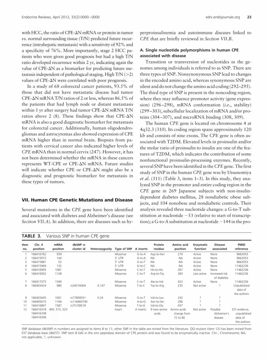
with HCC, the ratio of CPE-�N mRNA or protein in tumorvs. normal surrounding tissue (T/N) predicted future recur-rence (intrahepatic metastasis) with a sensitivity of 92% anda specificity of 76%. More importantly, stage 2 HCC pa-tients who were given good prognosis but had a high T/Nratio developed recurrence within 2 yr, indicating again thevalue of CPE-�N as a biomarker for predicting future me-tastasis independent of pathological staging. High T/N (�2)values of CPE-�N were correlated with poor prognosis.
In a study of 68 colorectal cancer patients, 93.5% ofthose that did not have metastatic disease had tumorCPE-�N mRNA T/N ratios of 2 or less, whereas 86.5% ofthe patients that had lymph node or distant metastasiswithin 1 yr after surgery had tumor CPE-�N mRNA T/Nratios above 2 (8). These findings show that CPE-�NmRNA is also a good diagnostic biomarker for metastasisfor colorectal cancer. Additionally, human oligodendro-gliomas and astrocytomas also showed expression of CPEmRNA higher than in normal brain. Biopsies from pa-tients with cervical cancer also indicated higher levels ofCPE mRNA than in normal cervix (247). However, it hasnot been determined whether the mRNA in these cancersrepresents WT-CPE or CPE-�N mRNA. Future studieswill indicate whether CPE or CPE-�N might also be adiagnostic and prognostic biomarker for metastasis inthese types of tumors.
VII. Human CPE Genetic Mutations and Disease
Several mutations in the CPE gene have been identifiedand associated with diabetes and Alzheimer’s disease (seeSection VII.A). In addition, there are diseases such as hy-
perproinsulinemia and autoimmune diseases linked toCPE that are briefly reviewed in Section VII.B.
A. Single nucleotide polymorphisms in human CPE
associated with disease
Transition or transversion of nucleotides in the ge-nomes among individuals is referred to as SNP. There arethree types of SNP. Nonsynonymous SNP lead to changesin the encoded amino acid, whereas synonymous SNP aresilent and do not change the amino acid coding (292–295).The third type of SNP is present in the noncoding region,where they may influence promoter activity (gene expres-sion) (296–298), mRNA conformation (i.e., stability)(299–303), subcellular localization of mRNA and/or pro-teins (304–307), and microRNA binding (308, 309).
The human CPE gene is located on chromosome 4 at4q32.3 (310). Its coding region spans approximately 120kb and consists of nine exons. The CPE gene is often as-sociated with T2DM. Elevated levels in proinsulin and/orthe molar ratio of proinsulin to insulin are one of the fea-tures of T2DM, which indicates the contribution of somenonfunctional proinsulin-processing enzymes. Recently,several SNP have been identified in the CPE gene. The firststudy of SNP in the human CPE gene was by Utsunomiyaet al. (311) (Table 3, items 1–3). In this study, they ana-lyzed SNP in the promoter and entire coding region in theCPE gene in 269 Japanese subjects with non-insulin-dependent diabetes mellitus, 28 nondiabetic obese sub-jects, and 104 nonobese and nondiabetic controls. Theiranalysis revealed three nucleotide changes: a G-to-T sub-stitution at nucleotide �53 (relative to start of transcrip-tion); a G-to-A substitution at nucleotide �144 in the pro-
TABLE 3. Various SNP in human CPE gene
Item
no.
Chr. 4
position
mRNA
position
dbSNP rs
cluster id Heterozygosity Type of SNP A inserts
Protein
residue
Amino acid
position
Enzymatic
function
Disease
associated
PMID
reference
1 166416762 939 Missense G-to-A Asp-to-Asn 219 Active None 9662053
2 166415972 144 5� UTR G-to-A NA NA Active None 9662053
3 166415881 53 5� UTR G-to-T NA NA Active None 9662053
4 166415969 132 5� UTR G-to-C NA NA Active None 11462236
5 166416905 1081 Missense C-to-T His-to-His 267 Active None 11462236
6 166416952 1128 Missense C-to-T Arg-to-Trp 283 Less active Increased risk
of diabetes
11462236
7 166417373 1549 Missense C-to-T Ala-to-Val 423 Active None 11462236
8 166403424 980 rs34516004 0.147 Missense T-to-C Trp-to-Arg 235 Not active ? Unpublished
data of
the authors
9 166403445 1001 rs77809551 0.24 Missense G-to-T Val-to-Leu 242 ? ?
10 166405670 1164 rs116683740 Missense A-to-G Asn-to-Ser 296 ? ?
11 166416807 1587 rs75109518 Missense T-to-G Val-to-Gly 437 ? ?
12 166416318 495, 515, 523 Insert A inserts 9 new amino
acids
Amino acids
change from
72 to 80
Not active Possible
Alzheimer’s
disease
EST evidence,
unpublished
data of
the authors
166416338
166416346
SNP database (dbSNP) rs numbers are assigned to items 8 to 11; other SNP in the table are mined from the literature. QQ mutant (item 12) has been mined from
EST database base (dbEST). SNP item 8 falls in the zinc peptidase domain of CPE protein and was found to be enzymatically inactive. Chr., Chromosome; NA,
not applicable; ?, unknown.
Endocrine Reviews, April 2012, 33(2):0000–0000 edrv.endojournals.org 23
moter region; and a silent G-to-A substitution in codon219 in the coding region. However, none of these SNPcorrelated with non-insulin-dependent diabetes mellitusor obesity.
In another study conducted on a cohort of 272 Ashke-nazi Jews with T2DM (312), four novel SNP in the tran-script region and one at the promoter region of the CPE
gene were identified (Table 3, items 4–7). At codon posi-tion 283, a C-to-T transition led to a nonsynonymousArg-to-Trp (R283W) change at the protein level. TheArg283 residue is within the Zn-carboxypeptidase do-main of the CPE protein and is conserved among CPEorthologs and most metallocarboxypeptidases that are en-zymatically active, suggesting that this residue may con-tribute to the catalytic process. Indeed, the R283W CPEprotein had a narrower pH optimum and was much lessactive at pH 6.0 to 6.5, indicating that the R283W CPEvariant would be substantially less active than WT-CPE in
vivo. In addition, the R283W CPE variant was less stableat higher temperatures than the WT enzyme. Of theT2DM cohort of Ashkenazi pedigrees, four specific fam-ilies were found to segregate with the R283W mutation.Risk of early-onset T2DM was higher in patients withinthese four families who inherited one copy of this variant.Furthermore, the homozygous mutant is predicted to havea higher risk for the development of hyperproinsulinemiaand diabetes.
A CPE SNP, rs34516004 (Table 3, item 8), has recentlybeen analyzed (S. R. K. Murthy, unpublished data). ThisSNP consists of a T-to-C substitution that leads to a non-synonymous Trp (W)-to-Arg (R) (W235R) change, andwhen expressed and assayed, this mutant was shown to beenzymatically inactive (N. X. Cawley, S. R. K. Murthy,and Y. P. Loh, unpublished data). This W235R mutantwas also found in the AGI_ASP population, which is com-posed of African-Americans and Caucasians, with aheterozygosity value of 0.147, and as with the R283Wmutation, people carrying this W235R mutation in thehomozygous state could have higher risk of hyperproin-sulinemia and diabetes.
An undocumented mutant was found in three EST fromthe Helix Research Institute, Japan, in Alzheimer’s cortex,kidney tumor, and thalamus tissues (Table 3, item 12). Thismutation contains three nucleotide insertions (A and/or C)that result in the substitution of amino acids after the prodo-main of the CPE protein. The new amino acids in all threecases contain a common core sequence with two adjacentglutamine (Q) residues, hence termed QQ mutants. The mu-tationresults inninenewaminoacids replacingeightoriginalamino acids at the N terminus in the mutant protein com-pared with the WT-CPE. The new amino acids have a com-bined isoelectric point that is two units lower than the WT
sequence it replaces. The physicochemical properties of thismutant protein are predicted to be compromised due to thecritical location of the mutation, i.e., at the start of the first�-pleated sheet, which would be important in the structuralintegrity of the mutant protein. A complete analysis of thismutant is currently in progress (N. X. Cawley, Y. Cheng, T.Yanik, C. Liu, S. Young, S. R. K. Murthy, A. Papazian andY. P. Loh, manuscript in preparation).
Studies from several groups have screened Chinese pa-tients with symptoms of coronary atherosclerosis (313–315). In these studies, a number of point mutations werefound in the CPE gene, specifically exons 4 and 5, thatwhen present were genetically linked to an increased se-verity of the disease.
B. CPE-associated human disease
Familial hyperproinsulinemia (FH) is a human diseasethat results from specific mutations in the proinsulin genethat cause substantial increases in the levels of circulatingproinsulin (316). One site where the mutations occur re-sults in Arg65 of the proinsulin being changed to His, Pro,or Leu. Because Arg65 is part of the C peptide-A chainjunction processing site, changes at this site result inpoorer processing of the proinsulin to insulin. Anothermutation occurs at His10Asp on the B-chain of insulin.This His10Asp proinsulin mutant protein has been shownto be secreted through the CSP at a much higher rate com-pared with WT proinsulin (317, 318). Hence, traffickingof the mutant proinsulin is impaired. Recall that CPE canbind to prohormone sorting signals that aid trafficking tothe granules of the RSP. One such signal was described in(pro)insulin. Thus, proinsulin can bind to CPE (42). Mu-tation of this sorting signal in proinsulin resulted in im-paired sorting of the mutant insulin to the RSP. Further-more, some of the human mutants found in FH that wereexpressed in cells bound poorly to CPE in binding assays(121). These and other results suggest that in addition tothe lack of processing the Arg65 mutants and the inabilityof the His10Asp mutant to hexamerize to form a higherorder sorting signal, reduced binding of these mutants toCPE may play a role in their increased constitutive secre-tion and thereby contributes to FH.
Several other diseased states have been found to haveelevated CPE levels. Insulinomas are primary tumors ofinsulin-producing cells of the islets, and they contain thecomplement of expected processing enzymes for this celltype (250). Insulinomas can lead to excessive insulin se-cretion and organic hypoglycemia. Analysis of insulino-mas has shown that expression of CPE and the Gs� sig-naling subunit are often elevated (319). Thus, enhancedregulated secretion of insulin in the presence of elevatedCPE may contribute to the hyperinsulinemia. Some sup-
24 Cawley et al. Carboxypeptidase E in Endocrine and Neural Function Endocrine Reviews, April 2012, 33(2):0000–0000
port for this idea comes from cell culture experimentswhere overexpression of CPE in proinsulin-secreting cellshas been shown to exert a marginal, but significant, in-crease in regulated insulin secretion (320).
The presence of CPE autoantibodies in the circulationhas been studied extensively in cohorts of Chinese patientsthat exhibit latent autoimmune diabetes in adults. Here,there appears to be increased diagnostic value of analyzingCPE antibodies in this condition compared with the anal-ysis of antibodies to glutamic acid decarboxylase (321) orSOX13 (322, 323). In an effort to identify latent autoim-mune diabetes in adults within a phenotypic T2DM co-hort, the prevalence of CPE antibodies was found to be4.8%, suggesting that analysis of CPE antibodies may al-low discrimination of a more latent subset of the adult-onset autoimmune diabetes compared with patients withantibody-negative T2DM (324).
Experimental autoimmune encephalomyelitis (EAE) isa mouse model for multiple sclerosis because it is a poly-genic chronic inflammatory demyelinating disease of thenervous system. Oligonucleotide microarray analysis ofinflamed spinal cord from EAE mice found that expressionof Cpe was decreased by 1.5- to 4.1-fold as the severity ofthe disease progressed (325). Another study identifyingtrait loci for causes of EAE mapped one such loci to Cpe
on chromosome 8 (326). Although presently it is unclearhow CPE may be linked to this phenotype, the recent find-ings that Caspr2 and CPE can interact (140) suggests thatCPE may be involved indirectly in myelination becausecontactin is implicated in neurite outgrowth, axon fascic-ulation, and myelin formation.
Finally, a deletion of both cathepsin B and L in miceresults in early-onset neurodegeneration, a phenotypereminiscent of neuronal ceroid lipofuscinoses in humans(327). Interestingly, of the 19 proteins significantly in-creased, four were increased over 10-fold: Rab14, the Del-ta/Notch-like epidermal growth factor-related receptor,calcyon, and CPE. IHC showed swollen axons in the earlydevelopmental stages of the double KO mice, leading theinvestigators to suggest a role of cathepsin B and L in therecycling process during axon growth and synapse forma-tion. Presently, it is unclear how CPE could compensate inthis process.
VIII. Conclusions
In this review, we have emphasized new functions for an“old” protein, CPE. This protein has been well recognizedas a peptide-processing enzyme for at least three decades.However, over the past 10 yr, evidence has been accumu-lating that CPE may serveadditional roles as an interacting
molecule or as mRNA that is highly expressed or sup-pressed in a variety of paradigms and diseases. In the past,much of the CPE data from screens were ignored due to alack of understanding of why or how a prohormone-processing enzyme like CPE could be involved in cancermetastasis or interact with molecules found in the nucleusor cytoplasm when it is localized within secretory vesicles.Nevertheless, it has always been intriguing that CPEshould be present at 10-fold greater amounts than theother prohormone-processing enzymes such as PC1 andPC2 in endocrine tissues and is far in excess of what isneeded for the enzymatic reactions. Additionally, CPE isexpressed in embryos before the development of the pep-tide hormone endocrine system, hinting at a trophic ratherthan an enzymatic role for CPE—perhaps in cell prolifer-ation and/or differentiation. The present review has at-tempted to highlight the many studies on CPE and to em-phasize some of its newer roles. These new roles include anumber of features. 1) CPE appears to function at thecell-biological level as a sorting receptor for directing pro-hormones into the RSP and in the transport of peptidergicvescicles to their release sites. These attributes are medi-ated by different domains of the protein (cytoplasmic tail)that are not involved in its enzymatic activity. 2) CPE alsoseems to exert antiapoptotic/protective effects againststress in endocrine cells and neurons, as well as contrib-uting to synaptic transmission and dendritic pruning. 3)New insights have been gained using the CPE KO andCpefat/fat mice. These functional roles include bone remod-eling and effects on behavior that involve mood, learning,and memory. 4) The identification of the splice isoform,CPE-�N, has opened up new avenues of investigation,especially with regard to its possible role in tumorigenesisand metastatic disease. Importantly, CPE-�N also seemsto have high potential as a prognostic biomarker for pre-dicting future metastasis of some endocrine and nonen-docrine tumors. 5) Finally, SNP in the human CPE genehave been linked to endocrinological and neurological dis-eases such as diabetes and Alzheimer’s disease. In conclu-sion, CPE is a multifunctional protein that can subserveenzymatic as well as many nonenzymatic roles during de-velopment and in the adult endocrine and nervous sys-tems. Mutations in the CPE gene are associated with avariety of disease states, and expression of its splice variantCPE-�N in tumors promotes metastatic disease inhumans.
IX. Future Directions
Although the mechanisms of action of CPE in peptide pro-cessing are well-known and those mediating prohormone
Endocrine Reviews, April 2012, 33(2):0000–0000 edrv.endojournals.org 25
sorting and vesicle transport for secretion and neurotrans-mission are becoming better understood, considerablework lies ahead in understanding the mechanisms under-lying the rolesof CPE in four majorareas: 1) cell protectionand survival; 2) embryonic development; 3) neuropsychi-atric disorders; and 4) tumor metastasis. The present re-view of the literature suggests that CPE promotes endo-crine and neuronal cell survival in adults and mayinfluence cell growth and proliferation in developing cellsin young animals and in embryos. Thus, it is possiblethat secreted CPE might serve as an autocrine/paracrinetrophic factor to activate different signaling pathways thattrigger transcription of specific genes to mediate these ef-fects. This possibility needs to be explored in detail in thefuture. Some examples include how secreted CPE mayprotect endocrine cells and neurons from cell death underconditions of palmitate-induced ER stress in �-cells, aswell as under oxidative stress and amyloid protein-in-duced toxicity. In embryonic development, the expressionpattern of the CPE splice variants, especially CPE-�N,should be investigated because molecules expressed in tu-mors often are expressed only during embryogenesis andhave a role in proliferation and differentiation. Moreover,CPE-�N has been shown to activate gene transcription,and therefore overexpression and analysis of all its down-stream target genes in different cell types should providenew insights into its mechanism of action. Generating andstudying mouse models bearing SNP of human CPE willalso likely provide a better understanding of how thesemutations cause disease states. Additionally, identifica-tion of both endogenous and environmental factors thatcan induce and regulate CPE expression and the splicing ofthe CPE gene to generate CPE-�N will greatly facilitatethe understanding of how CPE can mediate abnormal be-havioral responses and cause cancer and metastatic dis-ease, respectively. Continued clinical studies on variousendocrine and nonendocrine tumors should further vali-date CPE-�N as a promising multitumor prognostic bio-marker for predicting future metastasis. One can envisagethat future experiments will further reveal novel mecha-nisms of action for CPE and CPE-�N, especially as a primeregulator of many metabolic and endocrine functions, po-tentially as a survival and differentiating factor for neu-rons and endocrine cells, as well as a novel therapeutictarget for cancer and degenerative diseases.
Acknowledgments
We thank Dr. Harold Gainer (National Institute of Neurological andDisorders and Stroke) for his comments and critical reading of themanuscript.
Address all correspondence and requests for reprints to: Y. Peng Loh,Section on Cellular Neurobiology, Program on Developmental Neuro-science, Eunice Kennedy Shriver National Institute of Child Health andHuman Development, National Institutes of Health, 49 Convent Drive,Building 49, Room 5A-22, Bethesda, Maryland 20892. E-mail: [email protected].
Research in the authors’ laboratories was supported in part by theIntramural Research Program of the Eunice Kennedy Shriver NationalInstitute of Child Health and Human Development (NICHD) (toN.X.C., S.R.K.M., and K.P.), and NICHD K22 and American Recoveryand Reinvestment Act grants (to J.J.P.).
Disclosure Summary: The authors have nothing to disclose.
References
1. Steiner DF 1998 The proprotein convertases. Curr OpinChem Biol 2:31–39
2. Fricker LD, Snyder SH 1982 Enkephalin convertase: pu-rification and characterization of a specific enkephalin-synthesizing carboxypeptidase localized to adrenal chro-maffin granules. Proc Natl Acad Sci USA 79:3886–3890
3. Hook VY, Eiden LE, Brownstein MJ 1982 A carboxypep-tidase processing enzyme for enkephalin precursors. Na-ture 295:341–342
4. Bartolomucci A, Possenti R, Mahata SK, Fischer-ColbrieR, Loh YP, Salton SRK 2011 The extended granin family:structure, function and biomedical implications. EndocrRev 32:755–797
5. Lewis MJ, Pelham HR 1990 A human homologue of theyeast HDEL receptor. Nature 348:162–163
6. Naggert JK, Fricker LD, Varlamov O, Nishina PM, RouilleY, Steiner DF, Carroll RJ, Paigen BJ, Leiter EH 1995 Hy-perproinsulinaemia in obese fat/fat mice associated with acarboxypeptidase E mutation which reduces enzyme ac-tivity. Nat Genet 10:135–142
7. Cawley NX, Zhou J, Hill JM, Abebe D, Romboz S, YanikT, Rodriguiz RM, Wetsel WC, Loh YP 2004 The carboxy-peptidase E knockout mouse exhibits endocrinological andbehavioral deficits. Endocrinology 145:5807–5819
8. Lee TK, Murthy SR, Cawley NX, Dhanvantari S, HewittSM, Lou H, Lau T, Ma S, Huynh T, Wesley RA, Ng IO,Pacak K, Poon RT, Loh YP 2011 An N-terminal truncatedcarboxypeptidase E splice isoform induces tumor growthand is a biomarker for predicting future metastasis in hu-man cancers. J Clin Invest 121:880–892
9. Reznik SE, Fricker LD 2001 Carboxypeptidases from A toZ: implications in embryonic development and Wnt bind-ing. Cell Mol Life Sci 58:1790–1804
10. Rawlings ND, Barrett AJ 1995 Evolutionary families ofmetallopeptidases. Methods Enzymol 248:183–228
11. Osterman AL,Grishin NV, SmulevitchSV, Matz MV, Zag-nitko OP, Revina LP, Stepanov VM 1992 Primary struc-ture of carboxypeptidase T: delineation of functionally rel-evant features in Zn-carboxypeptidase family. J ProteinChem 11:561–570
12. Fricker LD, Evans CJ, Esch FS, Herbert E 1986 Cloningand sequence analysis of cDNA for bovine carboxypepti-dase E. Nature 323:461–464
13. Rodríguez C, Brayton KA, Brownstein M, Dixon JE 1989Rat preprocarboxypeptidaseH.Cloning, characterization,and sequence of the cDNA and regulation of the mRNA by
26 Cawley et al. Carboxypeptidase E in Endocrine and Neural Function Endocrine Reviews, April 2012, 33(2):0000–0000

corticotropin-releasing factor. J Biol Chem 264:5988–5995
14. Jung YK, Kunczt CJ, Pearson RK, Dixon JE, Fricker LD1991 Structural characterization of the rat carboxypepti-dase-E gene. Mol Endocrinol 5:1257–1268
15. Billova S, Galanopoulou AS, Seidah NG, Qiu X, Kumar U2007 Immunohistochemical expression and colocalizationof somatostatin, carboxypeptidase-E and prohormoneconvertases 1 and 2 in rat brain. Neuroscience 147:403–418
16. Manser E, Fernandez D, Loo L, Goh PY, Monfries C, HallC, Lim L 1990 Human carboxypeptidase E. Isolation andcharacterization of the cDNA, sequence conservation, ex-pression and processing in vitro. Biochem J 267:517–525
17. Zhang X, Zhu J, Loh YP, Berghman LR 2009 Carboxy-peptidase E, an essential element of the regulated secretorypathway, is expressed and partially co-localized with chro-mogranin A in chicken thymus. Cell Tissue Res 337:371–379
18. Jacob TC, Kaplan JM 2003 The EGL-21 carboxypeptidaseE facilitates acetylcholine release at Caenorhabditis elegansneuromuscular junctions. J Neurosci 23:2122–2130
19. Sakate R, Suto Y, Imanishi T, Tanoue T, Hida M, Haya-saka I, Kusuda J, Gojobori T, Hashimoto K, Hirai M 2007Mapping of chimpanzee full-length cDNAs onto the hu-man genome unveils large potential divergence of the tran-scriptome. Gene 399:1–10
20. Osada N, Hirata M, Tanuma R, Kusuda J, Hida M, SuzukiY, Sugano S, Gojobori T, Shen CK, Wu CI, Hashimoto K2005 Substitution rate and structural divergence of 5�UTRevolution: comparative analysis between human and cyn-omolgus monkey cDNAs. Mol Biol Evol 22:1976–1982
21. Cargill EJ, Baskin LC, Pomp D 1998 Rapid communica-tion: localization of the porcine carboxypeptidase-E geneby linkage analysis further extends the region of syntenybetween human chromosome 4 and porcine chromosome8. J Anim Sci 76:2211–2212
22. Klein SL, Strausberg RL, Wagner L, Pontius J, Clifton SW,Richardson P 2002 Genetic and genomic tools for Xenopusresearch: the NIH Xenopus initiative. Dev Dyn 225:384–391
23. Fan X, Nagle GT 1996 Molecular cloning of Aplysia neu-ronal cDNAs that encode carboxypeptidases related tomammalian prohormone processing enzymes. DNA CellBiol 15:937–945
24. Vihtelic TS, Fadool JM, Gao J, Thornton KA, Hyde DR,Wistow G 2005 Expressed sequence tag analysis of ze-brafish eye tissues for NEIBank. Mol Vis 11:1083–1100
25. Heinrich B, Zhang Z, Raitskin O, Hiller M, Benderska N,Hartmann AM, Bracco L, Elliott D, Ben-Ari S, Soreq H,Sperling J, Sperling R, Stamm S 2009 Heterogeneous nu-clear ribonucleoprotein G regulates splice site selection bybinding to CC(A/C)-rich regions in pre-mRNA. J BiolChem 284:14303–14315
26. Pickrell JK, Pai AA, Gilad Y, Pritchard JK 2010 Noisysplicing drives mRNA isoform diversity in human cells.PLoS Genet 6:e1001236
27. Tsai KW, Chan WC, Hsu CN, Lin WC 2010 Sequencefeatures involved in the mechanism of 3� splice junctionwobbling. BMC Mol Biol 11:34
28. Zhang J, Kuo CC, Chen L 2011 GC content around splice
sites affects splicing through pre-mRNA secondary struc-tures. BMC Genomics 12:90
29. Karambataki M, Malousi A, Maglaveras N, Kouidou S
2010 Synonymous polymorphisms at splicing regulatorysites are associated with CpGs in neurodegenerative dis-ease-related genes. Neuromolecular Med 12:260–269
30. Palaniswamy R, Teglund S, Lauth M, Zaphiropoulos PG,
Shimokawa T 2010 Genetic variations regulate alternativesplicing in the 5� untranslated regions of the mouse glioma-associated oncogene 1, Gli1. BMC Mol Biol 11:32
31. Kumari S, Bugaut A, Huppert JL, Balasubramanian S 2007An RNA G-quadruplex in the 5� UTR of the NRAS proto-oncogene modulates translation. Nat Chem Biol 3:218–221
32. Kozak M 1991 Structural features in eukaryotic mRNAsthat modulate the initiation of translation. J Biol Chem266:19867–19870
33. Clark TA, Schweitzer AC, Chen TX, Staples MK, Lu G,
Wang H, Williams A, Blume JE 2007 Discovery of tissue-specific exons using comprehensive human exon microar-rays. Genome Biol 8:R64
34. Castle JC, Zhang C, Shah JK, Kulkarni AV, Kalsotra A,
Cooper TA, Johnson JM 2008 Expression of 24,426 hu-man alternative splicing events and predicted cis regulationin 48 tissues and cell lines. Nat Genet 40:1416–1425
35. Ishikawa T, Murakami K, Kido Y, Ohnishi S, Yazaki Y,
Harada F, Kuroki K 1998 Cloning, functional expression,and chromosomal localization of the human and mousegp180-carboxypeptidase D-like enzyme. Gene 215:361–370
36. Song L, Fricker L 1995 Processing of procarboxypeptidaseE into carboxypeptidase E occurs in secretory vesicles.J Neurochem 65:444–453
37. Parkinson D 1990 Two soluble forms of bovine carboxy-peptidase H have different NH2-terminal sequences. J BiolChem 265:17101–17105
38. Song L, Fricker LD 1997 The pro region is not required forthe expression or intracellular routeing of carboxypepti-dase E. Biochem J 323:265–271
39. Song L, Fricker LD 1995 Calcium- and pH-dependent ag-gregation of carboxypeptidase E. J Biol Chem 270:7963–7967
40. Rindler MJ 1998 Carboxypeptidase E, a peripheral mem-brane protein implicated in the targeting of hormones tosecretory granules, co-aggregates with granule contentproteins at acidic pH. J Biol Chem 273:31180–31185
41. Cool DR, Normant E, Shen F, Chen HC, Pannell L, Zhang
Y, Loh YP 1997 Carboxypeptidase E is a regulated secre-tory pathway sorting receptor: genetic obliteration leads toendocrine disorders in Cpe(fat) mice. Cell 88:73–83
42. Cool DR, Loh YP 1998 Carboxypeptidase E is a sortingreceptor for prohormones: binding and kinetic studies.Mol Cell Endocrinol 139:7–13
43. Zhang CF, Snell CR, Loh YP 1999 Identification of a novelprohormone sorting signal-binding site on carboxypepti-dase E, a regulated secretory pathway-sorting receptor.Mol Endocrinol 13:527–536
44. Fricker LD 1988 Activation and membrane binding of car-boxypeptidase E. J Cell Biochem 38:279–289
45. Fricker LD, Das B, Angeletti RH 1990 Identification of the
Endocrine Reviews, April 2012, 33(2):0000–0000 edrv.endojournals.org 27

pH-dependent membrane anchor of carboxypeptidase E(EC 3.4.17.10). J Biol Chem 265:2476–2482
46. Mitra A, Song L, Fricker LD 1994 The C-terminal regionof carboxypeptidase E is involved in membrane bindingand intracellular routing in AtT-20 cells. J Biol Chem 269:19876–19881
47. Zhang CF, Dhanvantari S, Lou H, Loh YP 2003 Sorting ofcarboxypeptidase E to the regulated secretory pathway re-quires interaction of its transmembrane domain with lipidrafts. Biochem J 369:453–460
48. Dhanvantari S, Loh YP 2000 Lipid raft association of car-boxypeptidase E is necessary for its function as a regulatedsecretory pathway sorting receptor. J Biol Chem 275:29887–29893
49. Dhanvantari S, Arnaoutova I, Snell CR, Steinbach PJ,Hammond K, Caputo GA, London E, Loh YP 2002 Car-boxypeptidase E, a prohormone sorting receptor, is an-chored to secretory granules via a C-terminal transmem-brane insertion. Biochemistry 41:52–60
50. Klemm RW, Ejsing CS, Surma MA, Kaiser HJ, Gerl MJ,Sampaio JL, de Robillard Q, Ferguson C, Proszynski TJ,Shevchenko A, Simons K 2009 Segregation of sphingolip-ids and sterols during formation of secretory vesicles at thetrans-Golgi network. J Cell Biol 185:601–612
51. Fricker LD, Devi L 1993 Posttranslational processing ofcarboxypeptidase E, a neuropeptide-processing enzyme, inAtT-20 cells and bovine pituitary secretory granules.J Neurochem 61:1404–1415
52. Hook VY 1985 Differential distribution of carboxypepti-dase-processing enzyme activity and immunoreactivity inmembrane and soluble components of chromaffin gran-ules. J Neurochem 45:987–989
53. Arnaoutova I, Jackson CL, Al-Awar OS, Donaldson JG,Loh YP 2003 Recycling of raft-associated prohormonesorting receptor carboxypeptidase E requires interactionwith ARF6. Mol Biol Cell 14:4448–4457
54. Wu CM, Chang HT, Chang MD 2004 Membrane-boundcarboxypeptidase E facilitates the entry of eosinophil cat-ionic protein into neuroendocrine cells. Biochem J 382:841–848
55. Ren J, Kachel K, Kim H, Malenbaum SE, Collier RJ, Lon-don E 1999 Interaction of diphtheria toxin T domain withmolten globule-like proteins and its implications for trans-location. Science 284:955–957
56. Brambillasca S, Yabal M, Soffientini P, Stefanovic S, Ma-karow M, Hegde RS, Borgese N 2005 Transmembranetopogenesis of a tail-anchored protein is modulated bymembrane lipid composition. EMBO J 24:2533–2542
57. Brambillasca S, Yabal M, Makarow M, Borgese N 2006Unassisted translocation of large polypeptide domainsacross phospholipid bilayers. J Cell Biol 175:767–777
58. Luuk H, Toots U, Koks S, Vasar E 2005 N-terminal do-main of Wolframin interacts with carboxypeptidase E. EurNeuropsychopharmacol 15:S5–S6
59. Koks S, Luuk H, Nelovkov A, Areda T, Vasar E 2004 Ascreen for genes induced in the amygdaloid area during catodor exposure. Genes Brain Behav 3:80–89
60. Saito N, Takeuchi T, Kawano A, Hosaka M, Hou N, ToriiS 2011 Luminal interaction of phogrin with carboxypep-tidase E for effective targeting to secretory granules. Traffic12:499–506
61. Machen TE, Leigh MJ, Taylor C, Kimura T, Asano S,
Moore HP 2003 pH of TGN and recycling endosomes ofH�/K�-ATPase-transfected HEK-293 cells: implicationsfor pH regulation in the secretory pathway. Am J PhysiolCell Physiol 285:C205–C214
62. De Matteis MA, Luini A 2008 Exiting the Golgi complex.Nat Rev Mol Cell Biol 9:273–284
63. Hook VY, Mezey E, Fricker LD, Pruss RM, Siegel RE,
Brownstein MJ 1985 Immunochemical characterization ofcarboxypeptidase B-like peptide-hormone-processing en-zyme. Proc Natl Acad Sci USA 82:4745–4749
64. Lynch DR, Braas KM, Hutton JC, Snyder SH 1990 Car-boxypeptidase E (CPE): immunocytochemical localizationin the rat central nervous system and pituitary gland. J Neu-rosci 10:1592–1599
65. Lynch DR, Strittmatter SM, Snyder SH 1984 Enkephalinconvertase localization by [3H]guanidinoethylmercapto-succinic acid autoradiography: selective association withenkephalin-containing neurons. Proc Natl Acad Sci USA81:6543–6547
66. Strittmatter SM, Lynch DR, Snyder SH 1984 [3H]guanidi-noethylmercaptosuccinic acid binding to tissue homoge-nates. Selective labeling of enkephalin convertase. J BiolChem 259:11812–11817
67. Strittmatter SM, Lynch DR, De Souza EB, Snyder SH 1985Enkephalin convertase demonstrated in the pituitary andadrenal gland by [3H]guanidinoethylmercaptosuccinicacid autoradiography: dehydration decreases neurohy-pophyseal levels. Endocrinology 117:1667–1674
68. Lynch DR, Strittmatter SM, Venable JC, Snyder SH 1986Enkephalin convertase: localization to specific neuronalpathways. J Neurosci 6:1662–1675
69. Strittmatter SM, Lynch DR, Snyder SH 1986 Differentialontogeny of rat brain peptidases: prenatal expression ofenkephalin convertase and postnatal development of ang-iotensin-converting enzyme. Brain Res 394:207–215
70. Lynch DR, Strittmatter SM, Venable JC, Snyder SH 1987Enkephalin convertase in the gastrointestinal tract and as-sociated organs characterized and localized with [3H]gua-nidinoethylmercaptosuccinic acid. Endocrinology 121:116–126
71. Lynch DR, Venable JC, Snyder SH 1988 Enkephalin con-vertase in the heart: similar disposition to atrial natriureticfactor. Endocrinology 122:2683–2691
72. Lynch DR, Venable JC, Strittmatter SM, Snyder SH 1988Enkephalin convertase: characterization and localization us-ing [3H]guanidinoethylmercaptosuccinic acid. Biochimie 70:57–64
73. Hougaard DM, Larsson LI 2004 Carboxypeptidase E inrat antropyloric mucosa: distribution in progenitor andmature endocrine cell types. Histochem Cell Biol 121:55–61
74. Krajnik M, Schafer M, Sobanski P, Kowalewski J, Bloch-
Boguslawska E, Zylicz Z, Mousa SA 2010 Enkephalin, itsprecursor, processing enzymes, and receptor as part of alocal opioid network throughout the respiratory system oflung cancer patients. Hum Pathol 41:632–642
75. Mousa SA, Krajnik M, Sobanski P, Kowalewski J, Bloch-
Boguslawska E, Zylicz Z, Schafer M 2010 Dynorphin ex-pression, processing and receptors in the alveolar macro-
28 Cawley et al. Carboxypeptidase E in Endocrine and Neural Function Endocrine Reviews, April 2012, 33(2):0000–0000

phages, cancer cells and bronchial epithelium of lungcancer patients. Histol Histopathol 25:755–764
76. Fricker LD, Adelman JP, Douglass J, Thompson RC, vonStrandmann RP, Hutton J 1989 Isolation and sequenceanalysis of cDNA for rat carboxypeptidase E [EC3.4.17.10], a neuropeptide processing enzyme. Mol Endo-crinol 3:666–673
77. Zheng M, Streck RD, Scott RE, Seidah NG, Pintar JE 1994The developmental expression in rat of proteases furin,PC1, PC2, and carboxypeptidase E: implications for earlymaturation of proteolytic processing capacity. J Neurosci14:4656–4673
78. Birch NP, Rodriguez C, Dixon JE, Mezey E 1990 Distri-bution of carboxypeptidase H messenger RNA in rat brainusing in situ hybridization histochemistry: implications forneuropeptide biosynthesis. Brain Res Mol Brain Res7:53–59
79. MacCumber MW, Snyder SH, Ross CA 1990 Carboxy-peptidase E (enkephalin convertase): mRNA distributionin rat brain by in situ hybridization. J Neurosci 10:2850–2860
80. Schafer MK, Day R, Cullinan WE, Chretien M, Seidah NG,Watson SJ 1993 Gene expression of prohormone and pro-protein convertases in the rat CNS: a comparative in situhybridization analysis. J Neurosci 13:1258–1279
81. Kershaw EE, Flier JS 2004 Adipose tissue as an endocrineorgan. J Clin Endocrinol Metab 89:2548–2556
82. Ramis JM, Franssen-van Hal NL, Kramer E, Llado I, Bouil-laud F, Palou A, Keijer J 2002 Carboxypeptidase E andthrombospondin-1 are differently expressed in subcutane-ous and visceral fat of obese subjects. Cell Mol Life Sci59:1960–1971
83. Greene D, Das B, Fricker LD 1992 Regulation of carboxy-peptidase E. Effect of pH, temperature and Co2� on ki-netic parameters of substrate hydrolysis. Biochem J 285:613–618
84. Fricker LD 2004 Carboxypeptidase E. In: Barrett AJ, Raw-lings ND, Woessner JF, eds. Handbook of proteolytic en-zymes. 2nd ed. London: Academic Press; 840–844
85. Nalamachu SR, Song L, Fricker LD 1994 Regulation ofcarboxypeptidase E. Effect of Ca2� on enzyme activityand stability. J Biol Chem 269:11192–11195
86. Song L, Fricker LD 1995 Purification and characterizationof carboxypeptidase D, a novel carboxypeptidase E-likeenzyme, from bovine pituitary. J Biol Chem 270:25007–25013
87. Fricker LD, Plummer Jr TH, Snyder SH 1983 Enkephalinconvertase: potent, selective, and irreversible inhibitors.Biochem Biophys Res Commun 111:994–1000
88. Bures EJ, Courchesne PL, Douglass J, Chen K, Davis MT,Jones MD, McGinley MD, Robinson JH, Spahr CS, Sun J,Wahl RC, Patterson SD 2001 Identification of incom-pletely processed potential carboxypeptidase E substratesfrom CpEfat/CpEfat mice. Proteomics 1:79–92
89. Fricker LD 2007 Neuropeptidomics to study peptide pro-cessing in animal models of obesity. Endocrinology 148:4185–4190
90. Fricker LD, McKinzie AA, Sun J, Curran E, Qian Y, Yan L,Patterson SD, Courchesne PL, Richards B, Levin N, Mzha-via N, Devi LA, Douglass J 2000 Identification and char-acterization of proSAAS, a granin-like neuroendocrine
peptide precursor that inhibits prohormone processing.J Neurosci 20:639–648
91. Gelman JS, Sironi J, Castro LM, Ferro ES, Fricker LD 2010Hemopressins and other hemoglobin-derived peptides inmouse brain: comparison between brain, blood, and heartpeptidome and regulation in Cpefat/fat mice. J Neurochem113:871–880
92. Gomes I, Grushko JS, Golebiewska U, Hoogendoorn S,Gupta A, Heimann AS, Ferro ES, Scarlata S, Fricker LD,Devi LA 2009 Novel endogenous peptide agonists of can-nabinoid receptors. FASEB J 23:3020–3029
93. Che FY, Yan L, Li H, Mzhavia N, Devi LA, Fricker LD2001 Identification of peptides from brain and pituitary ofCpe(fat)/Cpe(fat) mice. Proc Natl Acad Sci USA 98:9971–9976
94. Che FY, Biswas R, Fricker LD 2005 Relative quantitationof peptides in wild-type and Cpe(fat/fat) mouse pituitaryusing stable isotopic tags and mass spectrometry. J MassSpectrom 40:227–237
95. Che FY, Fricker LD 2005 Quantitative peptidomics ofmouse pituitary: comparison of different stable isotopictags. J Mass Spectrom 40:238–249
96. Che FY, Yuan Q, Kalinina E, Fricker LD 2005 Peptidomicsof Cpe fat/fat mouse hypothalamus: effect of food depri-vation and exercise on peptide levels. J Biol Chem 280:4451–4461
97. Decaillot FM, Che FY, Fricker LD, Devi LA 2006 Peptido-mics of Cpefat/fat mouse hypothalamus and striatum: ef-fect of chronic morphine administration. J Mol Neurosci28:277–284
98. Che FY, Vathy I, Fricker LD 2006 Quantitative peptido-mics in mice: effect of cocaine treatment. J Mol Neurosci28:265–275
99. Kelly RB 1985 Pathways of protein secretion in eu-karyotes. Science 230:25–32
100. Wieland FT, Gleason ML, Serafini TA, Rothman JE 1987The rate of bulk flow from the endoplasmic reticulum tothe cell surface. Cell 50:289–300
101. von Zastrow M, Link R, Daunt D, Barsh G, Kobilka B1993 Subtype-specific differences in the intracellular sort-ing of G protein-coupled receptors. J Biol Chem 268:763–766
102. Kelly RB, Buckley KM, Burgess TL, Carlson SS, Caroni P,Hooper JE, Katzen A, Moore HP, Pfeffer SR, Schroer TA1983 Membrane traffic in neurons and peptide-secretingcells. Cold Spring Harb Symp Quant Biol 48:697–705
103. Green R, Shields D 1984 Somatostatin discriminates be-tween the intracellular pathways of secretory and mem-brane proteins. J Cell Biol 99:97–104
104. Guillemin R, Vargo T, Rossier J, Minick S, Ling N, RivierC, Vale W, Bloom F 1977 �-Endorphin and adrenocorti-cotropin are selected concomitantly by the pituitary gland.Science 197:1367–1369
105. Schwartz MW, Woods SC, Porte Jr D, Seeley RJ, BaskinDG 2000 Central nervous system control of food intake.Nature 404:661–671
106. Thoenen H 2000 Neurotrophins and activity-dependentplasticity. Prog Brain Res 128:183–191
107. Egan MF, Kojima M, Callicott JH, Goldberg TE, Ko-lachana BS, Bertolino A, Zaitsev E, Gold B, Goldman D,Dean M, Lu B, Weinberger DR 2003 The BDNF val66met
Endocrine Reviews, April 2012, 33(2):0000–0000 edrv.endojournals.org 29

polymorphism affects activity-dependent secretion ofBDNF and human memory and hippocampal function.Cell 112:257–269
108. Arvan P, Kuliawat R, Prabakaran D, Zavacki AM, Elahi D,
Wang S, Pilkey D 1991 Protein discharge from immaturesecretory granules displays both regulated and constitutivecharacteristics. J Biol Chem 266:14171–14174
109. Kuliawat R, Arvan P 1992 Protein targeting via the “con-stitutive-like” secretory pathway in isolated pancreatic is-lets: passive sorting in the immature granule compartment.J Cell Biol 118:521–529
110. Loh YP, Snell CR, Cool DR 1997 Receptor-mediated tar-geting of hormones to secretory granules: role of carboxy-peptidase E. Trends Endocrinol Metab 8:130–137
111. Van Horssen AM, Van Kuppeveld FJ, Martens GJ 1998Manipulation of disulfide bonds differentially affects theintracellular transport, sorting, and processing of neuroen-docrine secretory proteins. J Neurochem 71:402–409
112. Mulcahy LR, Barker AJ, Nillni EA 2006 Disruption ofdisulfide bond formation alters the trafficking of prothy-rotropin releasing hormone (proTRH)-derived peptides.Regul Pept 133:123–133
113. Gerdes HH, Rosa P, Phillips E, Baeuerle PA, Frank R,
Argos P, Huttner WB 1989 The primary structure of hu-man secretogranin II, a widespread tyrosine-sulfated se-cretory granule protein that exhibits low pH- and calcium-induced aggregation. J Biol Chem 264:12009–12015
114. Chanat E, Huttner WB 1991 Milieu-induced, selective ag-gregation of regulated secretory proteins in the trans-Golginetwork. J Cell Biol 115:1505–1519
115. Bauerfeind R, Huttner WB 1993 Biogenesis of constitutivesecretory vesicles, secretory granules and synaptic vesicles.Curr Opin Cell Biol 5:628–635
116. Canaff L, Brechler V, Reudelhuber TL, Thibault G 1996Secretory granule targeting of atrial natriuretic peptidecorrelates with its calcium-mediated aggregation. ProcNatl Acad Sci USA 93:9483–9487
117. Dikeakos JD, Reudelhuber TL 2007 Sending proteins todense core secretory granules: still a lot to sort out. J CellBiol 177:191–196
118. Normant E, Loh YP 1998 Depletion of carboxypeptidaseE, a regulated secretory pathway sorting receptor, causesmisrouting and constitutive secretion of proinsulin andproenkephalin, but not chromogranin A. Endocrinology139:2137–2145
119. Loh YP, Maldonado A, Zhang C, Tam WH, Cawley N
2002 Mechanism of sorting proopiomelanocortin andproenkephalin to the regulated secretory pathway of neu-roendocrine cells. Ann NY Acad Sci 971:416–425
120. Lou H, Kim SK, Zaitsev E, Snell CR, Lu B, Loh YP 2005Sorting and activity-dependent secretion of BDNF requireinteraction of a specific motif with the sorting receptorcarboxypeptidase E. Neuron 45:245–255
121. Dhanvantari S, Shen FS, Adams T, Snell CR, Zhang C,
Mackin RB, Morris SJ, Loh YP 2003 Disruption of a re-ceptor-mediated mechanism for intracellular sorting ofproinsulin in familial hyperproinsulinemia. Mol Endocri-nol 17:1856–1867
122. Irminger JC, Verchere CB, Meyer K, Halban PA 1997 Pro-insulin targeting to the regulated pathway is not impaired
in carboxypeptidase E-deficient Cpefat/Cpefat mice. J BiolChem 272:27532–27534
123. Cawley NX, Rodriguez YM, Maldonado A, Loh YP 2003Trafficking of mutant carboxypeptidase E to secretorygranules in a �-cell line derived from Cpe(fat)/Cpe(fat)mice. Endocrinology 144:292–298
124. Cawley NX, Yanik T, Woronowicz A, Chang W, MariniJC, Loh YP 2010 Obese carboxypeptidase E knockoutmice exhibit multiple defects in peptide hormone process-ing contributing to low bone mineral density. Am J PhysiolEndocrinol Metab 299:E189–E197
125. Hosaka M, Watanabe T, Sakai Y, Kato T, Takeuchi T2005 Interaction between secretogranin III and carboxy-peptidase E facilitates prohormone sorting within secre-tory granules. J Cell Sci 118:4785–4795
126. Kemppainen RJ, Behrend EN 2010 Acute inhibition ofcarboxypeptidase E expression in AtT-20 cells does notaffect regulated secretion of ACTH. Regul Pept 165:174–179
127. Hall DH, Hedgecock EM 1991 Kinesin-related gene unc-104 is required for axonal transport of synaptic vesicles inC. elegans. Cell 65:837–847
128. Otsuka AJ, Jeyaprakash A, García-Anoveros J, Tang LZ,Fisk G, Hartshorne T, Franco R, Born T 1991 The C.elegans unc-104 gene encodes a putative kinesin heavychain-like protein. Neuron 6:113–122
129. Hamm-Alvarez SF, Da Costa S, Yang T, Wei X, Gierow JP,Mircheff AK 1997 Cholinergic stimulation of lacrimal aci-nar cells promotes redistribution of membrane-associatedkinesin and the secretory protein, �-hexosaminidase, andincreases kinesin motor activity. Exp Eye Res 64:141–156
130. Senda T, Yu W 1999 Kinesin cross-bridges between neu-rosecretory granules and microtubules in the mouse neu-rohypophysis. Neurosci Lett 262:69–71
131. Varadi A, Tsuboi T, Johnson-Cadwell LI, Allan VJ, RutterGA 2003 Kinesin I and cytoplasmic dynein orchestrateglucose-stimulated insulin-containing vesicle movementsin clonal MIN6 �-cells. Biochem Biophys Res Commun311:272–282
132. Park JJ, Cawley NX, Loh YP 2008 A bi-directional car-boxypeptidase E-driven transport mechanism controlsBDNF vesicle homeostasis in hippocampal neurons. MolCell Neurosci 39:63–73
133. Park JJ, Cawley NX, Loh YP 2008 Carboxypeptidase Ecytoplasmic tail-driven vesicle transport is key for activity-dependent secretion of peptide hormones. Mol Endocrinol22:989–1005
134. Rose SD, Lejen T, Casaletti L, Larson RE, Pene TD, Tri-faro JM 2003 Myosins II and V in chromaffin cells: myosinV is a chromaffin vesicle molecular motor involved in se-cretion. J Neurochem 85:287–298
135. Rudolf R, Kogel T, Kuznetsov SA, Salm T, Schlicker O,Hellwig A, Hammer 3rd JA, Gerdes HH 2003 Myosin Vafacilitates the distribution of secretory granules in the F-actin rich cortex of PC12 cells. J Cell Sci 116:1339–1348
136. Eichler TW, Kogel T, Bukoreshtliev NV, Gerdes HH 2006The role of myosin Va in secretory granule trafficking andexocytosis. Biochem Soc Trans 34:671–674
137. Shakiryanova D, Tully A, Levitan ES 2006 Activity-de-pendent synaptic capture of transiting peptidergic vesicles.Nat Neurosci 9:896–900
30 Cawley et al. Carboxypeptidase E in Endocrine and Neural Function Endocrine Reviews, April 2012, 33(2):0000–0000

138. Goldstein LS, Yang Z 2000 Microtubule-based transportsystems in neurons: the roles of kinesins and dyneins. AnnuRev Neurosci 23:39–71
139. Zhang H, Li S, Wang M, Vukusic B, Pristupa ZB, Liu F2009 Regulation of dopamine transporter activity by car-boxypeptidase E. Mol Brain 2:10
140. Oiso S, Takeda Y, Futagawa T, Miura T, Kuchiiwa S,Nishida K, Ikeda R, Kariyazono H, Watanabe K, YamadaK 2009 Contactin-associated protein (Caspr) 2 interactswith carboxypeptidase E in the CNS. J Neurochem 109:158–167
141. Zhu X, Wu K, Rife L, Cawley NX, Brown B, Adams T,Teofilo K, Lillo C, Williams DS, Loh YP, Craft CM 2005Carboxypeptidase E is required for normal synaptic trans-mission from photoreceptors to the inner retina. J Neuro-chem 95:1351–1362
142. Lou H, Park JJ, Cawley NX, Sarcon A, Sun L, Adams T,Loh YP 2010 Carboxypeptidase E cytoplasmic tail medi-ates localization of synaptic vesicles to the pre-active zonein hypothalamic pre-synaptic terminals. J Neurochem 114:886–896
143. Berezuk MA, Schroer TA2007 Dynactin enhances the pro-cessivity of kinesin-2. Traffic 8:124–129
144. Park JJ, Loh YP 2008 How peptide hormone vesicles aretransported to the secretion site for exocytosis. Mol En-docrinol 22:2583–2595
145. Klyachko VA, Jackson MB 2002 Capacitance steps andfusion pores of small and large-dense-core vesicles in nerveterminals. Nature 418:89–92
146. Zhang Z, Bhalla A, Dean C, Chapman ER, Jackson MB2009 Synaptotagmin IV: a multifunctional regulator ofpeptidergic nerve terminals. Nat Neurosci 12:163–171
147. Weihe E, Tao-Cheng JH, Schafer MK, Erickson JD, EidenLE 1996 Visualization of the vesicular acetylcholine trans-porter in cholinergic nerve terminals and its targeting to aspecific population of small synaptic vesicles. Proc NatlAcad Sci USA 93:3547–3552
148. Liu Y, Edwards RH 1997 Differential localization of ve-sicular acetylcholine and monoamine transporters in PC12cells but not CHO cells. J Cell Biol 139:907–916
149. Krantz DE, Waites C, Oorschot V, Liu Y, Wilson RI, TanPK, Klumperman J, Edwards RH 2000 A phosphorylationsite regulates sorting of the vesicular acetylcholine trans-porter to dense core vesicles. J Cell Biol 149:379–396
150. Yao J, Erickson JD, Hersh LB 2004 Protein kinase A affectstrafficking of the vesicular monoamine transporters inPC12 cells. Traffic 5:1006–1016
151. Rizzoli SO, Betz WJ 2005 Synaptic vesicle pools. Nat RevNeurosci 6:57–69
152. Ludwig M, Leng G 2006 Dendritic peptide release andpeptide-dependent behaviours. Nat Rev Neurosci 7:126–136
153. Hou JC, Min L, Pessin JE 2009 Insulin granule biogenesis,trafficking and exocytosis. Vitam Horm 80:473–506
154. Liu Y, Schirra C, Edelmann L, Matti U, Rhee J, Hof D,Bruns D, Brose N, Rieger H, Stevens DR, Rettig J 2010Two distinct secretory vesicle-priming steps in adrenalchromaffin cells. J Cell Biol 190:1067–1077
155. Eiden LE 1998 The cholinergic gene locus. J Neurochem70:2227–2240
156. Ferguson SM, Savchenko V, Apparsundaram S, Zwick M,
Wright J, Heilman CJ, Yi H, Levey AI, Blakely RD 2003Vesicular localization and activity-dependent traffickingof presynaptic choline transporters. J Neurosci 23:9697–9709
157. Cai Q, Lu L, Tian JH, Zhu YB, Qiao H, Sheng ZH 2010Snapin-regulated late endosomal transport is critical forefficient autophagy-lysosomal function in neurons. Neu-ron 68:73–86
158. Lorenzen A, Samosh J, Vandewark K, Anborgh PH, Seah
C, Magalhaes AC, Cregan SP, Ferguson SS, Pasternak SH
2010 Rapid and direct transport of cell surface APP to thelysosomedefines a novel selective pathway. Mol Brain 3:11
159. Coleman DL, Eicher EM 1990 Fat (fat) and tubby (tub):two autosomal recessive mutations causing obesity syn-dromes in the mouse. J Hered 81:424–427
160. Berman Y, Mzhavia N, Polonskaia A, Devi LA 2001 Im-paired prohormone convertases in Cpe(fat)/Cpe(fat) mice.J Biol Chem 276:1466–1473
161. Varlamov O, Leiter EH, Fricker L 1996 Induced and spon-taneous mutations at Ser202 of carboxypeptidase E. Effecton enzyme expression, activity, and intracellular routing.J Biol Chem 271:13981–13986
162. Varlamov O, Fricker LD, Furukawa H, Steiner DF, Lan-
gley SH, Leiter EH 1997 �-Cell lines derived from trans-genic Cpe(fat)/Cpe(fat) mice are defective in carboxypep-tidase E and proinsulin processing. Endocrinology 138:4883–4892
163. Leiter EH, Kintner J, Flurkey K, Beamer WG, Naggert JK
1999 Physiologic and endocrinologic characterization ofmale sex-biased diabetes in C57BLKS/J mice congenic forthe fat mutation at the carboxypeptidease E locus. Endo-crine 10:57–66
164. Prager R, Schernthaner G 1982 Receptor binding proper-ties of human insulin (recombinant DNA) and human pro-insulin and their interaction at the receptor site. DiabetesCare 5(Suppl 2):104–106
165. Michael DJ, Geng X, Cawley NX, Loh YP, Rhodes CJ,
Drain P, Chow RH 2004 Fluorescent cargo proteins inpancreatic �-cells: design determines secretion kinetics atexocytosis. Biophys J 87:L03–L05
166. Yuan Q, Fontenele-Neto JD, Fricker LD 2004 Effect ofvoluntary exercise on genetically obese Cpefat/fat mice:quantitative proteomics of serum. Obes Res 12:1179–1188
167. Myers MG, Cowley MA, Munzberg H 2008 Mechanismsof leptin action and leptin resistance. Annu Rev Physiol70:537–556
168. Moran TH, Ameglio PJ, Schwartz GJ, McHugh PR 1992Blockade of type A, not type B, CCK receptors attenuatessatiety actions of exogenous and endogenous CCK. Am JPhysiol 262:R46–R50
169. Turton MD, O’Shea D, Gunn I, Beak SA, Edwards CM,
Meeran K, Choi SJ, Taylor GM, Heath MM, Lambert PD,
Wilding JP, Smith DM, Ghatei MA, Herbert J, Bloom SR
1996 A role for glucagon-like peptide-1 in the central reg-ulation of feeding. Nature 379:69–72
170. Cammisotto PG, Bukowiecki LJ 2002 Mechanisms of lep-tin secretion from white adipocytes. Am J Physiol CellPhysiol 283:C244–C250
171. Elmquist JK, Maratos-Flier E, Saper CB, Flier JS 1998 Un-
Endocrine Reviews, April 2012, 33(2):0000–0000 edrv.endojournals.org 31
raveling the central nervous system pathways underlyingresponses to leptin. Nat Neurosci 1:445–450
172. Kristensen P, Judge ME, Thim L, Ribel U, ChristjansenKN, Wulff BS, Clausen JT, Jensen PB, Madsen OD, VrangN, Larsen PJ, Hastrup S 1998 Hypothalamic CART is anew anorectic peptide regulated by leptin. Nature 393:72–76
173. Wardlaw SL 2011 Hypothalamic proopiomelanocortinprocessing and the regulation of energy balance. EurJ Pharmacol 660:213–219
174. Plum L, Lin HV, Dutia R, Tanaka J, Aizawa KS, MatsumotoM, Kim AJ, Cawley NX, Paik JH, Loh YP, DePinho RA,Wardlaw SL, Accili D 2009 The obesity susceptibility geneCpe links FoxO1 signaling in hypothalamic pro-opiomela-nocortin neurons with regulation of food intake. Nat Med15:1195–1201
175. del Giudice EM, Santoro N, Cirillo G, D’Urso L, Di ToroR, Perrone L 2001 Mutational screening of the CART genein obese children: identifying a mutation (Leu34Phe) as-sociated with reduced resting energy expenditure and co-segregating with obesity phenotype in a large family. Di-abetes 50:2157–2160
176. Yanik T, Dominguez G, Kuhar MJ, Del Giudice EM, Loh YP2006 The Leu34Phe ProCART mutation leads to cocaine-and amphetamine-regulated transcript (CART) deficiency: apossible cause for obesity in humans. Endocrinology 147:39–43
177. Asnicar MA, Smith DP, Yang DD, Heiman ML, Fox N,Chen YF, Hsiung HM, Koster A 2001 Absence of cocaine-and amphetamine-regulated transcript results in obesity inmice fed a high caloric diet. Endocrinology 142:4394–4400
178. Wierup N, Richards WG, Bannon AW, Kuhar MJ, AhrenB, Sundler F 2005 CART knock out mice have impairedinsulin secretion and glucose intolerance, altered � cellmorphology and increased body weight. Regul Pept 129:203–211
179. Stanley BG, Kyrkouli SE, Lampert S, Leibowitz SF 1986Neuropeptide Y chronically injected into the hypothala-mus: a powerful neurochemical inducer of hyperphagiaand obesity. Peptides 7:1189–1192
180. Baldock PA, Lee NJ, Driessler F, Lin S, Allison S, StehrerB, Lin EJ, Zhang L, Enriquez RF, Wong IP, McDonaldMM, During M, Pierroz DD, Slack K, Shi YC, Yulyaning-sih E, Aljanova A, Little DG, Ferrari SL, Sainsbury A, Eis-man JA, Herzog H 2009 Neuropeptide Y knockout micereveal a central role of NPY in the coordination of bonemass to body weight. PLoS One 4:e8415
181. Lacourse KA, Friis-Hansen L, Samuelson LC, Rehfeld JF1998 Altered processing of procholecystokinin in carboxy-peptidase E-deficient fat mice: differential synthesis in neu-rons and endocrine cells. FEBS Lett 436:61–66
182. Wang W, Cain BM, Beinfeld MC 1998 Adult carboxy-peptidase E-deficient fat/fat mice have a near-total deple-tion of brain CCK 8 accompanied by a massive accumu-lation of glycine and arginine extended CCK: identificationof CCK 8 Gly as the immediate precursor of CCK 8 inrodent brain. Endocrine 9:329–332
183. Maeda K, Okubo K, Shimomura I, Mizuno K, MatsuzawaY, Matsubara K 1997 Analysis of an expression profile ofgenes in the human adipose tissue. Gene 190:227–235
184. Lawler J 2000 The functions of thrombospondin-1 and-2.Curr Opin Cell Biol 12:634–640
185. Chen H, Herndon ME, Lawler J 2000 The cell biology ofthrombospondin-1. Matrix Biol 19:597–614
186. Cammisotto PG, Bukowiecki LJ, Deshaies Y, Bendayan M2006 Leptin biosynthetic pathway in white adipocytes.Biochem Cell Biol 84:207–214
187. Takeda S, Elefteriou F, Levasseur R, Liu X, Zhao L, ParkerKL, Armstrong D, Ducy P, Karsenty G 2002 Leptin regu-lates bone formation via the sympathetic nervous system.Cell 111:305–317
188. Elefteriou F 2008 Regulation of bone remodeling by thecentral and peripheral nervous system. Arch Biochem Bio-phys 473:231–236
189. Ferron M, Hinoi E, Karsenty G, Ducy P 2008 Osteocalcindifferentially regulates � cell and adipocyte gene expres-sion and affects the development of metabolic diseases inwild-type mice. Proc Natl Acad Sci USA 105:5266–5270
190. Baldock PA, Sainsbury A, Couzens M, Enriquez RF,Thomas GP, Gardiner EM, Herzog H 2002 HypothalamicY2 receptors regulate bone formation. J Clin Invest 109:915–921
191. Ahn JD, Dubern B, Lubrano-Berthelier C, Clement K,Karsenty G 2006 Cart overexpression is the only identifi-able cause of high bone mass in melanocortin 4 receptordeficiency. Endocrinology 147:3196–3202
192. Zhang M, Pritchard MR, Middleton FA, Horton JA,Damron TA 2008 Microarray analysis of perichondraland reserve growth plate zones identifies differential geneexpressions and signal pathways. Bone 43:511–520
193. Bray GA, YorkDA 1971 Genetically transmittedobesity inrodents. Physiol Rev 51:598–646
194. Srinivasan S, Bunch DO, Feng Y, Rodriguiz RM, Li M,Ravenell RL, Luo GX, Arimura A, Fricker LD, Eddy EM,Wetsel WC 2004 Deficits in reproduction and pro-gonad-otropin-releasing hormone processing in male Cpefatmice. Endocrinology 145:2023–2034
195. Wetsel WC, Mellon PL, Weiner RI, Negro-Vilar A 1991Metabolism of pro-luteinizing hormone-releasing hor-mone in immortalized hypothalamic neurons. Endocrinol-ogy 129:1584–1595
196. Wetsel WC, Liposits Z, Seidah NG, Collins S 1995 Ex-pression of candidate pro-GnRH processing enzymes in rathypothalamus and an immortalized hypothalamic neuro-nal cell line. Neuroendocrinology 62:166–177
197. Beamer WG, Wilson MC, Leiter EH 1983 Endocrinology.In: Foster HL, Small JD, Fox JG, eds. The mouse in bio-medical research. Vol III. Normative biology, immunol-ogy, and husbandry. New York: Academic Press; 166–245
198. Vandenbergh JG 1994 Pheromones and mammalian re-production. In: Knobil E, Neill J, eds. The physiology ofreproduction. New York: Raven Press; 343–359
199. Woronowicz A, Koshimizu H, Chang SY, Cawley NX, HillJM, Rodriguiz RM, Abebe D, Dorfman C, Senatorov V,Zhou A, Xiong ZG, Wetsel WC, Loh YP 2008 Absence ofcarboxypeptidase E leads to adult hippocampal neuronaldegeneration and memory deficits. Hippocampus 18:1051–1063
200. Koshimizu H, Senatorov V, Loh YP, Gozes I 2009 Neu-roprotective protein and carboxypeptidase E. J Mol Neu-rosci 39:1–8
32 Cawley et al. Carboxypeptidase E in Endocrine and Neural Function Endocrine Reviews, April 2012, 33(2):0000–0000
201. Nickells RW, Schlamp CL, Newton AC, Williams DS 1993Gene expression of the neuropeptide-processing enzymecarboxypeptidase E in rat photoreceptor cells. J Neuro-chem 61:1891–1900
202. Schlamp CL, Nickells RW 1996 Light and dark cause ashift in the spatial expression of a neuropeptide-processingenzyme in the rat retina. J Neurosci 16:2164–2171
203. Coca-Prados M, Escribano J, Ortego J 1999 Differentialgene expression in the human ciliary epithelium. Prog Re-tin Eye Res 18:403–429
204. Ida H, Boylan SA, Weigel AL, Hjelmeland LM 2003 Age-related changes in the transcriptional profile of mouseRPE/choroid. Physiol Genomics 15:258–262
205. Schaefer AT, Larkum ME, Sakmann B, Roth A 2003 Co-incidence detection in pyramidal neurons is tuned by theirdendritic branching pattern. J Neurophysiol 89:3143–3154
206. Jan YN, Jan LY 2003 The control of dendrite development.Neuron 40:229–242
207. Singh KK, Park KJ, Hong EJ, Kramer BM, Greenberg ME,
Kaplan DR, Miller FD 2008 Developmental axon pruningmediated by BDNF-p75NTR-dependent axon degenera-tion. Nat Neurosci 11:649–658
208. Woronowicz A, Cawley NX, Chang SY, Koshimizu H,
Phillips AW, Xiong ZG, Loh YP 2010 CarboxypeptidaseE knockout mice exhibit abnormal dendritic arborizationand spine morphology in central nervous system neurons.J Neurosci Res 88:64–72
209. Pokorny J, Trojan S 1983 Chronic changes in the receptivefield of the pyramidal cells of the rat hippocampus afterintermittent postnatal hypoxia. Physiol Bohemoslov 32:393–402
210. Pokorny J, Trojan S 1986 The development of hippocam-pal structure and how it is influenced by hypoxia. ActaUniv Carol Med Monogr 113:1–79
211. Groc L, Petanjek Z, Gustafsson B, Ben-Ari Y, Hanse E,
Khazipov R 2002 In vivo blockade of neural activity altersdendritic development of neonatal CA1 pyramidal cells.Eur J Neurosci 16:1931–1938
212. Silva-Gomez AB, Rojas D, Juarez I, Flores G 2003 De-creased dendritic spine density on prefrontal cortical andhippocampalpyramidalneurons in postweaning social iso-lation rats. Brain Res 983:128–136
213. Parrish JZ, Emoto K, Kim MD, Jan YN 2007 Mechanismsthat regulate establishment, maintenance, and remodelingof dendritic fields. Annu Rev Neurosci 30:399–423
214. Cobos I, Calcagnotto ME, Vilaythong AJ, Thwin MT,
Noebels JL, Baraban SC, Rubenstein JL 2005 Mice lackingDlx1 show subtype-specific loss of interneurons, reducedinhibition and epilepsy. Nat Neurosci 8:1059–1068
215. HandR, Bortone D, Mattar P, NguyenL,Heng JI, Guerrier
S, Boutt E, Peters E, Barnes AP, Parras C, Schuurmans C,
Guillemot F, Polleux F 2005 Phosphorylation of neuroge-nin 2 specifies the migration properties and the dendriticmorphology of pyramidal neurons in the neocortex. Neu-ron 48:45–62
216. Cobos I, Borello U, Rubenstein JL 2007 Dlx transcriptionfactors promote migration through repression of axon anddendrite growth. Neuron 54:873–888
217. Lee KF, Bachman K, Landis S, Jaenisch R 1994 Depen-
dence on p75 for innervation of some sympathetic targets.Science 263:1447–1449
218. Kohn J, Aloyz RS, Toma JG, Haak-Frendscho M, MillerFD 1999 Functionally antagonistic interactions betweenthe TrkA and p75 neurotrophin receptors regulate sym-pathetic neuron growth and target innervation. J Neurosci19:5393–5408
219. Singh KK, Miller FD 2005 Activity regulates positive andnegative neurotrophin-derived signals to determine axoncompetition. Neuron 45:837–845
220. Jaworski J, Sheng M 2006 The growing role of mTOR inneuronal development and plasticity. Mol Neurobiol 34:205–219
221. Johnson EM, Craig ET, Yeh HH 2007 TrkB is necessaryfor pruning at the climbing fibre-Purkinje cell synapse inthe developing murine cerebellum. J Physiol 582:629–646
222. Brzustowicz LM, Simone J, Mohseni P, Hayter JE,Hodgkinson KA, Chow EW, Bassett AS 2004 Linkagedisequilibrium mapping of schizophrenia susceptibilityto the CAPON region of chromosome 1q22. Am J HumGenet 74:1057–1063
223. Xu B, Wratten N, Charych EI, Buyske S, Firestein BL,Brzustowicz LM2005 Increased expression in dorsolateralprefrontal cortex of CAPON in schizophrenia and bipolardisorder. PLoS Med 2:e263
224. Zheng Y, Li H, Qin W, Chen W, Duan Y, Xiao Y, Li C,Zhang J, Li X, Feng G, He L 2005 Association of the car-boxyl-terminal PDZ ligand of neuronal nitric oxide syn-thase gene with schizophrenia in the Chinese Han popu-lation. Biochem Biophys Res Commun 328:809–815
225. Kremeyer B, García J, Kymalainen H, Wratten N, RestrepoG, Palacio C, Miranda AL, Lopez C, Restrepo M, BedoyaG, Brzustowicz LM, Ospina-Duque J, Arbelaez MP, Ruiz-Linares A 2009 Evidence for a role of the NOS1AP (CA-PON) gene in schizophrenia and its clinical dimensions: anassociation study in a South American population isolate.Hum Hered 67:163–173
226. Wratten NS, Memoli H, Huang Y, Dulencin AM, Matte-son PG, Cornacchia MA, Azaro MA, Messenger J, HayterJE, Bassett AS, Buyske S, Millonig JH, Vieland VJ, Brzus-towicz LM 2009 Identification of a schizophrenia-associ-ated functional noncoding variant in NOS1AP. Am J Psy-chiatry 166:434–441
227. Carrel D, Du Y, Komlos D, Hadzimichalis NM, Kwon M,Wang B, Brzustowicz LM, Firestein BL 2009 NOS1APregulates dendrite patterning of hippocampal neuronsthrough a carboxypeptidase E-mediated pathway. J Neu-rosci 29:8248–8258
228. Matsuzaki M 2007 Factors critical for the plasticity ofdendritic spines and memory storage. Neurosci Res 57:1–9
229. Bourne JN, Harris KM 2008 Balancing structure and func-tion at hippocampal dendritic spines. Annu Rev Neurosci31:47–67
230. De Roo M, Klauser P, Garcia PM, Poglia L, Muller D 2008Spine dynamics and synapse remodeling during LTP andmemory processes. Prog Brain Res 169:199–207
231. Jin K, Graham SH, Nagayama T, Goldsmith PC, Green-berg DA, Zhou A, Simon RP 2001 Altered expression ofthe neuropeptide-processing enzyme carboxypeptidase Ein the rat brain after global ischemia. J Cereb Blood FlowMetab 21:1422–1429
Endocrine Reviews, April 2012, 33(2):0000–0000 edrv.endojournals.org 33

232. Zhou A, Minami M, Zhu X, Bae S, Minthorne J, Lan J,Xiong ZG, Simon RP 2004 Altered biosynthesis of neuro-peptide processing enzyme carboxypeptidase E after brainischemia: molecular mechanism and implication. J CerebBlood Flow Metab 24:612–622
233. Cawley NX, Woronowicz A, Koshimizu H, Gainer H, LohYP 2010 Carboxypeptidase E is neuroprotective againstepileptic-like seizures in mouse hippocampal neurons. SanDiego: Society for Neuroscience
234. Murthy SR, Thouennon E, Li W-S, Cheng Y, Bhupatkar J,Cawley NX, Lane M, Merchenthaler I, Loy YP 2011 Glu-cocorticoid-induced neuroprotection of hippocampal neu-rons in stressed mice is mediated by carboxypeptidase E.J Neurosci (submitted)
235. Thouennon E, Murthy SRK, Li W, Bhupatkar J, Lane M,Merchenthaler I, Loh YP Stress up-regulates the expressionof the neuroprotective protein CPE, via the glucocorticoidpathway. Program of the 41st Annual Meeting of the So-ciety for Neuroscience, Washington D.C., 2011 (Abstract821.14/VV42)
236. Qian Y, Varlamov O, Fricker LD 1999 Glu300 of rat car-boxypeptidase E is essential for enzymatic activity but notsubstrate binding or routing to the regulated secretorypathway. J Biol Chem 274:11582–11586
237. Cheng Y, Cawley NX, Yanik T, Liu C, Murthy S, Loh Y,Carboxypeptidase E in neuroprotection: links to neurode-generation and Alzheimer’s disease. Program of 41st An-nual Meeting of the Society for Neuroscience, Washington,D.C., 2011 (Abstract 48.01/O11)
238. Jeffrey KD, Alejandro EU, Luciani DS, Kalynyak TB, HuX, Li H, Lin Y, Townsend RR, Polonsky KS, Johnson JD2008 Carboxypeptidase E mediates palmitate-induced�-cell ER stress and apoptosis. Proc Natl Acad Sci USA105:8452–8457
239. Johnson JD 2009 Proteomic identification of carboxypep-tidase E connects lipid-induced �-cell apoptosis and dys-function in type 2 diabetes. Cell Cycle 8:38–42
240. Dobkin RD, Leiblum SR, Rosen RC, Menza M, Marin H2006 Depression and sexual functioning in minority wom-en: current status and future directions. J Sex Marital Ther32:23–36
241. Wetsel WC, Rodriguiz RM, Wilkins JJ, Creson T, BiswasR, Berezniuk I, Fricker AD, Fricker LD Emergence of anx-iety-like behavior in depressive-like Cpefat/fat mice. Forty-seventh Annual Meeting of the American College of Neu-ropsychopharmacology, Scottsdale, AZ, 2008, (Poster 81)
242. Tollefson GD, Rosenbaum JF 1998 Selective serotonin re-uptake inhibitors. In: Schatzberg AF, Nemeroff CB, ed.Textbook of psychopharmacology. 2nd ed. Washington,DC: American Psychiatric Press, Inc.; 219–237
243. Zimmerman M, Chelminski I, McDermut W 2002 Majordepressive disorder and axis I diagnostic comorbidity.J Clin Psychiatry 63:187–193
244. Morris RG, Schenk F, Tweedie F, Jarrard LE 1990 Ibote-nate lesions of hippocampus and/or subiculum: dissociat-ing components of allocentric spatial learning. Eur J Neu-rosci 2:1016–1028
245. Kogan JH, Frankland PW, Silva AJ 2000 Long-term mem-ory underlying hippocampus-dependent social recognitionin mice. Hippocampus 10:47–56
246. Dere E, Huston JP, De Souza Silva MA 2007 The phar-
macology, neuroanatomy and neurogenetics of one-trialobject recognition in rodents. Neurosci Biobehav Rev 31:673–704
247. Murthy SR, Pacak K, Loh YP 2010 Carboxypeptidase E:elevated expression correlated with tumor growth and me-tastasis in pheochromocytomas and other cancers. CellMol Neurobiol 30:1377–1381
248. Weber RG, Rieger J, Naumann U, Lichter P, Weller M2001 Chromosomal imbalances associated with responseto chemotherapy and cytotoxic cytokines in human ma-lignant glioma cell lines. Int J Cancer 91:213–218
249. Kim M, Gans JD, Nogueira C, Wang A, Paik JH, Feng B,Brennan C, Hahn WC, Cordon-Cardo C, Wagner SN,Flotte TJ, Duncan LM, Granter SR, Chin L 2006 Com-parative oncogenomics identifies NEDD9 as a melanomametastasis gene. Cell 125:1269–1281
250. Azzoni C, D’Adda T, Tamburrano G, Coscelli C, MadsenOD, Scopsi L, Bordi C 1998 Functioning human insulino-mas. An immunohistochemical analysis of intracellular in-sulin processing. Virchows Arch 433:495–504
251. Hocker M, John M, Anagnostopoulos J, Buhr HJ, Soli-mena M, Gasnier B, Henry JP, Wang TC, Wiedenmann B1999 Molecular dissection of regulated secretory path-ways in human gastric enterochromaffin-like cells: an im-munohistochemical analysis. Histochem Cell Biol 112:205–214
252. Fan X, Olson SJ, Blevins LS, Allen GS, Johnson MD 2002Immunohistochemical localization of carboxypeptidasesD, E, and Z in pituitary adenomas and normal humanpituitary. J Histochem Cytochem 50:1509–1516
253. North WG, Du J 1998 Key peptide processing enzymes areexpressed by a variant form of small-cell carcinoma of thelung. Peptides 19:1743–1747
254. He P, Varticovski L, Bowman ED, Fukuoka J, Welsh JA,Miura K, Jen J, Gabrielson E, Brambilla E, Travis WD,Harris CC 2004 Identification of carboxypeptidase E and�-glutamyl hydrolase as biomarkers for pulmonary neu-roendocrine tumors by cDNA microarray. Hum Pathol35:1196–1209
255. Rozengurt E 2002 Neuropeptides as growth factors fornormal and cancerous cells. Trends Endocrinol Metab 13:128–134
256. Morris DG, Musat M, Czirjak S, Hanzely Z, LillingtonDM, Korbonits M, Grossman AB 2005 Differential geneexpression in pituitary adenomas by oligonucleotide arrayanalysis. Eur J Endocrinol 153:143–151
257. Tatsumi KI, Tanaka S, Takano T, Tahara S, Murakami Y,Takao T, Hashimoto K, Kato Y, Teramoto A, Amino N2003 Frequent appearance of autoantibodies against pro-hormone convertase 1/3 and neuroendocrine protein 7B2in patients with nonfunctioning pituitary macroadenoma.Endocrine 22:335–340
258. DeLellis RA, Lloyd RV, Heitz PU, Eng C 2004 Tumours ofendocrine organs. Lyon, France: IARC Press
259. Manger WM, Eisenhofer G 2004 Pheochromocytoma: di-agnosis and management update. Curr Hypertens Rep6:477–484
260. Pacak K, Lenders JWM, Eisenhofer G 2007 Pheochromo-cytoma: diagnosis, localization and treatment. Malden,MA: Blackwell Publishing
261. Eisenhofer G, Pecorella W, Pacak K, Hooper D, Kopin IJ,
34 Cawley et al. Carboxypeptidase E in Endocrine and Neural Function Endocrine Reviews, April 2012, 33(2):0000–0000

Goldstein DS 1994 The neuronal and extraneuronal ori-gins of plasma 3-methoxy-4-hydroxyphenylglycol in rats.J Auton Nerv Syst 50:93–107
262. Eisenhofer G, Keiser H, Friberg P, Mezey E, Huynh TT,
Hiremagalur B, Ellingson T, Duddempudi S, Eijsbouts A,
Lenders JW 1998 Plasma metanephrines are markers ofpheochromocytoma produced by catechol-O-methyltrans-ferase within tumors. J Clin Endocrinol Metab 83:2175–2185
263. van Duinen N, Steenvoorden D, Kema IP, Jansen JC, Vr-
iends AH, Bayley JP, Smit JW, Romijn JA, Corssmit EP
2010 Increased urinary excretion of 3-methoxytyramine inpatients with head and neck paragangliomas. J Clin En-docrinol Metab 95:209–214
264. Eisenhofer G, Lenders JW,TimmersH,MannelliM, Grebe
SK, Hofbauer LC, Bornstein SR, Tiebel O, Adams K, Brat-
slavsky G, Linehan WM, Pacak K 2011 Measurements ofplasma methoxytyramine, normetanephrine, and meta-nephrine as discriminators of different hereditary forms ofpheochromocytoma. Clin Chem 57:411–420
265. Eisenhofer G, Lenders JW, Linehan WM, Walther MM,
Goldstein DS, Keiser HR 1999 Plasma normetanephrineand metanephrine for detecting pheochromocytoma in vonHippel-Lindau disease and multiple endocrine neoplasiatype 2. N Engl J Med 340:1872–1879
266. Lenders JW, Pacak K, Walther MM, Linehan WM, Man-
nelli M, Friberg P, Keiser HR, Goldstein DS, Eisenhofer G
2002 Biochemical diagnosis of pheochromocytoma: whichtest is best? JAMA 287:1427–1434
267. Sawka AM, Jaeschke R, Singh RJ, Young Jr WF 2003 Acomparison of biochemical tests for pheochromocytoma:measurement of fractionated plasma metanephrines com-pared with the combination of 24-hour urinary metaneph-rines and catecholamines. J Clin Endocrinol Metab 88:553–558
268. Sawka AM, Gafni A, Thabane L, Young Jr WF 2004 Theeconomic implications of three biochemical screening al-gorithms for pheochromocytoma. J Clin EndocrinolMetab 89:2859–2866
269. Unger N, Pitt C, Schmidt IL, Walz MK, Schmid KW,
Philipp T, Mann K, Petersenn S 2006 Diagnostic value ofvarious biochemical parameters for the diagnosis of pheo-chromocytoma in patients with adrenal mass. Eur J Endo-crinol 154:409–417
270. Grouzmann E, Drouard-Troalen L, Baudin E, Plouin PF,
Muller B, Grand D, Buclin T 2010 Diagnostic accuracy offree and total metanephrines in plasma and fractionatedmetanephrines in urine of patients with pheochromocy-toma. Eur J Endocrinol 162:951–960
271. Sutton MG, Sheps SG, Lie JT 1981 Prevalence of clinicallyunsuspected pheochromocytoma. Review of a 50-year au-topsy series. Mayo Clin Proc 56:354–360
272. McNeil AR, Blok BH, Koelmeyer TD, Burke MP, Hilton
JM 2000 Phaeochromocytomas discovered during coro-nial autopsies in Sydney, Melbourne andAuckland.Aust NZ J Med 30:648–652
273. Amar L, Baudin E, Burnichon N, Peyrard S, Silvera S, Ber-
therat J, Bertagna X, Schlumberger M, Jeunemaitre X,
Gimenez-Roqueplo AP, Plouin PF 2007 Succinate dehy-drogenase B gene mutations predict survival in patients
with malignant pheochromocytomas or paragangliomas.J Clin Endocrinol Metab 92:3822–3828
274. Young Jr WF 2007 Clinical practice. The incidentally dis-covered adrenal mass. N Engl J Med 356:601–610
275. Cascon A, Pita G, Burnichon N, Landa I, Lopez-Jimenez E,Montero-Conde C, Leskela S, Leandro-García LJ, LetonR, Rodríguez-Antona C, Díaz JA, Lopez-Vidriero E,Gonzalez-Neira A, Velasco A, Matias-Guiu X, Gimenez-Roqueplo AP, Robledo M 2009 Genetics of pheochromo-cytoma and paraganglioma in Spanish patients. J Clin En-docrinol Metab 94:1701–1705
276. O’Riordain DS, Young Jr WF, Grant CS, Carney JA, vanHeerden JA 1996 Clinical spectrum and outcome of func-tional extraadrenal paraganglioma. World J Surg 20:916–921; discussion 922
277. Mannelli M, Ianni L, Cilotti A, Conti A 1999 Pheochro-mocytoma in Italy: a multicentric retrospective study. EurJ Endocrinol 141:619–624
278. Park J, Song C, Park M, Yoo S, Park SJ, Hong S, Hong B,Kim CS, Ahn H 2011 Predictive characteristics of malig-nant pheochromocytoma. Korean J Urol 52:241–246
279. Zelinka T, Musil Z, Duskova J, Burton D, Merino MJ,Milosevic D, Widimsky Jr J, Pacak K 2011 Metastaticpheochromocytoma: does the size and age matter? EurJ Clin Invest 41:1121–1128
280. Neumann HP, Bausch B, McWhinney SR, Bender BU,Gimm O, Franke G, Schipper J, Klisch J, Altehoefer C,Zerres K, Januszewicz A, Eng C, Smith WM, Munk R,Manz T, Glaesker S, Apel TW, Treier M, Reineke M, WalzMK, Hoang-Vu C, Brauckhoff M, Klein-Franke A, KloseP, Schmidt H, Maier-Woelfle M, Peczkowska M, Szmigiel-ski C, Eng C 2002 Germ-line mutations in nonsyndromicpheochromocytoma. N Engl J Med 346:1459–1466
281. BennDE,Gimenez-RoqueploAP,Reilly JR,Bertherat J,Bur-gess J, Byth K, Croxson M, Dahia PL, Elston M, Gimm O,Henley D, Herman P, Murday V, Niccoli-Sire P, Pasieka JL,Rohmer V, Tucker K, Jeunemaitre X, Marsh DJ, Plouin PF,Robinson BG 2006 Clinical presentation and penetrance ofpheochromocytoma/paraganglioma syndromes. J Clin En-docrinol Metab 91:827–836
282. Plouin PF, Gimenez-Roqueplo AP 2006 The genetic basisof pheochromocytoma: who to screen and how? Nat ClinPract Endocrinol Metab 2:60–61
283. Petri BJ, van Eijck CH, de Herder WW, Wagner A, deKrijger RR 2009 Phaeochromocytomas and sympatheticparagangliomas. Br J Surg 96:1381–1392
284. Bayley JP, Kunst HP, Cascon A, Sampietro ML, Gaal J,Korpershoek E, Hinojar-Gutierrez A, Timmers HJ, Hoef-sloot LH, Hermsen MA, Suarez C, Hussain AK, VriendsAH, Hes FJ, Jansen JC, Tops CM, Corssmit EP, de KnijffP, Lenders JW, Cremers CW, Devilee P, Dinjens WN, deKrijger RR, Robledo M 2010 SDHAF2 mutations in fa-milial and sporadic paraganglioma and phaeochromocy-toma. Lancet Oncol 11:366–372
285. Lenders JW, Eisenhofer G, Mannelli M, Pacak K 2005Phaeochromocytoma. Lancet 366:665–675
286. Muller U, Troidl C, Niemann S 2005 SDHC mutations inhereditary paraganglioma/pheochromocytoma. Fam Can-cer 4:9–12
287. Schiavi F, Savvoukidis T, Trabalzini F, Grego F, Piazza M,Amista P, Dematte S, Del Piano A, Cecchini ME, Erlic Z,
Endocrine Reviews, April 2012, 33(2):0000–0000 edrv.endojournals.org 35

De Lazzari P, Mantero F, Opocher G 2006 Paragangliomasyndrome: SDHB, SDHC, and SDHD mutations in headand neck paragangliomas. Ann NY Acad Sci 1073:190–197
288. Burnichon N, Briere JJ, Libe R, Vescovo L, Riviere J, Tis-sier F, Jouanno E, Jeunemaitre X, Benit P, Tzagoloff A,Rustin P, Bertherat J, Favier J, Gimenez-Roqueplo AP2010 SDHA is a tumor suppressor gene causing paragan-glioma. Hum Mol Genet 19:3011–3020
289. Qin Y, Yao L, King EE, Buddavarapu K, Lenci RE, Cho-cron ES, Lechleiter JD, Sass M, Aronin N, Schiavi F,Boaretto F, Opocher G, Toledo RA, Toledo SP, Stiles C,Aguiar RC, Dahia PL 2010 Germline mutations inTMEM127 confer susceptibility to pheochromocytoma.Nat Genet 42:229–233
290. Yao L, Schiavi F, Cascon A, Qin Y, Inglada-Perez L, KingEE, Toledo RA, Ercolino T, Rapizzi E, Ricketts CJ, MoriL, Giacche M, Mendola A, Taschin E, Boaretto F, Loli P,Iacobone M, Rossi GP, Biondi B, Lima-Junior JV, KaterCE, Bex M, Vikkula M, Grossman AB, Gruber SB, Bar-ontini M, Persu A, Castellano M, Toledo SP, Maher ER,Mannelli M, Opocher G, Robledo M, Dahia PL 2010 Spec-trum and prevalence of FP/TMEM127 gene mutations inpheochromocytomas and paragangliomas. JAMA 304:2611–2619
291. Neumann HP, Sullivan M, Winter A, Malinoc A, Hoff-mann MM, Boedeker CC, Bertz H, Walz MK, Moeller LC,Schmid KW, Eng C 2011 Germline mutations of theTMEM127 gene in patients with paraganglioma of headand neck and extraadrenal abdominal sites. J Clin Endo-crinol Metab 96:E1279–E1282
292. Krawczak M, Reiss J, Cooper DN 1992 The mutationalspectrum of single base-pair substitutions in mRNA splicejunctions of human genes: causes and consequences. HumGenet 90:41–54
293. Martin N, Boomsma D, Machin G 1997 A twin-prongedattack on complex traits. Nat Genet 17:387–392
294. Halushka MK, Fan JB, Bentley K, Hsie L, Shen N, WederA, Cooper R, Lipshutz R, Chakravarti A 1999 Patterns ofsingle-nucleotide polymorphisms in candidate genes forblood-pressure homeostasis. Nat Genet 22:239–247
295. Lohrer HD, Tangen U 2000 Investigations into the mo-lecular effects of single nucleotide polymorphism. Patho-biology 68:283–290
296. LeVan TD, Bloom JW, Bailey TJ, Karp CL, Halonen M,Martinez FD, Vercelli D 2001 A common single nucleotidepolymorphism in the CD14 promoter decreases the affinityof Sp protein binding and enhances transcriptional activ-ity. J Immunol 167:5838–5844
297. Seibold MA, Wise AL, Speer MC, Steele MP, Brown KK,Loyd JE, Fingerlin TE, Zhang W, Gudmundsson G, Gro-shong SD, Evans CM, Garantziotis S, Adler KB, DickeyBF, du Bois RM, Yang IV, Herron A, Kervitsky D, TalbertJL, Markin C, Park J, Crews AL, Slifer SH, Auerbach S,Roy MG, Lin J, Hennessy CE, Schwarz MI, Schwartz DA2011 A common MUC5B promoter polymorphism andpulmonary fibrosis. N Engl J Med 364:1503–1512
298. Sonuga-Barke EJ, Kumsta R, Schlotz W, Lasky-Su J,Marco R, Miranda A, Mulas F, Oades RD, BanaschewskiT, Mueller U, Andreou P, Christiansen H, Gabriels I, UebelH, Kuntsi J, Franke B, Buitelaar J, Ebstein R, Gill M, Anney
R, Roeyers H, Rothenberger A, Sergeant J, SteinhausenHC, Asherson P, Faraone SV 2011 A functional variant ofthe serotonin transporter gene (SLC6A4) moderates im-pulsive choice in attention deficit/hyperactivity disorderboys and siblings. Biol Psychiatry 70:230–236
299. Fritsche LG, Loenhardt T, Janssen A, Fisher SA, Rivera A,Keilhauer CN, Weber BH 2008 Age-related macular degen-eration is associatedwithanunstableARMS2(LOC387715)mRNA. Nat Genet 40:892–896
300. Magyar I, Colman D, Arnold E, Baumgartner D, BottaniA, Fokstuen S, Addor MC, Berger W, Carrel T, SteinmannB, Matyas G 2009 Quantitative sequence analysis of FBN1premature termination codons provides evidence for in-complete NMD in leukocytes. Hum Mutat 30:1355–1364
301. Pointon JJ, Lok CY, Shearman JD, Suckling RJ, RochetteJ, Merryweather-Clarke AT, Robson KJ 2009 A novelHFE mutation (c.del478) results in nonsense-mediated de-cay of the mutant transcript in a hemochromatosis patient.Blood Cells Mol Dis 43:194–198
302. Shin JA, Chang HS, Park SM, Jang AS, Park SW, Park JS,Uh ST, Il Lim G, Rhim T, Kim MK, Choi IS, Chung IY,Park BL, Shin HD, Park CS 2009 Genetic effect of Cys-LTR2 polymorphisms on its mRNA synthesis and stabili-zation. BMC Med Genet 10:106
303. Hoffmann M, Schirmer MA, Tzvetkov MV, Kreuz M,Ziepert M, Wojnowski L, Kube D, Pfreundschuh M,Trumper L, Loeffler M, Brockmoller J 2010 A functionalpolymorphism in the NAD(P)H oxidase subunit CYBA isrelated to gene expression, enzyme activity, and outcome innon-Hodgkin lymphoma. Cancer Res 70:2328–2338
304. Garcia-Rostan G, Tallini G, Herrero A, D’Aquila TG, Car-cangiu ML, Rimm DL 1999 Frequent mutation and nu-clear localization of �-catenin in anaplastic thyroid carci-noma. Cancer Res 59:1811–1815
305. Kammerer S, Burns-Hamuro LL, Ma Y, Hamon SC, Ca-naves JM, Shi MM, Nelson MR, Sing CF, Cantor CR,Taylor SS, Braun A 2003 Amino acid variant in the kinasebinding domain of dual-specific A kinase-anchoring pro-tein 2: a disease susceptibility polymorphism. Proc NatlAcad Sci USA 100:4066–4071
306. Bohren KM, Nadkarni V, Song JH, Gabbay KH, Ower-bach D 2004 A M55V polymorphism in a novel SUMOgene (SUMO-4) differentially activates heat shock tran-scription factors and is associated with susceptibility totype I diabetes mellitus. J Biol Chem 279:27233–27238
307. Leskela TT, Markkanen PM, Alahuhta IA, Tuusa JT,Petaja-Repo UE 2009 Phe27Cys polymorphism alters thematuration and subcellular localization of the human �
opioid receptor. Traffic 10:116–129308. Chen K, Song F, Calin GA, Wei Q, Hao X, Zhang W 2008
Polymorphisms in microRNA targets: a gold mine for mo-lecular epidemiology. Carcinogenesis 29:1306–1311
309. Saunders MA, Liang H, Li WH 2007 Human polymor-phism at microRNAs and microRNA target sites. ProcNatl Acad Sci USA 104:3300–3305
310. Hall C, Manser E, Spurr NK, Lim L 1993 Assignment ofthe human carboxypeptidase E (CPE) gene to chromosome4. Genomics 15:461–463
311. Utsunomiya N, Ohagi S, Sanke T, Tatsuta H, Hanabusa T,Nanjo K 1998 Organization of the human carboxypepti-dase E gene and molecular scanning for mutations in Jap-
36 Cawley et al. Carboxypeptidase E in Endocrine and Neural Function Endocrine Reviews, April 2012, 33(2):0000–0000
anese subjects with NIDDM or obesity. Diabetologia 41:701–705
312. Chen H, Jawahar S, Qian Y, Duong Q, Chan G, Parker A,Meyer JM, Moore KJ, Chayen S, Gross DJ, Glaser B, Per-mutt MA, Fricker LD 2001 Missense polymorphism in thehuman carboxypeptidase E gene alters enzymatic activity.Hum Mutat 18:120–131
313. Jia EZ, Wang J, Yang ZJ, Zhu TB, Wang LS, Chen B, CaoKJ, Huang J, Ma WZ 2008 Molecular scanning of thehuman carboxypeptidase E gene for mutations in Chinesesubjects with coronary atherosclerosis. Mol Cell Biochem307:31–39
314. Wang J, Zhang Y, Yang ZJ, Zhu TB, Wang LS, Chen B,Cao KJ, Huang J, Ma WZ, Jia EZ 2008 Association ofhuman carboxypeptidase E exon5 gene polymorphismswith angiographical characteristics of coronary athero-sclerosis in a Chinese population. Acta Pharmacol Sin 29:736–744
315. Jia EZ, Wang J, Yang ZJ, Zhu TB, Wang LS, Wang H, LiCJ, Chen B, Cao KJ, Huang J, Ma WZ 2009 Associationof the mutation for the human carboxypeptidase E geneexon 4 with the severity of coronary artery atherosclerosis.Mol Biol Rep 36:245–254
316. Gabbay KH, DeLuca K, Fisher Jr JN, Mako ME, Ruben-stein AH 1976 Familial hyperproinsulinemia. An auto-somal dominant defect. N Engl J Med 294:911–915
317. Chan SJ, Seino S, Gruppuso PA, Schwartz R, Steiner DF1987 A mutation in the B chain coding region is associatedwith impaired proinsulin conversion in a family with hy-perproinsulinemia. Proc Natl Acad Sci USA 84:2194–2197
318. Carroll RJ, Hammer RE, Chan SJ, Swift HH, RubensteinAH, Steiner DF 1988 A mutant human proinsulin is se-creted from islets of Langerhans in increased amounts viaan unregulated pathway. Proc Natl Acad Sci USA 85:8943–8947
319. Wang XC, Xu SY, Wu XY, Song HD, Mao YF, Fan HY,Yu F, Mou B, Gu YY, Xu LQ, Zhou XO, Chen Z, Chen JL,Hu RM 2004 Gene expression profiling in human insuli-noma tissue: genes involved in the insulin secretion path-way and cloning of novel full-length cDNAs. Endocr RelatCancer 11:295–303
320. Polastri L, Galbiati F, Folli F, Davalli AM 2002 Effects ofcarboxypeptidase E overexpression on insulin mRNA lev-els, regulated insulin secretion, and proinsulin processingof pituitary GH3 cells transfected with a furin-cleavablehuman proinsulin cDNA. Cell Transplant 11:803–811
321. Zhou ZG, Yang L, Huang G 2003 Diagnostic value ofcarboxypeptidase-H autoantibodies in detecting latent au-toimmune diabetes in adults. Hunan Yi Ke Da Xue XueBao 28:549–552
322. Li LR, Zhou ZG, Huang G, Yang L, Li X, Chen XY, He L2005 [Diagnostic role of SOX13 antibody in latent auto-immune diabetes of adults]. Zhonghua Yi Xue Za Zhi 85:235–239
323. Zhang D, Zhou Z, Li L, Weng J, Huang G, Jing P, ZhangC, Peng J, Xiu L 2006 Islet autoimmunity and genetic mu-tations in Chinese subjects initially thought to have type 1Bdiabetes. Diabet Med 23:67–71
324. Yang L, Zhou ZG, Tan SZ, Huang G, Jin P, Yan X, Li X,Peng H, Hagopian W 2008 Carboxypeptidase-H autoan-
tibodies differentiate a more latent subset of autoimmunediabetes from phenotypic type 2 diabetes among Chineseadults. Ann NY Acad Sci 1150:263–266
325. Ibrahim SM, Mix E, Bottcher T, Koczan D, Gold R, RolfsA, Thiesen HJ 2001 Gene expression profiling of the ner-vous system in murine experimental autoimmune enceph-alomyelitis. Brain 124:1927–1938
326. Mazon Pelaez I, Vogler S, Strauss U, Wernhoff P, PahnkeJ, Brockmann G, Moch H, Thiesen HJ, Rolfs A, IbrahimSM 2005 Identification of quantitative trait loci control-ling cortical motor evoked potentials in experimental au-toimmune encephalomyelitis: correlation with incidence,onset and severity of disease. Hum Mol Genet 14:1977–1989
327. Stahl S, Reinders Y, Asan E, Mothes W, Conzelmann E,Sickmann A, Felbor U 2007 Proteomic analysis of cathep-sin B- and L-deficient mouse brain lysosomes. Biochim Bio-phys Acta 1774:1237–1246
328. Castresana J 2000 Selection of conserved blocks from mul-tiple alignments for their use in phylogenetic analysis. MolBiol Evol 17:540–552
329. Edgar RC 2004 MUSCLE: multiple sequence alignmentwith high accuracy and high throughput. Nucleic AcidsRes 32:1792–1797
330. Lee NK, Karsenty G 2008 Reciprocal regulation of boneand energy metabolism. Trends Endocrinol Metab 19:161–166
331. Evans JF, Niu QT, Canas JA, Shen CL, Aloia JF, Yeh JK2004 ACTH enhances chondrogenesis in multipotentialprogenitor cells and matrix production in chondrocytes.Bone 35:96–107
332. Zhong Q, Sridhar S, Ruan L, Ding KH, Xie D, Insogna K,Kang B, Xu J, Bollag RJ, Isales CM 2005 Multiple mela-nocortin receptors are expressed in bone cells. Bone 36:820–831
333. Fu Y, Luo N, Klein RL, Garvey WT 2005 Adiponectinpromotes adipocyte differentiation, insulin sensitivity, andlipid accumulation. J Lipid Res 46:1369–1379
334. Elefteriou F, Takeda S, Ebihara K, Magre J, Patano N, KimCA, Ogawa Y, Liu X, Ware SM, Craigen WJ, Robert JJ,Vinson C, Nakao K, Capeau J, Karsenty G 2004 Serumleptin level is a regulator of bone mass. Proc Natl Acad SciUSA 101:3258–3263
335. Elefteriou F, Ahn JD, Takeda S, Starbuck M, Yang X, LiuX, Kondo H, Richards WG, Bannon TW, Noda M, Clem-ent K, Vaisse C, Karsenty G 2005 Leptin regulation of boneresorption by the sympathetic nervous system and CART.Nature 434:514–520
336. Lacey DL, Timms E, Tan HL, Kelley MJ, Dunstan CR,Burgess T, Elliott R, Colombero A, Elliott G, Scully S, HsuH, Sullivan J, Hawkins N, Davy E, Capparelli C, Eli A,Qian YX, Kaufman S, Sarosi I, Shalhoub V, Senaldi G, GuoJ, Delaney J, Boyle WJ 1998 Osteoprotegerin ligand is acytokine that regulates osteoclast differentiation and acti-vation. Cell 93:165–176
337. Bae S, Ha TS, Yoon Y, Lee J, Cha HJ, Yoo H, Choe TB, LiS, Sohn I, Kim JY, Kim CS, Jin HO, Lee HC, Park IC, KimCS, Jin YW, Ahn SK 2008 Genome-wide screening andidentification of novel proteolytic cleavage targets ofcaspase-8 and -10 in vitro. Int J Mol Med 21:381–386
338. Bogan RL, Murphy MJ, Stouffer RL, Hennebold JD 2008
Endocrine Reviews, April 2012, 33(2):0000–0000 edrv.endojournals.org 37
Systematic determination of differential gene expression inthe primate corpus luteum during the luteal phase of themenstrual cycle. Mol Endocrinol 22:1260–1273
339. Chikuma T, Inomata Y, Tsuchida K, Hojo H, Kato T 2002Effect of monensin on the levels of tachykinins and theirprocessing enzyme activity in rat dorsal root ganglia. Neu-rosci Lett 326:89–92
340. Cook JL, Giardina JF, Zhang Z, Re RN 2002 Intracellularangiotensin II increases the long isoform of PDGF mRNAin rat hepatoma cells. J Mol Cell Cardiol 34:1525–1537
341. Dong W, Fricker LD, Day R 1999 Carboxypeptidase D isa potential candidate to carry out redundant processingfunctions of carboxypeptidase E based on comparative dis-tribution studies in the rat central nervous system. Neuro-science 89:1301–1317
342. Du J, Keegan BP, North WG 2001 Key peptide processingenzymes are expressed by breast cancer cells. Cancer Lett165:211–218
343. Grigoriants O, Devi L, Fricker LD 1993 Dopamine antag-onist haloperidol increases carboxypeptidase E mRNA inrat neurointermediate pituitary but not in various other rattissues. Brain Res Mol Brain Res 19:161–164
344. Guyonnet B, Marot G, Dacheux JL, Mercat MJ, Schwob S,Jaffrezic F, Gatti JL 2009 The adult boar testicular andepididymal transcriptomes. BMC Genomics 10:369
345. Haegeman A, Williams JL, Law A, Van Zeveren A, Peel-man LJ 2003 Mapping and SNP analysis of bovine candi-date genes for meat and carcass quality. Anim Genet 34:349–353
346. Ishizuka N, Minami K, Okumachi A, Okuno M, Seino S2007 Induction by NeuroD of the components required forregulated exocytosis. Biochem Biophys Res Commun 354:271–277
347. Johnston RA, Theman TA, Shore SA 2006 Augmentedresponses to ozone in obese carboxypeptidase E-deficientmice. Am J Physiol Regul Integr Comp Physiol 290:R126–R133
348. Johnston RA, Zhu M, Hernandez CB, Williams ES, ShoreSA 2010 Onset of obesity in carboxypeptidase E-deficientmice and effect on airway responsiveness and pulmonaryresponses to ozone. J Appl Physiol 108:1812–1819
349. Maresh JG, Xu H, Jiang N, Gairola CG, Shohet RV 2005Tobacco smoke dysregulates endothelial vasoregulatorytranscripts in vivo. Physiol Genomics 21:308–313
350. Marquínez AC, Andreetta AM, Gonzalez N, Wolfenstein-Todel C, Scacciati de Cerezo JM 2003 Identification ofgp17 glycoprotein and characterization of prostatic acidphosphatase (PAP) and carboxypeptidase E (CPE) frag-ments in a human seminal plasma fraction interacting withconcanavalin A. J Protein Chem 22:423–429
351. Mousa SA, Shakibaei M, Sitte N, Schafer M, Stein C 2004Subcellular pathways of �-endorphin synthesis, process-ing, and release from immunocytes in inflammatory pain.Endocrinology 145:1331–1341
352. Mukhina ES, Saldaev DA, Vernigora AN, Gengin MT2005 [The effect of ethanol consumption by dams on theoffspring locomotion in the open field test and carboxy-peptidase activities in the rat brain and adrenal medulla].Ross Fiziol Zh Im I M Sechenova 91:239–245
353. Nakajima E, David LL, Riviere MA, Azuma M, Shearer
TR 2009 Human and monkey lenses cultured with calciumionophore form �B-crystallin lacking the C-terminallysine, a prominent feature of some human cataracts. In-vest Ophthalmol Vis Sci 50:5828–5836
354. Pilot-Storck F, Chopin E, Rual JF, Baudot A, DobrokhotovP, Robinson-Rechavi M, Brun C, Cusick ME, Hill DE,Schaeffer L, Vidal M, Goillot E 2010 Interactome mappingof the phosphatidylinositol 3-kinase-mammalian target ofrapamycin pathway identifies deformed epidermal auto-regulatory factor-1 as a new glycogen synthase kinase-3interactor. Mol Cell Proteomics 9:1578–1593
355. Prasad SS, Cynader MS 1994 Identification of cDNAclones expressed selectively during the critical period forvisual cortex development by subtractive hybridization.Brain Res 639:73–84
356. Rawdon BB, Larsson LI 2000 Development of hormonalpeptides and processing enzymes in the embryonic avianpancreas with special reference to co-localisation. His-tochem Cell Biol 114:105–112
357. Reznik SE, Salafia CM, Lage JM, Fricker LD 1998 Immu-nohistochemical localization of carboxypeptidases E andD in the human placenta and umbilical cord. J HistochemCytochem 46:1359–1368
358. Sasson R, Dantes A, Tajima K, Amsterdam A 2003 Novelgenes modulated by FSH in normal and immortalized FSH-responsive cells: new insights into the mechanism of FSHaction. FASEB J 17:1256–1266
359. Sekiguchi T, Kawashima T, Satou Y, Satoh N 2007 Fur-ther EST analysis of endocrine genes that are preferentiallyexpressed in the neural complex of Ciona intestinalis: re-ceptor and enzyme genes associated with endocrine systemin the neural complex. Gen Comp Endocrinol 150:233–245
360. Singh U, Yu Y, Kalinina E, Konno T, Sun T, Ohta H,Wakayama T, Soares MJ, Hemberger M, Fundele RH2006 Carboxypeptidase E in the mouse placenta. Differ-entiation 74:648–660
361. TangSS,ZhangJH,LiuHX,LiHZ2009PC2/CPE-mediatedpro-protein processing in tumor cells and its differentiatedcells or tissues. Mol Cell Endocrinol 303:43–49
362. Tang SS, Zhang JH, Liu HX, Zhou D, Qi R 2009 Pro-protein convertase-2/carboxypeptidase-E mediated neuro-peptide processing of RGC-5 cell after in vitro ischemia.Neurosci Bull 25:7–14
363. Vilijn MH, Das B, Kessler JA, Fricker LD 1989 Culturedastrocytes and neurons synthesize and secrete carboxypep-tidase E, a neuropeptide-processing enzyme. J Neurochem53:1487–1493
364. Wang X, Huynh H, Gjorloff-Wingren A, Monosov E,Stridsberg M, Fukuda M, Mustelin T 2002 Enlargement ofsecretory vesicles by protein tyrosine phosphatase PTP-MEG2 in rat basophilic leukemia mast cells and Jurkat Tcells. J Immunol 168:4612–4619
365. Eisenhofer G, Lenders JW, Siegert G, Bornstein SR, FribergP, Milosevic D, Mannelli M, Linehan WM, Adams K, Tim-mers HJ, Pacak K Plasma methoxytyramine: A novel bio-marker of metastatic pheochromocytoma and paragangli-oma in relation to established risk factors of tumour size,location and SDHB mutation status. Eur J Cancer Oct 28,2011 10.1016/j.ejca.2011.07.016
38 Cawley et al. Carboxypeptidase E in Endocrine and Neural Function Endocrine Reviews, April 2012, 33(2):0000–0000
Copyright © 2022 FDOKUMEN