New prenylated isoflavonoids as protein tyrosine phosphatase 1B (PTP1B) inhibitors from Erythrina...
6
New prenylated isoflavonoids as protein tyrosine phosphatase 1B (PTP1B) inhibitors from Erythrina addisoniae Phi Hung Nguyen a , Govinda Sharma a , Trong Tuan Dao a , Mohammad Nasir Uddin a , Keon Wook Kang b , Derek Tantoh Ndinteh c , Joseph Tanyi Mbafor c , Won Keun Oh a,⇑ a Korea Bioactive Natural Material Bank, College of Pharmacy, Chosun University, 375 Seosuk-dong, Dong-gu, Gwangju 501-759, Republic of Korea b College of Pharmacy and Research Institute of Pharmaceutical Sciences, Seoul National University, Seoul 151-742, Republic of Korea c Faculty of Science, University of Yaoundé I, PO Box 812, Yaoundé, Cameroon article info Article history: Received 2 July 2012 Revised 19 August 2012 Accepted 21 August 2012 Available online 7 September 2012 Keywords: Erythrina addisoniae Protein tyrosine phosphatase 1B (PTP1B) Cytotoxic activity Breast cancer cells abstract Bioassay-guided fractionation of the EtOAc extract of the root of Erythrina addisoniae (Leguminosae) resulted in the isolation of four new (1–4), along with 2 known prenylated isoflavonoids (5–6). The struc- tures of the isolates were assigned on the basis of spectroscopic data analysis, focusing on interpretation of 1D and 2D NMR, and MS data. All the isolates were evaluated for their inhibitory effects on protein tyrosine phosphatase 1B (PTP1B), as well as their growth inhibition on MCF7, adriamycin-resistant MCF7 (MCF7/ADR), and MDA-MB-231 breast cancer cell lines. Compounds which exhibited PTP1B inhib- itory activity (IC 50 values ranging from 4.6 ± 0.3 to 24.2 ± 2.1 lM) showed potential cytotoxic activity (IC 50 values ranging from 3.97 ± 0.17 to 11.4 ± 1.9 lM). Taken together, our data suggest that prenylated isoflavonoids, especially the isoflavone-type skeleton could be considered as new lead compounds against breast cancer via PTP1B inhibition. Ó 2012 Elsevier Ltd. All rights reserved. 1. Introduction Breast cancer is the most common malignant tumor accounting for approximately 23% of all cancers in woman. 1 The current ap- proaches to breast cancer treatment include chemotherapy, radia- tion, and surgery but all of them have disadvantages like severe side effects and difficult procedures. Tamoxifen is discredited with the increased risk of endometrial hyperplasia and cancer, similarly adriamycin causes cardiotoxicity. 2,3 The continuous usage of che- motherapeutic agents like tamoxifen and adriamycin has been also known as the cause of multidrug resistance. Therefore, new lead molecules which can overcome the resistance and have reduced undesirable toxicity are needed to be discovered. Protein tyrosine phosphatases (PTPs) working in concert with protein tyrosine kinases ensure the proper functioning of a variety of proteins involved in signal transduction process of the cells. PTP1B is well known about its critical role in regulating body weight and glucose homeostasis by acting as a key negative regu- lator of insulin and leptin signaling pathway. 4 In addition to its involvement in obesity and diabetes, its importance in some can- cers has been highlighted. 5 PTP1B is overexpressed in a significant subset of breast and ovarian cancers, especially in those over- expressing HER2/Neu (HER2(+) tumors). 6,7 When PTP1B-deficient mice were crossed with transgenic mice of the ErbB2 (Neu) onco- protein in mammary epithelial cells, the onset of ErbB2 (Neu)-dri- ven breast cancer was delayed significantly in the absence of PTP1B, whereas transgenic PTP1B overexpression was sufficient to induce breast tumors in the absence of exogenous ErbB2. 8a These observations suggest the potential importance of PTP1B inhibitors as therapeutic targets in cancer therapy. 8b Thus, many efforts need to be focused on PTP1B inhibitors as a double thera- peutic target for not only diabetes and obesity but also cancer treatment. 8c As part of an ongoing investigation aimed at finding PTP1B inhibitors from plants, the genus Erythrina 9 was studied using in vitro assay on both the cytotoxic activity and PTP1B inhibitory activity. 10 In this research, four new prenylated isoflavonoids along with two known ones were purified by activity-guided isolation. Among them, as five compounds (2–6) were found as significant PTP1B inhibitors, we also investigated their effects on various can- cer cell lines which are MCF-7, MDA-MB-231, and adriamycin resistant MCF7/ADR. 2. Result and discussion Bioassay-guided fractionation of an EtOAc-soluble extract of the root bark of this plant led to the isolation of a series of prenylated isoflavonoids, consisting of two new isoflavones (erythraddison I–II, 1–2), and two isoflavanones (erythraddison III–IV, 3–4), along with 0968-0896/$ - see front matter Ó 2012 Elsevier Ltd. All rights reserved. http://dx.doi.org/10.1016/j.bmc.2012.08.024 ⇑ Corresponding author. Tel./fax: +82 62 230 6370. E-mail address: [email protected] (W.K. Oh). Bioorganic & Medicinal Chemistry 20 (2012) 6459–6464 Contents lists available at SciVerse ScienceDirect Bioorganic & Medicinal Chemistry journal homepage: www.elsevier.com/locate/bmc
-
Upload
independent -
Category
Documents
-
view
1 -
download
0
Transcript of New prenylated isoflavonoids as protein tyrosine phosphatase 1B (PTP1B) inhibitors from Erythrina...
Bioorganic & Medicinal Chemistry 20 (2012) 6459–6464
Contents lists available at SciVerse ScienceDirect
Bioorganic & Medicinal Chemistry
journal homepage: www.elsevier .com/locate /bmc
New prenylated isoflavonoids as protein tyrosine phosphatase 1B (PTP1B)inhibitors from Erythrina addisoniae
Phi Hung Nguyen a, Govinda Sharma a, Trong Tuan Dao a, Mohammad Nasir Uddin a, Keon Wook Kang b,Derek Tantoh Ndinteh c, Joseph Tanyi Mbafor c, Won Keun Oh a,⇑a Korea Bioactive Natural Material Bank, College of Pharmacy, Chosun University, 375 Seosuk-dong, Dong-gu, Gwangju 501-759, Republic of Koreab College of Pharmacy and Research Institute of Pharmaceutical Sciences, Seoul National University, Seoul 151-742, Republic of Koreac Faculty of Science, University of Yaoundé I, PO Box 812, Yaoundé, Cameroon
a r t i c l e i n f o
Article history:Received 2 July 2012Revised 19 August 2012Accepted 21 August 2012Available online 7 September 2012
Keywords:Erythrina addisoniaeProtein tyrosine phosphatase 1B (PTP1B)Cytotoxic activityBreast cancer cells
0968-0896/$ - see front matter � 2012 Elsevier Ltd. Ahttp://dx.doi.org/10.1016/j.bmc.2012.08.024
⇑ Corresponding author. Tel./fax: +82 62 230 6370.E-mail address: [email protected] (W.K. Oh).
a b s t r a c t
Bioassay-guided fractionation of the EtOAc extract of the root of Erythrina addisoniae (Leguminosae)resulted in the isolation of four new (1–4), along with 2 known prenylated isoflavonoids (5–6). The struc-tures of the isolates were assigned on the basis of spectroscopic data analysis, focusing on interpretationof 1D and 2D NMR, and MS data. All the isolates were evaluated for their inhibitory effects on proteintyrosine phosphatase 1B (PTP1B), as well as their growth inhibition on MCF7, adriamycin-resistantMCF7 (MCF7/ADR), and MDA-MB-231 breast cancer cell lines. Compounds which exhibited PTP1B inhib-itory activity (IC50 values ranging from 4.6 ± 0.3 to 24.2 ± 2.1 lM) showed potential cytotoxic activity(IC50 values ranging from 3.97 ± 0.17 to 11.4 ± 1.9 lM). Taken together, our data suggest that prenylatedisoflavonoids, especially the isoflavone-type skeleton could be considered as new lead compoundsagainst breast cancer via PTP1B inhibition.
� 2012 Elsevier Ltd. All rights reserved.
1. Introduction
Breast cancer is the most common malignant tumor accountingfor approximately 23% of all cancers in woman.1 The current ap-proaches to breast cancer treatment include chemotherapy, radia-tion, and surgery but all of them have disadvantages like severeside effects and difficult procedures. Tamoxifen is discredited withthe increased risk of endometrial hyperplasia and cancer, similarlyadriamycin causes cardiotoxicity.2,3 The continuous usage of che-motherapeutic agents like tamoxifen and adriamycin has been alsoknown as the cause of multidrug resistance. Therefore, new leadmolecules which can overcome the resistance and have reducedundesirable toxicity are needed to be discovered.
Protein tyrosine phosphatases (PTPs) working in concert withprotein tyrosine kinases ensure the proper functioning of a varietyof proteins involved in signal transduction process of the cells.PTP1B is well known about its critical role in regulating bodyweight and glucose homeostasis by acting as a key negative regu-lator of insulin and leptin signaling pathway.4 In addition to itsinvolvement in obesity and diabetes, its importance in some can-cers has been highlighted.5 PTP1B is overexpressed in a significantsubset of breast and ovarian cancers, especially in those over-expressing HER2/Neu (HER2(+) tumors).6,7 When PTP1B-deficient
ll rights reserved.
mice were crossed with transgenic mice of the ErbB2 (Neu) onco-protein in mammary epithelial cells, the onset of ErbB2 (Neu)-dri-ven breast cancer was delayed significantly in the absence ofPTP1B, whereas transgenic PTP1B overexpression was sufficientto induce breast tumors in the absence of exogenous ErbB2.8a
These observations suggest the potential importance of PTP1Binhibitors as therapeutic targets in cancer therapy.8b Thus, manyefforts need to be focused on PTP1B inhibitors as a double thera-peutic target for not only diabetes and obesity but also cancertreatment.8c
As part of an ongoing investigation aimed at finding PTP1Binhibitors from plants, the genus Erythrina9 was studied usingin vitro assay on both the cytotoxic activity and PTP1B inhibitoryactivity.10 In this research, four new prenylated isoflavonoids alongwith two known ones were purified by activity-guided isolation.Among them, as five compounds (2–6) were found as significantPTP1B inhibitors, we also investigated their effects on various can-cer cell lines which are MCF-7, MDA-MB-231, and adriamycinresistant MCF7/ADR.
2. Result and discussion
Bioassay-guided fractionation of an EtOAc-soluble extract of theroot bark of this plant led to the isolation of a series of prenylatedisoflavonoids, consisting of two new isoflavones (erythraddison I–II,1–2), and two isoflavanones (erythraddison III–IV, 3–4), along with
6460 P. H. Nguyen et al. / Bioorg. Med. Chem. 20 (2012) 6459–6464
2 known isoflavones (5–6). Chemical structures of the known com-pounds were determined to be echrenone b10 (5) and erysubin F(6) from a comparison of the physical and spectroscopic data (IR,UV, [a]D, NMR, and MS) with those reported in the literature (Fig. 1).11
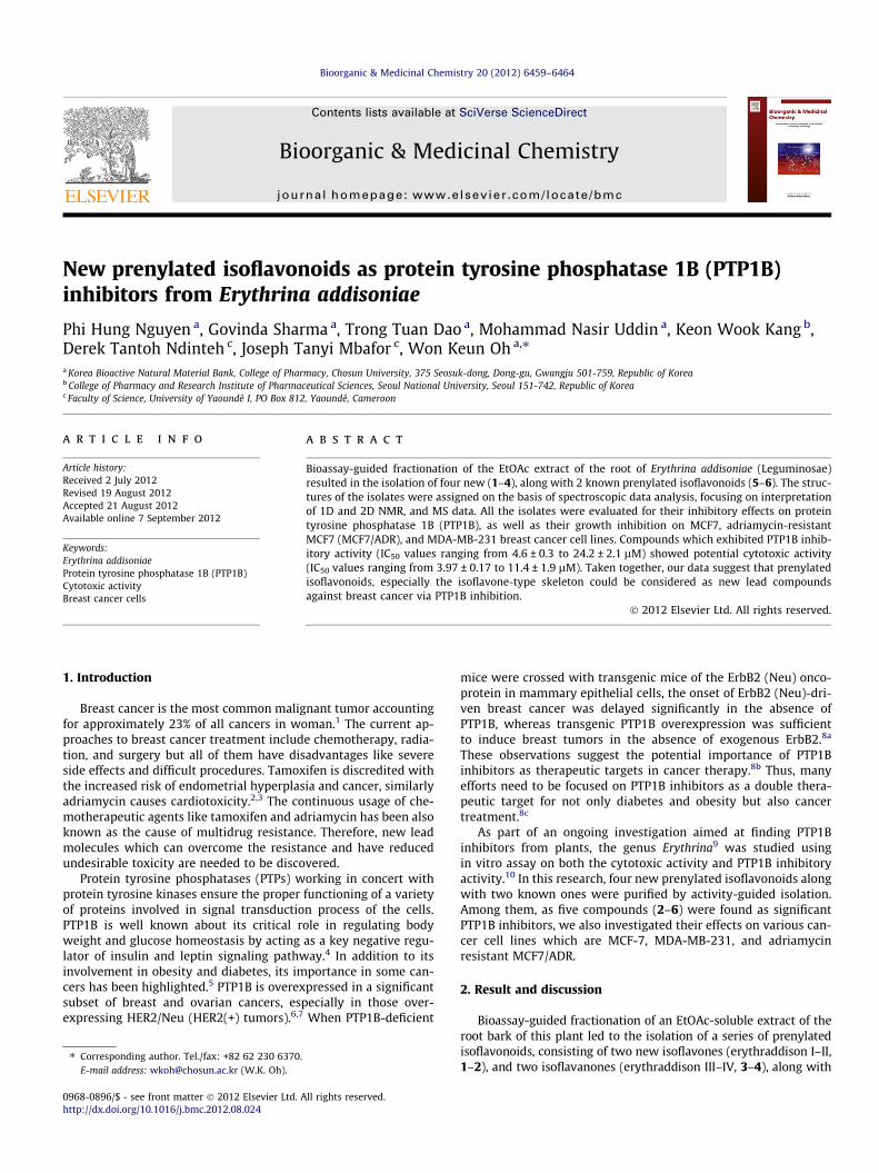
Compounds 1 and 2 were isolated as yellowish amorphouspowders, and their UV spectra exhibited absorption maxima at271, 276 and 272 nm, respectively. The 1H and 13C NMR spectra(Table 1) of compounds 1 and 2 displayed the characteristic signalsof H-2 at dH 7.91 and dH 8.10 (each 1H, s) with corresponding ole-finic oxymethine signals at dC 152.5 and 154.7, respectively, and aketone carbon resonance (dC 179.4–181.3). These observationswere indicative of an isoflavone skeleton.12,13
A molecular formula of compound 1 was determined asC26H28O8 from the molecular ion peak at m/z 468.1784 (calcd forC26H28O8, 468.1776) in the HREIMS spectrum. A pair of coupleddoublets at dH 7.42 and 6.92 (each 2H, d, J = 8.4 Hz, H-20/H-60, H-30/H-50); dC 130.5 (each CH, C-20/60), 115.8 (each CH, C-30/50) wasdeduced as the presence of a para disubstituted ring B, while thatof a hydroxy group at C-40 was established from the correlation be-tween a single proton (dH 5.02, 40-OH) to carbons at dC 115.8 (C-30
and C-50), respectively in the HMBC experiment. The 1H NMR spec-trum displayed characteristic signals attributable to a 2,2-dimeth-ylpyrano ring [dH 6.76 (1H, d, J = 10.0 Hz, H-400), 5.65 (1H, d,J = 9.6 Hz, H-300), 1.48 (3H, s, H-500) and 1.49 (3H, s, H-600). The HMBCcorrelations from H-400 to C-5, and a single proton signal at dH 13.2in the 1H NMR spectrum further indicated that hydroxy group withan intramolecular hydrogen bond was located at C-5 and the 2,2-dimethylpyrano ring was fused at C-6 and C-7. In addition, the1H and 13C NMR spectra of compound 1 exhibited a 1n-methoxy-2n,3-dihydroxy-methylbutyl group [dH 4.70 (1H, d, J = 7.2 Hz, H-1000), 3.70 (1H, d, J = 7.2 Hz, H-2000), 3.39 (3H, s, 1000–OCH3), 1.29(3H, s, H-4000) and 1.20 (3H, s, H-5000); dC 75.1 (C-1000), 65.8 (C-2000),57.7 (C-3000), 57.1 (1000–OCH3), 25.1 (C-4000) and 19.7 (C-5000)]. The loca-tion of each functional group was further demonstrated in theHMBC, and suggested that the 1n-methoxy-2n,3-dihydroxy-meth-ylbutyl group was attached at C-8 by the long-range correlationsfrom proton H-1000 to C-7 (dC 157.7), C-8 (dC 104.2) and C-9 (dC
155.7) (Fig. 2). In addition, HMBC correlations between H3-OCH3/C-1000, H-2000/C-1000, and H3-4000 and H3-5000/C-3000, C-2000, further
OO
H3CO
OH
OH
OHOOH
OHO
O
R1
OH
1
OHO
2 R1 = Prenyl, R2
6 R1 = R2 = H, R3
OHO
OOOH
OCH3
4
2
3
456
8
72'
4'
5'
2''
3''
4''
6'' 1'''
2'''3'' '
4''' 5'''
2
3
45
7
9
2'
4'
5'2''
3''
4''
5''
6''
Figure 1. Chemical structures of isolated com
supported for the elucidation of the 1n-methoxy-2n,3-dihydroxy-methylbutyl group. However, the configurations at C-1000 and C-2000 have not determined due to the low amount of the compoundobtained. Thus, compound 1 was assigned as 5,40-dihydroxy-(1000n-methoxy-2000n,3000-dihydroxy-3000-dimethylbutyl)-[200,200-di-methyl-300,400-dehydro-pyrano-(100,400:-7,6)]isoflavone (as shown inFig. 2), and named erythraddison I.
Compound 2 was also obtained as yellowish amorphous pow-der and its molecular formula of C25H26O5 was determined froma molecular ion peak at m/z 406.1780 (calcd for C25H26O5,406.1780) obtained by HREIMS. The 1H NMR spectrum of com-pound 2 displayed an aromatic ABX spin system [dH 7.05 (d,J = 8.0 Hz, H-60), 6.48 (dd, J = 8.0, 2.8 Hz, H-50) and 6.58 (d,J = 2.8 Hz, H-30)], an aromatic single proton at dH 8.00 (1H, s), butshowed no proton resonance for a hydroxy group at C-5 (dH
12.1–13.5 Hz).14 This was further evidenced by an HMBC correla-tion between H-5 (dH 8.00) and the ketone carbon at C-4 (dC
179.4) (Supplementary data and Fig. 2). The 1H and 13C NMR spec-tra of compound 2 exhibited two prenyl groups (Table 1), and theirplacements were assigned to be at C-6 and C-8 by HMBC experi-ments from H-100 (dH 3.48 (2H, d, J = 7.2 Hz))/C-5 (dC 124.6), C-6(dC 127.6), and C-7 (dC 158.8), and from H-1000 (dH 3.65 (2H, d,J = 7.2 Hz))/C-7, C-8 (dC 114.7), and C-9 (dC 154.3), respectively.1D (Table 1) and HMBC (Fig. 2) NMR data of compound 2 revealedthat three hydroxy groups were attached to C-7, C-20, and C-40,respectively. The hydroxyl position at C-20 was confirmed by HMBCcorrelations between H-30 (dH 6.58 (1H, d, J = 2.8 Hz)) and C-40 (dC
158.3), C-20 (dC 158.0). Thus, compound 2 was characterized asnew natural product, 7,20,40-trihydroxy-6,8-di(cc-dimethylal-lyl)isoflavone, and named erythraddison II.
Compounds 3 and 4 were isolated as yellow amorphous pow-der. The 1H and 13C NMR spectra (Table 1) of compounds 3 and 4displayed an AMX spin system for H-2ax, H-2eq, and H-3, andthe corresponding carbon signals for C-2 (dC 71.2–72.3) and C-3(dC 47.4–51.1), and ketone carbon resonances (dC 197.9–198.2).Their UV spectra showed absorption maxima near 270, 305, and335 nm. These observations were indicative of an isoflavanoneskeleton.15 Compound 3 was obtained as a yellow amorphouspowder with ½a�25
D �11.8 (c 0.11, MeOH). A molecular formula of
OH
R2
O
OHO
R3
5
= OH, R3 = H
= Prenyl
OHO
OCH3OOH
3
1''
2' '
4''
5''
Prenyl group =
pounds 1–6 from Erythrina addisoniae.
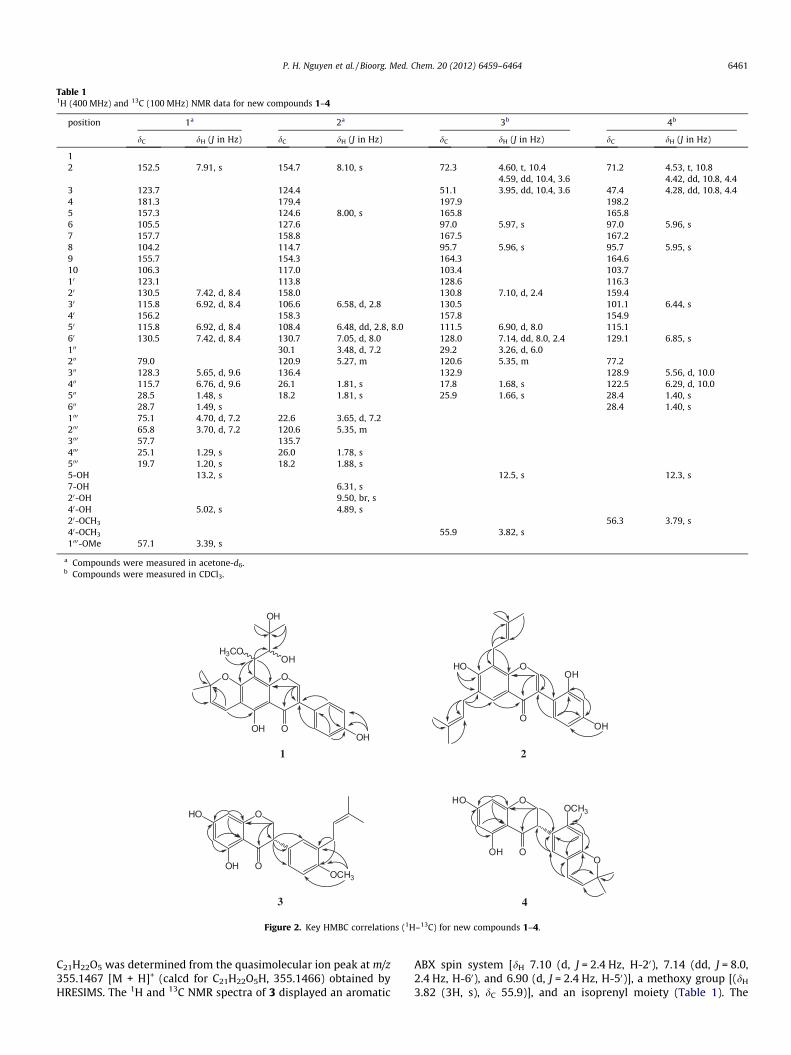
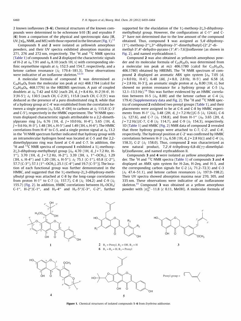
Table 11H (400 MHz) and 13C (100 MHz) NMR data for new compounds 1–4
position 1a 2a 3b 4b
dC dH (J in Hz) dC dH (J in Hz) dC dH (J in Hz) dC dH (J in Hz)
12 152.5 7.91, s 154.7 8.10, s 72.3 4.60, t, 10.4
4.59, dd, 10.4, 3.671.2 4.53, t, 10.8
4.42, dd, 10.8, 4.43 123.7 124.4 51.1 3.95, dd, 10.4, 3.6 47.4 4.28, dd, 10.8, 4.44 181.3 179.4 197.9 198.25 157.3 124.6 8.00, s 165.8 165.86 105.5 127.6 97.0 5.97, s 97.0 5.96, s7 157.7 158.8 167.5 167.28 104.2 114.7 95.7 5.96, s 95.7 5.95, s9 155.7 154.3 164.3 164.610 106.3 117.0 103.4 103.710 123.1 113.8 128.6 116.320 130.5 7.42, d, 8.4 158.0 130.8 7.10, d, 2.4 159.430 115.8 6.92, d, 8.4 106.6 6.58, d, 2.8 130.5 101.1 6.44, s40 156.2 158.3 157.8 154.950 115.8 6.92, d, 8.4 108.4 6.48, dd, 2.8, 8.0 111.5 6.90, d, 8.0 115.160 130.5 7.42, d, 8.4 130.7 7.05, d, 8.0 128.0 7.14, dd, 8.0, 2.4 129.1 6.85, s100 30.1 3.48, d, 7.2 29.2 3.26, d, 6.0200 79.0 120.9 5.27, m 120.6 5.35, m 77.2300 128.3 5.65, d, 9.6 136.4 132.9 128.9 5.56, d, 10.0400 115.7 6.76, d, 9.6 26.1 1.81, s 17.8 1.68, s 122.5 6.29, d, 10.0500 28.5 1.48, s 18.2 1.81, s 25.9 1.66, s 28.4 1.40, s600 28.7 1.49, s 28.4 1.40, s1000 75.1 4.70, d, 7.2 22.6 3.65, d, 7.22000 65.8 3.70, d, 7.2 120.6 5.35, m3000 57.7 135.74000 25.1 1.29, s 26.0 1.78, s5000 19.7 1.20, s 18.2 1.88, s5-OH 13.2, s 12.5, s 12.3, s7-OH 6.31, s20-OH 9.50, br, s40-OH 5.02, s 4.89, s20-OCH3 56.3 3.79, s40-OCH3 55.9 3.82, s1000-OMe 57.1 3.39, s
a Compounds were measured in acetone-d6.b Compounds were measured in CDCl3.
OO
H3CO
OH
OH
OHOOH
OHO
OHO
OH
1 2
OHO
OCH3OOH
OHO
OOOH
OCH3
3 4
Figure 2. Key HMBC correlations (1H–13C) for new compounds 1–4.
P. H. Nguyen et al. / Bioorg. Med. Chem. 20 (2012) 6459–6464 6461
C21H22O5 was determined from the quasimolecular ion peak at m/z355.1467 [M + H]+ (calcd for C21H22O5H, 355.1466) obtained byHRESIMS. The 1H and 13C NMR spectra of 3 displayed an aromatic
ABX spin system [dH 7.10 (d, J = 2.4 Hz, H-20), 7.14 (dd, J = 8.0,2.4 Hz, H-60), and 6.90 (d, J = 2.4 Hz, H-50)], a methoxy group [(dH
3.82 (3H, s), dC 55.9)], and an isoprenyl moiety (Table 1). The
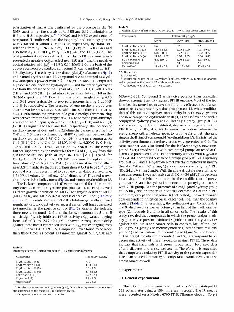
Table 3Growth inhibitory effects of isolated compounds 1–6 against breast cancer cell lines
Compounds Cell lines/IC50a (lM)
MCF7 MCF7/ADR MDA-MB-231
Erythraddison I (1) NA NA NAErythraddison II (2) 11.41 ± 1.97 6.75 ± 1.00 4.57 ± 0.60Erythraddison III (3) 6.84 ± 0.11 9.22 ± 0.21 6.92 ± 0.27Erythraddison IV (4) 11.00 ± 1.02 6.06 ± 0.85 6.34 ± 0.40Echrenone b10 (5) 4.32 ± 0.10 5.76 ± 0.23 3.97 ± 0.17Erysubin F (6) NT NT NTTamoxifenb 11.44 ± 0.9 11.13 ± 0.8 12.41 ± 0.8
NA: Not active.NT: Not tested.
a Results are expressed as IC50 values (lM), determined by regression analysesand expressed as the mean ± SD of three replicates.
b Compound was used as positive control.
6462 P. H. Nguyen et al. / Bioorg. Med. Chem. 20 (2012) 6459–6464
substitution of ring A was confirmed by the presence in the 1HNMR spectrum of the signals at dH 5.96 and 5.97 attributable toH-6 and H-8, respectively.16,17 HMQC and HMBC experiments ofcompound 3 confirmed that the isoprenyl and methoxy groupswere attached to carbons C-30 and C-40, respectively, by HMBC cor-relations from dH 3.26 (H-100)/dC 130.5 (C-30) to 157.8 (C-40) andalso from dH 3.82 (OCH3) to dC 157.8 (C-40) and 111.5 (C-50). Theconfiguration at C-3 was inferred to be S by its CD spectrum, whichpresented a negative Cotton effect near 330 nm,18 and the negativeoptical rotation with ½a�25
D �11.8 (c 0.11, MeOH). On the basis of theabove spectroscopic studies, compound 3 was identified as 3(S)-5,7-dihydroxy-40-methoxy-30-(cc-dimethylallyl)isoflavanone (Fig. 2)and named erythraddison III. Compound 4 was obtained as a yel-low amorphous powder with ½a�25
D �5.6 (c 0.15, MeOH). Compound4 possessed one chelated hydroxy at C-5 and the other hydroxy atC-7 from the presence of the signals at dH 12.33 (1H, s, 5-OH), 5.96(1H, s), and 5.95 (1H, s) attributable to protons H-6 and H-8 in the1H NMR spectrum.16,17 Two sharp one proton singlets at dH 6.85and 6.44 were assignable to two para protons in ring B at H-60
and H-30, respectively. The presence of one methoxy group wasalso shown by signal at dH 3.79 with corresponding carbon at dC
56.3. Furthermore, the presence of a gem-dimethyl chromene ringwas derived from the 6H singlet at dH 1.40 due to the gem-dimethylgroup and an AB spin system at dH 5.56 (d, J = 10.0) and 6.29 (d,J = 10.0) assignable to H-300 and H-400, respectively. The location ofmethoxy group at C-20 and the 2,2-dimethylpyrano ring fused toC-40 and C-50 were confirmed by HMBC correlations between themethoxy protons (dH 3.79)/C-20 (dC 159.4), one para proton at dH
6.44 (H-30)/C-20 and C-40 (dC 154.9), H-400 (dH 6.29)/C-40, C-50 (dC
128.9), and C-60 (dC 129.1), and H-300 (dH 5.56)/C-60. These werefurther supported by the molecular formula of C21H20O6 from thequasimolecular ion peak at m/z 369.1267 [M+H]+ (calcd forC21H20O6H, 369.1276) in the HRESIMS spectrum. The optical rota-tion value ½a�25
D �5.6 (c 0.15, MeOH) and the negative Cotton effectnear 328 nm indicate that the configuration at C-3 is to be S.18 Com-pound 4 was thus determined to be a new prenylated isoflavanone,3(S)-5,7-dihydroxy-20-methoxy-[200,200-dimethyl-300,400-dehydro-pyr-ano-(100,400:-40,50)]isoflavanone (Fig. 2), and named erythraddison IV.
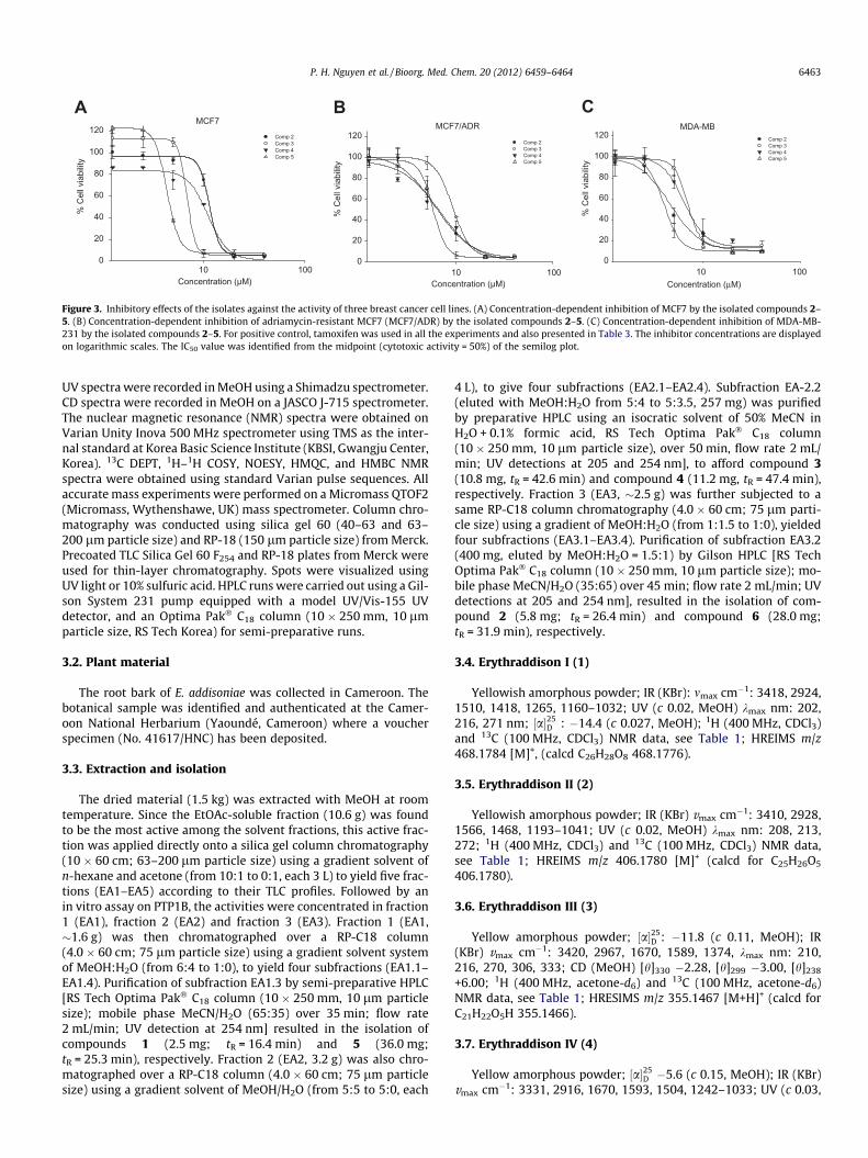
The isolated compounds (1–6) were evaluated for their inhibi-tory effects on protein tyrosine phosphatase-1B (PTP1B), as wellas their growth inhibition on MCF7, adriamycin-resistant MCF7(MCF7/ADR), and MDA-MB-231 breast cancer cell lines (Tables 2and 3). Compounds 2–6 with PTP1B inhibition generally showedsignificant cytotoxic activity on several cancer cell lines comparedto tamoxifen as the positive control (Fig. 3). Among the isolates,three new compounds 2–4 and the known compounds 5 and 6which significantly inhibited PTP1B activity (IC50 values rangingfrom 4.6 ± 0.3 to 24.2 ± 2.1 lM), showed strong cytotoxicityagainst three breast cancer cell lines with IC50 values ranging from3.97 ± 0.17 to 11.41 ± 1.97 lM. Compound 5 was found to be morethan three times as potent as tamoxifen against MCF7/ADR and
Table 2Inhibitory effects of isolated compounds 1–6 against PTP1B enzyme
Compounds Inhibitory activitya
Erythraddison I (1) >30Erythraddison II (2) 17.4 ± 1.1Erythraddison III (3) 4.6 ± 0.3Erythraddison IV (4) 13.8 ± 1.8Echrenone b10 (5) 24.2 ± 2.1Erysubin F (6) 7.8 ± 0.5Ursolic acidb 3.6 ± 0.2
a Results are expressed as IC50 values (lM), determined by regression analysesand expressed as the mean ± SD of three replicates.
b Compound was used as positive control.
MDA-MB-231. Compound 3 with twice potency than tamoxifenshowed strongest activity against PTP1B enzyme. Most of the iso-lates bearing prenyl group gave the inhibitory effects on both breastcancer cells and protein tyrosine phosphatase-1B, while modifica-tion of this moiety displayed non-activity in both assay systems.The new compound erythraddison III (3) is an isoflavanone with aconjugated hydroxy group at C-5, bearing a prenyl group at C-30
and a 40-methyl ether substituted, displayed strongest effect onPTP1B enzyme (IC50 4.6 lM). However, cyclization between theprenyl group with a hydroxy group to form the 2,2-dimethylpyranoring on the B ring of compound 4 (erythraddison IV), diminished theactivity even through a methoxy group was presented at C-20. Thesame manner was also found for the isoflavone-type, new com-pound 2 (erythraddison II) with two prenyl groups attached at C-6 and C-8 possessed high PTP1B inhibitory activity with IC50 valueof 17.4 lM. Compound 5 with one prenyl group at C-8, a hydroxygroup at C-5, and a 1-hydroxy-1-methylethyldihydrofuran moiety(fused at C-6 and C-7 in ring A) showed weaker activity on PTP1B(IC50 24.2 lM) than 2 and 6. With the same structure skeleton, how-ever compound 1 was not active at all (IC50 > 30 lM). This decreasein activity of 1 might be induced by the modification of prenylgroup at C-8, and the cyclization between the prenyl group at C-6with 7-OH group. And the presence of a conjugated hydroxy groupat C-5 may also be responsible for this decrease. All of the PTP1Binhibitors, except for compound 6 (not tested), showed strongerdose-dependent inhibition on all cancer cell lines than the positivecontrol (Table 3). Interestingly, the isoflavone-type (Compounds 2and 5) displayed a stronger potency than that of the isoflavanone-type (Compounds 3 and 4) in all cancer cells. The results of thisstudy revealed that compounds in which the prenyl and/or meth-oxy groups are present exhibited significant inhibitory activitiesagainst both PTP1B and cancer cells. In contrast, lack of the lipo-philic groups (prenyl and methoxy moieties) in the structure (Com-pound 1) and cyclization (Compounds 1 and 4), and/or modificationof the prenyl moiety (Compounds 1 and 5), are responsible fordecreasing activity of these flavonoids against PTP1B. These dataindicate that flavonoids with prenyl group might be a new classof anti-diabetes and anticancer agents. Therefore, it is suggestedthat compounds reducing PTP1B activity or the genetic expressionlevels can be used for treating not only diabetes and obesity but alsobreast cancer as well.
3. Experimental
3.1. General experimental
The optical rotations were determined on a Rudolph Autopol AP589 polarimeter using a 100 mm glass microcell. The IR spectrawere recorded on a Nicolet 6700 FT-IR (Thermo electron Corp.).
MCF7
Concentration (µM)10 100
% C
ell v
iabi
lity
0
20
40
60
80
100
120Comp 2Comp 3Comp 4Comp 5
MCF7/ADR
Concentration (µM)10 100
% C
ell v
iabi
lity
0
20
40
60
80
100
120Comp 2Comp 3Comp 4Comp 5
MDA-MB
Concentration (µM)10 100
% C
ell v
iabi
lity
0
20
40
60
80
100
120 Comp 2Comp 3Comp 4 Comp 5
BA C
Figure 3. Inhibitory effects of the isolates against the activity of three breast cancer cell lines. (A) Concentration-dependent inhibition of MCF7 by the isolated compounds 2–5. (B) Concentration-dependent inhibition of adriamycin-resistant MCF7 (MCF7/ADR) by the isolated compounds 2–5. (C) Concentration-dependent inhibition of MDA-MB-231 by the isolated compounds 2–5. For positive control, tamoxifen was used in all the experiments and also presented in Table 3. The inhibitor concentrations are displayedon logarithmic scales. The IC50 value was identified from the midpoint (cytotoxic activity = 50%) of the semilog plot.
P. H. Nguyen et al. / Bioorg. Med. Chem. 20 (2012) 6459–6464 6463
UV spectra were recorded in MeOH using a Shimadzu spectrometer.CD spectra were recorded in MeOH on a JASCO J-715 spectrometer.The nuclear magnetic resonance (NMR) spectra were obtained onVarian Unity Inova 500 MHz spectrometer using TMS as the inter-nal standard at Korea Basic Science Institute (KBSI, Gwangju Center,Korea). 13C DEPT, 1H–1H COSY, NOESY, HMQC, and HMBC NMRspectra were obtained using standard Varian pulse sequences. Allaccurate mass experiments were performed on a Micromass QTOF2(Micromass, Wythenshawe, UK) mass spectrometer. Column chro-matography was conducted using silica gel 60 (40–63 and 63–200 lm particle size) and RP-18 (150 lm particle size) from Merck.Precoated TLC Silica Gel 60 F254 and RP-18 plates from Merck wereused for thin-layer chromatography. Spots were visualized usingUV light or 10% sulfuric acid. HPLC runs were carried out using a Gil-son System 231 pump equipped with a model UV/Vis-155 UVdetector, and an Optima Pak� C18 column (10 � 250 mm, 10 lmparticle size, RS Tech Korea) for semi-preparative runs.
3.2. Plant material
The root bark of E. addisoniae was collected in Cameroon. Thebotanical sample was identified and authenticated at the Camer-oon National Herbarium (Yaoundé, Cameroon) where a voucherspecimen (No. 41617/HNC) has been deposited.
3.3. Extraction and isolation
The dried material (1.5 kg) was extracted with MeOH at roomtemperature. Since the EtOAc-soluble fraction (10.6 g) was foundto be the most active among the solvent fractions, this active frac-tion was applied directly onto a silica gel column chromatography(10 � 60 cm; 63–200 lm particle size) using a gradient solvent ofn-hexane and acetone (from 10:1 to 0:1, each 3 L) to yield five frac-tions (EA1–EA5) according to their TLC profiles. Followed by anin vitro assay on PTP1B, the activities were concentrated in fraction1 (EA1), fraction 2 (EA2) and fraction 3 (EA3). Fraction 1 (EA1,�1.6 g) was then chromatographed over a RP-C18 column(4.0 � 60 cm; 75 lm particle size) using a gradient solvent systemof MeOH:H2O (from 6:4 to 1:0), to yield four subfractions (EA1.1–EA1.4). Purification of subfraction EA1.3 by semi-preparative HPLC[RS Tech Optima Pak� C18 column (10 � 250 mm, 10 lm particlesize); mobile phase MeCN/H2O (65:35) over 35 min; flow rate2 mL/min; UV detection at 254 nm] resulted in the isolation ofcompounds 1 (2.5 mg; tR = 16.4 min) and 5 (36.0 mg;tR = 25.3 min), respectively. Fraction 2 (EA2, 3.2 g) was also chro-matographed over a RP-C18 column (4.0 � 60 cm; 75 lm particlesize) using a gradient solvent of MeOH/H2O (from 5:5 to 5:0, each
4 L), to give four subfractions (EA2.1–EA2.4). Subfraction EA-2.2(eluted with MeOH:H2O from 5:4 to 5:3.5, 257 mg) was purifiedby preparative HPLC using an isocratic solvent of 50% MeCN inH2O + 0.1% formic acid, RS Tech Optima Pak� C18 column(10 � 250 mm, 10 lm particle size), over 50 min, flow rate 2 mL/min; UV detections at 205 and 254 nm], to afford compound 3(10.8 mg, tR = 42.6 min) and compound 4 (11.2 mg, tR = 47.4 min),respectively. Fraction 3 (EA3, �2.5 g) was further subjected to asame RP-C18 column chromatography (4.0 � 60 cm; 75 lm parti-cle size) using a gradient of MeOH:H2O (from 1:1.5 to 1:0), yieldedfour subfractions (EA3.1–EA3.4). Purification of subfraction EA3.2(400 mg, eluted by MeOH:H2O = 1.5:1) by Gilson HPLC [RS TechOptima Pak� C18 column (10 � 250 mm, 10 lm particle size); mo-bile phase MeCN/H2O (35:65) over 45 min; flow rate 2 mL/min; UVdetections at 205 and 254 nm], resulted in the isolation of com-pound 2 (5.8 mg; tR = 26.4 min) and compound 6 (28.0 mg;tR = 31.9 min), respectively.
3.4. Erythraddison I (1)
Yellowish amorphous powder; IR (KBr): mmax cm�1: 3418, 2924,1510, 1418, 1265, 1160–1032; UV (c 0.02, MeOH) kmax nm: 202,216, 271 nm; ½a�25
D : �14.4 (c 0.027, MeOH); 1H (400 MHz, CDCl3)and 13C (100 MHz, CDCl3) NMR data, see Table 1; HREIMS m/z468.1784 [M]+, (calcd C26H28O8 468.1776).
3.5. Erythraddison II (2)
Yellowish amorphous powder; IR (KBr) vmax cm�1: 3410, 2928,1566, 1468, 1193–1041; UV (c 0.02, MeOH) kmax nm: 208, 213,272; 1H (400 MHz, CDCl3) and 13C (100 MHz, CDCl3) NMR data,see Table 1; HREIMS m/z 406.1780 [M]+ (calcd for C25H26O5
406.1780).
3.6. Erythraddison III (3)
Yellow amorphous powder; ½a�25D : �11.8 (c 0.11, MeOH); IR
(KBr) vmax cm�1: 3420, 2967, 1670, 1589, 1374, kmax nm: 210,216, 270, 306, 333; CD (MeOH) [h]330 �2.28, [h]299 �3.00, [h]238
+6.00; 1H (400 MHz, acetone-d6) and 13C (100 MHz, acetone-d6)NMR data, see Table 1; HRESIMS m/z 355.1467 [M+H]+ (calcd forC21H22O5H 355.1466).
3.7. Erythraddison IV (4)
Yellow amorphous powder; ½a�25D �5.6 (c 0.15, MeOH); IR (KBr)
vmax cm�1: 3331, 2916, 1670, 1593, 1504, 1242–1033; UV (c 0.03,
6464 P. H. Nguyen et al. / Bioorg. Med. Chem. 20 (2012) 6459–6464
MeOH) kmax nm: 210, 233, 272, 304, 335; CD (MeOH) [h]240 +5.50,[h]285 +3.45, [h]328 �1.15; 1H (400 MHz, acetone-d6) and 13C(100 MHz, acetone-d6) NMR data, see Table 1; HRESIMS m/z369.1267 [M+H]+ (calcd for C21H20O6H, 369.1276).
3.8. PTP1B assay
PTP1B (human, recombinant) was purchased from BIOMOLInternational LP (USA) and the enzyme activity was measuredusing p-nitrophenyl phosphate (p-NPP) as a substrate. To each96-well (final volume: 200 lL) were added 2 mM p-NPP and PTP1B(0.05–0.1 lg) in a buffer containing 50 mM citrate (pH 6.0), 0.1 MNaCl, 1 mM EDTA, and 1 mM dithiothreitol (DTT) with or withouttest compounds. Following incubation at 37 �C for 30 min, thereaction was terminated with 10 M NaOH. The amount of pro-duced p-nitrophenol was estimated by measuring the absorbanceat 405 nm. The nonenzymatic hydrolysis of 2 mM p-NPP wascorrected by measuring the increase in absorbance at 405 nmobtained in the absence of PTP1B enzyme.
3.9. Cell culture
The screening cell lines (MCF7 and MDA-MB-231 human breastcarcinoma cells, and the Adriamycin resistant cell line MCF7/ADR)were maintained at 37 �C in a humidified atmosphere containing5% CO2. All the media used were DMEM supplemented with 10%heat-inactivated fetal bovine serum, 4.5 g/L D-glucose, 100 mg/Lsodium pyruvate and L-glutamine. The cells were subculturedevery 3 days using the standard trypsinization procedure.
3.10. Cytotoxicity assay
The cell viability was assessed using a MTT (3-(4,5-dimethyl-2-thiazolyl)-2,5-diphenyl-2H-tetrazolium bromide MTT meth-ylthiazolyldiphenyl-tetrazolium bromide) based cytotoxicity assayto determine the IC50 of the isolated compounds (M5655 SIGMA;Sigma–Aldrich). In these assays, 1 � 104 ER positive, highly inva-sive (MDA-MB), ER negative weakly-invasive (MCF7), Adriamycinresistant (MCF7/ADR) cells in 100 lL of the culture medium perwell were seeded in 96-well plates and allowed to adhere for24 h prior to treatment. At various concentrations the cells in 96well plates were treated and incubated for 48 h. The final concen-tration of DMSO in the culture medium was maintained at 0.05%(v/v) to avoid solvent toxicity. Subsequently, 20 lL of the 2 mg/mL MTT solution was added to each well of the plate and incubated4 h. Then the absorbance was measured at 550 nm. The percentagecell viability is expressed as toxicities of the compounds, where thehigher the toxicity, the lower the cell viability. Percentage cellviability is defined as the absorbance in the experiment wellcompared to that in the control wells. The cytotoxicity results
are expressed as the mean ± standard deviation and represent theconcentration inhibiting 50% cell growth (IC50). Each experimentwas carried out in triplicates.
Acknowledgments
This research was supported in part by Grants from the KoreaHealthcare Technology R&D Project, Ministry for Health, Welfare& Family Affairs, Republic of Korea (A084335) and from the Pro-curement and Development of Foreign Biological Resources(2011-00402) funded by the Ministry of Education Science andTechnology of the Korean government.
Supplementary data
Supplementary data (1D (1H and 13C) and 2D (including COSY,HSQC, and HMBC) NMR spectra of the new compounds (1�4))associated with this article can be found, in the online version, athttp://dx.doi.org/10.1016/j.bmc.2012.08.024.
References and notes
1. Ferlay, J.; Shin, H. R.; Bray, F.; Forman, D.; Mathers, C.; Parkin, D. M. Int. J. Cancer2010, 127, 2893.
2. Berthiaume, J. M.; Wallace, K. B. Cell Biol. Toxicol. 2006, 23, 15.3. Yasue, A.; Hasegawa, K.; Udagawa, Y. Hum. Cell 2011, 24, 65.4. Johnson, T. O.; Ermolieff, J.; Jirousek, M. R. Nat. Rev. Drug Disc. 2002, 1, 696.5. Combs, A. P. J. Med. Chem. 2010, 53, 2333.6. Wiener, J. R.; Kerns, B. J.; Harvey, E. L.; Conaway, M. R.; Iglehart, J. D.; Berchuck,
A., ; Bast, R. C., Jr. J. Natl. Cancer Inst. 1994, 86, 372.7. Tanner, M. M.; Grenman, S.; Koul, A.; Johannsson, O.; Meltzer, P.; Pejovic, T.;
Borg, A.; Isola, J. J. Clin. Cancer Res. 1833, 2000, 6.8. (a) Julien, S. G.; Dubé, N.; Read, M.; Penney, J.; Paquet, M.; Han, Y.; Kennedy, B.
P.; Muller, W. J.; Tremblay, M. L. Nat. Genet. 2007, 39, 338; (b) Tonks, N. K.;Muthuswamy, S. K. Cancer Cell 2007, 11, 214; (c) Yip, S. C.; Saha, S.; Chernoff, J.Trends Biochem. Sci. 2010, 35, 442.
9. (a) Djiogue, S.; Halabalaki, M.; Alexi, X.; Njamen, D.; Fomum, Z. T.; Alexis, M. N.;Skaltsounis, A. L. J. Nat. Prod. 2009, 72, 1603; (b) Kaur, K.; Jain, M.; Kaur, T.; Jain,R. Bioorg. Med. Chem. 2009, 17, 3229.
10. (a) Nguyen, P. H.; Le, T. V. T.; Thuong, P. T.; Dao, T. T.; Ndinteh, D. T.; Mbafor, J.T.; Kang, K. W.; Oh, W. K. Bioorg. Med. Chem. Lett. 2009, 19, 6745; (b) Dao, T. T.;Nguyen, P. H.; Thuong, P. T.; Kang, K. W.; Na, M. K.; Ndinteh, D. T.; Mbafor, J. T.;Oh, W. K. Phytochemistry 2009, 70, 2053.
11. (a) Tanaka, H.; Etoh, H.; Watanabe, N.; Shimizu, H.; Ahmad, M.; Rizwani, G. H.Phytochemistry 2001, 56, 769; (b) Tanaka, H.; Doi, M.; Etoh, H.; Watanabe, N.;Shimizu, H.; Hirata, M.; Ahmad, M.; Qurashi, I.; Khan, M. R. J. Nat. Prod. 2001,64, 1336.
12. Mabry, T. J.; Markham, K. R.; Thomas, M. B. The systematic identification offlavonoids; Springer: New York, 1970. pp 41–64.
13. Agrawal, P. K. Carbon-13 NMR of flavonoids; Elsevier: New York, 1989. p 507.14. Na, M. K.; Jang, J. P.; Njamen, D.; Mbafor, J. T.; Fomum, Z. T.; Kim, B. Y.; Oh, W.
K.; Ahn, J. S. J. Nat. Prod. 2006, 69, 1572.15. Zhao, M.; Duan, J. A.; Che, C. T. Phytochemistry 2007, 68, 1471.16. Atindehou, K. K.; Queiroz, E. F.; Terreaux, C.; Traore, D.; Hostettmann, K. Planta
Med. 2002, 68, 181.17. Iinuma, M.; Ohyama, M.; Tanaka, T. Phytochemistry 1994, 37, 1713.18. Slade, D.; Ferreira, D.; Marais, J. P. J. Phytochemistry 2005, 66, 2177.