SUPPLEMENTARY MATERIAL Unprecedented New Nonadecyl para-Hydroperoxycoumaroate isolated from...
13
SUPPLEMENTARY MATERIAL Unprecedented New Nonadecyl para-Hydroperoxycoumaroate isolated from Erythrina excelsa and its Cytotoxic Activity Guy M. N. Kwamou a , Louis P. Sandjo a, *, Victor Kuete b,c , Anaelle A. K. Wandja a , Simplice B. Tankeo b , Thomas Efferth** c and Augustin E. Nkengfack a a Department of Organic Chemistry, University of Yaoundé I, P. O. Box 812 Yaoundé, Cameroon b Department of Biochemistry, Faculty of Science, University of Dschang, P. O. Box 67 Dschang Cameroon c Department of Pharmaceutical Biology, Institute of Pharmacy and Biochemistry, University of Mainz, Staudinger weg 5, 55128 Mainz, Germany Corresponding author: Dr. Louis P. Sandjo E-mail: [email protected] Prof. Thomas Efferth Phone: +49-6131-3925751; Fax: +49-6131- 39-23752; E-mail: [email protected] 1
-
Upload
independent -
Category
Documents
-
view
0 -
download
0
Transcript of SUPPLEMENTARY MATERIAL Unprecedented New Nonadecyl para-Hydroperoxycoumaroate isolated from...

SUPPLEMENTARY MATERIAL
Unprecedented New Nonadecyl para-Hydroperoxycoumaroate isolated
from Erythrina excelsa and its Cytotoxic Activity
Guy M. N. Kwamoua, Louis P. Sandjoa,*, Victor Kueteb,c, Anaelle
A. K. Wandjaa, Simplice B. Tankeob, Thomas Efferth**c and
Augustin E. Nkengfacka
aDepartment of Organic Chemistry, University of Yaoundé I, P. O. Box 812 Yaoundé,
CameroonbDepartment of Biochemistry, Faculty of Science, University of Dschang, P. O. Box 67
Dschang CamerooncDepartment of Pharmaceutical Biology, Institute of Pharmacy and Biochemistry,
University of Mainz, Staudinger weg 5, 55128 Mainz, Germany
Corresponding author:
Dr. Louis P. Sandjo E-mail: [email protected]
Prof. Thomas Efferth Phone: +49-6131-3925751; Fax: +49-6131-
39-23752; E-mail: [email protected]
1

Abstract: A new unprecedented cinnamate derivative (1) was
obtained from Erythrina excelsa (Leguminosae) and identified as
nonadecyl para-hydroperoxycoumaroate. This compound was isolated
together with three known compounds namely lupeol (2), mixture
of Sitosterol and Stigmasterol (3), and Isoneorautenol (4).
Their structures were established on the base of NMR and mass
spectroscopic data in conjunction to those reported in the
literature. Compound 1 was evaluated for its capability of
inhibiting cancer cell lines and a panel of microbial strains
growth. It turned out that 1 is moderately to significantly
cytotoxic against six cancer cell lines and shows weak to no
antimicrobial activity.
Keywords: Anticancer activity; Drug resistance; para-
Hydroperoxycoumaroate; Fabaceae.
2

Figure S1 HRMS-ESI spectrum
Figure S2 1H NMR spectrum (CDCl3, 400 MHz)
3
cyclohexane
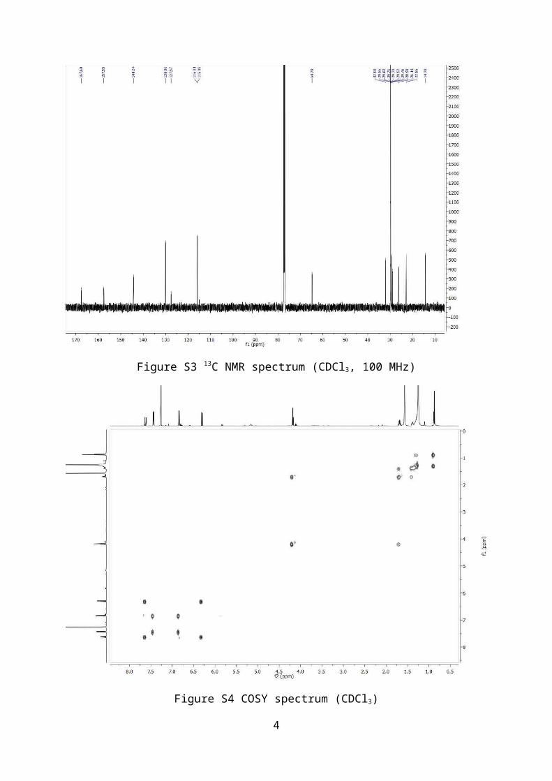
Figure S3 13C NMR spectrum (CDCl3, 100 MHz)
Figure S4 COSY spectrum (CDCl3)
4

Figure S5 HSQC spectrum (CDCl3)
Figure S6 HMBC spectrum (CDCl3)
5

Figure S7 NOESY spectrum (CDCl3)
Figure S8 Enlarged NOESY spectrum (CDCl3)
6

Figure S9 LC-ESI MSChromatogram og compound 1
7

Figure S10: Key HMBC correlations of compound 1
Figure S11: NOESY and COSY correlations of compound 1
Figure S12: proposal of biosynthetic pathway of compound 1
8

Figure S13: IR spectrum of compound 1
1. Antibacterial assay
The studied microorganisms included reference (ATCC) and
multidrug resistant strains of Pseudomonas aeruginosa (PA01),
Klebsiella pneumoniae (KP55, KP63), Escherichia coli (ATCC8739, AG100,
AG102), and Enterobacter aerogenes (ATCC13048, EA3, EA27, CM64)
obtained from the American Type Culture Collection. They were
maintained on agar slant at 4 oC and sub-cultured on a fresh
appropriate agar plates 24 h prior to any antimicrobial test.
Nutrient Agar and Sabouraud Glucose Agar were used for the
activation of bacteria and fungi respectively. The Mueller
Hinton Broth (MHB) was used for the MIC and MMC
determinations. The Mueller Hinton Agar (MHA) was also used
for the determination of the MMC on these species (Kuete et
al., 2011). Tetracycline (Sigma-Aldrich, St. Quentin
9

Fallavier, France) was used as reference antibiotics (RA)
respectively against bacteria. p-Iodonitrotetrazolium chloride
(INT, Sigma-Aldrich) was used as microbial growth indicator
(Mativandlela et al., 2006). The MIC determinations on
bacteria was conducted using rapid INT colorimetric assay
according to described methods (Mativandlela et al., 2006)
with some modifications. Briefly, the test sample was first of
all dissolved in 10% (v/v) DMSO/MHB to give a final
concentration of 512 µg/mL and serially diluted twofold to
obtain concentration ranges. 100 µL of each concentration was
added in a well (96-well microplate) containing 95 µL of MHB
and 5 µL of inoculum (standardized at 1.5×10⁶ CFU/mL by
adjusting the optical density to 0.1 at 600 nm SHIMADZU UV-
120-01 spectrophotometer) (Tereschuk et al., 1997). The final
concentration of DMSO in the well was less than 3%
(preliminary analyses with 3% (v/v) DMSO do not alter the
growth of the test organisms). The negative control well
consisted of 195 µl of MHB and 5 µl of the standard inoculum
(Zgoda and Porter 200). The plates were covered with a sterile
plate sealer, then agitated to mix the contents of the wells
using a plate shaker and incubated at 37 oC for 24 h. The assay
was repeated three times in triplicate. The MIC of samples was
detected following addition (40 µL) of 0.2 mg/mL p-
iodonitrotetrazolium chloride and incubation at 37 oC for 30
min (Mativandlela et al., 2006). Viable microorganisms reduced
the yellow dye to a pink colour. MIC was defined as the lowest
sample concentration that prevented this change and exhibited
complete inhibition of bacterial growth (Kuete et al., 2008).
10

2. Cytotoxicity Assay
The resazurin reduction assay (O’Brien et al., 2000) was
performed to assess the cytotoxicity of compound 1 and
doxorubicin towards various sensitive and drug-resistant
cancer cell lines, including the leukemia CCRF-CEM and
CEM/ADR5000, breast MDA-MB231, colon HCT116, glioblastoma
U87MG and hepatocarcinoma HepG2. The assay is based on the
reduction of the indicator dye, resazurin, to the highly
fluorescent resorufin by viable cells. Non-viable cells
rapidly lose their metabolic capacity to reduce resazurin and,
thus, do not produce fluorescent signals anymore. Briefly,
adherent cells were detached by treatment with 0.25%
trypsin/EDTA (Invitrogen, Darmstadt Germany) and an aliquot of
1 × 10⁴ cells was placed in each well of a 96-well cell culture
plate (Thermo Scientific, Langenselbold, Germany) in a total
volume of 200 μL. Cells were allowed to attach overnight and
then were treated with different concentrations of compounds.
For suspension cells, aliquots of 2 × 10⁴ cells per well were
seeded in 96-well-plates in a total volume of 100 μL. The
studied compound was immediately added in varying
concentrations in an additional 100 μL of culture medium to
obtain a total volume of 200 μl/well. After 72 h, resazurin
(Sigma-Aldrich, Schnelldorf, Germany) (20 μL, 0.01% w/v) in
distilled H2O was added to each well and the plates were
incubated at 37 °C for 4 h. Fluorescence was measured on an
Infinite M2000 ProTM plate reader (Tecan, Crailsheim, Germany)
using an excitation wavelength of 544 nm and an emission
11

wavelength of 590 nm. Each assay was done at least twice with
six replicates each. The viability was evaluated based on a
comparison with untreated cells. IC50 values represent the
compound concentrations required to inhibit 50% of cell
proliferation and were calculated from a calibration curve by
linear regression using Microsoft Excel.
References
De-Eknamkul, W., Potduang B. Biosynthesis of beta-sitosterol
and stigmasterol in Croton sublyratus proceeds via a mixed
origin of isoprene units. Phytochemistry 62, 389–398
(2003).
Iinuma, M., Ohyama, M., Tanaka T. Flavonoids in roots of
Sophora prostrate. Phytochemistry, 38, 539–543 (1995).
Lee, D.-Y., Jung, L., Park, J.-H., Yoo, K.-H., Chung, I.-S.,
Baek N.-I. Cytotoxic triterpenoids from Cornus kousa fruits.
Chem. Nat. Compd. 46, 142–145 (2010).
Mativandlela, S. P. N., Lall, N., Meyer, J. J. M.
Antibacterial, antifungal and antitubercular activity of
(the roots of) Pelargonium reniforme (CURT) and Pelargonium
sidoides (DC) (Geraniaceae) root extracts S. Afr. J. Bot.,
72, 232–237 (2006).
Kuete, V., Kamga, J., Sandjo, L. P., Ngameni, B., Poumale, H.
M., Ambassa, P., Ngadjui B. T. Antimicrobial activities of
the methanol extract, fractions and compounds from Ficus
polita Vahl. (Moraceae). BMC Complement Altern. Med. 11, 1-
6 (2011).
12

Kuete, V., Ngameni, B., Simo, F. C. C., Kengap, T. R.,
Ngadjui, B. T., Meyer, J. J. M., Lall, N., Kuiate J. R.
Antimicrobial activity of the crude extracts and compounds
from Ficus chlamydocarpa and Ficus cordata (Moraceae). J.
Ethnopharmacol., 120, 17–24 (2008).
O’Brien, J., Wilson, I., Orton, T., Pognan F. Investigation of
the Alamar Blue (resazurin) fluorescent dye for the
assessment of mammalian cell cytotoxicity. Eur. J.
Biochem. 267, 5421–5426 (2000).
Tereschuk, M. L., Riera, M. V. Q., Castro, G. R., Abdala L. R.
Antimicrobial activity of flavonoids from leaves of
Tagetes minuta. J. Ethnopharmacol. 56, 227–232 (1997).
Zgoda, J. R., Porter J. R. A Convenient Microdilution method
for screening natural products against Bacteria and Fungi.
Pharmaceut. Biol., 39, 221–225 (2001).
13