SEEDLING CHARACTERISTICS OF ERYTHRINA SUBEROSA ROXB.
17
INT. J. BIOL. BIOTECHN., 11 (4): 563-579, 2014. SEEDLING CHARACTERISTICS OF ERYTHRINA SUBEROSA ROXB. D. Khan, Zulfiqar Ali Sahito and M. Javed Zaki Department of Botany, University of Karachi, Karachi-75270, Pakistan. ABSTRACT Seedling characteristics of Erythrina suberosa Roxb. are described. Its seeds were collected from a tree growing in the Campus of University of Karachi and germinated without any dormancy breaking treatment in pots filled with garden sandy loam soil maintained at 75% MWHC. Maximum germination was 50% achieved within a week. The seedlings were studied when they were 20-day (younger) and 50-day old (elder). The seedling was of Phanerocotylar – Epigeal Reserve type. The major allocation of biomass in 20-day seedlings was in leaves and in 50-day seedlings in leaves and hypocotylar stem. The major growth spur in seedlings during the 30-day period from 20 th to 50 th day was in hypocotylar and epicotylar stem and root. Tap root had profuse laterals. Numerous root nodules 3.5 - 5mm in diameter were present generally on the main root. Cotyledons were large, green fleshy –food laden, concave inside and convex outside with no visible venation. They were wholly consumed within 50 days after germination. Internode size reduced from base to apex regularly. The hypocotyl was green, shining and provided with little prickles. Epicotyl is hairy. The primary leaves were simple opposite, erect, glaucous dorsally and pubescent ventrally. The subsequent leaves were pinnately trifoliate (ternate) with three leaflets. Each leaf had small, green and linear-lanceolate stipules. Glanduliform stiples present. Epicotyl was longer than hypocotyl. The total leaf area of elder seedlings was (209.08 ± 15.71 cm 2 ) -1.6 times to that in the younger seedlings. The leaf venation was pinnate camptodromous (festooned brachidodromous) type. Vein-endings were straight or curved and unbranched. Two types of trichomes were seen – branched trichomes and capitate glandular trichomes. The leaves were hypo-amphistomatic – paucistomatic dorsally and multistomatic ventrally. The cotyledonary stomata were of paracytic type but on ventral surface of leaf five types of stomata (sensu Prabhakar, 2004) - paracytic, anisocytic, anisotricytic, anomocytic and staurocytic were present; paracytic being the most abundant and staurocytic the least. Stomata on dorsal side of leaf were rare and of paracytic type only along the main vein. Both surfaces of leaf had capitate glandular trichomes (6.16 per mm 2 on ventral surface and much infrequent on the dorsal side). The number of stomata on ventral surface of the leaf tended to be normally-distributed amongst the 100 sampling fields of the microscope vision (each of 0.10174 mm 2 ) at 45 x 10 X magnification. The mean density of stomata per mm 2 was 110.28 ± 2.07 (68.80 – 157.3; CV: 18.73%). Key Words: Erythrina suberosa Roxb., seedling characteristics, stomatal types; Stomatal density. Glandular Trichomes, Seedling leaf area. INTRODUCTION The genus Erythrina has 112 species, 70 Neotropical, 31 African and 12 Asian (Kass, 1998). da Silva et al., 2013) have reported 120 species in genus Erythrina. In Pakistan, genus Erythrina is reported to be represented by three species – E. suberosa Roxb., E. glabrescens (Prain) Parker, and E. herbacea L. (Ali, 1977). E. suberosa has been introduced in Pakistan. E. glabrescens, however, according to the Annonated Checklist of the Flowering Plants of Nepal (www.efloras.org) and ILDIS (International Legume Database Information – www.ildis.org/Legumeweb?genus =Erythrina &species=glabrescens) is accepted as the synonym of E. suberosa Roxb. [www.efloras.org/florataxon .aspx?flora_id=110&taxon_id=242422319; www.theplantlist.org/tpl/record/ild- 46920; www.legume-online.net /ildis/aweb/td_15999.htm]. E. suberosa is well-known for its multiple uses and alkaloids (Soto-Hernandez et al., 2012). The base chromosome number of Erythrina is 21 which is not found in other legumes (Kass, 1998). The morphological characterization of seed and seedlings provide subsidies for not only species differentiation but also species recognition (Matheus and Lopes, 2007) particularly at juvenile stage in the field. Seedling morphology in dicotyledons has been comprehensively treated in Vogel (1980). Deb and Paria (1986) have published an account on seedling morphology of some economic species of India which is valuable document to identify them in juvenile form. Das and Paria (1999) have described seedling structure of nine Bauhinia species from India. Nenggan (1983-84) and Sinjushin and Akopian (2011) are useful publications on the seedling structure of legumes. ElKhalifa and Aref (2004) have studied seedlings of 14 Acacias of Saudi Arabia. Miller and Miller (2011) have investigated seedling development in 287 spp. of genus Acacia and described two seedling form in Acacia – pinnate: bipinnate and pinnate: pinnate forms based on primary and secondary leaves. Reddy and Shah (1979) investigated cotyledonary and hypocotylar stomata and trichomes in some Caesalpiniaceae. Matheus and Lopes (2007) have studied E. variegata seedlings. Abubakar and Yunusa (1998) studied epidermal structure and stomatal ontogeny in Acacias. Wright et al. (2000) described seedling traits based on cotyledonary, hypocotylar, first leaf and colour of mature embryo across 1744 species of Australian dicotyledons and concluded that all seedling traits studies were evolutionarily malleable. They have assorted more or less independently of each other
-
Upload
independent -
Category
Documents
-
view
2 -
download
0
Transcript of SEEDLING CHARACTERISTICS OF ERYTHRINA SUBEROSA ROXB.
INT. J. BIOL. BIOTECHN., 11 (4): 563-579, 2014.
SEEDLING CHARACTERISTICS OF ERYTHRINA SUBEROSA ROXB.
D. Khan, Zulfiqar Ali Sahito and M. Javed Zaki
Department of Botany, University of Karachi, Karachi-75270, Pakistan.
ABSTRACT
Seedling characteristics of Erythrina suberosa Roxb. are described. Its seeds were collected from a tree growing in
the Campus of University of Karachi and germinated without any dormancy breaking treatment in pots filled with
garden sandy loam soil maintained at 75% MWHC. Maximum germination was 50% achieved within a week. The
seedlings were studied when they were 20-day (younger) and 50-day old (elder). The seedling was of Phanerocotylar –
Epigeal Reserve type. The major allocation of biomass in 20-day seedlings was in leaves and in 50-day seedlings in
leaves and hypocotylar stem. The major growth spur in seedlings during the 30-day period from 20th to 50th day was in
hypocotylar and epicotylar stem and root. Tap root had profuse laterals. Numerous root nodules 3.5 - 5mm in diameter
were present generally on the main root. Cotyledons were large, green fleshy –food laden, concave inside and convex
outside with no visible venation. They were wholly consumed within 50 days after germination. Internode size reduced
from base to apex regularly. The hypocotyl was green, shining and provided with little prickles. Epicotyl is hairy. The
primary leaves were simple opposite, erect, glaucous dorsally and pubescent ventrally. The subsequent leaves were
pinnately trifoliate (ternate) with three leaflets. Each leaf had small, green and linear-lanceolate stipules. Glanduliform
stiples present. Epicotyl was longer than hypocotyl. The total leaf area of elder seedlings was (209.08 ± 15.71 cm2) -1.6
times to that in the younger seedlings. The leaf venation was pinnate camptodromous (festooned brachidodromous)
type. Vein-endings were straight or curved and unbranched. Two types of trichomes were seen – branched trichomes
and capitate glandular trichomes. The leaves were hypo-amphistomatic – paucistomatic dorsally and multistomatic
ventrally. The cotyledonary stomata were of paracytic type but on ventral surface of leaf five types of stomata (sensu
Prabhakar, 2004) - paracytic, anisocytic, anisotricytic, anomocytic and staurocytic were present; paracytic being the
most abundant and staurocytic the least. Stomata on dorsal side of leaf were rare and of paracytic type only along the
main vein. Both surfaces of leaf had capitate glandular trichomes (6.16 per mm2 on ventral surface and much infrequent
on the dorsal side). The number of stomata on ventral surface of the leaf tended to be normally-distributed amongst the
100 sampling fields of the microscope vision (each of 0.10174 mm2) at 45 x 10 X magnification. The mean density of
stomata per mm2 was 110.28 ± 2.07 (68.80 – 157.3; CV: 18.73%).
Key Words: Erythrina suberosa Roxb., seedling characteristics, stomatal types; Stomatal density. Glandular
Trichomes, Seedling leaf area.
INTRODUCTION
The genus Erythrina has 112 species, 70 Neotropical, 31 African and 12 Asian (Kass, 1998). da Silva et al.,
2013) have reported 120 species in genus Erythrina. In Pakistan, genus Erythrina is reported to be represented by
three species – E. suberosa Roxb., E. glabrescens (Prain) Parker, and E. herbacea L. (Ali, 1977). E. suberosa has
been introduced in Pakistan. E. glabrescens, however, according to the Annonated Checklist of the Flowering Plants
of Nepal (www.efloras.org) and ILDIS (International Legume Database Information –
www.ildis.org/Legumeweb?genus =Erythrina &species=glabrescens) is accepted as the synonym of E. suberosa
Roxb. [www.efloras.org/florataxon .aspx?flora_id=110&taxon_id=242422319; www.theplantlist.org/tpl/record/ild-
46920; www.legume-online.net /ildis/aweb/td_15999.htm]. E. suberosa is well-known for its multiple uses and
alkaloids (Soto-Hernandez et al., 2012). The base chromosome number of Erythrina is 21 which is not found in
other legumes (Kass, 1998). The morphological characterization of seed and seedlings provide subsidies for not only
species differentiation but also species recognition (Matheus and Lopes, 2007) particularly at juvenile stage in the
field. Seedling morphology in dicotyledons has been comprehensively treated in Vogel (1980). Deb and Paria
(1986) have published an account on seedling morphology of some economic species of India which is valuable
document to identify them in juvenile form. Das and Paria (1999) have described seedling structure of nine Bauhinia
species from India. Nenggan (1983-84) and Sinjushin and Akopian (2011) are useful publications on the seedling
structure of legumes. ElKhalifa and Aref (2004) have studied seedlings of 14 Acacias of Saudi Arabia. Miller and
Miller (2011) have investigated seedling development in 287 spp. of genus Acacia and described two seedling form
in Acacia – pinnate: bipinnate and pinnate: pinnate forms based on primary and secondary leaves. Reddy and Shah
(1979) investigated cotyledonary and hypocotylar stomata and trichomes in some Caesalpiniaceae. Matheus and
Lopes (2007) have studied E. variegata seedlings. Abubakar and Yunusa (1998) studied epidermal structure and
stomatal ontogeny in Acacias. Wright et al. (2000) described seedling traits based on cotyledonary, hypocotylar,
first leaf and colour of mature embryo across 1744 species of Australian dicotyledons and concluded that all
seedling traits studies were evolutionarily malleable. They have assorted more or less independently of each other

564 D. KHAN ET AL.,
INTERNATIONAL JOURNAL OF BIOLOGY AND BIOTECHNOLOGY 11 (4): 563-579, 2014.
and provided no evidence of being functional groups. Biradar et al., 2013) performed pharmacognostic studies in E.
suberosa Roxb. and E. variegata L. adult plants on the basis of several epidermal and anatomical characteristics and
other properties like colour, odour and taste etc. Considering the size of genus Erythrina, our knowledge of seedling
morphology of this genus is scarce. We have undertaken to describe here the seedling characteristics of E. suberosa
Roxb. (Coral tree), one of the introduced ornamentals in Karachi and an economically useful tree. Such studies are
essential in constructing modes of biodiversity management (Amritphale et al., 2008).
MATERIALS AND METHODS
The seeds of E. suberosa were collected from its tree growing in the Campus of University of Karachi and
germinated without any dormancy-breaking treatment in pots filled with garden loam soil maintained at 75% water
holding capacity. Maximum germination was 50% achieved within a week. The seedlings were studied, when they
were 20 and 50-day old for their morphological characters including stomatal types and biomass allocation into
various seedling components. Wherever necessary, 6-month old saplings were also studied for comparison.
Seedlings morphology was described and seedling type was described according to Garwood (1996). Hickey (1973)
and LWG (1999) were followed for description of leaf architecture. Leaf epidermal impressions were made with
clear nail polish (Wang et al., 2006).Stomatal nomenclature suggested by Prabhakar (2004) being simple and based
upon structure of stomata and not their ontogenetic pathways was adopted to ascertain stomatal types. This
nomenclature does not recognize actinocytic and stephanocytic stomata and categorize them as anomocytic type. As
a basic criterion, all the cells abutting the guard cells are considered distinct by Prabhakar (2004) from the other
epidermal cells by virtue of their position (i.e. abutting nature to the guard cells) hence he prefers to call them
subsidiaries. The abundance of stomata and trichomes was determined by counting them in 100 fields of vision
(called frames) at 45 x 10 X magnification. Each frame at this magnification was around 0.10174 mm2 in area. The
density of stomata and trichomes was expressed per mm2. The sampling of stomatal abundance was random in the
laminar region of the leaf. For biomass determination, various components of seedlings were dried in oven at 60oC
for 24 h. The data was analyzed statistically (Zar, 2010). Aereolation was studied as given in Misra et al. (2010).
RESULTS AND DISCUSSION
Seedling type, Seedling growth and Biomass Apportionment
“Seedling” is considered to be the final stage of the regenerative process of a plant from a seed. The use of this
term is quite liberal. We have used this term as ecologists employ i.e. stage up to which the cotyledons are attached
with the juvenile. We studied E. suberosa seedlings at two stages i.e. when they were 20-day old (younger
seedlings) and 50-day old (elder seedlings - when cotyledons have generally exhausted and abscized). The
germination of seeds in this species was around 50% when untreated seeds were sown in garden soil. The seeds of
E. variegata have also been reported not to require any pre-germination treatment (Matheus and Lopes. 2007). The
comparative morphometric data of 20- and 50-day old seedlings is prented in Table 1.
Table 1. Age related morphometric data of E. suberosa seedlings.
S. No. Morphometric
Parameters
20-day old seedlings
50-day old seedlings
1. Root Length (cm) 14.73 ± 1.47 (12.5 – 17.5) 16.56 ± 1.76
2. Shoot Length (cm) 17.00 ± 0.76 (16.4 – 18.5) 19.83 ± 1.19
3. Hypocotyl (cm) 6.50 ± 0.57 (5.5 -7.5) 7.27 ± 0.37
4. Epicotyl (cm) 10.66 ± 1.16 (9.-12) 11.70 ± 1.23
5. Number of Leaves 3.67 ± 0.33 5.67 ± 0.44
6. Number of Internodes 3.0 4.0
7. Root dry Wt. (mg) 142.63 ± 2.08 282.87 ± 51.40
8. Hypocotyl dry Wt. (mg) 138.2 ± 0.002 458.17 ± 59.61
9. Cotyledons dry weight (mg) 150.30 ± 6.30 Abscised
10. Epicotyl dry Wt. (mg)* (Stem) 98.8 ± 17.08 296.63 ± 12.50
11. Leaves dry Wt. (mg) 299.83 ± 25.30 507.93 ± 63.51
12. Shoot Wt. (mg) ** 687.40 ± 36.35 1262.73 ± 116.94
13. Seedling dry Wt. (mg) 830.04 ± 56.6 1545.60 ± 75.37
14. Number of Nodules 38.00 ± 4.58 44.0 ± 2.31
15. Area of Primary Leaves (cm2) 102.86 ± 5.24 (92.85- 105.18) 93.55 ± 13.67 (69.28- 116.57)
16. Area of imparipinnate Leaves 31.96 ± 8.53 (15.10- 42.59) 115.53 ± 2.35 (113.1 – 120.24)
17 Total Leaf area (cm2) 134.82 ± 8.22 (120.28- 148.75) 209.08 ± 15.71 (182.41- 236.81)
*, Sans cotyledonary weight; **, inclusive cotyledonary weight.

SEEDLING CHARACTERISTICS OF ERYTHRINA SUBEROSA ROXB. 565
INTERNATIONAL JOURNAL OF BIOLOGY AND BIOTECHNOLOGY 11 (4): 563-579, 2014.
Fig, 1. Juvenile individuals of E. suberosa. 5-day old individual with foliage leaves still entangled together (A) Note
the thick food-laden cotyledons and relatively longer hypocotyl; 8-day old individual (B and C). The primary
opposite leaves are glaucous and shiny dorsally (B) but light green pubescent ventrally (C). Epicotyl and petiole
are also pubescent.
As per scheme of seedling classification of Garwood (1996), E. suberosa seedling may be designated as
“Phanerocotylar Epigeal Reserve Type”. It is evident from the Fig. 3 and 4 that main activity of growth up till 50th
day of growth, in terms of dry mass, is located in hypocotylar and epicotylar parts of seedling. The cotyledons
remained associated with the seedlings for c 45-50 days. They were completely consumed within 50-days (Fig. 4).
The growth rates in various seedling components during 30 days of growth from 2oth
to 50th
day were found to be as
–
Hypocotylar stem > Epicotylar stem > Root > Leaves.
C B A
D
Prickles
Stipules
Fig. 2. A, 20-day old seedlings
(cotyledons still attached); B,
Stipules and prickles on the
hypocotylar stem; C, Trifoliate
secondary leaves; D and E,
Root systems of 20-day and 50-
day old seedling, respectively,
showing nodules confined to
the tap root only.
A
B C
E

566 D. KHAN ET AL.,
INTERNATIONAL JOURNAL OF BIOLOGY AND BIOTECHNOLOGY 11 (4): 563-579, 2014.
0
20
40
60
80
100PERCENT
PROPORTION
20-DAY 50-DAY
AGE OF SEEDLINGS
COTYLEDONS
LEAVES
EPICOTYLER STEM
HYPOCOTYLAR STEM
ROOT
Fig. 3. Biomass apportionment among various components of seedlings.
98.32
231.48200.2
69.41
-100
-100
-50
0
50
100
150
200
250
PR
OM
OT
ION
OR
RE
DU
CT
ION
ROOT HYPOCOTYLAR
STEM
EPICOTYLAR
STEM
LEAVES COTYLEDONS
SEEDLING PARTS
Fig. 4. Per cent promotion or reduction in growth (dry mass) of structural components of 50-day old seedlings over
20-day old seedlings. The cotyledons were completely consumed in elder seedlings.
Nanggan (1983-84) has described the seedling morphology of Erythrina vespertilio which deiffers from E.
suberosa in many ways – 1) Cotyledons in E. vespertilio remain in the testa subterranean and thus seedling is
Crypto-cotylar hypogeal Reserve type, 2) cotyledons are smaller, and 3) first through fourth leaves are opposite or
nearly so otherwise alternate and fifth leaf is compound trifoliate. E. suberosa seedlings with simple opposite
primary foliage leaves and trifoliate leaves produced subsequently with three leaflets and leaflets with no axillary
bud resembled to the seedling of Glycine max (www.amnh.org) in this respect.
Stem
Green, hairy with conical prickles present in most of the seedlings but absent in few seedlings. The prickles are
said to fall off after the third year (Ali, 1977).
Root
The tap root was light brown in colour with numerous light brown lateral roots (Fig.2 D and E). The nodules in
younger as well elder seedlings were mostly confined to the tap root (Fig. 2 D and E). However, in 100-day old
seedlings few lateral roots also harboured nodules. The nodules were large (3.5 to 5mm in size), ball-like quite
SEEDLING CHARACTERISTICS OF ERYTHRINA SUBEROSA ROXB. 567
INTERNATIONAL JOURNAL OF BIOLOGY AND BIOTECHNOLOGY 11 (4): 563-579, 2014.
copious in number, pinkish brown in colour and densely clustered in 50-day old seedlings (Fig. 2 E). Allen (1981)
has also described root nodules in Erythrina to be quite large, spherical and clustering on the central tap root system.
Cotyledons
The cotyledons were photosyntthetic storage type (c. 2.3 x 1.0 x 0.3 cm in size). They were thick, large and
green on both sides with no visible venation. Their main function was to export reserves to the developing seedling.
They remained attached with the seedlings up to around 50 days when they were completely exhausted. The fast
growing hypocotyl raised the cotyledons quite high above the soil (Fig,. 1A). Single cotyledopn weighed 76.83 ±
6.39 mg of dry mass.
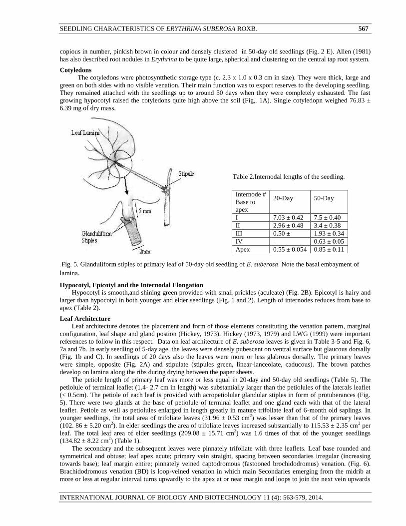
Fig. 5. Glanduliform stiples of primary leaf of 50-day old seedling of E. suberosa. Note the basal embayment of
lamina.
Hypocotyl, Epicotyl and the Internodal Elongation
Hypocotyl is smooth,and shining green provided with small prickles (aculeate) (Fig. 2B). Epicotyl is hairy and
larger than hypocotyl in both younger and elder seedlings (Fig. 1 and 2). Length of internodes reduces from base to
apex (Table 2).
Leaf Architecture
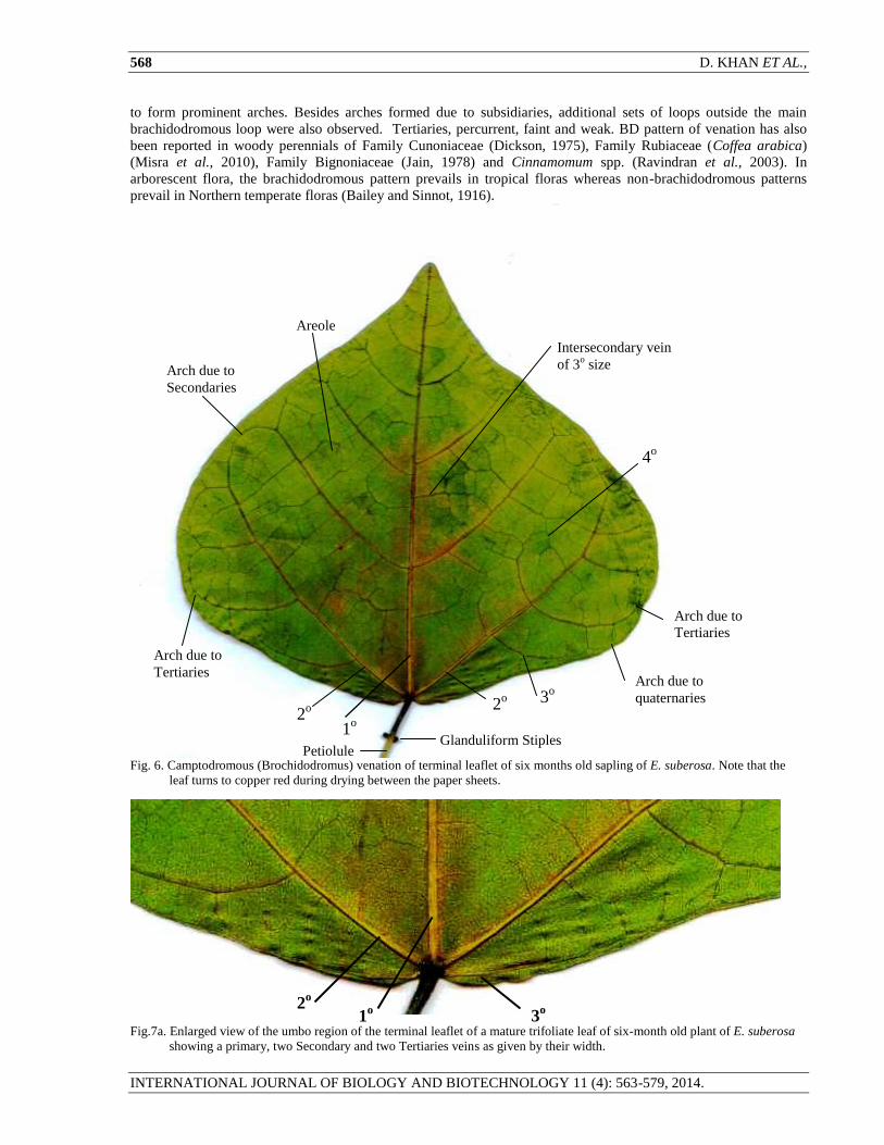
Leaf architecture denotes the placement and form of those elements constituting the venation pattern, marginal
configuration, leaf shape and gland postion (Hickey, 1973). Hickey (1973, 1979) and LWG (1999) were important
references to follow in this respect. Data on leaf architecture of E. suberosa leaves is given in Table 3-5 and Fig. 6,
7a and 7b. In early seedling of 5-day age, the leaves were densely pubescent on ventral surface but glaucous dorsally
(Fig. 1b and C). In seedlings of 20 days also the leaves were more or less glabrous dorsally. The primary leaves
were simple, opposite (Fig. 2A) and stipulate (stipules green, linear-lanceolate, caducous). The brown patches
develop on lamina along the ribs during drying between the paper sheets.
The petiole length of primary leaf was more or less equal in 20-day and 50-day old seedlings (Table 5). The
petiolule of terminal leaflet (1.4- 2.7 cm in length) was substantially larger than the petiolules of the laterals leaflet
(< 0.5cm). The petiole of each leaf is provided with acropetiolular glandular stiples in form of protuberances (Fig.
5). There were two glands at the base of petiolule of terminal leaflet and one gland each with that of the lateral
leaflet. Petiole as well as petiolules enlarged in length greatly in mature trifoliate leaf of 6-month old saplings. In
younger seedlings, the total area of trifoliate leaves (31.96 ± 0.53 cm2) was lesser than that of the primary leaves
(102. 86 ± 5.20 cm2). In elder seedlings the area of trifoliate leaves increased substantially to 115.53 ± 2.35 cm
2 per
leaf. The total leaf area of elder seedlings (209.08 ± 15.71 cm2) was 1.6 times of that of the younger seedlings
(134.82 ± 8.22 cm2) (Table 1).
The secondary and the subsequent leaves were pinnately trifoliate with three leaflets. Leaf base rounded and
symmetrical and obtuse; leaf apex acute; primary vein straight, spacing between secondaries irregular (increasing
towards base); leaf margin entire; pinnately veined captodromous (fastooned brochidodromus) venation. (Fig. 6).
Brachidodromous venation (BD) is loop-veined venation in which main Secondaries emerging from the midrib at
more or less at regular interval turns upwardly to the apex at or near margin and loops to join the next vein upwards
Internode #
Base to
apex
20-Day
50-Day
I 7.03 ± 0.42 7.5 ± 0.40
II 2.96 ± 0.48 3.4 ± 0.38
III 0.50 ± 1.93 ± 0.34
IV - 0.63 ± 0.05
Apex 0.55 ± 0.054 0.85 ± 0.11
Table 2.Internodal lengths of the seedling.
568 D. KHAN ET AL.,
INTERNATIONAL JOURNAL OF BIOLOGY AND BIOTECHNOLOGY 11 (4): 563-579, 2014.
to form prominent arches. Besides arches formed due to subsidiaries, additional sets of loops outside the main
brachidodromous loop were also observed. Tertiaries, percurrent, faint and weak. BD pattern of venation has also
been reported in woody perennials of Family Cunoniaceae (Dickson, 1975), Family Rubiaceae (Coffea arabica)
(Misra et al., 2010), Family Bignoniaceae (Jain, 1978) and Cinnamomum spp. (Ravindran et al., 2003). In
arborescent flora, the brachidodromous pattern prevails in tropical floras whereas non-brachidodromous patterns
prevail in Northern temperate floras (Bailey and Sinnot, 1916).
Fig. 6. Camptodromous (Brochidodromus) venation of terminal leaflet of six months old sapling of E. suberosa. Note that the
leaf turns to copper red during drying between the paper sheets.
Fig.7a. Enlarged view of the umbo region of the terminal leaflet of a mature trifoliate leaf of six-month old plant of E. suberosa
showing a primary, two Secondary and two Tertiaries veins as given by their width.
Arch due to
Secondaries
4o
3o
Petiolule
2o
Areole
1o
2o
Glanduliform Stiples
Arch due to
Tertiaries
Arch due to
Tertiaries
Intersecondary vein
of 3o size
Arch due to
quaternaries
1o
2o
3o
SEEDLING CHARACTERISTICS OF ERYTHRINA SUBEROSA ROXB. 569
INTERNATIONAL JOURNAL OF BIOLOGY AND BIOTECHNOLOGY 11 (4): 563-579, 2014.
In the umbo region of terminal leaflet, there are five veins (one main mid-vein, 2 secondaries and two tertiaries
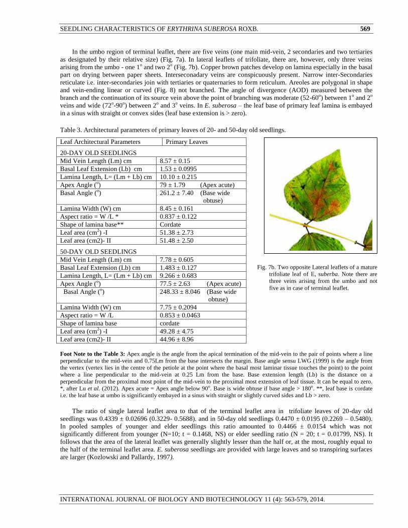
as designated by their relative size) (Fig. 7a). In lateral leaflets of trifoliate, there are, however, only three veins
arising from the umbo - one 1o and two 2
o (Fig. 7b). Copper brown patches develop on lamina especially in the basal
part on drying between paper sheets. Interseconadary veins are conspicuously present. Narrow inter-Secondaries
reticulate i.e. inter-secondaries join with tertiaries or quaternaries to form reticulum. Areoles are polygonal in shape
and vein-ending linear or curved (Fig. 8) not branched. The angle of divergence (AOD) measured between the
branch and the continuation of its source vein above the point of branching was moderate (52-60o) between 1
o and 2
o
veins and wide (72o-90
o) between 2
o and 3
o veins. In E. suberosa – the leaf base of primary leaf lamina is embayed
in a sinus with straight or convex sides (leaf base extension is > zero).
Table 3. Architectural parameters of primary leaves of 20- and 50-day old seedlings.
Leaf Architectural Parameters Primary Leaves
20-DAY OLD SEEDLINGS
Mid Vein Length (Lm) cm 8.57 ± 0.15
Basal Leaf Extension (Lb) cm 1.53 ± 0.0995
Lamina Length, L= (Lm + Lb) cm 10.10 ± 0.215
Apex Angle (o) 79 ± 1.79 (Apex acute)
Basal Angle (o) 261.2 ± 7.40 (Base wide
obtuse)
Lamina Width (W) cm 8.45 ± 0.161
Aspect ratio = W /L * 0.837 ± 0.122
Shape of lamina base** Cordate
Leaf area (cm2) -I 51.38 ± 2.73
Leaf area (cm2)- II 51.48 ± 2.50
50-DAY OLD SEEDLINGS
Mid Vein Length (Lm) cm 7.78 ± 0.605
Basal Leaf Extension (Lb) cm 1.483 ± 0.127
Lamina Length, L= (Lm + Lb) cm 9.266 ± 0.683
Apex Angle (o) 77.5 ± 2.63 (Apex acute)
Basal Angle (o) 248.33 ± 8.046 (Base wide
obtuse)
Lamina Width (W) cm 7.75 ± 0.2094
Aspect ratio = W /L 0.853 ± 0.0463
Shape of lamina base cordate
Leaf area (cm2) -I 49.28 ± 4.75
Leaf area (cm2)- II 44.96 ± 8.96
Foot Note to the Table 3: Apex angle is the angle from the apical termination of the mid-vein to the pair of points where a line
perpendicular to the mid-vein and 0.75Lm from the base intersects the margin. Base angle sensu LWG (1999) is the angle from
the vertex (vertex lies in the centre of the petiole at the point where the basal most laminar tissue touches the point) to the point
where a line perpendicular to the mid-vein at 0.25 Lm from the base. Base extension length (Lb) is the distance on a
perpendicular from the proximal most point of the mid-vein to the proximal most extension of leaf tissue. It can be equal to zero.
*, after Lu et al. (2012). Apex acute = Apex angle below 90o. Base is wide obtuse if base angle > 180o. **, leaf base is cordate
i.e. the leaf base at umbo is significantly embayed in a sinus with straight or slightly curved sides and Lb > zero.
The ratio of single lateral leaflet area to that of the terminal leaflet area in trifoliate leaves of 20-day old
seedlings was 0.4339 ± 0.02696 (0.3229- 0.5688). and in 50-day old seedlings 0.4470 ± 0.0195 (0.2269 – 0.5480).
In pooled samples of younger and elder seedlings this ratio amounted to 0.4466 ± 0.0154 which was not
significantly different from younger (N=10; t = 0.1468, NS) or elder seedling ratio (N = 20; t = 0.01799, NS). It
follows that the area of the lateral leaflet was generally slightly lesser than the half or, at the most, roughly equal to
the half of the terminal leaflet area. E. suberosa seedlings are provided with large leaves and so transpiring surfaces
are larger (Kozlowski and Pallardy, 1997).
Fig. 7b. Two opposite Lateral leaflets of a mature
trifoliate leaf of E, suberba. Note there are
three veins arising from the umbo and not
five as in case of terminal leaflet.
570 D. KHAN ET AL.,
INTERNATIONAL JOURNAL OF BIOLOGY AND BIOTECHNOLOGY 11 (4): 563-579, 2014.
Fig. 8. Areolation in mature leaf of E. suberosa.
Leaf Ornamentation
E. suberosa showed a great epidermal ornamentation in form of trichomes and various stomatal types.
1. Trichomes
Two types of trichomes were observed- branched trichomes and capitate glandular trichomes (Fig. 9 and 10).
Branched trichomes are denser near the basal part of the leaf and the petiole than apical part of the mature leaf of 6-
months old seedling. Being situated on a delicate stock cell, they are easily removed. The branched trichomes were
composed of 5-8 arms arranged in varying patterns (Fig. 9).
Fig.10. Capitate glandular trichomes of lower
surface of leaf of E. suberosa (A-D) and
epidermal cells of the leaf blade (E, dorsal
side and F, ventral side) as seen with 45 x
10 X magnification. Periclinal cell walls
of dorsal epidermal cells are more
undulate. Figures, not drawn to scale.
Capitate Glandular
Trichome
A B
C D
Stoma
E
F
Epidermal
cells
Glandular
Trichome in
the rib region
* Vein- lets
3o Vein
Fig. 9. Branched trichomes from petiole and ventral leaf
surface of six months old sapling. Marked with asterisks are
from the leaf surface. Such trichomes on dorsal side of leaf are
infrequent – more on the basal part of the leaf than apical part.
*
*

SEEDLING CHARACTERISTICS OF ERYTHRINA SUBEROSA ROXB. 571
INTERNATIONAL JOURNAL OF BIOLOGY AND BIOTECHNOLOGY 11 (4): 563-579, 2014.
4%
55%
41%
Fig. 11. Frequency of occurrence of capitate glandular trichome in the fields of vision (N = 75) of compound microscope at 45 x
10 X magnification of ventral surface of leaf of 20-day old E. suberosa seedling. Each field of vision occupied an area
of 0.10174 mm2.
Table 4. Architectural parameters of trifoliate leaves of 20- and 50-day old and six-month old seedlings of E. suberosa.
Leaf Architectural Parameters
Secondary Leaf ( Trifoliate )
Terminal leaflet Lateral leaflet I Lateral Leaflet II
20-DAY OLD SEEDLING
Mid Vein Length (Lm) cm 4.26 ± 0.68 (2.75-5,6) 2.76 ± 0.31 (1.5-3.9) 2.61 ± 0.27 (1.9-3.7)
Basal Leaf Extension (Lb) cm Zero* Zero Zero
Lamina Length, L= (Lm + Lb) cm 4.26 ± 0.68 (2.75-5,6) 2.76 ± 0.31 (1.5-3.9) 2.61 ± 0.27 (1.9-3.7)
Apex Angle (o) 80.60 ± 2.0 (75-88) 81.83 ± 5.60 (70-108) 78.16 ± 1.64 (70-80)
Basal Angle (o) 120.40 ± 4.55 (110-137) 89.14 ± 4.51 (78-108) 94.0 ± 5.53 (78-120)
Lamina Width (W) cm 4.18 ± 0.52 (2.1-5.8) 2.160 ± 0.056 1.923 ± 0.098
Aspect ratio =W /L * 0.9814 ± 0.052 0.76 ± 0.061 (0.59-.80) 0.74 ± 0.063(0.67-0.93)
Lamina shape Rhomboidal Ovate*** Ovate
Leaf area (cm2) 10.63 ± 3.66 4.26 ± 1.18 4.21 ± 1.95
50-DAY OLD SEEDLING
Mid Vein Length (Lm) cm 5.42 ± 0.46 (2.7-7.7) 4.44 ± 0.352 (3.5-6.1) 3.74 ± 0.48 (2.8-5.3)
Basal Leaf Extension (Lb) cm Negligible* - -
Lamina Length, L= (Lm + Lb) cm 5.42 ± 0.46 (2.7-7.7) 4.44 ± 0.352 (3.5-6.1) 3.74 ± 0.48 (2.8-5.3)
Apex Angle (o) 78.36 ± 2.54 (65-95) 66.30 ± 2.42 (60-85) 68.20 ± 2.42 (62-88)
Basal Angle (o) 123.82 ± 1.71 (115-131) 105.9 ± 2.80 (101 -128) 106.50 ± 1.85(100-115)
Lamina Width (W) cm 4.47 (2.2-6.8) 3.0 (2.0-3.7) 2.50 ± 0 (1.00 -3.7)
Aspect ratio = W /L 0.8613 ± 0.0394 0.673 ± 0.026 0.663 ± 0.036
Lamina shape Rhomboidal Deltoid / Ovate deltoid /Ovate
Leaf area (cm2) 29.70 ± 6.46 13.41 ± 3.12 14.84 ± 3.64
SIX-MONTH OLD SEEDLINGS Trifoliate Leaf
Mid Vein Length (Lm) cm 9.250 ± 7.55 ± 7.50 ±
Basal Leaf Extension (Lb) cm Zero* Zero Zero
Lamina Length, L= (Lm + Lb) cm 9.25± 7.55 ± 7.50 ±
Apex Angle (o) 88.4 ± 70.6 ± 70.3 ±
Basal Angle (o) 122.4 ± 110 .8± 112.6 ±
Lamina Width (W) cm 9.73 ± 5.52 ± 5.50 ±
Aspect ratio = W /L 1.0543 0.733 0.805
Lamina shape Rhomboidal Deltoid / Ovate deltoid /Ovate
Apex acute = Apex angle below 90o. *, leaf base at the umbo is very slightly embayed in a sinus (Lb is negligible);
**, After Lu et al. (2012); ***, Lamina shape is ovate if the widest part of the leaf is on an axis in the basal 2/5 of
the leaf (LWG, 1999).
(ONE TRICHOME
PER FRAME)
(NO TRICHOME
IN FRAMES)
(TWO TRICHOMES
PER FRAME)

572 D. KHAN ET AL.,
INTERNATIONAL JOURNAL OF BIOLOGY AND BIOTECHNOLOGY 11 (4): 563-579, 2014.
Capitate glandular multicellular trichomes (Fig.10) were found to be distributed irregularly with a mean density
of 6.16 ± 0.64 per mm2
on the ventral surface of simple leaf of 20-day seedling (Table 6). These trichomes were
present on the veins and also the leaf blade surrounded by the veins. These trichomes measured 47.25 ± 0.75 μm in
length and 30.6 ± 0.50 μm in width at the widest. On dorsal surface such trichomes were very rare. Even on ventral
surface of young leaf of 20-day old seedling, the frequency of not finding a trichome in a field of microscope vision
(Frame) was as high as 41%. The frequency of finding one trichome per frame was 55% and two trichomes per
frame merely 4% (Fig. 11). On terminal leaflet of a mature leaf the density of glandular trichomes was quite high
(2.17 ± 0. 094 per frame of vision of 0.10174mm2
corresponding to 21.3 ± 0.92 trichomes per mm2) varying from
zero to 5 but predominantly 1 to 3 per frame of vision; their distribution tended to significantly deviate from the
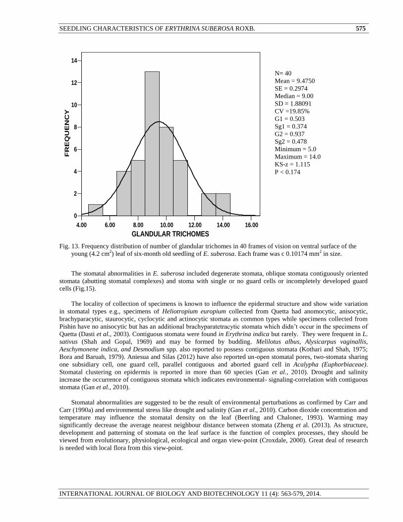
normal distribution (Fig. 12). The distribution of glandular trichome on terminal leaflet of a younger trifoliate leaf
of 4.2 cm2 of six months old seedling tended to follow normal distribution (KS-z: 1.115, p < 0.174) and exhibited
significantly larger density (9.47 ± 0.27 per frame of vision of 0.10174 mm2 corresponding to 93.08 ± 8.65 per mm
2)
(Fig. 13). The reason for such a variation of trichome frequency is not known. It may be somehow related with the
age of the age of the leaf or due to their easy removal from the leaf surface.
It was noted that where a glandular trichome was present, the shape of the epidermal cells underneath was
greatly different from the normal epidermal ground cells. The epidermal cells of the dorsal side of leaf exhibited
comparatively lesser undulation of cell wall as compared to the lower epidermal cells (Fig. 10 E & F). Glandular
trichomes have also been reported in Erythrina velutina by da Silva et al. (2013) on both adaxial and abaxial
surfaces. Like E. velutina, E. suberosa had branched trichomes. Metcalfe and Chalk (1950) considered branched
trichomes to be characteristic to genus Erythrina. E. falcata and E. speciosa have been reported to bear branched-
trichomes (Almeida, 2010, 2011), however, epidermis in E. cristagalli presents no such adoration (Cratieri-Sosselsa,
2005). The trichome frequency is, however, reported to be environmentally-controlled (Metcalfe and Chalk, 1979).
Table 5. Petiole / petiolule lengths (cm) of primary (simple) and secondary (trifoliate) leaves of 20- and 50-day old seedlings.
Primary or
Simple Leaves
Secondary
or Trifoliate
20-DAY OLD SEEDLINGS
Petiole
Petiole Terminal leaf let
(Petiolule)
Lateral leaf let - - I
(Petiolule)
Lateral leaf let - II
(Petiolule)
N = 5
5.75 ± 0.112
(5.5-6.0)
N = 5
7.8 ± 0.73
( 5-10)
N = 5
2.2 ± 0.32
(1.6-2.7)
N = 5
0.4 ± 0.10
0.3-0.5
N = 5
0.4 ± 0.10
0.3-0.5
50-DAY OLD SEEDLINGS
N = 6
5.28 ± 0.13
(5-5.8)
N = 6
6.64 ± 0.41
5.2 -9.3
N = 6
2.46 ± 0.18
1.4 -2.6
N = 6
0.30 ± 0.10
0.25 – 0.45
N = 6
0.36 ± 0.03
0.25 – 0.45
6- MONTH OLD SAPLING
---. 13.5 4.6 0.9 0.8
2. Stomata
Cotyledonary Stomata
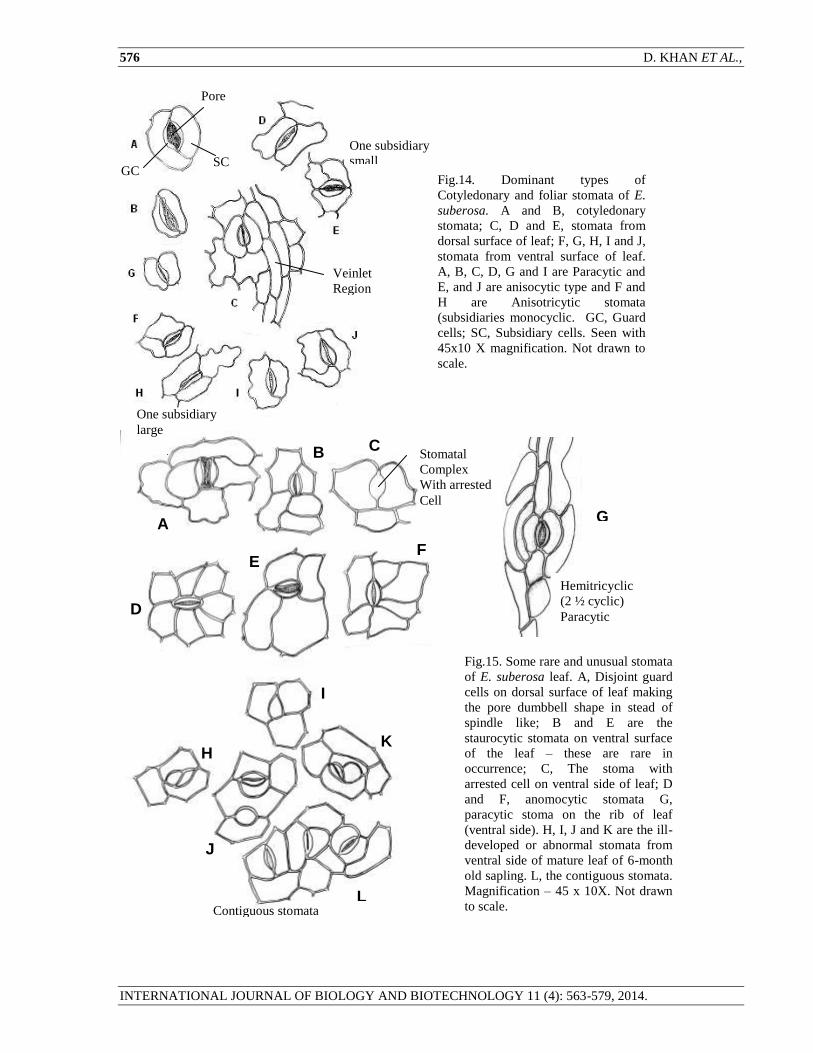
Cotyledonary stomata are of paracytic type (Fig. 14, A and B). They are smaller than the foliar stomata.
Foliar Stomata
The leaves of E. suberosa seedlings were hypo-amphistomatic. The upper surface was paucistomatic
(stomata rare) and the lower surface multistomatic. There were five types of stomata on ventral side of leaf of E.
suberosa (Fig. 14 and 15) – 1) paracytic (A stomatal complex in which one or more of the subsidiary cells that flank
the stoma are parallel with the long axis of the guard cells), 2) anisocytic (a stoma completely surrounded by only
three subsidiaries variable in position and shape but one of the subsidiaries is distinctly small 3) anisotricytic (A
stoma completely surrounded by only three subsidiaries variable in position and shape but one of subsidiaries is
distinctly large) 4) anomocytic (Stoma completely surrounded by four or more subsidiaries, variable in position,
shape and size and 5) staurocytic (Stoma completely surrounded by only four subsidiaries variable in shape and size
but two of their conjoint wall polar, while the other two are lateral to the guard cells (cf. Prabhakar, 2004).
Staurocytic stomata are known to develop from anisocytic stomatal complexes of the seedling leaves in
Monocalyptus (Eucalyptus, Myrtaceae) (Carr and Carr, 1990b).
The number of subsidiary cells associated with different types of stomata varied from two to seven. Regarding
the number of subsidiaries associated with stomata, the studies by Car and Car (1990a), Obiremi and Oladale (2001)
and Oyeleke et al. (2004) had confirmed that larger the number of subsidiaries cells surrounding the guard cells, the
SEEDLING CHARACTERISTICS OF ERYTHRINA SUBEROSA ROXB. 573
INTERNATIONAL JOURNAL OF BIOLOGY AND BIOTECHNOLOGY 11 (4): 563-579, 2014.
faster the opening of the stomata i.e. more transpiration and CO2 absorption. It is well known that most of the CO2
used in stomata is absorbed by the stomata.
In E. suberosa seedlings, stomata were rarely present on dorsal side and they were of paracytic type mainly
along the veins. Staurocytic stomata and other types of stomata were only seen on ventral surface (Fig. 6). This
signifies the diversity of stomatal types even on the same surface of a leaf as also been reported by Saheed and Illoh
(2010) and Aniesua and Silas (2012). Metcalfe and Chalk (1979) have reported several types of stomata in
Papilionaceae – Anomocytic, Paracytic, and Parallelocytic restricted on adaxial surface or found on both surfaces
singly or in groups. They have reported no anisocytic stomata in Papilionaceae. The thirteen species of the family
Fabaceae (Genus Ademsia, Galega, Lotus, Lupinus, Melilotus, Parkinsonia, Senna, Trifolium and Vicia) were
reported to be characterized with anisocytic, anomocytic stomata. Stomata are predominantly paracytic in leaves of
Citrus spp. (Obiremi and Oladele, 2001) and many Macaranga spp. (Norfaizal et al., 2012). Stomata are extremely
variable even in the members of a tribe and even within a genus (Metcalfe and Chalk, 1950) and in a species as well.
That is more than one type of stomata frequently occur on the same leaf surface.
Paracytic stomata are most frequent in several papilionaceous plants (Alysicarpus bupleurifolius, A. monilifer,
A. rugosus, Arachis hypogea, Cajanus cajan, Canavalvia gladiata, Clitoria terneata, Erythrina cristagalli, E.
indica, Lathyrus sativus, Lens esculentus, Medicago sativa, and Tephrosia purpurea. The genus Senna has been
reported to have paracytic stomata (Freire et al., 2005). There are, however, anisocytic stomata in Glycine soja,
Pisum sativum and Sesbania sesban and anomocytic stomata in Sesbania grandiflora and Trigonella foenum-
graceum. The stomata on leaf of Alhagi maurorum (Fabaceae) are paracytic and anisocytic types and on stem
anomocytic type (Bokhari and Dasti, 1991). Thirty-six dicotyledonous species of 34 genera and 20 families of
district Tank (Khyber Pukhtoonkhwah, Pakistan) were examined by Ahmad et al. (2009). Most of them were
amphistomatic. Anisocytic type of stomata were the dominant type in 12 spp. Staurocytic and diacytic stomata were
only present in seven and six species, respectively. In six species two or three types of stomata were present
simultaneously. Staurocytic stomata are reported in Erythrina subrosa L. (? E. suberosa Roxb.) by Khan et al.
(2011). Several species of genus Erythrina have been reported to have paracytic stomata for instance, E. speciosa
and E. falcata (Almeida 2010, 2011); E. velutina (da Silva et al., 2013), E. variegata (Matheus and Lopes, 2007), E.
suberosa (Khan et al, 2011; Biradar, et al., 2013), and E. indica (Tripathi and Mondal, 2012). Contrary to Khan et
al. (2011) E. suberosa is found to be amphistomatic and not hypostomatic. Stomata on dorsal surface of leaf are very
rare and but not absolutely absent. Of 45 species of order Leguminales, 31 species are reported to be amphistomatic
and only 14 spp. hypostomatic by Tripathi and Mondal (2012). According to them, three stomata types of
Leguminales were paracytic, anisocytic and anomocytic - found in various combinations. The most common
stomata in legumes are of the paracytic type and paracytic and anomocytic types may although occur together in
Caesalpiniaceae but never occur together in Fabaceae. Family Fabaceae is more diverse in stomata than Families
Caesalpiniaceae and Mimosaceae (Tripathi and Mondal, 2012).
The stomatal frequency is known to vary on upper and lower surfaces of leaf (Ekenayake et al., 1998). Greater
stomatal density on ventral surface of leaf is common in species that occur in xeromorphic environments, a fact
explained as a feature that minimizes water loss by ostiolar evapo-transpiration (Esau, 1974; Cutter, 1986). In E.
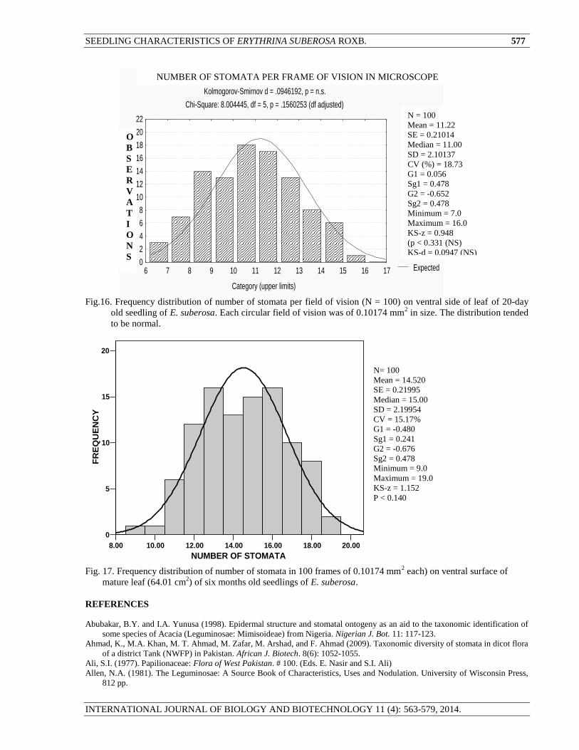
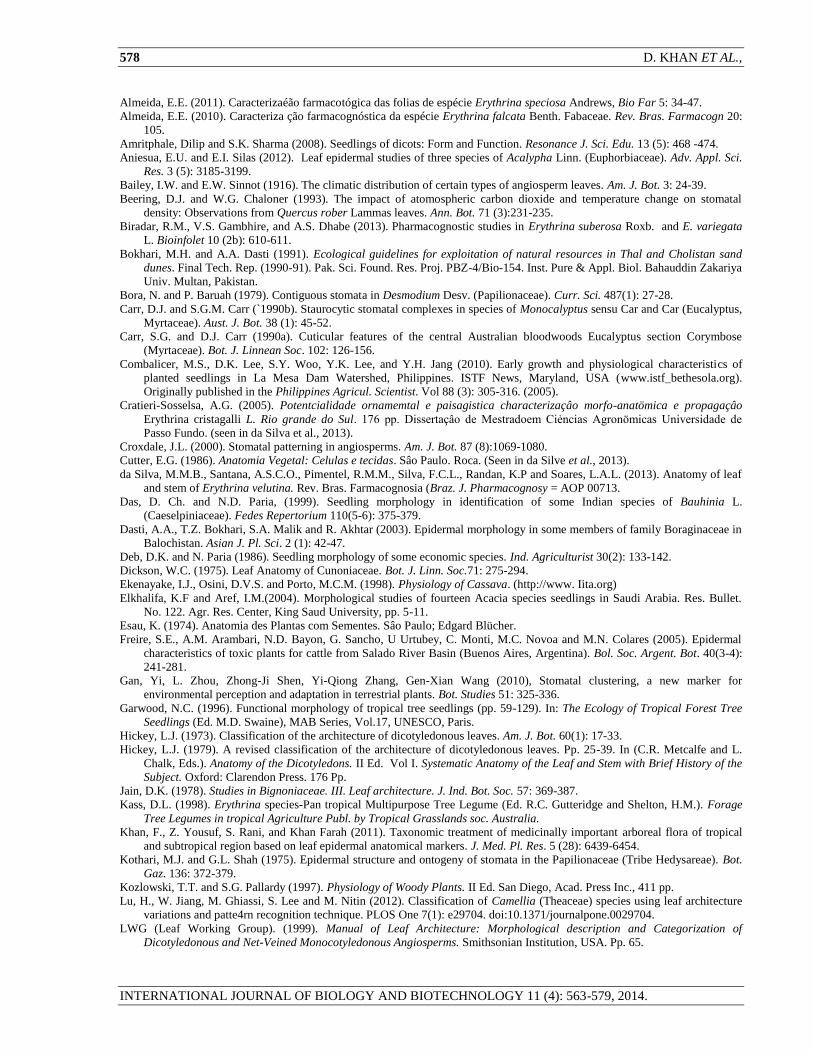
suberosa, the number of stomata per frame of vision of microscope ranged from 7 to 16 with overall variation of
18.73% (mean = 11.22 ± 0.210 per frame. The mid-region classes of distribution (10-14 stomata per frame)
occupied a large proportion of 69% of the total observations (N = 100). The distribution of number of stomata per
frame of vision of microscope was normal in both younger and mature leaf (Fig. 16 and 17) as the KS-d and KS-z
both were found to be insignificant in both cases. It indicated that spatial distribution of stomata on the leaf surface,
irrespective of their kind, was heterogeneous. Stomata were comparatively denser in case of mature leaf as
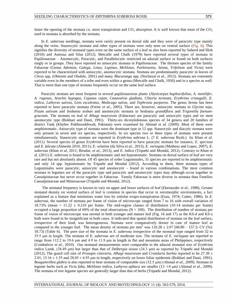
compared to the younger leaf. The mean density of stomata per mm2 was 110.28 ± 2.07 [68.80 – 157.3; CV (%):
18.73] (Table 6). The pore size of the stomata in E. suberosa irrespective of the stomatal type ranged from 32 to
35.5 μm in length. The stomata of E. suberosa are of moderate size. The stomata of E. variegata are reported to
range from 112.2 to 19.6 μm and 4.9 to 11.9 μm in length in flat and mountain areas of Philippines, respectively
(Combalicer et al., 2010). Our stomatal measurements were comparable to the adaxial stomatal size of Erythrina
indica Lamk. (38.49 μm) but larger than that of Dalbergia sissoo (24.3 μm) as reported by Tripathi and Mondal
(2012). The guard cell size of Prosopis cineraria, Alhagi maurorum and Crotalaria burhia reported to be 27.39 ±
2.01, 15.16 ± 1.55 and 28.05 ± 6.93 μm in length, respectively on lower foliar epidermis (Bokhari and Dasti, 1991).
Bougainvillea glabra is also reported to bear stomata of comparable size (32.5 μm) (Ahmad et al., 2009). Stomata in
legume herbs such as Vicia faba, Melilotus indica, Lathyrus aphaca are smaller (13 -14 μm) (Ahmad et al., 2009).
The stomata of tree legume species are generally larger than that of herbs (Tripathi and Mondal, 2012).
574 D. KHAN ET AL.,
INTERNATIONAL JOURNAL OF BIOLOGY AND BIOTECHNOLOGY 11 (4): 563-579, 2014.
Table 6. Density of stomata and Capitate Glandular trichomes per mm2 calculated on the basis of observation in 100
fields of vision on ventral surface of leaf of 20-day old seedling of E. suberosa.
Diversity of stomatal types even on the same surface (leaf) as noted in the present studies has also previously
been reported (Shah and Gopal, 1969; Ahmed et al., 2009). In spite of diversity, most frequent type of stomata can,
however, be used as a taxonomic character. Epidermal (stomatal) studies may act as markers in taxonomic
delimitation. Saheed and Illoh (2010) justified the separation of new genera Senna and Chamaecrista from their
initial genus Cassia on the basis of epidermal surface ornamentation. Paliwal (1969) considered that stomatal studies
may have little taxonomic value unless the development of different stomata types is studies. In Papilionaceae, Shah
and Gopal (1969) reported that different types of stomata follow a similar pattern of development. The diversity of
stomatal types, even on the same surface of an organ, indicates the weakness in using stomata as a taxonomic
character (Pant and Kidwai, 1964). Shah and Gopal (1970), however, asserted that in spite of diversity, the most
frequent type of stomata can be used as taxonomic character. Epidermal surface structure is reported to bear definite
diagnostic features justifying the separation of the genera Senna and Chamaecrista from their initial genus Cassia
(Saheed and Illoh, 2010).
25%
38%
29%
6% 1% 1%
Fig. 12. Distribution of glandular trichome in 100 frames on ventral side of mature leaf (64.01 cm
2) of six-month old
seedling. Each frame had a size of 0.10174 mm2. The data distributed asymmetrically.
Statistical Parameter
of density
Density / mm2
Stomata Capitate Glandular
trichome
Number of observations 100 100
Mean 110.28 6.1595
Standard error of Mean 2.065 0.64022
Median 108.119 9.8290
Standard Deviation 20.654 5.4443
CV (%) 18.73 90.01
Skewness 0.056 0.1765
SE of Skewness 0.478 0.277
Kurtosis -0.6 54 -0.774
SE of Kurtosis 0.478 0.548
Minimum 68.80 0.0
Maximum 157.26 19.60
KS-d 0.0948 (NS) 2.881 (p < 0.0001)
(No Trichome) N= 100
Mean = 2.170
SE = 0.09434
Median = 2.00
SD = 0.9430
CV = 43.47
G1 = 0.314
Sg1 = 0.241
G2 = -0.190
Sg2 = 0.478
Minimum = 0.0
Maximum = 5.00
KS-z = 2.115
P < 0.0001
(One Trichome)
(Four
Trichomes)
(Five trichomes)
(Two Trichomes)
(Three
Trichomes)
SEEDLING CHARACTERISTICS OF ERYTHRINA SUBEROSA ROXB. 575
INTERNATIONAL JOURNAL OF BIOLOGY AND BIOTECHNOLOGY 11 (4): 563-579, 2014.
4.00 6.00 8.00 10.00 12.00 14.00 16.00
GLANDULAR TRICHOMES
0
2
4
6
8
10
12
14F
RE
QU
EN
CY
Fig. 13. Frequency distribution of number of glandular trichomes in 40 frames of vision on ventral surface of the
young (4.2 cm2) leaf of six-month old seedling of E. suberosa. Each frame was c 0.10174 mm
2 in size.
The stomatal abnormalities in E. suberosa included degenerate stomata, oblique stomata contiguously oriented
stomata (abutting stomatal complexes) and stoma with single or no guard cells or incompletely developed guard
cells (Fig.15).
The locality of collection of specimens is known to influence the epidermal structure and show wide variation
in stomatal types e.g., specimens of Heliotropium europium collected from Quetta had anomocytic, anisocytic,
brachyparacytic, staurocytic, cyclocytic and actinocytic stomata as common types while specimens collected from
Pishin have no anisocytic but has an additional brachyparatetracytic stomata which didn’t occur in the specimens of
Quetta (Dasti et al., 2003). Contiguous stomata were found in Erythrina indica but rarely. They were frequent in L.
sativus (Shah and Gopal, 1969) and may be formed by budding. Melilotus albus, Alysicarpus vaginallis,
Aeschymonene indica, and Desmodium spp. also reported to possess contiguous stomata (Kothari and Shah, 1975;
Bora and Baruah, 1979). Aniesua and Silas (2012) have also reported un-open stomatal pores, two-stomata sharing
one subsidiary cell, one guard cell, parallel contiguous and aborted guard cell in Acalypha (Euphorbiaceae).
Stomatal clustering on epidermis is reported in more than 60 species (Gan et al., 2010). Drought and salinity
increase the occurrence of contiguous stomata which indicates environmental- signaling-correlation with contiguous
stomata (Gan et al., 2010).
Stomatal abnormalities are suggested to be the result of environmental perturbations as confirmed by Carr and
Carr (1990a) and environmental stress like drought and salinity (Gan et al., 2010). Carbon dioxide concentration and
temperature may influence the stomatal density on the leaf (Beerling and Chaloner, 1993). Warming may
significantly decrease the average nearest neighbour distance between stomata (Zheng et al. (2013). As structure,
development and patterning of stomata on the leaf surface is the function of complex processes, they should be
viewed from evolutionary, physiological, ecological and organ view-point (Croxdale, 2000). Great deal of research
is needed with local flora from this view-point.
N= 40
Mean = 9.4750
SE = 0.2974
Median = 9.00
SD = 1.88091
CV =19.85%
G1 = 0.503
Sg1 = 0.374
G2 = 0.937
Sg2 = 0.478
Minimum = 5.0
Maximum = 14.0
KS-z = 1.115
P < 0.174
576 D. KHAN ET AL.,
INTERNATIONAL JOURNAL OF BIOLOGY AND BIOTECHNOLOGY 11 (4): 563-579, 2014.
D
E
C
A
B
F
Stomatal
Complex
With arrested
Cell
G
I
H
J
K
L
Fig.14. Dominant types of
Cotyledonary and foliar stomata of E.
suberosa. A and B, cotyledonary
stomata; C, D and E, stomata from
dorsal surface of leaf; F, G, H, I and J,
stomata from ventral surface of leaf.
A, B, C, D, G and I are Paracytic and
E, and J are anisocytic type and F and
H are Anisotricytic stomata
(subsidiaries monocyclic. GC, Guard
cells; SC, Subsidiary cells. Seen with
45x10 X magnification. Not drawn to
scale.
Fig.15. Some rare and unusual stomata
of E. suberosa leaf. A, Disjoint guard
cells on dorsal surface of leaf making
the pore dumbbell shape in stead of
spindle like; B and E are the
staurocytic stomata on ventral surface
of the leaf – these are rare in
occurrence; C, The stoma with
arrested cell on ventral side of leaf; D
and F, anomocytic stomata G,
paracytic stoma on the rib of leaf
(ventral side). H, I, J and K are the ill-
developed or abnormal stomata from
ventral side of mature leaf of 6-month
old sapling. L, the contiguous stomata.
Magnification – 45 x 10X. Not drawn
to scale.
SC GC
Pore
Veinlet
Region
Hemitricyclic
(2 ½ cyclic)
Paracytic
One subsidiary
small
One subsidiary
large
Contiguous stomata
SEEDLING CHARACTERISTICS OF ERYTHRINA SUBEROSA ROXB. 577
INTERNATIONAL JOURNAL OF BIOLOGY AND BIOTECHNOLOGY 11 (4): 563-579, 2014.
Expected
Variable VAR1 ; distribution: Normal
Kolmogorov-Smirnov d = .0946192, p = n.s.
Chi-Square: 8.004445, df = 5, p = .1560253 (df adjusted)
Category (upper limits)
No o
f obs
0
2
4
6
8
10
12
14
16
18
20
22
6 7 8 9 10 11 12 13 14 15 16 17
Fig.16. Frequency distribution of number of stomata per field of vision (N = 100) on ventral side of leaf of 20-day
old seedling of E. suberosa. Each circular field of vision was of 0.10174 mm2 in size. The distribution tended
to be normal.
8.00 10.00 12.00 14.00 16.00 18.00 20.00
NUMBER OF STOMATA
0
5
10
15
20
FR
EQ
UE
NC
Y
Fig. 17. Frequency distribution of number of stomata in 100 frames of 0.10174 mm
2 each) on ventral surface of
mature leaf (64.01 cm2) of six months old seedlings of E. suberosa.
REFERENCES
Abubakar, B.Y. and I.A. Yunusa (1998). Epidermal structure and stomatal ontogeny as an aid to the taxonomic identification of
some species of Acacia (Leguminosae: Mimisoideae) from Nigeria. Nigerian J. Bot. 11: 117-123.
Ahmad, K., M.A. Khan, M. T. Ahmad, M. Zafar, M. Arshad, and F. Ahmad (2009). Taxonomic diversity of stomata in dicot flora
of a district Tank (NWFP) in Pakistan. African J. Biotech. 8(6): 1052-1055.
Ali, S.I. (1977). Papilionaceae: Flora of West Pakistan. # 100. (Eds. E. Nasir and S.I. Ali)
Allen, N.A. (1981). The Leguminosae: A Source Book of Characteristics, Uses and Nodulation. University of Wisconsin Press,
812 pp.
N= 100
Mean = 14.520
SE = 0.21995
Median = 15.00
SD = 2.19954
CV = 15.17%
G1 = -0.480
Sg1 = 0.241
G2 = -0.676
Sg2 = 0.478
Minimum = 9.0
Maximum = 19.0
KS-z = 1.152
P < 0.140
NUMBER OF STOMATA PER FRAME OF VISION IN MICROSCOPE
N = 100
Mean = 11.22
SE = 0.21014
Median = 11.00
SD = 2.10137
CV (%) = 18.73
G1 = 0.056
Sg1 = 0.478
G2 = -0.652
Sg2 = 0.478
Minimum = 7.0
Maximum = 16.0
KS-z = 0.948
(p < 0.331 (NS)
KS-d = 0.0947 (NS)
O
B
S
E
R
V
A
T
I
O
N
S
578 D. KHAN ET AL.,
INTERNATIONAL JOURNAL OF BIOLOGY AND BIOTECHNOLOGY 11 (4): 563-579, 2014.
Almeida, E.E. (2011). Caracterizaéão farmacotógica das folias de espécie Erythrina speciosa Andrews, Bio Far 5: 34-47.
Almeida, E.E. (2010). Caracteriza ção farmacognóstica da espécie Erythrina falcata Benth. Fabaceae. Rev. Bras. Farmacogn 20:
105.
Amritphale, Dilip and S.K. Sharma (2008). Seedlings of dicots: Form and Function. Resonance J. Sci. Edu. 13 (5): 468 -474.
Aniesua, E.U. and E.I. Silas (2012). Leaf epidermal studies of three species of Acalypha Linn. (Euphorbiaceae). Adv. Appl. Sci.
Res. 3 (5): 3185-3199.
Bailey, I.W. and E.W. Sinnot (1916). The climatic distribution of certain types of angiosperm leaves. Am. J. Bot. 3: 24-39.
Beering, D.J. and W.G. Chaloner (1993). The impact of atomospheric carbon dioxide and temperature change on stomatal
density: Observations from Quercus rober Lammas leaves. Ann. Bot. 71 (3):231-235.
Biradar, R.M., V.S. Gambhire, and A.S. Dhabe (2013). Pharmacognostic studies in Erythrina suberosa Roxb. and E. variegata
L. Bioinfolet 10 (2b): 610-611.
Bokhari, M.H. and A.A. Dasti (1991). Ecological guidelines for exploitation of natural resources in Thal and Cholistan sand
dunes. Final Tech. Rep. (1990-91). Pak. Sci. Found. Res. Proj. PBZ-4/Bio-154. Inst. Pure & Appl. Biol. Bahauddin Zakariya
Univ. Multan, Pakistan.
Bora, N. and P. Baruah (1979). Contiguous stomata in Desmodium Desv. (Papilionaceae). Curr. Sci. 487(1): 27-28.
Carr, D.J. and S.G.M. Carr (`1990b). Staurocytic stomatal complexes in species of Monocalyptus sensu Car and Car (Eucalyptus,
Myrtaceae). Aust. J. Bot. 38 (1): 45-52.
Carr, S.G. and D.J. Carr (1990a). Cuticular features of the central Australian bloodwoods Eucalyptus section Corymbose
(Myrtaceae). Bot. J. Linnean Soc. 102: 126-156.
Combalicer, M.S., D.K. Lee, S.Y. Woo, Y.K. Lee, and Y.H. Jang (2010). Early growth and physiological characteristics of
planted seedlings in La Mesa Dam Watershed, Philippines. ISTF News, Maryland, USA (www.istf_bethesola.org).
Originally published in the Philippines Agricul. Scientist. Vol 88 (3): 305-316. (2005).
Cratieri-Sosselsa, A.G. (2005). Potentcialidade ornamemtal e paisagistica characterizaçâo morfo-anatömica e propagaçâo
Erythrina cristagalli L. Rio grande do Sul. 176 pp. Dissertaçâo de Mestradoem Ciẻncias Agronömicas Universidade de
Passo Fundo. (seen in da Silva et al., 2013).
Croxdale, J.L. (2000). Stomatal patterning in angiosperms. Am. J. Bot. 87 (8):1069-1080.
Cutter, E.G. (1986). Anatomia Vegetal: Celulas e tecidas. Sâo Paulo. Roca. (Seen in da Silve et al., 2013).
da Silva, M.M.B., Santana, A.S.C.O., Pimentel, R.M.M., Silva, F.C.L., Randan, K.P and Soares, L.A.L. (2013). Anatomy of leaf
and stem of Erythrina velutina. Rev. Bras. Farmacognosia (Braz. J. Pharmacognosy = AOP 00713.
Das, D. Ch. and N.D. Paria, (1999). Seedling morphology in identification of some Indian species of Bauhinia L.
(Caeselpiniaceae). Fedes Repertorium 110(5-6): 375-379.
Dasti, A.A., T.Z. Bokhari, S.A. Malik and R. Akhtar (2003). Epidermal morphology in some members of family Boraginaceae in
Balochistan. Asian J. Pl. Sci. 2 (1): 42-47.
Deb, D.K. and N. Paria (1986). Seedling morphology of some economic species. Ind. Agriculturist 30(2): 133-142.
Dickson, W.C. (1975). Leaf Anatomy of Cunoniaceae. Bot. J. Linn. Soc.71: 275-294.
Ekenayake, I.J., Osini, D.V.S. and Porto, M.C.M. (1998). Physiology of Cassava. (http://www. Iita.org)
Elkhalifa, K.F and Aref, I.M.(2004). Morphological studies of fourteen Acacia species seedlings in Saudi Arabia. Res. Bullet.
No. 122. Agr. Res. Center, King Saud University, pp. 5-11.
Esau, K. (1974). Anatomia des Plantas com Sementes. Sâo Paulo; Edgard Blücher.
Freire, S.E., A.M. Arambari, N.D. Bayon, G. Sancho, U Urtubey, C. Monti, M.C. Novoa and M.N. Colares (2005). Epidermal
characteristics of toxic plants for cattle from Salado River Basin (Buenos Aires, Argentina). Bol. Soc. Argent. Bot. 40(3-4):
241-281.
Gan, Yi, L. Zhou, Zhong-Ji Shen, Yi-Qiong Zhang, Gen-Xian Wang (2010), Stomatal clustering, a new marker for
environmental perception and adaptation in terrestrial plants. Bot. Studies 51: 325-336.
Garwood, N.C. (1996). Functional morphology of tropical tree seedlings (pp. 59-129). In: The Ecology of Tropical Forest Tree
Seedlings (Ed. M.D. Swaine), MAB Series, Vol.17, UNESCO, Paris.
Hickey, L.J. (1973). Classification of the architecture of dicotyledonous leaves. Am. J. Bot. 60(1): 17-33.
Hickey, L.J. (1979). A revised classification of the architecture of dicotyledonous leaves. Pp. 25-39. In (C.R. Metcalfe and L.
Chalk, Eds.). Anatomy of the Dicotyledons. II Ed. Vol I. Systematic Anatomy of the Leaf and Stem with Brief History of the
Subject. Oxford: Clarendon Press. 176 Pp.
Jain, D.K. (1978). Studies in Bignoniaceae. III. Leaf architecture. J. Ind. Bot. Soc. 57: 369-387.
Kass, D.L. (1998). Erythrina species-Pan tropical Multipurpose Tree Legume (Ed. R.C. Gutteridge and Shelton, H.M.). Forage
Tree Legumes in tropical Agriculture Publ. by Tropical Grasslands soc. Australia.
Khan, F., Z. Yousuf, S. Rani, and Khan Farah (2011). Taxonomic treatment of medicinally important arboreal flora of tropical
and subtropical region based on leaf epidermal anatomical markers. J. Med. Pl. Res. 5 (28): 6439-6454.
Kothari, M.J. and G.L. Shah (1975). Epidermal structure and ontogeny of stomata in the Papilionaceae (Tribe Hedysareae). Bot.
Gaz. 136: 372-379.
Kozlowski, T.T. and S.G. Pallardy (1997). Physiology of Woody Plants. II Ed. San Diego, Acad. Press Inc., 411 pp.
Lu, H., W. Jiang, M. Ghiassi, S. Lee and M. Nitin (2012). Classification of Camellia (Theaceae) species using leaf architecture
variations and patte4rn recognition technique. PLOS One 7(1): e29704. doi:10.1371/journalpone.0029704.
LWG (Leaf Working Group). (1999). Manual of Leaf Architecture: Morphological description and Categorization of
Dicotyledonous and Net-Veined Monocotyledonous Angiosperms. Smithsonian Institution, USA. Pp. 65.
SEEDLING CHARACTERISTICS OF ERYTHRINA SUBEROSA ROXB. 579
INTERNATIONAL JOURNAL OF BIOLOGY AND BIOTECHNOLOGY 11 (4): 563-579, 2014.
Matheus, M.T. and J.C. Lopes (2007). Fruit, seed and seedling morphology and seed germination in Erythrina variegata L. Rev.
Bras. Sementes (online vol. 27(3): 8-12.
Metcalfe, C.R. and L. Chalk (1950). Anatomy of the dicotyledons: Leaves, Stem and Wood in Relation to Taxonomy with Notes
on Economic Uses. Oxford, Clarendon Press.
Metcalfe, C.R. and L. Chalk (1979). Anatomy of the Dicotyledons (Second Ed.). Vol. I. Systematics, Anatomy of Leaf and Stem
with Brief history of the Subject. Oxford, 176 pp.
Miller, J.T and C. Miller (2011). Acacia seedling morphology: phyllotaxy and its relationship for seed mass. Aust. J. Bot. 59 (2):
185-196.
Misra, M.K., D. Padamajyoti, N.S. Prakash, A.S. Rana C.S. Srinivasan and M.S. Sreenivassan (2010). Leaf architecture in Indian
coffee (Coffea arabica L.) cultivars and their adaptive significance. World J. Fungal and Pl. Biol. 1(2): 37-41.
Nenggan, Ye (1983-84). Description of various seedlings of leguminous plants. Phytologia V.54 (3): 190-218.
Norfaizal, G.M., H. Khalijah and A.R. Muhammad Ruzi (2012). Leaf anatomical study of five Macaranga species
(Euphorbiaceae). J. Trop. Agric. & Food. Sci. 40(2): 289-296.
Obiremi, E.O, and F.A. Oladale (2001). Water conserving stomatal systems in selected Citrus species. South Afr. J. Bot. 67: 258-
260.
Oyeleke, M.O., A.A. Abdul Rahman, and F.A. Oladele (2004). Stomatal anatomy and transpiration rate in some afforestation
species. Nigerian Soc. Exp. Biology Journal 4 (2): 83-90.
Paliwal, G.S. (1966). Structure and ontogeny of stomata in some Acanthaceae. Phytomorphology 16: 527-532.
Pant, D.D. and P.K. Kidwai (1954). On the diversity in the development and organization of stomata in Phyla nodiflora Michx.
Curr. Sci. 33: 653-654.
Patil, A.M. and D.A. Patil (2011). Investigations on foliar epidermal characteristics in some Acanthaceae. Curr. Bot. 2 (9):01-08.
Prabhakar, M. (2004). Structure, delimitation, nomenclature and classification of stomata. Acta Botanica Sinica 46 (2): 242-252.
Ravindran, P.N., K. Nirmal-Babu, and M. Shylaja (2003). Cinnamon and Cassia: The genus Cinnamomum. CRC Press, 384 Pp.
Reddy, P.K.R. and G.L. Shah (1979). Observations on the cotyledonary and hypocotyledonary stomata and trichomes in some
Caesalpiniaceae with a note on their taxonomy. Feddes Repertorium 90: 239 -250.
Saheed, S.A and H.C. Illoh (2010). A taxonomic study of some species in Cassiinae (Leguminosae) using leaf epidermis
characters. Nortulae Bot. Hort. Agrobot. Cluj. 38 (1): 21-27.
Shah, G.L. and B.V. Gopal (1969). Development of stomata in some Papilionaceae. Can. J. Bot. 47: 387-393.
Sinjushin, A.A. and J.A. Akopian (2011). On seedling structure in Pisum L., Lathyrus L. and Vavilova Fed. (Fabae: Fabaceae).
Wulfenia 18: 81-93.
Soto-Hernández, R.M., Garcia-Mateos, R., Mignet-Chávez, R.S., Kite, G., Martinez-Vásquiz and A.C. Ramos-Valdiva, (2012).
Erythrina, a potential source of chemicals from the Neotropics (Chap. # 9). In; Bioactive compounds in Phytomedicine (Ed.
Iraj Rasooli). ISBN: 978-953-307-805-2 (http://www.intechopen.com/books/bioactive-compounds-in-
phytomedicine/erythrina-a-poytential-source-of-chemicals-from-the n-neotropics). 218 pp.
Tripathi, S. and Mondal, A.K. (2012). Taxonomic diversity in epidermal cells (stomata) of some selected Anthophyta under the
order Leguminales (Caeselpiniaceae, Mimosaceae and Fabaceae) based on numerical analysis: A systematic approach. IJSN
3(4): 788-798.
Vogel, E.F de (1980). Seedlings of dicotyledons: structure, development, types: Distribution of 150 woody Malesian taxa.
Wageningen.
Wang, Xiu-Wei, Mao Zi-Jun, Choi, Kyung and Park, Kwang-Woo (2006). Significance of the leaf epidermis fingerprint for
taxonomy of Genus Rhododendron. J. Forest. Res. 17(3): 171-176.
Wright, I.J., H.T. Clifford, R. Kidson, M.L. Reed, B.L. Rice and M. Westoby (2000). A survey of seed and seedling characters in
1744 Australian dicotyledons species: cross-species trait correlations and correlated trait-shifts within evolutionary lineages.
Biol. L. Linnean. Soc. 69: 521-547.
Zamora-Carnelio, L.F., Ochoa-Gaona, G.V. Simon, J.C. Albores and B.H. Jong (2012). Seed germination and key to seedling
identification for six native tree species of wetlands from Southeast Mexico. Rev. Biol. Trop. 58(2): 717 – 732.
Zar, J.H. (2010). Biostatistical Analysis. 5th Ed. Prentice-Hall, Englewood Cliffs. New Jersey, USA.
Zheng, Y., M. Xu, R. Hou, R. Shen, S. Qiu, and Z. Ouyang (2013). Effect of experimental warming on stomatal traits in leaves of
maize (Zea mays L.). Ecology and Evolution 3(9): 3095-3111.
(Accepted for publication September, 2014)