New Insights Into Drug Discovery Targeting Tau Protein
24
REVIEW published: 03 December 2020 doi: 10.3389/fnmol.2020.590896 Frontiers in Molecular Neuroscience | www.frontiersin.org 1 December 2020 | Volume 13 | Article 590896 Edited by: Javier Egea, Princess University Hospital, Spain Reviewed by: Diana Laura Castillo-Carranza, University of Monterrey, Mexico Fei Liu, New York State Institute for Basic Research in Developmental Disabilities (IBR), United States *Correspondence: Yoshiyuki Soeda [email protected] Received: 03 August 2020 Accepted: 10 November 2020 Published: 03 December 2020 Citation: Soeda Y and Takashima A (2020) New Insights Into Drug Discovery Targeting Tau Protein. Front. Mol. Neurosci. 13:590896. doi: 10.3389/fnmol.2020.590896 New Insights Into Drug Discovery Targeting Tau Protein Yoshiyuki Soeda* and Akihiko Takashima Laboratory for Alzheimer’s Disease, Department of Life Science, Faculty of Science, Gakushuin University, Tokyo, Japan Microtubule-associated protein tau is characterized by the fact that it is an intrinsically disordered protein due to its lack of a stable conformation and high flexibility. Intracellular inclusions of fibrillar forms of tau with a β-sheet structure accumulate in the brain of patients with Alzheimer’s disease and other tauopathies. Accordingly, detachment of tau from microtubules and transition of tau from a disordered state to an abnormally aggregated state are essential events preceding the onset of tau-related diseases. Many reports have shown that this transition is caused by post-translational modifications, including hyperphosphorylation and acetylation. The misfolded tau is self-assembled and forms a tau oligomer before the appearance of tau inclusions. Animal and pathological studies using human samples have demonstrated that tau oligomer formation contributes to neuronal loss. During the progression of tauopathies, tau seeds are released from cells and incorporated into other cells, leading to the propagation of pathological tau aggregation. Accumulating evidence suggests several potential approaches for blocking tau-mediated toxicity: (1) direct inhibition of pathological tau aggregation and (2) inhibition of tau post-translational modifications that occur prior to pathological tau aggregation, (3) inhibition of tau propagation and (4) stabilization of microtubules. In addition to traditional low-molecular-weight compounds, newer drug discovery approaches such as the development of medium-molecular-weight drugs (peptide- or oligonucleotide-based drugs) and high-molecular-weight drugs (antibody-based drugs) provide alternative pathways to preventing the formation of abnormal tau. Of particular interest are recent studies suggesting that tau droplet formation by liquid-liquid phase separation may be the initial step in aberrant tau aggregation, as well results that implicate roles for tau in dendritic and nuclear functions. Here, we review the mechanisms through which drugs can target tau and consider recent clinical trials for the treatment of tauopathies. In addition, we discuss the utility of these newer strategies and propose future directions for research on tau-targeted therapeutics. Keywords: tau protein, post-translational modifications, aggregation, microtubule stabilizer, immunotherapy, oligonucleotide therapy, liquid-liquid phase separation, inflammation INTRODUCTION Two classes of drugs for dementia treatment have been approved by the major regulatory agencies (US Food and Drug Administration, FDA; European Medicines Agency, EMA; Pharmaceuticals and Medical Devices Agency, PMDA): acetylcholinesterase inhibitors, which treat mild to moderate Alzheimer’s disease (AD), and N-methyl-D-aspartate receptor antagonists (e.g., memantine), which treat moderate to severe AD. Although these drugs can slow progression and control
-
Upload
khangminh22 -
Category
Documents
-
view
1 -
download
0
Transcript of New Insights Into Drug Discovery Targeting Tau Protein

REVIEWpublished: 03 December 2020
doi: 10.3389/fnmol.2020.590896
Frontiers in Molecular Neuroscience | www.frontiersin.org 1 December 2020 | Volume 13 | Article 590896
Edited by:
Javier Egea,
Princess University Hospital, Spain
Reviewed by:
Diana Laura Castillo-Carranza,
University of Monterrey, Mexico
Fei Liu,
New York State Institute for Basic
Research in Developmental Disabilities
(IBR), United States
*Correspondence:
Yoshiyuki Soeda
Received: 03 August 2020
Accepted: 10 November 2020
Published: 03 December 2020
Citation:
Soeda Y and Takashima A (2020) New
Insights Into Drug Discovery Targeting
Tau Protein.
Front. Mol. Neurosci. 13:590896.
doi: 10.3389/fnmol.2020.590896
New Insights Into Drug DiscoveryTargeting Tau ProteinYoshiyuki Soeda* and Akihiko Takashima
Laboratory for Alzheimer’s Disease, Department of Life Science, Faculty of Science, Gakushuin University, Tokyo, Japan
Microtubule-associated protein tau is characterized by the fact that it is an intrinsically
disordered protein due to its lack of a stable conformation and high flexibility. Intracellular
inclusions of fibrillar forms of tau with a β-sheet structure accumulate in the brain of
patients with Alzheimer’s disease and other tauopathies. Accordingly, detachment of
tau from microtubules and transition of tau from a disordered state to an abnormally
aggregated state are essential events preceding the onset of tau-related diseases. Many
reports have shown that this transition is caused by post-translational modifications,
including hyperphosphorylation and acetylation. The misfolded tau is self-assembled
and forms a tau oligomer before the appearance of tau inclusions. Animal and
pathological studies using human samples have demonstrated that tau oligomer
formation contributes to neuronal loss. During the progression of tauopathies, tau seeds
are released from cells and incorporated into other cells, leading to the propagation
of pathological tau aggregation. Accumulating evidence suggests several potential
approaches for blocking tau-mediated toxicity: (1) direct inhibition of pathological tau
aggregation and (2) inhibition of tau post-translational modifications that occur prior
to pathological tau aggregation, (3) inhibition of tau propagation and (4) stabilization
of microtubules. In addition to traditional low-molecular-weight compounds, newer
drug discovery approaches such as the development of medium-molecular-weight
drugs (peptide- or oligonucleotide-based drugs) and high-molecular-weight drugs
(antibody-based drugs) provide alternative pathways to preventing the formation of
abnormal tau. Of particular interest are recent studies suggesting that tau droplet
formation by liquid-liquid phase separation may be the initial step in aberrant tau
aggregation, as well results that implicate roles for tau in dendritic and nuclear functions.
Here, we review themechanisms through which drugs can target tau and consider recent
clinical trials for the treatment of tauopathies. In addition, we discuss the utility of these
newer strategies and propose future directions for research on tau-targeted therapeutics.
Keywords: tau protein, post-translational modifications, aggregation, microtubule stabilizer, immunotherapy,
oligonucleotide therapy, liquid-liquid phase separation, inflammation
INTRODUCTION
Two classes of drugs for dementia treatment have been approved by the major regulatory agencies(US Food and Drug Administration, FDA; European Medicines Agency, EMA; PharmaceuticalsandMedical Devices Agency, PMDA): acetylcholinesterase inhibitors, which treatmild tomoderateAlzheimer’s disease (AD), and N-methyl-D-aspartate receptor antagonists (e.g., memantine),which treat moderate to severe AD. Although these drugs can slow progression and control

Soeda and Takashima Direction for Tau-Targeted Therapies
dementia-related symptoms, the treatments are not definitive.Systematic reviews have reported that the drugs are efficaciousagainst dementia (Loveman et al., 2006; Tan et al., 2014), butthey are also controversial in terms of cost effectiveness (Bondet al., 2012). There are currently many research and developmentefforts to provide disease-modifying therapies for AD treatment(Cummings et al., 2019). The main targets are amyloid-β (Aβ),a major component of senile plaques, and tau, neurofibrillarytangles (NFT). To investigate the Aβ cascade hypothesis, manyclinical trials of therapeutic approaches targeting Aβ havebeen conducted. However, clinical trials for targeting Aβ haverepeatedly failed (Holmes et al., 2008; Rosenblum, 2014). Becausehistological analysis and tau positron emission tomographicstudies have revealed that cognitive impairment correlate betterwith tau and neuronal loss than with Aβ pathology (Bondareffet al., 1989; Bobinski et al., 1996; Gomez-Isla et al., 1996; Guillozetet al., 2003; Schöll et al., 2016; Schwarz et al., 2016; Bejanin et al.,2017), AD drug discovery research has increasingly shifted fromAβ to tau protein (Giacobini andGold, 2013), with some reachingclinical trial stages. In this review, we describe and discuss thestructure and mechanisms of action of drugs that target tau(Figure 1). We also discuss perspectives for drug development inthis area.
TAU PROTEIN AND TAUOPATHIES
Tau is a stabilizing microtubule-associated protein that wasdiscovered in 1975 (Weingarten et al., 1975). The protein ismainly found in the axonal compartment of neurons (Morriset al., 2011; Mandelkow and Mandelkow, 2012; Guo et al.,2017). In the adult human brain, alternative splicing from theMAPT gene on chromosome 17 yields six tau isoforms (352–441amino acid residues; 37–46 kDa) (Goedert et al., 1991), whichare distinguished by the absence or presence of one or two N-terminal inserts, and the presence of three (3R) or four (4R)microtubule-binding repeats in the C-terminal half of tau (Guoet al., 2017).
Tauopathies are neurological disorders (Avila et al., 2004;Götz and Götz, 2019), characterized by aberrant tau aggregates(NFT and tau inclusions) in neurons and glial cells. Theseaggregates represent tau gene mutations or hyperphosphorylatedtau (Kovacs, 2015). The majority of tauopathic patients alsoshow depositions of Aβ, α-synuclein, or huntingtin (Guo et al.,
Abbreviations: a. a., amino acid; Aβ, amyloid-β; AD, Alzheimer’s disease; APP,
amyloid precursor protein; ASO, antisense oligonucleotides; CBD, corticobasal
degeneration; cryo-EM, electron cryomicroscopy; CSF, cerebrospinal fluid; CTE,
chronic traumatic encephalopathy; EMA, European Medicines Agency; FDA,
US Food and Drug Administration; FTDP-17, frontotemporal dementia and
parkinsonism linked to chromosome 17; GSK3, glycogen synthase kinase 3;
IDP, intrinsically disordered protein; Ig, immunoglobulin; LLPS, liquid–liquid
phase separation; MB, methylene blue; MCI, mild cognitive impairment; NFT,
neurofibrillary tangles; OGA, O-GlcNAcase; O-GlcNAcylation, addition of β-
linked N-acetylglucosamine; PHF, paired helical filaments; PiD, Pick’s disease;
PMDA, Pharmaceuticals andMedical Devices Agency; PP2A, protein phosphatase
2A; PSP, patients with progressive supranuclear palsy; 3R, three microtubule-
binding repeats; 4R, four microtubule-binding repeats; scFv, single-chain variable
fragment; SF, straight filaments, STAND, ultra-stable cytoplasmic antibody.
2017). These observations suggest that tau abnormalities have acommon pathological role across neurodegenerative diseases.
AD is the most common and best-studied tauopathy. Thedisease is caused by extensive atrophy of the brain beginningin the temporal and parietal lobes. Analysis of cell lysates fromthe AD brain by sodium dodecyl sulfate polyacrylamide gelelectrophoresis reveals three major electrophoresis bands: tauproteins with relative molecular weights of 68,000, 64,000, and60,000 (Lee et al., 1991; Goedert et al., 1992; Greenberg et al.,1992; Delacourte et al., 1999). Although the actual molecularweight of tau is 37–46 kDa, treatment of AD-derived sampleswith phosphatases shows that the band pattern of tau wassimilar to that of recombinant human tau (Hanger et al.,2002). This finding indicates that the tau aggregates foundin AD undergo post-translational modifications (Guo et al.,2017). Indeed, structural biology studies have revealed that themajor components of tangles in AD are paired helical filaments(PHF) and straight filaments (SF), and both types are composedprimarily of abnormally phosphorylated tau proteins (Kosiket al., 1988; Gendron and Petrucelli, 2009). Like the tangles inthe healthy adult human brain, those in the AD brain consistof 3R and 4R isoforms (1:1 ratio) (Williams, 2006). However,some other tauopathies are characterized by an imbalance in theratio of 4R/3R tau isoforms. For example, brains from patientswith progressive supranuclear palsy (PSP) and corticobasaldegeneration (CBD) predominantly exhibit 4R tau, whereas theinsoluble tau of Pick’s disease (PiD) is mainly 3R tau (Arai et al.,2003). In frontotemporal dementia and parkinsonism linked tochromosome 17 (FTDP-17), the predominance of isoforms variesaccording to the type of disease-causing tau mutation (de Silvaet al., 2006; Andreadis, 2012; Rossi and Tagliavini, 2015).
TAU-TARGETED THERAPIES
Tau-targeted drugs may be a promising disease-modifyingtherapy because previous studies focusing on the correlationof AD neuropathological changes (Aβ plaques and NFT) withcognitive impairment have shown that the severity of cognitiveimpairment correlated best with the burden of abnormal tau(Nelson et al., 2012). Accordingly, many clinical trials of drugstargeting tau have been conducted.
Post-translational ModificationsTau undergoes a variety of post-translational modifications,including phosphorylation, acetylation, glycation, nitration,addition of β-linked N-acetylglucosamine (O-GlcNAcylation),oxidation, polyamination, sumoylation, and ubiquitination(Martin et al., 2011; Morris et al., 2015). Here, we discuss someof the post-translational modifications of tau, its function andrelationship to disease, and drugs that have been developed toprevent or ameliorate these modifications (Figure 2).
Tau PhosphorylationPhosphorylation is the best known post-translationalmodification of tau. Tau bears 85 phosphorylation sites,including 45 serine residues, 35 threonine residues, and fivetyrosine residues (Hanger et al., 2009). Tau phosphorylation
Frontiers in Molecular Neuroscience | www.frontiersin.org 2 December 2020 | Volume 13 | Article 590896
Soeda and Takashima Direction for Tau-Targeted Therapies
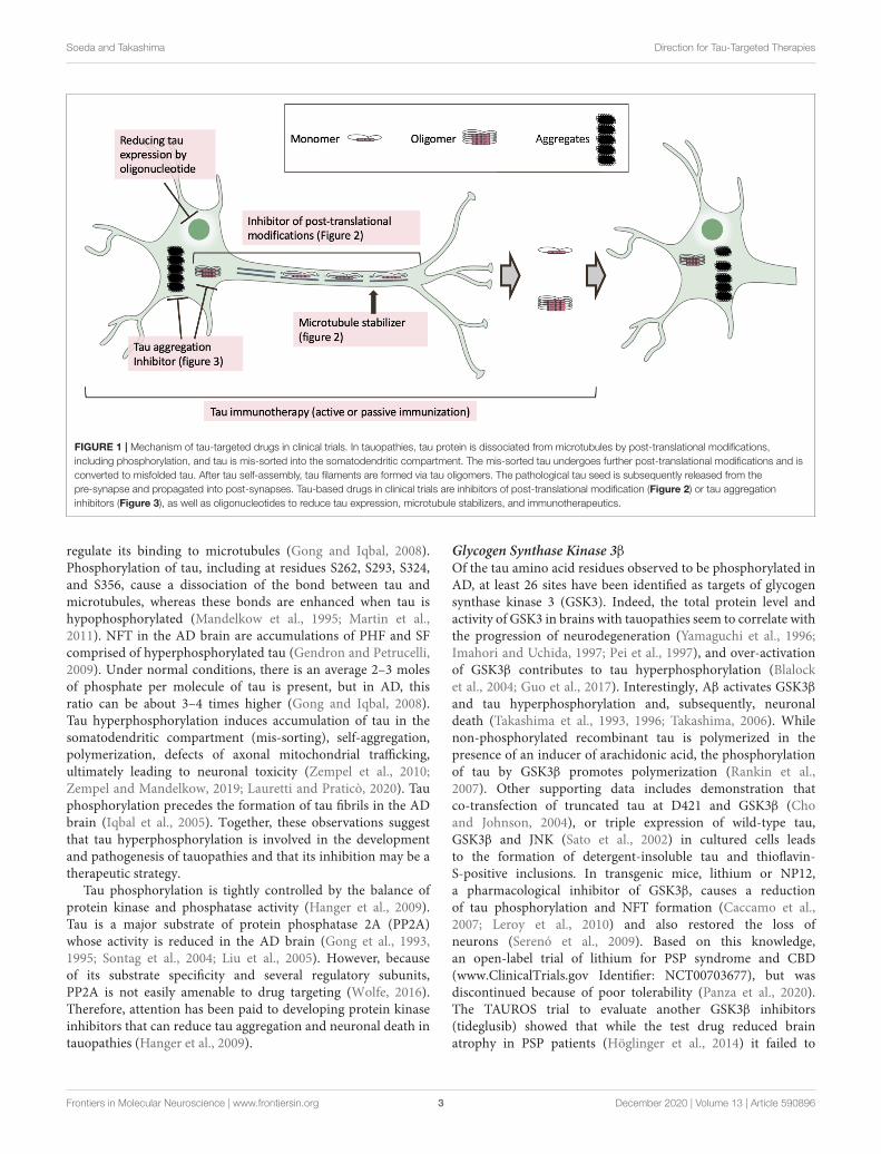
FIGURE 1 | Mechanism of tau-targeted drugs in clinical trials. In tauopathies, tau protein is dissociated from microtubules by post-translational modifications,
including phosphorylation, and tau is mis-sorted into the somatodendritic compartment. The mis-sorted tau undergoes further post-translational modifications and is
converted to misfolded tau. After tau self-assembly, tau filaments are formed via tau oligomers. The pathological tau seed is subsequently released from the
pre-synapse and propagated into post-synapses. Tau-based drugs in clinical trials are inhibitors of post-translational modification (Figure 2) or tau aggregation
inhibitors (Figure 3), as well as oligonucleotides to reduce tau expression, microtubule stabilizers, and immunotherapeutics.
regulate its binding to microtubules (Gong and Iqbal, 2008).Phosphorylation of tau, including at residues S262, S293, S324,and S356, cause a dissociation of the bond between tau andmicrotubules, whereas these bonds are enhanced when tau ishypophosphorylated (Mandelkow et al., 1995; Martin et al.,2011). NFT in the AD brain are accumulations of PHF and SFcomprised of hyperphosphorylated tau (Gendron and Petrucelli,2009). Under normal conditions, there is an average 2–3 molesof phosphate per molecule of tau is present, but in AD, thisratio can be about 3–4 times higher (Gong and Iqbal, 2008).Tau hyperphosphorylation induces accumulation of tau in thesomatodendritic compartment (mis-sorting), self-aggregation,polymerization, defects of axonal mitochondrial trafficking,ultimately leading to neuronal toxicity (Zempel et al., 2010;Zempel and Mandelkow, 2019; Lauretti and Praticò, 2020). Tauphosphorylation precedes the formation of tau fibrils in the ADbrain (Iqbal et al., 2005). Together, these observations suggestthat tau hyperphosphorylation is involved in the developmentand pathogenesis of tauopathies and that its inhibition may be atherapeutic strategy.
Tau phosphorylation is tightly controlled by the balance ofprotein kinase and phosphatase activity (Hanger et al., 2009).Tau is a major substrate of protein phosphatase 2A (PP2A)whose activity is reduced in the AD brain (Gong et al., 1993,1995; Sontag et al., 2004; Liu et al., 2005). However, becauseof its substrate specificity and several regulatory subunits,PP2A is not easily amenable to drug targeting (Wolfe, 2016).Therefore, attention has been paid to developing protein kinaseinhibitors that can reduce tau aggregation and neuronal death intauopathies (Hanger et al., 2009).
Glycogen Synthase Kinase 3βOf the tau amino acid residues observed to be phosphorylated inAD, at least 26 sites have been identified as targets of glycogensynthase kinase 3 (GSK3). Indeed, the total protein level andactivity of GSK3 in brains with tauopathies seem to correlate withthe progression of neurodegeneration (Yamaguchi et al., 1996;Imahori and Uchida, 1997; Pei et al., 1997), and over-activationof GSK3β contributes to tau hyperphosphorylation (Blalocket al., 2004; Guo et al., 2017). Interestingly, Aβ activates GSK3βand tau hyperphosphorylation and, subsequently, neuronaldeath (Takashima et al., 1993, 1996; Takashima, 2006). Whilenon-phosphorylated recombinant tau is polymerized in thepresence of an inducer of arachidonic acid, the phosphorylationof tau by GSK3β promotes polymerization (Rankin et al.,2007). Other supporting data includes demonstration thatco-transfection of truncated tau at D421 and GSK3β (Choand Johnson, 2004), or triple expression of wild-type tau,GSK3β and JNK (Sato et al., 2002) in cultured cells leadsto the formation of detergent-insoluble tau and thioflavin-S-positive inclusions. In transgenic mice, lithium or NP12,a pharmacological inhibitor of GSK3β, causes a reductionof tau phosphorylation and NFT formation (Caccamo et al.,2007; Leroy et al., 2010) and also restored the loss ofneurons (Serenó et al., 2009). Based on this knowledge,an open-label trial of lithium for PSP syndrome and CBD(www.ClinicalTrials.gov Identifier: NCT00703677), but wasdiscontinued because of poor tolerability (Panza et al., 2020).The TAUROS trial to evaluate another GSK3β inhibitors(tideglusib) showed that while the test drug reduced brainatrophy in PSP patients (Höglinger et al., 2014) it failed to
Frontiers in Molecular Neuroscience | www.frontiersin.org 3 December 2020 | Volume 13 | Article 590896

Soeda and Takashima Direction for Tau-Targeted Therapies
FIGURE 2 | Mechanism of tau post-translational modification inhibitors in clinical trials. Post-translational modifications, including phosphorylation and acetylation,
regulate the binding of tau to microtubules. Microtubule instability and depolymerization are observed in tauopathies, suggesting a therapeutic role for microtubule
stabilizers. Phosphorylation, acetylation, or both, enhance tau aggregation. O-GlcNAcylation at serine and threonine compete with phosphorylation of the same
residues. Tau degradation is inhibited by acetylation. The post-translational modifications are tightly regulated by various enzymes that mediate the addition and
removal of the modifying groups. In clinical trials, tau kinase inhibitors or P300 acetyltransferase inhibitors have been investigated for their ability to inhibit tau
phosphorylation or tau acetylation. The usefulness of O-GlcNAcase inhibitors to elevate tau O-GlcNAcylation has also been examined in clinical trials. Ac, acetylation;
Gly, O-GlcNAcylation; P, phosphorylation.
demonstrate clinical efficacy in patients with mild to moderatePSP (Tolosa et al., 2014).
Cyclin-Dependent Kinase 5At least 17 kinases have been identified as tau phosphorylationkinases (Martin et al., 2013), with GSK3β and cyclin-dependentkinase 5 (CDK5) being the most frequently reported amongthem. CDK5 is a proline-directed serine/threonine-proteinkinase. Physiological activation is controlled by binding theregulatory subunit, p35 or p39, to CDK5, leading to braindevelopment and synaptic activity. The p35 and p39 are cleavedby calpain, producing p25 or p29. The binding of p25 orp29 to CDK5 leads to pathological hyperactivation (Kimuraet al., 2014a). CDK5 phosphorylates tau at 9–13 sites (Kimuraet al., 2014a). CDK5 was also found in the neurons havingpretangle or NFT (Pei et al., 1998). An experiment on cross-transgenic mice overexpressing p25 and P301L tau transgenicmice (JNPL3) showed increased tau phosphorylation level andnumber of NFT (Noble et al., 2003). The silencing of CDK5by si-RNA reduced tau phosphorylation in triple-transgenic ADmice (Piedrahita et al., 2010). Roscovitine is a small-moleculedrug that inhibits CDK5 activity. CDK5 is also involved invarious cancers (Pozo and Bibb, 2016); therefore, clinical trials onroscovitine have been conducted in patients with cancer (Cicenas
et al., 2015). No trials on CDK5 inhibitors have been reportedin tauopathies. Notably, Wen et al. reported that administrationof CP681301, a CDK5 inhibitor, enhanced tau phosphorylationin p25 overexpression transgenic mice (Wen et al., 2008). CDK5indirectly phosphorylates GSK3β at S9 and inhibits its activity(Engmann and Giese, 2009), suggesting that CDK5 inhibitionenhances tau phosphorylation by activating GSK3β. As CDK5can phosphorylate molecules other than tau, therapeutic agentstargeting CDK5 should be developed with great caution.
FynTau protein has five tyrosine residues (18, 29, 197, 310, and 394sites) that are phosphorylated by tyrosine kinases. Src familykinase, including Fyn, modulates neurotransmitter function andNMDA trafficking (Ohnishi et al., 2011). Interestingly, taureduction improved Aβ-induced cognitive impairments in J20transgenic mice that express a human APP with the Swedish(K670M/N671L) and Indiana (V717F) mutants (Roberson et al.,2007; Yoshikawa et al., 2018). Fyn is located at the PSD95-rich post-synapse by binding to tau and phosphorylates theNMDA receptor subunit NR2b. This complex promoted Aβ-induced excitotoxicity (Ittner et al., 2010). Fyn preferentiallyphosphorylated Tyr18 among the five tyrosine residues in tau(Scales et al., 2011). Biochemical and immunocytochemical
Frontiers in Molecular Neuroscience | www.frontiersin.org 4 December 2020 | Volume 13 | Article 590896

Soeda and Takashima Direction for Tau-Targeted Therapies
assays showed that phosphorylated tau at Y18 was observed inthe NFT from the AD brain (Lee et al., 2004). Fyn deficiencyreduced tau NFT formation and hyperphosphorylation in miceoverexpressing P301L-tau (Liu et al., 2020). These facts suggestthat Fyn inhibition is a potential target for tauopathy treatment.Saracatinib is a small-molecule inhibitor of Fyn. Preclinicalstudies showed that saracatinib rescued synaptic depletion andspatial memory deficits in APP (Swe)/presenilin 1(1E9) mice(Kaufman et al., 2015; Smith et al., 2018). A phase 1b trial inmild and moderate AD showed that saracatinib is safe, has goodtolerability, and can penetrate into the central nervous system(Nygaard et al., 2015). Unfortunately, the phase 2 trial showed nopositive therapeutic effects of the drug in patients with AD (vanDyck et al., 2019).
Thousand-and-One Amino Acid KinasesRecently, thousand-and-one amino acid kinases (TAOKs) havebeen identified as tau kinases, which may be involved in the onsetof AD pathology and dementia (Tavares et al., 2013; Giacominiet al., 2018). TAOKs are referred to as prostate-derived sterile20-like kinases (PSKs), i.e., serine/threonine kinase. TAOKs havetwo isoforms: TAOK1 (PSK2) and TAOK2 (PSK1). TAOKsphosphorylated ≥40 sites on recombinant human tau (Tavareset al., 2013). High TAOK activation (pS181) was observed in NFTand pretangles of the entorhinal cortex in subjects with Braakstage II but not in control subjects (Giacomini et al., 2018). ATAOK inhibitor, compound 43, inhibited tau phosphorylationat AT8 and 12E8 sites in vitro and in vivo (Giacomini et al.,2018). Furthermore, the drug also inhibited phosphorylation atT123 and T427 sites, newly found in AD (Giacomini et al.,2018), suggesting that TAOKs may be a novel target to improvetau-related pathogenesis. A previous report showed that TAOKsmodulate microtubule dynamics and organization (Mitsopouloset al., 2003). Compound 43 promoted cell death in a culturedcancer cell line (Koo et al., 2017). These findings suggest that thedevelopment of TAOK inhibitor should proceed with caution.
Because many tau kinases are involved in physiologicalintracellular signaling pathways, tau kinase inhibitordevelopment appropriately avoiding physiological on targetsmight be difficult. Meanwhile, based on the view that aspecific phosphorylation pattern is required to induce tauself-assembly (Fichou et al., 2019; Lauretti and Praticò, 2020),several groups have reported data indicating that the phospho-S396/404 epitope constitutes an effective therapeutic target(Boutajangout et al., 2011; Gu et al., 2013; Liu et al., 2016;Rosenqvist et al., 2018). Thus, studies have used immunotherapytargeting tau phosphorylation at S396/404. In a preclinicalstudy, subcutaneous injection of the liposome-based vaccineACI-35 into wild-type mice and mice carrying the P301Ltau mutation induced the formation of antibodies specificto phospho-S396 and S404 tau and reduced soluble andinsoluble tau in the brain. This vaccine also improved bodyweight loss and clasping frequency and survival (Theuniset al., 2013). Thus, far, ACI-35 has been used in a phase 1 trial(Main ID in the WHO International Clinical Trials RegistryPlatform: ISRCTN13033912) (see section on Tau Clearanceand Immunotherapy).
Tau AcetylationThere are≥30 lysine residues that are potentially acetylated in thetau sequence (Kontaxi et al., 2017), mainly located in the proline-rich region, the microtubule-binding region, and the C-terminaldomain (Kontaxi et al., 2017). The level of their acetylation isregulated by acetyltransferases (p300 and CREB-binding protein;Min et al., 2010; Cohen et al., 2011; Cook et al., 2014b) anddeacetylases (histone deacetylase 6 and sirtuin 1; Cook et al.,2014a). Tau proteins promote the self-acetylation of autologouslysine residues via catalytic cysteine residues (C291 and C322)in the microtubule-binding domain (Cohen et al., 2013). Lysineresidues in tau are more highly acetylated in the brains ofAD and other tauopathy patients than in healthy brains (Irwinet al., 2012, 2013). Specific acetylation at residues K280/K281 ontau inhibits microtubule stabilization and promotes fibrillar tauaggregate formation (Trzeciakiewicz et al., 2017). An increasein acetylated tau by deletion of sirtuin 1, a class III proteindeacetylase, inhibits its degradation, leading to the accumulationof pathogenic phospho-tau in vivo (Min et al., 2010). Thesefacts suggest that tau acetylation may be important for tau-induced toxicity.
Salsalate is an old salicylate derivative which has withanti-inflammatory properties related to its ability to inhibitactivation of the NF-κB pathway (Panza et al., 2019). Min et al.reported that salsalate inhibits tau acetylation by blocking p300acetyltransferase activity and acetylation of K174 in the PS19transgenic mouse line which overexpresses P301S-tau. Moreover,these authors found that salsalate prevents hippocampal atrophyand memory impairment (Min et al., 2015). An open-label pilotstudy (phase 1) of salsalate (2,250 mg/day) in 10 PSP patientsfound that although salsalate was safe andwell-tolerated, the drugdid not significantly improve cognitive performance in patients(VandeVrede et al., 2020b). This may be explained by either thepoor penetration of salsalate into the brain (<3%), or by anincrease in tau aggregation following reduced tau acetylation.
Tau UbiquitinationLysine residues undergo not only acetylation but alsoubiquitination which is closely related to the proteasomaldegradation pathway (Cook et al., 2014b). Hyperphosphorylatedtau is ubiquitinated in patients with AD (Mori et al., 1987; Perryet al., 1987; Cripps et al., 2006) and interestingly, dysfunctionof either the proteasomal or lysosomal degradation pathwaysmay lead to accumulation of excessive ubiquitinated tau speciesin AD patients that can contribute to NFT formation in disease(Wang and Mandelkow, 2012; Cook et al., 2014b). Given this,it is plausible that tau acetylation competes with ubiquitinationand therefore reduces tau ubiquitination and NFT formation.Another observation warranting the role of tau acetylation intauopathies is thar Aβ-induced tau bead (mostly acetylated andoligomeric tau) formation in neurites is inhibited by the HDAC6inhibitor, Tubastatin A (Tseng et al., 2017).
O-GlcNAcylationGlycosylated tau is present in PHF from Alzheimer diseasebrains (Wang et al., 1996). The addition of β-linked N-acetylglucosamine (O-GlcNAcylation) is the non-canonical form
Frontiers in Molecular Neuroscience | www.frontiersin.org 5 December 2020 | Volume 13 | Article 590896

Soeda and Takashima Direction for Tau-Targeted Therapies
is glycosylation, and the levels are strictly regulated by O-GlcNAc transferase and neutral β-hexosaminidase known as O-GlcNAcase (OGA). Since serine and threonine residues undergoO-GlcNAcylation (Arnold et al., 1996), there is a competitionbetween O-GlcNAcylation and phosphorylation (Liu et al., 2004;Hart et al., 2007; Di Domenico et al., 2019). In P301L tautransgenic mice (JNPL3), an OGA inhibitor was found toincrease tau O-GlcNAcylation, thereby inhibiting the formationof tau aggregates and neuronal loss (Yuzwa et al., 2012). Inthe AD brain, reduction of tau O-GlcNAcylation (Liu et al.,2004; Wang et al., 2016) is linked to neurofibrillary pathology(Liu et al., 2009). On the other hand, forebrain-specific O-GlcNAc transferase conditional knockout mice display increasedneurodegeneration and tau phosphorylation and cognitiveimpairment (Wang et al., 2016). These findings suggest thatupregulation of tau O-GlcNAcylation may be a therapeuticstrategy for tau-related neurodegeneration.
Thiamet G is an inhibitor of OGA, reportedly with goodbioavailability (Yu et al., 2012; Yuzwa et al., 2012; Borghgraefet al., 2013). Acute injection of thiamet G into the lateralventricle of wild-type tau transgenic mice decreased the site-specific phosphorylation of T181, T212, S214, S262/S356, S404,and S409 residues (Yu et al., 2012). Also, oral administrationof thiamet G in the drinking water increased O-GlcNAcylation,and inhibited tau aggregates and neuronal cell loss (Yuzwaet al., 2012). A low-molecular-weight OGA inhibitor, MK-8719,developed in a collaboration between Alectos Therapeutics andMerck, was found to elevate brain O-GlcNAc levels, reducepathological tau, and ameliorate brain atrophy in an rTg4510mouse model of tauopathy (Wang et al., 2020). Recently, aclinical trial in 16 healthy controls showed that MK-8719 waswell-tolerated (VandeVrede et al., 2020a). Administration ofanother OGA inhibitor, ASN120290 (developed by Asceneuron)to P301S transgenic mice leads to increased O-GlcNAcylated tauand decreased tau phosphorylation (VandeVrede et al., 2020a);subsequently ASN120290 was found to be safe and well-toleratedin a phase 1 study involving 61 healthy volunteers (VandeVredeet al., 2020a).
TAU AGGREGATION
Onset and progression of tauopathies involve the formation ofmisfolded and oligomerized tau and the appearance of NFT.Classically, NFT gradually overload nerve cells and eventuallycause neuronal cell death (Ward et al., 2012; Guo et al., 2017).The appearance of tau deposition is a typical pathological sign inmany tauopathies, including AD, and has been used to classifydisease stage in the Braak system (Braak and Braak, 1995).Tau is self-assembled through the microtubule-binding domainand then converted to aggregates. In the microtubule-bindingregion, at least two amino acid sequences are involved in tauaggregation (Schweers et al., 1995; von Bergen et al., 2000, 2001).Hexapeptide segments known as PHF6 (306VQIVYK311) andPHF6∗ (275VQIINK280) are present in R3 and R2, respectively.The segments are enriched in hydrophobic amino acids, andinter-molecular interaction is essential for forming the β-sheet
structure (von Bergen et al., 2000, 2001). In vitro and in silicoexperiments showed that intact tau monomer has a β-hairpinstructure in regions including the PHF6 segment. In the presenceof P301L-tau mutation, PHF6 was shifted to disfavor the localcompact structure, which enhanced the aggregation propensity(Chen et al., 2019). Disulfide bridges formed between cysteineresidues contribute to protein structure or protein–protein(peptide) interaction. Although the role of cysteine residues intau aggregation remains disputable, some reports showed that anintermolecular disulfide bond is involved in the seed formationto initiate tau polymerization (Bhattacharya et al., 2001) andtau oligomer (Schweers et al., 1995; Sahara et al., 2007). Ourfinding that inhibition of tau oligomer formation by cappingcysteine residues with 1,2-dihydroxybenzene provides supportfor the role of cysteine in the oligomer formation (Soeda et al.,2015). Hyper-phosphorylated tau at various sites is observed inNFT. It has been shown that tau kinase inhibitors reduce tauphosphorylation at multiple sites and inhibit tau aggregation (Leeet al., 2011; Noble et al., 2020). While tau phosphorylation atspecific sites promotes tau aggregation (Jeganathan et al., 2008;Despres et al., 2017), phosphorylation at some sites inhibitstau aggregation (Schneider et al., 1999). The facts suggest thatcompounds that directly target tau aggregation may be moreeffective tauopathies than tau kinase inhibitors. Here, we describetau aggregation inhibitors.
The recombinant tau protein is polymerized in the presenceof polyanion, including heparin (Goedert et al., 1996) orRNA (Kampers et al., 1996), and the aggregation level canbe monitored by fluorescent dye Thioflavin-T (S). Using thisexperimental system, many researchers screened tau aggregationinhibitors (Taniguchi et al., 2005; Bulic et al., 2009; Crowe et al.,2009). Many of the aggregation inhibitors discovered share acommon characteristic: a negative or positive charge in theirstructure, antioxidant properties, and natural compounds.
CurcuminCurcumin is a primary component of the Indian turmeric spiceextracted from the rhizome of Curcuma longa. Turmeric is anherbal medicine used to treat respiratory conditions, abdominalpain, sprains, and swelling (Chen et al., 2018), and curcuminhas multifaceted actions as antioxidant, anti-angiogenic, anti-inflammatory, and neuroprotective effects (Maheshwari et al.,2006). Due to these actions, curcumin has been repeatedlyreported to have potential benefit for cognitive function (Donget al., 2012; Cox et al., 2015). Curcumin inhibits amyloidogenicprotein aggregation, including not only Aβ (Ono et al., 2004)but also tau (Rane et al., 2017; Bijari et al., 2018). Theinhibitory mechanisms of tau aggregation by curcumin areinvolved in the reduction of tau oligomer level (Rane et al.,2017) and the interaction to PHF6 segment (Bijari et al.,2018). These facts suggest that curcumin may contribute to tau-related neurodegeneration therapy. However, curcumin is poorlybioavailable and is rapidly degraded in the body (Vareed et al.,2008). Furthermore, clinical trials in AD showed no therapeuticbenefit of curcumin (Chen et al., 2018). This has led to thedevelopment of analogs that improved bioavailability (Okuda
Frontiers in Molecular Neuroscience | www.frontiersin.org 6 December 2020 | Volume 13 | Article 590896

Soeda and Takashima Direction for Tau-Targeted Therapies
et al., 2017; Lo Cascio et al., 2019). The results of clinical trialsof these drugs are expected.
ResveratrolResveratrol is a non-flavonoid polyphenol rich in grape skinand red wine (Xia et al., 2010). Resveratrol extends theirlifespan in species, including mammals (Bauer et al., 2004;Viswanathan et al., 2005; Baur et al., 2006). Wine consumptionhas had beneficial effects on dementia (Orgogozo et al., 1997).These reports suggest that resveratrol may be beneficial forthe treatment of AD. The Aβ fibrillary level was reduced byresveratrol in cultured cells (Feng et al., 2009; Ge et al., 2012)and APP/PS-1 transgenic mice (Porquet et al., 2014). Resveratrolinhibited the aggregation of the repeat domain of tau (K18) invitro (PubChem BioAssay AID 1460, CID 445154). The level oftau phosphorylation at AT8 sites was reduced by resveratrol inP301L tau transgenic mice (JNPL3) (Yu et al., 2018). Resveratrolenhanced the tau dephosphorylation through PP2A activation(Schweiger et al., 2017) or downregulation of ERK1/2 and GSK3βsignaling pathways (Jhang et al., 2017). The drug treatmentrescued cognitive deficits in P301S tau transgenic mice (PS19)(Sun et al., 2019). Thus, resveratrol appears to both directly andindirectly inhibit tau aggregation. Alternatively, antioxidationand anti-inflammatory actions by resveratrol may contributeto the inhibitory effect on tau aggregation. Resveratrol haslow bioavailability through rapid metabolism in the liver andintestine, leading to the development of nanocarriers and analogs(Chimento et al., 2019).
PurpurinPurpurin is a natural dye obtained from the madder extractand has an anthraquinone skeleton. In vitro, purpurin inhibitedtau fibrillization by heparin through interaction with PHF6segment (Viswanathan et al., 2020). Moreover, the drug brokedown the pre-formed fibrils (Viswanathan et al., 2020). InDrosophila, overexpressing human tau, eye neurodegenerationwas prevented by purpurin (Viswanathan et al., 2020). Thepurpurin permeability was observed in the cultured blood–brainbarrier (BBB) model (Viswanathan et al., 2020), suggesting it maybe suitable for the treatment of tau-related dementia.
GinsengGinseng is the root of Panax ginseng Meyer and has been used asan herbal medicine for various diseases. Red ginseng is believedto be a processed form of ginseng with enhanced pharmacologicalefficacy (Lee et al., 2015). Tau aggregation was inhibited by thered ginseng treatment in vitro (Shin et al., 2020b). As ginsengincludes saponin and flavonoids (Choi, 2008), the inhibitoryeffect may be involved in the surfactant action or antioxidationby ginseng.
Metal NickelThe association between metals and neurodegenerative diseases,including AD, has been frequently reported (Aizenman andMastroberardino, 2015). Focusing on the relationship betweentau and metals, accumulation of iron (Spotorno et al., 2020)or aluminum (Walton, 2010) is associated with the NFT
formation in patients with AD. Zinc (Huang et al., 2014), lead(Zhu et al., 2011), or aluminum (Shin et al., 1994) interactedwith the microtubule-binding region or phosphorylation siteson tau, leading to aggregation in vitro. Metal nickel and itssynthetic morpholine conjugate at 100µM, conversely, inhibitedtau aggregation in vitro (Gorantla et al., 2020). Unlike othermetals, the inhibitory mechanism seems to be involved inthe degradation and fragmentation of soluble tau. However,subcutaneous administration of nickel solution showed theaccumulation of nickel and morphological changes in theliver, kidney, and spleen of mice (Pereira et al., 1998). Nickelconcentrations in the liver, kidney, and spleen are 1.23–1.27µg/g(9.79–10.11 nmol/g), 0.95–0.96µg/g (7.56–7.64 nmol/g), and4.96–4.98µg/g (39.49–39.65 nmol/g), respectively (Pereira et al.,1998), indicating that the nickel administered to humans requirescareful observation.
Folic AcidLow folate level in the serum is strongly associated with mildcognitive impairment (Quadri et al., 2004). Administration offolic acid (1.25 mg/day) improved cognitive scores for patientswith AD treated with donepezil in a randomized trial (Chenet al., 2016), indicating that the supplementationmay be clinicallybeneficial. An in vitro study showed that folic acid inhibited tauaggregation by stabilizing the tau native state (Ghasemzadeh andRiazi, 2020). Further, in cultured cells, folic acid reduced thetau phosphorylation level by regulating PP2A methylation (Liet al., 2015). Results of clinical trials on drugs against tauopathiesbesides AD are expected.
Methylene BlueMethylene blue (MB) or phenothiazine was first developed inthe late 1800’s for the treatment of malaria (VandeVrede et al.,2020a). MB was found in vitro to block tau–tau interaction andprevent self-aggregation (Wischik et al., 1996). Its administrationto transgenic mice decreases the amount of phosphorylated tauaggregates (Hosokawa et al., 2012; Hochgrafe et al., 2015) andprevents memory impairment (Stack et al., 2014; Hochgrafe et al.,2015). Akuory et al. reported that MB inhibits tau aggregationby modifying cysteine residues in tau protein (Akoury et al.,2013) such that its disordered monomeric form is retained,preventing formation of filaments and their toxic precursors(Akoury et al., 2013). A phase 2 clinical trial of MB (Rember R©;TauRx, Singapore) in 321 patients with moderate AD showedthat, as compared to placebo, 24 weeks of MB administration at amedium dose (138mg/day) resulted in a significant improvementin AD subjects’ ADAS-Cog score (Wischik et al., 2014). However,200mg of LMTM (LMTX, TRx0237), a MB-derived compoundwith greater tolerability and absorption, failed to achieve the pre-specified primary endpoint in two phase 3 trials involving∼1,700patients with mild AD and 220 patients with the behavioralvariant frontotemporal dementia (Gauthier et al., 2016; Wilcocket al., 2018). Because urine and stools were stained blue by MBand related compounds, blinding was deemed necessary; for this,a low dose of the active compound (8 mg/day) was given to theplacebo group, but since a cohort analysis showed that the low
Frontiers in Molecular Neuroscience | www.frontiersin.org 7 December 2020 | Volume 13 | Article 590896

Soeda and Takashima Direction for Tau-Targeted Therapies
dose might be effective (Wilcock et al., 2018), a phase-3 low-dose study of LMTM in AD (LUCIDITY; NCT03446001) waseventually launched.
A number of findings suggest that tau-induced toxicity is notdue to tau filaments but rather to tau abnormality before the taufilaments (Kimura et al., 2010; Shafiei et al., 2017; Maeda andTakashima, 2019). For example, (i) the amount of neuronal lossin the AD brain exceeds the accumulation of NFT (Gómez-Islaet al., 1997) (ii) suppression of human tau in human P301L tautransgenic mice (rTg4510) by doxycycline does not inhibit taufilament formation but reduces neuronal loss (Santacruz et al.,2005); (iii) human Tau overexpression in Drosophila, results inneuronal loss without NFT formation (Wittmann et al., 2001);(iv) analysis of in vivo multiphoton imaging suggests that theformation of tangles is off-pathway to acute neuronal death (deCalignon et al., 2010); and (v) neurons containing NFT arefunctionally intact in cortical circuits in vivo (Kuchibhotla et al.,2014).
Analysis using recombinant protein showed the existence oftwo different intermediate aggregates called tau oligomers andgranular tau oligomers before the formation of tau filaments(Maeda et al., 2007). Importantly, granular tau oligomers canbe detected in the brain before the onset of clinical symptomsof AD (Maeda et al., 2006). Analysis of wild-type tau (Kimuraet al., 2007) and P301L tau transgenic mice (Kimura et al., 2010)indicated that hyperphosphorylated tau/oligomeric tau andgranular tau oligomer are involved in synaptic loss and neuronalloss, respectively (Takashima, 2013). Screening of a libraryof natural compound derivatives identified low-molecular-weight compounds bearing a 1,2-dihydroxybenzene backboneas inhibitors of tau aggregation, specifically, of tau oligomerformation. In P301L tau transgenic mice, one such compound,DL-isoproterenol, decreased detergent-insoluble aggregated tauand neuronal cell loss (Soeda et al., 2015). Together with themorerecent finding that MB reduces the number of tau fibrils andincreased the number of granular tau oligomers when applied torecombinant tau protein (Soeda et al., 2019), these results supportthe notion that tau-mediated toxicity is not due to tau fibrilformation but rather to formation of intermediate tau aggregates(Figure 3).
Immunotherapy is one potential therapeutic approach forpreventing tau aggregation, a line followed by a number ofacademic and industrial research groups. These efforts haveresulted in the generation of the TOC1 (Patterson et al., 2011),TOMA (Castillo-Carranza et al., 2014), and T22 (Lasagna-Reeves et al., 2012) antibodies that recognize intermediate tauaggregates. While ongoing tau immunotherapy-based clinicaltrials mainly targeted phosphorylated tau, monomeric tau, andaberrant conformational changes in tau (details below), it is likelythat the next generation of antibodies will be directed at tauintermediate aggregates.
STABILIZATION OF MICROTUBULES
Because axonopathy, including microtubule instability anddisruption, have been observed in cultured cell models (Arawaka
FIGURE 3 | Processes of tau aggregation. Hyperphosphorylated tau is
detached from microtubules and mislocalized in the somatodendritic
compartment of neurons. In vitro studies have shown that tau is
self-assembled to form tau oligomers and granular tau oligomers before
forming NFTs. Tau aggregation inhibitors that halt these processes may be
useful in the treatment of tauopathies.
et al., 1999) and animal models (Spittaels et al., 1999; Probstet al., 2000) of tauopathy, and in the brains of tauopathypatients (Kneynsberg et al., 2017), microtubule stabilizers havebeen developed to prevent axonal/dendritic degeneration andtherefore, to improve symptoms of tauopathies, including AD(Lee et al., 1994; Khanna et al., 2016). Davunetide (NAP, AL-108),an 8-amino acid peptide (Asn-Ala-Pro-Val-Ser-Ile-Pro-Gln)derived from the activity-dependent neuroprotective protein(Gozes et al., 1999), was shown to reduce tau pathology (AT8,AT180, and CP13 site-positive phosphorylated tau) and enhancecognitive function in triple-transgenic AD mice (Matsuokaet al., 2008). In a phase 2 trial of 144 patients with mildcognitive impairment (MCI), davunetide treatment for 12 weekswas shown to be safe and tolerable (Morimoto et al., 2013).However, the drug was found to be inefficacious (primary andsecondary outcomes) in a phase 2/3 trial on 360 patients withPSP treated for 52 weeks (Boxer et al., 2014) and its developmentwas discontinued.
Epothilone D (BMS-241027) is a microtubule stabilizerisolated from the myxobacterium Sorangium cellulosum.Epothilone D can cross the BBB (VandeVrede et al., 2020a)whereas taxanes with a similar structure are less penetrant(Fellner et al., 2002). Administration of Epothilone D reducedthe number of dystrophic axons and inhibited cognitive deficitsin P301S-tau transgenic mice (Brunden et al., 2010). The drugcaused only one grade 3 hypersensitivity reaction when given tohealthy women in a phase 1 study (VandeVrede et al., 2020a)but its development was halted without a report of its effectsin a phase 1/2 study (NCT01492374; 40 patients with mild AD;Medina, 2018).
The traxane-derivative TPI287 (abeo-taxane) which has highBBB permeability proved safe and well-tolerated in a phase 1study (NCT02133846) in patients with CBD or PSP (n = 44)and a phase 1 study (NCT01966666) in patients with mild to
Frontiers in Molecular Neuroscience | www.frontiersin.org 8 December 2020 | Volume 13 | Article 590896

Soeda and Takashima Direction for Tau-Targeted Therapies
moderate AD (n= 33). However, the drug caused adverse effectsin the AD group and worsened dementia symptoms in the CBDand PSP patients. It appears that TPI287 is no longer beingdeveloped for clinical use (VandeVrede et al., 2020a).
Recent reviews propose that tau is not a stabilizer ofmicrotubules in the axon but rather confers flexibility tothe labile domain of microtubules and leads to microtubuleelongation (Qiang et al., 2018; Baas and Qiang, 2019). Further, ananalysis of fast single-molecule tracking showed that microtubuleassembly is regulated by more rapid tau dynamics, kiss-and-hop mechanism, than previously reported (Janning et al., 2014).These observations suggest that microtubule stabilizers may notbe suitable as inhibitors of tau-related dysfunction.
TAU CLEARANCE AND IMMUNOTHERAPY
Tau is degraded in both the ubiquitin–proteasome system and theautophagy–lysosome system (Lee et al., 2013), both of which aredisrupted in AD and followed by the emergence of aberrant formsof tau (Chesser et al., 2013). Previous research reported that director indirect enhancement of the ubiquitin–proteasome system(Shimura et al., 2004b; Myeku et al., 2016) or the autophagy–lysosome system (Majumder et al., 2011; Di Meco et al., 2017;Lauretti et al., 2017) can significantly enhance the clearanceof toxic forms of tau with improvements in neuronal healthand synaptic function. To date, however, drugs targeting thesesystems have not been tested in clinical trials. Protein clearanceis enhanced by treatment with vaccines or antibodies and both,active (Troquier et al., 2012; Ando et al., 2014) and passiveimmunotherapies (Courade et al., 2018; Albert et al., 2019) totarget tau have yielded promising results, with several clinicaltrials for AD and related tauopathies now in progress (Table 1)(Sandusky-Beltran and Sigurdsson, 2020).
Active Immunotherapy (Vaccinations)AADvac-1Active immunization is an attractive therapeutic approachbecause it can induce a sustained autoantibody response in smalldoses. Moreover, unlike passive immunity, the therapeutic effectsshould not be limited by the production of anti-drug antibodies.
The first approach for tau active immunization wasto immunize normal C57BL6J mice with full-lengthrecombinant human tau whose response included the displayof encephalomyelitis, axonal damage, and inflammation(Rosenmann et al., 2006). To circumvent these effects,active immunization subsequently used fragmented tau orphosphorylated peptides of tau. Injection of a 30-amino acid tauphospho-peptide (aa 379–408 residues, including phospho-S396and S404) into P301L tau transgenic mice (JNPL3) reducedaggregated tau in the brain and slowed progression of the tangle-related behavioral phenotype (Asuni et al., 2007). AADvac1,a synthetic peptide consisting of amino acids 294–305 of thetau (KDNIKHVPGGS) reduced tau pathology and pathology-associated behavioral deficits in transgenic rats (Kontsekovaet al., 2014). The vaccine was the first anti-tau vaccine to enterclinical trials in 30 patients with mild to moderate AD, and hada favorable safety profile and excellent immunogenicity. A phase
I pilot trial of AADvac1 (40 or 160 µg) for 2 years in patients(n = 30) with the non-fluent/agrammatic variant of primaryprogressive aphasia was conducted (AIDA) (NCT03174886). Aphase 2 trial to evaluate its safety and efficacy was conductedin patients with mild AD for 24 months (ADAMANT)(NCT02579252). Axon Neuroscience presented, at the 2020virtual AAT-AD/PD FocusMeeting, that the incidence of adverseevents under the treatment of the antibody did not differ fromthe placebo group. More than 80% of participants immunizedby the vaccine acquired high-affinity tau antibodies. In the trial,AADvac-1 decreased the level of blood neurofilament, a markerfor neurodegeneration. While the vaccine treatment did notimprove the cognitive score in an analysis for participants of allages, a preplanned age subgroup analysis showed that treatmenttrended to slow cognitive decline among younger participants.The vaccine treatment decreased phosphorylated tau levelsin cerebrospinal fluid (CSF), but the CSF changes were notstatistically significant. Axon Neuroscience is appeared to plana phase 3 trial (Alzforum.org, Therapeutics; AADvac1. https://www.alzforum.org/therapeutics/aadvac1).
ACI-35ACI-35 is a liposomal vaccine developed by AC Immune(Switzerland); its constituents include tau fragment (393–408)with several phosphorylated serine residues (S396/S404). InDecember 2013, AC Immune initiated a 6 month phase 1bstudy to compare low-, moderate-, and high-dose ACI-35 inmild to moderate AD subjects (ISRCTN, ISRCTN13033912)(Alzforum.org, Therapeutics; ACI-35. https://www.alzforum.org/therapeutics/aci-35). Results, presented at the virtualAAT-AD/PD Focus Meeting in 2020, showed that althoughthe vaccine was well-tolerated, it elicited a weak immuneresponse (Alzforum.org, news; https://www.alzforum.org/news/conference-coverage/active-tau-vaccine-hints-slowing-neurodegeneration). ACI-35 was redesigned, and ACI-35.030was produced. This showed a more robust immune responsethan ACI-35 in rhesus monkeys. The vaccine is now licensed toJanssen, and a multicenter phase 1b/2a study is being conductedto evaluate the safety and immunogenicity of this vaccine inAD patients (NCT04445831). In July 2020, AC Immune firstlyannounced that positive safety and immunogenicity data wereobtained in the lowest dose of ACI-35.030 (AC immune pressrelease; https://ir.acimmune.com/news-releases/news-release-details/ac-immune-advances-phospho-tau-alzheimers-vaccine-phase-1b2a).
Passive ImmunotherapyThe target for passive immunotherapy is commonly anextracellular protein because antibodies having a molecularweight of ∼150 kDa cannot efficiently penetrate cells. Studiesin AD patients (Braak and Braak, 1995) as well as in vitro(Frost et al., 2009) and in vivo (Clavaguera et al., 2009), haveshown that the spread of tau accumulation in tauopathy isrelated to the prion-like properties of tau (Mudher et al., 2017;Duyckaerts et al., 2019). Thus, the tau propagation hypothesisposits that pathological forms of tau (tau seeds) released fromdonor cells can be taken up by recipient cells where they induce
Frontiers in Molecular Neuroscience | www.frontiersin.org 9 December 2020 | Volume 13 | Article 590896
https://www.alzforum.org/news/conference-coverage/active-tau-vaccine-hints-slowing-neurodegeneration
https://www.alzforum.org/news/conference-coverage/active-tau-vaccine-hints-slowing-neurodegeneration

SoedaandTaka
shim
aDire
ctio
nforTau-Ta
rgetedTherapies
TABLE 1 | Summary of tau immunotherapies.
Drug Epitope Preclinical study Clinical Trial
Subject Current
stage
Trials No. Sponsor/Company References
AADvac-1 Tau a.a. 294–305 AADvac-1 reduced AD-type
hyperphosphorylation of tau and improved
the sensorimotor functions of transgenic
rats (Kontsekova et al., 2014).
AD Phase 2 NCT02579252 Axon
Neuroscience
SE
Novak et al., 2017,
2018b, 2019
ACI-35 Phospho-S396/404 ACI-35 reduced insoluble tau level and
improved survival in P301L tau transgenic
mice (Theunis et al., 2013).
Early AD Phase 1b/2a NCT04445831 AC Immune
SA—Janssen
RG7345 Phospho-S422 RG7345 inhibited tau pathology in
3xTg-AD mice (Collin et al., 2014).
Healthy volunteers Phase 1 -
discontinued
NCT02281786 F. Hoffmann-La
Roche
BIIB092 Secreted N-terminal
tau fragments
(Tau a.a. 15–24)
IPN002, the murine analog of BIIB092,
reduced the secretion of extracellular tau
in cell culture and in P301L tau JNPL
transgenic mice (Bright et al., 2015).
Early AD Phase 2 NCT03352557 Biogen
(Bristol-Meyers
Squibb; iPerian)
Qureshi et al., 2018;
Boxer et al., 2019
C2N-8E12 Extracellular form
of pathological tau
(Tau a.a. 25–30)
HJ8.5, the original mouse antibody of
C2N-8E12, reduced tau seeding activity in
vitro and in vivo (Yanamandra et al., 2013,
2015).
Early AD Phase 2 NCT02880956
NCT03712787
AbbVie
UCB0107 Mid-region of tau
(Tau a.a. 235–246)
Antibody D, the original mouse antibody of
UCB010, inhibited tau propagation in vivo
and in vitro (Courade et al., 2018; Albert
et al., 2019).
PSP Phase 1 NCT04185415 UCB Biopharma
LY3303560 Same as MC1
antibody
(Tau a.a. 7–9,
313–322)
MC1 injection reduced tau pathology in
tau transgenic mice (Chai et al., 2011;
d’Abramo et al., 2013).
Patients with early
symptomatic AD
Phase 2 NCT03518073 Eli Lilly
BIIB076 Monomeric and fibrillar
tau
Healthy volunteers
and AD
Phase 1 NCT03056729 Biogen
JNJ-63733657 Mid-region of tau Healthy subjects
and AD
Phase 1 NCT03375697
NCT03689153
Janssen
Lu AF87908 Phospho-S396 The original mouse antibody inhibited tau
propagation in vitro and in vivo
(Rosenqvist et al., 2018).
Healthy subjects
and AD
Phase 1 NCT04149860 H. Lundbeck
A/S
PNT001 cis-phospho-T231 The original mouse antibody improved
traumatic brain injury-related structural
and functional sequelae in a mouse model
(Kondo et al., 2015).
Healthy volunteers Phase 1 NCT04096287 Pinteon
Therapeutics
RO7105705 N-terminal region of
tau
RO7105705 reduced brain pathology in
P301L tau transgenic mice (Lee et al.,
2016).
AD Phase 2 NCT03289143
NCT03828747
AC Immune
SA—
Genentech—F.
Hoffmann-La
Roche
Kerchner et al., 2017
3xTg-AD mice harbor a PSEN1 mutation (M146V) and the co-injected Swedish mutant amyloid precursor protein and tauP301L transgenes.
Frontiers
inMolecularNeurosc
ience|www.fro
ntiersin
.org
10
December2020|Volume13|A
rticle590896

Soeda and Takashima Direction for Tau-Targeted Therapies
the formation of intracellular tau aggregates. Based on this, taupassive immunotherapy is considered a suitable treatment forremoving extracellular tau (Colin et al., 2020). On the otherhand, anti-taumonoclonal antibodies reportedly invade neurons,probably through clathrin-mediated endocytosis, indicating thatintracellular tau can also be targeted by tau antibodies (Congdonet al., 2013). At present, there are 10 tau antibodies that haveentered clinical trials.
RG7345 (RO6926496)The first passive immunization test in a clinical trial (RO6926496)was carried out with the antibody RG7345 which targetsphosphorylated tau at the S422 residue found in pathologicaltau aggregates. In a triple-transgenic mouse model harboringthree mutations (presenilin 1 M146V, Swedish mutant amyloidprecursor protein (APP), and tauP301L), RG7345 was previouslyshown to inhibit tau pathology (Collin et al., 2014). A phase 1trial by Hoffman-La Roche in healthy volunteers was initiatedto evaluate the safety, tolerability, and pharmacokinetics ofRG7345 (NCT02281786). The development of this drug has beendiscontinued. While the results have not been published to thebest of our knowledge, negative speculations about the antibody’spharmacokinetic has been expressed (Congdon and Sigurdsson,2018; Sandusky-Beltran and Sigurdsson, 2020).
BIIB092 (BMS-986168, IPN007, Gosuranemab)BIIB092 is a humanized monoclonal antibody against an N-terminal fragment of tau (extracellular tau) secreted fromfamilial AD patient-derived pluripotent stem cells. The epitopeis considered to be N-terminal amino acids 15–24 and notably,IPN002, the murine analog of BIIB092 was reported to reducethe secretion of extracellular tau in cell culture and in P301L tauJNPL transgenic mice (Bright et al., 2015). Results from phase 1trials (NCT02294851) (NCT02460094) indicate that BIIB092 issafe and well-tolerated (Qureshi et al., 2018; Boxer et al., 2019).The phase 1 trial reported a marked reduction in CSF-free N-terminal tau post-immunization in healthy participants (Qureshiet al., 2018) and PSP (Boxer et al., 2019). The PASSPORT(NCT03068468) phase 2 trial, conducted in PSP patients wasdiscontinued however because of a lack of efficacy in the interimanalysis (Sandusky-Beltran and Sigurdsson, 2020). The lack ofefficacy might be explained by the fact that the amount of CSFtau did not change between the control subjects and PSP patients(Sandusky-Beltran and Sigurdsson, 2020) and that IPN002 didnot reduce amounts of intracellular free tau in cultured cells(Bright et al., 2015). BIIB092 is in a phase 2 clinical trial for AD(TANGO; NCT03352557).
C2N-8E12 (ABBV-8E12)C2N-8E12 is a humanized immunoglobulin (Ig)G4 antibodythat recognizes an aggregated extracellular form of pathologicaltau. HJ8.5, the original mouse antibody of C2N-8E12, reducedtau seeding activity in vitro (Yanamandra et al., 2013) and invivo (Yanamandra et al., 2013, 2015). Epitope of HJ8.5 was atresidues 25–30 aa (Yanamandra et al., 2013). While a phase 1trial (NCT03413319) in PSP patients showed safety and goodtolerability (Sandusky-Beltran and Sigurdsson, 2020), interim
results of a phase 2 trial (NCT02985879) in patients with PSPsymptoms for <5 years failed to find any therapeutic beneficialeffects of this antibody (Alzforum.org, News, https://www.alzforum.org/news/research-news/abbvies-tau-antibody-flops-progressive-supranuclear-palsy), leading to a discontinuationof the PSP trial (Sandusky-Beltran and Sigurdsson, 2020).However, trials with C2N-8E12 in AD patients were conducted(NCT02880956). An extension study (NCT03712787) is beingconducted for patients who have successfully completed thephase 2 trial to evaluate long-term safety and tolerability.
UCB0107The monoclonal antibody UCB0107 binds to the mid-regionof tau (amino acids 235–246). Its preceding mouse version(antibody D) reportedly inhibited transneuronal propagation ofpathogenic and aggregated tau in vivo (Albert et al., 2019), andseeding activity of human AD and PSP tau in vitro (Couradeet al., 2018). Two phase 1 studies aimed to evaluate thesafety, tolerability, and pharmacokinetic properties of UCB0197in healthy adult males were conducted (NCT03464227 andNCT03605082). A phase 1 study is ongoing in patients with PSP(NCT04185415). The results of the trials are not yet available.
LY3303560 (Zagotenemab)LY3303560 is a humanized anti-tau monoclonal antibody, itsprototype being monoclonal anti-mouse MC1 which recognizesconformation-specific epitopes that appear along with tauaggregation. In vivo experiments showed that MC1 injectionreduced tau pathology in tau transgenic mice (Chai et al.,2011; d’Abramo et al., 2013) and since MC1 antibodies arenot incorporated by neurons (d’Abramo et al., 2013), theirmechanism of action is thought to involve binding and removalof extracellular PHF tau. The epitopes recognized by LY3303560are two discontinuous portions of tau, 7–9 and 313–322amino acid residues (Jicha et al., 1997, 1999). LY3303560 bindspreferentially to tau aggregates rather than monomers. To date,a phase 1 trial of LY3303560 has been conducted to evaluatesafety in MCI and mild to moderate AD (NCT03019536) whilean ongoing phase 2 trial (NCT03518073) is being undertaken inearly symptomatic AD patients. The results of both studies arecurrently awaited.
BIIB076BIIB076 is a human recombinant monoclonal anti-tau antibody.Although the epitope to which this antibody is directed hasnot been revealed, it is known that it recognizes monomericand fibrillar tau (Alzforum.org, Therapeutics; BIIB076. https://www.alzforum.org/therapeutics/biib076). So far, a phase 1 trialto examine the safety of BIIB076 has been conducted in healthyvolunteers and AD patients (NCT03056729).
JNJ-63733657JNJ-63733657 is a monoclonal antibody that recognizes thecentral domain of tau (Sigurdsson, 2018), but the exact epitopeis unknown and results of preclinical testing are not available. Aphase 1 trial of JNJ-63733657 in healthy volunteers and patientswith prodromal or mild AD has been conducted (NCT03375697and NCT03689153).
Frontiers in Molecular Neuroscience | www.frontiersin.org 11 December 2020 | Volume 13 | Article 590896

Soeda and Takashima Direction for Tau-Targeted Therapies
Lu AF87908The monoclonal antibody Lu AF87908 binds to the phospho-S396 region of tau (Sandusky-Beltran and Sigurdsson, 2020).A preclinical study showed that the original mouse antibodyinhibited tau propagation induced by exposure of tau transgenicbrain lysates in vivo and in vitro (Rosenqvist et al., 2018).Currently, a phase 1 trial (NCT04149860) is assessing thetolerability of Lu AF87908 in healthy individuals and ADpatients (NCT04149860).
PNT001Phosphorylated T231 on tau has a cis- or trans-structure(Albayram et al., 2016), but only the cis form appears earlyin the brains of patients with MCI where it is localized todystrophic neurites during the progression of AD (Nakamuraet al., 2012). Monoclonal antibodies against cis-phospho-T231 improved traumatic brain injury-related structural andfunctional sequelae in a mouse model (Kondo et al., 2015)of chronic traumatic encephalopathy (CTE) in which taupathology is observed (Katsumoto et al., 2019). Currently aphase 1 study is being undertaken in healthy volunteers toevaluate the safety, tolerability, and pharmacokinetics of thisantibody (NCT04096287).
RO7105705 (MTAU9937A, RG6100, Semorinemab)RO7105705 is a monoclonal IgG4 antibody, which is modifiedto reduce effector function, such as microglial activation (Leeet al., 2016). This antibody recognizes N-terminus on tauand reacts with all six isoforms of human tau, both withor without phosphorylation. Further, the antibody can bindmonomeric and oligomeric tau, and reduced brain pathologyin P301L tau transgenic mice (Alzforum.org, THERAPEUTICS,https://www.alzforum.org/therapeutics/semorinemab). A phase1 study was conducted in healthy volunteers and patients withmild-to-moderate AD (Kerchner et al., 2017) (NCT02820896).The trial showed that no serious adverse events occurred,and RO7105705 plasma half-life was 32 days. Two phase2 trials were conducted in AD patients to evaluate efficacyand safety (NCT03289143 and NCT03828747). Genentechannounced top-line results from a phase 2 trial. Unfortunately,RO7105705 did not meet the primary efficacy end-pointof reducing the decline of the cognitive score (Genentechpress release).
Tau immunotherapy can directly target tau via binding tospecific sequences or specific conformational changes. Sincephosphorylation of S396/S404 (Augustinack et al., 2002) andS422 (Augustinack et al., 2002; Vana et al., 2011; Kanaan et al.,2016) was increased with the progression of AD (Augustinacket al., 2002; Vana et al., 2011) and CTE (Kanaan et al.,2016) stage, antibodies such as Lu AF87908 and ACI-35target phosphorylation may be effective in halt progressionof the disease. Antibodies that bind to the N-terminusregion or mid-domain region target extracellular tau. Sincean antibody against the mid-region of tau (antibody D) wasmore efficient at preventing both seeding and propagation(Albert et al., 2019) than were antibodies targeting the N-terminal region or C-terminal region (Nobuhara et al., 2017;
Courade et al., 2018), mid-domain targeted antibodies, suchas JNJ-63733657 and UCB0107 may be more effective thanN-terminal-directed antibodies at disrupting the seeding andpropagation of aberrant tau. Recently, Genentech showed noefficacy of the N-terminal-directed antibody, RO7105705, in aphase 2 trial.
REDUCING TAU EXPRESSION BYOLIGONUCLEOTIDE THERAPY
Oligonucleotide therapy, including antisense and Si-RNA, isa new strategy for difficult-to-treat hereditary diseases. Thetherapies aim to control the onset and progression of thedisease by regulating protein expression levels. Previous reportsusing tau-knockout mice have shown a beneficial effect on theelectrophysiological and/or behavioral deficits in models of AD(Roberson et al., 2007; Yoshikawa et al., 2018). In P301S tautransgenic mice, tau antisense oligonucleotides (ASO) reducedthe amounts of tau mRNA by ∼50%, inhibiting hippocampalatrophy, neuronal loss, and a behavioral abnormality (DeVoset al., 2017). Immunocytochemical analysis revealed thatstereotactic injections of Si-RNA against tau into the brains ofthe mice suppress pathological tau phosphorylation (AT180 andCP13) and conformational changes in tau (MC1), suggesting thatpotential gene therapeutic value of Si-RNA against tauopathies(Xu et al., 2014). The ASO drug BIIB080 is in a phase 1/2, double-blind, placebo-controlled trial (NCT03186989) (Alzforum.org,THERAPEUTICS, https://www.alzforum.org/therapeutics/biib080; https://ir.ionispharma.com/static-files/4ab8c591-c51b-45e1-8b0d-ef83a46c0853) in which subjects with mild AD (aged50–74 years old) are being intrathecally injected with BIIB080 toevaluate safety and pharmacokinetics.
Regulation of tau alternative splicing may also be worthy ofexploration for the treatment of tauopathies. In FTDP-17, aninherited tauopathy, single nucleotide polymorphisms [SNP,≥47(Rossi and Tagliavini, 2015)] are present on the MAPT gene arecausally related to a wide variety of clinical symptoms. ManySNPs are located on exon 10 and intron 10. Some of theseSNPs (N279K, N296H, 1K280) alter the ratio of 3R and 4Rtau isoforms (Andreadis, 2012): whereas the N279K or N296Hmutations increased 4R tau transcripts, a 1K280 decreased thenumber of 4R tau transcripts (Rossi and Tagliavini, 2015). Theratio of tau isoforms is also altered in other primary tauopathies.For example, while PSP and CBD predominantly exhibit 4Rtau, the insoluble tau in the brain of PiD is mainly 3R tau(Arai et al., 2003). Therefore, oligonucleotide therapy, which canspecifically reduce 4R or 3R tau, is useful in treating tauopathies.In a mouse model of human tau expression, ASO treatmentsignificantly shifted the splicing pattern to lower 4R tau withoutaltering the abundance of total tau (Schoch et al., 2016) and ledto a phenotypic improvement (Schoch et al., 2016). Importantly,because of the physiological functions of tau proteins, such assynaptic plasticity (Kimura et al., 2014b), signaling (Marciniaket al., 2017) and nucleic acid protection (Sultan et al., 2011; Violetet al., 2014), closemonitoring of adverse effects of oligonucleotidetherapies is essential.
Frontiers in Molecular Neuroscience | www.frontiersin.org 12 December 2020 | Volume 13 | Article 590896

Soeda and Takashima Direction for Tau-Targeted Therapies
NEW TAU-TARGET THERAPY FORTAUOPATHIES
Liquid–Liquid Phase Separation (LLPS)In addition to the above, candidate therapeutic targets fortauopathies include tau truncation (Novak et al., 2018a),impairment of axonal transport (Combs et al., 2019), dysfunctionof tau in the nucleus (Bukar Maina et al., 2016), andfunctional impairment of dendritic tau (Ittner and Ittner, 2018).As previously mentioned, tau protein is a large intrinsicallydisordered protein (IDP) (Tompa, 2002) that can interact withmultiple binding partners (Morris et al., 2011; Guo et al., 2017;Salvi, 2019). Tau is well-known to partner with microtubules(Butner and Kirschner, 1991; Gustke et al., 1994) and alsointeracts with other cytoskeletal proteins, such as actin (Griffithand Pollard, 1978; Fulga et al., 2007; Whiteman et al., 2009), andneurofilaments (Mandelkow and Mandelkow, 2012). Tau alsointeracts with kinases involved in post-translational modificationof tau (Takashima et al., 1998; Sun et al., 2002), chaperones(Shimura et al., 2004a; Dickey et al., 2007) and nucleic acids(Loomis et al., 1990; Kampers et al., 1996; Sultan et al., 2011).
Currently, many cell biologists are turning their attentionto liquid–liquid phase separation (LLPS) which can make anon-membrane-bound compartment in cells via liquid–liquidde-mixing and separation from the liquid cytoplasm (Hymanet al., 2014). Change of phase transition state by LLPS leads toformation of a protein condensate referred to as liquid droplets.These droplets are transient in nature owing to their weakinteractions (electrostatic, cation-π, and π-π) (Brangwynneet al., 2015). In 2015, Uversky et al. hypothesized that intrinsicallydisordered IDP serve as essential drivers of intracellular LLPSthat generate various membrane-less organelles (Uversky et al.,2015). Abnormal LLPS is associated with neurodegenerativediseases; for example, RNA-binding protein fused in sarcoma hasa low-complexity domain within the N-terminal domain, whichis SYGQ-rich (serine-, tyrosine-, glycine-, glutamine-rich), canbe transformed to liquid droplets through LLPS (Patel et al.,2015) and converted to an aggregate state over time as seen inamyotrophic lateral sclerosis patients (Patel et al., 2015). OtherIDP, including TAR DNA-binding protein-43 (Mann et al., 2019)and heterogeneous nuclear ribonucleoprotein A1 (Molliex et al.,2015) with a low-complexity domain, have also been reportedto form liquid droplets by LLPS and are involved in formingpathological inclusions. These findings suggest that LLPS is anessential physiological and pathological event and indeed, severalstudies have shown that LLPS-mediated conversion of tau proteinto liquid droplets (Ambadipudi et al., 2017; Zhang et al., 2017;Wegmann et al., 2018; Boyko et al., 2019; Kanaan et al., 2020).
Stress granules are membrane-less organelles produced viaLLPS. In stress granules, proteins and RNA interact with manypartners to form reversible complexes. The granules bear acytoprotective function against stress since they temporarilyinhibit the translation of non-essential mRNA and promotetranslation of transcripts necessary for cell survival (Kedershaet al., 2000; Liu-Yesucevitz et al., 2011). Tau has been shown toundergo polymerization by RNA (as well as heparin) (Kamperset al., 1996; Wang et al., 2006). Tau can binds to RNA in
living cells (Zhang et al., 2017) and it has been shown thatmultiple tau molecules bound to tRNA leads to an increase intau levels and formation of coacervates (Kosik and Han, 2019);thus, incubation of a tau-poly (U) RNA complex under droplet-forming conditions results in a gradual increase in thioflavinT (ThT) fluorescence over 15 h. These observations have beensupported by others showing that the formation of seeding-competent tau aggregates from tau condensates (Wegmannet al., 2018). Thus, the fact that β-sheets form by prolongedretention of tau droplets, suggests that the highly condensed-phase state of tau is a precursor to protofibrillation. Tau LLPSmay be initiated by localized gathering of tau into cellularcompartments (e.g., dendrites). Although the physiologicalintracellular concentration of tau has been suggested to be∼2µM (Wegmann et al., 2018), more than 50% of taumolecules are bound to microtubules (Ackmann et al., 2000).During NFT formation, non-fibrillar and hyperphosphorylatedtau accumulate in the soma and dendrites of neurons (Götzet al., 1995; Uchihara et al., 2001). Therefore, tau dissociatedfrom microtubules migrates into the cytoplasm and forms thefirst aggregates. Presumably, the localized gathering of tau is asingularity of aggregation, but the mechanism is unknown.
Molecular crowding tunes the physical properties of RNPcondensates (Kaur et al., 2019). Different independently-performed studies have reported the formation of tau liquiddroplets in vitro (tau concentrations from 1 to 25µM) in thepresence of crowding agents that mimic molecular crowding(∼100–200 mg/ml proteins) in an intracellular environment(Hernández-Vega et al., 2017; Wegmann et al., 2018). Tauphosphorylation by microtubule affinity-regulating kinase 2,which dissociates tau from microtubules, is converted to liquiddroplets (Wegmann et al., 2018). Together, these findings suggestthat LLPS is involved in local tau localization (Wegmann, 2019).
A relationship between tau and stress granules has beenreported (Apicco et al., 2018; Piatnitskaia et al., 2019) in which T-cell intracellular antigen-1 (TIA-1), a DNA/RNA-binding proteintransferred from the nucleus to the cytoplasm during stressmay play an important role (Kedersha et al., 2000). In fact,TIA-1 co-localizes with pathological tau in human tissue fromAD patients (Vanderweyde et al., 2012), and a 50% reductionin TIA-1 expression of cytoplasmic stress granules in P301Stau transgenic (PS-19) mice leads to improvements in behaviorand lifespan in parallel with reduced neuronal and synapticdegeneration (Apicco et al., 2018). In cultured hippocampalneurons, knockdown or knockout of TIA-1 also reduced taumisfolding and toxicity, and kinase inhibitors that reduce stressgranule formation reduced tau abnormalities (Vanderweydeet al., 2016). Therefore, inhibition of tau droplet formationcould be an important focus of therapeutic developments fortauopathies. Since a droplet is a reversible structure formed byweak interactions, tau droplets may be a better drug target thanirreversible tau aggregation (Figure 4). On the other hand, sinceit has been shown that the condensed tau phase is involvedin the nucleation of microtubules in vitro (Hernández-Vegaet al., 2017), identification of the droplets formed specifically intauopathies and the search for inhibitors of the droplets will be achallenge for the future.
Frontiers in Molecular Neuroscience | www.frontiersin.org 13 December 2020 | Volume 13 | Article 590896

Soeda and Takashima Direction for Tau-Targeted Therapies
FIGURE 4 | Tau droplet formation by liquid-liquid phase separation (LLPS) is a
key initial step in aberrant tau aggregation. Tau must be abundant before it
begins to aggregate. LLPS-mediated formation of droplets supersaturated
with tau may be a key step in the latter process. A droplet is a reversible
structure formed by weak interactions, suggesting that tau droplets may be a
better drug target than irreversible tau aggregation.
InflammationGWAS studies reported 40 risk loci associated with AD (Andrewset al., 2020), many of which appear to be associated withimmune response/inflammation, APP processing, endocytosis,lipid metabolism, tau pathology, and cell migration (Ikezuand Gendelman, 2016). Several reports link dysregulation ofinflammation with AD (McGeer et al., 1989; Akiyama et al., 2000;Kinney et al., 2018). In the central nervous system, microgliahave major roles in the inflammatory process (Kim and Joh,2006). Carriers of the R47H allele on TREM2 have a 2 to 4.5-fold increased risk of developing late-onset AD, suggesting thatTREM2 is the secondmost relevant risk gene after apolipoproteinE-ε4 (Kinney et al., 2018). TREM2 is specifically expressed inmicroglia and is essential for maintaining microglial metabolicfitness during stress events (Ulland and Colonna, 2018). Inmice expressing humanized TREM2, R47H impairs TREM2function, restores Aβ-induced microgliosis and microglialactivation (Song et al., 2018). Some authors, studying the roleof TREM2 in tauopathies, have reported that TREM2 deficiencyexacerbates tau pathology [e.g., tau hyperphosphorylation andaggregation via activated neuronal stress kinases (Bemiller et al.,2017)]. Further, TREM2 deficiency was reported to attenuateneuroinflammation and protects against neurodegeneration inP301S mutant human tau transgenic (PS19) mice (Leyns et al.,2017). Even though the role of TREM2 in tau pathologyand tau-mediated toxicity remains a matter of debate, it isnotable that a correlation between microglia activation andformation of pathological tau has been recorded (Laurent et al.,2018). Other recent studies have focused on the link betweenNLRP3 inflammasomes formed within the microglia and tau.Extracellular tau monomers and oligomers stimulate NLRP3
activation, increasing tau hyperphosphorylation. Since loss offunction of the NLRP3 inflammasome has been shown to reducetau pathology (Ising et al., 2019; Zhang et al., 2020b), it appearsthat properly-regulated inflammation may protect against taupathology and tau-mediated toxicity.
Tau Loss of FunctionA number of studies have suggested that correcting the loss oftau function may be valuable or more valuable than correctingthe toxic gain-of-tau function (including hyperphosphorylationand aggregation), in drug discovery programs for tauopathies(Trojanowski and Lee, 2005). The majority of tau-targeted drugsare designed to reduce the gain-of-toxic-tau and increase theamount of normal tau (e.g., microtubule stabilizers). Since the2010’s, a number of studies have pointed to the role of tauin dendrites and nuclei. For example, in vivo and ex vivoelectrophysiological studies have shown dysregulation of synapticplasticity to be associated with tau deficiency (Ahmed et al.,2014; Kimura et al., 2014b; Regan et al., 2015; Marciniak et al.,2017) and studies in a tau deficiency model showed that nucleartau is protective against the cellular response to stress (Sultanet al., 2011; Violet et al., 2014). These findings indicate that tauhas other physiological functions other than its better-knownmicrotubule-stabilizing effects; therefore, treatments that restoretau function may be helpful the management of tauopathies.
DISCUSSION
There has been great interest in conducting tau-targetedtherapeutics in clinical trials to slow the onset and progressionof tauopathies such as AD (Giacobini and Gold, 2013). Thesehave been driven by the fact that phase 3 clinical trials for ADusing Aβ-targeted drugs failed to reach their primary predictedoutcome (Holmes et al., 2008; Rosenblum, 2014). Importantly,the shift in focus on tau has led to the discovery of atypicaltau functions (Sotiropoulos et al., 2017) that may uncover newstrategies for preventing and treating AD.
Recent electron cryomicroscopy (cryo-EM) studies on thestructure of tau fibrils in postmortem brain samples fromtauopathy patients have provided interesting perspectives intothe direction of current and future tau-targeted therapies. Thepattern of tau isoform deposition varies with each tauopathy;all six isoforms are presented in AD and CTE, and 3R-tau and4R-tau are predominately expressed in Pick’s disease and CBD,respectively. In 3R/4R tauopathies, the tau filament folds arevery similar to those found in AD (Fitzpatrick et al., 2017) andCTE (Falcon et al., 2019), although the filament folds in Pick’sdisease (Falcon et al., 2018) and CBD (Zhang et al., 2020a)differ from those observed in 3/4R tauopathies and the taufilament core in Pick’s disease is distinct from that in CBD.These observations suggest that different tau sequences mustbe targeted in 3/4R, 3R, and 4R tauopathies. A combination ofcryo-EM and mass spectrometric post-translational modificationmapping indicate that ubiquitination of tau can mediate inter-protofilament interfaces in fibrils from CBD and AD (Arakhamiaet al., 2020). While classical biochemical assays have detected tau
Frontiers in Molecular Neuroscience | www.frontiersin.org 14 December 2020 | Volume 13 | Article 590896

Soeda and Takashima Direction for Tau-Targeted Therapies
ubiquitinations in PHF from AD (Mori et al., 1987; Morishima-Kawashima et al., 1993), a re-focusing on the regulation of tauubiquitination may help efforts to inhibit tau aggregation.
The ability of immunotherapies (vaccines and antibodies)to reach the brain at sufficient doses remains questionable.Preclinical studies have shown that CSF penetration of someIgG monoclonal antibodies is only 0.1–1% (Shin et al., 2020a),prompting efforts to improve BBB permeability by designing bi-specific antibodies with a therapeutic arm and a BBB-crossingarm (targeting the transferrin receptor and insulin receptor) havebeen developed (Neves et al., 2016). The presence of tau releasedfrom neurons in the CSF (Meredith et al., 2013; Sato et al.,2018) and interstitial fluid (Yamada et al., 2011, 2014) has ledto the development of immunotherapies targeting extracellulartau. Since targetable extracellular tau is estimated to be 0.01–0.001% of intracellular tau, targeting intracellular tau has beensuggested to be a more promising approach (Sandusky-Beltranand Sigurdsson, 2020). Conventional monoclonal antibodies are150-kDamultimeric proteins containing a heavy chain and a lightchain. To reduce manufacturing costs and to target previouslyneglected molecules, smaller recombinant antibody fragments,such as antigen-binding fragment and single-chain variablefragment (scFv), have been developed. The US FDA has approvedtwo scFvs: brolucizumab, a humanized scFv for neovascular(wet) age-related macular degeneration, and blinatumomab, amouse scFv for acute lymphoblastic leukemia. Camelid single-domain (12–15 kDa) antibodies (sdAbs or VHHs, also widelyknown as nanobodies) have full antigen-binding potential andstrong affinity to their cognate antigen (Arbabi-Ghahroudi, 2017;Jovcevska and Muyldermans, 2020). Caplacizumab, a nanobody,has been approved by the EMA and FDA to treat thromboticthrombocytopenic purpura. Although an ScFv could be used asan intrabody (an intracellular antibody), they are destabilized byintracellular reduction and often fail to function as antibodies(Stocks, 2005). Kabayama and colleagues reported a methodfor engineering an ultra-stable cytoplasmic antibody (STAND),which fuses scFv into peptide tags with a highly negative chargeand a low isoelectric point (Kabayama et al., 2020). It has beenshown that STAND-A36 binds to the presynaptic vesicle proteinsynaptotagmin and inhibits dopamine release in cultured cells(Kabayama et al., 2020). Since tau is an intracellular protein, genetherapy, in which cDNA expressing a tau intrabody is loaded intoneurons, may also prove efficacious for treating tauopathies.
We are attempting to approach both tau gain- and loss-of-function to discover the therapeutic potential of tau. One
tau gain-of-function is tau oligomer formation that emergesbefore forming tau fibrils. In particular, granular tau oligomershave been suggested to be correlated with neuronal loss(Takashima, 2013).We found that isoproterenol reduces granulartau oligomer formation and inhibits cell death and behavioraldisorders (Soeda et al., 2015). Recently, we screened monoclonalantibodies that recognize granular tau oligomers, which may beused to develop therapeutic agents in the future. We reportedthat tau has a critical physiological function in LTD (Kimuraet al., 2014b). We are currently investigating the mechanism,which may lead to discovering new drugs targeting tau loss-of-function.
Clinical trials of tau-based drugs aimed at gain-of-toxic-taufunction (e.g., dysregulation of post-translational modificationsand tau aggregation) or loss-of-function (microtubule instability)have been conducted in tauopathy patients. Once these clinicaltrials have been completed, the potential benefit of tau in thetreatment of progressive neurodegenerative dementias may berevealed. However, even if these trials fail, they will serve asa foundation for the next generation of tau-based drugs. Newtau abnormalities in the pathological and (previously unknown)physiological functions of tau have been reported inmany papers.
The research reviewed here amply shows that basic researchrelated to tau drug discovery, integration of research results, andclinical studies on candidate therapeutics must continue in oureffort to treat tauopathies.
AUTHOR CONTRIBUTIONS
YS and AT conceived and designed this article and revisedand approved the final version of the manuscript revision. YSdrafted the manuscript. All authors contributed to the article andapproved the submitted version.
FUNDING
This work was supported by a Grant-in-Aid for ScientificResearch on Innovative Areas ‘Singularity Biology (No. 8007)’(18H05414) of MEXT to AT, JSPS KAKENHI Grant Number20K06896 to YS, and Takeda Science Foundation to YS.
ACKNOWLEDGMENTS
The authors would like to thank Enago (www.enago.jp) for theEnglish language review.
REFERENCES
Ackmann, M., Wiech, H., and Mandelkow, E. (2000). Nonsaturable binding
indicates clustering of tau on the microtubule surface in a paired
helical filament-like conformation. J. Biol. Chem. 275, 30335–30343.
doi: 10.1074/jbc.M002590200
Ahmed, T., Van der Jeugd, A., Blum, D., Galas, M. C., D’Hooge, R.,
Buee, L., et al. (2014). Cognition and hippocampal synaptic plasticity in
mice with a homozygous tau deletion. Neurobiol. Aging 35, 2474–2478.
doi: 10.1016/j.neurobiolaging.2014.05.005
Aizenman, E., and Mastroberardino, P. G. (2015). Metals and neurodegeneration.
Neurobiol. Dis. 81:12. doi: 10.1016/j.nbd.2015.08.012
Akiyama, H., Barger, S., Barnum, S., Bradt, B., Bauer, J., Cole,
G. M., et al. (2000). Inflammation and Alzheimer’s disease.
Neurobiol. Aging. 21, 383–421. doi: 10.1016/s0197-4580(00)
00124-x
Akoury, E., Pickhardt, M., Gajda, M., Biernat, J., Mandelkow, E., and
Zweckstetter, M. (2013). Mechanistic basis of phenothiazine-driven
inhibition of Tau aggregation. Angew. Chem. Int. Ed Engl. 52, 3511–3515.
doi: 10.1002/anie.201208290
Frontiers in Molecular Neuroscience | www.frontiersin.org 15 December 2020 | Volume 13 | Article 590896
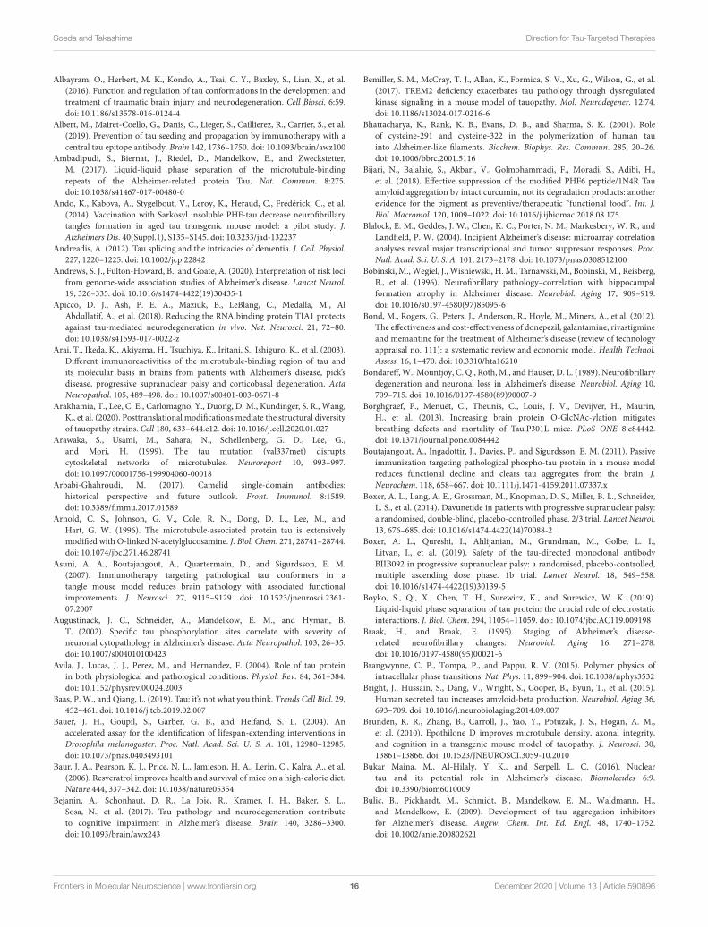
Soeda and Takashima Direction for Tau-Targeted Therapies
Albayram, O., Herbert, M. K., Kondo, A., Tsai, C. Y., Baxley, S., Lian, X., et al.
(2016). Function and regulation of tau conformations in the development and
treatment of traumatic brain injury and neurodegeneration. Cell Biosci. 6:59.
doi: 10.1186/s13578-016-0124-4
Albert, M., Mairet-Coello, G., Danis, C., Lieger, S., Caillierez, R., Carrier, S., et al.
(2019). Prevention of tau seeding and propagation by immunotherapy with a
central tau epitope antibody. Brain 142, 1736–1750. doi: 10.1093/brain/awz100
Ambadipudi, S., Biernat, J., Riedel, D., Mandelkow, E., and Zweckstetter,
M. (2017). Liquid-liquid phase separation of the microtubule-binding
repeats of the Alzheimer-related protein Tau. Nat. Commun. 8:275.
doi: 10.1038/s41467-017-00480-0
Ando, K., Kabova, A., Stygelbout, V., Leroy, K., Heraud, C., Frédérick, C., et al.
(2014). Vaccination with Sarkosyl insoluble PHF-tau decrease neurofibrillary
tangles formation in aged tau transgenic mouse model: a pilot study. J.
Alzheimers Dis. 40(Suppl.1), S135–S145. doi: 10.3233/jad-132237
Andreadis, A. (2012). Tau splicing and the intricacies of dementia. J. Cell. Physiol.
227, 1220–1225. doi: 10.1002/jcp.22842
Andrews, S. J., Fulton-Howard, B., and Goate, A. (2020). Interpretation of risk loci
from genome-wide association studies of Alzheimer’s disease. Lancet Neurol.
19, 326–335. doi: 10.1016/s1474-4422(19)30435-1
Apicco, D. J., Ash, P. E. A., Maziuk, B., LeBlang, C., Medalla, M., Al
Abdullatif, A., et al. (2018). Reducing the RNA binding protein TIA1 protects
against tau-mediated neurodegeneration in vivo. Nat. Neurosci. 21, 72–80.
doi: 10.1038/s41593-017-0022-z
Arai, T., Ikeda, K., Akiyama, H., Tsuchiya, K., Iritani, S., Ishiguro, K., et al. (2003).
Different immunoreactivities of the microtubule-binding region of tau and
its molecular basis in brains from patients with Alzheimer’s disease, pick’s
disease, progressive supranuclear palsy and corticobasal degeneration. Acta
Neuropathol. 105, 489–498. doi: 10.1007/s00401-003-0671-8
Arakhamia, T., Lee, C. E., Carlomagno, Y., Duong, D. M., Kundinger, S. R., Wang,
K., et al. (2020). Posttranslational modifications mediate the structural diversity
of tauopathy strains. Cell 180, 633–644.e12. doi: 10.1016/j.cell.2020.01.027
Arawaka, S., Usami, M., Sahara, N., Schellenberg, G. D., Lee, G.,
and Mori, H. (1999). The tau mutation (val337met) disrupts
cytoskeletal networks of microtubules. Neuroreport 10, 993–997.
doi: 10.1097/00001756-199904060-00018
Arbabi-Ghahroudi, M. (2017). Camelid single-domain antibodies:
historical perspective and future outlook. Front. Immunol. 8:1589.
doi: 10.3389/fimmu.2017.01589
Arnold, C. S., Johnson, G. V., Cole, R. N., Dong, D. L., Lee, M., and
Hart, G. W. (1996). The microtubule-associated protein tau is extensively
modified with O-linked N-acetylglucosamine. J. Biol. Chem. 271, 28741–28744.
doi: 10.1074/jbc.271.46.28741
Asuni, A. A., Boutajangout, A., Quartermain, D., and Sigurdsson, E. M.
(2007). Immunotherapy targeting pathological tau conformers in a
tangle mouse model reduces brain pathology with associated functional
improvements. J. Neurosci. 27, 9115–9129. doi: 10.1523/jneurosci.2361-
07.2007
Augustinack, J. C., Schneider, A., Mandelkow, E. M., and Hyman, B.
T. (2002). Specific tau phosphorylation sites correlate with severity of
neuronal cytopathology in Alzheimer’s disease. Acta Neuropathol. 103, 26–35.
doi: 10.1007/s004010100423
Avila, J., Lucas, J. J., Perez, M., and Hernandez, F. (2004). Role of tau protein
in both physiological and pathological conditions. Physiol. Rev. 84, 361–384.
doi: 10.1152/physrev.00024.2003
Baas, P. W., and Qiang, L. (2019). Tau: it’s not what you think. Trends Cell Biol. 29,
452–461. doi: 10.1016/j.tcb.2019.02.007
Bauer, J. H., Goupil, S., Garber, G. B., and Helfand, S. L. (2004). An
accelerated assay for the identification of lifespan-extending interventions in
Drosophila melanogaster. Proc. Natl. Acad. Sci. U. S. A. 101, 12980–12985.
doi: 10.1073/pnas.0403493101
Baur, J. A., Pearson, K. J., Price, N. L., Jamieson, H. A., Lerin, C., Kalra, A., et al.
(2006). Resveratrol improves health and survival of mice on a high-calorie diet.
Nature 444, 337–342. doi: 10.1038/nature05354
Bejanin, A., Schonhaut, D. R., La Joie, R., Kramer, J. H., Baker, S. L.,
Sosa, N., et al. (2017). Tau pathology and neurodegeneration contribute
to cognitive impairment in Alzheimer’s disease. Brain 140, 3286–3300.
doi: 10.1093/brain/awx243
Bemiller, S. M., McCray, T. J., Allan, K., Formica, S. V., Xu, G., Wilson, G., et al.
(2017). TREM2 deficiency exacerbates tau pathology through dysregulated
kinase signaling in a mouse model of tauopathy. Mol. Neurodegener. 12:74.
doi: 10.1186/s13024-017-0216-6
Bhattacharya, K., Rank, K. B., Evans, D. B., and Sharma, S. K. (2001). Role
of cysteine-291 and cysteine-322 in the polymerization of human tau
into Alzheimer-like filaments. Biochem. Biophys. Res. Commun. 285, 20–26.
doi: 10.1006/bbrc.2001.5116
Bijari, N., Balalaie, S., Akbari, V., Golmohammadi, F., Moradi, S., Adibi, H.,
et al. (2018). Effective suppression of the modified PHF6 peptide/1N4R Tau
amyloid aggregation by intact curcumin, not its degradation products: another
evidence for the pigment as preventive/therapeutic “functional food”. Int. J.
Biol. Macromol. 120, 1009–1022. doi: 10.1016/j.ijbiomac.2018.08.175
Blalock, E. M., Geddes, J. W., Chen, K. C., Porter, N. M., Markesbery, W. R., and
Landfield, P. W. (2004). Incipient Alzheimer’s disease: microarray correlation
analyses reveal major transcriptional and tumor suppressor responses. Proc.
Natl. Acad. Sci. U. S. A. 101, 2173–2178. doi: 10.1073/pnas.0308512100
Bobinski, M., Wegiel, J., Wisniewski, H. M., Tarnawski, M., Bobinski, M., Reisberg,
B., et al. (1996). Neurofibrillary pathology–correlation with hippocampal
formation atrophy in Alzheimer disease. Neurobiol. Aging 17, 909–919.
doi: 10.1016/s0197-4580(97)85095-6
Bond, M., Rogers, G., Peters, J., Anderson, R., Hoyle, M., Miners, A., et al. (2012).
The effectiveness and cost-effectiveness of donepezil, galantamine, rivastigmine
and memantine for the treatment of Alzheimer’s disease (review of technology
appraisal no. 111): a systematic review and economic model. Health Technol.
Assess. 16, 1–470. doi: 10.3310/hta16210
Bondareff,W.,Mountjoy, C. Q., Roth,M., andHauser, D. L. (1989). Neurofibrillary
degeneration and neuronal loss in Alzheimer’s disease. Neurobiol. Aging 10,
709–715. doi: 10.1016/0197-4580(89)90007-9
Borghgraef, P., Menuet, C., Theunis, C., Louis, J. V., Devijver, H., Maurin,
H., et al. (2013). Increasing brain protein O-GlcNAc-ylation mitigates
breathing defects and mortality of Tau.P301L mice. PLoS ONE 8:e84442.
doi: 10.1371/journal.pone.0084442
Boutajangout, A., Ingadottir, J., Davies, P., and Sigurdsson, E. M. (2011). Passive
immunization targeting pathological phospho-tau protein in a mouse model
reduces functional decline and clears tau aggregates from the brain. J.
Neurochem. 118, 658–667. doi: 10.1111/j.1471-4159.2011.07337.x
Boxer, A. L., Lang, A. E., Grossman, M., Knopman, D. S., Miller, B. L., Schneider,
L. S., et al. (2014). Davunetide in patients with progressive supranuclear palsy:
a randomised, double-blind, placebo-controlled phase. 2/3 trial. Lancet Neurol.
13, 676–685. doi: 10.1016/s1474-4422(14)70088-2
Boxer, A. L., Qureshi, I., Ahlijanian, M., Grundman, M., Golbe, L. I.,
Litvan, I., et al. (2019). Safety of the tau-directed monoclonal antibody
BIIB092 in progressive supranuclear palsy: a randomised, placebo-controlled,
multiple ascending dose phase. 1b trial. Lancet Neurol. 18, 549–558.
doi: 10.1016/s1474-4422(19)30139-5
Boyko, S., Qi, X., Chen, T. H., Surewicz, K., and Surewicz, W. K. (2019).
Liquid-liquid phase separation of tau protein: the crucial role of electrostatic
interactions. J. Biol. Chem. 294, 11054–11059. doi: 10.1074/jbc.AC119.009198
Braak, H., and Braak, E. (1995). Staging of Alzheimer’s disease-
related neurofibrillary changes. Neurobiol. Aging 16, 271–278.
doi: 10.1016/0197-4580(95)00021-6
Brangwynne, C. P., Tompa, P., and Pappu, R. V. (2015). Polymer physics of
intracellular phase transitions. Nat. Phys. 11, 899–904. doi: 10.1038/nphys3532
Bright, J., Hussain, S., Dang, V., Wright, S., Cooper, B., Byun, T., et al. (2015).
Human secreted tau increases amyloid-beta production. Neurobiol. Aging 36,
693–709. doi: 10.1016/j.neurobiolaging.2014.09.007
Brunden, K. R., Zhang, B., Carroll, J., Yao, Y., Potuzak, J. S., Hogan, A. M.,
et al. (2010). Epothilone D improves microtubule density, axonal integrity,
and cognition in a transgenic mouse model of tauopathy. J. Neurosci. 30,
13861–13866. doi: 10.1523/JNEUROSCI.3059-10.2010
Bukar Maina, M., Al-Hilaly, Y. K., and Serpell, L. C. (2016). Nuclear
tau and its potential role in Alzheimer’s disease. Biomolecules 6:9.
doi: 10.3390/biom6010009
Bulic, B., Pickhardt, M., Schmidt, B., Mandelkow, E. M., Waldmann, H.,
and Mandelkow, E. (2009). Development of tau aggregation inhibitors
for Alzheimer’s disease. Angew. Chem. Int. Ed. Engl. 48, 1740–1752.
doi: 10.1002/anie.200802621
Frontiers in Molecular Neuroscience | www.frontiersin.org 16 December 2020 | Volume 13 | Article 590896

Soeda and Takashima Direction for Tau-Targeted Therapies
Butner, K. A., and Kirschner, M. W. (1991). Tau protein binds to microtubules
through a flexible array of distributed weak sites. J. Cell Biol. 115, 717–730.
doi: 10.1083/jcb.115.3.717
Caccamo, A., Oddo, S., Tran, L. X., and LaFerla, F. M. (2007). Lithium reduces
tau phosphorylation but not A beta or working memory deficits in a
transgenic model with both plaques and tangles. Am. J. Pathol. 170, 1669–1675.
doi: 10.2353/ajpath.2007.061178
Castillo-Carranza, D. L., Sengupta, U., Guerrero-Munoz, M. J., Lasagna-
Reeves, C. A., Gerson, J. E., Singh, G., et al. (2014). Passive immunization
with Tau oligomer monoclonal antibody reverses tauopathy phenotypes
without affecting hyperphosphorylated neurofibrillary tangles. J. Neurosci. 34,
4260–4272. doi: 10.1523/JNEUROSCI.3192-13.2014
Chai, X., Wu, S., Murray, T. K., Kinley, R., Cella, C. V., Sims, H., et al. (2011).
Passive immunization with anti-Tau antibodies in two transgenic models:
reduction of Tau pathology and delay of disease progression. J. Biol. Chem. 286,
34457–34467. doi: 10.1074/jbc.M111.229633
Chen, D., Drombosky, K. W., Hou, Z., Sari, L., Kashmer, O. M., Ryder,
B. D., et al. (2019). Tau local structure shields an amyloid-forming
motif and controls aggregation propensity. Nat. Commun. 10:2493.
doi: 10.1038/s41467-019-10355-1
Chen, H., Liu, S., Ji, L., Wu, T., Ji, Y., Zhou, Y., et al. (2016). Folic acid
supplementation mitigates alzheimer’s disease by reducing inflammation:
a randomized controlled trial. Mediators Inflamm. 2016:5912146.
doi: 10.1155/2016/5912146
Chen, M., Du, Z. Y., Zheng, X., Li, D. L., Zhou, R. P., and Zhang, K. (2018). Use
of curcumin in diagnosis, prevention, and treatment of Alzheimer’s disease.
Neural Regen. Res. 13, 742–752. doi: 10.4103/1673-5374.230303
Chesser, A. S., Pritchard, S. M., and Johnson, G. V. (2013). Tau clearance
mechanisms and their possible role in the pathogenesis of Alzheimer disease.
Front. Neurol. 4:122. doi: 10.3389/fneur.2013.00122
Chimento, A., De Amicis, F., Sirianni, R., Sinicropi, M. S., Puoci, F., Casaburi, I.,
et al. (2019). Progress to improve oral bioavailability and beneficial effects of
resveratrol. Int. J. Mol. Sci. 20:1381. doi: 10.3390/ijms20061381
Cho, J. H., and Johnson, G. V. (2004). Glycogen synthase kinase. 3 beta induces
caspase-cleaved tau aggregation in situ. J. Biol. Chem. 279, 54716–54723.
doi: 10.1074/jbc.M403364200
Choi, K. T. (2008). Botanical characteristics, pharmacological effects andmedicinal
components of Korean Panax ginseng C A Meyer. Acta Pharmacol. Sin. 29,
1109–1118. doi: 10.1111/j.1745-7254.2008.00869.x
Cicenas, J., Kalyan, K., Sorokinas, A., Stankunas, E., Levy, J., Meskinyte, I., et al.
(2015). Roscovitine in cancer and other diseases. Ann. Transl. Med. 3:135.
doi: 10.3978/j.issn.2305-5839.2015.03.61
Clavaguera, F., Bolmont, T., Crowther, R. A., Abramowski, D., Frank, S., Probst,
A., et al. (2009). Transmission and spreading of tauopathy in transgenic mouse
brain. Nat. Cell Biol. 11, 909–913. doi: 10.1038/ncb1901
Cohen, T. J., Friedmann, D., Hwang, A. W., Marmorstein, R., and Lee,
V. M. (2013). The microtubule-associated tau protein has intrinsic
acetyltransferase activity. Nat. Struct. Mol. Biol. 20, 756–762. doi: 10.1038/
nsmb.2555
Cohen, T. J., Guo, J. L., Hurtado, D. E., Kwong, L. K., Mills, I. P., Trojanowski,
J. Q., et al. (2011). The acetylation of tau inhibits its function and
promotes pathological tau aggregation. Nat. Commun. 2:252. doi: 10.1038/
ncomms1255
Colin, M., Dujardin, S., Schraen-Maschke, S., Meno-Tetang, G., Duyckaerts, C.,
Courade, J. P., et al. (2020). From the prion-like propagation hypothesis to
therapeutic strategies of anti-tau immunotherapy.Acta Neuropathol. 139, 3–25.
doi: 10.1007/s00401-019-02087-9
Collin, L., Bohrmann, B., Göpfert, U., Oroszlan-Szovik, K., Ozmen, L., and
Grüninger, F. (2014). Neuronal uptake of tau/pS422 antibody and reduced
progression of tau pathology in a mouse model of Alzheimer’s disease. Brain
137, 2834–2846. doi: 10.1093/brain/awu213
Combs, B., Mueller, R. L., Morfini, G., Brady, S. T., and Kanaan, N. M. (2019). Tau
and axonal transport misregulation in tauopathies. Adv. Exp. Med. Biol. 1184,
81–95. doi: 10.1007/978-981-32-9358-8_7
Congdon, E. E., Gu, J., Sait, H. B., and Sigurdsson, E. M. (2013). Antibody uptake
into neurons occurs primarily via clathrin-dependent Fcγ receptor endocytosis
and is a prerequisite for acute tau protein clearance. J. Biol. Chem. 288,
35452–35465. doi: 10.1074/jbc.M113.491001
Congdon, E. E., and Sigurdsson, E. M. (2018). Tau-targeting
therapies for Alzheimer disease. Nat. Rev. Neurol. 14, 399–415.
doi: 10.1038/s41582-018-0013-z
Cook, C., Carlomagno, Y., Gendron, T. F., Dunmore, J., Scheffel, K., Stetler, C.,
et al. (2014a). Acetylation of the KXGS motifs in tau is a critical determinant in
modulation of tau aggregation and clearance. Hum. Mol. Genet. 23, 104–116.
doi: 10.1093/hmg/ddt402
Cook, C., Stankowski, J. N., Carlomagno, Y., Stetler, C., and Petrucelli, L. (2014b).
Acetylation: a new key to unlock tau’s role in neurodegeneration. Alzheimers.
Res. Ther. 6:29. doi: 10.1186/alzrt259
Courade, J. P., Angers, R., Mairet-Coello, G., Pacico, N., Tyson, K., Lightwood, D.,
et al. (2018). Epitope determines efficacy of therapeutic anti-Tau antibodies in a
functional assay with human Alzheimer Tau. Acta Neuropathol. 136, 729–745.
doi: 10.1007/s00401-018-1911-2
Cox, K. H., Pipingas, A., and Scholey, A. B. (2015). Investigation of the effects of
solid lipid curcumin on cognition and mood in a healthy older population. J.
Psychopharmacol. 29, 642–651. doi: 10.1177/0269881114552744
Cripps, D., Thomas, S. N., Jeng, Y., Yang, F., Davies, P., and Yang, A. J. (2006).
Alzheimer disease-specific conformation of hyperphosphorylated paired helical
filament-Tau is polyubiquitinated through Lys-48, Lys-11, and Lys-6 ubiquitin
conjugation. J. Biol. Chem. 281, 10825–10838. doi: 10.1074/jbc.M512786200
Crowe, A., Huang, W., Ballatore, C., Johnson, R. L., Hogan, A. M., Huang, R.,
et al. (2009). Identification of aminothienopyridazine inhibitors of tau assembly
by quantitative high-throughput screening. Biochemistry 48, 7732–7745.
doi: 10.1021/bi9006435
Cummings, J., Lee, G., Ritter, A., Sabbagh, M., and Zhong, K. (2019). Alzheimer’s
disease drug development pipeline: 2019. Alzheimers Dement. 5, 272–293.
doi: 10.1016/j.trci.2019.05.008
d’Abramo, C., Acker, C. M., Jimenez, H. T., and Davies, P. (2013). Tau passive
immunotherapy in mutant P301L mice: antibody affinity versus specificity.
PLoS ONE 8:e62402. doi: 10.1371/journal.pone.0062402
de Calignon, A., Fox, L. M., Pitstick, R., Carlson, G. A., Bacskai, B. J., Spires-Jones,
T. L., et al. (2010). Caspase activation precedes and leads to tangles.Nature 464,
1201–1204. doi: 10.1038/nature08890
de Silva, R., Lashley, T., Strand, C., Shiarli, A. M., Shi, J., Tian, J., et al. (2006). An
immunohistochemical study of cases of sporadic and inherited frontotemporal
lobar degeneration using. 3R- and. 4R-specific tau monoclonal antibodies. Acta
Neuropathol. 111, 329–340. doi: 10.1007/s00401-006-0048-x
Delacourte, A., David, J. P., Sergeant, N., Buée, L., Wattez, A., Vermersch, P., et al.
(1999). The biochemical pathway of neurofibrillary degeneration in aging and
Alzheimer’s disease. Neurology 52, 1158–1165. doi: 10.1212/wnl.52.6.1158
Despres, C., Byrne, C., Qi, H., Cantrelle, F. X., Huvent, I., Chambraud,
B., et al. (2017). Identification of the Tau phosphorylation pattern that
drives its aggregation. Proc. Natl. Acad. Sci. U. S. A. 114, 9080–9085.
doi: 10.1073/pnas.1708448114
DeVos, S. L., Miller, R. L., Schoch, K. M., Holmes, B. B., Kebodeaux, C. S.,
Wegener, A. J., et al. (2017). Tau reduction prevents neuronal loss and reverses
pathological tau deposition and seeding in mice with tauopathy. Sci. Transl.
Med. 9:eaag0481. doi: 10.1126/scitranslmed.aag0481
Di Domenico, F., Lanzillotta, C., and Tramutola, A. (2019). Therapeutic potential
of rescuing protein O-GlcNAcylation in tau-related pathologies. Expert Rev.
Neurother. 19, 1–3. doi: 10.1080/14737175.2019.1540932
Di Meco, A., Li, J. G., Blass, B. E., Abou-Gharbia, M., Lauretti, E., and
Praticò, D. (2017). 12/15-lipoxygenase inhibition reverses cognitive
impairment, brain amyloidosis, and tau pathology by stimulating
autophagy in aged triple transgenic Mice. Biol. Psychiatry 81, 92–100.
doi: 10.1016/j.biopsych.2016.05.023
Dickey, C. A., Kamal, A., Lundgren, K., Klosak, N., Bailey, R. M., Dunmore, J.,
et al. (2007). The high-affinity HSP90-CHIP complex recognizes and selectively
degrades phosphorylated tau client proteins. J. Clin. Invest. 117, 648–658.
doi: 10.1172/JCI29715
Dong, S., Zeng, Q., Mitchell, E. S., Xiu, J., Duan, Y., Li, C., et al. (2012).
Curcumin enhances neurogenesis and cognition in aged rats: implications for
transcriptional interactions related to growth and synaptic plasticity. PLoS ONE
7:e31211. doi: 10.1371/journal.pone.0031211
Duyckaerts, C., Clavaguera, F., and Potier, M. C. (2019). The prion-like
propagation hypothesis in Alzheimer’s and Parkinson’s disease. Curr. Opin.
Neurol. 32, 266–271. doi: 10.1097/wco.0000000000000672
Frontiers in Molecular Neuroscience | www.frontiersin.org 17 December 2020 | Volume 13 | Article 590896

Soeda and Takashima Direction for Tau-Targeted Therapies
Engmann, O., and Giese, K. P. (2009). Crosstalk between Cdk5 and
GSK3beta: implications for Alzheimer’s disease. Front. Mol. Neurosci. 2:2.
doi: 10.3389/neuro.02.002.2009
Falcon, B., Zhang, W., Murzin, A. G., Murshudov, G., Garringer, H. J., Vidal,
R., et al. (2018). Structures of filaments from Pick’s disease reveal a novel tau
protein fold. Nature 561, 137–140. doi: 10.1038/s41586-018-0454-y
Falcon, B., Zivanov, J., Zhang, W., Murzin, A. G., Garringer, H. J., Vidal, R., et al.
(2019). Novel tau filament fold in chronic traumatic encephalopathy encloses
hydrophobic molecules. Nature 568, 420–423. doi: 10.1038/s41586-019-1026-5
Fellner, S., Bauer, B., Miller, D. S., Schaffrik, M., Fankhänel, M., Spru,ß, T., et al.
(2002). Transport of paclitaxel (Taxol) across the blood-brain barrier in vitro
and in vivo. J. Clin. Invest. 110, 1309–1318. doi: 10.1172/jci200215451
Feng, Y., Wang, X. P., Yang, S. G., Wang, Y. J., Zhang, X., Du, X.
T., et al. (2009). Resveratrol inhibits beta-amyloid oligomeric cytotoxicity
but does not prevent oligomer formation. Neurotoxicology 30, 986–995.
doi: 10.1016/j.neuro.2009.08.013
Fichou, Y., Al-Hilaly, Y. K., Devred, F., Smet-Nocca, C., Tsvetkov, P. O., Verelst, J.,
et al. (2019). The elusive tau molecular structures: can we translate the recent
breakthroughs into new targets for intervention? Acta Neuropathol. Commun.
7:31. doi: 10.1186/s40478-019-0682-x
Fitzpatrick, A. W. P., Falcon, B., He, S., Murzin, A. G., Murshudov, G., Garringer,
H. J., et al. (2017). Cryo-EM structures of tau filaments from Alzheimer’s
disease. Nature 547, 185–190. doi: 10.1038/nature23002
Frost, B., Jacks, R. L., and Diamond, M. I. (2009). Propagation of tau misfolding
from the outside to the inside of a cell. J. Biol. Chem. 284, 12845–12852.
doi: 10.1074/jbc.M808759200
Fulga, T. A., Elson-Schwab, I., Khurana, V., Steinhilb, M. L., Spires, T. L.,
Hyman, B. T., et al. (2007). Abnormal bundling and accumulation of F-actin
mediates tau-induced neuronal degeneration in vivo. Nat. Cell Biol. 9, 139–148.
doi: 10.1038/ncb1528
Gauthier, S., Feldman, H. H., Schneider, L. S., Wilcock, G. K., Frisoni, G. B.,
Hardlund, J. H., et al. (2016). Efficacy and safety of tau-aggregation inhibitor
therapy in patients with mild or moderate Alzheimer’s disease: a randomised,
controlled, double-blind, parallel-arm, phase. 3 trial. Lancet 388, 2873–2884.
doi: 10.1016/s0140-6736(16)31275-2
Ge, J. F., Qiao, J. P., Qi, C. C., Wang, C. W., and Zhou, J. N. (2012). The binding of
resveratrol tomonomer and fibril amyloid beta.Neurochem. Int. 61, 1192–1201.
doi: 10.1016/j.neuint.2012.08.012
Gendron, T. F., and Petrucelli, L. (2009). The role of tau in neurodegeneration.
Mol. Neurodegener. 4:13. doi: 10.1186/1750-1326-4-13
Ghasemzadeh, S., and Riazi, G. H. (2020). Inhibition of Tau amyloid fibril
formation by folic acid: In-vitro and theoretical studies. Int. J. Biol. Macromol.
154, 1505–1516. doi: 10.1016/j.ijbiomac.2019.11.032
Giacobini, E., and Gold, G. (2013). Alzheimer disease therapy–moving from
amyloid-β to tau. Nat. Rev. Neurol. 9, 677–686. doi: 10.1038/nrneurol.2013.223
Giacomini, C., Koo, C. Y., Yankova, N., Tavares, I. A., Wray, S., Noble, W.,
et al. (2018). A new TAO kinase inhibitor reduces tau phosphorylation at sites
associated with neurodegeneration in human tauopathies. Acta Neuropathol.
Commun. 6:37. doi: 10.1186/s40478-018-0539-8
Goedert, M., Crowther, R. A., and Garner, C. C. (1991). Molecular characterization
of microtubule-associated proteins tau and MAP2. Trends Neurosci. 14,
193–199. doi: 10.1016/0166-2236(91)90105-4
Goedert, M., Jakes, R., Spillantini, M. G., Hasegawa,M., Smith,M. J., and Crowther,
R. A. (1996). Assembly of microtubule-associated protein tau into Alzheimer-
like filaments induced by sulphated glycosaminoglycans. Nature 383, 550–553.
doi: 10.1038/383550a0
Goedert, M., Spillantini, M. G., Cairns, N. J., and Crowther, R. A. (1992). Tau
proteins of Alzheimer paired helical filaments: abnormal phosphorylation of
all six brain isoforms. Neuron 8, 159–168. doi: 10.1016/0896-6273(92)90117-v
Gómez-Isla, T., Hollister, R., West, H., Mui, S., Growdon, J. H., Petersen, R. C.,
et al. (1997). Neuronal loss correlates with but exceeds neurofibrillary tangles
in Alzheimer’s disease. Ann. Neurol. 41, 17–24. doi: 10.1002/ana.410410106
Gomez-Isla, T., West, H. L., Rebeck, G.W., Harr, S. D., Growdon, J. H., Locascio, J.
J., et al. (1996). Clinical and pathological correlates of apolipoprotein E epsilon.
4 in Alzheimer’s disease. Ann. Neurol. 39, 62–70. doi: 10.1002/ana.410390110
Gong, C. X., and Iqbal, K. (2008). Hyperphosphorylation of microtubule-
associated protein tau: a promising therapeutic target for Alzheimer disease.
Curr. Med. Chem. 15, 2321–2328. doi: 10.2174/092986708785909111
Gong, C. X., Shaikh, S., Wang, J. Z., Zaidi, T., Grundke-Iqbal, I., and
Iqbal, K. (1995). Phosphatase activity toward abnormally phosphorylated
tau: decrease in Alzheimer disease brain. J. Neurochem. 65, 732–738.
doi: 10.1046/j.1471-4159.1995.65020732.x
Gong, C. X., Singh, T. J., Grundke-Iqbal, I., and Iqbal, K. (1993). Phosphoprotein
phosphatase activities in Alzheimer disease brain. J. Neurochem. 61, 921–927.
doi: 10.1111/j.1471-4159.1993.tb03603.x
Gorantla, N. V., Das, R., Balaraman, E., and Chinnathambi, S. (2020). Transition
metal nickel prevents Tau aggregation in Alzheimer’s disease. Int. J. Biol.
Macromol. 156, 1359–1365. doi: 10.1016/j.ijbiomac.2019.11.176
Götz, J., Probst, A., Spillantini, M. G., Schäfer, T., Jakes, R., Bürki, K., et al.
(1995). Somatodendritic localization and hyperphosphorylation of tau protein
in transgenic mice expressing the longest human brain tau isoform. EMBO J.
14, 1304–1313
Götz, J. J., and Götz, J. (2019). Experimental models of tauopathy—
from mechanisms to therapies. Adv. Exp. Med. Biol. 1184, 381–391.
doi: 10.1007/978-981-32-9358-8_28
Gozes, I., Bassan, M., Zamostiano, R., Pinhasov, A., Davidson, A., Giladi, E.,
et al. (1999). A novel signaling molecule for neuropeptide action: activity-
dependent neuroprotective protein. Ann. N. Y. Acad. Sci. 897, 125–135.
doi: 10.1111/j.1749-6632.1999.tb07884.x
Greenberg, S. G., Davies, P., Schein, J. D., and Binder, L. I. (1992). Hydrofluoric
acid-treated tau PHF proteins display the same biochemical properties as
normal tau. J. Biol. Chem. 267, 564–569
Griffith, L. M., and Pollard, T. D. (1978). Evidence for actin filament-microtubule
interaction mediated by microtubule-associated proteins. J. Cell Biol. 78,
958–965. doi: 10.1083/jcb.78.3.958
Gu, J., Congdon, E. E., and Sigurdsson, E. M. (2013). Two novel Tau
antibodies targeting the. 396/404 region are primarily taken up by neurons
and reduce Tau protein pathology. J. Biol. Chem. 288, 33081–33095.
doi: 10.1074/jbc.M113.494922
Guillozet, A. L., Weintraub, S., Mash, D. C., and Mesulam, M. M. (2003).
Neurofibrillary tangles, amyloid, and memory in aging and mild cognitive
impairment. Arch. Neurol. 60, 729–736. doi: 10.1001/archneur.60.5.729
Guo, T., Noble, W., and Hanger, D. P. (2017). Roles of tau protein in
health and disease. Acta Neuropathol. 133, 665–704. doi: 10.1007/s00401-017-
1707-9
Gustke, N., Trinczek, B., Biernat, J., Mandelkow, E. M., and Mandelkow, E. (1994).
Domains of tau protein and interactions with microtubules. Biochemistry. 33,
9511–9522. doi: 10.1021/bi00198a017
Hanger, D. P., Anderton, B. H., and Noble, W. (2009). Tau phosphorylation:
the therapeutic challenge for neurodegenerative disease. Trends Mol. Med. 15,
112–119. doi: 10.1016/j.molmed.2009.01.003
Hanger, D. P., Gibb, G. M., de Silva, R., Boutajangout, A., Brion, J. P., Revesz,
T., et al. (2002). The complex relationship between soluble and insoluble tau
in tauopathies revealed by efficient dephosphorylation and specific antibodies.
FEBS Lett. 531, 538–542. doi: 10.1016/s0014-5793(02)03611-6
Hart, G. W., Housley, M. P., and Slawson, C. (2007). Cycling of O-linked beta-
N-acetylglucosamine on nucleocytoplasmic proteins. Nature 446, 1017–1022.
doi: 10.1038/nature05815
Hernández-Vega, A., Braun, M., Scharrel, L., Jahnel, M., Wegmann, S., Hyman,
B. T., et al. (2017). Local nucleation of microtubule bundles through
tubulin concentration into a condensed tau phase. Cell Rep. 20, 2304–2312.
doi: 10.1016/j.celrep.2017.08.042
Hochgrafe, K., Sydow, A., Matenia, D., Cadinu, D., Konen, S., Petrova, O., et al.
(2015). Preventive methylene blue treatment preserves cognition in mice
expressing full-length pro-aggregant human Tau. Acta Neuropathol. Commun.
3:25. doi: 10.1186/s40478-015-0204-4
Höglinger, G. U., Huppertz, H. J., Wagenpfeil, S., Andrés, M. V., Belloch, V.,
León, T., et al. (2014). Tideglusib reduces progression of brain atrophy in
progressive supranuclear palsy in a randomized trial.Mov. Disord. 29, 479–487.
doi: 10.1002/mds.25815
Holmes, C., Boche, D., Wilkinson, D., Yadegarfar, G., Hopkins, V., Bayer, A.,
et al. (2008). Long-term effects of Abeta42 immunisation in Alzheimer’s
disease: follow-up of a randomised, placebo-controlled phase I trial. Lancet 372,
216–223. doi: 10.1016/s0140-6736(08)61075-2
Hosokawa, M., Arai, T., Masuda-Suzukake, M., Nonaka, T., Yamashita,
M., Akiyama, H., et al. (2012). Methylene blue reduced abnormal
Frontiers in Molecular Neuroscience | www.frontiersin.org 18 December 2020 | Volume 13 | Article 590896

Soeda and Takashima Direction for Tau-Targeted Therapies
tau accumulation in P301L tau transgenic mice. PLoS ONE 7:e52389.
doi: 10.1371/journal.pone.0052389
Huang, Y., Wu, Z., Cao, Y., Lang, M., Lu, B., and Zhou, B. (2014). Zinc binding
directly regulates tau toxicity independent of tau hyperphosphorylation. Cell
Rep. 8, 831–842. doi: 10.1016/j.celrep.2014.06.047
Hyman, A. A., Weber, C. A., and Julicher, F. (2014). Liquid-liquid
phase separation in biology. Annu. Rev. Cell Dev. Biol. 30, 39–58.
doi: 10.1146/annurev-cellbio-100913-013325
Ikezu, T., and Gendelman, H. E. (2016). Neuroimmune Pharmacology.
New York, NY: Springer.
Imahori, K., and Uchida, T. (1997). Physiology and pathology of tau protein
kinases in relation to Alzheimer’s disease. J. Biochem. 121, 179–188.
Iqbal, K., Alonso Adel, C., Chen, S., Chohan,M. O., El-Akkad, E., Gong, C. X., et al.
(2005). Tau pathology in Alzheimer disease and other tauopathies. Biochim.
Biophys. Acta. 1739, 198–210. doi: 10.1016/j.bbadis.2004.09.008
Irwin, D. J., Cohen, T. J., Grossman, M., Arnold, S. E., McCarty-Wood,
E., Van Deerlin, V. M., et al. (2013). Acetylated tau neuropathology
in sporadic and hereditary tauopathies. Am. J. Pathol. 183, 344–351.
doi: 10.1016/j.ajpath.2013.04.025
Irwin, D. J., Cohen, T. J., Grossman, M., Arnold, S. E., Xie, S. X., Lee, V. M., et al.
(2012). Acetylated tau, a novel pathological signature in Alzheimer’s disease and
other tauopathies. Brain. 135, 807–818. doi: 10.1093/brain/aws013
Ising, C., Venegas, C., Zhang, S., Scheiblich, H., Schmidt, S. V., Vieira-Saecker, A.,
et al. (2019). NLRP3 inflammasome activation drives tau pathology.Nature 575,
669–673. doi: 10.1038/s41586-019-1769-z
Ittner, A., and Ittner, L. M. (2018). Dendritic Tau in Alzheimer’s Disease. Neuron
99, 13–27. doi: 10.1016/j.neuron.2018.06.003
Ittner, L. M., Ke, Y. D., Delerue, F., Bi, M., Gladbach, A., van Eersel, J., et al. (2010).
Dendritic function of tau mediates amyloid-beta toxicity in Alzheimer’s disease
mouse models. Cell 142, 387–397. doi: 10.1016/j.cell.2010.06.036
Janning, D., Igaev, M., Sundermann, F., Bruhmann, J., Beutel, O., Heinisch, J.
J., et al. (2014). Single-molecule tracking of tau reveals fast kiss-and-hop
interaction with microtubules in living neurons.Mol. Biol. Cell. 25, 3541–3551.
doi: 10.1091/mbc.E14-06-1099
Jeganathan, S., Hascher, A., Chinnathambi, S., Biernat, J., Mandelkow, E. M.,
and Mandelkow, E. (2008). Proline-directed pseudo-phosphorylation at AT8
and PHF1 epitopes induces a compaction of the paperclip folding of Tau
and generates a pathological (MC-1) conformation. J. Biol. Chem. 283,
32066–32076. doi: 10.1074/jbc.M805300200
Jhang, K. A., Park, J. S., Kim, H. S., and Chong, Y. H. (2017). Resveratrol
ameliorates tau hyperphosphorylation at ser396 site and oxidative damage
in rat hippocampal slices exposed to vanadate: implication of ERK1/2
and GSK-3β signaling cascades. J. Agric. Food Chem. 65, 9626–9634.
doi: 10.1021/acs.jafc.7b03252
Jicha, G. A., Berenfeld, B., and Davies, P. (1999). Sequence requirements
for formation of conformational variants of tau similar to those
found in Alzheimer’s disease. J. Neurosci. Res. 55, 713–723.
doi: 10.1002/(sici)1097-4547(19990315)55:6<713::Aid-jnr6>3.0.Co;2-g
Jicha, G. A., Bowser, R., Kazam, I. G., and Davies, P. (1997). Alz-50 and MC-
1, a new monoclonal antibody raised to paired helical filaments, recognize
conformational epitopes on recombinant tau. J. Neurosci. Res. 48, 128–132.
doi: 10.1002/(sici)1097-4547(19970415)48:2<128::aid-jnr5>3.0.co;2-e
Jovcevska, I., and Muyldermans, S. (2020). The therapeutic potential
of nanobodies. BioDrugs 34, 11–26. doi: 10.1007/s40259-019-
00392-z
Kabayama, H., Takeuchi, M., Tokushige, N., Muramatsu, S. I., Kabayama, M.,
Fukuda, M., et al. (2020). An ultra-stable cytoplasmic antibody engineered
for in vivo applications. Nat. Commun. 11:336. doi: 10.1038/s41467-019-
13654-9
Kampers, T., Friedhoff, P., Biernat, J., Mandelkow, E. M., and Mandelkow,
E. (1996). RNA stimulates aggregation of microtubule-associated protein
tau into Alzheimer-like paired helical filaments. FEBS Lett. 399, 344–349.
doi: 10.1016/s0014-5793(96)01386-5
Kanaan, N. M., Cox, K., Alvarez, V. E., Stein, T. D., Poncil, S., and McKee,
A. C. (2016). Characterization of early pathological tau conformations and
phosphorylation in chronic traumatic encephalopathy. J. Neuropathol. Exp.
Neurol. 75, 19–34. doi: 10.1093/jnen/nlv001
Kanaan, N. M., Hamel, C., Grabinski, T., and Combs, B. (2020). Liquid-liquid
phase separation induces pathogenic tau conformations in vitro.Nat. Commun.
11:2809. doi: 10.1038/s41467-020-16580-3
Katsumoto, A., Takeuchi, H., and Tanaka, F. (2019). Tau pathology in chronic
traumatic encephalopathy and alzheimer’s disease: similarities and differences.
Front. Neurol. 10:980. doi: 10.3389/fneur.2019.00980
Kaufman, A. C., Salazar, S. V., Haas, L. T., Yang, J., Kostylev, M. A., Jeng, A. T.,
et al. (2015). Fyn inhibition rescues established memory and synapse loss in
Alzheimer mice. Ann. Neurol. 77, 953–971. doi: 10.1002/ana.24394
Kaur, T., Alshareedah, I., Wang, W., Ngo, J., Moosa, M. M., and Banerjee,
P. R. (2019). Molecular crowding tunes material states of ribonucleoprotein
condensates. Biomolecules. 9:71. doi: 10.3390/biom9020071
Kedersha, N., Cho, M. R., Li, W., Yacono, P. W., Chen, S., Gilks, N.,
et al. (2000). Dynamic shuttling of TIA-1 accompanies the recruitment
of mRNA to mammalian stress granules. J. Cell Biol. 151, 1257–1268.
doi: 10.1083/jcb.151.6.1257
Kerchner, G. A., Ayalon, G., Brunstein, F., Chandra, P., Datwani, A., Fuji, R. N.,
et al. (2017). A phase I study to evaluate the safety and tolerability of RO7105705
in healthy volunteers and patients with mild-to-moderate AD. Alzheimer’s
Dementia. 13:601. doi: 10.1016/j.jalz.2017.07.243
Khanna, M. R., Kovalevich, J., Lee, V. M., Trojanowski, J. Q., and Brunden, K.
R. (2016). Therapeutic strategies for the treatment of tauopathies: hopes and
challenges. Alzheimers Dement 12, 1051–1065. doi: 10.1016/j.jalz.2016.06.006
Kim, Y. S., and Joh, T. H. (2006). Microglia, major player in the brain
inflammation: their roles in the pathogenesis of Parkinson’s disease. Exp. Mol.
Med. 38, 333–347. doi: 10.1038/emm.2006.40
Kimura, T., Fukuda, T., Sahara, N., Yamashita, S., Murayama, M., Mizoroki,
T., et al. (2010). Aggregation of detergent-insoluble tau is involved in
neuronal loss but not in synaptic loss. J. Biol. Chem. 285, 38692–38699.
doi: 10.1074/jbc.M110.136630
Kimura, T., Ishiguro, K., and Hisanaga, S. (2014a). Physiological and
pathological phosphorylation of tau by Cdk5. Front. Mol. Neurosci. 7:65.
doi: 10.3389/fnmol.2014.00065
Kimura, T., Whitcomb, D. J., Jo, J., Regan, P., Piers, T., Heo, S., et al. (2014b).
Microtubule-associated protein tau is essential for long-term depression in
the hippocampus. Philos. Trans. R. Soc. Lond. B Biol. Sci. 369:20130144.
doi: 10.1098/rstb.2013.0144
Kimura, T., Yamashita, S., Fukuda, T., Park, J. M., Murayama, M., Mizoroki, T.,
et al. (2007). Hyperphosphorylated tau in parahippocampal cortex impairs
place learning in aged mice expressing wild-type human tau. EMBO J. 26,
5143–5152. doi: 10.1038/sj.emboj.7601917
Kinney, J. W., Bemiller, S. M., Murtishaw, A. S., Leisgang, A. M., Salazar, A. M.,
and Lamb, B. T. (2018). Inflammation as a central mechanism in Alzheimer’s
disease. Alzheimers Dement. 4, 575–590. doi: 10.1016/j.trci.2018.06.014
Kneynsberg, A., Combs, B., Christensen, K., Morfini, G., and Kanaan, N. M.
(2017). Axonal degeneration in tauopathies: disease relevance and underlying
mechanisms. Front. Neurosci. 11:572. doi: 10.3389/fnins.2017.00572
Kondo, A., Shahpasand, K., Mannix, R., Qiu, J., Moncaster, J., Chen, C. H., et al.
(2015). Antibody against early driver of neurodegeneration cis P-tau blocks
brain injury and tauopathy. Nature 523, 431–436. doi: 10.1038/nature14658
Kontaxi, C., Piccardo, P., and Gill, A. C. (2017). Lysine-directed post-translational
modifications of tau protein in alzheimer’s disease and related tauopathies.
Front Mol Biosci. 4:56. doi: 10.3389/fmolb.2017.00056
Kontsekova, E., Zilka, N., Kovacech, B., Novak, P., and Novak, M. (2014).
First-in-man tau vaccine targeting structural determinants essential
for pathological tau-tau interaction reduces tau oligomerisation and
neurofibrillary degeneration in an Alzheimer’s disease model. Alzheimers.
Res. Ther. 6:44. doi: 10.1186/alzrt278
Koo, C. Y., Giacomini, C., Reyes-Corral, M., Olmos, Y., Tavares, I. A., Marson, C.
M., et al. (2017). Targeting TAO kinases using a new inhibitor compound delays
mitosis and induces mitotic cell death in centrosome amplified breast cancer
cells.Mol. Cancer Ther. 16, 2410–2421. doi: 10.1158/1535-7163.Mct-17-0077
Kosik, K. S., and Han, S. (2019). Tau condensates. Adv. Exp. Med. Biol. 1184,
327–339. doi: 10.1007/978-981-32-9358-8_24
Kosik, K. S., Orecchio, L. D., Binder, L., Trojanowski, J. Q., Lee, V. M., and Lee,
G. (1988). Epitopes that span the tau molecule are shared with paired helical
filaments. Neuron 1, 817–825. doi: 10.1016/0896-6273(88)90129-8
Frontiers in Molecular Neuroscience | www.frontiersin.org 19 December 2020 | Volume 13 | Article 590896

Soeda and Takashima Direction for Tau-Targeted Therapies
Kovacs, G. G. (2015). Invited review: neuropathology of tauopathies: principles
and practice. Neuropathol. Appl. Neurobiol. 41, 3–23. doi: 10.1111/nan.12208
Kuchibhotla, K. V., Wegmann, S., Kopeikina, K. J., Hawkes, J., Rudinskiy, N.,
Andermann, M. L., et al. (2014). Neurofibrillary tangle-bearing neurons are
functionally integrated in cortical circuits in vivo. Proc. Natl. Acad. Sci. U. S.
A. 111, 510–514. doi: 10.1073/pnas.1318807111
Lasagna-Reeves, C. A., Castillo-Carranza, D. L., Sengupta, U., Sarmiento, J.,
Troncoso, J., Jackson, G. R., et al. (2012). Identification of oligomers at early
stages of tau aggregation in Alzheimer’s disease. FASEB J. 26, 1946–1959.
doi: 10.1096/fj.11-199851
Laurent, C., Buée, L., and Blum, D. (2018). Tau and neuroinflammation:
what impact for alzheimer’s disease and tauopathies? Biomed. J. 41, 21–33.
doi: 10.1016/j.bj.2018.01.003
Lauretti, E., Iuliano, L., and Praticò, D. (2017). Extra-virgin olive oil ameliorates
cognition and neuropathology of the. 3xTg mice: role of autophagy. Ann. Clin.
Transl. Neurol. 4, 564–574. doi: 10.1002/acn3.431
Lauretti, E., and Praticò, D. (2020). Alzheimer’s disease: phenotypic approaches
using diseasemodels and the targeting of tau protein. Expert Opin. Ther. Targets
24, 319–330. doi: 10.1080/14728222.2020.1737012
Lee, G., Thangavel, R., Sharma, V. M., Litersky, J. M., Bhaskar, K., Fang, S. M., et al.
(2004). Phosphorylation of tau by fyn: implications for Alzheimer’s disease. J.
Neurosci. 24, 2304–2312. doi: 10.1523/jneurosci.4162-03.2004
Lee, M. J., Lee, J. H., and Rubinsztein, D. C. (2013). Tau degradation: the ubiquitin-
proteasome system versus the autophagy-lysosome system. Prog Neurobiol.
105, 49–59. doi: 10.1016/j.pneurobio.2013.03.001
Lee, S. H., Le Pichon, C. E., Adolfsson, O., Gafner, V., Pihlgren, M., Lin,
H., et al. (2016). Antibody-mediated targeting of tau In vivo does not
require effector function and microglial engagement. Cell Rep. 16, 1690–1700.
doi: 10.1016/j.celrep.2016.06.099
Lee, S. M., Bae, B. S., Park, H. W., Ahn, N. G., Cho, B. G., Cho, Y. L., et al. (2015).
Characterization of Korean Red Ginseng (Panax ginseng Meyer): History,
preparation method, and chemical composition. J. Ginseng Res. 39, 384–391.
doi: 10.1016/j.jgr.2015.04.009
Lee, V. M., Balin, B. J., Otvos, L. Jr, and Trojanowski, J. Q. (1991). A68: a major
subunit of paired helical filaments and derivatized forms of normal Tau. Science
251, 675–678. doi: 10.1126/science.1899488
Lee, V. M., Brunden, K. R., Hutton, M., and Trojanowski, J. Q. (2011).
Developing therapeutic approaches to tau, selected kinases, and related
neuronal protein targets. Cold Spring Harb. Perspect. Med. 1:a006437.
doi: 10.1101/cshperspect.a006437
Lee, V. M., Daughenbaugh, R., and Trojanowski, J. Q. (1994). Microtubule
stabilizing drugs for the treatment of Alzheimer’s disease. Neurobiol. Aging
15(Suppl.2), S87–89. doi: 10.1016/0197-4580(94)90179-1
Leroy, K., Ando, K., Héraud, C., Yilmaz, Z., Authelet, M., Boeynaems, J. M., et al.
(2010). Lithium treatment arrests the development of neurofibrillary tangles
in mutant tau transgenic mice with advanced neurofibrillary pathology. J.
Alzheimers. Dis. 19, 705–719. doi: 10.3233/jad-2010-1276
Leyns, C. E. G., Ulrich, J. D., Finn, M. B., Stewart, F. R., Koscal, L. J., Remolina
Serrano, J., et al. (2017). TREM2 deficiency attenuates neuroinflammation and
protects against neurodegeneration in a mouse model of tauopathy. Proc. Natl.
Acad. Sci. U. S. A. 114, 11524–11529. doi: 10.1073/pnas.1710311114
Li, W., Jiang, M., Xiao, Y., Zhang, X., Cui, S., and Huang, G. (2015). Folic acid
inhibits tau phosphorylation through regulation of PP2A methylation in SH-
SY5Y cells. J. Nutr. Health Aging 19, 123–129. doi: 10.1007/s12603-014-0514-4
Liu, F., Grundke-Iqbal, I., Iqbal, K., and Gong, C. X. (2005). Contributions
of protein phosphatases PP1, PP2A, PP2B and PP5 to the
regulation of tau phosphorylation. Eur. J. Neurosci. 22, 1942–1950.
doi: 10.1111/j.1460-9568.2005.04391.x
Liu, F., Iqbal, K., Grundke-Iqbal, I., Hart, G. W., and Gong, C. X. (2004).
O-GlcNAcylation regulates phosphorylation of tau: a mechanism involved
in Alzheimer’s disease. Proc. Natl. Acad. Sci. U. S. A. 101, 10804–10809.
doi: 10.1073/pnas.0400348101
Liu, F., Shi, J., Tanimukai, H., Gu, J., Gu, J., Grundke-Iqbal, I., et al. (2009). Reduced
O-GlcNAcylation links lower brain glucose metabolism and tau pathology in
Alzheimer’s disease. Brain 132, 1820–1832. doi: 10.1093/brain/awp099
Liu, G., Fiock, K. L., Levites, Y., Golde, T. E., Hefti, M. M., and Lee, G. (2020). Fyn
depletion ameliorates tau(P301L)-induced neuropathology. Acta Neuropathol
Commun. 8:108. doi: 10.1186/s40478-020-00979-6
Liu, W., Zhao, L., Blackman, B., Parmar, M., Wong, M. Y., Woo, T., et al. (2016).
Vectored intracerebral immunization with the anti-tau monoclonal antibody
PHF1 markedly reduces Tau pathology in mutant tau transgenic Mice. J.
Neurosci. 36, 12425–12435. doi: 10.1523/jneurosci.2016-16.2016
Liu-Yesucevitz, L., Bassell, G. J., Gitler, A. D., Hart, A. C., Klann, E., Richter, J. D.,
et al. (2011). Local RNA translation at the synapse and in disease. J. Neurosci.
31, 16086–16093. doi: 10.1523/JNEUROSCI.4105-11.2011
Lo Cascio, F., Puangmalai, N., Ellsworth, A., Bucchieri, F., Pace, A., Palumbo
Piccionello, A., et al. (2019). Toxic tau oligomers modulated by novel curcumin
derivatives. Sci. Rep. 9:19011. doi: 10.1038/s41598-019-55419-w
Loomis, P. A., Howard, T. H., Castleberry, R. P., and Binder, L. I. (1990).
Identification of nuclear tau isoforms in human neuroblastoma cells. Proc. Natl.
Acad. Sci. U. S. A. 87, 8422–8426. doi: 10.1073/pnas.87.21.8422
Loveman, E., Green, C., Kirby, J., Takeda, A., Picot, J., Payne, E., et al. (2006).
The clinical and cost-effectiveness of donepezil, rivastigmine, galantamine
and memantine for Alzheimer’s disease. Health Technol. Assess. 10, 1–160.
doi: 10.3310/hta10010
Maeda, S., Sahara, N., Saito, Y., Murayama, M., Yoshiike, Y., Kim, H., et al. (2007).
Granular tau oligomers as intermediates of tau filaments. Biochemistry 46,
3856–3861. doi: 10.1021/bi061359o
Maeda, S., Sahara, N., Saito, Y., Murayama, S., Ikai, A., and Takashima,
A. (2006). Increased levels of granular tau oligomers: an early sign
of brain aging and Alzheimer’s disease. Neurosci. Res. 54, 197–201.
doi: 10.1016/j.neures.2005.11.009
Maeda, S., and Takashima, A. (2019). Tau Oligomers. Adv. Exp. Med. Biol. 1184,
373–380. doi: 10.1007/978-981-32-9358-8_27
Maheshwari, R. K., Singh, A. K., Gaddipati, J., and Srimal, R. C. (2006). Multiple
biological activities of curcumin: a short review. Life Sci. 78, 2081–2087.
doi: 10.1016/j.lfs.2005.12.007
Majumder, S., Richardson, A., Strong, R., and Oddo, S. (2011). Inducing
autophagy by rapamycin before, but not after, the formation of
plaques and tangles ameliorates cognitive deficits. PLoS ONE 6:e25416.
doi: 10.1371/journal.pone.0025416
Mandelkow, E. M., Biernat, J., Drewes, G., Gustke, N., Trinczek, B.,
and Mandelkow, E. (1995). Tau domains, phosphorylation, and
interactions with microtubules. Neurobiol. Aging 16, 355–362.
doi: 10.1016/0197-4580(95)00025-a
Mandelkow, E. M., and Mandelkow, E. (2012). Biochemistry and cell biology of
tau protein in neurofibrillary degeneration. Cold Spring Harb. Perspect. Med.
2:a006247. doi: 10.1101/cshperspect.a006247
Mann, J. R., Gleixner, A. M., Mauna, J. C., Gomes, E., DeChellis-Marks,
M. R., Needham, P. G., et al. (2019). RNA binding antagonizes
neurotoxic phase transitions of TDP-43. Neuron 102, 321–338.e28.
doi: 10.1016/j.neuron.2019.01.048
Marciniak, E., Leboucher, A., Caron, E., Ahmed, T., Tailleux, A., Dumont, J.,
et al. (2017). Tau deletion promotes brain insulin resistance. J. Exp. Med. 214,
2257–2269. doi: 10.1084/jem.20161731
Martin, L., Latypova, X., and Terro, F. (2011). Post-translational modifications of
tau protein: implications for Alzheimer’s disease. Neurochem. Int. 58, 458–471.
doi: 10.1016/j.neuint.2010.12.023
Martin, L., Latypova, X., Wilson, C. M., Magnaudeix, A., Perrin, M. L., Yardin, C.,
et al. (2013). Tau protein kinases: involvement in Alzheimer’s disease. Ageing
Res. Rev. 12, 289–309. doi: 10.1016/j.arr.2012.06.003
Matsuoka, Y., Jouroukhin, Y., Gray, A. J., Ma, L., Hirata-Fukae, C., Li, H. F., et al.
(2008). A neuronal microtubule-interacting agent, NAPVSIPQ, reduces tau
pathology and enhances cognitive function in a mouse model of Alzheimer’s
disease. J. Pharmacol. Exp. Ther. 325, 146–153. doi: 10.1124/jpet.107.130526
McGeer, P. L., Akiyama, H., Itagaki, S., and McGeer, E. G. (1989). Immune
system response in Alzheimer’s disease. Can. J. Neurol. Sci. 16, 516–527.
doi: 10.1017/s0317167100029863
Medina, M. (2018). An overview on the clinical development of tau-based
therapeutics. Int. J. Mol. Sci. 19:1160. doi: 10.3390/ijms19041160
Meredith, J. E. Jr, Sankaranarayanan, S., Guss, V., Lanzetti, A. J., Berisha, F., Neely,
R. J., et al. (2013). Characterization of novel CSF Tau and ptau biomarkers for
Alzheimer’s disease. PLoS ONE. 8:e76523. doi: 10.1371/journal.pone.0076523
Min, S. W., Chen, X., Tracy, T. E., Li, Y., Zhou, Y., Wang, C., et al. (2015). Critical
role of acetylation in tau-mediated neurodegeneration and cognitive deficits.
Nat. Med. 21, 1154–1162. doi: 10.1038/nm.3951
Frontiers in Molecular Neuroscience | www.frontiersin.org 20 December 2020 | Volume 13 | Article 590896

Soeda and Takashima Direction for Tau-Targeted Therapies
Min, S. W., Cho, S. H., Zhou, Y., Schroeder, S., Haroutunian, V., Seeley, W. W.,
et al. (2010). Acetylation of tau inhibits its degradation and contributes to
tauopathy. Neuron 67, 953–966. doi: 10.1016/j.neuron.2010.08.044
Mitsopoulos, C., Zihni, C., Garg, R., Ridley, A. J., and Morris, J. D.
(2003). The prostate-derived sterile. 20-like kinase (PSK) regulates
microtubule organization and stability. J. Biol. Chem. 278, 18085–18091.
doi: 10.1074/jbc.M213064200
Molliex, A., Temirov, J., Lee, J., Coughlin, M., Kanagaraj, A. P., Kim, H. J.,
et al. (2015). Phase separation by low complexity domains promotes stress
granule assembly and drives pathological fibrillization. Cell 163, 123–133.
doi: 10.1016/j.cell.2015.09.015
Mori, H., Kondo, J., and Ihara, Y. (1987). Ubiquitin is a component of
paired helical filaments in Alzheimer’s disease. Science 235, 1641–1644.
doi: 10.1126/science.3029875
Morimoto, B. H., Schmechel, D., Hirman, J., Blackwell, A., Keith, J., and Gold, M.
(2013). A double-blind, placebo-controlled, ascending-dose, randomized study
to evaluate the safety, tolerability and effects on cognition of AL-108 after. 12
weeks of intranasal administration in subjects with mild cognitive impairment.
Dement Geriatr. Cogn. Disord. 35, 325–336. doi: 10.1159/000348347
Morishima-Kawashima, M., Hasegawa, M., Takio, K., Suzuki, M., Titani,
K., and Ihara, Y. (1993). Ubiquitin is conjugated with amino-terminally
processed tau in paired helical filaments. Neuron 10, 1151–1160.
doi: 10.1016/0896-6273(93)90063-w
Morris, M., Knudsen, G. M., Maeda, S., Trinidad, J. C., Ioanoviciu, A., Burlingame,
A. L., et al. (2015). Tau post-translational modifications in wild-type and human
amyloid precursor protein transgenic mice. Nat. Neurosci. 18, 1183–1189.
doi: 10.1038/nn.4067
Morris, M., Maeda, S., Vossel, K., and Mucke, L. (2011). The many faces of tau.
Neuron 70, 410–426. doi: 10.1016/j.neuron.2011.04.009
Mudher, A., Colin, M., Dujardin, S., Medina, M., Dewachter, I., Alavi
Naini, S. M., et al. (2017). What is the evidence that tau pathology
spreads through prion-like propagation? Acta Neuropathol. Commun. 5:99.
doi: 10.1186/s40478-017-0488-7
Myeku, N., Clelland, C. L., Emrani, S., Kukushkin, N. V., Yu, W. H., Goldberg,
A. L., et al. (2016). Tau-driven. 26S proteasome impairment and cognitive
dysfunction can be prevented early in disease by activating cAMP-PKA
signaling. Nat. Med. 22, 46–53. doi: 10.1038/nm.4011
Nakamura, K., Greenwood, A., Binder, L., Bigio, E. H., Denial, S., Nicholson,
L., et al. (2012). Proline isomer-specific antibodies reveal the early
pathogenic tau conformation in Alzheimer’s disease. Cell 149, 232–244.
doi: 10.1016/j.cell.2012.02.016
Nelson, P. T., Alafuzoff, I., Bigio, E. H., Bouras, C., Braak, H., Cairns, N. J.,
et al. (2012). Correlation of Alzheimer disease neuropathologic changes with
cognitive status: a review of the literature. J. Neuropathol. Exp. Neurol. 71,
362–381. doi: 10.1097/NEN.0b013e31825018f7
Neves, V., Aires-da-Silva, F., Corte-Real, S., and Castanho, M. A. R. B. (2016).
Antibody approaches to treat brain diseases. Trends Biotechnol. 34, 36–48.
doi: 10.1016/j.tibtech.2015.10.005
Noble, W., Jimenez-Sanchez, M., Perez-Nievas, B. G., and Hanger, D. P. (2020).
Considerations for future tau-targeted therapeutics: can they deliver? Expert
Opin. Drug Discov. 15, 265–267. doi: 10.1080/17460441.2020.1685977
Noble, W., Olm, V., Takata, K., Casey, E., Mary, O., Meyerson, J., et al. (2003).
Cdk5 is a key factor in tau aggregation and tangle formation in vivo.Neuron 38,
555–565. doi: 10.1016/s0896-6273(03)00259-9
Nobuhara, C. K., DeVos, S. L., Commins, C., Wegmann, S., Moore, B. D.,
Roe, A. D., et al. (2017). Tau antibody targeting pathological species blocks
neuronal uptake and interneuron propagation of tau in vitro. Am. J. Pathol.
187, 1399–1412. doi: 10.1016/j.ajpath.2017.01.022
Novak, P., Cehlar, O., Skrabana, R., and Novak, M. (2018a). Tau conformation as a
target for disease-modifying therapy: the role of truncation. J. Alzheimers. Dis.
64, S535–S546. doi: 10.3233/jad-179942
Novak, P., Schmidt, R., Kontsekova, E., Kovacech, B., Smolek, T., Katina,
S., et al. (2018b). FUNDAMANT: an interventional. 72-week phase.
1 follow-up study of AADvac1, an active immunotherapy against tau
protein pathology in Alzheimer’s disease. Alzheimers Res. Ther. 10:108.
doi: 10.1186/s13195-018-0436-1
Novak, P., Schmidt, R., Kontsekova, E., Zilka, N., Kovacech, B., Skrabana, R., et al.
(2017). Safety and immunogenicity of the tau vaccine AADvac1 in patients with
Alzheimer’s disease: a randomised, double-blind, placebo-controlled, phase. 1
trial. Lancet Neurol. 16, 123–134. doi: 10.1016/s1474-4422(16)30331-3
Novak, P., Zilka, N., Zilkova, M., Kovacech, B., Skrabana, R., Ondrus, M., et al.
(2019). AADvac1, an active immunotherapy for alzheimer’s disease and non
alzheimer tauopathies: an overview of preclinical and clinical development. J.
Prev. Alzheimers Dis. 6, 63–69. doi: 10.14283/jpad.2018.45
Nygaard, H. B., Wagner, A. F., Bowen, G. S., Good, S. P., MacAvoy, M.
G., Strittmatter, K. A., et al. (2015). A phase Ib multiple ascending dose
study of the safety, tolerability, and central nervous system availability of
AZD0530 (saracatinib) in Alzheimer’s disease. Alzheimers. Res. Ther. 7:35.
doi: 10.1186/s13195-015-0119-0
Ohnishi, H., Murata, Y., Okazawa, H., and Matozaki, T. (2011). Src family
kinases: modulators of neurotransmitter receptor function and behavior.
Trends Neurosci. 34, 629–637. doi: 10.1016/j.tins.2011.09.005
Okuda, M., Fujita, Y., Hijikuro, I., Wada, M., Uemura, T., Kobayashi, Y., et al.
(2017). PE859, A novel curcumin derivative, inhibits amyloid-β and tau
aggregation, and ameliorates cognitive dysfunction in senescence-accelerated
mouse prone. 8. J. Alzheimers Dis. 59, 313–328. doi: 10.3233/jad-161017
Ono, K., Hasegawa, K., Naiki, H., and Yamada, M. (2004). Curcumin has potent
anti-amyloidogenic effects for Alzheimer’s beta-amyloid fibrils in vitro. J.
Neurosci. Res. 75, 742–750. doi: 10.1002/jnr.20025
Orgogozo, J. M., Dartigues, J. F., Lafont, S., Letenneur, L., Commenges, D.,
Salamon, R., et al. (1997). Wine consumption and dementia in the elderly: a
prospective community study in the Bordeaux area. Rev. Neurol. 153, 185–192
Panza, F., Imbimbo, B. P., Lozupone,M., Greco, A., Seripa, D., Logroscino, G., et al.
(2019). Disease-modifying therapies for tauopathies: agents in the pipeline.
Expert Rev. Neurother. 19, 397–408. doi: 10.1080/14737175.2019.1606715
Panza, F., Lozupone, M., Seripa, D., Daniele, A., Watling, M., Giannelli,
G., et al. (2020). Development of disease-modifying drugs for
frontotemporal dementia spectrum disorders. Nat. Rev. Neurol. 16, 213–228.
doi: 10.1038/s41582-020-0330-x
Patel, A., Lee, H. O., Jawerth, L., Maharana, S., Jahnel, M., Hein, M. Y., et al. (2015).
A liquid-to-solid phase transition of the ALS protein FUS accelerated by disease
mutation. Cell. 162, 1066–1077. doi: 10.1016/j.cell.2015.07.047
Patterson, K. R., Remmers, C., Fu, Y., Brooker, S., Kanaan, N. M., Vana, L., et al.
(2011). Characterization of prefibrillar Tau oligomers in vitro and in Alzheimer
disease. J. Biol. Chem. 286, 23063–23076. doi: 10.1074/jbc.M111.237974
Pei, J. J., Grundke-Iqbal, I., Iqbal, K., Bogdanovic, N., Winblad, B., and Cowburn,
R. F. (1998). Accumulation of cyclin-dependent kinase. 5 (cdk5) in neurons
with early stages of Alzheimer’s disease neurofibrillary degeneration. Brain Res.
797, 267–277. doi: 10.1016/s0006-8993(98)00296-0
Pei, J. J., Tanaka, T., Tung, Y. C., Braak, E., Iqbal, K., and Grundke-Iqbal,
I. (1997). Distribution, levels, and activity of glycogen synthase kinase-3
in the Alzheimer disease brain. J. Neuropathol. Exp. Neurol. 56, 70–78.
doi: 10.1097/00005072-199701000-00007
Pereira, M. C., Pereira, M. L., and Sousa, J. P. (1998). Evaluation
of nickel toxicity on liver, spleen, and kidney of mice after
administration of high-dose metal ion. J. Biomed. Mater. Res. 40, 40–47.
doi: 10.1002/(sici)1097-4636(199804)40:1<40::aid-jbm5>3.0.co;2-r
Perry, G., Friedman, R., Shaw, G., and Chau, V. (1987). Ubiquitin is detected in
neurofibrillary tangles and senile plaque neurites of Alzheimer disease brains.
Proc. Natl. Acad. Sci. U. S. A. 84, 3033–3036. doi: 10.1073/pnas.84.9.3033
Piatnitskaia, S., Takahashi, M., Kitaura, H., Katsuragi, Y., Kakihana, T., Zhang, L.,
et al. (2019). USP10 is a critical factor for Tau-positive stress granule formation
in neuronal cells. Sci. Rep. 9:10591. doi: 10.1038/s41598-019-47033-7
Piedrahita, D., Hernández, I., López-Tobón, A., Fedorov, D., Obara, B.,
Manjunath, B. S., et al. (2010). Silencing of CDK5 reduces neurofibrillary
tangles in transgenic alzheimer’s mice. J. Neurosci. 30, 13966–13976.
doi: 10.1523/jneurosci.3637-10.2010
Porquet, D., Griñán-Ferr,é, C., Ferrer, I., Camins, A., Sanfeliu, C., Del Valle, J.,
et al. (2014). Neuroprotective role of trans-resveratrol in a murine model of
familial Alzheimer’s disease. J. Alzheimers. Dis. 42, 1209–1220. doi: 10.3233/jad-
140444
Pozo, K., and Bibb, J. A. (2016). The emerging role of Cdk5 in cancer. Trends
Cancer 2, 606–618. doi: 10.1016/j.trecan.2016.09.001
Probst, A., Götz, J., Wiederhold, K. H., Tolnay, M., Mistl, C., Jaton, A. L., et al.
(2000). Axonopathy and amyotrophy in mice transgenic for human four-repeat
tau protein. Acta Neuropathol. 99, 469–481. doi: 10.1007/s004010051148
Frontiers in Molecular Neuroscience | www.frontiersin.org 21 December 2020 | Volume 13 | Article 590896

Soeda and Takashima Direction for Tau-Targeted Therapies
Qiang, L., Sun, X., Austin, T. O., Muralidharan, H., Jean, D. C., Liu,
M., et al. (2018). Tau does not stabilize axonal microtubules but rather
enables them to have long labile domains. Curr. Biol. 28, 2181–2189.e4.
doi: 10.1016/j.cub.2018.05.045
Quadri, P., Fragiacomo, C., Pezzati, R., Zanda, E., Forloni, G., Tettamanti, M., et al.
(2004). Homocysteine, folate, and vitamin B-12 in mild cognitive impairment,
Alzheimer disease, and vascular dementia. Am. J. Clin. Nutr. 80, 114–122.
doi: 10.1093/ajcn/80.1.114
Qureshi, I. A., Tirucherai, G., Ahlijanian, M. K., Kolaitis, G., Bechtold, C.,
and Grundman, M. (2018). A randomized, single ascending dose study of
intravenous BIIB092 in healthy participants. Alzheimers Dement. 4, 746–755.
doi: 10.1016/j.trci.2018.10.007
Rane, J. S., Bhaumik, P., and Panda, D. (2017). Curcumin inhibits tau aggregation
and disintegrates preformed tau filaments in vitro. J. Alzheimers. Dis. 60,
999–1014. doi: 10.3233/jad-170351
Rankin, C. A., Sun, Q., and Gamblin, T. C. (2007). Tau phosphorylation by GSK-
3beta promotes tangle-like filament morphology. Mol. Neurodegener. 2:12.
doi: 10.1186/1750-1326-2-12
Regan, P., Piers, T., Yi, J. H., Kim, D. H., Huh, S., Park, S. J., et al. (2015). Tau
phosphorylation at serine. 396 residue is required for hippocampal LTD. J.
Neurosci. 35, 4804–4812. doi: 10.1523/jneurosci.2842-14.2015
Roberson, E. D., Scearce-Levie, K., Palop, J. J., Yan, F., Cheng, I. H., Wu, T.,
et al. (2007). Reducing endogenous tau ameliorates amyloid beta-induced
deficits in an Alzheimer’s disease mouse model. Science 316, 750–754.
doi: 10.1126/science.1141736
Rosenblum, W. I. (2014). Why Alzheimer trials fail: removing soluble oligomeric
beta amyloid is essential, inconsistent, and difficult. Neurobiol. Aging 35,
969–974. doi: 10.1016/j.neurobiolaging.2013.10.085
Rosenmann, H., Grigoriadis, N., Karussis, D., Boimel, M., Touloumi, O., Ovadia,
H., et al. (2006). Tauopathy-like abnormalities and neurologic deficits in
mice immunized with neuronal tau protein. Arch. Neurol. 63, 1459–1467.
doi: 10.1001/archneur.63.10.1459
Rosenqvist, N., Asuni, A. A., Andersson, C. R., Christensen, S., Daechsel, J.
A., Egebjerg, J., et al. (2018). Highly specific and selective anti-pS396-tau
antibody C10.2 targets seeding-competent tau. Alzheimers Dement. 4, 521–534.
doi: 10.1016/j.trci.2018.09.005
Rossi, G., and Tagliavini, F. (2015). Frontotemporal lobar degeneration: old
knowledge and new insight into the pathogeneticmechanisms of taumutations.
Front. Aging Neurosci. 7:192. doi: 10.3389/fnagi.2015.00192
Sahara, N., Maeda, S., Murayama, M., Suzuki, T., Dohmae, N., Yen,
S. H., et al. (2007). Assembly of two distinct dimers and higher-
order oligomers from full-length tau. Eur. J. Neurosci. 25, 3020–3029.
doi: 10.1111/j.1460-9568.2007.05555.x
Salvi, N. (2019). Intrinsically Disordered Proteins: Dynamics, Binding, and
Function. (Academic Press).
Sandusky-Beltran, L. A., and Sigurdsson, E. M. (2020). Tau immunotherapies:
lessons learned, current status and future considerations. Neuropharmacology
175:108104. doi: 10.1016/j.neuropharm.2020.108104
Santacruz, K., Lewis, J., Spires, T., Paulson, J., Kotilinek, L., Ingelsson,
M., et al. (2005). Tau suppression in a neurodegenerative mouse model
improves memory function. Science 309, 476–481. doi: 10.1126/science.11
13694
Sato, C., Barthélemy, N. R., Mawuenyega, K. G., Patterson, B. W., Gordon, B. A.,
Jockel-Balsarotti, J., et al. (2018). Tau kinetics in neurons and the human central
nervous system. Neuron. 97, 1284–1298.e87. doi: 10.1016/j.neuron.2018.
02.015
Sato, S., Tatebayashi, Y., Akagi, T., Chui, D. H., Murayama, M., Miyasaka, T.,
et al. (2002). Aberrant tau phosphorylation by glycogen synthase kinase-3beta
and JNK3 induces oligomeric tau fibrils in COS-7 cells. J. Biol. Chem. 277,
42060–42065. doi: 10.1074/jbc.M202241200
Scales, T. M., Derkinderen, P., Leung, K. Y., Byers, H. L., Ward, M. A., Price, C.,
et al. (2011). Tyrosine phosphorylation of tau by the SRC family kinases lck and
fyn.Mol. Neurodegener. 6:12. doi: 10.1186/1750-1326-6-12
Schneider, A., Biernat, J., von Bergen, M., Mandelkow, E., and Mandelkow, E. M.
(1999). Phosphorylation that detaches tau protein from microtubules (Ser262,
Ser214) also protects it against aggregation into Alzheimer paired helical
filaments. Biochemistry 38, 3549–3558. doi: 10.1021/bi981874p
Schoch, K. M., DeVos, S. L., Miller, R. L., Chun, S. J., Norrbom, M., Wozniak, D.
F., et al. (2016). Increased. 4R-tau induces pathological changes in a human-tau
mouse model. Neuron 90, 941–947. doi: 10.1016/j.neuron.2016.04.042
Schöll, M., Lockhart, S. N., Schonhaut, D. R., O’Neil, J. P., Janabi, M.,
Ossenkoppele, R., et al. (2016). PET imaging of tau deposition in the aging
human brain. Neuron 89, 971–982. doi: 10.1016/j.neuron.2016.01.028
Schwarz, A. J., Yu, P., Miller, B. B., Shcherbinin, S., Dickson, J., Navitsky, M.,
et al. (2016). Regional profiles of the candidate tau PET ligand. 18F-AV-
1451 recapitulate key features of Braak histopathological stages. Brain 139,
1539–1550. doi: 10.1093/brain/aww023
Schweers, O., Mandelkow, E. M., Biernat, J., and Mandelkow, E. (1995). Oxidation
of cysteine-322 in the repeat domain of microtubule-associated protein tau
controls the in vitro assembly of paired helical filaments. Proc. Natl. Acad. Sci.
U. S. A. 92, 8463–8467. doi: 10.1073/pnas.92.18.8463
Schweiger, S., Matthes, F., Posey, K., Kickstein, E., Weber, S., Hettich, M. M., et al.
(2017). Resveratrol induces dephosphorylation of Tau by interfering with the
MID1-PP2A complex. Sci. Rep. 7:13753. doi: 10.1038/s41598-017-12974-4
Serenó, L., Coma, M., Rodríguez, M., Sánchez-Ferrer, P., Sánchez, M. B.,
Gich, I., et al. (2009). A novel GSK-3beta inhibitor reduces Alzheimer’s
pathology and rescues neuronal loss in vivo. Neurobiol. Dis. 35, 359–367.
doi: 10.1016/j.nbd.2009.05.025
Shafiei, S. S., Guerrero-Munoz, M. J., and Castillo-Carranza, D. L. (2017). Tau
oligomers: cytotoxicity, propagation, and mitochondrial damage. Front. Aging
Neurosci. 9:83. doi: 10.3389/fnagi.2017.00083
Shimura, H., Miura-Shimura, Y., and Kosik, K. S. (2004a). Binding of
tau to heat shock protein. 27 leads to decreased concentration of
hyperphosphorylated tau and enhanced cell survival. J. Biol. Chem. 279,
17957–17962. doi: 10.1074/jbc.M400351200
Shimura, H., Schwartz, D., Gygi, S. P., and Kosik, K. S. (2004b). CHIP-Hsc70
complex ubiquitinates phosphorylated tau and enhances cell survival. J. Biol.
Chem. 279, 4869–4876. doi: 10.1074/jbc.M305838200
Shin, J., Kim, H. J., and Jeon, B. (2020a). Immunotherapy targeting
neurodegenerative proteinopathies: α-synucleinopathies and tauopathies.
J Mov Disord. 13, 11–19. doi: 10.14802/jmd.19057
Shin, R. W., Lee, V. M., and Trojanowski, J. Q. (1994). Aluminum modifies the
properties of Alzheimer’s disease PHF tau proteins in vivo and in vitro. J.
Neurosci. 14, 7221–7233. doi: 10.1523/jneurosci.14-11-07221.1994
Shin, S. J., Park, Y. H., Jeon, S. G., Kim, S., Nam, Y., Oh, S. M., et al. (2020b). Red
ginseng inhibits tau aggregation and promotes tau dissociation in vitro. Oxid.
Med. Cell. Longev. 2020:7829842. doi: 10.1155/2020/7829842
Sigurdsson, E.M. (2018). Tau immunotherapies for Alzheimer’s disease and related
tauopathies: progress and potential pitfalls. J. Alzheimers. Dis. 64, S555–S565.
doi: 10.3233/JAD-179937
Smith, L. M., Zhu, R., and Strittmatter, S. M. (2018). Disease-modifying
benefit of Fyn blockade persists after washout in mouse Alzheimer’s model.
Neuropharmacology 130, 54–61. doi: 10.1016/j.neuropharm.2017.11.042
Soeda, Y., Saito, M., Maeda, S., Ishida, K., Nakamura, A., Kojima, S., et al.
(2019). Methylene blue inhibits formation of tau fibrils but not of granular
tau oligomers: a plausible key to understanding failure of a clinical trial for
alzheimer’s disease. J. Alzheimers. Dis. 68, 1677–1686. doi: 10.3233/jad-181001
Soeda, Y., Yoshikawa, M., Almeida, O. F., Sumioka, A., Maeda, S., Osada,
H., et al. (2015). Toxic tau oligomer formation blocked by capping of
cysteine residues with. 1,2-dihydroxybenzene groups. Nat Commun. 6:10216.
doi: 10.1038/ncomms10216
Song, W. M., Joshita, S., Zhou, Y., Ulland, T. K., Gilfillan, S., and Colonna,
M. (2018). Humanized TREM2 mice reveal microglia-intrinsic and -
extrinsic effects of R47H polymorphism. J. Exp. Med. 215, 745–760.
doi: 10.1084/jem.20171529
Sontag, E., Luangpirom, A., Hladik, C., Mudrak, I., Ogris, E., Speciale, S., et al.
(2004). Altered expression levels of the protein phosphatase. 2A ABalphaC
enzyme are associated with Alzheimer disease pathology. J. Neuropathol. Exp.
Neurol. 63, 287–301. doi: 10.1093/jnen/63.4.287
Sotiropoulos, I., Galas, M. C., Silva, J. M., Skoulakis, E., Wegmann, S., Maina, M.
B., et al. (2017). Atypical, non-standard functions of the microtubule associated
Tau protein.Acta Neuropathol. Commun. 5:91. doi: 10.1186/s40478-017-0489-6
Spittaels, K., Van den Haute, C., Van Dorpe, J., Bruynseels, K., Vandezande, K.,
Laenen, I., et al. (1999). Prominent axonopathy in the brain and spinal cord of
Frontiers in Molecular Neuroscience | www.frontiersin.org 22 December 2020 | Volume 13 | Article 590896

Soeda and Takashima Direction for Tau-Targeted Therapies
transgenic mice overexpressing four-repeat human tau protein. Am. J. Pathol.
155, 2153–2165. doi: 10.1016/s0002-9440(10)65533-2
Spotorno, N., Acosta-Cabronero, J., Stomrud, E., Lampinen, B., Strandberg,
O. T., van Westen, D., et al. (2020). Relationship between cortical
iron and tau aggregation in Alzheimer’s disease. Brain. 143, 1341–1349.
doi: 10.1093/brain/awaa089
Stack, C., Jainuddin, S., Elipenahli, C., Gerges, M., Starkova, N., Starkov, A. A., et al.
(2014). Methylene blue upregulates Nrf2/ARE genes and prevents tau-related
neurotoxicity. Hum. Mol. Genet. 23, 3716–3732. doi: 10.1093/hmg/ddu080
Stocks,M. (2005). Intrabodies as drug discovery tools and therapeutics.Curr. Opin.
Chem. Biol. 9, 359–365. doi: 10.1016/j.cbpa.2005.06.003
Sultan, A., Nesslany, F., Violet, M., Bégard, S., Loyens, A., Talahari, S., et al. (2011).
Nuclear tau, a key player in neuronal DNA protection. J. Biol. Chem. 286,
4566–4575. doi: 10.1074/jbc.M110.199976
Sun, W., Qureshi, H. Y., Cafferty, P. W., Sobue, K., Agarwal-Mawal, A.,
Neufield, K. D., et al. (2002). Glycogen synthase kinase-3beta is complexed
with tau protein in brain microtubules. J. Biol. Chem. 277, 11933–11940.
doi: 10.1074/jbc.M107182200
Sun, X. Y., Dong, Q. X., Zhu, J., Sun, X., Zhang, L. F., Qiu, M., et al.
(2019). Resveratrol rescues tau-induced cognitive deficits and neuropathology
in a mouse model of tauopathy. Curr. Alzheimer Res. 16, 710–722.
doi: 10.2174/1567205016666190801153751
Takashima, A. (2006). GSK-3 is essential in the pathogenesis of Alzheimer’s disease.
J. Alzheimers. Dis. 9, 309–317. doi: 10.3233/jad-2006-9s335
Takashima, A. (2013). Tauopathies and tau oligomers. J. Alzheimers. Dis. 37,
565–568. doi: 10.3233/jad-130653
Takashima, A., Murayama, M., Murayama, O., Kohno, T., Honda, T., Yasutake, K.,
et al. (1998). Presenilin. 1 associates with glycogen synthase kinase-3β and its
substrate tau. Proc. Natl. Acad. Sci. U. S. A. 95, 9637–9641
Takashima, A., Noguchi, K., Michel, G., Mercken, M., Hoshi, M., Ishiguro, K., et al.
(1996). Exposure of rat hippocampal neurons to amyloid beta peptide (25-35)
induces the inactivation of phosphatidyl inositol-3 kinase and the activation of
tau protein kinase I/glycogen synthase kinase-3 beta.Neurosci. Lett. 203, 33–36.
doi: 10.1016/0304-3940(95)12257-5
Takashima, A., Noguchi, K., Sato, K., Hoshino, T., and Imahori, K. (1993). Tau
protein kinase I is essential for amyloid beta-protein-induced neurotoxicity.
Proc. Natl. Acad. Sci. U. S. A. 90, 7789–7793. doi: 10.1073/pnas.90.16.7789
Tan, C. C., Yu, J. T., Wang, H. F., Tan, M. S., Meng, X. F., Wang, C., et al. (2014).
Efficacy and safety of donepezil, galantamine, rivastigmine, and memantine for
the treatment of Alzheimer’s disease: a systematic review and meta-analysis. J.
Alzheimers. Dis. 41, 615–631. doi: 10.3233/jad-132690
Taniguchi, S., Suzuki, N., Masuda, M., Hisanaga, S., Iwatsubo, T., Goedert,
M., et al. (2005). Inhibition of heparin-induced tau filament formation by
phenothiazines, polyphenols, and porphyrins. J. Biol. Chem. 280, 7614–7623.
doi: 10.1074/jbc.M408714200
Tavares, I. A., Touma, D., Lynham, S., Troakes, C., Schober, M., Causevic, M., et al.
(2013). Prostate-derived sterile. 20-like kinases (PSKs/TAOKs) phosphorylate
tau protein and are activated in tangle-bearing neurons in Alzheimer disease. J.
Biol. Chem. 288, 15418–15429. doi: 10.1074/jbc.M112.448183
Theunis, C., Crespo-Biel, N., Gafner, V., Pihlgren, M., López-Deber, M. P., Reis,
P., et al. (2013). Efficacy and safety of a liposome-based vaccine against protein
Tau, assessed in tau.P301L mice that model tauopathy. PLoS ONE 8:e72301.
doi: 10.1371/journal.pone.0072301
Tolosa, E., Litvan, I., Höglinger, G. U., Burn, D., Lees, A., Andrés, M. V., et al.
(2014). A phase. 2 trial of the GSK-3 inhibitor tideglusib in progressive
supranuclear palsy.Mov. Disord. 29, 470–478. doi: 10.1002/mds.25824
Tompa, P. (2002). Intrinsically unstructured proteins. Trends Biochem. Sci. 27,
527–533. doi: 10.1016/s0968-0004(02)02169-2
Trojanowski, J. Q., and Lee, V. M. (2005). Pathological tau: a loss of
normal function or a gain in toxicity? Nat. Neurosci. 8, 1136–1137.
doi: 10.1038/nn0905-1136
Troquier, L., Caillierez, R., Burnouf, S., Fernandez-Gomez, F. J., Grosjean,
M. E., Zommer, N., et al. (2012). Targeting phospho-Ser422 by active tau
immunotherapy in the THYTau22 mouse model: a suitable therapeutic
approach. Curr. Alzheimer Res. 9, 397–405. doi: 10.2174/156720512800492503
Trzeciakiewicz, H., Tseng, J. H., Wander, C. M., Madden, V., Tripathy, A., Yuan, C.
X., et al. (2017). A dual pathogenic mechanism links tau acetylation to sporadic
tauopathy. Sci. Rep. 7:44102. doi: 10.1038/srep44102
Tseng, J. H., Xie, L., Song, S., Xie, Y., Allen, L., Ajit, D., et al. (2017). The deacetylase
HDAC6 mediates endogenous neuritic tau pathology. Cell Rep. 20, 2169–2183.
doi: 10.1016/j.celrep.2017.07.082
Uchihara, T., Nakamura, A., Yamazaki, M., Mori, O., Ikeda, K., and Tsuchiya,
K. (2001). Different conformation of neuronal tau deposits distinguished by
double immunofluorescence with AT8 and thiazin red combined with Gallyas
method. Acta Neuropathol. 102, 462–466. doi: 10.1007/s004010100401
Ulland, T. K., and Colonna, M. (2018). TREM2 - a key player in
microglial biology and Alzheimer disease. Nat. Rev. Neurol. 14, 667–675.
doi: 10.1038/s41582-018-0072-1
Uversky, V. N., Kuznetsova, I. M., Turoverov, K. K., and Zaslavsky, B.
(2015). Intrinsically disordered proteins as crucial constituents of cellular
aqueous two phase systems and coacervates. FEBS Lett. 589, 15–22.
doi: 10.1016/j.febslet.2014.11.028
van Dyck, C. H., Nygaard, H. B., Chen, K., Donohue, M. C., Raman, R., Rissman,
R. A., et al. (2019). Effect of AZD0530 on cerebral metabolic decline in
alzheimer disease: a randomized clinical trial. JAMA Neurol. 76, 1219–1229.
doi: 10.1001/jamaneurol.2019.2050
Vana, L., Kanaan, N.M., Ugwu, I. C.,Wuu, J., Mufson, E. J., and Binder, L. I. (2011).
Progression of tau pathology in cholinergic Basal forebrain neurons in mild
cognitive impairment and Alzheimer’s disease. Am. J. Pathol. 179, 2533–2550.
doi: 10.1016/j.ajpath.2011.07.044
Vanderweyde, T., Apicco, D. J., Youmans-Kidder, K., Ash, P. E. A., Cook, C.,
Lummertz da Rocha, E., et al. (2016). Interaction of tau with the RNA-
binding protein TIA1 regulates tau pathophysiology and toxicity. Cell Rep. 15,
1455–1466. doi: 10.1016/j.celrep.2016.04.045
Vanderweyde, T., Yu, H., Varnum, M., Liu-Yesucevitz, L., Citro, A., Ikezu, T.,
et al. (2012). Contrasting pathology of the stress granule proteins TIA-1 and
G3BP in tauopathies. J. Neurosci. 32, 8270–8283. doi: 10.1523/jneurosci.1592-
12.2012
VandeVrede, L., Boxer, A. L., and Polydoro, M. (2020a). Targeting tau:
Clinical trials and novel therapeutic approaches. Neurosci. Lett. 731:134919.
doi: 10.1016/j.neulet.2020.134919
VandeVrede, L., Dale, M. L., Fields, S., Frank, M., Hare, E., Heuer, H. W.,
et al. (2020b). Open-label phase. 1 futility studies of salsalate and young
plasma in progressive supranuclear palsy.Mov. Disord. Clin. Pract. 7, 440–447.
doi: 10.1002/mdc3.12940
Vareed, S. K., Kakarala, M., Ruffin, M. T., Crowell, J. A., Normolle, D. P.,
Djuric, Z., et al. (2008). Pharmacokinetics of curcumin conjugate metabolites
in healthy human subjects. Cancer Epidemiol. Biomarkers Prev. 17, 1411–1417.
doi: 10.1158/1055-9965.Epi-07-2693
Violet, M., Delattre, L., Tardivel, M., Sultan, A., Chauderlier, A., Caillierez, R., et al.
(2014). A major role for Tau in neuronal DNA and RNA protection in vivo
under physiological and hyperthermic conditions. Front. Cell. Neurosci. 8:84.
doi: 10.3389/fncel.2014.00084
Viswanathan, G. K., Shwartz, D., Losev, Y., Arad, E., Shemesh, C., Pichinuk,
E., et al. (2020). Purpurin modulates Tau-derived VQIVYK fibrillization and
ameliorates Alzheimer’s disease-like symptoms in animal model. Cell. Mol. Life
Sci. 77, 2795–2813. doi: 10.1007/s00018-019-03312-0
Viswanathan, M., Kim, S. K., Berdichevsky, A., and Guarente, L. (2005). A role for
SIR-2.1 regulation of ER stress response genes in determining C. elegans life
span. Dev. Cell 9, 605–615. doi: 10.1016/j.devcel.2005.09.017
von Bergen, M., Barghorn, S., Li, L., Marx, A., Biernat, J., Mandelkow, E. M.,
et al. (2001). Mutations of tau protein in frontotemporal dementia promote
aggregation of paired helical filaments by enhancing local beta-structure. J. Biol.
Chem. 276, 48165–48174. doi: 10.1074/jbc.M105196200
von Bergen, M., Friedhoff, P., Biernat, J., Heberle, J., Mandelkow, E. M.,
and Mandelkow, E. (2000). Assembly of tau protein into Alzheimer paired
helical filaments depends on a local sequence motif ((306)VQIVYK(311))
forming beta structure. Proc. Natl. Acad. Sci. U. S. A. 97, 5129–5134.
doi: 10.1073/pnas.97.10.5129
Walton, J. R. (2010). Evidence for participation of aluminum in neurofibrillary
tangle formation and growth in Alzheimer’s disease. J. Alzheimers. Dis. 22,
65–72. doi: 10.3233/jad-2010-100486
Wang, A. C., Jensen, E. H., Rexach, J. E., Vinters, H. V., and Hsieh-Wilson, L.
C. (2016). Loss of O-GlcNAc glycosylation in forebrain excitatory neurons
induces neurodegeneration. Proc. Natl. Acad. Sci. U. S. A. 113, 15120–15125.
doi: 10.1073/pnas.1606899113
Frontiers in Molecular Neuroscience | www.frontiersin.org 23 December 2020 | Volume 13 | Article 590896

Soeda and Takashima Direction for Tau-Targeted Therapies
Wang, J. Z., Grundke-Iqbal, I., and Iqbal, K. (1996). Glycosylation of microtubule-
associated protein tau: an abnormal posttranslational modification in
Alzheimer’s disease. Nat. Med. 2, 871–875. doi: 10.1038/nm0896-871
Wang, X., Li, W., Marcus, J., Pearson, M., Song, L., Smith, K., et al. (2020). MK-
8719, a novel and selective O-GlcNAcase inhibitor that reduces the formation
of pathological tau and ameliorates neurodegeneration in a mouse model of
tauopathy. J. Pharmacol. Exp. Ther. 74, 252–263. doi: 10.1124/jpet.120.266122
Wang, X., Wang, D., Zhao, J., Qu, M., Zhou, X., He, H., et al. (2006).
The proline-rich domain and the microtubule binding domain of protein
tau acting as RNA binding domains. Protein Pept. Lett. 13, 679–685.
doi: 10.2174/092986606777790566
Wang, Y., and Mandelkow, E. (2012). Degradation of tau protein by
autophagy and proteasomal pathways. Biochem. Soc. Trans. 40, 644–652.
doi: 10.1042/BST20120071
Ward, S. M., Himmelstein, D. S., Lancia, J. K., and Binder, L. I. (2012). Tau
oligomers and tau toxicity in neurodegenerative disease. Biochem. Soc. Trans.
40, 667–671. doi: 10.1042/bst20120134
Wegmann, S. (2019). Liquid-liquid phase separation of tau protein in
neurobiology and pathology. Adv. Exp. Med. Biol. 1184, 341–357.
doi: 10.1007/978-981-32-9358-8_25
Wegmann, S., Eftekharzadeh, B., Tepper, K., Zoltowska, K. M., Bennett, R. E.,
Dujardin, S., et al. (2018). Tau protein liquid-liquid phase separation can initiate
tau aggregation. EMBO J. 37:e98049. doi: 10.15252/embj.201798049
Weingarten, M. D., Lockwood, A. H., Hwo, S. Y., and Kirschner, M. W. (1975). A
protein factor essential for microtubule assembly. Proc. Natl. Acad. Sci. U. S. A.
72, 1858–1862. doi: 10.1073/pnas.72.5.1858
Wen, Y., Planel, E., Herman, M., Figueroa, H. Y., Wang, L., Liu, L., et al. (2008).
Interplay between cyclin-dependent kinase. 5 and glycogen synthase kinase.
3 beta mediated by neuregulin signaling leads to differential effects on tau
phosphorylation and amyloid precursor protein processing. J. Neurosci. 28,
2624–2632. doi: 10.1523/jneurosci.5245-07.2008
Whiteman, I. T., Gervasio, O. L., Cullen, K. M., Guillemin, G. J., Jeong, E.
V., Witting, P. K., et al. (2009). Activated actin-depolymerizing factor/cofilin
sequesters phosphorylated microtubule-associated protein during the assembly
of alzheimer-like neuritic cytoskeletal striations. J. Neurosci. 29, 12994–13005.
doi: 10.1523/JNEUROSCI.3531-09.2009
Wilcock, G. K., Gauthier, S., Frisoni, G. B., Jia, J., Hardlund, J. H.,
Moebius, H. J., et al. (2018). Potential of low dose leuco-methylthioninium
bis(Hydromethanesulphonate) (LMTM) monotherapy for treatment of mild
alzheimer’s disease: cohort analysis as modified primary outcome in a phase
III clinical trial. J. Alzheimers. Dis. 61, 435–457. doi: 10.3233/jad-170560
Williams, D. R. (2006). Tauopathies: classification and clinical update on
neurodegenerative diseases associated with microtubule-associated protein tau.
Intern. Med. J. 36, 652–660. doi: 10.1111/j.1445-5994.2006.01153.x
Wischik, C. M., Edwards, P. C., Lai, R. Y., Roth, M., and Harrington, C.
R. (1996). Selective inhibition of Alzheimer disease-like tau aggregation
by phenothiazines. Proc. Natl. Acad. Sci. U. S. A. 93, 11213–11218.
doi: 10.1073/pnas.93.20.11213
Wischik, C. M., Harrington, C. R., and Storey, J. M. (2014). Tau-aggregation
inhibitor therapy for Alzheimer’s disease. Biochem. Pharmacol. 88, 529–539.
doi: 10.1016/j.bcp.2013.12.008
Wittmann, C. W., Wszolek, M. F., Shulman, J. M., Salvaterra, P. M., Lewis, J.,
Hutton, M., et al. (2001). Tauopathy in Drosophila: neurodegeneration without
neurofibrillary tangles. Science 293, 711–714. doi: 10.1126/science.1062382
Wolfe, M. S. (2016). Developing Therapeutics for Alzheimer’s Disease: Progress and
Challenges. Cambridge, MA: Academic Press.
Xia, E. Q., Deng, G. F., Guo, Y. J., and Li, H. B. (2010). Biological
activities of polyphenols from grapes. Int. J. Mol. Sci. 11, 622–646.
doi: 10.3390/ijms11020622
Xu, H., Rösler, T. W., Carlsson, T., de Andrade, A., Fiala, O., Hollerhage,
M., et al. (2014). Tau silencing by siRNA in the P301S mouse model of
tauopathy. Curr. Gene Ther. 14, 343–351. doi: 10.2174/156652321405140926
160602
Yamada, K., Cirrito, J. R., Stewart, F. R., Jiang, H., Finn, M. B., Holmes, B. B.,
et al. (2011). In vivo microdialysis reveals age-dependent decrease of brain
interstitial fluid tau levels in P301S human tau transgenic mice. J. Neurosci. 31,
13110–13117. doi: 10.1523/JNEUROSCI.2569-11.2011
Yamada, K., Holth, J. K., Liao, F., Stewart, F. R., Mahan, T. E., Jiang, H., et al. (2014).
Neuronal activity regulates extracellular tau in vivo. J. Exp. Med. 211, 387–393.
doi: 10.1084/jem.20131685
Yamaguchi, H., Ishiguro, K., Uchida, T., Takashima, A., Lemere, C. A., and
Imahori, K. (1996). Preferential labeling of Alzheimer neurofibrillary tangles
with antisera for tau protein kinase (TPK) I/glycogen synthase kinase-3 beta
and cyclin-dependent kinase. 5, a component of TPK II. Acta Neuropathol. 92,
232–241. doi: 10.1007/s004010050513
Yanamandra, K., Jiang, H., Mahan, T. E., Maloney, S. E., Wozniak, D. F., Diamond,
M. I., et al. (2015). Anti-tau antibody reduces insoluble tau and decreases brain
atrophy. Ann. Clin. Transl. Neurol. 2, 278–288. doi: 10.1002/acn3.176
Yanamandra, K., Kfoury, N., Jiang, H., Mahan, T. E., Ma, S., Maloney, S. E.,
et al. (2013). Anti-tau antibodies that block tau aggregate seeding in vitro
markedly decrease pathology and improve cognition in vivo. Neuron 80,
402–414. doi: 10.1016/j.neuron.2013.07.046
Yoshikawa, M., Soeda, Y., Michikawa, M., Almeida, O. F. X., and Takashima,
A. (2018). Tau depletion in APP transgenic mice attenuates task-
related hyperactivation of the hippocampus and differentially influences
locomotor activity and spatial memory. Front. Neurosci. 12:124.
doi: 10.3389/fnins.2018.00124
Yu, K. C., Kwan, P., Cheung, S. K. K., Ho, A., and Baum, L. (2018). Effects of
resveratrol and morin on insoluble tau in tau transgenic Mice. Transl. Neurosci.
9, 54–60. doi: 10.1515/tnsci-2018-0010
Yu, Y., Zhang, L., Li, X., Run, X., Liang, Z., Li, Y., et al. (2012). Differential effects
of an O-GlcNAcase inhibitor on tau phosphorylation. PLoS ONE. 7:e35277.
doi: 10.1371/journal.pone.0035277
Yuzwa, S. A., Shan, X., Macauley, M. S., Clark, T., Skorobogatko, Y., Vosseller, K.,
et al. (2012). Increasing O-GlcNAc slows neurodegeneration and stabilizes tau
against aggregation. Nat. Chem. Biol. 8, 393–399. doi: 10.1038/nchembio.797
Zempel, H., and Mandelkow, E. (2019). Mechanisms of axonal sorting of tau and
influence of the axon initial segment on tau cell polarity. Adv. Exp. Med. Biol.
1184, 69–77. doi: 10.1007/978-981-32-9358-8_6
Zempel, H., Thies, E., Mandelkow, E., and Mandelkow, E. M. (2010). Abeta
oligomers cause localized Ca(2+) elevation, missorting of endogenous Tau into
dendrites, Tau phosphorylation, and destruction of microtubules and spines. J.
Neurosci. 30, 11938–11950. doi: 10.1523/JNEUROSCI.2357-10.2010
Zhang, W., Tarutani, A., Newell, K. L., Murzin, A. G., Matsubara, T., Falcon, B.,
et al. (2020a). Novel tau filament fold in corticobasal degeneration. Nature 580,
283–287. doi: 10.1038/s41586-020-2043-0
Zhang, X., Lin, Y., Eschmann, N. A., Zhou, H., Rauch, J. N., Hernandez, I.,
et al. (2017). RNA stores tau reversibly in complex coacervates. PLoS Biol.
15:e2002183. doi: 10.1371/journal.pbio.2002183
Zhang, Y., Dong, Z., and Song, W. (2020b). NLRP3 inflammasome as a novel
therapeutic target for Alzheimer’s disease. Signal Transduct. Target Ther. 5:37.
doi: 10.1038/s41392-020-0145-7
Zhu, H. L., Meng, S. R., Fan, J. B., Chen, J., and Liang, Y. (2011). Fibrillization of
human tau is accelerated by exposure to lead via interaction with His-330 and
His-362. PLoS ONE 6:e25020. doi: 10.1371/journal.pone.0025020
Conflict of Interest: The authors declare that the research was conducted in the
absence of any commercial or financial relationships that could be construed as a
potential conflict of interest.
Copyright © 2020 Soeda and Takashima. This is an open-access article distributed
under the terms of the Creative Commons Attribution License (CC BY). The use,
distribution or reproduction in other forums is permitted, provided the original
author(s) and the copyright owner(s) are credited and that the original publication
in this journal is cited, in accordance with accepted academic practice. No use,
distribution or reproduction is permitted which does not comply with these terms.
Frontiers in Molecular Neuroscience | www.frontiersin.org 24 December 2020 | Volume 13 | Article 590896