Neural Control - International Continence Society
88
167 Committee 3 Neural Control Chair L. BIRDER (USA) Co-Chairman M. DRAKE (UK) Members W. DE GROAT (USA), C. FOWLER (U.K), E. MAYER (USA), J. MORRISON (UAE), J. P ATON (U.K) Consultants D. GRIFFITHS (Canada), I. MILLS (U.K), K. THOR (USA)
-
Upload
khangminh22 -
Category
Documents
-
view
3 -
download
0
Transcript of Neural Control - International Continence Society

167
Committee 3
Neural Control
Chair
L. BIRDER (USA)
Co-Chairman
M. DRAKE (UK)
Members
W. DE GROAT (USA),
C. FOWLER (U.K),
E. MAYER (USA),
J. MORRISON (UAE),
J. PATON (U.K)
Consultants
D. GRIFFITHS (Canada),
I. MILLS (U.K),
K. THOR (USA)

168
1. INTRODUCTION TO THE ANATOMY ANDBARRIER FUNCTION OF THE UROTHELIUM
2. RESPONSE TO THE UROTHELIUM TO INJURY
3. UROTHELIAL HETEROGENEITY
4. ROLES FOR UROTHELIAL CELLS INVISCERAL SENSATION
5. CLINICAL SIGNIFICANCE OF THE SENSORYWEB
1. PROPERTIES OF BLADDER AFFERENTNEURONS
2. UROTHELIAL AFFERENTS
3. SENSITIVITY OF AFFERENT ENDINGS
4. SPINAL CORD
5. SPINAL CORD GLIAL CELLS ANDMODULATION OF PELVIC AFFERENTS
1. STRUCTURAL ELEMENTS OF THE PELVICFLOOR
2. INNERVATION OF THE FEMALE LEVATOR ANI MUSCLES
3. INNERVATION OF URETHRAL AND ANALRHABDOSPHINCTERS
4. SPINAL URINE-STORAGE-REFLEX INHIBITORY CENTER (SUSRIC)
5. PHARMACOLOGY OF URETHRAL AND ANALRHABDOSPHINCTERS
1. PREGANGLIONIC NEURONS
2. GANGLIA
3. TERMINAL NERVE FIBRES
4. DESCENDING AND SPINAL SEGMENTALINFLUENCES ON SPINAL AUTONOMICCENTRES
5. NEURAL TRAFFIC
6. PELVIC ORGAN INTERACTIONS AT THE EFFERENT NEURAL LEVEL
7. EFFERENT INHIBITION
8. PERIPHERAL EXCITATORY MECHANISMS
1. AFFERENT PATHWAYS TO THE BRAINSTEM2. DEFINING BRAINSTEM CIRCUITRY
REGULATING BLADDER FUNCTION3. THE PONTINE MICTURITION CENTER (PMC) 4. BLADDER ‘FILLING’ NEURONES IN THE PMC
AND MEDIAL RETICULAR FOR-MATION:WHAT’S THEIR ROLE?
5. OFF-SWITCHING MICTURITION – THE PONTINE CONTINENCE CENTRE (PCC)
6. THE PERIAQUEDUCTAL GREY (PAG): ISTHIS AN ESSENTIAL REGION FORSUPPRESSING THE MICTURITIONREFLEX?
7. NEUROTRANSMITTERS & MODULATORSWITHIN BRAINSTEM NETWORKSCONTROLLING BLADDER FUNCTION
1. BACKGROUND2. ROLE AND IMPORTANCE OF CEREBRAL
CONTROL OF VOIDING 3. CORTICAL AND SUBCORTICAL CENTRES
INVOLVED IN BLADDER CONTROL.EVIDENCE FROM OBSERVATIONS OFLESIONS AND FROM FUNCTIONAL BRAINIMAGING IN HUMANS
4. WORKING MODEL OF BRAIN/BLADDERCONTROL
1. ABNORMALITIES INVOLVING INFLAMMATION
2. INVOLVING ABNORMAL URINE STORAGE 3. INVOLVING ABNORMAL VOIDING4. CO-MORBID DISORDERS
1. BACKGROUND2. ORGANIZATION OF HOMEOSTATIC
REFLEXES: PROCESSING OF PELVIC VISCERAL INFORMATION WITHINHIERARCHICAL ORGANIZEDHOMEOSTATIC REFLEXES
3. CONSCIOUS PERCEPTION OF INPUT FROM THE BODY AS ASPECTS OF HOMEOSTASIS
4. DESCENDING MODULATION OF HOMEO-STATIC REFLEXES AND FEELINGS
5. BRAIN CIRCUITS ACTIVATED BY ACUTEVISCERAL STIMULI: EVIDENCE FROMFUNCTIONAL BRAIN IMAGING STUDIES INHUMANS
VIII. BRAIN-GUT INTERACTIONS
VII. ABNORMAL LOWER URINARYTRACT FUNCTION
VI. CORTEX AND BRAINSTEMCONTROL OF BLADDER FUNCTION
V. MIDBRAIN-BRAINSTEM CONTROLOF BLADDER FUNCTION
IV. EFFERENT PATHWAYS TO THEBLADDER
III. NEURAL CONTROL OF FEMALEPELVIC FLOOR MUSCLES AND
RHABDOSPHINCTERS
II. AFFERENT NEURONES
I. THE UROTHELIUM
LEVELS OF EVIDENCE
OVERVIEW
CONTENTS

169
This chapter deals with individual componentsregulating the neural control of the urinary bladder. Thischapter has been completely remodeled and updatedwith the focus on factors and processes involved inthe two models of operation of the bladder: storageand elimination. There has been significant newinformation since the last consultation in a number offields:
• The urothelium and its roles in sensor andtransducer functions including interactions withother cell types within the bladder wall (‘sensoryweb’)
• The location and properties of bladder afferentsincluding factors involved in regulating afferentsensitization
• The neural control of the pelvic floor muscle andpharmacology of urethral and anal sphincters(focusing on monoamine pathways)
• Efferent pathways to the urinary bladder
• Abnormalities in bladder function includingmechanisms underlying co-morbid disordersassociated with bladder pain syndrome andincontinence
The next sections deal with the current understandingof brainstem neuronal networks, which regulate lowerurinary tract function (1-4). The importance is reflectedin the current advances in functional brain imaging(both positron emission topography or PET andfunctional magnetic resonance imaging or fMRI).These advances have had a major impact onunderstanding CNS control of the human bladder.This section has been completely revised to alsoinclude the recent advances in understanding ofbidirectional brain-gut interactions in addition todiscussion of homeostatic function and neuralmechanisms underlying fecal and anal continence.
This book attempts to use Levels of Evidencethroughout. The Oxford Centre for Evidence BasedMedicine has laid down guidelines that apply to Levelsof Therapeutic Interventions and Grades ofRecommendations to Patients; the existence of disputeregarding each major conclusion should bedocumented. However this advice does not reallyapply to the basic sciences, where randomizedcontrolled trials are not a common format ofinvestigation, and acute studies with internal controlsare more common.
Within this chapter we intend to be selective andreport scientific evidence that has appropriate controlsand achieves statistical significance. Other categoriesof evidence, e.g. uncontrolled studies, anecdotalinformation, hypothesis or speculation will be referredto as such.
Of some importance in this field are speciesdifferences, and efforts have been made to make itvery clear when each new topic is introduced in whichspecies the observation was made with specialemphasis as to the extent comparable data exists forhumans.
In this report, we intend to indicate whether theconclusions are based on (A) peer-reviewed papersin reputable journals (B) evidence in book chaptersor reviews, and (C) Abstracts: abstracts will only bementioned if they refer to a systematic study withgood statistical methodology.
LEVELS OF EVIDENCEOVERVIEW
Neural ControlL. BIRDER, M. DRAKE,
W. DE GROAT, C. FOWLER, E. MAYER, J. MORRISON, J. PATON
ConsultantsD. GRIFFITHS, I. MILLS, K. THOR

170
There is evidence that a number of functional painsyndromes are associated with changes in theepithelial layer. Alterations of bladder urothelium at themolecular and structural levels have been reported inboth patients and animals modeled for various bladderdisorders. It is likely that many therapies currentlyused in the treatment of bladder disease may targeturothelial receptors and/or their release mechanisms.
1. INTRODUCTION TO THE ANATOMY ANDBARRIER FUNCTION OF THE UROTHELIUM
The urothelium is the epithelial lining of the lowerurinary tract between the renal pelvis and the urinarybladder. Urothelium is composed of at least threelayers: a basal cell layer attached to a basementmembrane, an intermediate layer, and a superficial orapical layer composed of large hexagonal cells(diameters of 25-250 µm) known as ‘umbrella cells’[5, 6] (Figure 1) The umbrella cells are interconnectedby tight junctions (which are composed of multipleproteins such as the claudins) and are covered on theirapical surface (nearly 70-80%) by crystalline proteinscalled uroplakins that assemble into hexagonal plaques[7-10]. Uroplakins and other urothelial cellulardifferentiation markers, such as cytokeratin 20, are notexpressed in the stratified epithelium of the urethra.In some species, the umbrella cells and perhaps alsothe intermediate cells have projections to the basementmembrane [5].
The ability of the bladder to maintain the barrierfunction, despite large alterations in urine volume andincreases in pressure during bladder filling andemptying, is dependent on several features of theumbrella cell layer. These features include tight-junction complexes that reduce the movement of ionsand solutes between cells and specialized lipidmolecules and uroplakin proteins in the apical
membrane, which reduce the permeability of the cellsto small molecules (water, urea, protons) [5,11]. Theapical surface of the urothelium is also covered witha sulfated polysaccharide glycosaminoglycan (GAG)or mucin layer that is thought to act as a nonspecificanti-adherence factor and as a defense mechanismagainst infection [12-14]. In addition, during bladderfilling the umbrella cells become flat and squamousand this shape change is accompanied by vesiculartraffic (i.e. exocytosis/endocytosis), adding membraneto the apical surface thereby increasing overall urinarybladder surface area [7,15,16]. There is evidence thatthis stretch-induced exocytosis is dependent onactivation of epidermal growth factor receptor (EGFR)[17,18]. These processes allow the bladder toaccommodate increasing volumes of urine duringfilling without compromising the barrier function.Exocytosis/endocytosis (vesicular recycling) may alsoplay an important role in modulating the release of anumber of neurotransmitters/mediators as well asregulation of the function of many receptors and ionchannels in urothelial cells [19,20].
2. RESPONSE OF THE UROTHELIUM TO INJURY
Basal cells, which are thought to be precursors forother cell types, normally exhibit a low (3-6 month)turnover rate, in fact the slowest turnover of anymammalian epithelial cells [7,21]. It has been shownthat neither urine-derived factors nor cyclic mechanicalchanges contribute to urothelial proliferation anddifferentiation; however accelerated proliferation canoccur in pathology. For example, using a model(protamine sulfate) that selectively damages theumbrella cell layer, it has been shown that theurothelium rapidly undergoes both functional andstructural changes in order to restore the barrier inresponse to injury [22]. The initiation of urothelialproliferation is thought to involve up-regulation ofgrowth factors such as fibroblast growth factor andnerve growth factor (NGF) [23,24].
I. THE UROTHELIUM
Figure 1 : Ultrastructural features of umbrella cell apical membrane. Left panel: Scanning electron micro-graph (high magnification) of apical surface of rabbit umbrella cell layer (hinges “H” marked with arrows).Right panel, high power view of tight junctions. (from Apodaca, 2004; Truschel et al., 1999).
171
Though the urothelium maintains a tight barrier to ionand solute flux, a number of local factors such astissue pH, mechanical or chemical trauma, or bacterialinfection can modulate the barrier function of theurothelium [7,25]. Other conditions such as bladderpain syndrome/interstitial cystitis (BPS/IC) or spinalcord injury are also associated with changes inurothelial barrier [26,27]. When the barrier iscompromised, water, urea and toxic substances canpass into the underlying tissue (neural/muscle layers)resulting in urgency, frequency and pain during bladderfilling and voiding. In some pathological conditionsthe disruption of the urothelial barrier is associated withultrastructural changes and alterations in the levels ofchemical mediators such as nitric oxide (NO) andadenosine triphosphate (ATP) that may alter epithelialfunction and/or integrity. Disruption of urothelial barrierintegrity has also been linked to the expression ofsubstances such as antiproliferative factor (APF),which also slows urothelial cell growth [28-30]. APF,a frizzled 8 protein detected in the urine of patientswith BPS/IC, is secreted by bladder epithelial cellsobtained from these patients. Treatment of urothelialcells from normal patients with purified APF decreasesthe expression of adhesion and tight junction proteins.
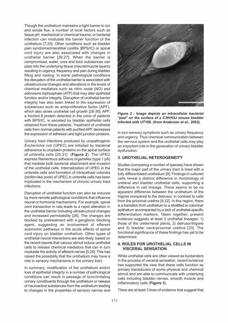
Urinary tract infections produced by uropathogenicEscherichia coli (UPEC) are initiated by bacterialadherence to uroplakin proteins on the apical surfaceof umbrella cells [25,31] (Figure 2). The UPECexpress filamentous adhesive organelles (type 1 pili)that mediate both bacterial attachment and invasionof the urothelial cells. Internalization of UPEC in theumbrella cells and formation of intracellular colonies(biofilm-like pods) of UPEC in umbrella cells has beenimplicated in the mechanism of chronic urinary tractinfections.
Disruption of urothelial function can also be inducedby more remote pathological conditions that influenceneural or hormonal mechanisms. For example, spinalcord transection in rats leads to a rapid alteration inthe urothelial barrier including ultrastructural changesand increased permeability [26]. The changes areblocked by pretreatment with a ganglionic blockingagent, suggesting an involvement of efferentautonomic pathways in the acute effects of spinalcord injury on bladder urothelium. Other types ofurothelial-neural interactions are also likely, based onthe recent reports that various stimuli induce urothelialcells to release chemical mediators that can in turnmodulate the activity of afferent nerves [5,20]. This hasraised the possibility that the urothelium may have arole in sensory mechanisms in the urinary tract.
In summary, modification of the urothelium and/orloss of epithelial integrity in a number of pathologicalconditions can result in passage of toxic/irritatingurinary constituents through the urothelium or releaseof neuroactive substances from the urothelium leadingto changes in the properties of sensory nerves and
in turn sensory symptoms such as urinary frequencyand urgency. Thus chemical communication betweenthe nervous system and the urothelial cells may playan important role in the generation of urinary bladderdysfunction.
3. UROTHELIAL HETEROGENEITY
Studies (comparing a number of species) have shownthat the major part of the urinary tract is lined with afully differentiated urothelium [9]. Findings in culturedcells reveal a distinct difference in morphology ofureteral and bladder urothelial cells, supporting adifference in cell lineage. There seems to be noapparent difference between the urothelium of thetrigone compared to the detrusor, in contrast to cellsfrom the proximal urethra [9,32]. In this region, thereis a transition from urothelium to a stratified or columnarepithelium accompanied by a lack of urothelial-specificdifferentiation markers. Taken together, presentevidence suggests at least 3 urothelial lineages: 1)those of the ureter/renal pelvis, 2) detrusor/trigoneand 3) bladder neck/proximal urethra [33]. Thefunctional significance of these findings has yet to bedetermined.
4. ROLES FOR UROTHELIAL CELLS INVISCERAL SENSATION
While urothelial cells are often viewed as bystandersin the process of visceral sensation, recent evidencehas supported the view that these cells function asprimary transducers of some physical and chemicalstimuli and are able to communicate with underlyingcells including bladder nerves, smooth muscle andinflammatory cells (Figure 3).
There are at least 3 lines of evidence that suggest that
Figure 2 : Image depicts an intracellular bacterial“pod” on the surface of a C3H/H3J mouse bladderinfected with UT189. (from Anderson et al., 2003).

172
Figure 3 : Hypothetical model depicting possible interactions between bladder afferent and efferent nerves,urothelial cells, smooth muscle and myofibroblasts. Stimulation of urothelial receptors and channels canrelease mediators that target bladder nerves and other cell types; urothelial cells can also be targets forneurotransmitters released from nerves or other cell types. Urothelial cells can be activated by eitherautocrine (i.e. autoregulation) or paracrine (release from nearby nerves or other cells) mechanisms.Abbreviations: ACh, acetylcholine; AdR, adrenergic receptor; BR, bradykinin receptor; H+, proton, MR,muscarinic receptor; NE, norepinephrine; NGF, nerve growth factor; NR, neurokinin receptor; NicR, nicotinicreceptor; NO, nitric oxide; P2R, purinergic 2 receptor unidentified subtype; P2X and P2Y; purinergic receptors;PG, prostaglandin; SP, substance P; Trk-A, receptor tyrosine kinase A, high affinity receptor for nerve growthfactor; TRPs, transient potential channels.

173
urothelial cells participate in the detection of bothphysical and chemical stimuli. First, bladder nerves(afferent and efferent) are localized in close proximity,and some within, the urothelium [20, 34-36]. A secondline of evidence suggesting that urothelial cells playa role in sensory function is the expression ofnumerous receptors/ion channels similar to that foundin both nociceptors and mechanoreceptors. And finally,these cells secrete a number of transmitters ormediators capable of modulating, activating orinhibiting sensory neurons.
a) Urothelial-Neuronal Signaling
Recent studies have shown that both afferent andautonomic efferent nerves are located in closeproximity to the urothelium. Peptidergic, P2X- andTRPV1- immunoreactive nerve fibers presumed toarise from afferent neurons in the lumbosacral dorsalroot ganglia are distributed throughout the urinarybladder musculature as well as in a plexus beneathand extending into the urothelium [20,34]. (Figure 4)In humans with neurogenic detrusor overactivityintravesical administration of resiniferatoxin, a C-fiberafferent neurotoxin, reduces the density of TRPV1and P2X3 immunoreactive suburothelial nerves,indicating that these are sensory nerves [37,36]. Inaddition, immunohistochemical studies have alsorevealed both adrenergic (tyrosine hydroxylase)positive as well as cholinergic (choline acetyltran-sferase, ChAT) positive nerves in close proximity tothe urothelium [35].
A network of cells with morphologic characteristicssimilar to those of myofibroblasts or interstitial cellsis also detected in the suburothelial space of thebladder in both humans and animals [39-41]. Thesecells, which are extensively linked by gap junctions andhave close contacts with nerves, can respond toneurotransmitters, such as ATP released from nervesor urothelial cells, suggesting that they could act as
intermediaries in urothelial-nerve interactions [40-42].Thus the anatomic substrates for bidirectionalurothelial-neural communication exist within the urinarybladder.
b) Involvement of the Urothelium in “Sensing”Chemical and Mechanical Stimuli
The involvement of urothelial function in sensorysignaling is suggested by the finding that urothelial cellsexpress various receptors that are linked to mechano-or nociceptive sensations. Examples of neuronal“sensor molecules” (receptors / ion channels) thathave been identified in urothelium include receptorsfor purines (P2X1-7 and P2Y1,2,4) adenosine (A1,A2a, A2b and A3), norepinephrine (α and β),acetylcholine (muscarinic and nicotinic), protease-activated receptors (PARs), amiloride- andmechanosensitive epithelial sodium channels (ENaC),bradykinin (B1 and B2), neurotrophins (p75, trkA,EGF family ErbB1-3), corticotrophin releasing factor(CRF1 and CRF2), estrogens (Erα and ERβ),endothelins and various TRP channels (TRPV1,TRPV2, TRPV4, TRPM8 and TRPA1) [34, 43-51].The expression of these various receptors enable theurothelium to respond to a number of “sensory inputs”from a variety of sources. These inputs includeincreased stretch during bladder filling, soluble factors(many found in the urine) such as epidermal growthfactor (EGF), or chemical mediators/ peptides/transmitters such as substance P, calcitonin gene-related peptide (CGRP), corticotrophin releasing factor(CRF), acetylcholine, adenosine or norepinephrinereleased from nerves, inflammatory cells and evenblood vessels [5, 19, 20, 52, 53].
Various stimuli can lead to secretion of numerouschemical substances such as neurotrophins, peptides,ATP, acetylcholine, prostaglandins, prostacyclin, nitricoxide (NO) and cytokines that are capable ofmodulating, activating or inhibiting sensory neurons.[19, 20]. For example, urothelial-derived NO can bereleased in response to mechanical as well aschemical stimulation and may either facilitate or inhibitthe activity of bladder afferent nerves [20, 54]. Releaseof various factors from the urothelium can alsomodulate the spontaneous activity of the underlyingsmooth muscle [42, 55].
The mechanism underlying release of chemicalmediators from the urothelium, including whether allsensory “inputs” stimulate membrane turnover (i.e.vesicular exocytosis) is not well understood. Whatlittle is known about the roles and dynamics ofmembrane-bound cytoplasmic vesicles in urothelial cellphysiology is derived from measurements ofmembrane capacitance and microscopy of fixedtissues and cells. For example, there is evidence thatonce released, ATP can act as an important autocrinemediator, which can induce membrane turnover as wellas enhance both stretch induced exocytosis and
Figure 4 : Confocal image of the urothelium depictsafferent nerve fibers (red) located in close proximi-ty to basal (green) urothelial cells.

174
endocytosis [56]. Alterations in membrane turnover cannot only increase apical surface area (as describedabove) but also regulate the number and function ofreceptors and channels at the cell surface.
1. PURINERGIC RECEPTORS
Since the first report of distension-evoked ATP releasefrom the urothelium there is now abundant evidencesupporting a role for urothelially-derived release ofATP in autocrine and paracrine signaling within thelower urinary tract. ATP is released from both theapical and basolateral urothelial surfaces in responseto bladder stretch and can act on P2X2 and P2X3urothelial receptors to stimulate stretch-inducedexocytosis [56]. (Figure 5) The expression of bothP2X and P2Y receptors in nerve fibers andmyofibroblasts in close proximitiy to the bladder lumenand the sensitivity of these cells to ATP suggests thatbasolateral ATP release from the urothelium may alsoinfluence function of myofibroblasts and bladder nerves[57, 58]; The amiloride-sensitive apical sodiumchannel, ENaC, may be involved in mechano-transduction by controlling basolateral release of ATP[59]. In addition, intercellular communication mediatedby gap junctions in myofibroblasts could provide amechanism for long-distance spread of signals fromthe urothelium to the detrusor muscle [42]. Adenosineis also produced and released by the urothelium, andmay play important roles in modulating sensory afferentfunction and smooth muscle contraction [60].
2. TRP CHANNELS
The ability of capsaicin to evoke NO release from raturothelium, reported in 1998, provided the first, albeitindirect, demonstration that TRPV1 channels areexpressed in urothelial cells and that urothelial cellsand afferent nerves, which also express thesechannels, share a number of common properties [61].This ion-channel protein is activated by capsaicin, aswell as to moderate heat, protons and lipid metabolites
such as anandamide (an endogenous ligand of bothcannabinoid and vanilloids receptors) [62, 63] TRPV1-positive nerves are in close contact with urothelialcells [64, 65]. Activation of urothelial cells with capsaicinor resiniferatoxin can increase intracellular calciumand evoke transmitter (nitric oxide, NO or ATP) release.Similar to that in sensory neurons, urothelial-responseto vanilloids are enhanced by low pH, blocked byTRPV1 antagonists and eliminated in TRPV1 nullmice [66]. In afferent neurons, TRPV1 is thought tointegrate/amplify the response to various stimuli andto play an essential role in the development ofinflammation-induced hyperalgesia. It seems likelythat urothelial-TRPV1 might participate in a similarmanner, in the detection of irritant stimuli followingbladder inflammation or infection.
Though TRPV1-null mice are anatomically normal,they exhibit a number of alterations in bladder function,including a reduction in stretch-evoked and hypotonic-evoked ATP release and stretch-evoked increase inmembrane capacitance [66]. In addition, TRPV1knockout mice have a higher frequency of low-amplitude, non-voiding bladder contractions suggestingthe possibility of a small but ongoing role for TRPV1in normal urine storage function. These relativelybenign changes may result from TRPV1 expressionnot only in afferent nerves that form close contacts withbladder epithelial (urothelial) cells but also in urothelialcells themselves. (Figure 6) These findingsdemonstrate that the functional significance of TRPV1in the bladder extends beyond pain sensation toinclude participation in normal voiding function, andis essential for mechanically evoked purinergicsignaling by the urothelium.
3. ADDITIONAL TRP CHANNELS
Much less is known about the involvement of otherTRPs in bladder function or disease. TRPV4 which isa nonselective cation channel activated by a numberof stimuli including heat, shear stress, changes inosmolarity and lipid ligands is expressed mainly withinthe epithelium of the urinary bladder [67]. While adefinitive role for TRPV4 in bladder function has notbeen established, there is evidence that null miceexhibit impaired voiding responses and, intravesicalinstillation of a TRPV4 agonist in the rat triggers anovel voiding reflex which could regulate the latephase of micturition [68, 69]. In addition, in the awakeewe, TRPV4 may also be involved in a urethra tobladder reflex, proposed to facilitate bladder emptying[70]. Another member of the TRP family, TRPA1(characterized as a thermoreceptor activated bynoxious cold), is expressed in C-fiber afferents aswell as urothelium and agonists to this channel inducebladder hyperreflexia [71]. Of interest is the finding thathydrogen sulfide, which may be formed duringinfection/inflammation, is an activator of TRPA1 [72].
Figure 5 : Image is a 3-dimensional reconstruction(taken with a confocal microscope) depicting local-ization of P2X3 (green; nuclei blue) in the urotheli-um (from Wang et al., 2005).

175
4. ACETYLCHOLINE AND THE UROTHELIUM
There is evidence that the urothelium expresses thefull complement of muscarinic receptors as well asenzymes necessary for the synthesis and release(except vesicular choline transporter) of acetylcholine.[53, 74]. Further, the urothelium is able to releaseacetylcholine following both chemical and mechanicalstimulation [53]. Once released, urothelial-derivedacetylcholine is likely to exert effects via a number ofsites including smooth muscle, nerves as well asurothelial associated-muscarinic or nicotinic receptors,the latter that could contribute to feedback mechanismsmodifying urothelial function. In addition, stimulationof urothelial-cholinergic receptors elicits release ofmediators such as nitric oxide as well as ATP, whichcould alter bladder sensation by stimulating nearbysensory afferent nerves [73, 75, 76].
Thus, targeting muscarinic receptors and/or urothelialrelease mechanisms may play an important role in thetreatment for a number of bladder disorders. In thisregard, recent evidence suggests that botulinum toxinsprevent the release of transmitters from the urothelium,which may suggest urothelial-released mediatorscontribute to sensory urgency [77].
5. CLINICAL SIGNIFICANCE OF THE SENSORYWEB
Defects in urothelial sensor molecules and urothelial-cell signaling are likely to contribute to thepathophysiology of bladder diseases. For example, anumber of bladder conditions (BPS/IC, spinal cordinjury (SCI), chemically-induced cystitis) are associatedwith augmented release of urothelial-derived ATP,which is likely to result in altered sensations or changesin bladder reflexes induced by excitation of purinergicreceptors on nearby sensory fibers [9, 10, 47, 73].
ATP can also act in an autocrine manner that wouldact to facilitate its own release from urothelial cells [10].Once released, ATP can alter the threshold foractivation of ion channels such as TRPV1. This novelmechanism, which likely reflects activation ofintracellular protein kinases and phosphorylation of theTRPV1 channel, represents a means by which largeamounts of ATP released from damaged or sensitizedcells, in response to injury or inflammation, couldtrigger the sensation of pain. Changes in epithelialsignaling/barrier function would not be unique to theurinary bladder. For example, airway epithelia inasthmatic patients as well as keratinocytes in certaintypes of skin diseases also exhibit a number of similarabnormalities and compromised repair processes [78-80]. This is particularly relevant given the highincidence of associated diseases that can includeboth visceral and somatic conditions, many of whichexhibit a shared loss of epithelial barrier function.Taken together, epithelial cells can respond to anumber of challenges (including environmentalpollutants and mediators released from nerves ornearby inflammatory cells) resulting in alteredexpression and/or sensitivity of various receptor/channels as well as changes in release of mediators,all of which could impact function.
There have been a number of new developments inthis area since the last consultation as a result of theintroduction of in vitro preparations, knockout animalsand an increasing influence of molecular techniques.Up until recently most experiments have taken placeon cats and rats, but several studies have beenreported in mice and guinea-pigs, mainly using invitro preparations. These involve recordings from thepelvic or hypogastric (lumbar splanchnic) nerves atperipheral sites near the bladder; hence the conductionvelocities of the single units, which provided informationrelevant to the degree of myelination, have not beenmeasured. These preparations have however beenable to increase the knowledge of the location ofafferent endings within the bladder wall and some ofthe physiological/pharmacological properties in relationto synaptic transmission at these nerve endings. Drugsthat are toxic in the whole animal have also beenused in isolated preparations, and some authors haveused drugs to minimise the smooth muscle movementsand the generation of inflammatory mediators; thelatter are useful in the analysis, but may give rise todifferences in the properties studied either in vivo orin vitro in the absence of such agents.
1. PROPERTIES OF BLADDER AFFERENTNEURONS
Afferent axons in the pelvic, hypogastric (lumbarsplanchnic) and pudendal nerves transmit information
II. AFFERENT NEURONES
Figure 6 : TRPV1 expression detected within the raturinary bladder urothelium. Image depicts basalcell TRPV1 (cy3, red) and cytokeratin (FITC, green)immunoreactivity.

176
from the lower urinary tract to the lumbosacral spinalcord [81-83] and studies in several species includingcats, rats and mice have shown some similarities inproperties.
The most sensitive afferents are excited by aphysiological increase in volume and by detrusorcontractions: it is believed that these low thresholdafferents have small myelinated axons, are A-deltafibres (which are larger in diameter and conduct actionpotentials more rapidly than C-fibres) and that theirendings are located in the detrusor smooth muscle.They have been called ‘in series tension receptors’ (84)because they are excited by bladder wall tensioncaused either by distension or by contraction, andneurons with this range of conduction velocities areless likely to contain peptides.
There have been a number of systematic studiesrecently in mice and guinea pigs that have provideddetailed information about the classes of receptors inthe pelvic and hypogastric nerves (Figure 7). In themouse, it seems there may be at least four classesof mechanosensitive afferents, which includemyelinated and unmyelinated fibres, and distributedin the serosa, muscle and urothelial layers of theorgan; one class has mechanosensitive endings inboth the muscle and urothelial layers. The lumbarsplanchnic nerves contain principally serosal and
muscular afferents whereas all four classes of afferentsare present in the pelvic nerve (63% of which aremuscular afferents) [85]. Another study found endingsin the urothelium and in the muscle layers, as well asstretch-insensitive afferents and chemosensitiveafferents [86]. The small myelinated afferents areinvolved in two processes: (a) sensing bladder volume,and (b) reinforcing reflex function by monitoring thecontractile state of the detrusor. In particular theseafferents, which form the most sensitive distensionreceptors, are most probably responsible for thesensation of fullness, and mediate the normalmicturition reflex that involves a spinobulbospinalpathway that passes through the brainstem.
The unmyelinated afferents contain peptides and mostappear to terminate within the lamina propria andwithin the transitional epithelium itself. Many of theseafferents discharge within the higher range ofphysiological bladder volumes, and are not usuallysensitive to detrusor contraction, possibly becauseonly the former causes stretch of the urinary epithelium.The C-fibres in the urothelium and lamina propriacontain peptides such as substance P and CGRP,which is a characteristic of one subgroup of afferentC-fibres. These and other C-fibre afferents maymediate the spinal C-fibre micturition reflex seenfollowing cord transection in the cat [87]. It is not
Figure 7 : Tension-mucosal mechanoreceptors. (A) Response of tension-mucosal mechanoreceptors tofast 3 mm stretch at 1000 µm’s, held for 10s. (B) Response of the same unit to mucosal stroking with a 0.1mN von Frey hair (five strokes, indicated by asterisks). (C) Activation of the same tension-mucosal unit bymannitol (1 M) applied to its receptor field in the mucosa.
177
clear whether they also contribute to normal voidingin this species, but there is increasing evidence thatthey may be involved in normal bladder control in ratsand mice. In rats and mice, these high threshold unitsrespond to a range of intravesical pressures thatoverlap with the sensitivity of the low threshold units,so that these together cover the spectrum of pressuresand volumes seen physiologically, and these maycontribute to spinal automatic micturition mediatedby the sacral cord.
A third group of unmyelinated bladder afferent axonsdoes not respond to normal distending volumes butonly become active during chemical irritation of thebladder, including high osmolality and high potassiumsolutions and during inflammation, when they behavelike the high volume sensing C-fibres. These havebeen demonstrated in cats and rats, and are usuallycalled ‘silent afferents’ (meaning that the last groupdo not respond to normal distensions, but can becomemechanosensitive in inflamed or over-distendedtissues). Thus it would be unwise to infer functionsimply on the basis of conduction velocity. This groupof afferents also appears to be sensitive to ATP.
Ultrastructural studies of nerves in the human bladderhave found only unmyelinated nerves in the urothelialand immediate suburothelial layer, the first smallmyelinated nerves appearing only close to the smoothmuscle layers [88]. Whether or not the suburothelialnerves become myelinated as they pass towards theserosal surface cannot be ascertained from this studybut it would be inadvisable to make deductions aboutthe relative number of C and A-delta fibres in thehuman based on these observations. Table 1 showsthe properties of afferent fibres, classified accordingto their volume thresholds.
2. UROTHELIAL AFFERENTS
Reference has already been made to the presenceof CGRP-containing afferent endings that branch
beneath and within the lamina propria, and within theurothelium itself. These axon collaterals can releaseneurotransmitters on to the various tissues in thelining of the bladder, including blood vessels, smoothmuscle, urothelium, connective tissue cells, mast cellsand other neurones. In addition there is evidence inthe human bladder that intramural neurones receiveaxonal contacts from axon collaterals that contain thepeptides characteristic of primary afferents (see thesection on ganglia, and on integrative physiology).
The plexus of afferent nerves is thickest in the neckof the bladder and in the initial portion of the urethra,and it becomes progressively less dense in theadjacent regions. It does not extend beyond theequatorial region, and therefore the lamina propria ofthe cranial region of the bladder has no afferent axons.In contrast, the afferent innervation of the musculatureis more diffuse, and appears uniform throughout thebladder. CGRP-immunofluorescence in urothelialafferent axons is enhanced in the surviving axons 5days after contralateral denervation, a change whichmay be an early sign of regeneration of these axons[89]. In the human bladder, CGRP together withSubstance P and NKA occur only infrequently innerves in the muscle but are moderately frequent inthe suburothelial layer. Also in the human thereappears to be another population of CGRP-containingfibres that co-localize with NPY and galanin and someof these synapse on intramural ganglia within thebladder [90-93]. There is also recent evidence thatnerves cross the basal lamina and enter the basallayers of the human urothelium [89]. Table 2 showsthe properties of afferent nerve endings in differentlocations.
3. SENSITIVITY OF AFFERENT ENDINGS
The term afferent sensitivity refers to the gain of theafferent signal, i.e. the number of impulses that arefired by an afferent ending at any level of distension.Sensitizing mediators are able to increase the size of
Table 1. Properties of low and high threshold afferents from the bladder in cat, rat, mouse and guinea pig

178
the sensory signal (the frequency of impulse traffic)at a given level of distension, so the sensations thatoccur at a particular rate of firing in an afferent occurat lower bladder volumes if the afferent endings havebeen sensitized. (Figure 8).
The sensitivity of afferent endings may be influencedby the release of mediators from different cell types,including possibly the urothelium, myofibroblasts,nerve endings, smooth muscle, mast cells and otherconnective tissue cells. It is likely that many or all ofthese can release ATP, and some may release othermediators including nitric oxide, tachykinins (SubstanceP, Neurokinin A, Neurokinin B), growth factors (NerveGrowth Factor [NGF], Brain Derived NeurotrophicFactor [BDNF] and others) and other endogenousmediators such as nociceptin.
The similarity of the properties of the urothelial cellsand the C-fibre afferents suggests that the most likelycontender for a sensory cell may be a urothelial cell,but it is clear that the afferent endings themselvesrespond to a variety of stimuli, and that surroundingcells may simply enhance the gain of the transducer.The following paragraphs refer to some of themediators that can sensitize bladder afferents.
a) ATP and P2X3 Receptors
Recent studies of mice have shown the P2X2/3receptor, is present in small sensory neuronesinnervating the bladder, and that the effects of bladderdistension on these sensory endings is markedlyattenuated if the gene for the P2X3 receptor is deleted.Knockout mice that do not express this receptor exhibit
Figure 8 : Correlation of afferent discharge withthe cystometrogram. Left: Diagram of afferentdischarge at different volumes for A-delta fibresand mechanosensitive C-fibres. Right: Diagramshowing the increased responses when the affer-ent endings are sensitized by inflammation or byadministration of sensitizing agents such as ATP,Nerve Growth Factor or Neurokinin A. In additionto the increased discharge rates in the A-deltaand C-fibres, a new population of ‘Silent C-fibres’is recruited into activity.
Table 2. Location and sensitivity of afferents in the hypogastric (lumbar splanchnic) and pelvic nerves

179
a marked urinary bladder hyporeflexia, characterizedby decreased voiding frequency and increased bladdercapacity, but normal bladder pressures [94, 95]. Inaddition, they have reduced pain-related behaviour inresponse to injection of ATP or formalin, and lose therapidly desensitizing ATP-induced currents in theirdorsal root ganglion neurons; they also have areduction in the sustained ATP-induced currents innodose ganglion neurons. Immunohistochemicalstudies localize P2X3 to nerve fibres innervating theurinary bladder of wild-type mice, and show that lossof P2X3 does not alter sensory neuron innervationdensity. Thus, P2X3 is critical for peripheral afferentpathways controlling urinary bladder volume reflexes,which take place at physiological volumes andpressures. Antagonists to P2X3 may therefore havetherapeutic potential in the treatment of disorders ofurine storage and voiding such as overactive bladder.In one recent study it was observed that humans withdetrusor overactivity treated with botulinum toxinshowed decreased levels of P2X3 and TRPV1receptors in the bladder biopsies [96].
Some groups of bladder afferents appear to besensitive to the release of ATP from the urothelium orother cells. In the last few years, the sensitivity ofbladder afferents to ATP and mechanical stimuli hasbeen studied intensively in the rat and mouse usingprotocols designed to avoid sensitization of theafferents [97-99]. In the rat, 90% of bladder afferentneurons gave persistent electrical responses to theP2X agonist α−β-methylene ATP that were inhibitedby the P2X antagonist 2’,3’-O-trinitrophenyl-ATP (TNP-ATP) which suggests that pelvic nerve afferents fromthe rat bladder express predominantly P2X (2/3)heteromeric receptors. In the mouse, Rong et al [99]found that the majority of the low threshold and nearlyall the high threshold receptors were sensitized byα−β−methylene ATP, i.e. there was a reduction in thethreshold and an increased peak activity duringdistensions. In addition some of the ‘silent’ afferentsbecame mechanosensitive. The absence ofsensitization in P2X3 knockout mice indicated thatthe responses were mediated by the P2X3 receptor.However there is a recent in vitro study in the guinea-pig that suggests that one group of afferents, the lowthreshold ‘in series’ tension receptors in detrusormuscle, are not sensitive to ATP [86].
b) Nitric Oxide
Nitric oxide (NO) is an important mediator that can bereleased from urothelium and from adjacent neurones.The detrusor however is not very sensitive to nitricoxide in contrast to the urethral outflow region whereit effectively relaxes the urethral smooth muscle,suggesting an involvement of nitric oxide in thedecrease in urethral pressure at the start of micturition[100].
NO may be involved in the control of afferent sensitivity,
and we now know that NO may increase the activityof capsaicin-sensitive nerves within the bladder wallafter spinal cord injury [101]. Basal release of nitricoxide has not been detected in the urothelium of thenormal cat; however it is released in cats with felineinterstitial cystitis [102], and from normal cats after theaddition of agonists. Nitric oxide release from neuronesdepends on the enzyme nitric oxide synthase (NOS)and increased expression of neuronal NOS in bladderafferent and spinal neurones occurs following cordinjury [103], and in bladder afferents following chronicbladder irritation with cyclophosphamide. There isalso evidence that nitric oxide can inhibit the functionof primary afferent neurons [104, 105]. This inhibitoryeffect may occur in the normal bladder becauseintravesical administration of a solution of oxyhe-moglobin, a nitric oxide scavenger, induced bladderoveractivity in the conscious rat [106] . The effect ofoxyhemoglobin was reduced by pretreatment withZD6169, a drug that suppresses capsaicin-sensitivebladder afferents, suggesting that oxyhemoglobinenhances afferent excitability.
Knockout mice that do not have neuronal NOS appearto have normal function in the lower urinary tract [107],and knockout animals that do not have inducible NOSdo not show major abnormalities. However, the latterappear to need iNOS in the response to urinaryobstruction [108].
c) Tachykinins: Substance P, Neurokinin A andNeurokinin B
The tachykinins are a group of neuropeptides thatincludes substance P and neurokinin A (which areproduced by the same gene), and neurokinin B. Theyare found in small diameter afferent neurones,particularly within the C-fibre population, and may bereleased, along with other peptides, by afferent endingswhen these become active, e.g. during the axon reflexin skin. A similar event occurs in the bladder and isassociated with the phenomenon of neurogenicinflammation. These peptides cause vasodilatationand an increase in capillary permeability, and arealgesic agents.
In addition some tachykinins can sensitize sensorynerve endings. This view is based on (a) autoradio-graphic studies that show the disappearance of NK-2 receptors in the lamina propria in capsaicin-treatedrats that are deficient in sensory nerves [109], (b) onstudies in which afferents or dorsal root ganglia canbe made hypersensitive using a NKA- analogue andother intravesical chemical stimuli such as high [K+]and high osmolality [110-112], and (c) the demons-tration that the development of hypersensitivity to anumber of sensitizing agents including high [K+] canbe blocked by an NK-2 receptor antagonist [113, 114].More recently it has been shown that rat dorsal rootganglion neurones are excited by NK2 agonists, butare inhibited by NK-3 agonists [115]. This NK2 action
180
is on L- and N-type Ca2+ channels, whereas the NK-3 action is only on the L-type channels. Both of theseeffects are blocked by inhibition of protein kinase C.
d) TRP (Transient Receptor Potential)Receptors
Ion channels that act as receptors because they areresponsible for transient receptor potentials (TRP)generally work by opening non-specific ion channels.Some are sensitive to capsaicin and other vanilloids(TRPV), while others are associated with othermediators, including cinnamaldehyde (TRPA1) andMelastatin (TRPM). TRP channels are opened by avariety of physical and chemical stimuli including heat,cold, mechanical stress, voltage, hydrogen ionconcentration and osmolality, as well as by specificligands, such as capsaicin, melastatin and trans-cinnamaldehyde. The nerve endings that containthese channels are therefore often polymodal in theirproperties, one of the characteristics of non-myelinatedsensory endings, and also in other cell types, includingthe urothelium. TRPV1, TRPV2, TRPV4, TRPM8, andTRPA1 have been described in different parts of theurogenital tract, and TRPV1 (the vanilloid receptor) hasreceived special attention, particularly in relation to itsrole in bladder sensation [116].
The TRPV1 receptor is a cation channel expressedby nociceptive neurones and can also [117, 118] beactivated by protons or temperature greater than 43degrees C [119, 120]. Within the bladder, it may bethat it is activated naturally by low pH, but suchchanges (e.g. in metabolic acidosis) are not usuallyassociated with bladder pain. The expression of theTRPV1 receptor in sensory neurones is regulated byNerve Growth Factor (NGF), and stimulation of theTRPV1 receptor with capsaicin causes the release ofCGRP [121]. Capsaicin and resiniferatoxin act onunmyelinated afferent fibres throughout the body, butit is also clear that capsaicin can act on the urotheliumby binding to TRPV1 receptors. Capsezepine is ablocker of this receptor and it has been found thatnitric oxide (NO) release and the increase inintracellular Ca2+ induced by capsaicin are blockedby this antagonist. In addition to capsaicin andresiniferatoxin, a new agonist, piperine, has beenshown to activate TRPV1 receptors and producebladder hyperactivity and activity of sensory nervesin this organ in the rat [117, 118]. Several groupshave searched for endogenous ligands for the TRPV1receptors, and anandamide, palmitoylethanolamideand nociceptin are three compounds that deserve amention, although much more work needs to be doneto elucidate their exact roles. Anandamide andpalmitoylethanolamide (PEA) are endogenouscannabinoids (acting on CB-1 and CB-2 receptorsrespectively) that also are agonists of TRPV1 receptors[122, 123] and may act on peripheral perivascularsensory terminals in a manner that is antagonized by
the capsaicin antagonist capsezepine. These agentscan also cause the release of CGRP and SubstanceP by increasing intracellular Ca2+, and have otheractions, such as activation of G-proteins [124]. Bothanandamide and PEA have been found to attenuatebladder hyper-reflexia induced by intra-vesical NGF[125-127]. The TRPV1 receptor seems to be importantfor normal bladder function and for excitation of lowthreshold distension-sensitive afferents in mice [128,129] (Figure 9).
• OTHER TRP RECEPTORS
The bladder-cooling reflex (induced by instillation ofintravescial cold saline and termed the ice water test)is believed to be triggered by menthol-sensitive coldreceptors in the bladder wall. Recently, this test hasbeen used to distinguish sensory symptoms in patientswith bladder pain syndrome and overactive disorders[130]. Because pain elicited in BPS patients was notaccompanied by reflex detrusor contractions, this testmay be of particular interest since the response ofafferent nerves to cold (and to menthol) depends ona particular TRP receptor subgroup, TRPM8. Inexperiments in the guinea-pig bladder, the afferentnerves innervating the organ have been shown toexpress TRPM8, and the bladder cooling reflex isenhanced in the presence of menthol [131].
The TRPA1 channel is also expressed in dorsal rootganglion cells that innervate the rat bladder. Trans-cinnamaldehyde or allyl isothiocyanate, agonists of thisreceptor, cause bladder hyperreflexia and appear toact through C fibres that might be mechanoreceptiveor nociceptive [132].
e) ORL Receptors
Nociceptin/orphanin FQ, another endogenous ligandthat binds with the opioid receptor-like 1 receptor(ORL1 receptor, now also known as the 4th categoryof opioid receptors, OP4) has been shown to havenaloxone resistant inhibitory effects on the micturitionreflex. These actions are mediated at several sitesincluding the capsaicin sensitive nerves in the bladder,and a central supraspinal site [133]. Nociceptinproduces a long-lasting protection against capsaicin-induced desensitization of TRPV1 in afferent nerves,such that a chemoceptive micturition reflex could berepeatedly evoked by topical capsaicin in nociceptin-pretreated rats. This is in sharp contrast to the effectsof nociceptin on the local response to capsaicin, whichcorresponds to the release of peptides from capsaicin-sensitive afferent neurons. Topical application ofnociceptin onto the bladder serosa evokes atachykinin-mediated contraction [133]. These resultssuggest that the afferent and ‘efferent’ functions ofcapsaicin-sensitive primary afferent neurons in therat bladder are differentiated by nociceptin, and thatnociceptin has a significant action on afferentsensitivity.

181
In humans nociceptin elicits a strong acute inhibitoryeffect on the micturition reflex in patients with aneurogenic bladder [134]). This was in contrast to theplacebo, and led to the conclusion that nociceptinand other orphan peptide receptor agonists may beuseful in future as drugs for the treatment of neurogenicurinary incontinence.
Local administration of kappa-opioid receptor agonistsby intra-arterial injection attenuated the responses ofpelvic nerve afferents to high pressure distension of theurinary bladder [135]. These agonists had essentiallythe same effects whether the bladder was inflamed ornot. The conclusion was that the ability of kappa opioidagonists to attenuate the responses of afferents tolarge bladder distensions indicated a potential use forperipherally acting kappa opioid receptor agonists in thecontrol of urinary bladder pain.
f) Neurotrophins
1. NERVE GROWTH FACTOR
(NGF; neurotrophin-1), the first of a group of growthfactors called neurotrophins, is produced in largerquantities in humans with detrusor overactivity [136],BPS/IC and bladder cancer [137] , in rats with inflamedbladders [138], spinal cord injury or chemically inducedcystitis [139] or bladder outlet obstruction [140], indiabetic rats [141] and a number of other states. Thisprotein is known to sensitize myelinated and unmye-linated afferents from the bladder [142, 143] and it isinvolved in the production of referred pain in bladderinflammation [144]. It also appears to stimulate theexpression of the vanilloid receptor TRPV1 [121], andthere is a suggestion that increased NGF levelsresulting from intrathecal injection of NGF can inducea decrease in A-type K+ current density in the afferentpathway that may influence the emergence of bladderoveractivity [145].
Recent studies in an animal model for BPS/IC termedfeline interstitial cystitis (FIC) in which TRPV1 responsesto capsaicin were measured in lumbo-sacral dorsalroot ganglion neurones have suggested that affectedneurones are increased in size and exhibit exaggeratedresponses to capsaicin that may be associated withenhanced activity of endogenous protein kinase C[146]. A further study from the same group suggestedthat the abnormal activity of afferent neurones fromcats with feline interstitial cystitis may be due to changesin the behavior of K+ currents which are restricted tocapsaicin-sensitive neurones [147].
2. BRAIN DERIVED NEUROTROPHIC FACTOR (BDNF)
levels in the urinary bladder and some other epitheliaare higher than those found in the brain or skin [148].In situ hybridization experiments showed that BDNFmRNA was made by visceral epithelial cells, in severaltypes of smooth muscle, and in neurons of themyenteric plexus. However the receptors from BDNF(trkB and p75[NTR]) are not present on the urotheliumbut are present in neurons of the peripheral nervoussystem. Hence it is thought that in the bladder thisneurotrophin is produced by the urothelium and canact on the afferent nerves. The mRNAs for NGF,BDNF and neurotrophin-3 all increase within 2 hoursof bladder inflammation in the rat, and this increasesexpression may contribute to sensory and reflexhyperactivity [138]. Inflammation of the colon alsoappears to induce up-regulation of CGRP and TrkB(suggesting an involvement of BDNF) in bladderafferent neurons, and suggests that there may becross sensitization of bladder afferent pathways bycolonic inflammation [149]. In spinal cord injury thereis also an increase in BDNF and galanin in the dorsalroot ganglia and spinal segments below the lesion;interestingly NGF expression was reduced below thelevel of the lesion [150].
Figure 9 : Stimulus-response profile of low threshold, LT (A) and high threshold, HT (B) bladder afferents.Note that the LT afferents have a blunted response profile in the TRPV1 -/- mice compared to wildtype(P<0.001, 2-way ANOVA and Bonferroni test). The response profile of HT afferents is unchanged (from Dalyet al., 2007).

182
• NGF and TTX-Resistant Na+ Channels
Sensitization of afferents appears to be an importantmechanism that leads to reflex hyperexcitability, anda number of studies have linked the tetrodotoxin(TTX)-resistant sodium channel, sometimes known asNav1.8 to this process. A number of sensitizing agentsincluding NGF are known to induce increasedexpression of this membrane channel; this appearsto be sufficient to change the properties of afferentsso as to lower the threshold for firing of bladder (lowervolume threshold for voiding) and induce spontaneousand burst firing (overactive contractions, urgency)[142]. TTX-resistant Na channels (NaV1.8 and NaV1.9)have been found in SP/CGRP immunoreactive smallDRG giving rise to C-fibers supplying the bladder[151, 152]; these also express the trkA receptor, whichbinds NGF and is necessary for its action. Plasticityof TTX-sensitive and TTX-resistant Na+ channels(NaV 1.8 and NaV 1.9) occurs in these neurones afterspinal cord injury, and a decreased expression of NaV1.8 channel immunoreactivity and a small increase inNaV 1.9 channel immunoreactivity in bladder DRGneurons can be observed [153, 154].
The dependence of the sensitization of these afferentneurones and the occurrence of overactivity on NGFand its actions on the NaV 1.8 channels has beenshown in experiments using immunoneutralization ofNGF or antisense oligonucleotide treatment to reducethe expression of these channels in sensory neurons[151, 155]. More recently studies on ralfinamide, adrug that interferes with TTX-resistant sodiumchannels, indicate that this drug reduces inflammatoryand neuropathic pain as well as bladder overactivityin rats. The ability of ralfinamide to reduce capsaicin-induced hyperexcitability and tonic activity of ratafferent neurones appears to be due to its action asa sodium channel antagonist [156].
In clinical studies the local anaesthetic lidocaine andthe oral Na+ channel blocker, mexiletine, which operateby reducing excitability in sensitized neurones havebeen used to treat urge incontinence and hyper-reflexic conditions [157-162] with variable degrees ofsuccess.
4. SPINAL CORD
This section is concerned with the central projectionsof the primary afferent neurons. Axonal tracingexperiments have been performed in many animalspecies [163, 164] and have localized the segmentaldistribution and spinal termination of afferent pathwaysin the pelvic, hypogastric and pudendal nerves. Theprimary afferent cell bodies of the pelvic and pudendalnerves are contained in lower lumbar and sacral dorsalroot ganglia depending on species; whereas afferentinnervation in the hypogastric arises in the rostrallumbar dorsal root ganglia. The central axons of thedorsal root ganglion neurons carry the sensory
information from the lower urinary tract to secondorder neurons in the spinal cord.
Trans-ganglionic transport of axonal tracers hasidentified the spinal projections and terminal fields ofvisceral and somatic primary afferent neurons. Thedorsal commissure (DCM), superficial dorsal hornand sacral parasympathetic nucleus (SPN) all containinterneurons with rostral projections that are activatedduring noxious [165, 166] or non-noxious stimulation[167] of the rat bladder and the urethra. These neuronsare the site of origin of ascending pathways that projectto various structures in the brainstem via spinalpathways that include the dorso-lateral funiculus [168,169]. In humans spinal tractotomies for intractablepelvic pain provide the only insight available as to theorganization of spinal pathways involved in bladdercontrol in man [170].
Visceral afferent fibers of the pelvic [171] and pudendal[163] nerves enter the cord and travel rostrocaudallywithin Lissauer’s tract, and transversely around thedorsal horn via the lateral (LCP) and medial collateralpathways (MCP) to reach the deeper layers of thespinal cord. Within the spinal gray matter, the LCP andMCP provide a dense innervation to laminae I, V, andVII and the dorsal commissure. Muscle and cutaneousafferents in the pudendal nerve terminate in differentregions of the cord.
Afferents from the Urethra, Bowel and GenitalOrgans
Studies have demonstrated that electrical stimulationof urethral afferent fibers when the bladder is full canevoke strong detrusor contractions sufficient for voidingin intact cats [172, 173] as well as acute spinalizedcats [174]. Similarly, using minimally invasive methodsto apply electrical stimulation within the proximalurethra via a catheter-mounted electrode, it has beenshown that reflex bladder contractions can begenerated in humans with complete paraplegia butthese do not seem to produce efficient voiding can beevoked in chronic SCI cats by stimulation of anexcitatory pudendal to bladder spinal reflex [176].
The excitability of the micturition reflex can beinfluenced by other sacral afferent pathways [177],including facilitatory effects resulting from stimulationof urethral afferents, and inhibition of bladder activityby stimulation of the dorsal nerve of the clitoris inkeeping with known interactions from the vagina andcolon [178]. Stimulation of urethral afferents by fluidflowing through the urethra can facilitate the micturitionreflex; however contraction of the urethral sphincterresulted in inhibition of bladder motility [179].
Excitability of spinal neurons receiving afferent inputfrom the bladder can also be modulated by input fromother pelvic structures such as the colon [175, 180,181].This convergence of sensory information from anumber of pelvic organs can occur at the level of the
183
spinal cord. In addition, the expansion of primary axonterminals within the spinal cord can also play a rolein altering bladder reflexes. For example, it has beenshown that expression of a number of peptidesincluding CGRP, VIP as well as PACAP is altered inprimary afferent terminals and may correlate withchanges in bladder function following SCI [182-184].
Glutamate is an important excitatory transmitter inthe afferent limb of the micturition reflex, and mediatesits effects by means of both NMDA and nonNMDAreceptors. This conclusion is based on studies of C-fos expression and the transmission of afferent activityrostrally, and the depressive effects of both NMDAand nonNMDA glutamatergic receptor antagonists[185, 186].
Bladder afferent neurons contain a number ofpeptidergic neurotransmitters, and the centraldistribution of bladder afferent terminals andpeptidergic immunoreactive fibers is quite similar.There has been considerable interest in the role oftachykinins in the micturition reflex [187] and innociception. Intrathecal treatment of adult rats withintrathecal capsaicin can result in a reversible blockof the micturition reflex [188]. Further, while normalmicturition is not altered following ablation of NK1-Rexpressing SC neurons using SSP-saporin, theresponse to a nociceptive stimulus was significantlyreduced. These and other studies [188, 189] suggestthat substance P and its receptors may play a part intransmission of bladder nociceptive responses at thefirst synapse in the micturition reflex.
5. SPINAL CORD GLIAL CELLS ANDMODULATION OF PELVIC AFFERENTS
a) Anatomy and Function of CNS Glia
The nervous systems of animals are generallycomposed of two cell types: neurons, which propagateelectrical currents and function as the signalingmoieties of the nervous system, and glia, the functionsof which are far less understood [190]. Central nervoussystem (CNS) glia, (which greatly outnumber theneuronal component [191, 192]), comprise ependymalcells, oligodendrocytes, astrocytes and microglia.
Astrocytes, (similar to neurons and oligodendrocytes)are ectodermal in origin. Two distinct types aredescribed based on their morphology: fibrousastrocytes, which have long thin processes and arecommonly found in white matter, and protoplasmicastrocytes, which have short thick processes and aretypically found in grey matter. In the reactive state,protoplasmic astrocytic processes become morepronounced [193].
Astrocytes contribute to the regulation of themicroenvironment in which neurons develop andfunction, maintaining a tight control on local ion (notablyK+) and pH homeostasis. In addition, they are involved
in the clearance of synaptically-releasedneurotransmitters, such as glutamate and GABA [194].These multifunctional cells are key players in the‘tripartite synapse’, which is composed of pre- andpost-synaptic membranes and extra-synaptic astrocyticcontacts; they have the potential to modify synaptictransmission and plasticity [195].
Microglia are considered to have a mesodermal originand to enter the brain during the neonatal period; theyare commonly considered to function as the residentmacrophages of the CNS and are more abundant ingrey than white matter. In the normal brain and spinalcord, they have a ramified morphology; when activated,such as when there is damage to the CNS, they rapidlyretract their processes and migrate toward the site ofinjury, where they turn into macrophages and removethe debris [193].
While astrocytes and microglia present as two verydifferent types of glial cell, it is now becomingincreasingly known that they have functionalsimilarities. Recent evidence has shown that spinalcord activation of either of the two cell types may beinvolved in both the development and maintenanceof central sensitization in various chronic painconditions. Thus, both astrocytes and microglia areattracting wide interest in the pain field [196].
b) Modulation of Neuronal Signaling by SpinalCord Glia
At the level of the spinal cord (the first relay site in thetransmission of nociceptive information from theperiphery to the brain [197], dorsal horn glia may beactivated by chemicals released from primary afferentterminals such as the neurotransmitters: SP, CGRP,NO, purinergic agents, glutamate, opioid peptides,and the chemokines: fractalkine or neuractin.Activation may result in altered cell morphology,changes in receptor expression, or release of factorsby astrocytes and microglia, which in turn can lead tochanges in neuronal function and ultimately influencepain transmission [198]. There is evidence thatmicroglia may mediate the activation of astrocytesseen in both somatic and visceral pain pathologies-the ‘ neuropathic pain triad ‘ [199] . Generally, microglialactivation is transient, while astrocytic activation ismuch longer-lasting. However, activation of either ofthe two cell types promotes pain [196, 200].
While most reports on the contribution of glia to pain,study somatic rather than visceral forms of chronicpain, there is now increasing evidence pointing to arole for glia as key modulators during inflammatorypain. Microglia probably play a pivotal role in theinitiation phase, while astrocytes are likely to contributeto the maintenance of the persistent pain state. Thereare reports in non- acute [201, 202] and acute [202]models of colonic irritation of altered glial cellmorphology, in segments L6-S1 of the spinal cord .
184
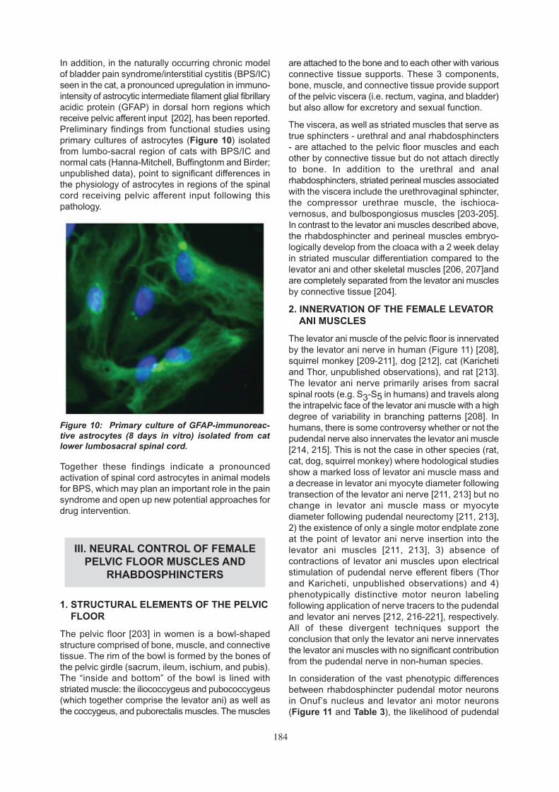
In addition, in the naturally occurring chronic modelof bladder pain syndrome/interstitial cystitis (BPS/IC)seen in the cat, a pronounced upregulation in immuno-intensity of astrocytic intermediate filament glial fibrillaryacidic protein (GFAP) in dorsal horn regions whichreceive pelvic afferent input [202], has been reported.Preliminary findings from functional studies usingprimary cultures of astrocytes (Figure 10) isolatedfrom lumbo-sacral region of cats with BPS/IC andnormal cats (Hanna-Mitchell, Buffingtonm and Birder;unpublished data), point to significant differences inthe physiology of astrocytes in regions of the spinalcord receiving pelvic afferent input following thispathology.
Together these findings indicate a pronouncedactivation of spinal cord astrocytes in animal modelsfor BPS, which may plan an important role in the painsyndrome and open up new potential approaches fordrug intervention.
1. STRUCTURAL ELEMENTS OF THE PELVIC FLOOR
The pelvic floor [203] in women is a bowl-shapedstructure comprised of bone, muscle, and connectivetissue. The rim of the bowl is formed by the bones ofthe pelvic girdle (sacrum, ileum, ischium, and pubis).The “inside and bottom” of the bowl is lined withstriated muscle: the iliococcygeus and pubococcygeus(which together comprise the levator ani) as well asthe coccygeus, and puborectalis muscles. The muscles
are attached to the bone and to each other with variousconnective tissue supports. These 3 components,bone, muscle, and connective tissue provide supportof the pelvic viscera (i.e. rectum, vagina, and bladder)but also allow for excretory and sexual function.
The viscera, as well as striated muscles that serve astrue sphincters - urethral and anal rhabdosphincters- are attached to the pelvic floor muscles and eachother by connective tissue but do not attach directlyto bone. In addition to the urethral and analrhabdosphincters, striated perineal muscles associatedwith the viscera include the urethrovaginal sphincter,the compressor urethrae muscle, the ischioca-vernosus, and bulbospongiosus muscles [203-205].In contrast to the levator ani muscles described above,the rhabdosphincter and perineal muscles embryo-logically develop from the cloaca with a 2 week delayin striated muscular differentiation compared to thelevator ani and other skeletal muscles [206, 207]andare completely separated from the levator ani musclesby connective tissue [204].
2. INNERVATION OF THE FEMALE LEVATOR ANI MUSCLES
The levator ani muscle of the pelvic floor is innervatedby the levator ani nerve in human (Figure 11) [208],squirrel monkey [209-211], dog [212], cat (Karichetiand Thor, unpublished observations), and rat [213].The levator ani nerve primarily arises from sacralspinal roots (e.g. S3-S5 in humans) and travels alongthe intrapelvic face of the levator ani muscle with a highdegree of variability in branching patterns [208]. Inhumans, there is some controversy whether or not thepudendal nerve also innervates the levator ani muscle[214, 215]. This is not the case in other species (rat,cat, dog, squirrel monkey) where hodological studiesshow a marked loss of levator ani muscle mass anda decrease in levator ani myocyte diameter followingtransection of the levator ani nerve [211, 213] but nochange in levator ani muscle mass or myocytediameter following pudendal neurectomy [211, 213],2) the existence of only a single motor endplate zoneat the point of levator ani nerve insertion into thelevator ani muscles [211, 213], 3) absence ofcontractions of levator ani muscles upon electricalstimulation of pudendal nerve efferent fibers (Thorand Karicheti, unpublished observations) and 4)phenotypically distinctive motor neuron labelingfollowing application of nerve tracers to the pudendaland levator ani nerves [212, 216-221], respectively.All of these divergent techniques support theconclusion that only the levator ani nerve innervatesthe levator ani muscles with no significant contributionfrom the pudendal nerve in non-human species.
In consideration of the vast phenotypic differencesbetween rhabdosphincter pudendal motor neuronsin Onuf’s nucleus and levator ani motor neurons(Figure 11 and Table 3), the likelihood of pudendal
III. NEURAL CONTROL OF FEMALEPELVIC FLOOR MUSCLES AND
RHABDOSPHINCTERS
Figure 10: Primary culture of GFAP-immunoreac-tive astrocytes (8 days in vitro) isolated from catlower lumbosacral spinal cord.

185
Figure 11 : A) Illustration of the course of the levator ani nerve in a left hemipelvis, sagittal view (arrow pointsto levator ani nerve). Abbreviations: S, Sacrum; S1-S5, sacral foramina; Cm, coccygeal muscle; LAN, levatorani nerve; IS, ischial spine; ICm, iliococcygeal muscle; OIm, obturator internal muscle; PCm, pubococcygealmuscle; PRm, puborectal muscle; ATLA, arcus tendinous levator ani; C, coccyx; V, vagina; U, urethra; R, rectum(from Barber, 2002). B-E) Photomicrographs of a single transverse section of sacral spinal cord from asquirrel monkey with pubocaudalis muscle injected with cholera toxin B (CTB) and the anal rhabdosphincterinjected with fast blue. B) Bright field illumination shows cytoarchitecture of gray and white matter; whitebox indicates area shown in high power in panels D and E. C) Epifluorescent illumination showing CTB-labeled(bright green) levator ani motor neurons; white box indicates area shown in high power in panels D and E.D) High power photomicrograph of boxed area in panels B and C using epifluorescent illumination to showCTB-labeled levator ani motor neurons. Note large αα and small γγ CTB-labeled motor neurons; also CTB-labeled processes extending from levator ani motor neurons into Onuf’s neucleus (dashed circle) and theventrolateral funiculus. E) Same area and section as panel D viewed with epifluorescent illumination to showfast blue-labeled (bright blue) anal sphincter motor neurons in Onuf’s nucleus (dashed circle). Close appositionbetween CTB-labeled levator ani motor neuron processes and fast blue-labeled rhabdosphincter motorneurons were observed (from Pierce, 2005).
186
motor neurons innervating the levator ani muscle inhumans seems very small. Similarly, the distinctembryological origins of levator ani muscles versusrhabdosphincter and perineal muscles (the latteroriginating from the cloaca [206, 207]), as well as aseparate “compartmentalization” of the rhabdos-phincter and perineal muscles by connective tissue[204], are parsimonious with distinctive “specialsomatic” motor innervation by the pudendal versustypical skeletal motor innervation by the levator aninerve. Possibly, the confusing morass of small muscles(puborectalis, compressor urethrae, urethrovaginalsphincter, urethral and anal rhabdosphincter, ischio-cavernosus, bulbocavernosus), blood vessels,connective tissues, and nerves in the perineal regionmakes dissection, identification, and nomenclatureof specific nerve branches and muscles difficult incadavers.
Indeed, this morass has led to confusion regardingeven the nomenclature of the muscles themselves[204, 205] and, without extreme care, a contributionof the pudendal nerve to levator ani muscle innervationmight be confused. Alternatively, there may be truespecies differences between humans and othermammals in regards to a minor pudendal nervecontribution to minor levator ani muscles (e.g.puborectalis). However, the ability to conduct preciseexperimental manipulations in animals provides a
clear conclusion that the pudendal nerve does notinnervate the major muscles of the pelvic floor, i.e.iliococcygeus, pubococcygeus, or coccygeus musclesto a significant degree.
The positioning of the levator ani nerve on theintrapelvic surface of the muscles may expose it todamage as the fetal head passes through the birthcanal [208]. This positioning also puts it in a favorableposition to be activated when current is applied witha St. Mark’s electrode placed in the rectum. Thepositioning of the levator ani nerve, close to the ischialspine, also risks entrapment by sutures used forvarious suspension surgeries for pelvic organ prolapse(POP) and may account for dyspareunia, pelvic pain,and/or recurrent prolapse [222] associated with suchsurgery. Finally, since the ischial spine is used as alandmark for needle placement when applying atransvaginal “pudendal nerve” block [223], thepossibility that this procedure also anesthetizes thelevator ani nerve and pelvic floor muscles must beconsidered. These complicating factors of historically-accepted clinical concepts may explain why a possibleinnervation of the levator ani muscle by the pudendalnerve in humans remains controversial.
a) Levator Ani Motor Neurons and SensoryInnervation
Retrograde tracing studies involving injection of cholera
Table 3 : Neuronal markers preferentially associated with rhabdosphincer motor neurons in Onuf’s nucleus.

187
toxin B (CTB) into the levator ani muscle and fastblue into the anal rhabdosphincter muscle of squirrelmonkeys [217] show that levator ani motor neuronsare located in the sacral ventral horn (Figure 11 B-E) in a longitudinal column. In contrast to the verydense packing of sphincter motor neurons in Onuf’snucleus [216, 218-221], the levator ani motor neuronsare more diffusely distributed. Furthermore in contrastto the uniform intermediate size of pudendal motorneurons, levator ani motor neurons (Figure 11D)show a bimodal distribution of large neurons(presumably α motor neurons) and small neurons(presumably γ motor neurons). These 2 sizes of motorneurons are in keeping with the presence of musclespindles (whose intrafusal muscle fibers are innervatedby γ motor neurons) in levator ani muscle [224, 225]and the absence of muscle spindles in rhabdosphinctermuscle [224, 226-230]; consequently levator ani mayexhibit Ia (muscle spindle) evoked monosynapticstretch reflexes, whereas the EUS does not [231-234].
Levator ani motor neuron processes (dendrites oraxon collaterals) project into two important areas inthe sacral spinal cord [217]. The first, medial laminaVI, is a region where primary afferent fibers frommuscle spindle and Golgi tendon organs terminate[235, 236]. This again suggests an important role forstretch-activated contraction of levator ani muscles.Importantly, the second projection of levator ani motorneurons is to Onuf’s nucleus (Figure 11D-E), whichcontains rhabdosphincter motor neurons. Theselevator ani motor neuron processes form closeappositions with sphincter motor neurons in bothmonkey [217] and rat (Thor, unpublished observations).
Presumably, these appositions reflect a neuroana-tomical substrate for coordination of the rhabdo-sphincter and the pelvic floor muscles during micturitionand defecation. Whether these projections aredendrites designed to receive common afferent inputto levator ani and rhabdosphincter motor neurons, orif they are axon collaterals transmitting informationfrom levator ani motor neurons to rhabdosphinctermotor neurons to coordinate contractions is not knownand will require electron microscopic or electrophy-siological analysis to be resolved.
Dual-labeling immunohistochemistry combined withcholera toxin-B (CTB) tracing studies of the levator animuscles of squirrel monkeys has shown that thereare approximately 4 times as many afferent neuronsversus motor neurons labeled following injection oftracer into the levator ani muscle [210]. About ? of theneurons were large, myelinated (i.e. RT-97 neurofila-ment positive) that did not contain the peptidetransmitter CGRP, binding sites for isolectin-B4 (IB-4), or the growth factor receptor, TrkA, immuno-reactivity. Of the remaining small RT97 negativeneurons, approximately 50% contained CGRP, IB-4binding sites, and TrkA. It is tempting to speculate
that the large, myelinated afferent neurons signalproprioceptive information from muscle spindle andGolgi tendon organs, while the small peptidergic, IB-4, TrkA positive neurons signal nociceptive information.A possible role for the large sensory neurons, inaddition to control of levator ani contractility, mayinvolve regulation of bladder reflex pathways duringon-going levator ani contractile activity; while the smallpeptidergic fibers may play a role in detrusoroveractivity associated with pelvic floor trauma ornerve entrapment by sutures during suspensionsurgeries, in addition to classical sensation of musclenociception.
Transganglionic transport of CTB from the primaryafferent cell body to their synaptic terminals in thespinal cord was only seen in medial lamina VI of thelumbosacral spinal cord, an area of termination forlarge, myelinated proprioceptive terminals [235, 236].Since many of the CTB-labeled primary afferentneurons were positive for CGRP, IB-4, and TrkA, theabsence of transganglionic CTB labeling in thesuperficial dorsal horn is likely due to an inability ofsmall primary afferent neurons to transport CTB ratherthan a true absence of levator ani nociceptive terminalsin the region. Experiments with a tracer (e.g.horseradish Peroxidase, HRP) that is transported tonociceptive spinal terminals should be done to confirmthis.
In contrast to the rhabdosphincters [224, 226-230],levator ani muscles have been shown to containmuscle spindles [224] and evidence has beenpresented that they contain Golgi tendon organs [225].
b) Role of the Levator Ani Innervation in PelvicOrgan Prolapse in Monkeys
Because the pelvic floor is responsible for providingsupport of the viscera, and because one might expectcontraction of pelvic floor muscles to be necessary foradequate support, damage to the levator aniinnervation and subsequent muscle flaccidity might beexpected to promote pelvic organ prolapse (POP).To test this expectation, the levator ani muscles werebilaterally denervated in 7 squirrel monkeys [209],which is a species that shows age and parity correlatedPOP similar to humans [237]. Surprisingly, thesemonkeys showed no POP following this procedurefor 2-3 years after surgery, despite showing statisticallysignificant decreases in levator ani muscle mass andmyocyte diameter. However, a slight increase inbladder and cervical descent with abdominal pressurewas seen on MRI evaluation compared to nulliparouscontrols. Of possible significance was the finding that,after a single birth, 2 of 4 bilateral levator anineurectomy animals showed POP, which is unusual.A larger study is needed to confirm whether levatorani nerve damage may accelerate parity-related POP.
Thus, these experiments indicate that, in the absence

188
of childbirth, the pelvic floor muscle plays a minor rolein providing visceral support and suggests that theconnective tissue plays the major role. Possibly afterstretching of the pelvic connective tissue accom-panying childbirth, the muscle plays a compensatoryrole. In support of this possibility, it was shown thatlevator ani muscle mass and myocyte diameter inmonkeys with naturally occurring POP was equal toor greater than measurements in age-, parity-, andweight-matched monkeys without POP [209, 238],suggesting that the levator ani muscle attempts toprevent visceral descent by working harder, whichinduces hypertrophy.
3. INNERVATION OF URETHRAL AND ANALRHABDOSPHINCTERS
At the level of the pelvic floor, the urethra and rectumare surrounded by intimately associated bands ofstriated muscle fibers; the urethral and analrhabdosphincters, respectively. The muscles do nothave “dedicated” attachments to skeletal structuresand thus act as a true sphincters (i.e. contractionproduces virtually no movement except constrictionof the lumen). In addition, there are small, thin bandsof striated muscle (compressor urethra, urethrovaginalsphincter, bulbocavernosus, and ischiocavernosus)that surround the urethra, vagina, and/or rectum andhave connective tissue attachments to the perinealbody [203].
Extensive studies of the urethral rhabdosphincter,anal rhabdosphincter, bulbocavernosus, and ischioca-vernosus muscles have shown that these musclesare innervated by the pudendal nerve [208, 216, 218-221, 239, 240], which originates from the sacral rootsand passes along the lateral surface of the internalobturator and coccygeus muscles, through Alcock’scanal, to eventually approach these muscles laterallyfrom the extrapelvic surface of the pelvic floor. Thespecific innervation of the smaller bands of musclesattached to the perineal body has not been chara-cterized.
The nerve fascicles [241], as well as the motor nerveterminals and end plates [242] of the urethralrhabdosphincter, are preferentially located along thelateral aspects of the urethra in rat. Overlap, or crossingof the midline, between the left and right pudendalnerve terminal fields has been described in monkeys[243]. The urethral rhabdosphincter of both men andwomen contain neuronal nitric oxide synthase (nNOS)which is contained in a subpopulation (43%) of themuscle fibers, as well as nerve fibers, withconcentration at the neuromuscular junction [244-246]. Additionally, nNOS has been localized topudendal motor neurons which innervate therhabdosphincter [247, 248]. nNOS is responsible forproducing the transmitter nitric oxide. While NO isknown to increase cGMP levels in many types ofsmooth muscle; its role in control of striated muscle
and neuromuscular junction function is not wellestablished [249]. An NO donor has been shown toreduce urethral pressures at the level of therhabdosphincter [250], but it is difficult to conclude ifthe effect is on smooth or striated muscle.
Some evidence suggesting that the rhabdosphincterreceives a “triple innervation” from somatic, para-sympathetic, and sympathetic nerves [251] was basedon anatomical studies that showed these fibers withinthe portion of the urethra that contains striated musclefibers. However, this suggestion has been disputedby subsequent studies [252] that showed nophysiological effects of autonomic nerve stimulationon striated sphincter function and showed that theautonomic fibers are only “passing through” the outerlayer of striated muscle to reach the inner layers ofsmooth muscle.
a) Urethral and Anal Rhabdosphincter MotorNeurons and Sensory Innervation
Extensive hodological studies locate pudendal motorneurons that innervate the urethral and analrhabdosphincters (and bulbocavernosus andischiocavernosus) muscles along the lateral border ofthe sacral ventral horn in Onuf’s nucleus (Figure 11E)in man [253] monkey [218, 254], dog [212], cat [219,220], hamster [255] and guinea pig [256]. Studies incats [219], monkey [218], and man [253] show thatwithin Onuf’s nucleus, urethral rhabdosphincter motorneurons occupy a ventrolateral position and analrhabdosphincter motor neurons occupy a dorsomedialposition within the confines of Onuf’s nucleus.
However in other species, urethral and anal sphinctermotor neurons are located in separate nuclei. Inrat(216), anal sphincter (and bulbospongiosus) motorneurons are located medially in the ventral horn, justventrolateral to the central canal; while the urethralsphincter (and ischiocavernosus) motor neurons arelocated in the same region as others species, i.e.along the lateral edge of the ventral horn. In thedomestic pig [257] and Mongolian gerbil [258], analsphincter (and bulbospongiosus) motor neurons arelocated just dorsolateral to the central canal.
Sphincter motor neurons are exceptionally differentfrom motor neurons that innervate other striated (i.e.skeletal) muscles. They are densely packed withinthe confines of Onuf’s nucleus and exhibit tightly-bundled dendrites that run rostrocaudally within theconfines of the nucleus, and transversely into thelateral funiculus, dorsally towards the sacralparasympathetic nucleus, and dorsomedially towardsthe central canal [219, 259]. This is similar to dendriticprojections of bladder preganglionic neurons [260]andvery different from that of limb motor neurons,suggesting that EUS motor neurons and preganglionicneurons receive inputs from similar regions of thespinal cord. It has been suggested that the dense
189
packing and dendritic bundling of sphincter motorneurons may be related to their special sphinctericfunction and may facilitate simultaneous activation ofall sphincter motor units. Recurrent axon collaterals[259] in the absence of recurrent inhibition [232, 261]suggests a recurrent facilitation that may also reinforcesimultaneous activation. Because the rhabdos-phincters are not attached to bone, efficient closureof the sphincter requires symmetrical contraction ofall motor units simultaneously, i.e. partial contractionin one part of the “circle” would be defeated byrelaxation in another region, like squeezing a balloononly on one side. It has been suggested that thearrangement of somatic motor nerve terminalsbilaterally at dorsolateral and ventrolateral positionsin the urethra provide symmetrical activation and forcegeneration [242].
In addition to their unique morphology, rhabdosphinctermotor neurons are also physiologically distinctive fromskeletal muscle motor neurons in that they do notexhibit significant monosynaptic inputs [232], Renshawcell inhibition [232], nor crossed disynaptic inhibition[261]. The passive membrane properties (e.g. highinput resistance, low rheobase, short after-hyperpo-larization, membrane bistability, non-linear responsesto depolarizing current injection, which was recentlyreviewed [262]) are uniquely conducive to simul-taneous, prolonged, tonic activity, in keeping with theanatomical and functional properties described above.
While various studies have characterized primaryafferent neurons of the pudendal nerve [216, 219,263], it is difficult to specifically characterize thesensory innervation of the sphincters per se sincethe pudendal nerve innervates many visceral structures(e.g. urethra, genitalia, rectum, vagina) in addition torhabdosphincters. However, the absence of labelingof the largest sensory neurons in sacral dorsal rootganglia following application of tracers to the pudendalnerve suggests that the sensory innervation of therhabdosphincters does not contain large fiber sensoryendings such as muscle spindles, Golgi tendon organs,or Pacinian corpuscles. Furthermore, multipleinvestigators using various techniques have not foundmuscle spindles or Golgi tendon organs in therhabdosphincters themselves, nor have they foundevidence of the large myelinated nerve fibers (i.e.Type Ia and Ib) associated with muscle spindle andGolgi tendon organs in the pudendal nerve [224, 226-230]. This is consistent with the absence of small γmotor neuron axons (which innervate muscle spindles)in the pudendal nerve [228] and the absence ofrhabdosphincter connections to bone by tendons.
Local injection of HRP targeted to the urethral and analrhabdosphincters [219] produced labeling of the spinalterminals of primary afferent neurons in lateral andmedial lamina I, the intermediate gray matter, andthe dorsal commissure gray matter. Since the HRPlikely spread beyond striated muscle fibers into other
tissues of the urethra and rectum, it is possible thatthis afferent labeling is associated with the visceraand not necessarily the striated muscle fibers.However, since no labeling was seen in large diameterprimary afferent neurons nor in terminals in mediallamina VI, an area where large diameter myelinatedfibers of muscle spindle and Golgi tendon organsnerves terminate [235, 236] it is again reasonable toconclude that the rhabdosphincters are not significantlyinnervated by large myelinated nerve fibers typicallyassociated with other striated muscle.
b) Segmental Activation of Urethral and AnalRhabdosphincters
Rhabdosphincter motor neurons can be activated viasegmental [231-233, 264, 265] and descendingpathways [232, 266, 267]. The segmental inputs canbe activated by stretch receptors and nociceptors inthe bladder or urethra or genitalia [268-271].Electrophysiological studies [231, 233, 234, 264, 265,272] show that stimulation of either pelvic nerve orpudendal nerve afferent fibers can activatepolysynaptic spinal segmental reflexes that can berecorded at a latency of about 10 msec from electrodesplaced on pudendal nerve efferent fibers or inserteddirectly into the urethral or anal rhabdosphinctermuscles.
Previously, the afferent inputs from the urinary bladderhave been emphasized as being of primary importancefor activation of the segmental reflex by pelvic nervestimulation and is often referred to as the “guardingreflex” or “continence reflex”. However, recent studiesare placing greater emphasis on urethral afferentfibers [269, 271, 273] mediating spinal reflex activationof the urethral rhabdosphincter. It is tempting tospeculate that the guarding reflex is actually activatedmore vigorously by urethral afferent fibers if urineinadvertently begins to pass through the bladder neckand into the proximal urethra, with a requirement fora rapid closure of the more distal urethral sphincter(i.e. guarding against urine loss) compared to simplebladder distension or increases in intravesicalpressure.
The greater importance of urethral afferent fibers isalso suggested by experiments where bladder afferentfibers are electrically stimulated. For example, [233]were unable to evoke pudendal nerve firing withelectrical stimulation of pelvic nerve fibers close tothe bladder in a high number of cats but were able toevoke firing with placement of electrodes morecentrally on the pelvic nerve. In our lab (Karicheti andThor, unpublished observations), we have also foundthat stimulating nerve bundles close to the bladder isoften ineffective in producing a spinal reflex to theurethral rhabdosphincter and/or pudendal nerve, whilein the same animals it is possible to consistently evokea reflex by moving centrally on the pelvic nerve, whichwould include fibers from the urethra. Since the more
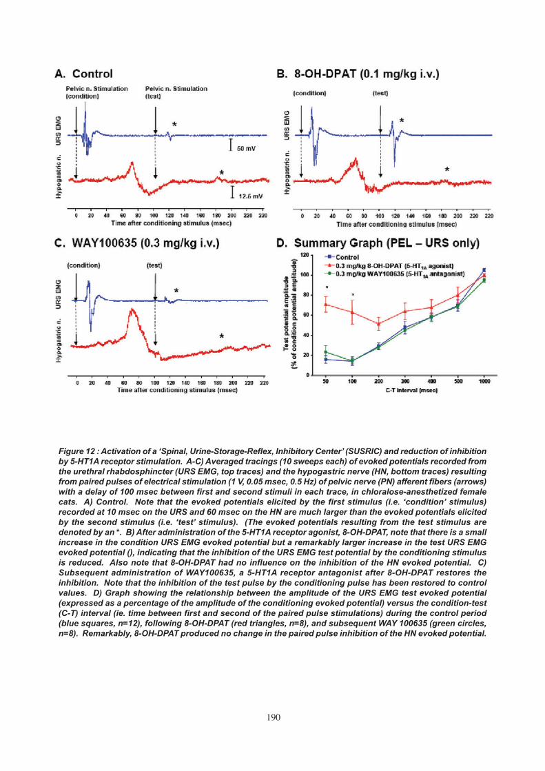
190
Figure 12 : Activation of a ‘Spinal, Urine-Storage-Reflex, Inhibitory Center’ (SUSRIC) and reduction of inhibitionby 5-HT1A receptor stimulation. A-C) Averaged tracings (10 sweeps each) of evoked potentials recorded fromthe urethral rhabdosphincter (URS EMG, top traces) and the hypogastric nerve (HN, bottom traces) resultingfrom paired pulses of electrical stimulation (1 V, 0.05 msec, 0.5 Hz) of pelvic nerve (PN) afferent fibers (arrows)with a delay of 100 msec between first and second stimuli in each trace, in chloralose-anesthetized femalecats. A) Control. Note that the evoked potentials elicited by the first stimulus (i.e. ‘condition’ stimulus)recorded at 10 msec on the URS and 60 msec on the HN are much larger than the evoked potentials elicitedby the second stimulus (i.e. ‘test’ stimulus). (The evoked potentials resulting from the test stimulus aredenoted by an *. B) After administration of the 5-HT1A receptor agonist, 8-OH-DPAT, note that there is a smallincrease in the condition URS EMG evoked potential but a remarkably larger increase in the test URS EMGevoked potential (), indicating that the inhibition of the URS EMG test potential by the conditioning stimulusis reduced. Also note that 8-OH-DPAT had no influence on the inhibition of the HN evoked potential. C)Subsequent administration of WAY100635, a 5-HT1A receptor antagonist after 8-OH-DPAT restores theinhibition. Note that the inhibition of the test pulse by the conditioning pulse has been restored to controlvalues. D) Graph showing the relationship between the amplitude of the URS EMG test evoked potential(expressed as a percentage of the amplitude of the conditioning evoked potential) versus the condition-test(C-T) interval (ie. time between first and second of the paired pulse stimulations) during the control period(blue squares, n=12), following 8-OH-DPAT (red triangles, n=8), and subsequent WAY 100635 (green circles,n=8). Remarkably, 8-OH-DPAT produced no change in the paired pulse inhibition of the HN evoked potential.
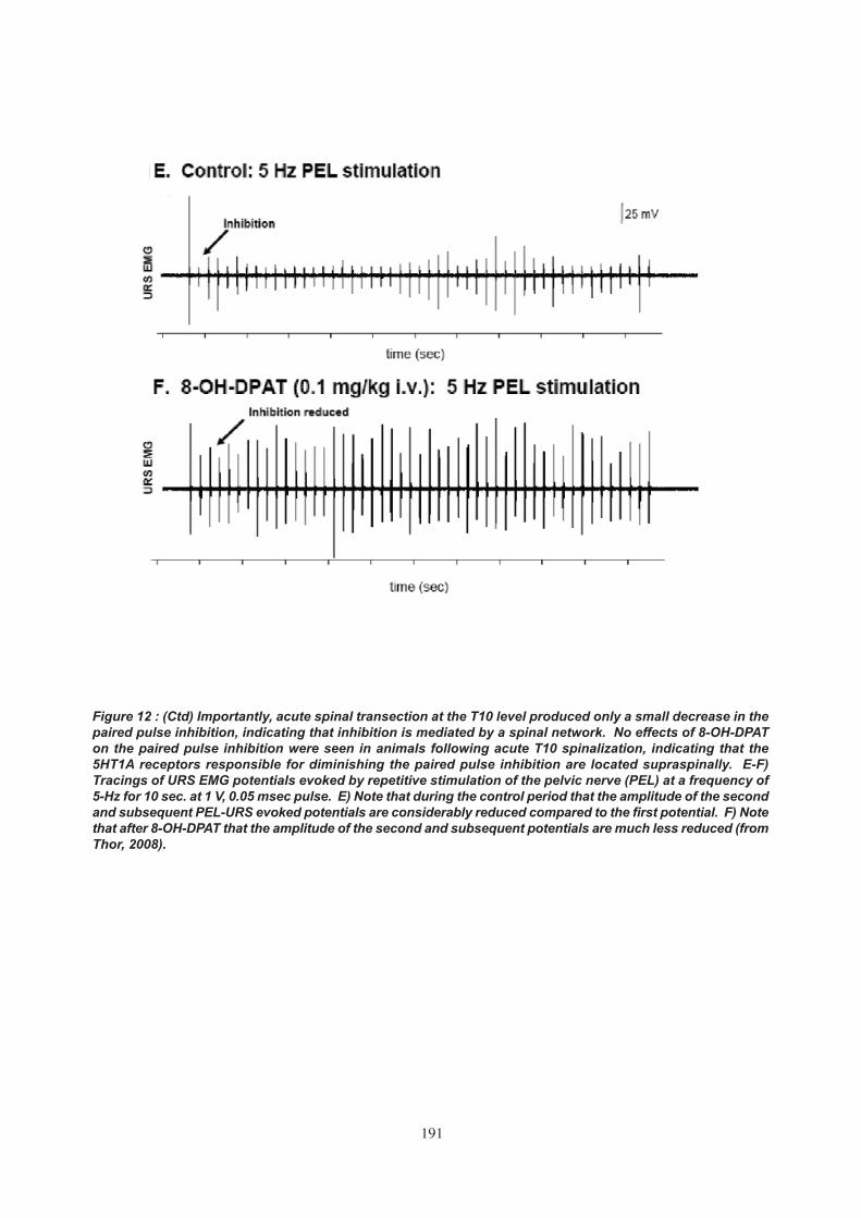
191
Figure 12 : (Ctd) Importantly, acute spinal transection at the T10 level produced only a small decrease in thepaired pulse inhibition, indicating that inhibition is mediated by a spinal network. No effects of 8-OH-DPATon the paired pulse inhibition were seen in animals following acute T10 spinalization, indicating that the5HT1A receptors responsible for diminishing the paired pulse inhibition are located supraspinally. E-F)Tracings of URS EMG potentials evoked by repetitive stimulation of the pelvic nerve (PEL) at a frequency of5-Hz for 10 sec. at 1 V, 0.05 msec pulse. E) Note that during the control period that the amplitude of the secondand subsequent PEL-URS evoked potentials are considerably reduced compared to the first potential. F) Notethat after 8-OH-DPAT that the amplitude of the second and subsequent potentials are much less reduced (fromThor, 2008).

192
central electrode placement would also activate colonicand genital afferent fibers, additional experiments areneeded to specifically compare urethral versus bladderversus colonic pelvic afferent fibers in evoking the“guarding reflex”.
Electrical stimulation of pudendal afferent fibers alsoevokes a spinal reflex activation of the rhabdosphincter[233, 274]. Since some urethral afferent fibers (aswell as rectal, genital, and cutaneous afferent fibers)also travel in the pudendal nerve, it is possible thatthe spinal urethral rhabdosphincter activation bypudendal afferent stimulation is also a manifestationof the “guarding reflex”.
4. SPINAL URINE-STORAGE-REFLEX INHIBITORY CENTER (SUSRIC)
While electrical stimulation of pelvic and pudendalafferent fibers can activate urethral sphincter motorneurons via a spinal reflex, these same stimuli alsoproduce an inhibition of urethral sphincter motorneurons in cats. In early studies of McMahon et al.,[233] an inhibition of pudendal nerve spontaneousactivity occurred for a period of 50 - 1,000 msecfollowing pelvic nerve stimulation.
Recently, this inhibitory effect has been characterizedusing electrical stimulation of pelvic (PEL) andpudendal (PUD) nerve afferent fibers and recordingof evoked potentials with urethral rhabdosphincter(URS) EMG electrodes (i.e. PEL-URS and PUD-URSreflexes, respectively) in cats [275]. In addition torecording these somatic motor urine storage reflexes,
electrodes were also placed on the hypogastric (HgN)nerve (i.e. PEL-HgN and PUD-HgN reflexes,respectively) to record sympathetic urine storagereflexes. Both the somatic and sympathetic urinestorage reflexes are reliably evoked when thefrequency of PEL or PUD stimulation is below 1 Hz(Figure 12A-C). However, when the frequency israised to 5 Hz or higher, the amplitudes of the reflexesimmediately drop (Figure 12E). This is not a “rundown”phenomenon because the reflex drop occursimmediately after the first stimulus (i.e. upon applicationof the second impulse in a given frequency’s seriesof pulses). This suggested that the first stimulus wasactivating an inhibitory circuit that blocked the secondand subsequent stimuli’s ability to evoke the reflex(Figure 13).
The activation of an inhibitory circuit was shown inanesthetized cats [275] using a paired-pulse orcondition-test paradigm, where the first stimulus of apair (i.e. the condition pulse) precedes the secondstimulus of the pair (i.e. the test pulse) by incrementaltimes (i.e. 10, 20, 50, 100, 200, 500, 1,000 msec). Thisparadigm showed that when a second (i.e. paired ortest stimuli) stimulus was applied to the PEL and/orPUD nerve 50 - 500 msec after the first stimulus, thereflexes evoked by the second stimulus were reliablyinhibited (Figure 12A,D). The paired pulse inhibitionwas maximal (2nd stimulus evoked potential was < 20% of the 1st stimulus evoked potential) at pairedintervals of 50 - 100 msec and gradually disappearedat paired intervals of 1,000 msec or more (Figure12B,D). A possible clinical correlation of SUSRIC
Figure 13 : Diagram showing the somatic and sympathetic urine storage reflexes as previously described(from Thor, 2004). Pelvic and pudendal nerve stimulation, in addition to activating the rhabdosphincter motorneurons in Onuf’s nucleus and the sympathetic preganglionic neurons (SYM) to cause contraction of urethralstriated and smooth muscle, respectively, also activates a “spinal, urine-storage-reflex, inhibitory center(SUSRIC) that inhibits both storage reflexes for 50-1,000 msec.
193
activation may be the elegant demonstration thatconditioning stimuli applied to the dorsal nerve of thepenis (i.e. pudendal nerve afferent fibers) inhibitedurethral rhabdosphincter contractions reflexivelyevoked by magnetic stimulation of the spinal cord atintervals of 20 - 100 msec [276].
The threshold stimulus intensity for evoking inhibitionin cats was the same as the threshold stimulus intensityfor evoking the reflex, suggesting the same, or similarsized, fibers activate both the reflex and SUSRIC.Inhibition of both the PEL - URS EMG and PEL - HgNevoked potentials showed similar intensity-responsecharacteristics. The inhibition could be evoked byhomologous (PEL after PEL and PUD after PUD) orheterologous (PUD after PEL or vice-versa) pulsepairing. Furthermore, stimulation of PEL afferent fibersclose to the bladder, which did not evoke a URS reflex,also produced inhibition of the PEL - URS reflexevoked by an electrode placed more centrally on thePEL nerve that did evoke a PEL - URS reflex. Thisindicates that the inhibition is not due to refractorinessof the rhabdosphincter motor neurons.
The paired pulse inhibition was slightly reduced (10-15%) but otherwise similar after acute spinaltransection at T10, indicating that the inhibitory circuitactivated by the conditioning stimulus is located inthe spinal cord. Inhibition of the PEL - URS reflexalso remained after a spinal transection at the L5level suggesting that the inhibitory circuit is locatedwithin the same segment as the PEL - URS reflexpathway (i.e. sacral cord). (Since L5 spinalizationinterrupts the PEL - HgN reflex, which precludestesting for its inhibition, we cannot speculate on thelocation of the inhibitory circuit for the PEL - HgNreflex except that it is caudal to T10.) Since thisinhibitory center is located in the spinal cord, andinhibits both the somatic and sympathetic storagereflexes evoked by either PEL or PUD stimulation,the name, “spinal, urine-storage-reflex, inhibitorycenter” (SUSRIC), is suggested.
The two nerves that are effective for activating theSUSRIC, the pelvic and pudendal nerves, have spinalterminals that densely project to the dorsal graycommissure region of the sacral spinal cord [219,277]. This is also an area that contains inhibitoryGABAergic and glycinergic neurons that are thoughtto mediate urethral rhabdosphincter inhibition duringmicturition [278, 279]. Because pelvic and pudendalnerves densely innervate this same region of thespinal cord, it is tempting to speculate that theseGABAergic and glycinergic neurons are also theinhibitory cellular substrate of SUSRIC. Requirementof multiple, parallel inputs to SUSRIC from pelvicnerve axons (e.g. bladder afferent neurons), pudendalnerve axons (urethral afferent neurons), anddescending axons from the pontine micturition centermight be valuable to prevent sphincter relaxationunless all 3 systems “agreed” that micturition should
proceed. On the other hand, redundant inputs mightalso be valuable to ensure initiation and maintenanceurethral sphincter inhibition until micturition is completeand all urine has flowed out of the bladder andcompletely through the urethra prior to urethral closure.Experiments to test this model are needed.In additionto its physiological role, overactivity of SUSRIC maybe involved in the pathology of stress urinaryincontinence, while under activity may be involved inbladder-sphincter dyssynergia or retentive urinarydysfunction such as Fowler’s syndrome [280].
• 5-HT1A Receptor Regulation of SUSRIC
Administration of the 5-HT1A receptor agonist, 8-hydroxy-dipropylaminotetraline, 8-OH-DPAT, signifi-cantly reduced the paired pulse inhibition of theSUSRIC to 20% of control in spinal intact animals(Figure 12A-D) [275]. The 8-OH-DPAT-inducedreduction in paired pulse inhibition was reversed byWAY100,635, a highly selective 5-HT1A receptorantagonist, which confirms the role of 5-HT1Areceptors in suppressing SUSRIC. 8-OH-DPAT alsoenhanced the ability of the PEL - URS reflex to followhigh frequency (i.e. 2 - 10 Hz) stimulation (Figure12E-F). Importantly, 8-OH-DPAT reduced the inhibitionin spinal cord intact cats but not in animals with anacute T10 spinal transection, indicating that 5-HT1Areceptors are working supraspinally, probably to inhibita descending excitatory input to SUSRIC (i.e.disfacilitation of the inhibitory center).
Previous urodynamic studies in anesthetized cats[281-283] support the supraspinal location of 5-HT1Areceptors that facilitate urethral rhabdosphincteractivity. This support stems from urodynamic studiesthat showed a remarkable enhancement of spon-taneous urethral rhabdosphincter EMG activity in thechloralose-anesthetized cat irritated bladder modelwhen the spinal cord is intact [283] but not when thespinal cord is transected, even after spinal bladderreflexes have emerged following spinal shock [281,282]. In other words, it is proposed that 8-OH-DPAT’senhancement of urethral rhabdosphincter activityresults from stimulation of supraspinal 5-HT1Areceptors, which subsequently results in a reductionof SUSRIC-mediated inhibition of rhabdosphinctermotor neurons, i.e. excitation results from disinhibition(Figure 13).
a) Supraspinal Activation of Rhabdosphinctersand Pelvic Floor Muscles
Supraspinal activation of urethral and analrhabdosphincter motor neurons includes voluntaryinputs (i.e. corticospinal [254]), as well as involuntaryreflexic inputs (e.g. during coughing, sneezing,vomiting) presumably from nucleus retroambiguus inthe caudal medulla [267, 284-286]. Nucleusretroambiguus also innervates the pelvic floor muscle[285, 286], as well as the abdominal muscles; consist
194
with a role in raising intra-abdominal pressure duringValsalva maneuvers. Generally, the pelvic floor andrhabdosphincter muscles are activated as a functionalunit when voluntarily contracted. However, differencesin activation between the rhabdosphincter and thepelvic floor muscles have been documented [287],indicating distinct CNS control systems andinnervation. Clinical EMG recordings show that evenduring sleep, activity can be recorded from specificrhabdosphincter motor units [288].
Rhabdosphincter motor neurons are unique amongsomatic motor neurons in receiving input from theparaventricular hypothalamus[266], although thefunction of this input has not been determined. Inaddition, their input from brainstem serotonergic andnoradrenergic neurons is among the most dense inthe spinal cord [289-292]. Finally, rhabdosphinctermotor neurons also receive input from the “L region”of the pons that might be important for maintainingcontinence, since a lesion in this area producedcontinuous incontinence in a cat [293].
1. RELATIVE CONTRIBUTION FROM LEVATOR ANI NERVE,PUDENDAL NERVE, AND HYPOGASTRIC NERVE IN
CONTINENCE MECHANISMS DURING SNEEZING IN RATS
AND CATS
Analysis of the urethral closure mechanisms duringsneeze-induced stress conditions in anesthetizedfemale rats and cats has revealed that pressureincreases in the middle portion of the urethra aremediated by reflex contractions of the rhabdosphincteras well as the pelvic floor muscles [294, 295].Transection of the pudendal nerves reduced sneeze-induced urethral reflex responses by 67% andtransecting the nerves to the iliococcygeus andpubococcygeus muscles reduced urethral reflexresponses by an additional 25%.
Transecting the hypogastric nerves and visceralbranches of the pelvic nerves did not affect the urethralreflexes indicating that sneeze-evoked urethral reflexesin normal rats were not mediated by these autonomicpathways. However, hypogastric nerve transection inconscious, chronic spinal cord injured, female ratsreduced urethral baseline pressure, reduced post-void residual urine volumes, reduced maximal voidingpressure, and increased voiding efficiency. Thisindicates that sympathetic pathways to the bladderneck and proximal urethra contribute to urethralpressure and functional outlet obstruction and voidingdysfunction after spinal cord injury in unanesthetizedanimals(296).
2. NEUROCHEMICAL ANATOMY OF URETHRAL RHABDO-SPHINCTER MOTOR NEURONS
In addition to their unique morphology, neuro-physiology, and supraspinal inputs, rhabdosphinctermotor neurons in Onuf’s nucleus also exhibit a plethoraof unique and highly diverse neurotransmitter inputs,
receptors, ion channels, and growth factors (Table3). While many of the markers listed in Table 3 are likelyinvolved in rhabdosphincter control, it is dangerous toassume a role in rhabdosphincter control based strictlyon anatomical association with Onuf’s nucleus, sincesome motor neurons in Onuf’s nucleus innervate theischiocavernosus and bulbospongiosus muscles andthus may be involved in control of sexual function.The following section will offer some guidanceregarding which transmitter and receptor systems areinvolved in control of rhabdosphincter function.
5. PHARMACOLOGY OF URETHRAL AND ANALRHABDOSPHINCTERS
The excitatory amino acid neurotransmitter, glutamate,mediates initiation of action potentials in rhabdo-sphincter motor neurons (and subsequent rapidcontraction of the muscle) by binding to NMDA andAMPA receptors. Both spinal reflex activation andsupraspinal activation of the rhabdosphincter aresensitive to NMDA and AMPA receptor antagonists[297-299]. Thus it is useful to think of these transmittersas part of the “hardwired” reflex circuitry that is involvedin all or none activation of consistent and reliablestorage reflexes, as compared to monoamines andpeptide transmitters (see below), that play a role asmodulators of the reflexes, increasing or decreasingthe gain of the reflexes transmitted by the excitatoryamino acids.
The inhibitory amino acids glycine, acting throughstrychnine-sensitive ionotropic receptors [279, 300],and GABA, acting through both GABA-A (ionotropic)and GABA-B receptors (metabotropic) [257, 301, 302]are thought to be major inhibitory transmittersregulating rhabdosphincter activity. Clinical studieshave indicated that systemic [303] and intrathecal[304, 305] administration of the GABA-B agonist,baclofen, may reduce bladder-sphincter dyssynergiain some neurogenic bladder patients. However,because of the ubiquity of glycine and GABA inmediating inhibition of multiple systems, pharma-cological studies linking either of these transmitters(or any other inhibitory transmitter) to the inhibition ofrhabdosphincter activity during voiding are notdefinitive.
In addition to amino acid transmitters, the monoaminetransmitters (serotonin and norepinephrine) are alsoimportant in modulating rhabdosphincter motor neuronactivity [306]. It was the preferential association ofnorepinephrine and serotonin terminals [289-292] thatled to extensive studies of noradrenergic andserotonergic control of rhabdosphincter function andeventual clinical studies of duloxetine, a norepinephrineand serotonin reuptake inhibitor, as a treatment forstress urinary incontinence [270, 307-311]. Elegantstudies in humans using magnetic stimulation of brainand sacral nerve roots [312] have indicated thatduloxetine increases the excitability of rhabdosphincter

195
motor neurons to both supraspinal and segmentalinputs and to double urethral pressure responses tosacral nerve root magnetic stimulation. Importantly,duloxetine’s ability to increase urethral rhabdosphincteractivity did not interfere with the inhibition of sphincteractivity during voiding (i.e. bladder-sphincter synergywas well-maintained). Similar clinical results havebeen seen with S,S-reboxetine, a selective norepi-nephrine reuptake inhibitor [313, 314]. This approachof increasing synaptic levels of serotonin and/ornorepinephrine is logical since, it has been shownthat noradrenergic and serotonergic terminalsassociated with rhabdosphincter motor neurons showan age-dependent decrease in density in rats [315]that might explain the increased incidence of stressincontinence with aging.
Multiple adrenergic receptor subtypes play a role incontrol of the rhabdosphincter, and the results withnorepinephrine reuptake inhibitors indicate that thesereceptors can be activated by endogenousnorepinephrine in anesthetized cats [272]. Strongevidence exists that α1 adrenoceptors excite rhab-dosphincter motor neurons [272, 316, 317]. Patchclamp studies [318] have shown a direct depolarizingeffect of norepinephrine on rhabdosphincter motorneurons (Figure 14D) that can be blocked by the α1adrenoceptor antagonist, prazosin. Similar conclusionsregarding the excitatory effects of α1 adrenoceptorson rhabdosphincter neurons have been reached inclinical studies [317] where decreases in rhabdo-sphincter activity were seen after administration ofprazosin to human subjects. On the other hand, strongevidence exists that α2 adrenoceptor stimulation hasthe opposite effect, i.e. inhibition, of rhabdosphincteractivity [272, 319]. Importantly, the innervation of theurethral and anal smooth muscle (i.e. the hypogastricnerve) shows similar adrenergic pharmacology - anenhancement of activity by α1 adrenoceptors [272,316, 320] and inhibition of activity by α2 adrenoceptors[272, 316, 321].
Multiple subtypes of serotonin (5-hydroxytrptamine,5-HT) receptors are also involved in modulatingrhabdosphincter motor neuron excitability. Strongevidence exists that 5-HT2 receptors can excitesphincter motor neurons [274]. Indeed duloxetine’sfacilitatory effects on rhabdosphincter activity inanesthetized cats are mediated in part throughactivation of 5-HT2 receptors [270]. Both 5-HT2A and5-HT2C receptor agonists increase rhabdosphincterEMG activity in dogs, guinea pigs, and rats [322, 323].Recent in vitro rat spinal cord slice patch clamp studiesshow that part of this effect may be directly onrhabdosphincter motor neurons, as opposed tointerneurons [318], since 5-HT induces a directdepolarization of rhabdosphincter motor neurons(Figure 14E). Interestingly, substance P, a peptidetransmitter that is co-localized with 5-HT in raphespinal nerve terminals, also produces direct
depolarization of rhabdosphincter motor neurons in ratspinal cord slices [324], and thyrotropin releasinghormone (TRH), another peptide transmitter co-localized with 5-HT in nerve terminals, inducesexcitation of rat sphincter activity [325, 326] in vivo.
A peculiar finding regarding 5-HT2 receptor-inducedactivity of pelvic nerve-evoked rhabdosphincterreflexes in chloralose-anesthetized cats is that theeffect is highly reproducible when the spinal cord isisolated (i.e. acute T11 transection), but it is highlyvariable and statistically insignificant when the spinalcord is intact [274]. This suggests that supraspinal 5-HT2 receptors have an opposing, inhibitory, effectthat counteracts their spinal, excitatory, effect. Whetherthis phenomenon is an artifact of anesthesia and/oris specific to cats is important to determine.
Immunohistochemical and molecular studies inhumans and dogs [322] have shown that 5-HT2A, 5-HT2B, and 5-HT2C receptor subtypes are associatedwith Onuf’s nucleus motor neurons. In addition, 5-HT2C receptor mRNA has been localized to analsphincter motor neurons in the rat [327]. On the otherhand, another immunohistochemistry study withretrograde labeling of urethral rhabdosphincter motorneurons and ischiocavernosus motor neurons in ratsindicates that the 5-HT5A receptor is associated withthe former, while the 5-HT2A receptor is preferentiallyassociated with the latter [328].
As described in the previous section on the SUSRIC,supraspinal 5-HT1A receptor stimulation enhancesrhabdosphincter activity in cats [275, 281-283].Importantly, the excitatory effects of 5-HT1A receptoragonists on the rhabdosphincter can still be “overridden” by inhibitory mechanisms during voiding, i.e.bladder-sphincter synergy remains despite 8-OH-DPAT-induced enhancement of rhabdosphincteractivity [275, 281-283].
The effect of 5-HT1A receptors on rhabdosphincteractivity in rats is more complicated. Firstly, rats donot show an inhibition of rhabdosphincter activityduring voiding like cats and humans but, instead,show a high frequency bursting (or “phasic firing”) ofrhabdosphincter EMG activity during micturition [329].This bursting is actually necessary for efficient voidingin rats presumably by creating a “pumping” actionthrough the urethra. When the spinal cord istransected, this bursting disappears and rats presentwith urinary retention. Although the bursting is notseen in anesthetized spinal rats at any time [329], inconscious spinal rats, the rhabdosphincter EMGbursting gradually returns in some animals across aperiod of 6 weeks [330]. Interestingly, 5-HT1A receptorstimulation can unmask the bursting rhabdosphincterEMG activity in the anesthetized spinal rat [282, 331].Chang et al [331] further showed that the centerresponsible for bursting activity in spinal rats waslocated between the T11 and L4 spinal segments. In

196
Figure 14 : Monoaminergic modulation of sphincter motor neurons recorded using patch clamp electrophysiology.A-C) Visual identification of rat urethral sphincter motor neurons. A. Retrograde labeling of dorsolateralnucleus (DLN) motor neurons was accomplished by fluorescent dye injections into the urethral sphincter(adult), or the ischiorectal fossa (neonate). Dye was transported through the motor branch of the pudendal nerveto the lumbar spinal cord. B. Composite fluorescence and low-light image showing DiI-labeled DLN neuronsin the ventral horn of an adult rat lumbar spinal cord section. The dotted line denotes the grey/white matterboundary, and the dorsal surface is oriented toward the top of the page. C. Enlarged neurons from B with apatch-clamp electrode schematic to demonstrate electrophysiological recordings from DLN neurons. Calibrationbars are 80 µm (B) and 20 µm (C). D-E) Either norepinephrine (NE) or serotonin (5-HT) was bath applied to ratlumbar spinal cord slices containing DiI-labeled dorsolateral nucleus neurons. D. NE (1 µM) produced areversible depolarization and action potential firing. E. 5-HT (10 µM) also produced a reversible depolarizationand action potential firing (from Burgard, 2008).

197
the studies of Gu et al [282], 5-HT1A receptorstimulation had no influence on the asynchronousrhabdosphincter activity that precedes, and follows,micturition-associated bursting, it only re-organized theactivity into the bursting or phasic characteristics thataccompany micturition.
Another aspect of 5-HT1A receptor function in rats canbe seen from studies of pelvic nerve-to-rhabdo-sphincter reflex potentiation. Early studies showedthat this reflex can be potentiated by prolonged, lowfrequency electrical stimulation of the pelvic nerveand by bladder distention [332-334]. Subsequentstudies showed that the potentiation involved a 5-HT1A receptor-based link [335].
An interesting and possibly important comparisonbetween drug effects on rhabdosphincter activity in catsand rats is also shown with κ2 opioid receptors. In cats,the κ opioid receptor agonist, ethylketocyclazocine,selectively inhibits spinal rhabdosphincter reflexes[234]. In rats, it was found that a κ2 opioid receptoragonist inhibits the rhabdosphincter bursting patternassociated with micturition, leading to decreasedvoiding efficiency [336]. In these studies, κ opioidreceptor stimulation had no influence on theasynchronous rhabdosphincter activity that precedesand follows micturition-associated bursting.
Thus there may be parallels between excitation ofrhabdosphincter reflexes in cats with enhancement ofrhabdosphincter bursting activity in rats on one hand(5-HT1A receptor-mediated), and inhibition of rhabdo-sphincter motor neurons in cats and suppression ofbursting activity in rats on the other hand (κ opioidreceptor-mediated). The fact that asynchronousrhabdosphincter activity in rat was not affected byeither 5-HT1A or κ opioid receptor stimulation highlightsdifferences in the organization of the asynchronous(spinal) and micturition associated-bursting (su-praspinal) reflex control in rats.
Another possible parallel for exploration ofrhabdosphincter function in rats might lie in comparingthe above spinal micturition-associated rhabdo-sphincter bursting center with the spinal ejaculationcenter in rats: both centers are located in the mid-lumbar spinal cord [331, 337, 338], both centers exhibit“bursting” [329, 339]; 5-HT1A receptor agonists canfacilitate both activities [340-342], κ opioid receptoragonists inhibit both activities [336, 343]. Of courseit must be appreciated that differences between thetwo “bursting centers” exist (i.e. the former activatesrhabdosphincter motor neurons while the latteractivates bulbospongiosus motor neurons, the formerhas a period of 6 Hz while the latter has a period of0.5 Hz).
Future Pelvic Floor Research Needs
The controversy regarding a contribution of thepudendal nerve to the innervation of the levator ani
muscles should be addressed. As a first step,agreements regarding nomenclature should beestablished in regards to which muscles actuallycomprise the levator ani (i.e. should the definitioninclude only the pubococcygeus, iliococcygeus, andcoccygeus muscles or should it include thepuborectalis, urethralis, urethrovaginal sphincter, thecompressor urethrae; etc.). Careful characterizationof these various muscles based on physiologicalcriteria such as embryological origin, skeletal sites oforigin and insertion, compartmentalization imposed byconnective tissue boundaries, myofiber phenotype, andsensory structures (e.g. Golgi tendon organs andmuscle spindles) is likely to provide insight into whichtypes of motor neurons (i.e. alpha, gamma, or “specialsomatic”) innervate each muscle and which nervecarries their motor axons. Resolving this controversyis important for a number of reasons: surgicalguidance, understanding iatrogenic pathologies, andunderstanding physiological and pharmacologicalcontrol of the individual muscles.
A second important area regards the reflex control ofthe levator ani muscles and viscerosomatic interactionsbetween pelvic floor muscles and pelvic viscera. Whilevisceral and somatic reflex control of therhabdosphincters has been extensively studied,studies of levator ani muscle reflexes are few.Understanding whether there are spinal reflexconnections between, for example, bladder afferentfibers and levator ani motor neurons would furtherour understanding of continence control. Similarly,understanding anatomical connections betweenrhabdosphincter and levator ani muscles are important.Determining whether the cholera toxin B-labeledprocesses of levator ani motor neurons located inOnuf’s nucleus (Figure 11 D-E) are dendrites or axonalcollaterals may provide important insight intounderstanding physiological coordination betweenthe pelvic floor and rhabdosphincters. Finally,understanding the role of levator ani primary afferentfibers in controlling lower urinary tract and bowelfunction might explain indirect bladder symptoms thatmight be induced by pelvic floor injury or suspensionsurgeries, as well as physiological controls of levatorani muscles over excretory function and vice-versa.The abundant small diameter peptidergic afferentfibers innervating pelvic floor muscle might also playa role in the etiology of chronic pelvic pain syndromeor interstitial cystitis; two conditions for which a frankpathological cause has not been determined.
A third important area for study is the relativeimportance of bladder afferent fibers versus urethral/ bladder neck afferent fibers in eliciting the “guarding”or “continence” reflex. This is important since there arephenotypic differences between these two groups ofafferent neurons in regards to neurotransmitter andion channel phenotypes(344) that might be exploitedto independently control urine storage and voidingdysfunction.

198
The motor arm of the lower urinary tract drives bladdercontraction during voiding and the outlet contractionrequired for urine storage. In this section, spinal andperipheral elements contributing to the motor activityof the bladder is described, while the equivalentstructures for the bladder outlet are discussed insection III.
1. PREGANGLIONIC NEURONS
Parasympathetic preganglionic neurons are locatedin the lateral part of the sacral intermediolateral graymatter in a region termed the sacral parasympatheticnucleus (SPN) and are small, fusiform-shaped cellswhich send dendrites into lateral lamina I of the dorsalhorn, the lateral funiculus and medially into the dorsalgrey commissure (DGC). Bladder pre-ganglionic motorneurons are located in the S1-S3 in the cat [361], dog[362] and monkey [363]. In cat these motor neuronsare located ventrolaterally in the intermediolateralcolumn, with cells innervating the colon lyingdorsomedial. The guinea pig is similar; retrogradelabelling following bladder wall injections revealsneurons bilaterally in the ventrolateral part of theintermediolateral column at S1 [256]. The rat isdifferent, as preganglionic motor neurons are locatedat L6-S1 [364]; unilateral ventral root rhizotomy at theL5 level in the rat decreases peak cystometricpressures [365].
The parasympathetic preganglionic neurones projectthrough the ventral spinal roots to the major pelvicganglion [366-368], releasing the excitatory transmitter,acetylcholine. They are divided functionally into tonicand phasic types. In some species they also releaseopioid peptide transmitters and express nitric oxidesynthase [369]; there is also evidence of involvementof pituitary adenylate cyclase activating peptide(PACAP), a peptide present in visceral afferentneurones, and of prostaglandins within the spinal cord[370]. The DGC also contains a group of interneurones,which are likely to be active during micturition [371,372]. These DGC interneurones may influence thefunction of the parasympathetic preganglionic neurons[279, 371, 372].
At spinal levels L1–L2, both the intermediolateral hornand the DGC contain sympathetic preganglionicneurones whose axons project to the major pelvicganglion. With ageing, there is selective attrition ofpreganglionic sympathetic neurones in L1–L2, whichproject to the pelvic ganglion, with reductions in theextent of the dendritic arbors of remaining cells [366,367].
In man the preganglionic parasympathetic motornerves to the bladder (and other pelvic organs, therectum and descending colon) course through the
pelvic nerves from the sacral anterior roots S2-S4.Stimulating these roots with implanted electrodesdesigned principally for bladder emptying in spinalcord injury [373] elicits two principal responses at S3;at low levels of stimulation, the external urethralsphincter, external anal sphincter and pelvic floormuscles are contracted. At high levels of stimulation,parasympathetic activation contracts the detrusormuscle, leading to efficient emptying of the bladderwhen the sphincter muscle relaxes [374]. Attemptsto use extracorporeal magnetic stimulation to achievethe same effect [375] have shown insufficient powerto activate the small parasympathetic neurons at thelevel of the lumbar-sacral roots [376].
2. GANGLIA
The peripheral ganglia convey the autonomicinnervation to the lower urinary tract and reproductiveorgans, along with a substantial part of the extrinsicmotor innervation of the lower bowel. There aresubstantial species differences in organization andneurochemistry of pelvic ganglion cells and their spinalinputs. Large mammals have a plexus of pelvic andintramural ganglia, containing both sympathetic andparasympathetic neurons. The guinea pig isintermediate in complexity, with separate posteriorand anterior plexuses innervating different pelvicorgans. In the rat and mouse, the pelvic plexus consistsof the major pelvic ganglia (MPG) and a number ofsmall accessory ganglia. In the rat there are two majorpelvic ganglia and small accessory ganglia, with lesscytological complexity and almost no intramuralganglia.
The preganglionic input releases acetylcholine, whichacts on nicotinic receptors. Patients with megacystis-microcolon-intestinal-hypoperistalsis syndrome(MMHIS) [377] have reduced or no alpha-3 nicotinicreceptor subunit [378]. Selective gene knockout micelacking the alpha-3 nicotinic receptor subunit alone orthe beta-2 and beta-4 subunits in combination [379],develop severe bladder distension soon after birth, andlater overflow incontinence. The detrusor muscle inthese animals contracts in response to field stimulationor muscarinic agonists, but not nicotinic agonists [380],indicating the potential importance of alpha-3, beta-2, and beta-4 nicotinic receptor components in controlof voiding, but not their functional location.
Within the pelvic plexus there is topographicalrepresentation of the pelvic organs. In the female dog,neurons supplying different pelvic organs are locatedin separate ganglia, which possess a distinctivecomposition of neurone types and differentpreganglionic supply [381]. Neurons retrogradelylabelled from the urinary bladder mainly occur inganglia located at the vesico-ureteric junction. Theycomprise catecholaminergic calbindin neurons andnoncatecholaminergic neurons containing calbindin orNOS, with relatively sparse pericellular varicose nervefibres.
IV. EFFERENT PATHWAYS TO THEBLADDER

199
In male mice [382], the major pelvic ganglia are closeto the dorsal surface of the prostate gland. Their maininputs are the pelvic nerves, and the hypogastricnerve from the inferior mesenteric ganglion. The majoroutputs are the penile (cavernous) nerve and thesupply to the urogenital organs. Tyrosine hydroxylase(TH) is expressed by one-third of neurons, almost allco-expressing dopamine beta hydroxylase (DBH).Numerous TH axons are present in the hypogastricnerve, but very few in the pelvic nerve, supporting aprimarily sympathetic origin. Non-neuronal cellscontaining TH are also present, resembling small,intensely fluorescent (SIF) cells observed in manyother autonomic ganglia [382]. Neurons immuno-stained for choline acetyl transferase (ChAT) have acomplementary distribution to noradrenenergicneurons. About half of the cholinergic ganglion cellscontain VIP, distributed throughout most of theganglion, with a cluster near the origin of the penilenerve [382]. Neurones with NPY are numerous andapparently randomly distributed throughout theganglion, with marked variation between mousestrains. All noradrenergic neurons contain NPY, butmany NPY neurons are not noradrenergic. Many ofthe cholinergic NPY neurons also contain VIP. ChATis seen in varicose axon terminals closely associatedwith ganglion neurons. Neither NPY nor VIP arepresent in preganglionic terminals, except for a smallnumber of individual neurones. The latter may arisefrom viscerofugal neurons in the myenteric plexus ofthe lower bowel [383].
Autonomic ganglia are also found in the vicinity ofthe bladder neck, trigone, proximal urethra andprostate. They receive noradrenergic and cholinergicexcitatory innervation and non-cholinergic, non-adrenergic inhibitory innervation [384]. Knowledge of
this distribution allows strategic planning for surgicaldissection (Figure 15). In ureteric reimplantation,several branches of the pelvic plexus travel to theureterovesical junction and surround the distal ureteras a fine network in the human. The main neuralelements are located 1.5 to 2 cm. dorsal and cranialto the bladder trigone, and dorsal and medial to theureter in both female and male cadavers [385]. In thecontext of hysterectomy, cadaveric dissections showpelvic autonomic nerves are at greatest risk duringsacrouterine dissection [386], corresponding withobserved innervation density [387]. In additionpathophysiological processes such as ageing caninfluence the pelvic ganglia, for example thepostganglionic sympathetic neurones in the majorpelvic ganglion of rats [388, 389]. In the proximalfemale urethra, virtually all of the NOS immunoreactivecells also contain the carbon monoxide-synthesisingenzyme, haem-oxygenase- (HO-) 2, but of the HO-2positive cells, 25% did not show NOS immunoreactivity[390].
The bladder wall itself contains intramural ganglia,and small clusters of autonomic ganglion cells arepresent in the adventitial connective tissue and amongthe detrusor muscle bundles. There is species variationin the extent of intramural innervation of the bladder;ganglia are present in many species such as theguinea pig [383], while the rat bladder contains thepost-synaptic innervation alone [391]. The gangliaare found throughout the bladder wall and varyconsiderably in size [91, 392]. They show immuno-reactivity to vasoactive intestinal polypeptide (VIP),nitric oxide synthase (NOS), neuropeptide Y (NPY) andgalanin (Gal) in varying amounts. However, they donot contain enkephalin (ENK), substance P (SP),calcitonin gene-related peptide (CGRP) or soma-
Figure 15 (left panel): Dissection of superior hypogastric plexus, hypogastric nerves and two main portionsof pelvic plexus in male cadaver. B, bladder, PP, pelvic plexus, LHN, left hypogastric nerve, RHN, righthypogastric nerve, IMA, inferior mesenteric artery, SHP, superior hypogastric plexus, U, ureter. Middlepanel: Left hypogastric nerve with network of pelvic plexus nerve bundles. Branches run to distal ureter(U) and bladder (B) trigone. PP pelvic plexus, A, aorta, C inferior vena cava, LHN, left hypogastric nerve,AMI, inferior mesenteric artery. Right panel: Preparation of distal ureter (U). Clamp is used to dissect nervebundles of pelvic plexus. B, bladder R, rectum N, pelvic plexus nerve fibre. From Leissner J., et al. 2001.
200
tostatin (Som) [90], suggesting that cell bodies ofsensory neurones are not located in the intramuralganglia. Postganglionic sympathetic nerves, identifiedwith antibodies to TH and NPY, also synapse on theseneurones. Nicotinic receptors have been identifiedon intramural nerve cell bodies within the bladder[380]. α1-adrenergic facilitatory receptors in bladderparasympathetic ganglia (393).
The endothelins (ET-1, ET-2 and ET-3) mediate variousbiological effects through two receptor subtypes ET-
A and ET-B. Electrostimulation of the sacral rootsresponsible for bladder contraction in the pig leads todetrusor activation with consecutive rise in bladderpressure, which can be partly inhibited by an ET-Areceptor antagonist, probably by influencing theatropine-resistant component of efferent detrusoractivation [394]. Both ET receptors are widelydistributed in the urinary tract. ET-1 modulates bladderfunction [395], eliciting potent and long-lastingcontractions of detrusor muscle strips [396, 397]. It issynthesized locally and may be a paracrine mediatorof detrusor contraction [397]. The predominant receptorsub-type in the bladder dome is ET-A [398].
3. TERMINAL NERVE FIBRES
The majority of nerves running in the detrusor stainpositively for acetylcholinesterase and for vesicularacetylcholine transferase (VAChT) [392, 399] and arethought to be parasympathetic. Putative postganglionicsympathetic fibres immunoreactive for TH or NPY arerare in the detrusor, although they are moderatelyfrequent in the suburothelium [92]. Nonetheless,presynaptic α1-adrenergic facilitatory receptors arepresent on efferent parasympathetic nerve terminalsin the bladder wall [400, 401]. The parasympatheticefferents release acetylcholine to stimulate muscarinicreceptors. However, the presence of additionalsubstances allows immunohistochemical subclas-sification of nerve fibres, and raises the question asto whether additional transmitters other than AChhave a role in normal micturition function or diseasepathophysiology. Many nerve fibres contain NPY andVIP, while some contain NOS or Gal. In the humanbladder, markers for sensory nerves (SP, CGRP andneurokinin A) occur infrequently in nerves running inthe detrusor, in contrast to the suburothelial layer [90-93]. In the mouse bladder, noradrenergic axons forma sparse supply in the trigone muscle, are quite rarein the detrusor muscle and are absent from the mucosa[382]. Cholinergic axons are prevalent in muscle andmucosa, with similar relative prevalence of co-localisedpeptides in the two regions. In the muscle of thetrigone, the most common axons also contain both VIPand NPY. In the detrusor muscle, the most commonaxon type varied for the two strains studied.Noradrenergic / NPY axons provide a dense supplyto blood vessels, in common with the other pelvicorgans. Many vessels also have a sparse supply ofVIP axons [382]. Cholinergic nerves are also present
in the suburothelium; most of them in addition containNPY and some contain NOS [92].
Smooth muscle cells in the bladder are grouped intofascicles, several of which make up a muscle bundle.They receive a dense innervation, which runs in linewith the axis of the fascicle and is derived from coarsenerve trunks in the connective tissue around thefascicles and bundles. This innervation mediates thewidespread co-ordinated detrusor contractionaccompanying voiding. The anatomical relationshipbetween the preterminal innervation and the musclefascicles has been described in a serial sectioningstudy in the human bladder [402]. The nerve supplyis distributed by a series of dichotomous branchings,illustrated schematically in Figure 16. Adjacent to themuscle bundles, 1 or 2 primary nerve trunks runparallel to the long axis of the bundle. These give riseto circumferential peribundle branches. Both thelongitudinal and circumferential trunks give offtransverse interfascicular branches, entering thebundle perpendicular to its long axis, approximatelyat the midpoint of the bundle. Within the bundle theygive axial interfascicular branches running along thelong axis within and closely adjacent to individualfascicles, ending in the preterminal and terminalvaricose intrafascicular axial innervation.
4. DESCENDING AND SPINAL SEGMENTALINFLUENCES ON SPINAL AUTONOMICCENTRES
In the spinal cord, several transmitters mediate theeffects of modulatory pathways that influence theonward progression of efferent activity from the SPN.Ascending and descending connections contributeto micturition reflex control, in many cases involvingexcitatory and inhibitory spinal interneurones betweensacral and lumbar spinal segments. In a rat model ofneurogenic bladder dysfunction (autoimmuneencephalomyelitis), an exaggerated descendingexcitatory control arises at the spinal segmental level,which gives rise to detrusor overactivity [403].
Some animals with autoimmune encephalomyelitisdevelop detrusor areflexia rather than overactivity; inthese animals, the excitatory control is probablydominated by segmental inhibition, mediated primarilyby glycine receptor activation. Spinal shock in ratsinduces an alteration of glycine/glutamateconcentration ratio [404]. A change in the ratio ofexcitation and inhibition was also observed in humanssuffering from spasticity and pain [405]. Ageing affectsmany synaptic inputs [368]. This balance of inputs, andthe potential plasticity of neuronal circuits, is crucialin understanding pathophysiological processes;following injury to spinal roots, plasticity can create newreflex circuits, including a somatic–CNS–bladderreflex, whereby scratching the skin in specificdermatomes may elicit bladder contractions [406-408].

201
Glutamate Glutamate is present in the terminals ofprimary afferent neurons in the spinal cord along withinterneurons and fibres originating in the medullaoblongata. In general, glutamatergic neurons tend tobe excitatory, contrasting with generally inhibitoryeffects of glycinergic neurons; however, excitatory/inhibitory effects of transmitters can be reversed bythe nature of the post-synaptic neuron. Thus,glutamatergic neurons can indirectly have an inhibitoryeffect if an inhibitory neuron is interposed before theultimate target [409]. With ageing, there is a decreasein the density of glutamatergic synaptic inputs, whichmay influence urinary tract function [368]. Glutamateacts on spinal neurons through a variety of receptorsubtypes. These include NMDA receptors, which areimportant in controlling polysynaptic reflex pathwaysat the lumbosacral levels. The NMDAR1 glutamatergicreceptor sub-unit is present in the spinal cord of malerats, and is expressed in the SPN. Glutamate is presentin the dorsal root ganglion cells supplying the bladder[410], and the NMDAR1 sub-unit is also present in L6dorsal root ganglion cells of the rat [411]. In female ratsintrathecal injection of an NMDA receptor antagonistdecreases bladder contraction pressure [412].NMDAR1 receptors may be activated by glutamatereleased by afferents from peripheral and supraspinalorigins to elicit bladder contractions [413].
Glycine/ gamma amine butyric acid Glycinergicand GABAergic interneurones have a major role inneural control processes mediating LUT function [414].Glycinergic/ GABAergic projections to the lumbosacralcord inhibit the micturition reflex and also inhibitglutamatergic neurons [404]. Rectal distention prolongsthe interval, decreases the amplitude and shortens theduration of bladder contractions in rats; this effect isnot seen after simultaneous intrathecal injection oflow dose strychnine (a selective glycine-receptorantagonist) and bicuculline (GABA-A receptorantagonist), suggesting that the inhibitory rectovesicalreflex involves glycinergic and GABAergic mechanismsin the lumbosacral spinal cord, which may besynergistic [415].
Serotonin Spinal reflex circuits involved in voidingfunction have a dense serotonergic innervation [416](Figure 17). Immunocytochemical studies in rats, catsand primates show that lumbo-sacral sympatheticand parasympathetic autonomic nuclei receiveserotonergic inputs from the raphe nuclei [291, 292,417, 418]. Activation of the central serotonergic systemcan suppress voiding by inhibiting the parasympatheticexcitatory input to the urinary bladder, and 5-HT elicitsa prolonged activation of thoracic sympatheticpreganglionic neurons. Stimulation of the raphe nucleiin the cat inhibits reflex bladder activity [419-421]. 5-
Figure 16 : Top panel: Diagram of the pelvicautonomic nerves in radical hysterectomy.Transverse section through the pelvis showingthe bladder, cervix and rectum. Left siderepresents the conventional technique, right siderepresents the nerve sparing technique. Scalebar 250 µm. A, Vesicouterine ligament,conventional, B, vesicouterine ligament, nervesparing; C, Sacrouterine ligament, conventional;D, Sacrouterine ligament, nerve sparing. Lowerpanel: Diagram of the pelvic autonomic nervesin radical hysterectomy. Frontal sections throughthe uterus and cardinal ligament. UA, uterineartery, UV, uterine vein, SN, splanchnic nerves,HN, hypogastric nerve. Left side represents theconventional technique, right side representsthe nerve sparing technique. Scale bar 250 µm.A, Posterior cardinal ligament, conventional; B,posterior cardinal ligament, nerve sparing. FromMaas C.P., et al. 2005

202
HT1A and 5-HT2 receptors are present in the SPN.However, in different species serotonin (5-hydroxytryptamine, 5-HT) may have varying functions in thecentral nervous control of bladder activity [416]. Forexample, activation of 5-HT1A receptors facilitatesreflex bladder activity in rats [416, 422]. It also inhibitsexcitatory amino acid–induced firing of vesical SPNneurones [423]. Inhibitory effects on bladder activityare most likely mediated primarily by 5-HT1A receptors[283].
5-HT1A and 5-HT2 receptor agonists depress sacralparasympathetic reflex firing elicited by bladderafferents, and enhance sympathetic and somaticstorage reflexes in the cat [283, 424]. Activation of5-HT1A receptors decreases the amplitude of
isovolumetric bladder contractions induced by bladderdistension and increases the bladder volume thresholdfor triggering the C-fiber afferent-mediated spinalmicturition reflex in chronic SCI cats, probably at thelevel of interneuronal pathways in the spinal cord oron the afferent limb of the micturition reflex [425].
Recordings from neurons in the raphe nucleusrevealed that the neurons are activated by bladderdistention [426], suggesting a possible spino-bulbospinal negative-feedback circuit, in whichascending sensory input from the bladder elicitsdescending inhibition.
Adrenergic Descending catecholaminergic neuronesare primarily located in the upper medulla or pons[315] (Figure 18). In clinical use, non-selective α1-adrenergic antagonists influence urine flow and storagephase lower urinary tract symptoms; the two effectsprobably occur by different mechanisms, and bothcentral or peripheral locations may be responsible[400]. Reflex bladder activity is modulated by at leasttwo spinal α1-adrenergic mechanisms.
Firstly, there is inhibitory control of reflex bladdercontractions, probably by modulation of afferentprocessing. Secondly, there is excitatory modulationof the amplitude of bladder contractions due toregulation of the descending glutamatergic limb ofthe spinobulbospinal bladder reflex pathway [427,428]. α1A- adrenoceptors comprise 70% and α1B-adrenoceptor 30% of the α-adrenergic receptors in therat lumbar spinal cord [429], while α-1D-adrenoceptorsdo not appear to have a significant role [428].
Substance P Substance P- containing terminals areclosely apposed to both sympathetic and para-sympathetic preganglionic neurones projecting to themajor pelvic ganglion [430]. Substance P- containingafferents in the pelvic nerve terminate in the outerlaminae of the dorsal horn and in the region of the SPNand DGC[364, 431]. Substance P is also located inintraspinal neurons located in the dorsal horn [432] orDGC [433]. In young adult rats, substance P in theventral horn is almost exclusively co-localized withserotonin and derived from descending axons ofmedullary neurones [430, 434, 435].
Notably, substance P is often co-localized with 5-HTin axon terminals in the lumbosacral spinal cord [436].Functionally, substance P affects micturition reflexactivity [437]; intrathecal administration of SubstanceP at spinal levels L5–S1 induces bladder contraction[438]. Substance P also increases the firing rate ofsympathetic preganglionic neurons [439]. Studies inthe rat show that substance P levels decline withageing in both the dorsal and ventral regions of thelumbosacral cord [440, 441]. Substance P-immuno-reactive innervation of the dorsolateral nucleus(supplying the EUS) is not obviously altered withageing [442].
Figure 17 : Serotonergic pathways controlling blad-der function in the cat. A neuroanatomic substratefor explaining the effects of serotonergic drugs onthe lower urinary tract is shown. Raphe neurons inthe brain stem send axons to the spinal cord to con-trol the processing of afferent input from the blad-der, the parasympathetic efferent outflow to the uri-nary bladder and somatic efferent outflow to thestriated muscle of the urethral sphincter. 5-HT1Areceptors mediate inhibitor effects on 1, parasym-pathetic preganglionic neurons (PGN), 2, spinalinterneurons (INT) that provide an inhibitory inputto sphincter multineurons (MN) and 3, raphe neu-rons in the brain stem. The transmitter released byinhibitory interneurons has not been identified.Activation of 5-HT1A and 5-HT3 receptors alsoinhibits afferent input passing from the bladder tothe brain. Blockade of 5-HT1A autoreceptors inraphe neurons would increase raphe neuron firingand enhance serotoniergic control of spinal reflexmechanisms. This effect would promote urine stor-age by enhancing sphincter activity and depressingbladder activity. From de Groat W.C., 2002.

203
5. NEURAL TRAFFIC
Low frequency (10 Hz) stimulation of the pudendalnerve elicits a continence-like response, by decreasingparasympathetic outflow in the pelvic nerve andincreasing sympathetic outflow in the hypogastricnerve, thus inhibiting the bladder and activating theexternal urethral sphincter. Stimulation of both genital[443] and anal [444] sensory branches in the pudendalnerve results in bladder inhibition. If low frequencystimulation is applied during a bladder contraction thebladder pressure decreases and the activity of theexternal sphincter increases. When the pudendalnerve is stimulated at higher frequency in the cat(intact or after spinal transection), the response isfundamentally different [444]. Following spinaltransection, pudendal nerve stimulation at 33 Hz elicitsbladder contraction synergically with reduction inexternal urinary sphincter activity, provided the bladdercontains more than a threshold volume. Indeed,efficient post stimulus voiding can be induced byintermittent high frequency pudendal nerve stimulation[425]. Such activity may be influenced by variousperipheral receptors, exemplified by menthol (TRPM8)[445].
6. PELVIC ORGAN INTERACTIONS AT THE EFFERENT NEURAL LEVEL
a) Bladder and Outlet
Neural coordination of physiological and behavioralfunctions depends on convergence within the nervoussystem of information from relevant areas, andconvergence could result in collateral effects ofpathology in one organ affecting function elsewhere.There is extensive convergence of pelvic organ input[446, 447] at the levels of the spinal cord, dorsalcolumn nuclei, solitary nucleus, medullary reticularformation, and thalamus [448]. Convergent processingunderpins the coherent functioning of systemscontrolled by efferent outflows diverging from acommon starting point, as exemplified by the synergicco-ordination of bladder and urethra required fornormal voiding. The fundamental role of supraspinalmechanisms in lower urinary tract synergy is wellrecognized. However, synergic lower urinary tractfunction may also be a feature of the peripheralinnervation, independent of CNS co-ordination. In thefemale minipig, pre-ganglionic pelvic nerve stimulationevokes a pressure increase in the bladder and apressure decrease in the urethra [449]. It remains tobe determined whether this observation reflectscoherent activation of separate motorneurones(excitatory to the bladder, inhibitory to the outlet), orwhether postganglionic motorneurones send brancheswhich supply both bladder and urethra. In the latterarrangement, release of different neuromusculartransmitters from branches of the same motorneurone,or interposition of an additional intermediary cell wouldbe required. The former is circumstantially supportedby the observed co-localization of acetylcholine- andnitric oxide-related enzymes [450].
b) Bladder and Bowel / Uterus
Clinicians are familiar with the detrimental effect ofbowel disorders on lower urinary tract activity.Physiologically, the efferent limb of the micturitionreflex is inhibited by afferent input from the rectum[451]; thus, rectal distention inhibits bladder activityvia glycinergic and GABAergic mechanisms in rats[415]. Dual labeling studies show that many neuronesin Barrington’s nucleus supply both colon and bladder,with smaller populations supplying the two organsseparately (Figure 19; 20). At the level of the majorpelvic ganglion, double-labeled cells are relativelyinfrequent, but processes of colonic-retrograde-labeledcells often surround cell bodies of equivalent cells forthe bladder. Dual-labeled cells in the spinal cord arerare [452].
The pelvic organs are supplied by sensory andautonomic fibers in the hypogastric nerve [453].Inflammation of the uterine horn or colon gives riseto inflammation in the bladder, an effect that can beeliminated by sectioning the hypogastric nerve {454].Bladder overactivity induced by inflammation is
Figure 18: Scheme showing putative mechanismsusing alpha 1 adrenoceptor subtypes in the L6-S1spinal cord that contribute to the control of reflexactivity of bladder in the rat. PGN preganglionicneurons, PMC pontine micturition centre.Descending noradrenergic pathways from thebrainstem may excite inhibitory ineterneurons toregulate the sensory pathways from the bladderand excitatory interneurons to regulate the efferentpathway to the bladder. From Yoshiyama M. and deGroat W.C., 2001
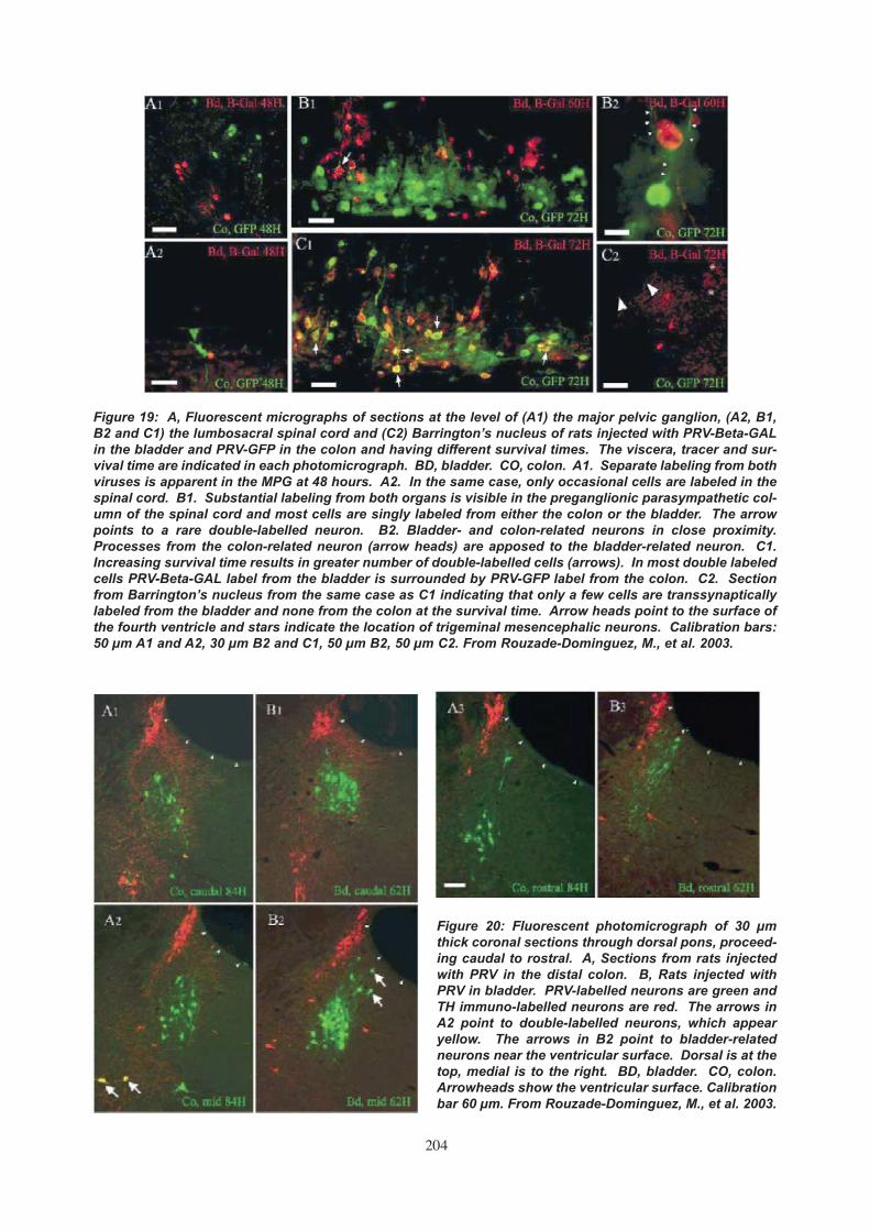
204
Figure 19: A, Fluorescent micrographs of sections at the level of (A1) the major pelvic ganglion, (A2, B1,B2 and C1) the lumbosacral spinal cord and (C2) Barrington’s nucleus of rats injected with PRV-Beta-GALin the bladder and PRV-GFP in the colon and having different survival times. The viscera, tracer and sur-vival time are indicated in each photomicrograph. BD, bladder. CO, colon. A1. Separate labeling from bothviruses is apparent in the MPG at 48 hours. A2. In the same case, only occasional cells are labeled in thespinal cord. B1. Substantial labeling from both organs is visible in the preganglionic parasympathetic col-umn of the spinal cord and most cells are singly labeled from either the colon or the bladder. The arrowpoints to a rare double-labelled neuron. B2. Bladder- and colon-related neurons in close proximity.Processes from the colon-related neuron (arrow heads) are apposed to the bladder-related neuron. C1.Increasing survival time results in greater number of double-labelled cells (arrows). In most double labeledcells PRV-Beta-GAL label from the bladder is surrounded by PRV-GFP label from the colon. C2. Sectionfrom Barrington’s nucleus from the same case as C1 indicating that only a few cells are transsynapticallylabeled from the bladder and none from the colon at the survival time. Arrow heads point to the surface ofthe fourth ventricle and stars indicate the location of trigeminal mesencephalic neurons. Calibration bars:50 µm A1 and A2, 30 µm B2 and C1, 50 µm B2, 50 µm C2. From Rouzade-Dominguez, M., et al. 2003.
Figure 20: Fluorescent photomicrograph of 30 µmthick coronal sections through dorsal pons, proceed-ing caudal to rostral. A, Sections from rats injectedwith PRV in the distal colon. B, Rats injected withPRV in bladder. PRV-labelled neurons are green andTH immuno-labelled neurons are red. The arrows inA2 point to double-labelled neurons, which appearyellow. The arrows in B2 point to bladder-relatedneurons near the ventricular surface. Dorsal is at thetop, medial is to the right. BD, bladder. CO, colon.Arrowheads show the ventricular surface. Calibrationbar 60 µm. From Rouzade-Dominguez, M., et al. 2003.
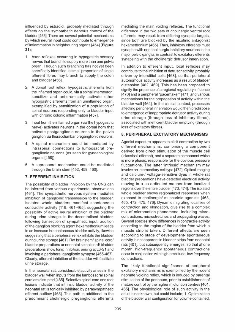
205
influenced by estradiol, probably mediated througheffects on the sympathetic nervous control of thebladder [455]. There are several potential mechanismsby which neural input could contribute to emergenceof inflammation in neighbouring organs [454] (Figure21);
1. Axon reflexes occurring in hypogastric sensorynerves that branch to supply more than one pelvicorgan. Though such branching has not yet beenspecifically identified, a small proportion of singleafferent fibres may branch to supply the colonand bladder [456].
2. A dorsal root reflex; hypogastric afferents fromthe inflamed organ could, via a spinal interneuron,sensitize and antidromically activate otherhypogastric afferents from an uninflamed organ,exemplified by sensitization of a population ofspinal neurons responding only to bladder inputwith chronic colonic inflammation [457].
3. Input from the inflamed organ (via the hypogastricnerve) activates neurons in the dorsal horn thatactivate postganglionic neurons in the pelvicganglion via thoracolumbar preganglionic neurons.
4. A spinal mechanism could be mediated byintraspinal connections to lumbosacral pre-ganglionic neurons (as seen for gynaecologicalorgans [458]).
5. A suprasacral mechanism could be mediatedthrough the brain stem [452, 459, 460].
7. EFFERENT INHIBITION
The possibility of bladder inhibition by the CNS canbe inferred from various experimental observations[461]. The sympathetic nervous system mediatesinhibition of ganglionic transmission to the bladder.Isolated whole bladders manifest spontaneouscontractile activity [118, 461-465], suggesting thepossibility of active neural inhibition of the bladderduring urine storage. In the decentralised bladder,following transection of sympathetic input, additionof the ganglion blocking agent hexamethonium leadsto an increase in spontaneous bladder activity, likewisesuggesting that a peripheral reflex inhibits the bladderduring urine storage [461]. Rat brainstem/ spinal cord/bladder preparations or neonatal spinal cord/ bladderpreparations show tonic inhibition, arising at L6-S1 andinvolving a peripheral ganglionic synapse [465-467].Clearly, efferent inhibition of the bladder will facilitateurine storage.
In the neonatal rat, considerable activity arises in thebladder wall when inputs from the lumbosacral spinalcord are disrupted [465]. Selective spinal cord and rootlesions indicate that intrinsic bladder activity of theneonatal rat is tonically inhibited by parasympatheticefferent outflow [465]. This path is additional to thepredominant cholinergic preganglionic efferents
mediating the main voiding reflexes. The functionaldifference in the two sets of cholinergic ventral rootefferents may result from differing synaptic targets,since both are blocked by the nicotinic antagonisthexamethonium [465]. Thus, inhibitory efferents mustsynapse with noncholinergic inhibitory neurons in themajor pelvic ganglia, in contrast to excitatory efferentssynapsing with the cholinergic detrusor innervation.
In addition to efferent input, local reflexes maycontribute to the inhibition of detrusor activity, probablydriven by interstitial cells [468], so that peripheralautonomous activity increases as a result of bladderdistension [462, 469]. This has been proposed tosignify the presence of a regional regulatory influence[470] and a peripheral “pacemaker” [471] and variousmechanisms for the propagation of activity within thebladder wall [464]. In the clinical context, processesaffecting peripheral innervation would then predisposeto emergence of inappropriate detrusor activity duringurine storage (through loss of inhibitory fibres),associated with inefficient bladder emptying (throughloss of excitatory fibres).
8. PERIPHERAL EXCITATORY MECHANISMS
Agonist exposure appears to elicit contraction by twodifferent mechanisms, comprising a componentderived from direct stimulation of the muscle cell(‘classical’ efferent), and a separate component whichis more phasic, responsible for the obvious pressurefluctuations. The latter ‘intrinsic’ mechanism mayinvolve an intermediary cell type [472]. Optical imagingand calcium-/ voltage-sensitive dyes in whole ratbladder preparations have detected electrical activitymoving in a co-ordinated manner from localizedregions over the entire bladder [473, 474]. The isolatedwhole bladder shows regionalized responses whenexposed to cholinergic/ muscarinic agonists [463,465, 472, 475, 476]. Dynamic migrating localities ofcontraction and elongation, give rise to a complexmix of micromotion phenomena, including micro-contractions, microstretches and propagating waves.Several species show differences in contractile activityaccording to the region of the bladder from which amuscle strip is taken. Different effects are seenaccording to stage of development- spontaneousactivity is not apparent in bladder strips from neonatalrats [401], but subsequently emerges, so that at onemonth, high-frequency spontaneous contractionsoccur in conjunction with high-amplitude, low frequencycontractions.
The likely functional significance of peripheralexcitatory mechanisms is exemplified by the rodentneonate voiding reflex, which is induced by parentalstimulation of the perineum, prior to establishment ofmature control by the higher micturition centres [401,465]. The physiological role of such activity in theadult is not known, but could include; 1. Optimizationof the bladder wall configuration for volume contained,

206
Figure 21: Five compatible mechanisms by which hypogastric nerve fibres can contribute to the process ofinflammatory induction between organs. A.Branching sensory afferents. B. Dorsal root reflex. C. Multisynapticroute involving sensory afferents from the inflamed organ to the T-13/L1 segment of the cord followed byoutput from preganglionic fibres in the T13/L1 segment to postganglionic in the pelvic ganglion that innervatethe uninflamed organ. D. Multisynaptic route involving sensory afferents from the inflamed organ to the T13/L1segment of the cord followed by output from preganglionic fibres in the L6-S2 segments to postganglionic fibresin the pelvic ganglion that innervate the uninflamed organ. E. Multisynaptic route involving sensory afferentsfrom the inflamed organ to the T-13/L1 segment of the cord followed by output from preganglionic fibres in theL6-S2 segments to postganglionic fibres in the pelvic ganglion that innervate the uninflamed organ. In thiscase the multisynaptic route includes ascending connections from spinal cord to brain and descendingconnections from brain to L6-S2. From Winnard, K.P. et al. 2006.

207
to ensure efficient voiding regardless of volume [477],2. Stimulation of ‘in series’ receptors for signalingbladder volume [476], 3. A mechanistic component ofaccommodation during filling, a counterintuitivesuggestion supported by the observation thataccommodation in the colon involves synchronouscontraction and relaxation [478], 4. Servoassistanceof voiding for maintenance of contraction [479]. Thepotential importance of peripheral functional structuresmay explain the recognition that nerve growth factorcan contribute to overactive bladder problems [480].
With the direct role of both limbs of the autonomicnervous system in regulating filling and voiding of thebladder, the involvement of brainstem circuitry isparamount in normal bladder function. The brainstemis involved in reflexes controlling filling, storing andemptying of the bladder. Clinical cases of lesions withinspecific brainstem regions, most notably the pons,can result in either bladder continence or incontinenceproblems. This section will review the currentunderstanding of brainstem neuronal networks thatinfluence the parasympathetic and sympathetic motoroutflows destined for the detrusor and smooth muscleof the urethral sphincter.
1. AFFERENT PATHWAYS TO THE BRAINSTEM
Sensations of bladder fullness are conveyed to thespinal cord by the pelvic and hypogastric nerves, whileinput from the bladder neck and urethra is carried inthe pudendal and hypogastric nerves. Afferents arisingfrom the bladder and urethra are mechanoreceptive(Aδ fibres) and nociceptive (C fibres). The mostimportant afferents for initiating micturition are thosepassing in the pelvic nerves, whose fibres terminatein discrete regions of the lateral aspect of the dorsalhorn of the lumbar and sacral spinal cord (see [481,482, 483] for reviews). Many of these dorsal hornneurones make spinal connections that mediatesegmentally organised reflex responses. However, aproportion of the spinal interneurones send ascendingprojections to the brain. These afferents are involvedin transmitting sensory information to the cerebralcortex and can trigger micturition mediated by neuralcircuitry within the brainstem.
Ascending fibres from dorsal horn neurones receivingafferent input from the bladder terminate in the gracilenucleus and the central aspect of the periaquaductalgrey (PAG; Figure 22); the latter has been describedas a most dense spinal innervation [483, 484], perhapsemphasizing its essential role. The gracile nucleusrelays information regarding nociception to the thalamusand cortex. Those PAG neurones receiving directspinal inputs project to multiple sites including thepontine micturition centre (PMC) and the thalamus for
onward relay to the cerebral cortex [483]. Althoughthese data are based on animal species, the pivotalrole of the PAG and PMC has been confirmed in manusing positron emission tomography [485, 486] andfunctional magnetic imaging [487] with and without afull bladder (see Figure 23). Indeed, the PAGprojections to the PMC originate from its lateral aspects[488]. Since the spinal projections relaying bladderdistension target the central PAG there is the possibilityof some integration prior to onward transmission tothe PMC. This is likely to include integration ofdescending information from higher brain regions suchas the limbic system and cerebral cortex [483], whichprovide contextual information concerning theappropriateness for micturition. Recently, theparabrachial nucleus appears activated during bladdercontraction and may also receive afferent inputs directlyfrom the spinal cord or via other central structures ( Fig23).
2. DEFINING BRAINSTEM CIRCUITRYREGULATING BLADDER FUNCTION
It is evident from recent reviews [481, 482] that mostregions of the brainstem involved in controlling bladderfunction are known. However, as is evident in figure4 of de Groat’s (2006) review, it remains unclear howthese central nervous structure are connected up(see Figures 23 & 24). Are there, for example,brainstem pathways running in parallel and/or seriesthat drive the preganglionic motoneurones? A methodof revealing key brainstem regions regulating endorgan function that has the ability to address this istransneuronal retrograde labelling using pseudorabiesvirus (PRV). Both the bladder wall and urethra of therat have been injected with PRV. This has resulted insimilar regions of the brainstem being labelled, whichsupports the close coordination of detrusor and urethralmuscle function as suggested previously [481]. Thebrainstem regions labelled included Barrington’snucleus (the pontine micturition centre), midline raphe(magnus and obscurus), locus coeruleus (A6),subcoeruleus, A5, reticularis gigantocellularis andnucleus paragigantocellularis [489, 490] but also inneonatal rats only the prepositus hypoglossal nucleusand lateral vestibular nucleus [489]. In addition, thePAG was labelled (Figure 22). Recent work has alsoindicated a role for the medial parabrachial nucleus(Figure 22). Whilst this provides informationconcerning the major players within the brainstem,the PRV technique does not imply precise informationabout connectivity. For example, are all thesebrainstem regions connected in parallel to the spinalinterneurones and / or pre-ganglionic neurones orare some positioned in series? Are reciprocalconnections between these nuclei present? Much ofthis information remains unknown but seems importantif one is to understand the way in which the braincontrols bladder storage and emptying and how oneobtains reversible switching between these essential
V. MIDBRAIN-BRAINSTEM CONTROLOF BLADDER FUNCTION

208
Figure 22: Schematic representation of the described sensory and motor pathways regulating bladder functionin animals. Of note are roles for gracile nuclei in transmitting afferent information from the spinal cord tothe thalamus and cortex; the pontine storage centre activated during bladder filling, the lateral parabrachialnucleus (as a sensory integration centre) and the rostral ventromedial medulla as a centre important duringvoiding. For further details and all references see text.
Figure 23: Consistent with animal studies, regionsof the human brain, imaged using positron emissiontomography including aspects of the midbrain(periacqaductal gray, the dorsal mesencephalicreticular formation) and brainstem (dorsolateralpontine tegmentum-pontine micturition center anddorsolateral pontine reticular formation-pontinestorage centre) are evident when the bladder is filled.(From Kavis et al., 2005).

209
functions. Most anatomical, electrophysiological andimaging studies to date have concentrated on thePMC as this appears pivotal regarding bladder functionin both animals and man.
3. THE PONTINE MICTURITION CENTER (PMC)
In the rat, the PMC is just dorsomedial to A6 [293, 491]whereas in the cat it appears to be within A6 extendingventromedial into the mesencephalic tract of thetrigeminal nerve [266]. It is these regions that aretermed Barrington’s nucleus- a region named afterBarrington, who in 1925 was the first to describe thispontine control centre for micturition in the cat (Figure22). In man, comparable regions in the pons can beimaged and found activated when the bladder is full(see Figure 23; [486]).
a) Barrington’s nucleus: is it the master ofceremonies for bladder emptying
Based on the finding that its activation (electrical orchemical stimulation) in rats and cats relaxes urethralsphincters and initiates bladder contractions [293,492, 493], it seems reasonable to propose a dominantrole for the PMC in micturition. Moreover, lesionslocalised to this pontine region in animals abolishmicturition and cause urine retention [293]. Similarly,a PMC lesion (on the right hand side) resulted inurinary retention in man [494]. Thus, the PMC wouldappear to play a major point of convergence ofnumerous pro- and anti micturition drivers. Thus, it maybe a major integrating centre and, indeed, possible‘command’ centre for initiating and orchestrating theact of bladder emptying.
b) How is the PMC connected up?
The inputs to, and outputs from, the PMC areconsidered below:
1. INPUTS: As described above, the lateral aspects ofthe PAG send information regarding the degree ofbladder distension. These inputs are powerful andcan trigger micturition as evidenced by the findingthat chemical stimulation of ventrolateral PAG cancause voiding [488]. Recently, Kuipers et al. [495]have examined the afferent inputs to the PMC usingconventional anterograde and retrograde tracingtechniques. In addition to the PAG (ventrolateral anddorsomedial aspects) inputs arose from: ventromedialpontomedullary tegmental field (Figure 22); medialpreoptic area, posterior hypothalamus (perifornicalregion). It is unclear as to the exact function of thepontomedullary tegmental field, although Holstegebelieves this is a diffuse input providing a ‘generallevel-setting’ of PMC neuronal activity [496]. Thismight be interpreted as a potential mechanism forestablishing a neuronal set-point or threshold beyondwhich micturition occurs. The medial pre-optic regionmay well provide inhibition and this may be importantduring sleep and/or sexual activity to suppress
micturition [266, 483, 497], although the previous workby Gjone demonstrated that micturition could beevoked following activation of this region [498], whichis inconsistent with an exclusive inhibitory role. Theposterior hypothalamic region is involved in fight orflight (defense response). Its connection to the PAGmay account for the urination reported in consciousanimals during a defense response, since regions ofthe PAG can also evoke the defense response [499]and, via PMC, could also initiate urination [500].
The study by Kuipers et al [495] in the cat failed toconfirm the numerous central nervous afferent inputsto the PMC described by Valentino et al. in the rat [501];these included the Kolliker Fuse nucleus, raphe,nucleus tractus solitarius, parabrachial nucleus,nucleus paragigantocellularis and the cuneiformnucleus. Whether a species difference can explainthis seems unlikely. Certainly the regions labelled byValentino et al. [501] map more closely to data obtainedusing PRV injected into the bladder wall or urethra (see[489, 490, 502]) as described above. With this said,it is likely that many of the nuclei that project to thePMC also connect to each other but this needsconfirming using localised injections of neuronalretrograde and anterograde tracers.
2. OUTPUTS: During micturition the PMC exertssimultaneous excitatory influences on parasympatheticoutflows that cause bladder contraction and completeemptying but inhibition of sympathetic preganglionicsto relax the urethral sphincter. It is, therefore, entirelyappropriate that within the PMC there are bulbospinalneurones (Figure 24): PMC sends direct projectionsto parasympathetic preganglionic motoneuronesinnervating the bladder [503]. The latter authorsprovided ultrastructural evidence of asymmetric(excitatory) synaptic contacts on sacral para-sympathetic preganglionic neurones originating fromthe PMC. Moreover, single neurones have beenrecorded extracellularly from Barrington’s nucleus incats (i.e. medial to the mesencephalic tract of thetrigeminal nerve) and found to have spinally projectingaxons based on antidromic invasion testing from thedorsolateral funiculus of the first sacral segment [504-506]. An involvement of glutamate as the mainneurotransmitter in this pathway has been described[507]. Such a pathway would explain bladdercontraction. Urethral relaxation requires an inhibitionof somatic motoneurones and this appears to bemediated from PMC projections onto inhibitoryinterneurones located in the intermediolateral cellcolumn at the sacral segmental level [278]. Bothglycine and gamma amino butyric acid are thought toplay a role here [279, 508].
c) On-switching micturition: a role for the PMC
With the PMC being essentially a pre-motor(parasympathetic/sympathetic) micturition nucleus, itshould fulfil certain criteria if it is involved in the initiation

210
of bladder contraction and sphincter relaxation. Forexample, neurones should project to the spinal cord,fire ahead of a bladder contraction and fire continuouslywith each bladder contraction. Previous studies havefound pontine units that fired in phase with bladdercontraction [509, 510], but whether these connectedto the spinal cord directly was not tested. However,subsequent single unit recordings by Sasaki [504,505] and Sugaya et al. [506] provide convincingevidence that many PMC neurones do project to thespinal cord. Studies have shown that feline spinallyprojecting neurones (as demonstrated using theantridromic collision test) located in Barrington’snucleus discharged in phase with bladder contractions(Figure 25). Many had firing patterns not different tothose recorded from bladder afferents. But this is notalways the case. Indeed, some of these units fired priorto the onset of bladder contraction consistent with arole in initiating bladder contractions (Figure 25).Whilst the proof of causality is always a difficult one,these neurones do make excellent candidates fordriving/initiating bladder contraction. Indeed, manywere also shown to be synaptically activated (ortho-dromically) following electrical stimulation of the spinalcord [506] raising the possibility of bladder afferentinput, which may assist in providing positive-feedbackand additional excitatory drive to both sustain andpossibly amplify firing during micturition. The latter ofcourse should be taken cautiously as it is not clearwhether the spinal input demonstrated originated frombladder mechanoceptors. Physiological stimulationof the bladder mechanoreceptors themselves, ratherthan electrical stimulation of the spinal descendingafferents, would be necessary to confirm this.
The issue of whether the PMC is the only pre-motormicturition initiating region is addressed by Sugaya etal. [506]. From their recordings, units localised to therostral medial medulla (reticularis gigantocellularisand magnocellularis) could also be activated
antidromically from the spinal cord (L1) and also firedin or out of phase with bladder contractions (Figures22, 23 & 25). These units included those that fired justbefore and/or with bladder contractions as well asfiring in between bladder contractions. Interestingly,many of these rostral medial medullary unitsresponded at short latency (2 to 3 ms) followingstimulation of the PMC, supporting direct orthodromicactivation. Thus, Sugaya et al. [506] proposed twodescending pathways from the PMC for initiatingmicturition: one direct to the parasympatheticpreganglionic neurones and the other via the medialreticular formation. Incidentally, the study also showedthat these medullary units could be orthodromicallyactivated from L1 indicating the potential for bladdermechanosensitive afferent drive to these neurones,although no direct proof for this was given that thisspinal input originated from the bladder wall.
A question arises as to whether the spinally projectingPMC and rostral medial medullary neurones areactually defined pre-parasympathetic versus pre-sympathetic neurones? Said differently, does a singleunit project to both autonomic preganglionic neuronaltypes in the spinal cord (see [508, 511]). This relatesto the current understanding that during micturition thebladder contracts (parasympathetic activation) butrelaxes the urethral sphincter (somatic inhibition). Nodata exist for this presently. However, in the nucleustractus solitarius located in the dorsomedial medulla,a similar issue exists. Here, neurones receivemechanoreceptor sensory inputs from arterialbaroreceptors activated by increases in bloodpressure, which when activated mediate simultaneousexcitation of cardiac parasympathetic motor outflowbut inhibition of vasomotor sympathetic activity, therebyreducing cardiac output and peripheral resistancerespectively that together lower arterial pressure tonormal levels. Simms et al. [512] recently reported thatthe cardiac parasympathetic limb was activated at a
Figure 24: Schematic depicting neuronaltypes (micturition or bladder contraction)versus inverse or bladder filling within themajor midbrain and ponto-meduallaryregions involved in central neural regulationof bladder function. A major future aimmust be to understand both the intra-andinter- nuclear connectivity of neuronsinvolved in bladder contraction and filling.Who drives who and what terminates firingof neurones during bladder filling andcontraction. Additionally, how are thevolitional drives integrated with theautomatic control circuitry of the lowerbrain. Abbreviations: P, parasympatheticneuron; PAG, periacqueductal grey; PMC,pontine micturition centre; PSC, pontinestorage (continence) centre; RVMM, rostralventromedial medulla; S, sympatheticneuron.

211
higher pressure threshold compared to thesympathoinhibition. It was surmised that outputneurones within NTS are dedicated and alreadydestined for either the parasympathetic or sympatheticnetworks [512] Thus, a similar organization may existwithin the PMC and could be teased apart bydifferences in the bladder pressures thresholdnecessary for bladder contraction versus urethralrelaxation. Moreover, each autonomic limb of thebaroreceptor reflex could be modulated differentially(e.g. [513-515].
4. BLADDER ‘FILLING’ NEURONES IN THE PMCAND MEDIAL RETICULAR FORMATION:WHAT’S THEIR ROLE?
Within both the vicinity of the PMC and medial reticularformation, neurones were encountered that fired duringthe interval between bladder contractions but weresilent during contractions. These were termed type 2neurones by Sugaya et al. [506] and ‘inverse neurones’by Sasaki et al. [516]. Many of these neuronal typeshad spinal projections (e.g. 26/35 type 2 neurones ofSugaya et al. [506]) but some could not beantidromically back fired. Although the latter is notdefinitive proof that these cells do not project spinally,the data are suggestive that they form a group ofinterneurones.
So, what is the function of these spinally and non-spinally projecting inverse neurones? Consideringthose with spinal projections, these cells might provideexcitatory drive to sympathetic pre-ganglionicsdestined for the detrusor muscle to bring about activerelaxation, via β2/3-adrenoceptor stimulation. In regardto the non-spinally projecting neurones that fire out ofphase with bladder contractions, these may beinvolved in ensuring that the neurones which areactive during bladder contractions that project spinallyto the parasympathetic preganglionic neurones remainsilent during filling via local inhibitory connections. Ifso, one might predict that they contain GABA or glycineas an inhibitory neurotransmitter.
Future studies could consider difference in neuronalphenotype spinally and non-spinally projectingneurones by juxtacellular labelling and post-hocimmunocytochemistry or in situ hybridisation to identifytheir neurochemical content (glutamatergic versusGABAergic, for example) as elegantly performed byothers in the cardio respiratory regions of the medulla[517, 518]. Equally, the non-spinally projecting PMCneurones that fired in phase with bladder contractionscould provide a source of synaptic inhibition to preventneurones involved in bladder relaxation from firingprematurely.
Figure 25: A montage of neuronal activities during bladder contraction and filling located in major regulatorybrainstem and midbrain regions. Note that not all neurons showed firing to a single phase; phase-spanning (i.e.from bladder contraction to relaxation) exists. A challenge for future studies is to confirm these neuronal activitiesas causal to bladder control (and not reactive to simultaneously occurring responses such as those in arterial pressure,breathing, heart rate that accompany micturition). Data compiled from Liu et al (2004), Sakakibara et al (2002), Sasaki(2004; 2005) and Sugaya et al (2003).

212
5. OFF-SWITCHING MICTURITION – THE PONTINE CONTINENCE CENTRE (PCC)
As Sasaki (2004) points out [505], since the majorityof units recorded from Barrington’s nucleus (60/76)continued to fire after the onset of bladder relaxationand that almost all inverse neurones do not start tofire until after relaxation is well underway (Fig 25),there must be a separate region for ‘off-switching’bladder contractions and for relaxing the detrusormuscle. Simultaneously the urethral sphincter shouldconstrict to allow filling and prevent incontinence. Forthese functions, the pontine continence centre or PCCis considered. This region was also termed the Lregion being distinct from the M region designated tobe the PMC (see [519]).
Where is the PCC, what types of neurones existand what are their connections?
The PCC is located in the dorsolateral pontinetegmentum (Figure 22). Neuroanatomical evidenceindicates that PCC neurones do not project to spinalregions influencing detrusor muscle. Rather,projections appear to be limited to Onuf’s nucleus inthe sacral cord. This nucleus contains the urethralsphincter motoneurones, suggesting that PCC drivestheir contraction. Perhaps some of the PCC neuronestherefore have a defined pre-motor autonomic functionas discussed above in the context of the arterialbaroreceptor reflex. However, activation of PCC canstop micturition contractions [520]. Importantly, 78%of PCC neurones fire during the relaxation phase ofbladder contractions (Figure 25) and the onset oftheir firing can be prior to the initiation of bladderrelaxation [521].
Indeed, this would make sense if their prime functionis to close the urethral sphincter. Another potentialrole is in off-switching micturition. One argumentagainst this is that there appear to be no connectionsbetween the PCC and PMC, although a small numberof PCC units could be either ortho- or anti- dromicallyactivated from the PMC using electrical stimulation[506], which does not necessarily indicate the originof the input or axon terminal. Others believe that thePMC and PCC act independently [520].
However, evidence supporting a role in off-switchingmicturition is that lesions within PCC causeincontinence, excessive detrusor activity, an inabilityto store urine and relaxation of the urethral sphincter.Further, activation of the PCC stops micturition, excitesthe pelvic floor musculature and contracts the urethralsphincter [278, 520]. Whether the PCC is activelyinvolved in off-switching micturition to allow filling andstorage and how it does this are questions requiringfuture experimental consideration. An area that mayplay a role in regulating micturition is the periaqueductalgrey or PAG.
6. THE PERIAQUEDUCTAL GREY (PAG): ISTHIS AN ESSENTIAL REGION FORSUPPRESSING THE MICTURITIONREFLEX?
The PAG is often referred to as a relay betweenascending bladder afferent information and PMCactivation of sacral parasympathetic preganglionicmotoneurones. The present evidence suggests thatthe PAG is not just a simple ‘relay’. As mentionedabove, afferents of sacral origin terminate in centralregions of the PAG but that onward transmission tothe PMC originates from more lateral aspects [484,488, 490, 502]. Therefore, some form of ‘neuralintegration’ within the PAG is very likely. Chemicalstimulation of the ventrolateral PAG can triggermicturition together with defensive behaviours (see[522]) but equally inhibition of micturition can beevoked by stimulation of rostral dorsal and caudalventral PAG regions as well as areas ventral to thePAG (e.g. dorsal mesencephalic reticular formation andnucleus reticularis pontis oralis; [523]). Lesions withinthe PAG abolish the micturition reflex in the pre-collicular decerebrate cat [524] but studies in humanalso report bladder overactivity [525]. Matsuura,Downie et al. (2000) have shown that micturitionevoked by chemical stimulation of the ventrolateralPAG is mediated by Barrington’s nucleus in theurethane anaesthetized rat. Moreover, these sameauthors were able to obtain coordinated sphincteractivation from the PAG (with bladder contraction) aswell as sphincter activation without bladder contractionat different sites within the PAG. This suggests the PAGis involved in both voiding and storage functions. Thisproposal is consistent with single unit recording fromthe PAG: Liu et al. [523] recorded from 84 micturition-related PAG neurones of which the majority werestorage neurones (i.e. fired during bladder filling; 58%;Figure 25). Further, PET imaging revealed PAGregions becoming active during urinary retention inman [485]. However, close examination of therecording sites in Liu et al. study [523] reveals that themajority of neurones (68 of the 84) were locatedventral to the lateral/ventrolateral PAG (in which 16were recorded) in the dorsal mesencephalic reticularformation and nucleus reticularis pontis oralis.Nevertheless, based on the prominence of storageneurones in this region the idea that it plays a role inboth urinary filling and suppression of the micturitionreflex was advanced by Liu et al. [523]. They alsoproposed that micturition neurones may reciprocallydepress the storage neurones. If true, this makes thispart of the midbrain important in terms of providing acommand locus for on- and off- switching micturition.In support of this was the observation of diverse firingpatterns of the neurones including augmenting anddecrementing [523]. Such firing patterns have beenascribed roles as ‘phase-switching neurones’ in therespiratory network [526], and could well play a role

213
in the transition between storage and voiding. Someof the midbrain cells recorded by Liu et al. [523] alsofired across voiding/filling or filling/voiding transitions(Figure 25); this again supports the possible role inswitching again akin to the brainstem respiratorynetwork [526] between filling and emptying of thebladder. With the well established connections of thePAG that include orbital and pre-frontal cortex,amygdale, pre-optic region of the hypothalamus [500],it is proposed that the PAG may form a site for theintegration of information from both higher centres,such as cortex, and bladder distension afferent inputs.With this said, the PAG is a clear contender for decisionmaking about when to void or not, which it couldexecute via connections to PMC and PCC respectively.
7. NEUROTRANSMITTERS & MODULATORSWITHIN BRAINSTEM NETWORKSCONTROLLING BLADDER FUNCTION
There is not the scope in this review to provide athorough detailed coverage of all transmitters andmodulators affecting brainstem regulation of the urinarybladder. For recent reviews see: [481, 482]. Theselatter reviews highlight the importance of classicaltransmitters (glutamate, GABA and glycine) and non-NMDA, NMDA, GABAA and glycine receptors withinthe brainstem (and PMC) for regulating bladdercapacity and micturition (see also: [297, 492]).Important observations include that both NMDA andnon-NMDA receptors within the brainstem areimportant for micturition [527] and that the PMC isunder tonic inhibitory tone mediated by GABAAreceptors [492]. This prompts the question as to whereand how this inhibitory tone is generated. Possibilitiescould be the PAG or higher centres, such as pre-optichypothalamic structures, for example. Below is a briefaccount of two important and clinically relevanttransmitter systems affecting bladder function viaeffects within the brainstem.
a) Serotonin
Many transmitter systems have been investigated bydrug applications into the brain ventricles. Whilst thisdetermines whether or not a modulating system hasan effect on micturition it is difficult to assess wherethis might be exerted. In their review [528], Burgardet al. emphasise that serotonin appears to affectnervous control of bladder function at multiple levelsincluding sensory processing of bladder wall afferentswithin the dorsal horn of the spinal cord and at the levelof the spinal motoneurones. In all cases this appearedto be inhibitory on detrusor muscle activity butexcitatory on urethral sphincter. A model proposed, wasthat 5-HT1a receptors were located on the terminalsof sensory afferent fibres to depress neurotransmitterrelease. Similarly, Ito et al. [529] have described apredominance of an inhibitory effect evoked from themidline raphe system extending from the pons tomedulla on micturition in cats. Where this effect is
mediated (i.e. supra-brainstem, pons or spinal cord)is unclear. Moreover, 79 raphe neurones wererecorded in which their firing was related to bladderpressure with 66% related to storage [529]. Thesedata support a role of the raphe system in suppressingmicturition and facilitating external urethral sphincteractivity in cats, which is consistent with earlier studiesidentifying a central inhibitory role for 5-HT1a receptors[283]. In stark contrast, this does not seem to be thecase in the rat where 5-HT1a system facilitatesmicturition [530].
b) Dopamine
In Parkinson’s disease, where there is a reduction indopamine availability, the bladder is hyperactive (409).Yoshimura et al. [531] discovered that D1 and D5receptors acting within supraspinal sites are inhibitoryto micturition whereas other dopamine receptorsubtypes were facilitatory. This is an important findinggiven the major clinical and psychological problemsof incontinence.
• Looking towards the horizon
A number of pertinent questions regarding brainstemmechanisms regulating detrusor and urethral musclehave evolved. First, the issue of the neuronalmechanisms underpinning the switching from storageto voiding but also off-switching detrusor activity at theend of voiding remain unclear. Most brainstemneurones relating to bladder voiding/filling appear toshow firing that is either in or out of phase with bladdercontractions (as indexed by increases in bladderpressure). Whether this neuronal firing is a cause orconsequence of the bladder contractions is unclear.Therefore, future studies could address the issue ofthe origin of neuronal activity (whether coincident withbladder contraction or relaxation) in terms of thatgenerated centrally versus that which is dependent onsensory afferent mechanoreceptor feedback formidbrain and brainstem regions. Second, and a relatedissue, is following the volitional command to void,which lower brain structures execute this demand? Isthis in the brainstem or midbrain (PAG) or highercentres? Based on the evidence presented here, amost likely candidate appears to be the PAG/dorsalmesencephalic reticular formation. This structureappears to have the right neuronal connections interms of both ascending spinal afferent inputs andoutputs to pontine micturition centres. In addition, itreceives inputs from higher centres. Third, switchingbetween voiding-filling-voiding has to be coordinatedwith the corresponding control of the urethralsphincters. This means there has to be cross talkbetween detrusor and sphincter neural circuits. Whilstevidence was discussed for a spinal inhibitorymechanism within the sacral cord, no such crossconnectivity was found between PMC and PCCregions but there remains a possibility that some crosstalk may exist in the PAG/dorsal mesencephalicreticular formation. Fourth, is whether there are within

214
the PMC/PCC (or PAG) neurones that are dedicatedto the sympathetic or parasympathetic nervous systemthat exist as pre-motor autonomic neurones. It wasdiscussed above that such organisation exists forbrainstem control of the baroreceptor reflex inregulating arterial pressure. Finally, some technicalissues. Most studies inflate the bladder and work onspontaneous isovolumetric contractions. This is clearlynot physiological as the bladder does not void. Thiswill affect detrusor muscle mechanoreceptor afferentfeedback as well as afferent feed back from the urinarytract. The bladder volume/pressure always remainsabove the micturition threshold, again non-physio-logical. Many of the early experimental studies werebased on the cat. Typically this was decerebrateraising the issue of how this relates to the consciousintact animal where higher centres are intact andknown to regulate micturition. It is clear that morerecent studies have ventured to the rat and mostrecently man. One wonders how compatible theregions regulating bladder function is between speciesand whether the connectivity and neuronalmechanisms are preserved or different. A goodexample is the opposing effects of 5-HT1a receptorsin cats (inhibit micturition) versus rat (pro-micturition;for references see above). This is an important issuein terms of translating work from cat/rat to man. Whilstserotonin seems to be inhibitory to detrusor muscleactivity in cat this is not the case in rat. Thus, the ideathat modulation of the descending serotonergic systemas a potential treatment strategy for incontinence inhuman [528] will need further validation. However, itis notable that areas identified as key brainstem sitesfor micturition in animal models do relate to regionsidentified in man using fMRI [486]. Finally, a universallyagreed consensus needs to be defined as to how tocharacterize a neuron involved in central control of thebladder. Basing it entirely upon its firing response toinflation or contraction of the bladder could give falsepositive information. Since bladder afferents can affectother autonomic variables (breathing, blood pressure,heart rate) one has to be sure that the neuronalresponse obtained is not a consequence of alterationsin these other variables. A proposal for stricter criteriato enable a more accurate characterization of neuronsinvolved in urinary storage and voiding function, andsphincter control.
1. BACKGROUND
The aim of this section is to present a synthesis of thehuman literature and review what this tells us aboutbrain regions mediating specific aspects of humanbladder behaviour.
Prior to the advent of functional brain imagingtechniques, our knowledge of the cerebral andbrainstem areas involved in the control of micturitionwas based on a small number of carefully observedclinical cases: patients presenting with specific bladdersymptoms who were found by investigation to havelesions at particular brain sites. However this “lesion”literature is really quite sparse, presumably mainlybecause if bladder symptoms occurred as a result ofa cerebral lesion the clinical picture was usuallydominated by a variety of other neurological deficitsand furthermore such bladder symptoms that therewere would not have been of interest to the majorityof neurologists. Initially the lesion studies were basedon observations made in life correlated with postmortem or pathological specimens, but withincreasingly better means of imaging it was possibleto correlate symptoms with smaller, more discreteabnormalities.
From what was written, the importance of the frontalcortex and pons was clearly recognized and thereare a number of case histories of patients reported withlesions at either site. Subsequent functional brainimaging studies confirmed the clinically based findingsand then greatly extended our understanding of thecomplex cortical and subcortical networks involvedin sensing bladder filling and activating appropriatevoiding. In fact the few further cases of “corticalbladders” described since the emergence of functionalimaging findings have reported lesions at sites thatpreviously would probably not have been recognizedas being important in “lesion” evidence presentedhere and summarized in Table 4. In fact functionalimaging and lesion studies should be regarded ascomplementary because, functional brain imagingprovides information about regional activation of greymatter, whereas lesions may give information aboutdamage to white-matter connecting pathways. Thelesion literature also improves our understanding ofpatients’ complaints. Most of the functional imagingstudies to be reviewed were carried out using eitherpositron emission tomography (PET) or functionalmagnetic resonance imaging (fMRI). Both provideindirect measures of regional blood flow, assumed tobe related to local neuronal activity, but their temporalresolutions are quite different: several minutes forPET versus a few seconds for fMRI. For this reasonPET is good for measuring long-lasting states of asystem, while fMRI is better for following relativelyfast events. Experimental paradigms are thereforequite different for the 2 techniques.
2. ROLE AND IMPORTANCE OF CEREBRALCONTROL OF VOIDING
In order to understand the cerebral control of voidingit is helpful first to examine what would happen if therewere no such control. Provided brainstem and midbrainare intact, micturition is organized in 2 phases, storage
VI. CORTEX AND BRAINSTEMCONTROL OF BLADDER FUNCTION
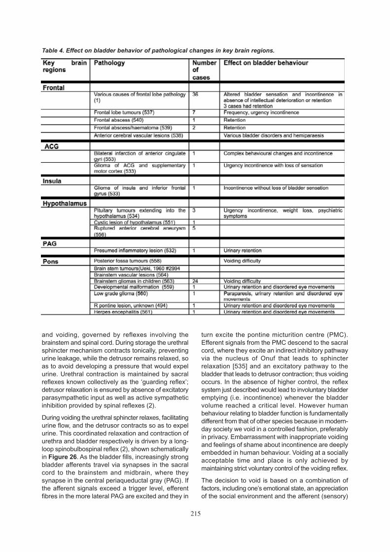
215
and voiding, governed by reflexes involving thebrainstem and spinal cord. During storage the urethralsphincter mechanism contracts tonically, preventingurine leakage, while the detrusor remains relaxed, soas to avoid developing a pressure that would expelurine. Urethral contraction is maintained by sacralreflexes known collectively as the ‘guarding reflex’;detrusor relaxation is ensured by absence of excitatoryparasympathetic input as well as active sympatheticinhibition provided by spinal reflexes (2).
During voiding the urethral sphincter relaxes, facilitatingurine flow, and the detrusor contracts so as to expelurine. This coordinated relaxation and contraction ofurethra and bladder respectively is driven by a long-loop spinobulbospinal reflex (2), shown schematicallyin Figure 26. As the bladder fills, increasingly strongbladder afferents travel via synapses in the sacralcord to the brainstem and midbrain, where theysynapse in the central periaqueductal gray (PAG). Ifthe afferent signals exceed a trigger level, efferentfibres in the more lateral PAG are excited and they in
turn excite the pontine micturition centre (PMC).Efferent signals from the PMC descend to the sacralcord, where they excite an indirect inhibitory pathwayvia the nucleus of Onuf that leads to sphincterrelaxation [535] and an excitatory pathway to thebladder that leads to detrusor contraction; thus voidingoccurs. In the absence of higher control, the reflexsystem just described would lead to involuntary bladderemptying (i.e. incontinence) whenever the bladdervolume reached a critical level. However humanbehaviour relating to bladder function is fundamentallydifferent from that of other species because in modern-day society we void in a controlled fashion, preferablyin privacy. Embarrassment with inappropriate voidingand feelings of shame about incontinence are deeplyembedded in human behaviour. Voiding at a sociallyacceptable time and place is only achieved bymaintaining strict voluntary control of the voiding reflex.
The decision to void is based on a combination offactors, including one’s emotional state, an appreciationof the social environment and the afferent (sensory)
Table 4. Effect on bladder behavior of pathological changes in key brain regions.

216
signals arising from the bladder. Knowledge of theextent to which one’s bladder content is comfortableand ‘safe’ is central in this process. Thus, voluntarycontrol of the bladder and urethra has two importantaspects, namely registration of bladder fillingsensations and manipulation of the firing of the voidingreflex. The PAG – the most rostral part of the reflexpathway shown in Figure 26 – appears to have apivotal role in both aspects. On the one hand it receives
bladder afferents [535] and transmits them to higherbrain centres and into the realm of conscioussensation. On the other hand it receives projectionsfrom many higher centres and controls the primaryinput to the PMC [536]. During bladder filling, suchhigher brain centres can suppress any excitatorysignal from the PAG to the PMC and prevent voidingor incontinence. Then, when voiding is consciouslydesired, they can allow the PAG, and thus the PMC,to be excited.
Thus, during the storage phase, the net effect of higher(voluntary) control is tonic suppression of the voidingreflex. This suppression can be interrupted to allowvoiding to occur if appropriate: i.e. if it is necessary(bladder volume is adequate), socially acceptable,and judged to be safe.
3. CORTICAL AND SUBCORTICAL CENTRESINVOLVED IN BLADDER CONTROL.EVIDENCE FROM OBSERVATIONS OFLESIONS AND FROM FUNCTIONAL BRAINIMAGING IN HUMANS
a) Frontal Lobes
Although Andrew and Nathan (1) were not the first todescribe disturbances of micturition resulting from avariety of causes of frontal lobe pathology, theircelebrated paper reporting the syndrome of frequency,urgency (and in some patients faecal) incontinencecausing distress to the patient, is regarded as seminalin the field. Their description of these patients cannotbe improved upon.
“The patients described here were not demented,indifferent or lacking in social awareness; they weremuch upset and embarrassed by these symptoms. Theacts of micturition and defecation occur in a normalmanner; what is disturbed by this frontal lesion is thehigher control of these acts. The lesion causesfrequency and extreme urgency of micturition whenthe patient is awake, incontinence when asleep. Thesensation of gradual awareness of increasing fullnessof the bladder and the sensation that micturition isimminent, are impaired. When the syndrome is lesspronounced, the sensation underlying the desire tomicturate is absent, whereas the sensation thatmicturition is imminent still occurs. Then the patientis waylaid by a sudden awareness that he is about topass urine; when neither sensation is experienced, thepatient is amazed to find that he has passed urine. Thethreshold of the micturition reflex is much lowered. Inthe most complete form of the syndrome, the patientcannot inhibit the detrusor contraction of the micturitionreflex; he is thus forced to empty his bladder as soonas the reflex occurs. When the syndrome is lesspronounced, the patient can make a conscious effortto stop the act of micturition, and he may or he maynot succeed. The lowering of the micturition reflexthreshold may account for the fact that the patient
Figure 26 : The voiding reflex. The lines on the right(green) show bladder afferent activity ascending tothe PAG during storage. Note there is no directafferent input into the PMC. On the left (blue), lines show activation of the PMCby the PAG, with descending activity to the sacralcord from where the parasympathetic innervationto the detrusor arises. An inhibitory synapse ontothe motor innervation of the sphincter (Onuf’snucleus) results in relaxation of the striatedsphincter muscle and thus synergistic detrusorsphincter activity. Note that higher brain regionscontrol the voiding reflex almost exclusively via theperiaqueductal grey (PAG), apart from a connectionvia the hypothalamus.

217
does not feel the normal gradual filling of the bladderthat underlies the desire to micturate; but it cannotaccount for the unawareness of the sensations arisingfrom the activity of pelvic and perineal muscles or ofthe sensation that urine is passing.”
The cases of leucotomy in Andrew and Nathan’sseries were regarded as most useful for localizingthe lesion causing the syndrome. The significant planeof the lesion was that lying immediately anterior to thetips of the ventricles and the genu of the corpuscallosum. Such lesions involved grey matter, inparticular the superomedial part of the frontal lobe(see Figure 27); but they caused a permanent disorderof the control of micturition and of defecation onlywhen they involved some of the white matter lateralto the anterior horns of the lateral ventricle. Lesionsthat did not affect this white matter caused onlytransient disordered control of micturition.Correspondingly, Andrew and Nathan’s Figure 10Asuggests that most of the critical lesion area was inthis white matter (Figure 27).
Subsequently the same features were observed in 7patients out of a series of 50 consecutive frontaltumours [537]. In an analysis of patients with acutehemispheric strokes the occurrence of disturbanceof micturition was found to be more common in frontalthan occipital lobe lesions and there was an associationwith hemiparesis [538]. In 3 cases, Andrew and Nathanobserved that lesions in similar areas to thosediscussed above led to urinary retention rather thanincontinence (Table 4). Three further cases of frontallesions with urinary retention have been reported.Successful treatment brought recovery of bladderfunction [539, 540].
Imaging studies, both PET [485, 541-544], and fMRI[545-548], are in agreement that, during bladder filling,storage and withholding of urine, there is activity in theright inferior frontal or dorsolateral prefrontal cortex,perhaps extending into the lateral part of the superiorfrontal cortex (Figure 28A,B). There is some right-sided predominance. In contrast there is little evidencefor activation of the medial parts of the frontal cortexduring storage. One PET study showed medial frontalactivity during sacral nerve stimulation [548], but thisis difficult to interpret; another study that showedmedial frontal activity during filling [549] employedSPECT imaging, which has poor spatial resolution. AnfMRI study showed abnormally weak activation inmedial prefrontal cortex in subjects with urgeincontinence [547], and further analysis revealed thatbladder filling tends to provoke deactivation in thisregion [550]. Regardless, there is little overlap with thesuperomedial frontal region described by Andrew andNathan (1), except in part of the anterior cingulategyrus (ACG; see Figure 29). These observations areconsistent with the concept that functional imagingreveals grey-matter activation or deactivation, whilelesions may damage critical links in white-matter
connecting pathways as well as grey-matter regions.
Neuroanatomical observations indicate that there isa strong and direct connection from medial prefrontalcortex to PAG [536]. It is tempting to postulate that thispathway is responsible for tonic PAG suppression ofthe voiding reflex, thus maintaining the storage phase.White-matter lesions in this pathway would then allowinvoluntary excitation of the voiding reflex, with resultingincontinence. It would appear from this account thatbilateral lesions would be required to causeincontinence, yet in practice a lesion on one sideseems to be sufficient. Andrew and Nathan’ssuggestion – that this is because a unilateral lesionis likely to involve the fibres connecting one side withthe other [551] – is difficult to follow.
Voiding has not been studied directly by fMRI, butthere are 3 PET studies of voiding [535, 542, 543]and one fMRI study of simulated voiding [552]. Allidentify activation of the right inferior frontal gyrus(dorsolateral prefrontal cortex) and in the medialprefrontal cortex or nearby ACG (see next section).
b) Anterior Cingulate Gyrus (ACG)
The anterior cingulate is an extensive area with partsserving varied functions. The cases of aneurysm inAndrew and Nathan’s series of frontal lobe pathologieswere thought to have involved the anterior end of thecingulate gyrus (1) (Figure 29, cyan circle).Incontinence was observed as part of a complexbehavioural disorder following bilateral infarction of theanterior cingulate gyri [553]. A recent report of a patientin whom a glioma in the right posterior ACG andsupplementary motor area was resected describedhow she experienced urgency incontinence and lossof bladder sensation following surgery [533].
Many functional imaging studies have observed ACGresponses – activation or occasionally deactivation –to bladder filling, storage or withholding [485, 541,542, 544, 547, 549]. The reported locations form a trailextending from dorsal to ventral ACG, suggesting thatdifferent parts of the ACG respond to the very variedexperimental paradigms that were used (Figure 29).There is a cluster of activations near the anterior areaidentified from lesion studies by Dr Nathan (personalcommunication), confirming that this part of the brainis indeed critical for control of micturition. Responseof the dorsal ACG to bladder filling is abnormallypronounced in patients with urge incontinence, evenin the absence of any detrusor contraction [547, 550],suggesting that it is involved both in recruitment ofaccessory pathways (to help avoid loss of control ofthe bladder) and in the sensation of urgency thataccompanies this situation. Imaging studies of voiding[541-543] and simulated voiding [552] have all revealedACG involvement.
c) Insula
Given what we now know from functional imaging

218
Figure 27 A, B and C : Location of white matter lesions causing persisting incontinence (red: based onAndrew and Nathan’s Figures 10 A, B and C (1)); and of grey matter lesions causing incontinence (cyan:personal communication to CJF from Dr Nathan).
Figure 28A : Locations of peaks of activation reported asright inferior frontal or lateral prefrontal cortex, duringbladder filling, storage and withholding of urine.Locations of peaks more than 20 mm from the midlineare projected onto right lateral surface of brain. Notesome overlap with insula shown in Figure 30.
Figure 28B : Medial prefrontal area activated in a fewstudies during withholding of urine. Locations of peakactivation less than 20 mm from the midline are project-ed onto the midline plane.
Figure 29: Cingulate gyrus areas activated on withhold-ing of urine or full bladder, projected onto midline plane(dots). The larger cyan circle is the projection of thesuperomedial grey matter area shown in Fig 27 onto themidline plane.

219
experiments about the importance of the insula inprocessing afferent activity from the bladder (seeFigure 30) it is remarkable that there have not beenmore reported cases of bladder dysfunction fromlesions at that site. Possibly the bilateral nature ofinsular connections, even though there is some right-sided predominance, protects against a drastic effectof insular lesions. One patient in whom a gliomaaffecting the inferior frontal gyrus and the insula wasexcised experienced incontinence without loss ofbladder sensation [533].
Imaging studies during storage or withholding of urineshow that regions reported as insula form a clusternear the expected location of that structure (Figure30) [541-544, 547, 549, 554]. There is some overlapwith the lateral frontal activations reported above andshown in Figure 28A. In the figure right and left sidesare projected on the same brain surface, maskingany right-sided predominance, and the insula is about20 mm deeper than the brain surface shown. In healthysubjects, insular activation becomes stronger withincreasing filling of the bladder, consistent with itspostulated role in bladder sensation [555]. In normalelderly, this insular response to bladder fillingdecreases with age, consistent with age-associatedloss of sensation. However, insula activity cannot byitself be responsible for conscious desire to void orurgency, because these sensations are lost followingextensive frontal lesions [551], suggesting that integrityof connecting pathways between insula and frontalcortex is essential for conscious sensation.
The insula was activated in only one [543] out of fourimaging studies of real or imagined voiding, suggestingthat it is not strongly involved in this phase ofmicturition.
d) Periaqueductal Grey (PAG)
A single case history describes a young manpresenting with urinary retention in whom the only
abnormality found was a small, presumed inflammatorylesion in the PAG [532]. Presumably in other casesthe clinical picture was dominated by other symptomsand deficits that were more striking to a neurologist.
Given the spatial resolution of PET and fMRI (a fewmm), it might be expected to be difficult to distinguishsmall but functionally different areas in the brainstemand midbrain. In fact however the brainstem/midbrainactivations reported during the storage and voidingphases seem to cluster in 3 distinct regions (Figure31), one of them being the PAG. PAG response tobladder filling is reported in 3 studies [485, 544, 547].This response may reflect increased afferent signalsarriving at the PAG (Figure 26) or increased inhibitoryactivity from the medial prefrontal cortex (MPFC),needed to prevent triggering of the voiding reflex (seeFigure 32). The PAG responded to imagined voidingin one fMRI study [552], but not to real voiding.
e) Hypothalamus
Lesions at this site as a cause of bladder symptomsare rare but 3 cases of pituitary tumours, extendingupwards into the hypothalamus have been describedwith urgency incontinence or retention, weight loss,psychiatric disturbance and bitemporal field restriction[534]. Other instances include gliomas involving thehypothalamus [551] or vascular disturbances of theanterior hypothalamus. Andrew and Nathan alsoreported five patients with bladder symptomsappearing after a ruptured cerebral aneurysm andspeculated that the site of lesion responsible was theanterior hypothalamus [556].
Animal observations suggest that the anterior andcaudal hypothalamus have monosynaptic connectionswith the PAG and PMC [536]. Correspondingly, twohuman brain imaging reports suggest response tobladder filling in a region near the caudal hypothalamus[485, 555], and one near the preoptic area [547]. It has
Figure 30: Locations of activation peaks reportedas insula, during bladder filling, storage and with-holding of urine, projected onto right lateral surfaceof brain. Note overlap with areas shown in Fig 28A.
Figure 31: Brainstem areas activated during stor-age or voiding, projected onto the midline plane.

220
been suggested that these connections allow thehypothalamus to inhibit voiding unless the situation isjudged to be safe.
f) Pons
The demonstration by Barrington [557] in the cat thata centre existed at the level of the pons necessary foractivation of micturition, provided the background forrecognizing a comparable centre in humans and theearly report of the association of difficulty withmicturition with posterior fossa tumours [558]. Laterhistories of individual cases of discrete pontine lesions[559, 560, 561], [494] and reports of difficulties withmicturition or retention as a feature of brainstemgliomas in children [562, 563] or vascular lesions[564], confirmed the likely existence of a comparablecentre in humans. Studies using MRI to visualize theprecise location of the responsible lesions, sited thisin the dorsolateral pons, including the pontine reticularnucleus and the reticular formation, adjacent to themedial parabrachial nucleus and locus coeruleus[564]. Lesions in this location are frequently associatedwith disturbances of consciousness and respiration andbladder symptoms may therefore be overlooked. Thecommonest clinical association of urinary retentionarising from a pontine lesion is an internuclearopthalmoplegia.or disorder of eye movements.
Functional imaging experiments have shown a clusterof activations near the postulated location of thispontine micturition centre (PMC), Figure 31. There are3 reports of PMC activation during voiding [541-543]and – surprisingly – one in response to bladder filling[555]. However, excitation during bladder filling mightbe inhibitory rather than excitatory, a distinction thatcannot be made by functional imaging.
A few studies suggest activation of the postulatedpontine L-region or continence centre (Figure 31),somewhat ventral, lateral and/or caudal to the PMC(Figure 26): during storage [485], during failed attemptto void [541, 542], during imagined voiding [554], andduring imaginary inhibition of voiding [552]. However,not all these studies recognized the L-region as such.
g) Other Regions
The regions discussed in sections 3.1 to 3.6 arereasonably well established since they are observedin lesion, imaging and (in some cases) animal studies.In particular, the triad insula/ACG/prefrontal cortex iswell known from studies of other aspects of brainfunction [565]. Other regions relevant to bladder controlhowever have been revealed only by functionalimaging. They include parts of parietal andfrontoparietal cortices, posterior cortex (precuneus,posterior cingulate cortex), putamen, caudate, partsof the limbic system (hippocampal complex,amygdala), and various parts of the cerebellum. Rarelyseen in functional imaging experiments is activity inthe basal ganglia, yet dopamine pathways are thought
to have a profound inhibitory effect on the PMC inhealth, which is lost in Parkinson’s Disease.
4. WORKING MODEL OF BRAIN/BLADDERCONTROL
The diagram in Figure 32 shows what are currentlythought to be the principal areas and connectionsinvolved in bladder control, with the more importantconnections identified by thicker lines. This schemeis hypothetical but offers, at this point in time, aframework based on evidence from the clinical reportsof the effects of “lesions”, the regions shown to beactivated in the PET imaging experiments (Figure33) and the results currently emerging from functionalMRI and connectivity studies.
The PMC is the final efferent brain nucleus involvedin bladder control – when activated there follows co-ordinated sphincter relaxation and detrusor contraction,resulting in voiding. However the PMC has been showto have only a few central connections (Figures 26and 32) namely, from the hypothalamus and from thePAG. The PAG is now recognized as having a centralrole in mediating the influences of higher function onthe PMC and so achieving voiding at brain- ratherthan bladder-determined volumes. To perform thisrole the PAG receives afferent input via the sacralroots conveying “bladder fullness” information, as wellmultiple inputs from higher centres including the
Figure 32 A: preliminary conceptual framework,based on current evidence, suggesting a scheme forthe connections between various forebrain andbrainstem structures that are involved in the controlof the bladder and the sphincter in humans. Arrowsshow probable directions of connectivity but do notpreclude connections in the opposite direction. PAG= periaqueductal grey; PMC = pontine micturitioncentre. Reproduced, with permission (2), based onthe work of (4).

221
prefrontal cortex, ACG, insula and hypothalamus andalso probably the basal ganglia and cerebellum. Theinsula (the right in particular) has been shown torespond in a way that suggests it is responsible formapping and processing bladder as well as othervisceral sensations and is sometimes referred to asthe “sensory cortex of the autonomic nervous system”.
The ACG monitors stress and conflict and generatesappropriate autonomic arousal. It may also determinehow much attention is paid to signals from the bladderand how one reacts to them. The medial prefrontalcortex (MPFC) has a role in response selection andso may be involved in evaluating afferent activity. TheMPFC is crucial for decision-making in an emotionaland social context and is thus evidentially importantin controlling micturition. It is close to the anteriorcingulate region revealed by lesion studies andfunctional imaging (Figure 29) and has strong anddirect connections with the PAG, suggesting that it maybe responsible for maintaining the tonic suppressionof the voiding reflex during the storage phase, whichnormally is only relaxed when the decision to voidhas been made. This decision is conveyed back to thePAG, which correspondingly excites or inhibits thePMC (Figure 34).
Figure 33 : Brain areas involved in the regulation ofurine storage. A meta-analysis of positron-emis-sion tomography and functional MRI studies thatinvestigated which brain areas are involved in theregulation of micturition reveals that the thalamus,the insula, the prefrontal cortex, the anterior cingu-late, the periaqueductal grey (PAG), the pons, themedulla and the supplementary motor area (SMA)are activated during the urinary storage.Reproduced with permission from (3)|
Figure 34: A preliminary working model of lower urinary tract control by higher brain centres. A: Duringstorage, ascending afferents (yellow) synapse on the midbrain periaqueductal grey (PAG); they are relayedvia the thalamus (TH) to the dorsal anterior cingulate gyrus (ACG: responsible for monitoring and warning)and to the right insula (RI: responsible for sensations such as desire to void) and lateral prefrontal cortex(LPFC); in the storage phase they pass to the medial prefrontal cortex (MPFC, red arrow) where the deci-sion to void or not is made; in this phase the decision is not to void, and this situation is maintained bychronic inhibition of the PAG via a long pathway (red arrows) from the MPFC; consequently the pontine mic-turition centre (PMC) is also suppressed, and voiding does not occur. B: When the decision to void is made,the MPFC relaxes its inhibition of the PAG (green arrow) and the hypothalamus (H) also provides a ‘safe’signal; consequently the PAG excites the PMC which in turn sends descending motor output (green arrow)to the sacral spinal cord that ultimately relaxes the urethral sphincter and contracts the detrusor, so thatvoiding occurs.
222
Dysfunction of neural control may underpin a widerange of clinical urinary tract problems. On the afferentside, neural dysfunction will alter reflex activity andinfluence sensation, which can either be enhanced,reduced or altered (e.g. with the emergence of paininstead of usual bladder filling sensations). On theefferent side, motor activity within the components ofthe lower urinary tract (bladder and outlet) can beincreased, reduced or uncoordinated. The followingis not a comprehensive description of the entire scopeof this complex arena, but focuses on key issuesrelevant in the clinical context.
1. ABNORMALITIES INVOLVING INFLAMMATION
• Bladder Pain Syndrome / Interstitial Cystitis (BPS/IC)
Although no consensus has been reached on thefundamental causes of Bladder pain syndrome/Interstitial Cystitis (BPS/IC), existing data suggest 3pathophysiological mechanisms: epithelial dysfunction,mast cell activation, and neurogenic inflammation[566].
1. MAST CELL ACTIVATION – Mast cells may be activatedby a number of mechanisms within the bladder wall.Increased permeability with influx of potassium ionsmay lead to sensory nerve up-regulation resulting inmast cell activation. Vasoactive, nociceptive and pro-inflammatory molecules released from mast cells canproduce neuronal sensitisation and secretion ofneurotransmitters that further stimulate mast cells.Mast cell – neuronal interactions may thereforeprovoke a vicious cycle in BPS/IC, contributing to thepainful symptoms of the disease [567].
2. NEUROGENIC INFLAMMATION – The close physicalrelationship between the C fibres and mast cells is ofparticular importance, as substance P (SP) releasedfrom the nerve fibres degranulates mast cells. Duringdegranulation, the mast cells release a multitude ofproinflammatory agents including nerve growth factor(NGF), tumour necrosis factor-α (TNFα), histamine,heparin, proteases, interleukins, serotonin and others.This induces an inflammatory response in the form ofvasodilatation (redness) and plasma proteinextravasation (oedema). Additionally, newly releasednerve growth factor (NGF) stimulates a further increasein the number of afferent fibres, thus increasing thepotential for further releases of SP and the transmissionof nociceptive impulses. Inflammatory models of cystitisresult in increased levels of TNF-alpha, SP and NGFproduction in the bladder, paralleling the hypothesizedneuro-inflammatory etiology of IC [568].
During mammalian development, NGF is required forthe survival and growth of several populations ofneurons. There is evidence to suggest that it alsoplays a role in the ongoing regulation of neural function,as well as inflammation and pain [569].
In the urinary tract, NGF is produced by the bladder,smooth muscle and urothelium, and levels of NGFincrease in response to interstitial cystitis. In onestudy, patients with BPS/IC were found to haveelevated levels of neurotrophic factors in their urine,including neurotrophin-3, nerve growth factor and glialcell line-derived neurotrophic factor [570]. This findingis consistent with the observation of increased NGFmRNA and protein in bladder biopsies from patientswith BPS/IC compared with controls [571].
Intravesical NGF is known to sensitize bladder afferentfibres [142], and sequestration of NGF can reduceinflammation associated with chemical cystitis in arat model [572]. Blockade of NGF using eitherendogenous antibody or antibody against the NGFreceptor, or a fusion protein that prevents interactionbetween NGF and its receptor, prevents neuralplasticity and bladder overactivity in experimentalmodels of these conditions [143, 573].
Other substances including neurotrophins,prostaglandins, and tachykinins may also contributeto altered afferent excitability [574]. The potentialrelevance of these changes has been demonstratedin cats diagnosed with feline interstitial cystitis (FIC)(which demonstrates nearly all the characteristics andsymptoms of human BPS/IC), in which capsaicinsensitive neurons are larger, with dorsal root gangliathat exhibit increased excitability and slowerdesensitisation to capsaicin, suggesting changes inthe properties of the primary afferent neurons [146,147], a finding that may be associated with tendencyof NGF to stimulate TRPV1 expression.
2. INVOLVING ABNORMAL URINE STORAGE
a) Overactive Bladder / Detrusor Overactivity
Detrusor overactivity can arise in neuropathicconditions, secondary to bladder outlet obstructionor idiopathically. Several observations on structural andfunctional properties of the bladder have been madein individuals with detrusor overactivity:
• Patchy denervation is present within the bladderwall, while sensory neurones and parasympatheticganglion cells are enlarged
• Exaggerated spontaneous myogenic activity canbe seen in isolated detrusor muscle strips, withincreased incidence of fused tetanic contractions
• Muscle strips also show altered responsiveness tonervous and pharmacological stimuli
VII. ABNORMAL LOWER URINARYTRACT FUNCTION

223
• Characteristic changes in smooth muscleultrastructure have been described.
Smooth muscle strips dissected from the bladder indetrusor overactivity often show altered responsesto nerve stimulation and to various agonists. Forexample, in obstructed overactive bladders there isreduced contractile response to intrinsic nervestimulation, along with supersensitivity to muscarinicagonists and potassium solutions [575-578]. Amongneuropathic conditions, spina bifida is associated withsupersensitivity to cholinergic agonists and potassiumsolutions, but there is no change in the sensitivity tointrinsic nerve stimulation [579]. In spinal cord injury,there is no reduction in sensitivity to electrical fieldstimulation, but the maximum force generated byeach milligram of bladder tissue is significantly reduced[92]. In idiopathic detrusor overactivity, bladder stripsshow supersensitivity to potassium, but not tomuscarinic agonists, and there is a reduced contractileresponse to intrinsic nerve stimulation [580]. Wherefunctional denervation is present, there appears tobe an increase in spontaneous contractile activity andpresence of fused tetanic contractions, a feature moretypical of well-coupled smooth muscles [581, 582]. Acommon ultra-structural feature of the overactivedetrusor is the emergence of protrusion junctions andultra-close abutments between the smooth musclecells [583]. Overall, the cells may be better coupledelectrically in detrusor overactivity, perhaps allowingspontaneous activity to propagate over a wider area.Bladder biopsies from overactive detrusor show apatchy denervation; some muscle bundles may becompletely denervated, whilst neighbouring onesappear normal and in other areas sparser innervationis also seen [92, 579, 580, 584]. A similar pattern isseen in animal models [576, 585]. Overall, theseobservations suggest that response to loss of localinnervation by the smooth muscle cell may explain thealtered behaviour of the bladder in detrusor overactivity[575, 586].
Preclinical studies in animal models of OAB / detrusoroveractivity contribute further to our understandingof the importance of neural control in this condition.The main models relevant to OAB include (i) instillationof irritative agents into the bladder during cystometry,(ii) partial bladder outflow obstruction, (iii) thespontaneously hypertensive rat, (iv) spinal cord injury,and other CNS lesions similar to those responsible forbladder dysfunction in humans [587].
Irritative agents are employed in animals to evoke apainful or irritant response, in particular through C-fibres. Both acetic acid and citric acid have been used,although the latter probably represents a better modelsince it is less irritant and therefore less likely to invokean acute inflammatory response. Both agents havebeen shown to increase bladder contractile activity,decrease bladder capacity and reduce bladdercompliance, while micturition pressure remains normal
or is increased, suggesting that this model maycontribute to understanding of increased bladdersensory activity, as may occur during the symptom ofurgency. These effects are due to stimulation ofnociceptive afferent fibres, confirmed throughdemonstration of increased c-fos expression in ratspinal cord and in regions of the periaqueductal grey[372, 588], an effect that can be eradicated bydesensitization of TRPV1 receptors on the sensoryneurones following pre-treatment with resiniferatoxin[589].
Partial bladder outflow obstruction has been achievedthrough application of urethral ligatures or a constrictingring that results in partial urethral occlusion, mimickingthe obstruction seen in men with BPH who oftendevelop secondary OAB [577]. The animals developcystometric features such as increased bladdercapacity and non-voiding bladder contractions, withassociated histological features (muscle hypertrophy,patchy denervation and enlarged sensory neuronesand parasympathetic ganglia) and detrusor functionalfeatures (spontaneous myogenic activity and alteredresponse to stimuli) reminiscent of the human condition[586]. Afferent plasticity in animals with bladder outflowobstruction involves NGF, the content of which isincreased in obstructed bladders prior to theenlargement of bladder neurons and the developmentof urinary frequency [590]. The relevance of NGF inthe response to obstruction in animals is suggestedby the finding that rats immunized with mouse NGFin order to develop autoantibodies do not developneural plasticity and urinary frequency in responseto obstruction [591]. While neuronal changes areevident in the obstructed model, further observationsappear to indicate that the overactive phenotype inobstructed rats derives from a combination ofincreased propensity to localized muscle contraction,together with a tendency towards wider propagationof this activity in an organ that may have developedgreater autonomy from central control as a result ofthe neuronal changes [592]. In this context, therelevance of peripheral contractile modules appear toemerge as the functional units of detrusor activity[477].
The spontaneously hypertensive rat (SHR) is a geneticmodel of hypertension, which is also known to exhibitabnormal bladder function; in particular, SHRs havebeen shown to have reduced bladder capacity andvoided volume, increased urinary frequency andincreased occurrence of non-voiding contractions,associated with altered detrusor innervation andphysiological response [450]. Again, increased bladdersmooth muscle NGF levels appear to be associatedwith these changes [593]. The bladders of SHRs alsoshow increased levels of calcitonin gene-relatedpeptide immunoreactive fibres (presumably afferent)with increased size of neuronal cross-sectional areaprofiles for bladder afferents in the L6–S1 dorsal root
224
ganglia as well as the major pelvic ganglia [594]. Thepotential importance of the afferent innervation in thismodel can be highlighted by the finding that intrathecalapplication of antisense oligonucleotide against thetetrodotoxin- resistant sodium channel (Nav1.8)reduces bladder hyperactivity [595].
Spinal cord injury (SCI) in animals has been shownto result in changes in lower urinary tract functionsimilar to those seen in humans [596]. Recovery ofbladder function from the initial areflexia after SCI inanimal models is dependent in part on plasticity ofbladder afferent pathways and the unmasking ofreflexes triggered by the normally silent capsaicin-sensitive C-fibre bladder afferent neurons, resultingin cystometric changes akin to those seen inneurogenic detrusor overactivity [596]. Studies in ratsindicate that the increased excitability in the C-fibresis associated with an increase in the expression ofsodium channels from a high-threshold TTX-resistanttype to a low-threshold TTX-sensitive type [597].
Other animal models of CNS lesions associated withbladder dysfunction have also been developed. Forexample, a model of Parkinsonism secondary to 6-hydroxydopamine injection into the substantia nigrapars compacta in rats has been developed, confirmingthe relevance of D1/D5 dopaminergic stimulation inimproving bladder capacity [531]. Furthermore, amodel of bladder overactivity associated with cerebralinfarction due to occlusion of the middle cerebralartery in rats has been shown to lead to significantischaemia within the putamen and cerebral cortex,confirming the importance of these areas in the controlof micturition [598]. In this latter model, a role forglutamatergic and dopaminergic stimulation in thedevelopment of bladder dysfunction has beendemonstrated [598, 599]. These findings are consistentwith the observation that anterior brain lesions inhumans are more likely to be associated withincontinence than posterior, occipital lesions [600].
Additional evidence for the importance of emergentC-fibre contribution to afferent signalling in OAB anddetrusor overactivity can be found in experimentalclinical studies. For example, patients with variousidiopathic and neurogenic bladder dysfunctions havereduced current perception threshold to direct bladderstimulation [601] and urethral stimulation [602] atfrequencies that selectively stimulate C-fibres,indicating increased excitability in the C-fibre afferentsin these patients. Similarly, ice water instillation (whichexcites C-fibres) into the bladder can trigger involuntarydetrusor contraction in patients with neurogenicdetrusor overactivity [603]. In addition, the efficacy ofcapsaicin and resiniferatoxin in the treatment ofpatients with both idiopathic and neurogenic detrusoroveractivity confirms the role of C-fibres and theafferent limb of the micturition reflex in bladder storageconditions [604]. While the hyper-excitable C-fibresappear to be the conduit for the abnormal sensory
signals from the overactive bladder, the origin of thesensations probably lies in the bladder wall itself,either in uncoordinated asynchronous localizeddetrusor contractile activity [476, 605] which may leadto urgency with negligible associated pressure risewithin the bladder, or in a more co-ordinated propa-gation of autonomous contractile activity via emergentgap junctions providing more syncytial characteristicsto the overactive detrusor [583].
b) Stress Urinary Incontinence
Stress urinary incontinence (SUI) is characterized byreduced outflow resistance during urinary storagedue to weakness in the urethral sphincter mechanism.It is often associated with weakness of the pelvic floorand urethral musculature, but peripheral nervedysfunction is also implicated, in particular pudendalnerve damage following childbirth in women [606].Animal models have contributed to understanding ofthe importance of the peripheral innervation of theurethra in SUI, with development of disease modelsassociation with pudendal nerve crush and vaginaldistension initiating neuropraxic nerve injury in rats[607] and mice [608]. The role of pharmacologicalneuromodulation in the treatment of SUI has shownthat urethral function and therefore incontinence canbe improved by augmenting somatic neuronaldischarge using the serotonergic (5HT) andnoradrenergic (NE) reuptake inhibitor duloxetine,which is thought to act in the sacral spinal cord atOnuf’s nucleus, the pudendal somatic motor nucleusof the spinal cord which is densely innervated by 5HTand NE terminals [307, 609]. More recent data on theeffect of the selective noradrenergic reuptake inhibitor,[S,S]-reboxetine in the treatment of SUI, indicate thatthe serotonergic activity of duloxetine may beredundant and that effects are dependent solely onnoradrenergic reuptake inhibition [610, 611].
3. INVOLVING ABNORMAL VOIDING
• Bladder Outflow Obstruction
Bladder outflow obstruction (BOO) is typicallyassociated with prostatic enlargement in men, althoughit is also seen in women following surgery for stressurinary incontinence and in children secondary toproximal urethral valves. Bladder overactivity is seenin many patients with BOO, and this storagedysfunction together with the associated neuronalchanges has been described above. The voidingdysfunction per se seen in patients with BOO is causedby a combination of both passive and dynamicobstruction of the proximal urethra and, in somepatients, by detrusor decompensation resulting in adeterioration in the contractile function of the bladderduring voiding.
During obstructed voiding, the bladder is subject to highpressures required to expel urine through a region ofhigh resistance. These pressures compromise detrusor
225
blood flow during voiding leading to periods ofischaemia and hypoxia, followed by reperfusion [612].Recurrent cycles of ischaemia and reperfusion resultin progressive neuronal damage in the bladder wall,an effect that has been seen in animal models ofobstruction and bladder distension, as well as in anin vitro model in which detrusor muscle is intermittentlyexposed to hypoxic glycopenic perfusate followed byoxygenated glucose-containing perfusate [384, 613].This neuronal damage initially has a patchyappearance, when it seems to be related to thedevelopment of an overactive phenotype in pigs [586].With time, however, bladder decompensation withincomplete voiding and chronic retention can develop,and this is associated with a more generalized detrusordenervation and reduced contractile response toneuronal stimulation in organ bath studies [614].
4. CO-MORBID DISORDERS
The significance of neural control mechanism in lowerurinary tract dysfunction is further implied by a numberof comorbid conditions that have been associatedwith BPS/IC and incontinence.
Patients with BPS/IC and SUI are more likely to reportdepressive symptoms. Findings on two validateddepression measures indicate that patients withinterstitial cystitis report significantly greater depressivesymptomatology than healthy controls, although lessthan 20% notice moderate or severe depressivesymptoms. This finding is consistent with theobservation that patients with chronic pain experiencea higher level of depressive symptoms than healthycontrols and other chronically ill populations. Patientsreport low mood (56%), fatigue (63%), difficultyconcentrating (49%), insomnia or excessive daytimesleepiness (49%) and are 3 to 4 times more likely toreport suicidal thoughts than the general population[615]. The association of IC/PBS [616] andincontinence [617] with depression has beencorroborated in other studies, although a more recentstudy suggests that, after multivariate adjustment forthe influence of other urogenital symptoms, only thesymptom of nocturia remains significantly associatedwith depression [618].
BPS/IC has also been associated with other chronicpain conditions, especially fibromyalgia [616, 619-621]. An association has also been shown for childabuse [616, 622]. Based on recent improvements inunderstanding of pain processing pathways in thecentral nervous system, and in particular the role oflimbic structures, especially the anterior cingulatecortex, hippocampus and amygdala, in chronic andaffective pain perception, a condition termed limbicassociated pelvic pain has been proposed to explainthe concurrence of these various chronic painconditions. This limbic dysfunction is manifest both asan increased sensitivity to nociceptive afferents frompelvic organs, and as an abnormal efferent innervation
of pelvic musculature, which undergoes toniccontraction as a result of limbic efferent stimulation,generating a further sensation of pain. The nociceptiveafferents from these pelvic organs then follow themedial pain pathway back to the sensitized,hypervigilant limbic system. Chronic stimulation ofthe limbic system by pelvic pain afferents againproduces an efferent contraction of the pelvic muscles,thus perpetuating the cycle [622].
The bidirectional interactions between different hindgutorgans (urinary bladder, rectum) show both similaritiesand differences. The primary function of both organsis to store waste products, and to empty them inaccordance with a) the state of filling as well as b) theoverall state of the organism, and environmental andsocial constraints. While the former is accomplishedby a series of hierarchal reflexes, the latter isaccomplished by modulatory input from the brain tochange the gain of these reflexes. While spinal andsupraspinal reflexes are involved in both the regulationof bladder and rectal emptying, peripheral reflexescontained within the enteric nervous system are uniqueto the colorectum, while supraspinal reflexes involvingthe periaqueductal grey and Barrington’s nucleus (orpontine micturition center) appear to play a moreprominent role in bladder emptying. Consciousperception of the state of these organs is the endresult of various afferent inputs to the insular cortex,including visceral afferents, and inputs from cognitiveand emotional arousal circuits. Based on this model,modulation of both emptying and perception ofperipheral events in both health and disease canoccur at multiple levels, and by various mechanisms.It is proposed that while peripheral changes in theend organ by inflammatory processes may play theprimary role in organic disorders, central changes inmodulatory circuits may play the primary role in socalled functional disorders, like irritable bowelsyndrome and bladder pain syndrome/interstitialcystitis (BPS/IC).
1. BACKGROUND
Functions required for the elimination of waste productsare coordinated with each other, with the body’s needsand with appropriate behaviors. This integration isaccomplished at different levels of the neuroaxis, fromthe lumbar spinal cord (reflex functions; bladder –rectal integration) to pontine nuclei such as Barrington’snucleus (integration of bladder and rectal functionwith other information about the state of the organism)to the periaqueductal grey (PAG) and to cortical regions(insula, anterior cingulate cortex, ACC). The PAG andBN receive inputs from cortical and emotional braincircuits, integrating pelvic visceral function withvoluntary behavior and with the emotional state of
VIII. BRAIN-GUT INTERACTIONS
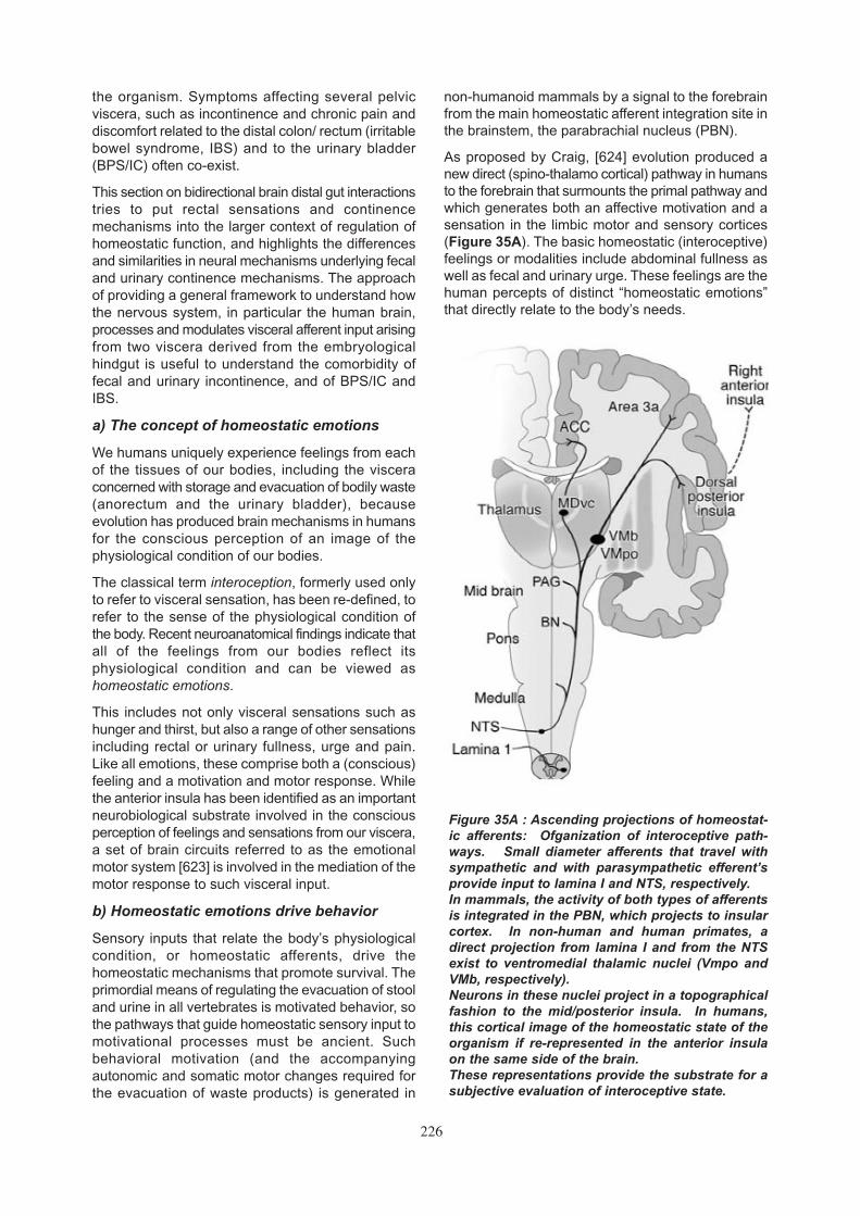
226
the organism. Symptoms affecting several pelvicviscera, such as incontinence and chronic pain anddiscomfort related to the distal colon/ rectum (irritablebowel syndrome, IBS) and to the urinary bladder(BPS/IC) often co-exist.
This section on bidirectional brain distal gut interactionstries to put rectal sensations and continencemechanisms into the larger context of regulation ofhomeostatic function, and highlights the differencesand similarities in neural mechanisms underlying fecaland urinary continence mechanisms. The approachof providing a general framework to understand howthe nervous system, in particular the human brain,processes and modulates visceral afferent input arisingfrom two viscera derived from the embryologicalhindgut is useful to understand the comorbidity offecal and urinary incontinence, and of BPS/IC andIBS.
a) The concept of homeostatic emotions
We humans uniquely experience feelings from eachof the tissues of our bodies, including the visceraconcerned with storage and evacuation of bodily waste(anorectum and the urinary bladder), becauseevolution has produced brain mechanisms in humansfor the conscious perception of an image of thephysiological condition of our bodies.
The classical term interoception, formerly used onlyto refer to visceral sensation, has been re-defined, torefer to the sense of the physiological condition ofthe body. Recent neuroanatomical findings indicate thatall of the feelings from our bodies reflect itsphysiological condition and can be viewed ashomeostatic emotions.
This includes not only visceral sensations such ashunger and thirst, but also a range of other sensationsincluding rectal or urinary fullness, urge and pain.Like all emotions, these comprise both a (conscious)feeling and a motivation and motor response. Whilethe anterior insula has been identified as an importantneurobiological substrate involved in the consciousperception of feelings and sensations from our viscera,a set of brain circuits referred to as the emotionalmotor system [623] is involved in the mediation of themotor response to such visceral input.
b) Homeostatic emotions drive behavior
Sensory inputs that relate the body’s physiologicalcondition, or homeostatic afferents, drive thehomeostatic mechanisms that promote survival. Theprimordial means of regulating the evacuation of stooland urine in all vertebrates is motivated behavior, sothe pathways that guide homeostatic sensory input tomotivational processes must be ancient. Suchbehavioral motivation (and the accompanyingautonomic and somatic motor changes required forthe evacuation of waste products) is generated in
non-humanoid mammals by a signal to the forebrainfrom the main homeostatic afferent integration site inthe brainstem, the parabrachial nucleus (PBN).
As proposed by Craig, [624] evolution produced anew direct (spino-thalamo cortical) pathway in humansto the forebrain that surmounts the primal pathway andwhich generates both an affective motivation and asensation in the limbic motor and sensory cortices(Figure 35A). The basic homeostatic (interoceptive)feelings or modalities include abdominal fullness aswell as fecal and urinary urge. These feelings are thehuman percepts of distinct “homeostatic emotions”that directly relate to the body’s needs.
Figure 35A : Ascending projections of homeostat-ic afferents: Ofganization of interoceptive path-ways. Small diameter afferents that travel withsympathetic and with parasympathetic efferent’sprovide input to lamina I and NTS, respectively. In mammals, the activity of both types of afferentsis integrated in the PBN, which projects to insularcortex. In non-human and human primates, adirect projection from lamina I and from the NTSexist to ventromedial thalamic nuclei (Vmpo andVMb, respectively). Neurons in these nuclei project in a topographicalfashion to the mid/posterior insula. In humans,this cortical image of the homeostatic state of theorganism if re-represented in the anterior insulaon the same side of the brain. These representations provide the substrate for asubjective evaluation of interoceptive state.

227
The concept of homeostatic emotions, regardless ofthe valence of the emotion, is consistent with thereported activation of similar brain regions (insula,dACC) by a variety of both pleasant and unpleasantstimuli (reviewed in Vogt, 2005 [625]).
2. ORGANIZATION OF HOMEOSTATICREFLEXES: PROCESSING OF PELVIC VISCERAL INFORMATION WITHINHIERARCHICAL ORGANIZEDHOMEOSTATIC REFLEXES
a) Enteric reflexes within the Enteric NervousSystem
The hierarchal organization of homeostatic reflexeswithin the central neuroaxis, and which are pertinentto both anorectum and urinary bladder is shown inFigure 35B. A fundamental difference in the regulationof the distal gut and the urinary bladder is the prominentrole of the enteric nervous system (ENS) as the primaryreflex circuit mediating both filling and emptying of
the distal colon. The ENS, which is considered the thirddivision of the ANS [626], is made up of twoganglionated plexus situated between the differentmuscle layers of the gut (myenteric plexus) and themucosa (submucosal plexus). Even in the absenceof any central input, these enteric (or intrinsic) reflexescan generate proximal to distal propulsion of coloniccontents (peristaltic reflex), and relaxation of theinternal anal sphincter [627]. While the enteric reflexcircuitry within the anorectum is unique to fecalcontinence, the spinal and supraspinal reflexesmediated by extrinsic afferents overlap considerablyfor bladder and rectum.
b) Lamina I afferents provide input to spinaland supraspinal reflexes.
The small-diameter afferent fibers that innervate thepelvic viscera course peripherally with sympatheticand parasympathetic nerves (e.g. splanchnic, pelvic,vagus), and with somatic nerves (e.g. pudendal).Many innervate the mesenteric ganglia and play arole in the communication between the central nervoussystem and the ganglionated plexus of the ENS. Manyof these extrinsic afferents respond to chemical andmechanical stimuli over a broad range of thresholdsand response slopes, so that they might be consideredto represent a broad continuum of response properties.However, they can also be regarded as separableinto different classes, including low-threshold, high-threshold (nociceptive), “silent” (unresponsive unlessinflamed), and thermosensitive (primarily warm) [628,629]. These classes may correspond to differentfunctional roles and different subjective feelings. Withinthe gut, there are also several distinct anatomicalpatterns of innervation, with some fibers ending withinthe longitudinal muscle sheets, others ending withinthe enteric ganglia (intraganglionic laminar endings,IGLEs), [630] and others in close proximity toenterochromaffin [631] or to mast cells [632].
c) Spinal and supraspinal reflexes
The lamina I neurons that receive the small-diameterfiber inputs have projections within the spinal cordand brainstem (Figure 36A). In the spinal cord, theironly major projection is to the sympathetic cell columnof the thoracolumbar spinal cord, where autonomic pre-ganglionic output neurons are located. In thebrainstem, they project exclusively to the recognizedhomeostatic integration sites (e.g. caudal and rostralventrolateral medulla, catecholamine cell groups A1-2 and A5-7, Barrington’s nucleus (“pontine micturitioncenter”, PMC), PBN, periaqueductal gray [PAG]),which also receive parasympathetic afferent activityby way of the NTS and which are heavilyinterconnected with the hypothalamus and theamygdala complex (including the bed nucleus striaterminalis, BNST). The NTS that receives small-diameter fibers from parasympathetic nerves similarlyprojects to all of these sites. These spinal and bulbar
Figure 35B: Ascending projections of homeostat-ic afferents: Spino-thalamo-cortical system.Summary diagram illustrating the projections inprimates of homeostatic afferent pathways fromlamina I (spinal) and NTS (vagal) to thalamicnuclei, and the two cortical regions involved in thesensory (insula) and motivational (ACC) dimen-sions of homeostatic emotions.

228
projections from lamina I and from the NTS providethe substrate for the hierarchical, modality-selectivesomato-autonomic reflexes activated by spinal small-diameter afferents that are crucial for homeostaticcontrol of all tissues of the body [633].
Traditionally, the pontine regulation of the pelvic viscerahad been considered specific for the urinary bladderand was accordingly referred to as the pontinemicturition center (PMC). However, more recent workin rodents has clearly established a role of this pontinenucleus (Barrington’s nucleus, BN) as the anatomicalsubstrate for the co-regulation of both bladder andrectosigmoid with central arousal circuits (viaprojections to the locus coeruleus) and with forebrainfunction [634] (Figure 37). Efferent projections fromBN to the dorsal motor nucleus of the vagus (DMV)and to the sacral parasympathetic nucleus give thisnucleus a unique role in the regulation of both upperand lower GI function through the two divisions of theparasympathetic nervous system [634]. Efferent
projections from BN to the noradrenergic locuscoeruleus are an important pathway by which afferentsignals from both pelvic viscera can activate centralarousal mechanisms, and hyperresponsiveness ofthese arousal circuits have been implicated in centralpain amplification in functional visceral disorders suchas IBS and PBS/IC [635]. The forebrain connectionsfrom the BN make this brain region ideally positionedto coordinate the activity of both pelvic viscera withbehaviors that are appropriate for evacuation. Valentinoet al. demonstrated evidence for the existence of 3types of neuronal populations in BN, one of which issynaptically linked to both the bladder and the colon,and the other two populations which are specificallylinked to either viscera [460, 452]. The authorsconcluded that these neuroanatomical substratesmay underlie the central coordination of bladder andcolon function and, in pathological states, may playa role in disorders characterized by a co-existence ofbladder and colon symptoms.
Figure 36: Hierarchical organization of homeosta-tic reflex systems involving the sympathetic nerv-ous system. A. Homeostatic afferents that reportthe physiological condition of all tissues in thebody, including the GI tract, terminate in lamina Iof the dorsal horn. The ascending projections ofthese neurons provide the basis for reflex arcs atthe spinal, medullary and mesencephalic levels.Limbic, paralimbic and prefrontal centers providemodulatory influences on the gain of these reflex-es.
Figure 36B: Cortical modulation of homeostaticafferent input to the central nervous system. PFCregions (dorsolateral PFC [dlPFC], orbitofrontalcortex [orbFC]) modulate activity in limbic and par-alimbic regions (amygdala [amy], ACC subregions,and hypothalamus [Hypoth]), which in turn regu-late activity of descending inhibitory and facilitato-ry descending pathways through the PAG andpontomedullary nuclei. Activity in these corticol-imbic pontine networks mediates the effect of cog-nitions and emotions on the perception of homeo-static feelings, including visceral pain and discom-fort. From Mayer et al. (635)
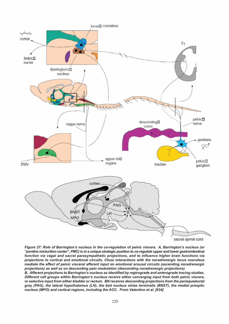
229
Figure 37: Role of Barrington’s nucleus in the co-regulation of pelvic viscera. A, Barrington’s nucleus (or“pontine micturition center”, PMC) is in a unique strategic position to co-regulate upper and lower gastrointestinalfunction via vagal and sacral parasympathetic projections, and to influence higher brain functions viaprojections to cortical and emotional circuits. Close interactions with the noradrenergic locus coeruleusmediate the effect of pelvic visceral afferent input on emotional arousal circuits (ascending noradrenergicprojections) as well as on descending pain modulation (descending noradrenergic projections). B, Afferent projections to Barrington’s nucleus as identified by regtrograde and anterograde tracing studies.Different cell groups within Barrington’s nucleus receive either converging input from both pelvic viscera,or selective input from either bladder or rectum. BN receives descending projections from the periaqueductalgrey (PAG), the lateral hypothalamus (LH), the bed nucleus striae terminalis (BNST), the medial preopticnucleus (MPO) and cortical regions, including the ACC. From Valentino et al. [634]

230
d) Spino-thalamic input to interoceptive cortex
In sub-primates, ascending lamina I homeostaticafferent activity is integrated mainly in severalbrainstem sites (A1, PBN, PAG), which then providean integrated signal to behavioral control regions inthe forebrain. In primates, however, there is a novel,additional lamina I STT projection to a specific thalamo-cortical relay nucleus (VMpo) in posterolateralthalamus, which in turn projects to a discrete portionof dorsal posterior insular cortex (Fig. 35) [636]. Thisinteroceptive cortex contains modality-selectiverepresentations of all afferent activity from lamina I (i.e.,sympathetic afferent input) and from the NTS (i.e.,parasympathetic input). In monkeys, this pathway isjust visible, but in humans, it is greatly enlarged. Manyfunctional imaging studies in humans confirm role ofthe insular cortex in pain, hunger, thirst, temperature,itch, muscle sensation, sensual touch, andcardiorespiratory activity, (reviewed in Craig, 2002[636]), and it can be regarded as primary sensorycortex for the physiological condition of the body inprimates. By contrast, in sub-primates the insularcortex appears to have a primordial role in modulatingbrainstem homeostatic integration (in PBN and othersites) and shows convergent, non-selective responsesto homeostatic afferent inputs. The differences in theneuroanatomy of the insular cortex and its subregionshave potentially important implications for thepathophysiology of IBS and BPS/IC. The modulationof visceral afferent input reaching the posterior insulais modulated by emotional arousal circuits and bycognitive input at the levels of the mid and anteriorinsula, respectively [624].
e) Spino thalamic input to limbic behavioralmotor cortex
The ascending homeostatic afferent lamina I pathwayin primates and humans also provides a direct thalamo-cortical pathway (by way of MDvc in medial thalamus)that activates the dACC (Fig. 35). In sub-primates, thedACC receives only integrated homeostatic input fromthe brainstem [636, 637]. On the basis of functionalimaging and lesion studies in humans, the dACC canbe directly associated with the affective aspect ofvisceral and somatic pain (unpleasantness), and withvolition, behavioral motivation, as well as autonomicand motor responses [636,637,638,639,640]. Itsinterconnections with PFC regions, insular, and ventralstriatal regions, along with its strong descendingprojections to the brainstem, particularly the PAG andthe BN, strongly support the idea that it can beregarded generally as the limbic behavioral motorcortex, just as the insula can be regarded as the limbicsensory cortex [625-641].
3. CONSCIOUS PERCEPTION OF INPUT FROM THE BODY AS ASPECTS OF HOMEOSTASIS
Pain or discomfort from the pelvic viscera is oftenregarded as a distinct feeling, but from the perspective
laid out above, pain can be viewed as anotherhomeostatic feeling. It has characteristics exactlycomparable to other feelings from the body. Painnormally originates with a change in the condition ofthe tissues of the body, a physiological imbalancethat automatic (subconscious) homeostatic systemsalone cannot rectify. It comprises both a sensationand an affective behavioral drive with accompanyingautonomic adjustments. Depending on conditions,pain can be unpleasant (as usual) or pleasant (suchas when it relieves an intense itch). Pain also generatescharacteristic reflexive motor patterns as doexperimental gut and bladder stimuli. The behavioralmotivation of pain is normally correlated with theintensity of the sensory input, but this can vary underdifferent behavioral, autonomic and emotionalconditions, so that pain can become intolerable or itcan disappear, similar to any other homeostaticemotion (e.g. hunger). The modulation of themotivational aspect of a homeostatic emotion likepain can occur via inhibitory or excitatory prefrontalinfluences on brain circuits involving the dACC [640-642,643].
Viewing rectal or urinary urge and pain as homeostaticemotions provides a ready explanation of theinteractions of these feelings from the body, including“gut feelings,” with other homeostatic conditions(including blood pressure, level of arousal, mood andaffect) because homeostasis is an integrated, dynamicprocess. This conceptual perspective also provides afirm basis for explaining the interactions of pain andnon-painful visceral discomfort with emotional status,emotional arousal or attention (i.e., the psychologicaldimension of pain), and it unifies the different conditionsthat can cause different types of pain/discomfort fromdifferent tissues under a common homeostatic function– the maintenance of the integrity of the body. Allanimals respond with emotional behavior to stimuli thatin humans cause a feeling of pain [644]. An examplerelevant to preclinical studies in visceral painmechanisms is the so called visceromotor reflex seenin response to colorectal or urinary bladder distensionin rodents [645]. In primates, novel thalamo-corticalprojections have emerged from this basic homeostaticsystem that provides direct pathways to encephalizedcortical mechanisms for highly resolved sensations andmotivations. In humans, these novel pathways arefurther elaborated, re-represented, and integratedwith other forebrain emotional components in theanterior insula and in the orbitofrontal cortex (thefronto-insular region) (Figures 35A, B).
The concept of homeostatic emotions has considerableimplications for the study of mechanisms underlyingvisceral pain and autonomic dysregulation in patientswith symptom based (or “functional) disorders of theGI and urinary tract. In the majority of these patients,primary symptoms (urgency, sensation of fullness,pain) are directly related to the altered perception ofhomeostatic feelings associated with the intestine,

231
rectum or urinary bladder (visceral hypersensitivity).Since rodents, the animals most commonly used inexperiments to model these disorders, do not have theforebrain structures to generate the same consciousemotional feelings of humans, findings obtained insuch animals (using so-called pseudo-affective reflexresponses) may reflect primarily the phylogeneticallyshared enteric, spinal and brainstem components ofhomeostatic pathways (e.g. reflexes), but may providelittle or no insight into the uniquely human experienceof abdominal pain and discomfort, nor to themodulatory cortical influences related to thisexperience.
The VMpo also has a collateral projection to a portionof sensorimotor cortex (area 3a) that is intercalatedbetween the primary somatosensory area and theprimary motor area. Similarly, vagal afferent activationoccurs in the lateral portion of area 3a of the primarysensorimotor region by way of the NTS and VMb.These projections can be associated with corticalcontrol of the reflex actions of skeletal muscle inresponse to homeostatic afferent inputs, a rolesubsumed under the term “viscero-somatic”integration.
The forebrain interoceptive representation of thebody’s condition in the dorsal posterior insula issuccessively re-represented in the middle insula andthen in the right (non-dominant) anterior insula (636).Functional imaging data show that the right anteriorinsula is associated with subjective awareness ofhomeostatic emotions (e.g. visceral and somaticpainful and non-painful sensations, temperature,sexual arousal, hunger, and thirst) as well as allemotions (e.g. anger, fear, disgust). This region isintimately interconnected with the dACC, which is co-active in such studies. Thus, viewing feelings fromthe body as homeostatic emotions enablesneuroanatomical explanations of interactions withother homeostatic functions and with emotions at thelevel of the forebrain. Finally, recent evidence showingasymmetry in the sympathetic and parasympatheticafferent re-representations and control mechanismsin the left and right insula/orbitofrontal cortex and ACCseems to accord with the psychophysiologicalevidence for the forebrain asymmetry of positive andnegative emotions [646]. In this context, it is ofconsiderable interest that studies of centralrepresentation of both bladder [482] and rectaldistensions [635] have repeatedly shown activation ofthe right ventrolateral PFC.
4. DESCENDING MODULATION OF HOMEO-STATIC REFLEXES AND FEELINGS
Lamina I neurons not only receive input via somaticand visceral afferents, but also receive descending(facilitatory and inhibitory) modulation directly frombrainstem pre-autonomic sources, includingserotonergic nuclei within the rostral ventral medulla
and the noradrenergic pontine locus coeruleuscomplex (Figure 36). Indeed, lamina I and the spinalautonomic columns are the only regions in the spinalcord that receive descending modulation from thehypothalamus. Thus, the activity of lamina I neuronsthat ultimately produces the various feelings from thebody is modulated by various tonic and phasicallyactive, descending inhibitory and facilitatory pathways[647,648,649], whose primary purpose is control ofhomeostatic integration [650,651]. As pointed out byMason [651,652,653], these serotonergic descendingpathways from the PAG and ventromedial medullaare not only involved in descending pain modulation,but play a prominent role in various homeostaticfunctions including micturition and continence.
Recent evidence (in rats) suggests that the gain of thespino-bulbo-spinal reflex loops originating fromneurokinin 1-receptor-containing lamina I neurons isnot constant but can be modulated by peripheralprimary afferent inputs from inflamed tissue ordamaged nerves, [654] adapting the homeostaticresponse to the overall state of the organism. Thus,since the descending forebrain control of these pre-autonomic brainstem regions originates in brainnetworks associated with attention, emotion generation(i.e. the limbic system) and emotion regulation (PFC),these anatomic connections provide the basis forcorticolimbic modulation of the afferent activityassociated with feelings from the body, including painand discomfort, at the spinal level as well as at thebrainstem and forebrain levels (Figure 36).
5. BRAIN CIRCUITS ACTIVATED BY ACUTEVISCERAL STIMULI: EVIDENCE FROMFUNCTIONAL BRAIN IMAGING STUDIES INHUMANS
Visceral sensations, including discomfort, hunger,fullness, early satiety, nausea, bloating or abdominalpain in humans are a subjective, conscious experience,that result from the modulation of homeostatic feelingsby cognitive (attention, expectation), emotional(arousal, anxiety) and motivational factors, as well asmemories of past experiences. Thus, the consciousexperience is an image of the homeostatic state of thebody represented in the insular cortex (Figure 35)and is further modified by these cortical and limbicinputs. In principal, altered perception of visceralstimuli could result from activity changes in visceralafferent signal processing areas alone (reflectingincreased visceral afferent input to the brain from thegut), from alterations in distinct but overlapping painmodulation circuits (“central pain amplification”), orfrom variable combinations of these two overlappingcircuitries [655,656,657] (Figure 37).
a) Activation of Regions involved in Homeo-static Emotions in Studies of the Human Brain-Gut Axis
Studies published during the past 5 years using widely

232
different experimental paradigms have confirmed theconsistent activation of the central homeostatic afferentnetwork consistent with the robust activation ofhomeostatic afferent pathways despite varyingexperimental paradigms and analysis techniques[658,659,660,661,662,663]. On the other hand, thevariable activations of PFC and limbic regions, and theseemingly contradictory results on sex differences inreported studies is reflective of the fact thatexperimental paradigms and analysis techniquesvaried, and that the majority of these studies did nottake into account cognitive and emotional aspects ofpain modulation.
b) Modulation of Homeostatic Afferent NetworkMatrix by Cognitive and Emotional Stimuli
The brain has multiple ways to modulate the perceptionof afferent information and this modulation is influencedby the environmental context, the emotional state ofthe individual (e.g. fear, anxiety, or anger), cognitivefactors (e.g. expectation, attention), or memories ofprevious sensory events (conditioned responses)(Figure 36B). As outlined in the earlier sections of thisreview, these top-down influences can modulatehomeostatic afferent input to the central nervoussystem at multiple levels (Figure 37A). Considerableprogress has been made both on a preclinical and,more recently, on a clinical level to identify brainregions, circuits and mechanisms which play a rolein the facilitation and inhibition of the subjective painexperience [649-664], Both clinical and preclinicalstudies support a role for right orbitofrontal and rightventrolateral PFC in mediating inhibition of emotionaland pain responses (reviewed in Lieberman et al.2004 [643]) and for dorsolateral PFC in painmodulation [642-665].
• COGNITIVE MODULATION
Attention. Aziz’s group (666) examined the modulatoryrole of attention on the brain responses to non-painfulvisceral (esophageal) distension in 7 healthy volunteers(6 males). Brain responses to phasic visual andesophageal stimuli were presented simultaneouslywhile subjects were asked to focus their attention oneither the esophageal or the visual stimulus (selectiveattention) or both (divided attention). Selective attentionon the esophageal stimulus was associated withactivation of sensory (somatosensory cortex) andcognitive (dACC) networks, while selective attentionon the visual stimulus activated the visual cortex.During the divided attention task, more brain regionsin the sensory and cognitive domains were activatedto process esophageal stimuli, in comparison to thoseprocessing visual stimuli. These findings emphasizethe importance of attentional processes in themodulation of sensory information from the body andthe relative biological importance placed on visceralsensation, compared to other sensory modalities.
Expectation. A variety of studies in the somatic painliterature have evaluated brain responses to theexpectation of an aversive stimulus [662-667, 668,669,670]. These studies suggest that the brain caneither up- or down-regulate sensory and limbic brainregions based on previous experience, familiarity withthe stimulus and expected intensity [668]. Severalearly studies have shown some evidence of activationof the homeostatic afferent network during conditionedanticipation of a visceral stimulus [662-667]. Bermanet al. studied the brain fMRI BOLD response toanticipated (cue condition) and delivered mild andmoderate rectal distension in 12 healthy women and14 female IBS patients [671]. Distension increasedactivity in the homeostatic brain regions and decreasedactivity in the infragenual cingulate. As in previousstudies comparing IBS patients and control subjects,the increases were more extensive in the IBS patients,with significant differences in midcingulate and dorsalbrainstem. During cued anticipation of distension,activity decreased in the insula, dACC, amygdala anddorsal brainstem in healthy women, but not in IBSpatients, consistent with a top-down modulation ofhomeostatic afferent networks by cortical regions.Three self-rated measures of negative affect duringscanning were higher in IBS patients than healthywomen (P < 0.001), and the anticipatory BOLDdecreases in bilateral dorsal brainstem, centered inthe pontine LCC, were inversely correlated with allthree measures. The amplitude of anticipatorydecrease in the LCC was associated with greateractivation by subsequent distension in right orbitofrontalcortex and bilateral supragenual ACC – regionspreviously implicated in corticolimbic inhibition. Whenthe cue was followed by a sham distension (e.g. nochange in distension pressure from the restingpressure of 5 mmHG occurred), only IBS patientsshowed activation of the insula cortex, and thisactivation was limited to the more anterior portions ofthe insula. These findings are consistent with top-down modulation of the anterior insula by PFCinfluences, and with an enhanced top-down facilitationin IBS patients. In summary, these findings suggestthat in healthy women, the brain decreases activitywithin the homeostatic brain matrix in expectation ofa certain, inescapable pelvic pain stimulus. A failureto generate this down-regulation in IBS patients, andan inappropriate activation of the anterior insula duringa sham distension, may be related to differences incortically-mediated coping styles, emotional factors andlinked arousal systems.
Yaguez et al. [672] studied brain responses duringdifferent phases of visceral aversive conditioning in 8healthy volunteers (5 males) using fMRI. The authorsused a classical conditioning paradigm in whichdifferent colored circles were used as conditionedstimuli and were paired with painful esophagealdistension (learning phase), airpuff to the wrist ornothing. Brain responses during the learning phase
233
(delivery of aversive esophageal distension) wereseen in the homeostatic brain matrix and insomatosensory cortex. During the anticipation andextinction phase of the paradigm, brain activityresembled that seen during actual esophagealdistension, including activation in insula and dACC.These findings emphasize the importance of cognitiveinfluences, such as expectation and memory recall intop-down modulation of brain regions involved in theprocessing of homeostatic information from the body.
Hypervigilance. Several lines of evidence indicate thatIBS patients and other functional disorders havehypervigilance for symptom-relevant sensations [673].Repeated exposure to experimental visceral stimulican lead to decreased hypervigilance and, therefore,discomfort. In a longitudinal study of IBS patientsexposed to 6 sessions of rectal inflations over a 1-yearperiod, we examined regional cerebral blood flow tothe inflations and anticipation of inflations usingH215O-PET at the first and last session [674].Subjective ratings of the rectal inflations normalizedover the 12 months of the study, while IBS symptomseverity did not, indicating decreased vigilanceindependent of changes in perceived disease activity.In response to rectal distension, stable activation ofregions of the homeostatic afferent network (includingthalamus and anterior insula) was observed over the12-month period, while activity in limbic, paralimbic andpontine regions decreased. During the anticipationcondition, there were significant decreases in dACC,amygdala, and dorsal brainstem (perhaps involvingthe LCC) activation at 12 months. One way to interpretthese findings is that brain regions processing thefeeling and the motivational dimension of thehomeostatic emotion were affected differentially by thehabituation process: while insula activation remainedconstant, dACC activation progressively decreased.An analysis examining the covariation of these brainregions, as well as preliminary results from an effectiveconnectivity modeling approach to the data [675]supported the hypothesis of changes in an arousalnetwork including limbic, pontine and cortical areasunderlying the decreased perception seen over themultiple stimulation studies.
In summary, brain imaging studies of cognitive andaffective modulation of perceptual and brain responsesto visceral stimuli strongly support the concept of ahomeostatic afferent network in the brain. The factthat components of this network can be modulateddifferentially by top-down corticolimbic influences hasimportant implications for a better understanding ofsymptom generation/modulation in functional GIdisorders.
c) Modulation of Homeostatic Emotions byDescending Modulation
Since the beginning of the 20th century it has beenknown that the brain can tonically inhibit spinal cord
excitability, thereby regulating the amount of peripheralsensory information reaching the central nervoussystem. More recent evidence has demonstrated theactivity of both pain inhibitory and facilitatorymechanisms which can tonically and phasicallyregulate spinal cord excitability [647,648,649]. Whiletop-down tonic pain inhibitory modulation appears topredominate in healthy individuals during basalconditions, an up-regulation of descending painfacilitatory systems has been demonstrated in themaintenance of hyperalgesia in animal models ofperipheral nerve injury [654]. An alteration in thebalance between inhibitory and facilitatory painmodulatory systems has been proposed as a possiblemechanism underlying chronic pain syndromes suchas fibromyalgia [676] and IBS [677,678].
Zambreanu and coworkers were the first todemonstrate the activation of brainstem regions inthe context of central sensitization in healthy humanvolunteers [679]. Using 3T fMRI, they compared wholebrain responses, including the brainstem, to punctuatemechanical stimulation in an area of secondaryhyperalgesia (induced by heat/capsaicin sensitizationmodel) or in a control area. They found greateractivation during stimulation of the hyperalgesic regionin several cortical regions, including posterior insula,ACC and posterior cingulate cortex, as well asthalamus and pons. The brainstem activation waslocalized to the NCF (possibly involving PBN) andthe PAG, brain regions that receive input fromcorticolimbic networks (including the rostral ACC),send projections to the rostroventral medulla and arepart of a cortico-limbic-pontine pain modulation circuit[680,681] (see also Figure 36). There is preliminaryevidence to suggest that patients with IBS may alsoshow abnormal activation of brain circuits involved inpain modulation [660-682,683]. Two studies wereperformed by Wilder-Smith and coworkers [660- 683]to study the central correlates of heterotopic paininhibition. In the first study, they performed an fMRIstudy in 10 female patients with IBS (5 constipated-,5 diarrhea-predominant bowel habit) and 10 femalehealthy control subjects to test the hypothesis thatIBS patients show abnormal activation of diffusenoxious inhibitory control (DNIC) systems in responseto a noxious stimulus [660]. DNIC activation can bequantified by the perceptual modulation of a painfulstimulus (in this case noxious rectal balloon distension)by a secondary heterotypically applied nociceptivestimulus (in this case ice water immersion of the foot).They found that subjective pain ratings of rectal volumedistension by the heterotypic cold pain stimulus wasreduced in healthy controls but not in the IBS patientgroup, suggesting an inadequate activation of DNICsin the IBS patients. Following the heterotypic coldstimulus, a complex set of differences in response torectal pain were found among the controls and the twoIBS bowel habit-based sub-groups. These includeddecreased activation in insula, thalamus and PAG in

234
the control group (perhaps reflecting the DNIC process)that was absent in the IBS patients. In the secondstudy, similar perceptual results were found anddifferences were also found between IBS and controlsduring expectation of rectal pain, without actualdistension [683].
More recent brain imaging studies in patientpopulations provide more direct support for alterationsin cortico-limbic-pontine pain modulation networks inIBS patients leading to visceral hypersensitivity. Mayeret al. [682] examined three groups of male subjects,ulcerative colitis patients with quiescent disease (n =9), patients with IBS (n = 9), and healthy male controls(n = 9), during actual and expected but undeliveredrectal distensions using H215O-PET. This study foundsimilar responses in all three groups in the homeostaticafferent network (anterior insula and dACC). However,IBS patients compared to both the ulcerative colitis andcontrol groups showed consistently greater activationof limbic/paralimbic brain regions (amygdala, hypo-thalamus, ventral/rostral ACC, dorsomedial PFC),suggestive of increased activation of pain facilitatorypathways. In addition, the results showed activationin the ulcerative colitis and control subjects, but notIBS patients, in the lateral frontal regions and a brainregion including the PAG. A connectivity analysis usingstructural equation modeling supported these regionsacting as part of a pain inhibition network that involveslateral and medial frontal influences on the PAG.
In summary, a small group of hypothesis-drivenstudies aimed at evaluating specific mechanisms ofvisceral pain modulation (e.g. descending modulation)are beginning to demonstrate the activation of cortico-limbic-pontine networks, which may be the biologicalsubstrate of such endogenous modulation. Theconcept emerging from these studies is the ability ofbrain regions and networks involved in emotionalgeneration (limbic circuits) as well as in emotionregulation (orbitofrontal, ventrolateral PFC) to modulateactivity within the homeostatic brain matrix [655, 656].Observations obtained with rectal and with bladderstimuli show many similarities in these activations[482]. Differences in the activation of these networksby cognitive or somatic stimuli between IBS patientsand control populations suggest a possible role ofsuch differences in the pathophysiology of enhancedvisceral perception in IBS.
The concept of homeostatic emotions with a sensoryfeeling dimension (processed in the anterior insula)and a motivational dimension (processed in the dACC)provides a general framework to understand theinteractions between peripheral afferent signals arisingfrom the pelvic viscera, and centrally-mediatedinfluences (including psychological factors, cognitionsand emotions) on the conscious perception andassociated autonomic responses to such stimuli. Thedeconstruction of symptoms into distinct contributionsof specific brain networks mediating a variety of
cognitive, emotional and motivational influences on thebasic homeostatic afferent processing network mayhelp to understand the pathophysiology of a varietyof chronic disorders, including IBS and PBS/IC. Awhole range of chronic disorders characterized byphysical or emotional discomfort and pain (includingfunctional visceral and somatic disorders, as well asdisorders of mood and affect) can be conceptualizedas disorders of homeostatic emotions. The ability tostudy a neurobiological substrate (e.g. brain activity)rather than relying on highly variable subjectivesymptoms, may make it possible to obtain insightsabout the role of genetic factors, receptor physiology,and drug interventions from much smaller samples ofsubjects, compared to epidemiological or traditionalpharmacological studies.
1. Andrew J, Nathan PW. Lesions of the anterior frontal lobesand disturbances of micturition and defaecation. Brain.1964;87:233-62.
2. Fowler CJ, Griffiths D, De Groat WC. The neural control ofmicturition. Nature Reviews Neuroscience. 2008;9:453-66.
3. DasGupta R, Kavia RB, Fowler CJ. Cerebral mechanismsand voiding dysfunction. BJU International. 2007;99:731-4.
4. Kavia RB, Dasgupta R, Fowler CJ. Functional imaging andthe central control of the bladder. J Comp Neurol.2005;493:27-32.
5. Apodaca G. The uroepithelium: Not just a passive barrier.Traffic. 2004;5:117-28.
6. Lewis SA. Everything you wanted to know about the bladderepithelium but were afraid to ask. Am J Physiol Renal Physiol.[Invited Review]. 2000;278:F867-F74.
7. Hicks M. The mammalian urinary bladder: an accomodatingorgan. Biol Rev. 1975;50:215-46.
8. Liang FX, Riedel,I., Deng,F.M., Zhou,G., Wu, C., Wu,W.R.,Kong,W.P., Moll,R., Sun,T.T. Organization of uroplakinsubunits: transmembrane topology, pair formation and plaquecomposition. J Biochem. 2001;355:13-8.
9. Wu X.R, Lin J.H, Walz T., Haner M., Yu J., Aebi U., Sun T.T.Mammalian uroplakins. A group of highly conserved urothelialdifferentiation-related membrane proteins. J. BiolChem.1994;269:13716-24.
10. Sun Y, Keay,S., DeDeyne,P., Chai,T.C. Augmented stretchactivated adenosine triphosphate release from bladderuroepithelial cells in patients with interstitial cystitis. J Urol.2001;166:1951-56.
11. Acharya P, Beckel JM, Ruiz WG, Wang E, Rojas R, BirderLA, et al. Distribution of the tight junction proteins ZPO-1,occludin, and claudin-4, -8, and -12 in bladder epithelium.Am J Physiol Renal Physiol. 2004 August 2004;287(2):F305-F18.
12. Parsons CL, Greenspan, C., Moore, S.W., Mulholland, S.G.Role of surface mucin in primary antibacterial defense ofbladder. Urology. 1977;9:48-52.
13. Parson CL, Boychuk,D., Jones,S., Hurst,R., Callahan, H.Bladder surface glycosaminoglycans: an epithelialpermeability barrier. Journal of Urology. 1990;143:139-42.
14. Parsons CL, Lilly JD, Stein P. Epithelial dysfunction innonbacterial cystitis (interstitial cystitis). J Urology.1991;145(4):732-5.
REFERENCES

235
15. Wang E, Truschel ST, Apodaca G. Analysis of hydrostaticpressure-induced changes in umbrella cell surface area.Methods. 2003;30:207-17.
16. Truschel ST, Wang E, Ruiz WG, Leung SM, Rojas R, LavelleJ, et al. Stretch-regulated exocytosis/endocytosis in bladderumbrella cells. Molecular Biology of the Cell. 2002;13:830-43.
17. Balestreire EM, Apodaca,G. Apical EGF receptor signaling:regulation of stretch-dependent exocytosis in bladder umbrellacells. Mol Biol Cell. 2007;18:1312-23.
18. Cheng J, Huang,H., Zhang,Z.T. Overexpression of epidermalgrowth factor receptor in urothelium elicits urothelialhyperplasia and promotes bladder tumor growth. CancerResearch. 2002;62:4157-63.
19. Apodaca G, Balestreire,E., Birder,L.A. The uroepithelial-associated sensory web. Kidney International. 2007;72:1057-64.
20. Birder LA, DeGroat WC. Mechanisms of disease: involvementof the urothelium in bladder dysfunction. Nature ClinicalPractice. [Review]. 2007;4(1):46-54.
21. Martin BF. Cell replacement and differentiation in transitionalepithelium: a histological and autographical study of theguinea-pig bladder and ureter. J Anat. 1972;112:433-55.
22. Lavelle J, Meyers,S., Ramage,R., Bastacky,S., Doty,D.,Apodaca,G., Zeidel,M.L. Bladder permeability barrier:recovery from selective injury of surface epithelial cells.American Journal of Physiology. 2002;283:F242-53.
23. Bassuk JA, Cockrane,K., Mitchell,M.E. Induction of urothelialcell proliferation by fibroblast growth factor-7 in RAG1-deficient mice. Adv Exp Med Biol. 2003;539:623-33.
24. de Boer WI, Vermeij,M., Diez de Medina, S.G., Bindels,E.,Radvanyi,F., van der Kwast, T., Chopin,D. Functions offibroblast and transforming growth factors in primary organoid-like cultures of normal human urothelium. Lab Invest.1996;75:147-56.
25. Anderson G, Palermo J, Schilling J, Roth R, Heuser J,Hultgren S. Intracellular bacterial biofilm-like pods in urinarytract infections. Science. 2003;301:105-7.
26. Apodaca G, Kiss S, Ruiz WG, Meyers S, Zeidel M, BirderLA. Disruption of bladder epithelium barrier function afterspinal cord injury. Am J Physiol Renal Physiol. 2003 May2003;284(5):F966-F76.
27. Lavelle JP, Meyers SA, Ruiz WG, Buffington CAT, Zeidel M,Apodaca G. Urothelial pathophysiological changes in felineinterstitial cystitis: A human model. Am J Physiol RenalPhysiol. 2000;278:F540-F53.
28. Conrads TP, Tocci,G.M., Hood,B.L., Zhang,C.O., Guo,L.,Koch,K.R., Michejda,C.J., Veenstra,T.D., Keay,S.K.CKAP4/p63 is a receptor for the frizzled-8 protein-relatedantiproliferative factor from interstitial cystitis patients. J BiolChem. 2006;281:37836-43.
29. Keay SK, Zhang CO, Shoenfelt J, Erickson DR, WhitmoreK, Warren JW, et al. Sensitivity and specificity ofantiproliferative factor, heparin-binding epidermal growthfactor-like growth factor, and epidermal growth factor asurine markers for interstitial cystitis. Urology. 2001;57(6Suppl 1):9-14.
30. Keay SK, Szekely Z, Conrads TP, Veenstra TD, Barchi JJJ,Zhang CO, et al. An antiproliferative factor from interstitialcystitis patients is a frizzled 8 protein-relatedsialoglycopeptide. Proc Natl Acad Sci USA.2004;101(32):11803-8.
31. Schilling J, Hultgren S. Recent advances into thepathogenesis of recurrent urinary tract infections: the bladderas a reservoir for uropathogenic Escherichia coli. Int JAntimicrob Agents. 2002;19:457-560.
32. Thomas JC, DeMarco,R.T., Pope,J.C. Molecular biology of
ureteral bud and trigonal development. Curr Urol Rep.2005;6:146-51.
33. Liang FX, Bosland,M.C., Huang,H., Romih,R., Baptiste,S.,Deng,F.M., Wu,X.R., Shapiro,E., Sun,T.T. Cellular basis ofurothelial squamous metaplasia: roles of lineageheterogeneity and cell replacement. J Cell Biol. 2005;171:835-44.
34. Birder LA, Nealen,M.L., Kiss,S., de Groat,W.C., Caterina,M.J.,Wang,E., Apodaca,G., Kanai,A.J. Beta-adrenoceptor agonistsstimulate endothelial nitric oxide synthase in rat urinarybladder urothelial cells. J Neuroscience. 2002;22:8063-70.
35. Jen PY, Dixon,J.S., Gosling,J.A. Immunohistochemicallocalization of neuromarkers and neuropeptides in humanfetal and neonatal urinary bladder. Br J Urol. 1995;75:230-5.
36. Kunze A, Neuhaus,J., Stolzenburg,J.U. Quantitativeimmunohistochemical study of the innervation of the guinea-pig lower urinary tract. BJU Int. 2006;98:424-9.
37. Apostolidis A, Brady,C.M., Yiangou,Y., Davis,J., Fowler,C.J.,Anand,P. Capsaicin receptor TRPV1 in urothelium ofneurogenic human bladders and effect of intravesicalresiniferatoxin. Urology. 2005;65:400-5.
38. Brady CM, Apostolidis,A., Yiangou,Y., Baecker,P.A., Ford,A.P.,Freeman,A., Jacques,T.S., Fowler,C.J., Anand,P. P2X3-immunoreactive nerve fibres in neurogenic detrusoroveractivity and the effect of intravesical resiniferatoxin. EurUrol. 2004;46:247-53.
39. Ost D, Roskams,T., Van der Aa,F., de Ridder,D. Topographyof the vanilloid receptor in the human bladder: more than justthe nerve fibers. Journal of Urology. 2002;168:293-7.
40. Sui GP, Wu,C., Fry,C.H. Electrical characteristics ofsuburothelial cells isolated from the human bladder. Journalof Urology. 2004;171:938-43.
41. Brading AF, McCloskey,K.D. Mechanisms of disease:specialized interstitial cells of the urothelium: an assessmentof current knowledge. Nature Clinical Practice Urology.2005;2:546-54.
42. Ikeda Y, Fry C, Hayashi F, Stolz D, Griffiths D, Kanai AJ. Roleof gap junctions in spontaneous activity of the rat bladder.Am J Physiol Renal Physiol. 2007;293(4):F1018-F25.
43. Chopra B, Gever J, Barrick SR, Hanna-Mitchell AT, BeckelJM, Ford AP, et al. Expression and function of rat urothelialP2Y receptors. Am J Physiol Renal Physiol.2008;294(4):F821-F9.
44. Chopra B, Barrick, S.R., Meyers,S., Beckel,J.M., Zeidel,M.L.,Ford,A.P., de Groat,W.C., Birder,L.A. Expression and functionof bradykinin B1 and B2 receptors in normal and inflamedrat urinary bladder urothelium. J Physiol. 2005;562:859-71.
45. Burnstock G. Purine-mediated signalling in pain and visceralperception. Trends in Pharmcol Sci. 2001;22:182-8.
46. Chess-Williams R. Muscarinic receptors of the urinarybladder: detrusor, urothelial and prejunctional. Auton AutocoidPharmacol. 2002;22:133-45.
47. Birder LA, Barrick SR, Roppolo JR, Kanai AJ, DeGroat WC,Kiss S, et al. Feline interstitial cystitis results in mechanicalhypersensitivity and altered ATP release from bladderurothelium. Am J Physiol Renal Physiol. 2003;285:F423-F9.
48. Ferguson DR, Kennedy,I., Burton,T.J. ATP is released fromrat urinary bladder epithelial cells by hydrostatic pressurechanges-a possible sensory mechanism? J Physiol.1997;505:503-11.
49. Beckel JM, Kanai AJ, Lee SJ, DeGroat WC, Birder LA.Expression of functional nicotinic acetylcholine receptors inrat urinary bladder epithelial cells. Am J Physiol Renal Physiol.2006;290(1):F103-F10.
50. Ossovskaya VS, Bunnett,N.W. Protease-activated receptors:contribution to physiology and disease. Physiol Rev.2004;84:579-621.

236
51. Carattino MD, Sheng,S., Kleyman,T.R. Mutations in the poreregion modify epithelial sodium channel gating by shearstress. J Biol Chem. 2005;280:4393-401.
52. LeBerge J, Malley SE, Zvarova K, Vizzard MA. Expressionof corticotropin-releasing factor and CRF receptors inmicturition pathways after cyclophosphamide-induced cystitis.Am J Physiol Regul Integr Comp Physiol.2006;291(3):R692-R703.
53. Hanna-Mitchell AT, Beckel JM, Barbadora S, Kanai AJ,DeGroat WC, Birder LA. Non-neuronal acetylcholine andurinary bladder urothelium. Life Sci. 2007;80(24-25):2298-302.
54. Andersson KE, Persson,K. Nitric oxide synthase and thelower urinary tract: posible implications for physiology andpathophysiology. Scand J Urol Nephrol. 1995;175:43-53.
55. Templeman L, Chapple CR, Chess-Williams R. Urotheliumderived inhibitory factor and cross-talk among receptors inthe trigone of the bladder of the pig. J Urology. 2002;167:742-5.
56. Wang EC, Lee JM, Ruiz WG, Balestreire EM, von BodungenM, Barrick SR, et al. ATP and purinergic receptor-dependentmembrane traffic in bladder umbrella cells. J Clin Invest.2005;115(9):2412-22.
57. Sui GP, Wu,C., Fry, C.H. Characterization of the purinergicreceptor subtype on guinea-pig suburothelial myofibroblasts.BJU Int. 2006;97:1327-31.
58. Fry CH, Sui,G.P., Kanai,A.J., Wu,C. The function ofsuburothelial myofibroblasts in the bladder. Neurourol Urodyn.2007;26:914-9.
59. Du S, Araki I, Mikami Y, Zakoji H, Beppu M, Yoshiyama M,et al. Amiloride-sensitive ion channels in urinary bladderepithelium involved in mechanosensory transduction bymodulating stretch-evoked adenosine triphosphate release.Urology.2007;69(3):590-5.
60. Yu W, Zacharia,L.C., Jackson,E.K., Apodaca,G. Adenosinereceptor expression and function in bladder uroepithelium.Am J Physiol. 2006;291:C254-65.
61. Birder LA, Apodaca,G., de Groat,W.C., Kanai,A.J. Adrenergicand capsaicin-evoked nitric oxide release from urotheliumand afferent nerves in urinary bladder. Am J Physiol.1998;275:F226-9.
62. Caterina MJ, Schumacher MA, Tominaga M, Rosen TA,Levine JD, Julius D. The capsaicin receptor: a heat-activatedion channel in the pain pathway. Nature. 1997;389(6653):816-24.
63. Hicks GA. TRP channels as therapeutic targets: hot property,or time to cool down? Neurogastroenterol Motil. 2006;18:590-4.
64. Chancellor MB, de Groat,W.C. Intravesical capsaicin andresiniferatoxin therapy: spicing up the ways to treat theoveractive bladder. J Urol. 1999;162:3-11.
65. Szallasi A, Blumber,P.M. Vanilloid (capsaicin) receptors andmechanisms. Pharmacol Rev. 1999;51:150-221.
66. Birder LA, Nakamura Y, Kiss S, Nealen ML, Barrick SR,Kanai AJ, et al. Altered urinary bladder function in micelacking the vanilloid receptor TRPV1. Nat Neurosci.2002;5(9):856-60.
67. Liedtke W. TRPV4 plays an evolutionary conserved role inthe transduction of osmotic and mechanical stimuli in liveanimals. Pflugers Arch. 2005;451:176-80.
68. Gevaert T, Vriens,J., Segal,A., Evaraerts,W., Roskams,T.,Talavera,K., Owsianik,G., Liedtke,W., Daelemans,D.,Dewachter,I., Van Leuven,F., Voets,T., De Ridder,D., Nilius,B.Deletion of the transient receptor potential cation channelTRPV4 impairs murine bladder voiding. J Clin Invest.2007;117:3453-62.
69. Birder LA, Kullmann,F.A., Lee,H., Barrick,S., de Groat,W.C.,Kanai,A., Caterina,M. Activation of urothelial transient receptopotential vanilloid 4 by 4alpha-phorbol 12,13-didecanoatecontributes to altered bladder reflexes in the rat. J Pharm ExpTher. 2007;323:227-35.
70. Combrisson H, Allix,S., Robain,G. Influence of temperatureon urethra to bladder micturition reflex in the awake ewe.Neurourol Urodyn. 2007;26:290-5.
71. Du S, Araki,I., Yoshiyama,M., Nomura,T., Takeda,M. Transientreceptor potential channel A1 involved in sensory transductionof rat urinary bladder through C-fiber pathway. Urology.2007;70:826-31.
72. Andrade EL, Ferreira,J., Andre,E., Calixto,J.B. Contractilemechanisms coupled to TRPA1 recepto activation in raturinary bladder. Biochem Pharmacol. 2006;72:104-14.
73. Sun Y, Chai,T.C. Augmented extracellular ATP signaling inbladder urothelial cells from patients with interstitial cystitis.Am J Physiol. 2006;290:C27-34.
74. Zarghooni S, Wunsch,J., Bodenbenner,M., Bruggmann,D.,Grando,S.A., Schwantes, U., Wess,J., Kummer,W., Lips,K.S.Expression of muscarinic and nicotinic acetylcholine receptorsin the mouse urothelium. Life Sci. 2007;80:2308-13.
75. Kullman FA, Artim,D.E., Birder,L.A., de Groat,W.C. Activationof muscarinic receptos in rat bladder sensory pathwaysalters reflex bladder activity. J Neuroscience. 2008;28:1977-87.
76. Kullman FA, Artim D, Beckel JM, Barrick SR, DeGroat WC,Birder LA. Heterogeneity of muscarinic receptor-mediatedCa2+ responses in cultured urothelial cells from rat. Am JPhysiol Renal Physiol. 2008;294(4):F971-F81.
77. Chancellor MB, Fowler,C.J., Apostolidis,A., de Groat,W.C.,Smith,C.P., Somogyi,G.T., Aoki,R. Drug insight: biologicaleffects of botulinum toxin A in the lower urinary tract. Nat ClinPrac Urol. 2008;5:319-28.
78. Bosse Y, Pare,P.D., Seow,C.Y. Airway wall remodeling inasthma: from the epithelial layer to the adventitia. Curr AllergyAsthma Rep. 2008;8:357-66.
79. Hendrix S. Neuroimmune communication in skin: Far fromperipheral. J Invest Derm. 2008;128:260-1.
80. Lumplin EA, Caterina,M.J. Mechanisms of sensorytransduction in the skin. Nature Rev. 2007;445:858-65.
81. Janig W, Morrison JF. Functional properties of spinal visceralafferents supplying abdominal and pelvic organs, with specialemphasis on visceral nociception. Prog Brain Res.1986;67:87-114.
82. de Groat WC. Spinal cord projections and neuropeptides invisceral afferent neurons. Prog Brain Res. 1986;67:165-87.
83. Torrens M, Morrison JFB. The physiology of the lower urinarytract. 1987.
84. Iggo A. Tension receptors in the stomach and the urinarybladder. J Physiol. 1955;128(3):593-607.
85. Xu L, Gebhart GF. Characterization of mouse lumbarsplanchnic and pelvic nerve urinary bladder mechanosensoryafferents. J Neurophysiol. 2007 Nov 14.
86. Zagorodnyuk VP, Costa M, Brookes SJ. Major classes ofsensory neurons to the urinary bladder. Auton Neurosci.2006 Jun 30;126-127:390-7.
87. de Groat WC, Booth AM, Yoshimura N, C.A.Maggi.Neurophysiology of micturition and its modification in animalmodels of human disease. The Autonomic Nervous System,Vol3, Chapter 8, Nervous Control of the Urogenital System.London: Harwood Academic Publishers, U.K.; 1993. p. 227-89.
88. Wiseman OJ, Brady CM, Hussain IF, Dasgupta P, Watt H,Fowler CJ, et al. The ultrastructure of bladder lamina proprianerves in healthy subjects and patients with detrusorhyperreflexia. JUrol. 2002;168(5):2040-5.

237
89. Gabella G, Davis C. Distribution of afferent axons in thebladder of rats. J Neurocytol. 1998;27(3):141-55.
90. Smet PJ, Edyvane KA, Jonavicius J, Marshall VR.Neuropeptides and neurotransmitter-synthesizing enzymesin intrinsic neurons of the human urinary bladder. Journal ofNeurocytology. 1996;25(2):112-24.
91. Smet PJ, Moore KH, Jonavicius J. Distribution andcolocalization of calcitonin gene-related peptide, tachykinins,and vasoactive intestinal peptide in normal and idiopathicunstable human urinary bladder. Lab-Invest. 1997;77(1):37-49.
92. Drake MJ, Hedlund P, Mills IW, McCoy R, McMurray G,Gardner BP, et al. Structural and functional denervation ofhuman detrusor after spinal cord injury. Lab Invest. 2000Oct;80(10):1491-9.
93. Gu J, Blank MA, Huang WM, Islam KN, McGregor GP,Christofides N, et al. Peptide-containing nerves in humanurinary bladder. Urology. 1984 Oct;24(4):353-7.
94. Cockayne DA, Hamilton SG, Zhu QM, Dunn PM, Zhong Y,Novakovic S, et al. Urinary bladder hyporeflexia and reducedpain-related behaviour in P2X3-deficient mice. Nature.2000;407(6807):1011-5.
95. Cook SP, McCleskey EW. ATP, pain and a full bladder.Nature. 2000;407(6807):951-2.
96. Apostolidis A, Popat R, Yiangou Y, Cockayne D, Ford AP,Davis JB, et al. Decreased sensory receptors P2X3 andTRPV1 in suburothelial nerve fibers following intradetrusorinjections of botulinum toxin for human detrusor overactivity.J Urol. 2005 Sep;174(3):977-82; discussion 82-3.
97. Namasivayam S, Eardley I, Morrison JF. Purinergic sensoryneurotransmission in the urinary bladder: an in vitro studyin the rat. BJUInt. 1999;84(7):854-60.
98. Vlaskovska M, et al., Burnstock G. P2x3 knock-out micereveal a major sensory role for urothelially released ATP. JNeurosci. 2001;21:5670-7.
99. Rong W, Spyer KM, Burnstock G. Activation and sensitisationof low and high threshold afferent fibres mediated by P2Xreceptors in the mouse urinary bladder. J Physiol. 2002;541(Pt2):591-600.
100. Andersson KE, Persson K. Nitric oxide synthase and thelower urinary tract: possible implications for physiology andpathophysiology. ScandJ UrolNephrolSuppl. 1995;175:43-53.
101. Birder LA, Kanai AJ, de Groat WC. DMSO: effect on bladderafferent neurons and nitric oxide release. JUrol.1997;158(5):1989-95.
102. Buffington CA, Kiss S, Kanai AJ, Dineley K, Roppolo JR,de Groat WC, et al. Alterations in urothelium and bladderafferents in feline interstitial cystitis. Society for NeuroscienceAbstracts. 2000;26 349.3.
103. de Groat WC, Kruse MN, Vizzard MA, Cheng CL, Araki I,Yoshimura N. Modification of urinary bladder function afterspinal cord injury. AdvNeurol. 1997;72:347-64.
104. Yoshimura N, Seki S, de Groat WC. Nitric oxide modulatesCa(2+) channels in dorsal root ganglion neurons innervatingrat urinary bladder. J Neurophysiol. 2001;86(1):304-11.
105. Ozawa H, Chancellor MB, Jung SY, Yokoyama T, Fraser MO,Yu Y, et al. Effect of intravesical nitric oxide therapy oncyclophosphamide-induced cystitis. JUrol. 1999;162(6):2211-6.
106. Pandita RK, Mizusawa H, Andersson KE. Intravesicaloxyhemoglobin initiates bladder overactivity in conscious,normal rats. J Urol. 2000;164(2):545-50.
107. Sutherland RS, Kogan BA, Piechota HJ, Bredt DS.Vesicourethral function in mice with genetic disruption ofneuronal nitric oxide synthase. J Urol. 1997;157(3):1109-16.
108. Lemack GE, Zimmern PE, Vazquez D, Connell JD, Lin VK.Altered response to partial bladder outlet obstruction inmice lacking inducible nitric oxide synthase. J Urol.2000;163(6):1981-7.
109. Morrison JFB, Nimmo AJ, Whitaker EM. The effect ofneonatal capsaicin treatment on the distribution of neurokininbinding sites in rat bladder. J Physiol. 1990;423:79-.
110. Wang MJ, Xiong SH, Li ZW. Neurokinin B potentiates ATP-activated currents in rat DRG neurons. Brain Res.2001;923(1-2):157-62.
111. Wen J, Morrison JF. The effects of high urinary potassiumconcentration on pelvic nerve mechanoreceptors and ‘silent’afferents from the rat bladder. AdvExpMedBiol.1995;385:237-9.
112. Kibble A, Morrison JFB. In vivo pelvic afferent responsesto a Neurokinin A agonist in the anaesthetised rat. J Physiol.1996;491:148-9.
113. Kibble A, Morrison JFB. Decreases in mechanosensitivityof urinary bladder afferents in the anaesthetised rat followingadministration of the selective NK-2 antagonist SR48968.J Physiol. 1997;497:18-28.
114. Shea VK, Cai R, Crepps B, Mason JL, Perl ER. Sensoryfibers of the pelvic nerve innervating the Rat’s urinarybladder. J Neurophysiol. 2000;84(4):1924-33.
115. Sculptoreanu A, de Groat WC. Protein kinase C is involvedin neurokinin receptor modulation of N- and L-type Ca2+channels in DRG neurons of the adult rat. J Neurophysiol.2003;90(1):21-31.
116. Everaerts W, Gevaert T, Nilius B, De Ridder D. On theorigin of bladder sensing: Tr(i)ps in urology. NeurourolUrodyn. 2007 Sep 11.
117. Gevaert T, Vandepitte J, Hutchings G, Vriens J, Nilius B, DeRidder D. TRPV1 is involved in stretch-evoked contractilechanges in the rat autonomous bladder model: a study withpiperine, a new TRPV1 agonist. Neurourol Urodyn.2007;26(3):440-50; discussion 51-3.
118. Gevaert T, Vandepitte J, Ost D, Nilius B, De Ridder D.Autonomous contractile activity in the isolated rat bladderis modulated by a TRPV1 dependent mechanism. NeurourolUrodyn. 2007;26(3):424-32; discussion 51-3.
119. Caterina MJ, Leffler A, Malmberg AB, Martin WJ, Trafton J,Petersen-Zeitz KR, et al. Impaired nociception and painsensation in mice lacking the capsaicin receptor. Science.2000;288(5464):306-13.
120. Caterina MJ, Julius D. The vanilloid receptor: a moleculargateway to the pain pathway. AnnuRevNeurosci.2001;24:487-517.
121. Winston J, Toma H, Shenoy M, Pasricha PJ. Nerve growthfactor regulates VR-1 mRNA levels in cultures of adultdorsal root ganglion neurons. Pain. 2001;89(2-3):181-6.
122. Gauldie SD, McQueen DS, Pertwee R, Chessell IP.Anandamide activates peripheral nociceptors in normaland arthritic rat knee joints. BrJ Pharmacol. 2001;132(3):617-21.
123. Tognetto M, Amadesi S, Harrison S, Creminon C, TrevisaniM, Carreras M, et al. Anandamide excites central terminalsof dorsal root ganglion neurons via vanilloid receptor-1activation. J Neurosci. 2001;21(4):1104-9.
124. Howlett AC, Mukhopadhyay S. Cellular signal transductionby anandamide and 2-arachidonoylglycerol.ChemPhysLipids. 2000;108(1-2):53-70.
125. Jaggar SI, Sellaturay S, Rice AS. The endogenouscannabinoid anandamide, but not the CB2 ligandpalmitoylethanolamide, prevents the viscero-visceral hyper-reflexia associated with inflammation of the rat urinarybladder. NeurosciLett. 1998;253(2):123-6.

238
126. Jaggar SI, Hasnie FS, Sellaturay S, Rice AS. The anti-hyperalgesic actions of the cannabinoid anandamide andthe putative CB2 receptor agonist palmitoylethanolamide invisceral and somatic inflammatory pain. Pain. 1998;76(1-2):189-99.
127. Farquhar-Smith WP, Rice AS. Administration ofendocannabinoids prevents a referred hyperalgesiaassociated with inflammation of the urinary bladder.Anesthesiology. 2001;94(3):507-13.
128. Birder LA, Nakamura Y, Kiss S, Nealen ML, Barrick S, KanaiAJ, et al. Altered urinary bladder function in mice lacking thevanilloid receptor TRPV1. Nat Neurosci. 2002 Sep;5(9):856-60.
129. Daly D, Rong W, Chess-Williams R, Chapple C, Grundy D.Bladder afferent sensitivity in wild-type and TRPV1 knockoutmice. J Physiol. 2007 Sep 1;583(Pt 2):663-74.
130. Mukerji G, Waters J, Chessell IP, Bountra C, Agarwal SK,Anand P. Pain during ice water test distinguishes clinicalbladder hypersensitivity from overactivity disorders. BMCUrol. 2006;6:31.
131. Tsukimi Y, Mizuyachi K, Yamasaki T, Niki T, Hayashi F. Coldresponse of the bladder in guinea pig: involvement oftransient receptor potential channel, TRPM8. Urology. 2005Feb;65(2):406-10.
132. Du S, Araki I, Yoshiyama M, Nomura T, Takeda M. Transientreceptor potential channel A1 involved in sensorytransduction of rat urinary bladder through C-fiber pathway.Urology. 2007 Oct;70(4):826-31.
133. Giuliani S, Lecci A, Tramontana M, Maggi CA. Nociceptinprotects capsaicin-sensitive afferent fibers in the rat urinarybladder from desensitization. Naunyn SchmiedebergsArchPharmacol. 1999;360(2):202-8.
134. Lazzeri M, Calo G, Spinelli M, Guerrini R, Salvadori S,Beneforti P, et al. Urodynamic effects of intravesicalnociceptin/orphanin FQ in neurogenic detrusor overactivity:a randomized, placebo-controlled, double-blind study.Urology. 2003;61(5):946-50.
135. Su X, Sengupta JN, Gebhart GF. Effects of opioids onmechanosensitive pelvic nerve afferent fibers innervatingthe urinary bladder of the rat. J Neurophysiol.1997;77(3):1566-80.
136. Tanner R, Chambers P, Khadra MH, Gillespie JI. Theproduction of nerve growth factor by human bladder smoothmuscle cells in vivo and in vitro. BJUInt. 2000;85(9):1115-9.
137. Okragly AJ, Niles AL, Saban R, Schmidt D, Hoffman RL,Warner TF, et al. Elevated tryptase, nerve growth factor,neurotrophin-3 and glial cell line-derived neurotrophic factorlevels in the urine of interstitial cystitis and bladder cancerpatients. J Urol. 1999;161(2):438-41.
138. Oddiah D, Anand P, McMahon SB, Rattray M. Rapid increaseof NGF, BDNF and NT-3 mRNAs in inflamed bladder.Neuroreport. 1998;9(7):1455-8.
139. Vizzard MA. Changes in urinary bladder neurotrophic factormRNA and NGF protein following urinary bladderdysfunction. ExpNeurol. 2000;161(1):273-84.
140. Spitsbergen JM, Clemow DB, McCarty R, Steers WD, TuttleJB. Neurally mediated hyperactive voiding in spontaneouslyhypertensive rats. Brain Res. 1998;790(1-2):151-9.
141. Steinbacher BC, Jr., Nadelhaft I. Increased levels of nervegrowth factor in the urinary bladder and hypertrophy ofdorsal root ganglion neurons in the diabetic rat. Brain Res.1998;782(1-2):255-60.
142. Dmitrieva N, McMahon SB. Sensitization of visceral afferentsby nerve growth factor in the adult rat. Pain. 1996;66(1):87-97.
143. Dmitrieva N, Shelton D, Rice AS, McMahon SB. The roleof nerve growth factor in a model of visceral inflammation.Neuroscience. 1997 May;78(2):449-59.
144. Jaggar SI, Scott HC, Rice AS. Inflammation of the rat urinarybladder is associated with a referred thermal hyperalgesiawhich is nerve growth factor dependent. BrJ Anaesth.1999;83(3):442-8.
145. Yoshimura N, Bennett NE, Hayashi Y, Ogawa T, NishizawaO, Chancellor MB, et al. Bladder overactivity andhyperexcitability of bladder afferent neurons after intrathecaldelivery of nerve growth factor in rats. J Neurosci. 2006Oct 18;26(42):10847-55.
146. Sculptoreanu A, de Groat WC, Buffington CA, Birder LA.Protein kinase C contributes to abnormal capsaicinresponses in DRG neurons from cats with feline interstitialcystitis. Neurosci Lett. 2005 Jun 10-17;381(1-2):42-6.
147. Sculptoreanu A, de Groat WC, Buffington CA, Birder LA.Abnormal excitability in capsaicin-responsive DRG neuronsfrom cats with feline interstitial cystitis. Exp Neurol. 2005Jun;193(2):437-43.
148. Lommatzsch M, Braun A, Mannsfeldt A, Botchkarev VA,Botchkareva NV, Paus R, et al. Abundant production ofbrain-derived neurotrophic factor by adult visceral epithelia.Implications for paracrine and target-derived Neurotrophicfunctions. AmJ Pathol. 1999;155(4):1183-93.
149. Qiao LY, Grider JR. Up-regulation of calcitonin gene-relatedpeptide and receptor tyrosine kinase TrkB in rat bladderafferent neurons following TNBS colitis. Exp Neurol. 2007Apr;204(2):667-79.
150. Zvarova K, Murray E, Vizzard MA. Changes in galaninimmunoreactivity in rat lumbosacral spinal cord and dorsalroot ganglia after spinal cord injury. J Comp Neurol. 2004Aug 2;475(4):590-603.
151. Yoshimura N, Seki S, Novakovic SD, Tzoumaka E, EricksonVL, Erickson KA, et al. The involvement of the tetrodotoxin-resistant sodium channel Na(v)1.8 (PN3/SNS) in a rat modelof visceral pain. J Neurosci. 2001;21(21):8690-6.
152. Bennett DL, Dmietrieva N, Priestley JV, Clary D, McMahonSB. trkA, CGRP and IB4 expression in retrogradely labelledcutaneous and visceral primary sensory neurones in the rat.NeurosciLett. 1996;206(1):33-6.
153. Yoshimura N, de Groat WC. Plasticity of Na+ channels inafferent neurones innervating rat urinary bladder followingspinal cord injury. JPhysiol. 1997;503 ( Pt 2):269-76.
154. Black JA, Cummins TR, Yoshimura N, de Groat WC,Waxman SG. Tetrodotoxin-resistant sodium channelsNa(v)1.8/SNS and Na(v)1.9/NaN in afferent neuronsinnervating urinary bladder in control and spinal cord injuredrats. Brain Res. 2003;963(1-2):132-8.
155. Seki S, Sasaki K, Fraser MO, Igawa Y, Nishizawa O,Chancellor MB, et al. Immunoneutralization of nerve growthfactor in lumbosacral spinal cord reduces bladderhyperreflexia in spinal cord injured rats. J Urol.2002;168(5):2269-74.
156. Yamane H, de Groat WC, Sculptoreanu A. Effects ofralfinamide, a Na(+) channel blocker, on firing properties ofnociceptive dorsal root ganglion neurons of adult rats. ExpNeurol. 2007 Nov;208(1):63-72.
157. Castleden CM, Duffin HM, Clarkson EJ. In vivo and in vitrostudies on the effect of sodium antagonists on the bladderin man and rat. Age Ageing. 1983;12(3):249-55.
158. Lapointe SP, Wang B, Kennedy WA, Shortliffe LM. Theeffects of intravesical lidocaine on bladder dynamics ofchildren with myelomeningocele. J Urol. 2001;165(6 Pt2):2380-2.
159. Yokoyama O, Komatsu K, Kodama K, Yotsuyanagi S, NiikuraS, Namiki M. Diagnostic value of intravesical lidocaine foroveractive bladder. J Urol. 2000;164(2):340-3.

239
160. Reuther K, Aagaard J, Jensen KS. Lignocaine test anddetrusor instability. BrJ Urol. 1983;55(5):493-4.
161. Sethia KK, Smith JC. The effect of pH and lignocaine ondetrusor instability. BrJ Urol. 1987;60(6):516-8.
162. Chalfin SA, Bradley WE. The etiology of detrusorhyperreflexia in patients with infravesical obstruction. JUrol. 1982;127(5):938-42.
163. Thor KB, Morgan,C., Nadelhaft,I., Houston,M., de Groat,W.C.Organization of afferent and efferent pathways in thepudendal nerve of the femal cat. J Comp Neurol.1989;288:263-79.
164. de Groat WC. Spinal cord projections and neuropeptidesin visceral afferent neurons. Prog Br Research. 1986;67:165.
165. Vanderhorst VG, Mouton,L.J., Blok,B.F., Holstege,G. Distinctcell groups in the lumbosacral cord of the cat project todifferent areas in the periaqueductal gray. J Comp Neurol.1996;376:361-85.
166. Vanderhorst VG, Ulfake,B. The organization of the brainstemand spinal cord of the mouse: relationships betweenmonoaminergic, cholinergic and spinal projection systems.J Chem Neuroanat. 2006;31:2-36.
167. Vizzard MA, Erickson,V.L., Card,J.P., Roppolo,J.R., deGroat,W.C. Transneuronal labeling of neurons in the adultrat brainstem and spinal cord after injection of pseudorabiesvirus into the urethra. J Comp Neurol. 1995;335:629-40.
168. McMahon SB, Morrison,J.F. Spinal neurones with longprojections activated from the abdominal viscera of the cat.J Physiol. 1982;322:1-20.
169. McMahon SB, Morrison,J.F. Two groups of spinalinterneurones that respond to stimulation of the abdominalviscera of the cat. J Physiol. 1982;322:21-34.
170. Nathan PW, Smith,M.C. The centripetal pathway from thebladder and urethra within the spinal cord. J Neurochemistry.1951;14:262.
171. Morgan C, Nadelhaft,I., de Groat,W.C. The distribution ofvisceral primary afferents from the pelvic nerve to Lissauer’stract and the spinal gray matter and its relationship to thesacral parasympathetic nucleus. J Comp Neurol.1981;201:415.
172. Jiang CH, Lindstrom,S. Prolonged enhancement of themicturition reflex in the repetitive stimulation of bladderafferents. J Physiol. 1999;517:599.
173. Gustafson KJ, Creasey,G.H., Grill,W.M. A urethral afferentmediated excitatory bladder reflex exists in humans.Neurosci Lett. 2004;360:9.
174. Shefchyk SJ, Buss,R.R. Urethral pudendal afferent-evokedbladder and sphincter reflexes in decerebrate and acutespinal cats. Neurosci Lett. 1998;244:137.
175. Pezzone MA, Liang,R., Fraser,M.O. A model of neural cross-talk and irritation in the pelvis: implications for the overlapof chronic pelvic pain disorders. Gastroenterology.2005;128:1953-64.
176. Tai C, Wang,J., Wang,X., de Groat, W.C., Roppolo, J.R.Bladder inhbition or voiding induced by pudendal nervestimulation in chronic spinal cord injured cats. NeurourolUrodyn. 2007;26:570-7.
177. Woock JP, Yoo,P.B., Grill,W.M. Activation and inhibition ofthe micturition reflex by penile afferents in the cat. Am JPhysiol. 2008;294:R1880-90.
178. Floyd K, McMahon,S.B., Morrison,J.F. Inhibitory interactionsbetween colonic and vesical afferents in the micturitionreflex of the cat. J Physiol. 1982;322:45.
179. deGroat WC, Fraser,M.O., Yoshiyama,M. Neural control ofthe urethra. Scand J Urol Nephrol. 2001;35.
180. Malykhina AP. Neural mechanisms of pelvic organ cross-sensitization. Neuroscience. 2007;149:660-72.
181. Clemens JQ, Meenan RT, O’Keeffe Rosetti MC, Kimes TA.Case-control study of medical comorbidities in women withinterstitial cystitis. J Urology. 2008 June 2008;179:2222-5.
182. Zinck ND, Rafuse,V.F., Downie,J.W. Sprouting of CGRPprimary afferents in lumbosacral spinal cord precedesemergence of bladder activity after spinal injury. Exp Neurol.2007;204:777-90.
183. Shehab SA, Spike,R.C., Todd,A.J. Do central terminals ofintact myelinated primary afferents sprout into the superficialdorsal horn of rat spinal cord after injury to a neighboringperipheral nerve? J Comp Neurol. 2004;28:474-37.
184. Zvara P, Braas,K.M., May,V., Vizzard,M.A. A role for pituitaryadenylate cyclase activating poly peptide (PACAP) indetrusor hyperreflexia after spinal cord injury. Ann NY AcadSci. 2006;1070:622-8.
185. Kakizaki H, Yoshiyama,M., de Groat,W.C. Role of NMDAand AMPA glutamatergic transmission in spinal c-fosexpression after urinary tract irritation. Am J Physiol.1996;270:R990-6.
186. Kakizaki H, Yoshiyama,M., Roppolo,J.R., Booth,A.M., deGroat,W.C. Role of spinal glutamatergic transmission inthe ascending limb of the micturition reflex pathway in therat. J Pharm Exp Ther. 1998;285:22-7.
187. Lecci A, Maggi,C.A. Tachykinins as modulators of themicturition reflex in the central and peripheral nervoussystem. Regul Peptides. 2001;15:1-18.
188. Seki S, Erickson,K.A., Seki,M., Nishizawa,O., Igawa,Y.,Ogawa,T., de Groat,W.C. Chancellor,M.B., Yoshimura,N.Elimination of rat spinal neurons expressing neurokinin 1receptors reduces bladder overactivity and spinal c-fosexpression induced by bladder irritation. Am J Physiol.2005;288:F466-73.
189. Cheng CL, Ma,C.P., de Groat,W.C. Effect of capsaicin onmicturition and associated reflexes in chronic spinal rats.Brain Res. 1995;678:40.
190. Shaham S. Glia-neuron interactions in the nervous systemof Caenorhabditis elegans. Curr Opin Neurobiol. 2006Oct;16(5):522-8.
191. Bass NH, Hess HH, Pope A, Thalheimer C. Quantitativecytoarchitectonic distribution of neurons, glia, and DNa inrat cerebral cortex. J Comp Neurol. 1971 Dec;143(4):481-90.
192. Swanson N, Swanson L. Cajal RS: Histologie du syste`menerveux de l’homme et des verte´bres. (1909) Englishtranslation. [Translation of title: Histology of the nervoussystem of man and vertebrates. Oxford University Press;1995.
193. Peters A, Palay,S,Webster,H. The Fine Structure of theNervous System. Third ed.: Oxford University Press; 1991.
194. Theodosis DT, Poulain DA, Oliet SH. Activity-dependentstructural and functional plasticity of astrocyte-neuroninteractions. Physiol Rev. 2008 Jul;88(3):983-1008.
195. Araque A, Parpura V, Sanzgiri RP, Haydon PG. Tripartitesynapses: glia, the unacknowledged partner. TrendsNeurosci. 1999 May;22(5):208-15.
196. DeLeo J, Sorkin,LS,Watkins,LR, editor. Immune and GlialRegulation of Pain: IASP Press; 2007.
197. D’Mello R, Dickenson AH. Spinal cord mechanisms of pain.Br J Anaesth. 2008 Jul;101(1):8-16.
198. Ren K, Dubner R. Neuron-glia crosstalk gets serious: rolein pain hypersensitivity. Curr Opin Anaesthesiol. 2008Oct;21(5):570-9.
199. Scholz J, Woolf CJ. The neuropathic pain triad: neurons,immune cells and glia. Nat Neurosci. 2007 Nov;10(11):1361-8.
200. Tanga FY, Raghavendra V, DeLeo JA. Quantitative real-

240
time RT-PCR assessment of spinal microglial and astrocyticactivation markers in a rat model of neuropathic pain.Neurochem Int. 2004 Jul-Aug;45(2-3):397-407.
201. Sun YN, Luo JY, Rao ZR, Lan L, Duan L. GFAP and Fosimmunoreactivity in lumbo-sacral spinal cord and medullaoblongata after chronic colonic inflammation in rats. WorldJ Gastroenterol. 2005 Aug 21;11(31):4827-32.
202. Hanna-Mitchell AT, Chib MK, Roppolo JR, Kanai AJ, deGroat WC, Buffington CA, et al. A Role for Spinal CordAstrocytes in the Pain Syndrome Interstitial Cystitis andColitis 12th World Congress on Pain; 2008 August 17-22,2008; Glasgow,UK. 2008.
203. Ashton-Miller JA, DeLancey JO. Functional anatomy of thefemale pelvic floor. Ann N Y Acad Sci. 2007 Apr;1101:266-96.
204. Dorschner W, Biesold M, Schmidt F, Stolzenburg JU. Thedispute about the external sphincter and the urogenitaldiaphragm. J Urol. 1999 Dec;162(6):1942-5.
205. Mirilas P, Skandalakis JE. Urogenital diaphragm: anerroneous concept casting its shadow over the sphincterurethrae and deep perineal space. J Am Coll Surg. 2004Feb;198(2):279-90.
206. Sebe P, Fritsch H, Oswald J, Schwentner C, Lunacek A,Bartsch G, et al. Fetal development of the female externalurinary sphincter complex: an anatomical and histologicalstudy. J Urol. 2005 May;173(5):1738-42; discussion 42.
207. Sebe P, Schwentner C, Oswald J, Radmayr C, Bartsch G,Fritsch H. Fetal development of striated and smooth musclesphincters of the male urethra from a common primordiumand modifications due to the development of the prostate:an anatomic and histologic study. Prostate. 2005 Mar1;62(4):388-93.
208. Barber MD, Bremer RE, Thor KB, Dolber PC, Kuehl TJ,Coates KW. Innervation of the female levator ani muscles.Am J Obstet Gynecol. 2002 Jul;187(1):64-71.
209. Pierce LM, Coates KW, Kramer LA, Bradford JC, Thor KB,Kuehl TJ. Effects of bilateral levator ani nerve injury onpelvic support in the female squirrel monkey. Am J ObstetGynecol. 2008 May;198(5):585 e1-8.
210. Pierce LM, Rankin MR, Foster RT, Dolber PC, Coates KW,Kuehl TJ, et al. Distribution and immunohistochemicalcharacterization of primary afferent neurons innervatingthe levator ani muscle of the female squirrel monkey. AmJ Obstet Gynecol. 2006 Oct;195(4):987-96.
211. Pierce LM, Reyes M, Thor KB, Dolber PC, Bremer RE,Kuehl TJ, et al. Innervation of the levator ani muscles in thefemale squirrel monkey. Am J Obstet Gynecol. 2003May;188(5):1141-7.
212. Thuroff JW, Bazeed MA, Schmidt RA, Luu DH, Tanagho EA.Regional topography of spinal cord neurons innervatingpelvic floor muscles and bladder neck in the dog: a studyby combined horseradish peroxidase histochemistry andautoradiography. Urol Int. 1982;37(2):110-20.
213. Bremer RE, Barber MD, Coates KW, Dolber PC, Thor KB.Innervation of the levator ani and coccygeus muscles of thefemale rat. Anat Rec A Discov Mol Cell Evol Biol. 2003Nov;275(1):1031-41.
214. Grigorescu BA, Lazarou G, Olson TR, Downie SA, PowersK, Greston WM, et al. Innervation of the levator ani muscles:description of the nerve branches to the pubococcygeus,iliococcygeus, and puborectalis muscles. Int Urogynecol JPelvic Floor Dysfunct. 2008 Jan;19(1):107-16.
215. Wallner C, van Wissen J, Maas CP, Dabhoiwala NF, DeruiterMC, Lamers WH. The Contribution of the Levator Ani Nerveand the Pudendal Nerve to the Innervation of the LevatorAni Muscles; a Study in Human Fetuses. Eur Urol. 2007 Nov20.
216. McKenna KE, Nadelhaft I. The organization of the pudendal
nerve in the male and female rat. J Comp Neurol. 1986Jun 22;248(4):532-49.
217. Pierce LM, Reyes M, Thor KB, Dolber PC, Bremer RE,Kuehl TJ, et al. Immunohistochemical evidence for theinteraction between levator ani and pudendal motor neuronsin the coordination of pelvic floor and visceral activity in thesquirrel monkey. Am J Obstet Gynecol. 2005May;192(5):1506-15.
218. Roppolo JR, Nadelhaft I, de Groat WC. The organizationof pudendal motoneurons and primary afferent projectionsin the spinal cord of the rhesus monkey revealed byhorseradish peroxidase. J Comp Neurol. 1985 Apr22;234(4):475-88.
219. Thor KB, Morgan C, Nadelhaft I, Houston M, De Groat WC.Organization of afferent and efferent pathways in thepudendal nerve of the female cat. J Comp Neurol. 1989 Oct8;288(2):263-79.
220. Ueyama T, Mizuno N, Nomura S, Konishi A, Itoh K, ArakawaH. Central distribution of afferent and efferent componentsof the pudendal nerve in cat. J Comp Neurol. 1984 Jan1;222(1):38-46.
221. Ueyama T, Arakawa H, Mizuno N. Central distribution ofefferent and afferent components of the pudendal nerve inrat. Anat Embryol (Berl). 1987;177(1):37-49.
222. Wieslander CK, Roshanravan SM, Wai CY, Schaffer JI,Corton MM. Uterosacral ligament suspension sutures:Anatomic relationships in unembalmed female cadavers. AmJ Obstet Gynecol. 2007 Dec;197(6):672 e1-6.
223. Wallner C, Maas CP, Dabhoiwala NF, Lamers WH, DeRuiterMC. Innervation of the pelvic floor muscles: a reappraisalfor the levator ani nerve. Obstet Gynecol. 2006 Sep;108(3Pt 1):529-34.
224. Gosling JA, Dixon JS, Critchley HO, Thompson SA. Acomparative study of the human external sphincter andperiurethral levator ani muscles. Br J Urol. 1981Feb;53(1):35-41.
225. Palmieri G, Panu R, Asole A, Sanna L, Farina V. Coccygeusand levator ani muscles in the rabbit: morphology andproprioceptive innervation. Biol Struct Morphog.1988;1(4):142-6.
226. Borghi F, Di Molfetta L, Garavoglia M, Levi AC. Questionsabout the uncertain presence of muscle spindles in thehuman external anal sphincter. Panminerva Med. 1991 Jul-Sep;33(3):170-2.
227. Martin WD, Fletcher TF, Bradley WE. Innervation of felineperineal musculature. Anat Rec. 1974 Sep;180(1):15-29.
228. Rockswold GL, Bradley WE, Chou SN. Innervation of theexternal urethral and external anal sphincters in higherprimates. J Comp Neurol. 1980 Sep 15;193(2):521-8.
229. Schroder HD, Reske-Nielsen E. Fiber types in the striatedurethral and anal sphincters. Acta Neuropathol. 1983;60(3-4):278-82.
230. Todd JK. Afferent Impulses in the Pudendal Nerves of theCat. J Exp Physiol Cogn Med Sci. 1964 Jul;49:258-67.
231. Bradley WE, Teague CT. Electrophysiology of pelvic andpudendal nerves in the cat. Exp Neurol. 1972 May;35(2):378-93.
232. Mackel R. Segmental and descending control of the externalurethral and anal sphincters in the cat. J Physiol. 1979Sep;294:105-22.
233. McMahon SB, Morrison JF, Spillane K. Anelectrophysiological study of somatic and visceralconvergence in the reflex control of the external sphincters.J Physiol. 1982 Jul;328:379-87.
234. Thor KB, Hisamitsu T, Roppolo JR, Tuttle P, Nagel J, deGroatWC. Selective inhibitory effects of ethylketocyclazocine on

241
reflex pathways to the external urethral sphincter of thecat. J Pharmacol Exp Ther. 1989 Mar;248(3):1018-25.
235. Brown AG FR. The morphology of group Ia afferent fibrecollaterals in the spinal cord of the cat. J Physiol.1978;274:111-27.
236. Brown AG FR. The morphology of group Ib afferent fibrecollaterals in the spinal cord of the cat. J Physiol.1979;296:215-26.
237. Coates KW, Galan HL, Shull BL, Kuehl TJ. The squirrelmonkey: an animal model of pelvic relaxation. Am J ObstetGynecol. 1995 Feb;172(2 Pt 1):588-93.
238. Kramer LA, Gendron JM, Pierce LM, Runge VM, Shull BL,Kuehl TJ. Magnetic resonance imaging of the levator aniin the squirrel monkey: a comparison of muscle volumebetween a cohort with pelvic organ prolapse and matchednormals. Am J Obstet Gynecol. 2006 May;194(5):1467-71.
239. Pacheco P, Camacho MA, Garcia LI, Hernandez ME, CarrilloP, Manzo J. Electrophysiological evidence for thenomenclature of the pudendal nerve and sacral plexus inthe male rat. Brain Res. 1997 Jul 25;763(2):202-8.
240. Pacheco P, Martinez-Gomez M, Whipple B, Beyer C,Komisaruk BR. Somato-motor components of the pelvicand pudendal nerves of the female rat. Brain Res. 1989 Jun19;490(1):85-94.
241. Kim RJ, Kerns JM, Liu S, Nagel T, Zaszczurynski P, LinDL, et al. Striated muscle and nerve fascicle distribution inthe female rat urethral sphincter. Anat Rec (Hoboken). 2007Feb;290(2):145-54.
242. Praud C, Sebe P, Mondet F, Sebille A. The striated urethralsphincter in female rats. Anat Embryol (Berl). 2003Sep;207(2):169-75.
243. Wunderlich M, Swash M. The overlapping innervation of thetwo sides of the external anal sphincter by the pudendalnerves. J Neurol Sci. 1983 Apr;59(1):97-109.
244. Gonzalez-Soriano J, Martin-Palacios S, Rodriguez-Veiga E,Triguero D, Costa G, Garcia-Pascual A. Nitric oxide synthasein the external urethral sphincter of the sheep:immunohistochemical and functional study. J Urol. 2003May;169(5):1901-6.
245. Ho KM, Borja MC, Persson K, Brading AF, Andersson KE.Expression of nitric oxide synthase immunoreactivity in thehuman female intramural striated urethral sphincter. J Urol.2003 Jun;169(6):2407-11.
246. Ho KM, McMurray G, Brading AF, Noble JG, Ny L, AnderssonKE. Nitric oxide synthase in the heterogeneous populationof intramural striated muscle fibres of the humanmembranous urethral sphincter. J Urol. 1998Mar;159(3):1091-6.
247. Pullen AH, Humphreys P. Protracted elevation of neuronalnitric oxide synthase immunoreactivity in axotomised adultpudendal motor neurons. J Anat. 1999 May;194 ( Pt 4):547-65.
248. Pullen AH, Humphreys P, Baxter RG. Comparative analysisof nitric oxide synthase immunoreactivity in the sacral spinalcord of the cat, macaque and human. J Anat. 1997 Aug;191( Pt 2):161-75.
249. Stamler JS, Meissner G. Physiology of Nitric Oxide inSkeletal Muscle. Physiological Reviews. 2001;81(1):209-37.
250. Reitz A BS, Knapp PA, Müntener M, Wefer B, Schurch B.The effect of nitric oxide on the resting tone and thecontractile behaviour of the external urethral sphincter: afunctional urodynamic study in healthy humans. Eur Urol.2004;45(3):367-73.
251. Elbadawi A, Schenk EA. A new theory of the innervation ofbladder musculature. 2. Innervation of the vesicourethraljunction and external urethral sphincter. J Urol. 1974May;111(5):613-5.
252. Creed KE, Van Der Werf BA, Kaye KW. Innervation of thestriated muscle of the membranous urethra of the maledog. J Urol. 1998 May;159(5):1712-6.
253. Onufrowicz B. On the arrangement and function of the cellgroups of the sacral region of the spinal cord in man. ArchNeurol Psychopathol. 1900;3:387-411.
254. Nakagawa S. Onuf’s nucleus of the sacral cord in a SouthAmerican monkey (Saimiri): its location and bilateral corticalinput from area 4. Brain Res. 1980 Jun 9;191(2):337-44.
255. Gerrits PO, Sie JA, Holstege G. Motoneuronal location ofthe external urethral and anal sphincters: a single anddouble labeling study in the male and female golden hamster.Neurosci Lett. 1997 May 2;226(3):191-4.
256. Kuipers R, Izhar Z, Gerrits PO, Miner W, Holstege G.Location of bladder and urethral sphincter motoneurons inthe male guinea pig (Cavia porcellus). Neurosci Lett. 2004May 13;362(1):57-60.
257. Blok BF, Roukema G, Geerdes B, Holstege G. Location ofexternal anal sphincter motoneurons in the sacral cord ofthe female domestic pig. Neurosci Lett. 1996 Oct4;216(3):203-6.
258. Ulibarri C, Popper P, Micevych PE. Motoneurons dorsolateralto the central canal innervate perineal muscles in theMongolian gerbil. J Comp Neurol. 1995 May 29;356(2):225-37.
259. Sasaki M. Morphological analysis of external urethral andexternal anal sphincter motoneurones of cat. J Comp Neurol.1994 Nov 8;349(2):269-87.
260. Nadelhaft I, Degroat WC, Morgan C. Location andmorphology of parasympathetic preganglionic neurons inthe sacral spinal cord of the cat revealed by retrogradeaxonal transport of horseradish peroxidase. J Comp Neurol.1980 Sep 1;193(1):265-81.
261. Jankowska E, Padel Y, Zarzecki P. Crossed disynapticinhibition of sacral motoneurones. J Physiol. 1978Dec;285:425-44.
262. Shefchyk SJ. Spinal mechanisms contributing to urethralstriated sphincter control during continence and micturition:“how good things might go bad”. Prog Brain Res.2006;152:85-95.
263. Kawatani M, Nagel J, de Groat WC. Identification ofneuropeptides in pelvic and pudendal nerve afferentpathways to the sacral spinal cord of the cat. J Comp Neurol.1986 Jul 1;249(1):117-32.
264. Rampal G, Mignard P. Behaviour of the urethral striatedsphincter and of the bladder in the chronic spinal cat.Implications at the Central Nervous System Level. PflugersArch. 1975;353(1):33-42.
265. Rampal G, Mignard P. Organization of the nervous controlof urethral sphincter. A study in the anaesthetized cat withintact central nervous system. Pflugers Arch. 1975;353(1):21-31.
266. Holstege G, Tan J. Supraspinal control of motoneuronsinnervating the striated muscles of the pelvic floor includingurethral and anal sphincters in the cat. Brain. 1987 Oct;110( Pt 5):1323-44.
267. Miller AD, Nonaka S, Siniaia MS, Jakus J. Multifunctionalventral respiratory group: bulbospinal expiratory neurons playa role in pudendal discharge during vomiting. J Auton NervSyst. 1995 Sep 5;54(3):253-60.
268. Bors E BK. Bulbocavernosus reflex. J Urol. 1959;82(1):128-30.
269. Chen Z, Anderson DL, Faison WL, Baer PG. Biphasicurethral sphincter responses to acetic acid infusion into thelower urinary tract in anesthetized cats. J Urol. 2001Oct;166(4):1539-48.

242
270. Thor KB, Katofiasc MA. Effects of duloxetine, a combinedserotonin and norepinephrine reuptake inhibitor, on centralneural control of lower urinary tract function in the chloralose-anesthetized female cat. J Pharmacol Exp Ther. 1995Aug;274(2):1014-24.
271. Thor KB, Muhlhauser MA. Vesicoanal, urethroanal, andurethrovesical reflexes initiated by lower urinary tract irritationin the rat. Am J Physiol. 1999 Oct;277(4 Pt 2):R1002-12.
272. Danuser H, Bemis K, Thor KB. Pharmacological analysisof the noradrenergic control of central sympathetic andsomatic reflexes controlling the lower urinary tract in theanesthetized cat. J Pharmacol Exp Ther. 1995Aug;274(2):820-5.
273. Hurtado EA, Smith PP, Smith CP, Boone TB, Somogyi GT.Urethral afferent signaling leads to activation of the externalurethral sphincter and abdominal wall muscles. Neurourologyand Urodynamics. 2008;27(2):105.
274. Danuser H, Thor KB. Spinal 5-HT2 receptor-mediatedfacilitation of pudendal nerve reflexes in the anaesthetizedcat. Br J Pharmacol. 1996 May;118(1):150-4.
275. Thor KB, Langdale C, Ukai M, Karicheti V. Characterizationof a Spinal Urine Storage Reflex Inhibitory Center (SUSRIC)and its regulation by 5-HT1A receptors. . Society forNeuroscience Annual Meeting abstracts. 2008.
276. Wefer B, Reitz A, Knapp PA, Bannowsky A, Juenemann KP,Schurch B. Conditioning stimulus can influence an externalurethral sphincter contraction evoked by a magneticstimulation. Neurourol Urodyn. 2005;24(4):311-7; discussion8.
277. Morgan C, Nadelhaft I, de Groat WC. The distribution ofvisceral primary afferents from the pelvic nerve to Lissauer’stract and the spinal gray matter and its relationship to thesacral parasympathetic nucleus. J Comp Neurol. 1981 Sep20;201(3):415-40.
278. Blok BF, van Maarseveen JT, Holstege G. Electricalstimulation of the sacral dorsal gray commissure evokesrelaxation of the external urethral sphincter in the cat.Neurosci Lett. 1998 Jun 12;249(1):68-70.
279. Sie JA, Blok BF, de Weerd H, Holstege G. Ultrastructuralevidence for direct projections from the pontine micturitioncenter to glycine-immunoreactive neurons in the sacraldorsal gray commissure in the cat. J Comp Neurol. 2001Jan 22;429(4):631-7.
280. Goodwin RJ SM, Fowler CJ. The neurophysiology of urinaryretention in young women and its treatment byneuromodulation. World J Urol. 1998;16(5):305-7.
281. Gu B, Olejar KJ, Reiter JP, Thor KB, Dolber PC. Inhibitionof bladder activity by 5-hydroxytryptamine1 serotoninreceptor agonists in cats with chronic spinal cord injury. JPharmacol Exp Ther. 2004 Sep;310(3):1266-72.
282. Gu B, Thor KB, Reiter JP, Dolber PC. Effect of 5-hydroxytryptamine serotonin receptor agonists on noxiouslystimulated micturition in cats with chronic spinal cord injury.J Urol. 2007 Jun;177(6):2381-5.
283. Thor KB, Katofiasc MA, Danuser H, Springer J, SchausJM. The role of 5-HT(1A) receptors in control of lower urinarytract function in cats. Brain Res. 2002 Aug 16;946(2):290-7.
284. Boers J, Ford TW, Holstege G, Kirkwood PA. Functionalheterogeneity among neurons in the nucleus retroambiguuswith lumbosacral projections in female cats. J Neurophysiol.2005 Oct;94(4):2617-29.
285. Vanderhorst VG, Holstege G. Caudal medullary pathwaysto lumbosacral motoneuronal cell groups in the cat: evidencefor direct projections possibly representing the final commonpathway for lordosis. J Comp Neurol. 1995 Aug28;359(3):457-75.
286. Vanderhorst VG, Holstege G. Nucleus retroambiguusprojections to lumbosacral motoneuronal cell groups in themale cat. J Comp Neurol. 1997 May 26;382(1):77-88.
287. Kenton K, Brubaker L. Relationship between levator anicontraction and motor unit activation in the urethral sphincter.Am J Obstet Gynecol. 2002 Aug;187(2):403-6.
288. Enck P, Vodusek DB. Electromyography of pelvic floormuscles. J Electromyogr Kinesiol. 2006 Dec;16(6):568-77.
289. Kojima M, Matsuura T, Kimura H, Nojyo Y, Sano Y.Fluorescence histochemical study on the noradrenergiccontrol to the anterior column of the spinal lumbosacralsegments of the rat and dog, with special reference tomotoneurons innervating the perineal striated muscles(Onuf’s nucleus). Histochemistry. 1984;81(3):237-41.
290. Kojima M, Takeuchi Y, Goto M, Sano Y.Immunohistochemical study on the distribution of serotoninfibers in the spinal cord of the dog. Cell Tissue Res.1982;226(3):477-91.
291. Kojima M, Takeuchi Y, Goto M, Sano Y.Immunohistochemical study on the localization of serotoninfibers and terminals in the spinal cord of the monkey (Macacafuscata). Cell Tissue Res. 1983;229(1):23-36.
292. Rajaofetra N, Passagia JG, Marlier L, Poulat P, Pellas F,Sandillon F, et al. Serotoninergic, noradrenergic, andpeptidergic innervation of Onuf’s nucleus of normal andtransected spinal cords of baboons (Papio papio). J CompNeurol. 1992 Apr 1;318(1):1-17.
293. Holstege G, Griffiths D, de Wall H, Dalm E. Anatomical andphysiological observations on supraspinal control of bladderand urethral sphincter muscles in the cat. J Comp Neurol.1986 Aug 22;250(4):449-61.
294. Bernabe J, Julia-Guilloteau V, Denys P, Chartier-Kastler E,Alexandre L, Peeters M, et al. Peripheral neural lesion-induced stress urinary incontinence in anaesthetized femalecats. BJU Int. 2008 Jun 11.
295. Kamo I, Torimoto K, Chancellor MB, de Groat WC,Yoshimura N. Urethral closure mechanisms under sneeze-induced stress condition in rats: a new animal model forevaluation of stress urinary incontinence. Am J PhysiolRegul Integr Comp Physiol. 2003 Aug;285(2):R356-65.
296. Yoshiyama M, de Groat WC. Effect of bilateral hypogastricnerve transection on voiding dysfunction in rats with spinalcord injury. Exp Neurol. 2002 May;175(1):191-7.
297. Yoshiyama M, Roppolo JR, De Groat WC. Interactionsbetween NMDA and AMPA/kainate receptors in the controlof micturition in the rat. Eur J Pharmacol. 1995 Dec4;287(1):73-8.
298. Yoshiyama M, Roppolo JR, de Groat WC. Effects ofLY215490, a competitive alpha-amino-3-hydroxy-5-methylisoxazole-4-propionic acid (AMPA) receptorantagonist, on the micturition reflex in the rat. J PharmacolExp Ther. 1997 Feb;280(2):894-904.
299. Yoshiyama M, Roppolo JR, Thor KB, de Groat WC. Effectsof LY274614, a competitive NMDA receptor antagonist, onthe micturition reflex in the urethane-anaesthetized rat. BrJ Pharmacol. 1993 Sep;110(1):77-86.
300. Shefchyk SJ, Espey MJ, Carr P, Nance D, Sawchuk M,Buss R. Evidence for a strychnine-sensitive mechanismand glycine receptors involved in the control of urethralsphincter activity during micturition in the cat. Exp BrainRes. 1998 Apr;119(3):297-306.
301. Miyazato M, Sasatomi K, Hiragata S, Sugaya K, ChancellorMB, de Groat WC, et al. GABA receptor activation in thelumbosacral spinal cord decreases detrusor overactivity inspinal cord injured rats. J Urol. 2008 Mar;179(3):1178-83.
302. Teague CT, Merrill DC. Effect of baclofen and dantroleneon bladder stimulator-induced detrusor-sphincterdyssynergia in dogs. Urology. 1978 May;11(5):531-5.

243
303. Leyson JF, Martin BF, Sporer A. Baclofen in the treatmentof detrusor-sphincter dyssynergia in spinal cord injurypatients. J Urol. 1980 Jul;124(1):82-4.
304. Nanninga JB, Frost F, Penn R. Effect of intrathecal baclofenon bladder and sphincter function. J Urol. 1989Jul;142(1):101-5.
305. Steers WD, Meythaler JM, Haworth C, Herrell D, Park TS.Effects of acute bolus and chronic continuous intrathecalbaclofen on genitourinary dysfunction due to spinal cordpathology. J Urol. 1992 Dec;148(6):1849-55.
306. Thor KB. Targeting serotonin and norepinephrine receptorsin stress urinary incontinence. Int J Gynaecol Obstet. 2004Jul;86 Suppl 1:S38-52.
307. Dmochowski RR, Miklos JR, Norton PA, Zinner NR, YalcinI, Bump RC. Duloxetine versus placebo for the treatmentof North American women with stress urinary incontinence.J Urol. 2003 Oct;170(4 Pt 1):1259-63.
308. Millard RJ, Moore K, Rencken R, Yalcin I, Bump RC.Duloxetine vs placebo in the treatment of stress urinaryincontinence: a four-continent randomized clinical trial. BJUInt. 2004 Feb;93(3):311-8.
309. Miyazato M, Kaiho Y, Kamo I, Chancellor MB, Sugaya K,de Groat WC, et al. Effect of duloxetine, a norepinephrineand serotonin reuptake inhibitor, on sneeze-induced urethralcontinence reflex in rats. Am J Physiol Renal Physiol. 2008Jul;295(1):F264-71.
310. Norton PA, Zinner NR, Yalcin I, Bump RC. Duloxetine versusplacebo in the treatment of stress urinary incontinence. AmJ Obstet Gynecol. 2002 Jul;187(1):40-8.
311. van Kerrebroeck P, Abrams P, Lange R, Slack M, WyndaeleJJ, Yalcin I, et al. Duloxetine versus placebo in the treatmentof European and Canadian women with stress urinaryincontinence. BJOG. 2004 Mar;111(3):249-57.
312. Boy S, Reitz A, Wirth B, Knapp PA, Braun PM, HaferkampA, et al. Facilitatory neuromodulative effect of duloxetine onpudendal motor neurons controlling the urethral pressure:a functional urodynamic study in healthy women. Eur Urol.2006 Jul;50(1):119-25.
313. Klarskov N, Scholfield D, Soma K, Darekar A, Mills I, G. L.Evaluation Of The Sensitivity Of Urethral PressureReflectometry And Urethral Pressure Profilometry To DetectPharmacological Augmentation Of Urethral Pressure, Using[S,S]-Reboxetine. J Urol. 2008;179(4, Suppl.):521-2.
314. Zinner N, Scholfield D, Soma K, Darekar A, Grant L, MillsI. A Phase 2, 8-Week, Multi-Center, Randomized Double-Blind, Placebo Controlled, Parallel Group Study EvaluatingThe Efficacy, Tolerability And Safety Of [S,S] - Reboxetine(Pnu-165442g) For Stress Urinary Incontinence In Women.J Urol. 2008;179(4, Suppl.):569-70.
315. Ranson RN, Dodds AL, Smith MJ, Santer RM, Watson AH.Age-associated changes in the monoaminergic innervationof rat lumbosacral spinal cord. Brain Res. 2003 May16;972(1-2):149-58.
316. Danuser H, Thor KB. Inhibition of central sympathetic andsomatic outflow to the lower urinary tract of the cat by thealpha 1 adrenergic receptor antagonist prazosin. J Urol.1995 Apr;153(4):1308-12.
317. Gajewski J, Downie JW, Awad SA. Experimental evidencefor a central nervous system site of action in the effect ofalpha-adrenergic blockers on the external urinary sphincter.J Urol. 1984 Aug;132(2):403-9.
318. Burgard EC, Portbury S, Thor KB. Electrophysiology ofmotor neurons innervating the urethral sphincter of femalerats. Society for Neuroscience Annual Meeting abstracts.2008.
319. Downie JW, Espey MJ, Gajewski JB. Alpha 2-adrenoceptorsnot imidazole receptors mediate depression of a sacralspinal reflex in the cat. Eur J Pharmacol. 1991 Mar26;195(2):301-4.
320. Ramage AG, Wyllie MG. A comparison of the effects ofdoxazosin and terazosin on the spontaneous sympatheticdrive to the bladder and related organs in anaesthetized cats.Eur J Pharmacol. 1995 Dec 29;294(2-3):645-50.
321. Krier J TK, de Groat WC. Effects of clonidine on the lumbarsympathetic pathways to the large intestine and urinarybladder of the cat. Eur J Pharmacol 1979;59 (1-2):47-53.
322. Conlon K, Miner W, Christy C, McCleary S, Brinkman H,Rees H, et al. Identification of 5-HT2C-mediatedmechanisms involved in urethral sphincter reflexes. AbstractSociety for Neuroscience. 2005;Program No. 48.14.
323. Mbaki Y, Ramage AG. Investigation of the role of 5-HT(2)receptor subtypes in the control of the bladder and theurethra in the anaesthetized female rat. Br J Pharmacol.2008 Jul 7.
324. Ogier R, Tribollet E, Suarez P, Raggenbass M. Identifiedmotoneurons involved in sexual and eliminative functionsin the rat are powerfully excited by vasopressin andtachykinins. J Neurosci. 2006 Oct 18;26(42):10717-26.
325. Holmes GM, Rogers RC, Bresnahan JC, Beattie MS.Differential effects of intrathecal thyrotropin-releasinghormone (TRH) on perineal reflexes in male rats. PhysiolBehav. 1997 Jan;61(1):57-63.
326. Kimura Y, Hamada K, Taniguchi N, Ukai Y, Yoshikuni Y,Kimura K. CNS-mediated influence of TRH and its analog,NS-3, on the function of the rabbit lower urinary tract. JAuton Nerv Syst. 1996 Aug 27;60(1-2):1-11.
327. Holmes GM. 5-Hydroxytryptamine2C receptors on pudendalmotoneurons innervating the external anal sphincter. BrainRes. 2005 Sep 28;1057(1-2):65-71.
328. Xu C, Giuliano F, Sun XQ, Brisorgueil MJ, Leclerc P, VergeD, et al. Serotonin 5-HT2A and 5-HT5A receptors areexpressed by different motoneuron populations in rat Onuf’snucleus. J Comp Neurol. 2007 Jun 1;502(4):620-34.
329. Kruse MN, Belton AL, de Groat WC. Changes in bladderand external urethral sphincter function after spinal cordinjury in the rat. Am J Physiol. 1993 Jun;264(6 Pt 2):R1157-63.
330. Leung PY, Johnson CS, Wrathall JR. Comparison of theeffects of complete and incomplete spinal cord injury on lowerurinary tract function as evaluated in unanesthetized rats.Exp Neurol. 2007 Nov;208(1):80-91.
331. Chang HY, Cheng CL, Chen JJ, de Groat WC. Serotonergicdrugs and spinal cord transections indicate that differentspinal circuits are involved in external urethral sphincteractivity in rats. Am J Physiol Renal Physiol. 2007Mar;292(3):F1044-53.
332. Lin SY, Chen GD, Liao JM, Pan SF, Chen MJ, Chen JC, etal. Estrogen modulates the spinal N-methyl-D-aspartic acid-mediated pelvic nerve-to-urethra reflex plasticity in rats.Endocrinology. 2006 Jun;147(6):2956-63.
333. Lin TB. Dynamic pelvic-pudendal reflex plasticity mediatedby glutamate in anesthetized rats. Neuropharmacology.2003 Feb;44(2):163-70.
334. Lin TB. Tetanization-induced pelvic-to-pudendal reflexplasticity in anesthetized rats. Am J Physiol Renal Physiol.2004 Aug;287(2):F245-51.
335. Chen GD, Peng HY, Tung KC, Cheng CL, Chen YJ, LiaoJM, et al. Descending facilitation of spinal NMDA-dependentreflex potentiation from pontine tegmentum in rats. Am JPhysiol Renal Physiol. 2007 Oct;293(4):F1115-22.
336. Gu B, Fraser MO, Thor KB, Dolber PC. Induction of bladdersphincter dyssynergia by kappa-2 opioid receptor agonistsin the female rat. J Urol. 2004 Jan;171(1):472-7.
337. Truitt WA, Coolen LM. Identification of a potential ejaculationgenerator in the spinal cord. Science. 2002 Aug30;297(5586):1566-9.

244
338. Xu C, Yaici ED, Conrath M, Blanchard P, Leclerc P, BenoitG, et al. Galanin and neurokinin-1 receptor immunoreactive[corrected] spinal neurons controlling the prostate and thebulbospongiosus muscle identified by transsynaptic labelingin the rat. Neuroscience. 2005;134(4):1325-41.
339. McKenna K, Chung S, McVary K. A model for the study ofsexual function in anesthetized male and female rats. AmJ Physiol. 1991;261(5 Pt 2):R1276-85.
340. Dolber PC, Gu B, Zhang X, Fraser MO, Thor KB, Reiter JP.Activation of the external urethral sphincter central patterngenerator by a 5-HT(1A) receptor agonist in rats with chronicspinal cord injury. Am J Physiol Regul Integr Comp Physiol.2007 Apr;292(4):R1699-706.
341. Looney C, Thor KB, Ricca D, Marson L. Differential effectsof simultaneous or sequential administration of paroxetineand WAY-100,635 on ejaculatory behavior. PharmacolBiochem Behav. 2005 Nov;82(3):427-33.
342. Mendelson SD, Gorzalka BB. 5-HT1A receptors: differentialinvolvement in female and male sexual behavior in the rat.Physiol Behav. 1986;37(2):345-51.
343. Leyton M, Stewart J. The stimulation of central kappa opioidreceptors decreases male sexual behavior and locomotoractivity. Brain Res. 1992 Oct 23;594(1):56-74.
344. Yoshimura N, Seki S, Erickson KA, Erickson VL, ChancellorMB, de Groat WC. Histological and electrical properties ofrat dorsal root ganglion neurons innervating the lower urinarytract. J Neurosci. 2003 May 15;23(10):4355-61.
345. Glazer EJ, Basbaum AI. Leucine enkephalin: localization inand axoplasmic transport by sacral parasympatheticpreganglionic neurons. Science. 1980 Jun27;208(4451):1479-81.
346. Blanco CE, Popper P, Micevych P. alpha-CGRP mRNAlevels in motoneurons innervating specific rat muscles.Brain Res Mol Brain Res. 1997 Mar;44(2):253-61.
347. Schroder HD. Somatostatin in the caudal spinal cord: animmunohistochemical study of the spinal centers involvedin the innervation of pelvic organs. J Comp Neurol. 1984Mar 1;223(3):400-14.
348. Holstege JC, Van Dijken H, Buijs RM, Goedknegt H, GosensT, Bongers CM. Distribution of dopamine immunoreactivityin the rat, cat and monkey spinal cord. J Comp Neurol.1996 Dec 23;376(4):631-52.
349. Mantyh PW, Allen CJ, Rogers S, DeMaster E, Ghilardi JR,Mosconi T, et al. Some sensory neurons expressneuropeptide Y receptors: potential paracrine inhibition ofprimary afferent nociceptors following peripheral nerveinjury. J Neurosci. 1994 Jun;14(6):3958-68.
350. Thor KB, Nickolaus S, Helke CJ. Autoradiographiclocalization of 5-hydroxytryptamine1A, 5-hydroxytryptamine1B and 5-hydroxytryptamine1C/2 bindingsites in the rat spinal cord. Neuroscience. 1993 Jul;55(1):235-52.
351. Kawatani M, Suzuki T, de Groat WC. Corticotropin releasingfactor-like immunoreactivity in afferent projections to thesacral spinal cord of the cat. J Auton Nerv Syst. 1996 Dec14;61(3):218-26.
352. van Dijken H, Dijk J, Voom P, Holstege JC. Localization ofdopamine D2 receptor in rat spinal cord identified withimmunocytochemistry and in situ hybridization. Eur JNeurosci. 1996 Mar;8(3):621-8.
353. Doly S, Fischer J, Brisorgueil MJ, Verge D, Conrath M. 5-HT5A receptor localization in the rat spinal cord suggestsa role in nociception and control of pelvic floor musculature.J Comp Neurol. 2004 Aug 30;476(4):316-29.
354. Charlton CG, Helke CJ. Autoradiographic localization andcharacterization of spinal cord substance P binding sites:high densities in sensory, autonomic, phrenic, and Onuf’smotor nuclei. J Neurosci. 1985 Jun;5(6):1653-61.
355. Lewinter RD, Skinner K, Julius D, Basbaum AI.Immunoreactive TRPV-2 (VRL-1), a capsaicin receptorhomolog, in the spinal cord of the rat. J Comp Neurol. 2004Mar 15;470(4):400-8.
356. Zhang M, Moller M, Broman J, Sukiasyan N, Wienecke J,Hultborn H. Expression of calcium channel CaV1.3 in catspinal cord: light and electron microscopicimmunohistochemical study. J Comp Neurol. 2008 Mar1;507(1):1109-27.
357. Koliatsos VE, Price DL, Clatterbuck RE. Motor neurons inOnuf’s nucleus and its rat homologues express the p75nerve growth factor receptor: sexual dimorphism andregulation by axotomy. J Comp Neurol. 1994 Jul22;345(4):510-27.
358. MacLennan AJ, Devlin BK, Neitzel KL, McLaurin DL,Anderson KJ, Lee N. Regulation of ciliary neurotrophicfactor receptor alpha in sciatic motor neurons followingaxotomy. Neuroscience. 1999;91(4):1401-13.
359. Nacimiento W, Topper R, Fischer A, Mobius E, OestreicherAB, Gispen WH, et al. B-50 (GAP-43) in Onuf’s nucleus ofthe adult cat. Brain Res. 1993 Jun 4;613(1):80-7.
360. Arvidsson U, Risling M, Frisen J, Piehl F, Fried K, HokfeltT, et al. trkC-like immunoreactivity in the primate descendingserotoninergic system. Eur J Neurosci. 1994 Feb 1;6(2):230-6.
361. Morgan C, Nadelhaft I, de Groat WC. Location of bladderpreganglionic neurons within the sacral parasympatheticnucleus of the cat. Neurosci Lett. 1979 Oct;14(2-3):189-94.
362. Petras JM, Cummings JF. Sympathetic and parasympatheticinnervation of the urinary bladder and urethra. Brain Res.1978 Sep 22;153(2):363-9.
363. Nadelhaft I, Roppolo J, Morgan C, de Groat WC.Parasympathetic preganglionic neurons and visceral primaryafferents in monkey sacral spinal cord revealed followingapplication of horseradish peroxidase to pelvic nerve. JComp Neurol. 1983 May 1;216(1):36-52.
364. Nadelhaft I, Booth AM. The location and morphology ofpreganglionic neurons and the distribution of visceralafferents from the rat pelvic nerve: a horseradish peroxidasestudy. J Comp Neurol. 1984 Jun 20;226(2):238-45.
365. Liao JM, Cheng CL, Lee SD, Chen GD, Chen KJ, YangCH, et al. Impaired micturition reflex caused by acuteselective dorsal or ventral root(s) rhizotomy in anesthetizedrats. Neurourol Urodyn. 2006;25(3):283-9.
366. Dering MA, Santer RM, Watson AH. Age-related changesin the morphology of preganglionic neurons projecting to therat hypogastric ganglion. J Neurocytol. 1996 Oct;25(10):555-63.
367. Dering MA, Santer RM, Watson AH. Age-related changesin the morphology of preganglionic neurons projecting to theparacervical ganglion of nulliparous and multiparous rats.Brain Res. 1998 Jan 12;780(2):245-52.
368. Santer RM, Dering MA, Ranson RN, Waboso HN, WatsonAH. Differential susceptibility to ageing of rat preganglionicneurones projecting to the major pelvic ganglion and oftheir afferent inputs. Auton Neurosci. 2002 Feb 28;96(1):73-81.
369. Vizzard MA, Erdman SL, Forstermann U, de Groat WC.Differential distribution of nitric oxide synthase in neuralpathways to the urogenital organs (urethra, penis, urinarybladder) of the rat. Brain Res. 1994;646:279-91.
370. Ishizuka O, Alm P, Larsson B, Mattiasson A, AnderssonKE. Facilitatory effect of pituitary adenylate cyclase activatingpolypeptide on micturition in normal, conscious rats.Neuroscience. 1995 Jun;66(4):1009-14.
371. Vera PL, Nadelhaft I. Anatomical evidence for two spinal
245
‘afferent-interneuron-efferent’ reflex pathways involved inmicturition in the rat: a ‘pelvic nerve’ reflex pathway and a‘sacrolumbar intersegmental’ reflex pathway. Brain Res.2000 Nov 10;883(1):107-18.
372. Birder LA, de Groat WC. Induction of c-fos expression inspinal neurons by nociceptive and nonnociceptive stimulationof LUT. Am J Physiol. 1993 Aug;265(2 Pt 2):R326-33.
373. Brindley GS. An implant to empty the bladder or close theurethra. J Neurol Neurosurg Psychiatry. 1977 Apr;40(4):358-69.
374. Brindley GS. The first 500 patients with sacral anterior rootstimulator implants: general description. Paraplegia. 1994Dec;32(12):795-805.
375. Lin VW, Wolfe V, Frost FS, Perkash I. Micturition by functionalmagnetic stimulation. J Spinal Cord Med. 1997Apr;20(2):218-26.
376. Bycroft JA, Craggs MD, Sheriff M, Knight S, Shah PJ. Doesmagnetic stimulation of sacral nerve roots cause contractionor suppression of the bladder? Neurourol Urodyn.2004;23(3):241-5.
377. Anneren G, Meurling S, Olsen L. Megacystis-microcolon-intestinal hypoperistalsis syndrome (MMIHS), an autosomalrecessive disorder: clinical reports and review of theliterature. Am J Med Genet. 1991 Nov 1;41(2):251-4.
378. Richardson CE, Morgan JM, Jasani B, Green JT, RhodesJ, Williams GT, et al. Megacystis-microcolon-intestinalhypoperistalsis syndrome and the absence of the alpha3nicotinic acetylcholine receptor subunit. Gastroenterology.2001;121:350-7.
379. Xu W, Orr-Urtreger A, Nigro F, Gelber S, Sutcliffe CB,Armstrong D, et al. Multiorgan autonomic dysfunction inmice lacking the beta2 and the beta4 subunits of neuronalnicotinic acetylcholine receptors. Journal of Neuroscience.1999;19:9298-305.
380. De Biasi M, Nigro F, Xu W. Nicotinic acetylcholine receptorsin the autonomic control of bladder function. Eur JPharmacol. 2000 Mar 30;393(1-3):137-40.
381. Li MZ, Masuko S. Target specific organization and neurontypes of the dog pelvic ganglia: a retrograde-tracing andimmunohistochemical study. Arch Histol Cytol. 2001Aug;64(3):267-80.
382. Wanigasekara Y, Kepper ME, Keast JR. Immunohisto-chemical characterisation of pelvic autonomic ganglia inmale mice. Cell Tissue Res. 2003 Feb;311(2):175-85.
383. Gabella G. Intramural neurons in the urinary bladder of theguinea-pig. Cell Tissue Res. 1990 Aug;261(2):231-7.
384. Brading AF, Greenland JE, Mills IW, McMurray G, SymesS. Blood supply to the bladder during filling. Scand J UrolNephrol Suppl. 1999;201:25-31.
385. Leissner J, Allhoff EP, Wolff W, Feja C, Hockel M, Black P,et al. The pelvic plexus and antireflux surgery: topographicalfindings and clinical consequences. J Urol. 2001May;165(5):1652-5.
386. Maas CP, Kenter GG, Trimbos JB, Deruiter MC. Anatomicalbasis for nerve-sparing radical hysterectomy:immunohistochemical study of the pelvic autonomic nerves.Acta Obstet Gynecol Scand. 2005 Sep;84(9):868-74.
387. Butler-Manuel SA, Buttery LD, A’Hern RP, Polak JM, BartonDP. Pelvic nerve plexus trauma at radical hysterectomyand simple hysterectomy: the nerve content of the uterinesupporting ligaments. Cancer. 2000 Aug 15;89(4):834-41.
388. Warburton AL, Santer RM. Sympathetic and sensoryinnervation of the urinary tract in young adult and agedrats: a semi-quantitative histochemical and immunohisto-chemical study. Histochem J. 1994 Feb;26(2):127-33.
389. Warburton AL, Santer RM. Localisation of NADPH-
diaphorase and acetylcholinesterase activities and of tyrosinehydroxylase and neuropeptide-Y immunoreactivity inneurons of the hypogastric ganglion of young adult andaged rats. J Auton Nerv Syst. 1993 Nov;45(2):155-63.
390. Ho KM, Ny L, McMurray G, Andersson KE, Brading AF,Noble JG. Co-localization of carbon monoxide and nitricoxide synthesizing enzymes in the human urethral sphincter.J Urol. 1999 Jun;161(6):1968-72.
391. Gabella G, Berggren T, Uvelius B. Hypertrophy and reversalof hypertrophy in rat pelvic ganglion neurons. J Neurocytol.1992;21:649-62.
392. Dixon JS, Gilpin SA, Gilpin CJ, Gosling JA. Intramuralganglia of the human urinary bladder. Br J Urol. 1983Apr;55(2):195-8.
393. Keast JR, Kawatani M, De Groat WC. Sympatheticmodulation of cholinergic transmission in cat vesical gangliais mediated by alpha 1- and alpha 2-adrenoceptors. Am JPhysiol. 1990 Jan;258(1 Pt 2):R44-50.
394. Scheepe JR, van den Hoek J, Junemann KP, Alken P.Endothelin-A-receptor antagonist LU 302146 inhibitselectrostimulation-induced bladder contractions in vivo.Neurourol Urodyn. 2006;25(5):468-72.
395. Sullivan ME, Mumtaz FH, Khan MA, Dashwood MR,Thompson CS, Mikhailidis DP, et al. Endothelins in theurinary tract. BJU Int. 2000 Jul;86(1):97-106.
396. Garcia-Pascual A, Larsson B, Andersson KE. Contractileeffects of endothelin-1 and localization of endothelin bindingsites in rabbit lower urinary tract smooth muscle. ActaPhysiol Scand. 1990 Dec;140(4):545-55.
397. Saenz de Tejada I, Mueller JD, de Las Morenas A, MachadoM, Moreland RB, Krane RJ, et al. Endothelin in the urinarybladder. I. Synthesis of endothelin-1 by epithelia, muscle andfibroblasts suggests autocrine and paracrine cellularregulation. J Urol. 1992 Oct;148(4):1290-8.
398. Latifpour J, Fukumoto Y, Weiss RM. Regional differencesin the density and subtype specificity of endothelin receptorsin rabbit urinary tract. Naunyn Schmiedebergs ArchPharmacol. 1995 Nov;352(5):459-68.
399. Ek A, Alm P, Andersson KE, Persson CG. Adrenergic andCholinergic Nerves of the Human Urethra and UrinaryBladder. A histochemical study. Acta Physiol Scand. 1977Mar;99(3):345-52.
400. Somogyi GT, Tanowitz M, de Groat WC. Prejunctionalfacilitatory alpha 1-adrenoceptors in the rat urinary bladder.Br J Pharmacol. 1995 Apr;114(8):1710-6.
401. Szell EA, Yamamoto T, de Groat WC, Somogyi GT. Smoothmuscle and parasympathetic nerve terminals in the raturinary bladder have different subtypes of alpha(1)adrenoceptors. Br J Pharmacol. 2000 Aug;130(7):1685-91.
402. Drake MJ, Gardner BP, Brading AF. Innervation of thedetrusor muscle bundle in neurogenic detrusor overactivity.BJU International. 2003;91:702-10.
403. Vignes JR, Deloire MS, Petry KG, Nagy F. Characterizationand restoration of altered inhibitory and excitatory controlof micturition reflex in experimental autoimmuneencephalomyelitis in rats. J Physiol. 2007 Jan 15;578(Pt2):439-50.
404. Miyazato M, Sugaya K, Nishijima S, Ashitomi K, Hatano T,Ogawa Y. Inhibitory effect of intrathecal glycine on themicturition reflex in normal and spinal cord injury rats. ExpNeurol. 2003 Sep;183(1):232-40.
405. Mertens P, Ghaemmaghami C, Bert L, Perret-Liaudet A,Sindou M, Renaud B. Amino acids in spinal dorsal horn ofpatients during surgery for neuropathic pain or spasticity.Neuroreport. 2000 Jun 5;11(8):1795-8.
406. Xiao CG, Du MX, Li B, Liu Z, Chen M, Chen ZH, et al. An
246
artificial somatic-autonomic reflex pathway procedure forbladder control in children with spina bifida. J Urol. 2005Jun;173(6):2112-6.
407. Xiao CG, Du MX, Dai C, Li B, Nitti VW, de Groat WC. Anartificial somatic-central nervous system-autonomic reflexpathway for controllable micturition after spinal cord injury:preliminary results in 15 patients. J Urol. 2003 Oct;170(4Pt 1):1237-41.
408. Xiao CG, de Groat WC, Godec CJ, Dai C, Xiao Q. “Skin-CNS-bladder” reflex pathway for micturition after spinalcord injury and its underlying mechanisms. J Urol. 1999Sep;162(3 Pt 1):936-42.
409. de Groat WC, Yoshimura N. Pharmacology of the lowerurinary tract. Annu Rev Pharmacol Toxicol. 2001;41:691-721.
410. Keast JR, Stephensen TM. Glutamate and aspartateimmunoreactivity in dorsal root ganglion cells supplyingvisceral and somatic targets and evidence for peripheralaxonal transport. J Comp Neurol. 2000 Sep 4;424(4):577-87.
411. Gougis S, Prud’homme MJ, Rampin O. Presence of theN-methyl-D-aspartic acid R1 glutamatergic receptor subunitin the lumbosacral spinal cord of male rats. Neurosci Lett.2002 May 3;323(3):224-8.
412. Nishizawa O, Igawa Y, Satoh T, Yamashiro S, Sugaya K.Effects of glutamate receptor antagonists on lower urinarytract function in conscious unanesthetized rats. Adv Exp MedBiol. 1999;462:275-81.
413. Matsumoto G, Hisamitsu T, de Groat WC. Role of glutamateand NMDA receptors in the descending limb of thespinobulbospinal micturition reflex pathway of the rat.Neurosci Lett. 1995 Jan 2;183(1-2):58-61.
414. Shefchyk SJ. Spinal cord neural organization controllingthe urinary bladder and striated sphincter. Prog Brain Res.2002;137:71-82.
415. Miyazato M, Sugaya K, Nishijima S, Ashitomi K, OhyamaC, Ogawa Y. Rectal distention inhibits bladder activity viaglycinergic and GABAergic mechanisms in rats. J Urol.2004 Mar;171(3):1353-6.
416. de Groat WC. Influence of central serotonergic mechanismson lower urinary tract function. Urology. 2002 May;59(5Suppl 1):30-6.
417. Mizukawa K. The segmental detailed topographicaldistribution of monoaminergic terminals and their pathwaysin the spinal cord of the cat. Anat Anz. 1980;147(2):125-44.
418. Skagerberg G, Bjorklund A. Topographic principles in thespinal projections of serotonergic and non-serotonergicbrainstem neurons in the rat. Neuroscience. 1985Jun;15(2):445-80.
419. Chen SY, Wang SD, Cheng CL, Kuo JS, De Groat WC,Chai CY. Glutamate activation of neurons in CV-reactiveareas of cat brain stem affects urinary bladder motility. AmJ Physiol. 1993 Oct;265(4 Pt 2):F520-9.
420. McMahon SB, Spillane K. Brain stem influences on theparasympathetic supply to the urinary bladder of the cat.Brain Res. 1982 Feb 25;234(2):237-49.
421. Sugaya K, Ogawa Y, Hatano T, Koyama Y, Miyazato T, OdaM. Evidence for involvement of the subcoeruleus nucleusand nucleus raphe magnus in urine storage and penileerection in decerebrate rats. J Urol. 1998 Jun;159(6):2172-6.
422. Lecci A, Giuliani S, Santicioli P, Maggi CA. Involvement of5-hydroxytryptamine1A receptors in the modulation ofmicturition reflexes in the anesthetized rat. J PharmacolExp Ther. 1992 Jul;262(1):181-9.
423. Ryall RW, DeGroat WC. The microelectrophoreticadministration of noradrenaline, 5-hydroxytryptamine,
acetylcholine and glycine to sacral parasympatheticpreganglionic neurones. Brain Res. 1972 Feb 25;37(2):345-7.
424. Thor KB, Hisamitsu T, de Groat WC. Unmasking of aneonatal somatovesical reflex in adult cats by the serotoninautoreceptor agonist 5-methoxy-N,N-dimethyltryptamine.Brain Res Dev Brain Res. 1990 Jun 1;54(1):35-42.
425. Tai C, Miscik CL, Ungerer TD, Roppolo JR, de Groat WC.Suppression of bladder reflex activity in chronic spinal cordinjured cats by activation of serotonin 5-HT1A receptors. ExpNeurol. 2006 Jun;199(2):427-37.
426. Lumb BM. Brainstem control of visceral afferent pathwaysin the spinal cord. Prog Brain Res. 1986;67:279-93.
427. de Groat WC, Yoshiyama M, Ramage AG, Yamamoto T,Somogyi GT. Modulation of voiding and storage reflexes byactivation of alpha1-adrenoceptors. Eur Urol. 1999;36 Suppl1:68-73.
428. Yoshiyama M, Yamamoto T, de Groat WC. Role of spinalalpha(1)-adrenergic mechanisms in the control of lowerurinary tract in the rat. Brain Res. 2000 Nov 3;882(1-2):36-44.
429. Wada T, Otsu T, Hasegawa Y, Mizuchi A, Ono H.Characterization of alpha 1-adrenoceptor subtypes in ratspinal cord. Eur J Pharmacol. 1996 Sep 26;312(2):263-6.
430. Wu W, Elde R, Wessendorf MW. Organization of theserotonergic innervation of spinal neurons in rats—III.Differential serotonergic innervation of somatic andparasympathetic preganglionic motoneurons as determinedby patterns of co-existing peptides. Neuroscience. 1993Jul;55(1):223-33.
431. Pascual JI, Insausti R, Gonzalo LM. Urinary bladderinnervation in male rat: termination of primary afferents inthe spinal cord as determined by transganglionic transportof WGA-HRP. J Urol. 1993 Aug;150(2 Pt 1):500-4.
432. Ljungdahl A, Hokfelt T, Nilsson G. Distribution of substanceP-like immunoreactivity in the central nervous system ofthe rat—I. Cell bodies and nerve terminals. Neuroscience.1978;3(10):861-943.
433. Sasek CA, Seybold VS, Elde RP. The immunohistochemicallocalization of nine peptides in the sacral parasympatheticnucleus and the dorsal gray commissure in rat spinal cord.Neuroscience. 1984 Jul;12(3):855-73.
434. Micevych PE, Coquelin A, Arnold AP. Immunohistochemicaldistribution of substance P, serotonin, and methionineenkephalin in sexually dimorphic nuclei of the rat lumbarspinal cord. J Comp Neurol. 1986 Jun 8;248(2):235-44.
435. Johansson O, Hokfelt T, Pernow B, Jeffcoate SL, White N,Steinbusch HW, et al. Immunohistochemical support forthree putative transmitters in one neuron: coexistence of 5-hydroxytryptamine, substance P- and thyrotropin releasinghormone-like immunoreactivity in medullary neuronsprojecting to the spinal cord. Neuroscience. 1981;6(10):1857-81.
436. Maxwell L, Maxwell DJ, Neilson M, Kerr R. A confocalmicroscopic survey of serotoninergic axons in the lumbarspinal cord of the rat: co-localization with glutamatedecarboxylase and neuropeptides. Neuroscience. 1996Nov;75(2):471-80.
437. Lecci A, Maggi CA. Tachykinins as modulators of themicturition reflex in the central and peripheral nervoussystem. Regul Pept. 2001 Sep 15;101(1-3):1-18.
438. Mersdorf A, Schmidt RA, Kaula N, Tanagho EA. Intrathecaladministration of substance P in the rat: the effect on bladderand urethral sphincteric activity. Urology. 1992 Jul;40(1):87-96.
439. Gilbey MP, McKenna KE, Schramm LP. Effects of substanceP on sympathetic preganglionic neurones. Neurosci Lett.1983 Oct 31;41(1-2):157-9.

247
440. Goettl VM, Tejwani GA, Neff NH, Hadjiconstantinou M.Decreased neuropeptide content in the spinal cord of agedrats: the effect of GM1 ganglioside. Neuroreport. 1999 Feb25;10(3):513-6.
441. Johnson H, Ulfhake B, Dagerlind A, Bennett GW, Fone KC,Hokfelt T. The serotoninergic bulbospinal system andbrainstem-spinal cord content of serotonin-, TRH-, andsubstance P-like immunoreactivity in the aged rat withspecial reference to the spinal cord motor nucleus. Synapse.1993 Sep;15(1):63-89.
442. Ranson RN, Priestley DJ, Santer RM, Watson AH. Changesin the substance P-containing innervation of the lumbosacralspinal cord in male Wistar rats as a consequence of ageing.Brain Res. 2005 Mar 2;1036(1-2):139-44.
443. Vodusek DB, Light JK, Libby JM. Detrusor inhibition inducedby stimulation of pudendal nerve afferents. NeurourolUrodyn. 1986;5:2381-9.
444. Sundin T, Carlsson CA, Kock NG. Detrusor inhibition inducedfrom mechanical stimulation of the anal region and fromelectrical stimulation of pudendal nerve afferents. Anexperimental study in cats. Invest Urol. 1974 Mar;11(5):374-8.
445. Nomoto Y, Yoshida A, Ikeda S, Kamikawa Y, Harada K,Ohwatashi A, et al. Effect of menthol on detrusor smooth-muscle contraction and the micturition reflex in rats. Urology.2008 Sep;72(3):701-5.
446. Foreman RD. Integration of viscerosomatic sensory inputat the spinal level. Prog Brain Res. 2000;122:209-21.
447. Kaddumi EG, Hubscher CH. Convergence of multiple pelvicorgan inputs in the rat rostral medulla. J Physiol. 2006 Apr15;572(Pt 2):393-405.
448. Bruggemann J, Shi T, Apkarian AV. Squirrel monkey lateralthalamus. II. Viscerosomatic convergent representation ofurinary bladder, colon, and esophagus. J Neurosci. 1994Nov;14(11 Pt 2):6796-814.
449. Dalmose AL, Rijkhoff NJ, Andersen IS, Stefania D,Jorgensen TM, Djurhuus JC. Bladder and urethral responsesto pelvic nerve stimulation in the pig. Scand J Urol NephrolSuppl. 2002(210):34-45.
450. Persson K, Pandita RK, Spitsbergen JM, Steers WD, TuttleJB, Andersson KE. Spinal and peripheral mechanismscontributing to hyperactive voiding in spontaneouslyhypertensive rats. Am J Physiol. 1998 Oct;275(4 Pt2):R1366-73.
451. Kruse MN, Mallory BS, Noto H, Roppolo JR, de Groat WC.Modulation of the spinobulbospinal micturition reflex pathwayin cats. Am J Physiol. 1992 Mar;262(3 Pt 2):R478-84.
452. Rouzade-Dominguez ML, Miselis R, Valentino RJ. Centralrepresentation of bladder and colon revealed by dualtranssynaptic tracing in the rat: substrates for pelvic visceralcoordination. Eur J Neurosci. 2003 Dec;18(12):3311-24.
453. Keast JR, De Groat WC. Segmental distribution and peptidecontent of primary afferent neurons innervating the urogenitalorgans and colon of male rats. J Comp Neurol. 1992 May22;319(4):615-23.
454. Winnard KP, Dmitrieva N, Berkley KJ. Cross-organinteractions between reproductive, gastrointestinal, andurinary tracts: modulation by estrous stage and involvementof the hypogastric nerve. Am J Physiol Regul Integr CompPhysiol. 2006 Dec;291(6):R1592-601.
455. Dmitrieva N, Berkley KJ. Influence of estradiol on micturitionthresholds in the rat: involvement of the hypogastric nerve.Am J Physiol Regul Integr Comp Physiol. 2005Dec;289(6):R1724-8.
456. Malykhina AP, Qin C, Greenwood-van Meerveld B, ForemanRD, Lupu F, Akbarali HI. Hyperexcitability of convergentcolon and bladder dorsal root ganglion neurons after colonic
inflammation: mechanism for pelvic organ cross-talk.Neurogastroenterol Motil. 2006 Oct;18(10):936-48.
457. Qin C, Malykhina AP, Akbarali HI, Foreman RD. Cross-organ sensitization of lumbosacral spinal neurons receivingurinary bladder input in rats with inflamed colon.Gastroenterology. 2005 Dec;129(6):1967-78.
458. Wall PD, Hubscher CH, Berkley KJ. Intraspinal modulationof neuronal responses to uterine and cervix stimulation inrat L1 and L6 dorsal horn. Brain Res. 1993 Sep 17;622(1-2):71-8.
459. Hubscher CH, Kaddumi EG, Johnson RD. Brain stemconvergence of pelvic viscerosomatic inputs via spinal andvagal afferents. Neuroreport. 2004 Jun 7;15(8):1299-302.
460. Rouzade-Dominguez ML, Pernar L, Beck S, Valentino RJ.Convergent responses of Barrington’s nucleus neurons topelvic visceral stimuli in the rat: a juxtacellular labellingstudy. Eur J Neurosci. 2003 Dec;18(12):3325-34.
461. Sugaya K, de Groat WC. Bladder volume-dependentexcitatory and inhibitory influence of lumbosacral dorsaland ventral roots on bladder activity in rats. Biomed Res.2007 Aug;28(4):169-75.
462. Lagou M, Gillespie J, Kirkwood T, Harvey I, Drake MJ.Muscarinic stimulation of the mouse isolated whole bladder:physiological responses in young and ageing mice. AutonAutacoid Pharmacol. 2006 Jul;26(3):253-60.
463. Gevaert T, Ost D, De Ridder D. Comparison study ofautonomous activity in bladders from normal and paraplegicrats. Neurourol Urodyn. 2006;25(4):368-78; discussion 79-80.
464. Finney SM, Stewart LH, Gillespie JI. Cholinergic activationof phasic activity in the isolated bladder: possible evidencefor M3- and M2-dependent components of a motor/sensorysystem. BJU Int. 2007 Sep;100(3):668-78.
465. Sugaya K, de Groat WC. Inhibitory control of the urinarybladder in the neonatal rat in vitro spinal cord-bladderpreparation. Brain Res Dev Brain Res. 2002 Sep20;138(1):87-95.
466. Sugaya K, de Groat WC. Effects of MK-801 and CNQX,glutamate receptor antagonists, on bladder activity inneonatal rats. Brain Res. 1994 Mar 21;640(1-2):1-10.
467. Sugaya K, De Groat WC. Micturition reflexes in the in vitroneonatal rat brain stem-spinal cord-bladder preparation.Am J Physiol. 1994 Mar;266(3 Pt 2):R658-67.
468. Lagou M, Gillespie JI, Andersson KE, Kirkwood T, Drake MJ.Bladder volume alters cholinergic responses of the isolatedwhole mouse bladder. J Urol. 2006 Feb;175(2):771-6.
469. Drake M, Gillespie J, Hedlund P, Harvey I, Lagou M,Andersson KE. Muscarinic stimulation of the rat isolatedwhole bladder: pathophysiological models of detrusoroveractivity. Auton Autacoid Pharmacol. 2006 Jul;26(3):261-6.
470. Grol S, van Koeveringe GA, de Vente J, van KerrebroeckPE, Gillespie JI. Regional differences in sensory innervationand suburothelial interstitial cells in the bladder neck andurethra. BJU Int. 2008 Sep;102(7):870-7.
471. de Jongh R, van Koeveringe GA, van Kerrebroeck PE,Markerink-van Ittersum M, de Vente J, Gillespie JI. Damageto the bladder neck alters autonomous activity and itssensitivity to cholinergic agonists. BJU Int. 2007Oct;100(4):919-29.
472. Gillespie JI, Harvey IJ, Drake MJ. Agonist and nerve inducedphasic activity in the isolated whole bladder of the guineapig. Experimental Physiology. 2003;88:343-57.
473. Kanai A, Golin F, Roppolo J, Birder L, Tai C, Meyers S, etal. Age-induced changes in urinary bladder activity detectedusing voltage- and calcium-sensitive dyes. FASEB J.2003:A649.
248
474. Kanai A, Roppolo J, Ikeda Y, Zabbarova I, Tai C, Birder L,et al. Origin of spontaneous activity in neonatal and adultrat bladders and its enhancement by stretch and muscarinicagonists. Am J Physiol Renal Physiol. 2007Mar;292(3):F1065-72.
475. Drake MJ, Harvey IJ, Gillespie JI. Autonomous activity inthe isolated guinea pig bladder. Experimental Physiology.2003;88(1):19-30.
476. Coolsaet BL, Van Duyl WA, Van Os-Bossagh P, De BakkerHV. New concepts in relation to urge and detrusor activity.Neurourol Urodyn. 1993;12(5):463-71.
477. Drake MJ, Mills IW, Gillespie JI. Model of peripheralautonomous modules and a myovesical plexus in normaland overactive bladder function. Lancet. 2001 Aug4;358(9279):401-3.
478. Smith TK, Robertson WJ. Synchronous movements of thelongitudinal and circular muscle during peristalsis in theisolated guinea-pig distal colon. J Physiol. 1998 Jan 15;506( Pt 2):563-77.
479. Drake MJ. The integrative physiology of the bladder. AnnR Coll Surg Engl. 2007 Sep;89(6):580-5.
480. Zvara P, Vizzard MA. Exogenous overexpression of nervegrowth factor in the urinary bladder produces bladderoveractivity and altered micturition circuitry in the lumbosacralspinal cord. BMC Physiol. 2007;7:9.
481. de Groat WC. Integrative control of the lower urinary tract:preclinical perspective. Br J Pharmacol. 2006 Feb;147Suppl 2:S25-40.
482. Fowler CJ, Griffiths D, de Groat WC. The neural control ofmicturition. Nat Rev Neurosci. 2008 Jun;9(6):453-66.
483. Holstege G. Micturition and the soul. J Comp Neurol. 2005Dec 5;493(1):15-20.
484. Blok BF, De Weerd H, Holstege G. Ultrastructural evidencefor a paucity of projections from the lumbosacral cord to thepontine micturition center or M-region in the cat: a newconcept for the organization of the micturition reflex with theperiaqueductal gray as central relay. J Comp Neurol. 1995Aug 21;359(2):300-9.
485. Athwal BS, Berkley KJ, Hussain I, Brennan A, Craggs M,Sakakibara R, et al. Brain responses to changes in bladdervolume and urge to void in healthy men. Brain. 2001Feb;124(Pt 2):369-77.
486. Kavia RB, Dasgupta R, Fowler CJ. Functional imaging andthe central control of the bladder. J Comp Neurol. 2005Dec 5;493(1):27-32.
487. Griffiths D, Derbyshire S, Stenger A, Resnick N. Braincontrol of normal and overactive bladder. J Urol. 2005Nov;174(5):1862-7.
488. Taniguchi N, Miyata M, Yachiku S, Kaneko S, YamaguchiS, Numata A. A study of micturition inducing sites in theperiaqueductal gray of the mesencephalon. J Urol. 2002Oct;168(4 Pt 1):1626-31.
489. Sugaya K, Roppolo JR, Yoshimura N, Card JP, de Groat WC.The central neural pathways involved in micturition in theneonatal rat as revealed by the injection of pseudorabiesvirus into the urinary bladder. Neurosci Lett. 1997 Feb28;223(3):197-200.
490. Vizzard MA, Erickson VL, Card JP, Roppolo JR, de GroatWC. Transneuronal labeling of neurons in the adult ratbrainstem and spinal cord after injection of pseudorabiesvirus into the urethra. J Comp Neurol. 1995 May15;355(4):629-40.
491. Holstege G, Kuypers HG, Boer RC. Anatomical evidencefor direct brain stem projections to the somatic motoneuronalcell groups and autonomic preganglionic cell groups in catspinal cord. Brain Res. 1979 Aug 3;171(2):329-33.
492. Mallory BS, Roppolo JR, de Groat WC. Pharmacologicalmodulation of the pontine micturition center. Brain Res.1991 Apr 19;546(2):310-20.
493. Noto H, Roppolo JR, Steers WD, de Groat WC. Excitatoryand inhibitory influences on bladder activity elicited byelectrical stimulation in the pontine micturition center in therat. Brain Res. 1989 Jul 17;492(1-2):99-115.
494. Komiyama A, Kubota A, Hidai H. Urinary retention associatedwith a unilateral lesion in the dorsolateral tegmentum ofthe rostral pons. J Neurol Neurosurg Psychiatry. 1998Dec;65(6):953-4.
495. Kuipers R, Mouton LJ, Holstege G. Afferent projections tothe pontine micturition center in the cat. J Comp Neurol. 2006Jan 1;494(1):36-53.
496. Holstege G. Descending motor pathways and the spinalmotor system: limbic and non-limbic components. ProgBrain Res. 1991;87:307-421.
497. Rizvi TA, Ennis M, Aston-Jones G, Jiang M, Liu WL,Behbehani MM, et al. Preoptic projections to Barrington’snucleus and the pericoerulear region: architecture andterminal organization. J Comp Neurol. 1994 Sep 1;347(1):1-24.
498. Gjone R, Setekleiv J. Excitatory and Inhibitory BladderResponses to Stimulation of the Cerebral Cortex in the Cat.Acta Physiol Scand. 1963 Dec;59:337-48.
499. Redfern WS, Yardley CP. Contribution of the periaqueductalgrey matter to the development of hypertension in thespontaneously hypertensive rat. J Auton Nerv Syst. 1990Dec;31(3):231-9.
500. Bandler R, Keay KA. Columnar organization in the midbrainperiaqueductal gray and the integration of emotionalexpression. Prog Brain Res. 1996;107:285-300.
501. Valentino RJ, Page ME, Luppi PH, Zhu Y, Van BockstaeleE, Aston-Jones G. Evidence for widespread afferents toBarrington’s nucleus, a brainstem region rich in corticotropin-releasing hormone neurons. Neuroscience. 1994Sep;62(1):125-43.
502. Nadelhaft I, Vera PL, Card JP, Miselis RR. Central nervoussystem neurons labelled following the injection ofpseudorabies virus into the rat urinary bladder. Neurosci Lett.1992 Aug 31;143(1-2):271-4.
503. Blok BF, Holstege G. Ultrastructural evidence for a directpathway from the pontine micturition center to theparasympathetic preganglionic motoneurons of the bladderof the cat. Neurosci Lett. 1997 Feb 7;222(3):195-8.
504. Sasaki M. Bladder contractility-related neurons inBarrington’s nucleus: axonal projections to the spinal cordin the cat. J Comp Neurol. 2002 Aug 5;449(4):355-63.
505. Sasaki M. Feed-forward and feedback regulation of bladdercontractility by Barrington’s nucleus in cats. J Physiol. 2004May 15;557(Pt 1):287-305.
506. Sugaya K, Ogawa Y, Hatano T, Nishijima S, Matsuyama K,Mori S. Ascending and descending brainstem neuronalactivity during cystometry in decerebrate cats. NeurourolUrodyn. 2003;22(4):343-50.
507. Matsumoto G, Hisamitsu T, de Groat WC. Non-NMDAglutamatergic excitatory transmission in the descendinglimb of the spinobulbospinal micturition reflex pathway ofthe rat. Brain Res. 1995 Sep 25;693(1-2):246-50.
508. Blok BF. Central pathways controlling micturition and urinarycontinence. Urology. 2002 May;59(5 Suppl 1):13-7.
509. Bradley WE, Conway CJ. Bladder representation in thepontine-mesencephalic reticular formation. Exp Neurol.1966 Nov;16(3):237-49.
510. Koshino K. Spontaneous potential activities related to theintravesical pressure in the pontine area of the cat. Jpn JPhysiol. 1970 Jun 15;20(3):272-80.

249
511. Blok BF. Brain control of the lower urinary tract. Scand J UrolNephrol Suppl. 2002(210):11-5.
512. Simms AE, Paton JF, Pickering AE. Hierarchical recruitmentof the sympathetic and parasympathetic limbs of thebaroreflex in normotensive and spontaneously hypertensiverats. J Physiol. 2007 Mar 1;579(Pt 2):473-86.
513. Paton JF, Deuchars J, Ahmad Z, Wong LF, Murphy D,Kasparov S. Adenoviral vector demonstrates that angiotensinII-induced depression of the cardiac baroreflex is mediatedby endothelial nitric oxide synthase in the nucleus tractussolitarii of the rat. J Physiol. 2001 Mar 1;531(Pt 2):445-58.
514. Paton JF, Lonergan T, Deuchars J, James PE, KasparovS. Detection of angiotensin II mediated nitric oxide releasewithin the nucleus of the solitary tract using electron-paramagnetic resonance (EPR) spectroscopy. AutonNeurosci. 2006 Jun 30;126-127:193-201.
515. Simms AE, Paton JF, Pickering AE. Disinhibition of thecardiac limb of the arterial baroreflex in rat: a role formetabotropic glutamate receptors in the nucleus tractussolitarii. J Physiol. 2006 Sep 15;575(Pt 3):727-38.
516. Sasaki M. Properties of Barrington’s neurones in cats: unitsthat fire inversely with micturition contraction. Brain Res. 2005Feb 1;1033(1):41-50.
517. Schreihofer AM, Guyenet PG. The baroreflex and beyond:control of sympathetic vasomotor tone by GABAergicneurons in the ventrolateral medulla. Clin Exp PharmacolPhysiol. 2002 May-Jun;29(5-6):514-21.
518. Stornetta RL, Rosin DL, Wang H, Sevigny CP, Weston MC,Guyenet PG. A group of glutamatergic interneuronsexpressing high levels of both neurokinin-1 receptors andsomatostatin identifies the region of the pre-Botzingercomplex. J Comp Neurol. 2003 Jan 20;455(4):499-512.
519. Holstege G, Kuypers HG. The anatomy of brain stempathways to the spinal cord in cat. A labeled amino acidtracing study. Prog Brain Res. 1982;57:145-75.
520. Blok BF, Holstege G. Two pontine micturition centers in thecat are not interconnected directly: implications for thecentral organization of micturition. J Comp Neurol. 1999Jan 11;403(2):209-18.
521. Sakakibara R, Nakazawa K, Shiba K, Nakajima Y, UchiyamaT, Yoshiyama M, et al. Firing patterns of micturition-relatedneurons in the pontine storage centre in cats. AutonNeurosci. 2002 Jul 31;99(1):24-30.
522. Vargas LC, Marques TA, Schenberg LC. Micturition anddefensive behaviors are controlled by distinct neuralnetworks within the dorsal periaqueductal gray and deepgray layer of the superior colliculus of the rat. NeurosciLett. 2000 Feb 11;280(1):45-8.
523. Liu Z, Sakakibara R, Nakazawa K, Uchiyama T, YamamotoT, Ito T, et al. Micturition-related neuronal firing in theperiaqueductal gray area in cats. Neuroscience.2004;126(4):1075-82.
524. Matsuura S, Allen GV, Downie JW. Volume-evokedmicturition reflex is mediated by the ventrolateralperiaqueductal gray in anesthetized rats. Am J Physiol.1998 Dec;275(6 Pt 2):R2049-55.
525. Sakakibara R, Hattori T, Kita K, Yamanishi T, Yasuda K.Urodynamic and cardiovascular measurements in patientswith micturition syncope. Clin Auton Res. 1997 Oct;7(5):219-21.
526. Smith JC, Abdala AP, Koizumi H, Rybak IA, Paton JF. Spatialand functional architecture of the mammalian brain stemrespiratory network: a hierarchy of three oscillatorymechanisms. J Neurophysiol. 2007 Dec;98(6):3370-87.
527. Yoshiyama M, de Groat WC. Supraspinal and spinal alpha-amino-3-hydroxy-5-methylisoxazole-4-propionic acid andN-methyl-D-aspartate glutamatergic control of the micturition
reflex in the urethane-anesthetized rat. Neuroscience.2005;132(4):1017-26.
528. Burgard EC, Fraser MO, Thor KB. Serotonergic modulationof bladder afferent pathways. Urology. 2003 Oct;62(4 Suppl1):10-5.
529. Ito T, Sakakibara R, Nakazawa K, Uchiyama T, YamamotoT, Liu Z, et al. Effects of electrical stimulation of the raphearea on the micturition reflex in cats. Neuroscience. 2006Nov 3;142(4):1273-80.
530. Conley RK, Williams TJ, Ford AP, Ramage AG. The role ofalpha(1)-adrenoceptors and 5-HT(1A) receptors in thecontrol of the micturition reflex in male anaesthetized rats.Br J Pharmacol. 2001 May;133(1):61-72.
531. Yoshimura N, Kuno S, Chancellor MB, De Groat WC, SekiS. Dopaminergic mechanisms underlying bladderhyperactivity in rats with a unilateral 6-hydroxydopamine (6-OHDA) lesion of the nigrostriatal pathway. Br J Pharmacol.2003 Aug;139(8):1425-32.
532. Yaguchi H, Soma H, Miyazaki Y, Tashiro J, Yabe I, KikuchiS, et al. A case of acute urinary retention caused byperiaqueductal grey lesion. J Neurol Neurosurg Psychiatry.2004;75(8):1202-3.
533. Duffau H, Capelle L. Incontinence after brain glioma surgery:new insights into the cortical control of micturition andcontinence. Case report. J Neurosurg. 2005;102(1):148-51.
534. Yamamoto T, Sakakibara R, Uchiyama T, Liu Z, Ito T,Yamanishi T, et al. Lower urinary tract function in patientswith pituitary adenoma compressing hypothalamus. J NeurolNeurosurg Psychiatry. 2005;76(3):390-4.
535. Blok BF, Holstege G. The central nervous system controlof micturition in cats and humans. Behav Brain Res. 1998May;92(2):119-25.
536. Holstege G. Micturition and the soul. J Comp Neurol.2005;493:15-20.
537. Maurice-Williams RS. Micturition symptoms in frontaltumours. Journal of Neurology Neurosurgery and Psychiatry.1974;37:431-6.
538. Sakakibara R, Hattori T, Yasuda K, Yamanishi T. Micturitionaldisturbance after acute hemispheric stroke: analysis of thelesion site by CT and MRI. Journal of Neurological Sciences.1996;137:47-56.
539. Lang EW, Chesnut RM, Hennerici M. Urinary retention andspace-occupying lesions of the frontal cortex. Eur Neurol.1996;36(1):43-7.
540. Yamamoto S, Soma T, Hatayama T, Mori H, Yoshimura N.Neurogenic bladder induced by brain abscess. BritishJournal of Urology. 1995;76:272.
541. Blok BF, Sturms LM, Holstege G. Brain activation duringmicturition in women. Brain. 1998 Nov;121(Pt 11):2033-42.
542. Blok BF, Willemsen AT, Holstege G. A PET study on braincontrol of micturition in humans. Brain. 1997 Jan;120(Pt1):111-21.
543. Nour S, Svarer C, Kristensen JK, Paulson OB, Law I.Cerebral activation during micturition in normal men. Brain.2000 Apr;123(Pt 4):781-9.
544. Matsuura S, Kakizaki H, Mitsui T, Shiga T, Tamaki N,Koyanagi T. Human brain region response to distention orcold stimulation of the bladder: a positron emissiontomography study. J Urol. 2002 Nov;168(5):2035-9.
545. Kuhtz-Buschbeck JP, Van der Horst C, Wolff S, Filippow N,Nabavi A, Jansen O, et al. Activation of the supplementarymotor area (SMA) during voluntary pelvic floor musclecontractions-An fMRI study. NeuroImage. 2007;35:449-57.
546. Di Gangi Herms AMR, Veit R, Reisenauer C, Herms A,Grodd W, Enck P, et al. Functional imaging of stress urinaryincontinence. NeuroImage. 2006;29:267-75.
250
547. Griffiths D, Derbyshire S, Stenger A, Resnick N. Braincontrol of normal and overactive bladder. J Urol.2005;174:1862-7.
548. Blok BFM, Groen J, Bosch JLHR, Veltman DJ, LammersmaAA. Different brain effects during chronic and acute sacralmodulation in urge incontinent patients with implantedneurostimulators. BJU Int. 2006;08:1238-43.
549. Yin Y, Shuke N, Okizaki A, Sato J, Aburano1 T, Li Y, et al.Cerebral activation during withholding urine with full bladderin healthy men using 99mTc-HMPAO SPECT. J Nucl Med.2006;47(7):1093-8.
550. Griffiths D, Tadic SD, Schaefer W, Resnick NM. Cerebralcontrol of the bladder in normal and urge-incontinent women.NeuroImage. 2007;37:1-7.
551. Andrew J, Nathan PW. The cerebral control of micturition.Proc RSM. 1965;58:553-5.
552. Seseke S, Baudewig J, Kallenberg K, R-H. R, Seseke F,Dechent P. Voluntary pelvic floor muscle control - an fMRIstudy. Neuroimage. 2006;31:1399-407.
553. Laplane D, Degos JD, Baulac M, Gray F. Bilateral infarctionof the anterior cingulate gyri and of the fornices. Report ofa case. J Neurol Sci. 1981;51(2):289-300.
554. Kuhtz-Buschbeck JP, van der Horst C, Pott C, Wolff S,Nabavi A, Jansen O, et al. Cortical representation of the urgeto void: a functional magnetic resonance imaging study. JUrol. 2005;174:1477-81.
555. Griffiths D, Tadic SD. Bladder control, urgency, and urgeincontinence: Evidence from functional brain imaging.Neurourol Urodyn. 2007;Dec 18, Epub ahead of print.
556. Andrew J, Nathan P, Spanos N. Disturbances of micturitionand defaecation due to aneurysms of anterior communicationor anterior cerebral arteries. Journal of Neurosurgery.1966;24:1-10.
557. Barrington F. The effect of lesions of the hind- and mid-brani on micturition in the cat. Quart J Exp Physiol Cogn Med.1925;15:81-102.
558. Holman E. Difficult urination associated with intracranialtumours of the posterior fossa. A physiologic and clinicalstudy. Arch Neurol Psychiat. 1926;15:371-80.
559. Betts CD, Kapoor R, Fowler CJ. Pontine pathology andmicturition dysfunction. Br J Urol. 1992;70:100-2.
560. Manente G, Melchionda D, Uncini A. Urinary retention inbilateral pontine tumour: evidence for a pontine micturitioncentre in humans. Journal of Neurology Neurosurgery andPsychiatry. 1996;61:528-36.
561. Sakakibara R, Hattori T, Fukutake T, Mori M, Yamanishi T,Yasuda K. Micturitional disturbance in herpetic brainstemencephalitis; contribution of the pontine micturition centre.J Neurol Neurosurg Psychiatry. 1998;64(2):269-72.
562. Ueki K. Disturbances of micturition observed in some patientswith brain tumour. Neurologica Medica Chirurgica.1960;2:25-33.
563. Renier WO, Gabreels FJM. Evaluation of diagnosis andnon-surgical therapy in 24 children with a pontine tumour.Neuropaediatrie. 1980;11:262-73.
564. Sakakibara R, Hattori T, Yasuda K, Yamanishi T. Micturitionaldisturbance and the pontine tegmental lesion: urodynamicand MRI analyses of vascular cases. Journal of NeurologicalSciences. 1996;141:105-10.
565. Critchley HD. Neural mechanisms of autonomic, affective,and cognitive integration. J Comp Neurol. 2005;493:154-66.
566. Nazif O, Teichman JM, Gebhart GF. Neural upregulation ininterstitial cystitis. Urology. 2007 Apr;69(4 Suppl):24-33.
567. Wein AJ, Hanno PM. Targets for therapy of the painfulbladder. Urology. 2002 May;59(5 Suppl 1):68-73.
568. Gonzalez RR, Fong T, Belmar N, Saban M, Felsen D, TeA. Modulating bladder neuro-inflammation: RDP58, a novelanti-inflammatory peptide, decreases inflammation andnerve growth factor production in experimental cystitis. J Urol.2005 Feb;173(2):630-4.
569. Steers WD, Tuttle JB. Mechanisms of Disease: the role ofnerve growth factor in the pathophysiology of bladderdisorders. Nat Clin Pract Urol. 2006 Feb;3(2):101-10.
570. Okragly AJ, Niles AL, Saban R, Schmidt D, Hoffman RL,Warner TF, et al. Elevated tryptase, nerve growth factor,neurotrophin-3 and glial cell line-derived neurotrophic factorlevels in the urine of interstitial cystitis and bladder cancerpatients. J Urol. 1999 Feb;161(2):438-41; discussion 41-2.
571. Liu HT, Kuo HC. Intravesical botulinum toxin A injections plushydrodistension can reduce nerve growth factor productionand control bladder pain in interstitial cystitis. Urology. 2007Sep;70(3):463-8.
572. Hu VY, Zvara P, Dattilio A, Redman TL, Allen SJ, DawbarnD, et al. Decrease in bladder overactivity with REN1820 inrats with cyclophosphamide induced cystitis. J Urol. 2005Mar;173(3):1016-21.
573. Tyagi P, Banerjee R, Basu S, Yoshimura N, Chancellor M,Huang L. Intravesical antisense therapy for cystitis using TAT-peptide nucleic acid conjugates. Mol Pharm. 2006 Jul-Aug;3(4):398-406.
574. Yoshimura N, de Groat WC. Neural control of the lowerurinary tract. Int J Urol. 1997 Mar;4(2):111-25.
575. Brading AF, Turner WH. The unstable bladder: towards acommon mechanism. Br J Urol. 1994 Jan;73(1):3-8.
576. Harrison SC, Ferguson DR, Doyle PT. Effect of bladderoutflow obstruction on the innervation of the rabbit urinarybladder. Br J Urol. 1990 Oct;66(4):372-9.
577. Sibley GN. An experimental model of detrusor instability inthe obstructed pig. Br J Urol. 1985 Jun;57(3):292-8.
578. Speakman MJ, Brading AF, Gilpin CJ, Dixon JS, Gilpin SA,Gosling JA. Bladder outflow obstruction—a cause ofdenervation supersensitivity. J Urol. 1987 Dec;138(6):1461-6.
579. German K, Bedwani J, Davies J, Brading AF, StephensonTP. Physiological and morphometric studies into thepathophysiology of detrusor hyperreflexia in neuropathicpatients. J Urol. 1995 May;153(5):1678-83.
580. Mills IW, Greenland JE, McMurray G, McCoy R, Ho KM,Noble JG, et al. Studies of the pathophysiology of idiopathicdetrusor instability: the physiological properties of thedetrusor smooth muscle and its pattern of innervation. J Urol.2000 Feb;163(2):646-51.
581. Kinder RB, Mundy AR. Atropine blockade of nerve-mediatedstimulation of the human detrusor. Br J Urol. 1985Aug;57(4):418-21.
582. Turner WH, Brading AF. Smooth muscle of the bladder inthe normal and the diseased state: pathophysiology,diagnosis and treatment. Pharmacol Ther. 1997Aug;75(2):77-110.
583. Elbadawi A, Yalla SV, Resnick NM. Structural basis ofgeriatric voiding dysfunction. III. Detrusor overactivity. JUrol. 1993 Nov;150(5 Pt 2):1668-80.
584. Charlton RG, Morley AR, Chambers P, Gillespie JI. Focalchanges in nerve, muscle and connective tissue in normaland unstable human bladder. BJU Int. 1999 Dec;84(9):953-60.
585. Williams JH, Turner WH, Sainsbury GM, Brading AF.Experimental model of bladder outflow tract obstruction inthe guinea-pig. Br J Urol. 1993 May;71(5):543-54.
586. Brading AF. A myogenic basis for the overactive bladder.Urology. 1997 Dec;50(6A Suppl):57-67; discussion 8-73.

251
587. McMurray G, Casey JH, Naylor AM. Animal models inurological disease and sexual dysfunction. Br J Pharmacol.2006 Feb;147 Suppl 2:S62-79.
588. Mitsui T, Kakizaki H, Matsuura S, Tanaka H, Yoshioka M,Koyanagi T. Chemical bladder irritation provokes c-fosexpression in the midbrain periaqueductal gray matter of therat. Brain Res. 2003 Mar 28;967(1-2):81-8.
589. Avelino A, Cruz F, Coimbra A. Intravesical resiniferatoxindesensitizes rat bladder sensory fibres without causingintense noxious excitation. A c-fos study. Eur J Pharmacol.1999 Jul 28;378(1):17-22.
590. Steers WD, Kolbeck S, Creedon D, Tuttle JB. Nerve growthfactor in the urinary bladder of the adult regulates neuronalform and function. J Clin Invest. 1991 Nov;88(5):1709-15.
591. Steers WD, Creedon DJ, Tuttle JB. Immunity to nervegrowth factor prevents afferent plasticity following urinarybladder hypertrophy. J Urol. 1996 Jan;155(1):379-85.
592. Drake MJ, Hedlund P, Harvey IJ, Pandita RK, AnderssonKE, Gillespie JI. Partial outlet obstruction enhances modularautonomous activity in the isolated rat bladder. J Urol. 2003Jul;170(1):276-9.
593. Clemow DB, Steers WD, McCarty R, Tuttle JB. Alteredregulation of bladder nerve growth factor and neurallymediated hyperactive voiding. Am J Physiol. 1998 Oct;275(4Pt 2):R1279-86.
594. Clemow DB, McCarty R, Steers WD, Tuttle JB. Efferentand afferent neuronal hypertrophy associated with micturitionpathways in spontaneously hypertensive rats. NeurourolUrodyn. 1997;16(4):293-303.
595. Lee K-S, Dean-McKinney T, Tuttle JB, Steers WD. Intrathecalantisense oligonucleotide against the tetrodotoxin-resistantsodium channel (Nav 1.8) reduces bladder hyperactivity inthe spontaneously hypertensive rat. J Urol. 2002;167:38.
596. de Groat WC, Yoshimura N. Mechanisms underlying therecovery of lower urinary tract function following spinal cordinjury. Prog Brain Res. 2006;152:59-84.
597. Yoshimura N, de Groat WC. Plasticity of Na+ channels inafferent neurones innervating rat urinary bladder followingspinal cord injury. J Physiol. 1997 Sep 1;503 ( Pt 2):269-76.
598. Yokoyama O, Yoshiyama M, Namiki M, de Groat WC. Roleof the forebrain in bladder overactivity following cerebralinfarction in the rat. Exp Neurol. 2000 Jun;163(2):469-76.
599. Yokoyama O, Mizuno H, Komatsu K, Akino H, Tanase K,Namiki M. Role of glutamate receptors in the developmentand maintenance of bladder overactivity after cerebralinfarction in the rat. J Urol. 2004 Apr;171(4):1709-14.
600. Sakakibara R, Hattori T, Yasuda K, Yamanishi T. Micturitionaldisturbance after acute hemispheric stroke: analysis of thelesion site by CT and MRI. J Neurol Sci. 1996 Apr;137(1):47-56.
601. Ukimura O, Ushijima S, Honjo H, Iwata T, Suzuki K, HiraharaN, et al. Neuroselective current perception thresholdevaluation of bladder mucosal sensory function. Eur Urol.2004 Jan;45(1):70-6.
602. Kenton K, Lowenstein L, Simmons J, Brubaker L. Agingand overactive bladder may be associated with loss ofurethral sensation in women. Neurourol Urodyn.2007;26(7):981-4.
603. Geirsson G, Fall M, Lindstrom S. The ice-water test—asimple and valuable supplement to routine cystometry. BrJ Urol. 1993 Jun;71(6):681-5.
604. Evans RJ. Intravesical therapy for overactive bladder. CurrUrol Rep. 2005 Nov;6(6):429-33.
605. Drake MJ, Harvey IJ, Gillespie JI, Van Duyl WA. Localizedcontractions in the normal human bladder and in urinaryurgency. BJU Int. 2005 May;95(7):1002-5.
606. Torrisi G, Sampugnaro EG, Pappalardo EM, D’Urso E,Vecchio M, Mazza A. Postpartum urinary stress incontinence:analysis of the associated risk factors and neurophysiologicaltests. Minerva Ginecol. 2007 Oct;59(5):491-8.
607. Damaser MS, Broxton-King C, Ferguson C, Kim FJ, KernsJM. Functional and neuroanatomical effects of vaginaldistention and pudendal nerve crush in the female rat. J Urol.2003 Sep;170(3):1027-31.
608. Lin YH, Liu G, Daneshgari F. A mouse model of simulatedbirth trauma induced stress urinary incontinence. NeurourolUrodyn. 2008;27(4):353-8.
609. Katofiasc MA, Nissen J, Audia JE, Thor KB. Comparisonof the effects of serotonin selective, norepinephrine selective,and dual serotonin and norepinephrine reuptake inhibitorson lower urinary tract function in cats. Life Sci. 2002 Aug2;71(11):1227-36.
610. Klarskov N, Scholfield D, Soma K, Darekar A, Mills I, LoseG. Evaluation of the sensitivity of urethral pressurereflectometry (UPR) and urethral pressure profilometry(UPP) to detect pharmacological augmentation of urethralpressure, using [s,s]-reboxetine. J Urol. 2008;179Suppl(4):521-2.
611. Zinner NR, Scholfield D, Soma K, Darekar A, Grant L, MillsI. A Phase 2, 8-week, multicenter, randomized, double-blind, placebo controlled, parallel group study evaluating theefficacy, tolerability and safety of [s,s]-reboxetine (PNU-165442G) for stress urinary incontinence in women. J Urol.2008;179 Suppl(4):569-70.
612. Greenland JE, Hvistendahl JJ, Andersen H, Jorgensen TM,McMurray G, Cortina-Borja M, et al. The effect of bladderoutlet obstruction on tissue oxygen tension and blood flowin the pig bladder. BJU Int. 2000 Jun;85(9):1109-14.
613. Pessina F, McMurray G, Wiggin A, Brading AF. The effectof anoxia and glucose-free solutions on the contractileresponse of guinea-pig detrusor strips to intrinsic nervestimulation and the application of excitatory agonists. JUrol. 1997 Jun;157(6):2375-80.
614. Barendrecht MM, Chichester P, Michel MC, Levin RM. Effectof short-term outlet obstruction on rat bladder nerve densityand contractility. Auton Autacoid Pharmacol. 2007Jan;27(1):47-53.
615. Rothrock NE, Lutgendorf SK, Hoffman A, Kreder KJ.Depressive symptoms and quality of life in patients withinterstitial cystitis. J Urol. 2002 Apr;167(4):1763-7.
616. Clemens JQ, Meenan RT, O’Keeffe Rosetti MC, Kimes TA,Calhoun EA. Case-control study of medical comorbiditiesin women with interstitial cystitis. J Urol. 2008Jun;179(6):2222-5.
617. Melville JL, Walker E, Katon W, Lentz G, Miller J, FennerD. Prevalence of comorbid psychiatric illness and its impacton symptom perception, quality of life, and functional statusin women with urinary incontinence. Am J Obstet Gynecol.2002 Jul;187(1):80-7.
618. van der Vaart CH, Roovers JP, de Leeuw JR, Heintz AP.Association between urogenital symptoms and depressionin community-dwelling women aged 20 to 70 years. Urology.2007 Apr;69(4):691-6.
619. Clauw DJ, Schmidt M, Radulovic D, Singer A, Katz P,Bresette J. The relationship between fibromyalgia andinterstitial cystitis. J Psychiatr Res. 1997 Jan-Feb;31(1):125-31.
620. Alagiri M, Chottiner S, Ratner V, Slade D, Hanno PM.Interstitial cystitis: unexplained associations with otherchronic disease and pain syndromes. Urology. 1997May;49(5A Suppl):52-7.
621. Brand K, Littlejohn G, Kristjanson L, Wisniewski S, HassardT. The fibromyalgia bladder index. Clin Rheumatol. 2007Dec;26(12):2097-103.

252
622. Fenton BW. Limbic associated pelvic pain: a hypothesis toexplain the diagnostic relationships and features of patientswith chronic pelvic pain. Med Hypotheses. 2007;69(2):282-6.
623. Holstege G, Bandler R, Saper CB, Holstege G, Bandler R,Saper CB. The Emotional Motor System. Amsterdam:Elsevier; 1996.
624. Craig AD. Interoception: The sense of the physiologicalcondition of the body. Current Opinion in Neurobiology.2003;13(4):500-5.
625. Vogt BA. Pain and emotion interactions in subregions of thecingulate gyrus. Nature Reviews Neuroscience.2005;6(7):533-44.
626. Furness J. The Enteric Nervous System. Oxford: Blackwell;2006.
627. Bharucha AE. Pelvic floor: anatomy and function.Neurogastroenterol Motil. 2006 Jul;18(7):507-19.
628. Cervero F, Belmonte C, Cervero F. Visceral nociceptors.Neurobiology of Nociceptors: Oxford University Press; 1996.p. 220-42.
629. Adelson DW, Wei JY, Kruger L. Warm-sensitive afferentsplanchnic C-fiber units in vitro. Journal of Neurophysiology.1997;77(6):2989-3002.
630. Grundy D, Al-Chaer ED, Aziz Q, Collins SM, Ke M, Tach‚Y, et al. Fundamentals of neurogastroenterology: Basicscience. Gastroenterology. 2006;130(5):1391-411.
631. Gershon MD. Nerves, reflexes, and the enteric nervoussystem: Pathogenesis of the irritable bowel syndrome.Journal of Clinical Gastroenterology. 2005;39(5 Suppl):S184-S93.
632. Barbara G, Stanghellini V, De Giorgio R, Cremon C, CottrellGS, Santini D, et al. Activated mast cells in proximity tocolonic nerves correlate with abdominal pain in irritablebowel syndrome. Gastroenterology. 2004;126(3):693-702.
633. Sato A, Schmidt RF. Somatosympathetic reflexes: Afferentfibers, central pathways, discharge characteristics.Physiological Reviews. 1973;53:916-47.
634. Valentino RJ, Miselis RR, Pavcovich LA. Pontine regulationof pelvic viscera: Pharmacological target for pelvic visceraldysfunction. Trends in Pharmacological Sciences.1999;20:253-60.
635. Mayer EA, Naliboff BD, Craig AD. Neuroimaging of thebrain-gut axis: From basic understanding to treatment offunctional GI disorders. Gastroenterology. 2006;131(6):1925-42.
636. Craig AD. How do you feel? Interoception: The sense of thephysiological condition of the body. Nature ReviewsNeuroscience. 2002;3(8):655-66.
637. Johansen JP, Fields HL, Manning BH. The affectivecomponent of pain in rodents: Direct evidence for acontribution of the anterior cingulate cortex. Proceedings ofthe National Academy of Sciences of the United States ofAmerica. 2001;98(14):8077-82.
638. Critchley HD, Corfield DR, Chandler MP, Mathias CJ, DolanRJ. Cerebral correlates of autonomic cardiovascular arousal:A functional neuroimaging investigation in humans. Journalof Physiology. 2000;523(Pt 1):259-70.
639. Critchley HD, Mathias CJ, Josephs O, O’Doherty J, ZaniniS, Dewar BK, et al. Human cingulate cortex and autonomiccontrol: Converging neuroimaging and clinical evidence.Brain. 2003;126(Pt 10):2139-52.
640. Rainville P, Duncan GH, Price DD, Carrier B, Bushnell MC.Pain affect encoded in human anterior cingulate but notsomatosensory cortex. Science. 1997;277:968-71.
641. Critchley HD. The human cortex responds to an interoceptivechallenge. Proceedings of the National Academy of Sciences
of the United States of America. 2004;101(17):6333-4.
642. Lorenz J, Minoshima S, Casey KL. Keeping pain out ofmind: the role of the dorsolateral prefrontal cortex in painmodulation. Brain. 2003;126(Pt 5):1079-91.
643. Lieberman MD, Jarcho JM, Berman S, Naliboff BD,Suyenobu BY, Mandelkern M, et al. The neural correlatesof placebo effects: A disruption account. Neuroimage.2004;22(1):447-55.
644. Darwin C. The Expression of the Emotions in Man andAnimals. Chicago, IL The University of Chicago Press;1965.
645. Ness TJ, Gebhart GF. Colorectal distension as a noxiousvisceral stimulus: Physiological and pharmacologicalcharacterization of pseudoaffective reflexes in the rat. BrainResearch. 1988;450(1-2):153-69.
646. Craig AD. Forebrain emotional asymmetry: Aneuroanatomical basis? Trends in Cognitive Sciences.2005;9(12):566-71.
647. Gebhart GF. Descending modulation of pain. Neuroscienceand Biobehavioral Reviews. 2004;27(8):729-37.
648. Porreca F, Ossipov MH, Gebhart GF. Chronic pain andmedullary descending facilitation. Trends in Neurosciences.2002;25(6):319-25.
649. Tracey I, Dunckley P. Importance of anti- and pro-nociceptivemechanisms in human disease. Gut. 2004;53(11):1553-5.
650. Foo H, Mason P. Brainstem modulation of pain during sleepand waking. Sleep Med Rev. 2003 Apr;7(2):145-54.
651. Mason P. Deconstructing endogenous pain modulations.Journal of Neurophysiology. 2005;94(3):1659-63.
652. Baez MA, Brink TS, Mason P. Roles for pain modulatory cellsduring micturition and continence. J Neurosci. 2005 Jan12;25(2):384-94.
653. Mason P. Ventromedial medulla: pain modulation andbeyond. J Comp Neurol. 2005 Dec 5;493(1):2-8.
654. Suzuki R, Morcuende S, Webber M, Hunt SP, DickensonAH. Superficial NK1-expressing neurons control spinalexcitability through activation of descending pathways.Nature Neuroscience. 2002;5(12):1319-26.
655. Petrovic P, Ingvar M. Imaging cognitive modulation of painprocessing. Pain. 2002;95(1-2):1-5.
656. Villemure C, Bushnell MC. Cognitive modulation of pain: Howdo attention and emotion influence pain processing? Pain.2002;95(3):195-9.
657. Price DD. Psychological and neural mechanisms of theaffective dimension of pain. Science. 2000;288:1769-72.
658. Toogood JA, Barr AM, Stevens TK, Gati JS, Menon RS,Martin RE. Discrete functional contributions of cerebralcortical foci in voluntary swallowing: A functional magneticresonance imaging (fMRI) “Go, No-Go” study. ExperimentalBrain Research. 2005;161(1):81-90.
659. Vandenbergh J, Dupont P, Fischler B, Bormans G, PersoonsP, Janssens J, et al. Regional brain activation during proximalstomach distention in humans: A positron emissiontomography study. Gastroenterology. 2005;128(3):564-73.
660. Wilder-Smith CH, Schindler D, Lovblad K, Redmond SM,Nirkko A. Brain functional magnetic resonance imaging ofrectal pain and activation of endogenous inhibitorymechanisms in irritable bowel syndrome patient subgroupsand healthy controls. Gut. 2004;53(11):1595-601.
661. Kern MK, Arndorfer RC, Hyde JS, Shaker R. Cerebralcortical representation of external anal sphincter contraction:Effect of effort. American Journal of Physiology -Gastrointestinal and Liver Physiology. 2004;286(2):G304-G11.
662. Naliboff BD, Berman S, Chang L, Derbyshire SWG,

253
Suyenobu B, Vogt BA, et al. Sex-related differences in IBSpatients: Central processing of visceral stimuli.Gastroenterology. 2003;124(7):1738-47.
663. Hobson AR, Furlong PL, Worthen SF, Hillebrand A, BarnesGR, Singh KD, et al. Real-time imaging of human corticalactivity evoked by painful esophageal stimulation.Gastroenterology. 2005;128(3):610-9.
664. Tracey I. Nociceptive processing in the human brain. CurrentOpinion in Neurobiology. 2005;15(4):478-87.
665. Apkarian AV, Sosa Y, Sonty S, Levy RM, Harden RN, ParrishTB, et al. Chronic back pain is associated with decreasedprefrontal and thalamic gray matter density. Journal ofNeuroscience. 2004;24(46):10410-5.
666. Gregory LJ, Yaguez L, Williams SC, Altmann C, Coen SJ,Ng V, et al. Cognitive modulation of the cerebral processingof human oesophageal sensation using functional magneticresonance imaging. Gut. 2003;52(12):1671-7.
667. Naliboff BD, Derbyshire SWG, Munakata J, Berman S,Mandelkern M, Chang L, et al. Cerebral activation in irritablebowel syndrome patients and control subjects duringrectosigmoid stimulation. Psychosomatic Medicine.2001;63(3):365-75.
668. Porro CA, Cettolo V, Francescato MP, Baraldi P. Functionalactivity mapping of the mesial hemispheric wall duringanticipation of pain. Neuroimage. 2003;19(4):1738-47.
669. Petrovic P, Kalso E, Petersson KM, Ingvar M. Placebo andopioid analgesia - imaging a shared neuronal network.Science. 2002;295(5560):1737-40.
670. Wager TD, Rilling JK, Smith EE, Sokolik A, Casey KL,Davidson RJ, et al. Placebo-induced changes in fMRI in theanticipation and experience of pain. Science.2004;303(5661):1162-7.
671. Berman SM, Naliboff BD, Suyenobu B, Chang L, Bueller J,Ruby K, et al. Abnormal CNS response to anticipation ofvisceral distension in female patients with irritable bowelsyndrome (IBS) an FMRI study. Gastroenterology.2006;130(4 Suppl 2):A-78.
672. Yaguez L, Coen S, Gregory LJ, Amaro E, Jr., Altman C,Brammer MJ, et al. Brain response to visceral aversiveconditioning: A functional magnetic resonance imagingstudy. Gastroenterology. 2005;128(7):1819-29.
673. Mayer EA, Craske MG, Naliboff BD. Depression, anxiety andthe gastrointestinal system. Journal of Clinical Psychiatry.2001;62(Suppl 8):28-36.
674. Naliboff BD, Berman S, Suyenobu B, Labus JS, Chang L,Stains J, et al. Longitudinal change in perceptual and brainactivation response to visceral stimuli in irritable bowelsyndrome patients. Gastroenterology. 2006;131(2):352-65.
675. Labus JS, Mayer EA, Berman SM, Suyenobu B, Chang L,Naliboff BD. Identification of brain networks associated withvisceral perception and hypervigilance in patients withirritable bowel syndrome (IBS). Gastroenterology.2006;130(4 Suppl 2):A-78.
676. Clauw DJ, Crofford LJ. Chronic widespread pain andfibromyalgia: What we know, and what we need to know.Best Practice & Research Clinical Rheumatology.2003;17(4):685-701.
677. Munakata J, Naliboff B, Harraf F, Kodner A, Lembo T, ChangL, et al. Repetitive sigmoid stimulation induces rectalhyperalgesia in patients with irritable bowel syndrome.Gastroenterology. 1997;112(1):55-63.
678. Chang L, Naliboff BD, Labus JS, Schmulson M, Lee OY,Olivas TI, et al. Effect of sex on perception of rectosigmoidstimuli in irritable bowel syndrome. American Journal ofPhysiology - Regulatory, Integrative and ComparativePhysiology. 2006;291:R277-R84.
679. Zambreanu L, Wise RG, Brooks JC, Iannetti GD, Tracey I.A role for the brainstem in central sensitisation in humans.Evidence from functional magnetic resonance imaging.Pain. 2005;114(3 ):397-407.
680. Petrovic P, Petersson KM, Hansson P, Ingvar M. Brainsteminvolvement in the initial response to pain. Neuroimage.2004;22(2):995-1005.
681. Petrovic P, Dietrich T, Fransson P, Andersson J, CarlssonK, Ingvar M. Placebo in emotional processing — Inducedexpectations of anxiety relief activate a generalizedmodulatory network. Neuron. 2005;46(6):957-69.
682. Mayer EA, Berman S, Suyenobu B, Labus J, MandelkernMA, Naliboff BD, et al. Differences in brain responses tovisceral pain between patients with irritable bowel syndromeand ulcerative colitis. Pain. 2005;115(3):398-409.
683. Song GH, Venkatraman V, Ho KY, Chee MW, Yeoh KG,Wilder-Smith CH. Cortical effects of anticipation andendogenous modulation of visceral pain assessed byfunctional brain MRI in irritable bowel syndrome patients andhealthy controls. Pain. 2006.

254