Neopágina Diciembre 2018 - Salud infantil
203
Neopágina Diciembre 2018 Tabla de contenido 1. Hipotermia terapéutica, asfixia perinatal y sepsis de inicio precoz. Página 3. 2. Lactancia materna : manejo y almacenamiento de leche materna en domicilio. Página 10. 3. Oximetría retinal y niveles de oxígeno arterial sistémico. Pagina 17. 4. Cafeína y flujo de arteria mesentérica superior en prematuros. Página 61. 5. Glicemia neonatal y neurodesarrollo. Página 66. 6. Outcomes neurodesarrollo de prematuros con peso nacimiento ≤ 500 grs. Página 77 7. Peso de nacimiento y curva Intergrowth. Página 87. 8. Sindrome de abstinencia neonatal. Página 94.
-
Upload
khangminh22 -
Category
Documents
-
view
0 -
download
0
Transcript of Neopágina Diciembre 2018 - Salud infantil

Neopágina Diciembre 2018
Tabla de contenido
1. Hipotermia terapéutica, asfixia perinatal y sepsis de inicio precoz. Página 3.
2. Lactancia materna : manejo y almacenamiento de leche materna en domicilio. Página 10.
3. Oximetría retinal y niveles de oxígeno arterial sistémico. Pagina 17.
4. Cafeína y flujo de arteria mesentérica superior en prematuros. Página 61.
5. Glicemia neonatal y neurodesarrollo. Página 66.
6. Outcomes neurodesarrollo de prematuros con peso nacimiento ≤ 500 grs. Página 77
7. Peso de nacimiento y curva Intergrowth. Página 87.
8. Sindrome de abstinencia neonatal. Página 94.
9. Displasia broncopulmonar y moduladores de inflamación. Página 99.
10. Displasia broncopulmonar : prevención con surfactante y esteroides. Página 111.
11. Displasia broncopulmonar : diagnóstico y manejo. Página 120.
12. Displasia bronco pulmonar : fisiopatología y tratamiento. Página 132.
13. Displasia broncopulmonar : variación genética en respuesta a corticoides. Página 152.
14. Displasia broncopulmonar : factores predisponentes neonatales. Página 177.
15.
16.
17.
Displasia broncopulmonar : outcomes pulmonar y neurodesarrollo.
Displasia broncopulmonar : manejo al alta.
Avances en Pediatría Neonatal. 7 a 10 Mayo-2019
Página189.
Página 196.
Página 203.

Original Paper
Neonatology 2019;115:127–133
Outcome of Infants with Therapeutic Hypothermia after Perinatal Asphyxia and Early-Onset Sepsis
Mariam Hakobyan
a Koen P. Dijkman
b Sabrina Laroche
c Gunnar Naulaers
d
Monique Rijken
e Katerina Steiner
f Henrica L.M. van Straaten
g
Renate M.C. Swarte
h Hendrik J. ter Horst
i Alexandra Zecic
j
Inge A. Zonnenberg
k Floris Groenendaal
a a
Department of Neonatology, Wilhelmina Children’s Hospital, University Medical Center Utrecht and Utrecht University, Utrecht, The Netherlands; b Department of Neonatology, Máxima Medical Centre, Veldhoven, The Netherlands; c Department of Neonatology, University Hospital, Antwerp, Belgium; d Department of Neonatology, University Hospital, Leuven, Belgium; e Department of Neonatology, Leiden University Medical Center, Leiden, The Netherlands; f Department of Neonatology, Radboud University Medical Center, Radboud Institute for Health Science, Amalia Children’s Hospital, Nijmegen, The Netherlands; g Department of Neonatology, Isala Clinics, Zwolle, The Netherlands; h Department of Neonatology, Erasmus Medical Center Sophia, Rotterdam, The Netherlands; i Department of Neonatology, Beatrix Children’s Hospital, University Medical Centre Groningen, Groningen, The Netherlands; j Department of Neonatology, University Hospital, Gent, Belgium; k Department of Neonatology, VU University Medical Center, Amsterdam, The Netherlands
Received: May 5, 2018Accepted after revision: August 27, 2018Published online: November 12, 2018
Floris GroenendaalDepartment of Neonatology, Room KE.04.123.1Wilhelmina Children’s Hospital, University Medical Center Utrecht, Lundlaan 6NL–3584 EA Utrecht (The Netherlands)E-Mail F.Groenendaal @ umcutrecht.nl
© 2018 The Author(s)Published by S. Karger AG, Basel
E-Mail [email protected]/neo
DOI: 10.1159/000493358
KeywordsEarly-onset sepsis · Perinatal asphyxia · Therapeutic hypothermia · Cerebral palsy · Neurodevelopmental impairment · Group B streptococcus
AbstractBackground: Animal models suggest that neuroprotective effects of therapeutic hypothermia (TH) after perinatal as-phyxia are reduced in infants with early-onset sepsis. Objec-tives: To assess the outcome of infants with perinatal as-phyxia, neonatal encephalopathy, and TH in the presence of early-onset sepsis. Methods: In a retrospective cohort of 1,084 infants with perinatal asphyxia and TH, the outcome of
42 infants (gestational age 36.1–42.6 weeks and birth weight 2,280–5,240 g) with proven sepsis (n = 14) and probable sep-sis (n = 28) was analyzed. Death, cerebral palsy, or a delayed development at 2 years was considered an adverse out-come. Results: Sepsis was caused mostly by group B strep-tococci (n = 17), other Gram-positive bacteria (n = 5), and Candida albicans (n = 1). Of the 42 infants, 9 (21.4%) died, and 5 (11.9%) showed impairments on follow-up. The outcome is comparable to the previously reported outcome of infants with TH without early-onset sepsis. Conclusion: A good out-come was reported in the majority of infants with perinatal asphyxia, TH, and early-onset sepsis. Cooling should not be withheld from these infants. © 2018 The Author(s)
Published by S. Karger AG, Basel
This article is licensed under the Creative Commons Attribution-NonCommercial-NoDerivatives 4.0 International License (CC BY-NC-ND) (http://www.karger.com/Services/OpenAccessLicense). Usage and distribution for commercial purposes as well as any dis-tribution of modified material requires written permission.
3

Hakobyan et al.Neonatology 2019;115:127–133 4DOI: 10.1159/000493358
Introduction
Neonatal encephalopathy (NE) following perinatal as-phyxia in term neonates is still a common and serious condition. The prevalence of NE after perinatal asphyxia is approximately 1–6 per 1,000 full-term live births [1, 2]. It is well known that infants with moderate-to-severe NE carry a high risk of adverse outcome, such as cerebral pal-sy (CP), neurodevelopmental impairment, or mortality, even after therapeutic hypothermia (TH) [1, 3, 4]. In ad-dition, early-onset sepsis which is mostly caused by group B streptococcus (GBS) or Gram-negative organisms, such as Escherichia coli, carries a high risk of an adverse out-come [5]. The outcome of infants with perinatal asphyxia, early-onset sepsis, and TH has not been reported in much detail. It has been suggested that encephalopathic new-borns with early-onset sepsis may have a worse outcome compared to nonseptic neonates [6]. Studies in adults with sepsis did not show benefits of hypothermia [7]. In addition, in the study by Geurts et al. [8] an increased risk for pneumonia and sepsis was observed, although the overall infection risk was not significantly higher. At present, little is known about the interplay of hypother-mia and sepsis. Several animal models have examined the neuroprotective effect of TH in the presence of bacterial infections and results are inconclusive [9–12].
In the present study, the outcome of infants with peri-natal asphyxia, NE treated with hypothermia, and early-onset sepsis was assessed.
Subjects and Methods
Infants with a gestational age between 36 + 0 and 42 + 0 weeks with perinatal asphyxia, NE, and TH admitted to one of the level III participating Neonatal Intensive Care Units (NICU) in the Netherlands or Flanders, Belgium, between January 2008 and De-cember 2016 were included. During this period, 1,084 infants were treated with TH in the participating hospitals. Retrospectively, data were collected from the medical files. Growth percentiles were calculated according to the Netherlands Perinatal Registry Birth Weight centiles (www.perined.nl) [13].
Infants with positive blood cultures within 48 h after birth and clinical signs of sepsis were considered to have a proven sepsis. In-fants with clinical signs of early-onset sepsis and an elevated CRP (≥50 mg/L) or positive surface cultures, but no positive blood cul-ture, were considered to have a probable sepsis. All infants were treated with antibiotics for at least 7 days, and all had signs of multi-organ failure. Most infants were too ill to undergo lumbar punctures.
The severity of encephalopathy was graded according to Sar-nat. TH was used as described previously [3]. Although 3 infants appeared to have a mild encephalopathy, aEEG showed a sup-pressed background pattern and TH was applied. In 3 infants with
a good aEEG background pattern on admission, TH was started because of a high Thompson score.
In all infants, aEEG was used routinely, and patterns were ana-lyzed as described previously [14]. Clinical and/or aEEG-detected seizures were treated according to the Dutch/Flemish neonatal sei-zure protocol which includes phenobarbital with midazolam and/or lidocaine as add-on therapy [15]. Brain imaging (cranial ultra-sound and MRI) was collected from the files. MRI abnormalities were reported as watershed lesions, lesions in the basal ganglia and thalamus (BGT), or near total injury [16].
OutcomeAfter discharge, follow-up assessments were performed in the
participating hospitals at regular intervals up to at least 18 months in the routine follow-up program. Death, CP, neurodevelopmental impairment of > 3 months, a Griffiths’ developmental quotient < 88 (–1 standard deviation, SD), or a score on the Bayley Scales of Infant and Toddler Development-III < 85 (–1 SD) were all consid-ered an adverse outcome. In addition, infants (n = 4) with a normal MRI at birth and having no neurological abnormalities at the age of 6 months, and 2 additional infants with a normal MRI and no follow-up data were categorized in the group with no adverse out-come.
Statistical AnalysisMortality and adverse neurodevelopmental outcome data were
compared to the data reported previously in our units [3] and a Cochrane review [4], using χ2 tests, Fisher tests, or analysis of vari-ance (ANOVA) where appropriate. Data were expressed as mean ± SD, median with interquartile range (IQR), or in percentages. With the number of 42 patients, it would be possible to compare neuroprotective effects of hypothermia in septic patients (both proven and probable sepsis combined) with all hypothermia pa-tients presented in the studies mentioned above [3, 4] with an al-pha of 0.05 and a power of 0.80. This retrospective study was ap-proved by the local ethics committee, and the requirement to ob-tain informed consent for this study with anonymous data analysis was waived according to national regulations.
Results
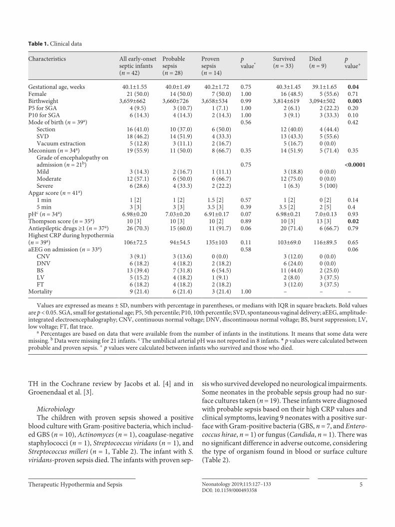
Between January 2008 and December 2017, 42 infants with perinatal asphyxia and TH showed early-onset sep-sis. Of these 42 infants, 14 infants had proven sepsis and 28 probable sepsis. Clinical data of our patients are pre-sented in Table 1. Clinical data were not significantly dif-ferent between the proven and probable sepsis groups. Gestational age and birth weight were lower in the neo-nates who died (n = 9) compared to the ones who survived (n = 33); however, the 5th and the 10th percentile birth weights were similar. Infants who died had a higher Thompson score and a more severe encephalopathy. Clinical data of the patients with sepsis, such as gesta-tional age, birth weight, and severity of encephalopathy were comparable to those reported in other studies of
Therapeutic Hypothermia and Sepsis 5Neonatology 2019;115:127–133DOI: 10.1159/000493358
TH in the Cochrane review by Jacobs et al. [4] and in Groenendaal et al. [3].
MicrobiologyThe children with proven sepsis showed a positive
blood culture with Gram-positive bacteria, which includ-ed GBS (n = 10), Actinomyces (n = 1), coagulase-negative staphylococci (n = 1), Streptococcus viridans (n = 1), and Streptococcus milleri (n = 1, Table 2). The infant with S. viridans-proven sepsis died. The infants with proven sep-
sis who survived developed no neurological impairments. Some neonates in the probable sepsis group had no sur-face cultures taken (n = 19). These infants were diagnosed with probable sepsis based on their high CRP values and clinical symptoms, leaving 9 neonates with a positive sur-face with Gram-positive bacteria (GBS, n = 7, and Entero-coccus hirae, n = 1) or fungus (Candida, n = 1). There was no significant difference in adverse outcome, considering the type of organism found in blood or surface culture (Table 2).
Table 1. Clinical data
Characteristics All early-onsetseptic infants(n = 42)
Probablesepsis(n = 28)
Provensepsis(n = 14)
pvalue*
Survived(n = 33)
Died(n = 9)
pvalue+
Gestational age, weeks 40.1±1.55 40.0±1.49 40.2±1.72 0.75 40.3±1.45 39.1±1.65 0.04Female 21 (50.0) 14 (50.0) 7 (50.0) 1.00 16 (48.5) 5 (55.6) 0.71Birthweight 3,659±662 3,660±726 3,658±534 0.99 3,814±619 3,094±502 0.003P5 for SGA 4 (9.5) 3 (10.7) 1 (7.1) 1.00 2 (6.1) 2 (22.2) 0.20P10 for SGA 6 (14.3) 4 (14.3) 2 (14.3) 1.00 3 (9.1) 3 (33.3) 0.10Mode of birth (n = 39a) 0.56 0.42
Section 16 (41.0) 10 (37.0) 6 (50.0) 12 (40.0) 4 (44.4)SVD 18 (46.2) 14 (51.9) 4 (33.3) 13 (43.3) 5 (55.6)Vacuum extraction 5 (12.8) 3 (11.1) 2 (16.7) 5 (16.7) 0 (0.0)
Meconium (n = 34a) 19 (55.9) 11 (50.0) 8 (66.7) 0.35 14 (51.9) 5 (71.4) 0.35Grade of encephalopathy on admission (n = 21b) 0.75 <0.0001Mild 3 (14.3) 2 (16.7) 1 (11.1) 3 (18.8) 0 (0.0)Moderate 12 (57.1) 6 (50.0) 6 (66.7) 12 (75.0) 0 (0.0)Severe 6 (28.6) 4 (33.3) 2 (22.2) 1 (6.3) 5 (100)
Apgar score (n = 41a)1 min 1 [2] 1 [2] 1.5 [2] 0.57 1 [2] 0 [2] 0.145 min 3 [3] 3 [3] 3.5 [3] 0.39 3.5 [2] 2 [5] 0.4
pHc (n = 34a) 6.98±0.20 7.03±0.20 6.91±0.17 0.07 6.98±0.21 7.0±0.13 0.93Thompson score (n = 35a) 10 [3] 10 [3] 10 [2] 0.89 10 [3] 13 [3] 0.02Antiepileptic drugs ≥1 (n = 37a) 26 (70.3) 15 (60.0) 11 (91.7) 0.06 20 (71.4) 6 (66.7) 0.79Highest CRP during hypothermia (n = 39a) 106±72.5 94±54.5 135±103 0.11 103±69.0 116±89.5 0.65aEEG on admission (n = 33a) 0.58 0.06
CNV 3 (9.1) 3 (13.6) 0 (0.0) 3 (12.0) 0 (0.0)DNV 6 (18.2) 4 (18.2) 2 (18.2) 6 (24.0) 0 (0.0)BS 13 (39.4) 7 (31.8) 6 (54.5) 11 (44.0) 2 (25.0)LV 5 (15.2) 4 (18.2) 1 (9.1) 2 (8.0) 3 (37.5)FT 6 (18.2) 4 (18.2) 2 (18.2) 3 (12.0) 3 (37.5)
Mortality 9 (21.4) 6 (21.4) 3 (21.4) 1.00 – – –
Values are expressed as means ± SD, numbers with percentage in parentheses, or medians with IQR in square brackets. Bold values are p < 0.05. SGA, small for gestational age; P5, 5th percentile; P10, 10th percentile; SVD, spontaneous vaginal delivery; aEEG, amplitude-integrated electroencephalography; CNV, continuous normal voltage; DNV, discontinuous normal voltage; BS, burst suppression; LV, low voltage; FT, flat trace.
a Percentages are based on data that were available from the number of infants in the institutions. It means that some data were missing. b Data were missing for 21 infants. c The umbilical arterial pH was not reported in 8 infants. * p values were calculated between probable and proven sepsis. + p values were calculated between infants who survived and those who died.

Hakobyan et al.Neonatology 2019;115:127–133 6DOI: 10.1159/000493358
OutcomeImagingFindings of cranial MRI examinations at follow-up are
presented in Table 3. The MRI showed no abnormalities in 51.4% of the infants with sepsis. Infants who died had more severe MRI abnormalities (p < 0.0001). Four infants (11.4%) had a near total pattern on the MRI. Of the 4 in-fants with a near total pattern, 3 died and 1 survived but developed neurological disabilities. The aEEG of these 4 neonates showed a flat trace or continuous low voltage and 2 had a Thompson score of > 11. Furthermore, 6 neo-nates (17.1%) had BGT involvement on the MRI, and 7 neonates (20%) had a watershed-type injury. One infant with a BGT pattern died and 2 developed neurological disabilities. Finally, there was no difference in MRI results between proven and probable sepsis (p = 0.992).
MortalityThe overall mortality among septic infants with TH
after asphyxia was 21.4%. Two infants died shortly after admission due to severe sepsis, 7 others died after redirec-tion of care following severe brain injury which was dem-onstrated using MRI. Postmortem examination was per-formed in 2 infants, confirming the multi-organ involve-ment and MRI findings. No significant difference was
found in mortality between the proven and probable sep-sis groups. The mortality in the present study (21.4%) was comparable to the previous study (31.8%) and the Co-chrane review (26.8%; Table 4).
Follow-UpOutcome data on Neurodevelopmental disabilities
or CP are presented in Table 4. Of the 42 neonates, 33 (78.6%) infants survived. Among the survivors, 5 (15.1%) had neurodevelopmental impairment including CP. Three infants were too young to be formally tested or had no follow-up. The remaining 25 infants with perinatal as-phyxia and early-onset sepsis were normal (59.5%). Hy-pothermia-treated survivors with sepsis had no differ-ence in the incidence of adverse outcome compared to the previous TH studies.
Discussion
In the present study, the outcome of septic neonates who underwent TH was reported. During the study pe-riod, 42 of the 1,084 infants (3.9%) had proven or prob-able early-onset sepsis. Whereas one-third had an adverse outcome, more than 60% was normal at 18 months or later. An additional 2 younger infants were too young to be formally tested but were normal at this younger age. These outcomes are comparable to the data reported in large RCTs and the results of previously reported patients in the Netherlands and Flanders, Belgium, without sepsis [4, 17–20].
Table 2. Bacteria cultured in infants with a proven or probable sep-sis
Provensepsis(n = 14)
Probablesepsis(n = 28)
Adverse outcome
Surface or blood culturea
GBS 10 (71.4) 7 (77.8) 6CNS 1 (7.1) – 0Actinomyces oris 1 (7.1) – 0Streptococcus milleri 1 (7.1) – 0Streptococcus viridansb 1 (7.1) – 1Enterococcus hirae – 1 (11.1) 0Candida – 1 (11.1) 0Unknownc – 19 (45.2) 7
Values are expressed as numbers with percentages in parentheses. GBS, group B streptococcus; CNS, coagulase-negative staphylococcus.
a Surface culture was taken from the ear and/or umbilicus, with missing data for 19 infants. b This infant with S. viridans sepsis developed a coinfection with CNS at birth. c These infants were included in the probable sepsis group due to clinical signs of sepsis and a CRP >50 mg/L.
Table 3. MRI data and survival
Characteristics Probablesepsis(n = 28)
Proven sepsis(n = 14)
Survived(n = 33)
Died(n = 9)
MRI (n = 35)a
Normal 13 (46.4) 5 (35.7) 18 (54.5) 0 (0.0)WS 5 (17.8) 2 (14.3) 7 (21.2) 0 (0.0)BGT 4 (14.3) 2 (14.3) 5 (15.2) 1 (11.1)NT 3 (10.7) 1 (7.1) 1 (3.0) 3 (33.3)
Not performed 3 (10.7) 4 (28.6) 2 (6.1) 5 (55.6)
Values are expressed as numbers with percentages in parentheses. MRI, magnetic resonance imaging; WS, watershed pattern of injury; BGT, basal ganglia and thalamus pattern of injury; NT, “near total” pattern of injury.
a MRI was not performed in 7 infants, of whom 5 died.

Therapeutic Hypothermia and Sepsis 7Neonatology 2019;115:127–133DOI: 10.1159/000493358
Infections with GBS are still an important cause of se-rious morbidity in neonates [21]. In the present study, the outcome of infants with infections caused by GBS was not different from infections caused by other organisms. In-fections with Gram-negative organisms were not seen in the present study. In the Netherlands, early-onset sepsis with Gram-negative organisms in full-term infants is very rare (data from the Netherlands Perinatal Registry, www.perined.nl).
TH has a neuroprotective effect by influencing differ-ent pathways including metabolism, cerebral blood flow, the release of excitatory amino acids, and apopto-sis. Furthermore, TH has an antioxidant effect, the abil-ity to block the proinflammatory cascade and reduce ATP loss [6, 22]. During sepsis, metabolic demands in different organs are high due to the inflammation re-sponse, which may increase neuronal apoptosis and subsequent neurological damage. Based on this theory, TH could also be effective in infants with early-onset sepsis. In contrast, hypothermia may suppress the po-tentially protective inflammatory cascade and may re-sult in functional immune compromise, leading to an adverse outcome in infants with sepsis [23]. Animal ex-periments have described conflicting results in models of perinatal asphyxia and infections. Neuroprotective effects have been described in neonatal models of Gram-positive sepsis and TH [11], whereas a lack of effects has been detected in neonatal models of Gram-negative sep-sis and TH [9, 10]. In contrast, prolonged survival in Gram-negative sepsis was documented in adult models of Gram-negative and Gram-positive sepsis and TH [12, 24].
The large trials of TH in perinatal asphyxia and NE have not described the effects of TH in infants with early-
onset sepsis in much detail. TH may increase the risk of infections [23]. A meta-analysis in adults strongly sug-gested an association between TH and the risk of pneu-monia and sepsis [8]. In main randomized trails, solely 5–11.3% of infants developed sepsis [17, 18, 20, 25]. How-ever, early-onset sepsis has not been defined in much de-tail in most trials, and in many studies no difference was reported between early and late-onset sepsis. In the pres-ent study, 14 of the infants developed late-onset sepsis which is higher number compared to the study of Jacobs et al. [25].
Our study has several limitations. First, it had a retro-spective design, and some clinical data were not reported in much detail. Furthermore, some units did not perform routine surface cultures, thereby limiting the detection of the causative organism in infants with clinical sepsis. The effect of TH on CRP levels is controversial [26, 27]. Nev-ertheless, by using very high cutoff values for CRP (> 50 mg/L), twice the upper level as those mentioned by oth-ers [28, 29], and a clinical picture of early-onset sepsis, we considered the risk of false positives to be low. Sec-ond, follow-up was not performed uniformly, which may have led to somewhat diverse outcome data. By using cutoff values of the separate tests, we were able to iden-tify infants with an adverse outcome. Third, the numbers of sepsis cases were too small to provide detailed infor-mation on the outcome of Gram-positive versus Gram-negative neonatal sepsis, but the sample size was large enough to demonstrate that neuroprotection by TH was retained in infants with perinatal asphyxia and early-on-set sepsis. Furthermore, no lumbar puncture was per-formed in most infants because of the severity of illness, and the presence of accompanying meningitis is un-known.
Table 4. Outcome of septic neonates with TH after asphyxia compared to previous studies [3, 4]
Outcome Early-onset sepsis(n = 42)
Groenendaal et al. [3](n = 308)
p value* Cochrane review [4] (n = 678)
p value+
Normal outcome 26 (61.9) 168 (54.5) 0.37 366 (54.0) 0.32Neurodevelopmental
impairment or CP 5 (11.9) 42 (13.6) 0.76 130 (19.2) 0.24Mortality 9 (21.4) 98 (31.8) 0.17 182 (26.8) 0.44Adverse outcome 14 (33.3) 140 (45.5) 0.14 312 (46.0) 0.11Too young to be tested 2 (4.8) – – – –
Values are expressed as numbers with percentages in parentheses. * Significant difference was calculated between the early-onset sepsis group and Groenendaal et al. [3]. + Significant difference was calculated between the early-onset sepsis group and the Cochrane review [4].

Hakobyan et al.Neonatology 2019;115:127–133 8DOI: 10.1159/000493358
Conclusion
A good outcome was reported in more than 60% of infants with perinatal asphyxia, sepsis, and therapeutic hypothermia. Therapeutic hypothermia should not be withheld from infants with perinatal asphyxia, neonatal encephalopathy, and early-onset sepsis.
Acknowledgements
The authors, who are members of the Dutch-Flemish Working Group on Neonatal Neurology, thank the other members for the valuable comments and suggestions.
Statement of Ethics
For this observational study analyzing and reporting a large set of anonymized data a waiver of informed consent was obtained according to European legislation.
Funding Sources
Financial support for publication of the study was received from the Stichting Neonatale Neurologie Utrecht (www.snnu.nl).
References
1 Pin TW, Eldridge B, Galea MP. A review of developmental outcomes of term infants with post-asphyxia neonatal encephalopathy. Eur J Paediatr Neurol. 2009 May; 13(3): 224–34.
2 Kurinczuk JJ, White-Koning M, Badawi N. Epidemiology of neonatal encephalopathy and hypoxic-ischaemic encephalopathy. Ear-ly Hum Dev. 2010 Jun; 86(6): 329–38.
3 Groenendaal F, Casaer A, Dijkman KP, Gavilanes AW, de Haan TR, ter Horst HJ, et al. Introduction of hypothermia for neonates with perinatal asphyxia in the Netherlands and Flanders. Neonatology. 2013; 104(1): 15–21.
4 Jacobs SE, Berg M, Hunt R, Tarnow-Mordi WO, Inder TE, Davis PG. Cooling for new-borns with hypoxic ischaemic encephalopa-thy. Cochrane Database Syst Rev. 2013 Jan;1(1):CD003311.
5 Jenster M, Bonifacio SL, Ruel T, Rogers EE, Tam EW, Partridge JC, et al. Maternal or neo-natal infection: association with neonatal en-cephalopathy outcomes. Pediatr Res. 2014 Jul;76(1): 93–9.
6 Hassell KJ, Ezzati M, Alonso-Alconada D, Hausenloy DJ, Robertson NJ. New horizons for newborn brain protection: enhancing en-dogenous neuroprotection. Arch Dis Child Fetal Neonatal Ed. 2015 Nov; 100(6):F541–52.
7 Fries M, Stoppe C, Brücken D, Rossaint R, Kuhlen R. Influence of mild therapeutic hy-pothermia on the inflammatory response af-ter successful resuscitation from cardiac ar-rest. J Crit Care. 2009 Sep; 24(3): 453–7.
8 Geurts M, Macleod MR, Kollmar R, Kremer PH, van der Worp HB. Therapeutic hypo-thermia and the risk of infection: a systematic review and meta-analysis. Crit Care Med. 2014 Feb; 42(2): 231–42.
9 Osredkar D, Thoresen M, Maes E, Flatebø T, Elstad M, Sabir H. Hypothermia is not neuro-protective after infection-sensitized neonatal hypoxic-ischemic brain injury. Resuscitation. 2014 Apr; 85(4): 567–72.
10 Osredkar D, Sabir H, Falck M, Wood T, Maes E, Flatebø T, et al. Hypothermia Does Not Re-verse Cellular Responses Caused by Lipopoly-saccharide in Neonatal Hypoxic-Ischaemic Brain Injury. Dev Neurosci. 2015; 37(4–5):
390–7.11 Falck M, Osredkar D, Maes E, Flatebø T,
Wood TR, Sabir H, et al. Hypothermic Neuronal Rescue from Infection-Sensitised Hypoxic-Ischaemic Brain Injury Is Patho- gen Dependent. Dev Neurosci. 2017; 39(1–4):
238–47.12 Rim KP, Kim K, Jo YH, Lee JH, Rhee JE, Kang
KW, et al. Effect of therapeutic hypothermia according to severity of sepsis in a septic rat model. Cytokine. 2012 Dec; 60(3): 755–61.
13 Hoftiezer L, Hukkelhoven CW, Hogeveen M, Straatman HM, van Lingen RA. Defining small-for-gestational-age: prescriptive versus descriptive birthweight standards. Eur J Pedi-atr. 2016 Aug; 175(8): 1047–57.
14 Thoresen M, Hellström-Westas L, Liu X, de Vries LS. Effect of hypothermia on ampli-tude-integrated electroencephalogram in infants with asphyxia. Pediatrics. 2010 Jul;126(1):e131–9.
15 van Rooij LG, Toet MC, van Huffelen AC, Groenendaal F, Laan W, Zecic A, et al. Effect of treatment of subclinical neonatal seizures detected with aEEG: randomized, controlled trial. Pediatrics. 2010 Feb; 125(2):e358–66.
16 de Vries LS, Groenendaal F. Patterns of neo-natal hypoxic-ischaemic brain injury. Neuro-radiology. 2010 Jun; 52(6): 555–66.
17 Gluckman PD, Wyatt JS, Azzopardi D, Bal-lard R, Edwards AD, Ferriero DM, et al. Selec-tive head cooling with mild systemic hypo-thermia after neonatal encephalopathy: mul-ticentre randomised trial. Lancet. 2005 Feb;
365(9460): 663–70.18 Shankaran S, Laptook AR, Ehrenkranz RA,
Tyson JE, McDonald SA, Donovan EF, et al.; National Institute of Child Health and Hu-man Development Neonatal Research Net-work. Whole-body hypothermia for neonates with hypoxic-ischemic encephalopathy. N Engl J Med. 2005 Oct; 353(15): 1574–84.
19 Azzopardi DV, Strohm B, Edwards AD, Dyet L, Halliday HL, Juszczak E, et al.; TOBY Study Group. Moderate hypothermia to treat peri-natal asphyxial encephalopathy. N Engl J Med. 2009 Oct; 361(14): 1349–58.
20 Simbruner G, Mittal RA, Rohlmann F, Muche R; neo.nEURO.network Trial Participants. Systemic hypothermia after neonatal enceph-alopathy: outcomes of neo.nEURO.network RCT. Pediatrics. 2010 Oct; 126(4):e771–8.
21 Tann CJ, Martinello KA, Sadoo S, Lawn JE, Seale AC, Vega-Poblete M, et al.; GBS Neona-tal Encephalopathy Investigator Group. Neo-natal Encephalopathy With Group B Strepto-coccal Disease Worldwide: Systematic Re-view, Investigator Group Datasets, and Meta-analysis. Clin Infect Dis. 2017 Nov; 65 suppl_2:S173–89.
22 Wassink G, Gunn ER, Drury PP, Bennet L, Gunn AJ. The mechanisms and treatment of asphyxial encephalopathy. Front Neurosci. 2014 Feb; 8: 40.
23 Jenkins DD, Lee T, Chiuzan C, Perkel JK, Rol-lins LG, Wagner CL, et al. Altered circulating leukocytes and their chemokines in a clinical trial of therapeutic hypothermia for neonatal hypoxic ischemic encephalopathy*. Pediatr Crit Care Med. 2013 Oct; 14(8): 786–95.

Therapeutic Hypothermia and Sepsis 9Neonatology 2019;115:127–133DOI: 10.1159/000493358
24 Chang YT, Wann SR, Tsai JS, Kao CH, Lee PT, Huang NC, et al. The role of autonomic nervous system function in hypothermia-me-diated sepsis protection. Am J Emerg Med. 2013 Feb; 31(2): 375–80.
25 Jacobs SE, Morley CJ, Inder TE, Stewart MJ, Smith KR, McNamara PJ, et al.; Infant Cool-ing Evaluation Collaboration. Whole-body hypothermia for term and near-term new-borns with hypoxic-ischemic encephalopa-thy: a randomized controlled trial. Arch Pedi-atr Adolesc Med. 2011 Aug; 165(8): 692–700.
26 Okumuş N, Beken S, Aydın B, Erol S, Dursun A, Fettah N, et al. Effect of therapeutic hypo-thermia on C-reactive protein levels in pa-tients with perinatal asphyxia. Am J Perinatol. 2015 Jun; 32(7): 667–74.
27 Chakkarapani E, Davis J, Thoresen M. T herapeutic hypothermia delays the C-reac-tive protein response and suppresses white blood cell and platelet count in infants with neonatal encephalopathy. Arch Dis Child Fetal Neonatal Ed. 2014 Nov;99(6):F458–63.
28 Celik IH, Demirel FG, Uras N, Oguz SS, Erdeve O, Biyikli Z, et al. What are the cut-off levels for IL-6 and CRP in neonatal sepsis? J Clin Lab Anal. 2010; 24(6): 407–12.
29 Xu L, Li Q, Mo Z, You P. Diagnostic value of C-reactive protein in neonatal sepsis: A meta-analysis. Eur J Inflamm. 2016; 14(2): 100–8.

ABM Protocol
ABM Clinical Protocol #8:Human Milk Storage Information for Home Use
for Full-Term Infants, Revised 2017
Anne Eglash,1 Liliana Simon,2 and The Academy of Breastfeeding Medicine
A central goal of The Academy of Breastfeeding Medicine is the development of clinical protocols, free fromcommercial interest or influence, for managing common medical problems that may impact breastfeedingsuccess. These protocols serve only as guidelines for the care of breastfeeding mothers and infants and do notdelineate an exclusive course of treatment or serve as standards of medical care. Variations in treatment maybe appropriate according to the needs of an individual patient.
Background
Breastfeeding mothers may encounter unforeseenreasons for separation from their infants, but more often
women express and store milk for planned events, lifestyleflexibility, and returning to work. Knowledge of appropriatehuman milk handling and storage is essential for breastfeed-ing success in these situations. One study indicated that al-though most women store their milk as recommended,*12%heated their milk in a microwave, and 17% rinsed bottlenipples/teats with only water before reuse,1 which may reducethe milk’s biological properties and increase risk of contam-ination, respectively. Another study showed that neonatalnurses’ knowledge and practice of breast milk collection andstorage were adequate, however, there was inadequacy re-lated to discarding, storing, and thawing breast milk.2
Human milk is a fresh, living food with many antioxidant,antibacterial, prebiotic, probiotic, and immune-boostingproperties in addition to nutrients. Although some of thesenutrients and health properties change with storage, there isgood evidence that human milk storage can be safe, allowingprovision of optimal nutrition to the child when breastfeedingor immediately expressed milk is not available. When directbreastfeeding is not possible, stored human milk maintainsunique qualities, such that it continues to be the gold standardfor infant feeding.
Preparation for Human Milk Storage
1. Washing: Women should wash their hands with soapand water, or a waterless hand cleanser if their handsdon’t appear dirty, before milk expression. Uncleanhands may transmit viruses and bacteria, some of
which can cause illness. Studies show that human milkcontaining fewer bacteria at the time of expressiondevelops less bacterial growth during storage and hashigher protein levels compared to milk that has anabundance of bacteria.3–5 Additional hand hygiene andcleaning of the breasts before expression are not nec-essary.6 (IIB) (Quality of evidence [levels of evidenceIA, IB, IIA, IIB, III, and IV] is based on levels ofevidence used for the National Guidelines ClearingHouse7 and is noted in parentheses.)
2. Hand or Pump: Milk expression can be achieved by handor by a pump. As long as the appropriate steps are takenfor hand cleansing and cleaning of pump parts as per thepump manufacturer’s instructions, there does not seem tobe a difference in milk contamination with pumpingversus hand expression.8,9 (IIB, IV) There is no need todiscard the first few drops of milk with initiating milkexpression. This milk is not more likely to be contami-nated than milk that is subsequently expressed.7 Onestudy found that milk expressed at home appears to havemore bacterial contamination than milk expressed at thehospital, possibly related to equipment at home ortransport, not related to personal hygiene.6 (IIB)
3. Storage Container Choice: Several studies have beendone to evaluate a range of available storage containers.There is a significant reduction in percent of fat and anincrease in total protein and carbohydrate concentra-tions with either glass or polyethylene, polypropylene,polycarbonate, or polyethersulfone bottles or bags.10
Glass and polypropylene containers appear similar intheir effects on adherence of lipid-soluble nutrients to thecontainer surface,11 the concentration of immunoglobulin
1Department of Family and Community Medicine, University of Wisconsin School of Medicine and Public Health, Madison, Wisconsin.2Department of Pediatrics, Pediatric Critical Care, University of Maryland School of Medicine, Baltimore, Maryland.
BREASTFEEDING MEDICINEVolume 12, Number 7, 2017ª Mary Ann Liebert, Inc.DOI: 10.1089/bfm.2017.29047.aje
10

A (IgA), and the numbers of viable white blood cells in thestored milk.12 Use of polyethylene containers was asso-ciated with a marked drop (60%) of IgA12 and milk’sbactericidal effect when compared to Pyrex, a type oftempered glass.13 Steel containers were associated with amarked decline in cell count and cell viability whencompared to polyethylene14 and glass.15 (IIB)
There has been concern about possible contamina-tion of milk stored in polypropylene bags because of therisk of contamination by puncturing the plastic.16 (IV)However, one study showed no difference betweencontamination and fat loss when comparing hard andsoft polypropylene containers.17 Therefore, plastic bagsused for human milk storage should be sturdy, sealedwell, and stored in an area of the freezer where damageto the bag would be minimized. (IIB) Containers madewith bisphenol A, which is found in several plasticcontainers including baby bottles, should be avoidedbased on strong evidence of its adverse effects as anendocrine disruptor.18 There should be caution aboutthe use of bottles with bisphenol S, a bisphenol A al-ternative, as it may also have deleterious effects, al-though this is not well established in the literature.
Human milk should not be stored in hospital plasticspecimen storage containers such as those used for ur-ine or other bodily fluids because there is insufficientevidence regarding their chemical safety and effects oninfants’ health;19 only food grade plastic containersshould be used for human milk storage. (IV)
4. Care of Containers: Containers for human milk storage andbreast pump milk collection kits must be completely dis-mantled, washed in hot soapy water and rinsed or washedin a dishwasher,8 and should always be thoroughly airdried or dried with paper towels.20 They do not need to besterilized. If soap is not available, then boiling water ispreferable. (IIB) Chemical disinfection is not ideal, as thedisinfectant can be easily deactivated and could exposeinfant to unnecessary risk of both inadequately cleancontainers and residual chemical disinfectant.20 (IV)
Storage of Human Milk
1. Freshly expressed human milk may be stored safely atroom temperature (10–29�C, 50–85�F) for some pe-riod of time. Studies suggest different optimal timesfor room temperature storage because conditions varygreatly in the cleanliness of milk expression techniqueand the room temperature. Warmer ambient tempera-tures are associated with faster growing bacterialcounts in stored milk. For room temperatures rangingfrom 27�C to 32�C (29�C = 85�F), 4 hours may be areasonable limit.5,21,22 For very clean expressed milkwith very low bacterial counts, 6–8 hours at lowerroom temperatures may be reasonable, but it is best tochill or refrigerate as soon as possible if the milk willnot be used during that time.4,23–25 (IIB)
2. Ice packs: Very few studies have evaluated milkstorage safety at 15�C (59�F), which would beequivalent to an ice pack in a small cooler. Hamoshet al.21 suggested that human milk is safe at 15�C for24 hours, based on minimal bacterial growth noted inthe samples from their study. (IIB)
3. Refrigeration: Several studies have demonstrated thesafety of refrigerating human milk (4�C, 39.2�F), eitherby evaluating the bactericidal capacity of stored milk asa marker for milk quality or by measuring bacterialgrowth in the stored milk samples. Bactericidal capacityof stored refrigerated human milk declines significantlyby 48–72 hours.26–28 However, studies of expressedhuman milk with little contamination at the time ofexpression demonstrate safe, low levels of bacteriagrowth in milk at 72 hours24 and even after 4–8 days ofrefrigeration.3,4,29
Few studies have been done on the change in milkcomposition during refrigerator storage. One studyfound that lipid composition and lipase activity re-mained stable up to 96 hours in the refrigerator.30
Lactoferrin levels are stable in the refrigerator for 4–5 days.31,32 Many immunologic factors in colostrumsuch as IgA, cytokines, and growth factors are not di-minished with refrigeration for 48 hours.33 (IIB)
4. Freezing expressed human milk (0�F, -18�C) has beendemonstrated to be safe for at least 3 months. Evidenceindicates that thawed human milk, previously frozen forat least 6 weeks at -20�C (-4�F), has the same bacterialviability and diversity as it did when it was freshly ex-pressed.34 The basic principles of freezing dictate thatfrozen foods at -18�C (0�F) are safe indefinitely frombacterial contamination, although enzymatic processesinherent in food could persist, with possible changes inmilk quality.35
Fat, protein, and calories decrease in human milk whenfrozen for 90 days compared to fresh human milk.36
Frozen human milk has a significant increase in acidity by3 months, likely due to ongoing lipase activity, that in-creases free fatty acids in the milk.37 Based on a fewstudies with very small samples sizes, vitamin E appearsstable in frozen milk over time, and vitamin C levels de-crease significantly after 1–5 months of storage.38,39 Thereis a paucity of research on how freezer storage affectsnearly all vitamins and minerals in human milk.38–40
Bioactive factors in human milk variably diminishwith freezing. Lactoferrin levels and bioactivity aresignificantly lower in human milk frozen at -20�C for 3months.13,31,32 However, several cytokines, IgA andgrowth factors from colostrum are stable for at least 6months at -20�C (-4�F).10,33 One trial evaluating milkfrozen for 9 months found a progressive decline in pHand in bacterial counts, and increases in nonesterifiedfatty acids. Other macronutrients, osmolality, and im-munoactive proteins remained unchanged in this studyafter 9 months.41 Frozen human milk should be storedin the back of the freezer to prevent intermittent re-warming due to freezer door opening, and should bekept away from the walls of self-defrosting freezers. Allcontainers with human milk should be well sealed toprevent contamination. (IIB)
5. Smell of stored milk: Refrigerated and frozen humanmilk may have an odor different from fresh milk due tolipase-mediated triglyceride breakdown, releasing fattyacids. The odor likely comes from oxidation of thesefatty acids.42,43 This lipolysis process has antimicrobialeffects preventing the growth of microorganisms inthawed refrigerated milk.44 There is no evidence to
ABM PROTOCOL 11
suggest that infants often reject human milk due to thisodor. Many foods that humans eat, such as eggs, cheese,and fish, have an unpleasant odor that does not affecttaste. One study demonstrated that freezing human milkto -80�C (-112�F) leads to less change in smell ascompared to conventional freezing to -19�C.43 Heatingmilk to above 40�C to deactivate lipase is not advisedbecause this may destroy many of the immunologicallyactive factors in human milk. (IIB)
6. Expansion while freezing: When filling a container withhuman milk, space should be left at the top to allow forexpansion with freezing. All stored containers of humanmilk should be labeled with the date of milk expressionand the name of the child if the milk will be used in achild-care setting. It is typical for infants in daycare totake 60–120 mL (2–4 ounces) of human milk at onefeeding. Therefore, storing human milk in a variety ofsmall increments such as 15–60 mL is a convenient wayto prevent waste of thawed human milk.
7. Mixing milk: Freshly expressed warm milk should notbe added to already cooled or frozen milk, to preventrewarming of the already stored milk. It is best to cooldown the newly expressed milk first before adding it toolder stored milk.
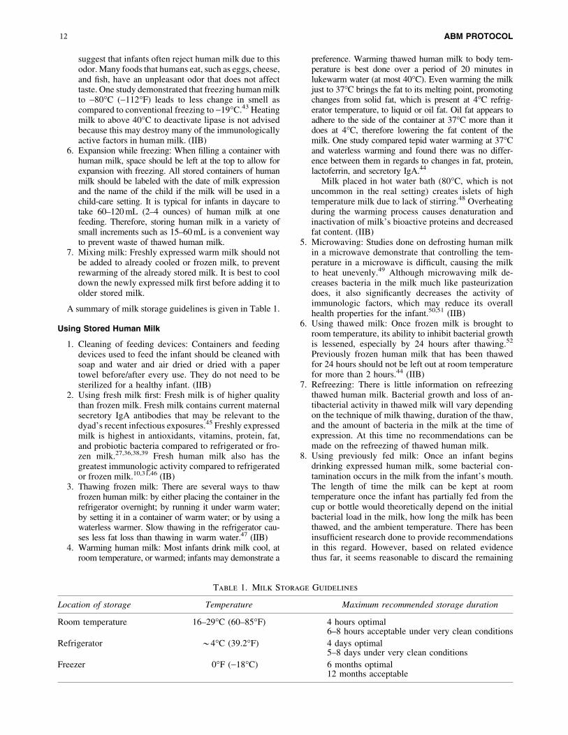
A summary of milk storage guidelines is given in Table 1.
Using Stored Human Milk
1. Cleaning of feeding devices: Containers and feedingdevices used to feed the infant should be cleaned withsoap and water and air dried or dried with a papertowel before/after every use. They do not need to besterilized for a healthy infant. (IIB)
2. Using fresh milk first: Fresh milk is of higher qualitythan frozen milk. Fresh milk contains current maternalsecretory IgA antibodies that may be relevant to thedyad’s recent infectious exposures.45 Freshly expressedmilk is highest in antioxidants, vitamins, protein, fat,and probiotic bacteria compared to refrigerated or fro-zen milk.27,36,38,39 Fresh human milk also has thegreatest immunologic activity compared to refrigeratedor frozen milk.10,31,46 (IB)
3. Thawing frozen milk: There are several ways to thawfrozen human milk: by either placing the container in therefrigerator overnight; by running it under warm water;by setting it in a container of warm water; or by using awaterless warmer. Slow thawing in the refrigerator cau-ses less fat loss than thawing in warm water.47 (IIB)
4. Warming human milk: Most infants drink milk cool, atroom temperature, or warmed; infants may demonstrate a
preference. Warming thawed human milk to body tem-perature is best done over a period of 20 minutes inlukewarm water (at most 40�C). Even warming the milkjust to 37�C brings the fat to its melting point, promotingchanges from solid fat, which is present at 4�C refrig-erator temperature, to liquid or oil fat. Oil fat appears toadhere to the side of the container at 37�C more than itdoes at 4�C, therefore lowering the fat content of themilk. One study compared tepid water warming at 37�Cand waterless warming and found there was no differ-ence between them in regards to changes in fat, protein,lactoferrin, and secretory IgA.44
Milk placed in hot water bath (80�C, which is notuncommon in the real setting) creates islets of hightemperature milk due to lack of stirring.48 Overheatingduring the warming process causes denaturation andinactivation of milk’s bioactive proteins and decreasedfat content. (IIB)
5. Microwaving: Studies done on defrosting human milkin a microwave demonstrate that controlling the tem-perature in a microwave is difficult, causing the milkto heat unevenly.49 Although microwaving milk de-creases bacteria in the milk much like pasteurizationdoes, it also significantly decreases the activity ofimmunologic factors, which may reduce its overallhealth properties for the infant.50,51 (IIB)
6. Using thawed milk: Once frozen milk is brought toroom temperature, its ability to inhibit bacterial growthis lessened, especially by 24 hours after thawing.52
Previously frozen human milk that has been thawedfor 24 hours should not be left out at room temperaturefor more than 2 hours.44 (IIB)
7. Refreezing: There is little information on refreezingthawed human milk. Bacterial growth and loss of an-tibacterial activity in thawed milk will vary dependingon the technique of milk thawing, duration of the thaw,and the amount of bacteria in the milk at the time ofexpression. At this time no recommendations can bemade on the refreezing of thawed human milk.
8. Using previously fed milk: Once an infant beginsdrinking expressed human milk, some bacterial con-tamination occurs in the milk from the infant’s mouth.The length of time the milk can be kept at roomtemperature once the infant has partially fed from thecup or bottle would theoretically depend on the initialbacterial load in the milk, how long the milk has beenthawed, and the ambient temperature. There has beeninsufficient research done to provide recommendationsin this regard. However, based on related evidencethus far, it seems reasonable to discard the remaining
Table 1. Milk Storage Guidelines
Location of storage Temperature Maximum recommended storage duration
Room temperature 16–29�C (60–85�F) 4 hours optimal6–8 hours acceptable under very clean conditions
Refrigerator *4�C (39.2�F) 4 days optimal5–8 days under very clean conditions
Freezer 0�F (-18�C) 6 months optimal12 months acceptable
12 ABM PROTOCOL
milk within 1–2 hours after the infant is finishedfeeding. (IV) To avoid wasting or discarding unfedmilk, mothers may consider storing milk in a variety ofincrements such as 15, 30, or 60 mL.
9. Handling: Expressed human milk does not requirespecial handling (such as universal precautions), as isrequired for other bodily fluids such as blood. It can bestored in a workplace refrigerator where other workersstore food, although it should be labeled with nameand date.53 (IV) Mothers may prefer to store their milkin a personal freezer pack or cooler, separate fromcommunal refrigerator areas.
10. Infections: Uncontaminated human milk naturallycontains nonpathogenic bacteria54,55 that are impor-tant in establishing the neonatal intestinal flora. Thesebacteria are probiotics—they create conditions in theintestine that are unfavorable to the growth of path-ogenic organisms.55 If a mother has breast or nipplepain from a bacterial or yeast infection, there is noevidence that her stored expressed milk needs to bediscarded. Human milk that appears stringy, foul, orpurulent should, however, be discarded and not befed to the infant. (IV)
Areas for Future Research
The evidence for some aspects of human milk storage islacking. Many studies are older, and because of differences inmethodology, are difficult to compare. The studies vary inmany respects, such as technique of milk collection, clean-liness and types of containers, duration of storage, method ofthawing and warming milk, temperature and type of storageunit, and culture techniques of milk samples. Large high-quality studies evaluating human milk storage in a variety ofcircumstances over a longer duration of time are needed.Standards for evaluating milk quality, such as culture tech-niques, need to be established. Although it is ideal to have auniversal international guideline for human milk storage, itmay be impossible for one guideline to represent unusual orlimited circumstances in some cultures.
Human milk naturally has both prebiotic and probioticactivity that is essential in establishing the infant gut micro-biome. Human milk’s prebiotic components are non-digestible factors such as oligosaccharides that promote thegrowth of beneficial microorganisms in the intestines. Humanmilk’s probiotic components are commensal organisms. Be-cause of the impact of refrigeration, freezing, thawing, andwarming on the bactericidal activity of human milk, feeding aninfant stored human milk may have different consequences oninfant intestinal health compared to breastfeeding, and thisshould be investigated further. Along the same lines, storedhuman milk changes in quality over time, as demonstrated bymany of the referenced articles included in this protocol. Theeffect of stored human milk versus fresh human milk on thehealth of a child should be studied.
There is also no agreed-upon definition of unsafe milk.Several studies describe the degree of milk contaminationover a period of time under certain temperature and storagetime conditions, typically described as the number of colony-forming units per milliliter. There is no accepted limit atwhich point milk should not be consumed, although 1 · 104
colony-forming units/mL has been suggested. Other studies
have investigated the bactericidal capacity of stored humanmilk, which would reflect its immunologic effectiveness forthe infant and the risk of the milk becoming contaminatedover time during storage. The percentage loss of bactericidalactivity that would render human milk unfit has not beendetermined. A definition for adequate milk quality should beestablished, with guidelines on what would constitute unsafemilk or lower-quality milk that would necessitate discardingof stored milk.
There is only one study investigating human milk qualityafter 6 months of freezing. This is particularly concerning,given that a few very small studies have demonstrated adecline in some vitamins after 3 months of freezing. Be-cause some infants rely entirely on frozen human milk fornutrition, studies should be done to confirm that this isnutritionally safe.
References
1. Labiner-Wolfe J, Fein SB. How US mothers store and handletheir expressed breast milk. J Hum Lact 2013;29:54–58.
2. Gharaibeh H, Al-Sheyab N, Malkawi S. Breast milk col-lection and storage in the neonatal intensive care unit: Nur-ses’ knowledge, practice, and perceived barriers. J ContinEduc Nurs 2016;47:551–557.
3. Sosa R, Barness L. Bacterial growth in refrigerated humanmilk. Am J Dis Child 1987;141:111–112.
4. Pardou A, Serruys E, Mascart-Lemone F, et al. Human milkbanking: Influence of storage processes and of bacterialcontamination on some milk constituents. Biol Neonate1994;65:302–309.
5. Eteng M, Ebong P, Eyong E, et al. Storage beyond threehours at ambient temperature alters the biochemical andnutritional qualities of breastmilk. Afr J Reprod Health2001;5:130–134.
6. Haiden N, Pimpel B, Assadian O, et al. Comparison ofbacterial counts in expressed breast milk following stan-dard or strict infection control regimens in neonatal inten-sive care units: Compliance of mothers does matter. J HospInfect 2016;92:226–228.
7. Shekelle P, Woolf S, Eccles M, et al. Developing guide-lines. Br Med J 1999;318:593–596.
8. Pittard WB 3rd, Geddes K, Brown S, et al. Bacterial con-tamination of human milk: Container type and method ofexpression. Am J Perinatol 1991;8:25–27.
9. Boo N, Nordiah A, Alfizah H, et al. Contamination ofbreast milk obtained by manual expression and breastpumps in mothers of very low birthweight infants. J HospInfect 2001;49:274–281.
10. Chang Y-C, Chen C-H, Lin M-C. The macronutrients inhuman milk change after storage in various containers.Pediatr Neonatol 2012;53:205–209.
11. Garza C, Johnson C, Harrist R, et al. Effects of methods ofcollection and storage on nutrients in human milk. EarlyHum Dev 1982;6:295–303.
12. Goldblum R, Garza C, Johnson C, et al. Human milkbanking I. Effects of container upon immunologic factors inhuman milk. Nutr Res 1981;1:449–459.
13. Takci S, Gulmez D, Yigit S, et al. Effects of freezing on thebactericidal activity of human milk. J Pediatr Gastro-enterol Nutr 2012;55:146–149.
14. Manohar A, Williamson M, Koppikar G. Effect of storageof colostrum in various containers. Indian Pediatr 1997;34:293–295.
ABM PROTOCOL 13

15. Williamson M, Murti P. Effect of storage, time, tempera-ture, and composition of containers on biologic componentsof human milk. J Hum Lact 1996;12:31–35.
16. Hopkinson J, Garza C, Asquith M. Human milk storage inglass containers. J Hum Lact 1990;6:104–105.
17. Janjindamai W, Thatrimontrichai A, Maneenil G, et al.Soft plastic bag instead of hard plastic container for long-term storage of breast milk. Indian J Pediatr 2013;80:809–813.
18. Vom Saal F, Hughes C. An extensive new literature con-cerning low dose effects of bisphenol A shows the need fora new risk assessment. Environ Health Perspect 2005;113:926–933.
19. Blouin M, Coulombe M, Rhainds M. Specimen plasticcontainers used to store expressed breast milk in neonatalcare units: A case of precautionary principle. Can J PublicHealth 2014;105:e218–e220.
20. Price E, Weaver G, Hoffman P, et al. Decontamination ofbreast pump milk collection kits and related items at homeand in hospital: Guidance from a Joint Working Group ofthe Healthcare Infection Society and Infection PreventionSociety. J Hosp Infect 2016;92:213–221.
21. Hamosh M, Ellis L, Pollock D, et al. Breastfeeding and theworking mother: Effect of time and temperature of short-term storage on proteolysis, lipolysis, and bacterial growthin milk. Pediatrics 1996;97:492–498.
22. Nwankwo M, Offor E, Okolo A, et al. Bacterial growthin expressed breast milk. Ann Trop Paediatr 1988;8:92–95.
23. Pittard WB 3rd, Anderson D, Cerutti E, et al. Bacteriostaticqualities of human milk. J Pediatr 1985;107:240–243.
24. Igumbor E, Mukura R, Makandiramba B, et al. Storage ofbreast milk: Effect of temperature and storage duration onmicrobial growth. Cent Afr J Med 2000;46:247–251.
25. Ajusi J, Onyango F, Mutanda L, Wamola. Bacteriology ofunheated expressed breastmilk stored at room temperature.East Afr Med J 1989;66:381–387.
26. Martınez-Costa C, Silvestre M, Lopez M, et al. Effects ofrefrigeration on the bactericidal activity of human milk: Apreliminary study. J Pediatr Gastroenterol Nutr 2007;45:275–277.
27. Silvestre D, Lopez M, March L, et al. Bactericidal activityof human milk: Stability during storage. Br J Biomed Sci2006;63:59–62.
28. Ogundele M. Effects of storage on the physicochemical andantibacterial properties of human milk. Br J Biomed Sci2002;59:205–211.
29. Slutzah M, Codipilly C, Potak D, et al. Refrigerator storageof expressed human milk in the neonatal intensive careunit. J Pediatr 2010;156:26–28.
30. Bertino E, Giribaldi M, Baro C, et al. Effect of prolongedrefrigeration on the lipid profile, lipase activity, and oxi-dative status of human milk. J Pediatr Gastroenterol Nutr2013;56:390–396.
31. Raoof NA, Adamkin DH, Radmacher PG, et al. Compar-ison of lactoferrin activity in fresh and stored human milk.J Perinatol 2016;36:207–209.
32. Rollo DE, Radmacher PG, Turcu RM, et al. Stability oflactoferrin in stored human milk. J Perinatol 2014;34:284–286.
33. Ramırez-Santana C, Perez-Cano FJ, Audı C, et al. Effectsof cooling and freezing storage on the stability of bioac-tive factors in human colostrum. J Dairy Sci 2012;95:2319–2325.
34. Marın ML, Arroyo R, Jimenez E, et al. Cold storage ofhuman milk: Effect on its bacterial composition. J PediatrGastroenterol Nutr 2009;49:343–348.
35. USDA. Freezing and food storage. 2013. Available athttps://www.fsis.usda.gov/wps/portal/fsis/topics/food-safety-education/get-answers/food-safety-fact-sheets/safe-food-handling/freezing-and-food-safety/ct_index/!ut/p/a1/jVFtT8IwEP417Nto55CgSWMWDCoiaFAZ-7IUetuajHa2h4K_3g4kEQNKm1zu5Xnae-5IQmKSKP4uc45SK17WcdJO6RNtBxdd2h9dBD16N3x9Gt13u7QzPneA6R-AYXgi_8iJ6H_8_gkfnJmH7kNOkopj4UuVaRLngD5X9gOMJXGmtfAtzwDXfsbn6NsCAF2hzvmbasGVKKXKHdgAfDrP0YX_g0niOaZSCViRCUn2u6KBu3fDcNy67Q9DOmr9BhwY2xZwfC5OeF7q2WZH00jNwo5TaCADA6a5NC5dIFb2skEb1AI386JZd7tttpnr91366o3tNHkzVDfNFaNbZ_3tsPE (AccessedApril 2, 2017).
36. Garcıa-Lara NR, Escuder-Vieco D, Garcıa-Algar O, et al.Effect of freezing time on macronutrients and energycontent of breastmilk. Breastfeed Med 2012;7:295–301.
37. Vazquez-Roman S, Escuder-Vieco D, Garcıa-Lara NR,et al. Impact of freezing time on dornic acidity in threetypes of milk: Raw donor milk, mother’s own milk, andpasteurized donor milk. Breastfeed Med 2016;11:91–93.
38. Romeu-Nadal M, Castellote A, Lopez-Sabater M. Effect ofcold storage on vitamins C and E and fatty acids in humanmilk. Food Chem 2008;106:65–70.
39. Buss I, McGill F, Darlow B, et al. Vitamin C is reduced inhuman milk after storage. Acta Paediatr 2001;90:813–815.
40. Bank MR, Kirksey A, West K, et al. Effect of storage timeand temperature on folacin and vitamin C levels in termand preterm human milk. Am J Clin Nutr 1985;41:235–242.
41. Ahrabi A, Handa D, Codipilly C, et al. Effects of extendedfreezer storage on the integrity of human milk. J Pediatr2016;177:140–143.
42. Spitzer J, Klos K, Buettner A. Monitoring aroma changesduring human milk storage at +4�C by sensory and quan-tification experiments. Clin Nutr 2013;32:1036–1042.
43. Sandgruber S, Much D, Amann-Gassner U, et al. Sensoryand molecular characterisation of the protective effect ofstorage at -80�C on the odour profiles of human milk. FoodChem 2012;130:236–242.
44. Handa D, Ahrabi AF, Codipilly CN, et al. Do thawing andwarming affect the integrity of human milk? J Perinatol2014;34:863–866.
45. Lonnerdal B. Bioactive proteins in breast milk. J PaediatrChild Health 2013;49 Suppl 1:1–7.
46. Akinbi H, Meinzen-Derr J, Auer C, et al. Alterations in thehost defense properties of human milk following prolongedstorage or pasteurization. J Pediatr Gastroenterol Nutr2010;51:347–352.
47. Thatrimontrichai A, Janjindamai W, Puwanant M. Fat lossin thawed breast milk: Comparison between refrigeratorand warm water. Indian Pediatr 2012;49:877–880.
48. Bransburg-Zabary S, Virozub A, Mimouni FB. Humanmilk warming temperatures using a simulation of currentlyavailable storage and warming methods. PLoS One 2015;10:e0128806.
49. Ovesen L, Jakobsen J, Leth T, et al. The effect of micro-wave heating on vitamins B1 and E, and linoleic and li-nolenic acids, and immunoglobulins in human milk. Int JFood Sci Nutr 1996;47:427–436.
50. Quan R, Yang C, Rubinstein S, et al. Effects of microwaveradiation on anti-infective factors in human milk. Pedia-trics 1992;89:667–669.
14 ABM PROTOCOL

51. Sigman M, Burke K, Swarner O, et al. Effects of micro-waving human milk: Changes in IgA content and bacterialcount. J Am Diet Assoc 1989;89:690–692.
52. Hernandez J, Lemons P, Lemons J, et al. Effect of storageprocesses on the bacterial growth-inhibiting activity ofhuman breast milk. Pediatrics 1979;63:597–601.
53. CDC. Are special precautions required for handling breastmilk? 2015. Available at https://www.cdc.gov/breastfeeding/faq/#Precautions (accessed June 26, 2017).
54. Delgado S, Arroyo R, Jimenez E, et al. Mastitis infecciosasdurante la lactancia: Un problema infravalorado. Acta Pe-diatr Esp 2009;67:564–571.
55. Heikkila M, Saris P. Inhibition of Staphylococcus aureusby the commensal bacteria of human milk. J Appl Micro-biol 2003;95:471–478.
ABM protocols expire 5 years from the date of publication.Content of this protocol is up-to-date at the time of pub-
lication. Evidence-based revisions are made within 5 years orsooner if there are significant changes in the evidence.
The 2004 and 2010 editions of this protocol were authoredby Anne Eglash.The Academy of Breastfeeding Medicine Protocol Committee:
Wendy Brodribb, MBBS, PhD, FABM, ChairpersonSarah Reece-Stremtan, MD, Co-Chairperson
Larry Noble, MD, FABM, Translations ChairpersonNancy Brent, MD
Maya Bunik, MD, MSPH, FABM,Cadey Harrel, MD
Ruth A. Lawrence, MD, FABMYvonne LeFort, MD, FABM
Kathleen A. Marinelli, MD, FABMCasey Rosen-Carole, MD, MPH, MSEd
Susan Rothenberg, MDTomoko Seo, MD, FABM
Rose St. Fleur, MDMichal Young, MD
For correspondence: [email protected]
ABM PROTOCOL 15

This article has been cited by:
1. Hernández-Aguilar Maria-Teresa, Bartick Melissa, Schreck Paula, Harrel Cadey, The Academy of Breastfeeding Medicine. 2018.ABM Clinical Protocol #7: Model Maternity Policy Supportive of Breastfeeding. Breastfeeding Medicine 13:9, 559-574. [Abstract][Full Text] [PDF] [PDF Plus]
2. Enrico Bertino, Chiara Peila, Francesco Cresi, Elena Maggiora, Stefano Sottemano, Diego Gazzolo, Sertac Arslanoglu, AlessandraCoscia. 2018. Donor Human Milk: Effects of Storage and Heat Treatment on Oxidative Stress Markers. Frontiers in Pediatrics6. . [Crossref]
3. Caroline Steele. 2018. Best Practices for Handling and Administration of Expressed Human Milk and Donor Human Milk forHospitalized Preterm Infants. Frontiers in Nutrition 5. . [Crossref]
4. Schlotterer Hannah R., Perrin Maryanne T.. 2018. Effects of Refrigerated and Frozen Storage on Holder-Pasteurized DonorHuman Milk: A Systematic Review. Breastfeeding Medicine 13:7, 465-472. [Abstract] [Full Text] [PDF] [PDF Plus]
5. 2018. Correction to: ABM Clinical Protocol #8: Human Milk Storage Information for Home Use for Full-Term Infants, Revised2017, by Eglash A, Simon L, and The Academy of Breastfeeding Medicine Breastfeed Med 2017;12(7):390–395. DOI: 10.1089/dna.2017.29047.aje. Breastfeeding Medicine 13:6, 459-459. [Citation] [Full Text] [PDF] [PDF Plus]
6. Sriraman Natasha K., Evans Amy E., Lawrence Robert, Noble Lawrence, the Academy of Breastfeeding Medicine's Board ofDirectors. 2018. Academy of Breastfeeding Medicine's 2017 Position Statement on Informal Breast Milk Sharing for the TermHealthy Infant. Breastfeeding Medicine 13:1, 2-4. [Citation] [Full Text] [PDF] [PDF Plus]
7. FouziaAbdulaziz AlHreashy. 2018. Non-maternal nursing in the muslim community: A health perspective review. Journal ofClinical Neonatology 7:4, 191. [Crossref]

Thesis
Retinal oximetry and systemic arterial oxygenlevels
Thorunn Scheving Eliasdottir CRNA
Department of Ophthalmology, University of Iceland, Reykjavik, Iceland
Acta Ophthalmol. 2018: 96, thesis 113: 1–44ª 2018 Acta Ophthalmologica Scandinavica Foundation. Published by John Wiley & Sons Ltd
doi: 10.1111/aos.13932
Thesis for the degree of PhilosophiaeDoctor
Supervisor:
Professor Guðr�un Kristj�ansd�ottir,DrPH., Ph.D.
Advisor:
Professor Einar Stef�ansson, M.D.,Ph.D.
Doctoral committee:
Professor Einar Stef�ansson, M.D.,Ph.D.
Professor Guðr�un Kristj�ansd�ottir,DrPH., Ph.D.
Professor Charles Vacchiano,CRNA., Ph.D.
Professor Þ�orarinn G�ıslason, M.D.,Ph.D.
Professor G�ısli Heimir Sigurðsson,M.D., Ph.D.
June 2017
Thesis for a doctoral degree at theUniversity of Iceland. All rightreserved. No part of this publicationmay be reproduced in any form with-out the prior permission of the copy-right holder.© Þ�orunn Scheving El�ıasd�ottir 2017ISBN 978-9935-9313-4-4Printing by H�ask�olaprentReykjavik, Iceland 2017
ABSTRACT.
Purpose: Continuous peripheral pulse oximetry for monitoring adequacy of oxygenation is probably the mostimportant technological advance for patients’ monitoring and safety in the last decades. Pulse oximetry has thedisadvantage of measuring the peripheral circulation, and the only mean to measure oxygen content of the centralcirculation is by invasive technology. Determination of blood oxyhaemoglobin saturation in the retinal vessels of theeye can be achieved noninvasively through spectrophotometric retinal oximetry which provides access to the centralnervous system circulation. The aim of the thesis was to determine whether retinal oximetry technique can be appliedfor estimation of the central nervous system circulation which until now has only been possible invasively. This wasachieved by measuring oxyhaemoglobin saturation in three adult subject study groups: in people with central retinalvein occlusion (CRVO) to observe local tissue hypoxia, in patients with severe chronic obstructive pulmonary disease(COPD) on long-term oxygen therapy to observe systemic hypoxaemia and in healthy subjects during hyperoxicbreathing to observe systemic hyperoxemia. In addition, the fourth study that is mentioned was performed to testwhether retinal oximetry is feasible for neonates.Methods: Retinal oximetry in central retinal vein occlusion: Sixteen subjects with central retinal vein occlusion participatedin the study.Theoxyhaemoglobin saturationof the central retinal vein occlusionaffected eyewas comparedwith the fellowunaffected eye. Retinal oximetry in healthy people under hyperoxia: Thirty healthy subjects participated in the study, andthe oxyhaemoglobin saturation of retinal arterioles and venules was compared between normoxic and hyperoxicbreathing.Retinal oximetry in severe chronic obstructive pulmonary disease:Eleven patientswith severe chronic obstructivepulmonary disease participated in the study. Retinal oximetry measurements were made with and without their dailysupplemental oxygen therapy. Retinal arteriolar oxyhaemoglobin saturation when inspiring ambient air was comparedwith blood samples from the radial artery and finger pulse oximetry and healthy controls. The healthy control group wasassembled from our database for comparison of oxyhaemoglobin saturation of retinal arterioles and venules during theambient air breathing. The retinal oximeter is based on a conventional fundus camera and a specialized software. A beamsplitter coupled with two high-resolution digital cameras allows for simultaneous acquisition of retinal images atseparative wavelengths for calculation of oxyhaemoglobin saturation. In addition, retinal images of 28 full-term healthyneonates were obtained with scanning laser ophthalmoscope combined with modified Oxymap analysis software forcalculation of the optical density ratio and vessel diameter
17
Acta Ophthalmologica 2018
17

Results: Retinal oximetry in central retinal vein occlusion: Mean retinal venous oxyhaemoglobin saturation was31 � 12% in CRVO eyes and 52 � 11% in unaffected fellow eyes (mean � SD, n = 14, p < 0.0001). Thearteriovenous oxygen difference (AV-difference) was 63 � 11% in CRVO eyes and 43 � 7% in fellow eyes(p < 0.0001). The variability of retinal venous oxyhaemoglobin saturation was considerable within and between eyesaffected by CRVO. There was no difference in oxyhaemoglobin saturation of retinal arterioles between the CRVO eyesand the unaffected eyes (p = 0.49). Retinal oximetry in healthy people under hyperoxia: During hyperoxic breathing, theoxyhaemoglobin saturation in retinal arterioles increased to 94.5 � 3.8% as compared with 92.0 � 3.7% at baseline(n = 30, p < 0.0001). In venules, the mean oxyhaemoglobin saturation increased to 76.2 � 8.0% from 51.3 � 5.6%(p < 0.0001) at baseline. The AV-difference was markedly lower during hyperoxic breathing as compared with thenormoxic breathing (18.3 � 9.0% versus 40.7 � 5.7%, p < 0.0001). Retinal oximetry in severe chronic obstructivepulmonary disease: During ambient air breathing, chronic obstructive pulmonary disease subjects had significantlylower oxyhaemoglobin saturation than healthy controls in both retinal arterioles (87.2 � 4.9% versus 93.4 � 4.3%,p = 0.02, n = 11) and venules (45.0 � 10.3% versus 55.2 � 5.5%, p = 0.01) but the AV-difference was not markedlydifferent (p = 0.17). Administration of their prescribed oxygen therapy significantly increased the oxyhaemoglobinsaturation in retinal arterioles (87.2 � 4.9% to 89.5 � 6.0%, p = 0.02) but not in venules (45.0 � 10.3% to46.7 � 12.8%, p = 0.3). Retinal oximetry values were slightly lower than finger pulse oximetry (mean percentagepoints difference = �3.1 � 5.5) and radial artery blood values (�5.0 � 5.4). Retinal oximetry study in neonates: Themodified version of the retinal oximetry instrument estimated the optical density ratio in retinal arterioles to be0.256 � 0.041 that was significantly different from the 0.421 � 0.089 in venules (n = 28, p < 0.001, paired t-test). Thevascular diameter of retinal arterioles was markedly narrower than of venules (14.1 � 2.7 and 19.7 � 3.7 pixels,p < 0.001).Conclusion: The results of this thesis indicate that spectrophotometric retinal oximetry is sensitive to both local andsystemic changes in oxyhaemoglobin saturation. Retinal oxyhaemoglobin saturation values are slightly lower thanradial artery blood sample and finger pulse oximetry values. The discrepancies between the different modalities areexpected to derive from countercurrent exchange between central retinal artery and vein within the optic nerve butcalibration issues cannot be excluded as contributing to this difference. Despite these differences, the findings indicatethe potential of retinal oximetry for noninvasive real-time measurements of oxyhaemoglobin saturation in centralnervous system vessels. Following calibration upgrade and technological improvement, verification retinal oximetrymay potentially be applied to critically ill and anaesthesia care patients. The study on combined scanning laserophthalmoscope and retinal oximetry supports the feasibility of the technique for oximetry analysis in newly bornbabies.Key words: central retinal vein occlusion – chronic obstructive pulmonary disease – oximetry – retinal vessels – systemic circulation
�AGRIP.
Tilgangur: Innleiðing p�ulsoximælinga til samfelldrar mælingar �a s�urefnismettun bl�oðs er ein mikilvægasta tækniþr�ounundanfarinna �aratuga fyrir €oryggi og v€oktun sj�uklinga. Þær hafa þ�o þann veikleika að mæla �utæðar (peripheralcirculation) og eina leiðin til að mæla s�urefnisstyrk �ı miðlægum æðum er með �ıfarandi slagæðamælingum. Mæling �as�urefnismettun �ı sj�onhimnuæðum augans með sj�onhimnu-s�urefnismæli er hins vegar mæling �a miðlægum æðum �an�ıfarandi tækni. Sj�onhimnan er hluti miðtaugakerfisins og eru sj�onhimnuæðar þv�ı miðlægar æðar, sem samsvaras�urefnis�astandi miðtaugakerfisins að nokkru leyti.Meginmarkmið verkefnisins er að meta hvort hægt s�e að notasj�onhimnu-s�urefnismælingar til að mæla s�urefnismettun �ı miðlægri bl�oðr�as sem hingað til hefur ekki verið m€ogulegtnema með �ıfarandi inngripum. Sannreynd er geta tækisins til að meta s�urefnismettun �ı miðlægum æðum með þv�ı aðskoða þrj�a h�opa fullorðins f�olks; F�olks með miðbl�aæðarlokun (central retinal vein occlusion, CRVO) sem veldurstaðbundnum s�urefnisskorti �ı innri sj�onhimnunni, sj�uklinga með alvarlega langvinna lungnateppu (chronic obstructivepulmonary disease, COPD) sem einkennist af kerfisbundnum s�urefnisskorti og heilbrigða einstaklinga til að metakerfisbundin �ahrif innandaðs s�urefnis. Fj�orða ranns�oknin sem komið er inn�a var framkvæmd til að meta hvortsj�onhimnu-s�urefnismælingar eru �alitlegur kostur fyrir n�ybura.Aðferðir: Sj�onhimnu-s�urefnismælingar �ı miðbl�aæðarlokun: Sext�an einstaklingar með miðbl�aæðarlokun t�oku þ�att �ıranns�okninni og var s�urefnismettun augans með bl�aæðast�ıfluna borin saman við s�urefnismettun �ı gagnstæða auganu.Sj�onhimnu-s�urefnismælingar hj�a heilbrigðum við inn€ondun 100% s�urefnis: Þrj�at�ıu heilbrigðir einstaklingar t�oku þ�att �ıranns�okninni og var s�urefnismettun sj�onhimnuæða við inn€ondun �a andr�umslofti borin saman við inn€ondun 100%s�urefnis. Sj�onhimnu-s�urefnismælingar �ı alvarlegri langvinnri lungnateppu: Ellefu einstaklingar með alvarlega langvinnalungnateppu með varanlega þ€orf fyrir s�urefni t�oku þ�att �ı ranns�okninni. S�urefnismettun sj�onhimnuæða hægra augansvar mæld bæði með og �an s�urefnismeðferðar. Niðurst€oðurnar voru bornar saman og jafnframt gerður samanburður �ans�urefnismeðferðar við bl�oðs�yni fr�a sveifarslagæð, við fingurmælingu (pulse oximeter) og heilbrigðan samanburðarh�opsem fengin var �ur gagnagrunni sem ranns�oknarh�opurinn hafði �aður safnað. S�urefnismælirinn samanstendur afhefðbundinni augnbotnamyndv�el og s�erst€okum hugb�unaði sem les �ur myndunum. Lj�osdeilir s�er til þess að tværstafrænarmyndav�elar taka samt�ımis myndir af sama svæðinu með sitthvorri bylgjulengdinni fyrir �utreikninga �a
18
Acta Ophthalmologica 2018

s�urefnismettun sj�onhimnuæða. Sj�onhimnu-s�urefnismælingar �ı n�yburum: Að auki voru teknar myndir af 28 fullburan�yburum með laser skanna augnbotnamyndav�el og fyrrgreindum hugb�unaði sem b�uið var að aðlaga laser skannatækninni, til �utreikninga �a æðav�ıdd og lj�osþ�ettnihlutfalli �ı slag- og bl�aæðlingum.Niðurst€oður: Sj�onhimnu-s�urefnismælingar �ı miðbl�aæðarlokun: Meðaltal s�urefnismettunar �ı bl�aæðlingum augna meðmiðbl�aæðarlokun mældist 31 � 12% og 52 � 11% �ı gagnstæðum augum (meðaltal � staðalfr�avik, n = 14,p < 0.0001). Mismunur s�urefnismettunar �ı slag- og bl�aæðlingum mældist 63 � 11% �ı augum með miðbl�aæðarlokunog 43 � 7% �ı gagnstæðum augum (p < 0.0001). Breytileiki bl�aæðamettunar reyndist umtalsverður bæði innan augnaog milli augna með miðbl�aæðarlokun. Ekki reyndist munur �a s�urefnismettun �ı slagæðlingum augna meðmiðbl�aæðarlokun og �ı gagnstæðum augum (p = 0.49). Sj�onhimnu-s�urefnismælingar hj�a heilbrigðum við inn€ondun100% s�urefnis: Inn€ondun 100% s�urefnis j�ok s�urefnismettun slagæðlinga �ı 94.5 � 3.8% til samanburðar við92.0 � 3.7% �aður en h�un h�ofst (n = 30, p < 0.0001). �I bl�aæðlingum j�okst s�urefnismettunin �ı 76.2 � 8.0% fr�a51.3 � 5.6% (p < 0.0001) �aður en inn€ondunin h�ofst. Mismunur s�urefnismettunar �ı slag- og bl�aæðlingum lækkaðimarktækt �a meðan �a inn€ondun s�urefnisins st�oð (18.3 � 9.0% versus 40.7 � 5.7% �aður, p < 0.0001). Sj�onhimnu-s�urefnismælingar �ı alvarlegri langvinnri lungnateppu: �An s�urefnismeðferðar mældist sj�onhimnu-s�urefnismettuninmarktækt lægri hj�a f�olki með alvarlega langvinna lungnateppu en hj�a heilbrigða samanburðarh�opnum bæði �ı slag-(87.2 � 4.9% versus 93.4 � 4.3%, p = 0.02, n = 11) og �ı bl�aæðlingum (45.0 � 10.3% versus 55.2 � 5.5%, p = 0.01).Ekki reyndist mærktækur munur �a mismuni s�urefnismettunar �ı slag- og bl�aæðlingum milli þessara h�opa (p = 0.17).Inn€ondun s�urefnismeðferðar j�ok marktækt s�urefnismettunina �ı slagæðlingum (87.2 � 4.9% versus 89.5 � 6.0%,p = 0.02) en ekki �ı bl�aæðlingum (45.0 � 10.3% versus 46.7 � 12.8%, p = 0.3). Sj�onhimnu-s�urefnismælingarnar s�yndul�ıtið eitt lægri gildi en fingurmælingar (mean percentage points difference = �3.1 � 5.5) og �ıfarandi slagæðamælingar(�5.0 � 5.4). Sj�onhimnu-s�urefnismælingar �ı n�yburum: Lj�osþ�ettnihlutfallið �ı slagæðlingum sj�onhimnunnar mældistmarktækt lægra en �ı bl�aæðlingum (0.256 � 0.041 versus 421 � 0.089, n = 28, p < 0.001, parað t-pr�of). Æðav�ıddslagæðlinga reyndist marktækt minni en �ı blæðlingum (14.1 � 2.7 versus 19.7 � 3.7 pixlar, p < 0.001).�Alyktanir: Niðurst€oður ranns�oknanna s�yna að sj�onhimnu-s�urefnismælirinn er næmur fyrir staðbundnum ogkerfisbundnum breytingum �a s�urefnismettun �ı miðlægum æðum. Sj�onhimnu-s�urefnismælingar s�yna eil�ıtið lægri gildien slagæða- og fingurmælingar. Mismuninn m�a að €ollum l�ıkindum rekja til n�alægrar legu miðslagæðarinnar viðmiðbl�aæðina innan sj�ontaugarinnar (countercurrent exchange) og kv€orðunar �a sj�onhimnu-s�urefnismælinum. Þr�att fyrirþennan mun, gefa ranns�oknirnar v�ısbendingar um að v�ıkka megi notagildi tækisins yfir �ı mælingar �a s�urefnisb�uskap �ımiðlægum æðum bl�oðr�asarinnar. Endurskoða þarf kv€orðunina �a mælitækinu og með tilkomu tækniframfara erm€ogulega unnt að sannreyna gildi mæliaðferðarinnar �a svæfingadeildum og hj�a alvarlega veikum sj�uklingum �agj€orgæslu. Ranns�oknin �a ungab€ornunum gefur v�ısbendingar um að sj�onhimnu-s�urefnismælingar s�eu �alitlegur kostur tilmats �a s�urefnismettun hj�a n�yburum.Lykilorð: Sj�onhimnuæðar, s�urefnismælingar, miðlæg bl�oðr�as, miðbl�aæðalokun �ı sj�onhimnu, langvinn lungnateppa.
Introduction
In situations of caring for patients inthe intensive care units (ICU), in acutecare settings and under sedation andgeneral anaesthesia continuous moni-toring of oxyhaemoglobin saturationusing noninvasive peripheral pulseoximetry has become a standard ofcare. The peripheral pulse oximetry,however, depends on pulsatile arterialblood volume, and its measurementsare therefore limited by inadequatetissue perfusion accompanying periph-eral vasoconstriction. Clinical experi-ence yields it difficult to obtainmeasurements under such conditionsand may leave no other options butinvasive measures. Unlike the centralnervous system which is protected andpreferred in shock and severe illnesses,peripheral pulse oximeter measure-ments do not represent the centralvasculature. The development of a
noninvasive retinal oximeter (Hardar-son et al. 2006) to measure oxy-haemoglobin saturation in retinalvessels provides a prospect for centralvascular oximetry. The retinal arteri-oles are derived from the ophthalmicartery which is the first branch from theinternal carotid artery, and representsthe central vasculature in the centralnervous system. Presuming the retinalarterial oxygen content is identical tothe systemic circulation, retinal oxime-try may provide relevant informationon oxygen delivery to the central ner-vous system. Such a method mayenhance the monitoring and treatmentof critically ill patients in the ICU, inthe field of emergency and anaesthesiacare. Thus, the aim of the thesis was todetermine whether retinal oximetrytechnique can be applied for estimationof the central nervous system circula-tion which until now has only beenpossible through invasive measures.
Oxyhaemoglobin saturation monitoring
For survival of human beings, oxygendelivery to tissues must be sufficient tomeet minimal oxygen consumption forcellular metabolism. Insufficient capil-lary oxygen supply leads to impairedcellular respiration (oxidative phospho-rylation) and energy production thatmay rapidly progress to hypoxic injuryand eventually death (Scheufler 2004).Early recognition of inadequate oxygendelivery and prompt intervention istherefore crucial for survival andhealth outcome of patients who arecritically ill and in unstable hemody-namic conditions (Perel 2015).
Numerous techniques have beenapplied to monitoring oxygen deliveryand tissue oxygenation but few haveproceeded into routine clinical practice.Direct measurements of oxygen partialpressure (PO2) with polarographicmicroelectrodes require a needle to be
19
Acta Ophthalmologica 2018

inserted into the preferred tissue, forexample to assess oxygenation of thebrain. Reflectance spectrometry is anoninvasive alternative technique forcontinuous monitoring of microvascu-lar oxyhaemoglobin saturation andintracellular oxygen availability(Scheufler 2004; Carreau et al. 2011).In clinical practice, the oxygen deliveryby the systemic circulation can eitherbe accessed directly by arterial bloodgas analysis of oxygen partial pressure(PaO2) and oxyhaemoglobin saturationor determined indirectly by transcuta-neous pulse oximetry.
Arterial blood gas monitoring
Invasive arterial blood gas monitoringnecessitates intermittent direct arterialblood sampling for estimation of oxy-haemoglobin saturation, most com-monly from a peripheral radial arteryor femoral artery in the groin. Thearterial blood sample is processed onarterial blood gas analyser which cal-culates the estimated oxyhaemoglobinsaturation based on empirical equa-tions, operating PO2 and pH values.Invasive arterial blood gas analysis isconsidered the gold standard techniquefor estimation on oxyhaemoglobin sat-uration (Haymond 2006; Collins et al.2015), especially in critically ill patientswhere precision and accurate values arenecessary for treatment and healthoutcome.
Transcutaneous pulse oximetry
The global marketing of transcuta-neous pulse oximetry in the mid-eighties and coinciding reduction(90%) in anaesthesia-related fatalities(Severinghaus 2007) marked a mile-stone in patient monitoring care. Sincethen, complimentary continuous pulseoximetry has become a routine stan-dard of care whenever tissue oxygena-tion is jeopardized such as in acute caresettings and anaesthesia practice. Con-currently, its establishment is consid-ered the most important technologicalmonitoring advances for patients’safety (Severinghaus 2011) and widelyviewed as the fifth vital sign in afore-mentioned hospital and out-of-hospitalsettings.
Pulse oximetry is based on photo-plethysmography which calculate lightabsorption amplification as transmittedlight intensity lessens when peripheralarterial blood volume increases duringsystolic left ventricular ejection. The
stroke volume permits arterial bloodsaturation to be distinguished fromvenous blood saturation and is respon-sible for the pulsatile nature of thistechnique. Pulse oximetry incorporatesthe optical technique of differencein dual light absorption spectra todistinguish the oxyhaemoglobin fromdeoxyhaemoglobin in arterial blood.Deoxyhaemoglobin absorbs greateramount of red light (660 nm) whereasoxyhaemoglobin absorbs greater amountof near-infrared light (940 nm). Thegreater absorbability of near-infraredlight and the scattering of red light isthe reason for the oxygen-rich arterialblood to be distinguished from theoxygen poor venous blood.
The oximeter probe is most com-monly put on a finger (also earlobe, toeand nose) where two light-emittingdiodes emit the two different wave-lengths of light through the peripheralvascular bed. A photodiode on theopposite site of the tissue receives thetransmitted red and near-infrared lightfor calculation of their relative amountof oxygenated haemoglobin. Eventually,the arterial oxyhaemoglobin saturation isillustrated as photoplethysmographicwaveform and digital number display(Sinex 1999; Alian et al. 2011; Chan &Chan 2013; Nitzan e al. 2014).
Several studies (Shamir et al. 1999;Golparvar et al. 2002; Westphal et al.2009) have suggested the possible roleof pulse oximetry other than monitor-ing arterial oxyhaemoglobin saturationand pulse rate. In this respect, a pulseoximetry analysis of the plethysmo-graphic waveform has been proposedto give useful information on hemody-namic changes, including fluid volumestatus (Cannesson et al. 2007; McGrathet al. 2011) and cardiac arrhythmias(Cripps et al. 1992; Marinskis et al.2006) in critically ill patients.
Limitations of peripheral pulse oximetry
For noninvasive pulse oximeter, theempirical calibration route is carriedout on healthy volunteers with simul-taneous assessment of the standarddeviation of difference between oxy-haemoglobin saturation values obtainedby pulse oximetry and invasive arterialblood sample. In general, the pro-claimed accuracy of a pulse oximetermeasurement is 2%. Derived fromclinical studies on critically ill patientsand preterm neonates, this numbertranslates into a probability of an
intrinsic error of 3–4% (Nitzan et al.2014). Such discrepancy may haveenormous impact on patients, whenaccurate values are warrant for preci-sion of their titrated supplementaloxygen therapy.
Studies have, in general, reportedgood correlations between oxy-haemoglobin saturation valuesobtained by pulse oximetry and inva-sive arterial blood gas measures overthe range of 70% to 100% in healthypeople and patients with adequateperfusion. This correlation is, however,lost and pulse oximetry readingsbecome inaccurate in patients withinadequate tissue perfusion and underhypoxic condition (Trivedi et al. 1997;Van de Louw et al. 2001; Perkins et al.2003; Wilson et al. 2010).
Peripheral vasoconstriction is one ofthe earliest response to compromisedcentral blood volume (Dutton 2007;Scheeren et al. 2012), severe hypoxia(Heistad & Abboud 1980) and acutepain (Hoiseth et al. 2015), signallingthe acute sympathetic nervous systemresponse for redistribution of bloodflow from lower priority organs to vitalorgans, including the central nervoussystem (Dutton 2007). Because periph-eral pulse oximetry depends on pul-satile arterial blood volume, their usagecan be limited by inadequate tissueperfusion (Chan & Chan 2013; Nitzanet al. 2014). Under such circumstances,it may become difficult or even impos-sible to obtain sufficient signal for apulse oximetry reading from peripheralvascular bed. In case of hypoxaemia,the pulse oximeter measurement mayalso lag behind the real-time oxygendeterioration of arterial blood (Fouzaset al. 2011).
Ongoing efforts to improve noninvasive
oximetry
Despite recent advances and ongoingefforts to improve the existing technol-ogy, noninvasive oximetry modalitiesare still inferior to invasive monitoring.The drawback of current technologyunderpins the need for ongoingendeavour to improve and developnoninvasive method for estimation ofoxyhaemoglobin saturation. Concur-rently, near-infrared spectroscopy hasgained increased attention in the acutepatient care. Unlike pulse oximetry,cerebral and tissue near-infrared spec-troscopy oximetry is independent ofthe pulsatile blood flow. It uses fixed
20
Acta Ophthalmologica 2018
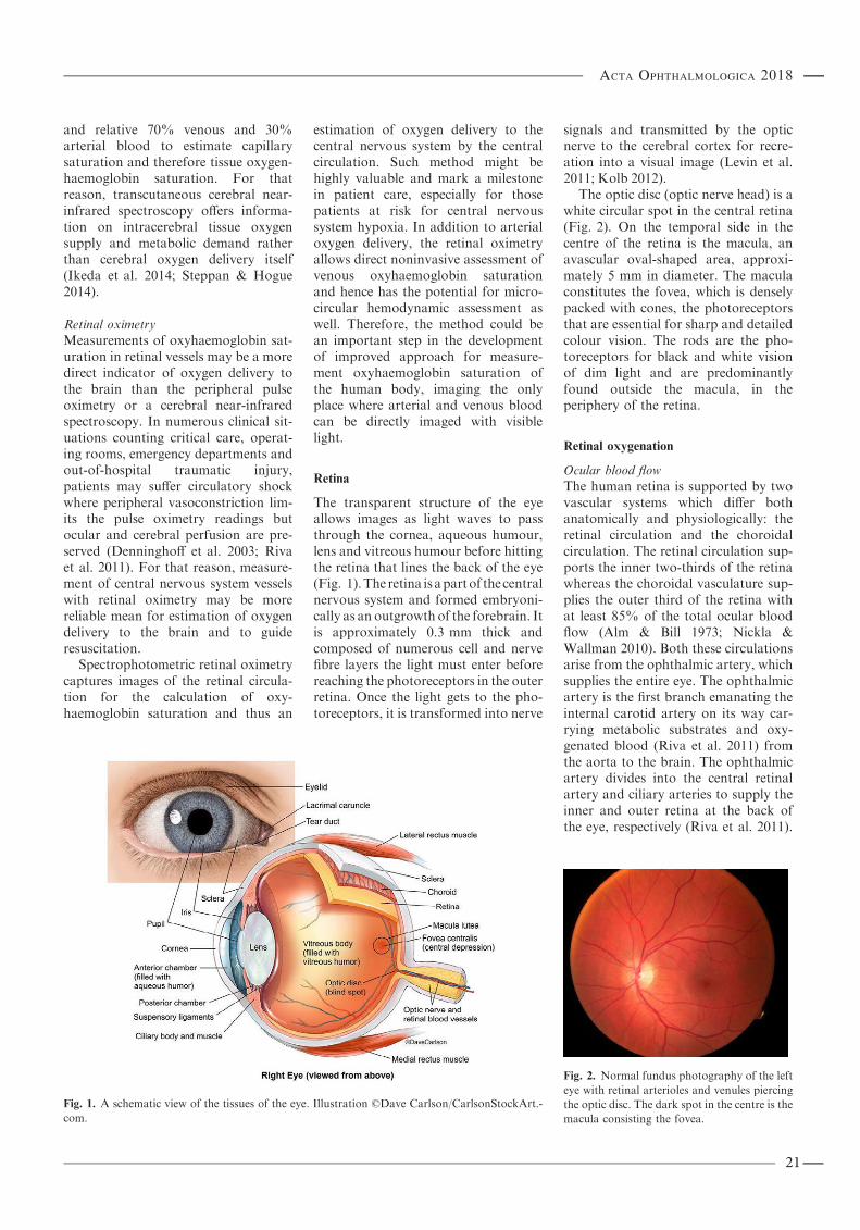
and relative 70% venous and 30%arterial blood to estimate capillarysaturation and therefore tissue oxygen-haemoglobin saturation. For thatreason, transcutaneous cerebral near-infrared spectroscopy offers informa-tion on intracerebral tissue oxygensupply and metabolic demand ratherthan cerebral oxygen delivery itself(Ikeda et al. 2014; Steppan & Hogue2014).
Retinal oximetry
Measurements of oxyhaemoglobin sat-uration in retinal vessels may be a moredirect indicator of oxygen delivery tothe brain than the peripheral pulseoximetry or a cerebral near-infraredspectroscopy. In numerous clinical sit-uations counting critical care, operat-ing rooms, emergency departments andout-of-hospital traumatic injury,patients may suffer circulatory shockwhere peripheral vasoconstriction lim-its the pulse oximetry readings butocular and cerebral perfusion are pre-served (Denninghoff et al. 2003; Rivaet al. 2011). For that reason, measure-ment of central nervous system vesselswith retinal oximetry may be morereliable mean for estimation of oxygendelivery to the brain and to guideresuscitation.
Spectrophotometric retinal oximetrycaptures images of the retinal circula-tion for the calculation of oxy-haemoglobin saturation and thus an
estimation of oxygen delivery to thecentral nervous system by the centralcirculation. Such method might behighly valuable and mark a milestonein patient care, especially for thosepatients at risk for central nervoussystem hypoxia. In addition to arterialoxygen delivery, the retinal oximetryallows direct noninvasive assessment ofvenous oxyhaemoglobin saturationand hence has the potential for micro-circular hemodynamic assessment aswell. Therefore, the method could bean important step in the developmentof improved approach for measure-ment oxyhaemoglobin saturation ofthe human body, imaging the onlyplace where arterial and venous bloodcan be directly imaged with visiblelight.
Retina
The transparent structure of the eyeallows images as light waves to passthrough the cornea, aqueous humour,lens and vitreous humour before hittingthe retina that lines the back of the eye(Fig. 1).The retina isapartof thecentralnervous system and formed embryoni-cally as an outgrowth of the forebrain. Itis approximately 0.3 mm thick andcomposed of numerous cell and nervefibre layers the light must enter beforereaching the photoreceptors in the outerretina. Once the light gets to the pho-toreceptors, it is transformed into nerve
signals and transmitted by the opticnerve to the cerebral cortex for recre-ation into a visual image (Levin et al.2011; Kolb 2012).
The optic disc (optic nerve head) is awhite circular spot in the central retina(Fig. 2). On the temporal side in thecentre of the retina is the macula, anavascular oval-shaped area, approxi-mately 5 mm in diameter. The maculaconstitutes the fovea, which is denselypacked with cones, the photoreceptorsthat are essential for sharp and detailedcolour vision. The rods are the pho-toreceptors for black and white visionof dim light and are predominantlyfound outside the macula, in theperiphery of the retina.
Retinal oxygenation
Ocular blood flow
The human retina is supported by twovascular systems which differ bothanatomically and physiologically: theretinal circulation and the choroidalcirculation. The retinal circulation sup-ports the inner two-thirds of the retinawhereas the choroidal vasculature sup-plies the outer third of the retina withat least 85% of the total ocular bloodflow (Alm & Bill 1973; Nickla &Wallman 2010). Both these circulationsarise from the ophthalmic artery, whichsupplies the entire eye. The ophthalmicartery is the first branch emanating theinternal carotid artery on its way car-rying metabolic substrates and oxy-genated blood (Riva et al. 2011) fromthe aorta to the brain. The ophthalmicartery divides into the central retinalartery and ciliary arteries to supply theinner and outer retina at the back ofthe eye, respectively (Riva et al. 2011).
Fig. 1. A schematic view of the tissues of the eye. Illustration ©Dave Carlson/CarlsonStockArt.-
com.
Fig. 2. Normal fundus photography of the left
eye with retinal arterioles and venules piercing
the optic disc. The dark spot in the centre is the
macula consisting the fovea.
21
Acta Ophthalmologica 2018

Retinal circulation. The central retinalartery (CRA) penetrates the opticnerve sheath about 10 mm behind theglobe (Fig. 3). It runs centrally withinthe optic nerve to the optic disc whereit bifurcates into superior and inferiorretinal branches. These branches inturn divide into temporal and nasalarterioles where each supplies onequadrant of the inner retina (Pournaraset al. 2008; Hayreh 2011). The retinalcirculation is an end-arterial circuitwithout anatomizes. Retinal arteriolesarcade by dichotomous and side-armbranching towards the periphery, untilterminating in two-layered capillaryplexuses that connect with postcapillary
venules. Larger arterioles and venulesare found in the innermost layers of theretina at the inner limiting membraneand the outer plexiform layer. Capil-lary density is greatest in the centre ofthe retina with the more superficialcapillary plexus situated in the gan-glion cell and nerve fibre layer. Thesecond capillary plexus lies deeper, atthe border of the inner nuclear andouter plexiform layer with a single-layered capillary network proceedinguntil it finally vanishes, leaving anavascular zone in the farthest retinalperiphery. In addition, a single-layeredcapillary network surrounds the area ofthe avascular fovea as well as the
superficial peripapillary capillaries thatenclose the optic disc, to chase thesuperior and inferior temporal retinalvessels.
The retinal venous system follows asimilar pattern to the arteriolar struc-ture. It runs independently in theperiphery of the retina but in closeproximity to the arterioles with occa-sionally crossing within the centre ofthe retina. The postcapillary venulesdrain into major branch venules thatmerge at the optic nerve head to form acentral retinal vein (CRV). The CRVleaves the eye adjacent (temporal) tothe CRA within the optic nerve beforeemptying either into the ophthalmicvein or directly into the cavernoussinus (Pournaras et al. 2008; Hayreh2011; Riva et al. 2011).Choroidal circulation. The choroidal cir-culation is located between the outerretina and the sclera that constitute theoutermost membrane of the eye. Itoriginates in 2–3 main ciliary arteriescoming off the ophthalmic artery tosupply the temporal and nasal portionsof the choroidal sphere. Main ciliaryarteries branches into 10–20 short pos-terior ciliary arteries and two longposterior ciliary arteries to form majorchoroidal arteries that support theposterior and anterior portion of thechoroid, respectively. The shortest pos-terior ciliary arteries aim for a vascularstructure near the macula to nourishthe vicinity of fovea. Occasionally, acilioretinal artery arises directly from ashort posterior ciliary artery or theperipapillary choroid. It normallypierces the retina temporal to the opticdisc to nourish the macular area butthe location and contribution to theoxygen supply vary.
The choroid is made of three layers:the outermost Haller0s layer of largevessels, the inner Sattler0s layer ofmedium and small arteries and arteri-oles and the dense innermost networkof single choriocapillary layer. Chorio-capillary blood drains into the vortexveins before entering orbital veins(Hayreh 1975, 2011; Nickla & Wall-man 2010; Riva et al. 2011) and even-tually emptying into the cavernoussinus.
Retinal metabolism
The retina is metabolically one of themost active tissues in the body, con-suming oxygen faster than the brain
Fig. 3. Anatomy of ocular circulation (a – artery, b – vein, n – nerve). (A) Cut away drawing along
the superior–inferior axis of the human eye through the optic nerve, showing the vascular supply
to the retina and choroid. (B) Drawing showing vasculature of the retina and choroid. Drawings
by Dave Schumick from Anand-Apte and Hollyfield (2009). Reprinted from Prog Retin Eye Res,
31(5), Kur et al. Cellular and physiological mechanisms underlying blood flow regulation in the
retina and choroid in health and disease, 377–406, © 2012, with permission of Elsevier Ltd.
22
Acta Ophthalmologica 2018
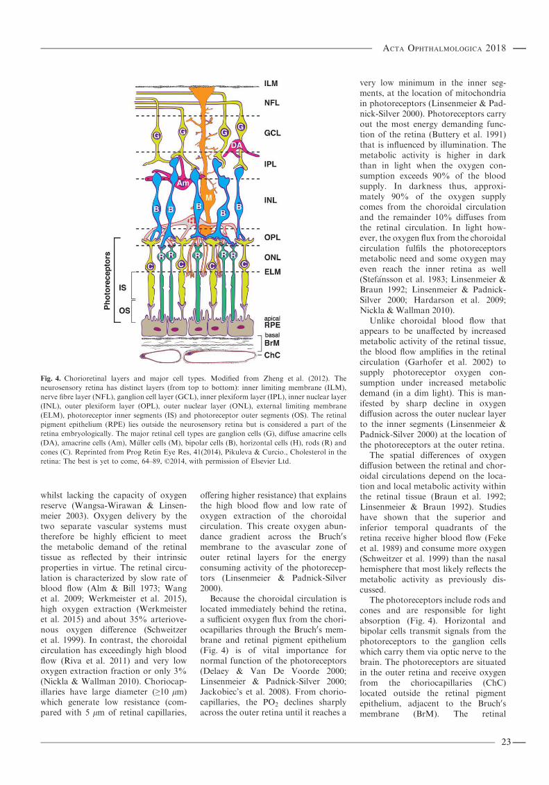
whilst lacking the capacity of oxygenreserve (Wangsa-Wirawan & Linsen-meier 2003). Oxygen delivery by thetwo separate vascular systems musttherefore be highly efficient to meetthe metabolic demand of the retinaltissue as reflected by their intrinsicproperties in virtue. The retinal circu-lation is characterized by slow rate ofblood flow (Alm & Bill 1973; Wanget al. 2009; Werkmeister et al. 2015),high oxygen extraction (Werkmeisteret al. 2015) and about 35% arteriove-nous oxygen difference (Schweitzeret al. 1999). In contrast, the choroidalcirculation has exceedingly high bloodflow (Riva et al. 2011) and very lowoxygen extraction fraction or only 3%(Nickla & Wallman 2010). Choriocap-illaries have large diameter (≥10 lm)which generate low resistance (com-pared with 5 lm of retinal capillaries,
offering higher resistance) that explainsthe high blood flow and low rate ofoxygen extraction of the choroidalcirculation. This create oxygen abun-dance gradient across the Bruch0smembrane to the avascular zone ofouter retinal layers for the energyconsuming activity of the photorecep-tors (Linsenmeier & Padnick-Silver2000).
Because the choroidal circulation islocated immediately behind the retina,a sufficient oxygen flux from the chori-ocapillaries through the Bruch0s mem-brane and retinal pigment epithelium(Fig. 4) is of vital importance fornormal function of the photoreceptors(Delaey & Van De Voorde 2000;Linsenmeier & Padnick-Silver 2000;Jackobiec’s et al. 2008). From chorio-capillaries, the PO2 declines sharplyacross the outer retina until it reaches a
very low minimum in the inner seg-ments, at the location of mitochondriain photoreceptors (Linsenmeier & Pad-nick-Silver 2000). Photoreceptors carryout the most energy demanding func-tion of the retina (Buttery et al. 1991)that is influenced by illumination. Themetabolic activity is higher in darkthan in light when the oxygen con-sumption exceeds 90% of the bloodsupply. In darkness thus, approxi-mately 90% of the oxygen supplycomes from the choroidal circulationand the remainder 10% diffuses fromthe retinal circulation. In light how-ever, the oxygen flux from the choroidalcirculation fulfils the photoreceptorsmetabolic need and some oxygen mayeven reach the inner retina as well(Stef�ansson et al. 1983; Linsenmeier &Braun 1992; Linsenmeier & Padnick-Silver 2000; Hardarson et al. 2009;Nickla & Wallman 2010).
Unlike choroidal blood flow thatappears to be unaffected by increasedmetabolic activity of the retinal tissue,the blood flow amplifies in the retinalcirculation (Garhofer et al. 2002) tosupply photoreceptor oxygen con-sumption under increased metabolicdemand (in a dim light). This is man-ifested by sharp decline in oxygendiffusion across the outer nuclear layerto the inner segments (Linsenmeier &Padnick-Silver 2000) at the location ofthe photoreceptors at the outer retina.
The spatial differences of oxygendiffusion between the retinal and chor-oidal circulations depend on the loca-tion and local metabolic activity withinthe retinal tissue (Braun et al. 1992;Linsenmeier & Braun 1992). Studieshave shown that the superior andinferior temporal quadrants of theretina receive higher blood flow (Fekeet al. 1989) and consume more oxygen(Schweitzer et al. 1999) than the nasalhemisphere that most likely reflects themetabolic activity as previously dis-cussed.
The photoreceptors include rods andcones and are responsible for lightabsorption (Fig. 4). Horizontal andbipolar cells transmit signals from thephotoreceptors to the ganglion cellswhich carry them via optic nerve to thebrain. The photoreceptors are situatedin the outer retina and receive oxygenfrom the choriocapillaries (ChC)located outside the retinal pigmentepithelium, adjacent to the Bruch0smembrane (BrM). The retinal
Fig. 4. Chorioretinal layers and major cell types. Modified from Zheng et al. (2012). The
neurosensory retina has distinct layers (from top to bottom): inner limiting membrane (ILM),
nerve fibre layer (NFL), ganglion cell layer (GCL), inner plexiform layer (IPL), inner nuclear layer
(INL), outer plexiform layer (OPL), outer nuclear layer (ONL), external limiting membrane
(ELM), photoreceptor inner segments (IS) and photoreceptor outer segments (OS). The retinal
pigment epithelium (RPE) lies outside the neurosensory retina but is considered a part of the
retina embryologically. The major retinal cell types are ganglion cells (G), diffuse amacrine cells
(DA), amacrine cells (Am), M€uller cells (M), bipolar cells (B), horizontal cells (H), rods (R) and
cones (C). Reprinted from Prog Retin Eye Res, 41(2014), Pikuleva & Curcio., Cholesterol in the
retina: The best is yet to come, 64–89, ©2014, with permission of Elsevier Ltd.
23
Acta Ophthalmologica 2018

circulation supplies the inner part ofthe retina with capillaries reachingdown to the plane of photoreceptors.
Autoregulation
Vascular beds in organs have an intrin-sic capacity to some extent to regulatethe perfusion pressure locally. Thisproperty is referred to as autoregula-tion and is produced by an intrinsiccapacity for a stretch response of thearteriolar vascular smooth muscle. Thisability for a local regulation maintainsthe blood flow relatively constantdespite of variations in the perfusionpressure (Arjamaa & Nikinmaa 2006)and keeps the PO2 of the inner retinarelatively unaffected by either systemichypoxaemia or hyperoxia. This is anunique characteristic trait of the retinaltissue. Dissimilar to most other tissuesof the body, the retinal vessels and theoptic nerve head lack autonomic inner-vations and subsequently relay on theautoregulation for regulation of bloodflow. The mechanism behind the retinalautoregulation is not fully understoodbut is balanced by the effects of myo-genic and metabolic factors on thevascular resistance in adjustment oflocal blood flow to changes in perfu-sion pressure and the metabolic need ofthe tissue.
Retinal arteries have remarkablywell-developed smooth muscle layerthat differ them from arteries of thesame size in other organs. Changes inarteriolar diameter are considered themain regulatory component of theretinal vasculature and are triggeredeither by systemic or by local factors(Delaey & Van De Voorde 2000;Pournaras et al. 2008; Hayreh 2011).Vascular resistance is generated alongthe entirely length of the arteriolar wallbut terminal arterioles between 20 lmand 50 lm in diameter offer the mainresistance, thereby playing a centralrole in the autoregulatory responseeither by vessel dilation or by vasocon-striction (Jeppesen et al. 2007; Hayreh2011; Schmidl et al. 2011a). Contractileproperties of intramural pericytes inretinal capillaries also play a role in theautoregulatory response (Anderson1996; Anderson & Davis 1996). Peri-cytes possess similar properties assmooth muscle cells in that they candilate and constrict the capillarylumen to some minor extent inresponse to vasoactive substances or
to provocation by local blood gases(Pournaras et al. 2008; Kur & New-man 2014).
Systemic and local factors
Autoregulation of the retinal blood flowpertains to metabolic and myogenicmechanisms in reaction to activationof systemic and local factors as men-tioned above. The autoregulatorymech-anism is evoked by vasoactivesubstances that are released from theendothelium in arterioles and the adher-ent retinal tissue. Metabolic autoregu-lation strives for retinal tissue localblood flow regulation in unity with itsmetabolic requirements. For instance,in the case of accumulation ofmetabolicwastes in the tissue, the rate of bloodflow increases. The myogenic autoregu-lation, however, is activated by alter-ations in the transmural pressure,secondary to constriction or stretchingon the endothelium in the vessel wall. Inreturn, vasoactive factors are releasedcausing either a dilatation or constric-tion of the vessel (Delaey & Van DeVoorde 2000; Pournaras et al. 2008).Systemic factors. Systemic blood pres-sure, circulating hormones, arterialblood gases and pH are among thesystemic factors that activate the auto-nomic local vascular reaction. Localfactors incorporate the PO2 and partialpressure of carbon dioxide (PCO2), pH,endothelial factors (endothelium-derived relaxing factors and constrict-ing factors) and retinal factors (Delaey& Van De Voorde 2000). Increase inarteriolar mural pressure and mechan-ical stretch trigger release of theendothelium-derived constricting fac-tor, endothelin-1, resulting in vasocon-striction (Polak et al. 2003). Nitricoxide (NO) is one of the major endothe-lium-derived relaxing factor (EDRF),maintaining the basal vascular tone andmediating vasodilatation by severalagonists (Schmetterer & Polak 2001).Acetylcholine, histamine and bradyki-nin are all examples of neurotransmit-ters that modulate the vascular tone byactivation of EDRF (Yu et al. 2003).The role of retinal relaxing factor(RRF) is yet relatively unknown. Itseems to play considerable role as anindirect mediator of the hypoxicresponse by the retinal tissue itself,independent of other vasoactive media-tors (Maenhaut et al. 2007). Other localcompounds that contribute to retinalblood flow regulation include adenosine
and prostacyclin by increasing the arte-riolar diameter. Angiotensin II, prosta-glandin and cyclooxygenase narrow thearteriolar diameter whereas lactatemodulates the vascular tone parallel tothe local metabolic need either by con-stricting or by widening the vessel(Gidday & Park 1993; Yamanishi et al.2006; Pournaras et al. 2008).
Circulating hormones like endothe-lin-1 and angiotensin II have negligibleeffects on the retinal circulation (Flam-mer & Mozaffarieh 2008) because thenonfenestrated endothelium of retinalcapillaries and the complex network oftight junctions, that resemble the blood–brain barrier (Patton et al. 2005), pre-vent large molecules penetrating theinner blood–retinal barrier. Conversely,the fenestrated endothelium of chorio-capillaries is highly permeable to mole-cules their size, allowing for their directeffects on smoothmuscle cells (Flammer& Mozaffarieh 2008; Riva et al. 2011).Hypoxaemia and hyperoxia. Hypox-aemia provokes reactivity in retinalvessels mainly through release of tissuemetabolites in response to the abnor-mally low PO2 in the arterial blood.Hypoxaemia frequently results inhypoxia, the inadequate level of oxy-genation for retinal tissue metabolism.Reports on the impact of adenosine onretinal vascular relaxation are conflict-ing (Gidday & Park 1993; Delaey et al.2000) but prostacyclin, lactate and theRRF are all released under hypoxiccondition and are found to increase theretinal blood flow secondary tovasodilatory response (Delaey et al.2000; Hata et al. 2000; Yamanishiet al. 2006; Maenhaut et al. 2007;Pournaras et al. 2008). Supposedly,RRF is one of the main modulatorsfor hypoxic vascular response. It isindependent of endothelial involve-ment in the arteriolar wall which mayexplain the slower onset of retinalvascular reaction as compared withthat of the cerebral circulation.
In hyperoxia, retinal vasoconstric-tion has faster onset than the vasodi-latation during hypoxaemia probablydue to faster release of vasoconstrictivesubstances from the endothelial cells inretinal arterioles (Delaey et al. 2000;Maenhaut et al. 2007; Cheng 2014).Gas challenges on retinal circulation. Sim-ilar to cerebral circulation (Pournaraset al. 2008), the retinal circulationadjusts the local retinal blood to changesin arteriolar PO2 and PCO2 by widening
24
Acta Ophthalmologica 2018

or narrowing the vascular lumen.Hyperoxia-induced vasoconstriction isgreater in retinal vessels than in cerebralvessels, in contrast with the hypercap-nic-induced vasodilatation that isgreater in the brain (Kisilevsky et al.2008; Cheng et al. 2016).
Hyperoxic and hypocapnic gas chal-lenges provoke vasoconstrictions inboth retinal arterioles (Gilmore et al.2004) and venules (Jean-Louis et al.2005; Palkovits et al. 2014a; Palkovitset al. 2014b; Werkmeister et al. 2015;Cheng et al. 2016; Rose et al. 2016)whereas hypercapnic (Venkataramanet al. 2008) and hypoxic gas mixturesinduce vasodilatation of those vessels(Brinchmann-Hansen et al. 1989;Choudhary et al. 2013; Palkovits et al.2014c; Cheng et al. 2016; Rose et al.2016). The blood flow regulation isfound to be relatively stable when PaO2
is above 32–37 mmHgbut beneath theselimits the autoregulatory response isfound to be lost (Cheng et al. 2016).According to electroretinography stud-ies, the inner retina show more sensitiv-ity to transient hypoxic stress (at thelevel of the retinal ganglion cells) thanthe outer retina which is more resistantto the hypoxic challenges (Tinjust et al.2002; Janaky et al. 2007; Caprara &Grimm 2012).Perfusion pressure. The ability for localautoregulation in vascular beds main-tains the retinal blood flow relativelyconstant and independent of fluctua-tions in the perfusion pressure, as longas the ocular perfusion pressure (OPP)is within a range of upper and lowerlimits of the autoregulatory plateau.Beyond these limits, the vascularreserve is lost and ocular blood flowbecomes directly dependent on thepressure (Arjamaa & Nikinmaa 2006).By most studies, the upper limits ofretinal blood flow regulation areattained when the mean arterial bloodpressure (MAP) reaches approximately40% above baseline (Schmidl et al.2011a). Other studies have found theautoregulatory limits of the mean OPPin the range of 34–60% over baseline(Pournaras et al. 2008), more than36% below baseline (Riva et al. 1981)or when the intraocular pressure(IOP) either reaches about 30 mmHg(Schulte et al. 1996) or drops below10 mmHg (Williamson & Harris 1994)Calculation of retinal blood flow. Retinalblood flow (BF) through the opticnerve head is directly proportional to
the perfusion pressure and inverselyproportional to the vascular resistance:
BF ¼ PP
R; ð1Þ
where perfusion pressure (PP) is theforce that drives blood through thevessel, determined by the differencebetween the arterial and venous pres-sure (ΔP). The resistance (R) of a vesselwall against the PP is a function of thevascular calibre and the vessel tone(Caprioli & Coleman 2010). Blood flowresistance can be calculated accordingto the Poiseulle’s law:
R ¼ 8gLr4
; ð2Þ
where vascular resistance (R) isdirectly related to the fluid viscosity(g) and the length of a vessel (L) andinversely related to the radius of thevessel in the fourth power (r). Forthat reason, even small changes in thevascular lumen have considerableeffects on blood flow resistance. Forinstance, lessen the retinal arteriolarlumen by half will 16-fold the increasein OPP.Ocular perfusion pressure. As alreadybrought up, the mean OPP is thepressure driving the blood throughthe optic nerve. It is determined bythe mean ophthalmic arterial pressure(MOAP) entering the eye, minus themean ophthalmic venous pressure(MOVP), leaving the eye:
OPP ¼ MOAP�MOVP: ð3Þ
The MOVP is close to the intraoc-ular pressure (IOP), and therefore, theOPP can be estimated as:
OPP ¼ MOAP� IOP¼ 2=3MAP� IOP: ð4Þ
The IOP is determined by the rateof aqueous humour production andthe drainage of aqueous humourthrough the trabecular meshwork.The normal lower and upper limitsof IOP are 10 and 22 mmHg, respec-tively, and the mean around 16 mmHg(Williamson & Harris 1994; ThariqBhatti 2008).Mean arterial pressure. Mean OPP isestimated to be two third of the meanbrachial blood pressure. The meanarterial pressure is the time-weighted
average calculation on arterial pres-sures during one pulse cycle (Butter-worth et al. 2013), determined by thefollowing equation:
MAP ¼ SBPþ 2ðDBPÞ3
: ð5Þ
The MAP calculation is based on thefact that during a pulse cycle, one thirdof the time is spent near the systolicblood pressure (SBP) and two thirdnear diastolic blood pressure (DBP;Butterworth et al. 2013)
The OPP may be affected by one ormore of the variables that are used tocalculate its value. For example, a lowsystemic arterial blood pressure or ahigh IOP (≥30 to 34 mmHg) willreduce the OPP whereas a high sys-temic blood pressure or a low IOP(<10 mmHg) will elevate the OPP(Williamson & Harris 1994). Conse-quently, the vessel lumen will distendor constrict to the magnitude of fall orrise in IOP, respectively, until reachingthe point the compensatory ability islost and retinal blood flow becomes adirect function of OPP.
Choroidal blood flow regulation
Regulation of retinal blood flow isbelieved to be more efficient than ofthe choroidal flowby the autoregulatorymechanism. Unlike the retinal circula-tion, the choroidal vascular bed and thecentral retinal artery up to the laminacribrosa are innervated by the auto-nomic nervous system (Nickla & Wall-man 2010). The choroidal circulationdemonstrates limited autoregulatorycapacity to changes in IOP (Schmidlet al. 2011b; Schmidl et al. 2012) butemerging evidence has verified someautoregulatory efforts to changes inMAP and OPP (Fuchsjager-Mayrlet al. 2003; Luksch et al. 2003; Schmidlet al. 2012)..
It is also likely that the choroidalcirculation is only autoregulated bychanges in PCO2 but not PO2 (Thyleforset al. 2009). Under system hypoxaemicconditions, the level of PO2 drops lin-early from the choriocapillaries acrossthe outer retina (Linsenmeier & Braun1992) so the oxygen flux becomes insuf-ficient for the outer retina oxygenrequirements. In case the systemicPaO2 becomes less than 60 mmHg, theretinal circulation contributes (10%)by oxygen diffusion to the outer
25
Acta Ophthalmologica 2018

retinal segments (Linsenmeier & Braun1992).
Hypercapnia on the other hand exertsstrong vasodilatory NO-mediatedeffects on the choroidal vasculature, justlike on the retinal circulation (Sch-metterer & Polak 2001). Carbonmonoxide seems to affect the rate ofchoroidal blood flow to some degreewhilst hyperoxia has negligible effectson the bloodflow (Schmidl et al. 2011a).Due to the lack of choroidal autoregu-latory response to system hyperoxia, theabundant oxygen flux for the durationof supplemental oxygenbreathing seemsbeneficiary for the inner retinal tissue byattenuating the effects of hyperoxicvasoconstriction on the retinal circula-tion (Palkovits et al. 2014a).
Oxygen transportation to tissues
Physiology of oxygenation
Oxygen is transported by the arterialblood to tissues in two forms: dissolvedand combined with haemoglobin.Under normal condition, about 97%of the oxygen is bound with haemoglo-bin and the residual oxygen (3%) isdissolved in plasma (Guyton and Hall2000). Oxygen combines reversibly withthe haem portion of the haemoglobinmolecule to form oxyhaemoglobin. Thischemical reaction is reliant on the con-centration of PO2 dissolved in plasma.Each haemoglobin molecule has thecapacity to combine with four oxygenmolecules. Accordingly, the percentageof haemoglobin molecules occupied byoxygen molecules to the total oxygenbinding capacity is expressed as oxy-haemoglobin saturation (West 2005).Oxyhaemoglobin dissociation curve. Therelationship between oxyhaemoglobinsaturation and the PO2 can bedescribed by the oxyhaemoglobin dis-sociation curve (Fig. 5).
The oxyhaemoglobin dissociationcurvehas a sigmoidal shape representingthe dynamic interaction between theoxyhaemoglobin saturation and thePO2 in plasma. The binding of the firstoxygen molecule is more difficultthan subsequent oxygen-haemoglobinattachments. After the first oxygencombines with one of the four hemeportions of the haemoglobin, configuralchanges of the haemoglobin proteinfacilitates additional binding tothe other heme groups. Subsequently,The affinity for the fourth oxygenmolecule is nearly 300-fold greater than
for the first one. The opposite is just truefor oxygen release in tissues in that thedissociation of the first oxygenmoleculefacilitates subsequent oxygen releasefrom the haemoglobin (Guyton andHall 2000; Butterworth et al. 2013).
The chemical reaction of oxygenbinding is illustrated by the steep sig-moidal upward shift in the region of 20–30 mmHg (Harvey and Ferrier 2011).Normally, 50% oxyhaemoglobin satu-ration (P50) is reached at approximately26.6 mmHg. When the haemoglobinaffinity for oxygen is increased, the50% saturation is reached at a lowerPO2 with subsequent shifting of theoxyhaemoglobin dissociation curve tothe left and less availability of oxygenfor tissues. The exact opposite is truewhen the haemoglobin affinity foroxygen is decreased. Then, the oxy-haemoglobin dissociation curve isshifted to the right and a higher PO2 isneeded to reach the 50% oxy-haemoglobin saturation, and more oxy-gen becomes available for tissues. Thiscondition is typical for a normal oxygendelivery in tissues where hydrogen ion(H+) concentration accompanying ele-vated CO2 production secondary tocellular metabolism shifts the oxy-haemoglobin dissociation curve to the
right. Hence, the haemoglobin affinity isdecreased and more oxygen is releasedat lower PO2. This condition is repre-sented at the steepest part on the curvewhere only small changes in PO2 dis-lodge oxygen molecules from the hae-moglobin. Other known factors thatshift the curve to the right includeincreased 2,3 diphosphoglycerate(DPG) and elevated blood temperature(Guyton and Hall 2000; Butterworthet al. 2013).Oxygen content of the blood. As previ-ously stated, the oxyhaemoglobin sat-uration (SO2) is the percentage ofavailable haemoglobin binding sitesthat are combined with oxygen givenby the following equation:
SO2ð%Þ ¼ ½HbO2�½Hb� þ ½HbO2� � 100; ð6Þ
where HbO2 is the oxygen combinedwith haemoglobin and Hb is the deoxy-genated haemoglobin. The SO2 is mul-tiplied by 100 to obtain the percentage.The SO2 of arterial blood (SaO2) at100 mmHg is approximately 97,5%and 75% for the mixed venous blood(SvO2) at PaO2 of 40 mmHg (Guyton& Hall 2000; West 2005).
According to the Henry’s law, thedissolved concentration of oxygen in theblood is proportional to the partial pres-sure. For each mmHg of PO2, there is0.003 ml oxygen (O2) dissolved in 100 mlof blood. Therefore, a normal arterialblood (at sea level) with PO2 of100 mmHg contains 0.3 ml O2 per100 ml blood. The oxygen capacity isabout 20.8 ml O2 • 100 ml�1 of bloodbecauseonegramofpureHbcancombinewith 1.39 ml O2 and normal blood hasabout 15 gram of Hb • 100 ml�1. How-ever, under normal physiological condi-tions of the body, other species ofhaemoglobin, namely dyshaemoglobins,may exist in theblood aswell (West 2005).
Because in reality the oxy-haemoglobin saturation never reachesthe theoretical maximum of 1.39 ml O2
and some measurements give 1.34 ml,the total oxygen content of blood isgiven by the following equation:
Oxygen concentration¼ ð1:34�Hb� SaO2Þ þ 0:003
� PO2; ð7Þ
where 1.34 is the oxygen carryingcapacity (ml/g) of haemoglobin (atsea level), Hb is the amount of
Fig. 5. The oxyhaemoglobin dissociation
curve (blue) illustrates how oxyhaemoglobin
saturation arises quickly up till approximately
PO2 of 50 mmHg, under normal physiological
conditions. From PO2 of 60 mmHg which
represents about 90% oxyhaemoglobin satu-
ration, the curve becomes flatter because of
fewer available oxygen-haemoglobin binding
sites, until complete saturation is reached at
100%. The red dotted line represents a condi-
tion of rightward shift of the curve and the
green line leftward shift of the oxy-
haemoglobin dissociation curve. Public
domain figure. https://upload.wikimedia.org/
wikipedia/commons/8/8a/Oxyhaemoglobin_d
issociation_curve.png.
26
Acta Ophthalmologica 2018

haemoglobin in the blood (g/dl), SaO2
is the oxyhaemoglobin saturation ofarterial blood at given PO2 and0.003 ml O2 dissolved in 100 ml ofblood per mmHg PO2.Dyshaemoglobin. Haemoglobin can beclassified as a normal haemoglobin thatis capable of carrying oxygen (Hb andHbO2) and dyshaemoglobin that arehaemoglobin derivatives and incapableof an oxygen holding. Dyshaemoglobinare further classified as methaemoglo-bin (MetHb), carboxyhaemoglobin(COHb) and sulfhemoglobin (SHb).Sulfhemoglobin is an uncommon formthat is caused by reaction of sulpha-containing compounds, usuallyfrom excessive use of sulpha-baseddrugs (Haymond 2006). Carboxy-haemoglobin carries carbon monoxidethat is formed during the metabolicpathway of haem into bilirubin andconstitutes normally less than 1–3% ofthe total haemoglobin in the body(McClatchey 2002). It can, however,be as much as fivefold in a heavysmoker (Whincup et al. 2006). Thegreater affinity (200- to 300-fold) ofcarbon monoxide for haemoglobinthan oxygen allows COHb to easilydisplace the HbO2, shifting the oxy-haemoglobin dissociation curve to theleft. Methaemoglobin is the oxidativedeoxy form of normal haemoglobinwhere the iron of the haem group is ina ferric (Fe3+) form instead of theferrous (Fe2+) state. The MetHb hasno oxygen carrying capacity and sim-ilar to the COHb can shift the oxy-haemoglobin dissociation curve to theleft (McClatchey 2002; Butterworthet al. 2013).
Some contemporary peripheral co-oximeters differentiate aforementionedabnormal dyshaemoglobin structuresfrom normal haemoglobin. However,at 660 nm, the COHb has an absor-bance parallel with that of HbO2 (Miller2000) thereby necessitating informationon whether the person is an activesmoker before spectroscopic retinaloximetry is performed. The half-life ofCOHb is 4–6 hr on a room air sosmoking should preferentially beabstained for 12–24 hr prior to retinaloximetry. Breathing 100% oxygenshortens COHb half-life to 40–80 minand hyperbaric oxygen breathing short-ens it still further, or to 15–30 min(Nagelhout & Zaglaniczny 2001).Fetal haemoglobin. Fetal haemoglobin(HgbF) constitutes about 75–84% of
the total haemoglobin in new bornbabies (Chestnut 2004) and 1% of thenormal adult haemoglobin (Schechter2008), or haemoglobin A (HgbA).Functionally, HgbF diverges fromHgbA in that it has somewhat higheraffinity for oxygen because ofdecreased interaction with 2,3–DPG(Mosca et al. 2009) with P50 around19–21 mmHg (Chestnut 2004) insteadof 26.6 mmHg in adults. During thefirst month of life, the HgbF amount isprogressively substituted by HgbAuntil dwindling off by 6 months of life(Edoh et al. 2006). As HgbA starts tosubstitute the HgbF, the level of2,3DPG raise and the affinity becomesanalogous to that of adults within thefirst few months, although the concen-tration of HgbF still remains 25%(Chestnut 2004). HgbF has nearly thesame absorption spectrum for oxy-haemoglobin and deoxyhaemoglobinas HgbA and hence should not affectthe oximetry outcome measures fromof adults (Miller 2000; Chestnut 2004).
Matching oxygen supply to demand
Oxygen transport to tissues depends onadequate respiratory and circulatoryfunction. Dry air at sea level (baromet-ric pressure of 760 mmHg) has a PO2
of 20.93% (~21%). The fully saturatedvapour pressure at a sea level is47 mmHg at normal body temperatureof 37°C. Thus, the PO2 of an inhaledair at sea level is expressed as follows:
ð20:93Þ100
�ð760�47Þ¼149mmHg: ð8Þ
When the inspired air reaches thealveoli of the lungs, the PO2 hasdropped to about 100 mmHg. Thereason is that the alveolar PO2 is aproduct of the balance between alveo-lar ventilation and the alveolar gasdiffusion across the capillary blood gasinterface (West 2005).Global oxygen delivery. Global oxygendelivery (DO2) to tissues is a product ofcardiac output (CO), or the blood flow,and the total oxygen content of thearterial blood (CaO2; McLellan andWalsh 2004). The DO2 is calculated bythe following formula:
DO2 ¼ CO� CaO2; ð9Þ
where cardiac output is a product ofthe heart rate (HR) and stroke volume(SV) and the stroke volume is the
amount of blood ejected by the leftventricle during one heart beat. Theoxygen concentration depends on bothoxyhaemoglobin saturation and PO2
dissolved in plasma, as previously dis-cussed. Therefore, an inadequate oxy-gen delivery may either originate fromimpaired cardiac output, insufficientPaO2 or a low haemoglobin count(Butterworth et al. 2013).
Global oxygen delivery to tissuesdepends on two processes: convectionof the blood down the arterial tree anddiffusion from capillaries to adjacenttissues (Leach & Treacher 2002). Elas-tic properties and calibre of the vesselwall determine the vascular resistanceagainst the cardiac output. Subse-quently, the compliance in the arterialsystem serves the function of dampingthe pulsatile output from the left ven-tricle. Hence, the pulsatile pressure isminimized and a continuous blood flowfacilitated down to the microvascularlevel (Guyton and Hall 2000).
The rate of oxygen uptake by pul-monary capillaries is governed by oxy-gen consumption in tissues that is fairlyconstant under normal resting condi-tions (West 2005). The cardiac outputis constantly adapted to the overallmetabolic need of the body. It iscontrolled by the sum of various fac-tors (as previously discussed) that reg-ulate local blood flow according tometabolic need of tissues. These factorsadd together to make the venous returnwhich in turn is delivered by thepumping activity of the heart andconvective flow down the arterial treeto capillaries (Guyton and Hall 2000).From the capillary blood, the oxygendiffuses down its concentration gradi-ent to the much lower PO2 of themitochondria of individual cells intissues (Leach & Treacher 2002).
Presuming the retinal arterial oxygencontent is identical to the systemiccirculation, the oxygen delivery to aretinal tissue can be derived fromequation 9, by substituting total retinalblood flow (TRBF) for the cardiacoutput:
DO2 ¼ TRBF� CaO2: ð10Þ
Oxygen consumption. Tissue oxygenconsumption (VO2) is equivalent tothe tissue metabolic rate under aerobicconditions per minute. The Fick’sequation describes the relationshipbetween oxygen consumption, cardiac
27
Acta Ophthalmologica 2018
output or retinal blood flow and theoxygen content in both retinal arteri-oles (CaO2) and venules (CvO2):
VO2 ¼ CO� ðCaO2 � CvO2Þ ð11Þ
or
VO2 ¼TRBF�ðCaO2 �CvO2Þ; ð11Þ
where CaO2 – CvO2 is the differencebetween the arterial and venous oxygencontent (arteriovenous oxygen differ-ence, AV-difference). The global arte-rial oxygen content is about 20 ml/100 ml blood and the venous oxygencontent about 15 ml/100 ml blood andthus producing arteriovenous oxygendifference of approximately 5 ml/100 ml blood (Butterworth et al. 2013).Arteriovenous oxygen difference. By rear-ranging equation 11, it is apparent theoxygen consumption and the localblood flow determine the arteriovenousoxygen difference in the retinal tissue:
AV-difference ¼ VO2
BF: ð12Þ
The AV-difference is directly relatedto the local tissue oxygen consumptionand inversely to the local blood flow.The relationship is clearly manifested inboth retinal and choroidal circulations.In this context, the retinal circulation ischaracterized by low VO2/BF ratio anda large AV-difference, resulting in rela-tively low oxygen content on the venousside of the circulation. In contrast, thechoroidal circulation is characterized byhigh VO2/BF ratio and low AV-differ-ence with high oxygen content on thevenous side of the circulation.Oxygen extraction fraction. The amountof oxygen consumption is reflected bythe fraction of oxygen extraction(OEF) across the perfused capillarynetwork. The oxygen extraction frac-tion can be calculated as follows:
OEF ¼ CaO2 � CvO2
CaO2: ð13Þ
Normal oxygen extraction fractionfor the majority of tissues is 25%(5 ml/20 ml). In other words, undernormal conditions, most tissues con-sume only one-fourth of the oxygendelivered to the capillary bed. Whenoxygen supply exceeds the oxygendemand for the metabolic activity, theextraction fraction becomes less than25%. However, when the oxygen
supply is less than the metabolicdemand, the extraction fractionbecomes greater than 25% (Butter-worth et al. 2013).
Blood flow distribution varies enor-mously among tissues, depending ontheir momentary functional require-ments. Some tissues, including theretina (Wangsa-Wirawan & Linsen-meier 2003) and the brain, have steadyenergy requirements whilst perfusionand energy utilization of others (e.g.the liver) are predominated by theiractivity level (Scheufler 2004). Conse-quently, tissues with greater energyexpenditure, like the inner retina, myo-cardium and the brain, extract higheroxygen fraction than less active tissues.The oxygen extraction fraction of thecerebral tissue is close to 40% and thearteriovenous oxygen difference isabout 34% (Hatazawa et al. 1995;Qin et al. 2011). Likewise, the innerretina oxygen extraction fraction is37% and arteriovenous oxygen differ-ence approximately 35% (Schweitzeret al. 1999; Felder et al. 2015). Conse-quently, a duration of reduced oxygensupply (hypoxaemia) will directly affectmetabolic processes in retinal and cere-bral tissue cells (Kergoat et al. 2006;Ozcimen et al. 2016).
The application of noninvasive fun-dus reflectometry in human studies hasmade calculation of the inner retinaloxygen extraction achievable. Studiesin young healthy individuals indicateno changes in oxygen extraction underacute mild systemic hypoxia (Palkovitset al. 2014c). During 100% oxygenbreathing however, the oxygen extrac-tion is lessened by more than 60%from the normoxic breathing (Palko-vits et al. 2014a). For those reasons,the retina autoregulatory responseseems to be efficient under both acutesystem hypoxaemic and hyperoxicconditions.
Inadequate tissue oxygenation
Oxygen diffusion froma capillary lumento tissues is directly related to thecapillary area and the PO2 differenceacross the vessel wall and is inverselyrelated to the distance between the twosites (Leach & Treacher 1998, 2002;West 2005). Normally above 1–3 mmHg of oxygen pressure is requiredto maintain cellular oxidative metabo-lism which allows for a large safetymargin in animals (Leach & Treacher1998; Guyton & Hall 2000; Scheufler
2004). This is true for the inner retina aswell which has remarkably higher PO2
that measures around 20 mmHg in ani-mals with circulatory structure that isanalogous to humans (Wangsa-Wira-wan & Linsenmeier 2003).
During the final step of a cellularrespiration, oxygen-dependent adeno-sine triphosphate (ATP) is made as anenergy basis for the tissue metabolism.Oxygen deficiency depletes the ATPstores and the speed of cellular injurywill be determined by the tissue oxygenconsumption and the ability to attainATP under anaerobic conditions.Anaerobic metabolism can only serveas a temporarily reserve because of theinability to match tissue oxygen con-sumption. Consequently, the rapidityof cellular damage differs remarkablyamong tissues where neurological cellscan only survive for few minutes(Leach & Treacher 1998; Guyton &Hall 2000). This holds true for thephotoreceptors and other retinalneurological cells that require highoxygen consumption for the energydemanding process of transmittinglight waves into neurological signs forinterpretation by the brain (Caprara &Grimm 2012). The physiologicalresponse of prioritization blood flowto the retina and brain is thus ofuttermost importance during hemody-namic stress and makes the retinaindeed the most advantageous sourcefor noninvasive measures of oxygendelivery and viability of the centralnervous system.Similarities between retina and the
brain. Embryologically, the retina is adirect outgrowth of the diencephalon,sharing similarities in microvascularstructure with the cerebral circulation,blood flow and regulatory mechanisms(Delaey & Van De Voorde 2000; Pat-ton et al. 2005). Based on the homo-geneity, emerging evidence suggeststhat the retinal vasculature may be asurrogate for pathological changes onthe cerebrovasculature and even thecardiovascular system as well (Pattonet al. 2005; McClintic et al. 2010;Flammer et al. 2013). Like in retina,the rate of oxygen consumption by thecerebral tissue is quite constant(3.5 ml/100 gm tissue, or nearly 49 mlO2/min the entire brain) except underintense brain activity (Clarke & Sokol-off 1999). Consequently, the high oxy-gen consumption and the inability tostore oxygen and any metabolic
28
Acta Ophthalmologica 2018

products such as glucose render thecerebral tissue extremely vulnerable toan ischaemic insult (Pittman 2011).When arterial blood PO2 level goesbelow about 60 mmHg (Ainslie &Poulin 2004), the oxyhaemoglobin sat-uration becomes less than 90% (Guptaet al. 1997) and the cerebral tissue PO2
becomes less than 30 mmHg (normal35–40 mmHg), a prompt provocationof compensatory response withincreased cerebral blood flow (Guyton& Hall 2000; Carreau et al. 2011) isinitiated secondary to amplification ofcardiac output (Zhou et al. 2007).
As previously discussed, the retinalblood is dependent on the blood flowdelivered by the carotid arteries to thebrain. Impaired global oxygen deliveryin case of severe hypoxaemia (Heistad&Abboud 1980) or haemorrhagic shockinitiates compensatory response ofintense peripheral vasoconstriction toredirect blood flow from lower priorityorgans to the vital ones (Dutton 2007)to preserve cerebral and ocular perfu-sion (Denninghoff et al. 2003; Rivaet al. 2011). The initial compensatoryresponse is aimed at increasing oxygenextraction to match the oxygen demandfor cellular metabolism as evident bydecreased oxyhaemoglobin saturationon the venous site of the circulation. Incontrast, a reduced oxygen consump-tion for example during hypothermia orwith tissue necrosis lessens the fractionof oxygen extraction as shown by
increased venous oxyhaemoglobin sat-uration (Schober & Schwarte 2012).Eventually, whenever inadequate oxy-genation jeopardizes the normal cellularfunction, vigilant oxygen monitoringwith timely restorative and preventivemeasures is crucial for viability andfunctionality of individual tissues andbody organs.
Retinal oximetry
The retina possesses highly sophisti-cated architecture of neurological lay-ers and vascular structure that isanalogous to the cerebral circulation.The transparent structure at the frontof the eye offers an unique opportunityfor a direct noninvasive observation ofthe retinal circulation. Dual-wave-length spectrophotometric retinaloximetry allows for a direct noninva-sive assessment of oxyhaemoglobinsaturation in retinal vessels which serveas a window to the central nervoussystem. The technique is based ondifferent light absorption of oxy-haemoglobin and deoxyhaemoglobinand gives information on the oxy-haemoglobin saturation.
Principles of retinal oximetry
Spectrophotometric oximetry measuresthe attenuation of light travellingthrough a blood column as a functionof wavelength, based on the fact thatoxy- and deoxyhaemoglobin have
different light absorption spectra(Fig. 6). Oxyhaemoglobin is light redin colour whilst deoxyhaemoglobin isdarker red in colour. These differencesof shades permit calculation for oxy-haemoglobin saturation in retinal arte-rioles and venules.
Like conventional oxyhaemoglobinsaturation measurements in a wholeblood sample, the retinal oximetryexploits the Beer–Lambert law to itssystem. Supposedly, the light absorp-tion is dependent on the extinctioncoefficient (molar absorptivity) of theblood (ɛ); the distance of light has totravel through the sample (d) and theconcentration (c):
I ¼ I0 � 10�ecd; ð14Þ
where I0 is the original light intensitynot interacting with the blood, and I isthe intensity of light transmittedthrough the blood.
Dual-wavelength spectrophotomet-ric oximetry simultaneously acquirestwo automatic monochromatic retinalimages at two different wavelengths.One is at the isosbestic point and one ata nonisosbestic wavelength. Vesselpoints are automatically selected(Fig. 7) for measurement of light inten-sity inside (I) and outside the vessel (IO)for calculation of optical density(OD). Because of light absorbance bythe erythrocytes, the intensity ofreflected light from the vessel is lessthan from the immediate surroundingretina.
Fig. 6. Light absorptivity at isosbestic (570 nm) and nonisosbestic (600 nm) wavelengths.
Isosbestic wavelengths are insensitive to oxyhaemoglobin saturation whereas nonisosbestic
wavelengths are sensitive to distinctive oxyhaemoglobin saturation. For the Oxymap T1 retinal
oximeter, wavelength of 570 nm is at the reference isosbestic point whilst 600 nm is sensitive to the
oxyhaemoglobin. The figure was created by Jona Valgerdur Kristjansdottir based on data from
Zijlstra et al. (2000).
Fig. 7. Monochrome retinal oximetry image
with a pseudocolour overlay of selected
measure points along the vessel. Green overlay
(600 nm) shows the light intensity of the
measure inside the vessel (I). Red measure-
points illustrate the light intensity of the
background, to the sides of the vessel (Io).
29
Acta Ophthalmologica 2018

The OD describes the light absor-bance and can be calculated as:
OD ¼ logðI0=IÞ: ð15Þ
The higher the OD, the higher is theabsorbance. The OD inside a vessel atthe isosbestic point depends on thevascular diameter in addition to thedistance that the light travels (d) andthe erythrocyte concentration (c) but isindependent of oxyhaemoglobin satu-ration. In addition to the vasculardiameter and the c�d, the OD at thenonisosbestic wavelength depends onthe oxyhaemoglobin saturation. Con-sequently, the OD ratio at these twowavelengths is sensitive to oxy-haemoglobin saturation whilst the vas-cular width and c�d tend to cancel out.
Optical density at the two differentwavelengths allows for calculation ofan optical density ratio (ODR):
ODR ¼ OD nonisosbestic
OD isosbestic: ð16Þ
The ODR has nearly linear relation-ship to the oxyhaemoglobin saturation(Beach et al. 1999; Harris et al. 2003;Hardarson et al. 2006) that permitscalculation of oxyhaemoglobin satura-tion (SO2):
SO2 ¼ aþ b�ODR: ð17Þ
In the equation, a and b are calibra-tion constants based. The calibrationconstant of the retinal oximeter usedfor the purpose of this thesis will bediscussed in Chapter 3.2.3.2.
Development of retinal oximetry
Noninvasive spectrophotometric anal-ysis dates back to 1959, when Hickamand associates used broadband lightfilters, two wavelengths of light andstandard photographic films to mea-sure oxyhaemoglobin saturation inretinal vessels (Hickam et al. 1960).Subsequently, photoelectric technique(Delori 1988), imaging spectroscopy(Schweitzer et al. 1999), multispectralconfocal imaging (Smith et al. 2000)and digital camera systems (Beachet al. 1999) evolved from otherresearch groups. For review, see Harriset al. (2003), Geirsdottir et al. (2012)and Beach (2014).
Currently, two different systems arecommercially available for retinaloximetry that allots noninvasive direct
measurement of arterioles and venules:the Imedos system (RVA; Imedos,Jena, Germany) and the Oxymap reti-nal oximeter (Oxymap; Oxymap ehf.Reykjavik, Iceland).Retinal oximetry with scanning laser oph-
thalmoscope (SLO). Recently, a scan-ning laser ophthalmoscope (SLO) wascombined with modified Oxymap anal-ysis software for development of SLOretinal oximetry with a dual-wave-length algorithm. SLO uses low powerlaser wavelengths (instead of whitelight) to scan through individual sepa-rate retinal layers at once from theinner sensory layers down to thechoroid. A green laser (532 nm) scansfrom the inner retina to the pigmentepithelium whereas red laser (366 nm)gives a choroidal view by scanningfrom the retinal pigment epitheliumdown to the choroid.
The SLO brings some theoreticaladvances to retinal oximetry. First, itminimizes any unnecessary light expo-sure to the fundus by using laser(instead of white light imaging in reg-ular fundus cameras) to obtain themonochromatic images at the dualdifferent wavelengths. Second, itacquires wide-field scanning of nearlythe entire fundus but conventionalfundus cameras are limited to quitenarrow images of the posterior pole.Third, pupil dilation using mydriaticeye drops is unnecessary. Finally, itpierces other optical opacities in the eyesuch as cataracts better than conven-tional spectrophotometric fundus cam-era (Kristjansdottir et al. 2014). Inaddition, SLO has a potential forapplication of retinal oximetry toneonates and infants. This could marka milestone in management of pretermneonates (and other paediatric groups)who are treated in incubators at neona-tal intensive care units.
The risk of retinopathy of prematu-rity (ROP) is inversely related to ges-tational age and weight at birth.Extremely premature infants and thosewith low birthweight are therefore athighest risk for severe ROP and conse-quently a poor visual outcome due tothe immaturity of retina at birth(Cavallaro et al. 2014). The interrup-tion of normal retinal vascular devel-opment following transition from therelatively hypoxic intrauterine environ-ment to the oxygen-rich ambient airafter birth which is further aggravatedby administration of supplemental
oxygen therapy is twofold: the firstphase is characterized by suppressionof insulin-like growth factor I andnormal vascular endothelial growthfactor (VEGF). The second phase ischaracterized by hypoxic-induced neo-vascularization that is analogous toother proliferative retinopathies, asthe retina becomes more metabolicallydemanding (Smith 2004). Careful titra-tion of supplemental oxygen to pretermneonates to reduce the risk of visualimpact (i.e. blindness) from hyperoxiais therefore of vital importance forretinal vascular development. Cur-rently, oxygen supplementation tothose babies is monitored by continu-ous peripheral pulse oximetry andintermittent invasive arterial bloodgas measurements.
The noninvasive dual-wavelengthconfocal SLO combined with oximetryanalysis for retinal oximetry mayimprove monitoring and managementof these babies. Prior to allowing oxy-haemoglobin saturation measurementsof retinal arterioles and venules, it isnecessary to determine the ODR infull-term healthy neonates, shortlyafter birth.
Retinal oximetry in healthy individuals
Retinal oximetry has shown sensitivityand reliability with high reproducibilityand repeatability of retinal oxy-haemoglobin saturation measurementsduring normoxia in healthy humans(Hardarson et al. 2006; Hardarsonet al. 2009; Blondal, Sturludottir, Har-darson, Halldorsson, and Stefansson2011; Geirsdottir et al. 2012; Palssonet al. 2012; Heitmar & Cubbidge 2013;Man et al. 2013; Man et al. 2014; Yipet al. 2014; Werkmeister et al. 2015),rhesus monkeys (Li et al. 2016) andpigs (Traustason et al. 2013) and dur-ing induced hypoxaemia (Choudharyet al. 2013; Palkovits et al. 2014c; Roseet al. 2016) and hyperoxia (Palkovitset al. 2014a; Werkmeister et al. 2015;Rose et al. 2016) in humans. Most ofthese studies have been carried out totest the reproducibility and sensitivityof the technique but also to studynormal physiological reactions to oxy-gen challenges and retinal metabolism.
Report on the groundwork of SLOwith dual-wavelength retinal oximetryrevealed sensitivity and repeatability inhealthy humans and that the emplace-ment is technically feasible and promis-ing for advancement of retinal
30
Acta Ophthalmologica 2018

oximetry in the future (Kristjansdottiret al. 2014).Normoxia. Recently, some researchreports were published on normal reti-nal vessel oxyhaemoglobin saturationin human subjects of multiethnicities(Table 1). These studies show similarresults although some small deviationsexist between different versions of reti-nal oximeters and between differentresearch groups. The differences areattributable to the retinal oximeterhardware, the calibration of the oxime-ter, the analysis software and the ana-lytical approaches. When comparingoxyhaemoglobin saturation measure-ments between different studies, theimportance of identical calibrationbetween retinal oximeters devices(Geirsdottir et al. 2012) must be keptin mind and whether methodologicalprocedures for oximetry analysis wereaccording to standardized protocol.None of the studies have found differ-ence in oxyhaemoglobin saturationbetween the left and right eyes but theeffects of age on oxyhaemoglobin sat-uration are conflicting.
Geirsdottir et al. (2012) found theoxyhaemoglobin saturation in retinalvenules to decrease significantly withadvanced age, or 1.9% for every10 years in males and 0.7% for females.The oxyhaemoglobin saturation in
retinal arterioles was not affected byage. Consequently, the AV-differenceincreased by 1.5 � 0.5% for every10 years in males and 1.0 � 0.4% infemales. The vessel diameter was inver-sely affected by the age whereas the OPProse with age which the authors sug-gested might partly compensate for theincrease in vascular resistance caused bythe decreased vessel lumen. The oxy-haemoglobin saturation measurementsof both retinal arterioles and venuleswere significantly lower in the infer-otemporal quadrants than other retinalquadrants (Geirsdottir et al. 2012). Janiet al. (2014) found no difference acrossethnicity, genders nor iris/fundus pig-mentations. They found both the arte-riolar and venous oxyhaemoglobinsaturation to decrease with age and anincrease in AV-difference. The oxy-haemoglobin saturation values werelowest in the inferotemporal quadrantand highest in the superonasal quad-rant. Conversely, the AV-difference wasgreatest in the inferotemporal quadrantand lowest in the superotemporal quad-rant (Jani et al. 2014). Yip et al. (2014)reported the age to act as a negativefactor for oxyhaemoglobin saturation invenules, similar to the finding of Geirs-dottir et al. and Jani et al. (Yip et al.2014). In contrast to the above studies,Mohan et al. (2015) found the arteriolar
and venous oxyhaemoglobin saturationto increase with age but the AV-differ-ence to remain unaffected. There was nostatistical correlation between the reti-nal oximeter measurements and fingerpulse oximetry readings. They specu-lated that heavily pigmented fundus likein Indians hasmoremelanin exposed forlight absorption which could hypothet-ically alter the ODR and hence theoximetry measures (Mohan et al.2015). Yang et al. (2016) obtained dif-ference in oxyhaemoglobin saturationmeasurements between retinal quad-rants. The arteriolar measures frominferotemporal quadrant gave the low-est value followed by the superotempo-ral, inferonasal and superonasalquadrants, respectively. The retinalvenules followed a similar pattern (Yanget al. 2016). Nakano et al. (2016) mea-sured increased oxyhaemoglobin satu-ration in major retinal arteries (0.67%per 10 years) with advanced age but notin venules. They found the mean oxy-haemoglobin saturation to be markedlylower in both retinal arterioles andvenules in the temporal hemispheres,particularly in the inferotemporal ves-sels that are similar to the findings ofGeirsdottir et al. Palsson et al. Mohanet al. and Yang et al. (Nakano et al.2016). Recently, Liu et al.(2017) pub-lished the first study on retinal oximetry
Table 1. Published data on retinal oxyhaemoglobin saturation (%) in healthy subjects. All values are mean � standard deviation, except*.
Author (year)
Ethnicity
(number of
subjects) Age (years) Device (software) Arterioles Venules AV-difference
Geirsdottir et al.
(2012)
Caucasians (120) 18 to 80 median 47 Oxymap T1 (2.2.1) 92.2 � 3.7 55.6 � 6.3 36.7 � 5.4
Jani et al. (2014) Caucasian (18)
Hispanic (13)
African-
American
(17) Asian (13)
19 to 74 mean
44.1 � 14.7
Oxymap T1 (2.3.1) 90.4 � 4.3 55.3 � 7.1 35.4 � 4.0
Yip et al. (2014) Asian (118) ≥40 Oxymap T1 (2.3.1) 93.64 � 6.9 54.22 � 6.9 39.43 � 8.9
Mohan et al. (2015) Asian Indian (98) 18 to 63 Mean 33 Oxymap T1 (2.4.2) 90.3 � 6.6 56.9 � 6.3 33.2 � 5.2
Yang et al. (2016) Chinese 19 to 30 Oxymap T1(2) 93.2 � 6.3 60.4 � 5.3 32.9 � 6.4
Nakano et al. (2016) Japanese (252) 20 to 93 Mean
61.1 � 18.8
Oxymap T1 (2.4.2) 97.0 � 6.9 52.8 � 8.3 44.2 � 9.2
Liu et al. (2017) Chinese (122) 5 to 13 Mean
13.0 � 2.9
Oxymap T1
(2.4.2 and 2.5.0)
85.5 � 7.1 48.2 � 5.5 37.3 � 6.5
Man et al. (2013) Caucasian (20) 19 to 45 Mean
30.1 � 7.56
Imedos 94.0 � 4.9 61.8 � 4.99
Man et al. (2014) Caucasian (50) 18 to 58 Median 26 Imedos *95.94 range:
91.53–98.49
*62.35 range
57.65–64.1733.79 � 3.37
Kristjansdottir
et al. (2014)
Caucasian (11) Mean: 34 � 10 SLO (modified
Oxymap analyser
Software)
92 � 13 57 � 12
SLO = scanning laser ophthalmoscope.
* Median of the right eye.
31
Acta Ophthalmologica 2018
in children. According to their results,the oxyhaemoglobin saturation of reti-nal arterioles and venules is lower inchildren than in adults. In arterioles, theoxyhaemoglobin saturation increasedconsiderable with age and showed asimilar trend in venules. They observeda statistical difference between measure-ments in all retinal quadrants, beinglowest in the inferotemporal region,followed by superotemporal, infer-onasal and superonasal quadrants,respectively. The AV-difference, how-ever, was similar between retinal hemi-spheres. The authors hypothesized thereason for lower oxyhaemoglobin satu-ration in children is due to their yet notfully developed retina, resulting in ahigher oxygen consumption and isinversely related to the retinal nervefibre layer thickness (Liu et al. 2017).
Man et al. (2014) reported similarfindings, using the Imedos system, toMohan et al. (2015) on increased oxy-haemoglobin saturation in both retinalarterioles and venules by age andunchanged AV-difference. They didnot observe any correlation of oxy-haemoglobin saturation in retinal ves-sels with OPP. They reported, however,a strong correlation of the finger pulseoximetry readings with oxy-haemoglobin saturation in retinal arte-rioles and the AV-difference which is incontrast with the findings of Geirsdot-tir et al. (2012) and Mohan et al.(2015).
The results on measured oxy-haemoglobin saturation by Kristjans-dottir et al. (2014) on the employedSLO retinal oximetry with a dual-wavelength algorithm of modified Oxy-map analysis software (Kristjansdottiret al. 2014) are in agreement with otherpublications of retinal oximetry nor-mative data.Induced hypoxaemia. Retinal oximetrystudies on induced acute systemichypoxaemia in healthy young adultsshow sensitivity to changes of oxygenconcentration in both retinal arteriolesand venules. In other words, thesestudies show the subnormal oxygenlevel delivered by the systemic circula-tion is being manifested in the centralcirculation by retinal oximetry.
Choudhary et al. (2013) used multi-spectral image-replicating imagingspectrometer to measure the effects ofacute mild hypoxaemia on retinal ves-sel oxyhaemoglobin saturation in 10healthy subjects with the mean age of
25 � 5 years. During normoxicbreathing, the mean oxyhaemoglobinsaturation of retinal arterioles was98.5 � 1.6%, 70.7 � 2.7% in venulesand the AV-difference was 27.8 �2.9%. During hypoxic breathing of15% oxygen, the oxyhaemoglobin sat-uration in retinal arterioles decreasedto 90.3 � 2.0% and 62.4 � 2.2% invenules. The AV-difference, however,remained unchanged whilst the vesseldiameter increased by 3% and 4%,respectively. The oxyhaemoglobin sat-uration in retinal arterioles was similarto their finger pulse oximetry readingsor 98.5 � 1.6% under normoxicbreathing and 89.6 � 0.5% withhypoxic breathing (Choudhary et al.2013).
Palkovits et al. (2014c) included 27healthy Caucasians in a statically anal-ysis of retinal oximeter measurementsand blood flow velocity during nor-moxia and different breathing regimensof hypoxic mixture. The participantsage ranged from 18 to 35 years (mean25.2 � 3.9 years). The Imedos retinaloximetry system was used to measureoxyhaemoglobin saturation and vesseldiameter in one major temporal retinalartery and venule. Blood velocity wasmeasured by bidirectional laser Dop-pler velocimetry at the same location asoxyhaemoglobin saturation and diam-eter were quantified. The study wasperformed in randomized two-waycrossover design and parameters wereobtained under normoxia and underisocapnic hypoxia when participantsinhaled oxygen 12% + nitrogen 88%or oxygen 15% + nitrogen 85%. Oxy-haemoglobin saturation in both retinalarterioles and venules, the AV-differ-ence and finger pulse oximetry readingsall decreased during the hypoxic gasmixtures breathing. The PO2 of acapillary earlobe blood measured 45.5 �7.0 mmHg and 56.9 � 4.3 mmHg dur-ing 12% and 15% oxygen breathing,respectively. The hypoxic breathingregimen induced vasodilatation in bothretinal arteries and venules as com-pared with normoxia that was moreprominent during 12% oxygen breath-ing. The blood flow increased duringboth breathing regimens but was moreaugmented during 12% oxygen breath-ing (Palkovits et al. 2014c). It appearsthat the retinal circulation was wellautoregulated during the hypoxic chal-lenges which is in agreement withearlier statements in that above a
PaO2 of 32–37 mmHg the autoregula-tory response of the retinal circulationis well preserved. The authors con-cluded that the retinal oxygen extrac-tion fraction was unaffected by thegraded systemic hypoxia. Rose et al.(2016) used metabolic hyperspectralretinal camera to measure oxy-haemoglobin saturation of retinal arte-rioles and venules of one eye in 11healthy people, under conditions ofnormoxia, isocapnic hyperoxia andisocapnic hypoxia. Total retinal bloodflow was quantified with Doppler spec-tral-domain optical coherence tomog-raphy (SD-OCT). Participants meanage was 33.36 � 6.03 years. When theend-tidal partial pressure of oxygen(EtO2), which is an indicator of arterialPO2, was reduced from baseline(100 mmHg) to 80, 60 and 50 mmHg,the retinal arterial and venous oxy-haemoglobin saturation decreased from99.3 � 5.8%and56.3 � 4.2%to95.6 �5.1% and 52.5 � 4.1%, 89.6 � 2.8%and 49.5 � 2.9%, 83.3 � 3.9% and45.0 � 6.1%, respectively. The retinalblood flow increased markedly duringthe hypoxic gas mixture breathing ascompared with the normoxic baselinebreathing (Rose et al. 2016).Induced hyperoxia. Like studies oninduced hypoxaemia, retinal oximetrydemonstrates sensitivity to changes ofoxyhaemoglobin saturation for theperiod of acute systemic hyperoxia.
Palkovits et al. (2014a) used theImedos system to measure oxyhe-moglobin saturation of one majortemporal retinal artery and vein in 41healthy humans during isocapnic 100%oxygen breathing. The age of the par-ticipants ranged from 18 to 35 years.Retinal venous blood velocity wasmeasured using bidirectional laserDoppler velocimetry, and the bloodflow was calculated. At baseline duringnormoxic breathing, the oxy-haemoglobin saturation in retinal arte-rioles measured 92.3 � 3.9% and61.8 � 4.4% in venules and the AV-difference 30.5 � 7.9%. During 100%oxygen breathing, these valuesincreased to 96.4 � 3.1%,73.9 � 10.0% and 22.4 � 10.4%,respectively. The increase in oxy-haemoglobin saturation was greater inretinal venules than arterioles and thecalculated blood flow was markedlyreduced during the hyperoxic breathing(Palkovits et al. 2014a). Werkmeisteret al. (2015) used the Imedos system to
32
Acta Ophthalmologica 2018

measure the oxyhaemoglobin satura-tion and vessel diameter in retinalarterioles and venules and dual-beamDoppler FD-OCT system for retinalblood velocities. Measurements wereperformed in one eye during bothnormoxic and 100% oxygen breathing.Participants included eight healthy per-sons between the age of 18 and35 years. During normoxic breathing,the mean oxygen saturation of retinalarterioles was 95.3 � 1.9% and68.0 � 3.9% of venules. During hyper-oxic breathing, these values raise to99.4 � 0.3% and 76.6 � 5.2%, respec-tively, with a drop in the AV-difference(Werkmeister et al. 2015).
Rose et al (2016) reported markedincrease in oxyhaemoglobin saturationof retinal venules during baseline levelversus 200 mmHg and baseline levelversus 300 mmHg (above-mentionedmethod in 1.8.3.2). The oxyhaemoglobinsaturation in retinal arterioles at 200and 300 mmHg remained unchangedas compared with 100 mmHg at base-line. Yet, the total retinal blood flow didsignificantly decrease under the hyper-oxic breathing (Rose et al. 2016). Theseresults of increased oxyhaemoglobinsaturation on the venous site of thecirculation and reduced blood flowunder system hyperoxia indicate theeffective autoregulatory response andreduced oxygen extraction of the innerretina with enhanced global oxygendelivery as already stated in formerchapters.
Retinal oximetry in eye diseases
Retinal oximetry has been widely usedfor retinal imaging and appraisal ofretinal vessel oxyhaemoglobin satura-tion in patients with variety of eyediseases. Those include glaucoma(Michelson & Scibor 2006; Olafsdottiret al. 2011; Vandewalle et al. 2012;Vandewalle et al. 2013), diabeticretinopathy (Hardarson & Stef�ansson2012b; Khoobehi et al. 2013; Jørgensen& Bek 2014; Jørgensen et al. 2014;Kashani et al. 2014; Klefter et al.2015; Dong et al. 2016; Klefter et al.2016; Man et al. 2015; Sin et al. 2016),retinitis pigmentosa (Eysteinsson et al.2014; Battu et al. 2015; Ueda-Con-solvo et al. 2015; Zong et al. 2016)age related macular degeneration(AMD; Geirsdottir et al. 2014), retinalvein occlusions (Hardarson & Stef�ansson2010, 2012a; Lin et al. 2016) and reti-nal arterial occlusions (Hardarson
et al. 2013). All these studies revealnotable changes in retinal vessel oxy-haemoglobin saturation and retinaltissue metabolism. For instance, insevere glaucomatous eyes, increasedvenous oxyhaemoglobin saturationand reduced AV-difference indicatereduced oxygen extraction secondaryto retinal tissue atrophy (Olafsdottiret al. 2011; Vandewalle et al. 2013). Inpatients with diabetic retinopathyincreased oxyhaemoglobin saturationin both retinal arterioles and venulesin addition to unaltered AV-difference(Hardarson & Stef�ansson 2012b;Jørgensen et al. 2014; Man et al.2015), are indicative of disease progres-sion to more advanced stages. Inretinitis pigmentosa, changes in retinaloximetry values and reduced vascularcalibre most likely mirror retinal tissueatrophy and decrease in oxygen con-sumption (Eysteinsson et al. 2014;Battu et al. 2015). In patients withexudative AMD, an increase in bothretinal venous oxyhaemoglobin satura-tion and AV-difference indicate abnor-mally low oxygen consumption of theinner retina (Geirsdottir et al. 2014). Inboth retinal artery occlusions and reti-nal vein occlusions, retinal oximetermeasurements give valuable informa-tion on the oxygen changes and theeffect on retinal tissue metabolic func-tion by comparing the affected eye withthe fellow unaffected eye. In centralretinal vein occlusion (CRVO), themean oxyhaemoglobin saturation hasshown to be markedly lower and theinter eye variability greater than of thefellow unaffected eye (Hardarson &Stef�ansson 2010; Traustason et al.2014). In branch retinal vein occlusion(BRVO), the oxyhaemoglobin satura-tion of retinal venules is decreased to avariable degree and in some cases thearteriolar saturation is higher in theaffected eye than in the opposite unaf-fected eye (Hardarson & Stef�ansson2012a; Lin et al. 2016).
Up to date, only a few studies havebeen published on retinal oximetry inpeople with branch and central retinalartery occlusion. These reports givenovel insight of the consequences ofobstructed arterial blood flow on theretinal tissue oxygen supply. Initially,after the clinical onset of symptoms theoxyhaemoglobin saturation of retinalarterioles is subnormal but gets betterwith time and treatment (Hammeret al. 2009; Hardarson et al. 2013)
signalling facilitation of the local bloodflow and hence improved oxygen deliv-ery to the inner retina.
Central retinal vein occlusion (CRVO)
Central retinal vein occlusion is a sightthreatening disease that is a frequentcause for visual loss in humans. Itusually affects only one eye (McAllister2012) although the incidence of CRVOin the contralateral eye may reach 7%within 5 years from the development inthe first eye (Hahn et al. 2013). Theclinical existence of the disease hasbeen known since 1878 (Hayreh 2014)but its pathophysiology is yet to befully elucidated (Glueck et al. 2005;Kang et al. 2011; Hayreh et al. 2012).The risk factors have been associatedwith variety of systemic factors, such ashypertension and diabetic mellitus,haematologic and local factors, includ-ing ocular hypertension and glaucoma(Elman et al. 1990; Hayreh et al. 2001,2004). In 2008, CRVO was estimatedto affect 2.5 million people worldwide,based on a prevalence ratio of 0,8 per1000 individuals from population-derived studies around the world(Rogers et al. 2010). As an advancedage has been identified as an importantrisk factor (McAllister 2012) forCRVO, the prevalence and diseaseburden may be expected to rise withthe growth rate of the ageing popula-tion during the 21st century.Pathophysiology. CRVO is caused bythrombotic occlusion of the centralretinal vein most commonly withinthe optic nerve at a variable locationposterior to the lamina cribrosa. Col-our Doppler imaging and fluoresceinangiography demonstrate central reti-nal venous outflow obstruction andretinal capillary nonperfusion to vari-ous degrees (Hayreh 2005), along withreduced venous blood flow velocity andprolonged arteriovenous transit time(Arsene et al. 2002). Clinical entities ofthe disease consists as well of visualimpairment (Martinet et al. 2012),intraretinal haemorrhage, venous tor-tuosity, vascular congestion and mac-ular oedema (London & Brown 2011).The impediment to the venous bloodflow amplifies the upstream intralumi-nal pressure (venous dilatation) whichelevates the hydrostatic pressure caus-ing extravasations of fluid into theextracellular space (Kaur et al. 2008)resulting in retinal tissue oedema. Reti-nal hypoxia stimulates VEGF
33
Acta Ophthalmologica 2018

production, which increases vascularpermeability and leakage of osmoticallyactive plasma proteins into tissue.Increased hydrostatic difference betweenvasculature and tissue and reducedosmotic pressure difference combine tocause retinal oedema according to Star-ling0s law. Eventually, the extent of thevenous stasis retinopathy together withor without tissue hypoxia depends onthe severity (Hayreh et al. 2011) andlocation of the obstruction against bloodflow within the central retinal vein.
Depending on the implicated retinaltissue hypoxic injury, the temporary andlong-term visual morbidities are primar-ily caused by vitreous haemorrhage, themagnitude of macular oedema, concomi-tant development of neovascularizationand the progression to neovascular glau-coma (McIntosh et al. 2010).Retinal blood flow in CRVO. Fluoresceinangiographies always manifest someevidence of blood flow within the vas-cular bed in CRVO. The obstruction tovenous outflow is determined by thenumber of tributaries anterior to theocclusion in the central retinal vein forcollateral flow. The farther away fromlamina cribrosa, the greater the amountof tributaries situated anterior to theocclusion to create anastomosis withnearby veins for rerouting the bloodaround the thrombus. On the contrary,the closer the thrombus is situated to thelamina cribrosa, fewer tributaries areavailable for this collateral flow (Hayrehet al. 2011). In addition, histopatholog-ical studies on enucleated eyes due toneovascular glaucoma after CRVO hasshown evidence of recanalization of thethrombus that most likely have an onsetearly on and proceeds over time (Greenet al. 1981).Classification of CRVO. CRVO is clas-sified as either nonischaemic or ischae-mic type of CRVO. In the former, thethrombi is located more distal in theoptic nerve whereas the occlusion iscloser to the lamina cribrosa in theischaemic type. Subsequently, the clin-ical entity and visual outcome of thenonischaemic and ischaemic CRVO arevery divergent based on the number oftributaries anterior to the occlusion forvenous blood shunting (Hayreh et al.2011).
Nonischaemic CRVO is relativelybenign with better prognosis and visualoutcome than the ischaemic type whichhas poor prognosis (Hayreh 2014).Macular oedema is seen in both types
but is markedly less and not alwayspresent in the nonischaemic form.Unlike the ischaemic type, neovascu-larization never develops in nonis-chaemic CRVO eyes. Transient visualdeterioration (central scotoma) sec-ondary to macular oedema is the majorcomplication in nonischaemic CRVOthat tends to resolve over time. Ischae-mic CRVO on the other hand is char-acterized by severe hypoxia andcapillary closure with the risk of neo-vascular glaucoma and permanentblindness due to an irreversible ischae-mic damage of macular ganglion cells(Hayreh et al. 1983; Hayreh 2014).Tissue hypoxia. Similar to chronic sys-temic hypoxic condition in cardio- andpulmonary diseases (Arjamaa & Nik-inmaa 2006), vascular disorders likeCRVO may result in local hypoxia ofthe inner retina tissue (Williamsonet al. 2009). The local hypoxia involvesonly the retinal circulation secondaryto the obstruction of the central retinalvein whereas the oxygen delivery by thesystemic circulation is normal.
Hypoxia sets off cascade of hypoxiasignals, primarily by the hypoxia-indu-cible factor (HIF) protein. HIF is a keyfactor and a common denominator forall hypoxia-dependent events in cellsincluding angiogenesis regulation andtranscription of several others genes.HIF comprises two subunits: a stableHIF-b unit which is continuouslyexpressed and a labile HIF-a unitwhich is regulated and stabilized bynormal cellular oxygen tension. Underhypoxic conditions, HIF-1a evades itsdegrading and starts to build up beforemoving into the nucleus of the cellwhere it proceeds as a transcriptionfactor for several gene arrays (Arjamaa& Nikinmaa 2006). These target genesdirect glucose transport to tissue cells,glycolysis, a modulation of the vasculartone and an erythropoiesis for home-ostasis and survival of retinal cells(Carreau et al. 2011). In oxygen-depleted tissues, the HIF-1a is a chiefstimulant for upregulation of VEGFfor neovascular sprouting (Pages &Pouyssegur 2005) and angiogenesisthroughout the body. The VEGFexpression and inflammatory inductiondisrupt the blood–retinal barrier (Arja-maa & Nikinmaa 2006; Kaur et al.2008; Flammer et al. 2013) that isanalogous to the breakdown of theblood–brain barrier (Schoch et al.2002; Yeh et al. 2007) in cerebral
ischaemia (Croll et al. 2004). Conse-quently, the inflammatory mediators(Kaur et al. 2008) and VEGF expres-sion jointly add to the vascular hyper-permeability, retinal oedema,intraretinal haemorrhage and the reti-nal capillary nonperfusion manifestedin CRVO (Boyd et al. 2002; Cam-pochiaro et al. 2008; Noma et al.2009; Campochiaro 2012).
In ischaemic CRVO, the angiogenicfactors diffuse across the disruptedblood–retinal barrier to establish neo-vascularization at remote locationsthroughout the intraocular tissues(Kaur et al. 2008), on the optic nervehead and the iris which carries the riskfor neovascular glaucoma. Forinstance, the concentration of VEGFin the aqueous humour was found to bedirectly correlated with the onset andprogression of neovascularization ofthe iris (Boyd et al. 2002) and to beinversely related with the patient0svisual outcome (Campochiaro 2012).Because the VEGF upregulation is achief stimulant for blood–retinal bar-rier breakdown, a treatment for anti-VEGF formation is considered aneffective means for improving visualoutcome in CRVO-affected eyes (Cam-pochiaro et al. 2011; Epstein et al.2012) either as a monotherapy or inconjunction with other forms of treat-ment such as vitrectomy or a panretinalphotocoagulation.Oxygen measurements in CRVO. Fourstudies have published their findings onoxygen content in humans with CRVO.All of them quantified lower oxygena-tion of the inner retina in CRVO-affected eyes than in the oppositeunaffected eyes. In 2002, Yoneya andassociates used spectral imaging tomeasure oxyhaemoglobin saturationof retinal vessel in eyes affected byischaemic CRVO. They reported semi-quantitative correlation between fluo-rescein angiography and decreasedoxyhaemoglobin saturation of retinalvenules (less than 40%) in patients withischaemic CRVO. They also noteddecreased oxyhaemoglobin saturationin adjacent capillary areas that seemedto be circulatory intact. In addition,unaffected retinal hemispheres inCRVO eyes appeared to be influencedby the circulatory disruption (Yoneyaet al. 2002). In 2009, Williamson andassociates used oxygen sensitive elec-trode probes to measure intravitrealPO2 during vitrectomy. Patients with
34
Acta Ophthalmologica 2018

ischaemic CRVO had lower preretinalPO2 than patients undergoing vitrec-tomy for either epiretinal membraneremoval or macular hole (Williamsonet al. 2009). In 2010, Hardarson andStef�ansson used the Oxymap, noninva-sive spectrophotometric retinal oxime-ter (2nd version), with a 45-degree viewof the fundus to measure retinal vesseloxyhaemoglobin saturation in eightpatients with unilateral CRVO. Themean oxyhaemoglobin saturation invenules was 49 � 12% in CRVO eyesand 65 � 6% in the unaffected eyes(p = 0.003). There was no difference,however, in arteriolar oxyhaemoglobinsaturation (99%) of CRVO-affectedand unaffected fellow eyes (Hardarson& Stef�ansson 2010). Most recently,Traustason et al. (2014) used a similarnoninvasive spectrophotometric tech-nique (Oxymap Retinal Oximeter P3)for retinal oximetry in 11 patients withunilateral CRVO. At baseline, beforeintravitreal VEGF inhibitor treatment(Ranibizumab) was initiated, the meanoxyhaemoglobin saturation of retinalvenules was 32 � 12% and 59 � 10%in the unaffected eyes (p = 0.001).Concurrently, the oxyhaemoglobin sat-uration of retinal arterioles was signif-icantly higher in CRVO eyes than infellow eyes, or 95 � 8% and 91 � 3%,respectively (p = 0.04). The oxy-haemoglobin saturation in CRVO eyesimproved with time and intravitrealanti-VEGF treatment but stillremained subnormal or roughly half-way normalized at 3 and 6 monthsfollow-up point in time (Traustasonet al. 2014).
Hayreh and associates have studiedthe effectiveness of different parametersfor delineating ischaemic CRVO fromnonischaemic CRVO in the early stagesof the disease. They found fluoresceinangiography to provide optimal reli-able information about retinal capillarynonperfusion in only 50–60% ofpatients whereas combining informa-tion from electroretinography and rel-ative afferent papillary defect captured97% of the cases and hence to be thebest tests (Hayreh 2014). Informationon retinal oxyhaemoglobin saturationmay be helpful in classification ofCRVO in the early stages of the diseasealthough the value of this is beyond thescope of this thesis. Retinal oximetrymay also be valuable in the manage-ment and observation of CRVOpatients over time. Timely intervention
on retinal tissue hypoxia is supposedlyessential for preventing and interrupt-ing any detrimental effects of thehypoxia signalling cascades in CRVOwhere retinal oximetry may possiblyplay a vital role in the future.
Earlier oximetry studies on CRVOhave been confined to imaging a smallportion of the fundus. In this thesis, weuse the Oxymap T1 oximeter to acquirewider images of the fundus, thanprevious studies, to analyse oxy-haemoglobin saturation in retinal ves-sels in patients with CRVO and toobserve the disease process over time.
Retinal oximetry in systemic diseases
Retinal oximetry has been shown to bereliable and valid in patients sufferingfrom chronic systemic hypoxia sec-ondary to Eisenmenger syndrome andin patients with severe chronic obstruc-tive pulmonary disease (COPD).
Traustason et al. (2011) utilized theOxymap device to detect hypoxaemia ofboth retinal arterioles and venules inclinically stable patients with Eisen-menger syndrome, a congenital cyanoticcardiac defect. The oxyhaemoglobinsaturation in retinal arterioles was81 � 9% and 44 � 12% in venules ascompared with 93 � 3% (p ˂ 0.001)and 59 � 5% (p ˂ 0.001), respectively,in healthy controls. The AV-differencewas not markedly different between thegroups (37% � 6% and 34% � 5%,respectively). The oxyhaemoglobin sat-uration of retinal arterioles in the Eisen-menger group correlated with bothintrafemoral artery oxyhaemoglobinsaturation (83 � 5%, p = 0.82;p ˂ 0.001) and finger pulse oximetry(88 � 5%). Also, the decrease in retinalvenous oxyhaemoglobin saturation wasdecreased in proportion to the decreasein femoral artery saturation. Palkovitset al. (2013) reported retinal vesselhypoxia in patients with severe COPDduring cessation of their supplementaloxygen therapy, using the Imedosdevice. The AV-difference remainedunchanged during both breathing regi-mens of the ambient air and oxygensupplementation. According to theirfindings, the retinal oxyhaemoglobinsaturation correlated with both capil-lary earlobe blood gas sample and fingerpulse oximeter measurements (Palko-vits et al. 2013).
In people with Giant cell arteritis,retinal oximetry has revealed reducedoxyhaemoglobin saturation in retinal
arterioles and elevation in venulesdespite no ophthalmological manifes-tation of the disease (Turksever et al.2014). Mild-to-moderate Alzheimerdisease also appears to be expressedby elevated oxyhaemoglobin saturationof both retinal arterioles and venules ascompared with healthy control group(Einarsdottir et al. 2015).
Although the literature is sparse onretinal oximetry in systemic diseases,those results suggest the retinal circu-lation represents the systemic andcentral nervous system involvement.Retinal oximetry imaging opens theopportunity to examine the centralnervous oxyhaemoglobin saturationin systemic diseases and thus gainnew insight into oxygen deliveryand retinal metabolism in systemicdisorders.
Chronic obstructive pulmonary disease
(COPD)
Chronic obstructive pulmonary dis-ease is characterized by progressiveairflow limitation (Global Initiativefor Chronic Obstructive Lung Disease(GOLD) 2016), chronic inflammationof the airways and systemic inflamma-tory response (van Eeden & Sin 2008).The airflow limitation eventually cre-ates ventilation–perfusion mismatch-ing that unavoidable leads tosystemic hypoxaemia, either with orwithout CO2 retention (West 2003).The severity of airflow limitation isclassified by spirometry measurementof a forced expiratory volume in onesecond (FEV1) to the forced vitalcapacity (FVC), after maximum inspi-ration. Patients with less than 50%and 30% of the predicted forcedexpiratory volume in one second(FEV1/FVC) are classified with severe(stage 3) and very severe (stage 4)COPD, respectively. Long-term oxy-gen therapy (>15 hr/day) has shownto increase survival in patients withsevere resting hypoxaemia and is rec-ommended for patients with a restingPO2 at or below (≤) 55 mmHg orarterial oxyhaemoglobin saturation≤88%, with or without hypercapnia(Global Initiative for ChronicObstructive Lung Disease (GOLD)2016). The aim of the supplementaloxygen therapy is to target the pul-monary oxygen concentration between60–80 mmHg and the oxygen flowtitrated according to arterial bloodgas and peripheral pulse oximeter
35
Acta Ophthalmologica 2018
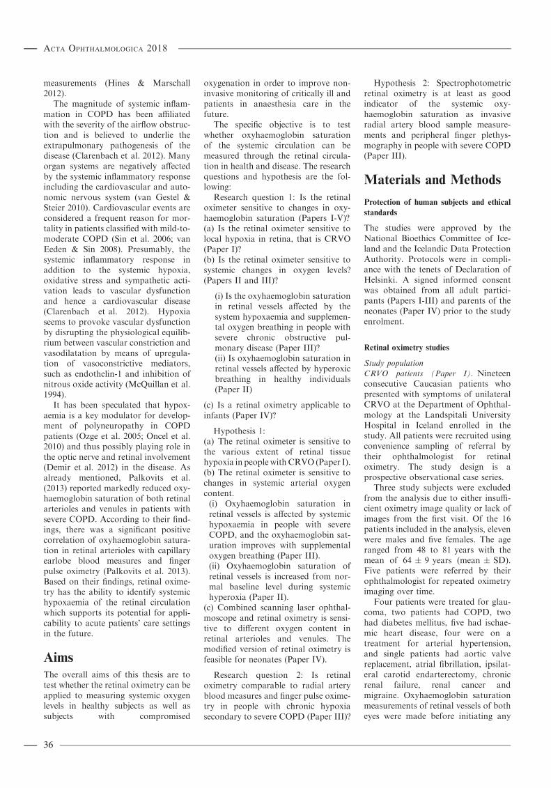
measurements (Hines & Marschall2012).
The magnitude of systemic inflam-mation in COPD has been affiliatedwith the severity of the airflow obstruc-tion and is believed to underlie theextrapulmonary pathogenesis of thedisease (Clarenbach et al. 2012). Manyorgan systems are negatively affectedby the systemic inflammatory responseincluding the cardiovascular and auto-nomic nervous system (van Gestel &Steier 2010). Cardiovascular events areconsidered a frequent reason for mor-tality in patients classified with mild-to-moderate COPD (Sin et al. 2006; vanEeden & Sin 2008). Presumably, thesystemic inflammatory response inaddition to the systemic hypoxia,oxidative stress and sympathetic acti-vation leads to vascular dysfunctionand hence a cardiovascular disease(Clarenbach et al. 2012). Hypoxiaseems to provoke vascular dysfunctionby disrupting the physiological equilib-rium between vascular constriction andvasodilatation by means of upregula-tion of vasoconstrictive mediators,such as endothelin-1 and inhibition ofnitrous oxide activity (McQuillan et al.1994).
It has been speculated that hypox-aemia is a key modulator for develop-ment of polyneuropathy in COPDpatients (Ozge et al. 2005; Oncel et al.2010) and thus possibly playing role inthe optic nerve and retinal involvement(Demir et al. 2012) in the disease. Asalready mentioned, Palkovits et al.(2013) reported markedly reduced oxy-haemoglobin saturation of both retinalarterioles and venules in patients withsevere COPD. According to their find-ings, there was a significant positivecorrelation of oxyhaemoglobin satura-tion in retinal arterioles with capillaryearlobe blood measures and fingerpulse oximetry (Palkovits et al. 2013).Based on their findings, retinal oxime-try has the ability to identify systemichypoxaemia of the retinal circulationwhich supports its potential for appli-cability to acute patients’ care settingsin the future.
Aims
The overall aims of this thesis are totest whether the retinal oximetry can beapplied to measuring systemic oxygenlevels in healthy subjects as well assubjects with compromised
oxygenation in order to improve non-invasive monitoring of critically ill andpatients in anaesthesia care in thefuture.
The specific objective is to testwhether oxyhaemoglobin saturationof the systemic circulation can bemeasured through the retinal circula-tion in health and disease. The researchquestions and hypothesis are the fol-lowing:
Research question 1: Is the retinaloximeter sensitive to changes in oxy-haemoglobin saturation (Papers I-V)?(a) Is the retinal oximeter sensitive tolocal hypoxia in retina, that is CRVO(Paper I)?(b) Is the retinal oximeter sensitive tosystemic changes in oxygen levels?(Papers II and III)?
(i) Is the oxyhaemoglobin saturationin retinal vessels affected by thesystem hypoxaemia and supplemen-tal oxygen breathing in people withsevere chronic obstructive pul-monary disease (Paper III)?(ii) Is oxyhaemoglobin saturation inretinal vessels affected by hyperoxicbreathing in healthy individuals(Paper II)
(c) Is a retinal oximetry applicable toinfants (Paper IV)?
Hypothesis 1:(a) The retinal oximeter is sensitive tothe various extent of retinal tissuehypoxia in people withCRVO (Paper I).(b) The retinal oximeter is sensitive tochanges in systemic arterial oxygencontent.(i) Oxyhaemoglobin saturation inretinal vessels is affected by systemichypoxaemia in people with severeCOPD, and the oxyhaemoglobin sat-uration improves with supplementaloxygen breathing (Paper III).(ii) Oxyhaemoglobin saturation ofretinal vessels is increased from nor-mal baseline level during systemichyperoxia (Paper II).
(c) Combined scanning laser ophthal-moscope and retinal oximetry is sensi-tive to different oxygen content inretinal arterioles and venules. Themodified version of retinal oximetry isfeasible for neonates (Paper IV).
Research question 2: Is retinaloximetry comparable to radial arteryblood measures and finger pulse oxime-try in people with chronic hypoxiasecondary to severe COPD (Paper III)?
Hypothesis 2: Spectrophotometricretinal oximetry is at least as goodindicator of the systemic oxy-haemoglobin saturation as invasiveradial artery blood sample measure-ments and peripheral finger plethys-mography in people with severe COPD(Paper III).
Materials and Methods
Protection of human subjects and ethical
standards
The studies were approved by theNational Bioethics Committee of Ice-land and the Icelandic Data ProtectionAuthority. Protocols were in compli-ance with the tenets of Declaration ofHelsinki. A signed informed consentwas obtained from all adult partici-pants (Papers I-III) and parents of theneonates (Paper IV) prior to the studyenrolment.
Retinal oximetry studies
Study population
CRVO patients (Paper I). Nineteenconsecutive Caucasian patients whopresented with symptoms of unilateralCRVO at the Department of Ophthal-mology at the Landspitali UniversityHospital in Iceland enrolled in thestudy. All patients were recruited usingconvenience sampling of referral bytheir ophthalmologist for retinaloximetry. The study design is aprospective observational case series.
Three study subjects were excludedfrom the analysis due to either insuffi-cient oximetry image quality or lack ofimages from the first visit. Of the 16patients included in the analysis, elevenwere males and five females. The ageranged from 48 to 81 years with themean of 64 � 9 years (mean � SD).Five patients were referred by theirophthalmologist for repeated oximetryimaging over time.
Four patients were treated for glau-coma, two patients had COPD, twohad diabetes mellitus, five had ischae-mic heart disease, four were on atreatment for arterial hypertension,and single patients had aortic valvereplacement, atrial fibrillation, ipsilat-eral carotid endarterectomy, chronicrenal failure, renal cancer andmigraine. Oxyhaemoglobin saturationmeasurements of retinal vessels of botheyes were made before initiating any
36
Acta Ophthalmologica 2018
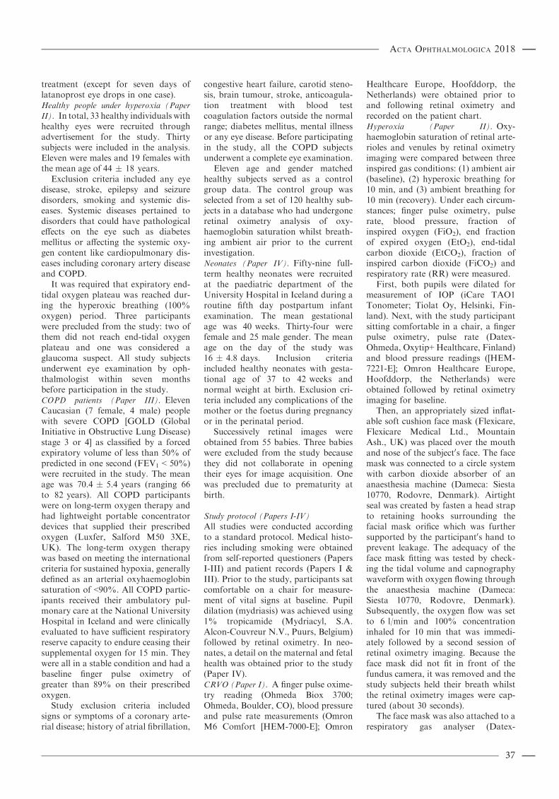
treatment (except for seven days oflatanoprost eye drops in one case).Healthy people under hyperoxia (Paper
II). In total, 33 healthy individuals withhealthy eyes were recruited throughadvertisement for the study. Thirtysubjects were included in the analysis.Eleven were males and 19 females withthe mean age of 44 � 18 years.
Exclusion criteria included any eyedisease, stroke, epilepsy and seizuredisorders, smoking and systemic dis-eases. Systemic diseases pertained todisorders that could have pathologicaleffects on the eye such as diabetesmellitus or affecting the systemic oxy-gen content like cardiopulmonary dis-eases including coronary artery diseaseand COPD.
It was required that expiratory end-tidal oxygen plateau was reached dur-ing the hyperoxic breathing (100%oxygen) period. Three participantswere precluded from the study: two ofthem did not reach end-tidal oxygenplateau and one was considered aglaucoma suspect. All study subjectsunderwent eye examination by oph-thalmologist within seven monthsbefore participation in the study.COPD patients (Paper III). ElevenCaucasian (7 female, 4 male) peoplewith severe COPD [GOLD (GlobalInitiative in Obstructive Lung Disease)stage 3 or 4] as classified by a forcedexpiratory volume of less than 50% ofpredicted in one second (FEV1 ˂ 50%)were recruited in the study. The meanage was 70.4 � 5.4 years (ranging 66to 82 years). All COPD participantswere on long-term oxygen therapy andhad lightweight portable concentratordevices that supplied their prescribedoxygen (Luxfer, Salford M50 3XE,UK). The long-term oxygen therapywas based on meeting the internationalcriteria for sustained hypoxia, generallydefined as an arterial oxyhaemoglobinsaturation of <90%. All COPD partic-ipants received their ambulatory pul-monary care at the National UniversityHospital in Iceland and were clinicallyevaluated to have sufficient respiratoryreserve capacity to endure ceasing theirsupplemental oxygen for 15 min. Theywere all in a stable condition and had abaseline finger pulse oximetry ofgreater than 89% on their prescribedoxygen.
Study exclusion criteria includedsigns or symptoms of a coronary arte-rial disease; history of atrial fibrillation,
congestive heart failure, carotid steno-sis, brain tumour, stroke, anticoagula-tion treatment with blood testcoagulation factors outside the normalrange; diabetes mellitus, mental illnessor any eye disease. Before participatingin the study, all the COPD subjectsunderwent a complete eye examination.
Eleven age and gender matchedhealthy subjects served as a controlgroup data. The control group wasselected from a set of 120 healthy sub-jects in a database who had undergoneretinal oximetry analysis of oxy-haemoglobin saturation whilst breath-ing ambient air prior to the currentinvestigation.Neonates (Paper IV). Fifty-nine full-term healthy neonates were recruitedat the paediatric department of theUniversity Hospital in Iceland during aroutine fifth day postpartum infantexamination. The mean gestationalage was 40 weeks. Thirty-four werefemale and 25 male gender. The meanage on the day of the study was16 � 4.8 days. Inclusion criteriaincluded healthy neonates with gesta-tional age of 37 to 42 weeks andnormal weight at birth. Exclusion cri-teria included any complications of themother or the foetus during pregnancyor in the perinatal period.
Successively retinal images wereobtained from 55 babies. Three babieswere excluded from the study becausethey did not collaborate in openingtheir eyes for image acquisition. Onewas precluded due to prematurity atbirth.
Study protocol (Papers I-IV)
All studies were conducted accordingto a standard protocol. Medical histo-ries including smoking were obtainedfrom self-reported questioners (PapersI-III) and patient records (Papers I &III). Prior to the study, participants satcomfortable on a chair for measure-ment of vital signs at baseline. Pupildilation (mydriasis) was achieved using1% tropicamide (Mydriacyl, S.A.Alcon-Couvreur N.V., Puurs, Belgium)followed by retinal oximetry. In neo-nates, a detail on the maternal and fetalhealth was obtained prior to the study(Paper IV).CRVO (Paper I). A finger pulse oxime-try reading (Ohmeda Biox 3700;Ohmeda, Boulder, CO), blood pressureand pulse rate measurements (OmronM6 Comfort [HEM-7000-E]; Omron
Healthcare Europe, Hoofddorp, theNetherlands) were obtained prior toand following retinal oximetry andrecorded on the patient chart.Hyperoxia (Paper II). Oxy-haemoglobin saturation of retinal arte-rioles and venules by retinal oximetryimaging were compared between threeinspired gas conditions: (1) ambient air(baseline), (2) hyperoxic breathing for10 min, and (3) ambient breathing for10 min (recovery). Under each circum-stances; finger pulse oximetry, pulserate, blood pressure, fraction ofinspired oxygen (FiO2), end fractionof expired oxygen (EtO2), end-tidalcarbon dioxide (EtCO2), fraction ofinspired carbon dioxide (FiCO2) andrespiratory rate (RR) were measured.
First, both pupils were dilated formeasurement of IOP (iCare TAO1Tonometer; Tiolat Oy, Helsinki, Fin-land). Next, with the study participantsitting comfortable in a chair, a fingerpulse oximetry, pulse rate (Datex-Ohmeda, Oxytip+Healthcare, Finland)and blood pressure readings ([HEM-7221-E]; Omron Healthcare Europe,Hoofddorp, the Netherlands) wereobtained followed by retinal oximetryimaging for baseline.
Then, an appropriately sized inflat-able soft cushion face mask (Flexicare,Flexicare Medical Ltd., MountainAsh., UK) was placed over the mouthand nose of the subject0s face. The facemask was connected to a circle systemwith carbon dioxide absorber of ananaesthesia machine (Dameca: Siesta10770, Rodovre, Denmark). Airtightseal was created by fasten a head strapto retaining hooks surrounding thefacial mask orifice which was furthersupported by the participant0s hand toprevent leakage. The adequacy of theface mask fitting was tested by check-ing the tidal volume and capnographywaveform with oxygen flowing throughthe anaesthesia machine (Dameca:Siesta 10770, Rodovre, Denmark).Subsequently, the oxygen flow was setto 6 l/min and 100% concentrationinhaled for 10 min that was immedi-ately followed by a second session ofretinal oximetry imaging. Because theface mask did not fit in front of thefundus camera, it was removed and thestudy subjects held their breath whilstthe retinal oximetry images were cap-tured (about 30 seconds).
The face mask was also attached to arespiratory gas analyser (Datex-
37
Acta Ophthalmologica 2018
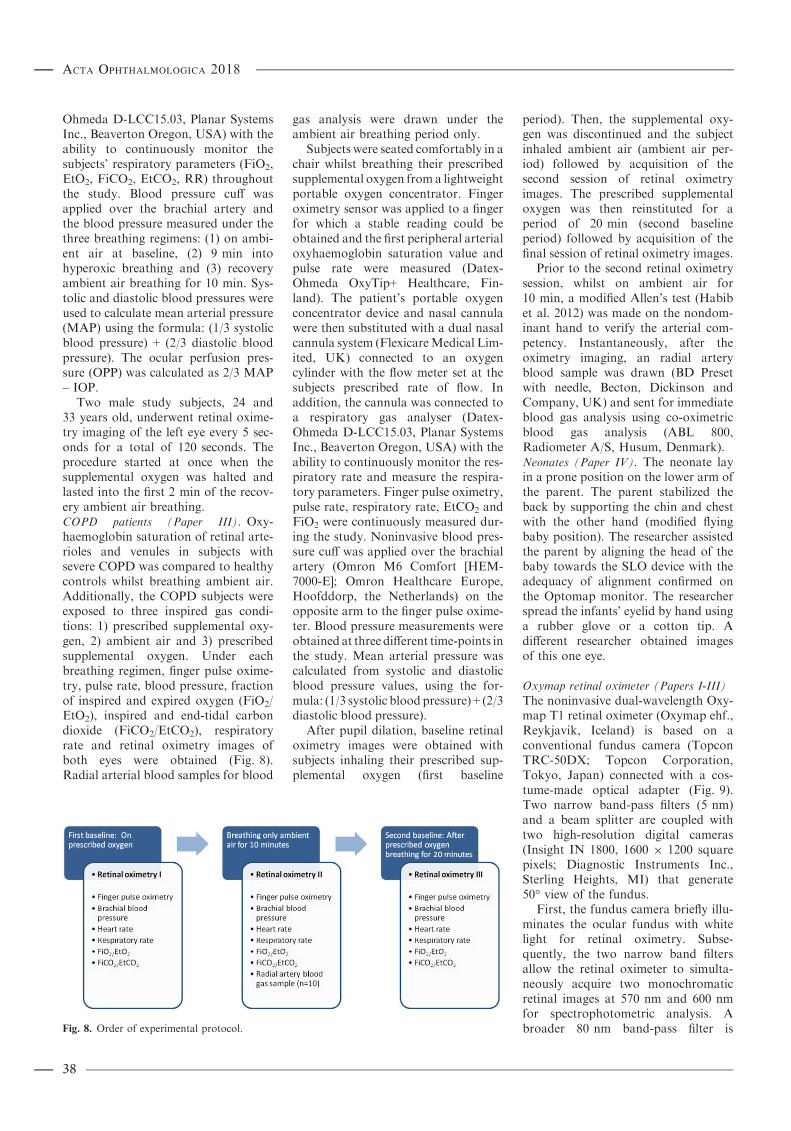
Ohmeda D-LCC15.03, Planar SystemsInc., Beaverton Oregon, USA) with theability to continuously monitor thesubjects’ respiratory parameters (FiO2,EtO2, FiCO2, EtCO2, RR) throughoutthe study. Blood pressure cuff wasapplied over the brachial artery andthe blood pressure measured under thethree breathing regimens: (1) on ambi-ent air at baseline, (2) 9 min intohyperoxic breathing and (3) recoveryambient air breathing for 10 min. Sys-tolic and diastolic blood pressures wereused to calculate mean arterial pressure(MAP) using the formula: (1/3 systolicblood pressure) + (2/3 diastolic bloodpressure). The ocular perfusion pres-sure (OPP) was calculated as 2/3 MAP– IOP.
Two male study subjects, 24 and33 years old, underwent retinal oxime-try imaging of the left eye every 5 sec-onds for a total of 120 seconds. Theprocedure started at once when thesupplemental oxygen was halted andlasted into the first 2 min of the recov-ery ambient air breathing.COPD patients (Paper III). Oxy-haemoglobin saturation of retinal arte-rioles and venules in subjects withsevere COPD was compared to healthycontrols whilst breathing ambient air.Additionally, the COPD subjects wereexposed to three inspired gas condi-tions: 1) prescribed supplemental oxy-gen, 2) ambient air and 3) prescribedsupplemental oxygen. Under eachbreathing regimen, finger pulse oxime-try, pulse rate, blood pressure, fractionof inspired and expired oxygen (FiO2/EtO2), inspired and end-tidal carbondioxide (FiCO2/EtCO2), respiratoryrate and retinal oximetry images ofboth eyes were obtained (Fig. 8).Radial arterial blood samples for blood
gas analysis were drawn under theambient air breathing period only.
Subjects were seated comfortably in achair whilst breathing their prescribedsupplemental oxygen from a lightweightportable oxygen concentrator. Fingeroximetry sensor was applied to a fingerfor which a stable reading could beobtained and the first peripheral arterialoxyhaemoglobin saturation value andpulse rate were measured (Datex-Ohmeda OxyTip+ Healthcare, Fin-land). The patient’s portable oxygenconcentrator device and nasal cannulawere then substituted with a dual nasalcannula system (FlexicareMedical Lim-ited, UK) connected to an oxygencylinder with the flow meter set at thesubjects prescribed rate of flow. Inaddition, the cannula was connected toa respiratory gas analyser (Datex-Ohmeda D-LCC15.03, Planar SystemsInc., Beaverton Oregon, USA) with theability to continuously monitor the res-piratory rate and measure the respira-tory parameters. Finger pulse oximetry,pulse rate, respiratory rate, EtCO2 andFiO2 were continuously measured dur-ing the study. Noninvasive blood pres-sure cuff was applied over the brachialartery (Omron M6 Comfort [HEM-7000-E]; Omron Healthcare Europe,Hoofddorp, the Netherlands) on theopposite arm to the finger pulse oxime-ter. Blood pressure measurements wereobtained at three different time-points inthe study. Mean arterial pressure wascalculated from systolic and diastolicblood pressure values, using the for-mula: (1/3 systolic blood pressure)+ (2/3diastolic blood pressure).
After pupil dilation, baseline retinaloximetry images were obtained withsubjects inhaling their prescribed sup-plemental oxygen (first baseline
period). Then, the supplemental oxy-gen was discontinued and the subjectinhaled ambient air (ambient air per-iod) followed by acquisition of thesecond session of retinal oximetryimages. The prescribed supplementaloxygen was then reinstituted for aperiod of 20 min (second baselineperiod) followed by acquisition of thefinal session of retinal oximetry images.
Prior to the second retinal oximetrysession, whilst on ambient air for10 min, a modified Allen’s test (Habibet al. 2012) was made on the nondom-inant hand to verify the arterial com-petency. Instantaneously, after theoximetry imaging, an radial arteryblood sample was drawn (BD Presetwith needle, Becton, Dickinson andCompany, UK) and sent for immediateblood gas analysis using co-oximetricblood gas analysis (ABL 800,Radiometer A/S, Husum, Denmark).Neonates (Paper IV). The neonate layin a prone position on the lower arm ofthe parent. The parent stabilized theback by supporting the chin and chestwith the other hand (modified flyingbaby position). The researcher assistedthe parent by aligning the head of thebaby towards the SLO device with theadequacy of alignment confirmed onthe Optomap monitor. The researcherspread the infants’ eyelid by hand usinga rubber glove or a cotton tip. Adifferent researcher obtained imagesof this one eye.
Oxymap retinal oximeter (Papers I-III)
The noninvasive dual-wavelength Oxy-map T1 retinal oximeter (Oxymap ehf.,Reykjavik, Iceland) is based on aconventional fundus camera (TopconTRC-50DX; Topcon Corporation,Tokyo, Japan) connected with a cos-tume-made optical adapter (Fig. 9).Two narrow band-pass filters (5 nm)and a beam splitter are coupled withtwo high-resolution digital cameras(Insight IN 1800, 1600 9 1200 squarepixels; Diagnostic Instruments Inc.,Sterling Heights, MI) that generate50° view of the fundus.
First, the fundus camera briefly illu-minates the ocular fundus with whitelight for retinal oximetry. Subse-quently, the two narrow band filtersallow the retinal oximeter to simulta-neously acquire two monochromaticretinal images at 570 nm and 600 nmfor spectrophotometric analysis. Abroader 80 nm band-pass filter isFig. 8. Order of experimental protocol.
38
Acta Ophthalmologica 2018
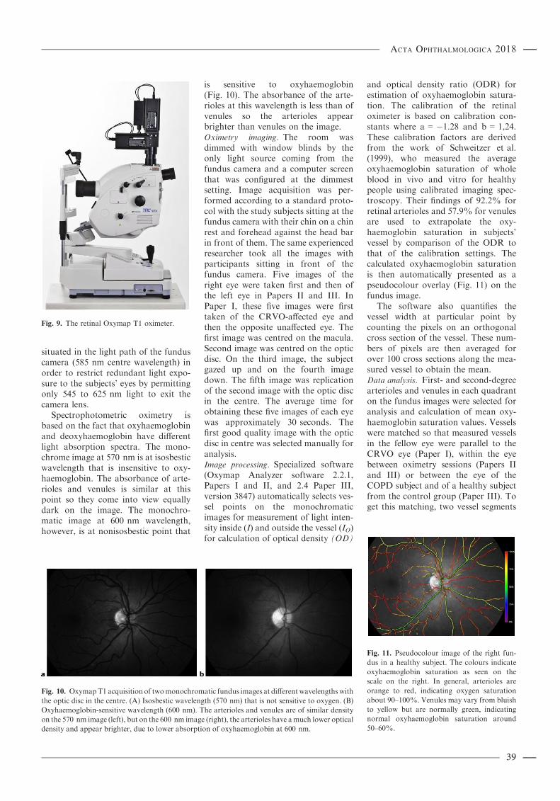
situated in the light path of the funduscamera (585 nm centre wavelength) inorder to restrict redundant light expo-sure to the subjects’ eyes by permittingonly 545 to 625 nm light to exit thecamera lens.
Spectrophotometric oximetry isbased on the fact that oxyhaemoglobinand deoxyhaemoglobin have differentlight absorption spectra. The mono-chrome image at 570 nm is at isosbesticwavelength that is insensitive to oxy-haemoglobin. The absorbance of arte-rioles and venules is similar at thispoint so they come into view equallydark on the image. The monochro-matic image at 600 nm wavelength,however, is at nonisosbestic point that
is sensitive to oxyhaemoglobin(Fig. 10). The absorbance of the arte-rioles at this wavelength is less than ofvenules so the arterioles appearbrighter than venules on the image.Oximetry imaging. The room wasdimmed with window blinds by theonly light source coming from thefundus camera and a computer screenthat was configured at the dimmestsetting. Image acquisition was per-formed according to a standard proto-col with the study subjects sitting at thefundus camera with their chin on a chinrest and forehead against the head barin front of them. The same experiencedresearcher took all the images withparticipants sitting in front of thefundus camera. Five images of theright eye were taken first and then ofthe left eye in Papers II and III. InPaper I, these five images were firsttaken of the CRVO-affected eye andthen the opposite unaffected eye. Thefirst image was centred on the macula.Second image was centred on the opticdisc. On the third image, the subjectgazed up and on the fourth imagedown. The fifth image was replicationof the second image with the optic discin the centre. The average time forobtaining these five images of each eyewas approximately 30 seconds. Thefirst good quality image with the opticdisc in centre was selected manually foranalysis.Image processing. Specialized software(Oxymap Analyzer software 2.2.1,Papers I and II, and 2.4 Paper III,version 3847) automatically selects ves-sel points on the monochromaticimages for measurement of light inten-sity inside (I) and outside the vessel (IO)for calculation of optical density (OD)
and optical density ratio (ODR) forestimation of oxyhaemoglobin satura-tion. The calibration of the retinaloximeter is based on calibration con-stants where a = �1.28 and b = 1,24.These calibration factors are derivedfrom the work of Schweitzer et al.(1999), who measured the averageoxyhaemoglobin saturation of wholeblood in vivo and vitro for healthypeople using calibrated imaging spec-troscopy. Their findings of 92.2% forretinal arterioles and 57.9% for venulesare used to extrapolate the oxy-haemoglobin saturation in subjects’vessel by comparison of the ODR tothat of the calibration settings. Thecalculated oxyhaemoglobin saturationis then automatically presented as apseudocolour overlay (Fig. 11) on thefundus image.
The software also quantifies thevessel width at particular point bycounting the pixels on an orthogonalcross section of the vessel. These num-bers of pixels are then averaged forover 100 cross sections along the mea-sured vessel to obtain the mean.Data analysis. First- and second-degreearterioles and venules in each quadranton the fundus images were selected foranalysis and calculation of mean oxy-haemoglobin saturation values. Vesselswere matched so that measured vesselsin the fellow eye were parallel to theCRVO eye (Paper I), within the eyebetween oximetry sessions (Papers IIand III) or between the eye of theCOPD subject and of a healthy subjectfrom the control group (Paper III). Toget this matching, two vessel segments
Fig. 9. The retinal Oxymap T1 oximeter.
Fig. 10. OxymapT1 acquisition of twomonochromatic fundus images at different wavelengths with
the optic disc in the centre. (A) Isosbestic wavelength (570 nm) that is not sensitive to oxygen. (B)
Oxyhaemoglobin-sensitive wavelength (600 nm). The arterioles and venules are of similar density
on the 570 nm image (left), but on the 600 nm image (right), the arterioles have amuch lower optical
density and appear brighter, due to lower absorption of oxyhaemoglobin at 600 nm.
Fig. 11. Pseudocolour image of the right fun-
dus in a healthy subject. The colours indicate
oxyhaemoglobin saturation as seen on the
scale on the right. In general, arterioles are
orange to red, indicating oxygen saturation
about 90–100%. Venules may vary from bluish
to yellow but are normally green, indicating
normal oxyhaemoglobin saturation around
50–60%.
39
Acta Ophthalmologica 2018

were sometimes averaged and insertedas one for calculation of the mean forthe eye. For example, in CRVO, if aselected vessel segment in the inferiortemporal venule in the fellow eye wastoo short (˂100 pixels) before branch-ing, both daughter vessels on the otherside of the bifurcation were measured.The calculated mean average was theninserted as one and compared with themean value of the matching parentvessel before branching (if ≥100 pixels)in the CRVO-affected eye.
The AV-difference was representedas the calculated difference betweenarterial and venous oxyhaemoglobinsaturation.CRVO (Paper I)—Vessel segmentsclose to haemorrhages were excludedto bypass artefacts. Vessels werematched so that measured vessels inthe fellow eye were parallel to theCRVO eye.
Of the sixteen patients included inthe analysis, single measurement wasmade in 11 cases and repeated mea-surements in five cases over a timeperiod ranging from two weeks to20 months. Data from two patientswere not included in the comparativestatistical analysis because images ofthe unaffected eye were lacking. Hence,the values of their oxyhaemoglobinsaturation and numbers of measuredvessels are given in the result Table 4(Chapter 4.1) for information only.Hyperoxia(PaperII)—Oxyhaemoglobinsaturation was calculated for all first- orsecond-degree arterioles and venules ineach quadrant of the right eye. Vesselswere matched between retinal images ofthe three breathing regimens.COPD (Paper III)—First- and second-degree arterioles and venules in eachquadrant of the right eye were matchedbetween the retinal images of the threebreathing conditions and with a retinalimage of a healthy control subject.Identical image acquisition and oxi-metric analyses were performed for thehealthy subjects.
Of the 11 COPD participants, onewas not breathing the prescribed oxy-gen on arrival so there was no mea-surement at first baseline for thissubject and we were unable to drawarterial blood sample from another.Hence, data from 10 subjects within theCOPD group underwent statisticalanalysis at each study time period forcomparison of mean and standarddeviation. Furthermore, one COPD
patient was ‘a mouth breather’, andtherefore, it was not possible to getadequate EtCO2 and FiO2 data fromthe dual nasal cannula at any of thethree study period. All 11 COPD sub-jects were included in the comparisonwith healthy controls under ambientair breathing (Table 2).
Neonates (Paper IV)
The Optomap 200Tx scanning laserophthalmoscope (Optos plc., Dun-fermline, Scotland, UK) for the oxime-try imaging in neonates, uses 532 nmand 633 nm laser wavelengths of lightto obtain two monochrome spectralimages of the fundus. Although thereference wavelength at 532 nm neareran isosbestic point, it is not completelyisosbestic. The great distinctionbetween oxy- and deoxyhaemoglobinlight absorption at 532 nm and 633 nm(oxygen sensitive wavelength) allowsfor application of SLO for retinaloximetry. The ODR is calculatedaccording to the following equation:
ODR ¼ OD633 nm
OD532 nm: ð18Þ
The calculated oxyhaemoglobin sat-uration is then inversely and nearlinearly related to the ODR.Oximetry imaging of neonates. The roomwas dimmed with window blinds andthe only light source coming from theSLO and a computer screen that wasconfigured at the dimmest setting. Thesame researcher took all the imageswith the babies aligned in front of theSLO in a modified baby flying position.
Both Ultra-Widefield (200°) andResMax (100°) retinal images wereobtained of a unilateral eye. For thepurpose of the study, 250 ResMaximages were acquired in total with themedian of four images (range 0–8) perneonate. Ultra-Widefield images wereobtained for further research purposeonly.Image processing. A modified version ofthe dual-wavelength Oxymap Analyzersoftware 2.3 (version 5206) algorithmswas used for the spectral imaging andto process the monochromatic ResMaximages for calculation of ODR andvessel diameter. The calibration of themodified version of Oxymap Analyzersoftware is based on calibration con-stants where a = �2.4733 and b =1.4388. These calibration factors arederived from the work of Kristjansdot-tir et al. (2014) which based on thework of Schweitzer et al. (1999) formean oxyhaemoglobin saturation of92.2% for retinal arterioles and of57.9% for venules for healthy peopleas has previously been discussed(Chapter 3.2.3.2). The oxyhaemoglobinsaturation can then be calculatedaccording to the (a 9 ODR + b) 9100. The calculated oxyhaemoglobinsaturation was automatically presentedas a pseudocolour map on a 100°oximetry fundus image (Fig. 12).Image analysis. In total, 28 fundusimages of 28 neonates were manuallyselected for analysis by the modifiedOxymap Analyzer software for calcu-lation of ODR and vessel diameter.The image analysis was standardizedprior to the study. The image of the
Table 2. Selection criteria for retinal vessel segment measurement of oxyhaemoglobin saturation
with the Oxymap T1 retinal oximeter. One pixel is approximately 9.3 lm.
Begin vessel selection Papers I and II:a) at least 15 pixels around the optic disc excludedb) at least 15 pixels on a border of any bright area around the optic disc
excludedc) start as close to the optic disc as possible
Paper III: An area that was demarcated 1.5 disc diameters (344 pixels)
End vessel selection Papers I and II: Never closer than 30 pixels to the rim of the image
Papers III: 3 disc diameters (690 pixels)
Minimum vessel
diameter
Papers I and III : 8.0 pixels (~75 lm)
Paper II: 6.0 pixels (~56 lm)
Vessel length Paper I: 100–300 pixels (as close to 300 pixels as possible)
Paper II: 50–200 pixels (as close to 200 pixels as possible)
Paper III: 50 pixels at minimum but as close to the total length of the
segment between the two circles (1.5 and 3 disc diameters) as possible.
Vessel segment
exclusion criteria
a) Vessel branching with 15 pixelsb) Vessel crossingc) Any extremes in background brightness such as haemorrhage to avoid
artifacts
40
Acta Ophthalmologica 2018
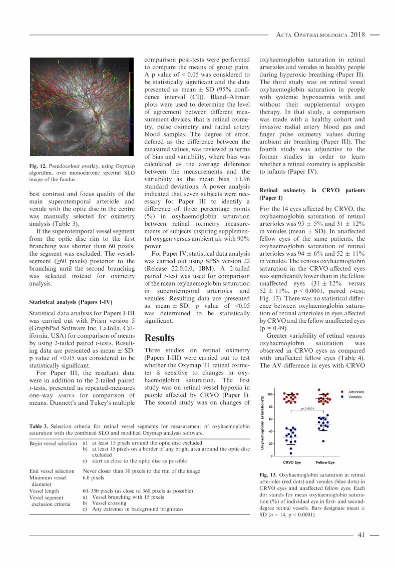
best contrast and focus quality of themain superotemporal arteriole andvenule with the optic disc in the centrewas manually selected for oximetryanalysis (Table 3).
If the superotemporal vessel segmentfrom the optic disc rim to the firstbranching was shorter than 60 pixels,the segment was excluded. The vesselssegment (≥60 pixels) posterior to thebranching until the second branchingwas selected instead for oximetryanalysis.
Statistical analysis (Papers I-IV)
Statistical data analysis for Papers I-IIIwas carried out with Prism version 5(GraphPad Software Inc, LaJolla, Cal-ifornia, USA) for comparison of meansby using 2-tailed paired t-tests. Result-ing data are presented as mean � SD.p value of <0.05 was considered to bestatistically significant.
For Paper III, the resultant datawere in addition to the 2-tailed pairedt-tests, presented as repeated-measuresone–way ANOVA for comparison ofmeans. Dunnett’s and Tukey0s multiple
comparison post-tests were performedto compare the means of group pairs.A p value of < 0.05 was considered tobe statistically significant and the datapresented as mean � SD (95% confi-dence interval (CI)). Bland–Altmanplots were used to determine the levelof agreement between different mea-surement devices, that is retinal oxime-try, pulse oximetry and radial arteryblood samples. The degree of error,defined as the difference between themeasured values, was reviewed in termsof bias and variability, where bias wascalculated as the average differencebetween the measurements and thevariability as the mean bias �1.96standard deviations. A power analysisindicated that seven subjects were nec-essary for Paper III to identify adifference of three percentage points(%) in oxyhaemoglobin saturationbetween retinal oximetry measure-ments of subjects inspiring supplemen-tal oxygen versus ambient air with 90%power.
For Paper IV, statistical data analysiswas carried out using SPSS version 22(Release 22.0.0.0, IBM). A 2-tailedpaired t-test was used for comparisonof themean oxyhaemoglobin saturationin superotemporal arterioles andvenules. Resulting data are presentedas mean � SD. p value of <0.05was determined to be statisticallysignificant.
Results
Three studies on retinal oximetry(Papers I-III) were carried out to testwhether the Oxymap T1 retinal oxime-ter is sensitive to changes in oxy-haemoglobin saturation. The firststudy was on retinal vessel hypoxia inpeople affected by CRVO (Paper I).The second study was on changes of
oxyhaemoglobin saturation in retinalarterioles and venules in healthy peopleduring hyperoxic breathing (Paper II).The third study was on retinal vesseloxyhaemoglobin saturation in peoplewith systemic hypoxaemia with andwithout their supplemental oxygentherapy. In that study, a comparisonwas made with a healthy cohort andinvasive radial artery blood gas andfinger pulse oximetry values duringambient air breathing (Paper III). Thefourth study was adjunctive to theformer studies in order to learnwhether a retinal oximetry is applicableto infants (Paper IV).
Retinal oximetry in CRVO patients
(Paper I)
For the 14 eyes affected by CRVO, theoxyhaemoglobin saturation of retinalarterioles was 95 � 5% and 31 � 12%in venules (mean � SD). In unaffectedfellow eyes of the same patients, theoxyhaemoglobin saturation of retinalarterioles was 94 � 6% and 52 � 11%in venules. The venous oxyhaemoglobinsaturation in the CRVO-affected eyeswas significantly lower than in the fellowunaffected eyes (31 � 12% versus52 � 11%, p < 0.0001, paired t-test;Fig. 13). There was no statistical differ-ence between oxyhaemoglobin satura-tion of retinal arterioles in eyes affectedbyCRVOand the fellow unaffected eyes(p = 0.49).
Greater variability of retinal venousoxyhaemoglobin saturation wasobserved in CRVO eyes as comparedwith unaffected fellow eyes (Table 4).The AV-difference in eyes with CRVO
Fig. 12. Pseudocolour overlay, using Oxymap
algorithm, over monochrome spectral SLO
image of the fundus.
Table 3. Selection criteria for retinal vessel segments for measurement of oxyhaemoglobin
saturation with the combined SLO and modified Oxymap analysis software.
Begin vessel selection a) at least 15 pixels around the optic disc excludedb) at least 15 pixels on a border of any bright area around the optic disc
excludedc) start as close to the optic disc as possible
End vessel selection Never closer than 30 pixels to the rim of the image
Minimum vessel
diameter
6.0 pixels
Vessel length 60–350 pixels (as close to 360 pixels as possible)
Vessel segment
exclusion criteria
a) Vessel branching with 15 pixelsb) Vessel crossingc) Any extremes in background brightness
CRVO Eye Fellow Eye
0
20
40
60
80
100
p<0.0001
VenulesArterioles
Oxy
hem
oglo
bin
satu
ratio
n(%
)
Fig. 13. Oxyhaemoglobin saturation in retinal
arterioles (red dots) and venules (blue dots) in
CRVO eyes and unaffected fellow eyes. Each
dot stands for mean oxyhaemoglobin satura-
tion (%) of individual eye in first- and second-
degree retinal vessels. Bars designate mean �SD (n = 14, p < 0.0001).
41
Acta Ophthalmologica 2018

was 63 � 11% compared with 43 �7% in unaffected fellow eyes (p <0.0001). The mean arteriolar vessel
diameter in pixels (each pixel is about9.3 lm) in CRVO eyes was 10 � 1 and12 � 1 in fellow eyes (p < 0.0001). Thediameter of retinal venules in eyes withCRVO was 17 � 2 and 16 � 2 inunaffected fellow eyes (p = 0.02).
There were no statistical differencesbetween finger pulse oximetry (96% �2%, n = 13) and oxyhaemoglobin sat-uration of retinal arterioles either in theCRVO eye (p = 0.28) or in the felloweye (p = 0.24).
Patients follow-up
All five patients who were followed withrepeated retinal oximetry imagesreceived treatment; dorzolamide eyedrops, intravitreal anti-VEGF beva-cizumab and/or laser photocoagulation.In all five CRVO cases, the venularoxyhaemoglobin saturation improvedover time (Fig. 14). In two patients,the clinical situation improved and thevenous oxyhaemoglobin saturationrecovered. In two eyes, the clinical signsand symptoms of CRVO completelyresolved but the venular oxy-haemoglobin saturation did not returnto normal. One eye developed iris neo-vascularization, glaucoma and poorvisual outcome where the venous oxy-haemoglobin saturation remainedslightly subnormal. Measured values atthe first and last visit are listed inTable 5.
Retinal oximetry images on thepatient who developed iris neovascu-larization, glaucoma and poor visualoutcome are presented in Fig. 15.Images on the patient who resolvedfrom the clinical signs and symptomsare presented in Fig. 16.
Retinal oximetry in healthy under
hyperoxemia (Paper II)
After 10 min of hyperoxic facemaskbreathing, the mean FiO2 was96 � 2% and the EtO2 91 � 4%(mean � SD, n = 30). The EtCO2 mea-sured 36 � 2 mmHg (n = 29). Thephysiological parameters are presentedin Table 6.
Both the brachial blood pressure andthe heart rate were unaffected by hyper-oxic breathing as compared with base-line normoxic breathing (p = 0.86,
Table 4. Oxyhaemoglobin saturation (%) in first- and second-degree retinal arterioles and venules in 16 patients with central retinal vein occlusion.
The values show mean � standard deviation and number of measured retinal vessels (parentheses). SpO2 is finger pulse oximetry. The diameter of
retinal venules is shown in pixels. Patients’ number 15 and number 16 were not included in statistical analysis. Reprinted from Paper I (Graefes Arch
Clin Exp Ophthalmol, 253(10), 1653–1661, ©2015, with permission of Graefe’s Archive for Clinical and Experimental Ophthalmology).
Patient no.
CRVO eye Fellow unaffected eye CRVO eye Fellow eye
Arterioles Venules Arterioles Venules SpO2 Retinal venule diameter Retinal venule diameter
1 87 � 5 (2) 23 � 26 (4) 87 � 6 (2) 49 � 6 (4) 94 16 17
2 87 � 3 (6) 5 � 32 (3) 85 � 8 (5) 24 � 6 (3) 93 14 14
3 101 � 4 (6) 24 � 12 (4) 101 � 8 (6) 62 � 4 (4) 98 20 16
4 96 � 16 (4) 33 � 22 (3) 101 � 6 (4) 54 � 5 (3) 95 18 15
5 95 � 5 (5) 42 � 13 (5) 93 � 11 (5) 61 � 4 (4) 97 16 16
6 98 � 7 (3) 38 � 17 (5) 94 � 1 (3) 55 � 6 (5) 97 13 13
7 95 � 3 (7) 45 � 12 (4) 98 � 7 (7) 60 � 7 (4) 95 18 20
8 92 � 12 (5) 46 � 24 (2) 90 � 4 (6) 55 � 2 (2) 97 19 17
9 102 � 8 (3) 33 � 24 (5) 108 � 2 (3) 62 � 13 (5) 97 17 17
10 87 � 10 (6) 12 � 15 (6) 89 � 5 (6) 43 � 6 (6) 95 18 14
11 102 � 7 (7) 32 � 22 (6) 92 � 11(7) 48 � 9 (6) 94 19 17
12 93 � 7 (4) 25 � 14 (3) 89 � 3 (4) 49 � 7 (3) 96 14 16
13 99 � 6 (5) 36 � 6 (5) 93 � 7 (5) 45 � 5 (4) 20 16
14 90 � 6 (5) 43 � 6 (6) 93 � 4 (5) 66 � 4 (6) 16 15
15 95 � 5 (7) 34 � 3 (4)
16 102 � 9 (4) 50 � 8 (4) 96
Patients 1–14 Mean � SD 95 � 5 31 � 12 94 � 6 52 � 11 17 � 2 16 � 2
Patients 1–16 Mean � SD 95 � 5 33 � 12
The standard deviation reflects the oxyhaemoglobin saturation variation within individual eyes.
0 50 1000
20
40
60
80
400 500
case 1case 2case 3case 4case 5
Time in days
Oxy
gen
satu
ratio
n(%
)
Fig. 14. Mean oxyhaemoglobin saturation of
retinal venules during the follow-up period of
five patients with CRVO. Each dot indicates
the oxyhaemoglobin saturation in first- and
second-degree venules at a point in time. Case
1. The patient developed iris neovasculariza-
tion, glaucoma (Fig. 15) and poor visual
outcome. Case 2. The patient received dorzo-
lamide eye drops. Clinical signs of CRVO
resolved (Fig. 16). Case 3. The patient pre-
sented with ischaemic CRVO, macular oedema
and elevated intraocular pressure. Treatment
consisted of dorzolamide eye drops, intravit-
real bevacizumab and panretinal photocoagu-
lation. At week two, the mean retinal venous
oxyhaemoglobin saturation had improved.
Case 4. The patient was treated with dorzo-
lamide eye drops for 4 weeks. During the 8-
week follow-up period, clinical signs and
symptoms resolved. Case 5. The patient pre-
sented with mild CRVO on arrival with no
macular oedema. The patient was treated with
dorzolamide eye drops and established full
recovery.
Table 5. Retinal venous oxyhaemoglobin sat-
uration values (%) in the five patients that
were followed with repeated oximetry imaging.
Case
Follow-up
period On arrival
Last
image
1 20 months 23 � 26 40 � 23
2 80 days 42 � 13 62 � 8
3 16 days 33 � 23 49 � 12
4 76 days 12 � 15 35 � 10
5 33 days 36 � 6 60 � 7
The values show mean � standard deviation.
42
Acta Ophthalmologica 2018
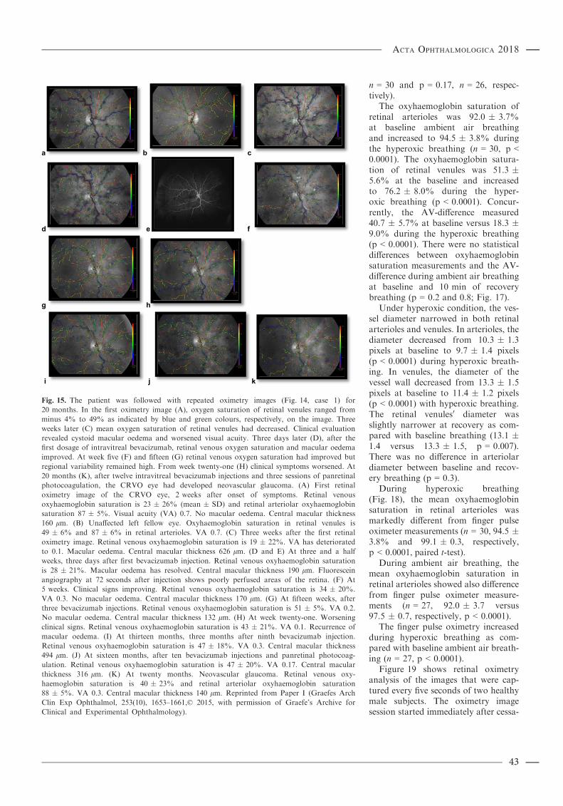
n = 30 and p = 0.17, n = 26, respec-tively).
The oxyhaemoglobin saturation ofretinal arterioles was 92.0 � 3.7%at baseline ambient air breathingand increased to 94.5 � 3.8% duringthe hyperoxic breathing (n = 30, p <0.0001). The oxyhaemoglobin satura-tion of retinal venules was 51.3 �5.6% at the baseline and increasedto 76.2 � 8.0% during the hyper-oxic breathing (p < 0.0001). Concur-rently, the AV-difference measured40.7 � 5.7% at baseline versus 18.3 �9.0% during the hyperoxic breathing(p < 0.0001). There were no statisticaldifferences between oxyhaemoglobinsaturation measurements and the AV-difference during ambient air breathingat baseline and 10 min of recoverybreathing (p = 0.2 and 0.8; Fig. 17).
Under hyperoxic condition, the ves-sel diameter narrowed in both retinalarterioles and venules. In arterioles, thediameter decreased from 10.3 � 1.3pixels at baseline to 9.7 � 1.4 pixels(p < 0.0001) during hyperoxic breath-ing. In venules, the diameter of thevessel wall decreased from 13.3 � 1.5pixels at baseline to 11.4 � 1.2 pixels(p < 0.0001) with hyperoxic breathing.The retinal venules0 diameter wasslightly narrower at recovery as com-pared with baseline breathing (13.1 �1.4 versus 13.3 � 1.5, p = 0.007).There was no difference in arteriolardiameter between baseline and recov-ery breathing (p = 0.3).
During hyperoxic breathing(Fig. 18), the mean oxyhaemoglobinsaturation in retinal arterioles wasmarkedly different from finger pulseoximeter measurements (n = 30, 94.5 �3.8% and 99.1 � 0.3, respectively,p < 0.0001, paired t-test).
During ambient air breathing, themean oxyhaemoglobin saturation inretinal arterioles showed also differencefrom finger pulse oximeter measure-ments (n = 27, 92.0 � 3.7 versus97.5 � 0.7, respectively, p < 0.0001).
The finger pulse oximetry increasedduring hyperoxic breathing as com-pared with baseline ambient air breath-ing (n = 27, p < 0.0001).
Figure 19 shows retinal oximetryanalysis of the images that were cap-tured every five seconds of two healthymale subjects. The oximetry imagesession started immediately after cessa-
b c
e f
g h
i j k
a
d
Fig. 15. The patient was followed with repeated oximetry images (Fig. 14, case 1) for
20 months. In the first oximetry image (A), oxygen saturation of retinal venules ranged from
minus 4% to 49% as indicated by blue and green colours, respectively, on the image. Three
weeks later (C) mean oxygen saturation of retinal venules had decreased. Clinical evaluation
revealed cystoid macular oedema and worsened visual acuity. Three days later (D), after the
first dosage of intravitreal bevacizumab, retinal venous oxygen saturation and macular oedema
improved. At week five (F) and fifteen (G) retinal venous oxygen saturation had improved but
regional variability remained high. From week twenty-one (H) clinical symptoms worsened. At
20 months (K), after twelve intravitreal bevacizumab injections and three sessions of panretinal
photocoagulation, the CRVO eye had developed neovascular glaucoma. (A) First retinal
oximetry image of the CRVO eye, 2 weeks after onset of symptoms. Retinal venous
oxyhaemoglobin saturation is 23 � 26% (mean � SD) and retinal arteriolar oxyhaemoglobin
saturation 87 � 5%. Visual acuity (VA) 0.7. No macular oedema. Central macular thickness
160 lm. (B) Unaffected left fellow eye. Oxyhaemoglobin saturation in retinal venules is
49 � 6% and 87 � 6% in retinal arterioles. VA 0.7. (C) Three weeks after the first retinal
oximetry image. Retinal venous oxyhaemoglobin saturation is 19 � 22%. VA has deteriorated
to 0.1. Macular oedema. Central macular thickness 626 lm. (D and E) At three and a half
weeks, three days after first bevacizumab injection. Retinal venous oxyhaemoglobin saturation
is 28 � 21%. Macular oedema has resolved. Central macular thickness 190 lm. Fluorescein
angiography at 72 seconds after injection shows poorly perfused areas of the retina. (F) At
5 weeks. Clinical signs improving. Retinal venous oxyhaemoglobin saturation is 34 � 20%.
VA 0.3. No macular oedema. Central macular thickness 170 lm. (G) At fifteen weeks, after
three bevacizumab injections. Retinal venous oxyhaemoglobin saturation is 51 � 5%. VA 0.2.
No macular oedema. Central macular thickness 132 lm. (H) At week twenty-one. Worsening
clinical signs. Retinal venous oxyhaemoglobin saturation is 43 � 21%. VA 0.1. Recurrence of
macular oedema. (I) At thirteen months, three months after ninth bevacizumab injection.
Retinal venous oxyhaemoglobin saturation is 47 � 18%. VA 0.3. Central macular thickness
494 lm. (J) At sixteen months, after ten bevacizumab injections and panretinal photocoag-
ulation. Retinal venous oxyhaemoglobin saturation is 47 � 20%. VA 0.17. Central macular
thickness 316 lm. (K) At twenty months. Neovascular glaucoma. Retinal venous oxy-
haemoglobin saturation is 40 � 23% and retinal arteriolar oxyhaemoglobin saturation
88 � 5%. VA 0.3. Central macular thickness 140 lm. Reprinted from Paper I (Graefes Arch
Clin Exp Ophthalmol, 253(10), 1653–1661,© 2015, with permission of Graefe’s Archive for
Clinical and Experimental Ophthalmology).
43
Acta Ophthalmologica 2018
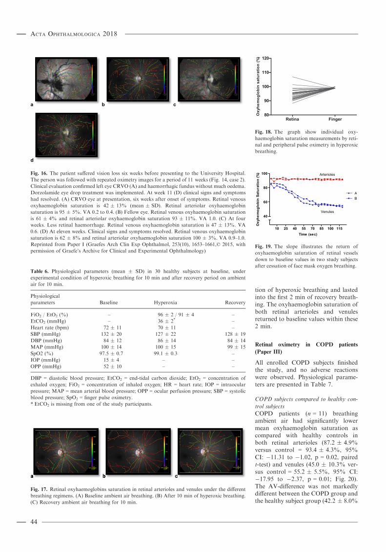
tion of hyperoxic breathing and lastedinto the first 2 min of recovery breath-ing. The oxyhaemoglobin saturation ofboth retinal arterioles and venulesreturned to baseline values within these2 min.
Retinal oximetry in COPD patients
(Paper III)
All enrolled COPD subjects finishedthe study, and no adverse reactionswere observed. Physiological parame-ters are presented in Table 7.
COPD subjects compared to healthy con-
trol subjects
COPD patients (n = 11) breathingambient air had significantly lowermean oxyhaemoglobin saturation ascompared with healthy controls inboth retinal arterioles (87.2 � 4.9%versus control = 93.4 � 4.3%, 95%CI: �11.31 to �1.02, p = 0.02, pairedt-test) and venules (45.0 � 10.3% ver-sus control = 55.2 � 5.5%, 95% CI:�17.95 to �2.37, p = 0.01; Fig. 20).The AV-difference was not markedlydifferent between the COPD group andthe healthy subject group (42.2 � 8.0%
a b c
d
Fig. 16. The patient suffered vision loss six weeks before presenting to the University Hospital.
The person was followed with repeated oximetry images for a period of 11 weeks (Fig. 14, case 2).
Clinical evaluation confirmed left eye CRVO (A) and haemorrhagic fundus without much oedema.
Dorzolamide eye drop treatment was implemented. At week 11 (D) clinical signs and symptoms
had resolved. (A) CRVO eye at presentation, six weeks after onset of symptoms. Retinal venous
oxyhaemoglobin saturation is 42 � 13% (mean � SD). Retinal arteriolar oxyhaemoglobin
saturation is 95 � 5%. VA 0.2 to 0.4. (B) Fellow eye. Retinal venous oxyhaemoglobin saturation
is 61 � 4% and retinal arteriolar oxyhaemoglobin saturation 93 � 11%. VA 1.0. (C) At four
weeks. Less retinal haemorrhage. Retinal venous oxyhaemoglobin saturation is 47 � 13%. VA
0.6. (D) At eleven weeks. Clinical signs and symptoms resolved. Retinal venous oxyhaemoglobin
saturation is 62 � 8% and retinal arteriolar oxyhaemoglobin saturation 100 � 3%. VA 0.9–1.0.Reprinted from Paper I (Graefes Arch Clin Exp Ophthalmol, 253(10), 1653–1661,© 2015, with
permission of Graefe’s Archive for Clinical and Experimental Ophthalmology)
Table 6. Physiological parameters (mean � SD) in 30 healthy subjects at baseline, under
experimental condition of hyperoxic breathing for 10 min and after recovery period on ambient
air for 10 min.
Physiological
parameters Baseline Hyperoxia Recovery
FiO2 / EtO2 (%) – 96 � 2 / 91 � 4 –EtCO2 (mmHg) – 36 � 2* –Heart rate (bpm) 72 � 11 70 � 11 –SBP (mmHg) 132 � 20 127 � 22 128 � 19
DBP (mmHg) 84 � 12 86 � 14 84 � 14
MAP (mmHg) 100 � 14 100 � 15 99 � 15
SpO2 (%) 97.5 � 0.7 99.1 � 0.3 –IOP (mmHg) 15 � 4 – –OPP (mmHg) 52 � 10 – –
DBP = diastolic blood pressure; EtCO2 = end-tidal carbon dioxide; EtO2 = concentration of
exhaled oxygen; FiO2 = concentration of inhaled oxygen; HR = heart rate; IOP = intraocular
pressure; MAP = mean arterial blood pressure; OPP = ocular perfusion pressure; SBP = systolic
blood pressure; SpO2 = finger pulse oximetry.
* EtCO2 is missing from one of the study participants.
a b c
Fig. 17. Retinal oxyhaemoglobins saturation in retinal arterioles and venules under the different
breathing regimens. (A) Baseline ambient air breathing. (B) After 10 min of hyperoxic breathing.
(C) Recovery ambient air breathing for 10 min.
Retina Finger80
90
100
110
120)%(
noitarutasnibolgo
mehyxO
Fig. 18. The graph show individual oxy-
haemoglobin saturation measurements by reti-
nal and peripheral pulse oximetry in hyperoxic
breathing.
10 25 40 55 70 85 100 115
40
60
80
100
AB
Arterioles
Venules
Time (sec)
noitarutaSnibolgo
mehyxO
(%)
Fig. 19. The slope illustrates the return of
oxyhaemoglobin saturation of retinal vessels
down to baseline values in two study subjects
after cessation of face mask oxygen breathing.
44
Acta Ophthalmologica 2018

versus control = 38.2 � 4.0%, 95%CI: �1.98 to 9.98, p = 0.17).
Administration of the prescribedsupplemental oxygen increased theoxyhaemoglobin saturation in retinalarterioles (87.2% � 4.9% to 89.5% �6.0%, 95% CI: �4.13 to �0.31,p = 0.02) but not in venules (45.0% �10.3% to 46.7% � 12.8%, 95% CI:�5.15 to 1.76, p = 0.3). When COPDsubjects were on their supplementaloxygen, the difference in oxy-haemoglobin saturation showed a non-significance as compared with thehealthy group in retinal arterioles(95% CI: �9.37 to 1.49, p = 0.14).
No differences were observedbetween COPD subjects and healthy
controls, respectively, in vessel diame-ter of arterioles (106.6 � 10.6 lm ver-sus 114.2 � 10.9 lm, 95% CI: �17.06to 2.00, p = 0.11) or venules (147.7 �14.1 lm versus 153.4 � 15.1 lm, 95%CI: �24.28 to 12.85, p = 0.51).
COPD subjects under experimental
protocol
After termination of inspiring supple-mental oxygen and 10 min of ambient
air breathing, the oxyhaemoglobin sat-uration in retinal arterioles and fingeroximetry reading decreased consider-ably (Table 8). Inhalation of supple-mental oxygen for 20 min (secondbaseline period) returned the oxy-haemoglobin saturation in retinal arte-rioles and finger nearly to the values atfirst baseline. Cessation or reapplica-tion of supplemental oxygen breathingneither significantly affected the retinalvenule oxyhaemoglobin saturation,AV-difference, nor retinal vessel diam-eter. No significant differences werefound between oximetric measure-ments at first and second baseline.
The comparison of retinal arteriolaroximetry with finger pulse oximetryand radial artery co-oximetry in theCOPD subjects during the ambient airbreathing is shown in Fig. 21.
Individual radial artery blood gasmeasurements are presented in Table 9.For the COPD group the meanPaO2 was 61.3 � 10.5 mmHg and42.2% � 5.9 mmHg for PCO2. Themean value for bicarbonate was 26.4 �3.4 mEq/l, and the pH 7.4 � 0.0.
Bland–Altman plots compared reti-nal arteriolar oximetric oxy-haemoglobin saturation values withradial artery blood gas and finger pulseoximetry under ambient air breathing.Radial artery blood gas measurementand finger pulse oximetry revealed abias and limit of agreement of
Table 7. Basic physiological parameters (mean � SD) at first baseline (on prescribed oxygen prior
to the first retinal oximetry image), then after 10 min of only ambient air breathing and at second
baseline after a recovery period of 20 min with oxygen breathing.
Oxygen therapy Ambient air Oxygen therapy
First baseline Breathing Second baseline
n = 10* n = 11 n = 11
SBP (mmHg) 133 � 21 127 � 19 129 � 13
DBP (mmHg) 82 � 15 78 � 10 82 � 11
MAP (mmHg) 99 � 15 94 � 11 97 � 11
SpO2 (%) 94 � 4 90 � 3 95 � 2
Heart rate (bpm) 82 � 10 77 � 13 76 � 12
RR (min�1) 18 � 4 15 � 3 15 � 3
n = 9† n = 10 n = 10
FiO2 (%) 39 � 20 21 � 3 43 � 15
EtCO2 (%) 34 � 8 33 � 7 33 � 8
DBP = diastolic blood pressure; EtCO2 = end-tidal carbon dioxide; FiO2 = concentration of
inhaled oxygen; MAP = mean arterial blood pressure; RR = respiratory rate; SBP = systolic blood
pressure; SpO2 = finger pulse oximetry.
* One participant was not breathing the prescribed supplemental oxygen on arrival. Therefore, no
basic physiological parameters at first baseline from that subject are presented on the table.† Reliable measures of FiO2 and EtCO2 from one of the participants were not possible to acquire
due to the subject being a mouth breather.
0
50
100 COPDHealthy
Arterioles Venules AV-difference
*
*
)%(
noitarutasnibolgo
mehyxO
Fig. 20. COPD subjects (n = 11) breathing
ambient air had significantly lower mean
oxyhaemoglobin saturation in both retinal
arterioles and venules as compared with
healthy controls. Oxyhaemoglobin values are
presented as mean � standard deviation. *Sig-nificant difference between COPD patients and
healthy control subjects.
Table 8. Comparison of oxyhaemoglobin saturation (%) between retinal vessels, finger pulse
oximetry and radial artery blood with and without oxygen therapy in 10 patients with severe
COPD. Arteriole and venule oxyhaemoglobin saturation difference and diameters are also shown.
n = 10
Oxygen therapy
First baseline
Ambient air
10 min
Oxygen therapy
recovery
Second baseline 95% CI of difference
Retinal arterioles 91.0 � 4.5 87.5 � 5.1 90.0 � 5.9 *1.03 to 6.05†
*�5.09 to �0.07‡
Finger pulse
oximetry
93.7 � 3.6 90.6 � 2.8 94.7 � 2.5 *0.14 to 6.05†
*�7.05 to �1.14‡
Radial artery – 92.5 � 3.6 –Retinal venules 47.6 � 12.7 45.6 � 10.6 46.5 � 13.5 ns �2.13 to 6.11†
ns �4.96 to 3.28‡
AV-difference 43.4 � 10.6 41.8 � 8.3 43.5 � 9.6 ns �2.68 to 5.79†
ns �5.98 to 2.49‡
Arteriolar diameter 107.8 � 18.4 104.7 � 8.7 102.6 � 10.1 ns �4.26 to 10.63†
ns �5.42 to 9.47‡
Venular diameter 142.7 � 19.2 147.3 � 14.8 143.5 � 17.5 ns �13.10 to 3.85†
ns �4.66 to 12.29‡
ns = nonsignificant.
Values are mean � SD. Repeated-measures one-way ANOVA and Tukey’s multiple comparison
post-test. (CI: 95% confidence interval of the difference).† Oxygen therapy at first baseline versus ambient air breathing for 10 min.‡ Ambient air breathing versus oxygen therapy at second baseline.
* p ˂ 0.05.
45
Acta Ophthalmologica 2018

�3.1 � 5.5; 95% CI: �14.05 to 7.84and �5.0 � 5.4; 95% CI: �15.68 to5.67, respectively (Fig. 22a,b).
Retinal oximetry in neonates with SLO
(Paper IV)
The mean ODR of 0.256 � 0.041 forarterioles and 0.421 � 0.089 forvenules is considerably different (n =28, p < 0.001, paired t-test). Themedian values were 0.255 (range0.150–0.337) and 0.409 (range 0.268–0.626), respectively. The average vesseldiameter measured 14.1 � 2.7 pixelsfor arterioles and 19.7 � 3.7 pixels forvenules (n = 28, p < 0.001; Fig. 23).
Discussion
The outcome of this thesis suggests thatoxyhaemoglobin saturation of the sys-temic circulation can be measuredthrough the retinal circulation. TheOxymap T1 retinal oximeter is shownto be sensitive to the changes in oxy-haemoglobin saturation with hypox-aemia and systemic hyperoxia. In thefollowing discussion, the main focus ison the clinical studies in the adultsubjects (Papers I-III) followed by abrief deliberation on the results of theneonatal study (Paper IV). The neona-tal study gives indications that theretinal oximetry may also be applicableto newborns when combined withscanning laser ophthalmoscope.
The major findings of each study willbe discussed independently before con-cluding on the results.
Retinal oximetry in CRVO patients
(Paper I)
The retinal oximetry study on CRVOpatients was carried out to test whetherthe retinal oximeter is sensitive toretinal tissue hypoxia. Central retinalvein occlusion creates obstruction tothe venous outflow with disturbancesagainst blood flow and retinal tissuehypoxia to a variable degree. Earlieroximetry studies on CRVO wererestricted to 20-degree field of viewimages on the fundus. The Oxymap T1oximeter that was used in this thesisyield 50-degree field of view andthereby wider fundus images than pre-vious studies for estimation of oxy-haemoglobin saturation of retinalvessels in CRVO. Following up on
patients with repeated oximetry imag-ing provides a novel insight on thechanges of oxyhaemoglobin saturationwith the course of the CRVO diseaseover time.
Earlier studies on CRVO patientswith the older versions of the retinaloximeter had revealed markedly loweroxyhaemoglobin saturation of retinalvenules in CRVO-affected eyes than infellow unaffected eyes (Hardarson &Stef�ansson 2010; Traustason et al.2014). Our results of lower mean oxy-haemoglobin saturation of 31 � 12%in CRVO eyes as compared with52 � 11% in the fellow eyes verifythese earlier findings. The results arealso in agreement with the semiquanti-tative findings reported by Yoneyaet al. (2002) of retinal tissue hypoxiain CRVO-affected human eyes.
The retinal oximeter captured localhypoxic areas to a variable degree inCRVO-affected eyes. The oxy-haemoglobin saturation in some ofthe venules was 6% which is equivalentto the average PO2 of 8 mmHg previ-ously measured with invasive oxygenprobe at the retinal surface in patients
Radial artery Retina Finger70
80
90
100
3
8106
2, 9
71, 4, 5
)%(
noitarutasnibolgo
mehyxO
Fig. 21. Comparison of 10 individual COPD
subjects’ oxyhaemoglobin saturation in retinal
arterioles, radial artery blood sample and
finger after 10 min of ambient air breathing.
The mean oxyhaemoglobin saturation in reti-
nal arterioles was 87.5 � 5.1% compared to
92.5 � 3.6% in the radial artery (95% CI:
�8.65 to �1.35, p ˂ 0.05) and 90.6 � 2.8% in
the finger (95% CI: �6.75 to 0.54, p > 0.05).
Each number indicates a COPD subject with
reference to Table 9. Each data point repre-
sents mean oxyhaemoglobin saturation (%) in
a single COPD subject. Repeated-measures
one-way ANOVA and Dunnett’s multiple com-
parison post-test.
Table 9. Selected characteristics of the COPD patients: arterial blood gas analysis, retinal
arteriolar oxyhaemoglobin saturation and finger pulse oximetry.
Subject Age SaO2 Retina SpO2 PaO2 PCO2 pH bicarb O2oxyhgl
1 68 95.0 92.2 91.0 64 43 7.43 28 92
2 64 94.0 89.8 94.0 68 43 7.39 25 92
3 71 85.0 91.3 89.0 42 52 7.44 33 84
4 67 95.0 86.7 94.0 78 38 7.37 22 94
5 77 95.0 89.5 89.0 60 40 7.45 27 94
6 76 93.0 87.4 90.0 69 49 7.4 29 92
7 68 96.0 84.7 95.0 66 42 7.39 25 95
8 66 88.0 74.5 88.0 55 43 7. 43 27 86
9 82 94.0 91.6 89.0 63 42 7.43 27 92
10 68 90.0 87.0 87.0 53 30 7.41 21 90
SaO2, arterial oxyhaemoglobin saturation; retina, arterial oxyhaemoglobin saturation; SpO2,
finger pulse oximetry; PaO2, mean partial pressure of oxygen; PaCO2, mean partial pressure of
carbon dioxide; Bicarb, bicarbonate; O2oxyhgl, oxyhaemoglobin.
a.
b.
Fig. 22. Bland–Altman plots comparing arte-
riole oxyhaemoglobin saturation values of
retinal oximetry with (A) peripheral pulse
oximetry from finger and (B) radial artery
blood during ambient air breathing in 10
patients with systemic hypoxaemia secondary
to severe COPD. Dotted lines indicate mean
difference between measurements, and dash
lines indicate 95% limits of agreement. SO2,
retinal arteriole oxyhaemoglobin saturation;
SpO2, finger pulse oximetry; SaO2, radial
artery oxyhaemoglobin saturation.
46
Acta Ophthalmologica 2018

with CRVO (Williamson et al. 2009).Animal studies have shown that thepreretinal PO2 is overall a good indi-cator of the intraretinal PO2 becausethe retinal circulation is the mainoxygen source for the vitreous(Wangsa-Wirawan & Linsenmeier2003). The difference between meanretinal oximetry values and preretinalfindings can at least be partiallyexplained by different methods used incalculating their values; at one hand,invasive PO2 values are determined bycloseness of the oxygen probe to bloodvessels across the retinal surface. Onthe other hand, retinal venous oxy-haemoglobin saturation is the weightedaverage of all venular segments that areanalysed in the pertaining eye.
Although calculations of PO2 fromthe haemoglobin dissociation curveholds for retinal vessels only, the mid-vitreous cavity PO2 of 19.8 mmHg thathad previously been measured inCRVO eyes (Williamson et al. 2009)matches our findings of mean oxy-haemoglobin saturation of 31% inCRVO-affected eyes. In healthy cats,an uniformity has been found betweenretinal venous PO2 and mid-vitreousPO2 (Alder & Cringle 1985) thatbecomes disrupted under pathologicalcondition like diabetic retinopathy(Linsenmeier et al. 1998). This is prob-ably the case in human CRVO-affectedeyes because the direction of oxygengradient is reversed and the vitreousmay indeed provide oxygen support forthe hypoxic retina instead (Williamsonet al. 2009).
Our hypoxic measures in obstructedvenules are also in agreement withinvasive measures on preretinal PO2
of BRVO areas in pigs, miniature pigs(Pournaras et al. 1990; Pournaras et al.2004; Noergaard et al. 2008) and cats(Stef�ansson et al. 1990). In addition,
our mean oxyhaemoglobin saturationfindings of 52% in the fellow unaf-fected eyes and 55.6% in healthy sub-jects (Geirsdottir et al. 2012) doconvert into 28 and 29 mmHg PO2,
respectively, that fit nicely to the pre-retinal PO2 of pigmented Long Evansrats (Lau & Linsenmeier 2012).
Effects of the venous occlusion on the
oxygen extraction
The retinal tissue has higher oxygenextraction under normal condition(8 ml per 100 ml blood) with lowerretinal venous and tissue PO2 thanmost other tissues in the body(Wangsa-Wirawan & Linsenmeier2003). The subnormal venous oxy-haemoglobin saturation in CRVO eyesindicates the effects of the venousobstruction on the retinal circulatoryclosed circuit, where the venous pres-sure is increased and the perfusionpressure decreased. Consequently,more oxygen is extracted per volumeas the sluggish blood flow movesthrough the microcirculatory network.The PO2 differences create a highergradient for oxygen to diffuse from theretinal capillaries into the hypoxic tis-sue, to be consumed in the oxidativesynthesis of ATP for cellular metabo-lism. Moreover, the obstruction of theretinal circulation will reduce the clear-ance rate of H+ and most likely induceH+ accumulation from the lactateproduction in the anaerobic glycolysisas well (Nielsen et al. 1998; Birol et al.2005). Consequently, the elevated H+
concentration and acidification of theinner retina shifts the oxyhaemoglobindissociation curve further to the right.As the oxygen0s affinity for haemoglo-bin decreases, more oxygen is unloadedfrom retinal capillaries to the hypoxictissue that adding to the subnormalvenous oxyhaemoglobin saturation as
shown by the retinal oximetry results.The increased AV-difference in CRVOis ultimately a biomarker of acuteretinal tissue hypoxia that mightbecome reversible with timely restora-tion of oxygen supply for the retinaltissue oxygen demand.
Increased AV-difference in CRVO
The enlarged AV-difference in CRVOeyes as compared with opposite unaf-fected eyes is in line with previousfindings of Hardarson & Stef�ansson(2010) and Traustason et al. (2014).The mean AV-difference is bigger infellow unaffected eyes (43 � 7%) ofCRVO patients than reported onhealthy people (36.7 � 5.4%) withhealthy eyes (Geirsdottir et al. 2012).The reason is unclear but may partiallybe explained by lack of control forconfounding variables, whereas thehealthy group is younger (median age47 years) and systemic risk factors inthe CRVO subjects are not controlledfor in this study. Some of the riskfactors may indeed lower the oxy-haemoglobin saturation of retinalvenules. A possible explanation couldalso be if the blood flow is slower in thefellow unaffected eyes of people withCRVO than of healthy subjects withhealthy eyes. We did not quantify theretinal blood flow so this speculationremains to be elucidated.
Intra- and interindividual variability
The retinal venous oxyhaemoglobinsaturation is variable within each eyeand between individual eyes as shownby the standard deviation. The incon-sistency is markedly greater in eyeswith CRVO than in unaffected felloweyes. The variability in the oxy-haemoglobin saturation is shown bythe colour-coded map on the oximetryimages, changing from normoxic greento bluish and purple, determined by thehypoxic state of the inner retina. Thiswide range of intra- and interindividualoxyhaemoglobin saturation of retinalvenules is similar to those previouslypublished by Hardarson & Stef�ansson(2010). The variability most certainlysignifies the individual distinction ofthe magnitude in retinal venousobstruction that is determined by thelocation (Hayreh 2005) and the variousreparatory mechanisms engagedagainst the occlusion. Collateral path-ways for establishment of venous drai-nage (Takahashi et al. 1998; Paques &
Fig. 23. Retinal oximetry image of an infant (left) without and (right) with a pseudocolour
overlay.
47
Acta Ophthalmologica 2018
Gaudric 2002; Hayreh et al. 2011) areundertaken to variable degree andprogressive channelizing of the throm-bus may start within days or weeksfrom the initial onset of the occlusion(Green et al. 1981), potentially con-tributing to the retinal oximetry andclinical improvement over course of thedisease.
Vessel diameter
Retinal venules in the CRVO-affectedeyes are characterized by vessel dilationand venous tortuosity most likely dueto elevated intraluminal pressure (Kris-tinsson et al. 1997), resulting frombackup pressure because of theobstruction to the central venous out-flow. The mean difference of venulardiameter by only one pixel is probablya matter of accuracy from resolution ofmeasurements. In some CRVOpatients, the mean vessel diameter iswider in the unaffected fellow eye thanthe CRVO eye. Some of the patientshave comorbidities that are known toinfluence the retinal vessel calibre andmay contribute to increased venousdiameter and tortuosity of the fellowunaffected eye.
Retinal arterioles
The oxyhaemoglobin saturation in reti-nal arterioles is unaffected by theCRVO, as compared with the fellowcontrol eye. The finding is in agreementwith the previous finding by Hardarson& Stef�ansson (2010) but Traustasonet al. (2014) found the oxy-haemoglobin saturation to be slightlyelevated in CRVO-affected eyes. Theyspeculated the elevation may in part beexplained by technique error since thenumbers of very high values were morefrequently observed in CRVO eyesthan in contralateral eyes. In this the-sis, the extremes of intra- and interindividual variability are similarbetween CRVO eyes and unaffectedcontrol eyes.
In arterioles, the mean vessel diam-eter was narrower in CRVO eyes thanin unaffected eyes. The reason for theunchanged arteriolar oxyhaemoglobinsaturation in CRVO eyes and narrow-ness of the vessel lumen is unclear.Whether it is of technological origin orperhaps a natural course of the diseaseremains to be answered. This latestversion of the Oxymap T1 softwareautomatically correct for any artifac-tual changes in the oxyhaemoglobin
saturation derived from widening ornarrowing of the vascular diameter. Assuch, the calculation of oxy-haemoglobin saturation of the innerretina should be unaffected by thevascular width of both retinal arteriolesand venules. Ultimately, reduced reti-nal arteriolar calibre in CRVO eyespoints towards diminished oxygendelivery of the inner retina secondaryto reduced blood flow, winding-up withhigher oxygen extraction and increasedarteriovenous oxygen difference than inthe fellow unaffected eyes.
Follow-up on patients over time
The retinal oximeter detects temporaland topographical variance of oxy-haemoglobin saturation of the retinalcirculation over time. By following upon patients, it becomes evident thatvenous oxyhaemoglobin saturation islowest at first and improves with timeand treatment as had previously beenreported by Traustason et al. (2014). Italso appears that even though clinicalsigns and symptoms resolve, thevenous oxyhaemoglobin saturationremains lower than normal. As waspreviously discussed, different compen-satory mechanisms against the veinocclusion are expected to be underlyingthe improved oxyhaemoglobin satura-tion. Increased oxyhaemoglobin satu-ration is a sign of better balancebetween oxygen supply and demandof the retinal tissue despite of theimpending hypoxic injury secondaryto CRVO.
Limitations of the retinal oximetry study
on CRVO
The study has several limitations someof which have already been discussed.Confounding variables were not con-trolled for and therefore systemic coex-isting diseases may have influenced theresultant differences between theCRVO eye and fellow control eye.The subject group is small and controlgroup for the unaffected eye is lacking.Affiliated blood flow measures were notperformed and hence its correlationwith variability of venous oxy-haemoglobin saturations within andbetween eyes is based on assumptions.However, one patient underwent fluo-rescein angiography that revealed cap-illary nonperfusion that was consistentwith hypoxic areas on retinal oximetryimages. Although a certain measureswere taken to avoid technical
limitations, they could not be entirelyavoided and some of the images wereof poor quality. Increased age andsome systemic (and ocular) diseasesare known to alter the media trans-parency. Cataract is known to alterlens morphology and thus creatingmore artificial scattering of light whichaffects the oxyhaemoglobin saturation(Patel et al. 2013) and lower measure-ments of venules (Hardarson et al.2015). Despite the above-mentionedlimitations, spectrophotometric retinaloximetry enables analysis of oxy-haemoglobin saturation in the innerretinal vasculature and our estimationof oxygen levels in CRVO seems real-istically accurate. In the future, studyon larger patient group with CRVO isneeded in order to investigate theimpact of the disease on retinal vesseloxyhaemoglobin saturation and toobserve the natural course of the dis-ease and the effectiveness of treatmentover time.
Retinal oximetry in healthy under
hyperoxemia (Paper II)
The retinal oximetry study on healthysubjects was performed to test whetherthe retinal oximeter is sensitive tohyperoxic changes of the retinal circu-lation. Inhalation of high oxygen con-centration elevates the systemic arterialoxygen content and optimizes the oxy-haemoglobin saturation. The increasedoxygen delivery is supposedly reflectedin increased oxyhaemoglobin satura-tion of retinal vessels by retinal oxime-ter measurements.
The retinal oximeter calculatesstable oxyhaemoglobin saturation levelat baseline. System hyperoxia elevatesthe oxyhaemoglobin saturation andnarrows the vascular lumen of bothretinal arterioles and venules. Thesechanges are more pronounced on thevenous site than the arteriolar site ofthe retinal circulation. Systemic hyper-oxia markedly lessen the AV-differ-ence. Retinal oximeter measurementsdemonstrate lower oxyhaemoglobinsaturation values than finger oximetryduring both ambient air and supple-mental oxygen breathing.
Choroidal interaction on the inner retina
during hyperoxia
Increased oxyhaemoglobin saturationof retinal arterioles and venules is inagreement with recent reports
48
Acta Ophthalmologica 2018

(Palkovits et al. 2014a; Werkmeisteret al. 2015) on the ability of retinaloximetry to recognize those systemicchanges of oxyhaemoglobin saturation.Continual retinal oximetry imagingreveals prompt retinal circulatoryrecovery when supplemental oxygen ishalted. Utilizing rebreathing circuitmade possible to induce isocapnichyperoxia and thus the vasoconstric-tion of both retinal arterioles andvenules is a marker of the effectiveautoregulatory response from thehyperoxic provocation. The pro-nounced elevation of venous oxy-haemoglobin saturation supports theformer belief which is based on inva-sive PO2 animal studies (Linsenmeier &Yancey 1989; Pournaras et al. 1989)that the high systemic oxygen concen-tration causes oxygen to flux from thepoorly regulated choroidal circulationto the innermost layers of the retinaltissue. It is likely that the influx con-tributes to the metabolic need of theinner retinal tissue and some oxygenmolecules end up in the retinal veins aswell. Because the retinal vessel diame-ter is reduced and the blood flow isknown to diminish under hyperoxiccondition (Palkovits et al. 2014a;Werkmeister et al. 2015), the concomi-tant decrease of oxygen extractionfrom the retinal circulation is the pro-posed mechanism behind the reducedAV-difference seen in our results.
Retinal oximetry readings during acute
hyperoxia
Both retinal oximetry and finger pulseoximetry measured considerable ele-vated oxyhaemoglobin saturation frombaseline with supplemental oxygenbreathing. Retinal oximeter measure-ments display relatively lower averageoxyhaemoglobin saturation valuesthan peripheral pulse oximeter mea-surements, both at a baseline andduring system hyperoxia. It would beexpected to see the oxyhaemoglobinbinding sites of retinal arterioles nearlyfully saturated during system hyper-oxia so the results are somewhat per-plexing and the reason for the disparitybetween the two methods is unclear.The causation can be twofold, eithertechnical or physiological, and both.The former reason is based on the factthat the calibration factor of the instru-ment is derived from laboratory valuesthat were calibrated in vivo and aresomewhat lower than given values in
the systemic circulation (Guyton andHall 2000). Palkovits et al. (2014) gotsimilar results on induced systemichyperoxia, using the Imedos systemthat utilizes the same calibration factoras ours. The latter reason is probable,due to countercurrent exchangebetween the central retinal artery andthe central retinal vein as they runadjacent each other within the opticnerve.
Limitations of oximetry study on healthy
subjects
Measurements on arteriolar oxy-haemoglobin saturation with the reti-nal oximeter give lower values thanexpected during systemic hyperoxia.This is most likely due to the above-mentioned biological countercurrentexchange mechanism but some calibra-tion issues of the instrument cannot beruled out.
The Oxymap T1 retinal oximeterquantifies retinal oxyhaemoglobin sat-uration based on calibration factorspresuming that average oxy-haemoglobin saturation in healthyindividuals is 92.2% for arterioles and57.9% for venules. These referencevalues were initially attained with alaboratory oximeter, which was cali-brated in vitro and are somewhat lowerthan quoted normal values in thesystemic circulation (Guyton and Hall2000). Inhalation of 100% oxygen byfacemask (FiO2 of approximately 96%)in healthy subjects in this thesisincreases the retinal arteriolar oxy-haemoglobin saturation to 94.5 � 3.8and 76.2 � 8.0% in venules which iscloser to normal mixed venous oxy-haemoglobin saturation during ambi-ent air breathing at sea level. Hence,the calibration factor may be account-able for the lower retinal arteriolaroxyhaemoglobin saturation thanexpected. The notion of lower esti-mated values during systemic hyper-oxia than expected is further supportedby the findings of Palkovits et al.(2014) with their arteriolar oxy-haemoglobin saturation results of96.4 � 3.1%. Of interest, their retinaloximeter (Imedos) is based on the samecalibration factor as the Oxymap T1instrument. They speculated the reasonfor not reaching oxyhaemoglobin saur-ian of 100% would most likely be dueto the calibration or the countercurrentexchange of oxygen within the opticnerve.
Although it may be speculated, thethird reason for lower values thanexpected during hyperoxic breathing,stem from study subjects removing theoxygen facemask whilst the oximetryimages were obtained, it is highlyunlikely; first, the inspired and expiredoxygen concentration had reach equi-librium (EtO2 91 � 4%) before oxime-try images were obtained. Second, theaverage time for oximetry imaging wasfast, only about 30 seconds. Third,study subjects did not inhale duringthe imaging but exhaled slowly ifneeded.
Ultimately, the lower oxy-haemoglobin saturation valuesobtained by the Oxymap T1 retinaloximeter than expected, give reason forits calibration to be reconsidered andto contemplate the countercurrenteffects of the retinal circulation.
Retinal oximetry in COPD patients
(Paper III)
The retinal oximetry study on patientswith severe COPD was conducted totest whether the retinal oximeter issensitive to systemic hypoxaemia.COPD is a systemic disease, character-ized by hypoxia and inflammatoryresponse that is known to negativelyaffect tissues and organ systems of thebody. In patients with severe COPD,the systemic hypoxaemia is expected tobe captured with retinal oximeter mea-surements. The daily supplementaloxygen therapy improves oxygenationand thus should be reflected inincreased oxyhaemoglobin saturationof the retinal vessels.
The retinal circulation offers directnoninvasive assessment of the centralnervous circulation and thus the sys-temic circulation. Spectrophotometricretinal oximetry is hypothetically atleast as good indicator of the systemicoxyhaemoglobin saturation as invasiveradial artery blood sample measure-ments and peripheral finger plethys-mography.
COPD subjects compared to healthy con-
trols
Retinal oximetry captures systemichypoxaemia in both retinal arteriolesand venules in people with severeCOPD. During ambient air breathing,the oxyhaemoglobin is substantiallylower in both retinal arterioles andvenules than of healthy controls. When
49
Acta Ophthalmologica 2018

COPD subjects inspire their prescribedoxygen therapy, the arterial oxy-haemoglobin saturation shows trendtowards that of the healthy controls.The tendency must, however, be inter-preted with caution since the numberof the study group is small (n = 11).The AV-difference in COPD subjects isgreater than in the control group andno considerable changes are observedwhen they are exposed to differentbreathing regimens during the experi-mental protocol. The mean vesseldiameter of both retinal arterioles andvenules is slightly narrower in COPDpatients but statistically nonsignificant.The variance of vascular width betweenvessels segments (standard deviation)both within eyes and between eyes issimilar to that healthy of controls.
Comparison with other studies
The improved arteriolar oxy-haemoglobin saturation and unalteredAV-difference are in agreement withrecent publication of similar patientgroup by Palkovits et al. (2013) usingthe Imedos retinal device (n = 15). Intheir study, the arteriolar oxy-haemoglobin saturation during supple-mental oxygen breathing was92.2 � 4.6% and decreased by 2.1 percent points during ambient air breath-ing. The AV-difference was close to25 � 5% under both breathing condi-tions. In contrast to our findings, theyreported the venous oxyhaemoglobinsaturation of 67.6 � 7.8% during thesupplemental oxygen breathing to behigher than in their healthy subjectgroup. They also found a tendency(p = 0.05) of decreased venous oxy-haemoglobin saturation with cessationof oxygen breathing whereas we didnot acquire statistical significance. It isimportant, however, to keep in mindthat both studies represent smallgroups of COPD subjects and thereforefuture studies should include morepatients in order to enhance the capa-bility to detect any latent changestherein.
Results on oxyhaemoglobin satura-tion of retinal arterioles and venules inour COPD subjects match to the find-ings on patients with congenital systemhypoxaemia secondary to Eisenmengersyndrome (Traustason et al. 2011),using the same instrument but an olderversion. Those patients are shown tohave abnormally low oxyhaemoglobinsaturation in retinal arterioles or
81 � 9% and in venules, or44 � 12% when breathing ambientair. These values are similar to ourfindings but information on measure-ments during supplemental oxygenbreathing is not available.
COPD subjects under the experimental
protocol
All COPD patients in this thesis werehemodynamically stable throughoutthe study period. In consonance withretinal oximeter measurements, allCOPD participants suffered from mod-erate-to-severe hypoxaemia during theambient air breathing as manifested bythe subnormal PO2 arterial blood gasanalysis. Two COPD patients wereCO2 retainers and all pH values werewithin normal limits. The bicarbonatelevel was mildly elevated in few of thecases, confirming the chronic hypoxiais compensated for. The unchangedAV-difference obtained by the retinaloximeter is indicative of the retinaltissue metabolic compensatoryresponse to the chronic hypoxia.
Effects of chronic systemic hypoxaemia on
the inner retina
Systemic arterial hypoxaemia is a lim-iting factor in the cellular metabolismof the inner retina as evident by thereduced venous oxyhaemoglobin satu-ration. The reduction in mean arterioleand venules oxyhaemoglobin satura-tion in patients with severe COPDinspiring ambient air indicates thatthey experience inadequate oxygendelivery to the inner retina and thus,hypoxaemic conditions of the tissue.
The retina has a high oxygendemand, and local blood vesselautoregulatory mechanisms maintainthe oxygen concentration of the innerretinal tissue at relatively stable levelswith a hypoxic vasodilatory thresholdto decreased PaO2 similar in both theretinal (Cheng et al. 2016) and cerebralcirculations (Kety & Schmidt 1948;Gupta et al. 1997). According to theFick0s principle, the circulatoryresponse to arterial hypoxaemia isdouble; additional capillaries arerecruited to enlarge the interface foroxygen exchange and shortening thediffusion distance. Secondly, the vas-cular resistance is reduced by arteriolarvasodilatation for enhanced oxygendelivery and tissue perfusion (Pittman2011). The increase in vascular calibreis evident in both retinal arterioles and
venules under induced acute systemichypoxaemia in healthy people (Palk-ovits et al. 2014a). In our COPD studysubjects however, the vasodilatoryresponse is seemingly lost. In fact, thevessel lumen of both retinal arteriolesand venules is slightly narrower ascompared with the control group, bothunder the ambient air and supplemen-tal oxygen breathing conditions.
Retinal adaption to chronic systemic
hypoxaemia
Absence of the inner retina vasodila-tory response is most likely caused bylong-term retinal adaption to the sys-tem hypoxaemic condition. Animalstudies have shown the initial cerebralblood flow augmentation is attenuatedby increased capillary density and oxy-gen carrying capacity of the blood(Boero et al. 1999; Dunn et al. 2004)over time. Although studies on cerebralblood flow in COPD patients are lack-ing, most human studies on acclimati-zation to high-altitude point towardsunchanged blood flow over time, hypo-thetically by offset of the initiallyhypoxic vasodilatory response. Pro-gressive ventilatory adjustment coun-teracts the initial rise in blood flowalong with increased release of localfactors and endothelium-derived vaso-constrictors and elevated sympatheticnervous system activity. These factorsare likely to pertain to the unchangedcerebral blood flow that has beenfound in COPD patients (Ainslie &Ogoh 2010) and hypothetically to thelong-term retinal circulatory adjust-ment as well.
It is known that the systemic inflam-matory response of COPD involvesaberration of the systemic vascularfunction although the role of theendothelial-dependent and endothe-lium-independent mechanism for theimpaired vasodilatation is of contro-versy (Eickhoff et al. 2008; Maclayet al. 2009). This vascular dysfunctionincluding arterial stiffness (Maclayet al. 2009) is expected to be the causefor retinal vasoconstriction in peoplewith severe COPD. However, retinalblood flow measurements and clarifi-cation on the mechanism behind theretinal circulatory hypoxic adjustmentremain to be elucidated for futurestudies.
Some studies have supported evi-dence of both functional and structuralchanges of the retina in stable COPD
50
Acta Ophthalmologica 2018

patients. As already mentioned, thesystem inflammatory response andchronic hypoxaemia are believed toprovoke pathological changes on theocular vasculature underlying thoseretinal changes. The average subfovealchoroidal thickness and peripapillaryretinal nerve fibre layer (RNFL) thick-ness are found to be thinner than inhealthy controls (Ozcimen et al. 2016;Ugurlu et al. 2016). Such degenerativetissue changes are indicative of thedetrimental effects of the hypoxaemia(Kergoat et al. 2006) on RNFL andconsequent ganglion cells death. Seem-ingly, the retinal circulation is unableto compensate for the metabolicchanges that underlie these structuraldamages (Kergoat et al. 2006; Ozcimenet al. 2016). Latencies of visual evokedpotentials and amplitude anomalieshave also been reported (Ozge et al.2005; Demir et al. 2012) along withdefect on the visual field, which impliesthe neuropathological aspect of mild-to-moderate hypoxaemia on the retinaand the optic nerve itself (Demir et al.2012).
Subnormal retinal venous oxyhaemoglobin
saturation
A hypoxic effect on the retinal tissuemetabolism is demonstrated by thereduced average retinal venous oxy-haemoglobin saturation which indi-cates increased oxygen extraction bythe retinal tissue. The intra- and intereye variability of those hypoxic effectsare demonstrated by the standard devi-ation. Increased CO2 production cou-pled with elevated H+ production andreduced pH in the retinal tissue shiftsthe oxyhaemoglobin dissociation curveto the right which facilitates dissocia-tion of the oxygen molecule from thehaemoglobin binding site into the cell.Subsequently, a higher oxygen fractionis removed from retinal capillaries andthe oxyhaemoglobin saturation on thevenous site becomes abnormally low inpatients with severe COPD.
The unchanged AV-difference dur-ing cessation and reapplication of thesupplemental oxygen therapy suggestsan unaltered mitochondrial consump-tion for ATP production of the hypoxicinner retinal tissue in patients withsevere COPD. Since the autoregulatoryresponse of the choroidal circulation tochanges in PaO2 is minimal, the oxygeninflux from choriocapillaries declineslinearly across the outer retinal tissue
under systemic hypoxaemia (Pournaraset al. 2008). Under normal conditions,increased photoreceptors metabolicactivity already demands some addi-tional oxygen flux from the retinalcirculation (Linsenmeier & Braun1992) to the retinal outer segments. Inpeople with severe COPD, the chronicsystemic hypoxaemia of the outer seg-ments most likely constantly demandsthe inner retinal circulation to con-tribute oxygen for the energy consum-ing photoreceptor activity. This willexceed the capacity of the alreadyhypoxaemic retinal circulation for oxy-gen contribution as manifested by lowvenous oxyhaemoglobin saturationand increased AV-difference from nor-mal, eventually leading to functionaland structural changes of the retina instable COPD patients that was men-tioned above.
Effects of prescribed oxygen on retina in
COPD
Supplemental oxygen therapy improvesglobal oxygen delivery and conse-quently the oxyhaemoglobin saturationof retinal arterioles and venules asshown in the COPD subjects group.A high FiO2 augments the oxygeninflux from choriocapillaries, not onlyto the outer retina but to the innerretina as well as is shown in thehyperoxia study of this thesis (Olafs-dottir et al. 2015). Although the effectof supplemental oxygen on retinalvenous oxyhaemoglobin saturation isnot statistically significant, it showsimprovement. Inference of posterioripower analysis implicates that the lackof significance may be owed to inade-quate power to detect a difference of5.5% in oxyhaemoglobin saturation(power 90%, p = 0.05). Nonetheless,retinal oximetry implicates local meta-bolic changes associated with systemichypoxaemia in people with COPD.Those metabolic changes are probablycausative for the retinal neuropatho-logical changes that have started toevolve around the systemic effects onthe retina in COPD.
Retinal oximetry compared with radial
artery blood and finger pulse oximetry
Despite of some intra-individual varia-tions, retinal arteriole oxyhaemoglobinsaturation values were in general lowerthan those measured from radial arteryblood samples and with finger pulseoximetry. The Bland–Altman plots
illustrate the tendency of retinal oxime-try to produce lower oxyhaemoglobinsaturation measures but show a fairagreement with the other two modali-ties. Bland–Altman plot of retinaloximetry and arterial blood sampleoxyhaemoglobin values indicate adegree of bias with three retinal oxime-try outliers contributing substantiallyto the width of the limits of agreement.Nevertheless, the calculated variabilityimplicates fair agreement between theradial artery blood sample and retinaloximetry measurements of oxy-haemoglobin saturation. It should bekept in mind that estimated oxy-haemoglobin saturation in arterialblood is quantified from normal valuesof PaO2 and pH and standard oxy-haemoglobin dissociation curve basedon probable oxygen-haemoglobinaffinity and 2,3-DPG concentration.Therefore, the radial artery oxy-haemoglobin saturation should beinterpreted with care in the presenceof pathology (Haymond 2006).
A Bland–Altman plot of the fingerand the retinal oximetry oxy-haemoglobin saturation values alsoshow some difference between the twomodalities. The plot illustrates the ten-dency of retinal oximetry to measurelower oxyhaemoglobin saturation val-ues but, like with radial artery bloodsample, demonstrated fair agreementbetween those two techniques.
The lower retinal arteriole valuesacquired by retinal oximetry couldsimply be due to calibration of thedevice. If the difference is real however,a likely reason for lower retinal arterialoxyhaemoglobin saturation is that thecentral retinal artery and vein lie adja-cent to each other (about 1 cm) withinthe optic nerve where the utmost oxy-gen countercurrent exchange mayoccur between the artery and vein. Thiscould result in somewhat lower oxy-haemoglobin saturation in retinal arte-rioles compared with the largerarteries. In addition, one possible rea-son is that the fairly small retinalarterioles that are measured loose moreoxygen through their vessel walls bydiffusion than arteries measured in thefinger or the wrist. Retinal measure-ments are made on vessel segments thatstretch for some length into the retinawhereas measurements in the fingerand the wrist are confined. Moreover,the retinal oximetry calculation isbased on average measurements of
51
Acta Ophthalmologica 2018

multiple vessel segments of verymetabolically active tissue of the eye.Peripheral radial artery and finger cal-culation, however, represent singlemeasure of oxygen delivery to a tissuethat is not that metabolically active.
Validity of retinal oximetry in systemic
hypoxaemia
In humans, noninvasive spectrophoto-metric retinal oximetry has shown sen-sitivity to alteration in oxyhaemoglobinsaturation in people with systemichypoxaemia secondary to Eisenmengersyndrome (Traustason et al. 2011) andsevere COPD (Palkovits et al. 2013).These retinal measures significantly cor-related with earlobe capillary blood(Palkovits et al. 2013), femoral artery(Traustason et al. 2011) and finger pulseoximeter measurements (Traustasonet al. 2011; Palkovits et al. 2013). Sig-nificant correlation has also beenrevealed between retinal vessel oxy-haemoglobin saturation and fingerpulse oximetry during induced hypox-aemia in healthy people (Palkovits et al.2014c). Moreover, the sensitivity of theOxymap retinal oximeter has been con-firmed in pigs exposed to acute hypox-aemia with a good correlation with theintravitreal and femoral artery oxygencontent (Traustason et al. 2013). Inaddition,Denninghoff et al. (1998) usednoninvasive low power scanning lasereye oximeter to estimate the sensitivityof retinal venous oxyhaemoglobin sat-uration for early haemorrhage andresuscitations in swine. They reporteda good correlation of retinal venousoxyhaemoglobin saturation with mixedvenous saturation, cardiac output,blood volume (Denninghoff et al.2003) and the rate of blood loss inwhich retina demonstrated higher sen-sitivity for blood loss than conventionalvital signs, that is blood pressure andheart rate (Denninghoff et al. 1998).These animal studies of exsanguinationsand resuscitation demonstrate thepotential of retinal oximetrymonitoringduring acute haemorrhage whilst thecompensatory hemodynamic responseis still intact.
Limitations on retinal oximetry in COPD
patients
The study on hypoxaemia in patientswith COPD has some limitation andsome have already been discussed pre-viously such as the small number ofsubjects, which may have resulted in
the wide confidence interval. Invasiveradial artery blood gas sample was onlyobtained after cessation of the pre-scribed oxygen breathing. Preferably, aradial artery blood sample would alsohave been drawn for oxyhaemoglobinsaturation measurement at baseline,when COPD patients were still on theirprescribed oxygen therapy. It was,however, deemed unjustifiable due toits invasive nature including complica-tion risks and local discomfort. Asalready argued, the calibration factorfor the oximeter is based on laboratoryvalues for healthy subject which arelower than the reference normal valuesin the systemic circulation and call forreassessment on calibration constantsof the device. The subjects of this studyare older than this reference group andsuffer from systemic hypoxaemia thatprobable cause bias on calculated val-ues based on those calibrational con-stants.
A number of experimental spec-trophotometric measurements of retinalarteriolar and venous oxyhaemoglobinsaturation in human subjects have beencarried out. Despite of that, no ‘normal’gold standard values exist because of theinvasive nature of the procedure neededto acquire the essential parameters toestablish such normal values in vivo.This obstacle is also held accountablefor the lack of an absolute margin forretinal vessel hypoxaemia. Neverthe-less, retinal oximeter measurements giverelative values (not absolute) and cangive important information with respectto relative and trend oxyhaemoglobinsaturation in retinal arterioles andvenules.
At the present, there is little knownabout the effects of chronic systemichypoxaemia on the central circulation inpatients with severe COPD. Most case–control studies are conducted on sub-jects that have less severe health condi-tions. Additional studies are needed toinvestigate the impact of acute andchronic systemic hypoxaemia on thecentral circulation in real-life situationsand retinal oximetrymight be a valuabletool in these investigations.
Retinal oximetry in neonates with SLO
(Paper IV)
The retinal oximetry study on neonateswas performed to test whether retinaloximetry can be applied to infants bycombining SLO with retinal oximetry.
At the present, retinal oximetry is onlyperformed in adult persons. Extendingthe technique to paediatrics could be avital step for managing neonates andpreterm babies at the neonates’ inten-sive care units in the future.
The software algorithm allows forassessment of oxyhaemoglobin satura-tion in retinal vessels by evaluating therelationship between oxyhaemoglobinand deoxyhaemoglobin in retinal arte-rioles and venules. Since this is a newtechnology in children, the initial stepis to test whether the method can beused in neonates and to estimate theoptical density ratio for retinal oxime-try analysis and future estimation ofnormal oxyhaemoglobin saturation.
Normative data were obtained onthe ODR for arterioles and venules inhealthy full-term neonates. The com-bined SLO and retinal oximetry tech-nique are sensitivity to oxyhaemoglobinand deoxyhaemoglobin content of theretinal circulation, as indicated by thestatistically significant differencebetween the ODR of arterioles andvenules. This is a novel technique onchildren and the first study to conductretinal oximetry analysis on neonates.Only one study has been published onthe combined SLO and retinal oximetrymethod. In that study, Kristjansdottirand associates measured the ODR ofarterioles (0.210) and venules (0.351)in healthy adults. They fitted theresults with calibrated oxyhaemoglobinsaturation values obtained in vitro bySchweitzer et al. (1999) and was pre-viously described in this thesis. By usingthe mean values of 92.2% for arteriolesand 57.9% they defined the calibrationconstants as: a = �2.4733 and b =1.4388 for the combined SLO andretinal oximetry method. Subse-quently, they calculated the mean oxy-haemoglobin saturation of the studysubjects to be 92 � 13% for arteriolesand 57 � 12% for venules. Therepeatability of the measures as demon-strated by standard deviation of 3.5%for arterioles and 4.4% for venules wasconsidered adequate bearing in mindthe early development of this technique.The sensitivity of the instrument wasconfirmed under induced hyperoxiccondition (oxygen 100%) and localhypoxia secondary to retinal vein occlu-sion in the eyes (Kristjansdottir et al.2014).
In neonates, the ODR was quitevariable with the optical density
52
Acta Ophthalmologica 2018
ranging from 0.150 to 0.337 for thearterioles and more pronounced invenules or 0.268 to 0.626. Up to date,there is no background work on appro-priate calibration factors for infantsand adult calibration constants areconsidered inapplicable to neonatesgiven the disparity between the ODRof these age groups. More work oncalibration is needed before a step canbe taken to estimate the oxy-haemoglobin saturation of central ner-vous system circulation in full-termneonates in the future.
Limitation on the oximetry study in
neonates with SLO
Given the fact that retinal oximetry is anovel technique, the study on neonateshas several limits some of which havealready been discussed above. First,additional studies are needed fordetermination of precise calibrationalconstants for calculation of oxy-haemoglobin saturation values to baseon. Secondly, technological advantagesare warrant to reduce the inconsistencyin measurements of the optical density.That could possibly be achieved bymodification of the software andimproving the laser imaging as theexisting system is optimized for colourimages. Currently, the bulk size of theinstrument is also a limiting factor for itsuse whereby the infant is needed to beheld in a flying baby position in order toadjust the eye next to the device.
Conclusions
This thesis demonstrates the spec-trophotometric retinal oximetry is sen-sitive to local and systemic changes inoxyhaemoglobin saturation. The Oxy-map T1 retinal oximeter identifies reti-nal tissue hypoxia and variability of thelocal hypoxia in people with centralretinal vein occlusion. The method isable to detect systemic hyperoxia inhealthy subjects and confines the recov-ery process back to the pre-experimen-tal baseline values. The instrumentcaptures system hypoxaemia inpatients with severe COPD and issensitive to changes of their inspiredoxygen concentration. Measured reti-nal oxyhaemoglobin saturation valuesare, however, shown to be slightlylower than finger pulse oximetry andradial artery blood values. These dis-crepancies are expected to originate ina countercurrent exchange between the
central retinal artery and vein withinthe optic nerve, where they lie closelytogether for a centimetre. The counter-current exchange would lower oxy-haemoglobin saturation in retinalarterioles compared with aorta andother central arteries. Calibrationissues however, cannot be excluded asa contributing factor to this difference.The reason for the differences in oxy-haemoglobin saturation of retinal arte-rioles and peripheral vascular bedsneeds to be clarified. Reconsiderationof the instrument calibration and fur-ther studies on larger groups of healthysubjects and patients suffering fromsystemic hypoxic condition are neces-sary to address the matter of absolutearterial oxyhaemoglobin saturationand retinal arteriolar relative values.
The thesis indicates the ability ofretinal oximetry to detect hypoxicmetabolic changes of the inner retinaltissue. Retinal oximetry is the onlysystem that allows for noninvasivevenous oxyhaemoglobin saturationmeasures. Up to date, such measuresstipulate invasive catheterization forcentral or mixed venous oxygen mon-itoring in patients who are critically ill.
The study on combined scanninglaser ophthalmoscope and retinaloximetry demonstrates the feasibilityof that technique in newly born babies.This is the first study on retinal oximetryin neonates and validates its sensitivityto oxyhaemoglobin and deoxy-haemoglobin in full-term neonates.The primary task with this novel tech-nique was to obtain optical density ratioon full-term healthy neonates for thepurpose of further oximetry analysis.Next steps need to aim at technologicaladvantages to improve consistency inoptical density measurements. Morestudies are required for furthering theprecision and determination of calibra-tion factors for the verification of thecalculations of oxyhaemoglobin satura-tion values. Extending the retinaloximetry technique to the neonatalpopulation is feasible and relevant forthe refinement of the method and futureapplication studies.
Future perspectives
This thesis suggests the oxy-haemoglobin saturation of the systemiccirculation can be measured throughthe retinal circulation. Following cali-bration upgrade and technological
improvement, experimental verificationretinal oximetry applied to critically illand anaesthesia care patients may beconsidered in order to test the feasibil-ity of the technique for noninvasivemonitoring in the future.
One of the significant barriers to theapplication of this technology in clin-ical settings is the sheer size and bulkof the oximeter equipment. Currently,study subjects are required to sit infront of the fundus camera aftermydriasis for on time retinal imaging.Handheld version without pupil dila-tion is a consequent plan for further-ing this technique. The instrument hasalready been miniaturized, and theprototype is currently being tested onhealthy subjects. The next step is tore-evaluate the calibration constantsbefore bringing the miniaturized ver-sion of the device to the bedside ofpatients for retinal oximetry analysisstudies of their central nervous systemvessels and hence the systemic circu-lation.
Acknowledgements
This project has become a realitythrough collaboration of many people.I want to emphasize my deepest grat-itude to the people that were willing todevote their effort and time to partic-ipate in the studies. Their contributionis the foundation for this thesis andfuture studies to build on. It is impos-sible to mention every person whocontributed to the project but Iacknowledge you all.
To my instructors: First of all Iexpress my sincere gratitude to myadvisor, Dr. Einar Stef�ansson, M.D.,Ph.D., Professor at the Ophthalmologyat the University of Iceland for invitingme to the fascinating world of retinaloximetry. His vast expertise on retinaloxygenation and over thirty years ofpioneering work in retinal oximetry isan endless inspiration and origin ofintelligence. His exceptional mentoringand leadership within the retinaloximetry team has always been encour-aging and a source to rise aboveobstacles. Einar is the key personbehind the conceptualization of thisthesis. He played a major role inplanning for the studies, interpretationof the data and in manuscript prepa-rations.
My sincere gratitude to my supervi-sor Dr. Guðr�un Kristj�ansd�ottir, RN,
53
Acta Ophthalmologica 2018

DrPH, Ph.D., Professor of at thefaculty of Nursing at the Universityof Iceland for welcoming my studies tothe field of advanced nursing. Her greatexpertise and experience in the method-ological field has been a great sourcefor challenging questions and intellec-tual discussions. Her encouragementand support has been invaluablethroughout my journey.
My sincere gratitude to my doctoralcommittee for their contribution to theproject; Þ�orarinn G�ıslason, M.D.,Ph.D., Charles Vacchiano, CRNA,Ph.D. and G�ısli H. Sigurðsson, M.D.,Ph.D. Þ�orarinn G�ıslason was involvedwith a study design, interpretation ofresults and a manuscript preparation.Charles Vacchiano helped with amanuscript preparation and sharedhis extensive experience as a colleagueand his expertise on physiology. G�ısliH. Sigurðsson made invaluable inputto my thesis and has been supportive ofthis project during the whole time.
To my research colleagues: Mydeepest thanks to �Ol€of Birna�Olafsd�ottir, Ph.D., one of the majorscientists on retinal oximetry in glau-coma worldwide. Her deep knowledgein the field of retinal oximetry has beena priceless resource along my studies.She is the first author of the study oninduced hyperoxia in healthy peopleand without her conceptualization thestudy would not be a part of my thesis.Her generous motivation and contri-bution to my project is very muchappreciated. I am extremely thankfulto Sveinn H�akon Harðarson, Ph.D.,which is one of the leading scientists inthe field of retinal oximetry. He is a co-author of all manuscripts of this thesis.His experience on retinal oximetry andthe tremendous help on data analysis,interpretation of the data, manuscriptpreparation along with general moti-vation for my project is highly valued.My sincere gratitude to Dav�ıð Þ�orBragason, M.D., for his collaborationon my research projects. He examinedthe study subjects when needed andhelped with conducting my studies. Heis also a co-author of the manuscriptswhere I am the first author. My sinceregratitude to Wouter B. Vehmeijer,M.D., Ph.D student, for his collabora-tion on retinal oximetry. He is the firstauthor for the novice study on retinaloximetry in neonates. It has beenintellectually stimulating and a plea-sure to be involved in his projects. I am
grateful to J�ona ValgerðurKristj�ansd�ottir, M.Sc., for gatheringthe data on oximetry imaging andcollaborating to the oximetry projectin so many ways. Also, many thanksto the other members of the retinaloximetry group in Reykjavik; includ-ing Þ�or Eysteinsson, Ph.D., �Asbj€orgGeirsd�ottir, M.D., Ph.D., and �OlafurP�alsson, M.D., for enhancing myunderstanding of retinal oximetryand the contribution to my work.Special thanks to the engineers andother personnel at Oxymap Ehf., whomade the Oxymap T1 into a realityand for the continuous, strive forimprovement of the software. Thoseinclude but are not limited to R�obertKarlsson, �Arni Þ�or �Arnason, Svava�Ag�usts�ottir and G�ısli HreinnHalld�orsson. In addition I like tothank Inga L�ıs €Ostrup Hauksd�ottirfor her helpfulness and delightfulconversations during my course ofwork.
To the people at Landsp�ıtali – theNational University Hospital of Ice-land: All the studies which the data inthis thesis are based on were conductedat the Department of Ophthalmologyat Landsp�ıtali - the National Univer-sity Hospital of Iceland. I want tothank Landsp�ıtali for supporting theproject including the housing. Alsothanks to all the good people thatwork there and contributed to my workon this project.
Stella S. Hrafnkelsd�ottir, RN.,B.S.,and R�osa Karlsd�ottir, RN.,B.S., at theDepartment of ambulatory pulmonarycare are thanked for their invaluableassistance with recruiting people forthe study of people with COPD andwith implementation of the studyprotocol. Sincere thanks to Margr�etJ�onasd�ottir, RNA., for her kindnessand support when I was beginning myresearch studies and to Aðalbj€ornÞorsteinsson, M.D., Ph.D., for hissupport and expert counseling. I alsowant to thank Krist�ın Einarsd�ottir,RN., at the pediatric department forher invaluable assistance with recruit-ing participants for the study onneonates.
I wish to express my deepest grati-tude to Rikka M�yrdal, RNA., andAlma D. M€oller, M.D., Ph.D., fortheir support and approval for a leaveof absence from my clinical practiceduring my study and preparation of thethesis.
To others: I thank �Olafur P�eturP�alsson for his advice on a statisticalanalysis and Jakob Torfi J€orundssonfor his technical computer support.
To my funding providers: I want toexpress my special gratitude to all theresearch funds that financially sup-ported my project: Icelandic Fund forPrevention of Blindness, The IcelandicCenter for Research (Rann�ıs), Land-sp�ıtali University Hospital ResearchFund, Memorial Fund of HelgaJ�onsd�ottir and Sigurliði Kristj�ansson,Ingibj€org R. Magn�usdottir ResearchFund, University of Iceland ResearchFund, the Icelandic Nurses0 Associa-tion Science Fund and the Krist�ınThoroddsen Memorial Fund.
To my relatives and friends: I wantto express a great appreciation to myfamily and friends who backed me upand bore with me during the years ofthis journey; Especially thanks to mymother, Sigr�un Scheving, and her hus-band Sigurgr�ımur J�onsson, for beingsuch cherishing grandparents to myson and taking care of him wheneverneeded. To my sister Guðn�y �OskScheving for embracing my son (andher sister) and making her home anadventurous place for him to stayduring the summer vocations. To mystepmother, �Ol€of Eyj�olfsd�ottir andfather El�ıas Einarsson, for their will-ingness to help out and for their caring.To my friends and neighbors, Ingibj€org�Olafsd�ottir and Valdimar Hjaltason forkeeping their home and family lifeopen for my son. To Robbie Makroczyand Colin Wolf for providing mesanctuary asylum at their Yyoga studioin the heart of Brussels. This kept mefocused and gave me the endurance toreach the final goal while staying sane.
Last but not least, my deepest grat-itude to my husband Ragnar G.Kristj�ansson for his unconditional sup-port and encouragement throughoutthe years; for being my best friend andfor always keeping faith in me. He hasbeen an exceptional father to our son,Matthias Tumi, and periodically takenthe sole responsibility for him when Ihave been busy working on this thesis.And finally to Matth�ıas Tumi whotruly is a blessing in my life. Thanksfor your patience and for being justwho you are.
Conflict of interest
None.
54
Acta Ophthalmologica 2018

References
Ainslie PN & Ogoh S (2010): Regulation of
cerebral blood flow in mammals during
chronic hypoxia: a matter of balance. Exp
Physiol 95: 251–262.Ainslie PN & Poulin MJ (2004): Ventilatory,
cerebrovascular, and cardiovascular interac-
tions in acute hypoxia: regulation by carbon
dioxide. J Appl Physiol (1985) 97: 149–159.Alder VA & Cringle SJ (1985): The effect of
retinal circulation on vitreal oxygen tension.
Curr Eye Res 4: 121–129.Alian AA, Galante NJ, Stachenfeld NS, Sil-
verman DG & Shelley KH (2011): Impact of
central hypovolemia on photoplethysmo-
graphic waveform parameters in healthy
volunteers part 2: frequency domain analy-
sis. J Clin Monit Comput, 25: 387–396.Alm A & Bill A (1973): Ocular and optic nerve
blood flow at normal and increased intraocu-
lar pressures in monkeys (Macaca irus): a
studywith radioactively labelledmicrospheres
including flow determinations in brain and
some other tissues. Exp Eye Res, 15: 15–29.Anand-Apte B & Hollyfield JG (2009): Devel-
opmental anatomy of the retinal and chor-
oidal vasculature. In: Besharse J, Bok D,
editors. Encyclopedia of the Eye. Academic
Press, Elsevier Books; London: 9–15.Anderson DR (1996): Glaucoma, capillaries
and pericytes. 1. Blood flow regulation.
Ophthalmologica, 210: 257–262.Anderson DR & Davis EB (1996): Glaucoma,
capillaries and pericytes. 2. Identification
and characterization of retinal pericytes in
culture. Ophthalmologica, 210: 263–268.Arjamaa O & Nikinmaa M (2006): Oxygen-
dependent diseases in the retina: role of
hypoxia-inducible factors. Exp Eye Res, 83:
473–483.Arsene S, Giraudeau B, Le Lez ML, Pisella PJ,
Pourcelot L & Tranquart F (2002): Follow
up by colour Doppler imaging of 102
patients with retinal vein occlusion over
1 year. Br J Ophthalmol, 86: 1243–1247.Battu R, Mohan A, Khanna A, Kumar A &
Shetty R (2015): Retinal oxygen saturation
in retinitis pigmentosa and macular dystro-
phies in asian-Indian eyes. Invest Ophthal-
mol Vis Sci 56: 2798–2802.Beach JM (2014): Pathway to retinal oximetry.
Transl Vis Sci Technol 3: 2.
Beach JM, Schwenzer KJ, Srinivas S, Kim D
& Tiedeman JS (1999): Oximetry of retinal
vessels by dual-wavelength imaging: calibra-
tion and influence of pigmentation. J Appl
Physiol 86: 748–758.Birol G, Budzynski E, Wangsa-Wirawan ND
& Linsenmeier RA (2005): Retinal arterial
occlusion leads to acidosis in the cat. Exp
Eye Res 80: 527–533.Blondal R, Sturludottir MK, Hardarson SH,
Halldorsson GH & Stefansson E (2011):
Reliability of vessel diameter measurements
with a retinal oximeter. Graefes Arch Clin
Exp Ophthalmol 249: 1311–1317.Boero JA, Ascher J, Arregui A, Rovainen C &
Woolsey TA (1999): Increased brain
capillaries in chronic hypoxia. J Appl Physiol
86: 1211.
Boyd SR, Zachary I, Chakravarthy U, Allen
GJ, Wisdom GB, Cree IA, Martin JF &
Hykin PG (2002): Correlation of increased
vascular endothelial growth factor with
neovascularization and permeability in
ischemic central vein occlusion. Arch Oph-
thalmol 120: 1644–1650.Braun RD, Linsenmeier RA & Yancey CM
(1992): Spontaneous fluctuations in oxygen
tension in the cat retina. Microvasc Res 44:
73–84.Brinchmann-Hansen O, Myhre K & Sandvik L
(1989): Retinal vessel responses to exercise
and hypoxia before and after high altitude
acclimatisation. Eye (Lond) 3: 768–776.Butterworth JF, Mackey DC & Wasnick JD
(2013): Morgan & Mikhail0s clinical anes-
thesiology, 5th edn. New York, NY: The
McGraw-Hill Companies, Inc.
Buttery RG, Hinrichsen CF, Weller WL &
Haight JR (1991): How thick should a retina
be? A comparative study of mammalian
species with and without intraretinal vascu-
lature Vision Res 31: 169–187.Campochiaro PA (2012): Anti-vascular
endothelial growth factor treatment for
retinal vein occlusions. Ophthalmologica
227: 30–35.Campochiaro PA, Hafiz G, Shah SM et al.
(2008): Ranibizumab for macular edema due
to retinal vein occlusions: implication of
VEGF as a critical stimulator. Mol Ther 16:
791–799.Campochiaro PA, Brown DM, Awh CC, Lee
SY, Gray S, Saroj N, Murahashi WY &
Rubio RG (2011): Sustained benefits from
ranibizumab for macular edema following
central retinal vein occlusion: twelve-month
outcomes of a phase III study. Ophthalmol-
ogy 118: 2041–2049.Cannesson M, Attof Y, Rosamel P, Desebbe
O, Joseph P, Metton O, Bastien O & Lehot
JJ (2007): Respiratory variations in pulse
oximetry plethysmographic waveform
amplitude to predict fluid responsiveness in
the operating room. Anesthesiology 106:
1105–1111.Caprara C & Grimm C (2012): From oxygen
to erythropoietin: relevance of hypoxia for
retinal development, health and disease.
Prog Retin Eye Res 31: 89–119.Caprioli J & Coleman AL (2010): Blood
pressure, perfusion pressure, and glaucoma.
Am J Ophthalmol 149: 704–712.Carreau A, El Hafny-Rahbi B, Matejuk A,
Grillon C & Kieda C (2011): Why is the
partial oxygen pressure of human tissues a
crucial parameter? Small molecules and
hypoxia J Cell Mol Med 15: 1239–1253.Cavallaro G, Filippi L, Bagnoli P et al. (2014):
The pathophysiology of retinopathy of pre-
maturity: an update of previous and recent
knowledge. Acta Ophthalmol 92: 2–20.Chan ED & Chan MM (2013): Pulse oximetry:
understanding its basic principles facilitates
appreciation of its limitations. Respir Med
107: 789–799.
Cheng R (2014): Vascular reactivity response
characteristics to hypoxia. (Thesis) Univer-
sity of Toronto. Available at: http://hdl.ha
ndle.net/1807/44005. (Accessed date May
20, 2015).
Cheng RW, Yusof F, Tsui E, JongM, Duffin J,
Flanagan JG, Fisher JA & Hudson C (2016):
Relationship between retinal blood flow and
arterial oxygen. J Physiol 594: 625–640.Chestnut DH (2004): Obstetric anesthesia:
principles and practice, 3rd edn. Philadel-
phia, PA: Elsevier Mosby.
Choudhary TR, Ball D, Fernandez Ramos J,
McNaught AI & Harvey AR (2013): Assess-
ment of acute mild hypoxia on retinal
oxygen saturation using snapshot retinal
oximetry. Invest Ophthalmol Vis Sci 54:
7538–7543.Clarenbach CF, Thurnheer R & Kohler M
(2012): Vascular dysfunction in chronic
obstructive pulmonary disease: current evi-
dence and perspectives. Expert Rev Respir
Med 6: 37–43.Clarke DD, Sokoloff L (1999): Regulation of
cerebral metabolic rate. In: Siegel GJ, Agra-
noff BW & Albers RW et al. (Ed.), Basic
neurochemistry: molecular, cellular and
medical aspects, 6th edn. Philadelphia, PA:
Lippincott-Raven, Chapter 31. Available at:
https://www.ncbi.nlm.nih.gov/books/NBK
28194/. (Accessed date March 18, 2015).
Collins JA, Rudenski A, Gibson J, Howard L
& O’Driscoll R (2015): Relating oxygen
partial pressure, saturation and content:
the haemoglobin-oxygen dissociation curve.
Breathe (Sheff) 11: 194–201.Cripps T, Rocker G & Stradling J (1992):
Nocturnal hypoxia and arrhythmias in
patients with impaired left ventricular func-
tion. Br Heart J 68: 382–386.Croll SD, Ransohoff RM, Cai N et al. (2004):
VEGF-mediated inflammation precedes
angiogenesis in adult brain. Exp Neurol
187: 388–402.Delaey C & Van De Voorde J (2000): Regula-
tory mechanisms in the retinal and choroidal
circulation. Ophthalmic Res 32: 249–256.Delaey C, Boussery K & Van de Voorde J
(2000): A retinal-derived relaxing factor
mediates the hypoxic vasodilation of retinal
arteries. Invest Ophthalmol Vis Sci 41:
3555–3560.Delori FC (1988): Noninvasive technique for
oximetry of blood in retinal vessels. Appl
Opt 27: 1113–1125.Demir HD, Inonu H, Kurt S, Doruk S, Aydin
E & Etikan I (2012): Evaluation of visual
field parameters in patients with chronic
obstructive pulmonary disease. Acta Oph-
thalmol 90: e349–e354.Denninghoff KR, Smith MH, Hillman LW,
Redden D & Rue LW (1998): Retinal
venous oxygen saturation correlates with
blood volume. Acad Emerg Med 5: 577–582.Denninghoff KR, Smith MH, Lompado A &
Hillman LW (2003): Retinal venous oxygen
saturation and cardiac output during con-
trolled hemorrhage and resuscitation. J Appl
Physiol 94: 891–896.
55
Acta Ophthalmologica 2018
Dong Y, Lin L, Yan H et al. (2016): Shifts in
retinal vessel diameter and oxygen satura-
tion in Chinese type 2 diabetes mellitus
patients. BMC Ophthalmol 16: 43.
Dunn JF, Roche MA, Springett R, Abajian M,
Merlis J, Daghlian CP, Lu SY & Makki M
(2004): Monitoring angiogenesis in brain
using steady-state quantification of DeltaR2
with MION infusion. Magn Reson Med 51:
55–61.Dutton RP (2007): Current concepts in hem-
orrhagic shock. Anesthesiol Clin 25: 23–34.Edoh D, Antwi-Bosaiko C & Amuzu D (2006):
Fetal hemoglobin during infancy and in
sickle cell adults. Afr Health Sci 6: 51–54.van Eeden SF & Sin DD (2008): Chronic
obstructive pulmonary disease: a chronic
systemic inflammatory disease. Respiration
75: 224–238.Eickhoff P, Valipour A, Kiss D, Schreder M,
Cekici L, Geyer K, Kohansal R & Burghu-
ber OC (2008): Determinants of systemic
vascular function in patients with stable
chronic obstructive pulmonary disease. Am
J Respir Crit Care Med 178: 1211–1218.Einarsdottir AB, Hardarson SH, Kristjans-
dottir JV, Bragason DT, Snaedal J &
Stef�ansson E (2015): Retinal oximetry
imaging in Alzheimer’s disease. J Alzhei-
mers Dis 49: 79–83.Elman MJ, Bhatt AK, Quinlan PM & Enger C
(1990): The risk for systemic vascular dis-
eases and mortality in patients with central
retinal vein occlusion. Ophthalmology 97:
1543–1548.Epstein DL, Algvere PV, von Wendt G,
Seregard S & Kvanta A (2012): Beva-
cizumab for macular edema in central retinal
vein occlusion: a prospective, randomized,
double-masked clinical study. Ophthalmol-
ogy 119: 1184–1189.Eysteinsson T, Hardarson SH, Bragason D &
Stef�ansson E (2014): Retinal vessel oxygen
saturation and vessel diameter in retinitis
pigmentosa. Acta Ophthalmol 92: 449–453.Feke GT, Tagawa H, Deupree DM, Goger
DG, Sebag J & Weiter JJ (1989): Blood flow
in the normal human retina. Invest Oph-
thalmol Vis Sci 30: 58–65.Felder AE, Wanek J, Blair NP & Shahidi M
(2015): Inner retinal oxygen extraction frac-
tion in response to light flicker stimulation in
humans. Invest Ophthalmol Vis Sci 56:
6633–6637.Flammer J & Mozaffarieh M (2008): Autoreg-
ulation, a balancing act between supply and
demand. Can J Ophthalmol 43: 317–321.Flammer J, Konieczka K, Bruno RM, Virdis
A, Flammer AJ & Taddei S (2013): The eye
and the heart. Eur Heart J 34: 1270–1278.Fouzas S, Priftis KN & Anthracopoulos MB
(2011): Pulse oximetry in pediatric practice.
Pediatrics 128: 740–752.Fuchsjager-Mayrl G, Luksch A, Malec M,
Polska E, Wolzt M & Schmetterer L (2003):
Role of endothelin-1 in choroidal blood flow
regulation during isometric exercise in
healthy humans. Invest Ophthalmol Vis Sci
44: 728–733.
Garhofer G, Huemer KH, Zawinka C, Sch-
metterer L & Dorner GT (2002): Influence
of diffuse luminance flicker on choroidal and
optic nerve head blood flow. Curr Eye Res
24: 109–113.Geirsdottir A, Palsson O, Hardarson SH,
Olafsdottir OB, Kristjansdottir JV &
Stef�ansson E (2012): Retinal vessel oxygen
saturation in healthy individuals. Invest
Ophthalmol Vis Sci 53: 5433–5442.Geirsdottir A, Hardarson SH, Olafsdottir OB
& Stef�ansson E (2014): Retinal oxygen
metabolism in exudative age-related macu-
lar degeneration. Acta Ophthalmol 92:
27–33.van Gestel AJ & Steier J (2010): Autonomic
dysfunction in patients with chronic obstruc-
tive pulmonary disease (COPD). J Thorac
Dis 2: 215–222.Gidday JM & Park TS (1993): Adenosine-
mediated autoregulation of retinal arteriolar
tone in the piglet. Invest Ophthalmol Vis Sci
34: 2713–2719.Gilmore ED, Hudson C, Venkataraman ST,
Preiss D & Fisher J (2004): Comparison of
different hyperoxic paradigms to induce
vasoconstriction: implications for the inves-
tigation of retinal vascular reactivity. Invest
Ophthalmol Vis Sci 45: 3207–3212.Global Initiative for Chronic Obstructive
Lung Disease (GOLD) (2016): From the
Global Strategy for the Diagnosis, Manage-
ment and Prevention of COPD.
Glueck CJ, Wang P, Bell H, Rangaraj V &
Goldenberg N (2005): Associations of
thrombophilia, hypofibrinolysis, and retinal
vein occlusion. Clin Appl Thromb Hemost
11: 375–389.Golparvar M, Naddafnia H & Saghaei M
(2002): Evaluating the relationship between
arterial blood pressure changes and indices
of pulse oximetric plethysmography. Anesth
Analg 95: 1686–1690.Green WR, Chan CC, Hutchins GM & Terry
JM (1981): Central retinal vein occlusion: a
prospective histopathologic study of 29 eyes
in 28 cases. Retina 1: 27–55.Gupta AK, Menon DK, Czosnyka M, Smie-
lewski P & Jones JG (1997): Thresholds for
hypoxic cerebral vasodilation in volunteers.
Anesth Analg 85: 817–820.Guyton AC & Hall JE (2000): Textbook of
medical physiology, 10th edn. Philadelphia,
PA: W.B. Saunders Company.
Habib J, Baetz L & Satiani B (2012): Assess-
ment of collateral circulation to the hand
prior to radial artery harvest. Vasc Med 17:
352–361.Hahn P, Mruthyunajaya P & Fekrat S (2013).
Central retinal vein occlusion. In: Ryan SJ
(ed.). Retina, 5th edn, vol. 2. Philadelphia,
PA: Elsevier Health Science 1039–1049.Hammer M, Riemer T, Vilser W, Gehlert S &
Schweitzer D (2009). A new imaging tech-
nique for retinal vessel oximetry: principles
and first clinical results in patients with
retinal arterial occlusion and diabetic
retinopathy. Ophthal Technol XIX: 7163
71630P-1.
Hardarson SH & Stef�ansson E (2010): Oxygen
saturation in central retinal vein occlusion.
Am J Ophthalmol 150: 871–875.Hardarson SH & Stef�ansson E (2012a):
Retinal oxygen saturation is altered in
diabetic retinopathy. Br J Ophthalmol 96:
560–563.Hardarson SH & Stef�ansson E (2012b): Oxy-
gen saturation in branch retinal vein occlu-
sion. Acta Ophthalmol 90: 466–470.Hardarson SH, Harris A, Karlsson RA et al.
(2006): Automatic retinal oximetry. Invest
Ophthalmol Vis Sci 47: 5011–5016.Hardarson SH, Basit S, Jonsdottir TE et al.
(2009): Oxygen saturation in human retinal
vessels is higher in dark than in light. Invest
Ophthalmol Vis Sci 50: 2308–2311.Hardarson SH, Elfarsson A, Agnarsson BA &
Stef�ansson E (2013): Retinal oximetry in
central retinal artery occlusion. Acta Oph-
thalmol 91: 189–190.Hardarson SH, Jonsson BS, Karlsson RA
et al. (2015): Image quality affects measure-
ments of retinal vessel oxygen saturation.
Invest Ophthalmol Vis Sci 56: 3316.
Harris A, Dinn RB, Kagemann L & Rechtman
E (2003): A review of methods for human
retinal oximetry. Ophthalmic Surg Lasers
Imaging 34: 152–164.Harvey RA & Ferrier DR (2011): Biochem-
istry, 5th edn. Philadelphia, PA: Wolters
Kluwer.
Hata Y, Clermont A, Yamauchi T et al.
(2000): Retinal expression, regulation, and
functional bioactivity of prostacyclin-stimu-
lating factor. J Clin Invest 106: 541–550.Hatazawa J, Fujita H, Kanno I et al. (1995):
Regional cerebral blood flow, blood volume,
oxygen extraction fraction, and oxygen uti-
lization rate in normal volunteers measured
by the autoradiographic technique and the
single breath inhalation method. Ann Nucl
Med 9: 15–21.Haymond S (2006): Oxygen saturation: a guide
to laboratory assessment. 10–12. Available
at: http://www.optimedical.com/pdf/article
s/oxygen-saturation-laboratory-assessment.
Hayreh SS (1975): Segmental nature of the
choroidal vasculature. Br J Ophthalmol 59:
631–648.Hayreh SS (2005): Prevalent misconceptions
about acute retinal vascular occlusive disor-
ders. Prog Retin Eye Res 24: 493–519.HayrehSS (2011):Acute retinal arterial occlusive
disorders. Prog Retin Eye Res 30: 359–394.Hayreh SS (2014): Ocular vascular occlusive
disorders: natural history of visual outcome.
Prog Retin Eye Res 41: 1–25.Hayreh SS, Rojas P, Podhajsky P, Montague P
& Woolson RF (1983): Ocular neovascular-
ization with retinal vascular occlusion-III
Incidence of ocular neovascularization with
retinal vein occlusion. Ophthalmology 90:
488–506.Hayreh SS, Zimmerman B, McCarthy MJ &
Podhajsky P (2001): Systemic diseases asso-
ciated with various types of retinal vein
occlusion. Am J Ophthalmol 131: 61–77.
56
Acta Ophthalmologica 2018

Hayreh SS, Zimmerman MB, Beri M &
Podhajsky P (2004): Intraocular pressure
abnormalities associated with central and
hemicentral retinal vein occlusion. Ophthal-
mology 111: 133–141.Hayreh SS, Podhajsky PA & Zimmerman MB
(2011): Natural history of visual outcome in
central retinal vein occlusion. Ophthalmol-
ogy 118: 119–133 e111–e112.Hayreh SS, Zimmerman MB & Podhajsky PA
(2012): Retinal vein occlusion and the optic
disk. Retina 32: 2108–2118.Heistad DD & Abboud FM (1980): Dickinson
W Richards Lecture: circulatory adjust-
ments to hypoxia. Circulation 61: 463–470.HeitmarRC&CubbidgeRP (2013):The impact
of flash intensity on retinal vessel oxygen
measurements using dual wavelength oxime-
try. Invest Ophthalmol Vis Sci 3: 2807–2811.Hickam JB, Sieker HO & Frayser R (1960):
Studies of retinal circulation and A-V dif-
ference in man. Trans Am Clin Climatol
Assoc 71: 34–44.Hines RL & Marschall KE (2012): Stoelting’s
anesthesia and co-existing diseases, 6th edn.
Philadelphia, PA: Saunders.
Hoiseth LO, Hisdal J, Hoff IE, Hagen OA,
Landsverk SA & Kirkeboen KA (2015):
Tissue oxygen saturation and finger perfu-
sion index in central hypovolemia: influence
of pain. Crit Care Med 43: 747–756.Ikeda K, MacLeod DB, Grocott HP, Moretti
EW, Ames W & Vacchiano C (2014): The
accuracy of a near-infrared spectroscopy
cerebral oximetry device and its potential
value for estimating jugular venous oxygen
saturation. Anesth Analg 119: 1381–1392.Jackobiec0s A, Miller JW, Azar B & & Blodi
BA (2008): Principles and practice of oph-
thalmology, 3rd edn. Philadelphia, PA:
Saunders.
Janaky M, Grosz A, Toth E, Benedek K &
Benedek G (2007): Hypobaric hypoxia
reduces the amplitude of oscillatory poten-
tials in the human ERG. Doc Ophthalmol
114: 45–51.Jani PD, Mwanza J, Billow KB, Waters AM,
Moyer S & Garg S (2014): Normative values
and predictors of retinal oxygen saturation.
Retina 34: 394–401.Jean-Louis S, Lovasik JV & Kergoat H (2005):
Systemic hyperoxia and retinal vasomotor
responses. Invest Ophthalmol Vis Sci 46:
1714–1720.Jeppesen P, Sanye-Hajari J & Bek T (2007):
Increased blood pressure induces a diameter
response of retinal arterioles that increases
with decreasing arteriolar diameter. Invest
Ophthalmol Vis Sci 48: 328–331.Jørgensen C & Bek T (2014): Increasing
oxygen saturation in larger retinal vessels
after photocoagulation for diabetic
retinopathy. Invest Ophthalmol Vis Sci 55:
5365–5369.Jørgensen CM, Hardarson SH & Bek T (2014):
The oxygen saturation in retinal vessels from
diabetic patients depends on the severity and
type of vision-threatening retinopathy. Acta
Ophthalmol 92: 34–39.
Kang MH, Balaratnasingam C, Yu PK,
Morgan WH, McAllister IL, Cringle SJ
& Yu DY (2011): Morphometric charac-
teristics of central retinal artery and vein
endothelium in the normal human optic
nerve head. Invest Ophthalmol Vis Sci 52:
1359–1367.Kashani AH, Lopez Jaime GR, Saati S,
Martin G, Varma R & Humayun MS
(2014): Noninvasive assessment of retinal
vascular oxygen content among normal and
diabetic human subjects: a study using
hyperspectral computed tomographic imag-
ing spectroscopy. Retina 34: 1854–1860.Kaur C, Foulds WS & Ling EA (2008): Blood-
retinal barrier in hypoxic ischaemic condi-
tions: basic concepts, clinical features and
management. Prog Retin Eye Res 27: 622–647.
Kergoat H, H�erard ME & Lemay M (2006):
RGC sensitivity to mild systemic hypoxia.
Invest Ophthalmol Vis Sci 46: 5423–5427.Kety SS & Schmidt CF (1948): The effects of
altered arterial tensions of carbon dioxide
and oxygen on cerebral blood flow and
cerebral oxygen consumption of normal
young men. J Clin Invest 27: 484–492.Khoobehi B, Firn K, Thompson H, Reinoso
M & Beach J (2013): Retinal arterial and
venous oxygen saturation is altered in dia-
betic patients. Invest Ophthalmol Vis Sci 54:
7103–7106.Kisilevsky M, Hudson C, Mardimae A, Wong
T & Fisher J (2008): Concentration-depen-
dent vasoconstrictive effect of hyperoxia on
hypercarbia-dilated retinal arterioles. Micro-
vasc Res 75: 263–268.Klefter ON, Vilsboll T, Knop FK & Larsen M
(2015): Retinal vascular and structural
dynamics during acute hyperglycaemia.
Acta Ophthalmol 93: 697–705.Klefter ON, Hommel E, Munch IC, Norgaard
K, Madsbad S & Larsen M (2016): Retinal
characteristics during 1 year of insulin pump
therapy in type 1 diabetes: a prospective,
controlled, observational study. Acta Oph-
thalmol 94: 540–547.Kolb H (2012): Simple anatomy of the retina
by Helga Kolb. In Kolb H, Nelson R,
Fernandez E & Jones B (eds). Webvision:
The Organization of the Retina and Visual
System. Available at: http://webvision.med.
utah.edu/book/part-i-foundations/simple-
anatomy-of-the-retina/
Kristinsson J, Gottfredsdottir MS & Stef�ans-
son E (1997): Retinal vessel dilatation and
elongation precedes diabetic macular
oedema. Br J Ophthalmol 81: 274–278.Kristjansdottir JV, Hardarson SH, Hall-
dorsson GH, Karlsson RA, Eliasdottir TS
& Stef�ansson E (2014): Retinal oximetry
with a scanning laser ophthalmoscope.
Invest Ophthalmol Vis Sci 55: 3120–3126.Kur J & Newman EA (2014): Purinergic
control of vascular tone in the retina. J
Physiol 592: 491–504.Lau JCM & Linsenmeier RA (2012): Oxygen
consumption and distribution in the Long-
Evans rat retina. Exp Eye Res 102: 50–58.
Leach RM & Treacher DF (1998): Oxygen
transport 2: tissue hypoxia. BMJ 317: 1370–1373.
Leach RM & Treacher DF (2002): The
pulmonary physician in critical care 2:
oxygen delivery and consumption in the
critically ill. Thorax 57: 170–177.Levin LA, Nilsson SFE, Ver Hoeve J, Wu SM,
Kaufman PL & Alm A (2011): Adler’s
physiology of the eye. New York, NY:
Elsevier Inc. Saunders.
Li J, Yang Y, Yang D, Liu X, Sun Y, Wei S &
Wang N (2016): Normative values of retinal
oxygen saturation in rhesus monkeys: the
Beijing intracranial and intraocular pressure
(iCOP) study. PLoS ONE 11: e0150072.
Lin LL, Dong YM, Zong Y et al. (2016):
Study of retinal vessel oxygen saturation in
ischemic and non-ischemic branch retinal
vein occlusion. Int J Ophthalmol 9: 99–107.Linsenmeier RA & Braun RD (1992): Oxygen
distribution and consumption in the cat
retina during normoxia and hypoxemia. J
Gen Physiol 99: 177–197.Linsenmeier RA & Padnick-Silver L (2000):
Metabolic dependence of photoreceptors on
the choroid in the normal and detached retina.
Invest Ophthalmol Vis Sci 41: 3117–3123.Linsenmeier RA & Yancey CM (1989): Effects
of hyperoxia on the oxygen distribution in
the intact cat retina. Invest Ophthalmol Vis
Sci 30: 612–618.Linsenmeier RA, Braun RD, McRipley MA,
Padnick LB, Ahmed J, Hatchell DL,
McLeod DS & Lutty GA (1998): Retinal
hypoxia in long-term diabetic cats. Invest
Ophthalmol Vis Sci 39: 1647–1657.Liu X, Wang S, Liu Y, Liu LJ, Lv YY, Tang P,
Jonas JB & Xu L (2017): Retinal oxygen
saturation in Chinese adolescents. Acta
Ophthalmol 95: 54–61.London NJ & Brown G (2011): Update and
review of central retinal vein occlusion. Curr
Opin Ophthalmol 22: 159–165.Luksch A, Polska E, Imhof A, Schering J,
Fuchsjager-Mayrl G, Wolzt M & Sch-
metterer L (2003): Role of NO in choroidal
blood flow regulation during isometric exer-
cise in healthy humans. Invest Ophthalmol
Vis Sci 44: 734–739.Maclay JD, McAllister DA, Mills NL et al.
(2009): Vascular dysfunction in chronic
obstructive pulmonary disease. Am J Respir
Crit Care Med 180: 513–520.Maenhaut N, Boussery K, Delaey C & Van de
Voorde J (2007): Control of retinal arterial
tone by a paracrine retinal relaxing factor.
Microcirculation 14: 39–48.Man RE, Kawasaki R, Wu Z, Luu CD, Wang
JJ, Wong TY & Lamoureux EL (2013):
Reliability and reproducibility of retinal
oxygen saturation measurements using a
predefined peri-papillary annulus.. Acta
Ophthalmol 91: e590–e594.Man RE, Sasongko MB, Kawasaki R, Noo-
nan JE, Lo TC, Luu CD, Lamoureux EL &
Wang JJ (2014): Associations of retinal
oximetry in healthy young adults. Invest
Ophthalmol Vis Sci 55: 1763–1769.
57
Acta Ophthalmologica 2018

Man RE, Sasongko MB, Xie J et al. (2015):
Associations of retinal oximetry in persons
with diabetes. Clin Experiment Ophthalmol
43: 124–131.Marinskis G, Lip GY, Aidietis A et al. (2006):
Blood oxygen saturation during atrio-ventri-
cular dissociation with wide-QRS complex
tachycardias. Int J Cardiol 107: 134–135.Martinet V, Guigui B, Glacet-Bernard A,
Zourdani A, Coscas G, Soubrane G &
Souied EH (2012): Macular edema in central
retinal vein occlusion: correlation between
optical coherence tomography, angiography
and visual acuity. Int Ophthalmol 32: 369–377.
McAllister IL (2012): Central retinal vein
occlusion: a review. Clin Experiment Oph-
thalmol 40: 48–58.McClatchey KD (2002). Blood cell counting
Clinical Laboratory Medicine, 2nd edn.
Philadelphia, PA: Wolters Kluwer 806.
McClintic BR, McClintic JI, Bisognano JD &
Block RC (2010): The relationship between
retinal microvascular abnormalities and
coronary heart disease: a review. Am J
Med 123: 374 e371-377.
McGrath SP, Ryan KL, Wendelken SM,
Rickards CA & Convertino VA (2011):
Pulse oximeter plethysmographic waveform
changes in awake, spontaneously breathing,
hypovolemic volunteers. Anesth Analg 112:
368–374.McIntosh RL, Rogers SL, Lim L et al. (2010):
Natural history of central retinal vein occlu-
sion: an evidence-based systematic review.
Ophthalmology 117: 1113–1123 e1115.
McLellan SA & Walsh TS (2004): Oxygen
delivery and haemoglobin. Cont Educ
Anaesth Crit Care Pain 4: 123–126.McQuillan LP, Leung GK, Marsden PA,
Kostyk SK & Kourembanas S (1994):
Hypoxia inhibits expression of eNOS via
transcriptional and posttranscriptional
mechanisms. Am J Physiol Heart Circ
Physiol 267: 1921–1927.Michelson G & Scibor M (2006): Intravascular
oxygen saturation in retinal vessels in nor-
mal subjects and open-angle glaucoma sub-
jects. Acta Ophthalmol Scand 84: 289–295.Miller RD (2000): Anesthesia, 5th edn. New
York, NY: Churchill Livingstone.
Mohan A, Dabir S, Yadav NK, Kummelil M,
Kumar RS & Shetty R (2015): Normative
database of retinal oximetry in Asian Indian
eyes. PLoS ONE 10: e0126179.
Mosca A, Paleari R, Leone D & Ivaldi G
(2009): The relevance of hemoglobin F
measurement in the diagnosis of tha-
lassemias and related hemoglobinopathies.
Clin Biochem 42: 1797–1801.Nagelhout J & Zaglaniczny K (2001): Nurse
anesthesia, 2nd edn. New York: W.B. Saun-
ders Co.
Nakano Y, Shimazaki T, Kobayashi N et al.
(2016): Retinal oximetry in a healthy Japa-
nese population. PLoS ONE 11: e0159650.
Nickla DL & Wallman J (2010): The multi-
functional choroid. Prog Retin Eye Res 29:
144–168.
Nielsen HB, Madsen P, Svendsen LB, Roach
RC & Secher NH (1998): The influence of
PaO2, pH and SaO2 on maximal oxygen
uptake. Acta Physiol Scand 164: 89–97.NitzanM, RomemA&Koppel R (2014): Pulse
oximetry: fundamentals and technology
update. Med Devices (Auckl) 7: 231–239.Noergaard MH, Bach-Holm D, Scherfig E,
Bang K, Jensen PK, Kiilgaard JF, Stef�ans-
son E & la Cour M (2008): Dorzolamide
increases retinal oxygen tension after branch
retinal vein occlusion. IOVS 49: 1136–1141.Noma H, Funatsu H, Mimura T, Harino S &
Hori S (2009): Vitreous levels of interleukin-
6 and vascular endothelial growth factor in
macular edema with central retinal vein
occlusion. Ophthalmology 116: 87–93.Olafsdottir OB, Hardarson SH, Gottfredsdot-
tir MS, Harris A & Stef�ansson E (2011):
Retinal oximetry in primary open-angle
glaucoma. Glaucoma 52: 6409–6413.Olafsdottir OB, Eliasdottir TS, Kristjansdottir
JV, Hardarson SH & Stef�ansson E (2015):
Retinal vessel oxygen saturation vessel oxy-
gen saturation during 100% oxygen breath-
ing in healthy individuals. PLoS ONE 10:
e0128780.
Oncel C, Baser S, Cam M, Akda�g B, Taspinar
B & Evyapan F (2010): Peripheral neuropa-
thy in chronic obstructive pulmonary dis-
ease. COPD 7: 11–16.Ozcimen M, Sakarya Y, Kurtipek E et al.
(2016): Peripapillary choroidal thickness in
patients with chronic obstructive pulmonary
disease. Cutan Ocul Toxicol 35: 26–30.Ozge C, Ozge A, Yilmaz A, Yalc�inkaya DE &
Caliko�glu M (2005): Cranial optic nerve
involvements in patients with severe COPD.
Respirology 10: 666–672.Pages G & Pouyssegur J (2005): Transcrip-
tional regulation of the Vascular endothelial
growth factor gene-a concert of activating
factors. Cardiovasc Res 65: 564–573.Palkovits S, Lasta M, Boltz A et al. (2013):
Measurement of retinal oxygen saturation in
patients with chronic obstructive pulmonary
disease. Invest Ophthalmol Vis Sci 54: 1008–1013.
Palkovits S, Lasta M, Told R et al. (2014a):
Retinal oxygen metabolism during nor-
moxia and hyperoxia in healthy subjects.
Invest Ophthalmol Vis Sci 55: 4707–4713.Palkovits S, Told R, Boltz A, Schmidl D, Popa
Cherecheanu A, Schmetterer L & Garhofer
G (2014b): Effect of increased oxygen ten-
sion on flicker-induced vasodilatation in the
human retina. J Cereb Blood Flow Metab
34: 1914–1918.Palkovits S, Told R, Schmidl D et al. (2014c):
Regulation of retinal oxygen metabolism in
humans during graded hypoxia. Am J
Physiol Heart Circ Physiol 307: H1412–H1418.
Palsson O, Geirsdottir A, Hardarson SH,
Olafsdottir OB, Kristjansdottir JV &
Stef�ansson E (2012): Retinal oximetry
images must be standardized: a methodolog-
ical analysis. Invest Ophthalmol Vis Sci 53:
1729–1733.
Paques M & Gaudric A (2002): Peripheral
retinochoroidal anastomosis after central
retinal vein occlusion. Br J Ophthalmol 86:
1446–1447.Patel SR, Hudson C, Flanagan JG & Heitmar
R (2013): The effect of simulated cataract
light scatter on retinal vessel oximetry. Exp
Eye Res 116: 185–189.Patton N, Aslam T, Macgillivray T, Pattie A,
Deary IJ & Dhillon B (2005): Retinal
vascular image analysis as a potential
screening tool for cerebrovascular disease:
a rationale based on homology between
cerebral and retinal microvasculatures. J
Anat 206: 319–348.Perel A (2015): Non-invasive monitoring of
oxygen delivery in acutely ill patients: new
frontiers. Ann Intens Care 5: 24.
Perkins GD, McAuley DF, Giles S, Routledge
H & Gao F (2003): Do changes in pulse
oximeter oxygen saturation predict equiva-
lent changes in arterial oxygen saturation?
Crit Care 7: R67.
Pittman RN (2011): Regulation of tissue oxy-
genation. Morgan & Claypool Life Sciences.
Polak K, Luksch A, Frank B, Jandrasits K,
Polska E & Schmetterer L (2003): Regula-
tion of human retinal blood flow by
endothelin-1. Exp Eye Res 76: 633–640.Pournaras CJ, Riva CE, Tsacopoulos M &
Strommer K (1989): Diffusion of O2 in the
retina of anesthetized miniature pigs in
normoxia and hyperoxia. Exp Eye Res 49:
347–360.Pournaras CJ, Tsacopoulos M, Strommer K,
Gilodi N & Leuenberger PM (1990): Exper-
imental retinal branch vein occlusion in
miniature pigs induces local tissue hypoxia
and vasoproliferative microangiopathy.
Ophthalmology 97: 1321–1328.Pournaras JA, Petropoulos IK, Munoz JL &
Pournaras CJ (2004): Experimental retinal
vein occlusion: effect of acetazolamide and
carbogen (95% O2/5% CO2) on preretinal
PO2. Invest Ophthalmol Vis Sci 45: 3669–3677.
Pournaras CJ, Rungger-Brandle E, Riva CE,
Hardarson SH & Stef�ansson E (2008): Reg-
ulation of retinal blood flow in health and
disease. Prog Retin Eye Res 27: 284–330.Qin Q, Grgac K & van Zijl PC (2011):
Determination of whole-brain oxygen
extraction fractions by fast measurement of
blood T(2) in the jugular vein. Magn Reson
Med 65: 471–479.Riva CE, Sinclair SH & Grunwald JE (1981):
Autoregulation of retinal circulation in
response to decrease of perfusion pressure.
Invest Ophthalmol Vis Sci 21: 34–38.Riva CE, Alm A & Pournaras CJ (2011):
Ocular circulation. In Levin LA, Nilsson
SFE, Ver Hoeve J, Wu SM, Kaufman PL &
Alm A (eds). Adler’s physiology of the eye,
11th edn. New York, NY: Elsevier Inc.
Saunders 243–273.Rogers S, McIntosh RL, Cheung N et al.
(2010): The prevalence of retinal vein occlu-
sion: pooled data from population studies
from the United States, Europe, Asia, and
58
Acta Ophthalmologica 2018

Australia. Ophthalmology 117: 313–319e311.
Rose K, Kulasekara SI & Hudson C (2016):
Intervisit repeatability of retinal blood
oximetry and total retinal blood flow under
varying systemic blood gas oxygen satura-
tions. IOVS 57: 188–197.Schechter AN (2008): Hemoglobin research
and the origins of molecular medicine.
Blood 112: 3927–3938.Scheeren TWL, Schober P & Schwarte LA
(2012): Monitoring tissue oxygenation by
near infrared spectroscopy (NIRS): back-
ground and current applications. J Clin
Monit Comput 26: 279–287.Scheufler KM (2004): Tissue oxygenation and
capacity to deliver O2 do the two go
together? Transfus Apher Sci 31: 45–54.Schmetterer L & Polak K (2001): Role of nitric
oxide in the control of ocular blood flow.
Prog Retin Eye Res 20: 823–847.Schmidl D, Garhofer G & Schmetterer L
(2011a): The complex interaction between
ocular perfusion pressure and ocular blood
flow – relevance for glaucoma. Exp Eye Res
93: 141–155.Schmidl D, Weigert G, Dorner GT, Resch H,
Kolodjaschna J, Wolzt M, Garhofer G &
Schmetterer L (2011b): Role of adenosine in
the control of choroidal blood flow during
changes in ocular perfusion pressure. Invest
Ophthalmol Vis Sci 52: 6035–6039.Schmidl D, Boltz A, Kaya S et al. (2012):
Comparison of choroidal and optic nerve
head blood flow regulation during changes
in ocular perfusion pressure. Invest Oph-
thalmol Vis Sci 53: 4337–4346.Schober P & Schwarte LA (2012): From
system to organ to cell: oxygenation and
perfusion measurement in anesthesia and
critical care. J Clin Monit Comput 26:
255–265.Schoch HJ, Fischer S & Marti HH (2002):
Hypoxia-induced vascular endothelial
growth factor expression causes vascular
leakage in the brain. Brain 125: 2549–2557.Schulte K, Wolf S, Arend O, Harris A, Henle
C & Reim M (1996): Retinal hemodynamics
during increased intraocular pressure. Ger J
Ophthalmol 5: 1–5.Schweitzer D, Hammer M, Kraft J, Thamm J,
K€onigsd€orffer E & Strobel J (1999): In vivo
measurement of the oxygen saturation of
retinal vessels in healthy volunteers. IEEE
Trans Biomed Eng 46: 1454–1465.Severinghaus JW (2007): Takuo Aoyagi: dis-
covery of pulse oximetry. Anesth Analg 105
(6 Suppl): S1–S4, tables of contents.Severinghaus JW (2011): Monitoring oxygena-
tion. J Clin Monit Comput 25: 155–161.Shamir M, Eidelman LA, Floman Y, Kaplan
L & Pizov R (1999): Pulse oximetry plethys-
mographic waveform during changes in
blood volume. Br J Anaesth 82: 178–181.Sin DD, Anthonisen NR, Soriano JB & Agusti
AG (2006): Mortality in COPD: role of
comorbidities. Eur Respir J 28: 1245–1257.Sin M, Chrapek O, Karhanova M, Sinova I,
Spackova K, Langova K & Rehak J (2016):
The effectofparsplanavitrectomyandnuclear
cataract on oxygen saturation in retinal ves-
sels, diabetic and non-diabetic patients com-
pared. Acta Ophthalmol 94: 41–47.Sinex JE (1999): Pulse oximetry: principles and
limitations. Am J Emerg Med 17: 59–67.Smith LE (2004): Pathogenesis of retinopathy
of prematurity. Growth Horm IGF Res 14
(Suppl A): S140–S144.Smith MH, Denninghoff KR, Lompado A &
Hillman LW (2000): Retinal vessel oximetry:
toward absolute calibration. Proceedings of
SPIE – The International Society for Optical
Engineering, Vol. 3908. Society of Photo-
Optical Instrumentation Engineers 217–226.Stef�ansson E, Wolbarsht ML & Landers MB
3rd (1983): In vivo O2 consumption in
rhesus monkeys in light and dark. Exp Eye
Res 37: 251–256.Stef�ansson E, Novack RL & Hatchell DL
(1990): Vitrectomy prevents retinal hypoxia
in branch retinal vein occlusion. Invest
Ophthalmol Vis Sci 31: 284–289.Steppan J & Hogue CW Jr (2014): Cerebral
and tissue oximetry. Best Pract Res Clin
Anaesthesiol 28: 429–439.Takahashi K, Muraoka K, Kishi S & Shimizu
K (1998): Formation of retinochoroidal
collaterals in crvo. Am J Ophthalmol 126:
91–99.Thariq Bhatti M (2008): Ophthalmologic com-
plications. In Lobato EB, Gravenstein N &
Kirby RR (eds), Complications in anesthe-
siology. Philadelphia, PA: Lippincott Wil-
liams & Wilkins 399–428.Thylefors J, Piitulainen E & Havelius U (2009):
Dark adaptation during systemic hypoxia
induced by chronic respiratory insufficiency.
Invest Ophthalmol Vis Sci 50: 1307–1312.Tinjust D, Kergoat H & Lovasik JV (2002):
Neuroretinal function during mild systemic
hypoxia. Aviat Space Environ Med 73:
1189–1194.Traustason S, Jensen AS, Arvidsson HS,
Munch IC, Søndergaard L & Larsen M
(2011): Retinal oxygen saturation in patients
with systemic hypoxemia. Invest Ophthal-
mol Vis Sci 52: 5064–5067.Traustason S, Kiilgaard JF, Karlsson RA,
Hardarson SH, Stef�ansson E & la Cour M
(2013): Spectrophotometric retinal oximetry
in pigs. Invest Ophthalmol Vis Sci 54: 2746–2751.
Traustason S, la Cour M & Larsen M (2014):
Retinal vascular oximetry during ranibizu-
mab treatment of central retinal vein occlu-
sion. Br J Ophthalmol 98: 1208–1211.Trivedi NS, Ghouri AF, Lai E, Shah NK &
Barker SJ (1997): Pulse oximeter perfor-
mance during desaturation and resaturation:
a comparison of seven models. J Clin Anesth
9: 184–188.Turksever C, Daikeler T, Konieczka K &
Todorova MG (2014): Retinal vessel oxygen
saturation in giant cell arteritis patients
without ocular symptoms. Klin Monbl
Augenheilkd 231: 442–446.Ueda-Consolvo T, Fuchizawa C, Otsuka M,
Nakagawa T & Hayashi A (2015): Analysis
of retinal vessels in eyes with retinitis
pigmentosa by retinal oximeter. Acta Oph-
thalmol 93: e446–e450.Ugurlu E, Pekel G, Altinisik G, Bozkurt K,
Can I & Evyapan F (2016): New aspect for
systemic effects of COPD: eye findings. Clin
Respir J 12: 247–252.Van de Louw A, Cracco C, Cerf C, Harf A,
Duvaldestin P, Lemaire F & Brochard L
(2001): Accuracy of pulse oximetry in the
intensive care unit. Intensive Care Med 27:
1606–1613.Vandewalle E, Abegao Pinto L, Olafsdottir
OB & Stalmans I (2012): Phenylephrine 5%
added to Tropicamide 0.5% eye drops does
not influence retinal oxygen saturation
values or retinal vessel diameter in glau-
coma patients. Acta Ophthalmol 91: 733–737.
Vandewalle E, Abeg~ao Pinto L, Olafsdottir
OB et al. (2013): Oximetry in glaucoma:
correlation of metabolic change with struc-
tural and functional damage. Acta Ophthal-
mol 92: 105–110.Venkataraman ST, Hudson C, Fisher JA,
Rodrigues L, Mardimae A & Flanagan JG
(2008): Retinal arteriolar and capillary vas-
cular reactivity in response to isoxic hyper-
capnia. Exp Eye Res 87: 535–542.Wang Y, Lu A, Gil-Flamer J, Tan O, Izatt JA
& Huang D (2009): Measurement of total
blood flow in the normal human retina using
Doppler Fourier-domain optical coherence
tomography. Br J Ophthalmol 93: 634–637.Wangsa-WirawanND&LinsenmeierR (2003):
Retinal Oxygen fundamental and clinical
aspects. Arch Ophthalmol 121: 547–557.Werkmeister RM, Schmidl D, Aschinger G
et al. (2015): Retinal oxygen extraction in
humans. Sci Rep 5: 15763.
West JB (2003): Pulmonary pathophysiology:
the essentials, 6th edn. Philadelphia, PA:
Lippincott Williams & Wilkins.
West JB (2005): Respiratory physiology: the
essentials, 7th edn. Philadelphia, PA: Lip-
pincott Williams & Wilkins.
Westphal GA, Silva E, Gonc�alves AR, Cal-
deira Filho M & Poli-de-Figueiredo LF
(2009): Pulse oximetry wave variation as a
noninvasive tool to assess volume status in
cardiac surgery. Clinics 64: 337–343.Whincup P, Papacosta O, Lennon L & Haines
A (2006): Carboxyhaemoglobin levels and
their determinants in older British men.
BMC Public Health 6: 189.
Williamson TH & Harris A (1994): Ocular
blood flow measurement. Br J Ophthalmol
78: 939–945.Williamson TH, Grewal J, Gupta B, Mokete
B, Lim M & Fry CH (2009): Measurement
of PO2 during vitrectomy for central retinal
vein occlusion, a pilot study. Graefes Arch
Clin Exp Ophthalmol 247: 1019–1023.Wilson BJ, Cowan HJ, Lord JA, Zuege DJ &
Zygun DA (2010): The accuracy of pulse
oximetry in emergency department patients
with severe sepsis and septic shock: a retro-
spective cohort study. BMC Emerg Med
10: 9.
59
Acta Ophthalmologica 2018

Yamanishi S, Katsumura K, Kobayashi T &
Puro DG (2006): Extracellular lactate as a
dynamic vasoactive signal in the rat retinal
microvasculature. Am J Physiol Heart Circ
Physiol 290: H925–H934.
Yang W, Fu Y, Dong Y, Lin L, Huang X, Li
Y, Lin X & Gao Q (2016): Retinal vessel
oxygen saturation in a healthy young Chi-
nese population. Acta Ophthalmol 94: 373–379.
Yeh WL, Lu DY, Lin CJ, Liou HC & Fu WM
(2007): Inhibition of hypoxia-induced
increase of blood-brain barrier permeability
by YC-1 through the antagonism of HIF-
1alpha accumulation and VEGF expression.
Mol Pharmacol 72: 440–449.
Yip W, Siantar R, Perera SA, Milastuti N, Ho
KK, Tan B, Wong TY & Cheung CY (2014):
Reliability and determinants of retinal vessel
oximetry measurements in healthy eyes.
Invest Ophthalmol Vis Sci 55: 7104–7110.Yoneya S, Saito T, Nishiyama Y, Deguchi T,
Takasu M, Gil T & Horn E (2002): Retinal
oxygen saturation levels in patients with
central retinal vein occlusion. Ophthalmol-
ogy 109: 1521–1526.Yu DY, Su EN, Cringle SJ & Yu PK (2003):
Isolated preparations of ocular vasculature
and their applications in ophthalmic
research. Prog Retin Eye Res 22: 135–169.Zheng W, Reem RE, Omarova S, Huang S,
DiPatre PL, Charvet CD, Curcio CA &
Pikuleva IA (2012): Spatial distribution of
the pathways of cholesterol homeostasis in
human retina. PLoS ONE 7: e37926.
Zhou H, Saidel GM & Cabrera ME (2007):
Multi-organ system model of O2 and CO2
transport during isocapnic and poikilocapnic
hypoxia. Physiol Neurobiol 156: 320–330.Zijlstra WG, Buursma A & Van Assendelft
OW (2000): Visible and near infrared
absorption spectra of human and animal
haemoglobin – determination and applica-
tion. Utrecht: VSP.
ZongY, Lin L, Yi C et al. (2016): Retinal vessel
oxygen saturation and vessel diameter in
retinitis pigmentosa at various ages. Graefes
Arch Clin Exp Ophthalmol 254: 243–252.
Acta Ophthalmologica 2018
60

ORIGINAL ARTICLE
Effect of caffeine on superior mesenteric artery blood flow velocities inpreterm neonates
Mohamed A. Abdel Waheda, Hanan M. Issab, Soha M. Khafagya and Shaimaa M. Abdel Raoufa
aDepartment of Pediatrics, Faculty of Medicine, Ain Shams University, Cairo, Egypt; bDepartment of Radiology, Faculty of Medicine,Ain Shams University, Cairo, Egypt
ABSTRACTObjective: To investigate the effect of caffeine infusion on superior mesenteric artery (SMA)blood flow velocities (BFV) in preterm infants.Methods: Prospective observational study on 38 preterm neonates 28–33þ6 weeks gestation,who developed apnea on their first day of life, and caffeine citrate infusion was initiated at aloading dose of 20mg/kg, followed by a maintenance dose of 5–10mg/kg/day. Duplexultrasound measurements of SMA BFV were recorded: peak systolic velocity (PSV), end diastolicvelocity (EDV) and resistive index (RI), at 15min before, 1-, 2- and 6-h after caffeine loading dose,and 2h after two maintenance doses.Results: There was a significant reduction in PSV 1-h (p¼ .008), a significant decrease in EDV 1-and 2-h (p¼ .000 and p¼ .005, respectively) and a significant increase in RI 1- and 2-h (p¼ .003and p¼ .005, respectively) following caffeine loading dose, as compared to values before caffeineinfusion. No significant effect of caffeine maintenance doses on SMA BFV was observed (p> .05).Conclusion: Blood flow in SMA is significantly reduced after caffeine citrate infusion at a loadingdose of 20mg/kg. This effect continues for at least 2 h. Meanwhile, SMA BFV seems not affectedby maintenance doses.
ARTICLE HISTORYReceived 2 August 2017Revised 29 August 2017Accepted 7 September 2017
KEYWORDSApnea of prematurity;duplex ultrasound;intestinal blood flow;necrotizing enterocolitis;respiratory center
Introduction
Caffeine is one of the most commonly prescribeddrugs in the Neonatal Intensive Care Unit (NICU), andit has been described as a “silver bullet” in neonat-ology [1]. Its effect has been well established in thetreatment and prevention of apnea of prematurity [2].Caffeine reduces the frequency of apnea, intermittenthypoxemia, and extubation failure in mechanicallyventilated preterm infants [3]. Evidence had also beensuggested for additional short-term benefits ondecreasing the incidence of bronchopulmonary dyspla-sia and patent ductus arteriosus (PDA) [4].
When the Food and Drug Administration approvedcaffeine citrate for the treatment of apnea of prema-turity, a warning was included on the label about thepossible association between the use of methyl xan-thines and the development of necrotizing enterocoli-tis (NEC) [5]. Although the precise etiology of NEC isunknown, it is generally accepted that the mostimportant etiologic factor in its development is intes-tinal ischemia or hypoperfusion leading to alteredmucosal integrity [6,7].
Few studies had evaluated the effects of caffeineloading dose infusion on intestinal blood flow in pre-term infants, but to the best of our knowledge, noone had assessed the effect of caffeine maintenancedoses on the intestinal blood flow and whether thereis a cumulative effect for caffeine on the splanchnicblood flow velocities (BFV). In this study, we aimedat investigating the effect of intravenous infusion ofcaffeine citrate at a loading dose of 20mg/kgfollowed by a maintenance dose of 5–10mg/kg onthe superior mesenteric artery (SMA) BFV in preterminfants.
Subjects and methods
This was a prospective study, carried out at the NICUof Obstetrics & Gynecology Hospital, Ain ShamsUniversity over a period of 10 months from September2012 to June 2013. The local ethical committee ofPediatric Department approved the study and aninformed consent was obtained from one of theparents.
CONTACT Soha M. Khafagy [email protected] Department of Pediatrics, Faculty of Medicine, Ain Shams University, Abbassayia, Cairo,Egypt� 2017 Informa UK Limited, trading as Taylor & Francis Group
THE JOURNAL OF MATERNAL-FETAL & NEONATAL MEDICINE, 2017https://doi.org/10.1080/14767058.2017.1378337
61

Subjects
This study was performed on 38 preterm neonateswith gestational ages between 28 and 33þ6 weeks,who developed apnea on their first day of life.Caffeine citrate therapy was initiated for each neonateby an intravenous infusion of a bolus and mainten-ance doses of caffeine, and was continued until thebabies completed 34 weeks of postmenstrual age, anddid not suffer apnea attacks for at least 1 week.
Preterm neonates were excluded from the study ifthey had history of perinatal asphyxia, congenitalanomalies, early onset sepsis (diagnosed based onRodwell hematologic sepsis score [8]), suspected orproven NEC, or hemodynamically significant PDA (PDAdiameter >1.4mm, left atrium to aorta ratio >1.4, anddiastolic reverse flow in the aorta [9]). Also, preterminfants with abnormal umbilical artery Doppler andpreterm infants with birth weights below third centileswere excluded.
Methods
Clinical data
A full history was taken for all neonates includingmaternal, obstetric, and perinatal history. Gestationalage was calculated based on the date of last men-strual period and confirmed by neonatal examinationusing the modified Ballard score [10]. Birth weights,sex, Apgar score at 1 and 5min were recorded.Complete physical examination was done includingheart rate and blood pressure monitoring throughoutthe study.
Laboratory investigations done on admissionincluded quantitative C-reactive protein (CRP) assay bylatex agglutination test, and complete blood countusing Max M. Counter (Coulter Corporation, Miami, FL).
A loading dose of 20mg/kg caffeine citrate (equiva-lent to 10mg/kg caffeine base) was given over 30minby intravenous infusion (Caffeinospire 60mg/ml, man-ufactured by Memphis for Pharmaceuticals & ChemicalIndustries, for Inspire Pharmaceutical Co., Cairo, Egypt)followed by maintenance doses of 5–10mg/kg caffeinecitrate (equivalent to 2.5–5mg/kg of caffeine base)every 24 h [11].
Assessment of BFV measurements in the SMA usingDuplex-pulsed Doppler ultrasound
A LOGIQ 400 PRO Series (General Electric, Boston, MA)Duplex-pulsed Doppler ultrasound machine with a5MHz short focus probe and high pass filter at 50Hzwas used for imaging the SMA at its origin from the
abdominal aorta. When the clinician decided to startcaffeine citrate, SMA peak systolic velocity (PSV) andend diastolic velocity (EDV) were measured usingDoppler ultrasound. The resistive index (RI) was calcu-lated as the difference between PSV and EDV dividedby the PSV: RI¼ (PSV� EDV)/(PSV).
The first measurements were taken 15min beforecaffeine administration. These measurements wererepeated, at 1-, 2- and 6-h after caffeine loading doseadministration, and were repeated 2 h after the caf-feine maintenance dose administration, for 2 succes-sive days. Each baby served as his or her own control.In all preterms, enteral feeding was started on the firstday at 10 cc/kg divided every 3 h with a daily incre-ment of 10 cc/kg. Feds were given after the caffeinecitrate loading or maintenance doses, just after record-ing the Doppler SMA BFV measurements.
All infants were also observed for signs of NECfor at least 72 h after completion of SMA BFVmeasurements.
Statistical analysis
Sample size justification
The sample size was calculated using Epi Info version7.0, setting the power at 0.8, and the two-sided confi-dence level at 95%. Data from a previous study [12]showed that caffeine administration was associatedwith an 18% reduction in the PSV of SMA. Calculationaccording to these values produced a sample size of38. Therefore, 38 neonates were recruited in the cur-rent study.
Statistical analysis was done using SPSS softwarepackage, version 15.0, 2006, Eco Soft Corporation. Datawas expressed descriptively as mean± SD for quantita-tive parametric data, and number (percentage) forqualitative data. Comparison between measurementsbefore and after caffeine administration was doneusing the paired t-test for parametric data andWilcoxon’s rank-sum test for skewed data. p value wasconsidered significant if < .05.
Results
Thirty-eight patients were included in the study(Figure 1). The demographic and clinical characteristicsand laboratory data of studied neonates are listed inTable 1.
None of the studied patients needed inotropesadministration during the period of the study andnone of them had a high umbilical catheter. Twenty-one of the patients were on nasal continuous positive
62 M.A. ABDEL WAHED ET AL.

airway pressure, five patients were mechanically venti-lated, while 12 patients did not need any respiratorysupport. Two of the studied patients (5.3%) developedNEC within 3 d of the loading dose of caffeine.
There was no statistically significant increase invalues of mean blood pressure of studied patients at1, 2 or 6 h after giving the loading dose of caffeine orafter giving the maintenance doses. Also, no signifi-cant variations in the heart rates were found along thestudy. None of the studied patients received prophy-lactic indomethacin.
There was a statistically significant decrease in val-ues of PSV of SMA 1-h (p¼ .008) after caffeine loadingdose infusion as compared to values before the load-ing dose. There was also a statistically significantdecrease in values of EDV of SMA 1-h (p¼ .000) and 2-h (p¼ .005) after caffeine loading dose as compared tovalues before loading dose infusion. Meanwhile, therewas a statistically significant increase in calculated val-ues of SMA RI after 1-h (p¼ .003) and 2-h (p¼ .005) ofinfusion of the caffeine-loading dose as compared tovalues before infusion. However, there were no statis-tically significant differences in the SMA BFV measure-ments 2 h after the maintenance doses of caffeine,recorded on 2 successive days as compared to the cor-responding initial SMA BFV values before starting thecaffeine citrate infusion therapy (Table 2).
Discussion
In this study, we used the usual standard dose for caf-feine citrate; 20mg/kg (10mg/kg caffeine base) load-ing dose followed by 5–10mg/kg per day (2.5–5mg/kg caffeine base) as maintenance dose [13]. Our resultsdemonstrated that an intravenous loading dose of20mg/kg of caffeine citrate given over a 30-minuteperiod causes significant changes in SMA blood flowmeasurements reflecting a significant reduction insplanchnic perfusion. These changes in SMA BFV meas-urements persisted for 2 h after the caffeine loadingdose infusion and returned to preinfusion levels after6 h. This observed decrease in SMA blood flow mightpossibly lead to an ischemic injury to the gastrointes-tinal tract.
Caffeine is known to inhibit adenosine-inducedvasodilatation [14], and it had been reported thatendothelium dependent and independent vasocon-strictions occur in canine SMA following caffeine infu-sion [15]. Thus, the reduction of BFV following caffeineadministration may result in increased incidence ofNEC due to this vasoconstrictor effect.
Caffeine citrate administration in preterm infants ata loading dose of 25–50mg/kg was reported to beassociated with a reduction of mesenteric BFV [16–18].However, when the high oral loading dose of
Table 1. Characteristics of the studied patients.N¼ 38 Mean (SD) Min Max
GA (wks) 30.29 (2.25) 27 34Wt. (kg) 1.37 (0.395) 0.6 2Maintenance dose (mg/kg) 2.48 (0.825) 2.5 5Duration of maintenance dose (days) 9.71 (7.47) 2 30CBC TLC 16.17 (7.27) 4.8 42Neutrophil 5.74 (3.26) 1.9 13.4Platelet 218.10 (71.73) 42 381HB 16.60 (2.33) 11.2 21.5Apgar 1min� 7 (6–7) 2 8Apgar 5min� 8 (7–9) 6 9Sex#
Male 16 (42.1%)Female 22 (57.9%)
MOD#Vaginal 4 (10.5%)C.S 34 (89.5%)
Wt for GA#AGA 31 (81.6%)LGA 3 (7.9%)SGA (3rd–10th centile) 4 (10.5%)
CRP# (mg/dl)Positive (>6) 4 (10.5%)Negative (<six) 34 (89.5%)
NEC within 3 d of loading dose#Yes 2 (5.3%)No 36 (94.75%)
GA: gestational age; wks: weeks; Wt: weight; kg: kilogram; C.S: caesareansection; AGA: appropriate for gestational age; LGA: large for gestationalage; SGA: small for gestational age; CBC: complete blood count; TLC: totalleucocyte count; HB: haemoglobin; CRP: C-reactive protein; NEC: necrotiz-ing enterocolitis.�: Median (interquartile range); #: Number (percentage).
57 Preterm baby were admitted
4 Patients had congenital
malformations
5 Parents refused the study
10 Patients were hemodynamically
unstable (7 of them had early onset neonatal sepsis)
38 Preterm were included in the study
2 Patients developed NEC within 3 days of giving
caffeine 4 patients were transferred to another NICU
4 Patients developed symptomatic PDA
28 Patients completed the
study
Figure 1. Algorithm of patients included in the study.
THE JOURNAL OF MATERNAL-FETAL & NEONATAL MEDICINE 63
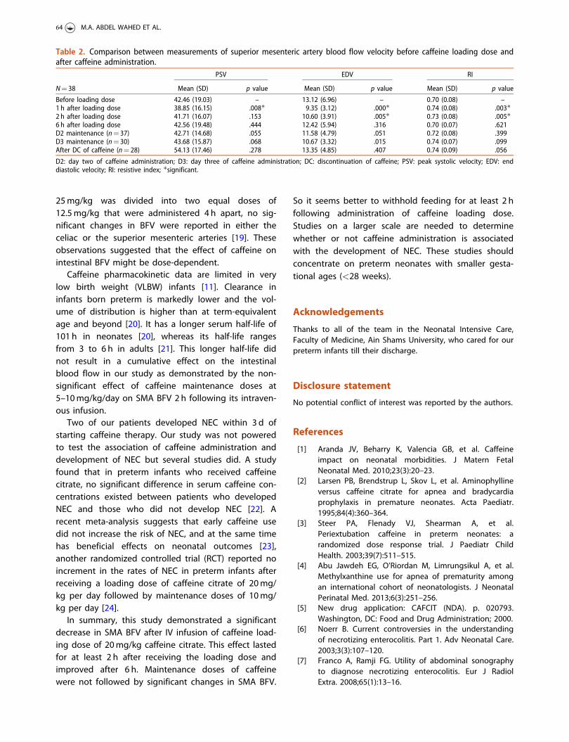
25mg/kg was divided into two equal doses of12.5mg/kg that were administered 4 h apart, no sig-nificant changes in BFV were reported in either theceliac or the superior mesenteric arteries [19]. Theseobservations suggested that the effect of caffeine onintestinal BFV might be dose-dependent.
Caffeine pharmacokinetic data are limited in verylow birth weight (VLBW) infants [11]. Clearance ininfants born preterm is markedly lower and the vol-ume of distribution is higher than at term-equivalentage and beyond [20]. It has a longer serum half-life of101 h in neonates [20], whereas its half-life rangesfrom 3 to 6 h in adults [21]. This longer half-life didnot result in a cumulative effect on the intestinalblood flow in our study as demonstrated by the non-significant effect of caffeine maintenance doses at5–10mg/kg/day on SMA BFV 2 h following its intraven-ous infusion.
Two of our patients developed NEC within 3 d ofstarting caffeine therapy. Our study was not poweredto test the association of caffeine administration anddevelopment of NEC but several studies did. A studyfound that in preterm infants who received caffeinecitrate, no significant difference in serum caffeine con-centrations existed between patients who developedNEC and those who did not develop NEC [22]. Arecent meta-analysis suggests that early caffeine usedid not increase the risk of NEC, and at the same timehas beneficial effects on neonatal outcomes [23],another randomized controlled trial (RCT) reported noincrement in the rates of NEC in preterm infants afterreceiving a loading dose of caffeine citrate of 20mg/kg per day followed by maintenance doses of 10mg/kg per day [24].
In summary, this study demonstrated a significantdecrease in SMA BFV after IV infusion of caffeine load-ing dose of 20mg/kg caffeine citrate. This effect lastedfor at least 2 h after receiving the loading dose andimproved after 6 h. Maintenance doses of caffeinewere not followed by significant changes in SMA BFV.
So it seems better to withhold feeding for at least 2 hfollowing administration of caffeine loading dose.Studies on a larger scale are needed to determinewhether or not caffeine administration is associatedwith the development of NEC. These studies shouldconcentrate on preterm neonates with smaller gesta-tional ages (<28 weeks).
Acknowledgements
Thanks to all of the team in the Neonatal Intensive Care,Faculty of Medicine, Ain Shams University, who cared for ourpreterm infants till their discharge.
Disclosure statement
No potential conflict of interest was reported by the authors.
References
[1] Aranda JV, Beharry K, Valencia GB, et al. Caffeineimpact on neonatal morbidities. J Matern FetalNeonatal Med. 2010;23(3):20–23.
[2] Larsen PB, Brendstrup L, Skov L, et al. Aminophyllineversus caffeine citrate for apnea and bradycardiaprophylaxis in premature neonates. Acta Paediatr.1995;84(4):360–364.
[3] Steer PA, Flenady VJ, Shearman A, et al.Periextubation caffeine in preterm neonates: arandomized dose response trial. J Paediatr ChildHealth. 2003;39(7):511–515.
[4] Abu Jawdeh EG, O’Riordan M, Limrungsikul A, et al.Methylxanthine use for apnea of prematurity amongan international cohort of neonatologists. J NeonatalPerinatal Med. 2013;6(3):251–256.
[5] New drug application: CAFCIT (NDA). p. 020793.Washington, DC: Food and Drug Administration; 2000.
[6] Noerr B. Current controversies in the understandingof necrotizing enterocolitis. Part 1. Adv Neonatal Care.2003;3(3):107–120.
[7] Franco A, Ramji FG. Utility of abdominal sonographyto diagnose necrotizing enterocolitis. Eur J RadiolExtra. 2008;65(1):13–16.
Table 2. Comparison between measurements of superior mesenteric artery blood flow velocity before caffeine loading dose andafter caffeine administration.
N¼ 38
PSV EDV RI
Mean (SD) p value Mean (SD) p value Mean (SD) p value
Before loading dose 42.46 (19.03) – 13.12 (6.96) – 0.70 (0.08) –1 h after loading dose 38.85 (16.15) .008� 9.35 (3.12) .000� 0.74 (0.08) .003�2 h after loading dose 41.71 (16.07) .153 10.60 (3.91) .005� 0.73 (0.08) .005�6 h after loading dose 42.56 (19.48) .444 12.42 (5.94) .316 0.70 (0.07) .621D2 maintenance (n¼ 37) 42.71 (14.68) .055 11.58 (4.79) .051 0.72 (0.08) .399D3 maintenance (n¼ 30) 43.68 (15.87) .068 10.67 (3.32) .015 0.74 (0.07) .099After DC of caffeine (n¼ 28) 54.13 (17.46) .278 13.35 (4.85) .407 0.74 (0.09) .056
D2: day two of caffeine administration; D3: day three of caffeine administration; DC: discontinuation of caffeine; PSV: peak systolic velocity; EDV: enddiastolic velocity; RI: resistive index; �significant.
64 M.A. ABDEL WAHED ET AL.
[8] Rodwell RL, Leslie AL, Tudehope DI. Early diagnosis ofneonatal sepsis using a hematologic scoring system.J Pediatr. 1988;112(5):761–767.
[9] Sehgal A, McNamara PJ. Does echocardiography facili-tate determination of hemodynamic significanceattributable to the ductus arteriosus? Eur J Pediatr.2009;168(8):907–914.
[10] Ballard JL, Khoury JC, Wedig K, et al. New Ballardscore, expanded to include extremely prematureinfants. J Pediatr. 1991;119(3):417–423.
[11] Leon AE, Michienzi K, Ma CX, et al. Serum caffeineconcentrations in preterm neonates. Am J Perinatol.2007;24(1):39–47.
[12] Soraisham AS, Elliott D, Amin H. Effect of singleloading dose of intravenous caffeine infusion onsuperior mesenteric artery blood flow velocities inpreterm infants. J Paediatr Child Health. 2008;44(3):119–121.
[13] Dobson NR, Hunt CE. Pharmacology review: caffeineuse in neonates: indications, pharmacokinetics,clinical effects, outcomes. NeoReviews. 2013;14(11):e540–e550.
[14] Smits P, Lenders JW, Thien T. Caffeine and theophyl-line attenuate adenosine-induced vasodilation inhumans. Clin Pharmacol Ther. 1990;48(4):410–418.
[15] Jino H, Kurahashi K, Usui H, et al. Pharmacologicalnatures of caffeine-induced endothelium- dependentand -independent contraction in canine mesentericartery. Life Sci. 1995;57(12):1155–1162.
[16] Hassanein SM, Gad GI, Ismail RI, et al. Effect of caf-feine on preterm infants’ cerebral cortical activity: an
observational study. J Matern Fetal Neonatal Med.2015;28(17):2090–2095.
[17] Lane AJP, Coombs RC, Evans DH, et al. Effect of caf-feine on neonatal splanchnic blood flow. Arch DisChild Fetal Neonatal Ed. 1999;80(2):F128–F129.
[18] Hoecker C, Nelle M, Poeschl J, et al. Caffeine impairscerebral and intestinal blood flow velocity in preterminfants. Pediatrics. 2002;109(5):784–787.
[19] Hoecker C, Nelle M, Beedgen B, et al. Effects of adivided high loading dose of caffeine on circulatoryvariables in preterm infants. Arch Dis Child FetalNeonatal Ed. 2006;91(1):F61–F64.
[20] Charles BG, Townsend SR, Steer PA, et al. Caffeinecitrate treatment for extremely premature infants withapnea: population pharmacokinetics, absolute bio-availability, and implications for therapeutic drugmonitoring. Ther Drug Monit. 2008;30(6):709–716.
[21] Carrillo JA, Benitez J. Clinically significant pharmacoki-netic interactions between dietary caffeine and medi-cations. Clin Pharmacokinet. 2000;39(2):127–153.
[22] Lampkin SJ, Turner AM, Lakshminrusimha S, et al.Association between caffeine citrate exposure andnecrotizing enterocolitis in preterm infants. Am JHealth Syst Pharm. 2013;70(7):603–608.
[23] Park HW, Lim G, Chung SH, et al. Early caffeine use invery low birth weight infants and neonatal outcomes:a systematic review and meta-analysis. J Korean MedSci. 2015;30(12):1828–1835.
[24] Mohammed S, Nour I, Shabaan AE, et al. High versuslow-dose caffeine for apnea of prematurity: a random-ized controlled trial. Eur J Pediatr. 2015;174(7):949–956.
THE JOURNAL OF MATERNAL-FETAL & NEONATAL MEDICINE 65

Systematic Review and Meta-Analysis
Neonatology 2019;115:116–126
Neonatal Glycaemia and Neurodevelopmental Outcomes: A Systematic Review and Meta-Analysis
Rajesh Shah
a Jane Harding
a Julie Brown
a Christopher McKinlay
a–c a
Liggins Institute, University of Auckland, Auckland, New Zealand; b Department of Paediatrics: Child and Youth Health, University of Auckland, Auckland, New Zealand; c Kidz First Neonatal Care, Counties Manukau Health, Auckland, New Zealand
Received: July 24, 2018Accepted after revision: August 12, 2018Published online: November 8, 2018
Christopher McKinlayLiggins Institute, University of Auckland, Private Bag 92019Victoria Street WestAuckland 1142 (New Zealand)E-Mail c.mckinlay @ auckland.ac.nz
© 2018 S. Karger AG, Basel
E-Mail [email protected]/neo
DOI: 10.1159/000492859
KeywordsHypoglycaemia · Infant · Newborn · Neurodevelopmental disorders · Child development
AbstractBackground: Hypoglycaemia is the most common metabol-ic problem in neonates but there is no universally accepted threshold for safe blood glucose concentrations due to un-certainty regarding effects on neurodevelopment. Objec-tive: To systematically assess the association between neo-natal hypoglycaemia on neurodevelopment outcomes in childhood and adolescence. Methods: We searched MED-LINE, EMBASE, CINAHL, and PsycINFO from inception until February 2018. We included studies that reported one or more prespecified outcomes and compared children ex-posed to neonatal hypoglycaemia with children not ex-posed. Studies of neonates with congenital malformations, inherited metabolic disorders and congenital hyperinsulin-ism were excluded. Two authors independently extracted data using a customized form. We used ROBINS-I to assess risk of bias, GRADE for quality of evidence, and REVMAN for meta-analysis (inverse variance, fixed effects). Results: 1,665 studies were screened, 61 reviewed in full, and 11 included (12 publications). In early childhood, exposure to neonatal hypoglycaemia was not associated with neurodevelopmen-tal impairment (n = 1,657 infants; OR = 1.16, 95% CI = 0.86–
1.57) but was associated with visual-motor impairment (n = 508; OR = 3.46, 95% CI = 1.13–10.57) and executive dysfunc-tion (n = 463; OR = 2.50, 95% CI = 1.20–5.22). In mid-child-hood, neonatal hypoglycaemia was associated with neuro-developmental impairment (n = 54; OR = 3.62, 95% CI = 1.05–12.42) and low literacy (n = 1,395; OR = 2.04, 95% CI = 1.20–3.47) and numeracy (n = 1,395; OR = 2.04, 95% CI = 1.21–3.44). No data were available for adolescents. Conclu-sions: Neonatal hypoglycaemia may have important long-lasting adverse effects on neurodevelopment that may be-come apparent at later ages. Carefully designed randomized trials are required to determine the optimal management of neonates at risk of hypoglycaemia with long-term follow-up at least to school age. © 2018 S. Karger AG, Basel
Introduction
Neonatal hypoglycaemia is the most common meta-bolic problem in newborn infants and a readily prevent-able cause of brain injury in infancy. However, clinical thresholds for diagnosis and treatment of neonatal hypo-glycaemia are widely debated, with no universally ac-cepted safe blood glucose concentration for newborns [1, 2]. This uncertainty is largely due to a lack of evidence regarding the effect of low neonatal glucose concentra-
66

Neonatal Glycaemia and Neurodevelopmental Outcomes
67Neonatology 2019;115:116–126DOI: 10.1159/000492859
tions on neurodevelopmental outcomes. Further, recent studies have suggested that higher glucose concentra-tions after hypoglycaemia may also contribute to brain injury [3], thus adding complexity to this common clini-cal problem.
Key risk factors for neonatal hypoglycaemia include being born preterm, large for gestational age or high birth weight, small for gestational age or low birth weight, and being an infant of a diabetic mother. Approximately 30% of all neonates are considered at risk, of whom approxi-mately 50% develop hypoglycaemia [4]. The most com-mon definition of neonatal hypoglycaemia is a blood glu-cose concentration < 47 mg/dL (2.6 mmol/L), but lower and higher thresholds have been recommended. For ex-ample, the American Academy of Pediatrics advises that intravenous treatment is not needed until glucose con-centrations are < 25 mg/dL (1.4 mmol/L) within the first 4 h after birth, or < 35 mg/dL (2.0 mmol/L) from 4 to 24 h [5]. However, the Pediatric Endocrine Society recommends that in babies at risk of hypoglycaemia, glu-cose concentrations should be maintained > 50 mg/dL (2.8 mmol/L), or > 60 mg/dL (3.3 mmol/L) if interven-tions beyond normal feeds are required [6]. This lack of consensus reflects the paucity of evidence about long-term outcomes after neonatal hypoglycaemia.
In 2006, Boluyt et al. [7] carried out a systematic review of the available studies on prognosis after neonatal hypo-glycaemia. The review concluded that the extent of neu-rodevelopment impairment after neonatal hypoglycae-mia in the first week of life was unclear, and thus the au-thors proposed an optimal study design to establish the relationship between neonatal hypoglycaemia and subse-quent neurodevelopment. In 2008, the Eunice Kennedy Shriver National Institute of Child Health and Human Development Workshop on Neonatal Hypoglycaemia also identified major gaps in knowledge about neonatal hypoglycaemia and its clinical implications and priori-tized it as a key area for research. Since then, although several review articles on the topic have appeared [8–10], no new systematic review has emerged.
This aim of this systematic review was to assess the as-sociation between neonatal hypoglycaemia on neurode-velopment outcomes at early childhood (2–5 years), mid-childhood (6–11 years), and adolescence (12–18 years).
Methods
This systematic review was conducted in accordance with the PRISMA statement, and was registered in PROSPERO (CRD42017073430, http://www.crd.york.ac.uk/PROSPERO/).
Search StrategyWe searched MEDLINE, EMBASE, CINAHL, and PsycINFO
databases using the search terms infant, newborn, hypoglycae-mia, neurodevelopmental disorders, neurological sequelae, neu-roimaging, brain imaging, computed tomography scan, ultraso-nography, and magnetic resonance imaging, including spelling variants (online suppl. material for full search strategy; see www.karger.com/doi/10.1159/000492859 for all online suppl. materi-al). The search was restricted to studies involving humans and published in English. There was no limit on the year of publica-tion. The search was last updated on February 12, 2018. We also hand-searched bibliographies of included studies, review papers and conference abstracts to identify additional items. One author conducted the search and initial title and abstract screening. Re-cords identified for full-text screening were reviewed by two au-thors. Screening and eligibility assessments were performed us-ing COVIDENCE (http://www.covidence.org/). Conflicts were resolved by consensus or after consultation with a third author.
Inclusion CriteriaWe included all studies (trials, cohort, and case-control) that
reported one or more of the primary or secondary outcomes and compared children or adolescents who were screened and found to be hypoglycaemic to those who were screened but were not hy-poglycaemic. Studies were limited to neonates born at ≥32 weeks’gestation and who were screened for hypoglycaemia in the first week after birth. We excluded case series, conference abstracts, and studies that reported outcomes in neonates with congenital mal-formations, inherited metabolic disorders or congenital hyperin-sulinism.
Primary outcomes were neurodevelopmental impairment, vi-sual-motor impairment, and executive dysfunction, as defined by authors. Secondary outcomes were cognitive impairment (as de-fined by authors), mild cognitive impairment (developmental or intelligence quotient from 2 to 1 standard deviation below the mean), moderate-severe cognitive impairment (developmental/intelligence quotient more than 2 standard deviations below the mean), epilepsy (afebrile seizures or as defined by authors), highest educational level (adolescence), death, measures of general health and health care utilization, emotional-behavioural difficulty, ab-normal brain imaging findings, visual impairment, hearing im-pairment, motor impairment, low literacy and low numeracy (mid-childhood and early adolescence), all as defined by authors.
Data Extraction and AnalysisData for primary and secondary outcomes were extracted in-
dependently by two authors using a customized data form. Con-flicts were resolved by consensus or following consultation with a third author.
We planned meta-analysis using the inverse variance, fixed ef-fects method in REVMAN (version 5.3), with the inclusion of ad-justed analyses where possible. If there were data for more than one age within an age band, then the most recent data were used. We assessed statistical heterogeneity using the I2 statistic; values > 30% were regarded as evidence of substantial heterogeneity. Forest plots are provided in the online supplementary material. We planned sensitivity analysis of the primary outcomes including only studies at low risk of bias and only those that used accurate methods for measuring glucose concentrations.
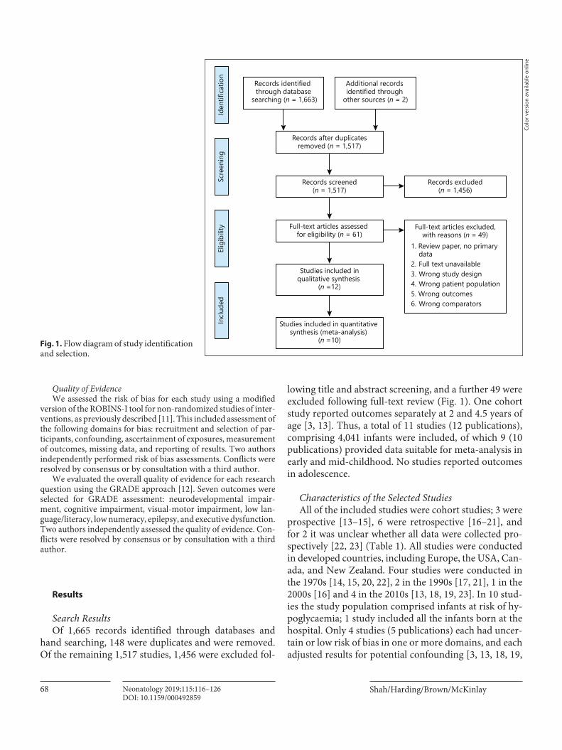
Shah/Harding/Brown/McKinlayNeonatology 2019;115:116–12668DOI: 10.1159/000492859
Quality of EvidenceWe assessed the risk of bias for each study using a modified
version of the ROBINS-I tool for non-randomized studies of inter-ventions, as previously described [11]. This included assessment of the following domains for bias: recruitment and selection of par-ticipants, confounding, ascertainment of exposures, measurement of outcomes, missing data, and reporting of results. Two authors independently performed risk of bias assessments. Conflicts were resolved by consensus or by consultation with a third author.
We evaluated the overall quality of evidence for each research question using the GRADE approach [12]. Seven outcomes were selected for GRADE assessment: neurodevelopmental impair-ment, cognitive impairment, visual-motor impairment, low lan-guage/literacy, low numeracy, epilepsy, and executive dysfunction. Two authors independently assessed the quality of evidence. Con-flicts were resolved by consensus or by consultation with a third author.
Results
Search ResultsOf 1,665 records identified through databases and
hand searching, 148 were duplicates and were removed. Of the remaining 1,517 studies, 1,456 were excluded fol-
lowing title and abstract screening, and a further 49 were excluded following full-text review (Fig. 1). One cohort study reported outcomes separately at 2 and 4.5 years of age [3, 13]. Thus, a total of 11 studies (12 publications), comprising 4,041 infants were included, of which 9 (10 publications) provided data suitable for meta-analysis in early and mid-childhood. No studies reported outcomes in adolescence.
Characteristics of the Selected StudiesAll of the included studies were cohort studies; 3 were
prospective [13–15], 6 were retrospective [16–21], and for 2 it was unclear whether all data were collected pro-spectively [22, 23] (Table 1). All studies were conducted in developed countries, including Europe, the USA, Can-ada, and New Zealand. Four studies were conducted in the 1970s [14, 15, 20, 22], 2 in the 1990s [17, 21], 1 in the 2000s [16] and 4 in the 2010s [13, 18, 19, 23]. In 10 stud-ies the study population comprised infants at risk of hy-poglycaemia; 1 study included all the infants born at the hospital. Only 4 studies (5 publications) each had uncer-tain or low risk of bias in one or more domains, and each adjusted results for potential confounding [3, 13, 18, 19,
Records after duplicatesremoved (n = 1,517)
Full-text articles assessed for eligibility (n = 61)
Studies included inqualitative synthesis
(n =12)
Studies included in quantitative synthesis (meta-analysis)
(n =10)
Records screened(n = 1,517)
Records excluded(n = 1,456)
Full-text articles excluded,with reasons (n = 49)
1. Review paper, no primarydata
2. Full text unavailable3. Wrong study design4. Wrong patient population5. Wrong outcomes6. Wrong comparators
Iden
tific
atio
nSc
reen
ing
Elig
ibili
tyIn
clud
ed
Records identified through database
searching (n = 1,663)
Additional records identified through
other sources (n = 2)
Fig. 1. Flow diagram of study identification and selection.
Colo
r ver
sion
avai
labl
e on
line

Neonatal Glycaemia and Neurodevelopmental Outcomes
69Neonatology 2019;115:116–126DOI: 10.1159/000492859
Tab
le 1
. Cha
ract
erist
ics o
f inc
lude
d st
udie
s
Firs
t aut
hor
and
date
Stud
y ty
peSt
udy
popu
latio
nSa
mpl
e siz
eD
efin
ition
of
hypo
glyc
aem
iaG
luco
se sc
reen
ing
prot
ocol
Test
met
hod
Targ
et ra
nge
for t
reat
men
tTr
eatm
ents
Age
at
follo
w-u
pN
euro
deve
lopm
enta
l tes
tsIn
clud
ed
in m
eta-
anal
ysis
Adj
ustm
ent
for p
oten
tial
conf
ound
ing
Gri
ffith
s [2
2], 1
971
Coh
ort
Neo
nate
s adm
itted
to
spec
ial c
are
unit
41 e
xpos
ed, 4
1 un
expo
sed
<1.1
1 m
mol
/L
(<20
mg/
dL)
Sing
le b
lood
glu
cose
mea
sure
men
t w
ithin
24
h of
adm
issio
n; fu
rthe
r bl
ood
gluc
ose
mea
sure
men
t onl
y if
hypo
glyc
aem
ic o
r if s
ympt
omat
ic
Cap
illar
y w
hole
blo
od;
mod
ified
Wat
son
met
hod
Not
spec
ified
Not
spec
ified
4.2
year
s (m
ean)
Cog
nitio
n: S
tanf
ord-
Bine
t or
Gri
ffith
sBe
havi
or: S
tott
Syst
emic
In
terv
iew
Mot
or: G
riffi
ths L
ocom
otor
Sc
ale
Visi
on: S
tyca
r
Yes
No
Koi
vist
o [2
0], 1
972
Retr
o-sp
ectiv
e co
hort
At-
risk
or
sym
ptom
atic
ne
onat
es sc
reen
ed
for h
ypog
lyca
emia
151
expo
sed,
56
unex
pose
d<1
.7 m
mol
/L
(<30
mg/
dL)
Bloo
d gl
ucos
e m
easu
red
3 tim
es
daily
for 2
–4 d
ays i
n at
-risk
and
sy
mpt
omat
ic in
fant
s, an
d co
ntin
ued
for 2
4 h
afte
r eu
glyc
aem
ic o
r disc
ontin
uatio
n of
tr
eatm
ent
Cap
illar
y w
hole
blo
od;
labo
rato
ry-m
odifi
ed
Hul
tman
or g
luco
se
oxid
ase
met
hod
Not
spec
ified
10–2
0%
intr
aven
ous
gluc
ose;
hy
droc
ortis
one
1–4
year
sC
ogni
tive,
lang
uage
and
mot
or
test
s not
spec
ified
Beha
viou
ral a
sses
smen
t not
sp
ecifi
edO
phth
alm
olog
ic e
xam
inat
ion
Yes
No
Pild
es
[15]
, 19
74
Pros
pect
ive
coho
rtA
t-ri
sk o
r sy
mpt
omat
ic
neon
ates
scre
ened
fo
r hyp
ogly
caem
ia
(mos
tly p
rete
rm o
r SG
A)
39 e
xpos
ed, 4
1 un
expo
sed
<1.1
1 m
mol
/L
(<20
mg/
dL)
Dai
ly b
lood
glu
cose
mea
sure
men
t fo
r 1–4
day
sC
apill
ary
who
le b
lood
; la
bora
tory
glu
cose
ox
idas
e m
etho
d
Not
spec
ified
Ora
l dex
tros
e;
10–1
5%
intr
aven
ous
dext
rose
; hy
droc
ortis
one
or A
CTH
1–7
year
sC
ogni
tive:
Cat
tell
Infa
nt S
cale
, St
anfo
rd-B
inet
or W
echs
ler
Inte
llige
nce
Scal
e fo
r Chi
ldre
nSo
cial
: Vin
elan
d So
cial
M
atur
ity S
cale
Elec
troe
ncep
halo
gram
Yes
No
Haw
orth
[1
4], 1
976
Pros
pect
ive
coho
rtIn
fant
s of d
iabe
tic
mot
hers
25 e
xpos
ed, 1
2 un
expo
sed
≤1.1
1 m
mol
/L
(≤20
mg/
dL)
in lo
w b
irth
w
eigh
t bab
ies
(<2.
5 kg
) and
≤1
.65
mm
ol/L
(≤
30 m
g/dL
) in
nor
mal
w
eigh
t bab
ies
0.5,
1, 2
, 3, 6
,12,
24,
48
and
72 h
, an
d m
ore
freq
uent
ly if
hy
pogl
ycae
mic
Cap
illar
y w
hole
blo
od;
labo
rato
ry H
ugge
tt an
d N
ixon
met
hod
Not
spec
ified
Intr
aven
ous
gluc
ose,
lo
ng-a
ctin
g ad
rena
lin o
r b
oth
4.5
year
s (m
ean)
Yale
Dev
elop
men
tal S
ched
ule
Yes
No
Sten
ning
er
[21]
, 199
8Re
tro-
spec
tive
coho
rt
Infa
nts o
f dia
betic
m
othe
rs13
exp
osed
15
unex
pose
d <1
.5 m
mol
/L
(<27
mg/
dL)
Glu
cose
mea
sure
men
ts fo
r the
firs
t 24
h, f
requ
ency
not
spec
ified
Cap
illar
y w
hole
blo
od;
labo
rato
ry g
luco
se
oxid
ase
met
hod
Not
spec
ified
Not
spec
ified
7–8
year
sC
ogni
tive:
Gri
ffith
s’ D
evel
opm
enta
l Sca
les
Mot
or: M
ovem
ent A
sses
smen
t Ba
ttery
for C
hild
ren
Beha
viou
r: qu
estio
nnai
res
(not
spec
ified
)V
alid
ated
neu
rolo
gica
l sc
reen
ing
test
for e
valu
atio
n of
m
inim
al b
rain
dys
func
tion
Elec
troe
ncep
halo
gram
Yes
No
Duv
anel
[1
7],
1999
Retr
o-sp
ectiv
e co
hort
Pret
erm
(≤
34 w
eeks
) and
SG
A
62 e
xpos
ed, 2
3 un
expo
sed
<2.6
mm
ol/L
(4
7 m
g/dL
)Bl
ood
gluc
ose
mea
sure
d ap
prox
imat
ely
ever
y 4–
5 h
for t
he
first
24
h
Dex
tros
tix fo
r scr
eeni
ng;
if <2
mm
ol/L
con
firm
ed
by la
bora
tory
ven
ous
sam
ple
usin
g gl
ucos
e ox
idas
e or
hex
okin
ase
met
hod
Not
spec
ified
10%
intr
a-ve
nous
de
xtro
se
6, 1
2,
and
18
mon
ths,
and
3.5
and
5 ye
ars
Cog
nitiv
e: G
riffi
th’s
Dev
elop
men
tal S
cale
s, M
cCar
thy
Scal
es o
f Apt
itude
No
No
Bran
d [1
6],
2005
Retr
o-sp
ectiv
e co
hort
Term
and
larg
e fo
r ges
tatio
nal a
ge60
exp
osed
, 15
unex
pose
d<2
.2 m
mol
/L
(<40
mg/
dL)
1, 3
, and
5 h
afte
r bir
th a
nd m
ore
freq
uent
ly if
hyp
ogly
caem
icC
apill
ary
who
le b
lood
; la
bora
tory
glu
cose
ox
idas
e m
etho
d
Not
spec
ified
Add
ition
al
feed
ing
or
intr
aven
ous
dext
rose
4 ye
ars
Cog
nitiv
e: D
enve
r D
evel
opm
enta
l Sca
le,
Snijd
ers-
Oom
en n
on-v
erba
l in
telli
genc
e te
stBe
havi
our:
Dut
ch v
ersio
n of
th
e C
hild
Beh
avio
ur C
heck
List
Yes
No
Ker
stje
ns
[23]
, 201
2C
ohor
tM
oder
ate
pret
erm
(3
2–35
wee
ks’
gest
atio
n)
67 e
xpos
ed, 7
65
unex
pose
d<1
.7 m
mol
/L
(<30
mg/
dL)
Bloo
d gl
ucos
e m
easu
red
seve
ral
times
dur
ing
the
first
24
hBe
dsid
e gl
ucom
eter
; la
bora
tory
con
firm
atio
n if
<3.0
mm
ol/L
(54
mg/
dL) o
r <2.
5 m
mol
/L
(45
mg/
dL),
depe
ndin
g on
site
pro
toco
l
Not
spec
ified
Not
spec
ified
3.5–
4 ye
ars
Age
s and
Sta
ges Q
uest
ionn
aire
Yes
Yes

Shah/Harding/Brown/McKinlayNeonatology 2019;115:116–12670DOI: 10.1159/000492859
Firs
t aut
hor
and
date
Stud
y ty
peSt
udy
popu
latio
nSa
mpl
e siz
eD
efin
ition
of
hypo
glyc
aem
iaG
luco
se sc
reen
ing
prot
ocol
Test
met
hod
Targ
et ra
nge
for t
reat
men
tTr
eatm
ents
Age
at
follo
w-u
pN
euro
deve
lopm
enta
l tes
tsIn
clud
ed
in m
eta-
anal
ysis
Adj
ustm
ent
for p
oten
tial
conf
ound
ing
McK
inla
y 20
15 [3
]Pr
ospe
ctiv
e co
hort
Term
and
late
-pr
eter
m n
eona
tes
born
at r
isk fo
r hy
pogl
ycae
mia
216
expo
sed,
18
8 un
expo
sed
<2.6
mm
ol/L
(<
47 m
g/dL
)1
h, th
en b
efor
e fe
eds 3
–4 h
for
first
24
h, th
en 6
–8 h
for n
ext 2
4 h
and
until
hyp
ogly
caem
ia n
o lo
nger
a
clin
ical
con
cern
. Mor
e fr
eque
nt
if hy
pogl
ycae
mic
or r
ecei
ving
in
trav
enou
s dex
tros
e
Cap
illar
y w
hole
blo
od;
labo
rato
ry g
luco
se
oxid
ase
met
hod;
mas
ked
cont
inuo
us in
ters
titia
l gl
ucos
e m
onito
ring
≥2.6
mm
ol/L
(4
7 m
g/dL
)A
dditi
onal
fe
edin
g, b
ucca
l de
xtro
se g
el,
intr
aven
ous
dext
rose
2 ye
ars
Cog
nitiv
e: B
ayle
y Sc
ales
of
Infa
nt D
evel
opm
ent I
II.
Exec
utiv
e: a
bat
tery
of f
our
task
s, an
d Be
havi
or R
atin
g In
vent
ory
of E
xecu
tive
Func
-tio
n (P
resc
hool
ver
sion)
Visi
on: v
isual
scre
enin
g us
ing
four
ass
essm
ent c
ateg
orie
s and
ra
ndom
-dot
kin
emat
ogra
ms o
f va
ryin
g co
here
nce
Hea
ring
: aud
iolo
gic
scre
enin
g
Yes
Yes
Kai
ser
[19]
, 20
15
Retr
o-sp
ectiv
e co
hort
All
neon
ates
89
exp
osed
, 1,
306
unex
pose
d<1
.94
mm
ol/L
(<
35 m
g/dL
)U
nive
rsal
new
born
glu
cose
sc
reen
ing
at 1
–3 h
afte
r bir
th;
repe
ated
afte
r 1 h
if h
ypog
lyca
emic
Labo
rato
ry g
luco
se
oxid
ase
Not
spec
ified
Intr
aven
ous
dext
rose
or
addi
tiona
l fe
edin
g
10 y
ears
Four
th G
rade
Ben
chm
ark
Exam
inat
ion
(lite
racy
and
m
athe
mat
ics)
Yes
Yes
Goo
de
[18]
, 20
16
Retr
o-sp
ectiv
e co
hort
Pret
erm
and
low
bi
rth
wei
ght
461
expo
sed/
282
unex
pose
d<2
.49
mm
ol/L
(<45
mg/
dL)
Not
spec
ified
Dex
tros
tix o
r pla
sma
gluc
ose,
met
hod
not
spec
ified
Not
spec
ified
Not
spec
ified
5, 8
, and
18
yea
rsC
ogni
tive:
Sta
nfor
d-Bi
net,
Peab
ody
Pict
ure
Voc
abul
ary
Test
, Wec
hsle
r Int
ellig
ence
Sc
ale
for C
hild
ren,
Wec
hsle
r A
bbre
viat
ed S
cale
of I
ntel
li-ge
nce.
Aca
dem
ic: W
oodc
ock-
John
son
Test
s of A
chie
vem
ent
Beha
viou
r: C
hild
Beh
avio
ur
Che
cklis
t, Yo
uth
Repo
rt
Beha
viou
r Sur
veill
ance
Sys
tem
No
Yes
McK
inla
y [1
3],
2017
Pros
pect
ive
coho
rtTe
rm a
nd
mod
erat
e to
late
pr
eter
m in
fant
s bo
rn a
t risk
of
hypo
glyc
aem
ia
280
expo
sed,
19
7 un
expo
sed
<2.6
mm
ol/L
(<
47 m
g/dL
)1
h, th
en b
efor
e fe
eds 3
–4 h
for
first
24
h, th
en 6
–8 h
for n
ext 2
4 h
and
until
hyp
ogly
caem
ia n
o lo
nger
a
clin
ical
con
cern
. Mor
e fr
eque
nt
if hy
pogl
ycae
mic
or r
ecei
ving
in
trav
enou
s dex
tros
e
Cap
illar
y w
hole
blo
od;
labo
rato
ry g
luco
se
oxid
ase
met
hod;
mas
ked
cont
inuo
us in
ters
titia
l gl
ucos
e m
onito
ring
≥2.6
mm
ol/L
(4
7 m
g/dL
)A
dditi
onal
fe
edin
g, b
ucca
l de
xtro
se g
el,
intr
aven
ous
dext
rose
4.5
year
sC
ogni
tive:
Wec
hsle
r Pre
scho
ol
and
Prim
ary
Scal
e of
Inte
lli-
genc
e (v
ersio
n 3)
Exec
utiv
e: a
bat
tery
of f
ive
grad
ed ta
sks,
Beha
viou
r Rat
ing
Inve
ntor
y of
Exe
cutiv
e Fu
nc-
tion
Mot
or: M
ovem
ent A
sses
smen
t Ba
ttery
for C
hild
ren
(ver
sion
2)
and
Beer
y Bu
kten
ica
Dev
elop
-m
enta
l Tes
t of V
isual
Mot
or
Inte
grat
ion
(ver
sion
6) (B
BV-
MI-
6)V
ision
: visu
al sc
reen
ing
usin
g six
ass
essm
ent c
ateg
orie
s, vi
sual
pr
oces
sing
subs
cale
of B
BV-
MI-
6, ra
ndom
dot
kin
etog
ram
s of
var
ying
coh
eren
ceA
udito
ry p
roce
ssin
g: a
udito
ry
subs
cale
of t
he P
help
s Kin
der-
gart
en R
eadi
ness
Sca
leEm
otio
nal a
nd b
ehav
iour
: St
reng
ths a
nd D
iffic
ultie
s Q
uest
ionn
aire
, Chi
ld B
ehav
-io
ur C
heck
list
Yes
Yes
SGA
, sm
all f
or g
esta
tiona
l age
; AC
TH, a
dren
ocor
ticot
ropi
n.
Tab
le 1
(con
tinue
d)
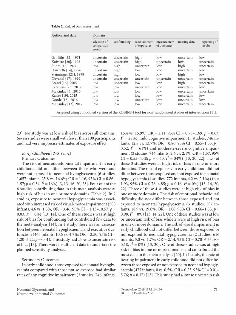
Neonatal Glycaemia and Neurodevelopmental Outcomes
71Neonatology 2019;115:116–126DOI: 10.1159/000492859
23]. No study was at low risk of bias across all domains. Seven studies were small with fewer than 100 participants and had very imprecise estimates of exposure effect.
Early Childhood (2–5 Years)Primary OutcomesThe risk of neurodevelopmental impairment in early
childhood did not differ between those who were and were not exposed to neonatal hypoglycaemia (6 studies, 1,657 infants; 25.8 vs. 16.6%; OR = 1.16, 95% CI = 0.86–1.57; p = 0.34; I2 = 16%) [3, 13–16, 20, 23]. Four out of the 6 studies contributing data to this meta-analysis were at high risk of bias in one or more domains (Table 2). In 2 studies, exposure to neonatal hypoglycaemia was associ-ated with increased risk of visual-motor impairment (508 infants; 4.6 vs. 1.5%; OR = 3.46, 95% CI = 1.13–10.57; p = 0.03; I2 = 0%) [13, 14]. One of these studies was at high risk of bias for confounding but contributed few data to the meta-analysis [14]. In 1 study, there was an associa-tion between neonatal hypoglycaemia and executive dys-function (463 infants; 10.6 vs. 4.7%; OR = 2.50, 95% CI = 1.20–5.22; p = 0.01). This study had a low to uncertain risk of bias [13]. There were insufficient data to undertake the planned sensitivity analyses.
Secondary OutcomesIn early childhood, those exposed to neonatal hypogly-
caemia compared with those not so exposed had similar rates of any cognitive impairment (3 studies, 746 infants,
15.4 vs. 15.9%; OR = 1.11, 95% CI = 0.73–1.69; p = 0.63; I2 = 28%), mild cognitive impairment (3 studies, 746 in-fants, 12.8 vs. 13.7%; OR = 0.86, 95% CI = 0.55–1.35; p = 0.52; I2 = 61%) and moderate-severe cognitive impair-ment (3 studies, 746 infants, 2.6 vs. 2.1%; OR = 1.57, 95% CI = 0.55–4.48; p = 0.40, I2 = 34%) [13, 20, 22]. Two of these 3 studies were at high risk of bias in one or more domains. The risk of epilepsy in early childhood did not differ between those exposed and not exposed to neonatal hypoglycaemia (4 studies, 772 infants, 4.2 vs. 2.1%; OR = 1.93, 95% CI = 0.76–4.85; p = 0.16, I2 = 0%) [13, 14, 20, 22]. Three of these 4 studies were at high risk of bias in one or more domains. The risk of emotional-behavioural difficulty did not differ between those exposed and not exposed to neonatal hypoglycaemia (3 studies, 587 in-fants, 18.9 vs. 19.0%; OR = 1.00, 95% CI = 0.66–1.53; p = 0.98, I2 = 0%) [13, 14, 22]. One of these studies was at low or uncertain risk of bias while 2 were at high risk of bias in one or more domains. The risk of visual impairment in early childhood did not differ between those exposed or not exposed to neonatal hypoglycaemia (2 studies, 616 infants, 5.0 vs. 1.7%; OR = 2.14, 95% CI = 0.70–6.53; p = 0.18, I2 = 0%) [13, 20]. One of these studies was at high risk of bias in one or more domains and contributed the most data to the meta-analysis [20]. In 1 study, the rate of hearing impairment in early childhood did not differ be-tween those exposed or not exposed to neonatal hypogly-caemia (477 infants, 0 vs. 0.5%; OR = 0.23, 95% CI = 0.01–5.76; p = 0.37) [13]. This study had a low to uncertain risk
Table 2. Risk of bias assessment
Author and date Domain
selection of comparison groups
confounding ascertainment of exposures
measurement of outcomes
missing data reporting of results
Griffiths [22], 1971 uncertain uncertain high low uncertain lowKoivisto [20], 1972 uncertain uncertain high uncertain low uncertainPildes [15], 1974 low high uncertain low high uncertainHaworth [14], 1976 uncertain high low low uncertain lowStenninger [21], 1998 uncertain high low low high lowDuvanel [17], 1999 uncertain uncertain uncertain uncertain uncertain uncertainBrand [16], 2005 low uncertain low low high uncertainKerstjens [23], 2012 low low uncertain low uncertain lowMcKinlay [3], 2015 low low low low uncertain uncertainKaiser [19], 2015 low low low low uncertain lowGoode [18], 2016 low low uncertain low uncertain lowMcKinlay [13], 2017 low low low low uncertain uncertain
Assessed using a modified version of the ROBINS-I tool for non-randomized studies of interventions [11].

Shah/Harding/Brown/McKinlayNeonatology 2019;115:116–12672DOI: 10.1159/000492859
of bias. The risk of motor impairment in early childhood did not differ between those who were and were not ex-posed to neonatal hypoglycaemia (4 studies, 777 infants, 17.5 vs. 17.8%; OR = 1.06, 95% CI = 0.70–1.60; p = 0.79, I2 = 6%) [13, 14, 20, 22]. Three out of 4 of these studies were at high risk of bias in one or more domains. One study reported higher rates of low language/literacy in those exposed to neonatal hypoglycaemia compared with those not so exposed but results were imprecise and not statistically significant (37 infants, 16 vs. 0%; OR = 5.23, 95% CI = 0.26–105.50; p = 0.28) [14]. This study had an uncertain to high risk of bias. One study reported on rates of cerebral palsy and found no difference between those exposed and not exposed to neonatal hypoglycaemia (401 infants, 0.9 vs. 1.1%; OR = 0.81, 95% CI = 0.11–6.07; p = 0.84) [3]. This study was at a low to uncertain risk of bias. None of the included studies reported on abnormal brain imaging, highest education level, death or measures of general health and health care utilization in early child-hood.
Quality of EvidenceFor the primary outcomes in early childhood, the qual-
ity of evidence was either low or very low (Table 3). For the selected secondary outcomes of any cognitive impair-ment, epilepsy, and low language/literacy, the quality of evidence was also very low (Table 3).
Mid-Childhood (6–11 Years)Primary OutcomesIn 2 small studies, those exposed to neonatal hypogly-
caemia compared with those not so exposed had a higher risk of neurodevelopmental impairment (54 infants, 47.8 vs. 22.6%; OR = 3.62, 95% CI = 1.05–12.42; p = 0.04, I2 = 0%) [15, 21]. Both of these studies were at an uncertain to high risk of bias in one or more domains. None of the in-cluded studies reported on visual-motor impairment or executive dysfunction in mid-childhood. There were in-sufficient data to undertake the planned sensitivity analy-ses.
Secondary OutcomesIn 1 study, the risk of emotional-behavioural difficulty
in mid-childhood was non-significantly increased in those exposed to neonatal hypoglycaemia than those not so exposed (28 infants, 30.8 vs. 6.7%; OR = 6.22, 95% CI = 0.60–64.97; p = 0.13) but rates of motor impairment were similar (28 infants, 15.4 vs. 13.3%; OR = 1.18, 95% CI = 0.14–9.83; p = 0.88) [21]. This study had an uncertain to high risk of bias in one or more domains. In another
study, those exposed to neonatal hypoglycaemia com-pared with those not so exposed had an increased risk of low language/literacy (1,395 infants, 67.4 vs. 43.0%; OR = 2.04, 95% CI = 1.20–3.47; p = 0.008) [19] and low numer-acy (1,395 infants, 53.9 vs. 34.0%; OR = 2.04, 95% CI = 1.21–3.44; p = 0.007) in mid-childhood [19]. This study had a low to uncertain risk of bias.
None of the included studies reported on any cognitive impairment, mild cognitive impairment, moderate-se-vere cognitive impairment, epilepsy, abnormal brain im-aging, visual impairment, hearing impairment, highest educational level, death, and measures of general health and health care utilization in mid-childhood.
Quality of EvidenceFor the primary outcome of neurodevelopmental im-
pairment in mid-childhood the quality of the evidence was very low (Table 3). For the selected secondary out-comes of low language/literacy and low numeracy, the quality of the evidence was low (Table 3).
Adolescence (12–18 Years)None of the included studies reported on primary or
secondary outcomes in adolescence.
Discussion
Neonatal hypoglycaemia is the most common meta-bolic condition in newborn infants [4] and has been as-sociated with widespread changes in the developing brain [24], yet the impact of neonatal hypoglycaemia on long-term neurodevelopment is widely debated [25]. We un-dertook this systematic review to determine the relation-ship between neonatal hypoglycaemia and neurodevel-opment throughout childhood. We found low-quality evidence that in early childhood (2–5 years) neonatal hy-poglycaemia is associated with specific cognitive deficits, including a two- to threefold increased risk of visual-mo-tor impairment and executive dysfunction. In later child-hood (6–11 years), we found low-quality evidence that neonatal hypoglycaemia is associated with a twofold in-creased risk of literacy and numeracy problems, and very low-quality evidence of an increased risk of general cog-nitive impairment. No data were available on outcomes in adolescence.
Visual-motor integration is the coordination of visual perception, the ability to extract and organize visual in-formation from the environment, and motor skills, espe-cially fine motor ones [26]. It allows the use of eyes and

Neonatal Glycaemia and Neurodevelopmental Outcomes
73Neonatology 2019;115:116–126DOI: 10.1159/000492859
hands in a coordinated and efficient way, enabling, for example, one to perceive and copy shapes, letters, and numbers. Thus, visual-motor integration is important for learning and academic achievement including reading, writing, and mathematics [27, 28].
The development of visual and motor systems is close-ly related [29], and coordination of visual-motor function
is thought to occur within the ventral and dorsal cortical visual streams. The ventral stream supports form pro-cessing and object recognition, and includes the occipital primary visual cortex and the inferior temporal lobe. The dorsal stream is responsible for motion perception and visually guided motor function and includes the occipital primary visual cortex, middle temporal lobe, and poste-
Table 3. GRADE summary of quality of evidence for effect of neonatal hypoglycaemia on neurodevelopmental outcomes
Outcome Exposure effectOR (95% CI)
Participants (studies)
Certainty/quality of evidence
Comments
Early childhood (2–5 years)Neurodevelopmental impairment
1.16 (0.86–1.57) 1,657 (6) Very low Initial level low. Downgraded as 4 stud-ies were at high risk of bias in several domains, and only 2 studies adjusted for confounding
Visual-motor impairment 3.46 (1.13–10.57) 508 (2) Low Initial level low. Downgraded as results were imprecise. Upgraded 1 level due to large treatment effect
Executive dysfunction 2.50 (1.20–5.22) 463 (1) Low Initial level low. Downgraded as there was only a single study. Upgraded 1 level due to large treatment effect
Any cognitive impairment 1.11 (0.73–1.69) 746 (3) Very low Initial level low. Downgraded as two studies were at high risk of bias, and only one study adjusted for confounding
Epilepsy 1.93 (0.76–4.85) 772 (4) Very low Initial level low. Downgraded as 2 stud-ies were at high risk of bias, results were imprecise, and only 1 study adjusted for confounding
Low language/literacy 5.23 (0.26–105.50) 37 (1) Very low Initial level low. Large treatment effect but downgraded as there was only 1 study at high risk of bias with imprecise results
Mid-childhood (6–11 years)Neurodevelopmental impairment
3.62 (1.05–12.42) 54 (2) Very low Initial level low. Large treatment effect but downgraded as both studies were at high risk of bias with imprecise results
Visual-motor impairment – – – No data
Executive dysfunction – – – No data
Any cognitive impairment – – – No data
Epilepsy – – – No data
Low language/literacy 2.04 (1.20–3.47) 1,395 (1) Low Initial level low. Downgraded as there was only a single study. Upgraded 1 level due to large treatment effect
Low numeracy 2.04 (1.21–3.44) 1,395 (1) Low Initial level low. Downgraded as there was only a single study. Upgraded 1 level due to large treatment effect
Evaluated using the GRADE approach [12].
Shah/Harding/Brown/McKinlayNeonatology 2019;115:116–12674DOI: 10.1159/000492859
rior parietal lobe. In the neonatal period, these cortical areas appear to be particularly susceptible to injury from neuroglycopenia, possibly because of higher metabolic activity [9, 30–32]. This provides a possible pathophysi-ological basis for the association between neonatal hypo-glycaemia and impaired visual-motor integration in early childhood.
Executive function is the collective capacity for prob-lem-solving, planning, attention control, and goal-direct-ed behaviour [33]. Children with impaired executive con-trol have difficulty remembering and carrying out in-structions, staying focused, and planning and monitoring progress with a specific task, which can affect not only daily activities but also learning. The prefrontal cortex is responsible for the proper development of executive function, and increased activation of this region is associ-ated with better performance on executive function tasks, as well as academic outcomes [34, 35]. The development of the prefrontal cortex and executive capacity is continu-ous from childhood through adolescence and into early adulthood [36, 37], and any abnormality in this region can result in executive function difficulties. Although neonatal hypoglycaemia has traditionally been associated with posterior brain injury, recent studies have suggested that its effects on the brain may be more widespread and include the frontal cortex [24, 38], potentially interfering with the normal development of executive capacity.
Demands on visual-motor and executive function in-crease with age, but we could not determine whether the changes seen in early childhood after neonatal hypogly-caemia persist or worsen over time due to the lack of lon-ger-term outcome data. However, the finding of a twofold increased risk of literacy and numeracy problems in mid-childhood suggests a trajectory of worsening function in skills that are important for learning [39, 40]. The fact that neonatal hypoglycaemia was associated with general cog-nitive impairment in mid-childhood but not in early childhood supports this hypothesis. Importantly, this sys-tematic review shows that tests of general development in infancy are unlikely to adequately assess the effects of neonatal hypoglycaemia on brain development. Thus, in-tervention studies will require longer-term end points, at least into mid-childhood, including specific tests of visu-al-motor and executive function.
It is more than a decade since Boluyt et al. [7] con-ducted the first systematic review of neurodevelopmental outcomes after neonatal hypoglycaemia. They concluded that there were insufficient data to quantify the effect of neonatal hypoglycaemia on neurodevelopment and pro-vided recommendations about an optimal study design.
Our systematic review identified 3 subsequent studies, but only 1 that followed these recommendations [3, 13], including prospective cohort design, nested randomized trial of treatment, gold standard glucose measurements, standardized neurodevelopmental assessment and suffi-cient sample size [7]. This is somewhat surprising given the recognition of neonatal hypoglycaemia as a priority research area and calls from the National Institute of Child Health and Human Development for further high-quality studies [1].
There are several differences between our systematic review and that of Boluyt et al. [7]. We excluded case se-ries because without contemporaneous controls it is not possible to account for confounding, especially relating to the reasons that babies were considered at risk of hypo-glycaemia and socio-economic factors. We also excluded studies that assessed outcomes at less than 2 years of age, due to the limited predictive value of very early develop-mental assessment [41], and studies that primarily in-cluded infants with congenital hyperinsulinism. We as-sessed not only the methodological quality of individual studies, but also the overall strength of the evidence for key outcomes using the GRADE approach.
Even with optimal study design, several challenges re-main in determining the effect of neonatal hypoglycae-mia on later neurodevelopment. As with any cohort study, the possibility of residual confounding cannot be excluded. Although neuroglycopenia can cause irrevers-ible brain injury, other mechanisms may underlie asso-ciations between episodes of hypoglycaemia and neuro-developmental impairment. For example, genetic poly-morphisms of ATP-dependent potassium channels could affect both pancreatic β-cells and neuronal function [42].
In addition, the relationship between the severity, fre-quency, and duration of neonatal hypoglycaemic epi-sodes and cerebral energy supply and utilization remains unclear [43], and thus the best measure of exposure for use in analyses is uncertain [25]. This is complicated by different approaches to screening, diagnosis, and treat-ment of hypoglycaemia, making characterization of the degree of exposure challenging. Further, masked contin-uous interstitial glucose monitoring has shown that the burden of hypoglycaemia in the early newborn period may be substantially greater than is detected by serial glu-cose measurements, even with frequent screening [3]. These undetected and thus untreated episodes may have an important influence on long-term outcomes [13]. However, there are few data on the effect of different ap-proaches to treatment on glucose concentrations after hy-poglycaemia [44].

Neonatal Glycaemia and Neurodevelopmental Outcomes
75Neonatology 2019;115:116–126DOI: 10.1159/000492859
Limitations A key limitation of this systematic review is that only
a limited number of studies were identified that met the inclusion criteria, leading to imprecise estimates of effect, and that data were not available for all prespecified out-comes at each epoch. There are several possible reasons for this including the difficulty of recruiting large cohorts around the time of birth, and the cost and complexity of long-term neurodevelopmental follow-up throughout childhood. Of note, only 3 of the included studies con-tributed data beyond 5 years of age [18, 21, 23]. Another limitation is the lack of adjustment for potential con-founding factors, with only half of the included studies attempting to control for this potential source of bias. Fi-nally, the description of hypoglycaemic management and treatment targets was generally poor. This may be impor-tant, as there is emerging evidence both in animals and humans that glucose reperfusion injury may exacerbate oxidative stress associated with hypoglycaemia if the cor-rection is too rapid or too high, even within the normal glucose range [3, 45, 46].
Recommendations for ResearchStudies are needed to determine the efficacy and cost-
effectiveness of different strategies for improving long-term outcomes in neonates born at risk of hypoglycae-mia. Future studies should involve large prospective cohorts with nested randomized trials of different ap-proaches to treatment, or large randomized trials of dif-ferent approaches to prevention or screening and diagno-sis of hypoglycaemia in neonates considered at risk. All
studies require the use of gold standard glucose assay methods [25, 47] and long-term follow-up at least to school age, with attention to visual-motor and executive function, and educational achievement. Consideration should be given to the use of masked continuous glucose monitoring to aid in the interpretation of study results, although retrospective point-to-point recalibration against all laboratory blood glucose values is important for accurate interstitial measurements in babies [48].
Conclusion
This systematic review found that neonatal hypogly-caemia is associated with a two- to threefold increased risk of specific cognitive deficits in early childhood (2–5 years), including visual-motor impairment and executive dysfunction, and general cognitive impairment and lit-eracy and numeracy problems in later childhood (6–11 years). Although the overall quality of evidence was low to very low, this review nevertheless suggests that neona-tal hypoglycaemia may have important long-lasting ad-verse effects on neurodevelopment. Carefully designed intervention trials are needed to determine the optimal management of neonates at risk of hypoglycaemia to im-prove long-term outcomes.
Disclosure Statement
The authors have no conflicts of interest to declare.
References
1 Hay WW Jr, Raju TN, Higgins RD, Kalhan SC, Devaskar SU. Knowledge gaps and re-search needs for understanding and treating neonatal hypoglycemia: workshop report from Eunice Kennedy Shriver National Insti-tute of Child Health and Human Develop-ment. J Pediatr. 2009 Nov; 155(5): 612–7.
2 Rozance PJ, Hay WW. Hypoglycemia in new-born infants: features associated with adverse outcomes. Biol Neonate. 2006; 90(2): 74–86.
3 McKinlay CJ, Alsweiler JM, Ansell JM, An-stice NS, Chase JG, Gamble GD, et al.; CHYLD Study Group. Neonatal glycemia and neuro-developmental outcomes at 2 years. N Engl J Med. 2015 Oct; 373(16): 1507–18.
4 Harris DL, Weston PJ, Harding JE. Incidence of neonatal hypoglycemia in babies identified as at risk. J Pediatr. 2012 Nov; 161(5): 787–91.
5 Adamkin DH; Committee on Fetus and New-born. Postnatal glucose homeostasis in late-preterm and term infants. Pediatrics. 2011;
127(3): 575–9.6 Thornton PS, Stanley CA, De Leon DD, Har-
ris D, Haymond MW, Hussain K, et al.; Pedi-atric Endocrine Society. Recommendations from the pediatric endocrine society for eval-uation and management of persistent hypo-glycemia in neonates, infants, and children. J Pediatr. 2015 Aug; 167(2): 238–45.
7 Boluyt N, van Kempen A, Offringa M. Neuro-development after neonatal hypoglycemia: a systematic review and design of an optimal future study. Pediatrics. 2006 Jun; 117(6):
2231–43. 8 Adamkin DH. Neonatal hypoglycemia. Se-
min Fetal Neonatal Med. 2017 Feb; 22(1): 36–41.
9 Paudel N, Chakraborty A, Anstice N, Jacobs RJ, Hegarty JE, Harding JE, et al.; CHYLD Study Group. Neonatal hypoglycaemia and visual development: a review. Neonatology. 2017; 112(1): 47–52.
10 Rozance PJ. Update on neonatal hypoglyce-mia. Curr Opin Endocrinol Diabetes Obes. 2014 Feb; 21(1): 45–50.
11 Bradford BF, Thompson JM, Heazell AE, Mc-cowan LM, McKinlay CJ. Understanding the associations and significance of fetal move-ments in overweight or obese pregnant wom-en: a systematic review. Acta Obstet Gynecol Scand. 2018 Jan; 97(1): 13–24.
12 Balshem H, Helfand M, Schünemann HJ, Ox-man AD, Kunz R, Brozek J, et al. GRADE guidelines: 3. Rating the quality of evidence. J Clin Epidemiol. 2011 Apr; 64(4): 401–6.

Shah/Harding/Brown/McKinlayNeonatology 2019;115:116–12676DOI: 10.1159/000492859
13 McKinlay CJ, Alsweiler JM, Anstice NS, Burakevych N, Chakraborty A, Chase JG, et al.; Children With Hypoglycemia and Their Later Development (CHYLD) Study Team. Association of neonatal glycemia with neuro-developmental outcomes at 4.5 years. JAMA Pediatr. 2017 Oct; 171(10): 972–83.
14 Haworth JC, McRae KN, Dilling LA. Progno-sis of infants of diabetic mothers in relation to neonatal hypoglycaemia. Dev Med Child Neurol. 1976 Aug; 18(4): 471–9.
15 Pildes RS, Cornblath M, Warren I, Page-El E, Di Menza S, Merritt DM, et al. A prospective controlled study of neonatal hypoglycemia. Pediatrics. 1974 Jul; 54(1): 5–14.
16 Brand PL, Molenaar NL, Kaaijk C, Wierenga WS. Neurodevelopmental outcome of hypo-glycaemia in healthy, large for gestational age, term newborns. Arch Dis Child. 2005 Jan;
90(1): 78–81.17 Duvanel CB, Fawer CL, Cotting J, Hohlfeld P,
Matthieu JM. Long-term effects of neonatal hypoglycemia on brain growth and psycho-motor development in small-for-gestational-age preterm infants. J Pediatr. 1999 Apr;
134(4): 492–8.18 Goode RH, Rettiganti M, Li J, Lyle RE, Whi-
teside-Mansell L, Barrett KW, et al. Develop-mental outcomes of preterm infants with neo-natal hypoglycemia. Pediatrics. 2016 Dec;
138(6): 55–55.19 Kaiser JR, Bai S, Gibson N, Holland G, Lin
TM, Swearingen CJ, et al. Association be-tween transient newborn hypoglycemia and fourth-grade achievement test proficiency: a population-based study. JAMA Pediatr. 2015 Oct; 169(10): 913–21.
20 Koivisto M, Blanco-Sequeiros M, Krause U. Neonatal symptomatic and asymptomatic hy-poglycaemia: a follow-up study of 151 chil-dren. Dev Med Child Neurol. 1972 Oct; 14(5):603–14.
21 Stenninger E, Flink R, Eriksson B, Sahlèn C. Long-term neurological dysfunction and neonatal hypoglycaemia after diabetic preg-nancy. Arch Dis Child Fetal Neonatal Ed. 1998 Nov; 79(3):F174–9.
22 Griffiths AD, Bryant GM. Assessment of ef-fects of neonatal hypoglycaemia. A study of 41 cases with matched controls. Arch Dis Child. 1971 Dec; 46(250): 819–27.
23 Kerstjens JM, Bocca-Tjeertes IF, de Winter AF, Reijneveld SA, Bos AF. Neonatal morbid-ities and developmental delay in moderately preterm-born children. Pediatrics. 2012 Aug;
130(2):e265–72.
24 Burns CM, Rutherford MA, Boardman JP, Cowan FM. Patterns of cerebral injury and neurodevelopmental outcomes after symp-tomatic neonatal hypoglycemia. Pediatrics. 2008 Jul; 122(1): 65–74.
25 Harding JE, Harris DL, Hegarty JE, Alsweiler JM, McKinlay CJ. An emerging evidence base for the management of neonatal hypoglycae-mia. Early Hum Dev. 2017 Jan; 104: 51–6.
26 Beery KE. The Beery-Buktenica developmen-tal test of visual-motor integration: Beery VMI, with supplemental developmental tests of visual perception and motor coordination, and stepping stones age norms from birth to age six. MN: NCS Pearson Minneapolis; 2004.
27 Barnhardt C, Borsting E, Deland P, Pham N, Vu T. Relationship between visual-motor in-tegration and spatial organization of written language and math. Optom Vis Sci. 2005 Feb;
82(2): 138–43.28 Taylor Kulp M. Relationship between visual
motor integration skill and academic perfor-mance in kindergarten through third grade. Optom Vis Sci. 1999 Mar; 76(3): 159–63.
29 Thompson B, McKinlay CJ, Chakraborty A, Anstice NS, Jacobs RJ, Paudel N, et al.; CHYLD Study Team. Global motion percep-tion is associated with motor function in 2-year-old children. Neurosci Lett. 2017 Sep;
658: 177–81.30 Aslan Y, Dinc H. MR findings of neonatal hy-
poglycemia. AJNR Am J Neuroradiol. 1997 May; 18(5): 994–6.
31 Barkovich AJ, Ali FA, Rowley HA, Bass N. Imaging patterns of neonatal hypoglycemia. AJNR Am J Neuroradiol. 1998 Mar; 19(3):
523–8.32 Spar JA, Lewine JD, Orrison WW Jr. Neonatal
hypoglycemia: CT and MR findings. AJNR Am J Neuroradiol. 1994 Sep; 15(8): 1477–8.
33 Anderson P. Assessment and development of executive function (EF) during childhood. Child Neuropsychol. 2002 Jun; 8(2): 71–82.
34 Shaw P, Greenstein D, Lerch J, Clasen L, Len-root R, Gogtay N, et al. Intellectual ability and cortical development in children and adoles-cents. Nature. 2006 Mar; 440(7084): 676–9.
35 Tamnes CK, Østby Y, Walhovd KB, Westlye LT, Due-Tønnessen P, Fjell AM. Neuroana-tomical correlates of executive functions in children and adolescents: a magnetic reso-nance imaging (MRI) study of cortical thick-ness. Neuropsychologia. 2010 Jul; 48(9): 2496–508.
36 Lebel C, Walker L, Leemans A, Phillips L, Beaulieu C. Microstructural maturation of the human brain from childhood to adult-hood. Neuroimage. 2008 Apr; 40(3): 1044–55.
37 Tamnes CK, Østby Y, Fjell AM, Westlye LT, Due-Tønnessen P, Walhovd KB. Brain matu-ration in adolescence and young adulthood: regional age-related changes in cortical thick-ness and white matter volume and micro-structure. Cereb Cortex. 2010 Mar; 20(3): 534–48.
38 Cakmakci H, Usal C, Karabay N, Kovanlikaya A. Transient neonatal hypoglycemia: cranial US and MRI findings. Eur Radiol. 2001;
11(12): 2585–8.39 Bull R, Espy KA, Wiebe SA. Short-term mem-
ory, working memory, and executive func-tioning in preschoolers: longitudinal predic-tors of mathematical achievement at age 7 years. Dev Neuropsychol. 2008; 33(3): 205–28.
40 Sortor JM, Kulp MT. Are the results of the Beery-Buktenica Developmental Test of Vi-sual-Motor Integration and its subtests relat-ed to achievement test scores? Optom Vis Sci. 2003 Nov; 80(11): 758–63.
41 Harris SR, Langkamp DL. Predictive value of the Bayley mental scale in the early detection of cognitive delays in high-risk infants. J Per-inatol. 1994 Jul-Aug; 14(4): 275–9.
42 Hattersley AT, Ashcroft FM. Activating mu-tations in Kir6.2 and neonatal diabetes: new clinical syndromes, new scientific insights, and new therapy. Diabetes. 2005 Sep; 54(9):2503–13.
43 Mujsce DJ, Christensen MA, Vannucci RC. Regional cerebral blood flow and glucose uti-lization during hypoglycemia in newborn dogs. Am J Physiol. 1989 Jun; 256(6 Pt 2):H1659–66.
44 Harris DL, Gamble GD, Weston PJ, Harding JE. What happens to blood glucose concen-trations after oral treatment for neonatal hy-poglycemia? J Pediatr. 2017 Nov; 190: 136–41.
45 Ennis K, Dotterman H, Stein A, Rao R. Hy-perglycemia accentuates and ketonemia at-tenuates hypoglycemia-induced neuronal in-jury in the developing rat brain. Pediatr Res. 2015 Jan; 77(1-1): 84–90.
46 Suh SW, Gum ET, Hamby AM, Chan PH, Swanson RA. Hypoglycemic neuronal death is triggered by glucose reperfusion and activa-tion of neuronal NADPH oxidase. J Clin In-vest. 2007 Apr; 117(4): 910–8.
47 Glasgow MJ, Harding JE, Edlin R; for the CHYLD Study Team. Cost analysis of cot-side screening methods for neonatal hypo-glycaemia. Neonatology. 2018; 114(2): 155–62.
48 McKinlay CJ, Chase JG, Dickson J, Harris DL, Alsweiler JM, Harding JE. Continuous glu-cose monitoring in neonates: a review. Ma-tern Health Neonatol Perinatol. 2017 Oct;3(1): 18.

ARTICLE
Neurodevelopmental Outcomes in Infants With Birth Weight ≤500 g at 3 Years of AgeHirosuke Inoue, MD, PhD, a, b Masayuki Ochiai, MD, PhD, a, b Yasunari Sakai, MD, PhD, a, b Kazuaki Yasuoka, MD, a, b Koichi Tanaka, MD, a, b Masako Ichiyama, MD, a, b Hiroaki Kurata, MD, a, b Junko Fujiyoshi, MD, a, b Yuki Matsushita, MD, PhD, a, b Satoshi Honjo, MD, PhD, c Kazuaki Nonaka, DDS, PhD, b Tomoaki Taguchi, MD, PhD, FACS, b Kiyoko Kato, MD, PhD, b Shouichi Ohga, MD, PhD, a, b on behalf of the Neonatal Research Network of Japan
OBJECTIVES: To determine neurodevelopmental outcomes at 3 years of age in children born with a birth weight (BW) of ≤500 g.METHODS: Infants who were born with a BW of ≤500 g from 2003 to 2012 in the Neonatal Research Network of Japan and survived to discharge from the NICU were eligible in this study. The study population consisted of 460 children (56.7% of 811 surviving infants) who were evaluated at 36 to 42 months of age. Neurodevelopmental impairment (NDI) was defined as having cerebral palsy, visual impairment, hearing impairment, or a developmental quotient score of <70.RESULTS: The overall proportion of NDI was 59.1% (95% confidence interval [CI]: 54.6%–63.5%). The trend revealed no significant change during the study period. In a multivariate modified Poisson regression analysis, NDI was associated with severe intraventricular hemorrhage (adjusted risk ratio [RR]: 1.42; 95% CI: 1.19–1.68; P < .01), cystic periventricular leukomalacia (adjusted RR: 1.40; 95% CI: 1.13–1.73; P < .01), severe necrotizing enterocolitis (adjusted RR: 1.31; 95% CI: 1.07–1.60; P < .01), surgical ligation for patent ductus arteriosus (adjusted RR: 1.29; 95% CI: 1.09–1.54; P < .01), and male sex (adjusted RR: 1.19; 95% CI: 1.01–2.40; P = .04).CONCLUSIONS: This cohort showed that neurodevelopmental outcomes of infants with a BW of ≤500 g have not improved from 2003 to 2012. Multivariate analysis revealed that severe intracranial hemorrhage and cystic periventricular leukomalacia were the strongest risk factors for NDIs. Our data suggested that measures aimed at reducing neurologic morbidities will be important for improving outcomes of infants with a BW of ≤500 g.
abstract
aDepartment of Pediatrics, Graduate School of Medical Sciences and bComprehensive Maternity and Perinatal Care Center, Kyushu University, Fukuoka, Japan; and cDepartment of Pediatrics, National Hospital Organization Fukuoka National Hospital, Fukuoka, Japan
Dr Inoue conceptualized and designed the study, contributed to the analysis and interpretation of the data, and drafted the initial manuscript; Drs Ochiai, Sakai, and Ohga conceptualized and designed the study, contributed to the data analysis, and critically reviewed and revised the manuscript; Drs Yasuoka, Tanaka, Ichiyama, Kurata, Fujiyoshi, and Matsushita made substantial contributions to the analysis and interpretation of the data and reviewed and revised the manuscript; Dr Honjo supervised and supported the statistical analyses for the completion of the study and reviewed and revised the manuscript; Drs Nonaka, Taguchi, and Kato supervised the study design, contributed to the interpretation of the data, and critically reviewed and revised the manuscript; and all authors approved the final manuscript as submitted.
DOI: https:// doi. org/ 10. 1542/ peds. 2017- 4286
Accepted for publication Sep 13, 2018
PEDIATRICS Volume 142, number 6, December 2018:e20174286
WHAT’S KNOWN ON THIS SUBJECT: The survival of infants born with a birth weight (BW) of ≤500g has improved considerably in Japan; however, few studies have revealed the neurodevelopmental outcomes of survivors born with a BW of ≤500 g.
WHAT THIS STUDY ADDS: Neurodevelopmental outcomes in early childhood among surviving infants born with a BW of ≤500 g did not improve in a recentdecade.
To cite: Inoue H, Ochiai M, Sakai Y, et al. Neurodevelopmental Outcomes in Infants With Birth Weight ≤500 g at 3 Years of Age. Pediatrics. 2018;142(6):e20174286
by guest on November 16, 2018www.aappublications.org/newsDownloaded from
77
Advances in perinatal and neonatal intensive care have improved the survival rate of periviable, or extremely preterm, infants near the limit of viability.1 – 3 On the other hand, researchers in recent studies have reported decreasing, 4 unchanged, 5 or increasing rates6, 7 in neurodevelopmental disabilities among periviable infants who survive compared with previous decades. These variable data lead to a concern that increased survival may come at the cost of later neurodevelopmental disabilities among survivors.8 Thus, providing accurate data on both mortality and long-term neurodevelopmental outcomes is important for families and professionals in neonatal care in making appropriate decisions for these high-risk infants.9
Gestational age (GA) and birth weight (BW), alone or combined, are the critical factors that affect the survival rate of extremely preterm and small infants.9 – 11 Early ultrasound assessment provides an accurate estimation of GA in most cases, but it is also known to cause an error as high as ±15%.12 On the other hand, BW is a direct measurement of the body size of offspring. We have recently shown favorable results of survival rates for infants with a BW of ≤500 g in Japan, whereas in-hospital morbidity rates have remained high in the last decade.13 We extended this study because the long-term outcomes of the infants with a BW of ≤500 g were currently unavailable. In the current study, we investigate neurodevelopmental outcomes at 3 years of age for surviving children born with a BW of ≤500 g using the nationwide cohort database in Japan.
METHODS
Study Population and Data Collection
The Neonatal Research Network of Japan (NRNJ) database prospectively registered all of the clinical information of infants with a BW
of ≤1500 g admitted to the 204 participating NICUs, accounting for 54.3% of 376 secondary and tertiary-level NICUs in Japan. The participants in this study were born between January 1, 2003, and December 31, 2012. Decisions regarding the pursuit of active treatment versus comfort care were made by neonatologists on the basis of fetal information, the status of the infants at birth, and their communications with the parents. In general, attending neonatologists attempt to save the lives of neonates at a GA of ≥23 weeks, and this principle has not changed during the study period. The perinatal data were collected as previously described.13 Children who were born with a BW of ≤500 g and survived to discharge from the NICU were included in this study. We excluded the infants who had died in the operating or delivery room before admission to the NICU, those transferred to other hospitals, those born at a GA of <22 weeks, and those without available records of mortality during the NICU stay. Chorioamnionitis was diagnosed clinically. Antenatal use of corticosteroids was defined as the administration of corticosteroids to the mother at any time before delivery. In most cases, GA was calculated from the date of the last menstrual period and verified by the fetal crown-rump length on ultrasonography in the first trimester. However, their menstrual histories or ultrasonographic findings were not recorded in the database. Small for gestational age (SGA) and severely SGA were defined as a BW but not intrauterine fetal weight of less than the 10th and third percentiles for the GA, respectively.14 Major congenital abnormalities were defined as chromosomal abnormalities, congenital heart defects, intestinal atresia, renal hypoplasia, skeletal dysplasia, and inborn errors of metabolism. Nitric oxide inhalation was applied for pulmonary hypertension. Systemic postnatal corticosteroid was used to
treat bronchopulmonary dysplasia (BPD) or refractory hypotension. Moderate to severe BPD was defined as the need for supplemental oxygen or positive pressure at 36 weeks’ postmenstrual age.15 Treatment with indomethacin or surgical ligation was conducted for closures of patent ductus arteriosus (PDA) diagnosed clinically and by echocardiography. Severe intraventricular hemorrhage (IVH) was defined as grade III or IV.16 Cystic periventricular leukomalacia (cPVL) was diagnosed by cranial ultrasound or head MRI. Sepsis was defined as culture-proven septicemia or bacteremia during the NICU stay. Severe necrotizing enterocolitis (NEC) was defined as pneumoperitoneum or Bell stage ≥2.17 Retinopathy of prematurity (ROP) was coded if treatment was required with laser coagulation or cryocoagulation. This study was approved by the Internal Review Board of Tokyo Women’s Medical University. Written informed consent was obtained from the parents or guardians of all infants in the NRNJ.
Outcomes and Neurodevelopmental Assessments
Our primary objective in this study was to determine neurodevelopmental impairment (NDI) in surviving children with a BW of ≤500 g at 3 years of age. NDI was defined as the following conditions: cerebral palsy (CP), visual impairment, hearing impairment, or cognitive impairment. Comprehensive neurodevelopmental and growth assessments were performed on the surviving children at 36 to 42 months of chronological age at each participating institute. According to the protocol of the Japanese Society for Follow-up Study of High-Risk Infants, 18 the postnatal development and growth of the surviving children were assessed at 18 months of corrected age and at 3, 6, and 9 years of chronological age. During these periods, parents were advised to follow periodic
INOUE et al78 by guest on November 16, 2018www.aappublications.org/newsDownloaded from

check-ups for their children. CP was defined as a nonprogressive central nervous system disorder characterized by abnormal muscle tone in at least 1 extremity and abnormal control of movement and posture, 19 and it was diagnosed by board-certified pediatricians who specialize in child neurology. Visual impairment included unilateral or bilateral blindness or severe myopia requiring corrective lenses diagnosed by ophthalmologists. Hearing impairment was diagnosed by otolaryngologists when serial test results indicated the loss of auditory functions. Cognitive impairment was defined as a developmental quotient (DQ) score of <70 with the Kyoto Scale of Psychological Development, a standard development-scoring system written in Japanese.20 The test was available at all participating institutes and was performed by certified psychologists who were trained to assess childhood development under blinding to perinatal histories at each institute.21, 22 Interscorer reliability was not examined. According to the protocol, developmental function was categorized as delayed (DQ <70), subnormal (DQ 70–84), and normal (DQ ≥85). No disability was defined as a DQ score of ≥85 with no CP or visual or hearing impairment. Height, weight, head circumference (HC), and BMI SD scores were calculated by using Japanese reference values for sex and age.23, 24
Statistical Analyses
Data were analyzed by using JMP 11.0 (SAS Institute, Inc, Cary, NC) and Stata 14.2 (Stata Corp, College Station, TX). Results with a 2-sided P value of <.05 were considered significant. Continuous and categorical variables were compared by using Wilcoxon rank sum tests and χ2 tests, respectively. The Cochran-Armitage test was used to assess temporal trends. Risks of NDI were calculated with modified Poisson
regression analysis.25 We included in the regression model all factors of neonatal characteristics, in-hospital interventions, and neonatal morbidities from univariate analysis with a P value of <.2. Adjusted risk ratios (with 95% confidence intervals [CIs]) were calculated.
RESULTS
Clinical Characteristics
A total of 1613 live-born infants with a BW of ≤500 g were registered in the NRNJ database.13 Of these, the following infants were excluded: 44 infants who were not admitted to the NICU, 71 infants who were transferred to other hospitals, 3 infants born at a GA of <22 weeks, and 22 infants without records of mortality status. Of 1473 live-born infants with a BW of ≤500 g at a GA of ≥22 weeks who were treated in the NICUs, 662 infants were excluded because of death during the NICU stay. Consequently, 811 children
discharged from 113 NICUs were considered eligible to participate in a follow-up evaluation at age 3 years. These children were born at a median GA of 24 6/7 (range: 22 0/7–33 2/7) weeks and BW of 462 (267–500) g. The study population finally consisted of 460 children (56.7%) in 78 NICUs who had recorded data of NDI at 3 years of age (median 37 months; Fig 1). The NRNJ database includes information concerning visual and hearing impairments (presence or absence) for 442 (96.1%) and 393 (85.4%) children, respectively. Drop-off children included 323 who were lost during the follow-up period and 28 whose follow-up data were missing at age 3 years.
To determine if the study population represented the whole eligible population, baseline characteristics from the study population (n = 460) were compared with the nonevaluated children (n = 351; Table 1). There were significant but small differences in the proportions
PEDIATRICS Volume 142, number 6, December 2018 79
FIGURE 1Patient enrollment. Our study population consisted of all children with a BW of ≤500 g and GA of ≥22 weeks who survived from the affiliated NICUs. VLBW, very low birth weight.
by guest on November 16, 2018www.aappublications.org/newsDownloaded from
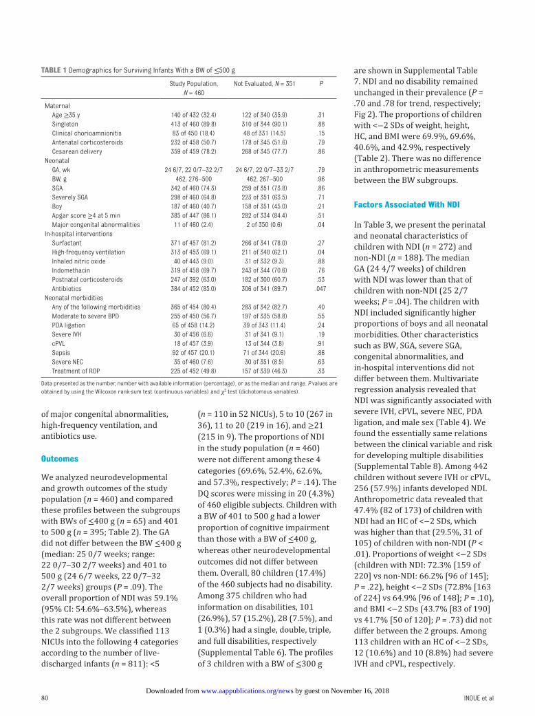
of major congenital abnormalities, high-frequency ventilation, and antibiotics use.
Outcomes
We analyzed neurodevelopmental and growth outcomes of the study population (n = 460) and compared these profiles between the subgroups with BWs of ≤400 g (n = 65) and 401 to 500 g (n = 395; Table 2). The GA did not differ between the BW ≤400 g (median: 25 0/7 weeks; range: 22 0/7–30 2/7 weeks) and 401 to 500 g (24 6/7 weeks, 22 0/7–32 2/7 weeks) groups (P = .09). The overall proportion of NDI was 59.1% (95% CI: 54.6%–63.5%), whereas this rate was not different between the 2 subgroups. We classified 113 NICUs into the following 4 categories according to the number of live-discharged infants (n = 811): <5
(n = 110 in 52 NICUs), 5 to 10 (267 in 36), 11 to 20 (219 in 16), and ≥21 (215 in 9). The proportions of NDI in the study population (n = 460) were not different among these 4 categories (69.6%, 52.4%, 62.6%, and 57.3%, respectively; P = .14). The DQ scores were missing in 20 (4.3%) of 460 eligible subjects. Children with a BW of 401 to 500 g had a lower proportion of cognitive impairment than those with a BW of ≤400 g, whereas other neurodevelopmental outcomes did not differ between them. Overall, 80 children (17.4%) of the 460 subjects had no disability. Among 375 children who had information on disabilities, 101 (26.9%), 57 (15.2%), 28 (7.5%), and 1 (0.3%) had a single, double, triple, and full disabilities, respectively (Supplemental Table 6). The profiles of 3 children with a BW of ≤300 g
are shown in Supplemental Table 7. NDI and no disability remainedunchanged in their prevalence (P = .70 and .78 for trend, respectively; Fig 2). The proportions of children with <−2 SDs of weight, height, HC, and BMI were 69.9%, 69.6%, 40.6%, and 42.9%, respectively (Table 2). There was no difference in anthropometric measurements between the BW subgroups.
Factors Associated With NDI
In Table 3, we present the perinatal and neonatal characteristics of children with NDI (n = 272) and non-NDI (n = 188). The median GA (24 4/7 weeks) of children with NDI was lower than that of children with non-NDI (25 2/7 weeks; P = .04). The children with NDI included significantly higher proportions of boys and all neonatal morbidities. Other characteristics such as BW, SGA, severe SGA, congenital abnormalities, and in-hospital interventions did not differ between them. Multivariate regression analysis revealed that NDI was significantly associated with severe IVH, cPVL, severe NEC, PDA ligation, and male sex (Table 4). We found the essentially same relations between the clinical variable and risk for developing multiple disabilities (Supplemental Table 8). Among 442 children without severe IVH or cPVL, 256 (57.9%) infants developed NDI. Anthropometric data revealed that 47.4% (82 of 173) of children with NDI had an HC of <−2 SDs, which was higher than that (29.5%, 31 of 105) of children with non-NDI (P < .01). Proportions of weight <−2 SDs (children with NDI: 72.3% [159 of 220] vs non-NDI: 66.2% [96 of 145]; P = .22), height <−2 SDs (72.8% [163 of 224] vs 64.9% [96 of 148]; P = .10), and BMI <−2 SDs (43.7% [83 of 190] vs 41.7% [50 of 120]; P = .73) did not differ between the 2 groups. Among 113 children with an HC of <−2 SDs, 12 (10.6%) and 10 (8.8%) had severe IVH and cPVL, respectively.
INOUE et al80
TABLE 1 Demographics for Surviving Infants With a BW of ≤500 g
Study Population, N = 460
Not Evaluated, N = 351 P
MaternalAge ≥35 y 140 of 432 (32.4) 122 of 340 (35.9) .31Singleton 413 of 460 (89.8) 310 of 344 (90.1) .88Clinical chorioamnionitis 83 of 450 (18.4) 48 of 331 (14.5) .15Antenatal corticosteroids 232 of 458 (50.7) 178 of 345 (51.6) .79Cesarean delivery 359 of 459 (78.2) 268 of 345 (77.7) .86
NeonatalGA, wk 24 6/7, 22 0/7–32 2/7 24 6/7, 22 0/7–33 2/7 .79BW, g 462, 276–500 462, 267–500 .96SGA 342 of 460 (74.3) 259 of 351 (73.8) .86Severely SGA 298 of 460 (64.8) 223 of 351 (63.5) .71Boy 187 of 460 (40.7) 158 of 351 (45.0) .21Apgar score ≥4 at 5 min 385 of 447 (86.1) 282 of 334 (84.4) .51Major congenital abnormalities 11 of 460 (2.4) 2 of 350 (0.6) .04
In-hospital interventionsSurfactant 371 of 457 (81.2) 266 of 341 (78.0) .27High-frequency ventilation 313 of 453 (69.1) 211 of 340 (62.1) .04Inhaled nitric oxide 40 of 443 (9.0) 31 of 332 (9.3) .88Indomethacin 319 of 458 (69.7) 243 of 344 (70.6) .76Postnatal corticosteroids 247 of 392 (63.0) 182 of 300 (60.7) .53Antibiotics 384 of 452 (85.0) 306 of 341 (89.7) .047
Neonatal morbiditiesAny of the following morbidities 365 of 454 (80.4) 283 of 342 (82.7) .40Moderate to severe BPD 255 of 450 (56.7) 197 of 335 (58.8) .55PDA ligation 65 of 458 (14.2) 39 of 343 (11.4) .24Severe IVH 30 of 456 (6.6) 31 of 341 (9.1) .19cPVL 18 of 457 (3.9) 13 of 344 (3.8) .91Sepsis 92 of 457 (20.1) 71 of 344 (20.6) .86Severe NEC 35 of 460 (7.6) 30 of 351 (8.5) .63Treatment of ROP 225 of 452 (49.8) 157 of 339 (46.3) .33
Data presented as the number, number with available information (percentage), or as the median and range. P values are obtained by using the Wilcoxon rank-sum test (continuous variables) and χ2 test (dichotomous variables).
by guest on November 16, 2018www.aappublications.org/newsDownloaded from
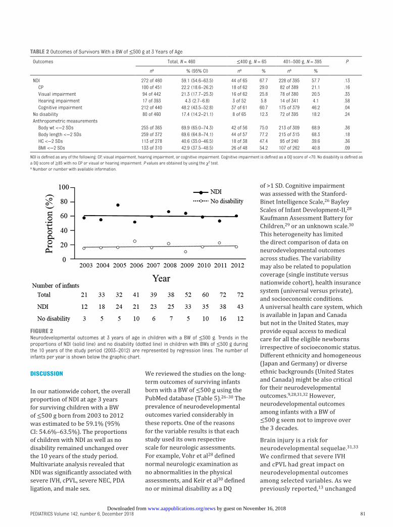
DISCUSSION
In our nationwide cohort, the overall proportion of NDI at age 3 years for surviving children with a BW of ≤500 g born from 2003 to 2012 was estimated to be 59.1% (95% CI: 54.6%–63.5%). The proportions of children with NDI as well as no disability remained unchanged over the 10 years of the study period. Multivariate analysis revealed that NDI was significantly associated with severe IVH, cPVL, severe NEC, PDA ligation, and male sex.
We reviewed the studies on the long-term outcomes of surviving infants born with a BW of ≤500 g using the PubMed database (Table 5).26 – 30 The prevalence of neurodevelopmental outcomes varied considerably in these reports. One of the reasons for the variable results is that each study used its own respective scale for neurologic assessments. For example, Vohr et al28 defined normal neurologic examination as no abnormalities in the physical assessments, and Keir et al30 defined no or minimal disability as a DQ
of >1 SD. Cognitive impairment was assessed with the Stanford-Binet Intelligence Scale, 26 Bayley Scales of Infant Development-II, 28 Kaufmann Assessment Battery for Children, 29 or an unknown scale.30 This heterogeneity has limited the direct comparison of data on neurodevelopmental outcomes across studies. The variability may also be related to population coverage (single institute versus nationwide cohort), health insurance system (universal versus private), and socioeconomic conditions. A universal health care system, which is available in Japan and Canada but not in the United States, may provide equal access to medical care for all the eligible newborns irrespective of socioeconomic status. Different ethnicity and homogeneous (Japan and Germany) or diverse ethnic backgrounds (United States and Canada) might be also critical for their neurodevelopmental outcomes.9, 28, 31, 32 However, neurodevelopmental outcomes among infants with a BW of ≤500 g seem not to improve over the 3 decades.
Brain injury is a risk for neurodevelopmental sequelae.31, 33 We confirmed that severe IVH and cPVL had great impact on neurodevelopmental outcomes among selected variables. As we previously reported, 13 unchanged
PEDIATRICS Volume 142, number 6, December 2018 81
TABLE 2 Outcomes of Survivors With a BW of ≤500 g at 3 Years of Age
Outcomes Total, N = 460 ≤400 g, N = 65 401–500 g, N = 395 P
na % (95% CI) na % na %
NDI 272 of 460 59.1 (54.6–63.5) 44 of 65 67.7 228 of 395 57.7 .13CP 100 of 451 22.2 (18.6–26.2) 18 of 62 29.0 82 of 389 21.1 .16Visual impairment 94 of 442 21.3 (17.7–25.3) 16 of 62 25.8 78 of 380 20.5 .35Hearing impairment 17 of 393 4.3 (2.7–6.8) 3 of 52 5.8 14 of 341 4.1 .58Cognitive impairment 212 of 440 48.2 (43.5–52.8) 37 of 61 60.7 175 of 379 46.2 .04
No disability 80 of 460 17.4 (14.2–21.1) 8 of 65 12.3 72 of 395 18.2 .24Anthropometric measurements
Body wt <−2 SDs 255 of 365 69.9 (65.0–74.3) 42 of 56 75.0 213 of 309 68.9 .36Body length <−2 SDs 259 of 372 69.6 (64.8–74.1) 44 of 57 77.2 215 of 315 68.3 .18HC <−2 SDs 113 of 278 40.6 (35.0–46.5) 18 of 38 47.4 95 of 240 39.6 .36BMI <−2 SDs 133 of 310 42.9 (37.5–48.5) 26 of 48 54.2 107 of 262 40.8 .09
NDI is defined as any of the following: CP, visual impairment, hearing impairment, or cognitive impairment. Cognitive impairment is defined as a DQ score of <70. No disability is defined as a DQ score of ≥85 with no CP or visual or hearing impairment. P values are obtained by using the χ2 test. a Number or number with available information.
FIGURE 2Neurodevelopmental outcomes at 3 years of age in children with a BW of ≤500 g. Trends in theproportions of NDI (solid line) and no disability (dotted line) in children with BWs of ≤500 g duringthe 10 years of the study period (2003–2012) are represented by regression lines. The number ofinfants per year is shown below the graphic chart.
by guest on November 16, 2018www.aappublications.org/newsDownloaded from

morbidities of brain injury among infants with a BW of ≤500 g might underlie the unimproved neurodevelopmental outcomes during the 10 years of our study period. cPVL, a severe form of cerebral white matter injury, has been observed in a small
portion of very preterm infants. By contrast, noncystic or diffuse periventricular leukomalacia is a predominant form of brain lesion and is not readily detected by neuroimaging.33 Therefore, early detection and prevention of white matter injuries will be the next
targets to control in perinatal medicine.
HCs of <−2 SDs at age 3 years were significantly associated with NDI in this study, as previously reported.34 HC can be considered a proxy for brain volume, 35 and head growth is affected by nutritional and nonnutritional factors.34 Diffuse periventricular leukomalacia is associated with volumetric deficits of the cerebral cortex and thalamus and delayed cortical maturation.33 These findings support the ongoing need for more extensive investigations into whether the children’s postnatal nutrition and/or environmental distress, besides perinatal brain injury, are associated with their small HC.
We have recently reported that the survival rate of infants with a BW of ≤500 g has greatly improved from 2003 to 2012 in Japan.13 Their survival was associated with several factors, including higher GA and BW, and the increasing rate of antenatal steroids and cesarean delivery contributed to improving their survival rate.13 Notably, these advantageous factors were not shown to reduce the risk of NDI for survivors with BWs of ≤500 g. The combined outcomes of mortality or morbidity can be determined for all births or live births and are associated with GA and BW.9 –11 However, neurologic outcomes can only be determined for survivors to the age of the follow-up assessment. Thus, analysis of NDI might mask the effects of GA or BW.32 The dissociated results are inevitable because severely affected infants are at high risk for death before discharge. Moreover, attrition in this study might reduce the power to detect effects of GA or BW. Because we selected surviving infants as eligible subjects, we were able to add variables of in-hospital interventions and neonatal complications in the current study. Consequently, severe IVH and cPVL were identified
INOUE et al
TABLE 3 Characteristics for NDI and Non-NDI Survivors With a BW of ≤500 g at 3 Years of Age
Characteristics NDI, N = 272 Non-NDI, N = 188 P
MaternalAge ≥35 y 78 of 258 (30.2) 62 of 174 (35.6) .24Singleton 242 of 272 (89.0) 171 of 188 (91.0) .49Clinical chorioamnionitis 53 of 266 (19.9) 30 of 184 (16.3) .33Antenatal corticosteroids 138 of 271 (50.9) 94 of 187 (50.3) .89Cesarean section 207 of 272 (76.1) 152 of 187 (81.3) .19
NeonatalGA, wk 24 4/7, 22 0/7–32 2/7 25 2/7, 22 0/7–29 3/7 .04BW, g 456, 276–500 467, 286–500 .14SGA 199 of 272 (73.2) 143 of 188 (76.1) .48Severely SGA 170 of 272 (62.5) 128 of 188 (68.1) .22Boy 124 of 272 (45.6) 63 of 188 (33.5) <.01Apgar score ≥4 at 5 min 218 of 261 (83.5) 167 of 186 (89.8) .06Major congenital abnormalities 6 of 272 (2.2) 5 of 188 (2.7) .75
In-hospital interventionsSurfactant 222 of 272 (81.6) 149 of 185 (80.5) .77High-frequency ventilation 189 of 269 (70.3) 124 of 184 (67.4) .52Inhaled nitric oxide 25 of 265 (9.4) 15 of 178 (8.4) .72Indomethacin 191 of 272 (70.2) 128 of 186 (68.8) .75Postnatal corticosteroids 155 of 236 (65.7) 92 of 156 (59.0) .18Antibiotics 234 of 270 (86.7) 150 of 182 (82.4) .22
Neonatal morbiditiesAny of the following morbidities 233 of 270 (86.3) 132 of 184 (71.7) <.01Moderate to severe BPD 165 of 268 (61.6) 90 of 182 (49.5) .01PDA ligation 48 of 272 (17.6) 17 of 186 (9.1) .01Severe IVH 28 of 271 (10.3) 2 of 185 (1.1) <.01cPVL 16 of 271 (5.9) 2 of 186 (1.1) <.01Sepsis 64 of 272 (23.5) 28 of 185 (15.1) .03Severe NEC 28 of 272 (10.3) 7 of 188 (3.7) <.01Treatment of ROP 150 of 270 (55.6) 75 of 182 (41.2) <.01
Data presented as the number, number with available information (percentage), or as the median and range. P values are obtained by using the Wilcoxon rank-sum test (continuous variables) and χ2 test (dichotomous variables).
TABLE 4 Factors Related to NDI in Infants With a BW of ≤500 g at 3 Years of Age
Crude RR (95% CI) P Adjusted RR (95% CI) P
Severe IVH 1.64 (1.44–1.86) <.01 1.42 (1.19–1.68) <.01cPVL 1.53 (1.28–1.84) <.01 1.40 (1.13–1.73) <.01Severe NEC 1.39 (1.16–1.68) <.01 1.31 (1.07–1.60) <.01PDA ligation 1.30 (1.09–1.53) <.01 1.29 (1.09–1.54) <.01Boy 1.22 (1.05–1.42) <.01 1.19 (1.01–1.40) .04Moderate to severe BPD 1.23 (1.04–1.44) .01 1.20 (0.99–1.45) .06Apgar score ≤3 at 5 min 1.22 (1.02–1.48) .03 1.10 (0.88–1.37) .40Treatment of ROP 1.26 (1.08–1.47) <.01 1.09 (0.93–1.29) .29Postnatal corticosteroids 1.12 (0.94–1.34) .19 1.08 (0.91–1.29) .38Sepsis 1.22 (1.04–1.44) .02 1.05 (0.88–1.26) .58GA (per wk) 0.97 (0.93–1.01) .09 1.01 (0.97–1.06) .63BW (per 100 g) 0.86 (0.74–1.00) .06 0.92 (0.78–1.07) .27
Risk ratios and P values are obtained from univariate and multivariate modified Poisson regression analyses. Multivariate analyses include 372 subjects for whom clinical variables are completely available, and adjusted risk ratios are obtained after adjusting for all covariates listed here. RR, risk ratio.
by guest on November 16, 2018www.aappublications.org/newsDownloaded from 82

among the survivors with BWs of ≤500 g as the highest risk factors for NDI but not GA, BW, or obstetric intervention. Given the improving survival rates13 and the unchanged neurodevelopmental outcomes, the number of both survivors with NDI and those without apparent disabilities were likely to increase in Japan. Nonetheless, these data provide useful information to guide the treatment of extremely small infants and social consensus, which is a goal of future research.
This study has several limitations. First, the follow-up rate was 56.7%. Because of the low follow-up rate, we failed to count the number of infants who died after discharge. In fact, the rates in drop-off were commonly high (30%–36%) in previous NRNJ studies.21, 22 One of the reasons might be that not all infants were followed-up at the same hospitals from which they were discharged. Therefore, their follow-up data were not recovered to a level comparable to those in former reports from other countries. Three perinatal parameters (congenital abnormalities, high-frequency ventilation, and antibiotics use) revealed significant differences between the study population and the nonevaluated survivors (Table 1). Two of these differences suggest the study population is sicker than the population without follow-up; therefore, the rate of NDI may be overestimated for the former. Second, there is no information on receiving the mode of active treatment. Therefore, neurodevelopmental outcomes in this study might be better than those for all births or live births. The third limitation is the lack of comparison with term-born infants, which may introduce expectation bias. However, the NDI rate at age 3 years in this study was higher than those of children with a BW of >500 g (501–750 g, 36.3%; 751–1000 g, 20.1%) in a previous NRNJ study.21 Lastly, other
PEDIATRICS Volume 142, number 6, December 2018 83
TABL
E 5
Neur
odev
elop
men
tal O
utco
mes
of I
nfan
ts W
ith a
BW
of ≤
500
g
Ref.
No.
Loca
tion
(Typ
e of
Stu
dy)
Year
of B
irth
Popu
latio
n na
Neur
odev
elop
men
tal O
utco
mes
b
ncFo
llow
-up
Rate
dSG
AeAg
e at
Ev
alua
tion,
yND
ICP
Visu
al
Impa
irm
ent
Hear
ing
Impa
irm
ent
Cogn
itive
Im
pair
men
tNo
Dis
abili
ty
25Ca
nada
1983
–199
4≤
500
g13
72.2
94.4
f3
69.2
46.2
15.3
7.7
61.5
30.8
(Reg
iona
l coh
ort)
113
26Un
ites
Stat
es19
89–2
009
≤50
0 g
4472
.190
.2f
2nd
15.9
9.1
22.7
ndnd
(Sin
gle
cent
er)
212
27Un
ites
Stat
es19
93–1
994
401–
500
g14
77.8
73.3
1.5
nd28
.621
.47.
135
.757
.1(M
ultic
ente
r)nd
28Ge
rman
y19
98–2
001
<501
g19
65.5
94.7
552
.636
.826
.310
.541
.226
.3(3
cen
ters
)48
29Au
stra
lia20
05–2
010
≤50
0 g
964
.388
.93
88.9
33.3
22.2
11.1
33.3
11.1
(Sin
gle
cent
er)
26Th
is s
tudy
Japa
n20
03–2
012
≤50
0 g
460
56.7
74.3
359
.122
.221
.34.
348
.217
.4(N
atio
nwid
e co
hort
)14
73
nd, n
ot d
escr
ibed
.a
Num
ber
of li
ve-b
orn
infa
nts
who
are
adm
itted
to N
ICUs
.b
Neur
odev
elop
men
tal o
utco
mes
dat
a ar
e ex
pres
sed
as th
e pe
rcen
tage
of e
valu
ated
infa
nts.
Not
e th
at th
e de
finiti
ons
of e
ach
outc
ome
are
diffe
rent
in e
ach
stud
y.c N
umbe
r of
sur
vivi
ng a
nd e
valu
ated
infa
nts.
d Fo
llow
-up
rate
s ar
e ex
pres
sed
as p
erce
ntag
es a
nd c
alcu
late
d by
div
idin
g th
e nu
mbe
r of
sur
vivi
ng a
nd e
valu
ated
infa
nts
by th
e to
tal n
umbe
r of
sur
viva
ls to
dis
char
ge fr
om N
ICUs
.e D
ata
are
expr
esse
d as
per
cent
ages
of s
urvi
ving
and
eva
luat
ed in
fant
s.f D
ata
are
expr
esse
d as
per
cent
ages
of a
ll su
rviv
ing
infa
nts.
by guest on November 16, 2018www.aappublications.org/newsDownloaded from

prognostic factors for neurodevelopmental disability such as parental education level, erythropoietin treatment, aggressive nutritional support, and specific strategies of ventilation and steroids administration32 were not analyzed. Identifying the key environmental factors among these variables will further unveil the potential targets of intervention for extremely small infants.
CONCLUSIONS
Neurodevelopmental outcomes in early childhood among surviving infants with a BW of ≤500 g did not improve in a recent decade. In this large-population study, we verified that perinatal brain injury was an unfavorable risk factor for NDI. Changes in medical management that are able to ameliorate brain injury would be expected to reduce adverse neurodevelopmental burdens for extremely small infants.
ACKNOWLEDGMENTS
Institutions enrolled in the study of the NRNJ were as follows: Sapporo City General Hospital, Asahikawa Kosei General Hospital, Engaru-Kosei General Hospital, Kushiro Red Cross Hospital, Obihiro-Kosei General Hospital, Tenshi Hospital, NTT Higashinihon Sapporo Hospital, Nikko Memorial Hospital, Nayoro City General Hospital, Sapporo Medical University, Asahikawa Medical University, Aomori Prefectural Central Hospital, Iwate Medical University, Iwate Prefectural Ofunato Hospital, Iwate Prefectural Kuji Hospital, Iwate Prefectural Ninohe Hospital, Sendai Red Cross Hospital, Akita Red Cross Hospital, Tsuruoka Municipal Shonai Hospital, Yamagata University, Yamagata Prefectural Central Hospital, Fukushima Medical University, Takeda General Hospital, Fukushima National
Hospital, Tsukuba University, Tsuchiura Kyodo Hospital, Ibaraki Children’s Hospital, Dokkyo Medical University, Jichi Medical University, Ashikaga Red Cross Hospital, Gunma Children’s Medical Center, Kiryu Kosei General Hospital, Fuji Heavy Industries Health Insurance Society Ota Memorial Hospital, Gunma University, Saitama Children’s Medical Center, Nishisaitama-chuo National Hospital, Saitama Medical University Saitama Medical Center, Kawaguchi Municipal Medical Center, Jichi Medical University Saitama Medical Center, Asahi General Hospital, Chiba Kaihin Municipal Hospital, Kameda Medical Center, Tokyo Women’s Medical University Yachiyo Medical Center, Juntendo University Urayasu Hospital, Tokyo Metropolitan Children’s Medical Center, Tokyo Women’s Medical University, Aiiku Hospital, Nihon University Itabashi Hospital, National Center for Global Health and Medicine, Tokyo Medical University, Teikyo University, Showa University, Japan Red Cross Medical Center, National Center for Child Health and Development, Tokyo Metropolitan Otsuka Hospital, Toho University, Tokyo Metropolitan Bokuto Hospital, Tokyo Jikei Medical University, Tokyo Medical and Dental University, Saint Luke’s International Hospital, Juntendo University, Sanikukai Hospital, Katsushika Red Cross Hospital, Yokohama Rosai Hospital, Yokohama City University Medical Center, St. Marianna University School of Medicine Hospital, Kanagawa Children’s Medical Center, Tokai University, Kitazato University, Odawara Municipal Hospital, Nippon Medical School Musashi Kosugi Hospital, Saiseikai Yokohamashi Tobu Hospital, National Hospital Organization Yokohama Medical Center, Yamanashi Prefectural Central Hospital, Nagano Children’s Hospital, Shinshu University, Iida Municipal Hospital, National Hospital Organization Shinshu Ueda Medical Center, Saku General
Hospital, Niigata University, Niigata Prefectural Central Hospital, Niigata Municipal Hospital, Nagaoka Red Cross Hospital, Koseiren Takaoka Hospital, Toyama Prefectural Central Hospital, Toyama University, Ishikawa Medical Center for Maternal and Child Health, Kanazawa Medical University, Kanazawa Medical Center, Fukui Prefectural Hospital, Fukui University, Gifu Prefectural General Medical Center, National Hospital Organization Nagara Medical Center, Takayama Red Cross Hospital, Seirei Hamamatsu Hospital, Shizuoka Saiseikai Hospital, Shizuoka Children’s Hospital, Hamamatsu Medical University, Numazu Municipal Hospital, Yaizu City Hospital, Fujieda Municipal General Hospital, Nagoya Red Cross Daini Hospital, Nagoya University, Nagoya Red Cross Daiichi Hospital, Toyohashi Municipal Hospital, Nagoya City West Medical Center, Anjo kosei Hospital, Tosei General Hospital, Komaki Municipal Hospital, TOYOTA Memorial Hospital, Okazaki Municipal Hospital, Konan Kosei Hospital, National Mie Central Medical Center, Ise Red Cross Hospital, Yokkaichi Municipal Hospital, Otsu Red Cross Hospital, Shiga University of Medical Science Hospital, Nagahama Red Cross Hospital, Uji Tokushukai Hospital, The Japan Baptist Hospital, Kyoto University, Kyoto Red Cross Daiichi Hospital, National Maizuru Medical Center, Fukuchiyama City Hospital, Kyoto Prefectural University of Medicine Hospital, Kyoto City Hospital, Mitsubishi Kyoto Hospital, Yodogawa Christian Hospital, Osaka Medical Center and Research Institute for Maternal and Child Health, Osaka University, Takatsuki General Hospital, Kansai Medical University, Osaka City General Hospital, Osaka City Sumiyoshi Hospital, Aizenbashi Hospital, Toyonaka Municipal Hospital, National Cerebral and Cardiovascular Center, Kitano Hospital, Saiseikai Suita Hospital, Chifune Hospital,
INOUE et al by guest on November 16, 2018www.aappublications.org/newsDownloaded from
84
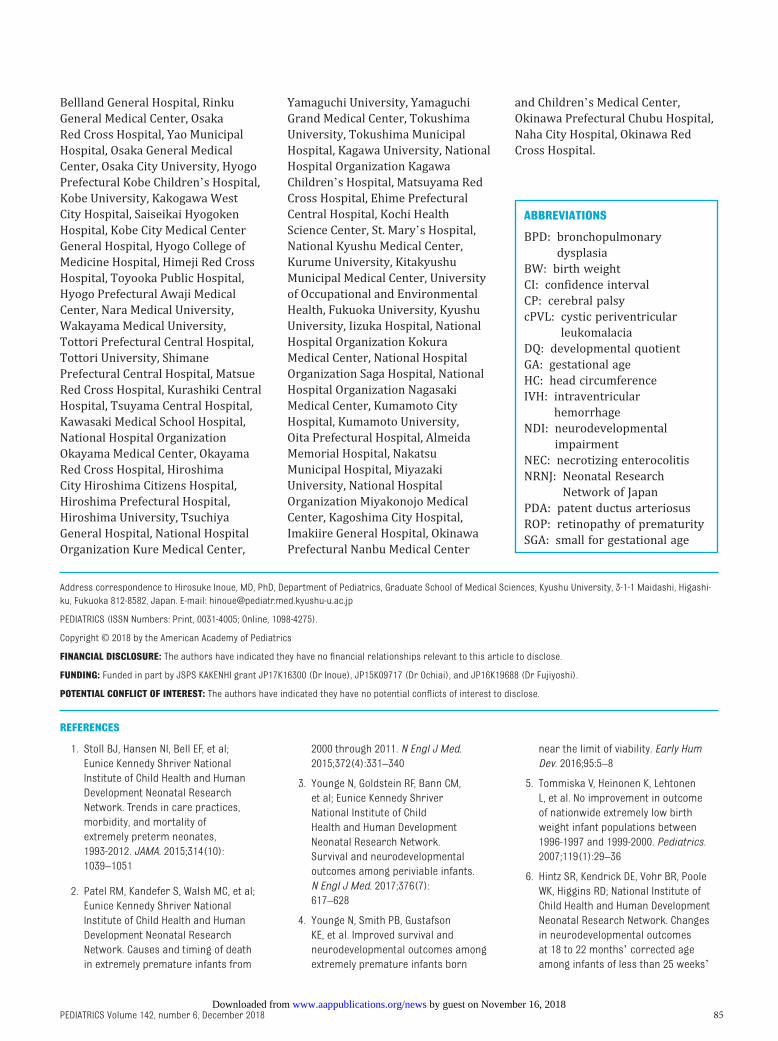
Bellland General Hospital, Rinku General Medical Center, Osaka Red Cross Hospital, Yao Municipal Hospital, Osaka General Medical Center, Osaka City University, Hyogo Prefectural Kobe Children’s Hospital, Kobe University, Kakogawa West City Hospital, Saiseikai Hyogoken Hospital, Kobe City Medical Center General Hospital, Hyogo College of Medicine Hospital, Himeji Red Cross Hospital, Toyooka Public Hospital, Hyogo Prefectural Awaji Medical Center, Nara Medical University, Wakayama Medical University, Tottori Prefectural Central Hospital, Tottori University, Shimane Prefectural Central Hospital, Matsue Red Cross Hospital, Kurashiki Central Hospital, Tsuyama Central Hospital, Kawasaki Medical School Hospital, National Hospital Organization Okayama Medical Center, Okayama Red Cross Hospital, Hiroshima City Hiroshima Citizens Hospital, Hiroshima Prefectural Hospital, Hiroshima University, Tsuchiya General Hospital, National Hospital Organization Kure Medical Center,
Yamaguchi University, Yamaguchi Grand Medical Center, Tokushima University, Tokushima Municipal Hospital, Kagawa University, National Hospital Organization Kagawa Children’s Hospital, Matsuyama Red Cross Hospital, Ehime Prefectural Central Hospital, Kochi Health Science Center, St. Mary’s Hospital, National Kyushu Medical Center, Kurume University, Kitakyushu Municipal Medical Center, University of Occupational and Environmental Health, Fukuoka University, Kyushu University, Iizuka Hospital, National Hospital Organization Kokura Medical Center, National Hospital Organization Saga Hospital, National Hospital Organization Nagasaki Medical Center, Kumamoto City Hospital, Kumamoto University, Oita Prefectural Hospital, Almeida Memorial Hospital, Nakatsu Municipal Hospital, Miyazaki University, National Hospital Organization Miyakonojo Medical Center, Kagoshima City Hospital, Imakiire General Hospital, Okinawa Prefectural Nanbu Medical Center
and Children’s Medical Center, Okinawa Prefectural Chubu Hospital, Naha City Hospital, Okinawa Red Cross Hospital.
REFERENCES
1. Stoll BJ, Hansen NI, Bell EF, et al;Eunice Kennedy Shriver NationalInstitute of Child Health and HumanDevelopment Neonatal ResearchNetwork. Trends in care practices,morbidity, and mortality ofextremely preterm neonates,1993-2012. JAMA. 2015;314(10):1039–1051
2. Patel RM, Kandefer S, Walsh MC, et al;Eunice Kennedy Shriver NationalInstitute of Child Health and HumanDevelopment Neonatal ResearchNetwork. Causes and timing of deathin extremely premature infants from
2000 through 2011. N Engl J Med. 2015;372(4):331–340
3. Younge N, Goldstein RF, Bann CM,et al; Eunice Kennedy ShriverNational Institute of ChildHealth and Human DevelopmentNeonatal Research Network.Survival and neurodevelopmentaloutcomes among periviable infants.N Engl J Med. 2017;376(7):617–628
4. Younge N, Smith PB, GustafsonKE, et al. Improved survival andneurodevelopmental outcomes amongextremely premature infants born
near the limit of viability. Early Hum Dev. 2016;95:5–8
5. Tommiska V, Heinonen K, LehtonenL, et al. No improvement in outcomeof nationwide extremely low birthweight infant populations between1996-1997 and 1999-2000. Pediatrics.2007;119(1):29–36
6. Hintz SR, Kendrick DE, Vohr BR, PooleWK, Higgins RD; National Institute ofChild Health and Human DevelopmentNeonatal Research Network. Changesin neurodevelopmental outcomesat 18 to 22 months’ corrected ageamong infants of less than 25 weeks’
PEDIATRICS Volume 142, number 6, December 2018 85
Address correspondence to Hirosuke Inoue, MD, PhD, Department of Pediatrics, Graduate School of Medical Sciences, Kyushu University, 3-1-1 Maidashi, Higashi-ku, Fukuoka 812-8582, Japan. E-mail: [email protected]
PEDIATRICS (ISSN Numbers: Print, 0031-4005; Online, 1098-4275).
Copyright © 2018 by the American Academy of Pediatrics
FINANCIAL DISCLOSURE: The authors have indicated they have no financial relationships relevant to this article to disclose.
FUNDING: Funded in part by JSPS KAKENHI grant JP17K16300 (Dr Inoue), JP15K09717 (Dr Ochiai), and JP16K19688 (Dr Fujiyoshi).
POTENTIAL CONFLICT OF INTEREST: The authors have indicated they have no potential conflicts of interest to disclose.
ABBREVIATIONS
BPD: bronchopulmonary dysplasia
BW: birth weightCI: confidence intervalCP: cerebral palsycPVL: cystic periventricular
leukomalaciaDQ: developmental quotientGA: gestational ageHC: head circumferenceIVH: intraventricular
hemorrhageNDI: neurodevelopmental
impairmentNEC: necrotizing enterocolitisNRNJ: Neonatal Research
Network of JapanPDA: patent ductus arteriosusROP: retinopathy of prematuritySGA: small for gestational age
by guest on November 16, 2018www.aappublications.org/newsDownloaded from

gestational age born in 1993-1999. Pediatrics. 2005;115(6):1645–1651
7. Hintz SR, Kendrick DE, Wilson-Costello DE, et al; NICHD NeonatalResearch Network. Early-childhoodneurodevelopmental outcomes arenot improving for infants born at <25weeks’ gestational age. Pediatrics.2011;127(1):62–70
8. Wilson-Costello D, Friedman H, MinichN, Fanaroff AA, Hack M. Improvedsurvival rates with increasedneurodevelopmental disabilityfor extremely low birth weightinfants in the 1990s. Pediatrics.2005;115(4):997–1003
9. Ecker JL, Kaimal A, Mercer BM, et al;American College of Obstetriciansand Gynecologists and the Society forMaternal–Fetal Medicine. Periviablebirth: interim update. Am J ObstetGynecol. 2016;215(2):B2–B12.e1
10. Salihu HM, Salinas-Miranda AA, HillL, Chandler K. Survival of pre-viablepreterm infants in the United States: asystematic review and meta-analysis.Semin Perinatol. 2013;37(6):389–400
11. Tyson JE, Parikh NA, Langer J, GreenC, Higgins RD; National Institute ofChild Health and Human DevelopmentNeonatal Research Network. Intensivecare for extreme prematurity–movingbeyond gestational age. N Engl J Med.2008;358(16):1672–1681
12. American College of Obstetriciansand Gynecologists. ACOG practicebulletin no. 101: ultrasonography inpregnancy. Obstet Gynecol. 2009;113(2,pt 1):451–461
13. Inoue H, Ochiai M, Yasuoka K, et al;Neonatal Research Network of Japan(NRNJ). Early mortality and morbidityin infants with birth weight of500 grams or less in Japan. J Pediatr.2017;190(11):112–117.e3
14. Itabashi K, Miura F, Uehara R,Nakamura Y. New Japaneseneonatal anthropometric charts forgestational age at birth. Pediatr Int.2014;56(5):702–708
15. Ehrenkranz RA, Walsh MC, Vohr BR,et al; National Institutes of ChildHealth and Human DevelopmentNeonatal Research Network.Validation of the National Institutesof Health consensus definition
of bronchopulmonary dysplasia. Pediatrics. 2005;116(6):1353–1360
16. Papile LA, Burstein J, Burstein R,Koffler H. Incidence and evolution ofsubependymal and intraventricularhemorrhage: a study of infants withbirth weights less than 1, 500 gm.J Pediatr. 1978;92(4):529–534
17. Bell MJ, Ternberg JL, Feigin RD, et al.Neonatal necrotizing enterocolitis.Therapeutic decisions basedupon clinical staging. Ann Surg.1978;187(1):1–7
18. Kono Y, Mishina J, Sato N, WatanabeT, Honma Y. Developmentalcharacteristics of very low-birthweightinfants at 18 months’ corrected ageaccording to birthweight. Pediatr Int.2008;50(1):23–28
19. Bax MC. Terminology and classificationof cerebral palsy. Dev Med ChildNeurol. 1964;6:295–297
20. Kono Y, Yonemoto N, Kusuda S,et al. Developmental assessmentof VLBW infants at 18 months ofage: a comparison study betweenKSPD and Bayley III. Brain Dev.2016;38(4):377–385
21. Kono Y, Mishina J, Yonemoto N, KusudaS, Fujimura M. Outcomes of very-low-birthweight infants at 3 years of ageborn in 2003-2004 in Japan. Pediatr Int.2011;53(6):1051–1058
22. Ishii N, Kono Y, Yonemoto N, KusudaS, Fujimura M; Neonatal ResearchNetwork, Japan. Outcomes of infantsborn at 22 and 23 weeks’ gestation.Pediatrics. 2013;132(1):62–71
23. Suwa S, Tachibana K. Standardgrowth charts for height and weightof Japanese children from birth to17 years based on a cross-sectionalsurvey of national data. Clin PediatrEndocrinol. 1993;2(2):87–97
24. Kato N, Takimoto H, Sudo N. The cubicfunctions for spline smoothed L, Sand M values for BMI reference dataof Japanese children. Clin PediatrEndocrinol. 2011;20(2):47–49
25. Zou GY, Donner A. Extension of themodified Poisson regression model toprospective studies with correlatedbinary data. Stat Methods Med Res.2013;22(6):661–670
26. Sauve RS, Robertson C, Etches P, ByrnePJ, Dayer-Zamora V. Before viability: a
geographically based outcome study of infants weighing 500 grams or less at birth. Pediatrics. 1998;101(3, pt 1): 438–445
27. Upadhyay K, Pourcyrous M,Dhanireddy R, Talati AJ. Outcomes ofneonates with birth weight⩽500 g:a 20-year experience. J Perinatol.2015;35(9):768–772
28. Vohr BR, Wright LL, Dusick AM, et al.Neurodevelopmental and functionaloutcomes of extremely low birthweight infants in the NationalInstitute of Child Health and HumanDevelopment Neonatal ResearchNetwork, 1993-1994. Pediatrics.2000;105(6):1216–1226
29. Rieger-Fackeldey E, Blank C, DingerJ, Steinmacher J, Bode H, Schulze A.Growth, neurological and cognitivedevelopment in infants with abirthweight <501 g at age 5 years. ActaPaediatr. 2010;99(9):1350–1355
30. Keir A, McPhee A, Wilkinson D. Beyondthe borderline: outcomes for inborninfants born at ≤500 grams. J PaediatrChild Health. 2014;50(2):146–152
31. Jarjour IT. Neurodevelopmentaloutcome after extreme prematurity: areview of the literature. Pediatr Neurol.2015;52(2):143–152
32. Linsell L, Malouf R, Morris J, KurinczukJJ, Marlow N. Prognostic factors forpoor cognitive development in childrenborn very preterm or with very lowbirth weight: a systematic review.JAMA Pediatr. 2015;169(12):1162–1172
33. Volpe JJ. Brain injury in prematureinfants: a complex amalgam ofdestructive and developmentaldisturbances. Lancet Neurol.2009;8(1):110–124
34. Raghuram K, Yang J, Church PT,et al; Canadian Neonatal Network;Canadian Neonatal Follow-Up NetworkInvestigators. Head growth trajectoryand neurodevelopmental outcomesin preterm neonates. Pediatrics.2017;140(1):e20170216
35. Cheong JL, Hunt RW, Anderson PJ,et al. Head growth in preterm infants:correlation with magnetic resonanceimaging and neurodevelopmentaloutcome. Pediatrics. 2008;121(6).Available at: www. pediatrics. org/ cgi/ content/ full/ 121/ 6/ e1534
INOUE et al86 by guest on November 16, 2018www.aappublications.org/newsDownloaded from
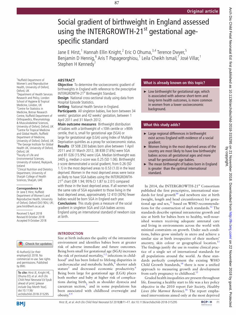
F1Hirst JE, et al. Arch Dis Child Fetal Neonatal Ed 2018;0:F1–F7. doi:10.1136/archdischild-2018-315295
Original article
Social gradient of birthweight in England assessed using the INTERGROWTH-21st gestational age-specific standardJane E Hirst,1 Hannah Ellin Knight,2 Eric O Ohuma,3,4 Terence Dwyer,5 Benjamin D Hennig,6 Aris T Papageorghiou,1 Leila Cheikh Ismail,7 José Villar,1 Stephen H Kennedy1
To cite: Hirst JE, Knight HE, Ohuma EO, et al. Arch Dis Child Fetal Neonatal Ed Epub ahead of print: [please include Day Month Year]. doi:10.1136/archdischild-2018-315295
1Nuffield Department of Women’s and Reproductive Health, University of Oxford, Oxford, UK2Department of Health Services Research and Policy, London School of Hygiene & Tropical Medicine, London, UK3Centre for Statistics in Medicine, Botnar Research Centre, Nuffield Department of Orthopaedics, Rheumatology & Musculoskeletal Sciences, University of Oxford, Oxford, UK4Centre for Tropical Medicine and Global Health, Nuffield Department of Medicine, University of Oxford, Oxford, UK5The George Institute for Global Health UK, University of Oxford, Oxford, UK6Faculty of Life and Environmental Sciences, University of Iceland, Reykjavik, Iceland7Clinical Nutrition and Dietetics Department, University of Sharjah College of Health Sciences, Sharjah, UAE
Correspondence toDr Jane E Hirst, Nuffield Department of Women’s and Reproductive Health, University of Oxford, Oxford OX3 9DU, UK; jane. hirst@ wrh. ox. ac. uk
Received 5 April 2018Revised 8 October 2018Accepted 9 October 2018
© Author(s) (or their employer(s)) 2018. No commercial re-use. See rights and permissions. Published by BMJ.
AbsTrACTObjective To determine the socioeconomic gradient of birthweights in England with reference to the prescriptive INTERGROWTH-21st Birthweight Standard.Design National cross-sectional study using data from Hospital Episode Statistics.setting National Health Service in England.Participants All singleton babies, live born between 34 weeks’ gestation and 42 weeks’ gestation, between 1 April 2011 and 31 March 2012.Main outcome measures Birthweight distribution of babies with a birthweight of <10th centile or >90th centile, that is, small for gestational age (SGA) or large for gestational age (LGA) using Index of Multiple Deprivation quintiles as a proxy for socioeconomic status.results Of 508 230 babies born alive between 1 April 2011 and 31 March 2012, 38 838 (7.6%) were SGA and 81 026 (15.9%) were LGA. Median birthweight was 3405 g, median z-score was 0.25 (SD 1.06). Birthweight z-score demonstrated a social gradient, from 0.26 (SD 1.1) in the most deprived areas to 0.53 (1.0) in the least deprived. Women in the most deprived areas were twice as likely to have SGA babies using the INTERGROWTH-21st chart (OR 1.94; 95% CI 1.87 to 2.01) compared with those in the least deprived areas. If all women had the same rate of SGA equivalent to those living in the least deprived areas, approximately 12 410 (30%) fewer babies would be born SGA in England each year.Conclusions This study gives a measure of the social gradient in singleton SGA and LGA babies across England using an international standard of newborn size at birth.
InTrODuCTIOnSize at birth indicates the quality of the intrauterine environment and identifies babies born at greater risk of adverse immediate and future outcomes. Being born small for gestational age (SGA) increases the risk of perinatal mortality,1 2 infections in child-hood3 and has been linked to lifelong disparities in cardiovascular and metabolic health,4 shorter adult stature5 and decreased economic productivity.6 Being born large for gestational age (LGA) places both mother and baby at higher risk of complica-tions during birth, such as shoulder dystocia and caesarean section,7 and in some populations has been associated with childhood overweight and obesity.8 9
In 2014, the INTERGROWTH-21st Consortium published the first prescriptive, international stan-dards for fetal growth10 and newborn size at birth (weight, length and head circumference) for gesta-tional age and sex,11 based on WHO recommenda-tions for the construction of such standards.12 The standards describe optimal intrauterine growth and size at birth for babies born to healthy, well-nour-ished women receiving adequate antenatal care and living in environments across the world with minimal constraints on growth. Under such condi-tions, babies grow similarly in utero and achieve a similar size at birth irrespective of their mothers’ ancestry, skin colour or geographical location.13 The findings justify the use in routine clinical prac-tice of a single set of international standards for all populations around the world. As these stan-dards perfectly complement the existing WHO Child Growth Standards,14 there is now a unified approach to measuring growth and development from early pregnancy to childhood.15
Graded health inequalities are present throughout life. Ensuring a healthy start to life was a key policy objective in the 2010 report Fair Society, Healthy Lives (the Marmot Review).16 The report empha-sised interventions aimed only at the most deprived
What is already known on this topic?
► Low birthweight for gestational age, which is associated with adverse short-term and long-term health outcomes, is more common in women from a lower socioeconomic background.
What this study adds?
► Large regional differences in birthweight exist across England with evidence of a social gradient.
► Women living in the most deprived areas of the country are most likely to have low birthweight babies across all gestational ages and more small for gestational age babies.
► The mean birthweight of babies born in England is greater than the optimal international standard.
by copyright. on 22 N
ovember 2018 by guest. P
rotectedhttp://fn.bm
j.com/
Arch D
is Child F
etal Neonatal E
d: first published as 10.1136/archdischild-2018-315295 on 21 Novem
ber 2018. Dow
nloaded from
87
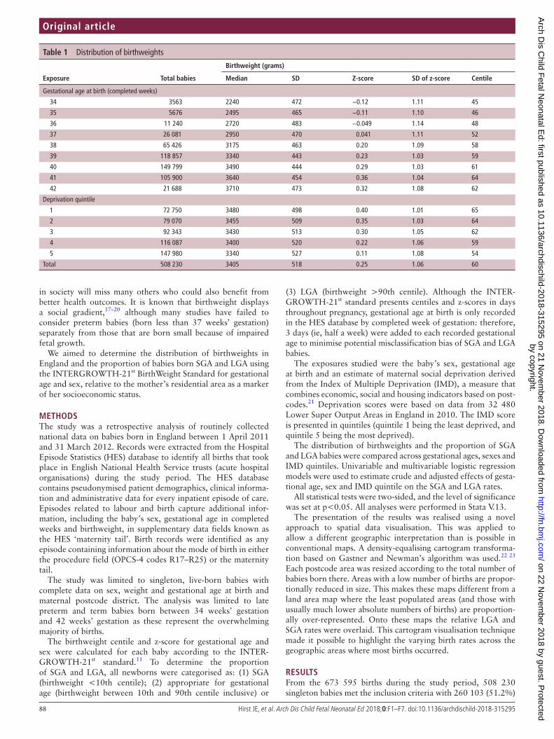
88 Hirst JE, et al. Arch Dis Child Fetal Neonatal Ed 2018;0:F1–F7. doi:10.1136/archdischild-2018-315295
Original article
in society will miss many others who could also benefit from better health outcomes. It is known that birthweight displays a social gradient,17–20 although many studies have failed to consider preterm babies (born less than 37 weeks’ gestation) separately from those that are born small because of impaired fetal growth.
We aimed to determine the distribution of birthweights in England and the proportion of babies born SGA and LGA using the INTERGROWTH-21st BirthWeight Standard for gestational age and sex, relative to the mother’s residential area as a marker of her socioeconomic status.
MeThODsThe study was a retrospective analysis of routinely collected national data on babies born in England between 1 April 2011 and 31 March 2012. Records were extracted from the Hospital Episode Statistics (HES) database to identify all births that took place in English National Health Service trusts (acute hospital organisations) during the study period. The HES database contains pseudonymised patient demographics, clinical informa-tion and administrative data for every inpatient episode of care. Episodes related to labour and birth capture additional infor-mation, including the baby’s sex, gestational age in completed weeks and birthweight, in supplementary data fields known as the HES ‘maternity tail’. Birth records were identified as any episode containing information about the mode of birth in either the procedure field (OPCS-4 codes R17–R25) or the maternity tail.
The study was limited to singleton, live-born babies with complete data on sex, weight and gestational age at birth and maternal postcode district. The analysis was limited to late preterm and term babies born between 34 weeks’ gestation and 42 weeks’ gestation as these represent the overwhelming majority of births.
The birthweight centile and z-score for gestational age and sex were calculated for each baby according to the INTER-GROWTH-21st standard.11 To determine the proportion of SGA and LGA, all newborns were categorised as: (1) SGA (birthweight <10th centile); (2) appropriate for gestational age (birthweight between 10th and 90th centile inclusive) or
(3) LGA (birthweight >90th centile). Although the INTER-GROWTH-21st standard presents centiles and z-scores in days throughout pregnancy, gestational age at birth is only recorded in the HES database by completed week of gestation: therefore, 3 days (ie, half a week) were added to each recorded gestational age to minimise potential misclassification bias of SGA and LGA babies.
The exposures studied were the baby’s sex, gestational age at birth and an estimate of maternal social deprivation derived from the Index of Multiple Deprivation (IMD), a measure that combines economic, social and housing indicators based on post-codes.21 Deprivation scores were based on data from 32 480 Lower Super Output Areas in England in 2010. The IMD score is presented in quintiles (quintile 1 being the least deprived, and quintile 5 being the most deprived).
The distribution of birthweights and the proportion of SGA and LGA babies were compared across gestational ages, sexes and IMD quintiles. Univariable and multivariable logistic regression models were used to estimate crude and adjusted effects of gesta-tional age, sex and IMD quintile on the SGA and LGA rates.
All statistical tests were two-sided, and the level of significance was set at p<0.05. All analyses were performed in Stata V.13.
The presentation of the results was realised using a novel approach to spatial data visualisation. This was applied to allow a different geographic interpretation than is possible in conventional maps. A density-equalising cartogram transforma-tion based on Gastner and Newman’s algorithm was used.22 23 Each postcode area was resized according to the total number of babies born there. Areas with a low number of births are propor-tionally reduced in size. This makes these maps different from a land area map where the least populated areas (and those with usually much lower absolute numbers of births) are proportion-ally over-represented. Onto these maps the relative LGA and SGA rates were overlaid. This cartogram visualisation technique made it possible to highlight the varying birth rates across the geographic areas where most births occurred.
resulTsFrom the 673 595 births during the study period, 508 230 singleton babies met the inclusion criteria with 260 103 (51.2%)
Table 1 Distribution of birthweights
exposure Total babies
birthweight (grams)
Median sD Z-score sD of z-score Centile
Gestational age at birth (completed weeks)
34 3563 2240 472 −0.12 1.11 45
35 5676 2495 465 −0.11 1.10 46
36 11 240 2720 483 −0.049 1.14 48
37 26 081 2950 470 0.041 1.11 52
38 65 426 3175 463 0.20 1.09 58
39 118 857 3340 443 0.23 1.03 59
40 149 799 3490 444 0.29 1.03 61
41 105 900 3640 454 0.36 1.04 64
42 21 688 3710 473 0.32 1.08 62
Deprivation quintile
1 72 750 3480 498 0.40 1.01 65
2 79 070 3455 509 0.35 1.03 64
3 92 343 3430 513 0.30 1.05 62
4 116 087 3400 520 0.22 1.06 59
5 147 980 3340 527 0.11 1.08 54
Total 508 230 3405 518 0.25 1.06 60
by copyright. on 22 N
ovember 2018 by guest. P
rotectedhttp://fn.bm
j.com/
Arch D
is Child F
etal Neonatal E
d: first published as 10.1136/archdischild-2018-315295 on 21 Novem
ber 2018. Dow
nloaded from
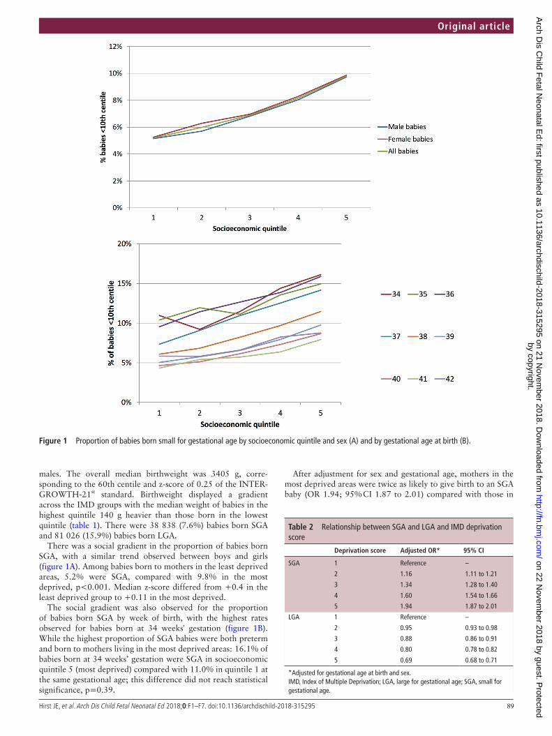
89Hirst JE, et al. Arch Dis Child Fetal Neonatal Ed 2018;0:F1–F7. doi:10.1136/archdischild-2018-315295
Original article
males. The overall median birthweight was 3405 g, corre-sponding to the 60th centile and z-score of 0.25 of the INTER-GROWTH-21st standard. Birthweight displayed a gradient across the IMD groups with the median weight of babies in the highest quintile 140 g heavier than those born in the lowest quintile (table 1). There were 38 838 (7.6%) babies born SGA and 81 026 (15.9%) babies born LGA.
There was a social gradient in the proportion of babies born SGA, with a similar trend observed between boys and girls (figure 1A). Among babies born to mothers in the least deprived areas, 5.2% were SGA, compared with 9.8% in the most deprived, p<0.001. Median z-score differed from +0.4 in the least deprived group to +0.11 in the most deprived.
The social gradient was also observed for the proportion of babies born SGA by week of birth, with the highest rates observed for babies born at 34 weeks' gestation (figure 1B). While the highest proportion of SGA babies were both preterm and born to mothers living in the most deprived areas: 16.1% of babies born at 34 weeks’ gestation were SGA in socioeconomic quintile 5 (most deprived) compared with 11.0% in quintile 1 at the same gestational age; this difference did not reach statistical significance, p=0.39.
After adjustment for sex and gestational age, mothers in the most deprived areas were twice as likely to give birth to an SGA baby (OR 1.94; 95% CI 1.87 to 2.01) compared with those in
Figure 1 Proportion of babies born small for gestational age by socioeconomic quintile and sex (A) and by gestational age at birth (B).
Table 2 Relationship between SGA and LGA and IMD deprivation score
Deprivation score Adjusted Or* 95% CI
SGA 1 Reference –
2 1.16 1.11 to 1.21
3 1.34 1.28 to 1.40
4 1.60 1.54 to 1.66
5 1.94 1.87 to 2.01
LGA 1 Reference –
2 0.95 0.93 to 0.98
3 0.88 0.86 to 0.91
4 0.80 0.78 to 0.82
5 0.69 0.68 to 0.71
*Adjusted for gestational age at birth and sex.IMD, Index of Multiple Deprivation; LGA, large for gestational age; SGA, small for gestational age.
by copyright. on 22 N
ovember 2018 by guest. P
rotectedhttp://fn.bm
j.com/
Arch D
is Child F
etal Neonatal E
d: first published as 10.1136/archdischild-2018-315295 on 21 Novem
ber 2018. Dow
nloaded from
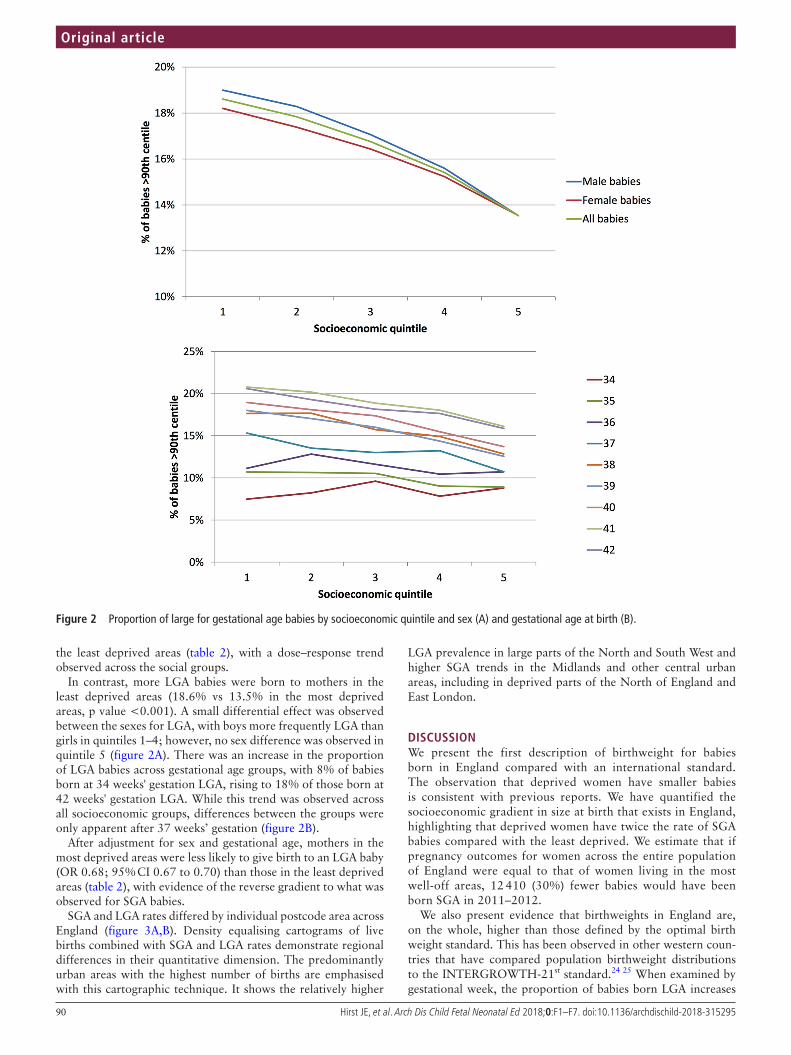
90 Hirst JE, et al. Arch Dis Child Fetal Neonatal Ed 2018;0:F1–F7. doi:10.1136/archdischild-2018-315295
Original article
the least deprived areas (table 2), with a dose–response trend observed across the social groups.
In contrast, more LGA babies were born to mothers in the least deprived areas (18.6% vs 13.5% in the most deprived areas, p value <0.001). A small differential effect was observed between the sexes for LGA, with boys more frequently LGA than girls in quintiles 1–4; however, no sex difference was observed in quintile 5 (figure 2A). There was an increase in the proportion of LGA babies across gestational age groups, with 8% of babies born at 34 weeks' gestation LGA, rising to 18% of those born at 42 weeks' gestation LGA. While this trend was observed across all socioeconomic groups, differences between the groups were only apparent after 37 weeks’ gestation (figure 2B).
After adjustment for sex and gestational age, mothers in the most deprived areas were less likely to give birth to an LGA baby (OR 0.68; 95% CI 0.67 to 0.70) than those in the least deprived areas (table 2), with evidence of the reverse gradient to what was observed for SGA babies.
SGA and LGA rates differed by individual postcode area across England (figure 3A,B). Density equalising cartograms of live births combined with SGA and LGA rates demonstrate regional differences in their quantitative dimension. The predominantly urban areas with the highest number of births are emphasised with this cartographic technique. It shows the relatively higher
LGA prevalence in large parts of the North and South West and higher SGA trends in the Midlands and other central urban areas, including in deprived parts of the North of England and East London.
DIsCussIOnWe present the first description of birthweight for babies born in England compared with an international standard. The observation that deprived women have smaller babies is consistent with previous reports. We have quantified the socioeconomic gradient in size at birth that exists in England, highlighting that deprived women have twice the rate of SGA babies compared with the least deprived. We estimate that if pregnancy outcomes for women across the entire population of England were equal to that of women living in the most well-off areas, 12 410 (30%) fewer babies would have been born SGA in 2011–2012.
We also present evidence that birthweights in England are, on the whole, higher than those defined by the optimal birth weight standard. This has been observed in other western coun-tries that have compared population birthweight distributions to the INTERGROWTH-21st standard.24 25 When examined by gestational week, the proportion of babies born LGA increases
Figure 2 Proportion of large for gestational age babies by socioeconomic quintile and sex (A) and gestational age at birth (B).
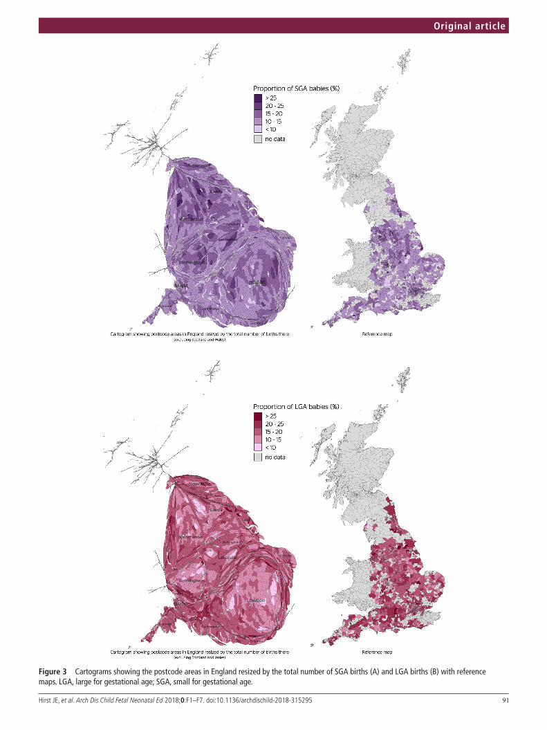
91Hirst JE, et al. Arch Dis Child Fetal Neonatal Ed 2018;0:F1–F7. doi:10.1136/archdischild-2018-315295
Original article
Figure 3 Cartograms showing the postcode areas in England resized by the total number of SGA births (A) and LGA births (B) with reference maps. LGA, large for gestational age; SGA, small for gestational age.
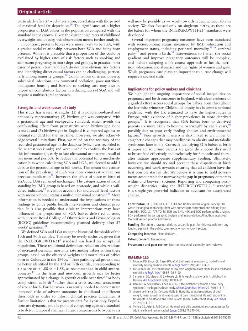
92 Hirst JE, et al. Arch Dis Child Fetal Neonatal Ed 2018;0:F1–F7. doi:10.1136/archdischild-2018-315295
Original article
particularly after 37 weeks’ gestation, correlating with the period of maximal fetal fat deposition.26 The significance of a higher proportion of LGA babies in the population compared with the standard is not known. Given the current high rates of childhood overweight and obesity, this observation merits further analysis.
In contrast, preterm babies were more likely to be SGA, with a graded social relationship between both SGA and being born preterm. While it is plausible that a proportion of this could be explained by higher rates of risk factors such as smoking and adolescent pregnancy in more deprived groups, in practice, most cases of preterm birth and SGA do not have obvious risk factors and identifying direct causal factors can be challenging, particu-larly among minority groups.27 Combinations of stress, poverty, subclinical infections, environmental pollution, poor nutrition, inadequate housing and barriers to seeking care may also be important contributory factors in reducing rates of SGA and will require a multisectoral approach.
strengths and weaknesses of studyThis study has several strengths: (1) it is population-based and nationally representative; (2) birthweight was compared with a gestational age and sex-specific standard, which avoids the confounding effect from prematurity when birthweight alone is used; and (3) birthweight in England is compared against an optimal standard for the first time. However, we also acknowl-edge several limitations. First, we relied on the accuracy of the recorded gestational age in the database (which was recorded to the nearest week only) and were unable to confirm the basis of this information (ie, early or late ultrasound or maternal recall of last menstrual period). To reduce the potential for a misclassifi-cation bias when calculating SGA and LGA, we elected to add 3 days to the gestational age in full weeks. This meant our estima-tion of the prevalence of LGA was more conservative than our previous publication28; however, the effect of place of birth of SGA and LGA remained unchanged. The categorisation of social standing by IMD group is based on postcode, and while a vali-dated indicator,29 it cannot account for individual level factors with socioeconomic status a multidimensional construct.30 More information is needed to understand the implications of these findings to guide public health interventions and clinical prac-tice. It is also possible that clinician interventions may have influenced the proportion of SGA babies delivered at term, with current Royal College of Obstetricians and Gynaecologists (RCOG) guidelines recommending planned delivery after 37 weeks' gestation.31
We defined SGA and LGA using the historical thresholds of the 10th and 90th centiles. This may be overly inclusive, given that the INTERGROWTH-21st standard was based on an optimal population. These traditional definitions relied on observations of increased perinatal mortality rate among babies within these groups, based on the observed weights and mortalities of babies born in Colorado in the 1960s.32 True pathological growth may be better identified by the 3rd or 97th centile, corresponding to a z-score of +1.88 or −1.88, as recommended in child anthro-pometry.14 In the fetus and newborn, growth may be better approximated by a change in growth centile over time33 or body composition at birth34 rather than a cross-sectional assessment of size at birth. Further work is urgently needed to demonstrate increased risks of adverse outcomes in childhood at different thresholds in order to inform clinical practice guidelines. A further limitation is that we present data for 1 year only. Popula-tions are dynamic, and the point of comparing against a standard is to detect temporal changes. Future comparisons between years
will now be possible as we work towards reducing inequality in society. We also focused only on singleton births, as these are the babies for whom the INTERGROWTH-21st standards were developed.
Several important pregnancy outcomes have been associated with socioeconomic status, measured by IMD, education and employment status, including perinatal mortality,35 36 cerebral palsy37 and preterm birth.38 Interventions to flatten the social gradient and improve pregnancy outcomes will be complex, and include adopting a life course approach to health, nutri-tion, education, social justice and the rights of women and girls. While pregnancy care plays an important role, true change will require a societal shift.
Implications for policy makers and cliniciansWe highlight the ongoing importance of social inequalities on pregnancy and birth outcomes in England, with clear evidence of a graded effect across social groups for babies born throughout the late third trimester. Childhood obesity has become a national epidemic, with the UK estimated to have the highest rates in Europe, with evidence of higher prevalence in more deprived groups.39 It is recognised that SGA babies born to deprived parents are more likely to become overweight by age 7 years,40 possibly due to poor early feeding choices and environmental factors.41 Poor growth in utero is also linked to a number of epigenetic changes that may predispose to obesity and metabolic syndromes later in life. Correctly identifying SGA babies at birth is important to ensure parents are given the support they need to breast feed effectively and exclusively for 6 months and there-after initiate appropriate supplementary feeding. Ultimately, however, we should try and prevent these disparities at birth occurring, and work towards ensuring that all children have the best possible start in life. We believe it is time to hold govern-ments accountable for narrowing the gap in pregnancy outcomes within and between societies. Reporting and comparing birth-weight disparities using the INTERGROWTH-21st standard is a simple yet powerful indicator to advocate for accelerated change.
Contributors JEH, SHK, HEK, ATP, EOO and JV devised the original concept. JEH wrote the original manuscript draft with subsequent conceptual and editing input from HEK, EOO, LCI, ATP, TD, JV, BDH and SHK. HEK and EOO performed the analysis. BDH performed the cartographic analysis and interpretation. All authors approved the final version prior to submission.
Funding The authors have not declared a specific grant for this research from any funding agency in the public, commercial or not-for-profit sectors.
Competing interests None declared.
Patient consent Not required.
Provenance and peer review Not commissioned; externally peer reviewed.
RefeRences 1 McIntire DD, Bloom SL, Casey BM, et al. Birth weight in relation to morbidity and
mortality among newborn infants. N Engl J Med 1999;340:1234–8. 2 McCormick MC. The contribution of low birth weight to infant mortality and childhood
morbidity. N Engl J Med 1985;312:82–90. 3 Samuelsen SO, Magnus P, Bakketeig LS. Birth weight and mortality in childhood in
Norway. Am J Epidemiol 1998;148:983–91. 4 Harville EW, Srinivasan S, Chen W, et al. Is the metabolic syndrome a small baby
syndrome?: the bogalusa heart study. Metab Syndr Relat Disord 2012;10:413–21. 5 Araújo de França GV, De Lucia Rolfe E, Horta BL, et al. Associations of birth
weight, linear growth and relative weight gain throughout life with abdominal fat depots in adulthood: the 1982 Pelotas (Brazil) birth cohort study. Int J Obes 2016;40:14–21.
6 Victora CG, Adair L, Fall C, et al. Maternal and child undernutrition: consequences for adult health and human capital. Lancet 2008;371:340–57.

93Hirst JE, et al. Arch Dis Child Fetal Neonatal Ed 2018;0:F1–F7. doi:10.1136/archdischild-2018-315295
Original article
7 Weissmann-Brenner A, Simchen MJ, Zilberberg E, et al. Maternal and neonatal outcomes of large for gestational age pregnancies. Acta Obstet Gynecol Scand 2012;91:844–9.
8 Mehta SH, Kruger M, Sokol RJ. Being too large for gestational age precedes childhood obesity in African Americans. Am J Obstet Gynecol 2011;204:265.e1–5.
9 Hawkins SS, Cole TJ, Law C, et al. An ecological systems approach to examining risk factors for early childhood overweight: findings from the UK Millennium Cohort Study. J Epidemiol Community Health 2009;63:147–55.
10 Papageorghiou AT, Ohuma EO, Altman DG, et al. International standards for fetal growth based on serial ultrasound measurements: the Fetal Growth Longitudinal Study of the INTERGROWTH-21st Project. Lancet 2014;384:869–79.
11 Villar J, Cheikh Ismail L, Victora CG, et al. International standards for newborn weight, length, and head circumference by gestational age and sex: the Newborn Cross-Sectional Study of the INTERGROWTH-21st Project. Lancet 2014;384:857–68.
12 WHO Working Group on Infant Growth. An evaluation of infant growth: the use and interpretation of anthropometry in infants. Bull World Health Organ 1995;73:165–74.
13 Villar J, Papageorghiou AT, Pang R, et al. The likeness of fetal growth and newborn size across non-isolated populations in the INTERGROWTH-21st Project: the Fetal Growth Longitudinal Study and Newborn Cross-Sectional Study. Lancet Diabetes Endocrinol 2014;2:781–92.
14 de Onis M, Garza C, Onyango AW, et al. WHO Child Growth Standards. Acta Paediatr Suppl 2006;450:1–101.
15 Villar J, Papageorghiou AT, Pang R, et al. Monitoring human growth and development: a continuum from the womb to the classroom. Am J Obstet Gynecol 2015;213:494–9.
16 Marmot M, Goldblatt P, Allen J, et al. Fair society, healthy lives (The Marmot Review): UCL Institute of Health Equity, 2010.
17 Glinianaia SV, Ghosh R, Rankin J, et al. No improvement in socioeconomic inequalities in birthweight and preterm birth over four decades: a population-based cohort study. BMC Public Health 2013;13:345.
18 Spencer N, Bambang S, Logan S, et al. Socioeconomic status and birth weight: comparison of an area-based measure with the Registrar General’s social class. J Epidemiol Community Health 1999;53:495–8.
19 Astone NM, Misra D, Lynch C. The effect of maternal socio-economic status throughout the lifespan on infant birthweight. Paediatr Perinat Epidemiol 2007;21:310–8.
20 Dibben C, Sigala M, Macfarlane A. Area deprivation, individual factors and low birth weight in England: is there evidence of an "area effect"? J Epidemiol Community Health 2006;60:1053–9.
21. The English Indices of Deprivation. Communities and Local Government Neighbourhoods, 2010. https://www. gov. uk/ government/ uploads/ system/ uploads/ attachment_ data/ file/ 6320/ 1870718. pdf
22 Hennig BD. Rediscovering the World: Map Transformations of Human and Physical Space. Heidelberg/New York/Dordrecht/London: Springer, 2013.
23 Gastner MT, Newman ME. From the cover: diffusion-based method for producing density-equalizing maps. Proc Natl Acad Sci U S A 2004;101:7499–504.
24 Anderson NH, Sadler LC, McKinlay CJD, et al. INTERGROWTH-21st vs customized birthweight standards for identification of perinatal mortality and morbidity. Am J Obstet Gynecol 2016;214:509.e1–7.
25 Liu S, Metcalfe A, León JA, et al. Evaluation of the INTERGROWTH-21st project newborn standard for use in Canada. PLoS One 2017;12:e0172910.
26 Villar J, Puglia FA, Fenton TR, et al. Body composition at birth and its relationship with neonatal anthropometric ratios: the newborn body composition study of the INTERGROWTH-21st project. Pediatr Res 2017;82:305–16.
27 Uphoff EP, Pickett KE, Wright J. Social gradients in health for Pakistani and White British women and infants in two UK birth cohorts. Ethn Health 2016;21:452–67.
28 Hirst JE, Villar J, Papageorghiou AT, et al. Preventing childhood obesity starts during pregnancy. Lancet 2015;386:1039–40.
29 Gordon D. Census based deprivation indices: their weighting and validation. J Epidemiol Community Health 1995;49:S39–44.
30 Braveman PA, Cubbin C, Egerter S, et al. Socioeconomic status in health research: one size does not fit all. JAMA 2005;294:2879–88.
31 Royal College of Obstetricians and Gynaecologists. The investigation and management of the small-for-gestational-age fetus: Green Top Guideline No. 31. 2nd Edn, 2013.
32 Battaglia FC, Lubchenco LO. A practical classification of newborn infants by weight and gestational age. J Pediatr 1967;71:159–63.
33 Sovio U, White IR, Dacey A, et al. Screening for fetal growth restriction with universal third trimester ultrasonography in nulliparous women in the Pregnancy Outcome Prediction (POP) study: a prospective cohort study. Lancet 2015;386:2089–97.
34 Carberry AE, Raynes-Greenow CH, Turner RM, et al. Is body fat percentage a better measure of undernutrition in newborns than birth weight percentiles? Pediatr Res 2013;74:730–6.
35 Hollowell J, Kurinczuk JJ, Brocklehurst P, et al. Social and ethnic inequalities in infant mortality: a perspective from the United kingdom. Semin Perinatol 2011;35:240–4.
36 Zeitlin J, Mortensen L, Prunet C, et al. Socioeconomic inequalities in stillbirth rates in Europe: measuring the gap using routine data from the Euro-Peristat Project. BMC Pregnancy Childbirth 2016;16:15.
37 Dolk H, Pattenden S, Bonellie S, et al. Socio-economic inequalities in cerebral palsy prevalence in the United Kingdom: a register-based study. Paediatr Perinat Epidemiol 2010;24:149–55.
38 Gray R, Bonellie SR, Chalmers J, et al. Social inequalities in preterm birth in Scotland 1980-2003: findings from an area-based measure of deprivation. BJOG 2008;115:82–90.
39 Ells LJ, Hancock C, Copley VR, et al. Prevalence of severe childhood obesity in England: 2006-2013. Arch Dis Child 2015;100:631–6.
40 Morgen CS, Andersen PK, Mortensen LH, et al. Socioeconomic disparities in birth weight and body mass index during infancy through age 7 years: a study within the Danish National Birth Cohort. BMJ Open 2017;7:e011781.
41 Goodell LS, Wakefield DB, Ferris AM. Rapid weight gain during the first year of life predicts obesity in 2-3 year olds from a low-income, minority population. J Community Health 2009;34:370–5.
POPULATION STUDY ARTICLE
Capturing the statewide incidence of neonatal abstinencesyndrome in real time: the West Virginia experienceAmna Umer1, Sean Loudin2, Stefan Maxwell3, Christa Lilly4, Meagan E. Stabler5, Lesley Cottrell1, Candice Hamilton1, Janine Breyel6,Christina Mullins7 and Collin John1
BACKGROUND: Neonatal abstinence syndrome (NAS) is one of the consequences at birth affecting the newborn afterdiscontinuation of prenatal drug exposure to mainly opioids. The objective of this study was to determine the extent of theproblem in the state of West Virginia (WV) using a real-time statewide surveillance system.METHODS: Project WATCH is a surveillance tool that since 1998 collects data on all infants born in the state of WV. NAS surveillanceitem was added to the tool in October 2016. This study examined all births (N= 23,667) in WV from October to December 2017. Thedata from six WV birthing facilities were audited for 1 month to evaluate how well this tool was capturing NAS data using κ-statistics.RESULTS: The 2017 annual incidence rate of NAS was 51.3 per 1000 live births per year for all births and 50.6 per 1000 live birthsper year for WV residents only. The κ-coefficient between the hospital medical records and Project WATCH data was 0.74 (95%confidence interval: 0.66–0.82) for NAS.CONCLUSION: The study provides justification to develop effective systems of care for the mother–infant dyad affected bysubstance use, especially targeting pregnant women in rural communities.
Pediatric Research _#####################_ ; https://doi.org/10.1038/s41390-018-0172-z
INTRODUCTIONNeonatal abstinence syndrome (NAS) is a multi-system withdrawalsyndrome of the newborn that presents shortly after birth when inutero exposure to illegal or prescription drugs (classically opioids)is suddenly discontinued at delivery.1,2 Some of the clinical signsthat may appear within the first few days after birth and mayinclude excessive sweating, tremors, high-pitched cry, poorfeeding, watery stools, and excessive weight loss.3,4 Negativelong-term outcomes are also evident as these children have anincreased risk of social and behavior abnormalities in early years,5
as well as poorer school performances than their peers.6
In the United States (US), the incidence rate of NAS in numberof cases per 1000 live births per year has increased from 1.2 in2000 to 3.4 in 20097 to 5.8 in 2012.8 In 2012, the estimated cost ofadmissions for infants diagnosed with NAS was nearly US$316million nationally.9 The aggregate hospital charges for NAS totaledUS$1.5 billion.8 Moreover, nearly 80% of the infants diagnosedwith NAS are born to families whose health insurance coverage isthrough state-funded Medicaid programs,7,9 thus escalating theburden on the already strained healthcare system.10
The incidence of NAS is disproportionately higher in rural countiescompared to urban counties and increasing much more rapidly in ruralareas compared to urban areas.11 There are geographical disparities inNAS rates based on Appalachian mountain regional status as well; in thestates of Kentucky and Tennessee, the rates of NAS were more in theAppalachian regions compared to the non-Appalachian regions.12,13
With the rise in the opioid epidemic nationally, the state of WestVirginia (WV) has experienced a much higher rate of substance usedue to the constellation of several socio-demographic factors thatnegatively impact health behaviors. WV is the only state that isentirely within the Appalachian mountain region and nearly half ofthe people in the state live in rural areas.14,15 WV also has the highestage-adjusted drug overdose death rate in the nation.16 A study in2009 showed that 19% of mothers used drugs or drank alcoholduring pregnancy in WV.17 Stabler et al.18 examined the 2007–2013WV Health Care Authority and Uniform Billing Data for WV and foundthat between 2007 to 2013 the incidence of NAS increased from 7.74per 1000 live birth per year to 31.56 per 1000 live birth per year.18
Most studies use administrative data based on billing andcoding from medical documentation to obtain NAS estimates,which leads to a few years of lag in presenting current NASestimates.18–21 With the steep increase of substance use in thestate of WV, this lag may lead to misrepresentation of the currentcrisis. The objective of the manuscript was to present the mostcurrent rate of NAS for WV as well as evaluate how well ProjectWATCH surveillance tool was capturing NAS data in real time forthe state. Given the rise in opioid epidemic and the parallel surgein infants with NAS nationally as well as in the state of WV, theresults of this study have the potential to contribute to addressingthis critical public health problem in one of the hardest hit statesin the country by informing policy makers as to the true extent ofthe problem in real time.
Received: 9 May 2018 Revised: 20 August 2018 Accepted: 23 August 2018
1Department of Pediatrics, School of Medicine, West Virginia University, Morgantown, WV 26506, USA; 2Department of Pediatrics, School of Medicine, Marshall University,Huntington, WV 25701, USA; 3PEDIATRIX Medical Group, Department of Pediatrics, WV School of Medicine (Charleston division), Women and Children’s Hospital, Charleston, SC,USA; 4Department of Biostatistics, School of Public Health, West Virginia University, Morgantown, WV 26506, USA; 5Department of Epidemiology, The Geisel School of Medicine atDartmouth, Hanover, NH 03755, USA; 6West Virginia Perinatal Partnership, Department of Obstetrics and Gynecology, School of Medicine, West Virginia University, Morgantown,WV 26506, USA and 7Office of Maternal, Child and Family Health, West Virginia Department of Health and Human Resources, Charleston, WV, USACorrespondence: Amna Umer ([email protected])
www.nature.com/pr
© International Pediatric Research Foundation, Inc 2018
94

METHODSThe study used data from Project WATCH (Working in Appalachiato identify at-risk infants, Critical congenital heart disease, andHearing loss). This project as a statewide mandate since 1998(House Bill 238822) focuses on collecting surveillance data onevery infant born in WV birthing hospitals/facilities. Moreinformation about this project can be found elsewhere.23 InOctober 2016, Project WATCH collaborated with The West VirginiaPerinatal Partnership and the WV Department of Health andHuman Resources to expand its surveillance tool to include real-time information on substance use during pregnancy andpresence of NAS at the time of infant discharge from the hospital.Given that Project WATCH has been used by all WV birthinghospitals for more than 20 years, the addition of a newsurveillance item to this tool that captures real-time dataappeared to be an ideal way to identify infants with NAS andaccurately estimate the extent of the problem in WV.The nurses at each WV birth facility completed the ques-
tionnaire form for Project WATCH before hospital discharge.Intrauterine substance exposure (IUSE) was assessed using severalpossible sources (self-report, documented in medical record, or/and positive drug screening test). In September 2014, WVneonatologists, pediatricians, hospital coders, and members ofthe West Virginia Perinatal Partnership met to develop astandardized definition for NAS as well as guidance fordocumenting exposure and withdrawal in newborns. The defini-tion that was agreed upon was as follows: NAS is diagnosed whena baby has intrauterine exposure to a neuro-active substance, andexhibited clinical signs of withdrawal, regardless of whether or notpharmacological treatment is required. Training sessions on thestandardized definition for NAS were conducted statewide at allbirth facilities in 2015–2016. Following the statewide trainingefforts, the IUSE and NAS surveillance items were added to theProject WATCH surveillance tool on 1 October 2016. Infants whohad IUSE were assessed for signs of NAS consistent with theagreed upon statewide definitionThis study examined all births in WV over a 15-month time
period (1 October 2016 to 31 December 2017). Nearly 16% ofinfants were born to mothers who live in the surrounding states ofKentucky, Maryland, Ohio, Pennsylvania, and Virginia, but gavebirth in WV. The data were analyzed for all births as well as birthsfor WV residents only. Within WV, the 55 counties were clusteredinto six regions. These regions are based on the sub-state regionsdefined by the 2012–2014 National Survey on Drug Use andHealth (NSDUH) conducted by the Substance Abuse and Mental
Health Services Administration (SAMHSA). These regions werecreated to understand the geographic variability of substance usewithin WV, which in turn provided vital information for policyefforts.24 Most substance use data are analyzed using theseregions, rather than using county-level data since not all countieshave the same level of resources and people cross county lines toaccess services. Regions were compared on both the prevalenceof IUSE and the incidence of NAS.25,26
One-month data collection auditSince Project WATCH started collecting data on substance use andNAS in October 2016, the research team wanted to assess thequality of data being recorded in Project WATCH database.Subsequently, a chart audit was performed on all births from sixlarge birthing hospitals in WV for the month of January 2017.Detailed and standardized review instructions were provided to allhospital personnel who were to be responsible for dataabstraction. Data abstraction was not blinded but data were de-identified before analysis. All births were reviewed to examinewhat was reported in the medical chart, which ICD-10 code wasentered and what was reported by Project WATCH. The ICD-10-CMcodes included P04.1–P04.4, P04.8, P04.9, and P96.1.27 Theκ-coefficient was calculated to examine the level of agreementbetween the hospital medical records and the data from ProjectWATCH.
RESULTSThe results showed that in the select project period there were23,667 births in the state of WV. The rate of IUSE and NAS for allbirths and for WV residents only (84%) is shown in Fig. 1. For theWV residents only, the incidence of NAS was 52.6 per 1000 livebirths per year (Fig. 1). The breakdown of the data by year 2016(3 months) and 2017 (12 months) as well as the total for the15-month period is shown in Table 1.The results of IUSE and NAS rates according to the six pre-
defined SAMHSA regions are displayed in Table 2 and Fig. 2.Region 1 had one of the highest prevalence of IUSE and thehighest incidence of NAS in the state. Similar patterns were notfound in other regions. For example, in region 4 the prevalence ofthe IUSE was the lowest, but the incidence of the NAS was thesecond highest in the state (Table 2).The information on IUSE was gathered from multiple sources
and could include more than one source per person. Of those withIUSE (n= 3350), nearly two-thirds (65.76%; confidence interval (CI):
All birthsN= 23,667
1 Oct 2016 – 31 Dec 2017(15 months)
WV residents onlyN= 19,974
NAS amongsubstance exposed
NAS amongsubstance exposed
NASN= 12575.31%
(CI: 5.03–5.60)
IUSEN= 289114.47%
(CI: 13.99–14.96)
NASN= 10515.26%
(CI: 4.95–5.57)
36.32%(CI: 34.57–38.07)
IUSEN= 335014.15%
(CI: 13.71–14.60)
37.49%(CI: 35.85–39.13)
Fig. 1 Prevalence of intra-uterine substance exposure (IUSE) and incidence of neonatal abstinence syndrome (NAS) in West Virginia from 1October 2016 to 31 December 2017 for all births (N= 23,667) and for West Virginia residents only (N= 19,974)
Capturing the statewide incidence of neonatal abstinence syndrome in real. . .A Umer et al.
95
Pediatric Research _#####################_
1234567890();,:

64.15–67.37) had a positive drug screen during their labor anddelivery hospital admission and nearly half of the women self-reported (52.72%; CI: 51.03–54.14) or had a positive substance useinformation in their past medical records (50.90%; CI: 49.20–52.59).
One-month data collection auditThere were 789 births in the six hospitals where the chart auditswere performed. This represented 52% of 1521 births in ProjectWATCH database for all birth in January 2017 in WV. Out of 133IUSE cases identified in the hospital medical records, 107 (81%)were identified as IUSE cases by the Project WATCH surveillancedata tool (κ-coefficient= 0.85 (CI: 0.83–0.91)). For the NAS data,out of 79 cases identified in the hospital medical records, 56 (71%)were identified as NAS cases by the Project WATCH surveillancetool (κ-coefficient= 0.74 (CI: 0.66–0.82). (Table 3). The ICD-10-CMcode for NAS was recorded by less than half (47.25%) of theinfants diagnosed with NAS in the combined data for the sixhospitals.
DISCUSSIONThe study used a real-time surveillance system to assess the extentof NAS in a state that has been significantly impacted by thecurrent opioid crisis. The rate of NAS was 53 cases per 1000 livebirths per year, which was much higher than rate of NAS reportedin earlier studies in 2013, that is, nearly 30 per 1000 live births peryear.18,19 This rise was expected, as national trends show asignificant increase in NAS diagnoses as well. However, based onthe discordance between coding data and Project WATCH data,there is a level of concern that the earlier rates may have beengrossly underestimated. WV is a rural Appalachian state and theresults from this study showed that NAS rate was seven timeshigher in WV than what was found in rural regions of the countryin 2013 (i.e., nearly 7 per 1000 live births per year).11 Moreover, thisstudy demonstrated that the rate of NAS in WV is one of the
highest in the country and nearly 10-fold the national estimate of5.8 per 1000 births live births per year in 2012.8
The rate of IUSE in WV was 14%, which is consistent with astudy conducted in 2009.17 Stitely et al.17 examined umbilical cordtissues of women who gave birth in 1 month in eight birthhospitals in WV and found 14% of mothers were positive for druguse. Based on the rising opioid epidemic in the state, wehypothesized that in 2017 the prevalence would be much higherthan what was observed in 2009. One of the likely reasons for theunderestimated prevalence in our study is the fact that data onIUSE was gathered through self-report, previous documentation,or drug testing at certain hospitals compared to the earlier studythat used universal umbilical cord tissue samples.17 WV does nothave a mandated maternal drug-screening program.Geographical disparities in the rates of IUSE and NAS were also
observed. The northern panhandle (region 1) has one of thehighest rates of IUSE as well as NAS incidence. The northerncentral region 4 has the lowest prevalence of IUSE, but thesecond highest NAS rates. We postulate that the inconsistency inthis particular region likely arises from a combination in obtainingaccurate drug history data from mother as well as the possibilitythat physicians in region 4 may be more conservative indiagnosing NAS. Interestingly, our regional results are notconsistent with the results from Stabler et al.,18 as they foundthe southern region 4 and region 5 had the highest rates of NASin 2013.18 Some of the reasons for this discrepancy may be, (1)the use of administrative data source (ICD-9-CM codes) comparedto the current study that uses the diagnosis made at the hospitaland recorded by the nurses before hospital discharge, and (2) thelikely steep increase of incidence between 2013 and 2017.Furthermore, in 2017 the high-intensity drug trafficking areas(HIDTAs) in WV include regions 1, 5, and 6, which may explain thehigh prevalence of IUSE and NAS in region 1, but nonetheless failsto explain the lower incidence in regions 5 and 6 in the currentstudy.28
Table 1. Prevalence of intra-substance use exposure (IUSE) and incidence of neonatal abstinence syndrome (NAS) in West Virginia by year 2016 and2017
Total 2016 Oct to Dec (3 months)(N=4,870)
2017 Jan to Dec (12 months)(N=18,787)
Total Oct 2016 to Dec 2017(15 months) (23,667)
n Percent (95% CI) n Percent (95% CI) n Percent (95% CI)
IUSE 720 14.78 (13.79 – 15.78) 2630 13.99 (13.50 – 14.49) 3350 14.15 (13.71 – 14.60)
NAS 295 6.06 (5.39 – 6.73) 962 5.12 (4.80 – 5.43) 1257 5.31 (5.03 – 5.60)
West Virginia only
(N=4131) (N=15,843) (N=19,974)
IUSE 626 15.15 (14.06–16.25) 2265 14.30 (13.75–14.84) 2891 14.47 (13.99–14.96)
NAS 249 6.03 (5.30–6.75) 820 5.06 (4.72–5.40) 1051 5.26 (4.95–5.57)
Table 2. Prevalence of intra-substance use exposure (IUSE) and incidence of neonatal abstinence syndrome (NAS) in West Virginia (October 2016 to31 Dec 2017) by SAMHSA sub-state regional classification system, N= 19,974
Regions Population frequency (%) IUSE % (95% CI) Rank NAS % (95% CI) Rank NAS % among IUSE (95% CI) Rank
1 1379 (6.92%) 18.49 (16.44–20.54) 1 8.41 (6.95–9.88) 1 45.49 (39.38–51.60) 1
2 1854 (9.30%) 15.32 (13.68–16.96) 2 4.21 (3.29–5.12) 5 27.46 (22.27–32.66) 5
3 1801 (9.04%) 14.99 (13.34–16.64) 3 2.67 (1.92–3.41) 6 17.78 (13.22–22.34) 6
4 4871 (24.44%) 13.49 (12.53–14.45) 6 5.79 (5.13–6.45) 2 42.77 (38.99–46.55) 2
5 6171 (30.96%) 13.79 (12.93–14.65) 5 5.46 (4.89–6.03) 3 39.60 (36.31–42.89) 3
6 3853 (19.33%) 14.69 (13.57–15.81) 4 4.88 (4.20–5.56) 4 33.22 (29.34–37.10) 4
Total 19,929 14.47 (13.98–14.95) 5.26 (4.95–5.57) 36.35 (34.60–38.11)
Missing 45 45 45 8
Capturing the statewide incidence of neonatal abstinence syndrome in real. . .A Umer et al.
96
Pediatric Research _#####################_

The results of the audit found that Project WATCH wascapturing the prevalence of IUSE and incidence of NAS reasonablywell. However, the agreement between the hospital chart reviewand the Project WATCH data for IUSE was higher than theagreement between the Project WATCH data and NAS. One likelyexplanation for this observation is that one of the six hospitals,which incidentally had one of the highest contributions of birth
data to the audit, ultimately contributed a higher rate of NAS,which may be reflective of biases from the chart reviewers. ProjectWATCH had already used this audit data for quality improvementof its data collection process by training nurses on data entry andreport. The audit data also revealed that only half of the infantsdiagnosed with NAS were identified utilizing the ICD-10-CM codes.Other studies have also shown that administrative data diagnosticcodes under-represent the actual number of NAS cases diagnosedin the hospital.20
Based on our results, Project WATCH appears to capture IUSEand NAS diagnoses via a novel, accurate, and cost-effectiveelectronic surveillance tool. Although these data does not includebirths delivered outside the hospital (~0.01%), Project WATCHcollects data on every infant born in WV birthing hospitals/facilities. This new data source shows improved ability to captureNAS cases compared to the traditional method of utilizingelectronic claims data via ICD codes. Additionally, Project WATCHdata are captured in real time allowing for timely surveillance of aleading public health issue in the state of WV. WV is one of a fewstates that collects surveillance data on NAS; four additional states(Florida, Georgia, Kentucky, and Tennessee) have legally man-dated NAS as a reportable condition and have a passivesurveillance system in place.29 States without a specific surveil-lance system for NAS report the condition via traditional methodssuch as diagnostic codes and hospital billing records (e.g., theState Hospital Discharge Database or State All Payer Claims data).Traditional methods used to survey the burden of NAS may have
REGION 4
REGION 3
Ohio
Kentucky
Virginia
REGION 1
IUSE–13.49%
IUSE–18.47%
NAS–5.79%
NAS–8.34%
IUSE–14.99%NAS–2.67%
REGION 5IUSE–13.79%NAS–5.46%
REGION 6IUSE–14.69%NAS–4.88%
REGION 2IUSE–15.32%NAS–4.21%
Maryland
Pennsylvania
MONONGALIA
MARSHALL
OHIO
BROOKE
HANCOCK
MARION
PRESTON
TAYLOR
WETZEL
TYLER
PLEASANTS
RITCHIEWOODDODDRIDGE
HARRISONBARBOUR
TUCKER
RANDOLPH
PENDLETON
HARDY
GRANT
MINERALHAMPSHIRE
MORGAN
BERKELEY
JEFFERSON
UPSHUR
LEWISGILMER
CALHOUNROANE
JACKSON
MASON
WIRT
CLAY
KANAWHA
PUTNAM
CABELL
WAYNE LINCOLN
BOONE
LOGAN
WYOMING
RALEIGH
FAYETTE GREENBRIER
Birthing hospitals/facilities in West Virginia
NICHOLAS
WEBSTER
BRAXTON
POCAHONTAS
SUMMERS MONROE
MERCERMCDOWELL
MINGO
Fig. 2 Project WATCH surveillance data on prevalence of intra-uterine substance exposure (IUSE) and neonatal abstinence syndrome (NAS) inWest Virginia by SAMHSA sub-state regional classification system, N= 20,002
Table 3. One-month (January 2017) audit data for six hospitals in thestate of WV comparing the project WATCH data with the hospitalmedical records and the ICD-10-CM codes (N= 789)
Data source Frequency Percent (95% CI)
Intrauterine substance exposure
Hospital data 134 17.07 (14.36–19.60)
Birth Score data 111 14.14 (11.64–16.49)
ICD-10-CM code in chart—code P04.x
57 7.23 (5.42–9.03)
Neonatal abstinence syndrome
Hospital data 79 10.1 (7.92–12.11)
Birth Score data 67 8.55 (6.55–10.44)
ICD-10-CM code in chart—codeP96.1
43 5.46 (3.87–7.03)
Capturing the statewide incidence of neonatal abstinence syndrome in real. . .A Umer et al.
97
Pediatric Research _#####################_
several months of lag from data collection to analysis.18–21 A real-time estimation of NAS has the potential to guide targetedresources and interventions in a timely manner.Some of the limitations include the fact that Project WATCH
does not gather information on the type(s) of specific substancesthat the infant is exposed to in utero. It is likely that many of theinfants in this study were exposed to multiple substancessimultaneously. Although the effect of polysubstance use on theoccurrence of NAS is controversial,30 results from a largepopulation-based cohort study showed significant increases inthe risk of NAS associated with opioid use along with co-exposureto antidepressants, benzodiazepines, and gabapentin comparedwith opioids alone.31 However, given the main purpose of thestudy was to document the incidence of NAS, this was not felt tobe a significant limitation. Moreover, despite a standardizedstatewide definition of NAS, there is a degree of subjectivity andindividual variability in approaches when making NAS diagnosisand these differences in identification approaches are difficult toassess.32 Data collection for Project WATCH started in October2016 and therefore cannot be retrospectively compared totraditional hospital billing data. Future research comparing ProjectWATCH and hospital billing data utilizing ICD codes is warranted.Moreover, non-punitive policies that enable early identification ofNAS, improve access to comprehensive prenatal care and opioidreplacement therapy, and increase funding for child welfaresystems are also warranted. Additional resources are needed,especially to those sub-state regions with the highest rates ofsubstance use and NAS. In addition, other states may benefit froma similar NAS surveillance system used by Project WATCH in WV.
ACKNOWLEDGEMENTSThe West Virginia WATCH/Birth Score Program is funded under an agreement withthe West Virginia Department of Health and Human Resources, Bureau for PublicHealth, Office of Maternal, Child, and Family Health.
ADDITIONAL INFORMATIONCompeting interests: The authors declare no competing interests.
Publisher's note: Springer Nature remains neutral with regard to jurisdictional claimsin published maps and institutional affiliations.
REFERENCES1. Kocherlakota, P. Neonatal abstinence syndrome. Pediatrics 134, e547–e561
(2014).2. Convertino, I. et al. Neonatal adaptation issues after maternal exposure to pre-
scription drugs: withdrawal syndromes and residual pharmacological effects.Drug Saf. 39, 903–924 (2016).
3. Logan, B. A., Brown, M. S. & Hayes, M. J. Neonatal abstinence syndrome: treat-ment and pediatric outcomes. Clin. Obstet. Gynecol. 56, 186–192 (2013).
4. McQueen, K. & Murphy-Oikonen, J. Neonatal abstinence syndrome. N. Engl. J. Med375, 2468–2479 (2016).
5. Klinger, G. et al. Long-term outcome following selective serotonin reuptakeinhibitor induced neonatal abstinence syndrome. J. Perinatol. 31, 615–620 (2011).
6. Oei, J. L. et al. Neonatal abstinence syndrome and high school performance.Pediatrics 139, e20162651 (2017).
7. Patrick, S. W. et al. Neonatal abstinence syndrome and associated health careexpenditures: United States, 2000–2009. JAMA 307, 1934–1940 (2012).
8. Patrick, S. W., Davis, M. M., Lehmann, C. U. & Cooper, W. O. Increasing incidenceand geographic distribution of neonatal abstinence syndrome: United States2009 to 2012. J. Perinatol. 35, 650–655 (2015).
9. Corr, T. E. & Hollenbeak, C. S. The economic burden of neonatal abstinencesyndrome in the United States. Addiction 112, 1590–1599 (2017).
10. Ridic, G., Gleason, S. & Ridic, O. Comparisons of health care systems in the UnitedStates, Germany and Canada. Mater. Sociomed. 24, 112–120 (2012).
11. Villapiano, N. L., Winkelman, T. N., Kozhimannil, K. B., Davis, M. M. & Patrick, S. W.Rural and urban differences in neonatal abstinence syndrome and maternalopioid use, 2004 to 2013. JAMA Pediatr. 171, 194–196 (2017).
12. Brown, J. D., Goodin, A. J. & Talbert, J. C. Rural and appalachian disparities inneonatal abstinence syndrome incidence and access to opioid abuse treatment.J. Rural Health 34, 6–13 (2017).
13. Warren, M. D. et al. Implementation of a statewide surveillance system for neo-natal abstinence syndrome—Tennessee, 2013. Morb. Mortal. Wkly Rep. 64,125–128 (2015).
14. Kaiser Family Foundation. KFF Medicaid’s Role in West Virginia (Kaiser FamilyFoundation, Washington, 2018).
15. Griffith, B. N., Lovett, G. D., Pyle, D. N. & Miller, W. C. Self-rated health in ruralAppalachia: health perceptions are incongruent with health status and healthbehaviors. BMC Public Health 11, 229 (2011).
16. Hedegaard, H., Warner, M. & Minino, A. M. Drug overdose deaths in the UnitedStates, 1999–2015. NCHS Data Brief 1–8 (2017).
17. Stitely, M. L., Calhoun, B., Maxwell, S., Nerhood, R. & Chaffin, D. Prevalence of druguse in pregnant West Virginia patients. W V Med. J. 106, 48–52 (2010).
18. Stabler, M. E. et al. Neonatal abstinence syndrome in West Virginia SubstateRegions, 2007–2013. J. Rural Health 33, 92–101 (2017).
19. Ko, J. Y. et al. Incidence of neonatal abstinence syndrome—28 States, 1999–2013.Morb. Mortal. Wkly Rep. 65, 799–802 (2016).
20. Burns, L. & Mattick, R. P. Using population data to examine the prevalence andcorrelates of neonatal abstinence syndrome. Drug Alcohol Rev. 26, 487–492(2007).
21. Traylor, J., Miller, A. & Warren, M. D. Evaluation of the web-based neonatalabstinence syndrome surveillance system in Tennessee. In Conference: 2014Council of State and Territorial Epidemiologists Annual Conference (2014). https://www.wvdhhr.org/birthscore/.
22. Article 22B. Birth Score Program (1998). https://www.wvdhhr.org/birthscore/.23. Mullett, M. D., Britton, C. M., John, C. & Hamilton, C. W. WV birth score: maternal
smoking and drugs of abuse. W V Med. J. 106, 16–18, 20 (2010).24. Zhang, Z., Infante, A., Meit, M. & English, N. An Analysis of Mental Health and
Substance Abuse Disparities and Access to Treatment Services in the AppalachianRegion. CHAPTER 2: Substance Use, Mental Disorders, and Access to TreatmentServices in Household Surveys (2008).
25. The Governor’s Initiative on Substance Abuse (2017). http://governorssubstanceabusetaskforceswv.com.
26. SAMHSA 2016 State Reports From the 2014 National Survey on Drug Use andHealth (NSDUH). https://www.samhsa.gov/data/nsduh/reports-detailed-tables-2016-NSDUH.
27. ICD-10-CM Codes. (2018). https://www.icd10data.com/ICD10CM/Codes.28. HIDTA 2017 High Intensity Drug Trafficking Areas Program (HIDTA), ONDCP.
Available at https://www.dea.gov/ops/HIDTA_2017_map.pdf. Accessed on 21 Jan2018.
29. Ko, J. Y. et al. CDC grand rounds: public health strategies to prevent neonatalabstinence syndrome. Morb. Mortal. Wkly Rep. 66, 242–245 (2017).
30. American Academy of Pediatrics Committee on Drugs. Neonatal drug with-drawal. Pediatrics 101,1079–1088 (1998).
31. Huybrechts, K. F. et al. Risk of neonatal drug withdrawal after intrauterine co-exposure to opioids and psychotropic medications: cohort study. BMJ 358, j3326(2017).
32. Sarkar, S. & Donn, S. M. Management of neonatal abstinence syndrome in neo-natal intensive care units: a national survey. J. Perinatol. 26, 15–17 (2006).
Capturing the statewide incidence of neonatal abstinence syndrome in real. . .A Umer et al.
98
Pediatric Research _#####################_

Available online at www.sciencedirect.com
Seminars in Perinatology
www.seminperinat.com
99
Modulators of inflammation in
Bronchopulmonary DysplasiaD1X XRashmin C. Savani D2X X
Center for Pulmonary & Vascular Biology, Division of Neonatal-Perinatal Medicine, The Department of Pediatrics, The University of Texas
Southwestern Medical Center, 5323 Harry Hines Boulevard, Dallas, TX 75390-9063, USA
E-mail address: rashmin.savani@utsout
https://doi.org/10.1053/j.semperi.2018.09.0090146-0005/� 2018 Elsevier Inc. All rights reser
A B S T R A C T
Over 50 years after its first description, Bronchopulmonary Dysplasia (BPD) remains a dev-
astating pulmonary complication in preterm infants with respiratory failure and develops
in 30�50% of infants less than 1000-gram birth weight. It is thought to involve ventilator-
and oxygen-induced damage to an immature lung that results in an inflammatory response
and ends in aberrant lung development with dysregulated angiogenesis and alveolariza-
tion. Significant morbidity and mortality are associated with this most common chronic
lung disease of childhood. Thus, any therapies that decrease the incidence or severity of
this condition would have significant impact on morbidity, mortality, human costs, and
healthcare expenditure. It is clear that an inflammatory response and the elaboration of
growth factors and cytokines are associated with the development of BPD. Numerous
approaches to control the inflammatory process leading to the development of BPD have
been attempted. This review will examine the anti-inflammatory approaches that are
established or hold promise for the prevention or treatment of BPD.
� 2018 Elsevier Inc. All rights reserved.
A R T I C L E I N F O
microvascular development is critical to normal secondary
Introduction
Bronchopulmonary Dysplasia is a complex condition that has
a multifactorial pathogenesis. Key contributors include oxy-
gen toxicity, ventilator-induced lung injury, inflammation
and an arrest of lung vascular development.1,2 Inflammation,
an accumulation of neutrophils and macrophages into the
injured preterm lung, results in the elaboration of pro-inflam-
matory and pro-fibrotic growth factors, an imbalance of the
proteolytic activity in the lung and increased vascular perme-
ability.3 Neutrophil and macrophage accumulation commen-
ces early in the first week of life and counts remain elevated
in infants that develop BPD.4 Coordinated development of the
distal epithelial and vascular compartments is a necessity for
the final ability to conduct adequate gas exchange. Lung
hwestern.edu
ved.
septation leading to alveolar formation. Multiple animal mod-
els and examination of autopsy samples of infants with BPD
have confirmed an arrest of vascular development and a sim-
plification of the distal lung architecture as a result of preterm
birth and associated lung injury.5 Indeed, alveolar septation
can be preserved in animal models by decreasing the effects
of contributors to BPD pathogenesis, including inflammation,
oxygen and mechanical ventilation.6 The pathogenesis of
BPD involves the effects of these multiple pathologic mecha-
nisms on lung development.
A number of models of BPD have been developed. Two large
animal models, the sheep and baboon preterm birth and ven-
tilation models, recapitulate the essential features of the
human condition, including preterm birth, oxygen exposure,
mechanical ventilation, inflammation and arrested lung

100 S E M I N A R S I N P E R I N A T O L O G Y 4 2 ( 2 0 1 8 )
development.7 However, these models do not support genetic
studies to identify the molecular determinants of BPD. There-
fore, rodent models of newborn lung injury, that involve
either hyperoxia or hypoxia exposure for up to 14 days and
result in pathology that mimics BPD, were established.8�10
Further, a model in which bleomycin, administered intra-
peritoneally to neonatal rodents, also recapitulates the fea-
tures of BPD.11 While these rodent models do not involve pre-
term birth or ventilation, several groups have confirmed
changes in inflammation, cytokines, oxidative stress, and
vascular defects in association with alveolar simplification
that validate the use of these models in the study of BPD.8�11
The fact that alveolar development is entirely a postnatal
event occurring in the first 2�3 weeks of life in rodents, pro-
vides an appropriate window to examine the effects of injury
on the alveolar phase of the developing lung. More recently,
the use of high oxygen exposures during the first 14 days of
life has been questioned since human preterm infants are
exposed to these conditions only during the saccular phase of
lung development.12,13 Thus, more appropriate oxygen expo-
sure in the first four to five days of life in rodents have been
developed and also demonstrate inflammatory changes and
alveolar defects.14,15
The recruitment of inflammatory cells to the injured lung
requires activation of bone marrow-derived cells, circulating
leukocytes and the endothelium. Initiation of injury results in
initial rolling, then firm adhesion of inflammatory cells to the
activated endothelium. This is followed by transmigration of
leukocytes through the endothelium and their accumulation
in areas of injury. Initially recruited neutrophils undergo apo-
ptosis and alveolar macrophages engulf these cells. This pro-
cess results in the production of active transforming growth
factor beta (TGFß) which promotes fibrosis. A number of key
molecules have been identified in the process of inflammatory
cell recruitment and include selectins, integrins, cytokines and
growth factors, the innate immune system including Toll-Like
Receptors (TLRs), Surfactant Protein A (SPA) and SPD, as well
as components of the extracellular matrix.16,17 Macrophage
subsets demonstrating differential functions have been identi-
fied. Classically activated macrophages (CAMs), also called M1
macrophages, result from TLR stimulation, bacterial infection
and interferon-gamma stimulation. Alternatively activated
macrophages (AAMs), also called M2macrophages, result from
Th2 cytokine (e.g. interleukin or IL4 and IL13) stimulation.18
Specific markers for M1 and M2 macrophages have emerged.
For instance, M1 macrophages can be identified by the expres-
sion of inducible nitric oxide synthase (iNOS) and CD11c,
whereas M2 macrophages can be identified by the expression
of Arginase 1 or YM1 (also known as chitinase-like 3 or
Chil3).19,20 While M1 macrophages have potent microbicidal
properties and promote Th1 responses, M2 macrophages play
a role in the resolution of inflammation, with high endocytic
clearance capacity and reduced pro-inflammatory cytokine
secretion. Given the large predominance of inflammation in
the pathogenesis of BPD, considerable efforts have been made
to develop anti-inflammatory strategies, ranging from gluco-
corticoids to newer more novel therapeutic targeting of com-
ponents of the innate immune system. The pathogenesis of
BPD and the multiple anti-inflammatory strategies discussed
in this review are summarized in Fig. 1.
Non-invasive respiratory support
The contribution of intubation and mechanical ventilation to
inflammation and the incidence and severity of BPD has been
established in animal models and in human preterm
infants.21,22 Mouse and lambmodels show thatmechanical ven-
tilation alone without hyperoxia or lipopolysaccharide (LPS)
exposure is sufficient for the development of lung inflamma-
tion, as well as altered alveolarization and lung function.23,24
Indeed, respiratory management with continuous positive air-
way pressure (CPAP) in baboons and mice, or high frequency,
non-invasive ventilation in lambs is associatedwith less inflam-
mation and better alveolarization.25�27 In large clinical trials, the
incidence of BPD in infants randomized to either non-invasive
respiratory support or selective intubation and surfactant ther-
apy were found to be equivalent, findings that have pushed
changes in neonatal practice significantly in favor of non-inva-
sive approaches to respiratorymanagement.28�30
Corticosteroids
First described in sheep in 196931 and in a human trial in 1972,
antenatal glucocorticoid therapy to enhance fetal lung maturity
was associated with decreased incidence of Respiratory Distress
Syndrome (RDS) and death.32 Indeed, this therapy Is also associ-
ated with decreased intraventricular hemorrhage (IVH), necrotiz-
ing enterocolitis (NEC) and early sepsis.33 Given the early
descriptions of inflammation in infants with BPD, postnatal glu-
cocorticoid therapy to decrease or abrogate the inflammatory
response, improve lung function and increase the likelihood of
extubation failure has been examined since the early 1970’s.
While benefits of early (in the first week of life) corticosteroid
therapy in preterm infants at risk of developing BPD include
decreased incidence of BPD at 28 days and at 36 weeks post-
menstrual age (PMA), decreased rates of failure of extubation and
decreased incidence of patent ductus arteriosus (PDA) and reti-
nopathy of prematurity (ROP), significant adverse events have
been noted including hyperglycemia, hypertension, hypertrophic
cardiomyopathy, growth failure, intestinal perforation and bleed-
ing, as well as increased incidence of cerebral palsy.34,35 The use
of postnatal steroids in infants that remain intubated past 7�14
days of life has also been reviewed recently.36 Similar benefits
with respect to decreased rates of death or BPD at 28 days and 36
weeks PMA and adverse outcomes including hyperglycemia and
hypertension were found, but longer term cerebral palsy or neu-
rodevelopmental complications were not significantly different
from those of controls.35 However, given the considerable
adverse effects described, the current recommendations are for
judicious use of postnatal corticosteroid therapy. Further,
although difficult to demonstrate in preterm infants, dexametha-
sone treatment of newborn rodents is associated with an arrest
of alveolarization that persists into adulthood.37�39
Caffeine
The methylxanthines caffeine, aminophylline and theophylline
bind to adenosine receptors and act as non-selective adenosine
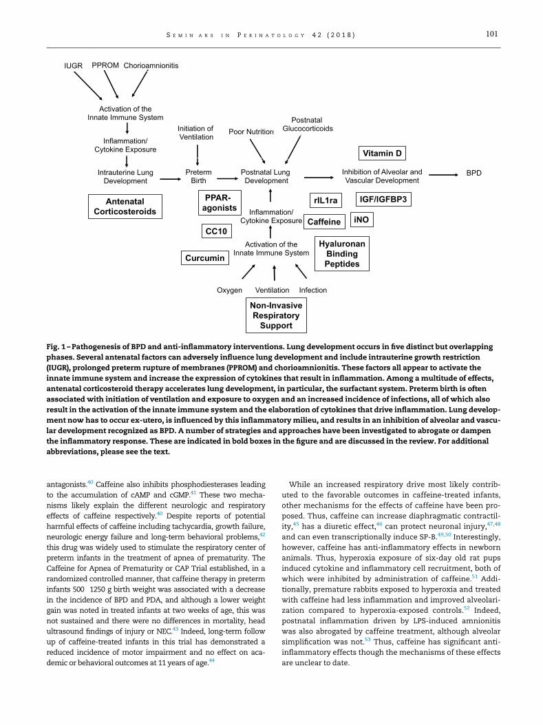
Poor NutritionPostnatal
GlucocorticoidsInitiation ofVentilation
Inhibition of Alveolar andVascular Development
Inflammation/Cytokine Exposure
VentilationOxygen Infection
IUGR Chorioamnionitis
AntenatalCorticosteroids
Intrauterine LungDevelopment
PretermBirth
Postnatal LungDevelopment
PPROM
Activation of theInnate Immune System
Activation of theInnate Immune System
Inflammation/Cytokine Exposure
BPD
Non-InvasiveRespiratorySupport
Caffeine iNO
Vitamin D
PPAR-agonists
CC10
IGF/IGFBP3rIL1ra
HyaluronanBindingPeptidesCurcumin
Fig. 1 –Pathogenesis of BPD and anti-inflammatory interventions. Lung development occurs in five distinct but overlapping
phases. Several antenatal factors can adversely influence lung development and include intrauterine growth restriction
(IUGR), prolonged preterm rupture of membranes (PPROM) and chorioamnionitis. These factors all appear to activate the
innate immune system and increase the expression of cytokines that result in inflammation. Among amultitude of effects,
antenatal corticosteroid therapy accelerates lung development, in particular, the surfactant system. Preterm birth is often
associated with initiation of ventilation and exposure to oxygen and an increased incidence of infections, all of which also
result in the activation of the innate immune system and the elaboration of cytokines that drive inflammation. Lung develop-
ment now has to occur ex-utero, is influenced by this inflammatorymilieu, and results in an inhibition of alveolar and vascu-
lar development recognized as BPD. A number of strategies and approaches have been investigated to abrogate or dampen
the inflammatory response. These are indicated in bold boxes in the figure and are discussed in the review. For additional
abbreviations, please see the text.
T AGGEDENDS E M I N A R S I N P E R I N A T O L O G Y 4 2 ( 2 0 1 8 ) 101
antagonists.40 Caffeine also inhibits phosphodiesterases leading
to the accumulation of cAMP and cGMP.41 These two mecha-
nisms likely explain the different neurologic and respiratory
effects of caffeine respectively.40 Despite reports of potential
harmful effects of caffeine including tachycardia, growth failure,
neurologic energy failure and long-term behavioral problems,42
this drug was widely used to stimulate the respiratory center of
preterm infants in the treatment of apnea of prematurity. The
Caffeine for Apnea of Prematurity or CAP Trial established, in a
randomized controlled manner, that caffeine therapy in preterm
infants 500�1250 g birth weight was associated with a decrease
in the incidence of BPD and PDA, and although a lower weight
gain was noted in treated infants at two weeks of age, this was
not sustained and there were no differences in mortality, head
ultrasound findings of injury or NEC.43 Indeed, long-term follow
up of caffeine-treated infants in this trial has demonstrated a
reduced incidence of motor impairment and no effect on aca-
demic or behavioral outcomes at 11 years of age.44
While an increased respiratory drive most likely contrib-
uted to the favorable outcomes in caffeine-treated infants,
other mechanisms for the effects of caffeine have been pro-
posed. Thus, caffeine can increase diaphragmatic contractil-
ity,45 has a diuretic effect,46 can protect neuronal injury,47,48
and can even transcriptionally induce SP-B.49,50 Interestingly,
however, caffeine has anti-inflammatory effects in newborn
animals. Thus, hyperoxia exposure of six-day old rat pups
induced cytokine and inflammatory cell recruitment, both of
which were inhibited by administration of caffeine.51 Addi-
tionally, premature rabbits exposed to hyperoxia and treated
with caffeine had less inflammation and improved alveolari-
zation compared to hyperoxia-exposed controls.52 Indeed,
postnatal inflammation driven by LPS-induced amnionitis
was also abrogated by caffeine treatment, although alveolar
simplification was not.53 Thus, caffeine has significant anti-
inflammatory effects though the mechanisms of these effects
are unclear to date.
102 S E M I N A R S I N P E R I N A T O L O G Y 4 2 ( 2 0 1 8 )
Nitric oxide (NO)
NO, generated by nitric oxide synthases (neuronal or nNOS,
endothelial or eNOS and iNOS) upon the conversion of L-argi-
nine to L-citrulline, plays a key role in developmental and
physiological processes in the lung.54,55 Studies in the mature
airway indicate that the epithelium is the primary source of
NO56 and that NO mediates neurotransmission, smooth mus-
cle relaxation, bacteriostasis, ciliary motility, mucin secre-
tion, and plasma exudation.54,55,57 Elegant in vitro studies
from the Shaul/Mineo laboratories implicate NO in anti-
inflammatory processes such as inhibition of monocyte adhe-
sion to activated endothelium and platelet aggregation.58,59
Indeed, inhaled NO (iNO) attenuates pulmonary inflamma-
tion in a neonatal rat hyperoxia model.60
NO plays an important role in pulmonary vascularization,
alveolarization, and airway branching.61�66 NO also has a
well-recognized role in mediating pulmonary vasomotor tone
around the time of birth.67 In studies of lungs from fetal
baboons, pulmonary NOS expression is upregulated during
normal fetal development. However, in the baboon model of
BPD, expression of nNOS and eNOS was significantly
decreased, and iNO given at 5ppm improved pulmonary
artery pressures, pulmonary mechanics, lung elastin deposi-
tion and was associated with modest increases in secondary
septation.68 Based on these and other animal and early stage
human studies,22,64,69,70 multiple trials of iNO therapy have
been performed in human preterm infants to prevent
BPD.71�74 These have yielded mixed results and the use of
iNO is not currently recommended for the prevention of BPD.
Vitamin D
Vitamin D or cholecalciferol is a fat-soluble vitamin that is syn-
thesized from 7-dehydrocholesterol in the skin after ultraviolet
B exposure or absorbed from the diet. Vitamin D is converted
to 25-hydroxy-vitamin D in the liver and to the biologically
active 1,25-hydroxy-vitamin D or calcitriol in the kidney. The
effects of 1,25-OH-vitamin D are mediated by a nuclear recep-
tor, the vitamin D receptor (VDR) that binds to Vitamin D-
responsive elements to regulate the transcription of a wide
number of genes. Vitamin D deficiency is highly prevalent, par-
ticularly in pregnancy and has been associated with a number
of adverse outcomes such as poor maternal weight gain, pre-
eclampsia, increased rates of operative delivery and shorter
gestation.75 In addition, the impact of vitamin D deficiency on
lung development and health is evident in VDR knockout mice
that have an imbalance of matrix metalloproteinases and
develop emphysema and inflammation.76,77
More recently, direct demonstration of the importance of
Vitamin D in pregnancy and BPD has been obtained from
experiments in which antenatal Vitamin D supplementation
was associated with improved placental vascularization and
fetal growth.78 Postnatal Vitamin D supplementation was
associated with an improvement in alveolarization in rats
that were exposed to intra-amniotic LPS,79 and Vitamin D
induced downregulation of TLR4 and therefore decreased
downstream cytokines such as IL1ß in neonatal rats exposed
to hyperoxia.80 The effects of Vitamin D on the activation of
eNOS likely contribute to the beneficial observations with
Vitamin D both on the placenta and postnatal vascular and
alveolar growth.81 Interestingly, a recent report demonstrated
beneficial effects of nebulized Vitamin D to enhance lung
maturation.82
Peroxisome proliferator-activated receptor gamma(PPAR-g) agonists
PPAR-g is a key anti-inflammatory nuclear hormone receptor
that regulates a wide array of downstream effects.83 When
ligands bind PPAR-g, the receptor dimerizes with retinoid X
receptors (RXRs) and this heterodimer translocates to the
nucleus, binding to PPAR response elements of target genes
that stimulate the transcription of genes regulating key path-
ways, including lipid metabolism, glucose homeostasis and
cell differentiation. A number of ligands for PPAR-g have been
described and include nitrated fatty acids and anti-diabetic
glitazones.84,85 Importantly, PPAR-g ligands inhibit transcrip-
tion factors such as nuclear factor kappa-light-chain-
enhancer of activated B cells (NFkB) and signal transducer
and activator of transcription 1 (STAT1), and decrease the
induction of pro-inflammatory molecules such as iNOS.86,87
Thus, a key feature of PPAR-g signaling is the inactivation of
macrophages. Proof of concept that PPAR-g agonists are use-
ful therapeutic agents in models of neonatal lung injury
include prevention of aberrant TGFß and Wnt signaling,
dampening of inflammation and improvement of secondary
septation changes in models of hyperoxia-mediated neonatal
lung injury.88,89 Indeed, antenatally administered and postna-
tally nebulized PPAR-g agonists have also shown promise in
preventing the changes seen in hyperoxia-induced neonatal
lung injury.90�92
Curcumin
Curcumin or diferuloylmethane, a yellow pigment found in
turmeric that is obtained from the turmeric plant Curcuma
longa, is a dietary spice used in Indian curry and in Ayurvedic
medicine.93 Curcumin has been evaluated in a wide range of
pulmonary diseases including chronic obstructive pulmonary
disease (COPD), acute lung injury and fibrosis.94�96 A wide
range of molecular actions have been ascribed to curcumin
but, in the case of lung inflammatory diseases, they largely fall
into three categories. First, it blocks activation of NFkB, a key
transcription factor that activates inflammatory pathways,97 it
acts as an antioxidant targeting superoxide among others,98
and inhibits key growth factors such as TGFß that contribute to
fibrosis.99 Another proposed mechanism, that curcumin acts
as a PPAR-g agonist, remains controversial.100,101
Curcumin has been evaluated in neonatal hyperoxia mod-
els of BPD.102�104 Neonatal hyperoxia in rodents is associated
with increased expression of pro-inflammatory cytokines and
growth factors, an inflammatory response, increased myofi-
broblasts and decreased alveolarization. Curcumin effectively
reverses these observations and promotes improved lung
architecture.102�104 The major issue with the development of

T AGGEDENDS E M I N A R S I N P E R I N A T O L O G Y 4 2 ( 2 0 1 8 ) 103
curcumin as a drug is its poor solubility, poor bioavailability
and rapid metabolism.105 A number of alternate approaches
have been explored, including co-administration of piperine
to enhance bioavailability, and the development of nanoparti-
cle delivery devices.106,107
Club cell protein 16 (CC16; Clara cell or CC10)
CC10, also known as uteroglobin, is a 10kDa homodimeric
secretory protein produced by Clara cells in the airways and
in the urogenital tract, This protein possesses a number of in
vitro anti-inflammatory properties including blocking of N-
Formyl-methionyl-leucyl—phenylalanine (fLMP)-stimulated
neutrophil and monocyte chemotaxis,108 inhibition of phos-
pholipase A2 (PLA2),109 inhibition of platelet aggregation,110
and suppression of IL2-stimulated production of tumor necro-
sis factor alpha (TNFa), IL1ß and interferon (IFN)g in lympho-
cytes.111 CC10 knockout mice exposed to hyperoxia,112,113 or
ozone,114 demonstrate an increased inflammatory response
consisting of an exaggerated pro-inflammatory cytokine pro-
file including IL1ß and an accumulation of inflammatory cells.
Given the low concentrations of CC10 that are found in tra-
cheal aspirate samples obtained from premature infants born
at 28�32 weeks, its anti-inflammatory properties, and its abil-
ity to inhibit PLA2, an enzyme that inactivates surfactant, it
was natural to propose recombinant human CC10 (rhCC10) as
a potential therapeutic agent to limit the incidence and sever-
ity of BPD.115 Safety of rhCC10 was demonstrated in several
animal models of lung injury, including neonatal pigs116, rab-
bits117 and lambs.118,119 Intratracheal administration of
rhCC10 in a Phase I trial demonstrated that, while endoge-
nous CC10 concentrations did not rise in tracheal aspirate
samples of control infants until 72 h of life, rhCC10 increased
the concentration in tracheal aspirates as early as 12 h of life
and showed systemic absorption and urinary excretion at
6�12 h of life. Treatment with rhCC10 was associated with a
lower neutrophil count, lower protein concentration and
lower IL6 concentrations.115 These encouraging results in 22
infants have prompted a larger study involving over 80
patients, but the results have yet to be published.
Insulin-like growth factor 1 (IGF-1) / IGF-bindingprotein 3 (IGFBP3)
IGF-1 is an important contributor to fetal growth and develop-
ment and low serum concentrations of IGF-1 have been
reported in premature infants in association with ROP,120
IVH,121 NEC122 and BPD.123 In fact, IGF-1 has a number of prop-
erties that define it as a reparative growth factor. For example,
IGF-1 promotes the endothelial survival signaling of vascular
endothelial growth factor (VEGF),124 and regulates glucose
metabolism in the brain.125 Interestingly, IGF-1 suppresses
neonatal immune responses to phorbol ester stimulation in
cord blood monocytes in association with decreased DNA
binding of NFkB, suggesting profound anti-inflammatory
properties.126 Collectively, these findings suggest that IGF-1
replacement in preterm neonates could hold benefit in a
number of diseases, but most likely in ROP and BPD.
Indeed, following pharmacokinetic studies of continuous
rhIGF-1 and its binding partner rhIGFBP3,127,128,129 a drug called
SHP607 (Shire Pharmaceuticals), a Phase I/IIa trial was con-
ducted to decrease the incidence and/or severity of ROP. The
results of this trial have been announced as a press release.
While this trial did not meet this goal, substantial decreases in
the severity of BPD and severe IVH were noted. Further confir-
mation of these findings is currently being pursued.
Nucleotide-binding oligomerization domain(NOD)-like receptor (NLR) family pyrin domaincontaining 3 (NLRP3) inflammasome blockade
IL1ß, a key initiator of inflammation, has a complex two-step
activation system, and has been implicated in inflammatory
disorders in humans. In the first step, endogenous danger-
associated molecular patterns (DAMPs) stimulate pattern rec-
ognition receptors (PRR) such as Toll-Like Receptors (TLRs) to
activate NFkB, resulting in transcriptional increases the
expression of pro-IL1ß.130 Further elucidation of this pathway
revealed the existence of a family of exclusively intracellular
proteins called NLRs that also bind to DAMPs.131 One such
NLR, NLRP3, forms a protein complex with the adaptor mole-
cule Apoptosis-associated Speck-like protein containing a CARD
(ASC) and pro-caspase1 to form theNLRP3 inflammasome.132,133
Thus, the second signal is the activation of the purinergic
receptor P2£ 7 by extracellular ATP that results in the forma-
tion of the NLRP3 inflammasome,134,135 a response that has
been demonstrated after hyperoxia exposure in adult mice135
and in our studies of neonatal mice exposed to hyperoxia.136
Activation of this complex cleaves pro-caspase1 to caspase1
(p20), which in turn proteolytically cleaves pro-IL1ß to pro-
duce mature IL1ß. This cytokine is released and interacts with
its receptor, IL1R, to signal inflammatory pathways. Impor-
tantly, Interleukin 1 receptor antagonist (IL1ra) is an endoge-
nous blocker of IL1R, and its recombinant form, rIL1ra is used
in monogenic disorders with elevated IL1ß as well as several
rheumatologic disorders.137,138
Elevated concentrations of IL1ß in amniotic fluid139 and
postnatally4 have been associated with the development of
BPD. Indeed, in preterm infants at risk of BPD, there is a bal-
ance of mature IL1ß to IL1ra in the first few days of life, but
the ratio of IL1ß to IL1ra is increased by days 5-7 in infants
that subsequently developed BPD.140 Further, in fetal and neo-
natal mice, conditional transgenic overexpression of IL1ß in
Clara cells, even in the absence of hyperoxia, results in
inflammation, airway remodeling and distal alveolar simplifi-
cation, changes reminiscent of BPD.141,142
A number of reports have now demonstrated at least partial
efficacy of rIL1ra treatment in neonatal rodent models of BPD.
Thus, rIL1ra treatment of neonatal rats exposed to 60% O2
resulted in decreased inflammation, improved alveolariza-
tion, and partially restored vessel density.143 Nold et al., using
a model of in utero LPS exposure followed by postnatal O2
exposure, showed that rIL1ra treatment up to 28 days of life
rescued inflammation and alveolarization at least at lower
concentrations, and less so at high concentrations of O2.144
Our own studies show that the NLRP3 inflammasome is criti-
cally involved in the neonatal hyperoxia model of BPD, and
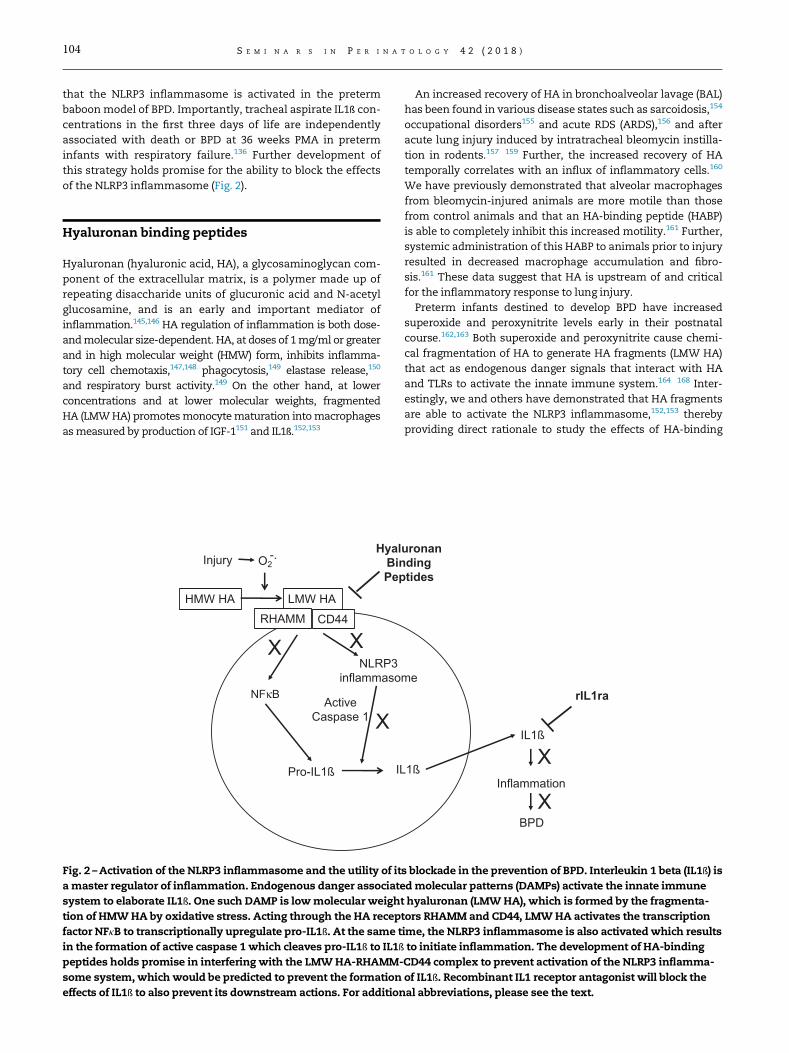
104 S E M I N A R S I N P E R I N A T O L O G Y 4 2 ( 2 0 1 8 )
that the NLRP3 inflammasome is activated in the preterm
baboonmodel of BPD. Importantly, tracheal aspirate IL1ß con-
centrations in the first three days of life are independently
associated with death or BPD at 36 weeks PMA in preterm
infants with respiratory failure.136 Further development of
this strategy holds promise for the ability to block the effects
of the NLRP3 inflammasome (Fig. 2).
Hyaluronan binding peptides
Hyaluronan (hyaluronic acid, HA), a glycosaminoglycan com-
ponent of the extracellular matrix, is a polymer made up of
repeating disaccharide units of glucuronic acid and N-acetyl
glucosamine, and is an early and important mediator of
inflammation.145,146 HA regulation of inflammation is both dose-
andmolecular size-dependent. HA, at doses of 1mg/ml or greater
and in high molecular weight (HMW) form, inhibits inflamma-
tory cell chemotaxis,147,148 phagocytosis,149 elastase release,150
and respiratory burst activity.149 On the other hand, at lower
concentrations and at lower molecular weights, fragmented
HA (LMWHA) promotesmonocytematuration intomacrophages
asmeasured by production of IGF-1151 and IL1ß.152,153
NLRP3inflammaso
LMW HAHMW HA
Injury
ActiveCaspase 1
Pro-IL1ß
CD44
HyalBinPep
RHAMM
X
X
O2-.
X
IL
Fig. 2 –Activation of the NLRP3 inflammasome and the utility of it
amaster regulator of inflammation. Endogenous danger associate
system to elaborate IL1ß. One such DAMP is lowmolecular weigh
tion of HMWHA by oxidative stress. Acting through the HA recep
factor NFkB to transcriptionally upregulate pro-IL1ß. At the same t
in the formation of active caspase 1 which cleaves pro-IL1ß to IL1ß
peptides holds promise in interfering with the LMWHA-RHAMM-
some system,which would be predicted to prevent the formation
effects of IL1ß to also prevent its downstream actions. For addition
An increased recovery of HA in bronchoalveolar lavage (BAL)
has been found in various disease states such as sarcoidosis,154
occupational disorders155 and acute RDS (ARDS),156 and after
acute lung injury induced by intratracheal bleomycin instilla-
tion in rodents.157�159 Further, the increased recovery of HA
temporally correlates with an influx of inflammatory cells.160
We have previously demonstrated that alveolar macrophages
from bleomycin-injured animals are more motile than those
from control animals and that an HA-binding peptide (HABP)
is able to completely inhibit this increased motility.161 Further,
systemic administration of this HABP to animals prior to injury
resulted in decreased macrophage accumulation and fibro-
sis.161 These data suggest that HA is upstream of and critical
for the inflammatory response to lung injury.
Preterm infants destined to develop BPD have increased
superoxide and peroxynitrite levels early in their postnatal
course.162,163 Both superoxide and peroxynitrite cause chemi-
cal fragmentation of HA to generate HA fragments (LMW HA)
that act as endogenous danger signals that interact with HA
and TLRs to activate the innate immune system.164�168 Inter-
estingly, we and others have demonstrated that HA fragments
are able to activate the NLRP3 inflammasome,152,153 thereby
providing direct rationale to study the effects of HA-binding
me
IL1ß
uronandingtides
XInflammation
BPD
1ß
rIL1ra
X
s blockade in the prevention of BPD. Interleukin 1 beta (IL1ß) is
dmolecular patterns (DAMPs) activate the innate immune
t hyaluronan (LMWHA), which is formed by the fragmenta-
tors RHAMMand CD44, LMWHA activates the transcription
ime, the NLRP3 inflammasome is also activatedwhich results
to initiate inflammation. The development of HA-binding
CD44 complex to prevent activation of the NLRP3 inflamma-
of IL1ß. Recombinant IL1 receptor antagonist will block the
al abbreviations, please see the text.
T AGGEDENDS E M I N A R S I N P E R I N A T O L O G Y 4 2 ( 2 0 1 8 ) 105
peptides inmodels of BPD, and eventually in preterm infants at
risk of developing BPD. These concepts are depicted in Fig. 2.
HA interacts with specific cell-associated receptors including
CD44 and Receptor for HA-Mediated Motility (RHAMM, CD168).
Both of these ubiquitously expressed proteins have been impli-
cated in acute lung injury. 145 CD44 expression is increased
after bleomycin injury,169,170 and acute lung injury in CD44
knockout (KO) mice results in the accumulation of inflamma-
tory cells and excess HA accumulation, suggesting that CD44 is
necessary for the resolution of inflammation.171 RHAMM is
expressed at the cell surface, in the cytoplasm and in the
nucleus, and regulates cell locomotion and proliferation.172�174
RHAMM expression is increased in macrophages,146 fibro-
blasts,175 epithelial cells,167 and smooth muscle cells174
responding to injury. After intratracheal bleomycin-induced
acute lung injury, RHAMM expression is increased in macro-
phages responding to injury, and the increased motility of
these cells is entirely dependent on RHAMM.161,176 Interest-
ingly, antibody blockade of RHAMM in vivo decreases the accu-
mulation of macrophages into the bleomycin-injured lung,
suggesting that RHAMM-HA interactions are crucial for the
recruitment of these cells to the site of injury.161,176 In order to
examine the role of RHAMM in acute lung injury using genetic
models, we recently studied the response to intratracheal bleo-
mycin in RHAMM KO mice and in mice that had transgenic
overexpression of RHAMM specifically in macrophages.177 As
expected, RHAMM KO mice had less respiratory distress,
decreased inflammatory cell accumulation and less fibrosis
than littermate controls. On the other hand, bleomycin injury
of mice with transgenic overexpression of RHAMM in macro-
phages resulted in worse respiratory distress, increased
inflammation and severe fibrosis compared to littermate wild
type animals injured with bleomycin.177 Therefore, RHAMM is
a potential novel therapeutic target to control inflammation
after lung injury. We propose that LMW HA uses RHAMM and
CD44 as a complex to allow signaling to activate the NLRP3
inflammasome (Fig. 2).
Summary
Despite over 50 years of efforts that have resulted in signifi-
cant improvements in the care of premature infants, includ-
ing antenatal glucocorticoid treatment, exogenous surfactant
administration, gentler ventilation techniques, judicious use
of fluids, prevention of infections and improved nutrition, the
incidence of BPD has remained essentially unchanged. Given
its importance in the pathogenesis of BPD, there continues to
be an intense focus on the mechanisms of inflammation in
this disorder. Significant progress has been made with the
use of animal models with promising therapies emerging.
Translational of these promising avenues into clinical use
that will decrease the incidence and/or severity of BPD will be
challenging and will require careful investigation and sub-
stantial support from public, private and commercial sources.
Conflict of interest
There are no conflicts of interest.
Acknowledgments
The author is mindful that this review is not intended to be all
encompassing and any omission of work that would be rele-
vant to the topic of this review is not intentional. The author
is a co-founder of Eravon Therapeutics, Inc. that is focused on
RHAMM-HA-based therapeutics. Funding for the studies
described that were conducted in the Savani laboratory was
from NIH R01 awards HL62868, HL62472 and HL093535, and
U01 award HL075900. Additional funding was obtained from
the William Buchanan Chair in Pediatrics and a Children’s
Hospital Foundation Dallas grant (#137).
R E F E R E N C E S
1. Ambalavanan N, Carlo WA. Bronchopulmonary dysplasia:new insights. Clin Perinatol. 2004;31(3):613–628.
2. Bhandari A, Bhandari V. Pathogenesis, pathology and patho-physiology of pulmonary sequelae of bronchopulmonarydysplasia in premature infants. Front Biosci. 2003;8:e370–e380.
3. Speer CP. Inflammation and bronchopulmonary dysplasia.Semin Neonatol. 2003;8(1):29–38.
4. Rindfleisch MS, Hasday JD, Taciak V, Boderick K, ViscardiRM. Potential role of interleukin-1 in the development ofbronchopulmonary dysplasia. J Interf Cytokine Res.1996;16:365–373.
5. Stenmark KR, Abman SH. Lung vascular development: impli-cations for the pathogenesis of bronchopulmonary dyspla-sia. Annu Rev Physiol. 2005;67:623–661.
6. Thebaud B. Angiogenesis in lung development, injury andrepair: implications for chronic lung disease of prematurity.Neonatology. 2007;91(4):291–297.
7. Coalson JJ. Experimental models of bronchopulmonary dys-plasia. Biol Neonate. 1997;71(Suppl 1):35–38.
8. Nakanishi H, Sugiura T, Streisand JB, Lonning SM, Roberts JDJr. TGF-beta-neutralizing antibodies improve pulmonaryalveologenesis and vasculogenesis in the injured newbornlung. Am J Physiol Lung Cell Mol Physiol. 2007;293(1):L151–L161.
9. ter Horst SA, Fijlstra M, Sengupta S, Walther FJ, WagenaarGT. Spatial and temporal expression of surfactant proteinsin hyperoxia-induced neonatal rat lung injury. BMC PulmMed. 2006;6:8.
10. Ambalavanan N, Nicola T, Hagood J, et al. Transforminggrowth factor-beta signaling mediates hypoxia-induced pul-monary arterial remodeling and inhibition of alveolar devel-opment in newborn mouse lung. Am J Physiol Lung Cell MolPhysiol. 2008;295(1):L86–L95.
11. Tourneux P, Markham N, Seedorf G, Balasubramaniam V,Abman SH. Inhaled nitric oxide improves lung structure andpulmonary hypertension in a model of bleomycin-inducedbronchopulmonary dysplasia in neonatal rats. Am J PhysiolLung Cell Mol Physiol. 2009;297(6):L1103–L1111.
12. Nardiello C, Mizikova I, Morty RE. Looking ahead: where tonext for animal models of bronchopulmonary dysplasia. CellTissue Res. 2017;367(3):457–468.
13. Berger J, Bhandari V. Animal models of bronchopulmonarydysplasia. The term mouse models. Am J Physiol Lung Cell MolPhysiol. 2014;307(12):L936–L947.
14. Yee M, Chess PR, McGrath-Morrow SA, et al. Neonatal oxy-gen adversely affects lung function in adult mice withoutaltering surfactant composition or activity. Am J Physiol LungCell Mol Physiol. 2009;297(4):L641–L649.

106 S E M I N A R S I N P E R I N A T O L O G Y 4 2 ( 2 0 1 8 )
15. Lingappan K, Jiang W, Wang L, Moorthy B. Sex-specific dif-ferences in neonatal hyperoxic lung injury. Am J Physiol LungCell Mol Physiol. 2016;311(2):L481–L493.
16. Ley K. Molecular mechanisms of leukocyte recruitment inthe inflammatory process. Cardiovasc Res. 1996;32(4):733–742.
17. Ley K, Laudanna C, Cybulsky MI, Nourshargh S. Getting tothe site of inflammation: the leukocyte adhesion cascadeupdated. Nat Rev Immunol. 2007;7(9):678–689.
18. Cuisset L, Jeru I, Dumont B, et al. Mutations in the autoinflam-matory cryopyrin-associated periodic syndrome gene: epide-miological study and lessons from eight years of geneticanalysis in France. Ann Rheumatic Dis. 2011;70(3):495–499.
19. Herlin T, Sorensen N, Veirum J. [Hereditary periodical feversyndromes]. Ugeskr Laeger. 2011;173(13):968–973.
20. Lachmann HJ, Quartier P, So A, Hawkins PN. The emergingrole of interleukin-1beta in autoinflammatory diseases.Arthritis Rheum. 2011;63(2):314–324.
21. Van Marter LJ, Allred EN, Pagano M, et al. Do clinical markersof barotrauma and oxygen toxicity explain interhospital var-iation in rates of chronic lung disease? The neonatologycommittee for the developmental network. Pediatrics.2000;105(6):1194–1201.
22. Bland RD. Neonatal chronic lung disease in the post-surfac-tant era. Biol Neonate. 2005;88(3):181–191.
23. Mokres LM, Parai K, Hilgendorff A, et al. Prolonged mechani-cal ventilation with air induces apoptosis and causes failureof alveolar septation and angiogenesis in lungs of newbornmice. Am J Physiol Lung Cell Mol Physiol. 2010;298(1):L23–L35.
24. Polglase GR, Hillman NH, Ball MK, et al. Lung and systemicinflammation in preterm lambs on continuous positive air-way pressure or conventional ventilation. Pediatric research.2009;65(1):67–71.
25. Thomson MA, Yoder BA, Winter VT, et al. Treatment ofimmature baboons for 28 days with early nasal continuouspositive airway pressure. Am J Respir Crit Care Med.2004;169(9):1054–1062.
26. Reyburn B, Di Fiore JM, Raffay T, et al. The effect of continu-ous positive airway pressure in a mouse model of hyperoxicneonatal lung injury. Neonatology. 2016;109(1):6–13.
27. Reyburn B, Li M, Metcalfe DB, et al. Nasal ventilation altersmesenchymal cell turnover and improves alveolarization inpreterm lambs. Am J Respir Crit Care Med. 2008;178(4):407–418.
28. Morley CJ, Davis PG, Doyle LW, et al. Nasal CPAP or intuba-tion at birth for very preterm infants. N Engl J Med.2008;358(7):700–708.
29. Network SSGotEKSNNRFiner NN, Carlo WA, et al. Early CPAPversus surfactant in extremely preterm infants. N Engl J Med.2010;362(21):1970–1979.
30. Bhandari V. Noninvasive respiratory support in the preterminfant. Clin Perinatol. 2012;39(3):497–511.
31. Liggins GC. Premature delivery of foetal lambs infused withglucocorticoids. J Endocrinol. 1969;45(4):515–523.
32. Liggins GC, Howie RN. A controlled trial of antepartum gluco-corticoid treatment for prevention of the respiratory distresssyndrome in premature infants. Pediatrics. 1972;50(4):515–525.
33. Roberts D, Brown J, Medley N, Dalziel SR. Antenatal cortico-steroids for accelerating fetal lung maturation for women atrisk of preterm birth. Cochrane Database Syst Rev. 2017;3:CD004454.
34. Doyle LW, Ehrenkranz RA, Halliday HL. Early (< 8 days) post-natal corticosteroids for preventing chronic lung disease inpreterm infants. Cochrane Database Syst Rev. 2014(5):CD001146.
35. Halliday HL. Update on postnatal steroids. Neonatology.2017;111(4):415–422.
36. Doyle LW, Ehrenkranz RA, Halliday HL. Late (>7 days) post-natal corticosteroids for chronic lung disease in preterminfants. Cochrane Database Syst Rev. 2014(5):CD001145.
37. Massaro D, Massaro GD. Invited review: pulmonary alveoli:formation, the “call for oxygen,” and other regulators. Am JPhysiol Lung Cell Mol Physiol. 2002;282(3):L345–L358.
38. Massaro D, Massaro GD. Critical period for alveologenesisand early determinants of adult pulmonary disease. Am JPhysiol Lung Cell Mol Physiol. 2004;287(4):L715–L717.
39. Garber SJ, Zhang H, Foley JP, et al. Hormonal regulation of alveo-larization: structure-function correlation. Respir Res. 2006;7:47.
40. Fredholm BB. Astra Award Lecture. Adenosine, adenosinereceptors and the actions of caffeine. Pharmacol Toxicol.1995;76(2):93–101.
41. Choi OH, Shamim MT, Padgett WL, Daly JW. Caffeine andtheophylline analogues: correlation of behavioral effectswith activity as adenosine receptor antagonists and as phos-phodiesterase inhibitors. Life Sci. 1988;43(5):387–398.
42. Millar D, Schmidt B. Controversies surrounding xanthinetherapy. Semin Neonatol. 2004;9(3):239–244.
43. Schmidt B, Roberts RS, Davis P, et al. Caffeine therapy forapnea of prematurity. N Engl J Med. 2006;354(20):2112–2121.
44. Schmidt B, Roberts RS, Anderson PJ, et al. Academic perfor-mance, motor function, and behavior 11 years after neonatalcaffeine citrate therapy for apnea of prematurity: an 11-yearfollow-up of the cap randomized clinical trial. JAMA Pediatr.2017;171(6):564–572.
45. Parikka V, Beck J, Zhai Q, Leppasalo J, Lehtonen L, Soukka H.The effect of caffeine citrate on neural breathing pattern inpreterm infants. Early Hum Dev. 2015;91(10):565–568.
46. Pacifici GM. Clinical pharmacology of theophylline in pre-term infants: effects, metabolism and pharmacokinetics.Curr Pediatr Rev. 2014;10(4):297–303.
47. Back SA, Craig A, Luo NL, et al. Protective effects of caffeineon chronic hypoxia-induced perinatal white matter injury.Ann Neurol. 2006;60(6):696–705.
48. Endesfelder S, Weichelt U, Strauss E, et al. Neuroprotectionby caffeine in hyperoxia-induced neonatal brain injury. Int JMol Sci. 2017;18(1).
49. Fehrholz M, Hutten M, Kramer BW, Speer CP, Kunzmann S.Amplification of steroid-mediated SP-B expression by physi-ological levels of caffeine. Am J Physiol Lung Cell Mol Physiol.2014;306(1):L101–L109.
50. Fehrholz M, Bersani I, Kramer BW, Speer CP, Kunzmann S.Synergistic effect of caffeine and glucocorticoids on expres-sion of surfactant protein B (SP-B) mRNA. PLoS One.2012;7(12):e51575.
51. Weichelt U, Cay R, Schmitz T, et al. Prevention of hyperoxia-mediated pulmonary inflammation in neonatal rats by caf-feine. Eur Respir J. 2013;41(4):966–973.
52. Nagatomo T, Jimenez J, Richter J, et al. Caffeine preventshyperoxia-induced functional and structural lung damage inpreterm rabbits. Neonatology. 2016;109(4):274–281.
53. Koroglu OA, MacFarlane PM, Balan KV, et al. Anti-inflamma-tory effect of caffeine is associated with improved lung func-tion after lipopolysaccharide-induced amnionitis. Neonatology.2014;106(3):235–240.
54. Barnes PJ. Nitric oxide and airway disease. Ann Med.1995;27(3):389–393.
55. Gaston B, Drazen JM, Loscalzo J, Stamler JS. The biology ofnitrogen oxides in the airways. Am J Respir Crit Care Med.1994;149(2 Pt 1):538–551.
56. Dweik RA, Laskowski D, Abu-Soud HM, et al. Nitric oxidesynthesis in the lung. Regulation by oxygen through akinetic mechanism. J Clin Invest. 1998;101(3):660–666.
57. Jain B, Rubinstein I, Robbins RA, Leise KL, Sisson JH. Modula-tion of airway epithelial cell ciliary beat frequency by nitricoxide. Biochem Biophys Res Commun. 1993;191(1):83–88.
58. Mineo C, Deguchi H, Griffin JH, Shaul PW. Endothelial andantithrombotic actions of HDL. Circ Res. 2006;98(11):1352–1364.
T AGGEDENDS E M I N A R S I N P E R I N A T O L O G Y 4 2 ( 2 0 1 8 ) 107
59. Mineo C, Shaul PW. Modulation of endothelial NO produc-tion by high-density lipoprotein. Cold Spring Harb Symp QuantBiol. 2002;67:459–469.
60. ter Horst SA, Walther FJ, Poorthuis BJ, Hiemstra PS, Wage-naar GT. Inhaled nitric oxide attenuates pulmonary inflam-mation and fibrin deposition and prolongs survival inneonatal hyperoxic lung injury. Am J Physiol Lung Cell MolPhysiol. 2007;293(1):L35–L44.
61. Balasubramaniam V, Tang JR, Maxey A, Plopper CG, AbmanSH. Mild hypoxia impairs alveolarization in the endothelialnitric oxide synthase-deficient mouse. Am J Physiol Lung CellMol Physiol. 2003;284(6):L964–L971.
62. Edwards YS, Sutherland LM, Murray AW. NO protects alveo-lar type II cells from stretch-induced apoptosis. A novel rolefor macrophages in the lung. Am J Physiol Lung Cell Mol Phys-iol. 2000;279(6):L1236–L1242.
63. Holopainen R, Aho H, Laine J, Halkola L, Kaapa P. Nitric oxideinhalation inhibits pulmonary apoptosis but not inflamma-tory injury in porcine meconium aspiration. Acta Paediatr.1999;88(10):1147–1155.
64. Tang JR, Seedorf G, Balasubramaniam V, Maxey A, MarkhamN, Abman SH. Early inhaled nitric oxide treatment decreasesapoptosis of endothelial cells in neonatal rat lungs after vas-cular endothelial growth factor inhibition. Am J Physiol LungCell Mol Physiol. 2007;293(5):L1271–L1280.
65. Tang ZL, Wasserloos KJ, Liu X, et al. Nitric oxide decreasesthe sensitivity of pulmonary endothelial cells to LPS-inducedapoptosis in a zinc-dependent fashion. Mol Cell Biochem.2002;234-235(1-2):211–217.
66. Young SL, Evans K, Eu JP. Nitric oxide modulates branchingmorphogenesis in fetal rat lung explants. Am J Physiol LungCell Mol Physiol. 2002;282(3):L379–L385.
67. Shaul PW. Nitric oxide in the developing lung. Adv Pediatr.1995;42:367–414.
68. Afshar S, Gibson LL, Yuhanna IS, et al. Pulmonary NO syn-thase expression is attenuated in a fetal baboon model ofchronic lung disease. Am J Physiol Lung Cell Mol Physiol.2003;284(5):L749–L758.
69. McCurnin DC, Pierce RA, Chang LY, et al. Inhaled NOimproves early pulmonary function and modifies lunggrowth and elastin deposition in a baboon model of neonatalchronic lung disease. Am J Physiol Lung Cell Mol Physiol.2005;288(3):L450–L459.
70. Tang JR, Markham NE, Lin YJ, et al. Inhaled nitric oxideattenuates pulmonary hypertension and improves lunggrowth in infant rats after neonatal treatment with a VEGFreceptor inhibitor. Am J Physiol Lung Cell Mol Physiol.2004;287(2):L344–L351.
71. Ballard RA, Truog WE, Cnaan A, et al. Inhaled nitric oxide inpreterm infants undergoing mechanical ventilation. N Engl JMed. 2006;355(4):343–353.
72. Kinsella JP, Cutter GR, Walsh WF, et al. Early inhaled nitricoxide therapy in premature newborns with respiratory fail-ure. N Engl J Med. 2006;355(4):354–364.
73. Schreiber MD, Gin-Mestan K, Marks JD, Huo D, Lee G, Srisu-parp P. Inhaled nitric oxide in premature infants with therespiratory distress syndrome. N Engl J Med.2003;349(22):2099–2107.
74. Van Meurs KP, Wright LL, Ehrenkranz RA, et al. Inhaled nitricoxide for premature infants with severe respiratory failure.N Engl J Med. 2005;353(1):13–22.
75. Christesen HT, Falkenberg T, Lamont RF, Jorgensen JS. Theimpact of vitamin D on pregnancy: a systematic review. ActaObstet Gynecol Scand. 2012;91(12):1357–1367.
76. Lykkedegn S, Sorensen GL, Beck-Nielsen SS, Christesen HT.The impact of vitamin D on fetal and neonatal lung matura-tion. A systematic review. Am J Physiol Lung Cell Mol Physiol.2015;308(7):L587–L602.
77. Sundar IK, Hwang JW, Wu S, Sun J, Rahman I. Deletion ofvitamin D receptor leads to premature emphysema/COPD byincreased matrix metalloproteinases and lymphoid aggre-gates formation. Biochem Biophys Res Commun.2011;406(1):127–133.
78. Cookson MW, Ryan SL, Seedorf GJ, Dodson RB, Abman SH,Mandell EW. Antenatal vitamin d preserves placental vascu-lar and fetal growth in experimental chorioamnionitis dueto intra-amniotic endotoxin exposure. Am J Perinatol. 2018.
79. Mandell E, Seedorf G, Gien J, Abman SH. Vitamin D treat-ment improves survival and infant lung structure afterintra-amniotic endotoxin exposure in rats: potential role forthe prevention of bronchopulmonary dysplasia. Am J PhysiolLung Cell Mol Physiol. 2014;306(5):L420–L428.
80. Yao L, Shi Y, Zhao X, et al. Vitamin D attenuates hyperoxia-induced lung injury through downregulation of Toll-likereceptor 4. Int J Mol Med. 2017;39(6):1403–1408.
81. Molinari C, Uberti F, Grossini E, et al. 1alpha,25-dihydroxy-cholecalciferol induces nitric oxide production in culturedendothelial cells. Cell Physiol Biochem. 2011;27(6):661–668.
82. Taylor SK, Sakurai R, Sakurai T, Rehan VK. Inhaled VitaminD: A Novel Strategy to Enhance Neonatal Lung Maturation.Lung. 2016;194(6):931–943.
83. Wang S, Dougherty EJ, Danner RL. PPARgamma signalingand emerging opportunities for improved therapeutics. Phar-macol Res. 2016;111:76–85.
84. Narala VR, Subramani PA, Narasimha VR, Shaik FB, Panati K.The role of nitrated fatty acids and peroxisome proliferator-activated receptor gamma in modulating inflammation. IntImmunopharmacol. 2014;23(1):283–287.
85. Moraes LA, Piqueras L, Bishop-Bailey D. Peroxisome prolifer-ator-activated receptors and inflammation. Pharmacol Ther.2006;110(3):371–385.
86. Ricote M, Huang JT, Welch JS, Glass CK. The peroxisome pro-liferator-activated receptor(PPARgamma) as a regulator ofmonocyte/macrophage function. J Leukoc Biol. 1999;66(5):733–739.
87. Ricote M, Li AC, Willson TM, Kelly CJ, Glass CK. The peroxi-some proliferator-activated receptor-gamma is a negativeregulator of macrophage activation. Nature.1998;391(6662):79–82.
88. Dasgupta C, Sakurai R, Wang Y, et al. Hyperoxia-inducedneonatal rat lung injury involves activation of TGF-{beta}and Wnt signaling and is protected by rosiglitazone. Am JPhysiol Lung Cell Mol Physiol. 2009;296(6):L1031–L1041.
89. Rehan VK, Wang Y, Patel S, Santos J, Torday JS. Rosiglita-zone, a peroxisome proliferator-activated receptor-gammaagonist, prevents hyperoxia-induced neonatal rat lunginjury in vivo. Pediatr Pulmonol. 2006;41(6):558–569.
90. Sakurai R, Lee C, Shen H, Waring AJ, Walther FJ, Rehan VK. Acombination of the aerosolized PPAR-gamma agonist piogli-tazone and a synthetic surfactant protein B peptide mimicprevents hyperoxia-induced neonatal lung injury in rats.Neonatology. 2018;113(4):296–304.
91. Morales E, Sakurai R, Husain S, et al. Nebulized PPARgammaagonists: a novel approach to augment neonatal lung matu-ration and injury repair in rats. Pediatr Res. 2014;75(5):631–640.
92. Rehan VK, Sakurai R, Corral J, et al. Antenatally administeredPPAR-gamma agonist rosiglitazone prevents hyperoxia-induced neonatal rat lung injury. Am J Physiol Lung Cell MolPhysiol. 2010;299(5):L672–L680.
93. Lelli D, Sahebkar A, Johnston TP, Pedone C. Curcumin use inpulmonary diseases: state of the art and future perspectives.Pharmacol Res. 2017;115:133–148.
94. Yuan J, Liu R, Ma Y, Zhang Z, Xie Z. Curcumin attenuates air-way inflammation and airway remolding by inhibiting NF-

108 S E M I N A R S I N P E R I N A T O L O G Y 4 2 ( 2 0 1 8 )
kappaB signaling and COX-2 in cigarette smoke-inducedCOPDmice. Inflammation. 2018.
95. Suresh MV, Wagner MC, Rosania GR, et al. Pulmonaryadministration of a water-soluble curcumin complexreduces severity of acute lung injury. Am J Respir Cell Mol Biol.2012;47(3):280–287.
96. Smith MR, Gangireddy SR, Narala VR, et al. Curcumin inhib-its fibrosis-related effects in IPF fibroblasts and in mice fol-lowing bleomycin-induced lung injury. Am J Physiol Lung CellMol Physiol. 2010;298(5):L616–L625.
97. Shishodia S, Potdar P, Gairola CG, Aggarwal BB. Curcumin(diferuloylmethane) down-regulates cigarette smoke-induced NF-kappaB activation through inhibition of Ikappa-Balpha kinase in human lung epithelial cells: correlationwith suppression of COX-2, MMP-9 and cyclin D1. Carcinogen-esis. 2003;24(7):1269–1279.
98. Kunwar A, Barik A, Sandur SK, Indira Priyadarsini K. Differ-ential antioxidant/pro-oxidant activity of dimethoxycurcu-min, a synthetic analogue of curcumin. Free Radic Res.2011;45(8):959–965.
99. Kumari A, Dash D, Singh R. Curcumin inhibits lipopolysac-charide (LPS)-induced endotoxemia and airway inflamma-tion through modulation of sequential release ofinflammatory mediators (TNF-alpha and TGF-beta1) inmurine model. Inflammopharmacology. 2017;25(3):329–341.
100. Lin J, Chen A. Activation of peroxisome proliferator-acti-vated receptor-gamma by curcumin blocks the signalingpathways for PDGF and EGF in hepatic stellate cells. LabInvest. 2008;88(5):529–540.
101. Narala VR, Smith MR, Adapala RK, et al. Curcumin is not aligand for peroxisome proliferator-activated receptor-gamma. Gene Ther Mol Biol. 2009;13(1):20–25.
102. Sakurai R, Li Y, Torday JS, Rehan VK. Curcumin augmentslung maturation, preventing neonatal lung injury by inhibit-ing TGF-beta signaling. Am J Physiol Lung Cell Mol Physiol.2011;301(5):L721–L730.
103. Sakurai R, Villarreal P, Husain S, et al. Curcumin protects thedeveloping lung against long-term hyperoxic injury. Am JPhysiol Lung Cell Mol Physiol. 2013;305(4):L301–L311.
104. Wei H, Zhang Y, Liu W, Zhang XP, Feng L. [Effect of curcuminon bronchopulmonary dysplasia induced by 600 ml/L oxygenin neonatal rats]. Sichuan Da Xue Xue Bao Yi Xue Ban.2014;45(2):225–229: 244.
105. Anand P, Kunnumakkara AB, Newman RA, Aggarwal BB. Bio-availability of curcumin: problems and promises. Mol Pharm.2007;4(6):807–818.
106. Shoba G, Joy D, Joseph T, Majeed M, Rajendran R, Srinivas PS.Influence of piperine on the pharmacokinetics of curcuminin animals and human volunteers. Planta Med.1998;64(4):353–356.
107. Wang W, Zhu R, Xie Q, et al. Enhanced bioavailability andefficiency of curcumin for the treatment of asthma by its for-mulation in solid lipid nanoparticles. Int J Nanomedicine.2012;7:3667–3677.
108. Vasanthakumar G, Manjunath R, Mukherjee AB, Warabi H,Schiffmann E. Inhibition of phagocyte chemotaxis by utero-globin, an inhibitor of blastocyst rejection. Biochem Pharma-col. 1988;37(3):389–394.
109. Levin SW, Butler JD, Schumacher UK, Wightman PD,Mukherjee AB. Uteroglobin inhibits phospholipase A2 activ-ity. Life Sci. 1986;38(20):1813–1819.
110. Manjunath R, Levin SW, Kumaroo KK, et al. Inhibition ofthrombin-induced platelet aggregation by uteroglobin. Bio-chem Pharmacol. 1987;36(5):741–746.
111. Dierynck I, Bernard A, Roels H, De Ley M. The human Claracell protein: biochemical and biological characterisation of anatural immunosuppressor.Mult Scler. 1996;1(6):385–387.
112. Johnston CJ, Mango GW, Finkelstein JN, Stripp BR. Alteredpulmonary response to hyperoxia in Clara cell secretory pro-tein deficient mice. Am J Respir Cell Mol Biol. 1997;17(2):147–155.
113. Reynolds SD, Mango GW, Gelein R, Boe IM, Lund J, Stripp BR.Normal function and lack of fibronectin accumulation inkidneys of Clara cell secretory protein/uteroglobin deficientmice. Am J Kidney Dis. 1999;33(3):541–551.
114. Mango GW, Johnston CJ, Reynolds SD, Finkelstein JN, PlopperCG, Stripp BR. Clara cell secretory protein deficiencyincreases oxidant stress response in conducting airways.Am J Physiol. 1998;275(2 Pt 1):L348–L356.
115. Levine CR, Gewolb IH, Allen K, et al. The safety, pharmacoki-netics, and anti-inflammatory effects of intratrachealrecombinant human Clara cell protein in premature infantswith respiratory distress syndrome. Pediatr Res. 2005;58(1):15–21.
116. Chandra S, Davis JM, Drexler S, et al. Safety and efficacy ofintratracheal recombinant human Clara cell protein in anewborn piglet model of acute lung injury. Pediatric research.2003;54(4):509–515.
117. Miller TL, Shashikant BN, Melby JM, Pilon AL, Shaffer TH,Wolfson MR. Recombinant human Clara cell secretory pro-tein in acute lung injury of the rabbit: effect of route ofadministration. Pediatr Crit Care Med. 2005;6(6):698–706.
118. Miller TL, Shashikant BN, Pilon AL, Pierce RA, Shaffer TH,Wolfson MR. Effects of recombinant Clara cell secretory pro-tein (rhCC10) on inflammatory-related matrix metalloprotei-nase activity in a preterm lamb model of neonatalrespiratory distress. Pediatr Crit Care Med. 2007;8(1):40–46.
119. Wolfson MR, Funanage VL, Kirwin SM, et al. Recombinanthuman Clara cell secretory protein treatment increases lungmRNA expression of surfactant proteins and vascular endo-thelial growth factor in a premature lamb model of respira-tory distress syndrome. Am J Perinatol. 2008;25(10):637–645.
120. Hellstrom A, Engstrom E, Hard AL, et al. Postnatal seruminsulin-like growth factor I deficiency is associated with reti-nopathy of prematurity and other complications of prema-ture birth. Pediatrics. 2003;112(5):1016–1020.
121. Hellstrom A, Ley D, Hansen-Pupp I, et al. Role of insulinlikegrowth factor 1 in fetal development and in the early postna-tal life of premature infants. Am J Perinatol. 2016;33(11):1067–1071.
122. Tian F, Liu GR, Li N, Yuan G. Insulin-like growth factor Ireduces the occurrence of necrotizing enterocolitis by reduc-ing inflammatory response and protecting intestinal muco-sal barrier in neonatal rats model. Eur Rev Med Pharmacol Sci.2017;21(20):4711–4719.
123. Lofqvist C, Hellgren G, Niklasson A, et al. Low postnatalserum IGF-I levels are associated with bronchopulmonarydysplasia (BPD). Acta Paediatr. 2012;101(12):1211–1216.
124. Hellstrom A, Perruzzi C, Ju M, et al. Low IGF-I suppressesVEGF-survival signaling in retinal endothelial cells: directcorrelation with clinical retinopathy of prematurity. ProcNatl Acad Sci U S A. 2001;98(10):5804–5808.
125. Cheng CM, Reinhardt RR, Lee WH, Joncas G, Patel SC, BondyCA. Insulin-like growth factor 1 regulates developing brainglucose metabolism. Proc Natl Acad Sci USA.2000;97(18):10236–10241.
126. Puzik A, Rupp J, Troger B, Gopel W, Herting E, Hartel C. Insu-lin-like growth factor-I regulates the neonatal immuneresponse in infection and maturation by suppression of IFN-gamma. Cytokine. 2012;60(2):369–376.
127. Chung JK, Hallberg B, Hansen-Pupp I, et al. Development andverification of a pharmacokinetic model to optimize physio-logic replacement of rhIGF-1/rhIGFBP-3 in preterm infants.Pediatr Res. 2017;81(3):504–510.

T AGGEDENDS E M I N A R S I N P E R I N A T O L O G Y 4 2 ( 2 0 1 8 ) 109
128. Hansen-Pupp I, Hellstrom A, Hamdani M, et al. Continuouslongitudinal infusion of rhIGF-1/rhIGFBP-3 in extremely pre-term infants: Evaluation of feasibility in a phase II study.Growth Horm IGF Res. 2017;36:44–51.
129. Hellstrom A, Ley D, Hallberg B, et al. IGF-1 as a drug for pre-term infants: a step-wise clinical development. Curr PharmDes. 2017;23(38):5964–5970.
130. Gasse P, Mary C, Guenon I, et al. IL-1R1/MyD88 signaling andthe inflammasome are essential in pulmonary inflammationand fibrosis in mice. J Clin Invest. 2007;117(12):3786–3799.
131. Ye Z, Ting JP. NLR, the nucleotide-binding domain leucine-rich repeat containing gene family. Curr Opin Immunol.2008;20(1):3–9.
132. Mariathasan S, Monack DM. Inflammasome adaptors andsensors: intracellular regulators of infection and inflamma-tion. Nat Rev Immunol. 2007;7(1):31–40.
133. Petrilli V, Dostert C, Muruve DA, Tschopp J. The inflamma-some: a danger sensing complex triggering innate immunity.Curr Opin Immunol. 2007;19(6):615–622.
134. Mariathasan S, Weiss DS, Newton K, et al. Cryopyrin acti-vates the inflammasome in response to toxins and ATP.Nature. 2006;440(7081):228–232.
135. Kolliputi N, Shaik RS, Waxman AB. The inflammasomemediates hyperoxia-induced alveolar cell permeability. JImmunol. 2010;184(10):5819–5826.
136. Liao J, Kapadia VS, Brown LS, et al. The NLRP3 inflamma-some is critically involved in the development of broncho-pulmonary dysplasia. Nat Commun. 2015;6:8977.
137. Goldbach-Mansky R, Dailey NJ, Canna SW, et al. Neonatal-onset multisystem inflammatory disease responsive tointerleukin-1beta inhibition. N Engl J Med. 2006;355(6):581–592.
138. Sibley CH, Plass N, Snow J, et al. Sustained response and pre-vention of damage progression in patients with neonatal-onset multisystem inflammatory disease treated with ana-kinra: a cohort study to determine three- and five-year out-comes. Arthritis Rheum. 2012;64(7):2375–2386.
139. Yoon BH, Romero R, Jun JK, et al. Amniotic fluid cytokines(interleukin-6, tumor necrosis factor-alpha, interleukin-1beta, and interleukin-8) and the risk for the development ofbronchopulmonary dysplasia. Am J Obstet Gynecol.1997;177(4):825–830.
140. Kakkera DK, Siddiq MM, Parton LA. Interleukin-1 balance inthe lungs of preterm infants who develop bronchopulmo-nary dysplasia. Biol Neonate. 2005;87(2):82–90.
141. Lappalainen U, Whitsett JA, Wert SE, Tichelaar JW, Bry K.Interleukin-1beta causes pulmonary inflammation, emphy-sema, and airway remodeling in the adult murine lung. Am JRespir Cell Mol Biol. 2005;32(4):311–318.
142. Bry K, Whitsett JA, Lappalainen U. IL-1beta disrupts postna-tal lung morphogenesis in the mouse. Am J Respir Cell MolBiol. 2007;36(1):32–42.
143. Johnson BH, Yi M, Masood A, et al. A critical role for the IL-1receptor in lung injury induced in neonatal rats by 60% O2.Pediatric Res. 2009;66(3):260–265.
144. Nold MF, Mangan NE, Rudloff I, et al. Interleukin-1 receptorantagonist prevents murine bronchopulmonary dysplasiainduced by perinatal inflammation and hyperoxia. Proc NatlAcad Sci U S A. 2013;110(35):14384–14389.
145. Savani RC, DeLisser HM. Hyaluronan and its receptors inlung health and disease. In: Garg HG, Roughley PJ, Hales CA,et al., eds. Proteoglycans and Lung Disease, New York: MarcelDekker; 2003. 73–106.
146. Jiang D, Liang J, Noble PW. Hyaluronan as an immune regula-tor in human diseases. Physiol Rev. 2011;91(1):221–264.
147. Tamoto K, Nochi H, Tada M, et al. High-molecular weighthyaluronic acids inhibit chemotaxis and phagocytosis butnot lysosomal enzyme release induced by receptor-
mediated stimulations in guinea pig phagocytes. MicrobiolImmunol. 1994;38(1):73–80.
148. Foley JP, Lam D, Jiang H, et al. Toll-like receptor 2 (TLR2),transforming growth factor-beta, hyaluronan (HA), andreceptor for HA-mediated motility (RHAMM) are required forsurfactant protein A-stimulated macrophage chemotaxis. JBiol Chem. 2012;287(44):37406–37419.
149. Suzuki Y, Yamaguchi T. Effects of hyaluronic acid on macro-phage phagocytosis and active oxygen release. AgentsActions. 1993;38:32–37.
150. Akatsuka M, Yamamoto Y, Tobetto K, Yasui K, Ando T. Sup-pressive effects of hyaluronic acid on elastase release fromrat peritoneal leucocytes. J Pharm Pharmacol. 1993;45:110–114.
151. Noble PW, Lake FR, Henson PM, Riches DW. Hyaluronateactivation of CD44 induces insulin-like growth factor-1expression by a tumor necrosis factor-alpha-dependentmechanism in murine macrophages. J Clin Invest.1993;91(6):2368–2377.
152. �sterholt HC, Dannevig I, Wyckoff MH, et al. Antioxidantprotects against increases in low molecular weight hyalur-onan and inflammation in asphyxiated newborn pigs resus-citated with 100% oxygen. PLoS One. 2012;7(6):e38839.
153. Yamasaki K, Muto J, Taylor KR, et al. NLRP3/cryopyrin is nec-essary for interleukin-1{beta} (IL-1{beta}) release in responseto hyaluronan, an endogenous trigger of inflammation inresponse to injury. J Biol Chem. 2009;284(19):12762–12771.
154. H€allgren R, Eklund A, Engstrom-Laurent A, Schmekel B. Hya-luronate in bronchoalveolar lavage fluid: a new marker insarcoidosis reflecting pulmonary disease. Br Med J.1985;290:1778–1781.
155. Bjermer L, Engstrom-Laurent A, Lundgren R, Rosenhall L,Hallgren R. Hyaluronate and type III procollagen peptideconcentrations in bronchoalveolar lavage fluid as markers ofdisease activity in farmer’s lung. Br Med J. 1987;295:803–806.
156. H€allgren R, Samuelsson T, Laurent TC, Modig J. Accumula-tion of hyaluronan (hyaluronic acid) in the lung in adultrespiratory distress syndrome. Am Rev Respir Dis.1989;139:682–687.
157. Bray BA, Sampson PM, Osman M, Giandomenico A, TurinoGM. Early changes in lung tissue hyaluronan (hyaluronicacid) and hyaluronidase in bleomycin-induced alveolitis inhamsters. Am Rev Resp Dis. 1991;143:284–288.
158. Nettelbladt O, Bergh J, SchenholmM, Tengblad A, Hallgren R.Accumulation of hyaluronic acid in the alveolar interstitialtissue in bleomycin-induced alveolitis. Am Rev Respir Dis.1989;139:759–762.
159. Nettelbladt O, Hallgren R. Hyaluronan (hyaluronic acid) inbronchoalveolar fluid during the development of bleomycin-induced alveolitis in the rat. Am Rev Respir Dis.1989;140:1028–1032.
160. Giri SN, Hyde DM, Nakashima JM. Analysis of bronchoalveo-lar lavage fluid from bleomycin-induced pulmonary fibrosisin hamsters. Toxicol Pathol. 1986;14(2):149–157.
161. Savani RC, Hou G, Liu P, et al. A role for hyaluronan in mac-rophage accumulation and collagen deposition after bleo-mycin-induced lung injury. Am J Respir Cell Mol Biol.2000;23(4):475–484.
162. Banks BA, Ischiropoulos H, McClelland M, Ballard PL, BallardRA. Plasma 3-nitrotyrosine is elevated in premature infantswho develop bronchopulmonary dysplasia. Pediatrics.1998;101(5):870–874.
163. Davis JM. Role of oxidant injury in the pathogenesis of neo-natal lung disease. Acta Paediatr Suppl. 2002;91(437):23–25.
164. Deguine V, Menasche M, Ferrari P, Fraisse L, Pouliquen Y,Robert L. Free radical depolymerization of hyaluronan byMaillard reaction products: role in liquefaction of aging vit-reous. Int J Biol Macromol. 1998;22(1):17–22.
110 S E M I N A R S I N P E R I N A T O L O G Y 4 2 ( 2 0 1 8 )
165. Hrabarova E, Juranek I, Soltes L. Pro-oxidative effect of per-oxynitrite regarding biological systems: a special focus onhigh-molar-mass hyaluronan degradation. Gen Physiol Bio-phys. 2011;30(3):223–238.
166. Li M, Rosenfeld L, Vilar RE, CowmanMK. Degradation of hyalur-onan by peroxynitrite.Arch Biochem Biophys. 1997;341(2):245–250.
167. Manzanares D, Monzon ME, Savani RC, Salathe M. Apicaloxidative hyaluronan degradation stimulates airway ciliarybeating via RHAMM and RON. Am J Respir Cell Mol Biol.2007;37(2):160–168.
168. Moseley R, Waddington RJ, Embery G. Degradation of glyco-saminoglycans by reactive oxygen species derived fromstimulated polymorphonuclear leukocytes. Biochimica et bio-physica acta. 1997;1362(2-3):221–231.
169. Kasper M, Bierhaus A, Whyte A, Binns RM, Schuh D, MullerM. Expression of CD44 isoforms during bleomycin- or radia-tion-induced pulmonary fibrosis in rats and mini-pigs. Histo-chem Cell Biol. 1996;105:221–230.
170. Teder P, Nettelbladt O, Heldin P. Characterization of themechanism involved in bleomycin-induced increased hya-luronan production in rat lung. Am J Respir Cell Mol Biol.1995;12:181–189.
171. Teder P, Vandivier RW, Jiang D, et al. Resolution of lunginflammation by CD44. Science. 2002;296(5565):155–158.
172. Hardwick C, Hoare K, Owens R, et al. Molecular cloning of anovel Hyaluronan receptor that mediates tumor cell motil-ity. J Cell Biol. 1992;117(6):1343–1350.
173. Hall CL, Yang B, Yang X, et al. Overexpression of the hyalur-onan receptor RHAMM is transforming and is also requiredfor H-ras transformation. Cell. 1995;82(1):19–26.
174. Savani RC, Wang C, Yang B, et al. Migration of bovine aorticsmooth muscle cells after wounding injury. The role of hya-luronan and RHAMM. J Clin Invest. 1995;95(3):1158–1168.
175. Hall CL, Collis LA, Bo AJ, et al. Fibroblasts require proteinkinase C activation to respond to hyaluronan with increasedlocomotion. Matrix Biol. 2001;20(3):183–192.
176. Zaman A, Cui Z, Foley JP, et al. Expression and role of thehyaluronan receptor RHAMM in inflammation after bleomy-cin injury. Am J Respir Cell Mol Biol. 2005;33(5):447–454.
177. Cui Z, Liao J, Cheong N, et al. The receptor for hyaluronan-mediatedmotility (CD168) promotes inflammation and fibro-sis after acute lung injury. Matrix Biol. 2018 Aug 8. https://doi.org/10.1016/j.matbio.2018.08.002. pii: S0945-053X(18)30029-5.

T AGGEDENDS E M I N A R S I N P E R I N A T O L O G Y 4 2 ( 2 0 1 8 )
Available online at www.sciencedirect.com
Seminars in Perinatology
www.seminperinat.com
111
Surfactant, steroids and non-in
vasive ventilation in the prevention of BPDVikramaditya Dumpaa, and Vineet Bhandarib*aDivision of Neonatology, Department of Pediatrics, NYUWinthrop Hospital, 259 First Street, Mineola, NY 11501, United StatesbSection of Neonatal-Perinatal Medicine, Department of Pediatrics, St. Christopher’s Hospital for Children, Drexel University College of Medi-
cine, 160 East Erie Avenue, Philadelphia, PA 19134, United States
* Corresponding author.E-mail addresses: vineet.bhandari@ameri
https://doi.org/10.1053/j.semperi.2018.09.0060146-0005/� 2018 Elsevier Inc. All rights reser
A B S T R A C T
Bronchopulmonary dysplasia (BPD) is a complex disorder with multiple factors implicated
in its etiopathogenesis. Despite the scientific advances in the field of neonatology, the inci-
dence of BPD has remained somewhat constant due to increased survival of extremely pre-
mature infants. Surfactant deficiency in the immature lung, exposure to invasive
mechanical ventilation leading to volutrauma, barotrauma and lung inflammation are
some of the critical contributing factors to the pathogenesis of BPD. Hence, strategies to
prevent BPD in the postnatal period revolve around mitigation of this injury and inflamma-
tion. This article reviews the progress made in the last 5 years in the development of new
preparations of surfactant, use of corticosteroids and non-invasive ventilation in the pre-
vention of BPD. Emerging techniques of surfactant delivery through minimally invasive
and non-invasive routes are also discussed.
� 2018 Elsevier Inc. All rights reserved.
A R T I C L E I N F O
Key Words:
Newborn, Surfactant
Antenatal Steroids
Postnatal Corticosteroids
NIPPV
Noninvasive Ventilation
Lung
Chronic Lung Disease
Bronchopulmonary Dysplasia
risk factor in the development of BPD, providing the rationale
Introduction
Bronchopulmonary dysplasia (BPD) is a common cause of
mortality and morbidity in extreme preterm infants.1 The
adverse effects of BPD extend well into childhood and adult-
hood with associated significant health care costs.2 Even
though there is currently a better understanding of the patho-
genesis of BPD, its incidence has remained somewhat con-
stant because of the increased survival of extreme preterm
infants.3,4 These infants are born at the late canalicular�sacc-
ular stage of lung development with resulting surfactant
inadequacy. Early surfactant administration in this popula-
tion permits the use of less aggressive ventilation strategies
including non-invasive ventilation, thus reducing the risk of
BPD development.5 Lung inflammation is another important
canacademic.com, vb366
ved.
for treating patients at risk for BPD with steroids. This review
focuses on studies involving the use of newer surfactants, use
of corticosteroids and non-invasive ventilation published in
the last 5 years, with the primary outcome of BPD (Fig. 1). For
the purpose of this review, BPD has been defined using the
National Institutes of Health consensus definition.6
Surfactant and BPD
While early exogenous surfactant therapy is effective in the
prevention and treatment of respiratory distress syndrome
(RDS), it has not been shown to decrease the incidence of
BPD. A multicenter randomized controlled trial (RCT) evaluat-
@drexel.edu (V. Bhandari).
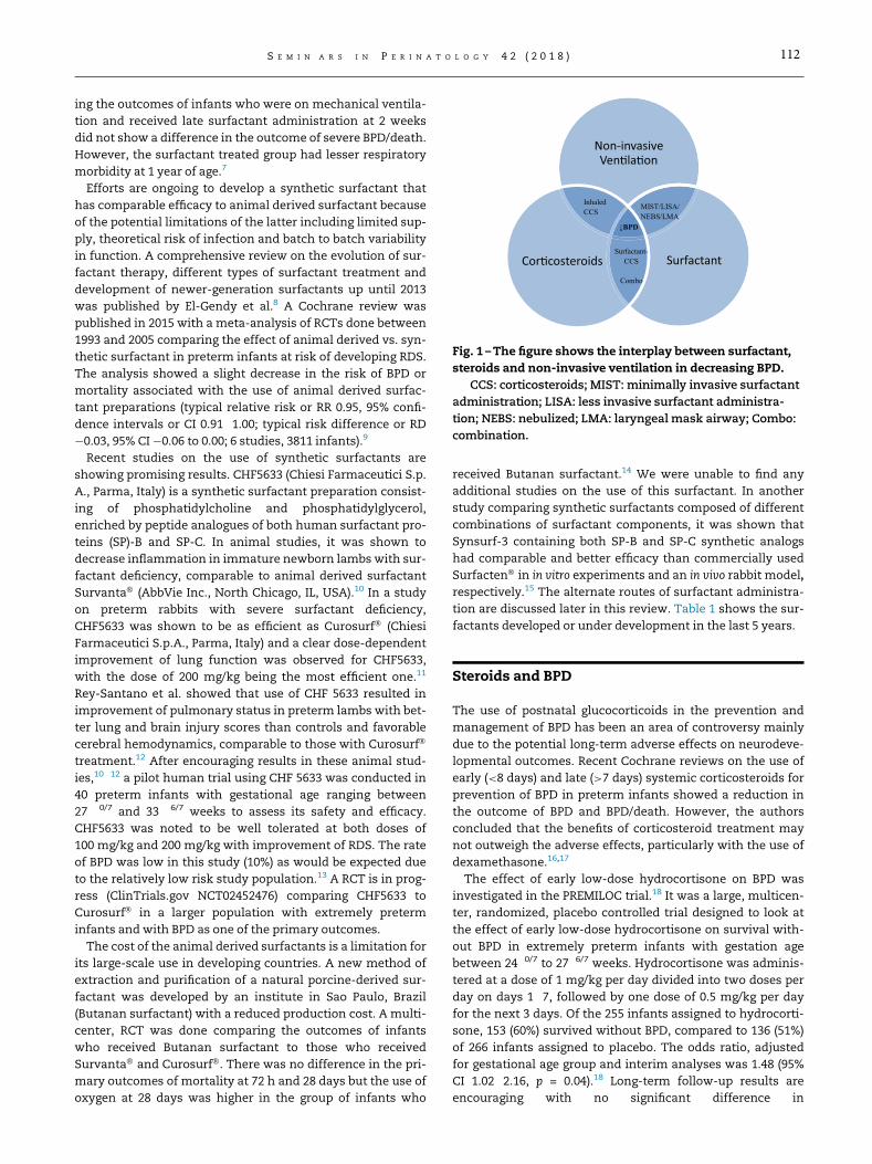
Non-invasive Ven�la�on
SurfactantCor�costeroids
Inhaled CCS
↓BPD
MIST/LISA/NEBS/LMA
Surfactant-CCS
Combo
Fig. 1 –The figure shows the interplay between surfactant,
steroids and non-invasive ventilation in decreasing BPD.
CCS: corticosteroids; MIST: minimally invasive surfactant
administration; LISA: less invasive surfactant administra-
tion; NEBS: nebulized; LMA: laryngeal mask airway; Combo:
combination.
T AGGEDENDS E M I N A R S I N P E R I N A T O L O G Y 4 2 ( 2 0 1 8 ) 112
ing the outcomes of infants who were on mechanical ventila-
tion and received late surfactant administration at 2 weeks
did not show a difference in the outcome of severe BPD/death.
However, the surfactant treated group had lesser respiratory
morbidity at 1 year of age.7
Efforts are ongoing to develop a synthetic surfactant that
has comparable efficacy to animal derived surfactant because
of the potential limitations of the latter including limited sup-
ply, theoretical risk of infection and batch to batch variability
in function. A comprehensive review on the evolution of sur-
factant therapy, different types of surfactant treatment and
development of newer-generation surfactants up until 2013
was published by El-Gendy et al.8 A Cochrane review was
published in 2015 with a meta-analysis of RCTs done between
1993 and 2005 comparing the effect of animal derived vs. syn-
thetic surfactant in preterm infants at risk of developing RDS.
The analysis showed a slight decrease in the risk of BPD or
mortality associated with the use of animal derived surfac-
tant preparations (typical relative risk or RR 0.95, 95% confi-
dence intervals or CI 0.91�1.00; typical risk difference or RD
¡0.03, 95% CI ¡0.06 to 0.00; 6 studies, 3811 infants).9
Recent studies on the use of synthetic surfactants are
showing promising results. CHF5633 (Chiesi Farmaceutici S.p.
A., Parma, Italy) is a synthetic surfactant preparation consist-
ing of phosphatidylcholine and phosphatidylglycerol,
enriched by peptide analogues of both human surfactant pro-
teins (SP)-B and SP-C. In animal studies, it was shown to
decrease inflammation in immature newborn lambs with sur-
factant deficiency, comparable to animal derived surfactant
Survanta� (AbbVie Inc., North Chicago, IL, USA).10 In a study
on preterm rabbits with severe surfactant deficiency,
CHF5633 was shown to be as efficient as Curosurf� (Chiesi
Farmaceutici S.p.A., Parma, Italy) and a clear dose-dependent
improvement of lung function was observed for CHF5633,
with the dose of 200 mg/kg being the most efficient one.11
Rey-Santano et al. showed that use of CHF 5633 resulted in
improvement of pulmonary status in preterm lambs with bet-
ter lung and brain injury scores than controls and favorable
cerebral hemodynamics, comparable to those with Curosurf�
treatment.12 After encouraging results in these animal stud-
ies,10�12 a pilot human trial using CHF 5633 was conducted in
40 preterm infants with gestational age ranging between
27 0/7 and 33 6/7 weeks to assess its safety and efficacy.
CHF5633 was noted to be well tolerated at both doses of
100 mg/kg and 200 mg/kg with improvement of RDS. The rate
of BPD was low in this study (10%) as would be expected due
to the relatively low risk study population.13 A RCT is in prog-
ress (ClinTrials.gov NCT02452476) comparing CHF5633 to
Curosurf� in a larger population with extremely preterm
infants and with BPD as one of the primary outcomes.
The cost of the animal derived surfactants is a limitation for
its large-scale use in developing countries. A new method of
extraction and purification of a natural porcine-derived sur-
factant was developed by an institute in Sao Paulo, Brazil
(Butanan surfactant) with a reduced production cost. A multi-
center, RCT was done comparing the outcomes of infants
who received Butanan surfactant to those who received
Survanta� and Curosurf�. There was no difference in the pri-
mary outcomes of mortality at 72 h and 28 days but the use of
oxygen at 28 days was higher in the group of infants who
received Butanan surfactant.14 We were unable to find any
additional studies on the use of this surfactant. In another
study comparing synthetic surfactants composed of different
combinations of surfactant components, it was shown that
Synsurf-3 containing both SP-B and SP-C synthetic analogs
had comparable and better efficacy than commercially used
Surfacten� in in vitro experiments and an in vivo rabbit model,
respectively.15 The alternate routes of surfactant administra-
tion are discussed later in this review. Table 1 shows the sur-
factants developed or under development in the last 5 years.
Steroids and BPD
The use of postnatal glucocorticoids in the prevention and
management of BPD has been an area of controversy mainly
due to the potential long-term adverse effects on neurodeve-
lopmental outcomes. Recent Cochrane reviews on the use of
early (<8 days) and late (>7 days) systemic corticosteroids for
prevention of BPD in preterm infants showed a reduction in
the outcome of BPD and BPD/death. However, the authors
concluded that the benefits of corticosteroid treatment may
not outweigh the adverse effects, particularly with the use of
dexamethasone.16,17
The effect of early low-dose hydrocortisone on BPD was
investigated in the PREMILOC trial.18 It was a large, multicen-
ter, randomized, placebo controlled trial designed to look at
the effect of early low-dose hydrocortisone on survival with-
out BPD in extremely preterm infants with gestation age
between 24 0/7 to 27 6/7 weeks. Hydrocortisone was adminis-
tered at a dose of 1 mg/kg per day divided into two doses per
day on days 1�7, followed by one dose of 0.5 mg/kg per day
for the next 3 days. Of the 255 infants assigned to hydrocorti-
sone, 153 (60%) survived without BPD, compared to 136 (51%)
of 266 infants assigned to placebo. The odds ratio, adjusted
for gestational age group and interim analyses was 1.48 (95%
CI 1.02�2.16, p = 0.04).18 Long-term follow-up results are
encouraging with no significant difference in

Table 1 – Surfactants developed or under development in the last 5 years.
Surfactant source Composition Surfactant proteins Surfactant name
Animal-porcine Minced lung extract SP-B, SP-C Butanan surfactant
Synthetic Synthetic DPCC, PG Analogs of SP-B, SP-C CHF 5633
Synthetic Synthetic DPCC, PG, PA KL-4 Aerosurf (Inhaled form of Lucinactant)
Synthetic Synthetic DPCC, PG, PA Analogs of SP-B, SP-C Synsurf-3
DPCC: Dipalmitoyl Phosphatidylcholine; PG: Phosphatidylglycerol; PA: Palmitic acid; SP-B: Surfactant protein B; SP-C: Surfactant protein-C; KL-
4: synthetic peptide developed to resemble the amino acid pattern of SP-B.
113 S E M I N A R S I N P E R I N A T O L O G Y 4 2 ( 2 0 1 8 )
neurodevelopmental outcomes between infants in the 2
groups at the 2 year follow-up.19 Follow up study done by
stratifying infants according to gestational age showed a sta-
tistically significant improvement in neurodevelopmental
outcomes in infants born at 24 and 25 weeks of gestation in
the hydrocortisone group.20
Inhaled glucocorticosteroids with their potential for less
systemic toxicity have been used in the management of BPD.
The effect of early inhaled corticosteroids on BPD was investi-
gated in the NEUROSIS trial.21 Inhaled budesonide vs. placebo
was administered to extremely premature infants (23 0/7
weeks to 27 6/7 weeks gestational age) starting within 24 h
after birth and continuing until the infants no longer needed
supplemental oxygen and positive-pressure support or
reached a postmenstrual age of 32 0/7 weeks. Budesonide was
administered at a dose of 2 puffs (200 micrograms per puff)
every 12 hours in the first 14 days of life and one puff adminis-
tered every 12 hours from day 15 until the last dose of the
study drug had been administered. There was a reduction in
composite outcome of BPD/death in the budesonide group.
Infants who received budesonide had a lower rate of BPD
(27.8% vs. 38%) but a higher death rate (16.9% vs. 13.6%).21
This finding of increased mortality among infants who
received budesonide was also noted at the 2 year follow up.22
Another recent RCT evaluated the effect of early inhaled
budesonide on preterm infants (gestational age 23�28 weeks,
n = 70) starting 12 h after birth and continued for a maximum
of 7 days. Even though the use of inhaled budesonide
decreased the incidence of BPD (defined as oxygen require-
ment for 28 days), the difference was not statistically signifi-
cant. (31.4% vs. 54.3%, p = 0.053).23 A meta-analysis of
published trials concluded that inhaled steroids (beclometha-
sone, budesonide, fluticasone, flunisolide, and dexametha-
sone) were associated with a significant reduction in BPD
(RR = 0.77, 95% CI 0.65�0.91).24 However, further long-term
studies are needed before the use of early inhaled corticoste-
roids can be routinely recommended as part of BPD manage-
ment.
A RCT by Yeh et al. reported the effect of intratracheal corti-
costeroid-surfactant combination on BPD.25 In this study,
very low birth weight (VLBW) infants (n = 265) with severe
RDS requiring mechanical ventilation and fraction of inspired
oxygen (FiO2) � 0.50 were randomized to receive either intra-
tracheal budesonide (0.25 mg/kg) combined with surfactant
(Survanta� 100 mg/kg) or surfactant (Survanta� 100 mg/kg)
alone within four hours of birth. The intervention group had
a significantly lower incidence of BPD or death (42.0% vs. 66%;
RR 0.58; 95% CI 0.44�0.77; p < 0.001; number needed to treat
or NNT, 4.1; 95% CI 2.8�7.8).25 A meta-analysis of two RCTs
demonstrated that intratracheal budesonide-surfactant mix-
ture was associated with a decreased risk of BPD (RR: 0.57;
95% CI: 0.43�0.76, NNT = 5) and the composite outcome of
death/BPD (RR: 0.60; 95%CI: 0.49�0.74, NNT = 3) in VLBW
infants.26 These results are promising and further large-scale
RCTs are needed to confirm the efficacy and establish the safe
side effect profile of the steroid-surfactant combination.
Ongoing trials: The Minidex trial is an ongoing multicenter
RCT evaluating the effect of low-dose dexamethasone (13
doses of 0.015 ml/kg/day) in preterm infants <30 weeks gesta-
tional age between postnatal days 10�24 (https://www.npeu.
ox.ac.uk/minidex). The STOP�BPD study is another multicen-
ter randomized double blind placebo controlled trial being
conducted in the Europe investigating if systemic hydrocorti-
sone started between postnatal days 7�14 decreases the com-
bined outcome of death/BPD in ventilated preterm infants
with gestational age <30 weeks and/or birth weight less than
1250 g. Hydrocortisone (cumulative dose 72.5 mg/kg) or pla-
cebo is being administered with a 22 day tapering schedule in
this study and the 2 year follow up is expected to be com-
pleted in 2019.27
Non-invasive ventilation and BPD
As it is increasingly being recognized that endotracheal
mechanical ventilation is associated with increased risk of
developing BPD, use of non-invasive ventilation techniques
in preterm infants is gaining popularity.28 A recent meta-
analysis comparing prophylactic continuous positive airway
pressure (CPAP) to assisted ventilation with or without surfac-
tant in preterm infants less than 32 weeks gestation demon-
strated that use of CPAP resulted in a reduction in the
incidence of BPD (RR 0.89, 95% CI 0.79�0.99, p = 0.04) and
death or BPD (RR 0.89, 95% CI 0.81�0.97).29 Another meta-
analysis investigating the effect of strategies that avoid endo-
tracheal mechanical ventilation (i.e. strategies employing
non-invasive ventilation) in preterm infants less than 30
weeks gestation demonstrated a decrease in BPD/death with
odds ratio of 0.83(0.71�0.96, p = 0.01).30
The type of non-invasive ventilation strategy does not seem
to have a significant impact on BPD. In a RCT (n = 987 ana-
lyzed) comparing the use of nasal intermittent positive pres-
sure ventilation (NIPPV) to CPAP in preterm infants less than
30 weeks gestation and birth weight less than 1000 g, the inci-
dence of BPD did not differ significantly between the 2 groups
(33.9% vs. 31%, OR 1.14 95% CI 0.84�1.54, p = 0.32).31 A

T AGGEDENDS E M I N A R S I N P E R I N A T O L O G Y 4 2 ( 2 0 1 8 ) 114
secondary, non-randomized analysis of the infants in the
NIPPV arm in this trial was done comparing the outcomes of
infants who received bi-level CPAP (Bi-PAP) vs. NIPPV via a
conventional mechanical ventilator. There was no significant
difference in the composite outcome of death/BPD between
infants in the two groups [adjusted OR 0.88 (95% CI
0.57�1.35)]. The rate of BPD was lower in the infants who
received Bi-PAP; however, this difference was not statistically
significant (30% vs. 37%) [adjusted OR 0.64 (95% CI 0.41 to
1.02)]. More deaths occurred in infants receiving Bi-PAP (9.4%)
than in NIPPV (2.3%) (adjusted OR 5.01: 95% CI 1.74�14.4).32 A
meta-analysis from the recent Cochrane reviews did not
demonstrate a significant reduction in BPD with the use of
NIPPV compared to CPAP (typical RR 0.94, 95% CI 0.80�1.10;
typical RR 0.78, 95% CI 0.58�1.06).33,34 A recent systematic
review noted that use of NIPPV was associated with higher
rates of successful extubation compared to CPAP.35
The duration of mechanical ventilation is another signifi-
cant predictor of the development and severity of BPD. In
infants who are intubated, the risk of developing BPD
increases with the duration of mechanical ventilation. In a
large retrospective cohort study of extremely LBW (ELBW)
infants, it was noted that the risk of developing BPD increased
with the cumulative duration of mechanical ventilation, but it
was not related to the number of ventilation courses. Based
on the results of the study, the authors recommend that extu-
bation be trialed once low ventilator settings are reached,
even if extubation success is not guaranteed, as the sooner an
extubation attempt is made, the lower the risk of BPD.36 Rob-
bins et al. reported in a cohort of 224 patients born at <27
weeks’ gestation that the age at first extubation attempt cor-
related directly with endotracheal mechanical ventilation
days and length of stay. In this study, the median mechanical
ventilation days were 32 and 65% of patients needed re-intu-
bation. Furthermore, they reported that the earlier an extuba-
tion attempt was made, the lower the rate of BPD.37
The above 2 studies essentially re-affirmed the findings of
an earlier retrospective study of 262 preterm infants with ges-
tational age �28 weeks and who were intubated on day of life
1, where it was noted that delaying extubation beyond the
first 7 days was associated with an increased risk of BPD and
BPD/death. Infants who were extubated early but had to be
re-intubated later were noted to have a lower incidence of
BPD/death compared to infants who remained intubated and
were extubated later.38
Prompt determination of extubation readiness helps to
minimize or prevent extubation failures. Pilot studies utiliz-
ing data on heart rate variability to determine extubation
readiness in extremely preterm infants met with success and
a RCT is ongoing to develop an automated prediction tool for
extubation readiness in extremely preterm infants.39,40
Alternate routes of surfactant delivery
Perhaps the most significant progress in the prevention/
reduction of BPD in the last decade has beenmade in develop-
ing techniques to administer exogenous surfactant through
non-invasive or minimally invasive routes. Some of the
routes of surfactant administration other than conventional
endotracheal tube instillation include aerosolized delivery
through a nebulizer, surfactant delivery through laryngeal
mask airway (LMA) and administration via a thin catheter
introduced endotracheally. All these strategies obviate the
need for endotracheal tube ventilation in the at-risk preterm
population, reducing the potential for lung injury and subse-
quent development of BPD. A comprehensive review on mini-
mally invasive surfactant therapy and non-invasive
respiratory support was published by Kribs.41 Table 2 summa-
rizes the clinical studies on surfactant administration
through alternate routes published in the last 5 years.
Aerosol delivery of surfactant continues to be an area of
active research. Multiple small studies in animal models and
neonates demonstrated the safety and feasibility of nebulized
surfactants but their superiority over intratracheally adminis-
tered surfactants needs to be proven in large trials.
Two studies done in animal models confirmed the feasibil-
ity of surfactant administration through the nebulization
technique utilizing the eFlow� nebulizer � a customized
vibrating perforated membrane nebulizer (Neonatal Neb-
ulizer system; PARI Pharma, Starnberg, Germany).42,43
Recently, a phase 2b clinical trial (n = 221 born between 28
and 32 weeks gestation) using Aerosurf (synthetic peptide
KL4 based lung surfactant) delivered through a vibrating neb-
ulizer mesh showed that Aerosurf had a similar level of effi-
cacy compared to the CPAP only controls. (http://windtreetx.
investorroom.com/2017-06-29-Windtree-Announces-Top-
Line-Results-from-AEROSURF-R-Phase-2b-Clinical-Trial-for-
the-Treatment-of-Respiratory-Distress-Syndrome-RDS-in-
Premature-Infants).
The CureNeb study by Minocchieri et al. is a pragmatic,
stratified, double blind RCT evaluating the impact of nebu-
lized surfactant on intubation requirement in preterm infants
with RDS. 64 infants with GA ranging from 29 0/7 to 33 6/7
weeks were randomized within the first four hours after birth
to receive either CPAP or aerosolized surfactant and CPAP.
The surfactant (poractant alfa, 200 mg/kg) was delivered via
eFlow�. The primary outcome of need for intubation within
72 h of birth was less in the nebulization group compared to
the CPAP alone group [RR (95% CI) = 0.526 (0.292�0.950)]. Nota-
bly, this difference was observed only in the 32 0/7�33 6/7 GA
group but not in the 29 0/7�31 6/7 GA stratification group. None
of the infants in the study had the outcome of BPD. The study
was closed to recruitment earlier than planned due to insuffi-
cient funding/personnel.44
The terms less invasive surfactant administration (LISA)
and minimally invasive surfactant therapy (MIST) are some-
times used interchangeably in literature. Originally LISA was
used to refer to surfactant administration through a feeding
catheter introduced endotracheally using a Magill forceps.
MIST was used to refer to surfactant administration through
a semi-rigid vascular catheter placed endotracheally without
the use of Magill forceps. Researchers have published studies
with slight modifications to these two techniques resulting in
varied nomenclature.
Another strategy to decrease lung injury involves intuba-
tion and surfactant administration followed by immediate
extubation (INSURE) technique. A recent meta-analysis dem-
onstrated no significant difference in the outcomes of BPD/
death with prophylactic INSURE vs. CPAP.45 A new modified

Table 2 – Clinical studies published in the last 5 years on surfactant administration through alternate routes.
Author/ref Type N Control Intervention Outcomes
Aerosolized
Minocchieri et al.44 RCT 64 CPAP alone CPAP+Curosurf via vibrating mem-
brane nebulizer
Decreased need for intubation in inter-
vention group. No difference in BPD
Segal et al. RCT Ongoing CPAP alone CPAP+Lucinactant (3 doses) Pending
Sood et al. RCT Ongoing Survanta 100 vs. 200 mg/kg Survanta;100 vs. 200 mg/kg via
nebulizer
Pending
LMA
Attridge et al.51 RCT 26 CPAP alone Surfactant by LMA followed by
CPAP
Decrease in supplemental oxygen
requirement. No difference in BPD
Sadeghnia et al.52 RCT 70 INSURE Surfactant by supraglottic airway
(iGel)
administration followed by CPAP
Higher A-a DO2 after surfactant in LMA
group (p = 0.001)
Pinheiro et al.53 RCT 61 INSURE Surfactant via LMA followed by
CPAP
Failure rate of surfactant resulting in
MV: 77% in control group vs. 30% in
LMA group (p < 0.001). No difference in
BPD
Roberts et al.54 RCT 103 CPAP alone Surfactant via LMA followed by
CPAP
Decreased rate of intubation and MV in
LMA group (38% vs. 64%, p = 0.006)
No difference in BPD
Barbosa et al.55 RCT 48 Surfactant via ETT Surfactant via LMA followed by
CPAP
FiO2 requirement similar in both groups.
No difference in BPD
LISA/MIST
Dargaville et al.56 Open feasibility
study
158 Historic controls who received CPAP MIST (16 G vascular catheter)-
Hobart technique
Decreased MV with MIST. No difference
in BPD
Klebermass-Shreh-
off et al.57Nonrandomized
study
406 Historic controls LISA (4 Fr feeding catheter) No significant difference in BPD:
Decreased oxygen at day 28 and CLD/
Death with LISA. 40% vs. 51% (p = 0.03)
Mirnia et al.58 RCT 80 INSURE LISA (5 Fr catheter) Shorter CPAP duration and lower NEC
with LISA. No difference in BPD
Kanmaz et al.59 RCT 200 INSURE LISA (5 Fr catheter)- Take Care
technique
BPD, need for MV, duration of CPAP and
MV lower with LISA
Aguar et al.60 Prospective cohort
study
75 Historic controls who received INSURE MIST (3.5�4 Fr nasogastric
catheter)
Higher requirement for second dose of
surfactant with MIST. No difference in
BPD
Kribs et al.61 RCT 211 Surfactant via ETT LISA (4 Fr catheter) No difference in survival without BPD
but survival without major complica-
tions higher with LISA
Krajewski et al.62 Prospective cohort
study
52 Historic controls who received INSURE MIST (0.04 ch feeding tube) Less BPD with MIST
Mohammadizadeh
et al.63RCT 38 Surfactant via ETT LISA (4 Fr feeding tube) Shorter duration of oxygen therapy with
LISA. No difference in BPD.
Bao et al.64 RCT 90 INSURE MIST (16 G vascular catheter) Shorter duration of MV and CPAP with
LISA
Gopel et al.65 Prospective cohort
study
2206 Matched controls LISA Lower rates of MV and BPD with LISA
47 MIST (16 G vascular catheter)
115S
EM
I N
A R
S
IN
P
ER
IN
AT
OL
OG
Y 42
(2018)

Table 2 (continued)
Author/ref Type N Control Intervention Outcomes
Canals Candela et
al.66Retrospective obser-
vational study
Historic controls who failed NIV and
needed Surfactant via ETT
Lesser need for ETT intubation with
MIST. No difference in BPD
Tomar et al.67 Prospective observa-
tional study
132 Historic controls who received INSURE MIST (5 Fr orogastric tube) Shorter duration of MV and CPAP with
MIST. No difference in BPD
Olivier et al.68 RCT 45 CPAP. Surfactant via ETT if needed MIST (5 Fr feeding tube) Need for MV and rate of pneumothora-
ces requiring chest tube lower with
MIST
Lau et al.69 Meta-analysis 328 INSURE LISA Decreased need for MV within first 72 h,
duration of MV, CPAP, and supple-
mental oxygen noted with LISA. A
trend towards a reduction in BPD
noted with LISA (p = 0.141)
Aldana-Aguirre et
al.70Meta-analysis 895 Surfactant via ETT LISA Decreased need for MV within 72 h,
need for MV, death/BPD with LISA
Hartel et al.49 Observational study 6319 Surfactant via ETT LISA Decreased BPD, death, BPD/death but
increased focal intestinal perforation
with LISA
Dargaville et al
OPTIMIST-A
trial.71
RCT Ongoing Sham treatment MIST (16 G vascular catheter)
Intra-amniotic instillation
Agrawal et al.72 Randomized pilot
study
40 28�34 week GA infants Intra-amniotic surfactant instilla-
tion 2�8 h before
delivery in 28�34 week GA
infants
Decreased severe RDS and need for
postnatal surfactant in intervention
group
RCT: randomized controlled trial; CPAP: continuous positive airway pressure; LMA: laryngeal mask airway; INSURE: intubate-surfactant-extubate; BPD: bronchopulmonary dysplasia; MV: mechanical
ventilation; A-a DO2: Alveolar-arterial oxygen gradient; ETT: Endotracheal tube; FiO2: fraction of inspired oxygen; CLD: chronic lung disease; LISA/MIST: less invasive surfactant administration/mini-
mally invasive surfactant administration; NIV: non-invasive ventilation; NEC: Necrotizing enterocolitis.
TAGGEDENDSE
MIN
AR
SIN
PE
RIN
AT
OL
OG
Y42
(2018)444�
452
116

117 S E M I N A R S I N P E R I N A T O L O G Y 4 2 ( 2 0 1 8 )
technique with alveolar recruitment maneuver before surfac-
tant administration (IN-REC-SUR-E) is currently being
investigated.46
A meta-analysis comparing 7 different ventilation strate-
gies for preterm infants including CPAP alone, INSURE,
LISA, NIPPV, nebulized surfactant administration, surfac-
tant administration via LMA, and mechanical ventilation
concluded that the use of LISA was associated with the low-
est likelihood of the composite outcome of death or BPD at
36 weeks postmenstrual age. However, the authors con-
clude that these findings were limited by the overall low
quality of evidence and lack of robustness in higher-quality
trials.47 Another meta-analysis evaluating 6 RCTs con-
cluded that LISA decreases the risk of BPD (RR = 0.71
[0.52�0.99]; NNT = 21), death or BPD (RR = 0.74 [0.58�0.94]
NNT = 15) and early CPAP failure or invasive ventilation
requirements (RR = 0.67 [0.53�0.84]; NNT = 8 and RR = 0.69
[0.53�0.88]; NNT = 6).48 Hartel et al. recently published an
observational cohort study from a large neonatal network
comparing the outcomes of infants who received surfactant
via ETT (n = 3695) to those who received LISA (n = 2624). In
this study, LISA was associated with a reduced risk of BPD
[OR 0.55 (95% CI: 0.49�0.62), p < 0.001], death [OR 0.66 (95%
CI: 0.51�0.84), p < 0.001] and BPD/death [OR 0.5 (95% CI:
0.44�0.57), p < 0.001], after adjusting for various factors in a
multivariable logistic regression model. However, there
was an increased incidence of focal intestinal perforation
in the LISA group [OR 1.49 (95% CI: 1.14�1.95), p = 0.003] in a
multivariable logistic regression model, primarily noted in
the VLBW infants born at less than 26 weeks. Future trials
should focus on the safety of LISA in the subgroup of
extremely premature infants.49
In a RCT by Oncel et al. (n = 200, preterm infants 26�32
weeks gestational age) comparing early NIPPV and NCPAP
with in the MIST approach, NIPPV decreased the need for
invasive mechanical ventilation within the first 72 h of life
and reduced the surfactant requirement. Infants in the NIPPV
group had significantly decreased moderate-to-severe BPD
(7% vs. 16%; p = 0.046), although this difference was not
observed onmultivariate analysis.50
Conclusions
Based on the current available evidence in the arena of BPD
prevention, it can be recommended that an attempt be made
to extubate the infant early (within the first 72 h of life) and to
use non-invasive ventilation strategies and MIST/LISA when-
ever feasible. While the use of postnatal corticosteroids still
remains controversial, early use of low-dose hydrocortisone
in an at-risk preterm population showed a decreased inci-
dence of BPD without significant neurodevelopmental
impairment on long-term follow-up. The use of intratracheal
surfactant-corticosteroid combination is promising but needs
to be evaluated in further large-scale trials. Research on the
use of synthetic and/or nebulized surfactants is ongoing and
if proven to have a comparable or higher efficacy than animal
derived surfactants, will be a boon in the management of RDS
and BPD worldwide.
R E F E R E N C E S
1. Bhandari A, Bhandari V. Pitfalls, problems, and progress inbronchopulmonary dysplasia. Pediatrics. 2009;123:1562–1573.
2. Underwood MA, Danielsen B, Gilbert WM. Cost, causes andrates of rehospitalization of preterm infants. J Perinatol Offic JCalif Perinat Assoc. 2007;27:614–619.
3. Smith VC, Zupancic JA, McCormick MC, et al. Trends in severebronchopulmonary dysplasia rates between 1994 and 2002. JPediatr. 2005;146:469–473.
4. Zysman-Colman Z, Tremblay GM, Bandeali S, Landry JS. Bron-chopulmonary dysplasia - trends over three decades. PaediatrChild Health. 2013;18:86–90.
5. Stevens TP, Harrington EW, Blennow M, Soll RF. Early surfac-tant administration with brief ventilation vs. selective surfac-tant and continued mechanical ventilation for preterminfants with or at risk for respiratory distress syndrome.Cochrane Database Syst Rev. 2007:Cd003063.
6. Jobe AH, Bancalari E. Bronchopulmonary dysplasia. Am JRespir Crit Care Med. 2001;163:1723–1729.
7. Hascoet JM, Picaud JC, Ligi I, et al. Late surfactant administra-tion in very preterm neonates with prolonged respiratory dis-tress and pulmonary outcome at 1 year of age: a randomizedclinical trial. JAMA Pediatr. 2016;170:365–372.
8. El-Gendy N, Kaviratna A, Berkland C, Dhar P. Delivery andperformance of surfactant replacement therapies to treat pul-monary disorders. Ther Deliv. 2013;4:951–980.
9. Ardell S, Pfister RH, Soll R. Animal derived surfactant extractversus protein free synthetic surfactant for the preventionand treatment of respiratory distress syndrome. CochraneDatabase Syst Rev. 2015;8:CD000144.
10. Sato A, Ikegami M. SP-B and SP-C containing new syntheticsurfactant for treatment of extremely immature lamb lung.PLoS One. 2012;7:e39392.
11. Ricci F, Murgia X, Razzetti R, Pelizzi N, Salomone F. In vitroand in vivo comparison between poractant alfa and the newgeneration synthetic surfactant CHF5633. Pediatr Res.2017;81:369–375.
12. Rey-Santano C, Mielgo VE, Murgia X, et al. Cerebral and lungeffects of a new generation synthetic surfactant with SP-Band SP-C analogs in preterm lambs. Pediatr Pulmonol.2017;52:929–938.
13. Sweet DG, Turner MA, Stranak Z, et al. A first-in-human clini-cal study of a new SP-B and SP-C enriched synthetic surfac-tant (CHF5633) in preterm babies with respiratory distresssyndrome. Arch Disease Childhood Fetal Neonat Ed. 2017;102:F497–f503.
14. Rebello CM, Precioso AR, Mascaretti RS. A multicenter, ran-domized, double-blind trial of a new porcine surfactant inpremature infants with respiratory distress syndrome. Ein-stein (Sao Paulo, Brazil). 2014;12:397–404.
15. Choi YS, Chung SH, Bae CW. A combination of short and sim-ple surfactant protein b and c analogues as a new syntheticsurfactant: in vitro and animal experiments. Yonsei Med J.2017;58:823–828.
16. Doyle LW, Cheong JL, Ehrenkranz RA, Halliday HL. Early (< 8days) systemic postnatal corticosteroids for prevention ofbronchopulmonary dysplasia in preterm infants. CochraneDatabase Syst Rev. 2017;10:Cd001146.
17. Onland W, Offringa M, van Kaam A. Late (>/= 7 days) inhala-tion corticosteroids to reduce bronchopulmonary dysplasia inpreterm infants. Cochrane Database Syst Rev. 2017;8:Cd002311.
18. Baud O, Maury L, Lebail F, et al. Effect of early low-dose hydro-cortisone on survival without bronchopulmonary dysplasia inextremely preterm infants (PREMILOC): a double-blind, pla-cebo-controlled, multicentre, randomised trial. Lancet.2016;387:1827–1836.
TAGGEDENDS E M I N A R S I N P E R I N A T O L O G Y 4 2 ( 2 0 1 8 ) 118
19. Baud O, Trousson C, Biran V, Leroy E, Mohamed D, Alberti C.Association between early low-dose hydrocortisone therapyin extremely preterm neonates and neurodevelopmental out-comes at 2 years of age. JAMA. 2017;317:1329–1337.
20. Baud O, Trousson C, Biran V, Leroy E, Mohamed D, Alberti C.Two-year neurodevelopmental outcomes of extremely preterminfants treated with early hydrocortisone: treatment effectaccording to gestational age at birth. Arch Diseas Childhood FetalNeonat Ed. 2018 Jan 10. pii: fetalneonatal-2017-313756. https://doi.org/10.1136/archdischild-2017-313756. [Epub ahead of print].
21. Bassler D, Plavka R, Shinwell ES, et al. Early inhaled budeso-nide for the prevention of bronchopulmonary dysplasia. NEngl J Med. 2015;373:1497–1506.
22. Bassler D, Shinwell ES, Hallman M, et al. Long-term effects ofinhaled budesonide for bronchopulmonary dysplasia. NewEngland J Med. 2018;378:148–157.
23. Sadeghnia A, Beheshti BK, Mohammadizadeh M. The effect ofinhaled budesonide on the prevention of chronic lung diseasein premature neonates with respiratory distress syndrome.Int J Prev Med. 2018;9:15.
24. Shinwell ES, Portnov I, Meerpohl JJ, Karen T, Bassler D.Inhaled corticosteroids for bronchopulmonary dysplasia: ameta-analysis. Pediatrics. Dec; 2016;138(6): pii: e20162511.
25. Yeh TF, Chen CM, Wu SY, et al. Intratracheal Administrationof Budesonide/Surfactant to Prevent Bronchopulmonary Dys-plasia. Am J Respir Crit Care Med. 2016;193:86–95.
26. Venkataraman R, Kamaluddeen M, Hasan SU, Robertson HL,Lodha A. Intratracheal administration of budesonide-surfac-tant in prevention of bronchopulmonary dysplasia in verylow birth weight infants: a systematic review and meta-anal-ysis. Pediatr Pulmonol. 2017;52:968–975.
27. Onland W, Merkus MP, Nuytemans DH, Jansen-van derWeide MC, Holman R, van Kaam AH. Systemic Hydrocorti-sone To Prevent Bronchopulmonary Dysplasia in preterminfants (the SToP-BPD study): statistical analysis plan. Tri-als. 2018;19:178.
28. Bhandari V. The potential of non-invasive ventilation todecrease BPD. Semin Perinatol. 2013;37:108–114.
29. Subramaniam P, Ho JJ, Davis PG. Prophylactic nasal continuouspositive airway pressure for preventing morbidity and mortalityin very preterm infants. Cochrane Database Syst Rev. 2016 Jun 14;(6):CD001243. https://doi.org/10.1002/14651858.CD001243.pub3.
30. Fischer HS, Buhrer C. Avoiding endotracheal ventilation toprevent bronchopulmonary dysplasia: a meta-analysis. Pedi-atrics. 2013;132:e1351–e1360.
31. Kirpalani H, Millar D, Lemyre B, et al. A trial comparing nonin-vasive ventilation strategies in preterm infants. N Engl J Med.2013;369:611–620.
32. Millar D, Lemyre B, Kirpalani H, Chiu A, Yoder BA, Roberts RS.A comparison of bilevel and ventilator-delivered non-inva-sive respiratory support. Arch Diseas Childh Fetal Neonat Ed.2016;101:F21–F25.
33. Lemyre B, Davis PG, De Paoli AG, Kirpalani H. Nasal intermit-tent positive pressure ventilation (NIPPV) versus nasal contin-uous positive airway pressure (NCPAP) for preterm neonatesafter extubation. Cochrane Database Syst Rev. 2017;2:Cd003212.
34. Lemyre B, Laughon M, Bose C, Davis PG. Early nasal intermit-tent positive pressure ventilation (NIPPV) versus early nasalcontinuous positive airway pressure (NCPAP) for preterminfants. Cochrane Database Syst Rev. 2016;12:Cd005384.
35. Ferguson KN, Roberts CT, Manley BJ, Davis PG. Interventionsto improve rates of successful extubation in preterm infants:a systematic review and meta-analysis. JAMA Pediatr.2017;171:165–174.
36. Jensen EA, DeMauro SB, Kornhauser M, Aghai ZH, GreenspanJS, Dysart KC. Effects of multiple ventilation courses andduration of mechanical ventilation on respiratory outcomes
in extremely low-birth-weight infants. JAMA Pediatr.2015;169:1011–1017.
37. Robbins M, Trittmann J, Martin E, Reber KM, Nelin L, Shep-herd E. Early extubation attempts reduce length of stay inextremely preterm infants even if re-intubation is necessary.J Neonat Perinat Med. 2015;8:91–97.
38. Berger J, Mehta P, Bucholz E, Dziura J, Bhandari V. Impact ofearly extubation and reintubation on the incidence of bron-chopulmonary dysplasia in neonates. Am J Perinatol.2014;31:1063–1072.
39. Kaczmarek J, Chawla S, Marchica C, Dwaihy M, Grundy L,Sant’Anna GM. Heart rate variability and extubation readi-ness in extremely preterm infants. Neonatology. 2013;104:42–48.
40. Shalish W, Kanbar LJ, Rao S, et al. Prediction of Extubationreadiness in extremely preterm infants by the automatedanalysis of cardiorespiratory behavior: study protocol. BMCPediatr. 2017;17:167.
41. Kribs A. Minimally invasive surfactant therapy and noninva-sive respiratory support. Clin Perinatol. 2016;43:755–771.
42. Hutten MC, Kuypers E, Ophelders DR, et al. Nebulization ofPoractant alfa via a vibrating membrane nebulizer in sponta-neously breathing preterm lambs with binasal continuouspositive pressure ventilation. Pediatr Res. 2015;78:664–669.
43. Linner R, Perez-de-Sa V, Cunha-Goncalves D. Lung depositionof nebulized surfactant in newborn piglets. Neonatology.2015;107:277–282.
44. Minocchieri S, Berry CA, Pillo JJ, Nebulised surfactant toreduce severity of respiratory distress: a blinded, parallel,randomised controlled trial. Arch Diseas Childh Fetal Neonat Ed.2018 Jul 26. pii: fetalneonatal-2018-315051. https://doi.org/10.1136/archdischild-2018-315051. [Epub ahead of print].
45. Isayama T, Chai-Adisaksopha C, McDonald SD. Noninvasiveventilation with vs without early surfactant to preventchronic lung disease in preterm infants: a systematic reviewand meta-analysis. JAMA Pediatr. 2015;169:731–739.
46. Vento G, Pastorino R, Boni L, et al. Efficacy of a new technique� INtubate-RECruit-SURfactant-Extubate � "IN-REC-SUR-E" �in preterm neonates with respiratory distress syndrome:study protocol for a randomized controlled trial. Trials.2016;17:414.
47. Isayama T, Iwami H, McDonald S, Beyene J. Association ofNoninvasive Ventilation Strategies With Mortality and Bron-chopulmonary Dysplasia Among Preterm Infants: A System-atic Review and Meta-analysis. JAMA. 2016;316:611–624.
48. Rigo V, Lefebvre C, Broux I. Surfactant instillation in sponta-neously breathing preterm infants: a systematic review andmeta-analysis. Eur J Pediatr. 2016;175:1933–1942.
49. Hartel C, Paul P, Hanke K, et al. Less invasive surfactantadministration and complications of preterm birth. Sci Rep.2018;8:8333.
50. Oncel MY, Arayici S, Uras N, et al. Nasal continuous positiveairway pressure versus nasal intermittent positive-pressureventilation within the minimally invasive surfactant therapyapproach in preterm infants: a randomised controlled trial.Arch Dis Child Fetal Neonatal Ed. 2016;101:F323–F328.
51. Attridge JT, Stewart C, Stukenborg GJ, Kattwinkel J. Adminis-tration of rescue surfactant by laryngeal mask airway: lessonsfrom a pilot trial. Am J Perinatol. 2013;30:201–206.
52. Sadeghnia A, Tanhaei M, Mohammadizadeh M, Nemati M. Acomparison of surfactant administration through i-gel andET-tube in the treatment of respiratory distress syndrome innewborns weighing more than 2000 grams. Adv Biomed Res.2014;3:160.
53. Pinheiro JM, Santana-Rivas Q, Pezzano C. Randomized trial oflaryngealmask airway versus endotracheal intubation for surfac-tant delivery. J Perinatol Offic J Califor Perin Assoc. 2016;36:196–201.

119 S E M I N A R S I N P E R I N A T O L O G Y 4 2 ( 2 0 1 8 )
54. Roberts KD, Brown R, Lampland AL, et al. Laryngeal mask air-way for surfactant administration in neonates: a randomized,controlled trial. J Pediatr. 2018;193:40–46.e41.
55. Barbosa RF, Simoes ESAC, Silva YP. A randomized controlledtrial of the laryngeal mask airway for surfactant administra-tion in neonates. J Pediatr. 2017;93:343–350.
56. Dargaville PA, Aiyappan A, De Paoli AG, et al. Minimally-inva-sive surfactant therapy in preterm infants on continuous pos-itive airway pressure. Arch Dis Child Fetal Neonatal Ed. 2013;98:F122–F126.
57. Klebermass-Schrehof K, Wald M, Schwindt J, et al. Less inva-sive surfactant administration in extremely preterm infants:impact on mortality and morbidity. Neonatology.2013;103:252–258.
58. Mirnia K, Heidarzadeh M, Hoseini MB, et al. Surfactantadministration via thin catheter during spontaneous breath-ing: randomized controlled trial in Alzahra hospital. Iran JNeonatol IJN. 2013;4:5–9.
59. Kanmaz HG, Erdeve O, Canpolat FE, Mutlu B, Dilmen U. Sur-factant administration via thin catheter during spontaneousbreathing: randomized controlled trial. Pediatrics. 2013;131:e502–e509.
60. Aguar M, Cernada M, Brugada M, Gimeno A, Gutierrez A,Vento M. Minimally invasive surfactant therapy with a gastrictube is as effective as the intubation, surfactant, and extuba-tion technique in preterm babies. Acta Paediatr (Oslo, Norway:1992). 2014;103:e229–e233.
61. Kribs A, Roll C, Gopel W, et al. Nonintubated surfactantapplication vs conventional therapy in extremely preterminfants: a randomized clinical trial. JAMA Pediatr.2015;169:723–730.
62. Krajewski P, Chudzik A, Strzalko-Gloskowska B, et al. Surfac-tant administration without intubation in preterm infantswith respiratory distress syndrome � our experiences. J MaterFetal Neonat Med Offic J Eur Assoc Perinat Med Feder Asia OceanPerinat Soc Int Soc Per Obst. 2015;28:1161–1164.
63. Mohammadizadeh M, Ardestani AG, Sadeghnia AR. Earlyadministration of surfactant via a thin intratrachealcatheter in preterm infants with respiratory distress
syndrome: feasibility and outcome. J Res Pharm Pract.2015;4:31–36.
64. Bao Y, Zhang G, Wu M, Ma L, Zhu J. A pilot study of less inva-sive surfactant administration in very preterm infants in aChinese tertiary center. BMC Pediatr. 2015;15:21.
65. Gopel W, Kribs A, Hartel C, et al. Less invasive surfactantadministration is associated with improved pulmonary out-comes in spontaneously breathing preterm infants. Acta Pae-diatr (Oslo, Norway: 1992). 2015;104:241–246.
66. Canals Candela FJ, Vizcaino Diaz C, Ferrandez Berenguer MJ,Serrano Robles MI, Vazquez Gomis C, Quiles Dura JL. [Surfac-tant replacement therapy with a minimally invasive tech-nique: experience in a tertiary hospital]. Anal Pediatr(Barcelona, Spain :2003). 2016;84:79–84.
67. Tomar RS, Ghuliani R, Yadav D. Effect of surfactant therapyusing orogastric tube for tracheal catheterization in pretermnewborns with respiratory distress. Indian J Pediatr.2017;84:257–261.
68. Olivier F, Nadeau S, Belanger S, et al. Efficacy of minimallyinvasive surfactant therapy in moderate and late preterminfants: a multicentre randomized control trial. Paediatr ChildHealth. 2017;22:120–124.
69. Lau CSM, Chamberlain RS, Sun S. Less invasive surfactantadministration reduces the need for mechanical ventilationin preterm infants: a meta-analysis. Glob Pediatr Health.2017;4: 2333794£ 17696683.
70. Aldana-Aguirre JC, Pinto M, Featherstone RM, Kumar M. Lessinvasive surfactant administration versus intubation for sur-factant delivery in preterm infants with respiratory distresssyndrome: a systematic review and meta-analysis. Arch Dis-ease Childhood Fetal Neonat Ed. 2017;102:F17–f23.
71. Dargaville PA, Kamlin CO, De Paoli AG, et al. The OPTIMIST-Atrial: evaluation of minimally-invasive surfactant therapy in pre-term infants 25�28 weeks gestation. BMC Pediatr. 2014;14:213.
72. Agarwal N, Bathwal S, Kriplani A, Deorari A, Bhatla N. Intra-amniotic instillation of surfactants for the prevention of neo-natal respiratory distress syndrome following preterm deliv-ery. Int J Gynaecol Obstetr Offic Organ Int Feder Gynaecol Obstetr.2016;135:196–199.

T AGGEDENDS E M I N A R S I N P E R I N A T O L O G Y 4 2 ( 2 0 1 8 )
Available online at www.sciencedirect.com
Seminars in Perinatology
www.seminperinat.com
120
An update on the diagnosis an
dmanagement of bronchopulmonary dysplasia (BPD)-associatedpulmonary hypertensionSara K. Berkelhamera*, Karen K. Mestanb, and Robin Steinhornc
aJacobs School of Medicine, University at Buffalo, Buffalo, NY, USAbFeinberg School of Medicine, Northwestern University, Chicago, IL, USAcGeorge Washington University, Washington, DC, USA
* Corresponding author.E-mail address: [email protected] (S.K
https://doi.org/10.1053/j.semperi.2018.09.0050146-0005/� 2018 Elsevier Inc. All rights reser
A B S T R A C T
The past decade of neonatal care has been highlighted by increased survival rates in smaller and
more premature infants. Despite reduction in mortality associated with extreme prematurity,
long termpulmonarymorbidities remain a concern, with growing recognition of the clinical bur-
den attributable to infants with bronchopulmonary dysplasia (BPD)-associated pulmonary
hypertension (PH). Recent publications shed light on the critical contributions of maternal pla-
cental pathology and compromised intrauterine growth to fetal pulmonary vascular develop-
ment. A body of literature has further clarified postnatal risk factors for PH, most notably the
severity of BPD but surprisingly the additional presence of non-pulmonarymorbidities including
necrotizing enterocolitis (NEC). Limitations of current diagnostics persist with growing consider-
ation of novel echocardiographic approaches as well as complementary non-invasive bio-
markers to better identify infants at risk. In 2015, a joint report published by the American Heart
Association and American Thoracic Society provided the first guidelines for the care of children
with PH with limited content to address BPD-associated PH. These guidelines were expanded
upon in an expert consensus report produced by the Pediatric Pulmonary HypertensionNetwork
(PPHNet). These recommendations encouraged the use of standardized screening protocols and
emphasized the importance of evaluation and treatment of comorbidities when PH is identified.
Cardiac catheterizationwas recommended prior to initiation of therapy formore accurate quan-
tification of pulmonary pressures, clarification of anatomy and guidance in the use of pharmaco-
therapy. Despite these guidelines, significant practice variation persists and gaps remain with
respect to optimal evaluation andmanagement of BPD-associated PH.
� 2018 Elsevier Inc. All rights reserved.
A R T I C L E I N F O
Keywords:
bronchopulmonary dysplasia
pulmonary hypertension
chronic lung disease
pulmonary vascular disease
diagnosis
management
and nutritional strategies, have resulted in increasing rates of
Introduction
Advances in neonatal care, including routine use of antenatal
steroids and surfactant, coupled with improved ventilation
. Berkelhamer).
ved.
survival of extremely premature infants.1 Despite increased
use of gentle and non-invasive ventilation strategies, these
infants remain at significant risk of prolonged pulmonary
morbidity, commonly suffering from compromised lung

Table 1 – Infant characteristics associated with the pres-ence of pulmonary hypertension.
Characteristic Infants with or without BPD Infants with BPD
RR(CI) RR(CI)
Male sex 1.1 (0.9, 1.3) 0.9 (0.7, 1.2)
SGA 1.8 (1.2, 2.) 0.5 (0.1, 1.8)
BPD 1.3 (1.1, 1.5) NA
Severe BPD 2.7 (1.7, 4.2) NA
NEC 1.8 (0.9, 3.9) 3.4 (1.1, 10.2)
PDA 1.3 (1.2, 1.5) 1.2 (1.0, 1.5)
PDA ligation 1.8 (1.3, 2.4) NA
Severe IVH 1.7 (0.8, 3.4) NA
ROP 1.9 (1.3, 2.7) 1.2 (0.8, 1.9)
BPD, bronchopulmonary dysplasia; RR, risk ratio; CI, confidence
interval; NA, data not available;
SGA, small for gestational age; ROP, retinopathy of prematurity;
NEC, necrotizing enterocolitis; PDA,
patent ductus arteriosus. Adapted from reference (9).
T AGGEDENDS E M I N A R S I N P E R I N A T O L O G Y 4 2 ( 2 0 1 8 ) 121
development with prolonged need for respiratory support
and/or supplemental oxygen, meeting the clinical definition
of bronchopulmonary dysplasia (BPD). Despite improved
rates of survival of extremely premature infants, there has
been relatively limited progress over the past two decades in
reducing rates of BPD with a relatively stable incidence of
approximately 40% in surviving infants less than or equal to
28 weeks gestation.1�4
While early characterization of BPD mainly focused on
parenchymal lung injury,5 there is growing appreciation for
the contribution of a dysmorphic pulmonary bed as well as
the impact of postnatal insults on pulmonary vascular
remodeling and development. Ultimately, these vascular find-
ings can result in elevated pulmonary resistance with
increased pulmonary pressures, resulting in compensatory
right ventricular hypertrophy and a clinical diagnosis of pul-
monary hypertension (PH). The presence of PH markedly
impacts clinical outcomes, with recent publications reporting
mortality rates ranging from 14 -38%.6�8 Meta-analysis of
studies which have looked specifically at death before dis-
charge report a cumulative mortality rate of 16% while those
that followed patients out to 2 years estimated 40% mortal-
ity.9 Beyond the increased risk of mortality, PH is associated
with significant pulmonary morbidity with added concern for
the impact on neurodevelopmental outcomes.10,11 While
progress has been made in the diagnosis and management of
BPD-associated PH, the natural course and long-term out-
comes of the disease remain poorly characterized and numer-
ous gaps remain in defining optimal care.
Prevalence
Several recent publications have defined rates of occurrence
of PH by BPD classification. While there is considerable het-
erogeneity with respect to timing of screening and defini-
tions, a recent meta-analysis of over 1400 patients from 25
publications confirmed the association of PH with severity of
pulmonary disease with cumulative prevalence of PH in 6%,
12%, and 39% of infants with mild, moderate and severe BPD,
respectively (Figure 1).9 While these data help to characterize
the overall risk for PH, a wide range within the included stud-
ies was noted, with single center studies reporting prevalence
rates ranging from 15 to 64% in infants with severe BPD.12,13
Of interest, this meta-analysis also identified a cumulative
prevalence of PH in 2% in extremely preterm infants in the
absence of BPD. Indeed, several recent studies have raised
concern that abnormal pulmonary vascular development
may occur as a result of prematurity alone.14�16
Pathophysiology
Premature infants are susceptible to altered lung develop-
ment and postnatal lung injury as a result of exposure to pul-
monary care, including ventilation and oxygen therapy.
These insults, along with infection, inflammation and com-
promised nutrition can lead to the phenotype of compro-
mised alveolar development and vascular pruning
characteristic of BPD.17 A subset of infants with BPD will
develop increased pulmonary vascular resistance with
elevated pulmonary arterial pressures as a result of a combi-
nation of arteriovenous maldevelopment, vascular remodel-
ing, increased vascular tone and/or altered vasoreactivity.
While pulmonary vascular disease is commonly attributed to
lung injury and the severity of lung disease, the presence of
PH in premature infants who were not classified as having
BPD suggests that additional risk factors or alternative patho-
physiology contribute to the development of PH. Recent publi-
cations have advanced our understanding of the
contributions of thematernal disease states and environmen-
tal exposures, compromised in utero fetal growth, and postna-
tal non-pulmonary morbidity to the development of neonatal
pulmonary vascular disease.
Maternal-fetal pathophysiology
While BPD and PH share many of the same risk factors, a grow-
ing body of evidence suggests that PH may have a distinct fetal
pathophysiology that arises in part from maternal vascular dis-
ease during pregnancy. This is supported by clinical observa-
tions that many infants with severe BPD, despite the usual
postnatal exposures and risk factors, never develop PH. In con-
trast, some babies with relatively mild disease and uncompli-
cated postnatal courses demonstrate evidence of early and
persistent PH.8,18,19 The presence of a “fixed” or prenatal risk of
PH is supported by studies of maternal, placental and cord
blood biomarkers that vary according to BPD and PH. Maternal
preeclampsia and intrauterine growth restriction, particularly
with severe features or accompanied by reversed or absent end
diastolic flow, are common indications for medically indicated
preterm delivery. These infants appear to be at highest risk for
persistent PH, even when BPD is mild or absent.20
The mechanisms by which maternal and prenatal events
impact fetal lung vascular development leading to PH remain
unclear. An important mediator appears to be the placenta.
At 28 weeks or earlier, placental changes of villus and vessel
maldevelopment are prominent in preterm neonates who
later develop BPD associated PH.21,22 Moreover, circulating
levels of pro-angiogenic growth factors are decreased at birth
in cord blood of infants who have placental vascular

122 S E M I N A R S I N P E R I N A T O L O G Y 4 2 ( 2 0 1 8 )
malperfusion and later develop BPD associated PH.23 These
findings contribute to the growing evidence of the fetal ori-
gins of PH in infants with BPD. The distinct mechanisms by
which cord blood angiogenic factors play a role at birth and in
the postnatal period remain to be elucidated. Possible mecha-
nisms include fetal endothelial cell dysfunction secondary to
placental hypoxia,24 paracrine signaling by factors produced
by the placenta,25,26 or epigenetic changes at the cellular and/
or tissue-specific level due to adverse intrauterine stressors
that have lasting negative impact on the development of the
fetal pulmonary vascular tree, neonatal lung vascular growth,
or both.27,28 These are key topics of ongoing investigation in
BPD-PH research. Defining these interactions could enhance
and refine recent developments in immune-mediated and
cell-based therapies to attenuate and perhaps even prevent
BPD-PH. 29�32
Risk factors
Risk factors for BPD-associated PH can be compiled from
numerous series, and a recent meta-analysis identified 12
non-overlapping studies of patients with and without BPD
and took into consideration co-morbidities of BPD. The com-
bined data provided by this meta-analysis suggest that PH is
associated with lower birthweight in infants with and with-
out BPD, but only with gestational age in BPD infants (Table
1). Similarly, SGA was associated with PH in infants with and
without BPD (Relative Risk or RR 1.8; Confidence Interval or CI
1.2, 2.7), however this relationship was not present in BPD
infants (Table 1).9 Collectively, these findings raise concerns
for smaller and more premature infants who may be
impacted by abnormal pulmonary vascular development
even before they are challenged by pulmonary insults associ-
ated with postnatal care.
Findings from the meta-analysis also support an association
with non-pulmonary morbidities, specifically retinopathy of
prematurity (ROP) and necrotizing enterocolitis (NEC), with a
particularly strong association between NEC and PH in infants
with BPD (RR 3.4; CI 1.1, 10.2). In addition, the presence of a pat-
ent ductus arteriosus (PDA) and need for ligation resulted in a
weak association with PH.9 In contrast, there was no associa-
tion of severe intraventricular hemorrhage (IVH), gender,
maternal hypertension, antenatal corticosteroids, chorioamnio-
nitis and prolonged and preterm rupture of membranes
(PPROM) with risk for PH. These associations suggest the possi-
bility that BPD-PH is in part triggered by inflammation.
Diagnosis
Screening
The adoption of standardized clinical algorithms for evaluation
of infants at risk of BPD-associated PH remains a challenge. In
2015, recommendations for care of pediatric PH were published
by the American Heart Association (AHA) and American Tho-
racic Society (ATS).33 While the guidelines provided content
related to the screening and care of BPD-associated PH, more
detailed recommendations were recently published by the
Pediatric Pulmonary Hypertension Network (PPHNet), a multi-
disciplinary panel of PH experts.34 This report provided practical
clinical recommendations for the evaluation, diagnosis and
management of PH in infants with BPD, classifying recommen-
dations by both their potential benefit as well as the level of
supportive evidence available.
With respect to screening, PPHNet recommendations
include obtaining an echocardiogram for premature infants
with severe hypoxic respiratory failure with minimal paren-
chymal disease, to identify those infants with life threatening
acute PH that might benefit from therapy. Echocardiographic
evaluation should also be considered for infants with contin-
ued need for ventilator support at 7 days or with significant
sustained need for respiratory support at any age, and at the
time of formal BPD diagnosis for all infants with moderate or
severe BPD (36 weeks postmenstrual age). Infants with mild
BPD can be monitored clinically, with echocardiographic
screening reserved for those infants with clinical deteriora-
tion or failure to improve. Standardized echocardiographic
parameters to be used with these evaluations were also pro-
vided with acknowledgement of the limitations of these
indicators.34
These screening guidelines would miss some cases of PH in
those the infants who only met criteria for mild BPD. Consid-
eration of additional risk factors, including birthweight, fetal
growth restriction, presence of high risk comorbidities (i.e.
ROP or NEC) or placental vascular pathology might allow
directed screening for higher risk infants. An evidence-based
risk calculator to assist in the identification of appropriate
cases of mild BPD to screen may be of clinical value. Such a
tool has not yet been developed but could guide clinicians in
management strategies and anticipation of resource utiliza-
tion for a given patient.
A survey performed in 2017 (2 years after publication of the
AHA guidelines) evaluated clinical practice of over 300 neona-
tologists who practice in tertiary care NICUs, most of whom
had over 20 years of experience.35 The majority of respond-
ents noted an increasing number of extremely premature
infants being diagnosed with PH, although reporting of formal
echocardiography screening protocols varied significantly.
Only 46% of respondents replied that institutional protocols
were in place that aligned with the AHA recommendations of
targeted PH screening for moderate and severe BPD, while
38% reported that existing institutional protocols screened all
infants with BPD.
Echocardiogram
While echocardiography is the most common screening
modality for PH in infants with BPD, there are significant limi-
tations to its use. Tricuspid regurgitant jet velocity (TRJV) is
the most frequently used measurement for evaluating PH,
however many infants do not have a detectable tricuspid
regurgitant jet, and lung hyperinflation may impact detec-
tion.34 Further, numerous studies have identified that pulmo-
nary arterial pressures as diagnosed by echocardiography
poorly correlate with data from cardiac catheterization in
infants with BPD.36,37 Evaluation of additional
T AGGEDENDS E M I N A R S I N P E R I N A T O L O G Y 4 2 ( 2 0 1 8 ) 123
echocardiographic parameters, including flattened intraven-
tricular septum, right ventricular (RV) dilation and/or hyper-
trophy and depressed function can improve PH detection.
While inter-rater reliability of cardiologists revealed strong
agreement on screening echocardiograms, standardized pro-
tocols are needed to optimize evaluation of PH.38 More
recently, advanced echocardiographic assessments, including
tissue Doppler imaging (TDI) and speckle tracking echocardi-
ography, have been suggested for identification of pathology
not detected by conventional methods.39 Specifically,
decreased RV longitudinal strain (a RV systolic function
parameter) has been identified by speckle tracking in infants
and children with a history of PH, suggesting this techniques
may be capable of identifying subclinical ventricular dysfunc-
tion where standard imaging was normal.39,40
With growing appreciation of the contribution of pulmonary
vein stenosis to pulmonary vascular disease, pulsed Doppler
interrogation of pulmonary veins should be performed with all
BPD screening. Indeed, there has been increasing identification
of acquired PV stenosis (PVS) resulting in obstructed pulmonary
blood flowand elevated pulmonary pressures.41,42 A recent series
of 213 patients with severe BPD identified that 5% had some
degree of PVS while 26% of infants catheterized in a Spanish reg-
istry were found to be affected.42,43 Of concern are data implicat-
ing PVS in poor outcomes, with long term survival rates of only
43-50% in affected infants.42,44 While echocardiography may be
capable of identifying many cases, cardiac catheterization, CT
angiography or MRI imaging are often required for a more com-
prehensive evaluation of the degree of vessel involvement.
Cardiac catheterization
While echocardiography remains first line for screening, inva-
sive catheterization remains the gold standard for detection
of elevated pulmonary pressures. The PPHNet recommenda-
tions state that cardiac catheterization should be considered
prior to initiation of pulmonary vasodilator therapy, but in
clinical practice, the procedure is often deferred at discretion
of the care team.34 Beyond providing a more reliable assess-
ment of pulmonary pressures, cardiac catheterization assists
in defining the reversible component of vasoconstriction
when therapy is being considered. Further, catheterization
assists in the detection of PVS, aortopulmonary collaterals
(APC), and left heart dysfunction. Each of these may compli-
cate clinical management, as use of pulmonary vasodilators
in affected patients can result in pulmonary vascular conges-
tion and disturbed gas exchange. However, catheterization
also has risks, including need for intubation and sedation,
risk of hemodynamic instability and vascular, thrombotic
and infectious complications associated with an invasive pro-
cedure. A single center retrospective study in pediatric PH
patients who underwent catheterization suggested a 6% risk
of need for resuscitation or death; however, more recent
reports have demonstrated much lower risks.45,46 Nonethe-
less, CT angiography may be an appropriate consideration in
infants who are suboptimal candidates for catheterization as
this study can also provide insight into presence of PVS and
APCs while characterizing parenchymal lung disease.
Biomarkers
Serum brain natriuretic peptide (BNP) and its prohormone, N-
terminal pro-BNP (NT-pro-BNP) are released by the myocardium
in response to stretch and have been proposed as biomarkers
for monitoring right ventricular strain. Studies have correlated
BNP and NT-pro-BNP with mean pulmonary artery pressure,
pulmonary vascular resistance and right atrial pressures in
adult and pediatric patients.47�49 Age specific reference ranges
have been defined, identifying that BNP levels peak at birth
with an acute decline over the first few days of life.50 Limited
data have suggested that BNP and NT-pro BNP may aid in the
diagnosis and monitoring of infants with BPD-associated
PH.51�53 However, as these tests are non-specific and not well
validated, results should be used in conjunction with echocardi-
ography and not replace more formal screening.34 Numerous
additional biomarkers have been explored in the adult and
pediatric cardiac literature including some which are detectable
and in urine samples alone.54 Identification of novel biomarkers
which reliably detect BPD-associated PH pathophysiology could
greatly impact screening and facilitate clinical care.
Management
Evaluation
Similar to the wide variation in screening practices, there is sig-
nificant variation in the clinical management of infants identi-
fied with BPD-associated PH. There has been growing
appreciation for the complex and variable phenotypes of the
disease, with recent evaluations carefully characterizing the co-
existence of parenchymal and large airway disease with PH. In
a cohort of 47 infants with severe BPD associated with PH
(defined as bidirectional or right to left PDA shunt, systolic pul-
monary artery pressures > 40mmHg by TRJV or a flattened/
bowed interventricular septum at the end of systole), 10% were
also found to have large airway disease (tracheomalacia or
bronchomalacia by bronchoscopy), 32% had coexisting severe
parenchymal lung injury (defined as an Ochiai score of 8 or
greater on chest computed tomography55) and 45% had evi-
dence of both airway and severe lung disease .56
The PPHNet recommendations suggest that evaluation and
treatment of comorbidities should be done before initiation of
therapy for BPD-associated PH (Fig. 2).9 This includes evaluation
for intermittent hypoxemia, aspiration, gastroesophageal reflux,
structural air disease, pulmonary artery or vein stenosis, APC
and LV dysfunction.34 Repeated episodes of hypoxemia, chronic
airway micro-aspiration, or airway comorbidities, including sub-
glottic stenosis, vocal cord movement abnormalities, laryngo-
and bronchomalacia can all lead to airway obstruction or lung
injury with CO2 retention, contributing to the risk and severity
of BPD-associated PH. Optimization of gas exchange and main-
tenance of adequate oxygen saturations (SpO2 between
92�95%) should occur prior to consideration of vasodilator ther-
apy for mild or moderate PH. Thresholds for initiation of vaso-
dilators should include significant disease as characterized by
TRJV > 3m/second, estimated RV/LV systolic pressure >0.5,
and septal flattening in absence of a significant left to right
shunt. As mentioned above, cardiac catheterization should be

Fig. 1 – Pulmonary hypertension by BPD classification. Reprinted with permission fromWiley Publishing from Pediatric Peri-
natal Epidemiology, Arjaans et al. Identification of gaps in the current knowledge on pulmonary hypertension in extremely preterm
infants: A systematic review and meta�analysis, 17 January 2018, 1�10.
124 S E M I N A R S I N P E R I N A T O L O G Y 4 2 ( 2 0 1 8 )
considered prior to initiation of pharmacotherapy. Catheteriza-
tion measurements considered significant include a ratio of pul-
monary artery to systemic pressure of � 0.5, indexed
pulmonary vascular resistance (PVR) � 3 wood units (WU) or
pulmonary to systemic vascular resistance (Rp:Rs) of � 0.5 and
a normal wedge or left ventricle end diastolic pressure without
evidence of significant PVS.33,34
Consistent with the reported variability in screening algo-
rithms, a wide range of practice exists with respect to evaluation
and treatment of BPD-associated PH. Most neonatologists report
that they would formally consult cardiology (83%) and pediatric
pulmonary (53%) for infants with BPD-associated PH. However,
only 41% routinely evaluate these patients for micro-aspiration,
29% for airway obstruction, and even fewer (11%) pursue cardiac
catheterization.35 Obstacles commonly encountered in perform-
ing cardiac catheterization include need for transport to another
center, need for intubationwith procedure and concerns for clin-
ical instability. Ninety % of responders to the survey reported
that they used systemic PHmedications (phosphodiesterase-5 or
PDE5 inhibitors, endothelin receptor antagonists (ERAs) or pros-
tacyclins), while only 8% responded that they “often” or “always”
catheterize patients prior to their initiation.35
Pharmacotherapy
While experience with pharmacotherapy for BPD-associated PH
is steadily growing, knowledge regarding long-term efficacy
and safety remains limited. In addition, there is growing use of
combined pharmacotherapy, with thoughts that additive
effects may result in optimal outcomes at lower doses or aug-
ment response to treatment.57 Dosing, common side effects
and special considerations for specific agents have been sug-
gested, although with the exception of nitric oxide (NO), none
are FDA approved for treatment of infants (Table 2).34
Phosphodiesterase inhibitors: Phosphodiesterases hydrolyze and
inactivate cyclic guanosine momophosphate (cGMP) and cyclic
adenosine monophosphate (cAMP), key regulators of intracellu-
lar calcium concentrations and pulmonary vasoconstriction.
� Sildenafil, a PDE5 inhibitor, is commonly used as a first line
therapy for BPD-associated PH, in part because it is well tol-
erated and easily administered enterally. PDE5 is highly
expressed in the lung, and not only uses cGMP as a sub-
strate but also contains a specific cGMP binding domain
that serves to activate its catabolic activity. As the primary
enzyme regulating levels of cGMP, PDE5 is a critical control-
ler of NO-mediated vasodilation.58
Despite wide spread adult and pediatric use, clinical data
remain limited regarding the efficacy of sildenafil with BPD-
associated PH. Small, retrospective studies have suggested
accelerated recovery of PH with potential improved right ven-
tricular function and reduced mortality as compared to his-
torical controls.59�61 The U.S. Food and Drug Administration
(FDA) issued a statement in 2012 recommending that sildena-
fil not be prescribed for children ages 1-17 with PAH, citing
the results of the STARTS-2 trial, which showed a dose-
dependent increase in mortality in pediatric patients with idi-
opathic PH on sildenafil as monotherapy.62 However,
whether these findings are relevant to the BPD-PH population
is not known, and low dose sildenafil (2 mg/kg/day or less) is
approved by the European Medication Authority for use in
children. There are anecdotal reports that sildenafil may
worsen gastroesophageal reflux. There was also concern that
by enhancing angiogenesis, sildenafil could worsen ROP, but
a recent report found that sildenafil did not result in ROP
worsening in infants with BPD-PH.63
A large retrospective cohort study using data from the Pedi-
atric Health Information System (PHIS) identified that 17%
of the 598 infants diagnosed with BPD-associated PH
received sildenafil. Likelihood of treatment correlated with
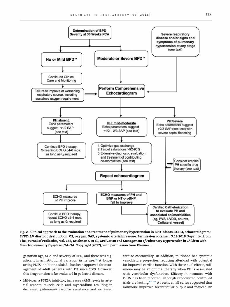
Fig. 2 –Clinical approach to the evaluation and treatment of pulmonary hypertension in BPD infants. ECHO, echocardiogram;
LVDD, LV diastolic dysfunction; O2, oxygen; SAP, systemic arterial pressure. Permission obtained, 3.19.2018: Reprinted from
The Journal of Pediatrics, Vol. 188, Krishnan U et al., Evaluation and Management of Pulmonary Hypertension in Children with
Bronchopulmonary Dysplasia, 24�34. Copyright (2017), with permission from Elsevier.
T AGGEDENDS E M I N A R S I N P E R I N A T O L O G Y 4 2 ( 2 0 1 8 ) 125
gestation age, SGA and severity of BPD, and there was sig-
nificant interinstitutional variation in its use.64 A longer
acting PDE5 inhibitor, tadalafil, has been approved for man-
agement of adult patients with PH since 2009. However,
this drug remains to be evaluated in pediatric disease.
� Milrinone, a PDE3A inhibitor, increases cAMP levels in arte-
rial smooth muscle cells and myocardium resulting in
decreased pulmonary vascular resistance and increased
cardiac contractility. In addition, milrinone has systemic
vasodilatory properties, reducing afterload with potential
for improved cardiac function. With these dual effects, mil-
rinone may be an optimal therapy when PH is associated
with ventricular dysfunction. Efficacy in neonates with
PPHN has been reported, although randomized controlled
trials are lacking.65�67 A recent small series suggested that
milrinone improved biventricular output and reduced RV

Table 2 – Pharmacotherapy of pulmonary hypertension in BPD.
Names Dose/titration Side effects Comments
Sildenafil phiospho-
diesterase-5
inhibitor
PO: 1 mg/kg 6�8 h; start with low dose
(0.3�0.5mg/kg/dose) and increase gradually to
1 mg/kg/dose as tolerated; slower as outpa-
tient. Maximal dose of 10mg q 8 h per EMA
guidelines for infants.
Hypotension, GER, irritability (headache), bron-
chospasm, nasal stuffiness, fever, rarely
priapism
Monitor for adverse effects, lower the
dose or switch to alternate therapy if
not tolerated
lntravenous: 0.25�0.5mg/kg/dose q 6�8 h
{titrate slowly and administer over 60min.)
Bosentan (Endothelin
receptor antagonist)
1mg/kg PO q 12 h as starting dose; may increase
to 2mg/kg BID in 2�4wk, if tolerated and liver
enzymes stable.
Liver dysfunction especially during viral infec-
tions, VOmismatch, hypotension, anemia
(edema and airway issues rare in infants)
Monitor LFT s monthly (earlier with
respiratory infections); monitor CBC
quarterly.
Teratogenicity precautions for
caregivers
Inhaled lloprost 2.5�5mcg every 2�4 h. Can be given as continu-
ous inhalation during mechanical ventilation.
Can titrate dose from 1 to 5mcg and frequency
from every 4 h to continuous.
Bronchospasm, hypotension, ventilator tube
crystallization and clogging, pulmonary hem-
orrhage, prostanoid side effects (GI disturban-
ces), may be teratogenic to caregivers.
Need close monitoring for clogged tub-
ing, may need further dilution. May
need bronchodilators or inhaled ste-
roid pretreatment with
bronchospasm.
Intravenous Epopros-
tenol (Flolan)
Start at 1�2 ng/kg/min, titrate up slowly every
4�6 h to 20 ng/kg/min; need to increase dose at
regular intervals because of tachyphylaxis. Fur-
ther increases as guided by clinical targets and
avoiding adverse effects.
Hypotension, VOmismatch, GI disturbances.
Needs dedicated line, very short half-life with
high risk for rebound PH with brief interruption
of therapy; line related complications include
infection, clogging, breaks in line, thrombosis,
arrhythmia)
Monitor closely if added to other vasodi-
lator therapies, such as milrinone;
careful attention to line care is
essential.
Treprostinil (Remodu-
lin) IV or
Subcutaneous
Start at 2 ng/kg/min and titrate every 4�6 h up to
20 ng/kg/min, then slow increase dose as toler-
ated (dose often 1.5�2 times greater than
equivalent epoprostenol dose, if switching
medications)
SO: local site pain; IV: similar risks as with epo-
prostenol, but treprostinil has a longer half-
life, which reduces risk for severe PH with
interruption of infusion
Site pain managed with local and sys-
temic measures
Milrinone (IV) (phos-
phodiesterase-3
inhibitor)
0.15�0.5mcg/kg/min � lower dosage range when
used with other vasodilators
Arrhythmogenic; systemic hypotension and high
risk for decreased myocardial perfusion; cau-
tion with renal dysfunction
May need to add a presser, such as vaso-
pressin, to mitigate effects of decrease
in systemic pressures.
BID, twice a day; CBC, complete blood count; EMA, European Medicines Agency; GER, gastroesophageal reflux; GI, gastrointestinal; IV, intravenous; kg, kilogram; LFT, liver function tests; mcg, micro-
gram; ng, nanogram; PO, oral; SC, subcutaneous; SR, sustained release; VO, ventilation-perfusion.
From The Journal of Pediatrics, Vol. 188, Krishnan U et al., Evaluation and Management of Pulmonary Hypertension in Children with Bronchopulmonary Dysplasia, 24�34. Copyright (2017), with per-
mission from Elsevier.
126S
EM
I N
A R
S
IN
P
ER
IN
AT
OL
OG
Y 42
(2018)
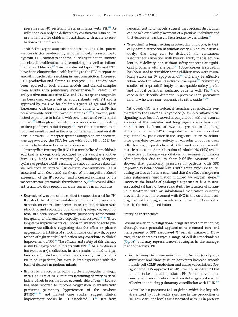
T AGGEDENDS E M I N A R S I N P E R I N A T O L O G Y 4 2 ( 2 0 1 8 ) 127
pressures in NO resistant preterm infants with PH.68 As
milrinone can only be delivered by continuous infusion, its
use is limited for children hospitalized with acute exacer-
bations of their disease.
Endothelin receptor antagonists: Endothelin-1 (ET-1) is a potent
vasoconstrictor produced by endothelial cells in response to
hypoxia. ET-1 promotes endothelial cell dysfunction, smooth
muscle cell proliferation and remodeling, as well as inflam-
mation and fibrosis.69 Two receptor subtypes (ETA and ETB)
have been characterized, with binding to the ETA receptor on
smooth muscle cells resulting in vasoconstriction. Increased
ET-1 production and altered ET receptor (ETR) activity have
been reported in both animal models and clinical samples
from adults with pulmonary hypertension.70 Bosentan, an
orally active non-selective ETA and ETB receptor antagonist,
has been used extensively in adult patients with PH and is
approved by the FDA for children 3 years of age and older.
Experience with bosentan in pediatric patients with PH has
been favorable with improved outcomes.71,72 However, pub-
lished experience in infants with BPD-associated PH remains
limited,73 although some institutions are now using this drug
as their preferred initial therapy.34 Liver functions need to be
followed monthly and in the event of an intercurrent viral ill-
ness. A newer ETA receptor specific antagonist, ambrisentan,
was approved by the FDA for use with adult PH in 2013 but
remains to be studied in pediatric disease.
Prostacyclins: Prostacyclin (PGI2) is a metabolite of arachidonic
acid that is endogenously produced by the vascular endothe-
lium. PGI2 binds to its receptor (IP), stimulating adenylate
cyclase to produce cAMP, resulting in smooth muscle relaxation
via reduction in intracellular calcium concentrations. PH is
associated with decreased synthesis of prostacyclin, reduced
expression of the IP receptor, and increased synthesis of the
vasoconstrictor prostanoid thromboxane A2.74,75 Several differ-
ent prostanoid drug preparations are currently in clinical use.
� Epoprostanol was one of the earliest therapeutics used for PH.
Its short half-life necessitates continuous infusion and
depends on central line access. In adults and children with
idiopathic and secondary pulmonary hypertension, epopros-
tenol has been shown to improve pulmonary hemodynam-
ics, quality of life, exercise capacity, and survival.76�81 These
long-term improvements can occur in absence of acute pul-
monary vasodilation, suggesting that the effect on platelet
aggregation, inhibition of smooth muscle cell growth, or pro-
tection of right ventricular function may contribute to clinical
improvement of PH.82 The efficacy and safety of this therapy
is still being explored in infants with BPD.83 As a continuous
intravenous (IV) medication, its use remains limited to inpa-
tient care. Inhaled epoprostanol is commonly used for acute
PH in adult patients, but there is little experience with this
form of delivery in preterm infants.
� Iloprost is a more chemically stable prostacyclin analogue
with a half-life of 20-30 minutes facilitating delivery by inha-
lation, which in turn reduces systemic side effects.84 Iloprost
has been reported to improve oxygenation in infants with
persistent pulmonary hypertension of the newborn
(PPHN)85�87 and limited case studies suggest clinical
improvement occurs in BPD-associated PH.88 Data from
neonatal test lung models suggest that optimal distribution
can be achieved with placement of a proximal nebulizer and
that delivery is feasible via high frequency ventilation.89
� Treprostinil, a longer acting prostacyclin analogue, is typi-
cally administered via inhalation every 4-6 hours. Alterna-
tively, this drug can be delivered via continuous
subcutaneous injection with bioavailability that is equiva-
lent to IV delivery, and without safety concerns or signifi-
cant problems with site pain.90 Subcutaneous treprostinil
has been used to transition some children who were chron-
ically stable on IV epoprostenol,91 and may be effective
when added to other vasodilator therapies.92 Preliminary
studies of treprostinil imply an acceptable safety profile
and clinical benefit in pediatric patients with PH,93 and
case series describe dramatic improvement in premature
infants who were non-responsive to nitric oxide.90,94
Nitric oxide (NO) is a biological signaling gas molecule syn-
thesized by the enzyme NO synthase (NOS). Alterations in NO
signaling have been observed in conjunction with, or even as
a cause of the vascular and lung injury characteristic of
BPD.95 Three isoforms of NOS are present in the lung,
although endothelial NOS is regarded as the most important
regulator of NO production in the lung vasculature. NO stimu-
lates guanylate cyclase activity in vascular smooth muscle
cells, leading to production of cGMP and vascular smooth
muscle relaxation. Administration of inhaled NO (iNO) results
in selective pulmonary vasodilation but requires continuous
administration due to its short half-life. Mourani et al.
showed that pulmonary pressures in patients with BPD
improved to near-normal levels with acute exposure to iNO
during cardiac catheterization, and that the effect was greater
than pulmonary vasodilation induced by oxygen alone.96
However, the benefit of prolonged exposure to iNO in BPD-
associated PH has not been evaluated. The logistics of contin-
uous treatment with an inhalational medication currently
prevent chronic management with iNO in the outpatient set-
ting; instead the drug is mainly used for acute PH exacerba-
tions in the hospitalized infant.
Emerging therapeutics
Several newer or investigational drugs are worth mentioning,
although their potential application to neonatal care and
management of BPD-associated PH remain unknown. How-
ever, these therapies target a range of cellular mechanisms
(Fig. 3)57 and may represent novel strategies in the manage-
ment of neonatal PH.
- Soluble guanylate cyclase simulators or activators (riociguat, a
stimulator and cinaciguat, an activator) increase smooth
muscle cell cGMP production and cause vasodilation. Rio-
ciguat was FDA approved in 2013 for use in adult PH but
remains to be studied in pediatric PH. Preliminary data on
cinaciguat from a newborn lamb model suggests it may be
effective in inducing pulmonary vasodilation with PPHN.97
- L-citrulline is a precursor to L-arginine, which is a key sub-
strate used by nitric oxide synthase in the production of
NO. Low citrulline levels are associated with PH in preterm
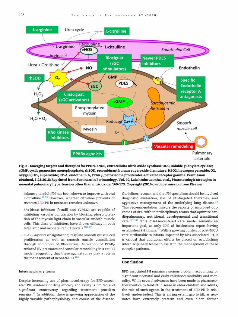
Fig. 3 – Emerging targets and therapies for PPHN. eNOS, extracellular nitric oxide synthase; sGC, soluble guanylate cyclase;
cGMP, cyclic guanosine monophosphate; rhSOD, recombinant human superoxide dismutase; H2O2, hydrogen peroxide; O2,
oxygen; O2-, superoxide; ET-A, endothelin A, PPAR g, peroxisome proliferator-activated receptor gamma. Permission
obtained, 3.23.2018: Reprinted from Seminars in Perinatology, Vol. 40, Lakshminrusimha, et al., Pharmacologic strategies in
neonatal pulmonary hypertension other than nitric oxide, 160-173. Copyright (2016), with permission from Elsevier.
128 S E M I N A R S I N P E R I N A T O L O G Y 4 2 ( 2 0 1 8 )
infants and adult PH has been shown to improve with oral
L-citrulline.98,99 However, whether citrulline prevents or
reverses BPD-PH in neonates remains unknown.
- Rho-kinase inhibitors (fasudil and Y27632) are capable of
inhibiting vascular contraction by blocking phosphoryla-
tion of the myosin light chain in vascular smooth muscle
cells. This class of inhibitors have shown efficacy in both
fetal lamb and neonatal rat PHmodels.100,101
- PPARg agonists (rosiglitazone) regulate smooth muscle cell
proliferation as well as smooth muscle vasodilation
through inhibition of Rho-kinase. Activation of PPARg
reduced RV pressures and vascular remodeling in a rat PH
model, suggesting that these agonists may play a role in
the management of neonatal PH.102
Interdisciplinary teams
Despite increasing use of pharmacotherapy for BPD-associ-
ated PH, evidence of drug efficacy and safety is limited and
significant controversy regarding treatment practices
remains.33 In addition, there is growing appreciation of the
highly variable pathophysiology and course of the disease.
Guidelines recommend that PH specialists should be involved
diagnostic evaluation, use of PH-targeted therapies, and
aggressive management of the underlying lung disease.34
This recommendation mirrors the reports of improved out-
comes of BPD with interdisciplinary teams that optimize car-
diopulmonary, nutritional, developmental and transitional
care.103,104 This disease-centered care model remains an
important goal, as only 30% of institutions report having
established PH clinics.35 With a growing burden of post-NICU
care attributable to infants impacted by BPD-associated PH, it
is critical that additional efforts be placed on establishing
interdisciplinary teams to assist in the management of these
complex patients.
Conclusion
BPD-associated PH remains a serious problem, accounting for
significant neonatal and early childhood morbidity and mor-
tality. While several advances have been made in pharmaco-
therapeutics to treat PH disease in older children and adults,
the role of such agents in the treatment of BPD-PH is rela-
tively understudied. This is an important gap to fill, as neo-
nates born extremely preterm and even older, former

T AGGEDENDS E M I N A R S I N P E R I N A T O L O G Y 4 2 ( 2 0 1 8 ) 129
preterm infants have distinct and diverse pathophysiology
leading to PH disease. Clinical and translational studies are
still urgently needed, not only to test the efficacy and safety
of certain agents but also to place into context the combined
use of clinical risk factors, novel biomarkers, screening and
diagnostic tests that will help navigate timing, dosing and
duration of therapy. Multidisciplinary efforts in both clinical
care and research will enhance strategies for prevention and
management.
R E F E R E N C E S
1. Stoll BJ, Hansen NI, Bell EF, et al. Trends in care practices,morbidity, and mortality of extremely preterm neonates,1993�2012. JAMA. 2015;314(10):1039–1051.
2. Zysman-Colman Z, Tremblay GM, Bandeali S, Landry JS.Bronchopulmonary dysplasia � trends over three decades.Paediatr Child Health. 2013;18(2):86–90.
3. Day CL, Ryan RM. Bronchopulmonary dysplasia: oldbecomes new again!. Pediatr Res. 2017;81(1-2):210–213.
4. Lapcharoensap W, Gage SC, Kan P, et al. Hospital variationand risk factors for bronchopulmonary dysplasia in a popu-lation-based cohort. JAMA Pediatr. 2015;169(2):e143676.
5. Northway WH Jr., Rosan RC, Porter DY. Pulmonary disease fol-lowing respirator therapy of hyaline-membrane disease. Bron-chopulmonary dysplasia.N Engl J Med. 1967;276(7):357–368.
6. Slaughter JL, Pakrashi T, Jones DE, South AP, Shah TA. Echo-cardiographic detection of pulmonary hypertension inextremely low birth weight infants with bronchopulmonarydysplasia requiring prolonged positive pressure ventilation.J Perinatol. 2011;31(10):635–640.
7. An HS, Bae EJ, Kim GB, et al. Pulmonary hypertension in pre-term infants with bronchopulmonary dysplasia. Korean CircJ. 2010;40(3):131–136.
8. Kim DH, Kim HS, Choi CW, Kim EK, Kim BI, Choi JH. Risk fac-tors for pulmonary artery hypertension in preterm infantswith moderate or severe bronchopulmonary dysplasia. Neo-natology. 2012;101(1):40–46.
9. Arjaans S, Zwart EAH, Ploegstra MJ, et al. Identification ofgaps in the current knowledge on pulmonary hypertensionin extremely preterm infants: a systematic review andmeta-analysis. Paediatr Perinat Epidemiol. 2018;32(3):258–267.https://doi.org/10.1111/ppe.12444: Epub 2018 Jan 17.
10. Anderson PJ, Doyle LW. Neurodevelopmental outcome ofbronchopulmonary dysplasia. Semin Perinatol. 2006;30(4):227–232.
11. Nakanishi H, Uchiyama A, Kusuda S. Impact of pulmonaryhypertension on neurodevelopmental outcome in preterminfants with bronchopulmonary dysplasia: a cohort study. JPerinatol. 2016;36(10):890–896.
12. Mirza H, Ziegler J, Ford S, Padbury J, Tucker R, Laptook A. Pul-monary hypertension in preterm infants: prevalence andassociation with bronchopulmonary dysplasia. J Pediatr.2014;165(5):909–914: e901.
13. Waruingi W, Mhanna MJ. Pulmonary hypertension inextremely low birth weight infants: characteristics and out-comes.World J Pediatr. 2014;10(1):46–52.
14. Mourani PM, Sontag MK, Younoszai A, et al. Early pulmonaryvascular disease in preterm infants at risk for bronchopulmo-nary dysplasia. Am J Respir Crit Care Med. 2015;191(1):87–95.
15. Weismann CG, Asnes JD, Bazzy-Asaad A, Tolomeo C,Ehrenkranz RA, Bizzarro MJ. Pulmonary hypertension in pre-term infants: results of a prospective screening program. JPerinatol. 2017;37(5):572–577.
16. Beshish AG, Haraldsdottir K, Barton G, et al. Pulmonary vas-cular response to exercise and hypoxia in adult survivors ofpreterm birth. Am J Resp Crit Care Med. 2017;195:A3109.
17. Baraldi E, Filippone M. Chronic lung disease after prematurebirth. N Engl J Med. 2007;357(19):1946–1955.
18. Farquhar M, Fitzgerald DA. Pulmonary hypertension in chronicneonatal lung disease. Paediatr Respir Rev. 2010;11(3):149–153.
19. Khemani E, McElhinney DB, Rhein L, et al. Pulmonary arteryhypertension in formerly premature infants with broncho-pulmonary dysplasia: clinical features and outcomes in thesurfactant era. Pediatrics. 2007;120(6):1260–1269.
20. Check J, Gotteiner N, Liu X, et al. Fetal growth restriction andpulmonary hypertension in premature infants with bron-chopulmonary dysplasia. J Perinatol. 2013;33(7):553–557.
21. Mestan KK, Check J, Minturn L, et al. Placental pathologicchanges of maternal vascular underperfusion in broncho-pulmonary dysplasia and pulmonary hypertension. Placenta.2014;35(8):570–574.
22. Yallapragada SG, Mestan KK, Palac H, et al. Placental villousvascularity is decreased in premature infants with broncho-pulmonary dysplasia-associated pulmonary hypertension.Pediatr Dev Pathol. 2016;19(2):101–107.
23. Mestan KK, Gotteiner N, Porta N, Grobman W, Su EJ, ErnstLM. Cord blood biomarkers of placental maternal vascularunderperfusion predict bronchopulmonary dysplasia-asso-ciated pulmonary hypertension. J Pediatr. 2017;185:33–41.
24. Su EJ, Xin H, Yin P, et al. Impaired fetoplacental angiogenesisin growth-restricted fetuses with abnormal umbilical arterydoppler velocimetry is mediated by aryl hydrocarbon recep-tor nuclear translocator (ARNT). J Clin Endocrinol Metab.2015;100(1):E30–E40.
25. Baker CD, Seedorf GJ, Wisniewski BL, et al. Endothelial col-ony-forming cell conditioned media promote angiogenesisin vitro and prevent pulmonary hypertension in experimen-tal bronchopulmonary dysplasia. Am J Physiol Lung Cell MolPhysiol. 2013;305(1):L73–L81.
26. Pipp F, Heil M, Issbrucker K, et al. VEGFR-1-selective VEGFhomologue PlGF is arteriogenic: evidence for a monocyte-mediated mechanism. Circul Res. 2003;92(4):378–385.
27. Madonna R, De Caterina R, Geng YJ. Epigenetic regulation ofinsulin-like growth factor signaling: a novel insight into thepathophysiology of neonatal pulmonary hypertension. Vas-cul Pharmacol. 2015;73:4–7.
28. Xu XF, Lv Y, Gu WZ, et al. Epigenetics of hypoxic pulmonaryarterial hypertension following intrauterine growth retardationrat: epigenetics in PAH following IUGR. Respir Res. 2013;14:20.
29. Wallace B, Peisl A, Seedorf G, et al. Anti-sFlt-1 therapy pre-serves lung alveolar and vascular growth in antenatal mod-els of bronchopulmonary dysplasia. Am J Respir Crit Care Med.2018;197(6):776–787.
30. Willis GR, Kourembanas S, Mitsialis SA. Toward exosome-based therapeutics: isolation, heterogeneity, and fit-for-pur-pose potency. Front Cardiovasc Med. 2017;4:63.
31. Chen CM, Lin W, Huang LT, Chou HC. Human mesenchymalstem cells ameliorate experimental pulmonary hyperten-sion induced by maternal inflammation and neonatal hyper-oxia in rats. Oncotarget. 2017;8(47):82366–82375.
32. Augustine S, Avey MT, Harrison B, et al. Mesenchymal stro-mal cell therapy in bronchopulmonary dysplasia: systematicreview and meta-analysis of preclinical studies. Stem CellsTransl Med. 2017;6(12):2079–2093.
33. Abman SH, Hansmann G, Archer SL, et al. Pediatric pulmo-nary hypertension: guidelines from the american heart asso-ciation and american thoracic society. Circulation. 2015;132(21):2037–2099.
34. Krishnan U, Feinstein JA, Adatia I, et al. Evaluation and man-agement of pulmonary hypertension in children with bron-chopulmonary dysplasia. J Pediatr. 2017;188:24–34: e21.

130 S E M I N A R S I N P E R I N A T O L O G Y 4 2 ( 2 0 1 8 )
35. Altit G, Lee HC, Hintz S, Tacy TA, Feinstein JA, Bhombal S.Practices surrounding pulmonary hypertension and bron-chopulmonary dysplasia amongst neonatologists caring forpremature infants. J Perinatol. 2018;38(4):361–367.
36. Mourani PM, Sontag MK, Younoszai A, Ivy DD, Abman SH.Clinical utility of echocardiography for the diagnosis andmanagement of pulmonary vascular disease in young chil-dren with chronic lung disease. Pediatrics. 2008;121(2):317–325.
37. Arcasoy SM, Christie JD, Ferrari VA, et al. Echocardiographicassessment of pulmonary hypertension in patients withadvanced lung disease. Am J Respir Crit Care Med.2003;167(5):735–740.
38. Carlton EF, Sontag MK, Younoszai A, et al. Reliability of echo-cardiographic indicators of pulmonary vascular disease inpreterm infants at risk for bronchopulmonary dysplasia. JPediatr. 2017;186:29–33.
39. Singh GK, Levy PT, Holland MR, Hamvas A. Novel methodsfor assessment of right heart structure and function in pul-monary hypertension. Clin Perinatol. 2012;39(3):685–701.
40. Kwon HW, KimHS, An HS, et al. Long-Term outcomes of pul-monary hypertension in preterm infants with bronchopul-monary dysplasia. Neonatology. 2016;110(3):181–189.
41. Latson LA, Prieto LR. Congenital and acquired pulmonaryvein stenosis. Circulation. 2007;115(1):103–108.
42. Swier NL, Richards B, Cua CL, et al. Pulmonary vein stenosisin neonates with severe bronchopulmonary dysplasia. Am JPerinatol. 2016;33(7):671–677.
43. del Cerro MJ, Sabate Rotes A, Carton A, et al. Pulmonaryhypertension in bronchopulmonary dysplasia: clinical find-ings, cardiovascular anomalies and outcomes. Pediatr Pulmo-nol. 2014;49(1):49–59.
44. Drossner DM, Kim DW, Maher KO, Mahle WT. Pulmonaryvein stenosis: prematurity and associated conditions. Pediat-rics. 2008;122(3):e656–e661.
45. Taylor CJ, Derrick G, McEwan A, Haworth SG, Sury MR. Risk ofcardiac catheterization under anaesthesia in children withpulmonary hypertension. Br J Anaesth. 2007;98(5):657–661.
46. Hill KD, Lim DS, Everett AD, Ivy DD, Moore JD. Assessment ofpulmonary hypertension in the pediatric catheterizationlaboratory: current insights from the Magic registry. CatheterCardiovasc Interv. 2010;76(6):865–873.
47. Mauritz GJ, Rizopoulos D, Groepenhoff H, et al. Usefulness ofserial N-terminal pro-B-type natriuretic peptide measure-ments for determining prognosis in patients with pulmo-nary arterial hypertension. Am J Cardiol. 2011;108(11):1645–1650.
48. Vijlbrief DC, Benders MJ, Kemperman H, van Bel F, de VriesWB. B-type natriuretic peptide and rebound during treat-ment for persistent pulmonary hypertension. J Pediatr.2012;160(1):111–115: e111.
49. Reynolds EW, Ellington JG, Vranicar M, Bada HS. Brain-typenatriuretic peptide in the diagnosis and management of per-sistent pulmonary hypertension of the newborn. Pediatrics.2004;114(5):1297–1304.
50. Nir A, Lindinger A, Rauh M, et al. NT-pro-B-type natriureticpeptide in infants and children: reference values based oncombined data from four studies. Pediatr Cardiol.2009;30(1):3–8.
51. Cuna A, Kandasamy J, Sims B. B-type natriuretic peptide andmortality in extremely low birth weight infants with pulmo-nary hypertension: a retrospective cohort analysis. BMCPediatr. 2014;14:68.
52. Kim GB. Pulmonary hypertension in infants with broncho-pulmonary dysplasia. Korean J Pediatr. 2010;53(6):688–693.
53. Joseph L, Nir A, Hammerman C, Goldberg S, Ben Shalom E,Picard E. N-terminal pro-B-type natriuretic peptide as amarker of bronchopulmonary dysplasia in prematureinfants. Am J Perinatol. 2010;27(5):381–386.
54. Minkin R, Sandhu G, Grosu H, et al. Desmosine and Isodes-mosine as a novel biomarker for pulmonary arterial hyper-tension: a pilot study. Am J Ther. 2017;24(4):e399–e404.
55. Ochiai M, Hikino S, Yabuuchi H, et al. A new scoring systemfor computed tomography of the chest for assessing the clin-ical status of bronchopulmonary dysplasia. J Pediatr.2008;152(1):90–95: e91�93.
56. Wu JE, White A, Wang Y, Biko D, Zhang H, Fraga M, Mercer-Rosa L, Kirpalani H. Phenotypes of Severe Bronchopulmo-nary Dysplasia. Philadelphia, PA: Abstract 111, Eastern Soci-ety for Pediatric Research; 2018.
57. Lakshminrusimha S, Mathew B, Leach CL. Pharmacologicstrategies in neonatal pulmonary hypertension other thannitric oxide. Semin Perinatol. 2016;40(3):160–173.
58. Farrow KN, Steinhorn RH. Phosphodiesterases: emergingtherapeutic targets for neonatal pulmonary hypertension.Handb Exp Pharmacol. 2011(204):251–277.
59. Mourani PM, Sontag MK, Ivy DD, Abman SH. Effects of long-term sildenafil treatment for pulmonary hypertension ininfants with chronic lung disease. J Pediatr. 2009;154(3):379–384:e371�372.
60. NypM, Sandritter T, Poppinga N, Simon C, TruogWE. Sildenafilcitrate, bronchopulmonary dysplasia and disordered pulmo-nary gas exchange: any benefits? J Perinatol. 2012;32(1):64–69.
61. Wardle AJ, Wardle R, Luyt K, Tulloh R. The utility of sildenafilin pulmonary hypertension: a focus on bronchopulmonarydysplasia. Arch Dis Child. 2013;98(8):613–617.
62. Barst RJ, Beghetti M, Pulido T, et al. STARTS-2: long-term sur-vival with oral sildenafil monotherapy in treatment-naivepediatric pulmonary arterial hypertension. Circulation.2014;129(19):1914–1923.
63. Aboudi D, Swaminathan N, Brumberg HL, et al. Sildenafil andretinopathy of prematurity in preterm infatns with broncho-pulmonary dysplasia. J Pediatr. 2018;199:16–21. https://doi.org/10.1016/j.jpeds.2018.04.005. Epub 2018 May 9.
64. Backes CH, Reagan PB, Smith CV, Jadcherla SR, Slaughter JL.Sildenafil treatment of infants with bronchopulmonary dys-plasia-associated pulmonary hypertension. Hosp Pediatr.2016;6(1):27–33.
65. McNamara PJ, Laique F, Muang-In S, Whyte HE. Milrinoneimproves oxygenation in neonates with severe persistentpulmonary hypertension of the newborn. J Crit Care.2006;21(2):217–222.
66. McNamara PJ, Shivananda SP, Sahni M, Freeman D, TaddioA. Pharmacology of milrinone in neonates with persistentpulmonary hypertension of the newborn and suboptimalresponse to inhaled nitric oxide. Pediatr Crit Care Med.2013;14(1):74–84.
67. Bassler D, Kreutzer K, McNamara P, Kirpalani H. Milrinonefor persistent pulmonary hypertension of the newborn.Cochrane Database Syst Rev. 2010;11:CD007802.
68. James AT, Corcoran JD, McNamara PJ, Franklin O, El-KhuffashAF. The effect of milrinone on right and left ventricular func-tion when used as a rescue therapy for term infants with pul-monary hypertension. Cardiol Young. 2016;26(1):90–99.
69. Abman SH. Role of endothelin receptor antagonists in thetreatment of pulmonary arterial hypertension. Annu RevMed. 2009;60:13–23.
70. Giaid A, Yanagisawa M, Lagleben D, et al. Expression ofendothelin-1 in the lungs of patients with pulmonary hyper-tension. New Engl J Med. 1993;328:1732–1739.
71. Beghetti M. Current treatment options in children with pul-monary arterial hypertension and experiences with oralbosentan. Eur J Clin Invest. 2006;36(Suppl 3):16–24.
72. Maiya S, Hislop AA, Flynn Y, Haworth SG. Response tobosentan in children with pulmonary hypertension. Heart.2006;92(5):664–670.
T AGGEDENDS E M I N A R S I N P E R I N A T O L O G Y 4 2 ( 2 0 1 8 ) 131
73. Rugolotto S, Errico G, Beghini R, Ilic S, Richelli C, PadovaniEM.Weaning of epoprostenol in a small infant receiving con-comitant bosentan for severe pulmonary arterial hyperten-sion secondary to bronchopulmonary dysplasia. MinervaPediatr. 2006;58(5):491–494.
74. Christman BW, McPherson CD, Newman JH, et al. An imbal-ance between the excretion of thromboxane and prostacy-clin metabolites in pulmonary hypertension. New Engl J Med.1992;327(2):70–75.
75. Lakshminrusimha S, Porta NF, Farrow KN, et al. Milrinoneenhances relaxation to prostacyclin and iloprost in pulmo-nary arteries isolated from lambs with persistent pulmonaryhypertension of the newborn. Pediatr Crit Care Med.2009;10(1):106–112.
76. Rosenzweig EB, Kerstein D, Barst RJ. Long-term prostacyclinfor pulmonary hypertension with associated congenitalheart defects. Circulation. 1999;99(14):1858–1865.
77. Barst RJ, Maislin G, Fishman AP. Vasodilator therapy for pri-mary pulmonary hypertension in children. Circulation.1999;99(9):1197–1208.
78. Barst RJ, Rubin LJ, Long WA, et al. A comparison of continu-ous intravenous epoprostenol (prostacyclin) with conven-tional therapy for primary pulmonary hypertension. ThePrimary Pulmonary Hypertension Study Group. New Engl JMed. 1996;334(5):296–302.
79. Barst RJ, Rubin LJ, McGoon MD, Caldwell EJ, Long WA, LevyPS. Survival in primary pulmonary hypertension with long-term continuous intravenous prostacyclin. Ann Intern Med.1994;121(6):409–415.
80. Sitbon O, Humbert M, Nunes H, et al. Long-term intravenousepoprostenol infusion in primary pulmonary hypertension:prognostic factors and survival. J Am Coll Cardiol.2002;40(4):780–788.
81. Yung D, Widlitz AC, Rosenzweig EB, Kerstein D, Maislin G,Barst RJ. Outcomes in children with idiopathic pulmonaryarterial hypertension. Circulation. 2004;110(6):660–665.
82. D’Alonzo GE, Barst RJ, Ayres SM, et al. Survival in patients withprimary pulmonary hypertension. Results from a national pro-spective registry.Ann Intern Med. 1991;115(5):343–349.
83. Zaidi AN, Dettorre MD, Ceneviva GD, Thomas NJ. Epoproste-nol and home mechanical ventilation for pulmonary hyper-tension associated with chronic lung disease. PediatrPulmonol. 2005;40(3):265–269.
84. Ewert R, Schaper C, Halank M, Glaser S, Opitz CF. Inhalativeiloprost � pharmacology and clinical application. Expert OpinPharmacother. 2009;10(13):2195–2207.
85. Ehlen M, Wiebe B. Iloprost in persistent pulmonary hyper-tension of the newborn. Cardiol Young. 2003;13(4):361–363.
86. Janjindamai W, Thatrimontrichai A, Maneenil G, ChanvitanP, Dissaneevate S. Effectiveness and safety of intravenousiloprost for severe persistent pulmonary hypertension of thenewborn. Indian Pediatr. 2013;50(10):934–938.
87. Kahveci H, Yilmaz O, Avsar UZ, et al. Oral sildenafil andinhaled iloprost in the treatment of pulmonary hypertensionof the newborn. Pediatr Pulmonol. 2014;49(12):1205–1213.
88. Hwang SK, O YC, KimNS, Park HK, YumMK. Use of inhaled ilo-prost in an infant with bronchopulmonary dysplasia and pul-monary artery hypertension. Korean Circ J. 2009;39(8):343–345.
89. DiBlasi RM, Crotwell DN, Shen S, Zheng J, Fink JB, Yung D.Iloprost drug delivery during infant conventional and high-frequency oscillatory ventilation. Pulm Circ. 2016;6(1):63–69.
90. Ferdman DJ, Rosenzweig EB, Zuckerman WA, Krishnan U.Subcutaneous treprostinil for pulmonary hypertension inchronic lung disease of infancy. Pediatrics. 2014;134(1):e274–e278.
91. Ivy DD, Claussen L, Doran A. Transition of stable pediatricpatients with pulmonary arterial hypertension from intrave-nous epoprostenol to intravenous treprostinil. Am J Cardiol.2007;99(5):696–698.
92. Levy M, Celermajer DS, Bourges-Petit E, Del Cerro MJ, BajolleF, Bonnet D. Add-on therapy with subcutaneous treprostinilfor refractory pediatric pulmonary hypertension. J Pediatr.2011;158(4):584–588.
93. Krishnan U, Takatsuki S, Ivy DD, et al. Effectiveness andsafety of inhaled treprostinil for the treatment of pulmonaryarterial hypertension in children. Am J Cardiol. 2012;110(11):1704–1709.
94. Park BY, Chung SH. Treprostinil for persistent pulmonaryhypertension of the newborn, with early onset sepsis in pre-term infant: 2 Case reports. Medicine (Baltimore). 2017;96(26):e7303.
95. Afshar S, Gibson LL, Yuhanna IS, et al. Pulmonary NO syn-thase expression is attenuated in a fetal baboon model ofchronic lung disease. Am J Physiol Lung Cell Mol Physiol.2003;284(5):L749–L758.
96. Mourani PM, Ivy DD, Gao D, Abman SH. Pulmonary vasculareffects of inhaled nitric oxide and oxygen tension in bron-chopulmonary dysplasia. Am J Respir Crit Care Med.2004;170(9):1006–1013.
97. Chester M, Seedorf G, Tourneux P, et al. Cinaciguat, a solubleguanylate cyclase activator, augments cGMP after oxidativestress and causes pulmonary vasodilation in neonatal pul-monary hypertension. Am J Physiol Lung Cell Mol Physiol.2011;301(5):L755–L764.
98. O’Connor MG, Suthar D, Vera K, et al. Pulmonary hyperten-sion in the premature infant population: analysis of echocar-diographic findings and biomarkers. Pediatr Pulmonol.2018;53(3):302–309.
99. Sharif Kashani B, Tahmaseb Pour P, Malekmohammad M,et al. Oral l-citrulline malate in patients with idiopathic pul-monary arterial hypertension and Eisenmenger Syndrome: aclinical trial. J Cardiol. 2014;64(3):231–235.
100. Chou HC, Huang LT, Yeh TF, Chen CM. Rho-kinase inhibitorY-27632 attenuates pulmonary hypertension in hyperoxia-exposed newborn rats. Acta Pharmacol Sin. 2013;34(10):1310–1316.
101. Parker TA, Roe G, Grover TR, Abman SH. Rho kinase activa-tion maintains high pulmonary vascular resistance in theovine fetal lung. Am J Physiol Lung Cell Mol Physiol.2006;291(5):L976–L982.
102. Zhang D, Wang G, Han D, et al. Activation of PPAR�gammaameliorates pulmonary arterial hypertension via inducingheme oxygenase-1 and p21(WAF1): an in vivo study in rats.Life Sci. 2014;98(1):39–43.
103. Abman SH, Collaco JM, Shepherd EG, et al. Interdisciplinarycare of children with severe bronchopulmonary dysplasia. JPediatr. 2017;181:12–28: e11.
104. Gien J, Kinsella J, Thrasher J, Grenolds A, Abman SH, BakerCD. Retrospective analysis of an interdisciplinary ventilatorcare program intervention on survival of infants with venti-lator-dependent bronchopulmonary dysplasia. Am J Perina-tol. 2017;34(2):155–163.

Accepted Manuscript
Review
Bronchopulmonary dysplasia: pathophysiology and potential anti-inflammatorytherapies
Paris C. Papagianis, J.J. Pillow, Timothy J. Moss
PII: S1526-0542(18)30079-4DOI: https://doi.org/10.1016/j.prrv.2018.07.007Reference: YPRRV 1281
To appear in: Paediatric Respiratory Reviews
Please cite this article as: P.C. Papagianis, J.J. Pillow, T.J. Moss, Bronchopulmonary dysplasia: pathophysiologyand potential anti-inflammatory therapies, Paediatric Respiratory Reviews (2018), doi: https://doi.org/10.1016/j.prrv.2018.07.007
This is a PDF file of an unedited manuscript that has been accepted for publication. As a service to our customerswe are providing this early version of the manuscript. The manuscript will undergo copyediting, typesetting, andreview of the resulting proof before it is published in its final form. Please note that during the production processerrors may be discovered which could affect the content, and all legal disclaimers that apply to the journal pertain.
132
133
Bronchopulmonary dysplasia: pathophysiology and potential anti-inflammatory
therapies.
Paris C. Papagianis1,2, J J. Pillow2 and Timothy J. Moss1
1The Ritchie Centre, Hudson Institute of Medical Research, Department of Obstetrics and Gynecology, Monash
University, Clayton, VIC 3168, Australia
2Human Sciences and Centre for Neonatal Research and Education, The University of Western Australia, Crawley,
WA, Australia
PC Papagianis: [email protected]
JJ Pillow: [email protected]
TJ Moss: [email protected]
Correspondence to:
Paris Papagianis, BSci (Hons), PhD candidate
Email: [email protected]
Tel: +61 430 127 197
Declarations of interest: none
Funding: NHMRC Grants: 1057514, 1057759, 1077769, 1077691 and 1043294
Acknowledgements: The Victorian Government’s Operational Infrastructure Support Program.

134
1 Introduction
Globally, 15 million infants are born preterm each year, representing more than 1 in 10 live births.1 Extremely
preterm infants (born before 28 weeks’ gestational age (GA)) are the most vulnerable, most commonly
complicated by the chronic lung disease, bronchopulmonary dysplasia (BPD).2 BPD is the only complication of
preterm birth for which the incidence is increasing at the same time that all other co-morbidities are decreasing2.
There is no cure for BPD. The use of antenatal corticosteroids and exogenous surfactant administration has
improved the acute respiratory complications of preterm birth dramatically, but these treatments do not prevent
BPD.3, 4 The early application of non-invasive respiratory support is an alternative strategy aimed at preventing
BPD but is unsuccessful at reducing its incidence.5
Although BPD is clearly a respiratory disease of prematurity, BPD may not simply be a consequence of lung
immaturity. The immature immune system of preterm neonates coupled with the immune modulating effects of
factors that commonly accompany preterm birth (e.g. antenatal corticosteroids, intrauterine
infection/inflammation and postnatal sepsis),6 may be responsible for aberrant regulation of lung inflammation.
Thus, targeting the chronic lung inflammation that underlies BPD with the use of new anti-inflammatory
therapies offers the prospect of preventative and reparative treatment for infants with BPD.
2 Bronchopulmonary dysplasia
BPD was first described by Northway in the 1960s as a form of chronic lung disease in moderately preterm
infants, characterised predominately by fibrosis.7 Northway attributed the injurious effects of high oxygen levels
and airway pressures used in mechanical ventilation as the main causes of the disease7. However, changes in
respiratory management strategies for preterm infants has transformed BPD into a disease characterised by
more subtle lung abnormalities, including alveolar hypoplasia (fewer and larger simplified alveoli),8, 9
dysmorphic pulmonary vasculature and chronic pulmonary inflammation.10-12 The lungs of infants with severe
BPD exhibit a lack of septation within the developing airspace, resulting in reduced surface area for gas exchange
in the lungs, hyperconstrictive vasculature that further compromises gas exchange, and impaired surfactant
synthesis,13 favouring atelectasis. The National Institute of Child Health and Human Development defines this
“New BPD” as the requirement for at least 28 days of supplemental oxygen, with the severity of BPD indicated by
the level of respiratory support required at 36 weeks postmenstrual age.14
3 The aetiology of BPD
3.1 Prenatal inflammation
Inflammation within the uterus during pregnancy is a common antecedent of preterm birth that manifests as
chorioamnionitis; inflammation of the chorion and amnion. Chorioamnionitis is commonly classified as clinical
or histological: clinical chorioamnionitis is diagnosed prior to labour, when women present with symptoms
including fever, a tender uterus and preterm rupture of membranes (PROM) with purulent liquor; histological
chorioamnionitis is an asymptomatic inflammation of the membranes.15 Histological chorioamnionitis is more
common than clinical chorioamnionitis but may be undiagnosed because post-partum histological examination

135
of the placenta is required.15 Histological chorioamnionitis complicates between 30–70 % of preterm births with
PROM and spontaneous labour, with incidence inversely related to GA. Thus, rates of histological
chorioamnionitis exceed 70 % for infants at highest risk of BPD (23 weeks GA).16
Prenatal inflammation alters fetal lung development, with consequences that may be detrimental or beneficial
for preterm newborns. Histological chorioamnionitis reduces the risk of respiratory distress syndrome (RDS),16-
18 likely due to elevated surfactant production in the lungs.19-21 However, despite lower risk of RDS, infants
exposed to chorioamnionitis may require longer term respiratory support and have higher rates of BPD and
persistent pulmonary hypertension of the newborn (PPHN).22 Although increases18, 23 or decreases16,24 in the
incidence of BPD are reported after exposure to chorioamnionitis, the relationship is complicated by low-birth-
weight and postnatal events, such as sepsis. The avoidance of prolonged mechanical ventilation in low-birth-
weight newborns exposed to chorioamnionitis is associated with decreased incidence of BPD, compared to
newborns exposed to chorioamnionitis who received prolonged ventilation.25 It is unclear whether a prenatal
(chorioamnionitis) or postnatal (ventilator-induced) origin of lung inflammation is the greater contributor to the
pathogenesis of BPD.
Fetal sheep exposed to inflammation induced by intra-amniotic (IA) injection of lipopolysaccharide (LPS) have
lung abnormalities like those observed in infants who die of BPD: alveolar hypoplasia19, 26 and decreased
septation,19, 27 impaired surfactant secretion (despite increased surfactant protein),19 and impaired pulmonary
vascular development and function.28 The similarities in the lungs of fetal lambs exposed to inflammation and
the pathological features of BPD support a prenatal origin of BPD. Although preterm infants exposed to
chorioamnionitis have less RDS, experimental intrauterine inflammation does not reduce the postnatal
respiratory support required by preterm baboons29 and lambs.30
3.2 Respiratory Support
Mechanical ventilation initiates an influx of neutrophils and macrophages into the alveoli.31 These cells produce
cytokines,31 which can disrupt lung development and may be used as biomarkers in serum and tracheal aspirates
to identify infants at risk of BPD.10-12 Infants who develop BPD have persistently elevated pro-inflammatory
cytokines (IL-1β, IL-6 and IL-8) in tracheal aspirates and blood, compared to infants who recover from initial
RDS.32 Preterm infants may become increasingly dependent on respiratory support, which exacerbates
pulmonary inflammation, inducing the production of reactive oxygen species (ROS).33 ROS promote
inflammation and epithelial cell death in the lungs via the cleavage, and thus activation, of caspase-1 (Figure 1).34
In preterm lambs, 2 hours of mechanical ventilation initiates inflammation within the lungs resulting in similar
upregulation of IL-1β, IL-6 and IL-8, the same biomarkers as infants that are ventilated or were exposed to
chorioamnionitis.35, 36 Longer ventilation of preterm lambs (3-4 weeks) increases neutrophil and macrophage
infiltration into the lungs and causes non-uniform inflation patterns and abnormal lung vascular development,
similar to that observed in infants who die from BPD.37
4 Long-term pulmonary consequences of BPD
The long-term sequelae of new BPD are described by very few studies. Autopsies reveal its major pathological
features: abnormal alveolar architecture (alveolar hypoplasia) and impaired pulmonary vascular development,
136
where vessels are distant from airspaces.8, 9 Pulmonary gas exchange is impaired in 2-year-old infants with BPD
compared to non-BPD controls; however, alveolar volume was normal in both cohorts, suggesting a lower
alveolar surface area, consistent with an alveolar hypoplasia lung phenotype.38 Persistent abnormalities in lung
parenchyma have long-term functional consequences: 7-to-8-year-olds who had BPD and received surfactant
during the perinatal period had lower forced expiratory volume and higher airway resistance, compared to age-
and sex- matched controls without BPD, indicative of increased work of breathing.39
5 Steroidal approaches to prevent pulmonary inflammation
5.1 Antenatal corticosteroids
Antenatal corticosteroids administered to women at risk of preterm delivery accelerate fetal lung maturation to
prevent RDS but do not prevent BPD.3 Antenatal corticosteroids cause remodeling of the lung parenchyma,
which improves gas exchange but results ultimately in fewer, larger alveoli,40 like the lungs of fetal sheep
exposed to prenatal inflammation.8, 19 Antenatal corticosteroids alter immune activity by suppressing
lymphocytes but increasing neutrophils in preterm infants,41 and altering immune cell function,42 which may
underlie an increased risk of early-onset sepsis.3
Antenatal corticosteroid treatment can suppress lung inflammation (induced by IA injection of LPS) in fetal
sheep but the reduction of LPS-induced inflammation is transient.43 Thus, the timing of antenatal corticosteroid
administration in humans may influence the lung inflammation that accompanies chorioamnionitis. The optimal
timing, dose, and frequency of administration of glucocorticoids to women at risk of preterm birth are unknown.
While chorioamnionitis is not a contraindication to the use of antenatal corticosteroids, their interaction likely
affects lung development differently to either in isolation;19, 44 the impact of this interaction on rates of BPD is
not known.
Few studies investigate any effect of antenatal glucocorticoids on postnatal ventilator requirements and lung
inflammation. In adult animals, pretreatment with corticosteroids reduces ventilation-induced lung injury.45, 46
Preterm lambs ventilated following exposure to antenatal glucocorticoids have less lung injury and inflammation
in comparison to ventilated preterm lambs exposed to antenatal saline.47 The maturational and anti-
inflammatory effects of antenatal glucocorticoids in the preterm lungs appear to be maintained for the initial
ventilation period, consistent with lower RDS incidence. However, meta-analyses show that antenatal
corticosteroids do not reduce BPD.3
5.2 Postnatal glucocorticoids
Postnatal glucocorticoid use peaked in the late 1990s48 after observations of improved extubation rates in a
small trial.49 Later meta-analyses revealed reduced BPD incidence in preterm infants who received postnatal
glucocorticoids, but increased incidence of cerebral palsy and death.50-53 Thus, postnatal glucocorticoids are now
used reluctantly in infants with intractable BPD: fewer than 10 % of preterm infants receive them.2
Ventilator management of preterm infants has evolved since the 1990s, aiming for respiratory management with
lower airway pressures and inspired oxygen concentrations, and avoidance of prolonged periods of intubation.5,
54 The practice of more ‘gentle’ ventilation coupled with significantly less postnatal steroid exposure may result

137
in infants receiving longer periods of respiratory support than may have been used prior to adoption of these
changes in practice. It is unclear whether the increasing BPD incidence is attributable to longer periods of
respiratory support and/or the decrease in steroid use. Typically, infants who receive postnatal steroids are
those who require prolonged periods of invasive ventilation, indicating a stronger predisposition for BPD
development, and complicating any assessment of the impact of postnatal steroids on BPD incidence.
Early studies of postnatal high-dose dexamethasone therapy (~1 mg/kg/day over 42 d)49 focused on immediate
respiratory outcomes, at the expense of neurological follow-up. Optimal postnatal steroid dosing regimens are
undefined, contributing to reluctance for their clinical use. Early administration of lower dexamethasone doses
may be safe and effective in infants at high-risk of developing BPD.53
Despite the key role of inflammation in the pathogenesis of BPD, few studies of postnatal steroids include
inflammation as an outcome. Small studies describe a reduction in neutrophils55, 56 and IL-1 concentration in
bronchoalveolar lavage (BAL) of ventilated preterm infants receiving dexamethasone.55, 57 Similarly, IL-1β
expression is lower in the lungs of preterm lambs receiving a single dexamethasone dose (0.5 mg/kg)
immediately before initiation of ventilation compared to placebo controls.47 No animal studies assess the ability
of postnatal dexamethasone to treat established lung inflammation and injury.
Neurological impairment is associated with high dose postnatal steroids. The risk of cerebral palsy increases by
40 % for every 1 mg/kg increase in dexamethasone dose.58 The most premature infants are not the worst
affected (despite presumably more immature organ systems); treatment after 33 weeks’ postmenstrual age is
associated with greatest neurological deficit at follow-up.58 Adverse clinical neurological outcomes associated
with dexamethasone are consistent with animal studies.59-61 Other complications of high-dose postnatal steroid
use include stunted growth62, intraventricular haemorrhage (IVH),53, 63 gastrointestinal bleeding,52 sepsis and
hypertension.51-53, 64 Combined use of dexamethasone and indomethacin (for closure of the ductus arteriosus)
increases the likelihood of gastrointestinal perforation three-fold.62
The DART trial aimed to investigate the ability of a low-dose 10-day tapered course of postnatal dexamethasone
(0.89 mg/kg cumulative over 10 days) to prevent BPD in preterm infants born before 28 weeks GA.65 The trial
was terminated because of low (10 %) recruitment,65 but infants who received dexamethasone spent less time
intubated on mechanical ventilation; although this did not reach statistical significance. Major disability and
cerebral palsy were not different between dexamethasone-treated and placebo-treated preterm infants at 2
years follow-up.66
6 Non-steroidal approaches to prevent pulmonary inflammation
Inflammation associated with BPD involves the activation or over-expression of a number of inflammatory
cytokines and pro-inflammatory mediators (Figure 1) providing opportunity for multiple therapeutic targets to
prevent lung inflammation in newborn infants. Inhibition of cell signaling that exacerbates inflammation or
inhibition of specific pro-inflammatory cytokines in the lungs may prevent BPD progression.

138
6.1 Suppressing inflammation at various sites: Pentoxifylline, NLRP3 inhibition, IL-1Ra and Adenosine
Monophosphate Proteins
6.1.1 Pentoxifylline
Pentoxifylline is a synthetic theobromine derivative, structurally similar to caffeine.67 Pentoxifylline is a an
immunological agent sometimes used in septic shock due to its ability to lower blood viscosity and improve
tissue perfusion.67 Pentoxifylline acts by inhibiting erythrocyte phosphodiesterase, which increases expression
of the anti-inflammatory protein adenosine monophosphate protein kinase (AMPK), suppressing neutrophils
and pro-inflammatory cytokines67, 68 and likely preventing chronic inflammation.
Pentoxifylline is well tolerated by neonates, in whom it is predominately used during sepsis. Pentoxifylline
infusion (5 mg/kg/hour for 6 hours) over six days lowers plasma IL-6 and TNF in preterm infants with sepsis,
when compared to placebo.69 Infants receiving pentoxifylline had less hypotension and overall improved clinical
course,69 highlighting its immuno- and vasculo-modulatory effects. However, comparison of pentoxifylline and
dexamethasone in low-birth-weight infants with oxygen requirements >30 % at 72 hours of age revealed neither
treatment impacted BPD incidence.70
It is unclear if pentoxifylline is beneficial for BPD prevention. Current studies are limited by small sample size
and poor design (e.g. no blinding is apparent in any of the studies). Hypotension, arrhythmia67 and more rarely,
IVH, are noted in adult trials using pentoxifylline.71, 72 It is unclear whether pentoxifylline may have particular
side effects in infants with BPD. Preclinical studies using pentoxifylline for BPD are rare, and animal studies are
warranted to ensure effective translation of pentoxifylline into larger randomised controlled trials (RCTs). One
RCT is currently recruiting preterm infants to receive either pentoxifylline or placebo for preventing sepsis and
necrotising enterocolitis (NEC), with BPD as a secondary outcome (ACTRN: 12616000405415). No RCTs using
pentoxifylline assess BPD as a primary outcome.
In newborn rats exposed to hyperoxia, pentoxifylline administration increased survival and expression of lung
vasculogenic markers compared to normoxic controls.73 However, hyperoxia produces a classic fibrotic BPD
phenotype, not contemporary BPD. Adult rats subjected to intratracheal hydrochloric acid (HCl) have lung
inflammation and develop acute RDS.74 Prophylactic, but not rescue, pentoxifylline reduces HCl-induced lung
inflammation and normalises alveolar architecture,74 indicating the timing of pentoxifylline may be important
when considering its application in preterm infants.
6.1.2 NLRP3 inflammasome
The NLRP3 inflammasome is part of the innate immune system, responsible for sensing pathogens and initiating
inflammation.75 The NLRP3 inflammasome is activated by numerous stimuli,76 forming a complex with other
molecules, including procaspase-1.75, 77 Procaspase-1 is activated upon formation of the NLRP3 complex and
induces IL-1β maturation.75, 77 Expression of IL-1β is tightly regulated by activation of the NLRP3 complex.
NLRP3 is implicated in adult ventilator-induced lung injury78 and is upregulated in BAL after 5 hours of
ventilation.78 There are no data suggesting activation of NLRP3 in neonatal ventilation, however NLRP3 is part of
the innate immune system and should be present at birth.
139
The NLRP3 inflammasome can be activated by ROS (through injurious ventilation), or TLR activation (through
LPS; Figure 1).79 Ventilation using low or high tidal volumes both stimulate NLRP3 and IL-1β expression in mice
lungs.78 Overexpression of NLRP3 interrupts alveolar formation and leads to abnormal lung morphogenesis of
mice.80 NLRP3-deficient mice are protected from ventilator-induced lung injury and have low IL-1β in their
lungs.78, 81
NLRP3 activity is altered in the presence of glucocorticoids and LPS, which may compromise NLPR3 blockade for
BPD prevention in preterm infants. Cultured human and mouse macrophages pre-treated with LPS have elevated
NLRP3 and IL-1β following exposure to dexamethasone, despite glucocorticoids being anti-inflammatory
(dexamethasone alone blocks IL-1β).79 The use of postnatal steroids may be less effective in reducing NLRP3-
induced inflammation in preterm infants exposed to both chorioamnionitis and ventilation. Targeting
inflammatory inhibition downstream of NLRP3 may be more appropriate in these infants.
Other avenues for NLRP3 suppression include administration of the antidiabetic drug glibenclamide and IL-1
inhibitors [see section 6.1.3]. In ventilated adult mice receiving glibenclamide, compared to placebo, NLRP3 and
IL-1β in the lungs was reduced.78 Glibenclamide is used antenatally in mothers with gestational diabetes82 and in
neonates with permanent neonatal diabetes mellitus,83 but not in neonatal lung inflammation.
6.1.3 IL-1 receptor antagonist
IL-1 plays a crucial role in inflammation.84 IL-1α and IL-1β enhance their own upregulation and recruit other
pro-inflammatory cytokines, including IL-6 and IL-8, to aggravate inflammation.84, 85 Upregulation of IL-1 is
apparent in tracheal aspirates of preterm infants who were exposed to chorioamnionitis86 or who have BPD.32
Elevated IL-1 in tracheal aspirates between days 1-3 of life may better predict BPD than GA alone for infants
born <27 weeks GA.81
Imbalance between IL-1 and its endogenous IL-1 receptor antagonist (Ra) may be involved in the pathogenesis
of BPD. Preterm infants <30 weeks GA have elevated IL-1 and lower IL-1Ra,87, 88 an imbalance that can persist for
the first month of life.87 However, levels of IL-1 and IL-1Ra are both higher than non-BPD controls,88 indicating
an inability to inhibit IL-1β by IL-1Ra in preterm infants at risk of BPD. Increases in IL-1:IL-1Ra, favouring
inflammation, may contribute to prolonged pulmonary inflammation and BPD development. Elevated IL-1 in
tracheal aspirates preceded increased macrophage activity between 7-10 days of life in preterm infants who
develop BPD, indicating a hyperactive immune system.
The ratio of IL-1:IL-1Ra increases exponentially in tracheal aspirates of baboons delivered at 70 % of full
gestation and ventilated for 2, 6 or 14 days,81 suggesting an ongoing inflammatory response. Synthetic IL-1Ra
prevents BPD-like lung pathology in mice and rats by reducing pulmonary inflammation and normalising
alveolar development.89,90 Synthetic IL-1Ra reduces lung inflammation and suppresses IL-1β in fetal lambs
exposed to LPS. 91
IL-1Ra has a well-established safety profile for therapeutic use in adults,92 but 100-fold levels of IL-1Ra to IL-1
are required for functional inhibition of IL-1.92 Synthetic IL-1Ra is used in neonatal-onset multisystem
inflammatory disease to control relapsing inflammation.93, 94 However, clinical use for BPD prevention is not
reported. There are no guidelines for IL-1Ra use in the neonate and only one IL-1Ra (Anakinra) has shown to be
140
safe in patients less than 2 years old.95, 96 Other FDA-approved IL-1 inhibitors have not been tested in
neonates.97,98
6.1.4 Adenosine monophosphate proteins
AMPs are produced by macrophages, neutrophils and epithelial cells in response to inflammation, ROS or
infection, and suppress the release of inflammatory mediators.99 Excess ROS activates AMP to AMPK, which
suppress NLRP3.100 AMPs are elevated in tracheal aspirates of newborn ventilated infants with pulmonary
infections compared to ventilated infants without infection101 but it is unclear if these AMPs are active.
Intratracheal LPS administration in mice increases lung endothelial cell permeability and white cell infiltration,
in parallel with AMPK inhibition.102 Pretreatment, but not rescue treatment, of wild-type mice with an AMPK
activator reduces LPS-induced inflammation and injury.102 Infants exposed to prenatal inflammation have
similar lung morphology to that observed in mice exposed to intratracheal LPS; thus these infants may have
reduced AMPK activity.
AMPK activity is enhanced by resveratrol in obese patients (still in clinical trials),103 consistent with inhibition of
inflammation in LPS-exposed macrophages from mice in vitro.104 The use of resveratrol has not been
investigated in lung inflammation, but stimulation of innate AMPK may inhibit inflammation and prevent BPD
lung pathology in preterm infants.
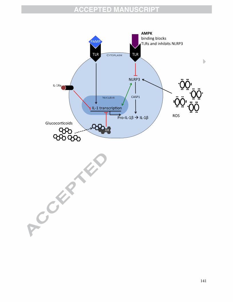
141

142
7 Cell therapies for prevention of pulmonary inflammation
7.1 Mesenchymal stem cells
Mesenchymal stem cells (MSCs) with multi-lineage differentiability are usually derived from bone marrow. MSCs
home to sites of injury and possess immunomodulatory functions.105 In culture, MSCs can differentiate into
alveolar epithelium.105, 106 In vivo, MSCs engraft in the lung and produce surfactant,106 but engraftment rates are
low,107, 108 suggesting MSCs work via paracrine effects. Prophylactic administration of MSCs mitigate lung injury
in mice but are ineffective in repairing established lung injury.108
Meta-analysis of RCTs in adult diseases indicates MSCs are safe;109 they do not increase rates of infection, death
or malignancy,109 despite previous concerns about tumourogenicity.110 Transient fever was noted in trials using
MSCs109 but it is unclear whether this is the consequence of an immune reaction to MSCs.
Clinical studies of MSCs in preterm infants are limited. One trial counted MSCs in BAL and another administered
MSCs before BPD diagnosis. The presence of endogenous MSCs within BAL of preterm infants was associated
with increased risk of developing BPD.111 However, the source of MSCs in BAL was unclear, and may be a result
of injured lung epithelium. A phase I dose-escalation trial examined administration of MSCs intratracheally to
preterm infants at risk of developing BPD (23-29 weeks GA) who required continuous ventilator support.112 Pro-
inflammatory cytokines in BAL were lower at day 7 compared to day 3 after MSC transplantation, highlighting
the paracrine effects of the cells. Thirty-three percent of preterm infants enrolled developed BPD. This trial
targeted infants before BPD developed, who may not have had established lung inflammation. Six patients in the
trial developed serious adverse events up to 84 days after MSC transplantation, including pneumothorax, NEC
and IVH (< grade 3). The majority of adverse events occurred in infants receiving the highest MSC dose (20
million cells/kg).
The origin or handling of MSCs may influence their therapeutic potential. The yield of cord-blood-derived MSCs
is low and MSCs need to be expanded for adequate cell numbers for delivery to a patient, but culturing induces
ageing.113 Cultured, MSCs have weaker immunomodulatory properties in vitro compared to primary MSCs,113
potentially compromising the therapeutic effects of MSCs. Additionally, MSC expansion often requires growth
factors (containing animal products),112 or plating onto a glycoprotein-rich fibronectin matrix.114 The impact of
Figure 1. Interplay of factors that may contribute to chronic lung inflammation and the development
of BPD in preterm infants. NLRP3 and pathogen-associated molecular patterns (PAMP) both increase
transcription of potent pro-inflammatory cytokine IL-1. Imbalances in IL-1 and IL-1 antagonists (i.e. IL-1
receptor antagonist: IL-1Ra) may predispose infants to BPD. IL-1 transcription can be prevented by IL-1Ra.
IL-1 production positively feeds back to increase NLRP3 activity (green arrow). Reactive oxygen species
(ROS) induced by ventilation also increase NLRP3 activity, inducing caspase-1 (CASP1), and thus IL-1
transcription. Glucocorticoids bind to glucocorticoid receptors (GR) in the cytoplasm and interfere with the
transcription of IL-1 in the nucleus. Adenosine monophosphate-activated protein kinase (AMPK) binding to
toll-like receptors (TLR) inhibits NLRP3 (red line).

143
media, growth factors or matrices used with MSCs, and whether these alter MSC function, must be considered
when proposing MSC transplantation in preterm infants.
7.2 Human amnion epithelial cells
The amniotic membrane is the innermost placental membrane surrounding the fetus,115 made up of a single
layer of cuboidal columnar epithelial cells.115 The amnion predominately provides the developing fetus with
protection, but also produces growth factors, cytokines, prostaglandins and erythropoietin.116 The amniotic
epithelium is formed before gastrulation, and thus is pluripotent even at term gestation.115
Amniotic membranes were first used over a century ago as biological dressings for skin wounds.117 This practice
continues, highlighting the safety of these cells as a therapeutic for human disease.118 Unlike MSCs, isolation of
human amnion epithelial cells (hAECs) from a single placenta yields enough cells for administration to multiple
patients. Primary hAECs may be used immediately after isolation, obviating concerns about cell manipulation.
Human AECs do not express telomerase,119 and have low tumourigenicity.120 Rejection does not occur when
hAECs are administered to humans121 or other animals,119, 122 likely due to low HLA class II expression.122 123
Thus, hAECs can be applied without concerns of tumorigenesis or rejection.
Human AECs reduce lung collagen and fibrosis in mice,124, 125 up to 14 days after bleomycin insult.125 Hyperoxia-
exposed mice treated with hAECs have increased expression of vascular endothelial growth factor receptor and
angiogenin1 in their lungs and this correlates with normalized pulmonary vasculature.126 Human AEC
administration normalises alveolar structure in hyperoxia-exposed mice,126 LPS-exposed fetal sheep127 and in
fetal and preterm sheep following injurious ventilation.128, 129 Thus, hAECs prevent BPD-like lung pathology,
independent of the model, and this reduction in BPD-like lung pathology is consistently accompanied by reduced
lung inflammation.
The immunomodulatory effects of hAECs are demonstrated by their ability to downregulate pro-inflammatory
cytokines, IL-6,128, 130-132 IL-1α and IL-1β,131 and upregulate anti-inflammatory cytokine IL-10.129 However, hAECs
increase total immune cell numbers in the lungs of LPS-exposed fetal sheep and mechanically ventilated preterm
lambs,127, 129 demonstrating the ability of hAECs’ to augment inflammatory cell recruitment but seemingly
without proinflammatory effects. IL-10 has a role in activating M2 pro-reparative macrophages and opposing
differentiation of pro-inflammatory M1 macrophages,133 and is upregulated following hAEC administration.129, 132
Indeed, lung and liver macrophage activity shifts from a predominately M1 to M2 phenotype after hAEC
administration in bleomycin-exposed mice.134, 135 Similar to MSCs, hAECs modulate the immune system and
prevent BPD-like lung pathology, likely via paracrine effects.
The safety of hAECs has been explored in an unpublished phase I trial of hAECs in infants with intractable BPD
(ACTRN: 12614000174684). Subsequent trials are required to determine the appropriate dose of hAECs and
when to administer hAECs in preterm infants with BPD.

144
8 Final comments and future research
BPD remains a major cause of morbidity and mortality in preterm infants. Historically, therapies like postnatal
steroids have been used without appropriate preclinical data regarding safety and long-term outcomes.
Pharmacological inhibitors of inflammation, such as IL-1 inhibitors, pentoxifylline and AMP activators, are
promising therapies for pulmonary inflammation but are in preclinical stages. Cellular therapies, arguably, have
more preclinical evidence surrounding their use for BPD and are approaching RCTs. However, it is unlikely that a
one-drug-fix-all approach to BPD will eventuate. Likely, the most beneficial outcomes for infants developing, or
who have developed, BPD will be achieved with combined therapies. If an infant is unresponsive to a therapy
(e.g. steroids or hAECs) within several days an alternative therapy should be considered. Future studies will
need to consider interactions between therapies in preterm infants if tailoring therapies for neonates with BPD
is necessary.
Educational aims
The reader will appreciate:
• The incidence of BPD is increasing, despite advancements in clinical care of preterm infants.
• Maturational agents to improve lung architecture, including antenatal glucocorticoids and surfactant
therapy, have not improved BPD incidence.
• Anti-inflammatory therapies may be beneficial over maturational agents in preventing BPD incidence.
• Glucocorticoids reduce BPD incidence but have poor neurological outcomes. Glucocorticoid use in the
newborn requires significant optimisation.
• Cellular therapies likely modulate lung inflammation associated with BPD, without adverse outcomes.
Future research directions
BPD is a complex disease involving aberrant regulation of lung inflammation. The use of preclinical, translational
studies to identify or optimise pre-existing therapies, such as postnatal steroids or IL-1Ra, need to be conducted
and compared to less conventional cellular therapies. Future studies should aim to encompass the inflammatory,
structural and vascular complications of BPD for the best outcomes in preterm infants.
References
1. Blencowe H, Cousens S, Oestergaard MZ, et al. National, regional, and worldwideestimates of preterm birth rates in the year 2010 with time trends since 1990 for selected
countries: a systematic analysis and implications. Lancet. 2012;379(9832):2162-72.
2. Stoll BJ, Hansen NI, Bell EF, et al. Trends in Care Practices, Morbidity, and Mortality of
Extremely Preterm Neonates, 1993-2012. Jama. 2015;314(10):1039-51.
3. Roberts D, Dalziel S. Antenatal corticosteroids for accelerating fetal lung maturation for
women at risk of preterm birth. The Cochrane database of systematic reviews. 2006(3):CD004454.

145
4. Bancalari E, del Moral T. Bronchopulmonary dysplasia and surfactant. Biology of the
neonate. 2001;80 Suppl 1:7-13. 5. Morley CJ, Davis PG, Doyle LW, et al. Nasal CPAP or intubation at birth for very preterm
infants. The New England journal of medicine. 2008;358(7):700-8.
6. Melville JM, Moss TJ. The immune consequences of preterm birth. Front Neurosci.2013;7:79.
7. Northway WH, Jr., Rosan RC, Porter DY. Pulmonary disease following respirator
therapy of hyaline-membrane disease. Bronchopulmonary dysplasia. The New England
journal of medicine. 1967;276(7):357-68.
8. Agrons GA, Courtney SE, Stocker JT, et al. From the archives of the AFIP: Lung disease
in premature neonates: radiologic-pathologic correlation. Radiographics. 2005;25(4):1047-
73.
9. Bhatt AJ, Pryhuber GS, Huyck H, et al. Disrupted pulmonary vasculature and decreasedvascular endothelial growth factor, Flt-1, and TIE-2 in human infants dying with
bronchopulmonary dysplasia. American journal of respiratory and critical care medicine.
2001;164(10 Pt 1):1971-80.
10. Vento G, Capoluongo E, Matassa PG, et al. Serum levels of seven cytokines in premature
ventilated newborns: correlations with old and new forms of bronchopulmonary dysplasia.
Intensive care medicine. 2006;32(5):723-30.
11. Kotecha S, Wilson L, Wangoo A, et al. Increase in interleukin (IL)-1 beta and IL-6 in
bronchoalveolar lavage fluid obtained from infants with chronic lung disease of prematurity. Pediatric research. 1996;40(2):250-6.
12. Munshi UK, Niu JO, Siddiq MM, et al. Elevation of interleukin-8 and interleukin-6precedes the influx of neutrophils in tracheal aspirates from preterm infants who develop
bronchopulmonary dysplasia. Pediatric pulmonology. 1997;24(5):331-6.
13. Husain AN, Siddiqui NH, Stocker JT. Pathology of arrested acinar development in
postsurfactant bronchopulmonary dysplasia. Hum Pathol. 1998;29(7):710-7.
14. Ehrenkranz RA, Walsh MC, Vohr BR, et al. Validation of the National Institutes of Health
consensus definition of bronchopulmonary dysplasia. Pediatrics. 2005;116(6):1353-60. 15. Goldenberg RL, Hauth JC, Andrews WW. Intrauterine infection and preterm delivery.
The New England journal of medicine. 2000;342(20):1500-7. 16. Lahra MM, Beeby PJ, Jeffery HE. Maternal versus fetal inflammation and respiratory
distress syndrome: a 10-year hospital cohort study. Archives of disease in childhood Fetal and
neonatal edition. 2009;94(1):F13-6. 17. Dempsey E, Chen MF, Kokottis T, et al. Outcome of neonates less than 30 weeks
gestation with histologic chorioamnionitis. American journal of perinatology.
2005;22(3):155-9.
18. Watterberg KL, Demers LM, Scott SM, et al. Chorioamnionitis and early lung
inflammation in infants in whom bronchopulmonary dysplasia develops. Pediatrics.
1996;97(2):210-5.
19. Moss TJ, Newnham JP, Willett KE, et al. Early gestational intra-amniotic endotoxin: lung
function, surfactant, and morphometry. American journal of respiratory and critical care medicine. 2002;165(6):805-11.
20. Jobe AH, Newnham JP, Willet KE, et al. Endotoxin-induced lung maturation in pretermlambs is not mediated by cortisol. American journal of respiratory and critical care medicine.
2000;162(5):1656-61.
21. Bachurski CJ, Ross GF, Ikegami M, et al. Intra-amniotic endotoxin increases pulmonary
surfactant proteins and induces SP-B processing in fetal sheep. American journal of
physiology Lung cellular and molecular physiology. 2001;280(2):L279-85.

146
22. Yum SK, Kim MS, Kwun Y, et al. Impact of histologic chorioamnionitis on pulmonary
hypertension and respiratory outcomes in preterm infants. Pulm Circ. 2018;8(2):2045894018760166.
23. Hartling L, Liang Y, Lacaze-Masmonteil T. Chorioamnionitis as a risk factor for
bronchopulmonary dysplasia: a systematic review and meta-analysis. Archives of disease in childhood Fetal and neonatal edition. 2012;97(1):F8-F17.
24. Soraisham AS, Singhal N, McMillan DD, et al. A multicenter study on the clinical
outcome of chorioamnionitis in preterm infants. American journal of obstetrics and
gynecology. 2009;200(4):372 e1-6.
25. Van Marter LJ, Dammann O, Allred EN, et al. Chorioamnionitis, mechanical ventilation,
and postnatal sepsis as modulators of chronic lung disease in preterm infants. The Journal of
pediatrics. 2002;140(2):171-6.
26. Westover AJ, Moss TJ. Effects of intrauterine infection or inflammation on fetal lungdevelopment. Clinical and experimental pharmacology & physiology. 2012;39(9):824-30.
27. Willet KE, Jobe AH, Ikegami M, et al. Antenatal endotoxin and glucocorticoid effects on
lung morphometry in preterm lambs. Pediatric research. 2000;48(6):782-8.
28. Galinsky R, Hooper SB, Wallace MJ, et al. Intrauterine inflammation alters
cardiopulmonary and cerebral haemodynamics at birth in preterm lambs. J Physiol.
2013;591(8):2127-37.
29. Yoder BA, Coalson JJ, Winter VT, et al. Effects of antenatal colonization with
ureaplasma urealyticum on pulmonary disease in the immature baboon. Pediatric research. 2003;54(6):797-807.
30. Polglase GR, Hillman NH, Pillow JJ, et al. Ventilation-mediated injury after pretermdelivery of Ureaplasma parvum colonized fetal lambs. Pediatric research. 2010;67(6):630-5.
31. Bhandari V. Postnatal inflammation in the pathogenesis of bronchopulmonary
dysplasia. Birth defects research Part A, Clinical and molecular teratology. 2014;100(3):189-
201.
32. Koksal N, Kayik B, Cetinkaya M, et al. Value of serum and bronchoalveolar fluid lavage
pro- and anti-inflammatory cytokine levels for predicting bronchopulmonary dysplasia in premature infants. Eur Cytokine Netw. 2012;23(2):29-35.
33. Barazzone C, White CW. Mechanisms of cell injury and death in hyperoxia: role ofcytokines and Bcl-2 family proteins. American journal of respiratory cell and molecular
biology. 2000;22(5):517-9.
34. Kim SR, Kim DI, Kim SH, et al. NLRP3 inflammasome activation by mitochondrial ROSin bronchial epithelial cells is required for allergic inflammation. Cell Death Dis.
2014;5:e1498.
35. Brew N, Hooper SB, Allison BJ, et al. Injury and repair in the very immature lung
following brief mechanical ventilation. American journal of physiology Lung cellular and
molecular physiology. 2011;301(6):L917-26.
36. Hillman NH, Kallapur SG, Pillow JJ, et al. Airway injury from initiating ventilation in
preterm sheep. Pediatric research. 2010;67(1):60-5.
37. Albertine KH, Jones GP, Starcher BC, et al. Chronic lung injury in preterm lambs.Disordered respiratory tract development. American journal of respiratory and critical care
medicine. 1999;159(3):945-58. 38. Balinotti JE, Chakr VC, Tiller C, et al. Growth of lung parenchyma in infants and toddlers
with chronic lung disease of infancy. American journal of respiratory and critical care
medicine. 2010;181(10):1093-7.
39. Korhonen P, Laitinen J, Hyodynmaa E, et al. Respiratory outcome in school-aged, very-
low-birth-weight children in the surfactant era. Acta paediatrica. 2004;93(3):316-21.
40. Polglase GR, Nitsos I, Jobe AH, et al. Maternal and intra-amniotic corticosteroid effectson lung morphometry in preterm lambs. Pediatric research. 2007;62(1):32-6.

147
41. Kavelaars A, van der Pompe G, Bakker JM, et al. Altered immune function in human
newborns after prenatal administration of betamethasone: enhanced natural killer cell activity and decreased T cell proliferation in cord blood. Pediatric research. 1999;45(3):306-
12.
42. Kramer BW, Kallapur SG, Moss TJ, et al. Intra-amniotic LPS modulation of TLR signalingin lung and blood monocytes of fetal sheep. Innate Immun. 2009;15(2):101-7.
43. Newnham JP, Kallapur SG, Kramer BW, et al. Betamethasone effects on
chorioamnionitis induced by intra-amniotic endotoxin in sheep. American journal of
obstetrics and gynecology. 2003;189(5):1458-66.
44. Moss TJ, Harding R, Newnham JP. Lung function, arterial pressure and growth in sheep
during early postnatal life following single and repeated prenatal corticosteroid treatments.
Early human development. 2002;66(1):11-24.
45. Nin N, Penuelas O, de Paula M, et al. Ventilation-induced lung injury in rats isassociated with organ injury and systemic inflammation that is attenuated by dexamethasone.
Critical care medicine. 2006;34(4):1093-8.
46. Held HD, Boettcher S, Hamann L, et al. Ventilation-induced chemokine and cytokine
release is associated with activation of nuclear factor-kappaB and is blocked by steroids.
American journal of respiratory and critical care medicine. 2001;163(3 Pt 1):711-6.
47. Hillman NH, Pillow JJ, Ball MK, et al. Antenatal and postnatal corticosteroid and
resuscitation induced lung injury in preterm sheep. Respiratory research. 2009;10:124.
48. Walsh MC, Yao Q, Horbar JD, et al. Changes in the use of postnatal steroids forbronchopulmonary dysplasia in 3 large neonatal networks. Pediatrics. 2006;118(5):e1328-35.
49. Cummings JJ, D'Eugenio DB, Gross SJ. A controlled trial of dexamethasone in preterminfants at high risk for bronchopulmonary dysplasia. The New England journal of medicine.
1989;320(23):1505-10.
50. Doyle LW, Halliday HL, Ehrenkranz RA, et al. Impact of postnatal systemic
corticosteroids on mortality and cerebral palsy in preterm infants: effect modification by risk
for chronic lung disease. Pediatrics. 2005;115(3):655-61.
51. Barrington KJ. The adverse neuro-developmental effects of postnatal steroids in thepreterm infant: a systematic review of RCTs. BMC pediatrics. 2001;1:1.
52. Doyle LW, Ehrenkranz RA, Halliday HL. Late (> 7 days) postnatal corticosteroids forchronic lung disease in preterm infants. The Cochrane database of systematic reviews.
2014(5):CD001145.
53. Doyle LW, Ehrenkranz RA, Halliday HL. Early (< 8 days) postnatal corticosteroids forpreventing chronic lung disease in preterm infants. The Cochrane database of systematic
reviews. 2014(5):CD001146.
54. Claure N, Bello JA, Jain D. Strategies to reduce mechanical ventilation and
bronchopulmonary dysplasia in preterm infants. Respir Care. 2013;58(7):1257.
55. Groneck P, Reuss D, Gotze-Speer B, et al. Effects of dexamethasone on chemotactic
activity and inflammatory mediators in tracheobronchial aspirates of preterm infants at risk
for chronic lung disease. The Journal of pediatrics. 1993;122(6):938-44.
56. Yoder MC, Jr., Chua R, Tepper R. Effect of dexamethasone on pulmonary inflammationand pulmonary function of ventilator-dependent infants with bronchopulmonary dysplasia.
The American review of respiratory disease. 1991;143(5 Pt 1):1044-8. 57. Groneck P, Gotze-Speer B, Oppermann M, et al. Association of pulmonary inflammation
and increased microvascular permeability during the development of bronchopulmonary
dysplasia: a sequential analysis of inflammatory mediators in respiratory fluids of high-risk
preterm neonates. Pediatrics. 1994;93(5):712-8.
58. Wilson-Costello D, Walsh MC, Langer JC, et al. Impact of postnatal corticosteroid use on
neurodevelopment at 18 to 22 months' adjusted age: effects of dose, timing, and risk of

148
bronchopulmonary dysplasia in extremely low birth weight infants. Pediatrics.
2009;123(3):e430-7. 59. Weichsel ME, Jr. The therapeutic use of glucocorticoid hormones in the perinatal
period: potential neurological hazards. Ann Neurol. 1977;2(5):364-6.
60. Howard E. Reductions in size and total DNA of cerebrum and cerebellum in adult miceafter corticosterone treatment in infancy. Experimental neurology. 1968;22(2):191-208.
61. Howard E, Benjamins JA. DNA, ganglioside and sulfatide in brains of rats given
corticosterone in infancy, with an estimate of cell loss during development. Brain research.
1975;92(1):73-87.
62. Stark AR, Carlo WA, Tyson JE, et al. Adverse effects of early dexamethasone treatment
in extremely-low-birth-weight infants. National Institute of Child Health and Human
Development Neonatal Research Network. The New England journal of medicine.
2001;344(2):95-101. 63. Garland JS, Alex CP, Pauly TH, et al. A three-day course of dexamethasone therapy to
prevent chronic lung disease in ventilated neonates: a randomized trial. Pediatrics.
1999;104(1 Pt 1):91-9.
64. Yeh TF, Lin YJ, Hsieh WS, et al. Early postnatal dexamethasone therapy for the
prevention of chronic lung disease in preterm infants with respiratory distress syndrome: a
multicenter clinical trial. Pediatrics. 1997;100(4):E3.
65. Doyle LW, Davis PG, Morley CJ, et al. Low-dose dexamethasone facilitates extubation
among chronically ventilator-dependent infants: a multicenter, international, randomized, controlled trial. Pediatrics. 2006;117(1):75-83.
66. Doyle LW, Davis PG, Morley CJ, et al. Outcome at 2 years of age of infants from theDART study: a multicenter, international, randomized, controlled trial of low-dose
dexamethasone. Pediatrics. 2007;119(4):716-21.
67. Harris E, Schulzke SM, Patole SK. Pentoxifylline in preterm neonates: a systematic
review. Paediatr Drugs. 2010;12(5):301-11.
68. Krause PJ, Kristie J, Wang WP, et al. Pentoxifylline enhancement of defective neutrophil
function and host defense in neonatal mice. The American journal of pathology. 1987;129(2):217-22.
69. Lauterbach R, Pawlik D, Kowalczyk D, et al. Effect of the immunomodulating agent,pentoxifylline, in the treatment of sepsis in prematurely delivered infants: a placebo-
controlled, double-blind trial. Critical care medicine. 1999;27(4):807-14.
70. Lauterbach R, Szymura-Oleksiak J, Pawlik D, et al. Nebulized pentoxifylline forprevention of bronchopulmonary dysplasia in very low birth weight infants: a pilot clinical
study. The journal of maternal-fetal & neonatal medicine : the official journal of the European
Association of Perinatal Medicine, the Federation of Asia and Oceania Perinatal Societies, the
International Society of Perinatal Obstet. 2006;19(7):433-8.
71. Aviado DM, Dettelbach HR. Pharmacology of pentoxifylline, a hemorheologic agent for
the treatment of intermittent claudication. Angiology. 1984;35(7):407-17.
72. Aviado DM, Porter JM. Pentoxifylline: a new drug for the treatment of intermittent
claudication. Mechanism of action, pharmacokinetics, clinical efficacy and adverse effects. Pharmacotherapy. 1984;4(6):297-307.
73. Almario B, Wu S, Peng J, et al. Pentoxifylline and prevention of hyperoxia-induced lung-injury in neonatal rats. Pediatric research. 2012;71(5):583-9.
74. Oliveira-Junior IS, Maganhin CC, Carbonel AA, et al. Effects of pentoxifylline on TNF-
alpha and lung histopathology in HCL-induced lung injury. Clinics. 2008;63(1):77-84.
75. Tschopp J, Schroder K. NLRP3 inflammasome activation: The convergence of multiple
signalling pathways on ROS production? Nature reviews Immunology. 2010;10(3):210-5.
76. He Y, Hara H, Nunez G. Mechanism and Regulation of NLRP3 Inflammasome Activation.Trends Biochem Sci. 2016;41(12):1012-21.

149
77. Baroja-Mazo A, Martin-Sanchez F, Gomez AI, et al. The NLRP3 inflammasome is
released as a particulate danger signal that amplifies the inflammatory response. Nat Immunol. 2014;15(8):738-48.
78. Kuipers MT, Aslami H, Janczy JR, et al. Ventilator-induced lung injury is mediated by
the NLRP3 inflammasome. Anesthesiology. 2012;116(5):1104-15. 79. Busillo JM, Azzam KM, Cidlowski JA. Glucocorticoids sensitize the innate immune
system through regulation of the NLRP3 inflammasome. The Journal of biological chemistry.
2011;286(44):38703-13.
80. Stouch AN, McCoy AM, Greer RM, et al. IL-1beta and Inflammasome Activity Link
Inflammation to Abnormal Fetal Airway Development. Journal of immunology.
2016;196(8):3411-20.
81. Liao J, Kapadia VS, Brown LS, et al. The NLRP3 inflammasome is critically involved in
the development of bronchopulmonary dysplasia. Nat Commun. 2015;6:8977. 82. Behrashi M, Samimi M, Ghasemi T, et al. Comparison of Glibenclamide and Insulin on
Neonatal Outcomes in Pregnant Women with Gestational Diabetes. Int J Prev Med. 2016;7:88.
83. Ilkhanipoor H, Karamizadeh Z. Changing the Treatment of Permanent Neonatal
Diabetes Mellitus from Insulin to Glibenclamide in a 4-Month-Old Infant with KCNJ11
Activating Mutation. Int J Prev Med. 2013;4(9):1078-81.
84. Arend WP. The balance between IL-1 and IL-1Ra in disease. Cytokine and Growth
Factor Review. 2002;13:323-40.
85. Dinarello CA, Ikejima T, Warner SJ, et al. Interleukin 1 induces interleukin 1. I.Induction of circulating interleukin 1 in rabbits in vivo and in human mononuclear cells in
vitro. Journal of immunology. 1987;139(6):1902-10. 86. Viscardi RM, Muhumuza CK, Rodriguez A, et al. Inflammatory markers in intrauterine
and fetal blood and cerebrospinal fluid compartments are associated with adverse pulmonary
and neurologic outcomes in preterm infants. Pediatric research. 2004;55(6):1009-17.
87. Rindfleisch MS, Hasday JD, Taciak V, et al. Potential role of interleukin-1 in the
development of bronchopulmonary dysplasia. J Interferon Cytokine Res. 1996;16(5):365-73.
88. Kakkera DK, Siddiq MM, Parton LA. Interleukin-1 balance in the lungs of preterminfants who develop bronchopulmonary dysplasia. Biology of the neonate. 2005;87(2):82-90.
89. Johnson BH, Yi M, Masood A, et al. A critical role for the IL-1 receptor in lung injuryinduced in neonatal rats by 60% O2. Pediatric research. 2009;66(3):260-5.
90. Nold MF, Mangan NE, Rudloff I, et al. Interleukin-1 receptor antagonist prevents
murine bronchopulmonary dysplasia induced by perinatal inflammation and hyperoxia. Proceedings of the National Academy of Sciences of the United States of America.
2013;110(35):14384-9.
91. Kallapur SG, Nitsos I, Moss TJ, et al. IL-1 mediates pulmonary and systemic
inflammatory responses to chorioamnionitis induced by lipopolysaccharide. American journal
of respiratory and critical care medicine. 2009;179(10):955-61.
92. Dinarello CA, Simon A, van der Meer JW. Treating inflammation by blocking
interleukin-1 in a broad spectrum of diseases. Nat Rev Drug Discov. 2012;11(8):633-52.
93. Goldbach-Mansky R, Dailey NJ, Canna SW, et al. Neonatal-onset multisysteminflammatory disease responsive to interleukin-1beta inhibition. The New England journal of
medicine. 2006;355(6):581-92. 94. Kone-Paut I, Galeotti C. Current treatment recommendations and considerations for
cryopyrin-associated periodic syndrome. Expert Rev Clin Immunol. 2015;11(10):1083-92.
95. Neven B, Marvillet I, Terrada C, et al. Long-term efficacy of the interleukin-1 receptor
antagonist anakinra in ten patients with neonatal-onset multisystem inflammatory
disease/chronic infantile neurologic, cutaneous, articular syndrome. Arthritis Rheum.
2010;62(1):258-67.
150
96. Stenerson M, Dufendach K, Aksentijevich I, et al. The first reported case of compound
heterozygous IL1RN mutations causing deficiency of the interleukin-1 receptor antagonist. Arthritis Rheum. 2011;63(12):4018-22.
97. Canakinumab (Ilaris). CADTH Common Drug Reviews. Ottawa (ON)2016.
98. Tarp S, Amarilyo G, Foeldvari I, et al. Efficacy and safety of biological agents forsystemic juvenile idiopathic arthritis: a systematic review and meta-analysis of randomized
trials. Rheumatology (Oxford). 2016;55(4):669-79.
99. Diamond G, Beckloff N, Weinberg A, et al. The roles of antimicrobial peptides in innate
host defense. Curr Pharm Des. 2009;15(21):2377-92.
100. Salminen A, Hyttinen JM, Kaarniranta K. AMP-activated protein kinase inhibits NF-
kappaB signaling and inflammation: impact on healthspan and lifespan. J Mol Med (Berl).
2011;89(7):667-76.
101. Schaller-Bals S, Schulze A, Bals R. Increased levels of antimicrobial peptides in tracheal aspirates of newborn infants during infection. American journal of respiratory and critical
care medicine. 2002;165(7):992-5.
102. Xing J, Wang Q, Coughlan K, et al. Inhibition of AMP-activated protein kinase
accentuates lipopolysaccharide-induced lung endothelial barrier dysfunction and lung injury
in vivo. The American journal of pathology. 2013;182(3):1021-30.
103. Konings E, Timmers S, Boekschoten MV, et al. The effects of 30 days resveratrol
supplementation on adipose tissue morphology and gene expression patterns in obese men.
International journal of obesity. 2014;38(3):470-3. 104. Yi CO, Jeon BT, Shin HJ, et al. Resveratrol activates AMPK and suppresses LPS-induced
NF-kappaB-dependent COX-2 activation in RAW 264.7 macrophage cells. Anat Cell Biol. 2011;44(3):194-203.
105. Chamberlain G, Fox J, Ashton B, et al. Concise review: mesenchymal stem cells: their
phenotype, differentiation capacity, immunological features, and potential for homing. Stem
cells. 2007;25(11):2739-49.
106. Krause DS, Theise ND, Collector MI, et al. Multi-organ, multi-lineage engraftment by a
single bone marrow-derived stem cell. Cell. 2001;105(3):369-77. 107. van Haaften T, Byrne R, Bonnet S, et al. Airway delivery of mesenchymal stem cells
prevents arrested alveolar growth in neonatal lung injury in rats. American journal of respiratory and critical care medicine. 2009;180(11):1131-42.
108. Ortiz LA, Gambelli F, McBride C, et al. Mesenchymal stem cell engraftment in lung is
enhanced in response to bleomycin exposure and ameliorates its fibrotic effects. Proceedings of the National Academy of Sciences of the United States of America. 2003;100(14):8407-11.
109. Lalu MM, McIntyre L, Pugliese C, et al. Safety of cell therapy with mesenchymal stromal
cells (SafeCell): a systematic review and meta-analysis of clinical trials. PloS one.
2012;7(10):e47559.
110. Djouad F, Plence P, Bony C, et al. Immunosuppressive effect of mesenchymal stem cells
favors tumor growth in allogeneic animals. Blood. 2003;102(10):3837-44.
111. Popova AP, Bozyk PD, Bentley JK, et al. Isolation of tracheal aspirate mesenchymal
stromal cells predicts bronchopulmonary dysplasia. Pediatrics. 2010;126(5):e1127-33. 112. Chang YS, Ahn SY, Yoo HS, et al. Mesenchymal stem cells for bronchopulmonary
dysplasia: phase 1 dose-escalation clinical trial. The Journal of pediatrics. 2014;164(5):966-72 e6.
113. Stolzing A, Sethe S, Scutt AM. Stressed stem cells: Temperature response in aged
mesenchymal stem cells. Stem cells and development. 2006;15(4):478-87.
114. Harting MT, Jimenez F, Xue H, et al. Intravenous mesenchymal stem cell therapy for
traumatic brain injury. J Neurosurg. 2009;110(6):1189-97.
115. Ilancheran S, Moodley Y, Manuelpillai U. Human fetal membranes: a source of stem cells for tissue regeneration and repair? Placenta. 2009;30(1):2-10.

151
116. Mamede AC, Carvalho MJ, Abrantes AM, et al. Amniotic membrane: from structure and
functions to clinical applications. Cell and tissue research. 2012;349(2):447-58. 117. Davis JS. II. Skin Grafting at the Johns Hopkins Hospital. Annals of surgery.
1909;50(3):542-9.
118. Kesting MR, Wolff KD, Hohlweg-Majert B, et al. The role of allogenic amniotic membrane in burn treatment. Journal of burn care & research : official publication of the
American Burn Association. 2008;29(6):907-16.
119. Miki T, Lehmann T, Cai H, et al. Stem cell characteristics of amniotic epithelial cells.
Stem cells. 2005;23(10):1549-59.
120. Shay JW, Wright WE. Role of telomeres and telomerase in cancer. Semin Cancer Biol.
2011;21(6):349-53.
121. Akle CA, Adinolfi M, Welsh KI, et al. Immunogenicity of human amniotic epithelial cells
after transplantation into volunteers. Lancet. 1981;2(8254):1003-5. 122. Ilancheran S, Michalska A, Peh G, et al. Stem cells derived from human fetal membranes
display multilineage differentiation potential. Biology of reproduction. 2007;77(3):577-88.
123. Banas RA, Trumpower C, Bentlejewski C, et al. Immunogenicity and
immunomodulatory effects of amnion-derived multipotent progenitor cells. Human
immunology. 2008;69(6):321-8.
124. Murphy S, Lim R, Dickinson H, et al. Human amnion epithelial cells prevent bleomycin-
induced lung injury and preserve lung function. Cell transplantation. 2011;20(6):909-23.
125. Vosdoganes P, Wallace EM, Chan ST, et al. Human amnion epithelial cells repair established lung injury. Cell transplantation. 2013;22(8):1337-49.
126. Zhu D, Muljadi R, Chan ST, et al. Evaluating the Impact of Human Amnion Epithelial Cells on Angiogenesis. Stem Cells Int. 2016;2016:4565612.
127. Vosdoganes P, Hodges RJ, Lim R, et al. Human amnion epithelial cells as a treatment for
inflammation-induced fetal lung injury in sheep. American journal of obstetrics and
gynecology. 2011;205(2):156 e26-33.
128. Hodges RJ, Jenkin G, Hooper SB, et al. Human amnion epithelial cells reduce
ventilation-induced preterm lung injury in fetal sheep. American journal of obstetrics and gynecology. 2012;206(5):448 e8-15.
129. Melville JM, McDonald CA, Bischof RJ, et al. Human amnion epithelial cells modulate the inflammatory response to ventilation in preterm lambs. PloS one. 2017;12(3):e0173572.
130. Murphy SV, Shiyun SC, Tan JL, et al. Human amnion epithelial cells do not abrogate
pulmonary fibrosis in mice with impaired macrophage function. Cell transplantation. 2012;21(7):1477-92.
131. Vosdoganes P, Lim R, Koulaeva E, et al. Human amnion epithelial cells modulate
hyperoxia-induced neonatal lung injury in mice. Cytotherapy. 2013;15(8):1021-9.
132. Moodley Y, Ilancheran S, Samuel C, et al. Human amnion epithelial cell transplantation
abrogates lung fibrosis and augments repair. American journal of respiratory and critical care
medicine. 2010;182(5):643-51.
133. Makita N, Hizukuri Y, Yamashiro K, et al. IL-10 enhances the phenotype of M2
macrophages induced by IL-4 and confers the ability to increase eosinophil migration. Int Immunol. 2015;27(3):131-41.
134. Tan JL, Chan ST, Wallace EM, et al. Human amnion epithelial cells mediate lung repair by directly modulating macrophage recruitment and polarization. Cell transplantation.
2014;23(3):319-28.
135. Manuelpillai U, Lourensz D, Vaghjiani V, et al. Human amniotic epithelial cell
transplantation induces markers of alternative macrophage activation and reduces
established hepatic fibrosis. PloS one. 2012;7(6):e38631.
152
Title: Genetic Variation in CRHR1 is Associated with Short-Term Respiratory Response to
Corticosteroids in Preterm Infants at risk for Bronchopulmonary Dysplasia
Running Title: Steroid Pharmacogenetics in BPD
Authors: Tamorah Lewis*1; William Truog1; Mike Norberg1, Philip L. Ballard2; Dara
Torgerson2; TOLSURF Study Group
1 – Children’s Mercy Hospital, Department of Pediatrics, University of Missouri Kansas City
School of Medicine, Kansas City, MO
2 - Department of Pediatrics, University of California San Francisco, San Francisco, CA
Corresponding Author:
Tamorah Lewis, MD, PhD
Assistant Professor, UMKC School of Medicine
Neonatology | Pediatric Clinical Pharmacology
Children's Mercy Hospital
2401 Gillham Rd., Kansas City, MO 64108
Phone: (816) 234-4295 Fax: 816-234-3590
Email: [email protected]
Financial Support: There was no external financial support for this project.
Disclosure: The authors have no conflicts of interest to disclose.
Article Type: Clinical Research
© 2018 Macmillan Publishers Limited, part of Springer Nature. All rights reserved.
152
153
Abstract
Background Bronchopulmonary Dysplasia (BPD) is an orphan disease and advances in
prevention and treatment are lacking. Clinical efficacy of systemic corticosteroid therapy to
reduce the severity of lung disease and BPD is highly variable. Our objective was to assess
whether candidate SNPs in corticosteroid metabolism and response genes are associated with
short-term phenotypic response to systemic corticosteroids in infants at high risk for BPD.
Methods Pharmacogenetic analysis of data from a large randomized controlled trial (TOLSURF)
in infants treated with dexamethasone or hydrocortisone using multivariate linear regression. The
primary outcome was change in Respiratory Severity Score (RSS, mean airway pressure x FiO2)
at day 7 of corticosteroid treatment.
Results rs7225082 in the intron of CRHR1 is significantly associated with the magnitude of
decrease in RSS 7 days after starting treatment with systemic corticosteroid (meta-analysis
p=2.8x10⁻⁴). Each T allele at rs7225082 is associated with a smaller absolute change in RSS at
day 7, i.e. less response to systemic corticosteroids.
Conclusion Genetic variability is associated with corticosteroid responsiveness with regard to
respiratory status in preterm infants. Identification of genetic markers of corticosteroid
responsiveness may allow for therapeutic individualization, with the goal of optimizing the risk
to benefit ratio for an individual child.
© 2018 Macmillan Publishers Limited, part of Springer Nature. All rights reserved.

154
Introduction
Bronchopulmonary dysplasia (BPD) is the most frequent complication of extreme prematurity. With increasing survival of the most immature
infants, rates of BPD are increasing. Among a large cohort of infants less than 1500 grams at birth, the incidence of BPD increased between 2009 and
2012, up to 55% in infants born at 26 weeks and 40% in infants born at 27 weeks(1). Preventive therapies for BPD are limited, but postnatal
treatment with systemic corticosteroids is a common practice grounded in studies that show improved pulmonary outcomes (2, 3). Corticosteroid
treatment is highly variable among centers(4), in part due to concern regarding historic associations of high dose dexamethasone with central nervous
system injury(5-7). In order for corticosteroid use to be optimized, the risk to benefit ratio must be improved such that a given infant is more likely to
be benefited than harmed. Precision therapeutics is a burgeoning field that utilizes genetic information to individualize therapy, and this approach can
be used to investigate target populations for optimal corticosteroid benefit. A long-term goal of pharmacogenetic studies in BPD is to preemptively
delineate likely responders and non-responders and treat accordingly, and to delineate the biology of differential drug response.
In the original Trial of Surfactant Treatment (TOLSURF) study, many infants received systemic corticosteroids to decrease severity of lung
disease and facilitate weaning from mechanical ventilation. In both dexamethasone- and hydrocortisone-treated infants, there is a known large range
in phenotypic response to corticosteroid treatment. Many factors can contribute to this variability, including severity of lung disease, variation among
centers in management of respiratory support and individual patient co-morbidities such as infections, etc. Genetic variation may also be an important
and understudied contributor to differential corticosteroid response.
Genetic variation contributes to the risk of BPD (8, 9). Although yet unstudied in the BPD population, there is a high probability that genetic
variability also contributes to variability in drug response. Variant alleles can influence drug metabolism, disposition and drug target sensitivity, and
explain some of the heterogeneity in efficacy and toxicity that is observed within a patient population. A recent study (10) links neonatal genetic
© 2018 Macmillan Publishers Limited, part of Springer Nature. All rights reserved.

155
variation to respiratory phenotype at birth after maternal treatment with antenatal corticosteroids. In asthma, multiple single nucleotide
polymorphisms (SNPs) in corticosteroid metabolism and response genes modify clinical response to inhaled corticosteroids(11-17). Though asthma
and BPD are separate clinical entities, significant overlap occurs in their pathophysiology (inflammation, pulmonary architecture remodeling,
bronchospasm, air trapping). Genes which are implicated in prenatal corticosteroid response and asthma corticosteroid response are potentially
relevant to clinical corticosteroid response for BPD.
Given this background, our objective was to identify pharmacogenetic variants from pediatric asthma and perinatal corticosteroid treatment
literature, and test these genes and variants for an association with clinical response to systemic corticosteroids in infants at high risk for BPD. Our
hypothesis states that variants associated with corticosteroid metabolism and response will correlate with short-term improvement in respiratory
phenotype among a clinically homogenous cohort of preterm infants.
Methods
Our investigation was a secondary analysis of existing data collected during a large multi-center, randomized, controlled trial of late
surfactant therapy (TOLSURF study)(18, 19). The parent clinical trial was IRB-approved and parental consent was obtained, including collection and
study of biospecimens (tracheal aspirate, urine and DNA).The Children’s Mercy Hospital IRB deferred review of this secondary study as non-human
subjects research (use of de-identified historical data). The source population from the TOLSURF study included preterm infants receiving inhaled
nitric oxide, birth weight 701±164 g, and gestational age 25.2±1.2 weeks. There was no differences found in the primary outcome of survival without
BPD at 36 weeks between surfactant-treated and placebo groups. Thus, the groups were combined for secondary analyses.
Study Cohort
© 2018 Macmillan Publishers Limited, part of Springer Nature. All rights reserved.

156
511 infants enrolled in the TOLSURF study were screened for eligibility. 126 infants met the following criteria: received systemic
dexamethasone (DEX) or hydrocortisone (HC) at greater than 14 days of life for a minimum of seven days of treatment and on invasive ventilation at
corticosteroids start. These criteria were selected to exclude corticosteroid exposure for blood pressure support and brief courses to treat airway
edema for extubation. Of these 126, 39 were excluded due to (1) no DNA collected/isolated or failed early sample quality control during genome-
wide SNP genotyping (n=26), (2) genotype call rates < 95% (n=3), (3) sibling from a multiple gestation (n=10, only one sibling was included to
account for genetic relatedness). Ten were excluded for no primary outcome data; these infants were extubated to low flow NC and mean airway
pressure (MAP) was not measureable.
Use of systemic corticosteroids was not prescribed by the study protocol but guidelines for their use were established by trial
investigators. Approximately 75% of the source population were treated with postnatal corticosteroids, for various indications, one being BPD. For
corticosteroid treatment for BPD, guidelines for use included withholding treatment until at least 2 weeks of age and only for infants with respiratory
severity scores (RSS, FIO2 x mean airway pressure) of ≥7. Biologically equivalent doses of either hydrocortisone (15 mg/kg over 9 days) or
dexamethasone (0.89 mg/kg over 10 days), based on doses and durations used in prior clinical trials, were recommended per TOLSURF study
guidelines (20). BPD at 36 and 40 weeks was defined in the original study with the following criteria: Infants discharged in room air before 36 weeks
were designated “No BPD.” Infants requiring ventilatory support and any level of supplemental oxygen, or with an effective FiO2 >30% by nasal
cannula, were diagnosed with BPD (“severe” BPD by the NIH workshop definition (21)). Infants receiving mechanical ventilation, NCPAP, or >4 L
of nasal cannula flow in room air were designated “Yes BPD.” Infants at 36 weeks receiving ≤0.3 effective FiO 2 at <2 L flow or on nasal flow <4 L
with room air were evaluated for their requirement for respiratory support, determined by an oxygen/flow reduction (room air) challenge test(22).
Corticosteroid Response Phenotyping
© 2018 Macmillan Publishers Limited, part of Springer Nature. All rights reserved.

157
In order to measure short-term phenotypic response to systemic corticosteroids, the change in RSS was used as a phenotypic marker. In order
to evaluate response to corticosteroids, we went back to original TOLSURF data and obtained Mean Airway Pressure (MAP) and FiO2 to calculate
RSS at each time point. Then, RSS values for the day were averaged for a 24 hour period and recorded on day 0 (day before treatment), day 4 of
treatment and day 7 of corticosteroid treatment. Mode of ventilation was not taken into account in the RSS calculation, but MAP is reliably recorded
from conventional ventilators, high frequency ventilators and BiPAP/SiPAP and CPAP modes. Only one infant in the analysis cohort was weaned to
HFNC and his average RSS corresponded to a MAP of 2 cm H2O. Absolute change in RSS at day 7 is the primary outcome because this is a
clinically meaningful time point and allows time for genetic effects to be assessed with respect to drug response.
Genotyping and Imputation Methods
DNA was isolated from cells in tracheal aspirates using an AutoGeneprep 965 instrument (Autogen, Holliston, MA) as per the manufacturer’s
recommended protocols. When protein contamination was evident, DNA was re-precipitated using 3 volumes of 100% ethanol and 3M ammonium
acetate at a 3:1 ratio after incubation at -80oC overnight. DNA was quantified by Nanodrop (Thermofisher Scientific, Inc., Waltman MA) and quality
was assessed using the Agilent 2100 Bioanalyzer (Agilent, Santa Clara, CA). Genotyping was performed on the Affymetrix Axiom LAT1 array
(WorldArray 4, >800,000 SNPs). SNPs were filtered based on call rates < 95%, and Hardy-Weinberg equilibrium p-values < 10-6 using PLINK(23).
Subjects were evaluated for call rates, consistency between genetic and reported sex, autosomal heterozygosity, and cryptic relatedness/genetic
identity using IBD/IBS estimates in PLINK(23). In the case of multiples, one individual was selected at random to be included in the study.
Using the complete set of ~800,000 markers, genomic levels of African and European ancestry were evaluated using ADMIXTURE(24)
assuming three ancestral populations (K=3). Individuals from the HapMap CEU and YRI were genotyped on the same array and included as
reference populations for European and African ancestry, respectively. Windows were offset by a factor of 0.2, the cutoff for linkage was set to 0.1,
© 2018 Macmillan Publishers Limited, part of Springer Nature. All rights reserved.

158
and a constant recombination rate was set to 10-8 (bp)-1. Genome-wide SNP genotypes were further used to impute candidate variants in the phase 3
1000 Genomes populations using the Michigan Imputation Server (https://imputationserver.sph.umich.edu/index.html). Variants were then filtered
for imputation quality scores > 0.3.
Candidate SNP Identification
Multiple PubMed queries using search terms “steroid, corticosteroid, pharmacogenetics, asthma, response” were performed. Of the papers
that discussed pharmacogenetics of corticosteroids, we chose to only include statistically significant SNPs from papers with an objective and
measurable phenotype (e.g. FEV1). Manuscripts reporting genetic associations of prenatal corticosteroid response and neonatal respiratory outcomes
were included. The references of pertinent review articles were also reviewed for candidate results.
Statistical Analysis
Continuous variables are presented as means (SDs). Genetic association testing was performed at individual candidate SNPs, and at a
collection of pooled SNPs within 50 Kb of candidate genes. Infants of maternal self-reported African-American (AA) and Non-Hispanic White
(NHW) race/ethnicity were analyzed separately, then combined in a meta-analysis using Fisher’s method. Genetic associations with respiratory
phenotypic response were tested at candidate SNPs within each racial/ethnic group using linear regression in PLINK (23), adjusting for gestational
age, sex, birth weight, multiple gestation, baseline RSS and African genetic ancestry (for infants of maternal AA race). Gene-based association
testing was performed using VEGAS2 v.02 by combining p-values across genotyped variants within each candidate gene, and including those within
50 Kb of the transcription start/stop site. Demographic difference between ethnic groups were assessed using Fisher’s exact t-tests. Associations
between change in RSS at day 7 and long-term diagnosis of BPD at 36 and 40 weeks were tested with univariate binomial logistic regression. Two
© 2018 Macmillan Publishers Limited, part of Springer Nature. All rights reserved.
159
post-hoc exploratory analyses were performed, one including infants with missing data at day 7 of treatment (those weaned to low flow nasal
cannula), and one with a small for gestational age (SGA) variable instead of birthweight, as birthweight and gestational age are highly correlated. P-
values less than 0.05 were considered statistically significant for these comparisons.
Results
Figure 1 depicts the selection of the final pharmacogenetic cohort. The final analysis cohort consisted of 34 AA and 36 NHW infants (Table
1), and the seven Hispanic White infants were excluded from the initial analysis due to lack of sufficient sample size and power for genetic
conclusions in this ethnic sub-group. There was a wide phenotypic range in response to systemic corticosteroids (Figure 2). Because the
hydrocortisone- and dexamethasone-treated groups were similar in clinical characteristics and clinical response variability, they were combined. Of
all Table 1 variables tested for an association with the primary outcome, only baseline RSS (day 0 before steroid treatment) was significant, so this
was adjusted for in all subsequent analyses. Day 7 change in RSS was not predictive of BPD status at 36 or 40 weeks gestational age (p-value 0.614
and 0.831 respectively), suggesting that the final diagnosis of BPD is multi-factorial and short-term response to steroids is one of many important
contributors.
The list of candidate SNPs tested for an association with phenotypic response are provided in Supplemental Table S1. We examined 21 SNPs
in the proximity of 10 genes for associations with the primary outcome. Analyses were run within a racial/ethnic group then combined in a meta-
analysis to avoid confounding due to population structure, differences in the distribution of steroid response, and differences in environmental
exposures. Because of the small number of Hispanic infants, we limited our primary analysis to the AA and NHW infants. For the primary outcome
of absolute change in RSS at day 7, the entire cohort had an average change of -3.07 with standard deviation of 2.76. Genetic associations between
candidate SNPs and absolute change in RSS at Day 7 are listed in Table 2.
© 2018 Macmillan Publishers Limited, part of Springer Nature. All rights reserved.

160
We identified a significant association between rs7225082 in the intron of CRHR1 and absolute change in RSS at Day 7 following Bonferroni
correction for 36 tests (meta-analysis p=2.8x10⁻⁴). In both AA and NHW infants, the T allele at rs7225082 was associated with a smaller absolute
change in RSS at day 7, i.e. lower response to systemic corticosteroids (AA: average decrease in absolute change in RSS=1.2, p=0.017; NHW:
average decrease in absolute change in RSS =1.74, p=0.016; meta p-value=2.8x10⁻⁴) (Table 2, Figure 3a,b). Rs7225082 was also significantly
associated with % change at Day 7 (meta p-value=1.8x10⁻⁴), as displayed in Figure 4. Compared to the average change in RSS of -3.07 for the entire
cohort, the difference in absolute change in RSS between genetic groups of -1.2 and -1.74 are relatively large. The T allele was at a frequency of 36%
in AA infants, and 64% in NHW infants, consistent with observations from African and European continental frequencies from the 1000 Genomes
Project (Figure 3c). Results were similar when all infants were pooled, including the 10 Hispanic white infants (total N=87), and adjusted by genomic
ancestry (Table 2).
In an exploratory analysis, we included the ten children who were originally excluded because they missed outcome data at day 7 of treatment
(Figure 1). We presume that these children were all weaned to low flow NC because they had no Mean Airway Pressure recorded in the TOLSURF
dataset. In order to analyze these children, we set the RSS at day 7 to 0.5 for all ten infants, and repeated the regression analysis. The direction of
effect of the T allele at rs7225082 was the same (p-values: AA 0.04; NHW 0.006). Since the T allele maintained its association with less
corticosteroid response in a larger cohort including ten more extremely good responders, it adds further support that rs7225082 has some influence on
variability in corticosteroid response. In the second exploratory analysis, we included a binary SGA variable instead of birthweight. The direction of
effect for the T allele at rs7225082 was the same (p-values: AA 0.03; NHW 0.02).
Four infants displayed continued worsening of their lung disease despite systemic corticosteroids, and all were TT homozygotes at rs7225082.
When these four outliers whose lung disease continued to worsen despite treatment with corticosteroids (increase in RSS > 1) were excluded, the
© 2018 Macmillan Publishers Limited, part of Springer Nature. All rights reserved.

161
association with rs7225082 is in the same direction, but does not reach statistical significance. This suggests that the underlying trend is the same
direction within infants showing either no change in RSS, or an improvement in RSS over time. The loss of significance could be from smaller
sample size or a “pushing over” effect of these four infants, but our goal in this study was to assess the complete range of clinical response to
corticosteroids, and this includes those infants who continue to get worse despite therapy. None of the other candidate SNP association tests met
Bonferroni corrected significance levels.
A gene based analysis was performed for two sets of candidate genes, those associated with corticosteroid response in asthma (eight genes)
and those associated with perinatal corticosteroid response (three genes) (Supplemental Table S1). Within each racial/ethnic group, none of the
candidate genes were significantly associated with phenotypic response to corticosteroids following Bonferroni correction for multiple comparisons
(Table 3). However, genetic variation in T showed an association at p=0.02 in infants of maternal AA race/ethnicity, and variation in SERPINA6
showed an association at p=0.03 in infants of maternal self-reported NHW race/ethnicity.
Discussion
To our knowledge, this is the first study to assess pharmacogenetic influences on drug response in premature infants at high risk for BPD. We
capitalized on published literature to formulate a list of candidate genes and SNPs, and used an existing dataset containing both phenotypic data and
genetic data for this proof of principle analysis. Our study is an important first step in bringing concepts of precision therapeutics to a very
heterogeneous and difficult to treat population of preterm infants with respiratory failure. It is of paramount importance to develop predictive
biomarkers of drug response in order to spare predicted “non-responders” the unwarranted risk of systemic corticosteroid therapy. This is especially
important for a medication such as dexamethasone which has the potential to significantly help a sub-set of infants with developing BPD. The risk to
benefit ratio of systemic steroids varies with your risk of developing BPD (25), as both “exposures” can lead to brain injury. And while a calculator
© 2018 Macmillan Publishers Limited, part of Springer Nature. All rights reserved.

162
that estimates BPD risk (26) may be useful in determining which infants to treat, genetic markers of likely responders could strengthen patient
selection for steroid treatment.
We identified SNP rs7225082 in CRHR1 as associated with corticosteroid responsiveness and we also present gene and SNP results that do
not meet Bonferroni adjustment for significance. We present all of the results because we also hope that this paper is hypothesis generating, and that
these genetic variations may become significant in larger prospective cohorts. Although the effect size of SNP rs7225082 is small, the genetic
variation in CRHR1 may be important because of the prevalence of this variant. All ethnic populations have a relatively large proportion of
individuals carrying the T allele at rs7225082 (Figure 3c), potentially contributing to partial or non-response to systemic corticosteroid treatment.
The most significant genetic association was between rs7225082 in the intron of CRHR1 and absolute change in RSS at day 7, whereby
individuals that carried the T allele had a smaller improvement in RSS scores. This was true for infants of both maternal Black/AA and NHW race,
and given our study design we indeed had the most power to identify genetic associations that are shared among the two racial/ethnic groups.
Interestingly, while the variant is common in both African and European populations, the allele associated with greater improvements in RSS scores
is at higher frequency in populations with African ancestry. Genotypes were imputed at this variant and passed quality thresholds (Rsq=0.89).
However, another variant in CRHR1 that was directly genotyped and in linkage disequilibrium with the imputed variant at R-squared=0.70 showed a
similar trend (rs242941, p=5.9x10⁻³). This suggests our results are not driven by errors in genotype imputation, and strengthens our findings that
genetic variation in CRHR1 contributes to drug response.
The difference in absolute change of RSS between the genetic groups must be interpreted within a clinical context. Absolute change in RSS is
not a variable commonly used in clinical practice, so ventilator settings which correspond to this genetic difference are provided as an example of the
clinical significance. If an infant starts on a MAP of 11 and FiO2 of 0.5, the baseline RSS is 5.5. An infant with the GG genotype at rs7225082 would
© 2018 Macmillan Publishers Limited, part of Springer Nature. All rights reserved.
163
have on average a decrease in RSS of 3.1 in response to systemic steroids, corresponding to, for example, a decrease in MAP to 8 and FiO2 to 0.3, a
clinically significant improvement in respiratory status. For each copy of the T allele, the change in RSS would likely be less. So a GT genotype
infant might have a corresponding decrease in MAP to 10 and FiO2 to 0.4, an RSS of 4 and absolute change of 1.5, a less robust response. A TT
genotype infant would respond even less. Depending on the size of the infant, this difference in steroid response may mean ability to wean from High
Frequency Ventilation to conventional ventilation, or the decrease in overall toxic oxygen exposure to the developing lung. The variability in
response to steroids is multi-factorial, and this SNP in CRHR1 explains a portion of this variability.
Corticotrophin releasing hormone receptor 1 (CRHR1) modulates inflammation through control of ACTH-induced cortisol production. SNPs
in this gene may affect baseline endogenous corticosteroid levels and baseline airway inflammation, leading to variation in response to exogenous
administration of systemic corticosteroids. The importance of genetic variation in CRHR1 to corticosteroid response has been described in Chronic
Obstructive Pulmonary Disease (COPD)(27) asthma(12) and persistent pulmonary hypertension of the newborn(28). Genetic variation in the ligand
for CRHR1, corticotropin releasing hormone (CRH), is associated with neonatal respiratory response to maternal prenatal corticosteroid therapy (10).
Fetuses who carry the risk allele at rs7225082 are more likely to require CPAP/ventilator postnatally (poor response to maternal corticosteroids) and
correspondingly in our study, preterm infants who carry the risk allele have a poorer response to postnatal corticosteroids. Given the importance of
this gene in other patient populations, the biologic plausibility, and the same directionality of effect in two patient populations, we feel confident that
genetic variation in CRHR1 is important for corticosteroid response in premature infants.
In the gene based analysis, we identified two genes associated with absolute change in RSS at day 7 at p<0.05 that warrant additional
investigation – SERPINA6 that was previously implicated in perinatal corticosteroid response, and the T gene that was previously implicated in
corticosteroid response in asthma. SERPINA6 encodes an alpha-globulin protein with corticosteroid-binding properties. This is the major transport
© 2018 Macmillan Publishers Limited, part of Springer Nature. All rights reserved.
164
protein for endogenous glucocorticoids and progestins in the blood of most vertebrates. SERPINA6 transcript number and protein function could
regulate systemic bioavailability of endogenous glucocorticoids, as the protein sequesters up to 80% of circulating cortisol in an inactive complex
(29). Increased levels of SERPINA6, genetically determined, could reduce unbound and biologically active levels of cortisol and influence tissue
response to treatment. It is biologically plausible that alterations in SERPINA6 expression could modulate response to corticosteroid therapy in BPD.
The T gene encodes a mesodermal developmental transcription factor and contributes to regulation in lung development. Alterations in the T
gene could alter the temporal pattern of lung development, and thus affect lung maturity and responsiveness to corticosteroids. The T gene is
expressed in adult lung(30), but the abundance in neonatal lung is not known. The importance of the T gene in corticosteroid response among
patients with asthma was first reported in a large GWAS study in which the significant two SNPs were found in a transcription factor located 50 kb
downstream of the gene(11). The SNP rs6456042 (intronic) is in tight linkage disequilibrium with three other SNPs with known functional
implications: rs3099266 (promoter region), rs1134481 (3’ UTR) and rs2305089 (nonsynomous within coding region). Thus, although the top SNP in
AA infants in our study is not in a coding region, it is likely a marker of carriage of other tightly linked functionally significant genetic changes.
Use of respiratory severity score (RSS) as the phenotypic biomarker provided an objective measure of pulmonary dysfunction at baseline and
served as an index of improvement. The RSS allows latitude among individual treating physician philosophy in choosing the perceived least harmful
combination of oxygen and positive airway pressure to achieve the patient’s ventilation goals. The RSS has been validated as a close correlate of
oxygenation index(31). In addition, RSS is used extensively in studies of preterm infants beyond the first week of life when paO2 is unavailable (32-
35). RSS at day of life thirty is predictive of clinically important outcomes of preterm infants needing protracted ventilation (36). While the change
in RSS may be exaggerated when an infant weans from High Frequency Ventilation to conventional ventilation because often this includes a drop in
the mean airway pressure, we believe that this improved RSS represents a change in lung pathology which is still valid to measure using this
© 2018 Macmillan Publishers Limited, part of Springer Nature. All rights reserved.
165
phenotypic marker. Our cohort did not have sufficient size to analyze genetic associations among infants only treated with one modality of invasive
ventilation.
Many placebo-controlled studies have documented a short term pulmonary response to corticosteroids using endpoints such as the respiratory
acuity score (37) peak inspiratory pressure (PIP) and oxygen need (38-40). More recently, McEvoy et al (41) compared short term effects of lower
dose dexamethasone and demonstrated improved lung mechanics at 3 and 5 days. Our study adds to the published literature by assessing contributors
to variability in clinical response to steroids, moving the field from a population-based approach towards a more personalized medicine approach.
Our study has some weaknesses including small sample size, and no PCR validation of the significant SNP in CRHR1. These are common
weaknesses among secondary analysis studies which use data collected for alternative scientific purposes. We believe that the question of steroid
pharmacogenetics in BPD is important enough that working within these initial limitations is worthwhile. Additionally, we lack a validation cohort
for the pharmacogenetic finding. Because of the challenges in making and reproducing gene-disease (or gene-drug response) conclusions in complex
disease based on small sample sizes(42), the research team is collaborating in a multi-site consortium and prospectively enrolling preterm infants at
risk for BPD who are being treated clinically with systemic corticosteroids in order to recruit a larger patient cohort and further study steroid
pharmacogenetics.
Overall our results implicate genetic variation in CRHR1 in modifying the acute respiratory response to systemic corticosteroids in preterm
infants treated for prevention of BPD. Although the identified significant SNP is likely not directly causal, these results add to the body of knowledge
that endogenous steroid homeostasis may contribute to variability in response to treatment with exogenous corticosteroids. Additional studies are
required to validate these associations and to identify the mechanistic effects of SNPs in CRHR1 on corticosteroid response.
© 2018 Macmillan Publishers Limited, part of Springer Nature. All rights reserved.

166References
© 2018 Macmillan Publishers Limited, part of Springer Nature. All rights reserved.

167
1. Stoll BJ, Hansen NI, Bell EF, et al. Trends in Care Practices, Morbidity, and Mortality of Extremely Preterm Neonates, 1993-2012. JAMA 314:1039-1051 (2015).
2. Doyle LW, Cheong JL, Ehrenkranz RA, Halliday HL Late (> 7 days) systemic postnatal corticosteroids for prevention of bronchopulmonary dysplasia inpreterm infants. Cochrane Database Syst Rev 10:CD001145 (2017).
3. Halliday HL, Ehrenkranz RA, Doyle LW Moderately early (7-14 days) postnatal corticosteroids for preventing chronic lung disease in preterm infants.Cochrane Database Syst Rev:CD001144 (2003).
4. Yoder BA, Harrison M, Clark RH Time-related changes in steroid use and bronchopulmonary dysplasia in preterm infants. Pediatrics 124:673-679 (2009).5. Shinwell ES, Karplus M, Reich D, et al. Early postnatal dexamethasone treatment and increased incidence of cerebral palsy. Arch Dis Child Fetal Neonatal
Ed 83:F177-181 (2000).6. Zhang R, Bo T, Shen L, Luo S, Li J Effect of dexamethasone on intelligence and hearing in preterm infants: a meta-analysis. Neural Regen Res 9:637-645
(2014).7. Powell K, Kerkering KW, Barker G, Rozycki HJ Dexamethasone dosing, mechanical ventilation and the risk of cerebral palsy. J Matern Fetal Neonatal Med
19:43-48 (2006).8. Bhandari V, Bizzarro MJ, Shetty A, et al. Familial and genetic susceptibility to major neonatal morbidities in preterm twins. Pediatrics 117:1901-1906
(2006).9. Lavoie PM, Pham C, Jang KL Heritability of bronchopulmonary dysplasia, defined according to the consensus statement of the national institutes of
health. Pediatrics 122:479-485 (2008).10. Borowski KS, Clark EA, Lai Y, et al. Neonatal Genetic Variation in Steroid Metabolism and Key Respiratory Function Genes and Perinatal Outcomes in
Single and Multiple Courses of Corticosteroids. Am J Perinatol 32:1126-1132 (2015).11. Tantisira KG, Damask A, Szefler SJ, et al. Genome-wide association identifies the T gene as a novel asthma pharmacogenetic locus. Am J Respir Crit Care
Med 185:1286-1291 (2012).12. Tantisira KG, Lake S, Silverman ES, et al. Corticosteroid pharmacogenetics: association of sequence variants in CRHR1 with improved lung function in
asthmatics treated with inhaled corticosteroids. Hum Mol Genet 13:1353-1359 (2004).13. Stockmann C, Fassl B, Gaedigk R, et al. Fluticasone propionate pharmacogenetics: CYP3A4*22 polymorphism and pediatric asthma control. J Pediatr
162:1222-1227, 1227 e1221-1222 (2013).14. Park TJ, Park JS, Cheong HS, et al. Genome-wide association study identifies ALLC polymorphisms correlated with FEV(1) change by corticosteroid. Clin
Chim Acta 436:20-26 (2014).15. Tantisira KG, Lasky-Su J, Harada M, et al. Genomewide association between GLCCI1 and response to glucocorticoid therapy in asthma. N Engl J Med
365:1173-1183 (2011).16. Hawkins GA, Lazarus R, Smith RS, et al. The glucocorticoid receptor heterocomplex gene STIP1 is associated with improved lung function in asthmatic
subjects treated with inhaled corticosteroids. J Allergy Clin Immunol 123:1376-1383 e1377 (2009).17. Dijkstra A, Postma DS, Bruinenberg M, et al. SERPINE1 -675 4G/5G polymorphism is associated with asthma severity and inhaled corticosteroid response.
Eur Respir J 38:1036-1043 (2011).18. Ballard RA, Keller RL, Black DM, et al. Randomized Trial of Late Surfactant Treatment in Ventilated Preterm Infants Receiving Inhaled Nitric Oxide. J
Pediatr 168:23-29 e24 (2016).19. Keller RL, Eichenwald EC, Hibbs AM, et al. The Randomized, Controlled Trial of Late Surfactant: Effects on Respiratory Outcomes at 1-Year Corrected Age.
J Pediatr 183:19-25 e12 (2017).
© 2018 Macmillan Publishers Limited, part of Springer Nature. All rights reserved.

168
20. Doyle LW, Davis PG, Morley CJ, McPhee A, Carlin JB, Investigators DS Low-dose dexamethasone facilitates extubation among chronically ventilator-dependent infants: a multicenter, international, randomized, controlled trial. Pediatrics 117:75-83 (2006).
21. Ehrenkranz RA, Walsh MC, Vohr BR, et al. Validation of the National Institutes of Health consensus definition of bronchopulmonary dysplasia. Pediatrics116:1353-1360 (2005).
22. Walsh MC, Wilson-Costello D, Zadell A, Newman N, Fanaroff A Safety, Reliability, and Validity of a Physiologic Definition of Bronchopulmonary Dysplasia.Journal Of Perinatology 23:451 (2003).
23. Purcell S, Neale B, Todd-Brown K, et al. PLINK: a tool set for whole-genome association and population-based linkage analyses. Am J Hum Genet 81:559-575 (2007).
24. Alexander DH, Novembre J, Lange K Fast model-based estimation of ancestry in unrelated individuals. Genome Res 19:1655-1664 (2009).25. Doyle LW, Halliday HL, Ehrenkranz RA, Davis PG, Sinclair JC An update on the impact of postnatal systemic corticosteroids on mortality and cerebral palsy
in preterm infants: effect modification by risk of bronchopulmonary dysplasia. J Pediatr 165:1258-1260 (2014).26. Laughon MM, Langer JC, Bose CL, et al. Prediction of bronchopulmonary dysplasia by postnatal age in extremely premature infants. Am J Respir Crit Care
Med 183:1715-1722 (2011).27. Kim WJ, Sheen SS, Kim TH, et al. Association between CRHR1 polymorphism and improved lung function in response to inhaled corticosteroid in patients
with COPD. Respirology 14:260-263 (2009).28. Byers HM, Dagle JM, Klein JM, et al. Variations in CRHR1 are associated with persistent pulmonary hypertension of the newborn. Pediatr Res 71:162-167
(2012).29. Moisan MP, Minni AM, Dominguez G, et al. Role of corticosteroid binding globulin in the fast actions of glucocorticoids on th e brain. Steroids 81:109-115
(2014).30. Park JC, Chae YK, Son CH, et al. Epigenetic silencing of human T (brachyury homologue) gene in non-small-cell lung cancer. Biochem Biophys Res
Commun 365:221-226 (2008).31. Iyer NP, Mhanna MJ Non-invasively derived respiratory severity score and oxygenation index in ventilated newborn infants. Pediatr Pulmonol 48:364-
369 (2013).32. Mercier JC, Hummler H, Durrmeyer X, et al. Inhaled nitric oxide for prevention of bronchopulmonary dysplasia in premature bab ies (EUNO): a
randomised controlled trial. Lancet 376:346-354 (2010).33. Laughon M, Bose C, Moya F, et al. A pilot randomized, controlled trial of later treatment with a peptide -containing, synthetic surfactant for the
prevention of bronchopulmonary dysplasia. Pediatrics 123:89-96 (2009).34. Ballard RA, Truog WE, Cnaan A, et al. Inhaled nitric oxide in preterm infants undergoing mechanical ventilation. N Engl J Med 355:343-353 (2006).35. Katz LA, Klein JM Repeat surfactant therapy for postsurfactant slump. J Perinatol 26:414-422 (2006).36. Malkar MB, Gardner WP, Mandy GT, et al. Respiratory severity score on day of life 30 is predictive of mortality and the length of mechanical ventilation
in premature infants with protracted ventilation. Pediatr Pulmonol 50:363-369 (2015).37. Scott SM, Backstrom C, Bessman S Effect of five days of dexamethasone therapy on ventilator dependence and adrenocorticotropic hormone-stimulated
cortisol concentrations. J Perinatol 17:24-28 (1997).38. Yoder MC, Jr., Chua R, Tepper R Effect of dexamethasone on pulmonary inflammation and pulmonary function of ventilator-dependent infants with
bronchopulmonary dysplasia. Am Rev Respir Dis 143:1044-1048 (1991).39. Durand M, Sardesai S, McEvoy C Effects of early dexamethasone therapy on pulmonary mechanics and chronic lung disease in very low birth weight
infants: a randomized, controlled trial. Pediatrics 95:584-590 (1995).
© 2018 Macmillan Publishers Limited, part of Springer Nature. All rights reserved.

169
40. Kari MA, Heinonen K, Ikonen RS, Koivisto M, Raivio KO Dexamethasone treatment in preterm infants at risk for bronchopulmonary dysplasia. Arch DisChild 68:566-569 (1993).
41. McEvoy C, Bowling S, Williamson K, McGaw P, Durand M Randomized, double-blinded trial of low-dose dexamethasone: II. Functional residual capacityand pulmonary outcome in very low birth weight infants at risk for bronchopulmonary dysplasia. Pediatr Pulmonol 38:55-63 (2004).
42. Ioannidis JP, Trikalinos TA, Khoury MJ Implications of small effect sizes of individual genetic variants on the design and in terpretation of geneticassociation studies of complex diseases. Am J Epidemiol 164:609-614 (2006).
© 2018 Macmillan Publishers Limited, part of Springer Nature. All rights reserved.

170
Figure Legends
Figure 1: Pharmacogenetic Analysis Cohort Identification
Figure 2: Distribution of short-term phenotypic steroid response at Day 7. Each bar represents an infant treated with systemic dexamethasone
(black bars) or hydrocortisone (grey bars). Negative values indicate a decrease in the respiratory severity score (a more positive response to steroids).
(a) absolute change in RSS at day 7 of treatment. (b) percent change in RSS at day 7.
© 2018 Macmillan Publishers Limited, part of Springer Nature. All rights reserved.

171
Figure 3: Change in Respiratory Severity Score. Each line represents the change in RSS between the day before treatment and 7 days of treatment.
(a) African American (b) Non-Hispanic White. (c) Frequency of alleles in continental populations from the 1000 Genomes Project.
Figure 4: Boxplot of % change in respiratory severity score at day 7, stratified by genotype at rs7225082 (Number of individuals: 19 GG, 38 GT, and
18 GT).
© 2018 Macmillan Publishers Limited, part of Springer Nature. All rights reserved.

172
Table 1 – Clinical characteristics of TOLSURF participants included in genetic studies of corticosteroid response by maternal self-reported
race/ethnicity
African-American (N=34)
Non-Hispanic White (n=36)
Demographics Gestational age (weeks) 24.7 ± 1.0 25.6 ± 1.3
Birth weight (g) 667 ± 119 742 ± 169
Birth weight centile 42 ± 25 43 ± 31
Antenatal corticosteroid exposure 30 (78.9) 34 (87.2) Cesarean delivery 23 (60.5) 29 (74.4)
Male sex 19 (50.0) 27 (69.2)
Product of multiple gestation 8 (21.1) 10 (25.6) RSS and STEROID Variables
Postnatal age at start of steroid treatment (days)
29.2 ± 15.8 22.8 ± 9.09
Dexamethasone (N, %) 18 (47) 13 (33) RSS at TOLSURF Study Entry 4.54 ± 2.52 4.66 ± 2.36
RSS at Day 0 6.85 ± 2.51 7.08 ± 3.12
RSS at Day 4 4.45 ± 2.74 4.97 ± 2.73 RSS at Day 7 3.69 ± 1.62 4.14 ± 2.80
BPD Diagnosis at 36 weeks* N (%) 26 (76.5) 32 (82.1)
BPD Diagnosis at 40 weeks* N (%) 16 (47.1) 23 (58.9)
RSS: Respiratory Severity Score, BPD: Bronchopulmonary Dysplasia
* Among living infants, 4 of 38 AAs died before 36 weeks with no further deaths before 40 weeks. There were no deaths among the 39 NHW infants.
© 2018 Macmillan Publishers Limited, part of Springer Nature. All rights reserved.
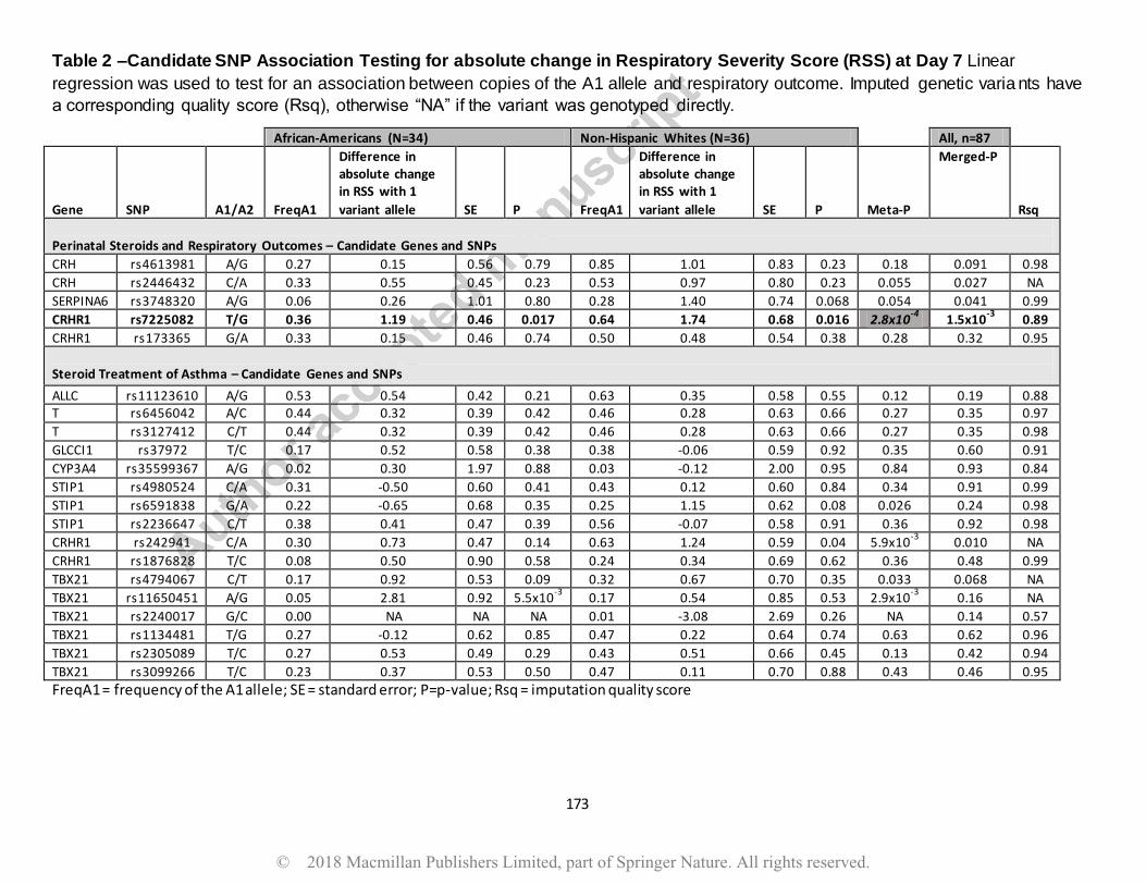
173
Table 2 –Candidate SNP Association Testing for absolute change in Respiratory Severity Score (RSS) at Day 7 Linear
regression was used to test for an association between copies of the A1 allele and respiratory outcome. Imputed genetic varia nts have
a corresponding quality score (Rsq), otherwise “NA” if the variant was genotyped directly.
African-Americans (N=34) Non-Hispanic Whites (N=36) All, n=87
Gene SNP A1/A2 FreqA1
Difference in absolute change in RSS with 1
variant allele SE P FreqA1
Difference in absolute change in RSS with 1
variant allele SE P Meta-P
Merged-P
Rsq
Perinatal Steroids and Respiratory Outcomes – Candidate Genes and SNPs
CRH rs4613981 A/G 0.27 0.15 0.56 0.79 0.85 1.01 0.83 0.23 0.18 0.091 0.98
CRH rs2446432 C/A 0.33 0.55 0.45 0.23 0.53 0.97 0.80 0.23 0.055 0.027 NA
SERPINA6 rs3748320 A/G 0.06 0.26 1.01 0.80 0.28 1.40 0.74 0.068 0.054 0.041 0.99
CRHR1 rs7225082 T/G 0.36 1.19 0.46 0.017 0.64 1.74 0.68 0.016 2.8x10⁻⁴
1.5x10⁻³
0.89
CRHR1 rs173365 G/A 0.33 0.15 0.46 0.74 0.50 0.48 0.54 0.38 0.28 0.32 0.95
Steroid Treatment of Asthma – Candidate Genes and SNPs
ALLC rs11123610 A/G 0.53 0.54 0.42 0.21 0.63 0.35 0.58 0.55 0.12 0.19 0.88 T rs6456042 A/C 0.44 0.32 0.39 0.42 0.46 0.28 0.63 0.66 0.27 0.35 0.97
T rs3127412 C/T 0.44 0.32 0.39 0.42 0.46 0.28 0.63 0.66 0.27 0.35 0.98
GLCCI1 rs37972 T/C 0.17 0.52 0.58 0.38 0.38 -0.06 0.59 0.92 0.35 0.60 0.91
CYP3A4 rs35599367 A/G 0.02 0.30 1.97 0.88 0.03 -0.12 2.00 0.95 0.84 0.93 0.84
STIP1 rs4980524 C/A 0.31 -0.50 0.60 0.41 0.43 0.12 0.60 0.84 0.34 0.91 0.99
STIP1 rs6591838 G/A 0.22 -0.65 0.68 0.35 0.25 1.15 0.62 0.08 0.026 0.24 0.98
STIP1 rs2236647 C/T 0.38 0.41 0.47 0.39 0.56 -0.07 0.58 0.91 0.36 0.92 0.98
CRHR1 rs242941 C/A 0.30 0.73 0.47 0.14 0.63 1.24 0.59 0.04 5.9x10⁻³
0.010 NA
CRHR1 rs1876828 T/C 0.08 0.50 0.90 0.58 0.24 0.34 0.69 0.62 0.36 0.48 0.99
TBX21 rs4794067 C/T 0.17 0.92 0.53 0.09 0.32 0.67 0.70 0.35 0.033 0.068 NA
TBX21 rs11650451 A/G 0.05 2.81 0.92 5.5x10⁻³
0.17 0.54 0.85 0.53 2.9x10⁻³
0.16 NA
TBX21 rs2240017 G/C 0.00 NA NA NA 0.01 -3.08 2.69 0.26 NA 0.14 0.57
TBX21 rs1134481 T/G 0.27 -0.12 0.62 0.85 0.47 0.22 0.64 0.74 0.63 0.62 0.96
TBX21 rs2305089 T/C 0.27 0.53 0.49 0.29 0.43 0.51 0.66 0.45 0.13 0.42 0.94
TBX21 rs3099266 T/C 0.23 0.37 0.53 0.50 0.47 0.11 0.70 0.88 0.43 0.46 0.95
FreqA1 = frequency of the A1 allele; SE = standard error; P=p-value; Rsq = imputation quality score
© 2018 Macmillan Publishers Limited, part of Springer Nature. All rights reserved.

174
Table 3 –Gene-Based Association Testing for absolute change in Respiratory
Severity Score (RSS) at Day 7. P-values were combined across all genotyped SNPs
within 50 kb of candidate genes using VEGAS2. No individual gene surpassed
Bonferroni adjustment for 18 comparisons (9 genes x 2 populations, 0.05/18=0.0028),
and no individual SNP within a gene surpassed Bonferroni adjustment for the total
number of SNPs within each gene (N SNPs).
African-Americans (N=34) Non-Hispanic Whites (N=36)
Gene N SNPs
Test
statistic p-value Top SNP
Top SNP p-
value N SNPs
Test
statistic p-value Top SNP
Top SNP
p-value
Perinatal Steroids and Respiratory Outcomes
CRH 33 30.8 0.457 rs7841890 0.028 28 36.65 0.21 rs7835948 0.082
CRHR1 15 13.3 0.495 rs114764500 0.067 10 10.69 0.36 rs242941 0.060 SERPINA6 56 46.5 0.616 rs56170999 0.002 44 87.70 0.03 rs3827896 0.0012
Steroid Treatment of Asthma – Candidate Genes
ALLC 47 46.4 0.435 rs1367275 0.003 53 74.48 0.13 rs1367276 0.008
CYP3A4 28 34.0 0.297 rs76994069 0.025 8 2.85 0.77 rs78676479 0.277
GLCCI1 58 69.8 0.220 rs17143586 0.002 76 45.47 0.95 rs10268598 0.088
SERPINE1 46 52.5 0.288 rs73712100 0.032 35 38.12 0.34 rs10953321 0.013
STIP1 16 10.7 0.699 rs2701527 0.113 14 16.47 0.30 rs118154228 0.085
T 31 63.5 0.020 rs6456056 0.009 35 30.80 0.53 rs10428822 0.024
© 2018 Macmillan Publishers Limited, part of Springer Nature. All rights reserved.
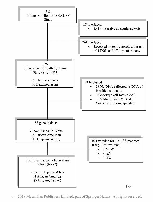
© 2018 Macmillan Publishers Limited, part of Springer Nature. All rights reserved.
175

© 2018 Macmillan Publishers Limited, part of Springer Nature. All rights reserved.
176

TAGGEDENDS E M I N A R S I N P E R I N A T O L O G Y 4 2 ( 2 0 1 8 )
Available online at www.sciencedirect.com
Seminars in Perinatology
www.seminperinat.com
177
Recent advances in antenatal f
actors predisposing to bronchopulmonary dysplasiaElizabeth Taglauera,1, Steven H. Abmanb,2, and Roberta L. Kellerc,*,3
aHarvard Neonatal-Perinatal Fellowship Program, Boston, MA USAbPediatric Heart Lung Center, Department of Pediatrics, University of Colorado Anschutz School of Medicine, Aurora, CO USAcDivision of Neonatology, Department of Pediatrics, University of California San Francisco, San Francisco, CA USA
*Corresponding author at:E-mail address: [email protected] (
1 Elizabeth Taglauer, Clinical Fellow in PedBoston MA 02115.
2 Steven H. Abman, Department of Pediatrirado, 13123 E 16th Ave B395, Aurora, CO 8004
3Roberta L. Keller, University of CaliforniaPediatrics, 550 16th Street, San Francisco CA
https://doi.org/10.1053/j.semperi.2018.09.0020146-0005/� 2018 Elsevier Inc. All rights reser
A B S T R A C T
Bronchopulmonary dysplasia (BPD) remains a major cause of late morbidities and death
after preterm birth. BPD is characterized by an arrest of vascular and alveolar growth and
high risk for pulmonary hypertension; yet mechanisms contributing to its pathogenesis
and early strategies to prevent BPD are poorly understood. Strong epidemiologic studies
have shown that the “new BPD” reflects the long-lasting impact of antenatal factors on
lung development, partly due to placental dysfunction, as reflected in recent data from ani-
mal models. Improved understanding of mechanisms through which antenatal stress
alters placental function and contributes to BPDmay lead to preventive therapies.
� 2018 Elsevier Inc. All rights reserved.
A R T I C L E I N F O
Keywords:
Alveolarization
Angiogenesis
Placenta
Vasculogenesis
Vascular endothelial growth factor
(VEGF)
Respiratory morbidity
gestation with birth weights, with approximately 10�15,000
Introduction
Despite striking improvements in perinatal care and diverse
neonatal outcomes related to premature birth, preterm
children remain at high risk for significant respiratory mor-
bidities and mortality due to the development of bronchopul-
monary dysplasia (BPD),1 Fig. 1. BPD is the chronic lung
disease of prematurity that develops in infants who require
respiratory support at birth due to immaturity of the preterm
lung.2 BPD is the most common sequel of prematurity, occur-
ring in roughly 45% of infants born at or less than 29 weeks
R.L. Keller).iatrics, Joint Program in
cs, University of Colorado5.San Francisco and UCSF B94158, United States.
ved.
new cases of BPD in the USA alone each year.1,3,4 Importantly,
the incidence of BPD has not changed over the past few deca-
des, likely reflecting improved survival of extremely low ges-
tational age newborns (ELGANS) who are at the highest risk
for developing moderate and severe BPD.5 BPD is also associ-
ated with significant neonatal intensive care unit (NICU)-
related complications, including the prolonged need for
mechanical ventilation, respiratory support and oxygen ther-
apy, longer duration of hospitalization and higher rates of
non-respiratory co-morbidities, such as retinopathy of pre-
maturity (ROP) and brain injury.5,6 Pathogenetic mechanisms
Neonatology, Boston Children’s Hospital, 300 Longwood Avenue,
Denver Anschutz Medical Campus and Children’s Hospital Colo-
enioff Children’s Hospital San Francisco Professor, Department of
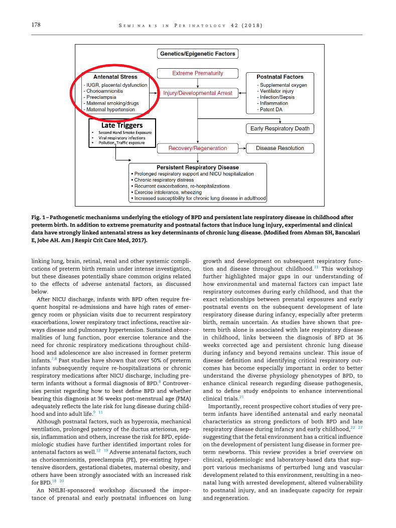
Fig. 1 – Pathogenetic mechanisms underlying the etiology of BPD and persistent late respiratory disease in childhood after
preterm birth. In addition to extreme prematurity and postnatal factors that induce lung injury, experimental and clinical
data have strongly linked antenatal stress as key determinants of chronic lung disease. (Modified from Abman SH, Bancalari
E, Jobe AH. Am J Respir Crit Care Med, 2017).
178 S E M I N A R S I N P E R I N A T O L O G Y 4 2 ( 2 0 1 8 )
linking lung, brain, retinal, renal and other systemic compli-
cations of preterm birth remain under intense investigation,
but these diseases potentially share common origins related
to the effects of adverse antenatal factors, as discussed
below.
After NICU discharge, infants with BPD often require fre-
quent hospital re-admissions and have high rates of emer-
gency room or physician visits due to recurrent respiratory
exacerbations, lower respiratory tract infections, reactive air-
ways disease and pulmonary hypertension. Sustained abnor-
malities of lung function, poor exercise tolerance and the
need for chronic respiratory medications throughout child-
hood and adolescence are also increased in former preterm
infants.7,8 Past studies have shown that over 50% of preterm
infants subsequently require re-hospitalizations or chronic
respiratory medications after NICU discharge, including pre-
term infants without a formal diagnosis of BPD.8 Controver-
sies persist regarding how to best define BPD and whether
bearing this diagnosis at 36 weeks post-menstrual age (PMA)
adequately reflects the late risk for lung disease during child-
hood and into adult life.9�11
Although postnatal factors, such as hyperoxia, mechanical
ventilation, prolonged patency of the ductus arteriosus, sep-
sis, inflammation and others, increase the risk for BPD, epide-
miologic studies have further identified important roles for
antenatal factors as well.12�19 Adverse antenatal factors, such
as chorioamnionitis, preeclampsia (PE), pre-existing hyper-
tensive disorders, gestational diabetes, maternal obesity, and
others have been strongly associated with an increased risk
for BPD.18�20
An NHLBI-sponsored workshop discussed the impor-
tance of prenatal and early postnatal influences on lung
growth and development on subsequent respiratory func-
tion and disease throughout childhood.11 This workshop
further highlighted major gaps in our understanding of
how environmental and maternal factors can impact late
respiratory outcomes during early childhood, and that the
exact relationships between prenatal exposures and early
postnatal events on the subsequent development of late
respiratory disease during infancy, especially after preterm
birth, remain uncertain. As studies have shown that pre-
term birth alone is associated with late respiratory disease
in childhood, links between the diagnosis of BPD at 36
weeks corrected age and persistent chronic lung disease
during infancy and beyond remains unclear. This issue of
disease definition and identifying critical respiratory out-
comes has become especially important in order to better
understand the diverse physiology phenotypes of BPD, to
enhance clinical research regarding disease pathogenesis,
and to define study endpoints to enhance interventional
clinical trials.21
Importantly, recent prospective cohort studies of very pre-
term infants have identified antenatal and early neonatal
characteristics as strong predictors of both BPD and late
respiratory disease during infancy and early childhood,22�27
suggesting that the fetal environment has a critical influence
on the development of persistent lung disease in former pre-
term newborns. This review provides a brief overview on
clinical, epidemiologic and laboratory-based data that sup-
port various mechanisms of perturbed lung and vascular
development related to this environment, resulting in a neo-
natal lung with arrested development, altered vulnerability
to postnatal injury, and an inadequate capacity for repair
and regeneration.
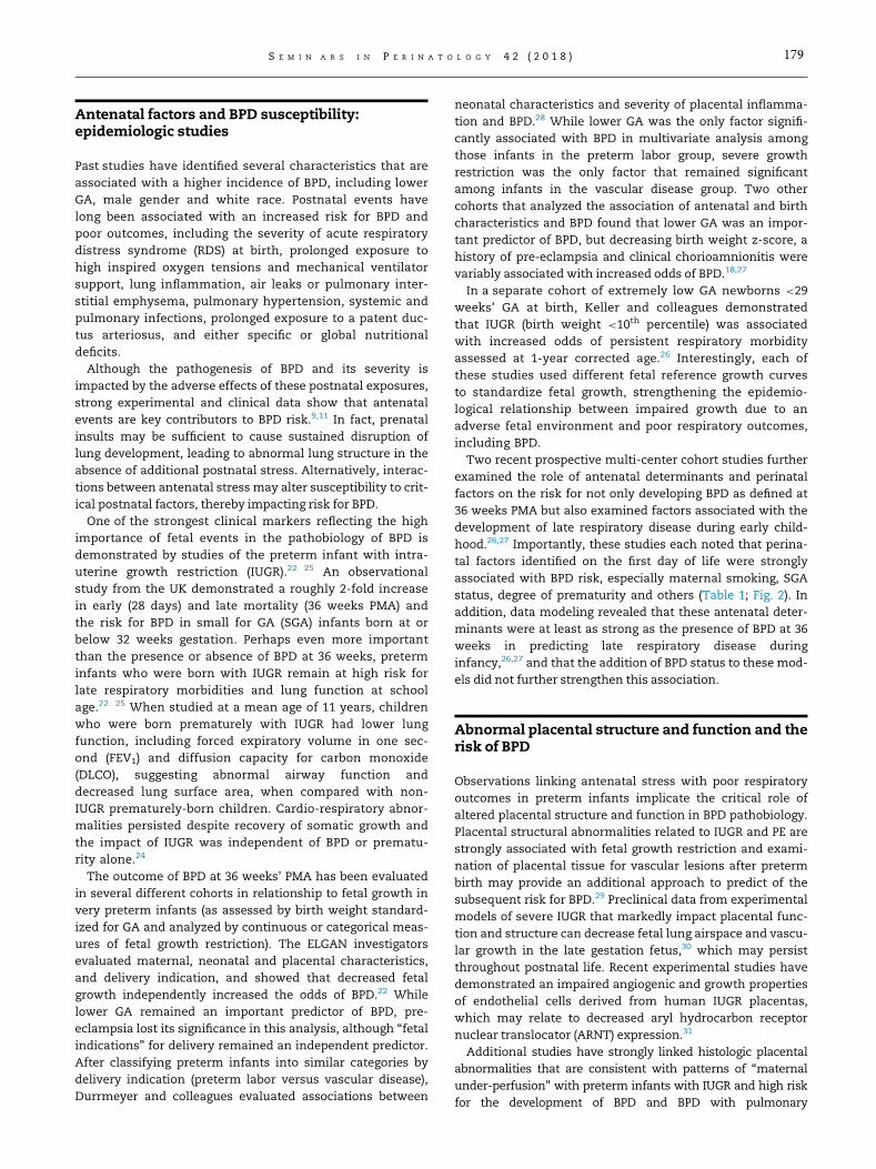
T AGGEDENDS E M I N A R S I N P E R I N A T O L O G Y 4 2 ( 2 0 1 8 ) 179
Antenatal factors and BPD susceptibility:epidemiologic studies
Past studies have identified several characteristics that are
associated with a higher incidence of BPD, including lower
GA, male gender and white race. Postnatal events have
long been associated with an increased risk for BPD and
poor outcomes, including the severity of acute respiratory
distress syndrome (RDS) at birth, prolonged exposure to
high inspired oxygen tensions and mechanical ventilator
support, lung inflammation, air leaks or pulmonary inter-
stitial emphysema, pulmonary hypertension, systemic and
pulmonary infections, prolonged exposure to a patent duc-
tus arteriosus, and either specific or global nutritional
deficits.
Although the pathogenesis of BPD and its severity is
impacted by the adverse effects of these postnatal exposures,
strong experimental and clinical data show that antenatal
events are key contributors to BPD risk.9,11 In fact, prenatal
insults may be sufficient to cause sustained disruption of
lung development, leading to abnormal lung structure in the
absence of additional postnatal stress. Alternatively, interac-
tions between antenatal stress may alter susceptibility to crit-
ical postnatal factors, thereby impacting risk for BPD.
One of the strongest clinical markers reflecting the high
importance of fetal events in the pathobiology of BPD is
demonstrated by studies of the preterm infant with intra-
uterine growth restriction (IUGR).22�25 An observational
study from the UK demonstrated a roughly 2-fold increase
in early (28 days) and late mortality (36 weeks PMA) and
the risk for BPD in small for GA (SGA) infants born at or
below 32 weeks gestation. Perhaps even more important
than the presence or absence of BPD at 36 weeks, preterm
infants who were born with IUGR remain at high risk for
late respiratory morbidities and lung function at school
age.22�25 When studied at a mean age of 11 years, children
who were born prematurely with IUGR had lower lung
function, including forced expiratory volume in one sec-
ond (FEV1) and diffusion capacity for carbon monoxide
(DLCO), suggesting abnormal airway function and
decreased lung surface area, when compared with non-
IUGR prematurely-born children. Cardio-respiratory abnor-
malities persisted despite recovery of somatic growth and
the impact of IUGR was independent of BPD or prematu-
rity alone.24
The outcome of BPD at 36 weeks’ PMA has been evaluated
in several different cohorts in relationship to fetal growth in
very preterm infants (as assessed by birth weight standard-
ized for GA and analyzed by continuous or categorical meas-
ures of fetal growth restriction). The ELGAN investigators
evaluated maternal, neonatal and placental characteristics,
and delivery indication, and showed that decreased fetal
growth independently increased the odds of BPD.22 While
lower GA remained an important predictor of BPD, pre-
eclampsia lost its significance in this analysis, although “fetal
indications” for delivery remained an independent predictor.
After classifying preterm infants into similar categories by
delivery indication (preterm labor versus vascular disease),
Durrmeyer and colleagues evaluated associations between
neonatal characteristics and severity of placental inflamma-
tion and BPD.28 While lower GA was the only factor signifi-
cantly associated with BPD in multivariate analysis among
those infants in the preterm labor group, severe growth
restriction was the only factor that remained significant
among infants in the vascular disease group. Two other
cohorts that analyzed the association of antenatal and birth
characteristics and BPD found that lower GA was an impor-
tant predictor of BPD, but decreasing birth weight z-score, a
history of pre-eclampsia and clinical chorioamnionitis were
variably associated with increased odds of BPD.18,27
In a separate cohort of extremely low GA newborns <29
weeks’ GA at birth, Keller and colleagues demonstrated
that IUGR (birth weight <10th percentile) was associated
with increased odds of persistent respiratory morbidity
assessed at 1-year corrected age.26 Interestingly, each of
these studies used different fetal reference growth curves
to standardize fetal growth, strengthening the epidemio-
logical relationship between impaired growth due to an
adverse fetal environment and poor respiratory outcomes,
including BPD.
Two recent prospective multi-center cohort studies further
examined the role of antenatal determinants and perinatal
factors on the risk for not only developing BPD as defined at
36 weeks PMA but also examined factors associated with the
development of late respiratory disease during early child-
hood.26,27 Importantly, these studies each noted that perina-
tal factors identified on the first day of life were strongly
associated with BPD risk, especially maternal smoking, SGA
status, degree of prematurity and others (Table 1; Fig. 2). In
addition, data modeling revealed that these antenatal deter-
minants were at least as strong as the presence of BPD at 36
weeks in predicting late respiratory disease during
infancy,26,27 and that the addition of BPD status to these mod-
els did not further strengthen this association.
Abnormal placental structure and function and therisk of BPD
Observations linking antenatal stress with poor respiratory
outcomes in preterm infants implicate the critical role of
altered placental structure and function in BPD pathobiology.
Placental structural abnormalities related to IUGR and PE are
strongly associated with fetal growth restriction and exami-
nation of placental tissue for vascular lesions after preterm
birth may provide an additional approach to predict of the
subsequent risk for BPD.29 Preclinical data from experimental
models of severe IUGR that markedly impact placental func-
tion and structure can decrease fetal lung airspace and vascu-
lar growth in the late gestation fetus,30 which may persist
throughout postnatal life. Recent experimental studies have
demonstrated an impaired angiogenic and growth properties
of endothelial cells derived from human IUGR placentas,
which may relate to decreased aryl hydrocarbon receptor
nuclear translocator (ARNT) expression.31
Additional studies have strongly linked histologic placental
abnormalities that are consistent with patterns of “maternal
under-perfusion” with preterm infants with IUGR and high risk
for the development of BPD and BPD with pulmonary

Table 1 – Perinatal factors at Day 1 of life andunivariate associationswithpost-prematurity respiratory disease (fromKellerRL et al, J Pediatr 201726).
Characteristic Whole cohort PRD No PRD P value
n = 724 n = 497 n = 227
Gestational age (weeks) 26.7 §1.4 26.6 §1.4 26.9 §1.3 0.009
Birth weight (g) 918 § 234 899 § 232 960 § 233 0.003
Intrauterine growth restriction 36 (5%) 32 (6%) 4 (2%) 0.02
Multiple gestation 187 (26%) 111 (22%) 76 (3%) 0.006
Smoking during pregnancy 139/723 (19%) 111/496 (22%) 28/227 (12%) 0.004
Stabilization at birth: intubation 564 (78%) 405 (82%) 159 (70%) 0.003
Maternal education 0.007
� High school 389 (54%) 283 (57%) 106 (47%)
Some college 143 (20%) 100 (20%) 43 (19%)
� College graduate 192 (27%) 114 (23%) 78 (34%)
Public insurance 470/721 (65%) 348/494 (70%) 122/227 (54%) <0.001
Parent with asthma 173/712 (24%) 129/485 (27%) 44/227 (19%) 0.04
*by trend test.
PRD, post-prematurity respiratory disease.
180 S E M I N A R S I N P E R I N A T O L O G Y 4 2 ( 2 0 1 8 )
hypertension32,33 (Table 2). Cord blood biomarkers, including
decreased vascular endothelial growth factor (VEGF) and soluble
VEGF antagonist fms-like tyrosine kinase-1 (sFLT-1) levels, were
strongly associated with IUGR and predictive of BPD in preterm
infants,34 suggesting that these circulating proteins may serve
as effective biomarkers for predicting later neonatal morbidities
associated with IUGR, including BPD. In addition, hypertensive
disorders of pregnancy, especially PE, have been strongly associ-
ated with high risk for BPD after preterm birth.18,27 In a cohort
study of 107 infants born <32 weeks, the risk for BPD was dra-
matically increased in the presence of PE (OR, 18.7; 95% CI,
2.44�145; p<0.005), even after accounting for IUGR.18
Fig. 2 –Antenatal factors are strongly associated with the risk of d
Med, 2016).
The placenta and neonatal disease
The placenta is a major contributor to changes in the intrauter-
ine environment that can lead to fetal and neonatal pathologies.
The placentamay also serve as a direct source of early signaling
for fetal disease in pregnancy, a concept that has been proposed
with placental-derived signals circulating in the maternal
serum such as cell free fetal DNA35 and placental-shed extracel-
lular vesicles.36 This may be particularly true for the developing
fetal lungs, which share some key aspects of structure and func-
tion with the placenta (Fig. 3). Insights into placental
eveloping BPD. (fromMorrow L et al. Am J Respir Crit Care
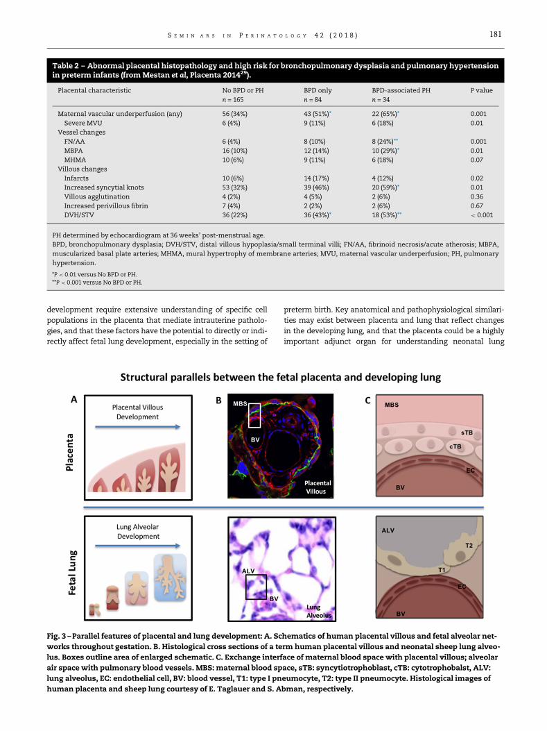
Table 2 – Abnormal placental histopathology and high risk for bronchopulmonary dysplasia and pulmonary hypertensionin preterm infants (fromMestan et al, Placenta 201429).
Placental characteristic No BPD or PH BPD only BPD-associated PH P value
n = 165 n = 84 n = 34
Maternal vascular underperfusion (any) 56 (34%) 43 (51%)* 22 (65%)* 0.001
Severe MVU 6 (4%) 9 (11%) 6 (18%) 0.01
Vessel changes
FN/AA 6 (4%) 8 (10%) 8 (24%)** 0.001
MBPA 16 (10%) 12 (14%) 10 (29%)* 0.01
MHMA 10 (6%) 9 (11%) 6 (18%) 0.07
Villous changes
Infarcts 10 (6%) 14 (17%) 4 (12%) 0.02
Increased syncytial knots 53 (32%) 39 (46%) 20 (59%)* 0.01
Villous agglutination 4 (2%) 4 (5%) 2 (6%) 0.36
Increased perivillous fibrin 7 (4%) 2 (2%) 2 (6%) 0.67
DVH/STV 36 (22%) 36 (43%)* 18 (53%)** < 0.001
PH determined by echocardiogram at 36 weeks’ post-menstrual age.
BPD, bronchopulmonary dysplasia; DVH/STV, distal villous hypoplasia/small terminal villi; FN/AA, fibrinoid necrosis/acute atherosis; MBPA,
muscularized basal plate arteries; MHMA, mural hypertrophy of membrane arteries; MVU, maternal vascular underperfusion; PH, pulmonary
hypertension.
*P < 0.01 versus No BPD or PH.**P < 0.001 versus No BPD or PH.
T AGGEDENDS E M I N A R S I N P E R I N A T O L O G Y 4 2 ( 2 0 1 8 ) 181
development require extensive understanding of specific cell
populations in the placenta that mediate intrauterine patholo-
gies, and that these factors have the potential to directly or indi-
rectly affect fetal lung development, especially in the setting of
Fig. 3 –Parallel features of placental and lung development: A. Sc
works throughout gestation. B. Histological cross sections of a te
lus. Boxes outline area of enlarged schematic. C. Exchange interf
air space with pulmonary blood vessels. MBS: maternal blood sp
lung alveolus, EC: endothelial cell, BV: blood vessel, T1: type I pn
human placenta and sheep lung courtesy of E. Taglauer and S. A
preterm birth. Key anatomical and pathophysiological similari-
ties may exist between placenta and lung that reflect changes
in the developing lung, and that the placenta could be a highly
important adjunct organ for understanding neonatal lung
hematics of human placental villous and fetal alveolar net-
rm human placental villous and neonatal sheep lung alveo-
ace of maternal blood space with placental villous; alveolar
ace, sTB: syncytiotrophoblast, cTB: cytotrophobalst, ALV:
eumocyte, T2: type II pneumocyte. Histological images of
bman, respectively.
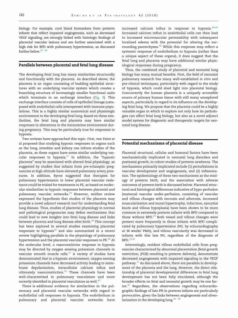
182 S E M I N A R S I N P E R I N A T O L O G Y 4 2 ( 2 0 1 8 )
biology. For example, cord blood biomarkers from preterm
infants that reflect impaired angiogenesis, such as decreased
VEGF signaling, are strongly linked with histologic findings of
placental vascular lesions and are further associated with a
high risk for BPD with pulmonary hypertension, as discussed
further below.32�34
Parallels between placental and fetal lung disease
The developing fetal lung has many similarities structurally
and functionally with the placenta. As described above, the
placenta is an organ consisting of budding epithelial struc-
tures with an underlying vascular system which creates a
branching structure of increasingly smaller functional units
which terminate in an exchange interface (Fig. 3). This
exchange interface consists of cells of epithelial lineage juxta-
posed with endothelial cells interspersed with immune popu-
lations. This is a highly similar anatomical and physiologic
environment to the developing fetal lung. Based on these sim-
ilarities, the fetal lung and placenta may have similar
responses to alterations in the intrauterine environment dur-
ing pregnancy. This may be particularly true for responses to
hypoxia.
Two reviews have approached this topic. First, van Patot et
al proposed that studying hypoxic responses in organs such
as the lung, intestine and kidney can inform studies of the
placenta, as these organs have some similar underlying vas-
cular responses to hypoxia.37 In addition, the “hypoxic
placenta” may be associated with altered fetal physiology, as
suggested by studies that infants from pre-eclamptic preg-
nancies at high altitude have elevated pulmonary artery pres-
sures. In addition, Byrne suggested that therapies for
pulmonary hypertension to lower placental vascular resis-
tance could be trialed for treatments in PE, as based onmolec-
ular similarities in hypoxic responses between placental and
pulmonary vascular networks.38 However, neither review
expressed the hypothesis that studies of the placenta may
provide a novel adjunct research tool for understanding fetal
lung disease. Thus, studies of placental physiology in normal
and pathological pregnancies may define mechanisms that
could lead to new insights into fetal lung disease and links
between placenta and lung disease after birth.39 This concept
has been explored in several studies examining placental
responses to hypoxia40 and also summarized in a recent
review highlighting parallels in the physiology of pulmonary
hypertension and the placental vascular responses to PE.38 At
the molecular level, a vasoconstrictor response to hypoxia
may be directed by oxygen sensing potassium channels in
vascular smooth muscle cells.41 A variety of studies have
demonstrated that in a hypoxic environment, oxygen sensing
potassium channels have decreased activity leading to mem-
brane depolarization, intracellular calcium influx and
ultimately vasoconstriction.42 These channels have been
well-characterized in pulmonary vasculature and more
recently identified in placental vasculature as well.43
There is additional evidence for similarities in the pul-
monary and placental vascular networks with regard to
endothelial cell responses to hypoxia. The endothelium in
pulmonary and placental vascular networks have
increased calcium influx in response to hypoxia.44,45
Increased calcium influx in endothelial cells can then lead
to increased microvascular permeability with subsequent
localized edema with the potential for altering the sur-
rounding parenchyma.46 While this response may reflect a
systemic response of endothelium to hypoxia (rather than
a unique aspect of these organs), it does suggest that the
fetal lung and placenta may have additional similar physi-
ological responses during pregnancy.
Thus, the combined study of placental and neonatal lung
biology has many mutual benefits. First, the field of neonatal
pulmonary research has many well-established in vitro and
pre-clinical techniques, particularly with regard to the study
of hypoxia, which could shed light into placental biology.
Concurrently the human placenta is a uniquely accessible
source of primary human tissue with still many unexplored
aspects, particularly in regard to its influence on the develop-
ing fetal lung. We propose that the placenta could be a highly
valuable organ in which to explore not only how its patholo-
gies can affect fetal lung biology, but also as a novel adjunct
model system for diagnostic and therapeutic targets for neo-
natal lung disease.
Potential mechanisms of placental disease
Placental structural, cellular and humoral factors have been
mechanistically implicated in neonatal lung disorders and
postnatal growth, in cohort studies of preterm newborns. The
mechanisms primarily implicated include (1) perturbations in
vascular development and angiogenesis, and (2) inflamma-
tion. The epidemiology of these two mechanisms as the etiol-
ogy of preterm birth, and the association with adverse
outcomes of preterm birth is discussed below. Placental struc-
tural and histological differences indicative of hypo-perfusion
(maternal vascular under-perfusion, consisting of vascular
and villous changes with necrosis and atherosis, increased
muscularization and mural hypertrophy, infarction, syncytial
knots and villous hypoplasia) have been shown to be more
common in extremely preterm infants with BPD compared to
those without BPD.29 Both vessel and villous changes were
present more frequently in those infants with BPD compli-
cated by pulmonary hypertension (PH, by echocardiography
at 36 weeks’ PMA), and villous vascularity was decreased in
infants with this late PH, regardless of the diagnosis of
BPD.29,45
Interestingly, resident villous endothelial cells from preg-
nancies characterized by abnormal placentation [fetal growth
restriction, (FGR) resulting in preterm delivery], demonstrate
decreased angiogenesis with impaired signaling in the VEGF
pathway.31 As discussed above, there are parallels in develop-
ment of the placenta and the lung. However, the direct rela-
tionship of placental developmental differences to fetal lung
development has not been fully elucidated, although the
broader effects on fetal and neonatal growth may be one fac-
tor.30 Regardless, the observations regarding echocardio-
graphic findings of late PH in preterm infants are particularly
provocative, given the links between angiogenesis and alveo-
larization in the developing lung.46�48

T AGGEDENDS E M I N A R S I N P E R I N A T O L O G Y 4 2 ( 2 0 1 8 ) 183
Additional cellular and humoral effects of this abnormal
placentation should also be considered when evaluating
these potential mechanisms. The placenta and umbilical
cord blood contain, respectively, resident and circulating
endothelial progenitor cells.49 Both cell populations have
been shown to have similar proliferative potential in term
placentas, although the placental population had greater
vasculogenic potential.49 Decreased numbers of endothe-
lial progenitor cells have been demonstrated in the cord
blood of preterm newborns who develop BPD,50,51 cells
which are also decreased in maternal diabetes and pre-
eclampsia.52,53 Interestingly, cord blood from pregnancies
with a placenta that demonstrated severe maternal vascu-
lar under-perfusion had lower levels of placental grow
factor (PIGF), granulocyte-colony stimulating factor (G-CSF)
and VEGF-A.34 This pattern was consistent in cord blood of
those infants who were later diagnosed with BPD compli-
cated by echocardiographic findings of PH, but only the
relationship with PIGF and G-CSF held up in a validation
cohort. In contrast to these findings, increased placental
VEGF levels were found in an analysis of protein expres-
sion from placental biopsies at birth from pregnancies
affected by PE and extremely preterm delivery.54 Addi-
tional protein expression patterns in the PE cluster
(distinct from pregnancies affected by preterm labor or
premature rupture of the membranes) include increased
levels of P-selectin and transforming growth factor-b (TGF-
b), further supporting placental vascular differences in
these pregnancies.
Finally, regarding the vulnerability of the lung following
pregnancies affected by placental vascular abnormalities
and the lung’s ability to repair following insult and injury
during the neonatal hospitalization, the growth of cord
blood endothelial progenitor cells from preterm pregnan-
cies is inhibited by exposure to hyperoxia.51,55 Hyperoxia
decreases nitric oxide (NO) production and endothelial
nitric oxide synthase (eNOS) and VEGF receptor-2 (VEGFR-
2) protein expression ex vivo, an effect that is mimicked by
NOS inhibition.55 The inhibitory growth effect of hyperoxia
was mitigated by treatment with NO or VEGF (with
increased eNOS expression), and antioxidant therapies.52,56
Fetal oxidative stress is inversely related to GA (assessed
by cord 8-isoprostane levels at birth), resulting in addi-
tional vulnerability for the most preterm infants.56 Later
lung function data (3�33 months corrected age) from for-
mer preterm newborns without BPD demonstrate that
alveolar volume and diffusing capacity are positively
related to the pro-angiogenic potential of circulating stem
cells from contemporaneous samples, supporting the long-
term implications of these antenatal perturbations in
angiogenesis and the consequences of neonatal care on
these vulnerable newborns. Together, these data suggest
that placental vascular abnormalities and the presence
and function of endothelial precursor cells derived from
the placenta influence both fetal lung development and
infant lung and vascular growth during the neonatal hos-
pitalization (manifested as increased susceptibility to BPD
and PH), with potential for persistent impact on the dys-
plastic lungs as these children grow and develop.
Other factors further alter the fetal environmentand impact lung development
Inflammation has long been considered key to the patho-
physiology of BPD.1,2 However, antenatal infection and
inflammation have been variably demonstrated to be a
risk factor for BPD, using clinical chorioamnionitis as the
marker for inflammation. Further investigations have eval-
uated the placenta for histologic chorioamnionitis, acute
and chronic inflammation, and the fetal inflammatory
response (FIR, assessed as neutrophil infiltration into fetal
vessels), and have more convincingly argued that antena-
tal inflammation (defined by placental histology), plays
a role in the later development of BPD.57 Although
placental signs of inflammation are associated with the
development of BPD, investigations of early neonatal
humoral inflammatory profiles fail to demonstrate consis-
tent relationships with BPD.58,59 Cord blood levels of
interleukin-6 (IL-6) and monocyte chemotactic protein-1
(MCP-1) have been shown to be significantly elevated in
very preterm infants who later developed BPD, although
these and other relationships are influenced by the inverse
correlation of these inflammatory markers with GA at
birth.60�62 Interestingly, higher C-reactive protein levels
were associated with decreased odds of BPD in former pre-
term newborns < 32 weeks’ GA at birth.62 The co-occurrence
of placental inflammation in the setting of maternal vascular
under-perfusion also mitigates the strong anti-angiogenic
relationships of abnormal placentation.29,34 Further, histologic
chorioamnionitis is associated with lower rates of early lung
disease (RDS), as some aspects of the inflammatory response
may have maturational effects on the lung.57 These variable
effects of inflammation may explain the inconsistent findings
regarding fetal humoral predictors of BPD in human cohort
studies.
One factor closely associated with later adverse lung func-
tion in term newborns is maternal smoking, which is associ-
ated with increased levels of oxidative stress; the effects of in
utero nicotine exposure may be mitigated by antenatal sup-
plemental vitamin C, initiated at <23 weeks’ GA.63,64 In very
preterm infants, maternal smoking during pregnancy is
strongly associated with both the development of BPD, and
later respiratory morbidity (assessed at 1�2 years corrected
age).26,27 In a small subset of these newborns, maternal smok-
ing during pregnancy was associated with lower levels of
Vitamin E isoforms over the first month of life, carrying
increased vulnerability to pro-inflammatory and oxidant
states.65 With respect to other environmental influences on
lung development, antenatal exposure to particulate matter
and other environmental pollutants has been shown to have
adverse effects on fetal growth and respiratory outcomes;66,67
preterm infants may be the most susceptible to the conse-
quences of these exposures.68
As suggested, the structural, cellular and humoral aspects
of the placenta are closely tied to the primary underlying eti-
ology of preterm birth, providing additional information
toward understanding the antenatal underpinnings of BPD
and its repercussions. The ELGAN investigators evaluated 8
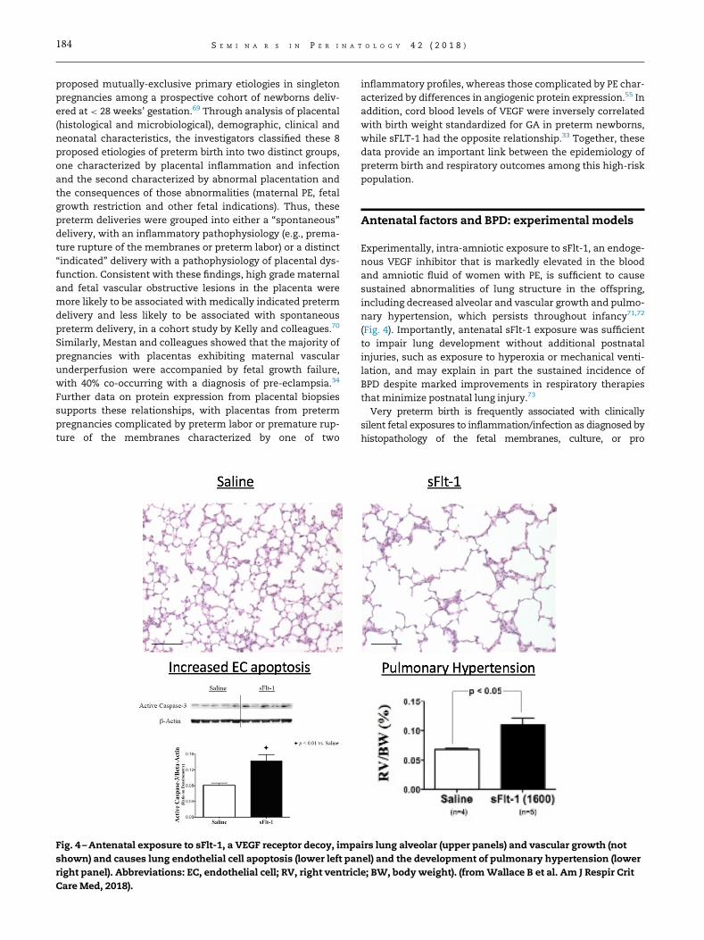
184 S E M I N A R S I N P E R I N A T O L O G Y 4 2 ( 2 0 1 8 )
proposed mutually-exclusive primary etiologies in singleton
pregnancies among a prospective cohort of newborns deliv-
ered at < 28 weeks’ gestation.69 Through analysis of placental
(histological and microbiological), demographic, clinical and
neonatal characteristics, the investigators classified these 8
proposed etiologies of preterm birth into two distinct groups,
one characterized by placental inflammation and infection
and the second characterized by abnormal placentation and
the consequences of those abnormalities (maternal PE, fetal
growth restriction and other fetal indications). Thus, these
preterm deliveries were grouped into either a “spontaneous”
delivery, with an inflammatory pathophysiology (e.g., prema-
ture rupture of the membranes or preterm labor) or a distinct
“indicated” delivery with a pathophysiology of placental dys-
function. Consistent with these findings, high grade maternal
and fetal vascular obstructive lesions in the placenta were
more likely to be associated with medically indicated preterm
delivery and less likely to be associated with spontaneous
preterm delivery, in a cohort study by Kelly and colleagues.70
Similarly, Mestan and colleagues showed that the majority of
pregnancies with placentas exhibiting maternal vascular
underperfusion were accompanied by fetal growth failure,
with 40% co-occurring with a diagnosis of pre-eclampsia.34
Further data on protein expression from placental biopsies
supports these relationships, with placentas from preterm
pregnancies complicated by preterm labor or premature rup-
ture of the membranes characterized by one of two
Fig. 4 –Antenatal exposure to sFlt-1, a VEGF receptor decoy, impa
shown) and causes lung endothelial cell apoptosis (lower left pan
right panel). Abbreviations: EC, endothelial cell; RV, right ventric
Care Med, 2018).
inflammatory profiles, whereas those complicated by PE char-
acterized by differences in angiogenic protein expression.55 In
addition, cord blood levels of VEGF were inversely correlated
with birth weight standardized for GA in preterm newborns,
while sFLT-1 had the opposite relationship.33 Together, these
data provide an important link between the epidemiology of
preterm birth and respiratory outcomes among this high-risk
population.
Antenatal factors and BPD: experimental models
Experimentally, intra-amniotic exposure to sFlt-1, an endoge-
nous VEGF inhibitor that is markedly elevated in the blood
and amniotic fluid of women with PE, is sufficient to cause
sustained abnormalities of lung structure in the offspring,
including decreased alveolar and vascular growth and pulmo-
nary hypertension, which persists throughout infancy71,72
(Fig. 4). Importantly, antenatal sFlt-1 exposure was sufficient
to impair lung development without additional postnatal
injuries, such as exposure to hyperoxia or mechanical venti-
lation, and may explain in part the sustained incidence of
BPD despite marked improvements in respiratory therapies
that minimize postnatal lung injury.73
Very preterm birth is frequently associated with clinically
silent fetal exposures to inflammation/infection as diagnosed by
histopathology of the fetal membranes, culture, or pro
irs lung alveolar (upper panels) and vascular growth (not
el) and the development of pulmonary hypertension (lower
le; BW, bodyweight). (fromWallace B et al. Am J Respir Crit

T AGGEDENDS E M I N A R S I N P E R I N A T O L O G Y 4 2 ( 2 0 1 8 ) 185
inflammatory cytokines in amniotic fluid or cord blood. Other
very preterm deliveries are associated with clinical chorioam-
nionitis, a non-specific diagnosis of limited clinical value.
Inflammation associated chorioamnionitis seldom results in
positive blood cultures or frank infection in the newborn, but
indicators of lung inflammation prior to birth are frequent.
While specific infections are seldom identified clinically, the
very preterm infant can have lung abnormalities resulting from
antenatal exposures that range from severe diffuse pneumonia
(indistinguishable from severe RDS) to verymature lungs for GA.
Experimental studies demonstrate this extreme range of
effects as well as modulation of fetal and postnatal immune
responses. Thus, these antenatal exposures can promote
early lung injury by decreasing lung function or decrease
the risk of BPD by decreasing the severity of RDS. However,
how fetal modulations of immune and inflammatory
responses contribute to postnatal lung injury and the devel-
opment of BPD are poorly understood. Antenatal exposure to
endotoxin (e.g., E. Coli lipopolysaccharide, or LPS) is sufficient
to cause BPD-type changes in infant rat lungs, even in the
absence hyperoxia. mechanical ventilation or other postnatal
injuries.73,74 This provides an additional model to dissect the
mechanisms leading to human BPD and the potential for
early, preventive interventions.
Early pulmonary vascular disease
Preclinical studies suggest that disruption of angiogenesis
due to adverse antenatal factors, such as chorioamnionitis,
PE or maternal smoking, and postnatal injury after preterm
birth, can cause pulmonary vascular disease (PVD) that not
only leads to pulmonary hypertension (PH) but can also
impair distal lung growth.33,34 Laboratory studies have shown
that the developing endothelial cell plays a key role in regula-
tion and coordination of epithelial growth and distal airspace
Fig. 5 –Echocardiogram evidence of pulmonary hypertension (PH
ease during early childhood. (fromMourani PM et al. Am J Respir
structure through the production of critical “angiocrines,”
such as NO, hepatocyte growth factor, vitamin A, insulin
growth factor-1 and others.11,33 As angiogenesis is necessary
for normal alveolarization,34 it has been suggested that pro-
tecting the developing pulmonary vasculature from early
injury may not only lower pulmonary vascular resistance as
one important goal but such strategies may enhance distal
lung growth and improve gas exchange, exercise intolerance
and other long-term outcomes.
As noted above, several studies have reported that altered
cord blood biomarker levels, including various angiogenic fac-
tors and endothelial progenitor cells, are associated with the
subsequent risk for BPD.32 More recently, clinical studies have
shown that early echocardiographic findings of PVD after pre-
term birth are strongly associated with the development and
severity of BPD and PH at 36 weeks corrected age.75�77 Inter-
estingly, these findings were also associated with a worse
respiratory course during the initial hospitalization, but also
late respiratory outcomes, including respiratory exacerba-
tions, hospitalizations and the need for asthma medica-
tions.77 (Fig. 5). Therapeutic strategies that target enhanced
endothelial survival, function and growth may provide novel
approaches towards the prevention of BPD after early diagno-
sis of high risk within preterm populations.
The need for preventive strategies
Thus, the need for preventive strategies persists and remains
a major challenge to better improve long-term respiratory
outcomes after preterm birth. Despite these remarkable
advances, many knowledge barriers and gaps towards
enhancing late respiratory outcomes of premature infants
continue. The current definition of BPD remains unsatisfying
for several reasons, including the concept that BPD at 36
weeks is merely a surrogate of more concerning endpoints,
) is strongly associated with subsequent late respiratory dis-
Crit Care Med. 2017).
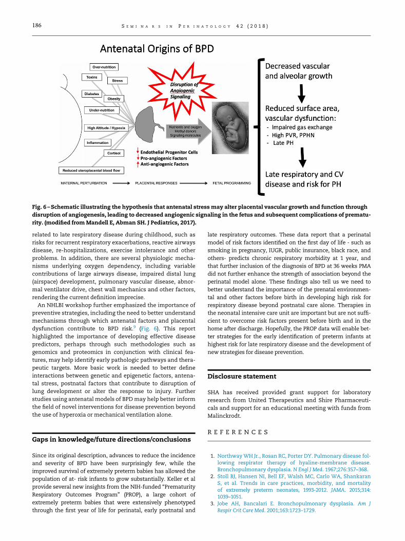
Fig. 6 –Schematic illustrating the hypothesis that antenatal stress may alter placental vascular growth and function through
disruption of angiogenesis, leading to decreased angiogenic signaling in the fetus and subsequent complications of prematu-
rity. (modified fromMandell E, Abman SH. J Pediatrics, 2017).
186 S E M I N A R S I N P E R I N A T O L O G Y 4 2 ( 2 0 1 8 )
related to late respiratory disease during childhood, such as
risks for recurrent respiratory exacerbations, reactive airways
disease, re-hospitalizations, exercise intolerance and other
problems. In addition, there are several physiologic mecha-
nisms underlying oxygen dependency, including variable
contributions of large airways disease, impaired distal lung
(airspace) development, pulmonary vascular disease, abnor-
mal ventilator drive, chest wall mechanics and other factors,
rendering the current definition imprecise.
An NHLBI workshop further emphasized the importance of
preventive strategies, including the need to better understand
mechanisms through which antenatal factors and placental
dysfunction contribute to BPD risk.9 (Fig. 6). This report
highlighted the importance of developing effective disease
predictors, perhaps through such methodologies such as
genomics and proteomics in conjunction with clinical fea-
tures, may help identify early pathologic pathways and thera-
peutic targets. More basic work is needed to better define
interactions between genetic and epigenetic factors, antena-
tal stress, postnatal factors that contribute to disruption of
lung development or alter the response to injury. Further
studies using antenatal models of BPDmay help better inform
the field of novel interventions for disease prevention beyond
the use of hyperoxia or mechanical ventilation alone.
Gaps in knowledge/future directions/conclusions
Since its original description, advances to reduce the incidence
and severity of BPD have been surprisingly few, while the
improved survival of extremely preterm babies has allowed the
population of at- risk infants to grow substantially. Keller et al
provide several new insights from the NIH-funded “Prematurity
Respiratory Outcomes Program” (PROP), a large cohort of
extremely preterm babies that were extensively phenotyped
through the first year of life for perinatal, early postnatal and
late respiratory outcomes. These data report that a perinatal
model of risk factors identified on the first day of life - such as
smoking in pregnancy, IUGR, public insurance, black race, and
others- predicts chronic respiratory morbidity at 1 year, and
that further inclusion of the diagnosis of BPD at 36 weeks PMA
did not further enhance the strength of association beyond the
perinatal model alone. These findings also tell us we need to
better understand the importance of the prenatal environmen-
tal and other factors before birth in developing high risk for
respiratory disease beyond postnatal care alone. Therapies in
the neonatal intensive care unit are important but are not suffi-
cient to overcome risk factors present before birth and in the
home after discharge. Hopefully, the PROP data will enable bet-
ter strategies for the early identification of preterm infants at
highest risk for late respiratory disease and the development of
new strategies for disease prevention.
Disclosure statement
SHA has received provided grant support for laboratory
research from United Therapeutics and Shire Pharmaceuti-
cals and support for an educational meeting with funds from
Malinckrodt.
R E F E R E N C E S
1. NorthwayWH Jr., Rosan RC, Porter DY. Pulmonary disease fol-lowing respirator therapy of hyaline-membrane disease.Bronchopulmonary dysplasia. N Engl J Med. 1967;276:357–368.
2. Stoll BJ, Hansen NI, Bell EF, Walsh MC, Carlo WA, ShankaranS, et al. Trends in care practices, morbidity, and mortalityof extremely preterm neonates, 1993-2012. JAMA. 2015;314:1039–1051.
3. Jobe AH, Bancalari E. Bronchopulmonary dysplasia. Am JRespir Crit Care Med. 2001;163:1723–1729.

T AGGEDENDS E M I N A R S I N P E R I N A T O L O G Y 4 2 ( 2 0 1 8 ) 187
4. Stoll BJ, Hansen NI, Bell EF, et al. Neonatal outcomes ofextremely preterm infants from the NICHD NeonatalResearch Network. Pediatrics. 2010;126:443.
5. Schmidt B, Roberts RS, Davis PG, et al. Prediction of late death ordisability at age 5 years using a count of 3 neonatal morbiditiesin very low birth weight infants. J Pediatr. 2015;167:982–986: e2.
6. Abman SH. Bronchopulmonary dysplasia. NY: Informa; 2010.7. Islam JY, Keller RL, Aschner JL, Hartert TV, Moore PE. Under-
standing the short and long-term respiratory outcomes ofprematurity and bronchopulmonary dysplasia. Am J RespirCrit Care Med. 2015;192:134–156.
8. Gunville CF, Sontag MK, Stratton K, Ranade DJ, Abman SH,Mourani PM. Scope and impact of early and late preterminfants admitted to PICU with respiratory illness. J Pediatr.2010;157:209–214.
9. McEvoy C, Jain L, Schmidt B, Abman SH, Bancalari E, AschnerJ. Bronchopulmonary dysplasia: NHLBI Workshop on the Pri-mary Prevention of Chronic Lung Diseases. Ann Am ThoracicSoc. 2014(Suppl 3):S146–S153.
10. Poindexter BB, Feng R, Schmidt B, et al. Comparisons and lim-itations of current definitions of bronchopulmonary dysplasiafor the prematurity and respiratory outcomes program. AnnAm Thorac Soc. 2015;12:1822–1830.
11. Manuck TA, Levy PT, Gyamfi-Bannerman C, Jobe AH, BlaisdellCJ. Prenatal and perinatal determinants of lung health anddisease in early life. JAMA Pediatr. 2016;170:e154577.
12. Farstad T, Bratlid D, Medbø S, Markestad T. NorwegianExtreme Prematurity Study Group. Bronchopulmonary dys-plasia � prevalence, severity and predictive factors in anational cohort of extremely premature infants. Acta Paediatr.2011;100:53–58.
13. Laughon MM, Langer JC, Bose CL, et al. Prediction of broncho-pulmonary dysplasia by postnatal age in extremely prema-ture infants. Am J Respir Crit Care Med. 2011;183:1715–1722.
14. Wai KC, KohnMA, Ballard RA, et al. Early cumulative supplemen-tal oxygen predicts bronchopulmonary dysplasia in high riskextremely low gestational age newborns. J Pediatr. 2016. https://doi.org/10.1016/j.jpeds.2016.06.079. pii: S0022-3476(16)30508-X[Epub aheadof print].
15. Bose C, van Marter LJ, Laughon M, O’Shea TM, Allred EN,Karna P, et al. Fetal growth restriction and chronic lung dis-ease among infants born before the 28th week of gestation.Pediatrics. 2009;124:e450–e458.
16. Watterberg KL, Demers LM, Scott SM, Murphy S. Chorioam-nionitis and early lung inflammation in infants in whombronchopulmonary dysplasia develops. Pediatrics. 1996;97(2):210–215.
17. Van Marter LJ, Dammann O, Allred EN, et al. Developmentalepidemiology network investigators. chorioamnionitis,mechanical ventilation, and postnatal sepsis as modulatorsof chronic lung disease in preterm infants. J Pediatr.2002;140(2):171–176.
18. Hansen AR, Barnes CM, Folkman J, McElrath TF. Maternal pre-eclampsia predicts development of BPD. J Pediatr. 2010-14;156:532–536.
19. Erikson L, Haglund B, Odlind V, Altman M, Ewald U, Keiler H.Perinatal conditions related to growth restriction and inflam-mation are associated with an increased risk of BPD. Acta Pae-diatrica. 2015;104:259–263.
20. Jobe AH. Effects of chorioamnionitis on the fetal lung. ClinPerinatol. 2012;39:441–457.
21. Steinhorn R, Davis JM, Gopel W, et al. Chronic pulmonaryinsufficiency of prematurity: developing optimal endpointsfor drug development. J Pediatrics. 2017;191:15–21.
22. Bose C, van Marter LJ, Laughon M, O’Shea TM, Allred EN,Karna P, et al. Fetal growth restriction and chronic lung dis-ease among infants born before the 28th week of gestation.Pediatrics. 2009;124:e450–e458.
23. Lal MK, Manktelow BN, Drpaer ES, Field DJ. Chronic lung dis-ease of prematurity and IUGR: a population-based study. Pedi-atrics. 2003;111:483–487.
24. Ronkainen E, Dunder T, Kaukola T, Marttila R, Hallman M.Intrauterine growth restriction predicts lung function atschool age in children born very preterm. Arch Dis Child FetalNeonatal Edit. 2016;101:F412–F417.
25. Greenough A, Yuksel B, Cheeseman P. Effect of in uterogrowth retardation on lung function at follow-up of prema-turely born infants. Eur Respir J. 2004;24:731–733.
26. Keller RL, Feng R, DeMauro SB, et al. Prematurity and respira-tory outcomes program. Bronchopulmonary dysplasia andperinatal characteristics predict 1-Year respiratory outcomesin newborns born at extremely low gestational age: a prospec-tive cohort study. J Pediatr. 2017;187:89–97: e3.
27. Morrow LA, Wagner BD, Ingram DA, et al. Antenatal determi-nants of bronchopulmonary dysplasia and late respiratorydisease in preterm infants. Am J Respir Crit Care Med.2017;196(3):364–374.
28. Durrmeyer X, Kayem G, Sinico M, Dassieu G, Danan C, Deco-bert F. Perinatal risk factors for bronchopulmonary dysplasiain extremely low gestational age infants: a pregnancy disor-der-based approach. J Pediatr. 2012;160(4):578–583.e2.
29. Mestan KK, Check J, Minturn L, et al. Placental pathologicchanges of maternal vascular under-perfusion in BPD andpulmonary hypertension. Placenta. 2014;35:570–574.
30. Rozance PJ, Seedorf GJ, Brown A, et al. IUGR decreased pulmo-nary alveolar and vessel growth and causes pulmonary arteryendothelial cell dysfunction in vitro in fetal sheep. Am J Phys-iol. LCMP. 2011;301:L860–L871.
31. Su E, Xing H, Yin P, et al. Impaired fetoplacental angiogenesisin growth-restricted fetuses with abnormal umbilical arterydoppler velocimetry is mediated by ARNT. J Clin EndocrinolMetab. 2015;100:E30–E40.
32. Check J, Gotteiner N, Liu X, et al. Fetal growth restriction andpulmonary hypertension in premature infants with BPD. JPerinatol. 2013;33:553–557.
33. Voller SB, Chock S, Ernst LM, et al. Cord blood biomarkers ofvascular endothelial growth (VEGF adn sFlt-1) and postnatalgrowth: a preterm birth cohort study. Early Hum Dev.2014;90:195–200.
34. Mestan KK, Gotteiner N, Porta N, et al. Cord blood biomarkersof placental maternal vascular under-perfusion predict bron-chopulmonary dysplasia-associated pulmonary hyperten-sion. J Pediatr. 2017;185:33–41.
35. Taglauer ES, Wilkins-Haug L, Bianchi DW. Review: cell-free fetal DNA in the maternal circulation as an indica-tion of placental health and disease. Placenta. 2014;35:S64–S68.
36. Tannetta D, Collett G, VatishM, Redman C, Sargent I. Syncytio-trophoblast extracellular vesicles - Circulating biopsies reflect-ing placental health. Placenta. 2017;52:134–138.
37. van Patot MC, Ebensperger G, Gassmann M, Llanos A. Thehypoxic placenta. High Alt Med Biol. 2012;13(3):176–184.
38. Byrne TJ. A “cure” for preeclampsia: Improving neonatal out-comes by overcoming excess fetal placental vascular resis-tance.Med Hypotheses. 2015 Sep;85(3):311–319.
39. Dunham-Snary KJ, et al. Hypoxic pulmonary vasoconstric-tion: from molecular mechanisms to medicine. Chest.2017;151(1):181–192.
40. Herrera EA, et al. The placental pursuit for an adequate oxi-dant balance between the mother and the fetus. Front Pharma-col. 2014;5:149.
41. Shimoda LA, Polak J. Hypoxia. 4. Hypoxia and ion channelfunction. Am J Physiol Cell Physiol. 2011;300(5):C951–C967.
42. Wareing M. Oxygen sensitivity, potassium channels, and reg-ulation of placental vascular tone. Microcirculation.2014;21(1):58–66.

188 S E M I N A R S I N P E R I N A T O L O G Y 4 2 ( 2 0 1 8 )
43. Tiruppathi C, et al. Role of Ca2+ signaling in the regulationof endothelial permeability. Vascul Pharmacol. 2002;39(4-5):173–185.
44. Aono Y, et al. Human umbilical vein endothelial cells(HUVECs) show Ca(2+) mobilization as well as Ca(2+) influxupon hypoxia. J Cell Biochem. 2000;78(3):458–464.
45. Yallapragada SG, Mestan KK, Palac H, et al. Placental villousvascularity is decreased in premature infants with broncho-pulmonary dysplasia-associated pulmonary hypertension.Pediatr Dev Pathol. 2016;19(2):101–107.
46. Abman S. Bronchopulmonary dysplasia: “a vascular hypoth-esis”. Am J Respir Crit Care Med. 2001;164:1755–1756.
47. Jakkula M, Le Cras TD, Gebb S, et al. Inhibition of angiogenesisdecreases alveolarization in the developing rat lung. Am JPhysiol Lung Cell Mol Physiol. 2000;279:L600–L609.
48. Le Cras TD Le Cras TD, Markham NE, Tuder RM, Voelkel NF,Abman SH. Brief neonatal treatment with a VEGF receptorinhibitor causes pulmonary hypertension and abnormal lungstructure in adult rats. Am J Phyiol. Lung Cell Mol Biol. 2002;283:L555–L562.
49. Rapp BM, Saadatzedeh MR, Ofstein RH, et al. Resident endo-thelial progenitor cells from human placenta have greatervasculogenic potential than circulating endothelial progeni-tor cells from umbilical cord blood. Cell Med. 2011;2(3):85–96.
50. Borghesi A, Massa M, Campanelli R, et al. Circulating endothe-lial progenitor cells in preterm infants with bronchopulmo-nary dysplasia. Am J Respir Crit Care Med. 2009;180(6):540–546.
51. Baker CD, Balasubramaniam V, Mourani PM, et al. Cord bloodangiogenic progenitor cells are decreased in bronchopulmo-nary dysplasia. Eur Respir J. 2012;40:1516–1522.
52. Acosta JC, Haas DM, Saha CK, Dimeglio LA, Ingram DA, Hane-line LS. Gestational diabetes mellitus alters maternal andneonatal circulating endothelial progenitor cell subsets. Am JObstet Gynecol. 2011;204(3):254.: e8-.e15.
53. Gumina DL, Black CP, Balasubramaniam V, Winn VD, BakerCD. Umbilical cord blood circulating progenitor cells andendothelial colony-forming cells are decreased in preeclamp-sia. Reprod Sci. 2017;24(7):1088–1096.
54. Faupel-Badger JM, Fichorova RN, Allred EN, et al. Cluster anal-ysis of placental inflammatory proteins can distinguish pre-eclampsia from preterm labor and premature membranerupture in singleton deliveries less than 28 weeks of gesta-tion. Am J Reprod Immunol. 2011;66(6):488–494.
55. Fujinaga H, Baker CD, Ryan SL, et al. Hyperoxia disrupts vas-cular endothelial growth factor-nitric oxide signaling anddecreases growth of endothelial colony-forming cells frompreterm infants. Am J Physiol Lung Cell Mol Physiol. 2009;297(6):L1160–L1169.
56. Mestan K, Matoba N, Arguelles L, et al. Cord blood 8-isopros-tane in the preterm infant. Early Hum Dev. 2012;88:683–689.
57. Roescher AM, Timmer A, Erwich JJ, Bos AF. Placental pathol-ogy, perinatal death, neonatal outcome, and neurologicaldevelopment: a systematic review. PLoS One. 2014;9(2):e89419.
58. Ambalavanan N, Carlo WA, D’Angio CT, et al. Eunice KennedyShriver National Institute of Child Health and Human Devel-opment Neonatal Research Network. Cytokines associatedwith bronchopulmonary dysplasia or death in extremely lowbirth weight infants. Pediatrics. 2009;123:1132–1141.
59. Bose C, Laughon M, Allred EN. ElGAN Study Investigators.Blood protein concentrations in the first two postnatal weeksthat predict bronchopulmonary dysplasia among infantsborn before the 28th week of gestation. Pediatr Res.2011;69(4):347–353.
60. Matoba N, Yu Y, Mestan K, et al. Differential patterns of 27cord blood immune biomarkers across gestational age. Pediat-rics. 2009;123(5):1320–1328.
61. Sorokin Y, Romero R, Mele L, et al. Umbilical cord seruminterleukin-6, C-reactive protein, and myeloperoxidase con-centrations at birth and association with neonatal morbid-ities and long-term neurodevelopmental outcomes. Am JPerinatol. 2014;31(8):717–726.
62. Otsubo Y, Hashimoto K, Kanbe T, Sumi M, Moriuchi H. Associ-ation of cord blood chemokines and other biomarkers withneonatal complications following intrauterine inflammation.PLoS One. 2017;12(5):e0175082.
63. McEvoy CT, Schilling D, Clay N, et al. Vitamin C supplementa-tion for pregnant smoking women and pulmonary functionin their newborn infants: a randomized clinical trial. JAMA.2014;311:2074–2082.
64. McEvoy CT, Milner KF, Scherman AJ, et al. Vitamin C toDecrease the Effects of Smoking in Pregnancy on Infant LungFunction (VCSIP): Rationale, design, and methods of a ran-domized, controlled trial of vitamin C supplementation inpregnancy for the primary prevention of effects of in uterotobacco smoke exposure on infant lung function and respira-tory health. Contemp Clin Trials. 2017;58:66–77.
65. Stone C, Qiu Y, Kurland IJ, et al. Effect of maternal smoking onplasma and urinary measures of Vitamin E isoforms in thefirst month after extreme preterm birth. J Pediatr.2018;197:280–285: e3.
66. Whyatt RM, Perzanowski MS, Just AC, et al. Asthma in inner-citychildren at 5-11 years of age and prenatal exposure to phtha-lates: the Columbia Center for Children’s Environmental HealthCohort. Environ Health Perspect. 2014;122(10):1141–1146.
67. Michikawa T, Morokuma S, Fukushima K, Kato K, Nitta H,Yamazaki S. Maternal exposure to air pollutants during thefirst trimester and foetal growth in Japanese term infants.Environ Pollut. 2017;230:387–393.
68. Stroustrup A, Teitelbaum SL, Aschner JL. The value of preterminfant environmental health cohorts: the canary in the coalmine. JAMA Pediatr. 2017;171(12):1139–1140.
69. McElrath TF, Hecht JL, Dammann O. ELGAN Study Investiga-tors. Pregnancy disorders that lead to delivery before the 28thweek of gestation: an epidemiologic approach to classifica-tion. Am J Epidemiol. 2008;168(9):980–989.
70. Kelly R, Holzman C, Senagore P, et al. Placental vascularpathology findings and pathways to preterm delivery. Am JEpidemiol. 2009;170(2):148–158.
71. Tang JR, Karumanchi SA, Seedorf G, Markham N, Abman SH.Excess soluble vascular endothelial growth factor receptor-1in amniotic fluid impairs lung growth in rats: linking pre-eclampsia with bronchopulmonary dysplasia. Am J PhysiolLung Cell Mol Physiol. 2012;302:L36–L46.
72. Wallace B, Peisl A, Seedorf G, et al. Anti-sFlt-1 monoclonalantibody therapy preserves lung alveolar and vascular growthin antenatal models of BPD. Am J Respir Crit Care Med.2018;197:776–787.
73. Tang JR, Seedorf G, Muehlethaler V, et al. Moderate hyperoxiaaccelerates lung growth and attenuates pulmonary hyperten-sion in infant rats after exposure to intra-amniotic endotoxin.Am J Physiol: Lung. 2010;299:L735–L748.
74. Tang JR, Michaelis KA, Nozik-Grayck E, et al. NF-kB regulatesthe responses of fetal pulmonary artery endothelial responseto antenatal inflammation: differential roles of IkBa and IkB.J Immunol. 2013;190:2913–2923.
75. Mourani PM, Abman SH. Pulmonary hypertension and vascu-lar abnormalities in BPD. Clin Perinatol. 2015;42:839–855.
76. Mirza H, Ziegler J, Ford S, Padbury J, Tucker R, Laptook A. Pul-monary hypertension in preterm infants: prevalence andassociation with BPD. J Pediatr. 2014;165:909–914.
77. Mourani PM, Sontag MK, Younoszai A, et al. Early pulmonaryvascular disease in preterm infants at risk for BPD. Am J RespirCrit Care Med. 2015;191:87–95.

TAGGED ENDS E M I N A R S I N P E R I N A T O L O G Y 4 2 ( 2 0 1 8 )
Available online at www.sciencedirect.com
Seminars in Perinatology
www.seminperinat.com
189
An update on pulmonary and n
eurodevelopmental outcomes of bronchopulmonary dysplasiaID1X XJeanie L.Y. CheongD2X Xa,b,c,*, and D3X XLex W. Doyle D4X Xa,b,c,d
aNeonatal Services, Royal Women’s Hospital, Melbourne, AustraliabDepartment of Obstetrics and Gynaecology, University of Melbourne, AustraliacClinical Sciences, Murdoch Children’s Research Institute, Melbourne, AustraliadDepartment of Paediatrics, University of Melbourne, Melbourne, Australia
I Supported by grants from the National HCareer Development Fellowship #1141354 to* Corresponding author at: Level 7, NewbornE-mail address: jeanie.cheong@thewomen
https://doi.org/10.1053/j.semperi.2018.09.0130146-0005/� 2018 Elsevier Inc. All rights reser
A B S T R A C T
Bronchopulmonary dysplasia (BPD) is a common complication of extreme prematurity, and
its rate is not improving, despite advances in perinatal intensive care. Children with BPD
diagnosed in the neonatal period have higher risks for hospitalizations for respiratory prob-
lems over the first few years of life, and they have more asthma in later childhood. Neo-
nates diagnosed with BPD have substantial airway obstruction on lung function testing in
later childhood and early adulthood, and many are destined to develop adult chronic
obstructive pulmonary disease. Survivors with neonatal BPD have more adverse motor
function, worse cognitive development and poorer academic progress than those without
BPD. Long-term outcomes for children born extremely preterm will improve if the rate of
BPD can be substantially reduced.
� 2018 Elsevier Inc. All rights reserved.
A R T I C L E I N F O
Keywords:
Bronchopulmonary dysplasia
preterm
neurodevelopment
long-term outcomes
not improved, and indeed may be rising.3,4 Not only are rates
Introduction
Bronchopulmonary dysplasia (BPD) was first described in the
1960s in preterm infants who had been exposed to invasive
mechanical ventilation and high concentrations of inspired
oxygen1; the mean birth weight (2158 g) and mean gesta-
tional age (34 weeks) of the children in that study reflected
the fact that very few extremely low birth weight (ELBW;
<1000 g) or extremely preterm (EP; <28 weeks’ gestational
age) infants survived in the 1960s. Today, most infants born
ELBW or EP survive. Although there are many possible solu-
tions to preventing BPD,2 rates in ELBW or EP infants have
ealth and Medical ReseaJC), and the Victorian GovResearch, Royal Womens.org.au (J.L.Y. Cheong).
ved.
of BPD not improving, but long-term cognitive,5 academic5
and motor6,7 performance, and executive function7 are also
not improving in children born ELBW or EP in recent eras;
improvements in long-term function might depend in part
on solving the problem of how to reduce rates of BPD in these
children.
Long-term pulmonary and neurodevelopmental outcomes
of children who had BPD in the newborn period have been
reviewed several times in the past.8�10 In this article we
update the associations of BPD with long term pulmonary
and neurodevelopmental outcomes of children born preterm,
focusing onmore recent data on the topic, where available.
rch Council of Australia (Centre of Research Excellence #1060733;ernment’s Operational Infrastructure Support Program.’s Hospital, 200 Flemington Road, Parkville, VIC 3052, Australia.
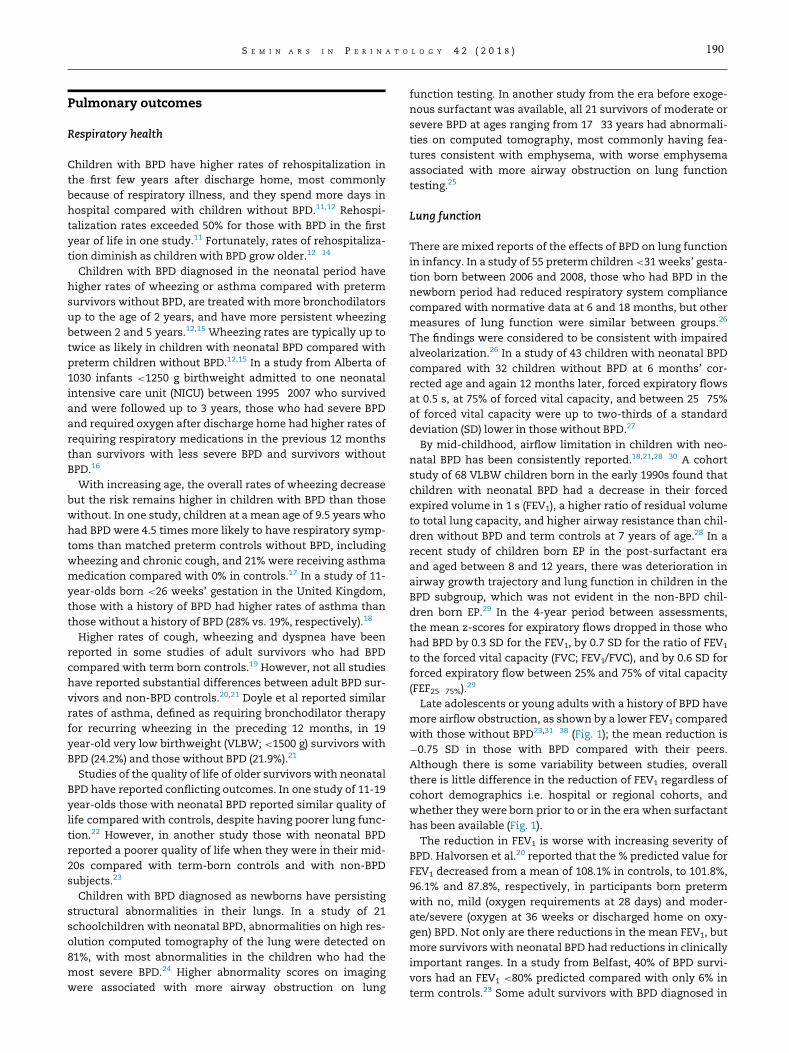
TAGGED ENDS E M I N A R S I N P E R I N A T O L O G Y 4 2 ( 2 0 1 8 ) 190
Pulmonary outcomes
Respiratory health
Children with BPD have higher rates of rehospitalization in
the first few years after discharge home, most commonly
because of respiratory illness, and they spend more days in
hospital compared with children without BPD.11,12 Rehospi-
talization rates exceeded 50% for those with BPD in the first
year of life in one study.11 Fortunately, rates of rehospitaliza-
tion diminish as children with BPD grow older.12�14
Children with BPD diagnosed in the neonatal period have
higher rates of wheezing or asthma compared with preterm
survivors without BPD, are treated with more bronchodilators
up to the age of 2 years, and have more persistent wheezing
between 2 and 5 years.12,15 Wheezing rates are typically up to
twice as likely in children with neonatal BPD compared with
preterm children without BPD.12,15 In a study from Alberta of
1030 infants <1250 g birthweight admitted to one neonatal
intensive care unit (NICU) between 1995�2007 who survived
and were followed up to 3 years, those who had severe BPD
and required oxygen after discharge home had higher rates of
requiring respiratory medications in the previous 12 months
than survivors with less severe BPD and survivors without
BPD.16
With increasing age, the overall rates of wheezing decrease
but the risk remains higher in children with BPD than those
without. In one study, children at a mean age of 9.5 years who
had BPD were 4.5 times more likely to have respiratory symp-
toms than matched preterm controls without BPD, including
wheezing and chronic cough, and 21% were receiving asthma
medication compared with 0% in controls.17 In a study of 11-
year-olds born <26 weeks’ gestation in the United Kingdom,
those with a history of BPD had higher rates of asthma than
those without a history of BPD (28% vs. 19%, respectively).18
Higher rates of cough, wheezing and dyspnea have been
reported in some studies of adult survivors who had BPD
compared with term born controls.19 However, not all studies
have reported substantial differences between adult BPD sur-
vivors and non-BPD controls.20,21 Doyle et al reported similar
rates of asthma, defined as requiring bronchodilator therapy
for recurring wheezing in the preceding 12 months, in 19
year-old very low birthweight (VLBW; <1500 g) survivors with
BPD (24.2%) and those without BPD (21.9%).21
Studies of the quality of life of older survivors with neonatal
BPD have reported conflicting outcomes. In one study of 11-19
year-olds those with neonatal BPD reported similar quality of
life compared with controls, despite having poorer lung func-
tion.22 However, in another study those with neonatal BPD
reported a poorer quality of life when they were in their mid-
20s compared with term-born controls and with non-BPD
subjects.23
Children with BPD diagnosed as newborns have persisting
structural abnormalities in their lungs. In a study of 21
schoolchildren with neonatal BPD, abnormalities on high res-
olution computed tomography of the lung were detected on
81%, with most abnormalities in the children who had the
most severe BPD.24 Higher abnormality scores on imaging
were associated with more airway obstruction on lung
function testing. In another study from the era before exoge-
nous surfactant was available, all 21 survivors of moderate or
severe BPD at ages ranging from 17�33 years had abnormali-
ties on computed tomography, most commonly having fea-
tures consistent with emphysema, with worse emphysema
associated with more airway obstruction on lung function
testing.25
Lung function
There are mixed reports of the effects of BPD on lung function
in infancy. In a study of 55 preterm children<31 weeks’ gesta-
tion born between 2006 and 2008, those who had BPD in the
newborn period had reduced respiratory system compliance
compared with normative data at 6 and 18 months, but other
measures of lung function were similar between groups.26
The findings were considered to be consistent with impaired
alveolarization.26 In a study of 43 children with neonatal BPD
compared with 32 children without BPD at 6 months’ cor-
rected age and again 12 months later, forced expiratory flows
at 0.5 s, at 75% of forced vital capacity, and between 25�75%
of forced vital capacity were up to two-thirds of a standard
deviation (SD) lower in those without BPD.27
By mid-childhood, airflow limitation in children with neo-
natal BPD has been consistently reported.18,21,28�30 A cohort
study of 68 VLBW children born in the early 1990s found that
children with neonatal BPD had a decrease in their forced
expired volume in 1 s (FEV1), a higher ratio of residual volume
to total lung capacity, and higher airway resistance than chil-
dren without BPD and term controls at 7 years of age.28 In a
recent study of children born EP in the post-surfactant era
and aged between 8 and 12 years, there was deterioration in
airway growth trajectory and lung function in children in the
BPD subgroup, which was not evident in the non-BPD chil-
dren born EP.29 In the 4-year period between assessments,
the mean z-scores for expiratory flows dropped in those who
had BPD by 0.3 SD for the FEV1, by 0.7 SD for the ratio of FEV1
to the forced vital capacity (FVC; FEV1/FVC), and by 0.6 SD for
forced expiratory flow between 25% and 75% of vital capacity
(FEF25�75%).29
Late adolescents or young adults with a history of BPD have
more airflow obstruction, as shown by a lower FEV1 compared
with those without BPD23,31�38 (Fig. 1); the mean reduction is
¡0.75 SD in those with BPD compared with their peers.
Although there is some variability between studies, overall
there is little difference in the reduction of FEV1 regardless of
cohort demographics i.e. hospital or regional cohorts, and
whether they were born prior to or in the era when surfactant
has been available (Fig. 1).
The reduction in FEV1 is worse with increasing severity of
BPD. Halvorsen et al.20 reported that the % predicted value for
FEV1 decreased from a mean of 108.1% in controls, to 101.8%,
96.1% and 87.8%, respectively, in participants born preterm
with no, mild (oxygen requirements at 28 days) and moder-
ate/severe (oxygen at 36 weeks or discharged home on oxy-
gen) BPD. Not only are there reductions in the mean FEV1, but
more survivors with neonatal BPD had reductions in clinically
important ranges. In a study from Belfast, 40% of BPD survi-
vors had an FEV1 <80% predicted compared with only 6% in
term controls.23 Some adult survivors with BPD diagnosed in

Fig. 1 –Mean values for forced expired volume in one second (either % predicted or as z-scores) and sample sizes compared
between those with and without BPD in late adolescence or early adulthood, for either hospital or regional cohort studies.
Differences between groups shown by standardizedmean differences and 95% confidence intervals (CIs). *participants born
when surfactant was available. Superscripts relate to reference numbers.
191 S E M I N A R S I N P E R I N A T O L O G Y 4 2 ( 2 0 1 8 )
the neonatal period also had fixed airway obstruction, occur-
ring in 25% of survivors who had BPD compared with none in
the controls.39 In the same study, impaired gas transfer was
detected in both BPD and non-BPD adults born preterm com-
pared with controls.39 In contrast, in another study from Nor-
way of 25-year-olds, although diffusion capacity in preterm
subjects was lower than term controls, BPD was not related to
the findings.37
Kotecha et al.40 reviewed results from studies of lung func-
tion performed in the pre- and post-surfactant era, in partici-
pants with age ranging from 5 to 23 years. FEV1 as % predicted
was decreased in preterm survivors with BPD compared with
term controls, with the size of the reduction related to the
severity of BPD. Participants born preterm but without BPD
had a reduction in their FEV1 of approximately ¡7.2%, while
those with mild BPD (supplemental oxygen at 28 days only)
and severe BPD (oxygen at 36 weeks) had greater deficits in
FEV1 of ¡16.2% and ¡18.9%, respectively.40 One interesting
finding from their review was that the mean FEV1 for survi-
vors with neonatal BPD had improved over time from those
born in the late 1960s through to those born in the early
1990s, whereas it was unchanged for controls born at term
over the same period, suggesting that lung injury in the new-
born period might be less severe with advances in perinatal
and neonatal intensive care. However, in a subsequent study
comparing all survivors born <28 weeks’ gestational age in
the state of Victoria, Australia, in three eras, 1991�92, 1997
and 2005, at 7,8 years of age those born in 2005 had higher
rates of BPD and more airway obstruction than those born in
the 1990s.4
Changes in lung function between 8 and 18 years have been
reported from a regional cohort of ELBW/EP survivors born in
the post-surfactant era.41 Those with BPD had lower spirome-
try values compared with those without BPD at both ages, but
most concerningly those with neonatal BPD deteriorated
more over time in most spirometry values; for example the
reduction in the z-score for FEV1/FVC between 8 and 18 years
was almost 0.5 SD greater in the participants born preterm
with neonatal BPD compared with those who did not. How-
ever, some other studies have reported stable lung function,
as survivors of BPD become older. In one study of 45 survivors
<1000 g BW or <29 weeks gestational age born in 1982�85 in
Norway, lung function between 18 and 25 years was similar
regardless of gestation at birth and BPD severity.37
Exercise capacity is reduced in BPD survivors. Children with
neonatal BPD walk shorter distances on the 6-minute walk
test than do children without BPD.15,42 Using a cycle ergome-
ter to measure exercise capacity in children born preterm,
those with neonatal BPD had lower % predicted oxygen
uptake compared with children without BPD.30 In a recent
study of young adults at a mean age 24 years, those who had
BPD had impaired exercise capacity compared with term con-
trols, although weekly activity levels were similar between
groups.39
Neurodevelopmental outcomes
BPD is strongly associated with neurodevelopmental deficits
in children born preterm.43,44 Preterm infants with BPD in the
newborn period have recurrent episodes of hypoxia, hyper-
capnia, and respiratory acidosis,45 which may predispose
them to hypoxic-related brain injury. In addition, postnatal
corticosteroids in the newborn period to prevent or treat BPD
are a risk factor for neurodevelopmental deficits in preterm
children.46
Cerebral palsy and other motor impairments
The rate of cerebral palsy (CP) is approximately 10�14% in
survivors born <28 weeks’ gestation,5,47 and 14% in those
born <26 weeks’ gestation.48 CP occurs more frequently in

TAGGED ENDS E M I N A R S I N P E R I N A T O L O G Y 4 2 ( 2 0 1 8 ) 192
survivors with neonatal BPD; in one study of 839 infants <25
weeks’ gestation the odds ratio (OR) for CP in those with neo-
natal BPD was 1.66 (95% confidence interval [CI] 1.01, 2.74),
after adjustment for major intraventricular hemorrhage and
cystic periventricular leukomalacia.49
The risk of CP may be related to the severity of BPD. In the
ELGAN study of more than 1000 infants born <28 weeks, the
risk of CP in children with the most severe BPD (receiving
mechanical ventilation at 36 weeks’ postmenstrual age) was
6-fold higher for quadriplegic CP, and 4-fold higher for diple-
gic CP compared with preterm children without BPD.50 Those
with less severe BPD (not receiving mechanical ventilation at
36 weeks’ postmenstrual age) still had increased risks of
quadriplegic and diplegic CP compared with preterm children
without BPD, but the size of the risk was lower.
In a study from the NICHD Neonatal Research Network of
ELBW infants born between 2006-2007, BPD was defined as
either a) any form of assisted ventilation or continuous posi-
tive airway pressure or supplemental oxygen with an effec-
tive fraction of inspired oxygen (FiO2) >0.3 at 36 weeks’
postmenstrual age, or b) supplemental oxygen via nasal can-
nula or hood with effective FiO2 <0.3 and failed the stepwise
oxygen reduction challenge in the 36th postmenstrual week
i.e. “physiological BPD”.51 At 18-22 months, children with
“physiological BPD” compared with non-BPD children had
higher rates of all CP phenotypes, of moderate to severe CP
(Gross Motor Function Classification System level 2 or greater)
(7.0% vs 2.1%), of spastic diplegia (7.8% vs. 4.1%) and of quadri-
plegia (3.9% vs. 0.9%).
Other non-CP motor impairments also occur more fre-
quently in children who had BPD. Several studies of children
aged 5�10 years have reported poorer motor performance in
BPD survivors compared with non-BPD and term-born con-
trols.52�54 In a study of 8-year-olds both gross and fine motor
skills were lower in children with neonatal BPD (42nd centile)
compared with the non-BPD children (58th centile) and term
controls (70th centile).55 In another study gross and fine
motor scores were approximately 1 SD lower, and upper
extremity postural stability was worse in BPD children than
in preterm controls assessed at 10 years of age.44 Conse-
quently, more survivors who had BPD have been reported to
access intervention services, such as occupational therapy
(71% vs 44%), and physical therapy (71% vs 41%), than do pre-
term children who did not have BPD.53
Some studies have assessed postnatal corticosteroid ther-
apy, to treat or prevent BPD, as a risk factor for motor dys-
function in preterm children. In the EPIPAGE study of births
<33 weeks’ gestation born in France, postnatal corticosteroid
therapy was associated with more minor neuromotor dys-
function (OR 1.8; 95% CI 1.3, 2.6), and with more moderate
neuromotor dysfunction (OR 2.7; 95% CI 1.2, 6.1) on multivari-
able analysis adjusting for other perinatal variables that affect
neurodevelopment.52 Davis et al.54 assessed the perinatal
associations with Developmental Coordination Disorder, or
minor motor dysfunction, in a geographic cohort of EP or
ELBW children at age 8-9 years. With both variables included
in a multivariable analysis, BPD, defined as oxygen require-
ment at 36 weeks’ corrected gestation, was associated with
Developmental Coordination Disorder (p=0.06), but the associ-
ation with postnatal corticosteroids was stronger (p=0.03).
Neurosensory impairments and disabilities
BPD is also an independent risk factor for major neurosensory
problems overall. The OR (95% CI) for any of CP, blindness,
deafness, or cognitive delay at 18 months was 2.4 (1.8, 3.2) in
the follow-up of a randomized controlled trial of indometha-
cin prophylaxis in infants <1000 g birthweight.43 In a similar
study, but this time from a randomized controlled trial of caf-
feine in the newborn period in infants with BW <1251 g, those
alive at 36 weeks’ postmenstrual age had a higher chance of
death or disability in one or more area (motor impairment,
cognitive impairment, behavior problems, poor general
health, deafness, or blindness) at 5 years than did those who
did not have BPD (OR 2.3; 95% CI 1.8, 3.0). In a study of survi-
vors born <28 weeks’ gestation between 2000�2008 in Swit-
zerland, BPD was an independent risk factor for a composite
adverse outcome of moderate/severe neurosensory disability
or death (OR 1.92; 95% CI 1.24, 2.99).56
Cognitive delay and academic performance
There are several studies that have reported persistence of
cognitive deficits from early childhood to school age. In one
study of VLBW children and term controls, between 8 months
to 3 years of age VLBW children with BPD diagnosed in the
neonatal period had persistently lower scores on the Mental
Developmental Index of the Bayley Scales than did VLBW
children without BPD, who in turn were always lower than
the term controls.57 When the children were reassessed at
age 8 years, performance on general intelligence, reading,
mathematics, motor performance, memory and attention fol-
lowed a similar pattern; children with neonatal BPD per-
formed the worst, followed by children without BPD, with the
best performance in term controls.53 Children who had more
severe BPD compared with children who had mild/moderate
BPD performed worse in Performance IQ (75 vs 86) and Per-
ceptual Organization (76 vs 86), representing clinically impor-
tant differences of approximately 0.6 SD.58 The differences
persisted despite adjusting for BW and brain injury, suggest-
ing that BPD is an independent risk factor for later cognitive
development. In a more recent study from the state of Victo-
ria investigating the associations between several biologic
and social variables and cognitive and academic performance
through childhood and into adolescence among survivors
born either ELBW or EP, postnatal corticosteroids were inde-
pendently associated with lower IQ and academic perfor-
mance into adolescence, whereas BPD not treated with
corticosteroids was not independently associated with those
outcomes.46 These findings are consistent with the possibility
that more severe BPD is related to adverse neurocognitive
outcomes, but part of the association might be explained by
the need to prevent or treat BPD with postnatal corticoste-
roids, and it was infants with more severe lung disease who
were treated with corticosteroids in that study.
Differences in cognitive development betweenBPDandnon-
BPD children persisted from early to mid-childhood in a study
from Cleveland, Ohio, of very low birth weight children born
1989�1991.55,57 Not only were the absolute scores on develop-
mental testing lower, but the proportions of children who
were significantly delayed i.e. performing at<¡2 SD below the
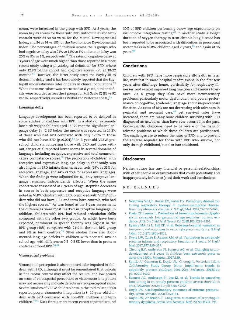
193 S E M I N A R S I N P E R I N A T O L O G Y 4 2 ( 2 0 1 8 )
mean, were increased in the group with BPD. At 3 years, the
mean Bayley scores for those with BPD, without BPD and term
controls were 84 vs 90 vs 96 for the Mental Developmental
Index, and 84 vs 98 vs 103 for the Psychomotor Developmental
Index. The percentages of children across the 3 groups who
had cognitive delaywas 21%vs 11%vs 4% andmotor delaywas
20% vs 9% vs 1%, respectively.57 The rates of cognitive delay at
3 years of ageweremuch higher than those reported in amore
recent study using a physiological definition for BPD, where
only 12.8% of the cohort had cognitive scores <70 at 18-22
months.51 However, the latter study used the Bayley-III to
determine delay, and it has beenwidely reported that the Bay-
ley-III underestimates rates of delay in clinical populations.59
When the same cohort was reassessed at 8 years, similar defi-
citswere recorded across the 3 groups for Full Scale IQ (85 vs 92
vs 102, respectively), aswell as Verbal and Performance IQ.55
Language delay
Language development has been reported to be delayed in
some studies of children with BPD. In a study of extremely
low birth weight children aged 18�22 months, significant lan-
guage delay (<¡2 SD below the mean) was reported in 24.2%
of those who had BPD compared with only 12.3% in those
who did not have BPD (p<0.001).51 In 3-year-old VLBW pre-
school children, comparing those with BPD and those with-
out, Singer et al reported lower scores in several domains of
language, including receptive, expressive and total communi-
cative competence scores.60 The proportion of children with
receptive and expressive language delay in that study was
also higher in BPD infants than term controls (49% vs 35% for
receptive language, and 44% vs 25% for expressive language).
When the findings were adjusted for IQ, only receptive lan-
guage remained independently affected. When the same
cohort were reassessed at 8 years of age, stepwise decreases
in scores in both expressive and receptive language were
noted in VLBW children with BPD, compared with VLBW chil-
dren who did not have BPD, and term-born controls, who had
the highest scores.55 As was found at the 3-year assessment,
the differences were most marked in receptive language. In
addition, children with BPD had reduced articulation skills
compared with the other two groups. As might have been
expected, enrolment in speech therapy was highest in the
BPD group (48%) compared with 21% in the non-BPD group
and 9% in term controls.55 Other studies have also docu-
mented language deficits in children with neonatal BPD at
school age, with differences 0.5�0.8 SD lower than in preterm
controls without BPD.58,61
Visuospatial problems
Visuospatial perception is also reported to be impaired in chil-
dren with BPD, although it must be remembered that deficits
in fine motor control may affect the results, and low scores
on tests of visuospatial perception or visuomotor integration
may not necessarily indicate defects in visuoperceptual skills.
Several studies of VLBW children born in the mid to late 1980s
reported poorer visuomotor integration in 8�10-year-old chil-
dren with BPD compared with non-BPD children and term
children.44,62 Data from a more recent cohort reported around
30% of BPD children performing below age expectations on
visuomotor integration testing.63 In another study a longer
duration of oxygen therapy to treat chronic lung disease has
been reported to be associated with difficulties in perceptual
motor tasks in VLBW children aged 7 years,64 and again at 16
years.65
Conclusions
Children with BPD have more respiratory ill-health in later
life, manifest in more hospital readmissions in the first few
years after discharge home, particularly for respiratory ill-
nesses, and exhibit impaired lung function and exercise toler-
ance. As a group they also have more neurosensory
problems, particularly motor dysfunction, and poorer perfor-
mance on cognitive, academic, language and visuoperceptual
function. As rates of BPD are not decreasing with advances in
perinatal and neonatal care,66 yet survival rates have
increased, there are many more children surviving with BPD
diagnosed as newborns than have ever occurred in the past.
Consequently, clinicians need to be aware of the risks of
adverse problems to which these children are predisposed.
The challenges are to reduce the rates of BPD, and to prevent
the adverse sequelae for those with BPD who survive, not
only through childhood, but also into adulthood.
Disclosures
Neither author has any financial or personal relationships
with other people or organizations that could potentially and
inappropriately influence (bias) their work and conclusions.
R E F E R E N C E S
1. NorthwayWH Jr., Rosan RC, Porter DY. Pulmonary disease fol-lowing respiratory therapy of hyaline-membrane disease.Bronchopulmonary dysplasia. N Engl J Med. 1967;276:357–368.
2. Poets CF, Lorenz L. Prevention of bronchopulmonary dyspla-sia in extremely low gestational age neonates: current evi-dence. Arch Dis Child Fetal Neonat Ed. 2018;103:F285–F291.
3. Rysavy MA, Li L, Bell EF, et al. Between-hospital variation intreatment and outcomes in extremely preterm infants. N EnglJ Med. 2015;372:1801–1811.
4. Doyle LW, Carse E, Adams AM, et al. Ventilation in extremelypreterm infants and respiratory function at 8 years. N Engl JMed. 2017;377:329–337.
5. Cheong JLY, Anderson PJ, Burnett AC, et al. Changing neuro-development at 8 years in children born extremely pretermsince the 1990s. Pediatrics. 2017;139.
6. Spittle AJ, Cameron K, Doyle LW, Cheong JL. Victorian InfantCollaborative Study Group. Motor impairment trends inextremely preterm children: 1991-2005. Pediatrics. 2018;141:pii: e20173410.
7. Burnett AC, Anderson PJ, Lee KJ, et al. Trends in executivefunctioning in extremely preterm children across three birtheras. Pediatrics. 2018;141: pii: e20173102.
8. Doyle LW. Cardiopulmonary outcomes of extreme prematu-rity. Semin Perinatol. 2008;32:28–34.
9. Doyle LW, Anderson PJ. Long-term outcomes of bronchopul-monary dysplasia. Semin Fetal Neonatal Med. 2009;14:391–395.

TAGGED ENDS E M I N A R S I N P E R I N A T O L O G Y 4 2 ( 2 0 1 8 ) 194
10. Doyle LW, Anderson PJ. Pulmonary and neurological follow-up of extremely preterm infants. Neonatology. 2010;97:388–394.
11. Ralser E, Mueller W, Haberland C, et al. Rehospitalization inthe first 2 years of life in children born preterm. Acta Paediatr.2012;101:e1–e5.
12. Skromme K, Leversen KT, Eide GE, Markestad T, Halvorsen T.Respiratory illness contributed significantly to morbidity inchildren born extremely premature or with extremely lowbirthweights in 1999-2000. Acta Paediatr. 2015;104:1189–1198.
13. McCormick MC, Workman-Daniels K, Brooks-Gunn J, Peck-ham GJ. Hospitalization of very low birth weight children atschool age. J Pediatr. 1993;122:360–365.
14. Doyle LW, Cheung MM, Ford GW, Olinsky A, Davis NM, Call-anan C. Birth weight <1501 g and respiratory health at age 14.Arch Dis Child. 2001;84:40–44.
15. Praprotnik M, Stucin Gantar I, Lucovnik M, Avcin T, Krivec U.Respiratory morbidity, lung function and fitness assessmentafter bronchopulmonary dysplasia. J Perinatol. 2015;35:1037–1042.
16. Lodha A, Ediger K, Rabi Y, et al. Does chronic oxygen depen-dency in preterm infants with bronchopulmonary dysplasiaat NICU discharge predict respiratory outcomes at 3 years ofage? J Perinatol. 2015;35:530–536.
17. Vom Hove M, Prenzel F, Uhlig HH, Robel-Tillig E. Pulmonaryoutcome in former preterm, very low birth weight childrenwith bronchopulmonary dysplasia: a case-control follow-upat school age. J Pediatr. 2014;164:40–45: e4.
18. Fawke J, Lum S, Kirkby J, et al. Lung function and respiratorysymptoms at 11 years in children born extremely preterm:the EPICure study. Am J Resp Crit Care Med. 2010;182:237–245.
19. Gough A, Spence D, Linden M, Halliday HL, McGarvey LP. Gen-eral and respiratory health outcomes in adult survivors ofbronchopulmonary dysplasia: a systematic review. Chest.2012;141:1554–1567.
20. Halvorsen T, Skadberg BT, Eide GE, Roksund OD, Carlsen KH,Bakke P. Pulmonary outcome in adolescents of extreme pre-term birth: a regional cohort study. Acta Paediatr.2004;93:1294–1300.
21. Doyle LW. and the Victorian Infant Collaborative StudyGroup. Respiratory function at age 8-9 years in extremely lowbirthweight/very preterm children born in Victoria in 1991-92.Pediat Pulmonol. 2006;41:570–576.
22. Bozzetto S, Carraro S, Tomasi L, Berardi M, Zanconato S, Bar-aldi E. Health-related quality of life in adolescent survivors ofbronchopulmonary dysplasia. Respirology. 2016;21:1113–1117.
23. Gough A, Linden M, Spence D, Patterson CC, Halliday HL,McGarvey LP. Impaired lung function and health status inadult survivors of bronchopulmonary dysplasia. Eur Resp J.2014;43:808–816.
24. Ronkainen E, Perhomaa M, Mattila L, Hallman M, Dunder T.Structural pulmonary abnormalities still evident in school-children with new bronchopulmonary dysplasia. Neonatology.2018;113:122–130.
25. Wong PM, Lees AN, Louw J, et al. Emphysema in young adultsurvivors of moderate-to-severe bronchopulmonary dyspla-sia. Eur Resp J. 2008;32:321–328.
26. Thunqvist P, Gustafsson P, Norman M, Wickman M, HallbergJ. Lung function at 6 and 18 months after preterm birth inrelation to severity of bronchopulmonary dysplasia. PediatrPulmonol. 2015;50:978–986.
27. Sanchez-Solis M, Garcia-Marcos L, Bosch-Gimenez V, Perez-Fernandez V, Pastor-Vivero MD, Mondejar-Lopez P. Lungfunction among infants born preterm, with or without bron-chopulmonary dysplasia. Pediatr Pulmonol. 2012;47:674–681.
28. Korhonen P, Laitinen J, Hyodynmaa E, Tammela O. Respira-tory outcome in school-aged, very-low-birth-weight childrenin the surfactant era. Acta Paediatr. 2004;93:316–321.
29. Fortuna M, Carraro S, Temporin E, et al. Mid-childhood lungfunction in a cohort of children with “new bronchopulmonarydysplasia”. Pediatr Pulmonol. 2016;51:1057–1064.
30. MacLean JE, DeHaan K, Fuhr D, et al. Altered breathingmechanics and ventilatory response during exercise in chil-dren born extremely preterm. Thorax. 2016;71:1012–1019.
31. Northway WH Jr., Moss RB, Carlisle KB, et al. Late pulmonarysequelae of bronchopulmonary dysplasia. N Engl J Med.1990;323:1793–1799.
32. Doyle LW, Faber B, Callanan C, Freezer N, Ford GW, Davis NM.Bronchopulmonary dysplasia in very low birth weight sub-jects and lung function in late adolescence. Pediatrics.2006;118:108–113.
33. Gibson AM, Reddington C, McBride L, Callanan C, Robertson C,Doyle LW. Lung function in adult survivors of very low birthweight, with and without bronchopulmonary dysplasia.Pediatr Pulmonol. 2015;50:987–994.
34. Saarenpaa HK, Tikanmaki M, Sipola-Leppanen M, et al. Lungfunction in very low birth weight adults. Pediatrics.2015;136:642–650.
35. Vrijlandt EJ, Gerritsen J, Boezen HM, Grevink RG, DuivermanEJ. Lung function and exercise capacity in young adults bornprematurely. Am J Resp Crit Care Med. 2006;173:890–896.
36. Vollsaeter M, Roksund OD, Eide GE, Markestad T, Halvorsen T.Lung function after preterm birth: development from mid-childhood to adulthood. Thorax. 2013;68:767–776.
37. Vollsaeter M, ClemmHH, Satrell E, et al. Adult respiratory out-comes of extreme preterm birth. A regional cohort study. AnnAm Thorac Soc. 2015;12:313–322.
38. Doyle LW, Adams AM, Robertson C, et al. Increasing airwayobstruction from 8 to 18 years in extremely preterm/low-birthweight survivors born in the surfactant era. Thorax.2017;72:712–719.
39. Caskey S, Gough A, Rowan S, et al. Structural and functionallung impairment in adult survivors of bronchopulmonarydysplasia. Ann Am Thorac Soc. 2016;13:1262–1270.
40. Kotecha SJ, Edwards MO, Watkins WJ, et al. Effect of pretermbirth on later FEV1: a systematic review and meta-analysis.Thorax. 2013;68:760–766.
41. Doyle LW, Adams AM, Robertson C, et al. Increasing airwayobstruction from 8 to 18 years in extremely preterm/low-birthweight survivors born in the surfactant era. Thorax.2017;72:712–719.
42. Vardar-Yagli N, Inal-Ince D, Saglam M, et al. Pulmonary andextrapulmonary features in bronchopulmonary dysplasia: acomparison with healthy children. J Phys Ther Sci.2015;27:1761–1765.
43. Schmidt B, Asztalos EV, Roberts RS, Robertson CM, Sauve RS,Whitfield MF. Impact of bronchopulmonary dysplasia, braininjury, and severe retinopathy on the outcome of extremelylow-birth-weight infants at 18 months: results from the trialof indomethacin prophylaxis in preterms. JAMA.2003;289:1124–1129.
44. Majnemer A, Riley P, Shevell M, Birnbaum R, Greenstone H,Coates AL. Severe bronchopulmonary dysplasia increasesrisk for later neurological and motor sequelae in preterm sur-vivors. Dev Med Child Neurol. 2000;42:53–60.
45. Sekar KC, Duke JC. Sleep apnea and hypoxemia in recentlyweaned premature infants with and without bronchopulmo-nary dysplasia. Pediatr Pulmonol. 1991;10:112–116.
46. Doyle LW, Cheong JL, Burnett A, et al. Biological and socialinfluences on outcomes of extreme-preterm/low-birth weightadolescents. Pediatrics. 2015;136:e1513–e1520.
47. Serenius F, Ewald U, Farooqi A, et al. Neurodevelopmentaloutcomes among extremely pretermiInfants 6.5 years afteractive perinatal care in Sweden. JAMA Pediatr. 2016;170:954–963.

195 S E M I N A R S I N P E R I N A T O L O G Y 4 2 ( 2 0 1 8 )
48. Moore T, Hennessy EM, Myles J, et al. Neurological and devel-opmental outcome in extremely preterm children born inEngland in 1995 and 2006: the EPICure studies. Br Med J.2012;345:e7961.
49. Hintz SR, Kendrick DE, Vohr BR, Poole WK, Higgins RD.Changes in neurodevelopmental outcomes at 18 to 22months’ corrected age among infants of less than 25 weeks’gestational age born in 1993-1999. Pediatrics. 2005;115:1645–1651.
50. Van Marter LJ, Kuban KC, Allred E, et al. Does bronchopulmo-nary dysplasia contribute to the occurrence of cerebral palsyamong infants born before 28 weeks of gestation? Arch DisChild Fetal Neonat Ed. 2011;96:F20–F29.
51. Natarajan G, Pappas A, Shankaran S, et al. Outcomes ofextremely low birth weight infants with bronchopulmonarydysplasia: impact of the physiologic definition. Early Hum Dev.2012;88:509–515.
52. Arnaud C, Daubisse-Marliac L, White-Koning M, et al. Preva-lence and associated factors of minor neuromotor dysfunc-tions at age 5 years in prematurely born children: theEPIPAGE study. Arch Pediatr Adolesc Med. 2007;161:1053–1061.
53. Short EJ, Klein NK, Lewis BA, Fulton S, Eisengart S, KercsmarC, et al. Cognitive and academic consequences of bronchopul-monary dysplasia and very low birth weight: 8-year-old out-comes. Pediatrics. 2003;112:e359.
54. Davis NM, Ford GW, Anderson PJ, Doyle LW. Developmentalcoordination disorder at 8 years of age in a regional cohort ofextremely-low-birthweight or very preterm infants. Dev MedChild Neurol. 2007;49:325–330.
55. Lewis BA, Singer LT, Fulton S, et al. Speech and language out-comes of children with bronchopulmonary dysplasia. J Com-mun Disord. 2002;35:393–406.
56. Schlapbach LJ, Adams M, Proietti E, et al. Outcome at twoyears of age in a Swiss national cohort of extremely pre-term infants born between 2000 and 2008. BMC Pediatr.2012;12:198.
57. Singer L, Yamashita T, Lilien L, Collin M, Baley J. A longitudi-nal study of developmental outcome of infants with broncho-pulmonary dysplasia and very low birth weight. Pediatrics.1997;100:987–993.
58. Short EJ, Kirchner HL, Asaad GR, et al. Developmentalsequelae in preterm infants having a diagnosis of broncho-pulmonary dysplasia: analysis using a severity-based classifi-cation system. Arch Pediatr Adolesc Med. 2007;161:1082–1087.
59. Anderson PJ, De Luca CR, Hutchinson E, Roberts G, Doyle LW.Underestimation of developmental delay by the new Bayley-III Scale. Arch Pediatr Adolesc Med. 2010;164:352–356.
60. Singer LT, Siegel AC, Lewis B, Hawkins S, Yamashita T, Baley J.Preschool language outcomes of children with history ofbronchopulmonary dysplasia and very low birth weight. J DevBehav Pediatr. 2001;22:19–26.
61. Robertson CM, Etches PC, Goldson E, Kyle JM. Eight-yearschool performance, neurodevelopmental, and growth out-come of neonates with bronchopulmonary dysplasia: a com-parative study. Pediatrics. 1992;89:365–372.
62. Hughes CA, O’Gorman LA, Shyr Y, Schork MA, Bozynski ME,McCormick MC. Cognitive performance at school age of verylow birth weight infants with bronchopulmonary dysplasia. JDev Behav Pediatr. 1999;20:1–8.
63. Gray PH, O’Callaghan MJ, Rogers YM. Psychoeducational out-come at school age of preterm infants with bronchopulmo-nary dysplasia. J Paediatr Child Health. 2004;40:114–120.
64. Taylor HG, Klein N, Schatschneider C, Hack M. Predictors ofearly school age outcomes in very low birth weight children. JDev Behav Pediatr. 1998;19:235–243.
65. Taylor HG, Minich N, Bangert B, Filipek PA, Hack M. Long-termneuropsychological outcomes of very low birth weight: asso-ciations with early risks for periventricular brain insults. J IntNeuropsychol Soc. 2004;10:987–1004.
66. Doyle LW, Ranganathan S, Cheong JLY. Ventilation in preterminfants and lung function at 8 years. N Engl J Med.2017;377:1601–1602.
TAGGEDENDS E M I N A R S I N P E R I N A T O L O G Y 4 2 ( 2 0 1 8 )
Available online at www.sciencedirect.com
Seminars in Perinatology
www.seminperinat.com
196
An update on the post-NICU di
scharge management of bronchopulmonary dysplasiaAnita Bhandari*, and Howard Panitch
Division of Pulmonary Medicine, Department of Pediatrics, Children’s Hospital of Philadelphia, Perelman School of Medicine at the University
of Pennsylvania, 11th floor Colket Building, 3501 Civic Center Boulevard, Philadelphia, PA 19446, United States
* Corresponding author.E-mail address: [email protected]
https://doi.org/10.1053/j.semperi.2018.09.0110146-0005/� 2018 Elsevier Inc. All rights reser
A B S T R A C T
Bronchopulmonary dysplasia (BPD) is a chronic lung disease which develops as a result of
neonatal/perinatal lung injury. It is the commonest cause of chronic lung disease in infancy
and the most frequent morbidity associated with prematurity. The incidence of BPD has
continued to rise despite many advances in neonatal care and this increase has been attrib-
uted to the increased survival of younger and more premature babies. There have been
many advances in the care of patients with early and evolving BPD, yet there is a paucity of
data regarding outpatient management of patients with established BPD. There are limited
adequately-powered high-quality studies/randomized controlled trials which assess com-
monly used therapies such as supplemental oxygen, bronchodilators, steroids and diu-
retics in patients with BPD, beyond short-term effects. Further research is needed to
improve our understanding of the role of currently used treatments on the long-term out-
comes of patients with established BPD, post-discharge from the neonatal intensive care
unit.
� 2018 Elsevier Inc. All rights reserved.
A R T I C L E I N F O
Keywords:
outpatient care
chronic lung disease
diuretics
inhaled steroids
days or greater but not at 36 weeks PMA or at the time of dis-
Introduction
Bronchopulmonary dysplasia (BPD) is a chronic lung disease,
which develops as a result of neonatal/ perinatal lung injury.1
It is the commonest cause of chronic lung disease in infancy
and the most frequent morbidity associated with prematu-
rity.2 Athough many definitions of BPD exist, the most widely
used is that suggested by the national institutes of health
(NIH) consensus statement, where BPD is defined as a need
for oxygen supplementation for 28 days and/or 36 weeks post
menstrual age (PMA).3
They further suggested criteria for assigning BPD severity.
Mild BPD was defined as a need for supplemental oxygen for 28
u (A. Bhandari).
ved.
charge. Moderate BPD was defined as a need for supplemental
oxygen for 28 days or greater and treatment with supplemental
oxygen not greater than 30% at 36 weeks PMA or at the time of
discharge. Severe BPD was defined as a need for supplemental
oxygen for 28 days or greater and treatment with supplemental
oxygen greater than 30% and/or need for positive pressure at 36
weeks PMA or at the time of discharge. This consensus defini-
tion has been found to identify risk for adverse pulmonary out-
comes in early infancy more accurately than others.4 A further
subclassification of severe BPD into Type 1 which includes
patients using high flow nasal cannula (HFNC) or continuous
positive airway pressure (CPAP) and Type 2 which includes
those who require mechanical ventilation, has been suggested.5
197 S E M I N A R S I N P E R I N A T O L O G Y 4 2 ( 2 0 1 8 )
The incidence of BPD from 2009�2012 has increased in pre-
term babies born between 26 and 27 weeks of gestation from
51% to 55% despite many advances in neonatal care.6 This
increase is attributed to increased survival of younger and
more immature babies. BPD occurs most commonly in babies
who are less than 1000 g and less than 28 weeks of gestation,
and its incidence is inversely proportional to gestational age
or its surrogate, birth weight (BW). The incidence of BPD has
been reported to be 42% (BW 501�750 g), 25% (751�1000 g),
11% (1001�1250), and 5% (1251�1500 g).7,8
BPD is often divided into early, evolving and established
BPD. Early BPD refers to the time from birth to the first week
of life. Evolving BPD refers to the period between 1 week of
life and 1 month of life and established BPD is considered to
occur after 36 weeks PMA.9 For the purpose of this review, we
will focus on therapies utilized for outpatient management of
patients with established BPD. While many newer therapies
and modes of ventilation have been proposed and studied for
management of early BPD, the management of established
BPD has changed little in the last two decades.
Diuretics
Diuretics are the most commonly used medications in the
management of established BPD. Infants with BPD are prone
to develop interstitial pulmonary edema from increased cap-
illary permeability caused by lung injury from volutrauma or
barotaruma, heart failure and generalized fluid overload. The
degree of free lung water has been correlated with the sever-
ity of BPD.10 These considerations form the basis for using
diuretics in BPD infants.
Furosemide is a loop diuretic. Its diuretic action is a result of
inhibition of the Na+�K+�2Cl¡ cotransporter located on the
luminal surface of the ascending loop of Henle. In addition,
vascular effects of furosemide result in fluid shifting out of
the alveoli into the venous system, which can lead to
improved oxygenation and pulmonarymechanics.11
In older infants with BPD, furosemide has been shown to
improve oxygenation and decrease PCO2. This improvement
in ventilation does not corelate well with diuresis suggesting
a different mechanism of action. One such mechanism may
be improved oxygenation via release of prostaglandins from
the renal or vascular endothelium. It is this vascular effect
that has also been implicated in increasing the risk of persis-
tent patent ductus areteriosus (PDA) that has been reported
with furosemide use in the neonatal intensive care unit
(NICU) in patients with early BPD.
Furosemide is the single most frequently used diuretic in
the NICU.12,13 Furosemide has been shown to decrease airway
resistance and increase dynamic airway compliance acutely
and sub-acutely in spontaneously breathing patients with
BPD.14,15 Others have shown improvement in lung mechanics
using nebulized furosemide, suggesting a direct effect of furo-
semide on airway smooth muscle.16,17
Thiazides (chlorothiazide, hydrochlorothiazide) and spiro-
nolactone together comprise the most commonly used
diuretic combination therapy in patients with established
BPD.18,19,20 Thiazides and spironolactone both act on the dis-
tal tubule. Thiazides act by binding to the chloride site of
electroneutral sodium chloride channel and spironolactone
acts by competitive binding of receptors at the aldosterone
dependent sodium�potassium exchange site in the distal
tubule. Thiazaides and spironolactone are less likely to cause
electrolyte abnormalities as compared to furesomide, hence
they are preferred for chronic use.
In a randomized double blind crossover trial with sequen-
tial analysis, oral diuretics improved pulmonary mechanics
in 10 infants with BPD.21 This was based on measurement of
pulmonary mechanics at the beginning and the end of the
week after therapy with chlorothiazide and spirinolcatone
combination or placebo.21
The long term use of this combination therapy for oxygen-
dependent BPD patients was also studied in a randomized
double blind, placebo-controlled study, where patients who
received spironolactone and chlorthiazide had a significant
improvement in respiratory scores and lung mechanics and a
decrease in fraction of inspired oxygen (FiO2) used, as com-
pared to those who received placebo. The therapy did not,
however, result in a significant decrease in the duration of
oxygen supplementation.22 A large meta-analysis showed
that in preterm infants greater than 3 weeks of age with
chronic lung disease, acute and chronic administration of
diuretics acting on distal segments of the renal tubule (distal
diuretics) improved pulmonary mechanics. The authors,
however, suggested that the positive effects should be inter-
preted with caution given the small number of patients stud-
ied in a few randomized controlled trials (RCTs).23 Of note,
RCTs looking at efficacy of diuretic use are from the pre-sur-
factant era which may limit their applicability in current
times. Furthermore, most studies evaluate short-term effects
of therapy. There are no trials that demonstrate a reduction
in NICU length of stay, duration of mechanical ventilation, or
duration of supplemental oxygen use resulting from chronic
diuretic therapy among infants with established BPD.
Recent data from a retrospective cohort study in patients
with evolving BPD conducted among 35 hospitals showed
that use of diuretics is common in US hospitals and that the
diuretic regimens and the frequency of diuretic use vary
markedly between institutions.24 The prescribed use of diu-
retics among patients with established BPD is quite inconsis-
tent as well. In a single center study the duration of
outpatient diuretic therapy was significantly variable;
patients treated with diuretics who required supplemental
oxygen were more likely to have a longer course of diuretic
therapy compared to patients receiving diuretics alone.25 In
contrast, a multicenter survey regarding oxygen weaning
practices of pediatric pulmonologists in North America dis-
closed that 58% of the respondents weaned diuretics before
weaning supplemental oxygen; these two studies highlight
the inconsistency in approach towards prescribing diuretics
in patients with established BPD.26
Long term diuretic therapy after discharge is often used in
infants with severe BPD requiring high levels of supplemental
oxygen, positive pressure ventilation, and those with under-
lying cardiac issues. Diuretic therapy may confer a benefit in
these unique situations, although this has not been systemat-
ically studied.
Chronic diuretic therapy has been associated with adverse
effects including electrolyte disturbances, calciuria,
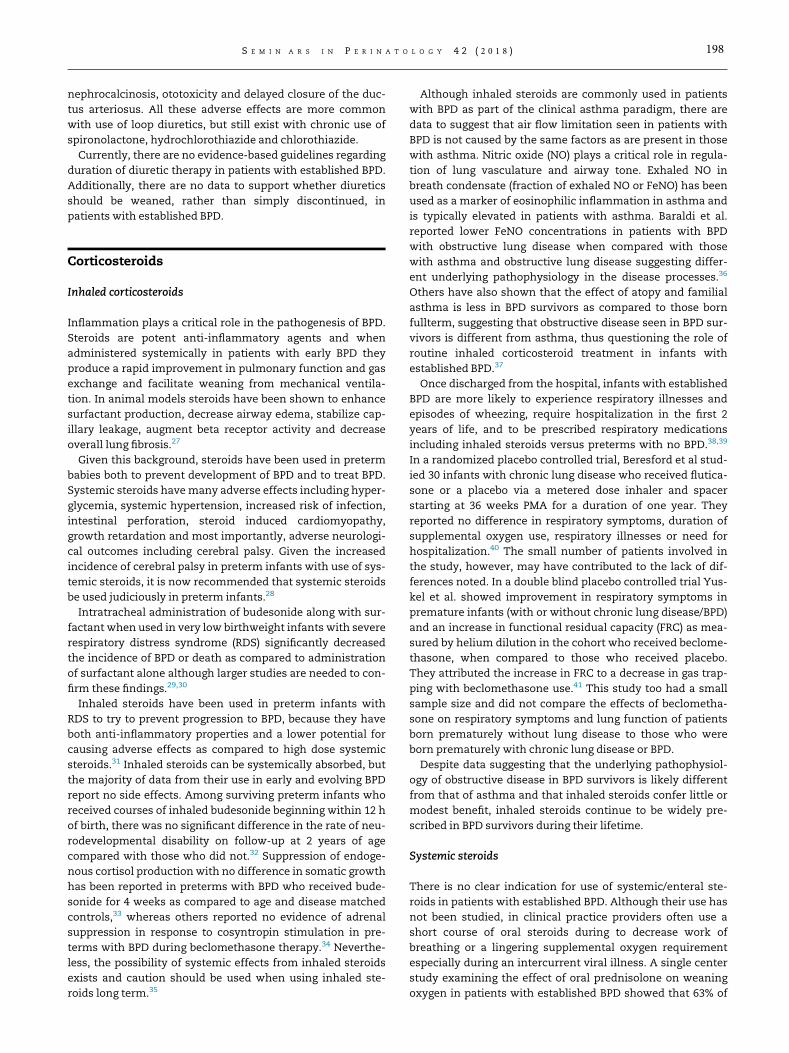
TAGGEDE NDS E M I N A R S I N P E R I N A T O L O G Y 4 2 ( 2 0 1 8 ) 198
nephrocalcinosis, ototoxicity and delayed closure of the duc-
tus arteriosus. All these adverse effects are more common
with use of loop diuretics, but still exist with chronic use of
spironolactone, hydrochlorothiazide and chlorothiazide.
Currently, there are no evidence-based guidelines regarding
duration of diuretic therapy in patients with established BPD.
Additionally, there are no data to support whether diuretics
should be weaned, rather than simply discontinued, in
patients with established BPD.
Corticosteroids
Inhaled corticosteroids
Inflammation plays a critical role in the pathogenesis of BPD.
Steroids are potent anti-inflammatory agents and when
administered systemically in patients with early BPD they
produce a rapid improvement in pulmonary function and gas
exchange and facilitate weaning from mechanical ventila-
tion. In animal models steroids have been shown to enhance
surfactant production, decrease airway edema, stabilize cap-
illary leakage, augment beta receptor activity and decrease
overall lung fibrosis.27
Given this background, steroids have been used in preterm
babies both to prevent development of BPD and to treat BPD.
Systemic steroids havemany adverse effects including hyper-
glycemia, systemic hypertension, increased risk of infection,
intestinal perforation, steroid induced cardiomyopathy,
growth retardation and most importantly, adverse neurologi-
cal outcomes including cerebral palsy. Given the increased
incidence of cerebral palsy in preterm infants with use of sys-
temic steroids, it is now recommended that systemic steroids
be used judiciously in preterm infants.28
Intratracheal administration of budesonide along with sur-
factant when used in very low birthweight infants with severe
respiratory distress syndrome (RDS) significantly decreased
the incidence of BPD or death as compared to administration
of surfactant alone although larger studies are needed to con-
firm these findings.29,30
Inhaled steroids have been used in preterm infants with
RDS to try to prevent progression to BPD, because they have
both anti-inflammatory properties and a lower potential for
causing adverse effects as compared to high dose systemic
steroids.31 Inhaled steroids can be systemically absorbed, but
the majority of data from their use in early and evolving BPD
report no side effects. Among surviving preterm infants who
received courses of inhaled budesonide beginning within 12 h
of birth, there was no significant difference in the rate of neu-
rodevelopmental disability on follow-up at 2 years of age
compared with those who did not.32 Suppression of endoge-
nous cortisol productionwith no difference in somatic growth
has been reported in preterms with BPD who received bude-
sonide for 4 weeks as compared to age and disease matched
controls,33 whereas others reported no evidence of adrenal
suppression in response to cosyntropin stimulation in pre-
terms with BPD during beclomethasone therapy.34 Neverthe-
less, the possibility of systemic effects from inhaled steroids
exists and caution should be used when using inhaled ste-
roids long term.35
Although inhaled steroids are commonly used in patients
with BPD as part of the clinical asthma paradigm, there are
data to suggest that air flow limitation seen in patients with
BPD is not caused by the same factors as are present in those
with asthma. Nitric oxide (NO) plays a critical role in regula-
tion of lung vasculature and airway tone. Exhaled NO in
breath condensate (fraction of exhaled NO or FeNO) has been
used as a marker of eosinophilic inflammation in asthma and
is typically elevated in patients with asthma. Baraldi et al.
reported lower FeNO concentrations in patients with BPD
with obstructive lung disease when compared with those
with asthma and obstructive lung disease suggesting differ-
ent underlying pathophysiology in the disease processes.36
Others have also shown that the effect of atopy and familial
asthma is less in BPD survivors as compared to those born
fullterm, suggesting that obstructive disease seen in BPD sur-
vivors is different from asthma, thus questioning the role of
routine inhaled corticosteroid treatment in infants with
established BPD.37
Once discharged from the hospital, infants with established
BPD are more likely to experience respiratory illnesses and
episodes of wheezing, require hospitalization in the first 2
years of life, and to be prescribed respiratory medications
including inhaled steroids versus preterms with no BPD.38,39
In a randomized placebo controlled trial, Beresford et al stud-
ied 30 infants with chronic lung disease who received flutica-
sone or a placebo via a metered dose inhaler and spacer
starting at 36 weeks PMA for a duration of one year. They
reported no difference in respiratory symptoms, duration of
supplemental oxygen use, respiratory illnesses or need for
hospitalization.40 The small number of patients involved in
the study, however, may have contributed to the lack of dif-
ferences noted. In a double blind placebo controlled trial Yus-
kel et al. showed improvement in respiratory symptoms in
premature infants (with or without chronic lung disease/BPD)
and an increase in functional residual capacity (FRC) as mea-
sured by helium dilution in the cohort who received beclome-
thasone, when compared to those who received placebo.
They attributed the increase in FRC to a decrease in gas trap-
ping with beclomethasone use.41 This study too had a small
sample size and did not compare the effects of beclometha-
sone on respiratory symptoms and lung function of patients
born prematurely without lung disease to those who were
born prematurely with chronic lung disease or BPD.
Despite data suggesting that the underlying pathophysiol-
ogy of obstructive disease in BPD survivors is likely different
from that of asthma and that inhaled steroids confer little or
modest benefit, inhaled steroids continue to be widely pre-
scribed in BPD survivors during their lifetime.
Systemic steroids
There is no clear indication for use of systemic/enteral ste-
roids in patients with established BPD. Although their use has
not been studied, in clinical practice providers often use a
short course of oral steroids during to decrease work of
breathing or a lingering supplemental oxygen requirement
especially during an intercurrent viral illness. A single center
study examining the effect of oral prednisolone on weaning
oxygen in patients with established BPD showed that 63% of
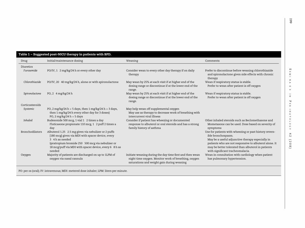
Table 1 – Suggested post-NICU therapy in patients with BPD.
Drug Initial/maintenance dosing Weaning Comments
Diuretics
Furosemide PO/IV, 1�2 mg/kg/24 h or every other day Consider wean to every other day therapy if on daily
therapy
Prefer to discontinue before weaning chlorothiazide
and spironolactone given side effects with chronic
therapy
Chlorothiazide PO/IV, 20�40 mg/kg/24 h, alone or with spironolactone May wean by 25% at each visit if at higher end of the
dosing range or discontinue if at the lower end of the
range.
Wean if respiratory status is stable.
Prefer to wean after patient is off oxygen
Spironolactone PO, 2�4 mg/kg/24 h May wean by 25% at each visit if at higher end of the
dosing range or discontinue if at the lower end of the
range.
Wean if respiratory status is stable.
Prefer to wean after patient is off oxygen
Corticosteroids
Systemic PO, 2 mg/kg/24 h£ 5 days, then 1mg/kg/24 h£ 3 days,
then 1 mg/kg/24 h every other day for 3 doses)
PO, 2 mg/kg/24 h£ 5 days
May help wean off supplemental oxygen
May use as therapy to decrease work of breathing with
intercurrent viral illness
Inhaled Budesonide 500 mcg, 1 vial 1�2 times a day
Fluticasone propionate 110 mcg, 1�2 puff 2 times a
day
Consider if patient has wheezing or documented
response to albuterol or oral steroids and has a strong
family history of asthma
Other inhaled steroids such as Beclomethasone and
Mometasone can be used. Dose based on severity of
symptoms
Bronchodilators Albuterol 1.25�2.5 mg given via nebulizer or 2 puffs
[180 mcg] given via MDI with spacer device, every
3�4 h as needed
Ipratropium bromide 250�500 mcg via nebulizer or
18mcg/puff via MDI with spacer device, every 6�8 h as
needed
Use for patients with wheezing or past history revers-
ible bronchospasm.
May be a useful adjunctive therapy especially in
patients who are not responsive to albuterol alone. It
may be better tolerated than albuterol in patients
with significant tracheomalacia.
Oxygen Majority of patients are discharged on up to 1LPM of
oxygen via nasal cannula
Initiate weaning during the day time first and then wean
night time oxygen. Monitor work of breathing, oxygen
saturations and weight gain during weaning
Wean in consultation with cardiology when patient
has pulmonary hypertension.
PO: per os (oral); IV: intravenous; MDI: metered dose inhaler; LPM: liters per minute.
199S
EM
I N
A R
S
IN
P
ER
IN
AT
OL
OG
Y 42
(208)

TAGGEDE NDS E M I N A R S I N P E R I N A T O L O G Y 4 2 ( 2 0 1 8 ) 200
patients were successfully weaned off supplemental oxygen
prior to hospital discharge. Patients who were successfully
weaned off supplemental oxygen after they received oral
prednisolone were able to do so after one course of oral ste-
roids; no additional benefit was conferred with multiple
courses.42
Bronchodilators
There are insufficient data to support the role of beta agonists
in the prevention of chronic lung disease; yet, inhaled bron-
chodilators are frequently used in US hospitals in patients
with evolving BPD, and a longer duration of mechanical venti-
lation increases the odds of receiving a bronchodilator.43 Fur-
thermore, there are no published data that address the role of
beta agonists on duration of mechanical ventilation, fre-
quency of rehospitalization, or mortality in infants and chil-
dren with BPD.44
BPD survivors are more likely to have respiratory symptoms
such as recurrent wheezing and cough, and are more likely to
have a diagnosis of asthma.45 Small airway dysfunction and
decreased expiratory flows are common lung abnormalities
found in infants.46 Significantly higher rates of airway reactiv-
ity in very low birth weight (VLBW) infants have been
reported.47 Data using the forced oscillation technique show
that 3�5 year old children with a history of BPD had higher
resonant frequency and lower mean reactance compared to
children without BPD, suggesting that the children with BPD
had a decreased peripheral airway patency.48 These airway
abnormalities have been shown to persist in children and
young adults with a history of BPD49,50,51 and bronchodilator
responsiveness is common in children with established
BPD.52
Although there are higher rates of airway reactivity in BPD
survivors, Kim et al reported that pre-school aged BPD survi-
vors showed bronchial hyperresponsiveness to methacho-
line, which stimulates airway smooth muscle directly, but no
response when challenged with adenosine 5-monophosphate
(AMP).53 The latter drug causes airway smooth muscle con-
traction through release of mast cell contents, suggesting
that the BPD patients do not have an inflammatory airway
response as seen in patients with asthma. This is in tandem
with low exhaled NO levels seen in patients with BPD and
obstructive lung disease, in contrast to patients with asthma
and a similar degree of obstruction. Taken together, there is
strong evidence against eosinophilic inflammation as the
basis of obstructive lung disease in BPD.36
Structural abnormalities in the airways along with other
factors including reduced small airway caliber and reduced
elastic recoil increase the propensity for airway narrowing
with smooth muscle contraction in BPD survivors. Together
these structural changes are thought to be the cause of persis-
tent obstruction seen on spirometry in BPD patients. A recent
study reported improvement in infant lung function tests in
VLBW babies with evolving BPD.54 In clinical practice, BPD
survivors who exhibit wheezing are often prescribed and will
respond to albuterol even when they do not fit the asthma
paradigm.
Other airway abnormalities such as tracheomalacia and
bronchomalacia are more common in patients with BPD.55 In
BPD infants with tracheobronchomalacia, use of beta agonists
can worsen wheezing by causing airway smooth muscle
relaxation in an already floppy airway.56
There are no randomized trials that evaluate the effect of
anticholinergic medications such as ipratropium bromide in
patients with BPD. They have been shown to be effective in
BPD patients in small studies and case reports.20 Ipratropium
bromide in low dose stiffens the central airway in animal
models via inhibition of presynaptic regulatory M2 receptors;
so it is occasionally used in BPD infants with tracheomalacia
to improve airwaymechanics.
Oxygen supplementation
Infants with BPD are more likely to have intermittent hypox-
emia (oxygen saturations less than 92%) during feeds and dur-
ing sleep. Intermittent hypoxemia has been shown to be
associated with poor growth.57 Over 30% of infants with BPD
require supplemental oxygen at the time of discharge
although most outgrow the need for supplemental oxygen by
2 years of life.38,39,58 Despite a third of patients with BPD being
discharged homewith supplemental oxygen, there are no evi-
dence-based guidelines or consensus statements regarding
oxygen weaning in BPD patients. Palm et al. found a wide var-
iation in practice among pediatric pulmonologists regarding
the minimum acceptable peripheral blood oxygen saturation
(SpO2), initial amount of supplemental oxygen being delivered
from which the weaning process should begin, signs consid-
ered to be important in determining readiness to wean, or the
methods used to wean patients with BPD from supplemental
oxygen.26 In their study, the majority of pediatric pulmonolo-
gists reported using nocturnal saturations as the primary
indication for weaning oxygen while only 8% reported using
standardized protocols. Minimum oxygen saturation in room
air required to discontinue supplemental oxygen varied from
90% to 95% and many respondents elected to wean flow rate
of oxygen delivered prior to weaning the duration of time that
the patient received oxygen supplementation. Although there
are no guidelines regarding oxygen weaning, experts recom-
mend weaning oxygen during the daytime first, followed by
weaning oxygen at nighttime.18 Most pediatric pulmonolgists
use clinical criteria including growth and clinical examination
to guide weaning oxygen supplementation in addition to
monitoring oxygen saturations. Room air challenge tests
have been used to guide oxygen weaning by some59 while
others recommend obtaining a polysomnogram to rule out
nocturnal hypoxemia prior to oxygen weaning.60,61 Although
weaning is typically guided by pediatric pulmonologists or
neonataologists this not always the case; 30% of infants fol-
lowed in a single center underwent discontinuation of sup-
plemental oxygen by their parents without guidance of a
physician.58
BPD is the most common cause of pulmonary arterial
hypertension (PAH) in pediatric patients, with an incidence of
up to 25% of BPD infants. Furthermore, the presence of PAH
correlates with the severity of BPD.62,63 Thus, it is more com-
mon in patients with severe BPD, who are also more likely to
201 S E M I N A R S I N P E R I N A T O L O G Y 4 2 ( 2 0 1 8 )
require supplemental oxygen and positive pressure support.
It can occur late, so a normal screening echocardiogram at 36
weeks PMA does not preclude the subsequent development
of PAH.64 Treatment of PAH includes use of supplemental
oxygen to minimize hypoxemic events and to encourage
growth64,65 in addition to pharmacotherapy. The presence of
PAH should be assessed when planning an oxygen wean in
this patient population.
Conclusions
There have beenmany advances in care of patients with early
and evolving BPD. There is a paucity of data regarding outpa-
tient management of patients with established BPD and lim-
ited studies that assess such interventions beyond short-
term effects. A suggested dosing/weaning schedule of the
common medications used in out-patient therapy of patients
with established BPD have been shown in Table 1. Further
research is needed to improve our understanding of the role
of currently used treatments on outcomes in a stable patient
with established BPD.
R E F E R E N C E S
1. Baraldi E, Carraro S, Filippone M. Bronchopulmonary dyspla-sia: definitions and long-term respiratory outcome. Early HumDev. 2009;85:S1–S3.
2. McEvoy CT, Jain L, Schmidt B, Abman S, Bancalari E, AschnerJL. Bronchopulmonary dysplasia: NHLBI workshop on the pri-mary prevention of chronic lung diseases. Ann Am Thorac Soc.2014;11:S146–S153.
3. Jobe AH, Bancalari E. Bronchopulmonary dysplasia. Am JRespir Crit Care Med. 2001;163:1723–1729.
4. Ehrenkranz RA, Walsh MC, Vohr BR, et al. Validation of theNational Institutes of Health consensus definition of broncho-pulmonary dysplasia. Pediatrics. 2005;116:1353–1360.
5. Abman SH, Collaco JM, Shepherd EG, et al. Interdisciplinarycare of children with severe bronchopulmonary dysplasia. JPediatr. 2017;181:12–28.
6. Stoll BJ, Hansen NI, Bell EF, et al. Trends in care practices,morbidity, and mortality of extremely preterm neonates,1993�2012. JAMA. 2015;314:1039–1051.
7. Fanaroff AA, Stoll BJ, Wright LL, et al. Trends in neonatal mor-bidity and mortality for very low birthweight infants. Am JObstet Gynecol. 2007;196(147):e141–e148.
8. Van Marter LJ. Epidemiology of bronchopulmonary dysplasia.Semin Fetal Neonatal Med. 2009;14:358–366.
9. Bhandari A, Bhandari V. Pitfalls, problems, and progress inbronchopulmonary dysplasia. Pediatrics. 2009;123:1562–1573.
10. Adams EW, Harrison MC, Counsell SJ, et al. Increased lungwater and tissue damage in bronchopulmonary dysplasia. JPediatr. 2004;145:503–507.
11. Cotton R, Suarez S, Reese J. Unexpected extra-renal effects ofloop diuretics in the preterm neonate. Acta Paediatr.2012;101:835–845.
12. Slaughter JL, Stenger MR, Reagan PB. Variation in the use ofdiuretic therapy for infants with bronchopulmonary dyspla-sia. Pediatrics. 2013;131:716–723.
13. Blaisdell CJ, Troendle J, Zajice A. Acute responses to diureticherapy in extremely low gestational age newborns: results
from the prematurity and respiratory outcomes programcohort study. J Pediatr. 2018;197:42–47.
14. Kao LC, Warburton D, Sargent CW, Platzker AC, Keens TG.Furosemide acutely decreases airways resistance inchronic bronchopulmonary dysplasia. J Pediatr. 1983;103:624–629.
15. Engelhardt B, Elliott S, Hazinski TA. Short- and long-termeffects of furosemide on lung function in infants with bron-chopulmonary dysplasia. J Pediatr. 1986;109:1034–1039.
16. Rastogi A, Luayon M, Ajayi OA, Pildes RS. Nebulized furose-mide in infants with bronchopulmonary dysplasia. J Pediatr.1994;125:976–979.
17. Prabhu VG, Keszler M, Dhanireddy R. Dose-dependent evalua-tion of the effects of nebulized furosemide on pulmonaryfunction in ventilated preterm infants. J Perinatol.1998;18:357–360.
18. Bancalari E, Wilson-Costello D, Iben SC. Management ofinfants with bronchopulmonary dysplasia in North America.Early Hum Dev. 2005;81:171–179.
19. Christou H, Brodsky D. Lung injury and bronchopulmonarydysplasia in newborn infants. J Intensive Care Med. 2005;20:76–87.
20. Tin W, Wiswell T E. Adjunctive therapies in chronic lung dis-ease: examining the evidence. Semin Fetal Neonatal Med.2008;13:44–52.
21. Kao LC, Warburton D, Cheng MH, Cede~no C, Platzker AC,Keens TG. Effect of oral diuretics on pulmonary mechanics ininfants with chronic bronchopulmonary dysplasia: results ofa double-blind crossover sequential trial. Pediatrics.1984;74:37–44.
22. Kao LC, Durand DJ, McCrea RC, Birch M, Powers RJ, NickersonBG. Randomized trial of long term diuretic therapy for infantswith oxygen dependent bronchopulmonary dysplasia. JPediatr. 1994;124:772–781.
23. Stewart A, Brion LP, Ambrosio-Perez I. Diuretics acting on thedistal renal tubule for preterm infants with (or developing)chronic lung disease. Cochrane Datab Syst Rev. 2011;9:CD001817.
24. Slaughter JL, Stenger MR, Reagan PB. Variation in the use ofdiuretic therapy for infants with bronchopulmonary dyspla-sia. Pediatrics. 2013;131:716–723.
25. Bhandari A, Chow U, Hagadorn JI. Variability in duration ofoutpatient diuretic therapy in bronchopulmonary dysplasia:a clinical experience. Am J Perinatol. 2010;27:529–535.
26. Palm K, Simoneau T, Sawicki G, Rhein L. Assessment of cur-rent strategies for weaning premature infants from supple-mental oxygen in the outpatient setting. Adv Neonatal Care.2011;11:349–356.
27. Baveja R, Christou H. Pharmacological strategies in the pre-vention and management of bronchopulmonary dysplasia.Semin Perinatol. 2006;30:209–218.
28. American Academy of Pediatrics. Committee on fetus andnewborn and Canadian paedtric society, fetus and newborncommittee. Post natal corticosteroids to treat or preventchronic lung disease in preterm infants. Pediatrics.2002;109:330–338.
29. Yeh TF, Chen CM, Wu SY, et al. Intratracheal administrationof budesonide/surfactant to prevent bronchopulmonary dys-plasia. Am J Respir Crit Care Med. 2016;193:86–95.
30. Venkataraman R, Kamaluddeen M, Hasan SU, Robertson HL,Lodha A. Intratracheal administration of budesonide-surfac-tant in prevention of bronchopulmonary dysplasia in verylow birth weight infants: a systematic review and meta-anal-ysis. Pediatr Pulmonol. 2017;52:968–975.
31. Bassler D, Plavka R, Shinwell ES, et al. Early inhaled budeso-nide for the prevention of bronchopulmonary dysplasia. NEngl J Med. 2015;373:1497–1506.

TAGGEDE NDS E M I N A R S I N P E R I N A T O L O G Y 4 2 ( 2 0 1 8 ) 202
32. Bassler D, Shinwell ES, Hallman M, et al. Long-term effects ofinhaled budesonide for bronchopulmonary dysplasia. N Engl JMed. 2018;378:148–157.
33. Bauer J, Teufel U, Maser-Gluth C, Doege C. Effects of budeso-nide inhalation on energy expenditure, somatic growth andsalivary cortisol levels in preterm infants with chronic lungdisease. Horm Res. 2009;72:146–152.
34. Cole CH, Shah B, Abbasi S, et al. Adrenal function in prema-ture infants during inhaled beclomethasone therapy. J Pediatr.1999;135:65–70.
35. Nelin LD, Logan JW. The use of inhaled corticosteroids inchronically ventilated preterm infants. Semin Fetal NeonatalMed. 2017;22:296–301.
36. Baraldi E, Bonetto G, Zacchello F, Filippone M. Low exhalednitric oxide in school-age children with bronchopulmonarydysplasia and airflow limitation. Am J Respir Crit Care Med.2005;171:68–72.
37. Halvorsen T, Skadberg BT, Eide GE, Røksund O, Aksnes L,�ymar K. Charachteristics of asthma and airway hyper-responsiveness after premature birth. Pediatr Allergy Immunol.2005;16:487–494.
38. Hennessy EM, Bracewell MA, Wood N, Wolke D, Costeloe K,Gibson A, Marlow N. EPI Cure Study Group. Respiratory healthin pre-school and school age children following extremelypreterm birth. Arch Dis Child. 2008;93:1037–1043.
39. Greenough A. Long term pulmonary outcomes of prematurebirth at less than 32 weeks of gestation. Early Hum Dev.2013;89:S25–S27.
40. Beresford MW, Primhak R, Subhedar NV, Shaw NJ. Rando-mised double blind placebo controlled trial of inhaled flutica-sone propionate in infants with chronic lung disease. Arch DisChild Fetal Neonatal Ed. 2002;87:F62–F63.
41. Yuskel B, Greenough A. Randomised trial of inhaled steroidsin preterm infants with respiratory symptoms at follow up.Thorax. 1992;47:910–913E.
42. Bhandari A, Schramm CS, Kimble C, Pappagallo M, Hussain N.Effect of a short course of prednisolone in infants with oxy-gen-dependent bronchopulmonary dysplasia. Pediatrics.2008;121:e344–e349.
43. Slaughter JL, Stenger MR, Reagan PB, Jadcherla SR. Inhaledbronchodilator use for infants with bronchopulmonary dys-plasia. J Perinatol. 2015;35:61–66.
44. Ng G, da Silva O, Ohlsson A. Bronchodilators for the preven-tion and treatment of chronic lung disease in preterm infants.Cochrane Database of Syst Rev. 2016;12:CD003214.
45. Fawke J, Lum S, Kirkby J, et al. Lung function and respiratorysymptoms at 11 years in children born extremely preterm:the EPICure study. Am J Respir Crit Care Med. 2010;182:237–245.
46. Filbrun AG, Popova AP, Linn MJ, McIntosh NA, HershensonMB. Longitudinal measures of lung function in infants withbronchopulmonary dysplasia. Pediatr Pulmonol. 2011;46:369–375.
47. Korhonen P, Laitinen J, Hyodynmaa E, Tammela O. Respira-tory outcome in school-aged, very-low-birth-weight childrenin the surfactant era. Acta Paediatr. 2004;93:316–321.
48. Vrijlandt EJ, Boezen HM, Gerritsen J, Stremmelaar EF, Duiver-man EJ. Respiratory health in prematurely born preschoolchildren with and without bronchopulmonary dysplasia. JPediatr. 2007;150:256–261.
49. Lum S, Kirkby J, Welsh L, Marlow N, Hennessy E, Stocks J.Nature and severity of lung function abnormalities inextremely pre-term children at 11 years of age. Eur Respir J.2011;37:1199–1207.
50. Gibson AM, Reddington C, McBride L, Callanan C, Robertson C,Doyle LW. Lung function in adult survivors of very low birthweight, with and without bronchopulmonary dysplasia.Pediatr Pulmonol. 2015;50:987–994.
51. Landry JS, Chan T, Lands L, Menzies D. Long-term impact ofbronchopulmonary dysplasia on pulmonary function. CanRespir J. 2011;18:265–270.
52. Fawke J, Lum S, Kirkby J, et al. Lung function and respiratorysymptoms at 11 years in children born extremely preterm:the EPICure study. Am J Respir Crit Care Med. 2010;182:237–245.
53. Kim DK, Choi SH, Yu J, Yoo Y, Kim B, Koh Y. Bronchial respon-siveness to methacholine and adenosine 5’monophosphatein preschool children with bronchopulomnoay dysplasia.Pediatr Pulmonol. 2006;41:538–548.
54. Morrow DK, Schilling D, McEvoy CT. Response to bronchodila-tors in very preterm infants with evolving bronchopulmonarydysplasia. Res Rep Neonatol. 2015;5:113–117.
55. Hysinger EB. Laryngomalacia, tracheomalacia and broncho-malacia. Curr Probl Pediatr Adolesc Health Care. 2018;48:113–118.
56. Panitch HB, Keklikian EN, Motley RA, Wolfson MR, SchidlowDV. Effect of altering smooth muscle tone onmaximal expira-tory flows in patients with tracheomalacia. Pediatr Pulmonol.1990;9:170–176.
57. Moyer-Mileur LJ, Nielson DW, Pfeffer KD, Witte MK, ChapmanDL. Eliminating sleep-associated hypoxemia improves growthin infants with bronchopulmonary dysplasia. Pediatrics.1996;98:779–783.
58. Yeh J, McGrath-Morrow SA, Collaco JM. Oxygen weaning afterhospital discharge in children with bronchopulmonary dys-plasia. Pediatr Pulmonol. 2016;51:1206–1211.
59. Trzaski JM, Hagadorn JI, Hussain N, Schwenn J, WittenzellnerC. Predictors of successful discontinuation of supplementaloxygen in very low-birth-weight infants with bronchopulmo-nary dysplasia approaching neonatal intensive care unit dis-charge. Am J Perinatol. 2012;29:79–86.
60. Saletti A, Stick S, Doherty D, Simmer K. Home oxygen therapyafter preterm birth in Western Australia. J Paediatr ChildHealth. 2004;40:519–523.
61. Khetan R, Hurley M, Spencer S, Bhatt JM. Bronchopulmonarydysplasia within and beyond the Neonatal Unit. Adv NeonatalCare. 2016;16:17–25: quiz E1.
62. Mourani PM, Abman SH. Pulmonary hypertension and vascu-lar abnormalities in bronchopulmonary dysplasia. Clin Perina-tol. 2015;42:839–855.
63. Al-Ghanem G, Shah P, Thomas S, et al. Bronchopulmonarydysplasia and pulmonary hypertension: a meta-analysis. JPerinatol. 2017;37:414–419.
64. Abman SH, Hansmann G, Archer SL, et al. Pediatric pulmo-nary hypertension: guidelines from the American Heart Asso-ciation and American Thoracic Society. Circulation.2015;132:2037–2099.
65. Papoff C, Cerasaro C, Caresta E, Barbara C, Midulla F, MorettiC. Current strategies for treating infants with severe broncho-pulmonary dysplasia. J Matern Fetal Neonatal Med. 2012;25:15–20.
medicina.uc.cl
Problemas respiratorios del recién nacido.Evaluación y manejo hemodinámico.Reanimación neonatal.NutriciónNeurología NeonatalMBE en NeonatologíaRedes NeonatalesEnfermería Neonatalotros
Información General Contenidos Expositores Internacionales
Avances en Pediatría NeonatalXVIII SEMINARIO INTERNACIONAL
7, 8, 9 y 10 de mayo 2019 | Centro de Extensión UC
Dr. Eduardo Bancalari University of Miami, Miami, EEUU
Dr. Peter Davis University of Melbourne, Victoria, Australia
Dra. Camilia Martin Harvard University, Boston, EEUU
Dr. Patrick McNamara University of Iowa, EEUU
Dr. Steven Miller University of Toronto, Canadá
Dr. Roger Soll Vermont University, EEUU
Organiza:Departamento de NeonatologíaDivisión de PediatríaEscuela de MedicinaPontificia Universidad Católica de Chile
Directores:Dra. Paulina TosoDr. José Luis TapiaDr. Javier KattanDr. Jorge FabresDr. Álvaro González
Comité Científico:Dr. Alberto EstayDr. Matías LucoDra. Patricia MenaDr. Ricardo UauyDra. Soledad Urzúa
medicina.uc.cl
7 mayo CURSOS PRE SEMINARIO
SIMPOSIO DE REANIMACIÓN NEONATAL SIMPOSIO DE HEMODINAMIA
203