c-Flip overexpression reduces cardiac hypertrophy in response to pressure overload
Upload
independentCategory
view
1download
0
NEONATAL NEURONAL OVEREXPRESSION OF GLYCOGEN SYNTHASEKINASE-3L REDUCES BRAIN SIZE IN TRANSGENIC MICE
K. SPITTAELS,a1 C. VAN DEN HAUTE,a1 J. VAN DORPE,a D. TERWEL,a K. VANDEZANDE,a
R. LASRADO,a K. BRUYNSEELS,a M. IRIZARRY,b M. VERHOYE,c J. VAN LINT,d
J. R. VANDENHEEDE,d D. ASHTON,e M. MERCKEN,e R. LOOS,f B. HYMAN,b
A. VAN DER LINDEN,c H. GEERTSe and F. VAN LEUVENa�
aExperimental Genetics Group, Department of Human Genetics, K.U. Leuven, Campus Gasthuisberg OpN 06,B-3000 Leuven, Belgium
bAlzheimer Disease Research Unit, Massachusetts General Hospital-East, CNY 6028, 149 13th St., Charlestown,MA 02120, USA
cBio-Imaging Lab, Department of Physics, University of Antwerp, Groenenborgerlaan 171, 2020 Antwerpen, BelgiumdDepartment of Biochemistry, K.U. Leuven, Gasthuisberg, B-3000 Leuven, Belgium
eJanssen Research Foundation, B-2340 Beerse, BelgiumfGenetic Epidemiology, Department of Human Genetics, K.U. Leuven, Leuven, Belgium
Abstract=Glycogen synthase kinase-3L (GSK-3L) is important in neurogenesis. Here we demonstrate that the kinasein£uenced post-natal maturation and di¡erentiation of neurons in vivo in transgenic mice that overexpress a constitutivelyactive GSK-3L[S9A]. Magnetic resonance imaging revealed a reduced volume of the entire brain, concordant with anearly 20% reduction in wet brain weight. The reduced volume was most prominent for the cerebral cortex, withouthowever, disturbing the normal cortical layering. The resulting compacted architecture was further demonstrated by anincreased neuronal density, by reduced size of neuronal cell bodies and of the somatodendritic compartment of pyramidalneurons in the cortex. No evidence for apoptosis was obtained. The marked overall reduction in the level of the micro-tubule-associated protein 2 in brain and in spinal cord, did not a¡ect the ultrastructure of the microtubular cytoskeletonin the proximal apical dendrites. The overall reduction in size of the entire CNS induced by constitutive active GSK-3Lcaused only very subtle changes in the psychomotoric ability of adult and ageing GSK-3L transgenic mice. E 2002IBRO. Published by Elsevier Science Ltd. All rights reserved.
Key words: glycogen synthase kinase-3L, microtubule-associated protein 2, microcephaly, transgenic mice.
Glycogen synthase kinase-3L (GSK-3L) is a multi-sub-strate serine/threonine protein kinase that has beenimplicated in development (Fiol et al., 1994; Ikeda etal., 1998), in cytoskeleton stabilization (Guidato et al.,1996; Guan et al., 1991; Garc|¤a-Pe¤rez et al., 1998;Berling et al., 1994; Sa¤nchez et al., 1996), in the induc-tion of cell processes (Leroy et al., 2000), in cell adhesion(Mackie et al., 1989) and in energy metabolism (Hoshi etal., 1996; Bollen et al., 1998).A major role for GSK-3L is evident in neurogenesis, as
downstream component of the wingless and Notch sig-naling pathways that are involved in the development of
the CNS in Drosophila, Caenorhabditis elegans and mam-mals (Lendahl, 1998; de la Pompa et al., 1997). GSK-3Ldetermines cell fate and early development of neuronaltissue as discovered in GSK-3/zeste-white 3 or shaggymutants of Drosophila (Perrimon and Smouse, 1989;Simpson et al., 1988) and con¢rmed in Xenopus(Dominguez et al., 1995; He et al., 1995).In adult neurons, GSK-3L regulates stabilization of
the cytoskeleton through phosphorylation of neuro¢la-ments and microtubule-associated proteins (MAPs),including protein tau (Avila et al., 1994; Hirokawa etal., 1996; Spittaels et al., 2000; Lucas et al., 2001).The goal of this study was to investigate in vivo the
in£uence of GSK-3L on the maturation and aging of amammalian CNS by overexpressing a constitutive activemutant, i.e. GSK-3L[S9A] (Woodgett, 1991), steered bythe mouse thy-1 gene promoter. The thy-1 cell-surfaceprotein is expressed in the thymus but also by neuronsthat have migrated to their ¢nal position and developdendrites (Morris, 1992). The engineered thy-1 constructused here and in previous studies (Moechars et al., 1999;Spittaels et al., 1999; Tesseur et al., 2000; Van Dorpe etal., 2000) begins expression of the transgene only post-natally and only in neurons.
797
1 These authors contributed equally to this work.*Corresponding author. Tel. : +32-16-345888; fax: +32-16-345871.E-mail address: [email protected] (F. Van Leuven).Abbreviations: GSK, glycogen synthase kinase; IA, interaural ;
IGF-1, insulin-like growth factor-1; M1, primary motor cortex;M2, secondary motor cortex; MAP2, microtubule-associatedprotein 2; MRI, magnetic resonance imaging; PKB, protein ki-nase B.
NSC 5679 12-8-02 Cyaan Magenta Geel Zwart
www.neuroscience-ibro.com
Neuroscience Vol. 113, No. 4, pp. 797^808, 2002E 2002 IBRO. Published by Elsevier Science Ltd
All rights reserved. Printed in Great BritainPII: S 0 3 0 6 - 4 5 2 2 ( 0 2 ) 0 0 2 3 6 - 1 0306-4522 / 02 $22.00+0.00
EXPERIMENTAL PROCEDURES
Generation of transgenic mice
Human GSK-3L[S9A] cDNA (Stambolic and Woodgett,1994) was ligated in the adapted mouse thy-1 gene vector(Moechars et al., 1996). The replacement of serine at position9 by alanine, i.e. denoted [S9A], prevents inactivation by phos-phorylation by p90 ribosomal S6 kinase, p70 ribosomal S6kinase and protein kinase B (PKB; Stambolic and Woodgett,1994). A linearized PvuI^NotI restriction fragment was micro-injected into 0.5-day FVB/N prenuclear mouse embryos and ¢veindependent transgenic founder mice were generated by stan-dard methods and maintained in the FVB/N background(Moechars et al., 1999; Spittaels et al., 1999). GSK-3L proteinin brain extracts was estimated by western blotting with a spe-ci¢c monoclonal antibody (tau protein kinase I/ GSK-3L) (A⁄-niti, Nottingham, UK). We previously reported an increase inenzymatic activity of about two-fold in strain 5 relative to non-transgenic mice (Spittaels et al., 2000). All experiments reportedbelow were performed with hemizygous human GSK-3L[S9A]transgenic mice from strain 5 and wild type littermates withFVB/N genetic background (unless stated otherwise). All e¡ortswere made to minimize the number of animals and their su¡er-ing. The experiments were carried out in accordance with theEuropean Communities Council Directive of 24 November 1986(86/609/EEC).
Immunohistochemical localization of the transgene
For immunohistochemical analysis, GSK-3L[S9A] and wildtype littermates were transcardially perfused with 4% parafor-maldehyde in phosphate-bu¡ered saline at the age of 5 dayspost-natally (P5), at P7, at P14 and at adult age. Microtomesections (6 Wm) of para⁄n-embedded brain (Bregma 31.46 to32.54) and spinal cord (thoracolumbal region) were stainedwith a polyclonal anti-GSK-3L antibody (Calbiochem, SanDiego, CA, USA) as described (Van den Haute et al., 2001).
Macroscopic and microscopic analysis of the CNS
GSK-3L[S9A] mice (n=6) and wild type littermates (n=7)were analyzed by magnetic resonance imaging (MRI) protocolsdescribed (Fransen et al., 1998; Kooy et al., 1999). Volumesfrom di¡erent brain structures were estimated to ¢nd di¡erencesbetween GSK-3L and wild type mice of 4.5 months old. Brainstructures were de¢ned on coronal MR images, according theMouse Brain atlas (Franklin and Paxinos, 1997). Their positionswere de¢ned in terms of antero-posterior distance to the inter-aural (IA) line. Image processing was performed using visual Croutines and interactive data language. To extract quantitativevolume information from a set of in vivo-obtained images of themouse, a semi-automatic 3D segmentation technique (Sijbers etal., 1997) was applied on the 3D MR images data set. Thevolumes of the total brain, cerebellum, hippocampus (locatedfrom IA 2.86 mm to IA 30.08 mm) and part of the cortexwere estimated. The cortex volume was calculated as the mergedsegmented volumes starting from IA 2.86 mm going caudal untilthe dorsal third ventricle was visual as a spot located IA 1.10mm. Only the upper half of the cortex (mainly somatosensorycortex, motor cortex, retrosplenial cortex, auditory cortex, pos-terior parietal association area and visual cortex) was segmentedlimited by the horizontal brain midline overlaid on each slice.For wet weight determinations of brain and spinal cord of
transgenic and wild type FVB mice (n=10330) at di¡erentages, the entire brain with olfactory bulbs, cerebrum, cerebellumand brain stem, was dissected rostrally to the fossa rhomboidea,while the spinal cord was dissected without the cauda equina.To determine the area of white and gray matter, para⁄n
sections (n=10) from the cervical intumescence of wild type(n=4) and GSK-3L transgenic mice (n=5) (2 months) werestained with Cresyl Violet and analyzed by Bioquant Imageanalysis system.
Para⁄n sections at Bregma 31.94 were stained with CresylViolet to investigate the general architecture of the neocortex.Neuronal density determinations and neuron counts were per-
formed in 16 Wm Nissl stained sagittal brain sections spaced 400Wm apart spanning one entire hemisphere. Layers II^VI of theentire medial to lateral extent of the neocortex were assessedfrom a rostral margin= superior to piriform cortex, to a caudalmargin= superior to entorhinal cortex. Neuron counts wereobtained by a systematic random sampling scheme usingapproximately 25 optical disectors consisting of a 50U50-Wmcounting box with extended exclusion lines at 100U waterobjective (West and Gundersen, 1990). Neurons were countedif they were not present in the initial plane of focus, but cameinto focus as the optical plane moved through the tissue. Theaverage coe⁄cient of error of the counting technique was 0.04(West, 1993). The estimation of total neurons was calculated bymultiplying the volume density of the neurons by the volume ofthe neocortex. The volume of the neocortex (with abovedescribed boundaries) was determined by the Cavalieri tech-nique using a Bioquant Image analysis system (Cavalieri,1966). Layer I was not included in the neocortical volume orneuron count measures. Seven wild type and seven GSK-3Ltransgenic mice of 2 months old were used in this study.The number of primary basal dendrites of pyramidal neurons
of layer V was counted on vibratome sections of Golgi^Cox-stained whole mouse brain. The Golgi^Cox impregnation ofneurons was performed as described (Gibb and Kolb, 1998).Only well-impregnated cells, showing clearly cell body andbasal dendrites, were selected in the primary and secondarymotor cortex (M1, M2), retrosplenial granular and agranularcortex (RSG, RSA), posterior parietal associated area (Ppta)and primary somatosensory cortex (S1) (Bregma 31.94 mm).Camera lucida drawings were generated of at least 75 pyramidalcortical neurons in vibratome sections (150 Wm) of six wild typeand six GSK-3L transgenic mice.Cell body areas of pyramidal neurons in cortical areas M1,
M2, RSG, RSA, Ppta, S1 and dorsal part of auditory cortex(Bregma 31.94 mm) were measured on paraformaldehyde-¢xedbrain sections stained with Cresyl Violet. The diameter of apicaldendrites of pyramidal neurons in cortical layer V was measuredin semi-thin sections (1 Wm) stained with Toluidine Blue. Theposterior parietal association cortex (Bregma 32.18 mm) wasexcised from 40-Wm vibratome sections ¢xed in paraformalde-hyde (4%) with glutaraldehyde (0.1%) and was embedded inepon. Microscope images collected with a 3CCD video camerawere analyzed with appropriate software (AIS/C 4.0) (ImagingResearch, St. Catharines, ON, Canada).Ultrathin sections derived from the same brain samples were
analyzed by transmission electron microscopy. The number ofmicrotubules in apical dendrites were counted and expressed asmean number per Wm based on four apical dendrites per mouse(three wild type and three GSK-3L mice) and 15 measurementsper dendrite.
Immunohistochemistry and western blot analysis of the CNS
Anesthetized mice were transcardially perfused either withparaformaldehyde (4%, v/v, in PBS) or methacarn and thebrain and spinal cord were further treated by standard methods(Spittaels et al., 1999; Van Dorpe et al., 2000). Monoclonalantibodies used were directed to MAP2 (phosphate-independentantibody; Sigma, St. Louis, MO, USA) or to neuro¢lamentproteins: SMI-31and SMI-32 (A⁄niti, Nottingham, UK) andNF-200 (Sigma) (Bregma 31.40 to 32.80).Apoptosis was detected with the Apoptosis Detection System
using the principle of the TUNEL assay (Promega, Madison,WI, USA) and a polyclonal activated caspase-3 antibody(Pharmingen, Heidelberg, Germany) (Bregma 31.40 to 32.80).Western blotting was performed as described (Spittaels et al.,
1999) for GSK-3L, MAP2 (Sigma, St. Louis, MO, USA), neu-ron-speci¢c class III L-tubulin (Promega), synaptophysin(DAKO, Glostrup, Denmark) and zinc-¢nger protein 37 (Zfp-37) (gift of N. Galjart, Rotterdam, The Netherlands) on brainand spinal cord extracts.
NSC 5679 12-8-02 Cyaan Magenta Geel Zwart
K. Spittaels et al.798
Glycogen quanti¢cation and detection
Levels of glycogen in cerebrum and cerebellum were enzy-matically determined as described (Chan and Exton, 1976). Inbrief, P14 mice were quick-frozen in 2-methylbutane, pre-cooledby liquid nitrogen. Frozen brains were dissected, cerebellum andcerebrum were separated and both parts were incubated in2.5 M KOH for 40 min at 90‡C. Brain tissue suspensionswere spotted on ¢lter paper strips, which were subsequentlythree times washed in 66% ethanol. Dry ¢lters were incubatedwith 0.2 mg/ml amyloglucosidase for 90 min at 37‡C and glu-cose was photometrically quanti¢ed using a standardized hexo-kinase method. Age-matched wild type (n=16) and GSK-3Ltransgenic mice (n=16) were analyzed.As glycogen synthase mRNA, glycogen synthase activity and
glycogen content are highest in the cerebellum and are locatedboth in astrocytes and neurons, histological staining of glycogenin Purkinje cells of P14 mice was performed by the periodicacid-Schi¡ (PAS) reaction on para⁄n-embedded sections asdescribed (Cheng et al., 2000). To prevent hydrolysis of glyco-gen during brain preparation, GSK-3L (n=10) and wild typemice (n=4) were quick-frozen in 2-methylbutane and brainwas dissected while frozen prior to ¢xation in 4% paraformal-dehyde. The number of PAS-positive Purkinje cells was deter-mined in the vermis.
Cognitive and behavioral tests
Mice were subjected to three cognitive tests: water-maze,E-maze and spontaneous alternation test. The water-maze testwas performed with F1 mice, aged 5 months, obtained as o¡-spring from male transgenic GSK-3L mice with female non-transgenic C57Bl/6J mice as described (Moechars et al., 1999).This strategy yields a homogenous group of hemizygous trans-genic F1 o¡spring, with perfectly matched non-transgenic litter-mates and overcomes the visual problems of FVB mice (Smith etal., 1997; Moechars et al., 1999). The E-shaped water-maze withhidden platform and a three-arm radial maze (the spontaneousalternation test) were used to determine spatial memory of thesame F1 mice at three di¡erent ages (254, 345 and 435 days) asnon-rewarded exploratory tasks (Sarter et al., 1988). Musclestrength and endurance was measured by the inverted wiregrid test (Lamberty and Gower, 1992) and forced swimmingtest (Spittaels et al., 1999) on GSK-3L FVB transgenic miceand wild type FVB littermates.
Statistical analysis
A two-tailed Student’s t-test was applied to volumetric data ofthe di¡erent brain structures determined by MRI and to weightsof brain and spinal cord. The Wilcoxon rank-sum test was per-formed on areas of gray matter, white matter and entire spinalcord in transversal sections, on measurements of neuronal den-sity, on determinations of neocortical volumes obtained by theCavalieri technique, on the number of primary basal dendrites,on cell body size and diameter of the apical dendrites and onglycogen levels. Analysis of variance and contingency M
2 testswere used for statistical comparison of groups of mice in thecognitive and behavioral tests. Di¡erences were considered sig-ni¢cant if P6 0.05.
RESULTS
Temporal and spatial expression of transgenicGSK-3L[S9A]
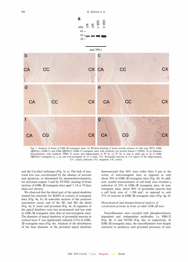
Expression of human GSK-3L[S9A] in brain, as deter-mined by western blotting, was highest in strain GSK-3L[S9A]-5 (2.5-fold increase) as opposed to strain GSK-3L[S9A]-1 (1.5-fold increase) compared to non-transgenicmice (Fig. 1a).
Immunohistochemically no di¡erences were observedbetween wild type and human GSK-3L[S9A] mice onP5 (Fig. 1b, c). From P7 onwards, expression of thehuman protein in the brain was widespread in neuronalcell bodies and processes of layer V of the cortex, in theCA regions of the hippocampus (Fig. 1d^g), many nucleiof the thalamus, caudate putamen and in brain stemnuclei. Human GSK-3L[S9A] was also detected in thegray matter of the spinal cord and in the Purkinje andgranular cell layer of the cerebellum (results not shown).
Overexpression of GSK-3L reduced the volume of brainand spinal cord
By the immunohistochemical analysis, it becameobvious to the observers that the brain of adult GSK-3L[S9A] mice of strains -5 and -1 that were analyzed(ns 50) was invariably smaller than that of age-matchednon-transgenic mice.The volume of di¡erent brain structures was measured
in vivo by MR imaging (Fig. 2a). The volume of cerebel-lum (81%), cerebrum (84%), hippocampus (85%) andcortex (73%) was signi¢cantly reduced in GSK-3L[S9A]transgenic mice relative to age-matched non-transgenicmice. Relative to the volume of the entire brain, onlythe volume of the cortex is signi¢cantly smaller in trans-genic mice compared to wild type littermates (Fig. 2b).Brain wet weight of neonatal non-transgenic and
GSK-3L[S9A]-5 mice aged 1 week was not di¡erent,whereas 2 weeks after birth a signi¢cant reduction wasalready evident. The di¡erence increased with age toreach 16.4^18.3% in adult male and female GSK-3L[S9A]-5 mice, respectively, relative to non-transgeniclittermates. In transgenic strain GSK-3L[S9A]-1, withlower expression of the transgene, reduction in adultbrain weight was less than in strain -5, i.e. 12.4^13.2%in males and females, respectively. The body weight inboth transgenic strains was not di¡erent from that ofnon-transgenic mice, demonstrating that the microce-phaly was speci¢c and not due to a general growth de¢cit(data not shown).The human GSK-3L[S9A] transgene was also
expressed in the neurons of the spinal cord and signi¢-cantly reduced its total wet weight (data not shown).Morphometric analysis of transversal sections of the spi-nal cord revealed a reduction of 23% of the total area,while white and gray matter were reduced 20 and 26%,respectively, relative to age-matched non-transgenicmice.
Microcephaly in GSK-3L transgenic mice is due toincreased density and reduced size of neurons
Routine hematoxylin/eosin and Cresyl Violet stainingdemonstrated that the overall cortical architecture inlayers I^VI was completely normal (Fig. 3a, b). How-ever, parallel with the reduction in brain volume, theneuronal density in the cortex of the GSK-3L transgenicmice was signi¢cantly increased (+42%). The total num-ber of cortical neurons remained unaltered, as deter-mined stereologically by the optical disector method
NSC 5679 12-8-02 Cyaan Magenta Geel Zwart
Microcephaly in GSK-3L transgenic mice 799
and the Cavalieri technique (Fig. 3c^e). The lack of neu-ronal loss was corroborated by the absence of necrosisand apoptosis, as determined by immunohistochemistryfor activated caspase 3 and by TUNEL staining of brainsections of GSK-3L transgenic mice aged 7, 14 or 70 days(data not shown).We observed that the distal part of the apical dendrites
stained less intensely for MAP2 in cortices of transgenicmice (Fig. 4a, b). In semi-thin sections of the posteriorassociation cortex and of the M1 and M2 the distal(Fig. 4a, b, inset) and proximal (Fig. 4c, d) segments ofthe apical dendrites were less pronounced and less widein GSK-3L transgenic mice than in non-transgenic mice.The diameter of apical dendrites of pyramidal neurons incortical layer V was signi¢cantly reduced (15%) in GSK-3L transgenic mice (Fig. 4e). Analysis of the distributionof the base diameter of the proximal apical dendrites
demonstrated that 60% were wider than 4 Wm in thecortex of non-transgenic mice as opposed to onlyabout 30% in GSK-3L transgenic mice (Fig. 4f). In addi-tion, careful measurements of cell body area revealed areduction of 23% in GSK-3L transgenic mice. In non-transgenic mice, about 80% of pyramidal neurons hada cell body area of s 200 Wm2, as opposed to only37% of neurons in GSK-3L transgenic mice (Fig. 4g, h).
Histochemical and immunochemical analysis ofcytoskeletal proteins in brain of adult GSK-3L mice
Neuro¢laments were revealed with phosphorylation-dependent and -independent antibodies, i.e. SMI-32(Fig. 4k, l) and NF200 (Fig. 4m, n) respectively. InGSK-3L transgenic mice, the neuro¢laments were largelyrestricted to perikarya and proximal processes of neu-
Fig. 1. Analysis of brain of GSK-3L transgenic mice. (a) Western blotting of brain protein extracts of wild type (WT), GSK-3L[S9A]-1 (GSK-1) and GSK-3L[S9A]-5 (GSK-5) transgenic mice with antibody tau protein kinase I (TPKI). (b^g) Immuno-histochemistry with antibody TPKI of cortex and hippocampus at P5 (b, c), P7 (d, e) and at adult age (f, g) of GSK-3L[S9A]-5 transgenic (c, e, g) and non-transgenic (b, d, f) mice. CA: Pyramidal neurons in CA region of the hippocampus,
CC: corpus callosum, CG: cingulum, CX: cortex.
NSC 5679 12-8-02 Cyaan Magenta Geel Zwart
K. Spittaels et al.800
rons as opposed to non-transgenic mice where theyextended far into the distal areas of the neurites. Anti-body SMI-32 clearly revealed the broader dendrites innon-transgenic mice. In addition, antibody SMI-31 wasused to demonstrate preferential staining of neuronal cell
bodies in GSK-3L transgenic mice, as opposed to mainlydistal apical dendrites in non-transgenic mouse brain(Fig. 4i, j).Camera lucida drawings of Golgi^Cox-impregnated
neurons demonstrated that the number of primary
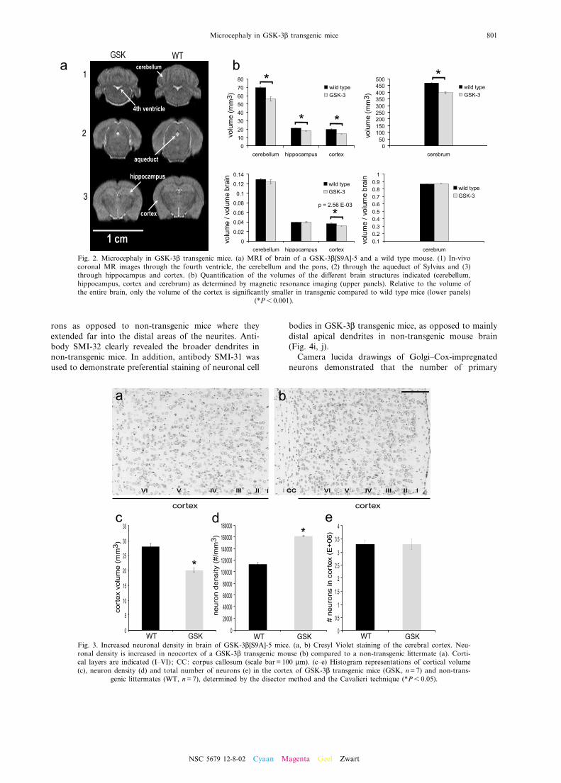
Fig. 3. Increased neuronal density in brain of GSK-3L[S9A]-5 mice. (a, b) Cresyl Violet staining of the cerebral cortex. Neu-ronal density is increased in neocortex of a GSK-3L transgenic mouse (b) compared to a non-transgenic littermate (a). Corti-cal layers are indicated (I^VI); CC: corpus callosum (scale bar = 100 Wm). (c^e) Histogram representations of cortical volume(c), neuron density (d) and total number of neurons (e) in the cortex of GSK-3L transgenic mice (GSK, n=7) and non-trans-
genic littermates (WT, n=7), determined by the disector method and the Cavalieri technique (*P6 0.05).
Fig. 2. Microcephaly in GSK-3L transgenic mice. (a) MRI of brain of a GSK-3L[S9A]-5 and a wild type mouse. (1) In-vivocoronal MR images through the fourth ventricle, the cerebellum and the pons, (2) through the aqueduct of Sylvius and (3)through hippocampus and cortex. (b) Quanti¢cation of the volumes of the di¡erent brain structures indicated (cerebellum,hippocampus, cortex and cerebrum) as determined by magnetic resonance imaging (upper panels). Relative to the volume ofthe entire brain, only the volume of the cortex is signi¢cantly smaller in transgenic compared to wild type mice (lower panels)
(*P6 0.001).
NSC 5679 12-8-02 Cyaan Magenta Geel Zwart
Microcephaly in GSK-3L transgenic mice 801
basal dendrites of pyramidal neurons of the ¢fth layer inthe neocortex was not signi¢cantly di¡erent betweennon-transgenic and GSK-3L transgenic mice (data notshown).Analysis by transmission electron microscopy of the
proximal part of the apical dendrite showed the normalappearance of microtubules, while also their density indendrites did not di¡er signi¢cantly in GSK-3L trans-genic mice from non-transgenic mice, i.e. 0.94Y 0.08and 0.87Y 0.12 microtubules/Wm (Ps 0.05), respectively(data not shown).Neuron-speci¢c L-tubulin (N-tubulin) and MAP2 were
compared by western blotting in protein extracts frombrain of young and adult mice. At P8, the levels ofN-tubulin and MAP2 in brain were very similar inmice with either genotype, whereas brain MAP2 levelswere already reduced by about 35% in GSK-3L trans-genic mice at P14. In adult GSK-3L transgenic mice,the MAP2 brain levels further declined to about 50%of those in age-matched non-transgenic mice (Fig. 5).Levels of N-tubulin were only reduced to about 85% ofthose in non-transgenic mice, whereas the levels ofnuclear Zfp-37, a heterochromatin-associated protein inneurons (Payen et al., 1998), and the levels of synapto-physin were both not di¡erent in GSK-3L transgenicmice (Fig. 5).
Histochemical and immunochemical analysis of spinal cordof adult GSK-3L mice
Immunohistochemical staining for MAP2 of sectionsof spinal cord revealed a reduced dendritic mass in GSK-3L transgenic mice compared to wild type littermates.This di¡erence was most outspoken in the ventral andlateral funiculi where the dendrites in the GSK-3L micewere penetrating less prominent into the white mattercompared to those in wild type mice (Fig. 6a, b).Western blotting for MAP2 revealed a dramatic reduc-
tion in the level of MAP2 protein in extracts of the spinalcord from GSK-3L transgenic mice, whereas the levels ofZfp-37 and of synaptophysin were again not markedlyreduced (Fig. 6c). Finally, N-tubulin levels were 25%lower in the spinal cord of GSK-3L transgenic mice rel-ative to wild type mice (Fig. 6c).
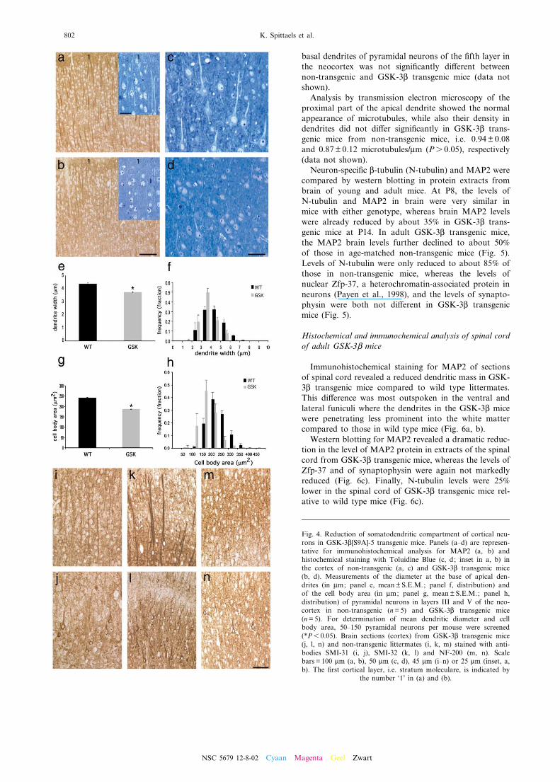
Fig. 4. Reduction of somatodendritic compartment of cortical neu-rons in GSK-3L[S9A]-5 transgenic mice. Panels (a^d) are represen-tative for immunohistochemical analysis for MAP2 (a, b) andhistochemical staining with Toluidine Blue (c, d; inset in a, b) inthe cortex of non-transgenic (a, c) and GSK-3L transgenic mice(b, d). Measurements of the diameter at the base of apical den-drites (in Wm; panel e, meanY S.E.M.; panel f, distribution) andof the cell body area (in Wm; panel g, meanYS.E.M.; panel h,distribution) of pyramidal neurons in layers III and V of the neo-cortex in non-transgenic (n=5) and GSK-3L transgenic mice(n=5). For determination of mean dendritic diameter and cellbody area, 50^150 pyramidal neurons per mouse were screened(*P6 0.05). Brain sections (cortex) from GSK-3L transgenic mice(j, l, n) and non-transgenic littermates (i, k, m) stained with anti-bodies SMI-31 (i, j), SMI-32 (k, l) and NF-200 (m, n). Scalebars = 100 Wm (a, b), 50 Wm (c, d), 45 Wm (i^n) or 25 Wm (inset, a,b). The ¢rst cortical layer, i.e. stratum moleculare, is indicated by
the number ‘1’ in (a) and (b).
NSC 5679 12-8-02 Cyaan Magenta Geel Zwart
K. Spittaels et al.802
Glycogen levels are normal in brain of GSK-3L transgenicmice
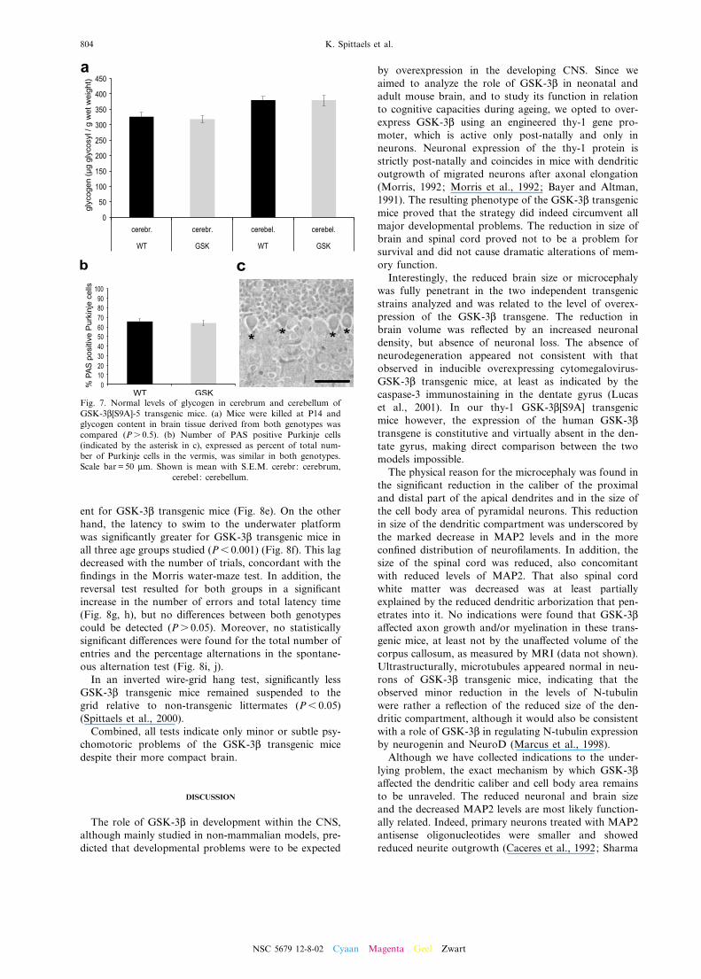
Glycogen is the largest energy reserve of the brain. AsGSK-3L can inactivate glycogen synthase through phos-phorylation, we quanti¢ed the glycogen level in the cere-brum and cerebellum of both wild type and transgenicmice. We focused on mice of 14 days old, since aroundthis age brain metabolism shifts from ketone bodies tocarbohydrate-based sources of energy and since reduc-tion in brain weight of GSK-3L transgenic mice is detect-able at P14. However, the amount of glycogen in bothcerebrum and cerebellum homogenates of GSK-3L micewas indistinguishable from the levels in brain of wildtype littermates (Fig. 7a).As glycogen synthase mRNA, glycogen synthase activ-
ity and glycogen content are highest in the cerebellum(Pellegri et al., 1996) but also present in glial cells, whileGSK-3L[S9A] expression is limited to neurons, we quan-ti¢ed the ratio of Purkinje cells that contained glycogenin their cytoplasm. The percentage of glycogen-positivePurkinje cells in both genotypes, however, was similar(Fig. 7b, c).
GSK-3L transgenic mice exhibit subtle psychomotoricde¢cits
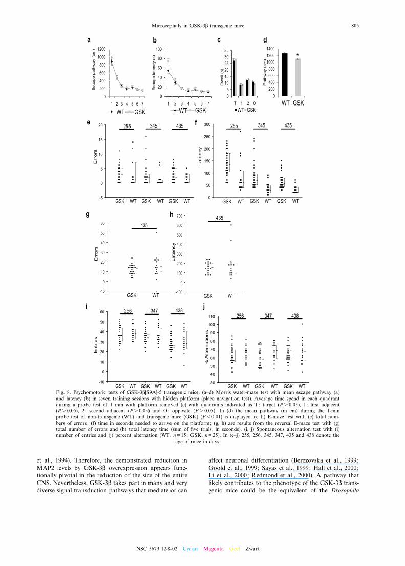
From the GSK-3L[S9A]-5 transgenic mouse strain wederived a cohort of F1 o¡spring for testing in the water-maze paradigm (Moechars et al., 1999). The transgenicF1 mice also showed microcephaly (data not shown) butdid learn to swim to the submerged platform in the clas-sic water-maze test as e⁄ciently as non-transgenic mice,without major di¡erences in ¢nal escape path (Fig. 8a) orlatency (Fig. 8b). Only in the ¢rst block of tests did theGSK-3L transgenic mice signi¢cantly longer (+37%) thanthe non-transgenic F1 mice to reach the hidden platform,although the escape path did not di¡er signi¢cantly(Fig. 8b). The subsequent probe trial with the hiddenplatform removed, demonstrated that all mice, indepen-dent of their genetic status, spent the same time explor-ing the expected quadrant (Fig. 8c). During this 1-minswim test it became obvious, however, that the swimspeed of the GSK-3L transgenic mice was signi¢cantlylower (13%) than that of non-transgenic mice (Fig. 8d).In the classical E-maze test, the number of entries in
the arm without the platform was not signi¢cantly di¡er-
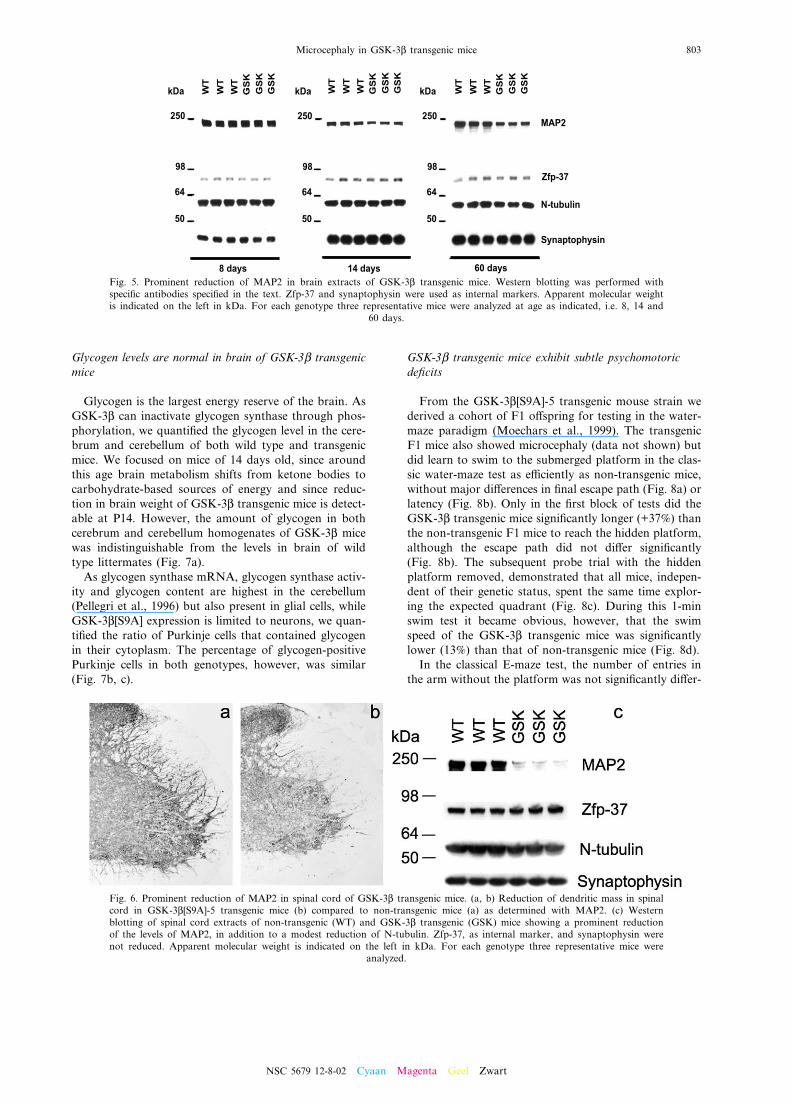
Fig. 5. Prominent reduction of MAP2 in brain extracts of GSK-3L transgenic mice. Western blotting was performed withspeci¢c antibodies speci¢ed in the text. Zfp-37 and synaptophysin were used as internal markers. Apparent molecular weightis indicated on the left in kDa. For each genotype three representative mice were analyzed at age as indicated, i.e. 8, 14 and
60 days.
Fig. 6. Prominent reduction of MAP2 in spinal cord of GSK-3L transgenic mice. (a, b) Reduction of dendritic mass in spinalcord in GSK-3L[S9A]-5 transgenic mice (b) compared to non-transgenic mice (a) as determined with MAP2. (c) Westernblotting of spinal cord extracts of non-transgenic (WT) and GSK-3L transgenic (GSK) mice showing a prominent reductionof the levels of MAP2, in addition to a modest reduction of N-tubulin. Zfp-37, as internal marker, and synaptophysin werenot reduced. Apparent molecular weight is indicated on the left in kDa. For each genotype three representative mice were
analyzed.
NSC 5679 12-8-02 Cyaan Magenta Geel Zwart
Microcephaly in GSK-3L transgenic mice 803
ent for GSK-3L transgenic mice (Fig. 8e). On the otherhand, the latency to swim to the underwater platformwas signi¢cantly greater for GSK-3L transgenic mice inall three age groups studied (P6 0.001) (Fig. 8f). This lagdecreased with the number of trials, concordant with the¢ndings in the Morris water-maze test. In addition, thereversal test resulted for both groups in a signi¢cantincrease in the number of errors and total latency time(Fig. 8g, h), but no di¡erences between both genotypescould be detected (Ps 0.05). Moreover, no statisticallysigni¢cant di¡erences were found for the total number ofentries and the percentage alternations in the spontane-ous alternation test (Fig. 8i, j).In an inverted wire-grid hang test, signi¢cantly less
GSK-3L transgenic mice remained suspended to thegrid relative to non-transgenic littermates (P6 0.05)(Spittaels et al., 2000).Combined, all tests indicate only minor or subtle psy-
chomotoric problems of the GSK-3L transgenic micedespite their more compact brain.
DISCUSSION
The role of GSK-3L in development within the CNS,although mainly studied in non-mammalian models, pre-dicted that developmental problems were to be expected
by overexpression in the developing CNS. Since weaimed to analyze the role of GSK-3L in neonatal andadult mouse brain, and to study its function in relationto cognitive capacities during ageing, we opted to over-express GSK-3L using an engineered thy-1 gene pro-moter, which is active only post-natally and only inneurons. Neuronal expression of the thy-1 protein isstrictly post-natally and coincides in mice with dendriticoutgrowth of migrated neurons after axonal elongation(Morris, 1992; Morris et al., 1992; Bayer and Altman,1991). The resulting phenotype of the GSK-3L transgenicmice proved that the strategy did indeed circumvent allmajor developmental problems. The reduction in size ofbrain and spinal cord proved not to be a problem forsurvival and did not cause dramatic alterations of mem-ory function.Interestingly, the reduced brain size or microcephaly
was fully penetrant in the two independent transgenicstrains analyzed and was related to the level of overex-pression of the GSK-3L transgene. The reduction inbrain volume was re£ected by an increased neuronaldensity, but absence of neuronal loss. The absence ofneurodegeneration appeared not consistent with thatobserved in inducible overexpressing cytomegalovirus-GSK-3L transgenic mice, at least as indicated by thecaspase-3 immunostaining in the dentate gyrus (Lucaset al., 2001). In our thy-1 GSK-3L[S9A] transgenicmice however, the expression of the human GSK-3Ltransgene is constitutive and virtually absent in the den-tate gyrus, making direct comparison between the twomodels impossible.The physical reason for the microcephaly was found in
the signi¢cant reduction in the caliber of the proximaland distal part of the apical dendrites and in the size ofthe cell body area of pyramidal neurons. This reductionin size of the dendritic compartment was underscored bythe marked decrease in MAP2 levels and in the morecon¢ned distribution of neuro¢laments. In addition, thesize of the spinal cord was reduced, also concomitantwith reduced levels of MAP2. That also spinal cordwhite matter was decreased was at least partiallyexplained by the reduced dendritic arborization that pen-etrates into it. No indications were found that GSK-3La¡ected axon growth and/or myelination in these trans-genic mice, at least not by the una¡ected volume of thecorpus callosum, as measured by MRI (data not shown).Ultrastructurally, microtubules appeared normal in neu-rons of GSK-3L transgenic mice, indicating that theobserved minor reduction in the levels of N-tubulinwere rather a re£ection of the reduced size of the den-dritic compartment, although it would also be consistentwith a role of GSK-3L in regulating N-tubulin expressionby neurogenin and NeuroD (Marcus et al., 1998).Although we have collected indications to the under-
lying problem, the exact mechanism by which GSK-3La¡ected the dendritic caliber and cell body area remainsto be unraveled. The reduced neuronal and brain sizeand the decreased MAP2 levels are most likely function-ally related. Indeed, primary neurons treated with MAP2antisense oligonucleotides were smaller and showedreduced neurite outgrowth (Caceres et al., 1992; Sharma
Fig. 7. Normal levels of glycogen in cerebrum and cerebellum ofGSK-3L[S9A]-5 transgenic mice. (a) Mice were killed at P14 andglycogen content in brain tissue derived from both genotypes wascompared (Ps 0.5). (b) Number of PAS positive Purkinje cells(indicated by the asterisk in c), expressed as percent of total num-ber of Purkinje cells in the vermis, was similar in both genotypes.Scale bar = 50 Wm. Shown is mean with S.E.M. cerebr: cerebrum,
cerebel : cerebellum.
NSC 5679 12-8-02 Cyaan Magenta Geel Zwart
K. Spittaels et al.804
et al., 1994). Therefore, the demonstrated reduction inMAP2 levels by GSK-3L overexpression appears func-tionally pivotal in the reduction of the size of the entireCNS. Nevertheless, GSK-3L takes part in many and verydiverse signal transduction pathways that mediate or can
a¡ect neuronal di¡erentiation (Berezovska et al., 1999;Goold et al., 1999; Sayas et al., 1999; Hall et al., 2000;Li et al., 2000; Redmond et al., 2000). A pathway thatlikely contributes to the phenotype of the GSK-3L trans-genic mice could be the equivalent of the Drosophila
Fig. 8. Psychomotoric tests of GSK-3L[S9A]-5 transgenic mice. (a^d) Morris water-maze test with mean escape pathway (a)and latency (b) in seven training sessions with hidden platform (place navigation test). Average time spend in each quadrantduring a probe test of 1 min with platform removed (c) with quadrants indicated as T: target (Ps 0.05), 1: ¢rst adjacent(Ps 0.05), 2: second adjacent (Ps 0.05) and O: opposite (Ps 0.05). In (d) the mean pathway (in cm) during the 1-minprobe test of non-transgenic (WT) and transgenic mice (GSK) (P6 0.01) is displayed. (e^h) E-maze test with (e) total num-bers of errors; (f) time in seconds needed to arrive on the platform; (g, h) are results from the reversal E-maze test with (g)total number of errors and (h) total latency time (sum of ¢ve trials, in seconds). (i, j) Spontaneous alternation test with (i)number of entries and (j) percent alternation (WT, n=15; GSK, n=25). In (e^j) 255, 256, 345, 347, 435 and 438 denote the
age of mice in days.
NSC 5679 12-8-02 Cyaan Magenta Geel Zwart
Microcephaly in GSK-3L transgenic mice 805
PI3PK/Akt (PKB) signaling pathway (Scanga et al.,2000; Verdu et al., 1999). GSK-3L is inhibited by phos-phorylation at serine 9 by PKB, and our data couldposition GSK-3L downstream of PKB as an importantdeterminant in the size of mammalian neurons. Circum-stantial evidence is provided by the insulin-like growthfactor 1 (IGF-1) that binds to its receptor to activate thePI3PK/PKB/GSK-3L pathway, in a later phase of neuro-nal di¡erentiation involving dendrite maturation andmyelinization. Mice de¢cient in IGF-1 are characterizedby reduced thickness of the cortex and compacting of cellbodies (Beck et al., 1995) and brain weight reductionwith smaller neurons with fewer processes (Cheng etal., 2000). The similarity in phenotype of IGF-1 de¢cientand GSK-3L overexpressing mice is obvious, and sug-gests that IGF-1 inactivates the downstream GSK-3Lvia activation of PKB. Interestingly, inhibition ofPI3PK in human neuroblastoma cells induced neuriteretraction via a mechanism which is dependent onGSK-3 activation (Sanchez et al., 2001). Collectively,all the data support the hypothesis that IGF-1/GSK-3Lmediates early post-natal neuronal development and thatGSK-3L determines directly or indirectly the somatoden-dritic volume of neurons by reducing the levels of MAP2most likely independent of the amount of glycogen theyaccumulate.Most interesting for our ongoing studies of GSK-3L in
the ageing process, was that the reduction in size of theCNS did not cause major cognitive problems in adultmice, as observed in the di¡erent test systems, i.e.water-maze, E-maze and spontaneous alternation test.
The reduction in swim speed and the decreased perfor-mance on the inverted wire-grid suggest a minor neuro-muscular problem. Routine histochemical analysis ofmuscle revealed no defects or di¡erences between GSK-3L transgenic and non-transgenic mice, while also noevidence was obtained for e¡ects on cerebellum or onmotor neurons in the spinal cord.In conclusion, constitutively active GSK-3L caused
microcephaly and a higher neuronal density by reducingthe volume of the somatodendritic compartment of neu-rons, i.e. dendrites and cell bodies. The postnatal arrestin neuronal maturation by GSK-3L did not prevent theGSK-3L transgenic mice to behave normal in terms ofgeneral cognition and ageing with however a minordecline in psychomotoric capability. These conclusionsdemonstrate that tight regulation of GSK-3L activity iscrucial for the normal maturation of neurons in vivo.
Acknowledgements0The intellectual, material and technical con-tributions of the following scientists are gratefully acknowl-edged: F. Vandesande, L. Arckens, J. Woodgett, H. Van DerPutten, M. Gilis, C. Kuipe¤ri, I. Laenen, E. Thiry, L. Wouters,H. Van Craenendonck, M. Mahieu, H. Uylings, N. Galjart, M.Bollen, J. Billen. We thank Christina Vochten for administra-tion. This investigation was supported by FWO-Vlaanderen, bythe Interuniversity-network for Fundamental Research (IUAP),by the Biotechnology program of the Flemish government(IWT/VLAB/COT-008), by the Flemish Institute for Biotechnol-ogy (VIB), by the Janssen Research Foundation (Beerse,Belgium), by a Seed Money grant from COSAT Johnson andJohnson (M.V.), by NFWO-Lotto, by the Rooms-fund, by K.U.Leuven RpD. We thank the K.U. Leuven for continuous sup-port.
REFERENCES
Avila, J., Dom|¤nguez, J., D|¤az-Nido, J., 1994. Regulation of microtubule dynamics by microtubule-associated protein expression and phosphor-ylation during neuronal development. Int. J. Dev. Biol. 38, 13^25.
Bayer, S.A., Altman, J., 1991. Neocortical Development. Raven, New York.Beck, K.D., Powell-Braxton, L., Widmer, H.R., Valverde, J., Hefti, F., 1995. Igf1 gene disruption results in reduced brain size, CNS hypomyeli-
nisation, and loss of hippocampal granule and striatal parvalbumin-containing neurons. Neuron 14, 717^730.Berezovska, O., McLean, P., Knowles, R., Frosh, M., Lu, F.M., Lux, S.E., Hyman, B.T., 1999. Notch1 inhibits neurite outgrowth in postmitotic
primary neurons. Neuroscience 93, 433^439.Berling, B., Wille, H., Ro«ll, B., Mandelkow, E.-M., Garner, C., Mandelkow, E., 1994. Phosphorylation of microtubule-associated proteins
MAP2a, b and MAP2c at Ser136 by proline-directed kinases in vivo and in vitro. Eur. J. Cell Biol. 64, 120^130.Bollen, M., Keppens, F., Stalmans, W., 1998. Speci¢c features of glycogen metabolism in the liver. Biochem. J. 336, 19^31.Caceres, A., Mautino, J., Kosik, K.S., 1992. Suppression of MAP2 in cultured cerebeller macroneurons inhibits minor neurite formation. Neuron
9, 607^618.Cavalieri, B., 1966. Geometria Degli Indivisibili. Unione Tipogra¢co, Torino.Chan, T.M., Exton, J.H., 1976. A rapid method for the determination of glycogen content and radioactivity in small quantities of tissue or isolated
hepatocytes. Ann. Biochem. 71, 96^105.Cheng, C.M., Reinhardt, R.R., Lee, W.-H., Joncas, G., Patel, S.C., Bondy, C.A., 2000. Insulin-like growth factor 1 regulates developing brain
glucose metabolism. Proc. Natl. Acad. Sci. USA 97, 10236^10241.de la Pompa, J.L., Wakeham, A., Correia, K.M., Samper, E., Brown, S., Aguilera, R.J., Nakano, T., Honjo, T., Mak, T.W., Rossant, J., Conlon,
R.A., 1997. Conservation of the Notch signaling pathway in mammalian neurogenesis. Development 124, 1139^1148.Dominguez, I., Itoh, K., Sokol, S.Y., 1995. Role of glycogen synthase kinase 3L as a negative regulator of dorsoventral axis formation in Xenopus
embryos. Proc. Natl. Acad. Sci. USA 92, 8498^8502.Fiol, C.J., Williams, J.S., Chou, C.H., Wang, Q.M., Roach, P.J., Andrisani, O.M., 1994. A secondary phosphorylation of CREB341 at Ser129 is
required for the cAMP-mediated control of gene expression. J. Biol. Chem. 269, 32187^32193.Franklin, K.B.J., Paxinos, G., 1997. The Mouse Brain in Stereotaxic Coordinates. Academic, San Diego, CA.Fransen, E., D’Hooghe, R., Van Camp, G., Verhoye, M., Sijbers, J., Reyniers, E., Soriano, P., Kamiguchi, H., Willemsen, R., Koekoek, K.E., De
Zeeuw, C.I., De Deyn, P.P., Van der Linden, A., Lemmon, V., Kooy, R.F., Willems, P., 1998. L1 knockout mice show dilated ventricles,vermis hypoplasia and impaired exploration patterns. Hum. Mol. Genet. 7, 999^1009.
Garc|¤a-Pe¤rez, J., Avila, J., D|¤az-Nido, J., 1998. Implication of cyclin-dependent kinases and glycogen synthase kinase 3 in the phosphorylation ofmicrotubule-associated protein 1B in developing neuronal cells. J. Neurosci. Res. 52, 445^452.
Gibb, R., Kolb, B., 1998. A method for vibratome sectioning of Golgi-Cox stained whole rat brain. J. Neurosci. Methods 79, 1^4.Goold, R.G., Owen, R., Gordon-Weeks, P.R., 1999. Glycogen synthase kinase 3L phosphorylation of microtubule-associated protein 1B regulates
the stability of microtubules in growth cones. J. Cell Sci. 112, 3373^3384.
NSC 5679 12-8-02 Cyaan Magenta Geel Zwart
K. Spittaels et al.806
Guan, R.J., Khatra, B.S., Cohlberg, J.A., 1991. Phosphorylation of bovine neuro¢lament proteins by protein kinase FA (glycogen synthase kinase3). J. Biol. Chem. 266, 8262^8267.
Guidato, S., Tsai, L.-H., Woodgett, J., Miller, C.C.J., 1996. Di¡erential cellular phosphorylation of neuro¢lament heavy side-arm by glycogensynthase kinase 3 and cyclin-dependent kinase-5. J. Neurochem. 66, 1698^1706.
Hall, A.C., Lucas, F.R., Salinas, P.C., 2000. Axonal remodeling and synaptic di¡erentiation in the cerebellum is regulated by WNT-7a signaling.Cell 100, 525^535.
He, H., Saint-Jeannet, J.-P., Woodgett, J.R., Varmus, H.E., Dawid, I.B., 1995. Glycogen synthase kinase-3 and dorsoventral patterning in Xenopusembryos. Nature 374, 617^622.
Hirokawa, N., Funakoshi, T., Sato-Harada, R., Kanai, Y., 1996. Selective stabilization of tau in axons and microtubule-associated protein 2C incell bodies and dendrites contributes to polarized localization of cytoskeletal proteins in mature neurons. J. Cell Biol. 132, 667^679.
Hoshi, M., Takashima, A., Noguchi, K., Murayama, M., Sato, M., Kondo, S., Saitoh, Y., Ishiguro, K., Hoshino, T., Imahori, I., 1996. Regulationof mitochondrial pyruvate dehydrogenase activity by tau protein kinase I/glycogen synthase kinase 3L in brain. Proc. Natl. Acad. Sci. USA 93,2719^2723.
Ikeda, S., Kishida, S., Yamamoto, H., Murai, H., Koyama, S., Kikuchi, A., 1998. Axin, a negative regulator of the Wnt signaling pathway, formsa complex with GSK-3L and L-catenin and promotes the GSK-3L-dependent phosphorylation of L-catenin. EMBO J. 17, 1371^1384.
Kooy, F., Reyniers, E., Verhoye, M., Sijbers, J., Cras, P., Oostra, B., Willems, P., Van der Linden, A., 1999. Neuroanatomy of the fragile Xknockout mouse brain studied using in vivo high resolution magnetic resonance imaging (MRI). Eur. J. Hum. Genet. 7, 526^532.
Lamberty, Y., Gower, A.J., 1992. Age-related changes in spontaneous behavior and learning in NMRI mice from middle to old age. Phys. Behav.51, 81^88.
Lendahl, U., 1998. A growing family of Notch ligands. BioEssays 20, 103^107.Leroy, K., Menu, R., Conreur, J.L., Dayanandan, R., Lovestone, S., Anderton, B.H., Brion, J.P., 2000. The function of the microtubule-
associated protein tau is variable modulated by graded changes in glycogen synthase kinase-3L activity. FEBS Lett. 465, 34^38.Li, Z., Van Aelst, L., Cline, H.T., 2000. Rho GTPases regulate distinct aspects of dendritic arbor growth in Xenopus central neurons in vivo. Nat.
Neurosci. 3, 217^225.Lucas, J.J., Herna¤ndez, F., Go¤mez-Ramos, P., Mora¤n, M.A., Hen, R., Avila, J., 2001. Decreased nuclear L-catenin, tau hyperphosphorylation and
neurodegeneration in GSK-3L conditional transgenic mouse. EMBO J. 20, 27^39.Mackie, K., Sorkin, B.C., Nairn, A.C., Greengard, P., Edelman, G.M., Cunningham, B.A., 1989. Identi¢cation of two protein kinases that
phosphorylate the neural cell-adhesion molecule, N-CAM. J. Neurosci. 9, 1883^1896.Marcus, E.A., Kintner, C., Harris, W., 1998. The role of GSK-3L in regulating neuronal di¡erentiation in Xenopus laevis. Mol. Cell Neurosci. 12,
269^280.Moechars, D., Dewachter, I., Lorent, K., Reverse¤, D., Baekelandt, V., Naidu, A., Tesseur, I., Spittaels, K., VandenHaute, C., Checler, F.,
Godaux, E., Cordell, B., Van Leuven, F., 1999. Early phenotypic changes in transgenic mice that overexpress di¡erent mutants of amyloidprecursor protein in brain. J. Biol. Chem. 274, 6483^6492.
Moechars, D., Lorent, K., De Strooper, B., Dewachter, I., Van Leuven, F., 1996. Expression in brain of amyloid precursor protein mutated in theK-secretase site causes disturbed behavior, neuronal degeneration and premature death in transgenic mice. EMBO J. 15, 1265^1274.
Morris, R., 1992. Thy-1, the enigmatic extrovert on the neuronal surface. BioAssay 14, 715^722.Morris, R.J., Tiveron, M.C., Xue, G.P., 1992. The relation of the expression and function of the neuronal glycoprotein Thy-1 to axonal growth.
Biochem. Soc. Trans. 20, 401^405.Payen, E., Verkerk, T., Michalovich, D., Dreyer, S.D., Winterpacht, A., Lee, B., De Zeeuw, C.I., Grosveld, F., Galjart, N., 1998. The centromeric/
nucleolar chromatin protein Zfp-37 may function to specify neuronal nuclear domains. J. Biol. Chem. 273, 9099^9109.Pellegri, G., Rossier, P., Magistretti, P.J., Martin, J.-L., 1996. Cloning, localization and induction of mouse brain glycogen synthase. Mol. Brain
Res. 38, 191^199.Perrimon, N., Smouse, D., 1989. Multiple functions of a Drosophila homeotic gene, zeste-white 3, during segmentation and neurogenesis. Dev.
Biol. 135, 287^305.Redmond, L., Oh, S.-R., Hicks, C., Weinmaster, G., Gosh, A., 2000. Nuclear Notch 1 signaling and the regulation of dendritic development. Nat.
Neurosci. 3, 30^40.Sa¤nchez, C., Tompa, P., Szu«cs, K., Friedrich, P., Avila, J., 1996. Phosphorylation and dephosphorylation in the proline-rich C-terminal domain of
microtubule-associated protein 2. Eur. J. Biochem. 241, 765^771.Sanchez, S., Sayas, C.L., Lim, F., Diaz-Nido, J., Avila, J., Wandosell, F., 2001. The inhibition of phophatidylinositol-3-kinase induces neurite
retraction and activates GSK3. J. Neurochem. 78, 468^481.Sarter, M., Bodewitz, G., Stephens, D.N., 1988. Attenuation of scopolamine-induced impairment of spontaneous alteration behaviour by antag-
onist but not inverse agonist and agonist beta-carbolines. Psychopharmacology 94, 491^495.Sayas, C.L., Moreno-Flores, M.T., Avila, J., Wandosell, F., 1999. The neurite retraction induced by lysophosphatidic acid increases Alzheimer’s
disease-like tau phosphorylation. J. Biol. Chem. 274, 37046^37052.Scanga, S.E., Ruel, L., Binari, R.C., Snow, B., Stambolic, V., Bouchard, D., Peters, M., Calvieri, B., Mak, T.W., Woodgett, J.R., Manoukian,
A.S., 2000. The conserved PI3PK/PTEN/Akt signalling pathway regulates both cell size and survival in Drosophila. Oncogene 19, 3971^3977.Sharma, N., Kress, Y., Sha¢t-Zagardo, B., 1994. Antisense MAP-2 oligonucleotides induce changes in microtubule assembly and neuritic
elongation in pre-existing neurites of rat cortical neurons. Cell Motil. Cytoskeleton 27, 234^247.Sijbers, J., Scheunders, P., Verhoye, M., Van der Linden, A., Van Dyck, D., Raman, E., 1997. Watershed-based segmentation of 3D MR data for
volume quantization. Magn. Reson. Imaging 15, 679^688.Simpson, P., El Messal, M., Moscoso de Prado, J., Ripoll, P., 1988. Stripes of positional homologies across the wing blade of Drosophila
melanogaster. Development 103, 391^401.Smith, D.J., Stevens, M.E., Sudanagunta, S.P., Bronson, R.T., Makhinson, M., Watabe, A.M., O’Dell, T.J., Fung, J., Weier, H.-U.G., Cheng,
J.-F., Rubin, E.M., 1997. Functional screening of 2 Mb of human chromosome 21q22.2 in transgenic mice implicates minibrain in learningdefects associated with Down syndrome. Nat. Genet. 16, 28^36.
Spittaels, K., Van den Haute, C., Van Dorpe, J., Bruynseels, K., Vandezande, K., Laenen, I., Geerts, H., Mercken, M., Sciot, R., Van Lommel,A., Loos, R., Van Leuven, F., 1999. Prominent axonopathy in the brain and spinal cord of transgenic mice overexpressing four-repeat humantau protein. Am. J. Pathol. 155, 2153^2165.
Spittaels, K., Van den Haute, C., Van Dorpe, J., Geerts, H., Mercken, M., Bruynseels, K., Lasrado, R., Vandezande, K., Laenen, I., Boon, T.,Van Lint, J., Vandenheede, J., Moechars, D., Loos, R., Van Leuven, F., 2000. Glycogen synthase kinase-3L phosphorylates protein tau andrescues the axonopathy in the central nervous system of human four-repeat tau transgenic mice. J. Biol. Chem. 275, 41340^413498.
Stambolic, V., Woodgett, J.R., 1994. Mitogen inactivation of glycogen synthase kinase-3L in intact cells via serine 9 phosphorylation. Biochem.J. 303, 701^704.
NSC 5679 12-8-02 Cyaan Magenta Geel Zwart
Microcephaly in GSK-3L transgenic mice 807
Tesseur, I., Van Dorpe, J., Spittaels, K., Van den Haute, C., Moechars, D., Van Leuven, F., 2000. Expression of human apolipoprotein E4 inneurons causes hyperphosphorylation of protein Tau in the brain of transgenic mice. Am. J. Pathol. 156, 951^964.
Van den Haute, C., Spittaels, K., Van Dorpe, J., Lasrado, R., Vandenzande, K., Laenen, I., Geerts, H., Van Leuven, F., 2001. Coexpression ofhuman cdk5 and its activator p35 with human protein tau in neurons in brain of triple transgenic mice. Neurobiol. Dis. 8, 32^44.
Van Dorpe, J., Smeijers, L., Dewachter, I., Nuyens, D., Spittaels, K., Van den Haute, C., Mercken, M., Moechars, D., Laenen, I., Kuiperi, C.,Bruynseels, K., Tesseur, I., Loos, R., Vanderstichele, H., Checler, F., Van Leuven, F., 2000. Prominent cerebral amyloid angiopathy intransgenic mice overexpressing the London mutant of human APP in neurons. Am. J. Pathol. 157, 1283^1298.
Verdu, J., Buratovich, M.A., Wilder, E.L., Birnbaum, M.J., 1999. Cell-autonomous regulation of cell and organ growth in Drosophila by Akt/PKB. Nat. Cell Biol. 1, 500^506.
West, M., 1993. New stereologic methods for counting neurons. Neurobiol. Aging 14, 275^285.West, M.J., Gundersen, H.J.G., 1990. Unbiased stereological estimation of the number of neurons in the human hippocampus. J. Comp. Neurol.
296, 1^22.Woodgett, J.R., 1991. A common denominator linking glycogen metabolism, nuclear oncogenes and development. TIBS 16, 177^181.
(Accepted 18 April 2002)
NSC 5679 12-8-02 Cyaan Magenta Geel Zwart
K. Spittaels et al.808
Copyright © 2022 FDOKUMEN