Pore-blocking and pore-assisting factors during capillary condensation and evaporation
Upload
independentCategory
view
2download
0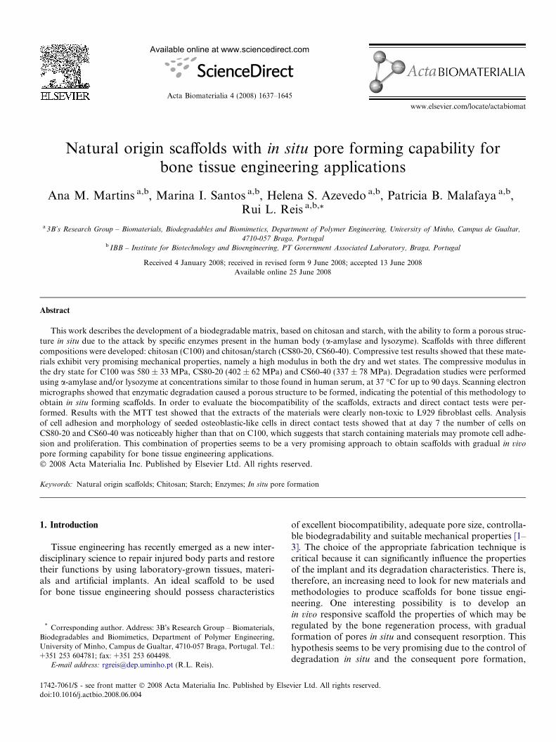
Available online at www.sciencedirect.com
Acta Biomaterialia 4 (2008) 1637–1645
www.elsevier.com/locate/actabiomat
Natural origin scaffolds with in situ pore forming capability forbone tissue engineering applications
Ana M. Martins a,b, Marina I. Santos a,b, Helena S. Azevedo a,b, Patricia B. Malafaya a,b,Rui L. Reis a,b,*
a 3B’s Research Group – Biomaterials, Biodegradables and Biomimetics, Department of Polymer Engineering, University of Minho, Campus de Gualtar,
4710-057 Braga, Portugalb IBB – Institute for Biotechnology and Bioengineering, PT Government Associated Laboratory, Braga, Portugal
Received 4 January 2008; received in revised form 9 June 2008; accepted 13 June 2008Available online 25 June 2008
Abstract
This work describes the development of a biodegradable matrix, based on chitosan and starch, with the ability to form a porous struc-ture in situ due to the attack by specific enzymes present in the human body (a-amylase and lysozyme). Scaffolds with three differentcompositions were developed: chitosan (C100) and chitosan/starch (CS80-20, CS60-40). Compressive test results showed that these mate-rials exhibit very promising mechanical properties, namely a high modulus in both the dry and wet states. The compressive modulus inthe dry state for C100 was 580 ± 33 MPa, CS80-20 (402 ± 62 MPa) and CS60-40 (337 ± 78 MPa). Degradation studies were performedusing a-amylase and/or lysozyme at concentrations similar to those found in human serum, at 37 �C for up to 90 days. Scanning electronmicrographs showed that enzymatic degradation caused a porous structure to be formed, indicating the potential of this methodology toobtain in situ forming scaffolds. In order to evaluate the biocompatibility of the scaffolds, extracts and direct contact tests were per-formed. Results with the MTT test showed that the extracts of the materials were clearly non-toxic to L929 fibroblast cells. Analysisof cell adhesion and morphology of seeded osteoblastic-like cells in direct contact tests showed that at day 7 the number of cells onCS80-20 and CS60-40 was noticeably higher than that on C100, which suggests that starch containing materials may promote cell adhe-sion and proliferation. This combination of properties seems to be a very promising approach to obtain scaffolds with gradual in vivo
pore forming capability for bone tissue engineering applications.� 2008 Acta Materialia Inc. Published by Elsevier Ltd. All rights reserved.
Keywords: Natural origin scaffolds; Chitosan; Starch; Enzymes; In situ pore formation
1. Introduction
Tissue engineering has recently emerged as a new inter-disciplinary science to repair injured body parts and restoretheir functions by using laboratory-grown tissues, materi-als and artificial implants. An ideal scaffold to be usedfor bone tissue engineering should possess characteristics
1742-7061/$ - see front matter � 2008 Acta Materialia Inc. Published by Else
doi:10.1016/j.actbio.2008.06.004
* Corresponding author. Address: 3B’s Research Group – Biomaterials,Biodegradables and Biomimetics, Department of Polymer Engineering,University of Minho, Campus de Gualtar, 4710-057 Braga, Portugal. Tel.:+351 253 604781; fax: +351 253 604498.
E-mail address: [email protected] (R.L. Reis).
of excellent biocompatibility, adequate pore size, controlla-ble biodegradability and suitable mechanical properties [1–3]. The choice of the appropriate fabrication technique iscritical because it can significantly influence the propertiesof the implant and its degradation characteristics. There is,therefore, an increasing need to look for new materials andmethodologies to produce scaffolds for bone tissue engi-neering. One interesting possibility is to develop anin vivo responsive scaffold the properties of which may beregulated by the bone regeneration process, with gradualformation of pores in situ and consequent resorption. Thishypothesis seems to be very promising due to the control ofdegradation in situ and the consequent pore formation,
vier Ltd. All rights reserved.
1638 A.M. Martins et al. / Acta Biomaterialia 4 (2008) 1637–1645
which allows the scaffold to have the required mechanicalproperties during the initial stage of implantation.
One of the present trends in implantable applications isfor materials that are derived from nature. Natural originmaterials have been demonstrated to promote healing ata faster rate and are expected to exhibit greater compatibil-ity with human tissues. The combination of chitosan withother materials appears to be a common theme in variousreports [4,5]. Chitosan is a linear copolymer of N-acetyl-D-glucosamine and D-glucosamine, and is a deacetylatedderivative of chitin. The degree of deacetylation (DD) rep-resents the proportion of D-glucosamine units with respectto the total number of units. DD is a structural parameterwhich influences physicochemical properties [6] such as sol-ubility, crystallinity, swelling behaviour and biologicalproperties [6], namely biodegradation by lysozyme [7,8],wound healing properties [9] and the enhancement ofosteogenesis [10]. One interesting feature of chitosan is itscationic nature, resulting from primary amine groups,which allows it to form water–insoluble ionic complexeswith a variety of polyanionic substances. It is normallyinsoluble in aqueous solution above pH 7. However, inweak acids (pH < 6), the free amino groups are protonatedand the polymer becomes soluble. Chitosan has been usedto induce extracellular matrix formation in tissue regenera-tive therapy [11]. The degradation of chitosan in the humanbody has been reported to be carried out by lysozyme [7,8].The degradation kinetics appears to be inversely related tothe degree of deacetylation [8,12]. Lysozyme, or murami-dase, is an enzyme that catalyzes the hydrolysis of the pep-tidoglycan layer of bacterial cell walls. Human lysozyme isfound in various body fluids in concentrations from 7 to13 mg l�1 [13–15] in serum and from 450 to 1230 mg l�1
in tears [13,14], saliva [13,14] and other fluids, includingthose surrounding cartilage [16]. Following implantationof a biomaterial, neutrophils and monocyte-derived macro-phages will be present around the foreign material in boththe acute and chronic phases of inflammation. A number ofenzymes, such as lysozyme, and reactive species will bereleased from these cells.
Biodegradable starch-based polymeric biomaterialshave been studied and proposed for a wide range of bio-medical applications. Starch is one of the most abundantnaturally occurring polymers, presenting a combinationof properties that is steadily increasing its use in severaltechnologies. Starch is a natural polymer that presentsexcellent characteristics for applications in the biomaterialsfield, primarily low toxicity [17,18], biodegradability [19]and biocompability [20,21]. It is inexpensive and, aboveall, reusable. The main enzymes involved in starch degra-dation are a- and b-amylase, glucosidase and other debran-ching enzymes. Starch is hydrolyzed to glucose, maltoseand dextrin. It is well known that salivary amylase isinvolved in the gastric and intestinal digestion of starchin food components. Amylase can also be found in humanserum.
The aim of this work was to develop a biodegradablematrix, based in chitosan and native starch, that will forma porous structure in vivo by the preferential attack of thematrix by specific enzymes present in the human body(namely the a-amylase and lysozyme). The inclusion ofan enzymatically degradable phase in biomaterials mayconstitute an interesting approach to obtain scaffolds withadequate mechanical properties and with a gradual in situ
pore forming ability. Using this innovative methodology,the developed scaffolds can exhibit very promising mechan-ical properties, due to the absence of macroporosity duringthe initial stage of implantation. The porosity is developedin situ by enzymes present in human body.
In this work, chitosan/starch scaffolds were developedusing a precipitation method. These systems were analyzedin terms of morphology, degradation behaviour andmechanical properties. This study also addressed the effectof leachables from developed scaffolds on the viability ofmouse fibroblasts and the influence of the construct’s sur-face on the morphology, adhesion and spreading of fibro-blast and human osteoblasts.
2. Materials and methods
2.1. Materials
Chitosan with medium molecular weight and DD of92% (determined by the titration method, as described inRef. [22]) and native corn starch were purchased fromSigma (St. Louis, USA). Sodium hydroxide (NaOH) andsodium sulphate (Na2SO4) were supplied from Panreac(Barcelona, Spain). a-Amylase from Bacillus amylolique-
faciens was obtained by Genencor International, Inc.(Rochester, NY, USA) and egg white lysozyme was fromSigma (St. Louis, USA).
2.2. Scaffolds preparation
Finely ground chitosan powder was dissolved in aceticacid 1% (v/v) to obtain a 5% (w/v) clear solution (C100)without any particulate material. Because of the relativelyhigh concentration, these solutions are quite viscous andconsequently can be stirred only slightly. However, flowis still observed and it is still possible to inject such viscoussolutions to fill out the moulds. Then, using the same pro-cedure, other formulations were prepared with the follow-ing ratios: 80/20 chitosan/starch scaffolds (CS80-20) and60/40 chitosan/starch scaffolds (CS60-40). The solutionswere casted into moulds and frozen (�18 �C) overnight.To produce chitosan and chitosan/starch scaffolds, thesolutions were immersed in a precipitation solution withcontaining 0.25 M NaOH and 0.375 M of Na2SO4 adaptedfrom Tuzlakoglu et al. [23]. After precipitation, the sampleswere washed repeatedly with distilled water to removeexcess of salts, dried at 37 �C and followed by successivewashings for 5 days until no pH changes were detected.

A.M. Martins et al. / Acta Biomaterialia 4 (2008) 1637–1645 1639
2.3. Degradation studies
Degradation studies were carried out by incubating thescaffolds in phosphate buffered saline (PBS) solution (pH7.4) containing a-amylase (150 U l�1 [15,24]) and/or lyso-zyme (13 mg l�1 [13–15]) at concentrations similar to theones found in human serum, at 37 �C up to 90 days. A con-trol was also performed by incubating the samples in bufferalone. At the end of the degradation period, the sampleswere removed and immediately weighed for determinationof water uptake, washed thoroughly with distilled waterand dried for later calculation of weight loss. Degradationsolutions were also analyzed to measure the concentrationof reducing sugars, released into the solution as a result ofstarch hydrolysis by a-amylase, using the dinitrosalicylicacid method.
2.4. Analysis of sample morphology by scanning electron
microscopy
The cross-sections morphology, before and after degra-dation, was observed by scanning electron microscopy(SEM) in a Leica Cambridge S360. All samples were previ-ously coated with a gold layer (Fisons Instruments, SputterCoater SC502, UK).
2.5. Mechanical properties – compression tests
The mechanical properties of the materials were evalu-ated on compression tests carried out in the dry and wet statein an Instron 4505 universal mechanical testing machine at acontrolled environment (23 �C and 55% RH). The cross-head speed was 2 mm min�1 until 1% strain.
2.6. Cell lines used for biocompatibility testing
For biocompatibility assessment of the different formu-lations of the developed scaffolds two different cell typeswere used: L929 (ECACC, European Collection of CellCultures), a mouse fibroblast cell line, and SAOS-2(ATCC, American Type Culture Collection), a cell linederived from human osteosarcoma. L929 is a cell line com-monly used in cytotoxicity testing and osteoblast-likeSAOS-2 cells were chosen because they are a model of a rel-evant cell type for the medical application of the implant.Both cell lines were cultured in Dulbecco’s modified Eagle’smedium (Sigma, St. Louis, USA), supplemented with 10%fetal bovine serum (Biochrome) and 1% antibiotic/antimi-cotic solution (Sigma, St. Louis, USA). In all the experi-ments cells were kept in an incubator at constanttemperature (37 �C) and CO2 concentration (5%).
2.7. Biocompatibility evaluation: extracts and cell adhesion
studies
In the scope of biocompatibility assessment, and accord-ing to ISO norms, two categories of in vitro cytotoxicity
assays were performed: extract and direct contact tests.The scaffolds, previously sterilized by ethylene oxide, wereextracted in complete culture medium for 24 h at constanttemperature (37 �C) and agitation (60 rpm). L929 cells wereseeded in 96-well cell culture plates (10,000 cells cm�2), inorder to reach 80% confluence in 24 h. The extracts werefiltered and placed in contact with the monolayer of L929cells for 72 h. A control, with cells grown in the presenceof complete culture medium, was included. Afterwards,the viability test MTT was performed. Briefly, the culturemedium was replaced by a solution of MTT and the cellswere incubated for 4 h. The viable cells, those with func-tional mitochondrial dehydrogenase, were able to reducethe yellow MTT into a purple formazan product. Theend product was quantified by spectroscopy and the resultsexpressed as percentage of cell viability.
In direct contact test, the developed scaffolds wereseeded with L929 cells (8 � 104/50 ll) and with osteo-blast-like cells (5 � 105 cells ml�1) for 3 and 7 days. Priorto cell seeding, the chitosan/starch scaffolds were immersedin culture medium overnight. This step is intended toreduce culture medium uptake by the highly hydrophilicmaterials. After each time point, the samples were fixedwith glutaraldehyde, dehydrated, critical point dried andgold sputtered for SEM observation.
3. Results and discussion
3.1. Characterization of the scaffolds morphology
In this work, degradable scaffolds based on chitosan andnative corn starch were developed for use in bone tissueengineering applications. Three different compositionswere prepared and tested. Fig. 1 shows the interior mor-phology of the obtained scaffolds. C100 material exhibitsa smooth surface (Fig. 1A), whereas in the micrographsof CS80-20 and CS60-40 scaffolds (Fig. 1B and C) it is pos-sible to observe that the starch granules are distributedhomogeneously along the chitosan matrix, as well as incor-porated into the matrix. Starch granules have dimensionsranging from 0.5 to 175 lm [25], which are mainly depen-dent upon their origin. SEM micrographs (Fig. 1B andC) show that starch granules from native corn starch havedimensions ranging from 5 to 10 lm. The proposed pro-cessing technique is based on precipitation from a solution.This technique was used with the aim of promoting theionic complexation. This precipitation method does notallow a macroporous structure to be obtained (Fig. 1).The macroporosity will be formed in situ by enzymes pres-ent in human serum.
3.2. Degradation behaviour
The main aim of the degradation studies, using a-amy-lase and lysozyme with similar concentrations to the onesfound in human serum, is to simulate the physiologicalconditions and subsequent pore formation. It is expected
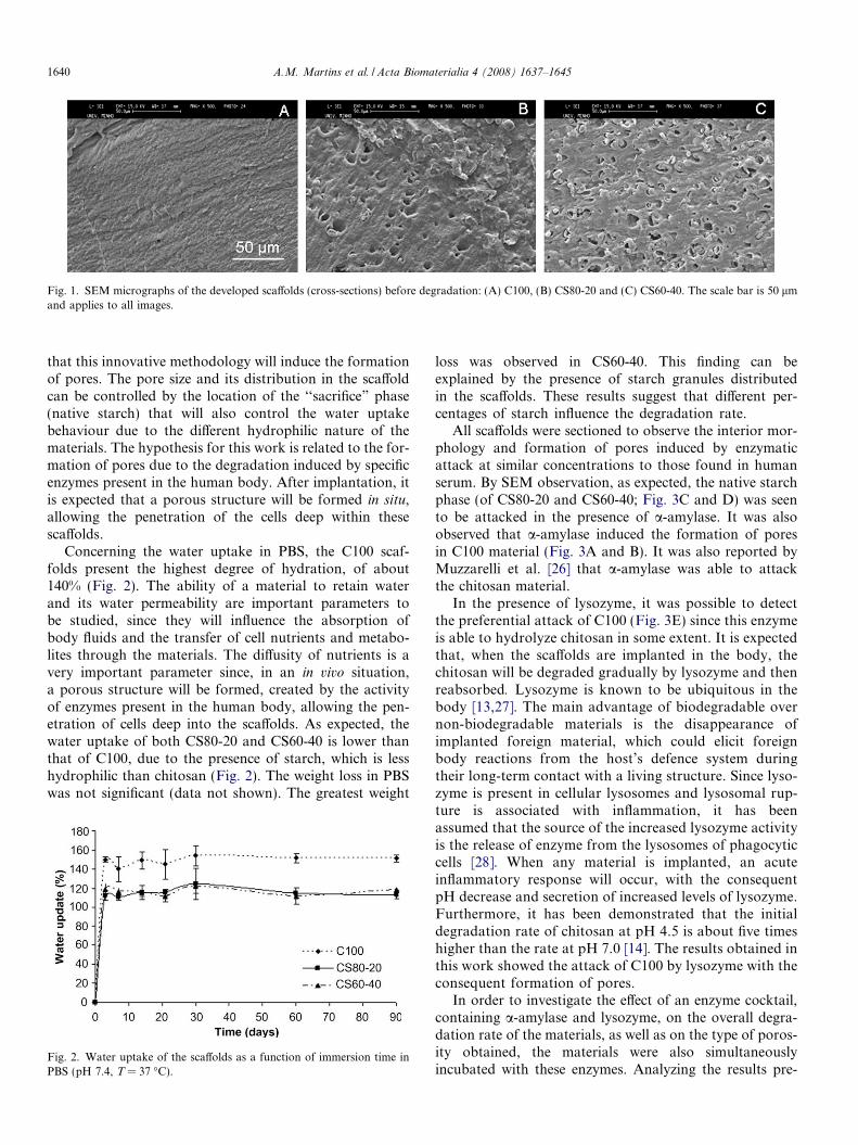
Fig. 1. SEM micrographs of the developed scaffolds (cross-sections) before degradation: (A) C100, (B) CS80-20 and (C) CS60-40. The scale bar is 50 lmand applies to all images.
1640 A.M. Martins et al. / Acta Biomaterialia 4 (2008) 1637–1645
that this innovative methodology will induce the formationof pores. The pore size and its distribution in the scaffoldcan be controlled by the location of the ‘‘sacrifice” phase(native starch) that will also control the water uptakebehaviour due to the different hydrophilic nature of thematerials. The hypothesis for this work is related to the for-mation of pores due to the degradation induced by specificenzymes present in the human body. After implantation, itis expected that a porous structure will be formed in situ,allowing the penetration of the cells deep within thesescaffolds.
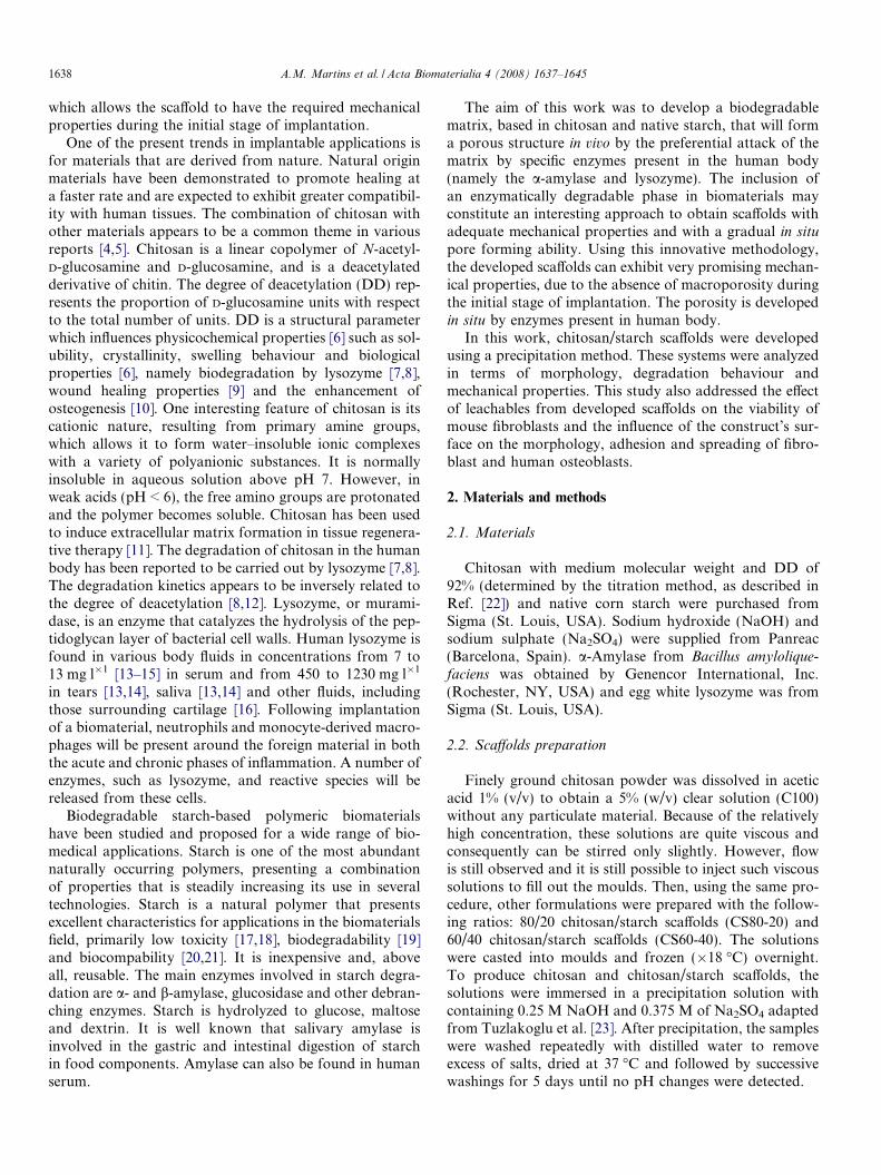
Concerning the water uptake in PBS, the C100 scaf-folds present the highest degree of hydration, of about140% (Fig. 2). The ability of a material to retain waterand its water permeability are important parameters tobe studied, since they will influence the absorption ofbody fluids and the transfer of cell nutrients and metabo-lites through the materials. The diffusity of nutrients is avery important parameter since, in an in vivo situation,a porous structure will be formed, created by the activityof enzymes present in the human body, allowing the pen-etration of cells deep into the scaffolds. As expected, thewater uptake of both CS80-20 and CS60-40 is lower thanthat of C100, due to the presence of starch, which is lesshydrophilic than chitosan (Fig. 2). The weight loss in PBSwas not significant (data not shown). The greatest weight
Fig. 2. Water uptake of the scaffolds as a function of immersion time inPBS (pH 7.4, T = 37 �C).
loss was observed in CS60-40. This finding can beexplained by the presence of starch granules distributedin the scaffolds. These results suggest that different per-centages of starch influence the degradation rate.
All scaffolds were sectioned to observe the interior mor-phology and formation of pores induced by enzymaticattack at similar concentrations to those found in humanserum. By SEM observation, as expected, the native starchphase (of CS80-20 and CS60-40; Fig. 3C and D) was seento be attacked in the presence of a-amylase. It was alsoobserved that a-amylase induced the formation of poresin C100 material (Fig. 3A and B). It was also reported byMuzzarelli et al. [26] that a-amylase was able to attackthe chitosan material.
In the presence of lysozyme, it was possible to detectthe preferential attack of C100 (Fig. 3E) since this enzymeis able to hydrolyze chitosan in some extent. It is expectedthat, when the scaffolds are implanted in the body, thechitosan will be degraded gradually by lysozyme and thenreabsorbed. Lysozyme is known to be ubiquitous in thebody [13,27]. The main advantage of biodegradable overnon-biodegradable materials is the disappearance ofimplanted foreign material, which could elicit foreignbody reactions from the host’s defence system duringtheir long-term contact with a living structure. Since lyso-zyme is present in cellular lysosomes and lysosomal rup-ture is associated with inflammation, it has beenassumed that the source of the increased lysozyme activityis the release of enzyme from the lysosomes of phagocyticcells [28]. When any material is implanted, an acuteinflammatory response will occur, with the consequentpH decrease and secretion of increased levels of lysozyme.Furthermore, it has been demonstrated that the initialdegradation rate of chitosan at pH 4.5 is about five timeshigher than the rate at pH 7.0 [14]. The results obtained inthis work showed the attack of C100 by lysozyme with theconsequent formation of pores.
In order to investigate the effect of an enzyme cocktail,containing a-amylase and lysozyme, on the overall degra-dation rate of the materials, as well as on the type of poros-ity obtained, the materials were also simultaneouslyincubated with these enzymes. Analyzing the results pre-

Fig. 3. SEM micrographs showing the morphology of chitosan-based scaffolds after 14 days in PBS with a-amylase solution (A, C100; C, CS80-20; D,CS60-40); after 30 days in same solution (B, C100); after 14 days in PBS with lysozyme solution (E, C100; F, CS80-20); and in the same period in PBS witha-amylase and lysozyme solution (G, C100; H, CS60-40).
A.M. Martins et al. / Acta Biomaterialia 4 (2008) 1637–1645 1641
sented in Fig. 4, it is possible to observe that CS60-40showed the highest weight loss compared with CS80-20and C100. The greater susceptibility of CS60-40 to enzy-matic degradation may be related to the presence of ahigher surface area for preferential attack, due to the inter-face between the two components. SEM micrographs(Fig. 3G and H) show that enzymatic degradation causeda porous structure is formed. Some studies showed thatpore sizes less than 15–50 lm result in fibrovascularingrowth, pore sizes of 50–150 lm encourage osteoid for-
mation, and pore sizes greater than 150 lm encourage theingrowth of mineralized bone [29].
Since the formation of pores in the presence of lysozymewas not so pronounced compared to those obtained with a-amylase or the two enzymes together (a-amylase and lyso-zyme), it may be necessary to look for alternative strategiesto enhance the scaffold degradation. One possible alterna-tive could be the incorporation of lysozyme in higher con-centrations at the surface of these materials. This could beachieved by incorporation of the enzyme into biomimetic
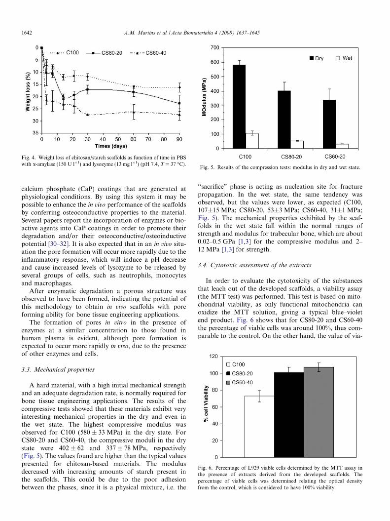
Fig. 4. Weight loss of chitosan/starch scaffolds as function of time in PBSwith a-amylase (150 U l�1) and lysozyme (13 mg l�1) (pH 7.4, T = 37 �C).
Fig. 5. Results of the compression tests: modulus in dry and wet state.
Fig. 6. Percentage of L929 viable cells determined by the MTT assay inthe presence of extracts derived from the developed scaffolds. Thepercentage of viable cells was determined relating the optical densityfrom the control, which is considered to have 100% viability.
1642 A.M. Martins et al. / Acta Biomaterialia 4 (2008) 1637–1645
calcium phosphate (CaP) coatings that are generated atphysiological conditions. By using this system it may bepossible to enhance the in vivo performance of the scaffoldsby conferring osteoconductive properties to the material.Several papers report the incorporation of enzymes or bio-active agents into CaP coatings in order to promote theirdegradation and/or their osteoconductive/osteoinductivepotential [30–32]. It is also expected that in an in vivo situ-ation the pore formation will occur more rapidly due to theinflammatory response, which will induce a pH decreaseand cause increased levels of lysozyme to be released byseveral groups of cells, such as neutrophils, monocytesand macrophages.
After enzymatic degradation a porous structure wasobserved to have been formed, indicating the potential ofthis methodology to obtain in vivo scaffolds with poreforming ability for bone tissue engineering applications.
The formation of pores in vitro in the presence ofenzymes at a similar concentration to those found inhuman plasma is evident, although pore formation isexpected to occur more rapidly in vivo, due to the presenceof other enzymes and cells.
3.3. Mechanical properties
A hard material, with a high initial mechanical strengthand an adequate degradation rate, is normally required forbone tissue engineering applications. The results of thecompressive tests showed that these materials exhibit veryinteresting mechanical properties in the dry and even inthe wet state. The highest compressive modulus wasobserved for C100 (580 ± 33 MPa) in the dry state. ForCS80-20 and CS60-40, the compressive moduli in the drystate were 402 ± 62 and 337 ± 78 MPa, respectively(Fig. 5). The values found are higher than the typical valuespresented for chitosan-based materials. The modulusdecreased with increasing amounts of starch present inthe scaffolds. This could be due to the poor adhesionbetween the phases, since it is a physical mixture, i.e. the
‘‘sacrifice” phase is acting as nucleation site for fracturepropagation. In the wet state, the same tendency wasobserved, but the values were lower, as expected (C100,107±15 MPa; CS80-20, 53±3 MPa; CS60-40, 31±1 MPa;Fig. 5). The mechanical properties exhibited by the scaf-folds in the wet state fall within the normal ranges ofstrength and modulus for trabecular bone, which are about0.02–0.5 GPa [1,3] for the compressive modulus and 2–12 MPa [1,3] for strength.
3.4. Cytotoxic assessment of the extracts
In order to evaluate the cytotoxicity of the substancesthat leach out of the developed scaffolds, a viability assay(the MTT test) was performed. This test is based on mito-chondrial viability, as only functional mitochondria canoxidize the MTT solution, giving a typical blue–violetend product. Fig. 6 shows that for CS80-20 and CS60-40the percentage of viable cells was around 100%, thus com-parable to the control. On the other hand, the value of via-

A.M. Martins et al. / Acta Biomaterialia 4 (2008) 1637–1645 1643
ble L929 cells in contact with C100 leachables was lower(75%). The MTT assay suggested that the extracts fromthe developed scaffolds, produced by the precipitationmethod, were non-toxic towards mouse fibroblasts.
3.5. Cell morphology evaluation
The absence of cytotoxicity does not confer any infor-mation about the biocompatibility of a biomaterial [33].
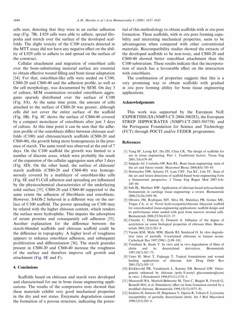
Fig. 7. SEM micrographs showing the surface the developed scaffolds: C100 (A7 days of culture with L929 cells (D–F).
Fig. 8. SEM micrographs of C100 (A,D), CS80-20 (B,E) and CS60-40 (C,F) scThe scale bar is 200 lm and applies to all images.
In order to verify whether the developed scaffolds supportthe functions shared by many cell types, such as membraneintegrity, adhesion to surfaces and replication, adhesionstudies with the cell line L929 were performed. At theend of 7 days, regardless of the scaffold formulation, allconstructs were covered with a monolayer of cells(Fig. 7). The fibroblasts had a typical spindle-shaped mor-phology and exhibited cytoplasmatic projections stronglyattached to the scaffolds. Only in CS80-20 were rounded
,D), CS80-20 (B,E) and CS60-40 (C,F), before cell culture (A–C) and after
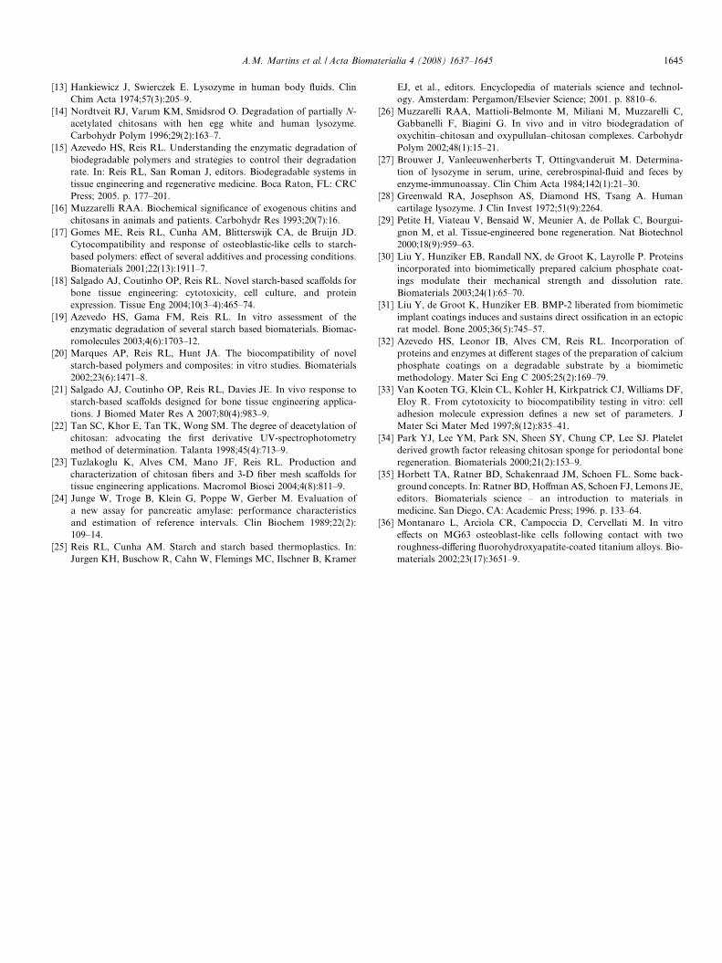
affolds cultured with osteoblast-like cells after 3 (A–C) and 7 days (D–F).
1644 A.M. Martins et al. / Acta Biomaterialia 4 (2008) 1637–1645
cells seen, denoting that they were in an earlier adhesionstep (Fig. 7B). L929 cells were able to adhere, spread filo-podia and stretch over the surface of the developed scaf-folds. The slight toxicity of the C100 extracts detected inthe MTT assay did not have any negative effect on the abil-ity of L929 cells to adhere to and spread on the surface ofthe construct.
Cellular attachment and migration of osteoblast cellsover the bone-substituting material surface are essentialto obtain effective wound filling and bone tissue adaptation[34]. For that, osteoblast-like cells were seeded on C100,CS80-20 and CS60-40 and the adhesion profile, as well asthe cell morphology, was documented by SEM. On day 3of culture, SEM examination revealed osteoblasts aggre-gates sparsely distributed over the surface of C100(Fig. 8A). At the same time point, the amount of cellsattached to the surface of CS80-20 was greater, althoughcells did not cover the entire surface of the scaffold(Fig. 8B). Fig. 8C shows the surface of CS60-40 coveredby a compact monolayer of osteoblasts after just 3 daysof culture. At this time point it can be seen that the adhe-sion profile of the osteoblasts differs between chitosan scaf-folds (C100) and chitosan/starch scaffolds (CS80-20 andCS60-40), the growth being more homogeneous in the pres-ence of starch. The same trend was observed at the end of 7days. On the C100 scaffold the growth was limited to anumber of discrete areas, which were probably the resultof the expansion of the cellular aggregates seen after 3 days(Fig. 8D). On the other hand, the surface of chitosan/starch scaffolds (CS80-20 and CS60-40) was homoge-neously covered by a multilayer of osteoblast-like cells(Fig. 8E and F).Cell adhesion and spreading are influencedby the physicochemical characteristics of the underlayingsolid surface [35]. CS80-20 and CS60-40 supported to thesame extent the adhesion of fibroblasts and osteoblasts.However, SAOS-2 behaved in a different way on the sur-face of C100 scaffold. The poorer spreading on C100 maybe related with the higher chitosan content, which rendersthe surface more hydrophilic. This impairs the adsorptionof serum proteins and consequently cell adhesion [35].Another explanation for the difference between thestarch-blended scaffolds and chitosan scaffold could bethe difference in topography. A higher level of roughnessappears to enhance osteoblast adhesion, and subsequentproliferation and differentiation [36]. The starch granulespresent in CS80-20 and CS60-40 increase the roughnessof the surface and therefore improve cell growth andattachment (Fig. 8E and F).
4. Conclusions
Scaffolds based on chitosan and starch were developedand characterised for use in bone tissue engineering appli-cations. The results of the compressive tests showed thatthese materials exhibit very good mechanical propertiesin the dry and wet states. Enzymatic degradation causedthe formation of a porous structure, indicating the poten-
tial of this methodology to obtain scaffolds with in situ poreformation. These scaffolds, with in situ pore forming capa-bility and interesting mechanical properties, seem to beadvantageous when compared with other conventionalmaterials. Biocompatibility studies showed the extracts ofthe developed scaffolds to be non-toxic, and CS80-20 andCS60-40 showed better osteoblast attachment than theC100 substratum. These results indicate that the incorpora-tion of starch has a favourable effect on the interactionwith osteoblasts.
The combination of properties suggests that this is avery promising way to obtain scaffolds with gradualin situ pore forming ability for bone tissue engineeringapplications.
Acknowledgements
This work was supported by the European NoEEXPERTISSUES (NMP3-CT-2004-500283), the EuropeanSTREP HIPPOCRATES (NMP3-CT-2003-505758) andthe Portuguese Foundation for Science and Technology(FCT) through POCTI and/or FEDER programmes.
References
[1] Yang SF, Leong KF, Du ZH, Chua CK. The design of scaffolds foruse in tissue engineering. Part 1. Traditional factors. Tissue Eng2001;7(6):679–89.
[2] Salgado AJ, Coutinho OP, Reis RL. Bone tissue engineering: state ofthe art and future trends. Macromol Biosci 2004;4(8):743–65.
[3] Hutmacher DW, Schantz JT, Lam CXF, Tan KC, Lim TC. State ofthe art and future directions of scaffold-based bone engineering froma biomaterials perspective. J Tissue Eng Regen Med 2007;1(4):245–60.
[4] Suh JK, Matthew HW. Application of chitosan-based polysaccharidebiomaterials in cartilage tissue engineering: a review. Biomaterials2000;21(24):2589–98.
[5] Oliveira JM, Rodrigues MT, Silva SS, Malafaya PB, Gomes ME,Viegas CA, et al. Novel hydroxyapatite/chitosan bilayered scaffoldfor osteochondral tissue-engineering applications: scaffold design andits performance when seeded with goat bone marrow stromal cells.Biomaterials 2006;27(36):6123–37.
[6] Chatelet C, Damour O, Domard A. Influence of the degree ofacetylation on some biological properties of chitosan films. Bioma-terials 2001;22(3):261–8.
[7] Varum KM, Myhr MM, Hjerde RJ, Smidsrod O. In vitro degrada-tion rates of partially N-acetylated chitosans in human serum.Carbohydr Res 1997;299(1–2):99–101.
[8] Tomihata K, Ikada Y. In vitro and in vivo degradation of films ofchitin and its deacetylated derivatives. Biomaterials1997;18(7):567–75.
[9] Ueno H, Mori T, Fujinaga T. Topical formulations and woundhealing applications of chitosan. Adv Drug Deliv Rev2001;52(2):105–15.
[10] Klokkevold PR, Vandemark L, Kenney EB, Bernard GW. Osteo-genesis enhanced by chitosan (poly-N-acetyl glucosaminoglycan)in vitro. J Periodontol 1996;67(11):1170–5.
[11] Muzzarelli RA, Mattioli-Belmonte M, Tietz C, Biagini R, Ferioli G,Brunelli MA, et al. Stimulatory effect on bone formation exerted by amodified chitosan. Biomaterials 1994;15(13):1075–81.
[12] Sashiwa H, Saimoto H, Shigemasa Y, Ogawa R, Tokura S. Lysozymesusceptibility of partially deacetylated chitin. Int J Biol Macromol1990;12(5):295–6.
A.M. Martins et al. / Acta Biomaterialia 4 (2008) 1637–1645 1645
[13] Hankiewicz J, Swierczek E. Lysozyme in human body fluids. ClinChim Acta 1974;57(3):205–9.
[14] Nordtveit RJ, Varum KM, Smidsrod O. Degradation of partially N-acetylated chitosans with hen egg white and human lysozyme.Carbohydr Polym 1996;29(2):163–7.
[15] Azevedo HS, Reis RL. Understanding the enzymatic degradation ofbiodegradable polymers and strategies to control their degradationrate. In: Reis RL, San Roman J, editors. Biodegradable systems intissue engineering and regenerative medicine. Boca Raton, FL: CRCPress; 2005. p. 177–201.
[16] Muzzarelli RAA. Biochemical significance of exogenous chitins andchitosans in animals and patients. Carbohydr Res 1993;20(7):16.
[17] Gomes ME, Reis RL, Cunha AM, Blitterswijk CA, de Bruijn JD.Cytocompatibility and response of osteoblastic-like cells to starch-based polymers: effect of several additives and processing conditions.Biomaterials 2001;22(13):1911–7.
[18] Salgado AJ, Coutinho OP, Reis RL. Novel starch-based scaffolds forbone tissue engineering: cytotoxicity, cell culture, and proteinexpression. Tissue Eng 2004;10(3–4):465–74.
[19] Azevedo HS, Gama FM, Reis RL. In vitro assessment of theenzymatic degradation of several starch based biomaterials. Biomac-romolecules 2003;4(6):1703–12.
[20] Marques AP, Reis RL, Hunt JA. The biocompatibility of novelstarch-based polymers and composites: in vitro studies. Biomaterials2002;23(6):1471–8.
[21] Salgado AJ, Coutinho OP, Reis RL, Davies JE. In vivo response tostarch-based scaffolds designed for bone tissue engineering applica-tions. J Biomed Mater Res A 2007;80(4):983–9.
[22] Tan SC, Khor E, Tan TK, Wong SM. The degree of deacetylation ofchitosan: advocating the first derivative UV-spectrophotometrymethod of determination. Talanta 1998;45(4):713–9.
[23] Tuzlakoglu K, Alves CM, Mano JF, Reis RL. Production andcharacterization of chitosan fibers and 3-D fiber mesh scaffolds fortissue engineering applications. Macromol Biosci 2004;4(8):811–9.
[24] Junge W, Troge B, Klein G, Poppe W, Gerber M. Evaluation ofa new assay for pancreatic amylase: performance characteristicsand estimation of reference intervals. Clin Biochem 1989;22(2):109–14.
[25] Reis RL, Cunha AM. Starch and starch based thermoplastics. In:Jurgen KH, Buschow R, Cahn W, Flemings MC, Ilschner B, Kramer
EJ, et al., editors. Encyclopedia of materials science and technol-ogy. Amsterdam: Pergamon/Elsevier Science; 2001. p. 8810–6.
[26] Muzzarelli RAA, Mattioli-Belmonte M, Miliani M, Muzzarelli C,Gabbanelli F, Biagini G. In vivo and in vitro biodegradation ofoxychitin–chitosan and oxypullulan–chitosan complexes. CarbohydrPolym 2002;48(1):15–21.
[27] Brouwer J, Vanleeuwenherberts T, Ottingvanderuit M. Determina-tion of lysozyme in serum, urine, cerebrospinal-fluid and feces byenzyme-immunoassay. Clin Chim Acta 1984;142(1):21–30.
[28] Greenwald RA, Josephson AS, Diamond HS, Tsang A. Humancartilage lysozyme. J Clin Invest 1972;51(9):2264.
[29] Petite H, Viateau V, Bensaid W, Meunier A, de Pollak C, Bourgui-gnon M, et al. Tissue-engineered bone regeneration. Nat Biotechnol2000;18(9):959–63.
[30] Liu Y, Hunziker EB, Randall NX, de Groot K, Layrolle P. Proteinsincorporated into biomimetically prepared calcium phosphate coat-ings modulate their mechanical strength and dissolution rate.Biomaterials 2003;24(1):65–70.
[31] Liu Y, de Groot K, Hunziker EB. BMP-2 liberated from biomimeticimplant coatings induces and sustains direct ossification in an ectopicrat model. Bone 2005;36(5):745–57.
[32] Azevedo HS, Leonor IB, Alves CM, Reis RL. Incorporation ofproteins and enzymes at different stages of the preparation of calciumphosphate coatings on a degradable substrate by a biomimeticmethodology. Mater Sci Eng C 2005;25(2):169–79.
[33] Van Kooten TG, Klein CL, Kohler H, Kirkpatrick CJ, Williams DF,Eloy R. From cytotoxicity to biocompatibility testing in vitro: celladhesion molecule expression defines a new set of parameters. JMater Sci Mater Med 1997;8(12):835–41.
[34] Park YJ, Lee YM, Park SN, Sheen SY, Chung CP, Lee SJ. Plateletderived growth factor releasing chitosan sponge for periodontal boneregeneration. Biomaterials 2000;21(2):153–9.
[35] Horbett TA, Ratner BD, Schakenraad JM, Schoen FL. Some back-ground concepts. In: Ratner BD, Hoffman AS, Schoen FJ, Lemons JE,editors. Biomaterials science – an introduction to materials inmedicine. San Diego, CA: Academic Press; 1996. p. 133–64.
[36] Montanaro L, Arciola CR, Campoccia D, Cervellati M. In vitroeffects on MG63 osteoblast-like cells following contact with tworoughness-differing fluorohydroxyapatite-coated titanium alloys. Bio-materials 2002;23(17):3651–9.
Copyright © 2022 FDOKUMEN