National Academy of Clinical Biochemistry Laboratory Medicine Practice Guidelines: Clinical...
48
National Academy of Clinical Biochemistry Laboratory Medicine Practice Guidelines for Use of Tumor Markers in Liver, Bladder, Cervical, and Gastric Cancers Catharine M. Sturgeon, 1* Michael J. Duffy, 2 Barry R. Hofmann, 3 Rolf Lamerz, 4 Herbert A. Fritsche, 5 Katja Gaarenstroom, 6 Johannes Bonfrer, 7 Thorsten H. Ecke, 8 H. Barton Grossman, 9 Peter Hayes, 10 Ralf-Thorsten Hoffmann, 11 Seth P. Lerner, 12 Florian Löhe, 13 Johanna Louhimo, 14 Ihor Sawczuk, 15 Kazuhisa Taketa, 16 and Eleftherios P. Diamandis 3 NACB SUB-COMMITTEE MEMBERS Liver Cancer: Rolf Lamerz, Chair; Peter Hayes; Ralf-Thorsten Hoffmann; Florian Löhe; and Kazuhisa Taketa; Bladder Cancer: Herbert A. Fritsche, Chair; Thorsten H. Ecke; H. Barton Grossman; Seth P. Lerner; and Ihor Sawczuk; Cervical Cancer: Katja Gaarenstroom, Chair; Johannes Bonfrer; Gastric Cancer: Johannes Bonfrer, Chair; and Johanna Louhimo BACKGROUND: Updated National Academy of Clinical Biochemistry Laboratory Medicine Practice Guidelines for the use of tumor markers in the clinic have been developed. METHODS: Published reports relevant to use of tumor markers for 4 cancer sites—liver, bladder, cervical, and gastric—were critically reviewed. RESULTS: -Fetoprotein (AFP) may be used in conjunc- tion with abdominal ultrasound for early detection of hepatocellular carcinoma (HCC) in patients with chronic hepatitis or cirrhosis associated with hepatitis B or C virus infection. AFP concentrations 200 g/L in cirrhotic patients with typical hypervascular lesions 2 cm in size are consistent with HCC. After a diagno- sis of HCC, posttreatment monitoring with AFP is rec- ommended as an adjunct to imaging, especially in the absence of measurable disease. Although several urine markers have been proposed for bladder cancer, none at present can replace routine cystoscopy and cytology in the management of patients with this malignancy. Some may, however, be used as complementary adjuncts to direct more effective use of clinical procedures. Although carcinoembryonic antigen and CA 19-9 have been proposed for use gastric cancer and squa- mous cell carcinoma antigen for use in cervical cancer, none of these markers can currently be recommended for routine clinical use. CONCLUSIONS: Implementation of these recommenda- tions should encourage optimal use of tumor markers for patients with liver, bladder, cervical, or gastric cancers. © 2009 American Association for Clinical Chemistry We present here to clinical chemists, clinicians, and other practitioners of laboratory and clinical medicine the latest update of the National Academy of Clinical 1 Department of Clinical Biochemistry, Royal Infirmary of Edinburgh, Edinburgh, UK; 2 Department of Pathology and Laboratory Medicine, St Vincent’s University Hospital and UCD School of Medicine and Medical Science, Conway Institute of Biomolecular and Biomedical Research, University College Dublin, Dublin, Ire- land; 3 Department of Pathology and Laboratory Medicine, Mount Sinai Hospi- tal, and Department of Laboratory Medicine and Pathobiology, University of Toronto, Ontario, Canada; 4 Department of Medicine, LMU-Klinikum- Grosshadern, University of Munich, Germany; 5 Department of Laboratory Med- icine, the University of Texas M. D. Anderson Cancer Center, Houston, TX; 6 Department of Gynecology, Leiden University Medical Center, Leiden, the Netherlands; 7 Department of Clinical Chemistry, Netherlands Cancer Institute, Amsterdam, the Netherlands; 8 Department of Urology, Helios Hospital, Bad Saarow, Germany; 9 Department of Urology, the University of Texas M. D. Anderson Cancer Center, Houston, TX; 10 Scottish Liver Transplant Unit, De- partment of Medicine, Royal Infirmary of Edinburgh, Edinburgh, UK; 11 Depart- ment of Clinical Radiology, LMU-Klinikum-Grosshadern, University of Munich, Germany; 12 Department of Surgery, LMU-Klinikum-Grosshadern, University of Munich, Germany; 13 Department of Urology, Baylor College of Medicine, Houston, Texas; 14 Department of Clinical Chemistry, Helsinki University Central Hospital, Finland; 15 Department of Urology, Hackensack University Medical Center, Hackensack, New Jersey; 16 Clinical Trial Center, Brain Attack Center, Oota Memorial Hospital, Fukuyama, Japan. * Address correspondence to this author at: the Department of Clinical Biochem- istry, Royal Infirmary, Edinburgh EH16 4SA, UK. Fax 44-1311-242-6882; e-mail [email protected]. Received July 3, 2009; accepted December 11, 2009. Previously published online at DOI: 10.1373/clinchem.2009.133124 Clinical Chemistry 56:6 e1–e48 (2010) Special Report e1
Transcript of National Academy of Clinical Biochemistry Laboratory Medicine Practice Guidelines: Clinical...
National Academy of Clinical Biochemistry LaboratoryMedicine Practice Guidelines for Use of Tumor Markers in
Liver, Bladder, Cervical, and Gastric Cancers
Catharine M. Sturgeon,1* Michael J. Duffy,2 Barry R. Hofmann,3 Rolf Lamerz,4 Herbert A. Fritsche,5
Katja Gaarenstroom,6 Johannes Bonfrer,7 Thorsten H. Ecke,8 H. Barton Grossman,9 Peter Hayes,10
Ralf-Thorsten Hoffmann,11 Seth P. Lerner,12 Florian Löhe,13 Johanna Louhimo,14 Ihor Sawczuk,15
Kazuhisa Taketa,16 and Eleftherios P. Diamandis3
NACB SUB-COMMITTEE MEMBERSLiver Cancer: Rolf Lamerz, Chair; Peter Hayes; Ralf-Thorsten Hoffmann; Florian Löhe; and Kazuhisa Taketa;
Bladder Cancer: Herbert A. Fritsche, Chair; Thorsten H. Ecke; H. Barton Grossman; Seth P. Lerner; andIhor Sawczuk; Cervical Cancer: Katja Gaarenstroom, Chair; Johannes Bonfrer; Gastric Cancer:
Johannes Bonfrer, Chair; and Johanna Louhimo
BACKGROUND: Updated National Academy of ClinicalBiochemistry Laboratory Medicine Practice Guidelinesfor the use of tumor markers in the clinic have beendeveloped.
METHODS: Published reports relevant to use of tumormarkers for 4 cancer sites—liver, bladder, cervical, andgastric—were critically reviewed.
RESULTS: �-Fetoprotein (AFP) may be used in conjunc-tion with abdominal ultrasound for early detection ofhepatocellular carcinoma (HCC) in patients withchronic hepatitis or cirrhosis associated with hepatitisB or C virus infection. AFP concentrations �200 �g/Lin cirrhotic patients with typical hypervascular lesions�2 cm in size are consistent with HCC. After a diagno-sis of HCC, posttreatment monitoring with AFP is rec-ommended as an adjunct to imaging, especially in theabsence of measurable disease.
Although several urine markers have been proposedfor bladder cancer, none at present can replace routine
cystoscopy and cytology in the management of patientswith this malignancy. Some may, however, be used ascomplementary adjuncts to direct more effective use ofclinical procedures.
Although carcinoembryonic antigen and CA 19-9have been proposed for use gastric cancer and squa-mous cell carcinoma antigen for use in cervical cancer,none of these markers can currently be recommendedfor routine clinical use.
CONCLUSIONS: Implementation of these recommenda-tions should encourage optimal use of tumor markersfor patients with liver, bladder, cervical, or gastriccancers.© 2009 American Association for Clinical Chemistry
We present here to clinical chemists, clinicians, andother practitioners of laboratory and clinical medicinethe latest update of the National Academy of Clinical
1 Department of Clinical Biochemistry, Royal Infirmary of Edinburgh, Edinburgh,UK; 2 Department of Pathology and Laboratory Medicine, St Vincent’s UniversityHospital and UCD School of Medicine and Medical Science, Conway Institute ofBiomolecular and Biomedical Research, University College Dublin, Dublin, Ire-land; 3 Department of Pathology and Laboratory Medicine, Mount Sinai Hospi-tal, and Department of Laboratory Medicine and Pathobiology, University ofToronto, Ontario, Canada; 4 Department of Medicine, LMU-Klinikum-Grosshadern, University of Munich, Germany; 5 Department of Laboratory Med-icine, the University of Texas M. D. Anderson Cancer Center, Houston, TX;6 Department of Gynecology, Leiden University Medical Center, Leiden, theNetherlands; 7 Department of Clinical Chemistry, Netherlands Cancer Institute,Amsterdam, the Netherlands; 8 Department of Urology, Helios Hospital, BadSaarow, Germany; 9 Department of Urology, the University of Texas M. D.Anderson Cancer Center, Houston, TX; 10 Scottish Liver Transplant Unit, De-
partment of Medicine, Royal Infirmary of Edinburgh, Edinburgh, UK; 11 Depart-ment of Clinical Radiology, LMU-Klinikum-Grosshadern, University of Munich,Germany; 12 Department of Surgery, LMU-Klinikum-Grosshadern, University ofMunich, Germany; 13 Department of Urology, Baylor College of Medicine,Houston, Texas; 14 Department of Clinical Chemistry, Helsinki University CentralHospital, Finland; 15 Department of Urology, Hackensack University MedicalCenter, Hackensack, New Jersey;16 Clinical Trial Center, Brain Attack Center,Oota Memorial Hospital, Fukuyama, Japan.
* Address correspondence to this author at: the Department of Clinical Biochem-istry, Royal Infirmary, Edinburgh EH16 4SA, UK. Fax �44-1311-242-6882;e-mail [email protected].
Received July 3, 2009; accepted December 11, 2009.Previously published online at DOI: 10.1373/clinchem.2009.133124
Clinical Chemistry 56:6e1–e48 (2010) Special Report
e1

Biochemistry (NACB)17 Laboratory Medicine PracticeGuidelines for the use of tumor markers in liver, blad-der, cervical, and gastric cancers. These guidelines areintended to encourage more appropriate use of tumormarker tests by primary care physicians, hospital phy-sicians, and surgeons, specialist oncologists, and otherhealth professionals.
Clinical practice guidelines are systematically de-veloped statements intended to assist practitioners andpatients in making decisions about appropriate healthcare for specific clinical circumstances (1 ). An expla-nation of the methods used when developing theseguidelines has previously been published (2 ). As mightbe expected, many of the NACB recommendationsare similar to those made by other groups, as is madeclear from the tabular comparisons presented for eachmalignancy (2 ). The disciplines of all authors andstatements of conflicts of interest, declared accordingto NACB requirements, are provided as required byClinical Chemistry. All comments received about theseguidelines, together with responses to these comments,are also recorded in the Comments Received Tablein the Data Supplement that accompanies the on-line version of this report at http://www.clinchem.org/content/vol56/issue6.
To prepare these guidelines, the literature relevantto the use of tumor markers was reviewed. Particularattention was given to reviews, including the few rele-vant systematic reviews, and to guidelines issued byexpert panels. If possible, the consensus recommenda-tions of the NACB panels reported here were based onavailable evidence, i.e., were evidence based. NACBrecommendations relating to general quality require-ments for tumor marker measurements, including tab-ulation of important causes of false-positive tumormarker results that must also be taken into account(e.g., heterophilic antibody interference, high-dosehooking) have previously been published (3 ).
Tumor Markers in Liver Cancer18,19
BACKGROUND
Hepatocellular carcinoma (HCC) is the fifth mostcommon cancer in men and the eighth most commoncancer in women worldwide (4, 5 ). It is also the thirdmost common cause of cancer-related death (6 ), with500 000 new cases diagnosed yearly. The age-adjustedworldwide incidence varies by geographic area, in-creasing from 5.5/100 000 of the population in the USand Europe to 14.9/100 000 in Asia and Africa (7 ). Thehigher incidence observed in Europe during the pastdecade probably reflects the increasing number of casesof hepatitis C infection (8, 9 ) and liver cirrhosis (10 ),both strong predisposing factors for HCC (11 ).
In most parts of Asia and Africa, hepatitis B virusinfection is most relevant (12 ), with ingestion of afla-toxin B1 from contaminated food an additional con-tributory factor (13 ). In the West and Japan, hepatitisC virus infection is the main risk factor (7, 14 –17 ),although patients with alcoholic cirrhosis or hemo-chromatosis are also at increased risk (18 ). In theseparts of the world, older patients are more likely thanyoung patients to develop HCC (15, 16 ). In contrast, indeveloping countries HCC more frequently affectsyounger individuals who have chronic hepatitis B (19 ),with carriers having twice the relative risk of develop-ing the disease. Cirrhotic patients have a higher riskthan noncirrhotic patients, with annual HCC inci-dences of 2%– 6.6% (20 ) and 0.4% (21 ), respectively.Worldwide, 350 million individuals are infected withhepatitis B and 170 million with hepatitis C (22 ). Pro-tective vaccination is possible for hepatitis B but nothepatitis C. Antiviral strategies (e.g., pegylated�-interferon combined with ribavirin for hepatitis Cor drugs such as lamivudine, adefovir, entecavir, andtenofovir for hepatitis B) are widely available (23–25 ).
The rationale behind screening for HCC by regu-lar liver ultrasound and tumor marker measurementin high-risk but asymptomatic groups is that screeningfacilitates early identification of tumors when they arestill potentially curable. In patients with cirrhosis orchronic viral hepatitis monitored in this way, an in-creasing serum �-fetoprotein (AFP) concentrationmay provide the first indication of malignancy,prompting additional imaging of the liver and addi-tional investigations (26 ). In an asymptomatic patient,a predominant solid nodule that is not consistent with
17 Nonstandard abbreviations: NACB, National Academy of Clinical Biochemistry;HCC, hepatocellular carcinoma; AFP, �-fetoprotein; CT, computed tomography;AASLD, American Association for the Study of Liver Diseases; BCLC, BarcelonaClinic liver cancer classification; LOE, level of evidence; IS, InternationalStandard; LCA, Lens culinaris agglutinin; AFP-L3, AFP from HCC; DCP, des-�-carboxy-prothrombin; AU, arbitrary units; EASL, European Association for theStudy of the Liver; NCCN, National Comprehensive Cancer Network; LOE, levelof evidence; SOR, Strength of Recommendation; RFA, radiofrequency ablation;GPC-3, glypican-3; sGPC-3, GPC-3 soluble serological marker; RT, reversetranscription; FDA, US Food and Drug Administration; CFH, complement factorH; NMP22, nuclear matrix protein 22; CK, cytokeratin; TPA, tissue polypeptideantigen; TPS, tissue polypeptide specific antigen; UBC, urinary bladder cancer;TRAP, Telomeric Repeat Amplification Protocol; hTR, human telomerase RNA;hTERT, human telomerase reverse transcriptase; BLCA, bladder cancer protein;HA, hyaluronic acid; HAase, hyaluronidase; HCG, human chorionic gonadotro-pin; PMF1, polyamine-modulated factor 1; CIN, cervical intraepithelial neopla-sia; HPV, human papilloma virus; VLP, viruslike particles; FIGO, InternationalFederation of Gynecology and Obstetrics; SCC, squamous cell carcinomaantigen; CEA, carcinoembryonic antigen.
18 NACB Liver Cancer Sub-Committee Members: Rolf Lamerz (Chair), Peter Hayes,Ralf-Thorsten Hoffmann, Florian Löhe, Kazuhisa Taketa.
19 All comments received about the NACB Recommendations for Liver Cancer areincluded in the online Data Supplement. Professor John Iredale was an invitedExpert Reviewer.
Special Report
e2 Clinical Chemistry 56:6 (2010)
hemangioma is suggestive of HCC (27 ), whereas hy-pervascular lesions associated with elevated AFP(�400 �g/L) are almost diagnostic for malignancy.Ideally, randomized, controlled trials should be carriedout to demonstrate the efficacy of screening in terms ofdecreased disease-related mortality and improved sur-vival and cost-effectiveness (28 ). It is unlikely that suchtrials will be undertaken, because it is already generallyaccepted that where surveillance has been systemati-cally implemented, it is beneficial for selected cirrhoticpatients (29 ). In developed countries, about 30%– 40%of patients with HCC are now diagnosed sufficientlyearly for curative treatments.
Because many patients with early disease areasymptomatic (30, 31 ), HCC is frequently diagnosedlate, by which time it is often untreatable (32 ). Suspi-cion of disease may first arise in patients with liver cir-rhosis who develop ascites, encephalopathy, or jaun-dice (33 ). Some patients initially present with upperabdominal pain, weight loss, early satiety, or a palpablemass in the upper abdomen (31 ). Other symptoms in-clude obstructive jaundice, diarrhea, bone pain, dys-pnea, intraperitoneal bleeding, paraneoplastic syn-dromes [e.g., hypoglycemia (34 ), erythrocytosis (35 ),hypercalcemia (36, 37 )], severe watery diarrhea (37 ),or cutaneous features (e.g. dermatomyositis) (38 ).
Diagnostic imaging modalities include ultra-sound, computed tomography (CT), and MRI (6, 39 ).Ultrasound is widely available, noninvasive, and com-monly used in patients with HCC to assess hepaticblood supply and vascular invasion by the tumor, aswell as intraoperatively to detect small tumor nodules.Although CT of the liver is sometimes used to investi-gate abnormalities identified on ultrasound, it is rarelyused for primary screening. American Association forthe Study of Liver Diseases (AASLD) guidelines specif-ically state that there are no data to support surveillancewith CT scanning (40 ). MRI provides high-resolutionimages of the liver.
Specimens for histopathology are usually obtainedby biopsy under ultrasound or CT guidance. Risks ofbiopsy include tumor spread along the needle track(1%–2.7% overall) (41, 42 ). The histological appear-ance of HCC ranges from well-differentiated to poorlydifferentiated lesions of large multinucleate anaplastictumor giant cells, with frequent central necrosis. Thereis ongoing debate about the relevance of grading thedysplasia in predicting HCC.
Except in Japan, patients are rarely diagnosed withHCC at the very early stage of carcinoma in situ malig-nancy (43 ), when 5-year survival rates are 89%–93%after resection and 71% after percutaneous treatment(44 ). Patients with early stage HCC have 1 tumor nod-ule of �5 cm or 2–3 nodules each �3 cm. Prognosisdepends on the number and size of the nodule(s), liver
function at the time of diagnosis, and the choice oftreatment (45, 46 ). The much greater disease heteroge-neity seen in more advanced disease complicates theselection of optimal treatment, which in turn is re-flected in the considerable variation in survival ratesreported in randomized, controlled trials [e.g., 1-year,10%–72%, 2-year, 8%–50% (47 )].
Curative treatments are offered to 30%– 40% ofHCC patients in referral centers in Western countriesand to 60%–90% of patients in Japan (6 ). Hepaticresection is the treatment of choice in noncirrhoticpatients, with 5-year survivals of 70% achievable incarefully selected patients. Similarly high survival ratescan be achieved by transplantation in appropriatelyselected cirrhotic patients, e.g., with 1 nodule �5 cmin diameter or 3–5 nodules �3 cm each. Modernmanagement of HCC has recently been reviewed(40, 48, 49 ).
Potential treatments include percutaneous abla-tion, chemoembolization, and chemotherapy. Percu-taneous treatments provide the best treatment optionsfor early unresectable HCC, destruction of neoplasticcells being achieved by chemical (alcohol, acetic acid)or physical (radiofrequency, microwave, laser, cryoab-lation) treatments (50 ). Percutaneous ethanol injec-tion has been associated with few adverse events, re-sponse rates of up to 90%–100% and 5-year survivalrates as high as 50% (51 ) in selected patient groups.Radiofrequency ablation or ethanol injection are verysuccessful for patients with 1 tumor �3 cm. Radiofre-quency ablation is also effective, with comparable ob-jective responses, fewer sessions needed (52 ) and better5-year survival rates for patients with larger tumors(53, 54 ).
Palliative treatments in advanced disease includearterial chemoembolization, with survival advantagesin well-selected candidates (47 ). Embolization agentssuch as gelfoam administered with selective chemo-therapy agents (e.g,. doxorubicin, mitomycin, or cis-platin) mixed with lipiodol (chemoembolization) candelay tumor progression and vascular invasion in15%–55% of patients. On the basis of improved under-standing and detection of aberrant activation of severalsignaling cascades involved in liver cell transformation,molecular targeted therapies for HCC are being devel-oped (55 ). In multicenter phase III placebo-controlledtrials 1 of these new drugs, the multikinase inhibitorSorafenib, has been shown to be modestly effective inthe treatment of advanced stage HCC [BarcelonaClinic liver cancer classification (BCLC) stages B andC] (55–57 ).
It is clear from the above discussion that early de-tection of HCC, preferably when still asymptomatic, isdesirable for a favorable outcome. The aim of this re-port is to present new NACB Guidelines for the use of
NACB LMPG: Liver, Bladder, Cervical and Gastric Cancer Special Report
Clinical Chemistry 56:6 (2010) e3

serum and tissue tumor markers in the early detectionof HCC and its management. To prepare these guide-lines, the literature relevant to the use of tumor mark-ers in HCC was reviewed. Particular attention wasgiven to reviews, including systematic reviews, pro-spective randomized trials that included the use ofmarkers, and guidelines issued by expert panels. Whenpossible, the consensus recommendations of theNACB Panel were based on available evidence, i.e.,were evidence based. A summary of guidelines on thesetopics published by other expert panels is alsopresented.
CURRENTLY AVAILABLE MARKERS FOR HCC
The most widely investigated tissue-based and serum-based tumor markers for HCC are listed in Table 1,together with the phase of development of each markerand the level of evidence (LOE) for its clinical use (58 )(level 1, evidence from a single, high-powered, pro-spective, controlled study that is specifically designedto test the marker, or evidence from a metaanalysis,pooled analysis, or overview of level II or III studies;level II, evidence from a study in which marker data aredetermined in relationship to a prospective therapeutictrial that is performed to test therapeutic hypothesisbut not specifically designed to test marker utility; levelIII, evidence from large prospective studies; level IV;evidence from small retrospective studies; level V, evi-dence from small pilot studies). Of the markers listed,only AFP is widely used in clinical practice.
TUMOR MARKERS IN LIVER CANCER: NACB RECOMMENDATIONS
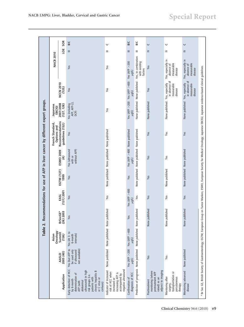
A summary of recommendations from representativeguidelines published on the use of AFP in HCC is pre-sented in Table 2. Table 2 also summarizes the currentNACB guidelines for the use of markers in this malig-nancy. Below, we present a more detailed discussion ofsome of the markers listed in Tables 1 and 2.
�-FETOPROTEIN
AFP is a 70-kD glycoprotein consisting of 591 aminoacids and 4% carbohydrate residues, encoded by a geneon chromosome 4q11-q13 [for reviews see (59, 60 )].Normally produced during gestation by the fetal liverand yolk sac, AFP is highly elevated in the circulation ofnewborns with concentrations decreasing during thenext 12 months to 10 –20 �g/L.
Analytical considerations: assay methods, standardiza-tion, and reference values. AFP is currently measured by2-site immunometric assays by using monoclonaland/or polyclonal antibodies, with results similar tothose of the RIAs that preceded them. Most commer-cial assays are calibrated against WHO InternationalStandard (IS) 72/225. Clinical results are reported in
mass units (�g/L) or in kiloUnits per liter of IS 72/225,for which 1 IU of AFP corresponds to 1.21 ng. Theupper reference limit used by most treatment centers is10 –15 �g/L (8.3–12.4 kU/L). AFP concentrations re-portedly increase with age, the upper reference limitincreasing from 11.3 �g/L in persons �40 years old to15.2 �g/L in those �40 years old (61 ). Ideally, refer-ence values should be established for each assay, be-cause there is some between-method variation inresults.
Analytical considerations: AFP carbohydrate microhet-erogeneity. AFP is a glycoprotein and contains 4% car-bohydrate as a single biantennary chain that isN-linked to asparagine-232 of the protein backbone(62, 63 ). The microheterogeneity of this carbohydratechain has been investigated extensively by use of bothlectin affinity electrophoresis (64 – 68 ) and isoelectricfocusing (69 –73 ). Distinct glycoform patterns charac-teristic of malignant or benign tissue have been found,raising the possibility of improving AFP specificity forHCC by measurement of an HCC-specific glycoform.
AFP glycoforms can be differentiated on the basisof their lectin-binding affinity (74 –76 ). AFP fromHCC patient sera, for example, binds more strongly toconcanavalin A than does AFP from nonseminoma-tous germ cell tumors, and both bind more strongly toLens culinaris lectin (LCA) than does AFP from pa-tients with benign liver disease. The affinity for LCA isslightly higher for AFP from HCC (AFP-L3) than thatfrom nonseminomatous germ cell tumors (AFP-L2).Assay kits are now available commercially that specifi-cally measure the AFP-L3 and AFP-P4 glycoforms(74, 76 ).
Numerous reported studies from Japan and otherAsian countries have demonstrated that an increase inthe AFP-L3 fraction of serum AFP correlates morestrongly than conventional serum AFP with adversehistological characteristics of HCC (e.g., greater portalvein invasion, more advanced tumor irrespective ofsize) and predicts unfavorable outcome (77– 81 ). In astudy comparing measurement of AFP-L3 and AFP in aUS referral population (166 patients with HCC, 77with chronic liver disease, and 29 with benign livermass), AFP-L3 concentrations were found to be rele-vant only at AFP concentrations between 10 and 200�g/L (82 ). Within this range, AFP-L3 exhibited sensi-tivity of 71% and specificity of 63% at a cutoff of 10%.At a cutoff of �35% sensitivity decreased to 33% butspecificity increased to 100%, enabling reliable diagno-sis of an additional 10% of HCC cases that would nothave been diagnosed using AFP alone at a cutoff of200 �g/L.
In a multicenter prospective 2-year longitudinalNorth American study, serum AFP was compared with
Special Report
e4 Clinical Chemistry 56:6 (2010)

Tabl
e1.
Curr
entl
yav
aila
ble
seru
man
dti
ssue
mar
kers
for
liver
canc
er.
Can
cer
mar
ker
Pro
po
sed
use
sPh
ase
of
dev
elo
pm
ent
LOE
Ref
eren
ce
Tiss
uem
arke
rs
GPC
3Di
ffere
ntia
ting
HCC
from
othe
rhe
patic
diso
rder
sat
the
tissu
ele
vel.
Und
ergo
ing
eval
uatio
n.V
(196
,197
)
GPC
3�
heat
shoc
kpr
otei
n70
�gl
utam
ine
synt
heta
se
Rais
edle
vels
of2
ofth
e3
mar
kers
indi
cate
ane
edfo
rbi
opsy
(acc
urac
y78
%at
100%
spec
ifici
ty).
Und
ergo
ing
eval
uatio
n.(5
11)
Telo
mer
ase
Inde
pend
ent
pred
ictio
nof
recu
rren
ceaf
ter
HCC
rese
ctio
n.U
nder
goin
gev
alua
tion.
V(5
12–5
15)
Prol
ifera
ting
cell
nucl
ear
antig
en–l
abel
ing
inde
xPr
edic
tion
ofre
curr
ence
and
surv
ival
insm
allH
CC.
Und
ergo
ing
eval
uatio
n.V
(516
)
Ki-6
7As
sess
men
tof
prog
nosi
saf
ter
rese
ctio
nof
HCC.
Und
ergo
ing
eval
uatio
n.V
(517
)
MIB
-1,E
-cad
herin
,�
-cat
enin
Prog
nost
icm
arke
rfo
rre
curr
ence
whe
nse
lect
ing
HCC
patie
nts
for
orth
otop
icliv
ertr
ansp
lant
atio
n.U
nder
goin
gev
alua
tion.
V(5
18)
Seru
mm
arke
rs
AFP
Scre
enin
gpa
tient
sat
high
risk
for
HCC,
espe
cial
lyth
ose
with
hepa
titis
B–an
dhe
patit
isC–
rela
ted
liver
cirr
hosi
s.In
clin
ical
use,
but
valu
eno
tva
lidat
edin
ahi
gh-le
vel
evid
ence
stud
y.III
(89,
90,9
9–10
4)
Inco
njun
ctio
nw
ithul
tras
ound
,dia
gnos
isof
HCC
inpa
tient
sat
high
risk
ofdi
seas
e.In
clin
ical
use,
but
valu
eno
tva
lidat
edin
ahi
gh-le
vel
evid
ence
stud
y.III
(30,
106–
115,
118–
120
)
Asse
ssin
gpr
ogno
sis
preo
pera
tivel
y.Va
lue
not
valid
ated
ina
high
-leve
levi
denc
est
udy.
III(3
2,15
4,16
6,17
0,17
9,51
9)
Mon
itorin
gHC
Cpa
tient
s,in
conj
unct
ion
with
ultr
asou
nd,t
ode
tect
early
recu
rren
ce.
Incl
inic
alus
e,bu
tva
lue
not
valid
ated
ina
high
-leve
lev
iden
cest
udy.
III(8
9,90
,99–
103,
179
)
Mon
itorin
gpa
tient
sw
ithno
evid
ence
ofdi
seas
eaf
ter
rese
ctio
nor
tran
spla
ntat
ion.
Incl
inic
alus
e,bu
tva
lue
not
valid
ated
ina
high
-leve
lev
iden
cest
udy.
IV(9
8,99
,101
,103
,168
)
Mon
itorin
gth
erap
yin
adva
nced
dise
ase.
Incl
inic
alus
e,bu
tva
lue
not
valid
ated
ina
high
-leve
lev
iden
cest
udy.
IV(1
72,1
74–1
78)
AFP–
conc
anav
alin
Abi
ndin
gDi
ffere
ntia
ting
sour
ceof
elev
ated
AFP
from
germ
cell
and
met
asta
ticliv
ertu
mor
s(h
igh)
from
HCC
(low
)(g
luco
sam
inyl
atio
nin
dex)
.
Not
inge
nera
lclin
ical
use,
but
effe
ctiv
ely
diffe
rent
iate
sAF
Pso
urce
asHC
Cor
GCT
.Not
valid
ated
ina
high
-le
vele
vide
nce
stud
y.
V(6
4–66
)
AFP–
LCA
bind
ing
Diffe
rent
iatin
gm
alig
nant
(hig
h)fro
mno
nmal
igna
nt(lo
w)
orig
inof
elev
ated
AFP,
inde
pend
ent
oflo
catio
n(fu
cosy
latio
nin
dex)
.
Not
inge
nera
lclin
ical
use,
but
effe
ctiv
efo
rAF
Pso
urce
orig
inon
susp
icio
nof
mal
igna
ntvs
beni
gnliv
erdi
seas
e.
V(6
6,52
0)
HCC-
spec
ific
AFP
band
onis
oele
ctric
focu
sing
(mon
osia
lyla
ted
AFP)
Earli
erde
tect
ion
ofHC
Cth
an“d
iagn
ostic
”AF
P(�
500
�g/
L),
posi
tive
pred
ictiv
eva
lue
73%
vs42
%,r
espe
ctiv
ely.
Not
incl
inic
alus
e.V
(69–
71)
Cont
inue
don
page
e6
NACB LMPG: Liver, Bladder, Cervical and Gastric Cancer Special Report
Clinical Chemistry 56:6 (2010) e5

Tabl
e1.
Curr
entl
yav
aila
ble
seru
man
dti
ssue
mar
kers
for
liver
canc
er.(
Cont
inue
dfr
ompa
gee5
)
Can
cer
mar
ker
Pro
po
sed
use
sPh
ase
of
dev
elo
pm
ent
LOE
Ref
eren
ce
AFP
lect
in-a
ffini
tysu
bgro
ups
(LCA
-rea
ctiv
eLC
A-L3
;er
ythr
oagg
lutin
atin
g-ph
ytoh
emag
glut
inin
-E4
reac
tive
AFP-
P4an
dP5
)
Pred
ictio
nof
mor
em
alig
nant
stag
ean
dpo
orou
tcom
e.AF
P-L3
isro
utin
ely
used
inJa
pan
whe
nAF
Pex
ceed
scu
toff
leve
l;AF
P-P4
ism
ore
sens
itive
,but
isno
tus
edro
utin
ely.
Inlim
ited
clin
ical
use
asa
com
mer
cial
lyav
aila
ble
test
ince
rtai
nco
untr
ies,
but
valu
eno
tva
lidat
edby
ahi
gh-le
vele
vide
nce
stud
y.
IV(6
7,68
,74,
75,7
7–85
,16
5,52
1)
Circ
ulat
ing
free
AFP-
IgM
com
plex
esPr
ovid
ing
info
rmat
ion
com
plem
enta
ryto
AFP.
Und
ergo
ing
eval
uatio
n.V
(522
)
DCP/
prot
hrom
bin
prod
uced
byvi
tam
inK
abse
nce
oran
tago
nism
II
Use
dw
ithAF
Pdu
ring
and
afte
rtr
eatm
ent
topr
edic
tad
vers
eou
tcom
e,ea
rlyre
curr
ence
,and
mal
igna
ntpo
tent
ial.
Fals
e-po
sitiv
ere
sults
may
occu
rin
patie
nts
with
seve
reob
stru
ctiv
eja
undi
ceor
vita
min
Kac
tion
impa
irmen
t(e
.g.,
patie
nts
onw
arfa
rinor
som
ean
tibio
tics)
.Thr
eeco
mm
erci
alas
says
with
diffe
ring
accu
racy
are
avai
labl
e.
Und
ergo
ing
eval
uatio
n.IV
(84,
85,1
73,1
81–1
90,
192–
194,
523
)
Solu
ble
NH2
fragm
ent
ofG
PC-3
,ahe
para
nsu
lfate
prot
eogl
ycan
Diag
nosi
san
dm
onito
ring
ofHC
Can
dci
rrho
sis.
Enab
les
dete
ctio
nof
smal
l-siz
eHC
Cm
ore
sens
itive
lyth
anAF
P.U
nder
goin
gev
alua
tion.
V(1
96,1
99)
Gol
gipr
otei
n73
Resi
dent
Gol
gigl
ycop
rote
in,f
ordi
agno
sis
ofea
rlyHC
C.U
nder
goin
gev
alua
tion.
V(5
24)
Iso-
�G
TPCo
mpl
emen
tary
toAF
Pas
adi
agno
stic
mar
ker
for
HCC.
Und
ergo
ing
eval
uatio
n.V
(525
,526
)
Ferr
itin
Mon
itorin
gHC
Cin
patie
nts
who
setu
mor
sdo
not
prod
uce
AFP.
No
high
-leve
levi
denc
eev
alua
tion.
V(5
27,5
28)
Varia
ntal
kalin
eph
osph
atas
eCo
mpl
emen
tary
toAF
P.U
nder
goin
gev
alua
tion.
V(5
29)
�1-
Antit
ryps
inCo
mpl
emen
tary
toAF
P.U
nder
goin
gev
alua
tion.
V(5
30,5
31)
�1-
Acid
glyc
opro
tein
Com
plem
enta
ryto
AFP.
Und
ergo
ing
eval
uatio
n.V
(532
)
Ost
eopo
ntin
Com
plem
enta
ryto
AFP.
Und
ergo
ing
eval
uatio
n.V
(533
)
Aldo
lase
ACo
mpl
emen
tary
toAF
P.U
nder
goin
gev
alua
tion.
V(5
34,5
35)
5�-N
ucle
otid
eph
osph
odie
ster
ase
Com
plem
enta
ryto
AFP.
Mon
itorin
gHC
Cin
patie
nts
who
setu
mor
sdo
not
prod
uce
AFP.
Und
ergo
ing
eval
uatio
n.V
(536
,537
)
CK18
,CK1
9,TP
A,TP
SCo
mpl
emen
tary
toAF
P.U
nder
goin
gev
alua
tion.
V(5
38,5
39)
Circ
ulat
ing
free
squa
mou
sce
llca
rcin
oma
antig
en–
IgM
com
plex
es
Com
plem
enta
ryto
AFP
indi
agno
sis
ofHC
C.U
nder
goin
gev
alua
tion.
V(5
40)
�-F
ucos
yl-t
rans
fera
seM
arke
rof
prog
ress
ion
ofHC
C.U
nder
goin
gev
alua
tion.
V(5
41)
�-L
-fuco
sida
seCo
mpl
emen
tary
toAF
P.U
nder
goin
gev
alua
tion.
V(5
42,5
43)
Cont
inue
don
page
e7
Special Report
e6 Clinical Chemistry 56:6 (2010)

Tabl
e1.
Curr
entl
yav
aila
ble
seru
man
dti
ssue
mar
kers
for
liver
canc
er.(
Cont
inue
dfr
ompa
gee6
)
Can
cer
mar
ker
Pro
po
sed
use
sPh
ase
of
dev
elo
pm
ent
LOE
Ref
eren
ce
Tran
sfor
min
ggr
owth
fact
or�
1Di
agno
sis
ofsm
allH
CCtu
mor
s.U
nder
goin
gev
alua
tion.
V(5
44)
Urin
ary
tran
sfor
min
ggr
owth
fact
or�
1Co
mpl
emen
tary
toAF
P.U
nder
goin
gev
alua
tion.
V(5
45)
Inte
rcel
lula
rce
llad
hesi
onm
olec
ule
1Pr
edic
tor
ofpr
ogno
sis
ofHC
C.U
nder
goin
gev
alua
tion.
V(5
46,5
47)
Anti-
p53
antib
ody
Com
plem
enta
ryto
AFP
indi
agno
sis
ofHC
C.U
nder
goin
gev
alua
tion.
V(5
48)
Inte
rleuk
in-8
Pred
icto
rof
prog
nosi
sof
HCC.
Und
ergo
ing
eval
uatio
n.V
(549
)
Inte
rleuk
in-6
Com
plem
enta
ryto
AFP
indi
agno
sis
ofHC
C,pr
edic
tor
ofHC
C.U
nder
goin
gev
alua
tion.
V(5
50,5
51)
Insu
lin-li
kegr
owth
fact
orII
Com
plem
enta
ryto
AFP.
Und
ergo
ing
eval
uatio
n.V
(552
)
Telo
mer
ase
orte
lom
eras
ere
vers
etr
ansc
ripta
sem
RNA
Diag
nosi
sof
HCC
and
pred
icto
rof
itsco
urse
ofHC
C(a
lso
assa
yed
inas
citic
fluid
).U
nder
goin
gev
alua
tion.
V(5
53,5
54)
Vasc
ular
endo
thel
ialg
row
thfa
ctor
Prog
nost
icm
arke
r.Pr
edic
tor
ofpo
orou
tcom
e.U
nder
goin
gev
alua
tion.
V(5
55)
Varia
ntw
ild-t
ype
estr
ogen
rece
ptor
Pred
icto
rof
unfa
vora
ble
prog
nosi
sin
HCC.
Und
ergo
ing
eval
uatio
n.V
(556
,557
)
Vita
min
B12–
bind
ing
prot
ein
Diag
nosi
sof
the
AFP-
nega
tive
fibro
lam
mel
lar
varia
ntof
HCC.
Und
ergo
ing
eval
uatio
n.V
(558
,559
)
Neu
rote
nsin
Diag
nosi
sof
the
AFP-
nega
tive
fibro
lam
mel
lar
varia
ntof
HCC.
Und
ergo
ing
eval
uatio
n.V
(560
)
Free
nucl
eic
acid
sEa
rlyde
tect
ion
and
mon
itorin
gof
HCC.
Und
ergo
ing
eval
uatio
n.V
(210
)
Circ
ulat
ing
cell-
free
seru
mDN
APr
edic
tive
mar
ker
for
dist
ant
met
asta
sis
ofhe
patit
isC
viru
s–re
late
dHC
C.U
nder
goin
gev
alua
tion.
V(5
61)
Epig
enet
icab
norm
aliti
essu
chas
p16
hype
rmet
hyla
tion
Early
dete
ctio
nof
HCC.
Und
ergo
ing
eval
uatio
n.V
(211
)
Prot
eom
ics
Early
dete
ctio
nan
dm
onito
ring
ofHC
C.U
nder
goin
gev
alua
tion.
V(2
08,2
09)
Tum
orce
llm
arke
rs
Circ
ulat
ing
tum
orce
llsin
perip
hera
lblo
odde
tect
edby
RT-P
CRof
AFP
mRN
A
Asse
ssm
ent
ofpr
ogno
sis
pre
and
post
oper
ativ
ely.
Pred
ictio
nof
early
recu
rren
cean
ddi
stan
tm
etas
tase
saf
ter
surg
ery.
Assi
stin
ther
apeu
ticde
cisi
ons.
Clin
ical
utili
tyis
cont
rove
rsia
l,an
dfin
ding
sof
publ
ishe
dst
udie
sar
ein
cons
iste
nt.
Und
ergo
ing
inve
stig
atio
n.IV
,V(2
00–2
04)
Cont
inue
don
page
e8
NACB LMPG: Liver, Bladder, Cervical and Gastric Cancer Special Report
Clinical Chemistry 56:6 (2010) e7

Tabl
e1.
Curr
entl
yav
aila
ble
seru
man
dti
ssue
mar
kers
for
liver
canc
er.(
Cont
inue
dfr
ompa
gee7
)
Can
cer
mar
ker
Pro
po
sed
use
sPh
ase
of
dev
elo
pm
ent
LOE
Ref
eren
ce
Plas
ma
prot
easo
me
Mar
ker
ofm
alig
nant
tran
sfor
mat
ion
inci
rrho
ticpa
tient
sin
clud
ing
thos
ew
ithlo
wtu
mor
mas
s.U
nder
goin
gev
alua
tion.
V(5
62)
Gen
etic
mar
kers
Plas
ma
glut
amat
eca
rbox
y-pe
ptid
ase,
phos
phol
ipas
esA2
G13
and
G7
and
othe
rcD
NA
mic
roar
ray-
deriv
eden
code
dpr
otei
ns.
Asse
ssm
ent
ofea
rlyHC
Cin
patie
nts
with
chro
nic
vira
lch
roni
che
patit
is;a
sses
smen
tof
met
asta
ticpo
tent
ialo
fHC
C.
Und
ergo
ing
eval
uatio
n.V
(215
,563
)
Mel
anom
aan
tigen
gene
1,3;
syno
vial
sarc
oma
onX
chro
mos
ome
1,2,
4,5;
sarc
opla
smic
calc
ium
-bi
ndin
gpr
otei
n1;
New
York
esop
hage
alsq
uam
ous
cell
carc
inom
a1
Com
plem
enta
ryto
AFP
inm
onito
ring
recu
rren
ce.C
andi
date
antig
ens
for
imm
unot
hera
py.
Und
ergo
ing
eval
uatio
n.V
(564
,565
)
Circ
ulat
ing
met
hyla
ted
DNA
(ras
asso
ciat
ion
dom
ain
fam
ily1A
)
Dete
ctio
nan
dqu
antif
icat
ion
ofci
rcul
atin
gm
ethy
late
dra
sas
soci
atio
ndo
mai
nfa
mily
1Aus
eful
for
HCC
scre
enin
g,de
tect
ion
and
prog
nosi
s.
Und
ergo
ing
eval
uatio
n.V
(566
)
Special Report
e8 Clinical Chemistry 56:6 (2010)
Tabl
e2.
Reco
mm
enda
tion
sfo
rus
eof
AFP
inliv
erca
ncer
bydi
ffer
ent
expe
rtgr
oups
.
Ap
plic
atio
nA
ASL
D20
05(4
0)
Asi
anO
nco
log
ySu
mm
it(1
36)
BrS
ocG
Ea
(26
)20
03EA
SL(1
31)
2001
EGTM
(137
)19
99ES
MO
2009
(4)
Fren
chSt
and
ard
,O
pti
on
san
dR
eco
mm
end
atio
ns
gu
idel
ines
(132
)
Jap
anes
eEB
ClG
l20
07/2
008
(127
,128
)N
CC
N20
10(1
35)
NA
CB
2010 LO
ESO
R
Early
dete
ctio
nof
HCC
by6-
mon
thde
term
inat
ion
ofAF
P(w
ithab
dom
inal
ultr
asou
nd)
inhi
ghris
kgr
oups
(i.e.
patie
nts
with
chro
nic
hepa
titis
Bor
Cvi
rus
orci
rrho
sis)
Yes
(but
AFP
tobe
used
only
iful
tras
ound
not
avai
labl
e)
Yes
(At
3-to
6-m
onth
inte
rval
s)
Yes
Yes
Yes
Yes
(ultr
asou
ndw
ithor
with
out
AFP)
Yes
Yes
(incl
udin
gAF
P,AF
P-L3
,DC
P)
Yes
Yes
IIIB/
C
Indi
cato
rof
incr
ease
dris
kof
HCC
whe
nin
crea
sed
orin
crea
sing
AFP
isac
com
pani
edby
nega
tive
ultr
asou
nd
Non
epu
blis
hed
Non
epu
blis
hed
Non
epu
blis
hed
Yes
Non
epu
blis
hed
Non
epu
blis
hed
Non
epu
blis
hed
Yes
Yes
Yes
IIIC
Conf
irmat
ion
ofdi
agno
sis
ofHC
CYe
s(A
FP�
200
�g/
L)Ye
s(A
FP�
400
�g/
L)Ye
sYe
s(A
FP�
400
�g/
L)Ye
sYe
s(A
FP�
400
�g/
L)N
one
publ
ishe
dYe
s(A
FP�
200
�g/
L)Ye
s(A
FP�
400
�g/
L)Ye
s(A
FP�
200
�g/
L)III
B/C
Pred
ictio
nof
prog
nosi
sN
one
publ
ishe
dN
one
publ
ishe
dN
one
publ
ishe
dYe
sN
one
publ
ishe
dN
one
publ
ishe
dN
one
publ
ishe
dN
one
publ
ishe
dN
one
publ
ishe
dYe
s,in
com
bina
tion
with
exis
ting
fact
ors
IIIB/
C
Post
trea
tmen
tm
onito
ring
(whe
repr
etre
atm
ent
AFP
rais
ed)
asan
adju
nct
toim
agin
g
Yes
Non
epu
blis
hed
Yes
Yes
Yes
Yes
Non
epu
blis
hed
Yes
Yes
IVC
Mon
itorin
gaf
ter
surg
ery,
tran
spla
ntat
ion
orpe
rcut
aneo
usth
erap
y
Yes
Non
epu
blis
hed
Yes
Non
epu
blis
hed
Yes
Yes
Non
epu
blis
hed
Yes,
espe
cial
lyin
abse
nce
ofm
easu
rabl
edi
seas
e
Yes,
espe
cial
lyin
abse
nce
ofm
easu
rabl
edi
seas
e
IVC
Mon
itorin
gad
vanc
eddi
seas
eN
one
publ
ishe
dN
one
publ
ishe
dYe
sN
one
publ
ishe
dN
one
publ
ishe
dYe
sN
one
publ
ishe
dYe
s,es
peci
ally
inab
senc
eof
mea
sura
ble
dise
ase
Yes,
espe
cial
lyin
abse
nce
ofm
easu
rabl
edi
seas
e
IVC
aBr
Soc
GE,
Briti
shSo
ciet
yof
Gas
troe
nter
olog
y;EG
TM,E
urop
ean
Gro
upon
Tum
orM
arke
rs;E
SMO
,Eur
opea
nSo
ciet
yfo
rM
edic
alO
ncol
ogy;
Japa
nese
EBCl
Gl,
Japa
nese
evid
ence
-bas
edcl
inic
algu
idel
ines
.
NACB LMPG: Liver, Bladder, Cervical and Gastric Cancer Special Report
Clinical Chemistry 56:6 (2010) e9
AFP-L3 and des-�-carboxy-prothrombin (DCP) (aninvestigational tumor marker for HCC) in 372 patientswith hepatitis C (83 ), including 40 initial HCC and 34HCC follow-up cases and 298 initially HCC-free cases(83 ). Sensitivity, specificity, and positive/negative pre-dictive values were, respectively, 61%, 71%, 34%, and88% for AFP (cutoff 20 �g/L) and 22%, 99%, 80%, and84% (cutoff 200 �g/L) compared with 37%, 92%, 52%,and 85% for AFP-L3 alone (cutoff 10%) and 39%,90%, 48%, and 86% for DCP alone (cutoff 7.5 �g/L)(83 ). When all 3 markers were combined, these figuresincreased to 77%, 59%, 32%, and 91%, respectively. Inpatients with raised AFP (20 –200 �g/L), high specific-ity was found for AFP-L3 and DCP (86.6% and 90.2%,respectively). Of 29 HCC patients with AFP values�20 �g/L, 13 had increased concentrations of AFP-L3or DCP. Compared with total AFP, AFP-L3 and DCPconcentrations within reference intervals correlatedmore strongly with an absence of HCC, with a higherspecificity and negative predictive value (83 ).
In a prospective study comparing AFP-L3 andDCP with AFP in 99 US patients with histologicallyconfirmed HCC, sensitivity rates were 62%, 73%,and 68%, respectively, with the highest sensitivity(86%) obtained when all 3 markers were combined(84 ). AFP-L3 was significantly related to portal veininvasion and patient outcome, suggesting it could bea useful prognostic marker for HCC (84 ). Use of thesame 3 markers to predict HCC recurrence after cur-ative percutaneous ablation has been investigated in416 HCC patients, 277 of whom had recurrence dur-ing the follow-up period (85 ). Pre- and postablationAFP �100 �g/L and AFP-L3 �15% were both sig-nificant predictors of recurrence and thus may com-plement imaging modalities in evaluating treatmentefficacy (85). A large and well-designed case-controlstudy comparing AFP, AFP-L3, and DCP has recentlybeen conducted in 7 academic medical centers in the US(86). The study cohort included 417 patients with cirrho-sis and 419 with HCC [77 with BCLC very early (BCLC 0)and 131 with early (BCLC A) stage disease]. ROC analysisrevealed that AFP had higher sensitivity (67%) than DCPor AFP-L3 for patients with BCLC 0 stage disease (86).Additional research is required to assess the value of AFPand related markers as surrogate endpoints for true healthoutcomes in clinical trials (87, 88).
AFP in screening and early detection. Cirrhotic patientswith AFP concentrations that are persistently elevatedare at increased risk of developing HCC compared withthose with AFP concentrations that fluctuate or remainwithin reference intervals (29% vs 13% vs 2.4%, re-spectively) (6 ). Lower serum AFP concentrations are
frequently encountered when HCC is detected duringscreening (89 ), and small HCC tumors are AFP nega-tive in up to 40% of cases (90 ). AFP immunostaining ofwell-differentiated small HCCs is often negative (91 ),rendering tissue AFP uninformative. In these in-stances, tumors may be detectable only by ultrasound(92 ). Malignant lesions undetectable by imaging arelikely to reach 2 cm in diameter in about 4 –12 months(93, 94 ). To detect tumors �2 cm in diameter, a sug-gested interval for surveillance in cirrhotic patients is6 months, with the use of both serum AFP and ultra-sound (95 ). Comparison of studies is often difficultowing to differences in study design. In addition, opin-ions differ as to how effectively AFP measurement con-tributes to programs for early detection or surveillance(96 ). Reliable markers are needed to complement ul-trasound, because the interpretation of ultrasound isoperator dependent and can be difficult to perform inpatients who are obese or have underlying cirrhosis(97 ).
In a systematic review of AFP test characteristicsfor diagnosis of HCC in HCV patients (98 ), only 5 of1239 studies met all the authors’ inclusion criteria (99 –103 ). In these 5 studies, with the use of an AFP cutoff of20 �g/L, sensitivity ranged from 41% to 65%, specific-ity from 80% to 94%, positive likelihood ratio from 3.1to 6.8, and negative likelihood ratio from 0.4 and 0.6,additional demonstrating the limited value of AFP as ascreening test. In 19 of 24 studies of patients with hep-atitis C published from 1985 to 2002, AFP sensitivitiesand specificities for HCC were 45%–100% and 70%–95%, respectively, at cutpoints between 10 and 19 �g/L(104 ). Ultrasound has been reported to have highersensitivity (71%) and specificity (93%) than serumAFP, but the positive predictive value of ultrasound islow, at about 14% (30 ). Because the success of ultra-sound detection is critically dependent on the skill ofthe ultrasonographer, investigation of patients with in-creases in serum AFP or suspicious screen-detectednodules is best performed in specialist referral centers.
The incidence of HCC in patients with chronichepatitis is lower than in patients with cirrhosis, whichmay decrease the benefit of screening in the former.Japanese studies suggest that differences in the naturalhistory of hepatitis B and C mean that hepatitis B pa-tients are more likely to develop HCC, even whenyoung and asymptomatic (105 ).
In one study, 1069 hepatitis B virus–infectedpatients with proven cirrhosis had to be screened todetect 14 cases of HCC, of which only 6 were at a suf-ficiently early stage to be amenable to surgical cure(106 ). The frequency of detection of curable malig-nancy was even lower in a study of 118 French patients
Special Report
e10 Clinical Chemistry 56:6 (2010)

with Child-Pugh A or B cirrhosis who were screened at6-month intervals with ultrasound, AFP, and DCP.Only 1 of 14 detected HCC cases (7%) was surgicallyresectable at the time of diagnosis (107 ). However,other studies have demonstrated benefit in screeningchronic hepatitis B carriers for HCC. A population-based Alaskan prospective screening study of 2230carriers with cirrhosis who were positive for hepatitis Bsurface antigen (108, 109 ) demonstrated that64%– 87% of detected HCCs were limited to single fociand that 43%–75% of tumors were �3 cm in size,which enabled curative surgery in 29%– 66% of the de-tected cancers (12, 110, 111 ). In another study, tumorsize was significantly reduced and survival improved(35% vs 10% at 30 months) when HCC was detected byscreening (112 ).
There is some evidence that screening high-riskpopulations for HCC can be cost-effective in high-prevalence regions such as Hong Kong (113 ) and thatscreening imparts a survival advantage, as demon-strated in an asymptomatic Asian Hawaiian popula-tion with chronic hepatitis B or C and cirrhosis (114 )and also in an Italian study of cirrhotic patients withscreen-detected HCC (115 ). These conclusions aresupported by results of a randomized, controlled trialof screening of 18 816 patients age 35–59 years re-cruited in urban Shanghai between 1993 and 1995 whohad hepatitis B infection or a history of chronic hepa-titis (116 ). Biannual screening with AFP and ultra-sound reduced HCC mortality by 37%. Although re-sults of a screening study of 5581 hepatitis B carriersbetween 1989 and 1995 in Qidong county demon-strated that screening with AFP resulted in earlier diag-nosis of liver cancer, the gain in lead time did not resultin any overall reduction in mortality (117 ). It seemslikely that this finding reflects differences in therapy inthe 2 studies, 75% of patients with subclinical HCCidentified in the Shanghai study having received radicaltreatment compared with only 25% in the Qidongstudy (116 ).
A national survey of practice in the US (118 ) hasdocumented that a majority of institutions routinelyscreen patients with cirrhosis for HCC, especially thosewith high-risk etiologies. Systematic screening withtwice yearly AFP and liver ultrasound is considered bymany to offer the best hope for early diagnosis of HCCin healthy carriers positive for hepatitis B surface anti-gen who have additional risk factors (e.g. active chronichepatitis, cirrhosis) and in patients with cirrhosis ofany etiology (119 ). Markov analysis has clearly demon-strated that in US patients with cirrhosis arising fromchronic hepatitis C, screening for HCC is as cost-effective as other accepted screening protocols (120 ).Biannual AFP and annual ultrasound gave the greatest
gain in terms of quality-adjusted life-years, while stillmaintaining a cost-effectiveness ratio of �$50 000/quality-adjusted life-year. The authors suggested thatbiannual AFP with annual CT screening might even becost-effective (120 ). Results of a later systematic reviewand economic analysis indicated that AFP measuredbiannually and ultrasound performed every 6 monthsprovide the most effective surveillance strategy in high-risk patients (121 ). Because of high costs, however, theauthors questioned whether ultrasound should be rou-tinely offered to those with serum AFP �20 �g/L, inview of the cost-benefit ratio, which depends on theetiology of cirrhosis.
These conclusions are generally supported by re-sults of a recent modeling study in which effectivenessand cost-effectiveness of surveillance for HCC wereevaluated in separate and mixed cohorts of individualswith cirrhosis due to alcoholic liver disease, hepatitis B,or hepatitis C (122 ). Algorithms including the use ofAFP and/or ultrasound at 6- and 12-month intervalswere compared. In the mixed cohort, the model foundthat AFP and ultrasound performed every 6 months tobe most effective, tripling the number of patients withoperable tumors at diagnosis and almost halving thenumber of deaths from HCC compared with no sur-veillance. Based on this report, the most cost-effectivestrategy would involve triage with 6-month AFP mea-surements. It was concluded that in the UK NationalHealth Service, surveillance of individuals with cirrho-sis at high risk for HCC should be considered to be botheffective and cost-effective (122 ).
Given the widespread use of AFP measurementsand liver ultrasound to screen prospectively for the on-set of HCC in cirrhotic patients, particularly thosewho are suitable candidates for curative therapy(109, 123, 124 ), there is an urgent need to establish andvalidate optimal follow-up protocols when suspiciousnodules are detected (10, 125, 126 ).
Recently published Japanese evidence-based clin-ical guidelines for diagnosis and treatment of HCC dif-ferentiate the risk of HCC in patients with cirrhosis asbeing super high (hepatitis B/C–related cirrhosis) orhigh (chronic hepatitis B/C or liver cirrhosis with acause other than hepatitis B/C) (127, 128 ). For the su-per high-risk group, ultrasound examination and mea-surements of AFP, DCP, and AFP-L3 are recom-mended at intervals of 3– 4 months, with a dynamic CTor MRI scan every 6 –12 months. For the high-riskgroup, ultrasound and tumor-marker measurementsare recommended every 6 months. Addition of DCP orAFP-L3 is considered necessary because these are diag-nostic markers whereas AFP is a marker of risk(129, 130 ). Detection of a nodular lesion by ultrasoundand/or a continuous rise in AFP (�200 �g/L), DCP [in
NACB LMPG: Liver, Bladder, Cervical and Gastric Cancer Special Report
Clinical Chemistry 56:6 (2010) e11
arbitrary units (AU) with 1 AU � 1 �g prothrombin](�40 mAU/mL), or AFP-L3 (�15%) requires furtherevaluation by dynamic CT or MRI (127, 128 ).
The European Association for the Study of theLiver (EASL) has recommended that nodules �1 cm indiameter be followed up with repeat ultrasound andAFP at 6 months, that fine-needle biopsy and histologybe added to investigate nodules of 1–2 cm (false-positive rate 30%– 40%), and that additional noninva-sive diagnostic criteria (e.g., 2 imaging techniques) beemployed for tumors �2 cm (131 ). French recom-mendations published in 2001 (132 ) state that the di-agnosis of HCC should be based on histopathologicalexamination of 1 or more liver samples obtained byopen surgery, laparoscopy, or ultrasound/CT-guidedbiopsy (standard) with the option of fine-needle aspi-ration for cytology if liver biopsy is impossible.
In a recent US retrospective study in which pa-tients with hepatic lesions suspicious for HCC under-went both fine-needle aspiration and core biopsy, re-sults were correlated with those from commonly usednoninvasive methods (133 ). Patients with positivebiopsy results had significantly higher serum AFP con-centrations than those with negative biopsy results, al-though the 2 groups were otherwise similar. Biopsy re-sults had greater sensitivity, specificity, and predictivevalue compared with noninvasive diagnostic criteria.The authors recommended an increased role forimage-guided biopsy of suspicion lesions �1 cm in sizeto allow adequate treatment planning, and commentedthat the risks of biopsy appear small and the potentialbenefits significant (133 ).
It is of course essential to be aware of the caveats ofuse of AFP, including the benign and malignant dis-eases that may cause raised serum AFP and the fact thata value within reference intervals never necessarily ex-cludes malignancy (99, 134 ). An elevated AFP detectedby a single measurement may be transient (e.g., arisingfrom an inflammatory flare of underlying chronic viralhepatitis), whereas elevated but stable concentrationsdecrease the likelihood that HCC is the causal agent.Sequential measurements of serum AFP may thereforeprovide useful information, but this is still under inves-tigation and not yet fully validated for routine clinicalpractice. A steadily rising pattern of elevated AFPshould always be rigorously investigated by using ultra-sound and other imaging techniques, which if initiallynegative should be repeated to identify any possibleoccult hepatic malignancy (131 ).
In 2003 the British Society of Gastroenterologypresented guidelines on the use of serial tumor markermeasurements to screen for HCC (26 ). The expertgroup concluded that in high-risk groups, screening byabdominal ultrasound and AFP compared to no sur-
veillance detected HCC of smaller size. Such detectionenables a greater proportion of curative therapies, withearlier detection leading to improved long-term sur-vival and/or cost savings. It was suggested that surveil-lance for HCC should be restricted to males and fe-males with cirrhosis due to hepatitis B or C virus orgenetic hemochromatosis and to males with cirrhosisdue to primary biliary cirrhosis and alcoholic cirrhosis(if abstinent or likely to comply with treatment). Thelikelihood of HCC arising in cirrhosis of other etiologywas considered to be low. Surveillance using AFP andabdominal ultrasound was recommended at 6-monthintervals, with appropriate equipment and skilled op-erators essential for the ultrasound component. Pa-tients should be counseled on the implications of earlydiagnosis and its lack of proven benefit (26 ).
These recommendations are in accord withNational Comprehensive Cancer Network (NCCN)guidelines (135 ), which recommend surveillance usingboth AFP and ultrasound in patients at risk for HCC(135 ). Those considered as being at risk include pa-tients with cirrhosis associated with hepatitis B or alco-hol, genetic hemochromatosis, autoimmune hepatitis,nonalcoholic steatohepatitis, primary biliary cirrhosis,or �1-antitrypsin deficiency. Surveillance is also rec-ommended for individuals without cirrhosis who arehepatitis B carriers or have other risk factors (e.g., ac-tive viral replication, high hepatitis B virus DNA con-centrations, family history of HCC, Asian males �40years old, females �50 years old, Africans �20 yearsold). The NCCN recommends additional imaging ifserum AFP is rising or after identification of a livermass nodule on ultrasound (135 ). The 2009 consensusstatement of the Asian Oncology Summit also recom-mends liver ultrasound and measurement of AFP con-centrations every 3– 6 months in all patients with livercirrhosis, regardless of etiology, with the caveat thatsuch surveillance is best established in hepatitis Bvirus–related liver cirrhosis, for which the LOE is rela-tively high (136 ). The AASLD currently recommendsuse of AFP for surveillance but only when ultrasound isnot available (40 ). This organization also states thatHCC screening should be “offered in the setting of aprogram or a process in which screening tests and recallprocedures have been standardized and in which qual-ity control procedures are in place” (40 ).
In accord with these and other recommenda-tions (26, 131, 132, 135, 137 ) (Table 2), the NACBsupports the use of determinations of AFP every6-months and abdominal ultrasound to screen pro-spectively for the onset of HCC in high-risk patients,especially those with liver cirrhosis related to hepa-titis B or C virus.
Special Report
e12 Clinical Chemistry 56:6 (2010)

NACB LIVER CANCER PANEL RECOMMENDATION 1:
AFP IN SCREENING PATIENTS AT HIGH RISK FOR HCC
AFP should be measured and abdominal ultra-sound performed at 6-month intervals in patientsat high risk of HCC, especially in those with livercirrhosis related to hepatitis B and hepatitis C virus.AFP concentrations that are �20 �g/L and increas-ing should prompt further investigation even if ul-trasound is negative [LOE, III/IV; Strength of Rec-ommendation (SOR), C].
AFP in diagnosis. Elevated serum AFP concentrationsare not specific for HCC because increased concentra-tions also occur in normal pregnancy, in certain benignliver diseases, and in some malignancies. Non-HCCmalignancies that may give rise to high AFP concentra-tions include nonseminomatous germ cell tumors, forwhich AFP is an important tumor marker with well-established clinical use (138 ). AFP may also be raised instomach cancer, biliary tract cancer, and pancreaticcancers (139 ). Elevated AFP concentrations exceeding1000 �g/L are, however, rare in these malignancies,occurring in �1% of cases.
Approximately 20%– 40% of adult patients withhepatitis or liver cirrhosis have raised AFP concentra-tions (�10 �g/L) (140 ). In these patients, an AFP con-centration between 400 and 500 �g/L was initiallygenerally accepted as the optimal decision pointto differentiate HCC from chronic liver disease(26, 136, 141–143 ). However, a Japanese study advo-cated an optimal cutoff of 150 �g/L based on ROCanalysis (sensitivity 54%, specificity 95.9%, comparingresults for patients with HCC and benign chronic liverdisease) (144 ). Using the same ROC technique, an Ital-ian group demonstrated the same specificity of 99.4%with cutoffs of 200 and 400 �g/L, but with higher sen-sitivity at the lower cutoff (99 ). The 2001 EASL guide-lines state that AFP �400 �g/L together with detectionof a suspicious liver node on imaging is diagnostic ofHCC (131 ). This guideline is in accord with recom-mendations of the Asian Oncology Summit panel,which concluded that a characteristic image on dy-namic CT or dynamic MRI, regardless of tumor size,will suffice for diagnosis of HCC, and obviate the needfor biopsy, with AFP �400 �g/L diagnostic in patientswith liver cirrhosis or chronic hepatitis (136 ). Thisgroup also recommended that needle biopsy beavoided when curative surgery is possible. Both theAASLD (40 ) and Japanese expert panels (131 ) statethat in patients with a suspicious liver node on imag-ing, AFP concentrations �200 �g/L are also suspiciousand should be investigated. After exclusion of hepaticinflammation, a sustained rise in AFP is suggestive of
HCC and should prompt further liver imaging studies,whereas stable or decreasing results make it less likely.
Circulating AFP concentrations in patients pre-senting with HCC range from within the reference in-terval to as high as 10 � 106 �g/L (i.e. 10 g/L), withpretreatment concentrations �1000 �g/L in approxi-mately 40% of patients (145 ). AFP has been reported tobe higher in patients with HCC arising from chronicviral conditions compared to those with alcoholic liverdisease (146 ) and in younger (147 ) and male (147 )patients. In one cohort study of 239 patients withchronic hepatitis, 277 with cirrhosis, and 95 with HCC,AFP gave sensitivities for HCC of 79% and 52.6% atdecision points of 20 �g/L and 200 �g/L, respectively,with corresponding specificities of 78% and 99.6%(148 ). According to some Japanese investigators (149 ),any circulating AFP value �10 �g/L in patients withchronic liver disease should be regarded as suspiciousof HCC and prompt further investigation, e.g., usingAFP-L3 (LCA) or AFP-P4 (E-PHA) lectin tests and im-aging. These investigators advocate a lower decisionpoint of 10 �g/L rather than 20 �g/L to take into ac-count the improvements in imaging that have led tomore HCC being detected when AFP is �20 �g/L. InJapan, for example, the percentage of HCC patientswith AFP concentrations �20 �g/L at presentation in-creased from 3.6% in 1978 to 38.1% in 2000. From2001 to 2003, after a change in AFP cutoff to �15 �g/L,36.4% of HCC patients had increased AFP concentra-tions (127 ). Introduction of a lower cutoff was sup-ported by a previous report that healthy Japanese indi-viduals do not have AFP concentrations �10 �g/L(150 ), but this finding may apply only to the popula-tion studied.
The Japanese guidelines state that HCC can bediagnosed by imaging (dynamic CT/MRI/contrast-enhanced ultrasound) or other techniques (hypervas-cularity in the arterial phase and wash-out in the portalvenous phase) (127, 128 ). Continuous increases inAFP (�200 �g/L) and/or DCP (�40 mAU/mL) and/orAFP-L3 (�15%) are highly suggestive of typical HCC,even in the absence of ultrasound evidence of an appar-ent liver nodule (127 ) and should prompt the use ofdynamic CT or MRI (128 ).
According to recent guidelines from the AASLD,surveillance/screening in patients at risk for HCCshould be performed by ultrasound scanning at inter-vals of 6 –12 months and AFP alone not be used unlessultrasound is not available (40 ), whereas the NCCNguidelines recommend periodic screening with ultra-sound and AFP every 6 –12 months (135 ). On ultra-sound detection of a nodule �1 cm, the AASLD panelrecommends follow-up by ultrasound at intervals of3– 6 months, reverting to routine surveillance if there isno growth after a period of up to 2 years (40 ). In con-
NACB LMPG: Liver, Bladder, Cervical and Gastric Cancer Special Report
Clinical Chemistry 56:6 (2010) e13
trast, the NCCN guidelines recommend imaging con-trol by CT/MRI/ultrasound every 3– 4 months for nod-ules �1 cm, reverting to routine surveillance if thenodule does not increase in size for 18 months (135 ).Nodules of 1–2 cm that are detected by ultrasound incirrhotic liver should be investigated by 2 dynamicstudies (e.g., CT, MRI) and treated as HCC if theirappearance is consistent with this diagnosis, but if notcharacteristic, the lesion should be biopsied.
For a nodule �2 cm at initial diagnosis with typicalHCC features (e.g., classic arterial enhancement ontriphasic CT or MRI) or cases in which AFP is �200�g/L, results can be considered diagnostic of HCC, andbiopsy unnecessary, but if the lesion is not characteris-tic, or the liver is noncirrhotic, biopsy is recommended.For small lesions that are negative on biopsy, ultra-sound or CT follow-up at 3- to 6-month intervals isrecommended, with repeat biopsy if the lesion enlargesbut remains atypical. Space-occupying lesions hy-poperfused by portal blood are considered an early signof HCC even in the absence of a coincident rise in cir-culating AFP.
The use of AFP as an adjunct in the diagnosis ofHCC is recommended by EASL(131 ), the British Soci-ety of Gastroenterology (26 ), the European Group onTumor Markers (137 ) and the NCCN (135 ). Theserecommendations are supported by the NACB Panel,which also stresses the importance of serial AFP mea-surements together with consideration of sustained in-creases in AFP even at low concentrations (Table 2).
NACB LIVER CANCER PANEL RECOMMENDATION 2:
AFP IN THE EARLY DETECTION OF HCC IN PATIENTS AT
HIGH RISK
In patients at risk for HCC, sustained increases inserum AFP may be used in conjunction with ultra-sound to aid early detection of HCC and guide fur-ther management. Ultrasound detected nodules�1 cm should be monitored at 3-month intervalswith ultrasound. Nodules of 1–2 cm in cirrhoticliver should be investigated by 2 imaging modalities(e.g., CT and MRI). If the appearance of the nod-ules is consistent with HCC, they should be treatedas such, with biopsy required if not. If lesions are�2 cm in size, AFP is �200 �g/L, and the ultra-sound appearance is typical of HCC, results may beconsidered diagnostic of HCC and biopsy is notnecessary (LOE, III; SOR, B).
AFP in prognosis. The TNM (tumor-node-metastasis)system (151 ) and the Okuda classification (152 ) are themost frequently used staging systems for HCC. Prog-nostic classifications from Japan (153 ), France (154 ),Italy (32, 155 ), Spain (156, 157 ), and China (158 ) have
also been published [see also (159, 160 )]. Of these, theSpanish BCLC staging system showed the best prog-nostic stratification (161 ) and was also adopted in theAASLD guidelines (40 ). Most of these systems includeas major prognostic factors severity of the underlyingliver disease, tumor size, tumor extension into adjacentstructures, and presence of metastases (152, 155 ). Ac-cording to AASLD guidelines (40 ), for optimal assess-ment of the prognosis of HCC patients, the staging sys-tem should include tumor stage, liver function, andphysical status and consider life expectancy, all ofwhich are included in the Spanish BCLC system.
The Chinese staging system (AFP cutoff 500 �g/L)(158 ) and 2 European staging systems include AFP.The French system includes the Karnofsky index, ultra-sonographic portal vein obstruction, and serum bili-rubin, alkaline phosphatase, and AFP (cutoff 35 �g/L)(154 ). Based on the score, patients are classified asbeing at low, moderate, or high risk for death, with1-year survival rates of 72%, 34%, and 7%, respec-tively. Another classification, proposed by the Cancerof the Liver Italian Program (155 ), includes Child-Pugh stage, morphology, portal vein thrombosis, andserum AFP (cutoff 400 �g/L). By use of a simple scor-ing system, patients are assigned to 1 of 7 categorieswith validated median survival rates (155 ). Both clas-sifications incorporate AFP as an indicator of tumorspread and burden, cellular differentiation, and aggres-sive potential. With the aim of improving available sys-tems for postoperative risk classification, a nomogrambased on clinicopathological variables including serumAFP, patient age, tumor size and margin status, post-operative blood loss, presence of satellite lesions, andvascular invasion has recently been developed (162 ).The nomogram reportedly enables accurate predictionof postoperative survival and risk stratification in pa-tients undergoing liver resection for HCC and is cur-rently undergoing evaluation (162 ).
It has been suggested that considering AFP andalkaline phosphatase, Child-Pugh score, and the ab-sence or presence of ascites could improve outcomeprediction (46, 154, 155 ). An Italian study of prognos-tic factors in 176 patients with HCC demonstrated thatlow albumin (�33 �g/L), high bilirubin (�22.5 �mol/L), elevated AFP (�32.5 kU/L), portal vein thrombo-sis, and an untreatable lesion were independent riskfactors for worse survival (163 ). Survival dependedmost strongly on the degree of functional liver impair-ment, presence of hepatitis B virus infection, type ofdiagnosis, and aggressiveness of the tumor. A more re-cent nationwide Japanese survey of prognostic factorsinfluencing survival after liver resection in HCC pa-tients demonstrated improvement in outcomes andoperative mortality rates over the last decade (164 ).Age, degree of liver damage, AFP concentration, max-
Special Report
e14 Clinical Chemistry 56:6 (2010)
imal tumor dimension, number of tumors, intrahe-patic extent of tumor, extrahepatic metastasis, portaland hepatic vein invasion, surgical curability, and freesurgical margins were all independent prognostic fac-tors for HCC patients undergoing liver resection (164 ).
Large studies using multivariate analyses confirmthat raised AFP concentrations predict poor progno-sis compared with AFP-negative cases in HCC(32, 154, 165 ). In a retrospective study of 309 HCC pa-tients stratified according to pretreatment AFP con-centrations (�20, 20 –399 or �400 �g/L), patientswith higher AFP concentrations tended to have largertumors, but there was no correlation with Okuda stage,degree of tumor differentiation, or extrahepatic metas-tasis(166 ). Incontrast,amorerecent largeItalianmulti-center survey that used the same 3 AFP groups in 1158HCC patients (167 ) revealed a low sensitivity (54%)for AFP in diagnosis of HCC, but confirmed its prog-nostic value by demonstrating its significant correla-tion with tumor size, lesion focality, TNM and Okudastage, Edmonson score, and survival (p � 0.0001) intreated as well as in untreated patients.
According to other authors (168, 169 ), AFP, aswell as tumor size, seems to be an independent predic-tor of survival. Survival of patients with serum AFP�10 000 �g/L at diagnosis was significantly shorterthan in those with AFP �200 �g/L (median survivaltime 7.6 vs 33.9 months, respectively) (170 ). AFP con-centrations �1000 �g/L predict a relatively worseprognosis, even after attempted curative resection(70 ). Serum AFP concentrations �12 000 �g/L are re-quired to meet UK criteria for liver transplantation(171 ).
AFP doubling time has also been reported to be animportant prognostic factor (172 ). Persistence of apositive AFP-L3 fraction after intervention also hasbeen reported to indicate residual or recurrent disease(77 ). The NACB supports the prognostic use of pre-treatment serum AFP concentration in combinationwith other prognostic factors (Table 2).
NACB LIVER CANCER PANEL RECOMMENDATION 3:
AFP FOR DETERMINING PROGNOSIS
In combination with other prognostic factors, AFPconcentrations may provide prognostic informa-tion in untreated HCC patients and in those under-going liver resection, with high concentrations in-dicating poor prognosis (LOE, IV; SOR, C).
AFP in monitoring patients after treatment. For patientswith increased AFP concentrations before therapy,monitoring treatment of HCC by use of serial AFPdeterminations is a well-accepted procedure. Aftercomplete removal of the tumor, AFP concentrations
typically decrease, with a half-life of 3.5– 4 days. In-complete resection yields a longer half-life, which isassociated with poorer survival (166, 172 ), whereasfailure of the AFP to normalize implies residual malig-nancy or severe liver damage. [Determination of theAFP-L3 fraction can help to differentiate these 2 con-ditions (81, 142, 173 ).] However, normalization ofAFP does not necessarily indicate complete clearanceof the disease. Recurrence after transplantation mayoccur, even when AFP is stable and within referencelimits (168, 172, 174 ), presumably reflecting the pres-ence of micrometastases too small to produce measur-able serum concentrations.
Changes in AFP concentrations also reflect tumorresponse after chemotherapy, with longer survival inpatients showing a significantly prolonged decrease inAFP than in those with slowly increasing concentra-tions (175, 176 ). In patients receiving new and effectivecombined systemic therapies (177 ), 75% have showndramatic decreases in serum AFP, with concentrationsnormalizing completely in some patients. Progressivedisease was found in patients with continued AFP in-crease and doubling times between 6.5 and 112 days(mean 41 days), again correlating with survival (172 ).Similar results were observed after radiotherapy forprimary and secondary liver tumors. Decreases in tu-mor markers reflected tumor regression more consis-tently than later changes in tumor size and volume asdetermined by CT (178 ). Discrepancies between tu-mor marker and imaging results may be due to residualfibrosis and other factors that can complicate interpre-tation of CT scans (178 ).
A recent phase III randomized trial of systemicchemotherapy in HCC patients evaluated clinical andradiological outcome and included prospectively col-lected serial AFP measurements (179 ). In 117 patientswith initially elevated serum AFP (cutoff 20 �g/L) andan AFP response (�20% decrease) after the second cy-cle of chemotherapy, 47 had improved survival com-pared with 70 AFP nonresponders (13.5 vs 5.6 months;P � 0.0001). AFP concentrations were strongly associ-ated with radiological response (P � 0.0001) and alsowith survival (multivariate analysis: hazard ratio 0.413,P � 0.0001). It was therefore concluded that in HCCpatients undergoing systemic chemotherapy, serialAFP determinations may be useful both for prognosisand for monitoring treatment response, as well as pro-viding a surrogate marker for the evaluation of newtherapeutic agents (179 ). Similarly, authors of a recentstudy from Massachusetts General Hospital CancerCenter and Harvard Medical School concluded thatserum AFP change during treatment may serve as auseful surrogate marker for clinical outcome in pa-tients with advanced HCC receiving systemic therapy(180 ).
NACB LMPG: Liver, Bladder, Cervical and Gastric Cancer Special Report
Clinical Chemistry 56:6 (2010) e15

According to the French Standard, Options andRecommendations guidelines (132 ), there is no con-sensus about patterns or modalities of follow-up otherthan clinical examination and surveillance plans thatmay incorporate ultrasound, AFP measurement, ab-dominal CT scan, chest x-ray, and/or MRI, with opti-mal choice and timing of these dependent on treatmentoptions. The NCCN is more specific, recommendingposttreatment follow-up of HCC patients that includesimaging every 3 to 6 months for 2 years and then an-nually, with AFP (if initially elevated) measured every3 months for 2 years, and then every 6 months (135 ).Similarly, ESMO recommends that patients under-going curative resection should be followed up withliver imaging and AFP measurement for 2 years at 3- to6-month intervals, and then annually, because curativetherapy can be offered to a minority of patients after re-lapse (4). After liver transplantation, follow-up should bemore frequent, i.e., monthly for 6 months, then once ev-ery 3 months up to 1 year posttransplantation, then twicea year up to 2 years, and annually thereafter (4).
In accord with other expert groups (131, 132,135 ), the NACB recommends serial determinations ofserum AFP (if elevated before treatment) to monitorefficacy of treatment, course of disease, and recurrence,and supports the frequency of measurement recom-mended by the NCCN (135 ).
NACB LIVER CANCER PANEL RECOMMENDATION 4:
AFP IN MONITORING TREATMENT
Measurement of AFP at follow-up visits is recom-mended to monitor disease status after liver resec-tion or liver transplantation for detection of recur-rence or after ablative therapies and application ofpalliative treatment. Although monitoring inter-vals are as yet undefined, current practice suggestsfollowing patients every 3 months for 2 years andthen every 6 months (LOE, IV; SOR, C).
Tumor markers other than AFP. Des-�-carboxy-pro-thrombin. DCP, also known as PIVKA II (prothrombinproduced by vitamin K absence or antagonism II), is anabnormal prothrombin devoid of coagulation activityand is potentially a marker for HCC. Mainly developedand investigated in Japan, DCP was first described inthe US in 1984 (181 ) and critically reviewed there in1993 (182 ). A single commercially available EIA kitfrom Japan has dominated the market for DCP testing.The sensitivity of this method has been markedly im-proved since 1996 and is currently 10 AU/L.
A number of published investigations have reportedDCP sensitivities for the diagnosis of HCC ranging from54% to 70% at a decision point of 40 AU/L, with corre-sponding specificities in cirrhotic patients between 87%
and 95%. AFP tested concurrently in the same patientshas shown, at a decision point of 20 �g/L, 47%–72% sen-sitivity and 72%–86% specificity. Combined DCP/AFPsensitivity was about 80% (183–186). DCP, AFP, andcombined DCP/AFP sensitivities for solitary HCC(�2 cm) were 30%–53%, 13%, and 57%, respectively,and for larger tumors (�3 cm) were 78%–81%, 49%–69%, and 84%–94%, respectively, (183, 184, 186). Thesensitivity of both markers was better for moderately topoorly differentiated tumors (DCP, 68%; AFP, 61%;DCP/AFP, 85%; n � 41) than for well-differentiated tu-mors (DCP, 13%; AFP, 33%; DCP/AFP, 40%; n � 15)(186). Both DCP and AFP concentrations correlated withtumor size and grading, but not significantly with eachother.
A cross-sectional case control study that comparedserum AFP and DCP in a US population has confirmedthe apparent superiority of DCP as a tumor marker forHCC (187 ). The study included 48 healthy adults, 51patients with chronic hepatitis (mostly hepatitis C), 53individuals with compensated cirrhosis, and 55 peoplewith proven HCC. With the use of ROC analysis, DCPwas found to perform better than AFP in differentiatingHCC from cirrhosis (sensitivity 90% vs 77%, specificity91% vs 71%, positive predictive value 85% vs 81%, nega-tive predictive value 90% vs 74%, area under the ROCcurve 0.921 vs 0.815). There was no improvement overDCP alone when the 2 markers were combined.
DCP has also been reported to have prognosticsignificance. In a study of HCC patients treated by per-cutaneous ethanol injection or microwave coagulationtherapy, multivariate analysis showed that after histo-logical grade and tumor differentiation, DCP was thestrongest predisposing factor for later development ofportal venous invasion (188 ), whereas ROC analysisresults suggested it was an effective predictor of HCCrecurrence after resection (189 ). In another study 237HCC patients were categorized into 4 groups accordingto concentrations of DCP (less than or greater than62.5 AU/L) and AFP (less than or greater than100 �g/L) (190 ). The 22 patients with low AFP andhigh DCP were predominantly male and had large le-sions but few nodules. Outcome was particularly poorin patients who had high concentrations of both DCPand AFP (190 ). According to a more recent reportcomparing serum AFP and DCP determinations in1377 HCC and 355 chronic liver disease patients, theutility of DCP was lower in smaller tumors (�3 cmdiameter) than in larger ones (�5 cm diameter) (191 ).
A retrospective analysis of 199 HCC patients withearly stage HCC in Child Pugh A cirrhotic patientstreated by resection or radiofrequency ablation (RFA)showed similar 3- and 5-year survival rates (90%/79% vs 87%/75%) (192 ). One- and 3-year tumorrecurrence-free survival rates were higher in the pa-
Special Report
e16 Clinical Chemistry 56:6 (2010)

tients treated by resection (83%/51% vs 83%/42% forRFA; P � 0.011) (192 ). With multivariate analysis,prothrombin time �80% was found to be an inde-pendent prognostic factor for the resected groupwhereas platelet count �100 000 and DCP concentra-tion �100 AU/L were prognostic for the RFA group. AtDCP concentrations �100 AU/L the treatment proce-dure became a significant prognostic factor for sur-vival. These results suggest that a high DCP concentra-tion reflects biological aggressiveness and that surgicalresection rather than RFA treatment is advantageous inthese patients. The prognostic value of pretreatmentconcentrations of AFP (cutoff 400 �g/L), AFP-L3 (cut-off 15%), and DCP (cutoff 100 AU/L) has been inves-tigated in HCC patients after curative treatment byhepatectomy (n � 345) and compared to locoregionalthermal ablation (n � 456) (173 ). Multivariate analysisresults in hepatectomy patients indicated that no tu-mor marker was associated with decreased survival. Inpatients who had undergone locoregional thermal ab-lation, elevation of AFP-L3 (P � 0.0171) or DCP (P �0.0004) was significantly associated with decreased sur-vival and DCP was also associated with increased rateof recurrence (P � 0.0001).
An investigation of AFP, AFP-L3, and DCP in 240patients with hepatitis B or C (144 HCC, 47 chronichepatitis, and 49 cirrhotic cases) at optimal cutoffs ac-cording to ROC analysis (DCP, 84 AU/L; AFP, 25 �g/L;AFP-L3, 10%) yielded sensitivity, specificity, and positivepredictive value rates of 87%, 85%, and 86.8% for DCP;69%, 87%, and 69.8% for AFP; and 56%, 90%, and 56.1%for AFP-L3 (193). DCP concentrations were below cutoffin all non-HCC cases but increased in all HCC cases in-cluding those with single lesions. DCP correlated with tu-mor size, high AFP concentrations with diffuse type HCC,and all 3 markers with metastatic HCC. The authors rec-ommended routine use of DCP for HCC detection.
False-positive elevated DCP concentrations arefound in patients with severe obstructive jaundice dueto intrahepatic cholestasis or in conditions in which theaction of vitamin K is impaired (e.g., in individualswith longstanding vitamin K deficiency and those whohave ingested warfarin and some wide-spectrum antibi-otics) (194). Despite these limitations, DCP is a promis-ing emerging marker with considerable potential.
Glypican-3. Glypican-3 (GPC-3), initially termedMXR7 (195 ), is another promising new tissue and se-rum marker for HCC. The gene glypican 3 (GPC3)20
codes for a member of the glypican family of glycosyl-
phosphatidylinositol–anchored cell-surface heparan sul-fate proteoglycans (196 ). GPC-3 was first detected viaits mRNA, which was increased in 75% of tissue sam-ples from patients with primary and recurrent HCCbut in only 3.2% of samples from normal liver tissue(195 ). These data were later confirmed immunohisto-chemically (196, 197 ). Elevated GPC-3 mRNA con-centrations were also found in the serum of HCC pa-tients (195 ). Sensitivity exceeded that of AFP (88% vs55%) for the entire group of HCC patients tested aswell as for those with smaller HCC tumors �3 cm(77% vs 43%). In a later study of 34 HCC patients(196 ), sensitivity was somewhat lower (53%) and sim-ilar to that of AFP (54%). However, specificity was ex-cellent, with no significant elevations in healthy sampledonors or patients with acute hepatitis, and in only 1 ofthe 20 patients with chronic hepatitis and cirrhosis.The combined sensitivity of the 2 markers was 82%.Neither marker correlated with the other.
Although another group has demonstrated thepresence of the C-terminus in serum (198 ), a recentreport on the GPC protein suggests that the only frag-ment present in the circulation is the amino terminal,which constitutes the GPC-3 soluble serologicalmarker (sGPC-3) (199 ). With the use of an ELISA withhighly specific monoclonal antibodies to analyze serafrom 69 HCC patients, 38 liver cirrhosis patients, and96 healthy adults, ROC analysis yielded sensitivity/specificity rates of 51%/90% for sGPC-3 (cutoff 2 �g/L)comparable to those of AFP [55%/90% (cutoff 20 �g/L]. The sensitivity of the 2 markers in a subset of earlystage HCC was essentially unchanged, and there was nocorrelation between sGPC-3 and AFP in the 69 patientswho had HCC. The combined marker sensitivity was72%. This preliminary study suggests that sGPC-3 mayhave some promise and that larger clinical trials to in-vestigate its potential are merited.
Other serum markers for liver cancer. Many other serummarkers have been reported for HCC (Table 1). Pre-and posttreatment detection of circulating HCC cellsby reverse transcription (RT)-PCR of AFP mRNA hasbeen suggested by some groups to be useful in predict-ing HCC recurrence and poor outcome (200, 201 ), al-though other investigators have questioned its value(202–204 ). Other techniques under investigation in-clude genetic profiling, transcriptomics (205–207 ),proteome analysis (208, 209 ), and determination offree nucleic acids (210 ) and epigenetic abnormalities(e.g., p16 hypermethylation) in serum or plasma (211 ).Also being explored are the prognostic implications ofCpG-island hypermethylation and DNA hypomethy-lation (212 ), microRNA profiling (213 ) and explora-tion of liver cancer stem cells (214 ). Fifty upregulatedHCC marker genes, which are potential tumor marker
20 Genes: GPC3, glypican 3; FGFR3, fibroblast growth factor receptor 3; CGB, cho-rionic gonadotropin, beta polypeptide (hCG�); PMF1, polyamine-modulatedfactor 1; TP53, tumor protein p53.
NACB LMPG: Liver, Bladder, Cervical and Gastric Cancer Special Report
Clinical Chemistry 56:6 (2010) e17

candidates, have been identified in hepatitis C virus–associated HCC by use of cDNA microarray analysis ofsurgical liver samples from patients infected with hep-atitis C virus (215 ).
The NACB panel does not recommend the use ofany HCC-related biomarkers except AFP for the rou-tine surveillance of patients with or at risk of HCC. TheNACB does, however, support further evaluation ofthe clinical utility of potential markers for which thereis increasing published evidence (e.g., AFP-L3, DCP,and GPC-3) in suitably designed prospective random-ized clinical studies.
NACB LIVER CANCER PANEL RECOMMENDATION 5:
TUMOR MARKERS OTHER THAN AFP
AFP is currently the only marker that can be recom-mended for clinical use in liver malignancies. Newliver cancer markers offer promise but their contri-bution to the current standard of care is unknownand further investigations in properly designedclinical trials are needed (LOE, not applicable; SOR,C).
KEY POINTS: TUMOR MARKERS IN HCC
HCC is one of the most common cancers worldwide,and is frequently preceded by chronic viral hepatitis Bor C or alcoholic liver disease. If treatment of thesediseases is instituted early, the risk of HCC can be de-creased or abolished. In patients who have already de-veloped HCC, surgical resection or transplantationwith curative intent requires early local detection ofsmall lesions. The clinical utility of AFP measurement,together with ultrasound and other more sensitive im-aging techniques, is already well established for this ap-plication, whereas other tumor markers require furtherinvestigation. Future developments in molecular ge-netics and proteomic analysis may lead to earlier diag-nosis and more effective treatment of HCC patients.
Tumor Markers in Bladder Cancer21,22
BACKGROUND
Each year in the US, nearly 71 000 new cases of bladdercancer are diagnosed and approximately 14 000 peopledie from this disease (216 ). The prevalence of bladdercancer in the US is estimated at almost 500 000 cases.Almost twice as many cases of bladder cancer occur inmen than in women, with cigarette smoking the lead-
ing cause (217 ). Other risk factors include exposure toindustrial carcinogens and chronic infection withSchistosomiasis haematobium.
The most common symptom of bladder cancer isintermittent hematuria (80%– 85% of patients). Otherurinary tract symptoms include increased frequency,urgency, and dysuria (15%–20% of patients). The di-agnosis is usually established by cystoscopic evaluation,prompted by hematuria or urinary tract symptoms, andbiopsy. In some cases, urine cytology is positive for tumorcells. Bladder cancer is staged according to the degree oftumor invasion into the bladder wall (218). Carcinoma insitu (stage Tis) and stages Ta and T1 are grouped as non-muscle invasive bladder cancers because they are re-stricted to the inner epithelial lining of the bladder and donot involve the muscle wall. Of the nonmuscle invasivetumors, stage Ta tumors are confined to the mucosa,whereas stage T1 tumors invade the lamina propria. T1tumors are regarded as being more aggressive than Tatumors (219). Muscle invasive tumors (stages T2, T3, andT4) extend into the muscle (stage T2), the perivesical fatlayer beyond the muscle (stage T3), and adjacent organs(T4). Metastatic tumors involve lymph nodes (N1–3) ordistant organs (M1).
The most common cell type of bladder cancer istransitional cell carcinoma, although adenocarcino-mas, squamous cell carcinomas, and sarcomas also oc-cur. The cellular morphology of nonmuscle invasivebladder tumors is graded according to the degree ofcellular differentiation. The grading consists of well-differentiated (grade 1), moderately differentiated(grade 2), and poorly differentiated (grade 3) tumors.Grading of cell morphology is important for establish-ing prognosis, because grade 3 tumors are the mostaggressive and the most likely to become invasive. Useof the WHO classification from 2004 is widely advo-cated, because it facilitates uniform diagnosis of tu-mors (220 ). A modified grading system (WHO Inter-national Society of Urological Pathology 1998), whichis increasingly being used (221 ), eliminates the numer-ical grades and categorizes most bladder cancers as ei-ther low grade or high grade.
The heterogeneity of urological tumors—in termsof both histological origin and clinical behavior(222 )—means that clinical parameters such as tumorgrade and stage are not sufficiently accurate to predictbiological behavior or to guide treatment reliably, es-pecially in high-risk cases (223–225 ). New markers toaid diagnosis, assess prognosis, identify optimal treat-ment, and monitor progression of urological cancersare urgently required.
Bladder cancer may be regarded as a genetic dis-ease caused by the multistep accumulation of geneticand epigenetic factors (226 –228 ). Nonmuscle invasivebladder tumors are generally treated by transurethral
21 NACB Bladder Cancer Sub-Committee Members: Herbert A. Fritsche (Chair),Thorsten H. Ecke, H. Barton Grossman, Seth P. Lerner, Ihor Sawczuk.
22 All comments received about the NACB Recommendations for Bladder Cancerare included in the online Data Supplement.
Special Report
e18 Clinical Chemistry 56:6 (2010)

resection of the bladder with or without intravesicaltreatments with bacille Calmette-Guerin immunother-apy or intravesical chemotherapy. Muscle invasive tu-mors are usually treated by cystectomy, or withbladder-sparing therapies that consist of chemother-apy and radiation. Patients who have metastatic diseaserequire systemic chemotherapy with multiple antican-cer agents (229 ). A thorough understanding of cancerprogression pathways facilitates development of drugtherapies against specific tumor targets (225 ).
The majority of bladder cancer patients are diag-nosed with nonmuscle invasive tumors. Even thoughthese tumors can be completely resected, there is a highrisk of recurrence; 50%–70% of these patients will de-velop tumor recurrence within 5 years. With intensivemedical surveillance, the 5-year survival rates for thesepatients range from 95% to 75% for Ta and T1 tumors,respectively. However, almost 25% of patients with Taand T1 noninvasive tumors will eventually develop in-vasive disease. The 5-year survival rate decreases withtumor invasiveness and the presence of metastasis. Pa-tients with stage T2 tumors have a 5-year survival rateof 60%, but only 35% of patients with stage T3 tumorsand 10% of patients with stage T4 metastatic tumorssurvive 5 years (218 ).
Lifelong surveillance is therefore required forbladder cancer patients who are initially diagnosedwith nonmuscle invasive disease. Current patient-monitoring protocols generally consist of regularlyscheduled cystoscopic evaluations, usually togetherwith urine cytology, performed every 3 months duringthe first 2 years of follow-up, twice a year during years 3and 4, and annually thereafter, until disease recurrenceis documented (230 ).
Urine tumor markers have been proposed for useas diagnostic aids in patients who present with hema-turia, as prognostic indicators of disease recurrenceand survival, and as early detectors of recurrent diseasein monitored patients. Potential applications of urinetumor marker tests in patient surveillance include se-rial tests for earlier detection of recurrent disease, ad-juncts to urine cytology to improve the detection ofdisease recurrence, less expensive and more objectivealternatives to urine cytology, and indicators to directthe frequency of cystoscopy evaluation in the follow-upof patients with bladder cancer.
To prepare these guidelines, we reviewed the liter-ature relevant to the use of tumor markers in bladdercancer. Particular attention was given to reviews, in-cluding systematic reviews, prospective randomizedtrials that included the use of markers, and guidelinesissued by expert panels. Where possible, the consensusrecommendations of the NACB panel were based onavailable evidence, i.e., were evidence based.
CURRENTLY AVAILABLE TUMOR MARKERS FOR BLADDER CANCER
Currently available bladder cancer tumor markers andsome of those in development are listed in Table 3, withan assessment of each marker and the LOE for its clin-ical use. The LOE grading system (58 ) and SOR (231 )have been applied as previously described (2 ) [SOR(231 ), A � high (further research is very unlikely tochange the panel’s confidence in the estimate of effect);B � moderate (further research is likely to have animportant impact on the panel’s confidence in the es-timate of effect and is likely to change the estimate);C � low (further research is very likely to have an im-portant effect on the panel’s confidence in the estimateof effect and is likely to change the estimate); D � verylow (any estimate of effect is very uncertain)]. As indi-cated in Table 3, 6 tumor marker tests, all of which aremeasured in urine, have been cleared by the US Foodand Drug Administration (FDA) for use in routine pa-tient care.
URINE TUMOR MARKERS IN BLADDER CANCER: NACB
RECOMMENDATIONS
At this time, and in accord with NCCN practice guide-lines for bladder cancer (232 ), no tumor marker testscan be recommended for use in the routine diagnosisand clinical management of bladder cancer. This in-cludes tests for making a differential diagnosis, assess-ing prognosis, staging the disease, and monitoring pa-tients for the early detection of recurrent disease. Thereare no prospective clinical trial data that establish theutility of any of the FDA-cleared markers or the pro-posed markers for increasing survival time, decreasingthe cost of treatment, or improving the quality of life ofbladder cancer patients. In the following report, we de-scribe the FDA-cleared markers and the variety ofnewly proposed markers.
FDA-CLEARED MARKERS FOR BLADDER CANCER
BTA-Stat and Trak tests for complement factor H andrelated proteins. The BTA-Stat test (Polymedco) detectscomplement factor H (CFH) and CFH-related proteins inurine (233). Factor H, a 155-kDa protein, has a centralrole in regulating the alternate pathway of complementactivation to prevent complement-mediated damage tohealthy cells. At least 4 other factor H–related proteinshave been identified as products of a cluster of genes onchromosome 1 called the regulators of complement acti-vation locus, and although some of these proteins possesscomplement regulatory activity, others do not (233).
The BTA-Stat test provides semiquantitative de-tection of CFH and the CFH-related protein antigensby use of a double monoclonal antibody, immuno-chromatographic point-of-care device. For both non-invasive (Tis, Ta, T1) and invasive (T2–T4) tumors, the
NACB LMPG: Liver, Bladder, Cervical and Gastric Cancer Special Report
Clinical Chemistry 56:6 (2010) e19
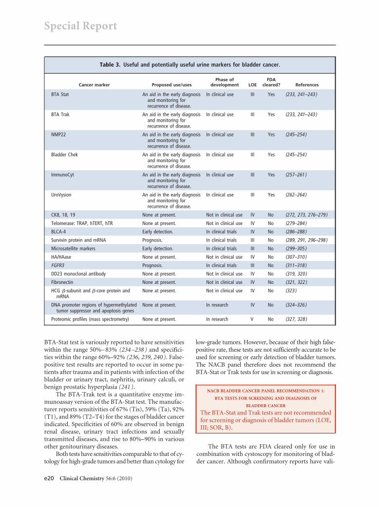
BTA-Stat test is variously reported to have sensitivitieswithin the range 50%– 83% (234 –238 ) and specifici-ties within the range 60%–92% (236, 239, 240 ). False-positive test results are reported to occur in some pa-tients after trauma and in patients with infection of thebladder or urinary tract, nephritis, urinary calculi, orbenign prostatic hyperplasia (241 ).
The BTA-Trak test is a quantitative enzyme im-munoassay version of the BTA-Stat test. The manufac-turer reports sensitivities of 67% (Tis), 59% (Ta), 92%(T1), and 89% (T2–T4) for the stages of bladder cancerindicated. Specificities of 60% are observed in benignrenal disease, urinary tract infections and sexuallytransmitted diseases, and rise to 80%–90% in variousother genitourinary diseases.
Both tests have sensitivities comparable to that of cy-tology for high-grade tumors and better than cytology for
low-grade tumors. However, because of their high false-positive rate, these tests are not sufficiently accurate to beused for screening or early detection of bladder tumors.The NACB panel therefore does not recommend theBTA-Stat or Trak tests for use in screening or diagnosis.
NACB BLADDER CANCER PANEL RECOMMENDATION 1:
BTA TESTS FOR SCREENING AND DIAGNOSIS OF
BLADDER CANCER
The BTA-Stat and Trak tests are not recommendedfor screening or diagnosis of bladder tumors (LOE,III; SOR, B).
The BTA tests are FDA cleared only for use incombination with cystoscopy for monitoring of blad-der cancer. Although confirmatory reports have vali-
Table 3. Useful and potentially useful urine markers for bladder cancer.
Cancer marker Proposed use/usesPhase of
development LOEFDA
cleared? References
BTA Stat An aid in the early diagnosisand monitoring forrecurrence of disease.
In clinical use III Yes (233, 241–243 )
BTA Trak An aid in the early diagnosisand monitoring forrecurrence of disease.
In clinical use III Yes (233, 241–243 )
NMP22 An aid in the early diagnosisand monitoring forrecurrence of disease.
In clinical use III Yes (245–254 )
Bladder Chek An aid in the early diagnosisand monitoring forrecurrence of disease.
In clinical use III Yes (245–254 )
ImmunoCyt An aid in the early diagnosisand monitoring forrecurrence of disease.
In clinical use III Yes (257–261 )
UroVysion An aid in the early diagnosisand monitoring forrecurrence of disease.
In clinical use III Yes (262–264 )
CK8, 18, 19 None at present. Not in clinical use IV No (272, 273, 276–279 )
Telomerase: TRAP, hTERT, hTR None at present. Not in clinical use IV No (279–284 )
BLCA-4 Early detection. In clinical trials IV No (286–288 )
Survivin protein and mRNA Prognosis. In clinical trials III No (289, 291, 296–298 )
Microsatellite markers Early detection. In clinical trials III No (299–305 )
HA/HAase None at present. Not in clinical use IV No (307–310 )
FGFR3 Prognosis. In clinical trials III No (311–318 )
DD23 monoclonal antibody None at present. Not in clinical use IV No (319, 320 )
Fibronectin None at present. Not in clinical use IV No (321, 322 )
HCG �-subunit and �-core protein andmRNA
None at present. Not in clinical use IV No (323 )
DNA promoter regions of hypermethylatedtumor suppressor and apoptosis genes
None at present. In research IV No (324–326 )
Proteomic profiles (mass spectrometry) None at present. In research V No (327, 328 )
Special Report
e20 Clinical Chemistry 56:6 (2010)

dated the high sensitivity of the BTA-Trak test in pa-tients with recurrent disease (242, 243 ), the test hasnot been generally accepted for patient surveillancebecause of its high false-positive rate (243 ). The NACBpanel does not recommend the use of either the BTA-Stat or -Trak test alone for monitoring patients with adiagnosis of bladder cancer, but in accord with theFDA, recognizes that when these tests are used in com-bination with cystoscopy they may be helpful in se-lected high-risk patients (243, 244 ).
NACB BLADDER CANCER PANEL RECOMMENDATION 2:
BTA TESTS FOR MONITORING PATIENTS WITH
BLADDER CANCER
The BTA-Stat and -Trak tests are not recom-mended for monitoring patients after treatment forbladder cancer (LOE, III; SOR, B). In selected pa-tients and when used in combination with cystos-copy, their measurement may provide additionalinformation, but there is no evidence that this im-proves outcome (LOE, III; SOR, B).
Nuclear matrix protein. The nuclear matrix protein 22(NMP22) test (Matritech) is a double monoclonal an-tibody test designed to measure quantitatively the nu-clear mitotic apparatus protein. This component of thenuclear matrix is overexpressed by bladder cancer andis released into the urine in increased quantity. NMP22is not stable in urine, and the use of a protein preserva-tive is recommended (228 ). Clinical trial data showedthat the NMP22 test, when performed 6 – 40 days post-surgery, correctly predicted the presence of recurrentdisease at the first cystoscopic follow-up visit in 71% ofpatients (24 of 34) with positive NMP22 results (245 ).In patients with negative NMP22 test values, 86% (61of 71) had no clinical evidence of disease at the firstfollow-up cystoscopy. Miyanaga et al. (246 ) reportedsimilar results for the NMP22 test but with a 35% false-positive rate. In that study and a follow-up report(247 ), NMP22 clearly performed better than voidedurine cytology in detecting bladder cancer. Similar re-sults were also reported by Stampfer et al. in a multi-center study involving 171 patients with 274 cystosco-pies (248 ) and by other investigators (249, 250 ).
A point-of-care version of the NMP22 test calledthe Bladder Chek NMP22 test is available (251 ). Onepublished report has considered the false-positive ef-fect of red blood cells on this test (252 ), whereas an-other recent report suggested that the presence of whiteblood cells was responsible for false-positive NMP22results (253 ). In a recent comparison of Bladder Chekwith cytology in which 1331 patients with hematuriawere tested, the Bladder Chek test had a sensitivity of55.7%, whereas cytology detected 15.8% of the cancers.
The specificity of Bladder Chek was 85.7% compared with99.2% specificity for urine cytology (254). The high false-positive rate of NMP22-based tests has limited their gen-eral acceptance for routine use in patient care.
Reported values for sensitivity of the NMP22ELISA test range from 47% to 100% (255 ). Otherstudies have shown that NMP22 performs less well insurveillance compared with primary detection ofbladder cancer, although NMP22 has a better sensitiv-ity for surveillance than cytology (256 ). A combinationof NMP22 and cystoscopy was reportedly more sensi-tive than cystoscopy alone in detecting recurrences(222 ). NMP22, however, was evaluated as an adjunctto cystoscopy or cytology alone (256 ). In conclusion,the NMP22 test is easy to perform with better sensitiv-ity than cytology and reasonable specificity and is alsosensitive in low-grade tumors (247, 249, 250 ). Al-though the false-positive rate is high, NMP22 may besuperior to cytology in sensitivity, and by careful pa-tient selection NMP22 specificity could be improved.
The FDA has cleared the NMP22 test for use as anaid in the diagnosis of patients at risk of or with symp-toms of bladder cancer (255 ).
NACB BLADDER CANCER PANEL RECOMMENDATION 3:
NMP22 AND BLADDER CHEK NMP22 TESTS FOR EARLY
DETECTION OF BLADDER CANCER AND SURVEILLANCE
MONITORING OF PATIENTS WITH BLADDER CANCER
The NMP22 and Bladder Chek NMP22 tests are notrecommended for primary detection of bladder can-cer or for routine monitoring of patients after treat-ment for bladder cancer (LOE, III; SOR, B). In se-lected patients and when used in combination withcystoscopy, NMP22 measurement by use of these testsmay provide additional information but there is noevidence that performing these measurements leadsto improved outcome (LOE, III; SOR, B).
ImmunoCyt test. The ImmunoCyt test (Diagno-Cure)detects bladder cancer–associated markers present onexfoliated cells using a cocktail of fluorescent antibod-ies (19A211, M344, and LDQ10) (257 ). The monoclo-nal antibody 19A211 detects high molecular weightcarcinoembryonic antigen, whereas M344 and LDQ10detect a cancer-related mucin. According to one recentreport, the test has a sensitivity of 81% and specificity of75% in detecting bladder cancer (258 ). The Immuno-Cyt test was evaluated in several earlier investigations(259, 260 ) with similar findings (259, 260 ). When usedwith cytology, the ImmunoCyt test appears to improvethe detection of low-grade tumors (261 ).
UroVysion test. Multitarget FISH detects cancer cellsbased on the aneuploidy of selected chromosomes. The
NACB LMPG: Liver, Bladder, Cervical and Gastric Cancer Special Report
Clinical Chemistry 56:6 (2010) e21

UroVysion test (Vysis) employs centromere probesspecific to chromosomes 3, 7, and 17 and a locus-specific probe for 9p21 to detect aneuploidy associatedwith bladder cancer (262 ). A multisite study of theUroVysion test demonstrated 71% sensitivity and94.5% specificity for bladder cancer, which is muchbetter than that of the BTA Stat test (263 ). A similarfinding was reported by Friedrich et al. in a comparisonof UroVysion with BTA Stat and NMP22 (264 ).
In other studies, the sensitivity of the UroVysiontest is between 69% and 87% (255, 265–267 ). The testhas excellent sensitivity to detect carcinoma in situ andhigh-grade/high-stage tumors (range 83% to 100%).Indeed FISH analysis may be useful in predicting occultdisease in those patients with no cystoscopic evidenceof tumor, thereby resolving cases with ambiguous cy-tology, and in monitoring response to therapy. A studydemonstrated that 89% of patients with a negativebladder biopsy results and atypical cytology in the set-ting of a positive FISH developed biopsy-proven tran-sitional cell carcinoma within 12 months (268 ). Resultsof recent studies suggest that different markers in theUroVysion test may have different significance whenused to predict the biologic behavior of bladder cancer(269 ). Several studies have shown that UroVysion mayalso be useful for monitoring patients after bacilleCalmette-Guerin treatment (270, 271 ).
Thus the UroVysion test appears to be a promisingtest for detection of high-grade bladder cancer, as wellas having the potential to predict bladder cancer recur-rence and progression within 6 –12 months. At present,FISH testing should be reserved for selected clinicalsituations in which it may provide more informationthan cytology. The high cost and complexity of the test,which requires highly trained personnel and sophisti-cated equipment, have slowed its adoption in routinepractice. Other limitations include the requirement forintact urothelial cells and lack of consensus about whatconstitutes a positive result (228 ).
NACB BLADDER CANCER PANEL RECOMMENDATION 4:
IMMUNOCYT AND UROVYSION TESTS FOR EARLY
DETECTION OF BLADDER CANCER AND SURVEILLANCE
MONITORING OF PATIENTS WITH BLADDER CANCER
The ImmunoCyt and UroVysion tests are not recom-mended for primary detection of bladder cancer or forroutine monitoring patients after treat-ment for blad-der cancer (LOE, III; SOR, B). In selected patients andwhen used in combination with cystoscopy, Immu-noCyt and UroVysion tests may provide additionalinformation but there is no evidence that this im-proves outcome (LOE, III; SOR, B).
PROPOSED BIOMARKERS NOT CLEARED BY THE FDA
Cytokeratins. Cytokeratins (CK) are intermediate fila-ment proteins characteristic of epithelial cells. Overex-pression of certain cytokeratins occurs in transitionalcell carcinoma of the bladder (272 ). Recent studiesusing an ELISA method to measure cytokeratin-19fragment (CYFRA 21-1) demonstrated 75% to 97%sensitivity and approximately 70% specificity (255 ). Aspecific assay for urinary CK19 (CYFRA 21-1) hasalso been shown to have high sensitivity and specificityfor bladder cancer (273 ). However, the performanceof this marker in early stage bladder cancer is disap-pointing, perhaps reflecting the fact that CYFRA 21-1concentrations are influenced by benign urologicaldiseases and intravesical instillations (274 ). CK20 con-centrations have been measured in exfoliated cells us-ing both RT-PCR and immunocytochemical tech-niques (255, 275 ). The sensitivity of CK20 detected byeither method varies between 78% and 87%, with spec-ificity between 55% and 80% (255, 275 ).
The tissue polypeptide antigen (TPA) test (SangtecMedical) employs polyclonal antisera for detection ofCK8, 18, and 19. Although the overall sensitivity is re-ported to be 80%, a false-positive rate of 30%– 40% haslimited TPA use in routine patient care (276 ). Subse-quently, a tissue polypeptide-specific (TPS) test (IDLBiotech) was developed, which employs monoclonalantibodies against CK8 and 18 (277 ). Another version,called the urinary bladder cancer (UBC) test (IDL),also detects CK8 and 18. A preliminary report suggestsa sensitivity of 65% and specificity of 92% for this test(276, 278 ). In one method comparison study, the UBCtest outperformed the BTA Stat and NMP22 tests,showing higher sensitivity and specificity for bladdercancer (279 ). In general, however, the relatively lowspecificity of cytokeratin markers, particularly in rela-tion to patients with benign inflammatory conditions,limits their clinical applicability.
Telomerase. Telomeres are regions located at the end ofhuman chromosomes and are composed of manyidentical short repetitive sequences of TTAGGG. Theirfunction is to stabilize and protect chromosomes(279, 280 ). With each cell cycle, the ends of the telo-meres shorten, until a critical length is reached afterwhich cell division leads to breakdown of the telomere.Telomerase is a ribonucleoprotein enzyme that addstelomere repeats to maintain telomere length. Telo-merase is inactivated in normal human epithelial tis-sue, but is reactivated in neoplasia (279 ). Telomerasehas 2 major components, an RNA template and an en-zymatic subunit.
The Telomeric Repeat Amplification Protocol(TRAP) assay (Geron) measures the enzymatic activity
Special Report
e22 Clinical Chemistry 56:6 (2010)

of telomerase. Telomeric repeats are synthesized invitro and amplified by PCR, and the products are visu-alized by various methods (279 ). In a tissue study ofbladder tumors, 86% of samples (48 of 56) were shownto be telomerase positive, but no activity was detectedin nonneoplastic bladder tissue. The same study evalu-ated exfoliated cells in 109 urine samples from urolog-ical patients, 26 of whom had bladder cancer. The au-thors reported 62% sensitivity and 96% specificity fortelomerase activity in exfoliated urothelial cells (280 ).Advances in the measurement of telomerase includeRT-PCR assays for the human telomerase RNA (hTR)and mRNA for human telomerase reverse transcriptase(hTERT). These assays have demonstrated a sensitivityof 83% for hTR and 80% for hTERT (281, 282 ).Sanchini et al. compared the TRAP and hTERT assaysand confirmed the high sensitivity of both assays fortelomerase, but suggested that the hTERT assay may besubject to a high false-positive rate in patients with in-flammation of the urinary tract (283 ). Saad et al. re-ported that the combined use of the TRAP assay withNMP22 gave sensitivity and specificity comparable tothat of voided urine cytology (284 ). However, manybladder cancer patients have other comorbidities, lim-iting the clinical applicability of telomerase assays. In 1study, the sensitivity was as low as 7% because of theinactivation of telomerase enzyme in urine (285 ). Inconclusion, telomerase assays are not useful in theircurrent form for detection and monitoring of bladdercancer.
BLCA-4. A bladder cancer–specific nuclear matrix pro-tein (BLCA-4) has been described (286, 287 ). TheBLCA proteins were identified on 2-dimensional gelsand sequenced; antibodies were subsequently raised tosynthetic peptides corresponding to those sequences.Preliminary immunoassay data showed the BLCA-4protein to be present in the urine of 53 of 54 bladdercancer patients (4 stage Tis, 25 stage Ta–T1, 13 stageT2–T3, and 6 stage T4). BLCA-4 urine concentrationsin all 51 healthy controls were below the upper limit ofthe reference interval. However, 38 of 202 patients withspinal cord injury had elevated values. Superficial tu-mor was subsequently found in only 1 of these 38 pa-tients (288 ). Because spinal cord injury patients are athigh risk for developing bladder cancer, these patientswill require additional follow-up to assess the diagnos-tic role of BLCA-4. Clinical studies are under way toconfirm the encouraging preliminary data on the util-ity of BLCA-4 in bladder cancer.
Survivin. The protein survivin is an inhibitor of apo-ptosis that is associated with the mitotic spindle (289 )and is expressed in most common cancers (290 ), withexpression low in normal adult tissues but high in can-
cer tissues and transformed cell lines (291 ). Survivinexpression can be detected in all bladder cancer tissues,but not in normal urothelium specimens (292, 293 ).The expression patterns of survivin in patients withbladder cancer can be examined in urine, as can thediagnostic potential of RT-PCR detection of survivinmRNA (294, 295 ). Smith et al. have developed a poly-clonal semiquantitative immunoassay to assess the roleof survivin as a urine marker for bladder cancer (291 ).The protein was detected in all 46 new and recurrentcases of bladder cancer, but in none of 17 healthy indi-viduals. Survivin was present in 3 of 35 patients whohad previously been treated for bladder cancer but whohad negative cystoscopic evaluations. More recently,Shariat et al. reported sensitivity and specificity andpositive and negative predictive values for the survivinprotein of 64%, 93%, 92%, and 67%, respectively, inprecystoscopy urine samples (296 ). In this study, urinesurvivin outperformed the NMP22 test in detectingbladder cancer. The detection of mRNA survivin tran-scripts in exfoliated cells and bladder washings ratherthan the survivin protein may further improve the de-tection of bladder cancer (297 ).
In one study, survivin mRNA detection in urinesediment by use of RT-PCR showed high sensitivity(94%) and specificity (95%) for bladder cancer andmay prove useful for the routine screening and moni-toring of patients (292 ). Similarly, Schultz et al. iden-tified survivin as the most promising candidate to dis-tinguish between patients with primary Ta urothelialcell carcinoma and a long (71.4%) or short (69.6%)recurrence-free interval (298 ). In the future, survivinmRNA expression analysis may help the urologist toindividualize patient treatment and prevent unneces-sary cystoscopy in a subgroup of patients with bladdercancer.
Microsatellite detection. Repetitive sequences of DNA,each containing 1 to 4 bp, are present throughout thegenome and may undergo mutational changes associ-ated with neoplasia, thereby serving as genetic cancermarkers. The most common genetic change seen inbladder cancer is loss of heterozygosity in chromosome9. From 60% to 70% of bladder neoplasms show loss ofheterozygosity in either the long or the short arm ofchromosome 9, which indicates that loss of suppressorgenes may be the early initiating event in bladder car-cinogenesis (299, 300 ).
Using 20 microsatellite DNA markers, Mao et al.(301 ) detected 95% of patients with bladder cancer.Steiner et al. (302 ) tested 2 microsatellite markers inserial urine samples from 21 patients who had beentreated for bladder cancer. Recurrent lesions were de-tected in 10 of 11 patients independently verified tohave recurrent disease. Results of several other studies
NACB LMPG: Liver, Bladder, Cervical and Gastric Cancer Special Report
Clinical Chemistry 56:6 (2010) e23
(303–305 ) that used different panels of DNA markerssuggest that it may be possible to identify a small set ofmicrosatellite markers that reflect key DNA alterationsspecific and sensitive for bladder cancer. All of thesereports suggest that microsatellite analysis of exfoliatedcells is potentially useful to detect bladder cancer.
A prospective multicenter validation study for de-tection of incident bladder cancer and prediction ofrecurrence initiated by investigators at Johns HopkinsUniversity and supported by the National Cancer In-stitute Early Detection Research Network has beencompleted and results are pending. A similar studyconducted in the Netherlands for detection andfollow-up of low-grade disease, which evaluated thevalue of microsatellite polymorphisms for bladder can-cer detection, demonstrated sensitivity of 58% andspecificity of 73% for detection of recurrence (306 ). Apersistently positive test was associated with an 83%probability of recurrence at 2 years.
Hyaluronic acid and hyaluronidase. Hyaluronic acid(HA), the glycosaminoglycan ligand for CD44, canpromote tumor cell adhesion, migration and angio-genesis. Hyaluronidase (HAase) degrades HA into an-giogenically active fragments. Lokeshwar et al. (307 )have demonstrated that the HA test has a sensitivity of83% and specificity of 90% for detecting bladder can-cer. In addition, they found that HAase was elevated5-fold to 8-fold in the urine of patients with grade 2 and3 tumors compared to healthy individuals. UrinaryHAase measurement has demonstrated a sensitivity of100% and a specificity of 89% for detection of thesehigh-grade bladder tumors in 139 patients (308 ).Hautmann and coworkers have used these analytes to-gether in a combined HA-HAase test (309 ). In 2method comparison studies, the HA-HAase test out-performed the ImmunoCyt test (309 ) and BTA-Statand UBC tests (310 ) in the detection of bladder cancer.
Fibroblast growth factor receptor 3. An important recentadvance in knowledge of the molecular pathogenesis ofbladder cancer has been the identification of activatingfibroblast growth factor receptor 3 (FGFR3) mutations(311, 312 ). FGFR3 regulates cell growth, differentia-tion, and angiogenesis (313 ). The FGFR3 mutationsidentified in bladder cancer are identical to thosepresent in autosomal dominant human skeletal disor-ders (314 ). FGFR3 mutations, which occur predomi-nantly in noninvasive papillary low-grade bladder tu-mor tissue, have been proposed to be associated with afavorable prognosis, and mutations are associated withimproved survival of patients with Ta and T1 tumors(315 ).
FGFR3 mutations characterize the papillary low-grade pathway of bladder carcinoma and the mutation
frequency decreases steadily among noninvasive tu-mors as stage and grade increase. The presence ofFGFR3 mutations might be a prognostic variable (316).However, no large study to date has shown whetherFGFR3 mutation has significant prognostic indepen-dence (317 ). FGFR3 mutation detection may in thefuture provide a useful tool in the standard manage-ment of patients with low-grade papillary bladder tu-mors (228, 316, 318 ). The NACB panel recommendsthat this should be studied further in prospectiveclinical trials.
Other proposed markers. DD23 monoclonal antibodyrecognizes a 185-kDa antigen expressed by bladdercancer cells and has been proposed as an adjunct tocytology for the detection of bladder cancer (319, 320 ).Urine fibronectin (321, 322 ) and human chorionic go-nadotropin (HCG) �-subunit and �-core (protein andmRNA transcript) may also be markers for transitionalcell carcinoma of the bladder (323 ). Detection of hy-permethylation of promoter regions of tumor suppres-sor genes and apoptosis genes also appears to have po-tential diagnostic value for bladder cancer (324 –326 ).Recently, the use of urine proteomic profiles has beensuggested as a diagnostic approach for bladder cancer(327, 328 ).
Role of urine markers in early detection of bladder cancer.Almost all cases of bladder cancer are found during theworkup of patients who present with hematuria (329 ),but most cases of hematuria are not caused by bladdercancer. Urologic disease is detected in 50% of patientswho present with hematuria (in whom benign pros-tatic hypertrophy is the most common abnormality),and bladder cancer is detected in 10% of patient withgross hematuria and 2%–3% of patients with micro-hematuria (330 –332 ). The workup of patients with he-maturia is costly and may require cytology, cystoscopy,intravenous urography, or CT (333 ). Thus, tumormarkers could be useful in identifying the patients inthis high-risk group, which requires more intensiveclinical workup for bladder cancer. Zippe et al. re-ported on the value of the urine NMP22 test in theevaluation of 330 patients with hematuria (334 ). TheNMP22 test, used with a cutoff value of 10.0 U/mL,detected all 18 cases of bladder cancer with 45 false-positive cases (sensitivity, 100%; specificity, 85%). Inthis study, 267 unnecessary cystoscopies could havebeen avoided if cystoscopy had been directed by theNMP22 test. In a clinical trial submitted to the FDA (aspremarket approval data), NMP22 test results were el-evated in 69.6% of 56 bladder cancer cases that weredetected in the high-risk group. In this report, the spec-ificity was 67.7% (335 ). The NMP22 test has beencleared by the FDA for use as an aid to diagnose bladder
Special Report
e24 Clinical Chemistry 56:6 (2010)

cancer in individuals with risk factors or who havesymptoms of bladder cancer. It is highly likely thatother urine markers (e.g., BTA-Stat, UroVysion, andImmunoCyt) may also have value for cancer detectionin subjects who present with hematuria. The high false-positive rate is the major criticism of the urine-basedtests when they are used to assess patients who presentwith hematuria or are used in patient surveillance. Thelow false-negative rate of these tests is their strength,leading to a high negative predictive value that effec-tively rules out disease in a significant proportion ofpatients, thereby eliminating unnecessary clinicalworkups for bladder cancer. The high false-positiverate of urine biomarkers has limited their role as anadjunct to cystoscopy and cytology for the detection ofrecurrent disease. More importantly, there are noevidence-based data to demonstrate that urine biomar-ker– based surveillance leads to improved patient sur-vival outcome, improved quality of life, or reduced costof care.
Role of tissue markers for prognosis. Considerable re-search continues to be directed toward the identifica-tion of markers that predict the aggressive potential ofnoninvasive bladder tumors. Such information maylead to more effective surveillance protocols and per-mit more aggressive treatment of those patients withtumors most likely to progress to invasive or metastaticdisease (336 ). Stein et al. have performed an exhaustivereview of a variety of biological markers reported tohave prognostic value (336 ). More recently, p53 andother cell cycle control genes (337, 338 ), chorionicgonadotropin, beta polypeptide (CGB; hCG�) genetranscripts (339 ), and various cell matrix and adhesionproteins and differentially expressed genes (early vs latestage tumors) have all been reported to have prognosticvalue (340 ). However, at the present time, none ofthese markers have yet been validated for use in routinepatient care.
Although many studies have demonstrated thatthe prevalence of p53 alterations in bladder cancer in-creases with stage and grade (341, 342 ), there is no de-finitive evidence that p53 overexpression is an inde-pendent prognostic factor (342 ). Some results,however, suggest that tumor protein p53 (TP53) ge-netic mutations may be independent prognostic fac-tors for poor progression-free survival in noninvasivebladder cancer (343–345 ). Furthermore, mutations atcertain sites of the TP53 gene, particularly at exon 8,may be responsible for worse prognosis because thesesites involve the biological function of p53 (346 ). Mu-tations in defined structural and functional domains ofp53 may therefore serve as useful molecular biologicalmarkers for determining prognosis and treatmentstrategies in patients with noninvasive transitional cell
carcinomas. This finding is potentially even more sig-nificant, because TP53 mutations can be analyzed inurine cells by noninvasive methods (347, 348 ). Asnewer and faster techniques for genetic analysis be-come available, such testing may become routine in thefuture.
Hypermethylation of the polyamine-modulatedfactor 1 (PMF1) gene has also been shown to be astrong indicator of tumor progression for bladder can-cer patients (349 ). In addition, the loss of PMF1 pro-tein expression has been reported to stratify bladdertumors histopathologically and predict clinical out-come (349, 350 ).
Role of urine markers for patient surveillance. Many re-ported studies have established the value of urine tu-mor marker tests in the early detection of recurrentbladder tumors, but as yet these urine tests cannot re-place routine cystoscopy and cytology in the manage-ment of bladder cancer patients. Instead, these markersmay be used as complementary adjuncts that directmore effective use of clinical procedures, thus poten-tially reducing the cost of patient surveillance. Patientswith superficial lesions of low-grade (Ta, grade 1 andII) are at lower risk for recurrence than patients with Tagrade III and T1 tumors, and these lower-risk patientsmay need less intensive follow-up (248 ).
The urine markers used in patient surveillancehave on occasion been criticized for their low sensitiv-ity in detecting disease (351, 352 ), but in most studiesthey have significantly improved the detection of blad-der cancer when used in conjunction with cytology andcystoscopy. Because of its low sensitivity, voided urinecytology has limitations in detecting carcinoma in situ(Tis) and low-grade bladder tumors (353 ). It appearsthat urine markers can assist in the early detection ofrecurrence in patients with carcinoma in situ and low-grade superficial tumors (354 ).
KEY POINTS: TUMOR MARKERS IN BLADDER CANCER
The availability of many new markers for bladdercancer raises the possibility of improving the rate ofcancer detection by combined use of selected markers,measured either simultaneously or sequentially (355 ).The objective of such panel testing should be to im-prove both the sensitivity and the specificity for blad-der cancer detection. Prospective clinical trials are un-doubtedly necessary to prove the clinical value of suchpanels before they can be implemented in routine pa-tient care (356 ). It should also be noted that the stabil-ity of these tumor marker analytes must be better de-fined to minimize false-negative test results. Improveddefinition of the disease conditions that can producefalse-positive test results for urine based markers could
NACB LMPG: Liver, Bladder, Cervical and Gastric Cancer Special Report
Clinical Chemistry 56:6 (2010) e25

lead to more effective use of these tests for cancer de-tection (357 ).
Tumor Markers in Cervical Cancer23,24
BACKGROUND
Cancer of the uterine cervix is the major cause of deathfrom gynecologic cancer worldwide. Reported inci-dence rates in developing countries are much higherthan those in developed countries, ranging from 83.2per 100 000 women in Recife, Brazil, to 3 per 100 000for non-Jews in Israel (358, 359 ). In 2008, cervical can-cer was diagnosed in an estimated 11 070 womenwithin the US, with 3870 estimated deaths (360 ). Themean age for cervical cancer is 51 years (358 ). Cervicalcancer progresses slowly from preinvasive cervical in-traepithelial neoplasia (CIN) or adenocarcinoma insitu to squamous cell carcinoma or adenocarcinoma,respectively. Screening asymptomatic women withregular Papanicolau smears allows diagnosis of treat-able preinvasive lesions (361 ). However, in developedcountries, most cases of cervical cancer occur inwomen who have not had regular Papanicolau smearscreening. In developing countries, screening facilitiesare not readily available and most women present withadvanced stage disease that may have already spreadinto the bladder, rectum, pelvic nerves, or bone (358 ).
Abnormal vaginal bleeding, including postcoital,intermenstrual, and postmenopausal bleeding, is themost common symptom of cervical cancer. In womenwho are not sexually active, however, cervical cancer isoften asymptomatic until relatively advanced (358 ).Large tumors may present with vaginal discharge. Inadvanced cases, pelvic pain, pressure symptoms per-taining to the bowel or bladder, and occasionally vagi-nal loss of urine or feces may occur (358 ).
Cervical cytology screening is the current methodfor early detection of premalignant cervical lesions andcancer. It has been shown to reduce both the incidenceand mortality of this malignancy in Western countries(361, 362 ). Screening techniques include conventionalPapanicolau smears or liquid-based cytology, andnational screening programs have been established in anumber of countries. Women with abnormal cytologyare referred for colposcopy and directed biopsy for his-tological diagnosis (361 ). Premalignant cervical lesionscan be treated by loop electrosurgical excision, cold-
knife conization, cryosurgery, CO2 laser, or hysterec-tomy (361, 363 ).
It is generally accepted that specific high-risk hu-man papilloma virus (HPV) types are causally involvedin the pathogenesis of cervical cancer. The HPV typesHPV-16, HPV-18, HPV-31, HPV-33, HPV-35, HPV-39, HPV-45, HPV-51, HPV-52, HPV-56, HPV-58,HPV-59, HPV-68, HPV-73, and HPV-82 are consid-ered oncogenic HPV types (364 ). Oncogenic types cancause cervical cancers and other anogenital cancers.Nononcogenic types HPV-6 and HPV-11 can cause be-nign or low-grade cervical cell changes, genital warts,and recurrent respiratory papillomatosis (364 ). It hasbeen demonstrated that 99% of cervical cancers world-wide are associated with high-risk HPV (364 –366 ).Most cervical cancers (70%) are caused by 2 high-riskHPV types, HPV-16 and HPV-18 (364, 366, 367 ). Per-sistent infection with high-risk HPV has been recog-nized as necessary for the development of cervical can-cer and its precursor lesions (368 –370 ). It has beensuggested that HPV testing can improve the efficacy ofcervical cancer screening. Recent follow-up data onlongitudinal population-based randomized controlledtrails have indicated that HPV testing leads to earlierdetection of high-grade CIN lesions or cervical cancercompared to cytological screening (371 ).
Because persistent infection with high-risk HPV isthe most important risk-factor for the development ofcervical cancer precursor lesions and cervical cancer,primary prevention of (pre)malignant cervical diseaseis feasible. The currently available prophylactic HPVvaccines are based on viruslike particles (VLPs) and arecomposed of HPV L1 proteins (372, 373 ). Three pro-phylactic HPV-VLP vaccines have been clinically eval-uated to date, including a monovalent HPV16 L1 VLPvaccine, a bivalent HPV16/18 L1 VLP vaccine, and aquadrivalent HPV6/11/16/18 L1 VLP vaccine (373 ).Efficacy data of the bivalent and quadrivalent vaccinesdemonstrate protection against persistent HPV-16and/or HPV-18 infections (lasting 6 months or more)for more than 90% of those vaccinated for up to at least5 years after vaccination (372, 373 ). The efficacyagainst high-grade CIN and adenocarcinoma in situ isdocumented as an intermediate endpoint becausethese lesions are the obligate precursors to invasivecancer. Estimation of the efficacy against cervical can-cer will require long-term follow-up in clinical trials(372, 373 ). It is expected that the maximum effect ofcurrent HPV vaccines in the long term (15–20 years)will be a reduction of 75%– 80% of cervical cancers(372, 373 ).
Approximately 85% of cervical cancers are of thesquamous cell type. Other histological types less fre-quently found include adenocarcinoma (approxi-mately 10%–15%) and adenosquamous carcinoma
23 NACB Cervical Cancer Sub-Committee Members: Katja N Gaarenstroom(Chair), Johannes Bonfrer.
24 All comments received about the NACB Recommendations for Cervical Cancerare included in the online Data Supplement. Professor Heather Cubie andProfessor Hextan Ngan were invited Expert Reviewers.
Special Report
e26 Clinical Chemistry 56:6 (2010)

(approximately 3%). Treatment planning of patientswith cervical cancer is primarily determined by theclinical stage of disease, usually according to the Inter-national Federation of Gynecology and Obstetrics(FIGO) staging criteria (358 ).
Early stage cervical cancer (stage IB1, IIA, tumor�4 cm diameter) is primarily treated with either radi-cal hysterectomy and pelvic lymphadenectomy or ra-diotherapy, which are equally effective (358, 374 ).However, with radical surgery, ovarian function can bepreserved and vaginal stenosis secondary to radiationavoided, which is of great advantage for younger pa-tients (374 ). Therefore, most patients with early stagecancer will be treated by radical hysterectomy and pel-vic lymphadenectomy. For cases in which preservationof fertility is desired, radical vaginal trachelectomy andlaparoscopic pelvic lymphadenectomy or abdominaltrachelectomy and pelvic lymphadenectomy may be anoption in patients with small tumors (�2 cm in diam-eter) (374 ). If there are pelvic lymph node metastases,parametrial involvement, or positive surgical margins,adjuvant radiation therapy to the pelvis is given to in-crease local control (374 ). In these cases, it has beenreported that concomitant chemoradiation withplatinum-based chemotherapy significantly improveddisease-free survival and survival compared to radio-therapy alone (375, 376 ). For lymph node–negativepatients with unfavorable prognostic factors such aslarge tumor volume, deep stromal invasion, or lym-phovascular invasion, adjuvant radiation therapy re-duces the risk of recurrence and prolongs progression-free survival (374, 377 ).
Bulky stage IB2 or IIA (tumor �4 cm) cancer canbe treated by radical surgery, concomitant chemora-diation, or neoadjuvant chemotherapy followed byradical surgery (358, 374, 378 –380 ). For locally ad-vanced cervical cancer (stage IIB, III, IVA), concomi-tant chemoradiation, with weekly single-agent cispla-tin, has been the standard treatment since 2000(374, 378, 379 ). A review including 24 randomizedcontrolled trials comparing concomitant chemother-apy and radiation therapy with radiotherapy for locallyadvanced cervical cancer strongly suggested thatchemoradiation improves overall survival andprogression-free survival with absolute benefits of 10%and 13%, respectively (378 ). Neoadjuvant chemother-apy followed by radiotherapy vs radiotherapy alone inlocally advanced cervical cancer has shown disappoint-ing results in terms of survival. However, a metaanaly-sis suggested that both dose intensity of cisplatin andinterval duration between the chemotherapy cyclesmight be of critical importance, but further study isrequired (380 ). A comparison of neoadjuvant chemo-therapy followed by surgery vs chemoradiation is pres-ently ongoing within the European Organisation for
the Research and Treatment of Cancer GynecologicCancer Group (Protocol 55994), in patients with StageIB2, Stage IIA � 4 cm, or Stage IIB cervical cancer. Therole of chemotherapy in patients with recurrent ormetastatic disease is merely palliative, although re-sponse rates up to 34% have been reported. Agentswith the greatest activity include paclitaxel, ifosfamide,bleomycin, and topotecan (381 ). Median survival aftertreatment with chemotherapy for recurrent or meta-static cervical cancer is 4 to 17 months (381 ).
Patients with stage IB or IIA disease (early stagedisease) have an overall 5-year survival rate of between66% and 95% (358 ). Patients with more advancedstage disease (stage IIB and higher) have a 5-year sur-vival rate between 9% and 64% (358 ). The FIGO stag-ing procedure fails to detect lymph node metastases inapproximately 15%–20% of patients with early stagecervical cancer (358 ). However, the presence of lymphnode metastases is the most important prognostic fac-tor associated with recurrent disease and poor survival(358, 374, 382–384 ). The 5-year survival rate of pa-tients with stage IB or IIA cervical cancer declines dra-matically from approximately 80%–95% in patientswithout lymph node metastases to approximately50%– 65% in patients with positive lymph nodes(358 ).
Follow-up of patients after primary treatmentconsists of gynecological investigation. Depending onclinical symptoms and physical findings, additional cy-tological or histological investigations, CT scan, MRI,or ultrasound can be performed. The aim of follow-upafter initial treatment is to detect recurrent disease inan early phase to improve prognosis. It has been sug-gested that tumor markers may be helpful in the man-agement of patients with cervical cancer, for example inpredicting prognosis, in selecting high-risk patientswho need adjuvant treatment, and in monitoring afterprimary treatment. The aim of this report is to presentguidelines on the possible clinical utility of tumormarkers in cervical cancer, especially squamous cellcervical cancer.
To prepare these guidelines, the literature relevantto the use of tumor markers in cervical cancer was re-viewed. Particular attention was given to reviews, in-cluding systematic reviews, prospective randomizedtrials that included the use of markers, and guidelinesissued by expert panels. Where possible, the consensusrecommendations of the NACB panel were based onavailable evidence, i.e., were evidence based.
CURRENTLY AVAILABLE MARKERS FOR CERVICAL CANCER
Tumor markers that may be helpful in the manage-ment of patients with cervical cancer are listed in Table4, together with the phase of development for eachmarker as well as the LOE for its clinical use. Only tu-
NACB LMPG: Liver, Bladder, Cervical and Gastric Cancer Special Report
Clinical Chemistry 56:6 (2010) e27

mor markers for which possible clinical usefulness hasbeen demonstrated in several studies are listed. Forsquamous cell cervical cancer, squamous cell carci-noma antigen (SCC) is the marker of choice. Serumconcentrations of SCC have been found to correlatewith tumor stage, tumor size, residual tumor aftertreatment, recurrent or progressive disease, and sur-vival in patients with squamous cell cervical cancer(385– 414 ). Carcinoembryonic antigen (CEA) andCA 125 have demonstrated possible utility in patientswith cervical adenocarcinoma (414 – 419 ). Theseguidelines focus on the use of SCC in squamous cellcervical cancer, the most prevalent histologic type ofcervical cancer.
TUMOR MARKERS IN CERVICAL CANCER: NACB
RECOMMENDATIONS
Table 5 summarizes the NACB guidelines for the use ofSCC in squamous cell cervical cancer. Although other
markers have been investigated (Table 4), based oncurrently available evidence SCC seems the most usefulmarker in squamous cell cervical cancer (420 ). De-tailed discussion of its use is presented here.
SCC biochemistry. SCC is a subfraction of TA-4, atumor-associated antigen first described. in 1977(421 ). SCC belongs to the family of serine proteaseinhibitors (422 ). In most studies evaluating clinicalutility, total SCC has been measured.
Molecular cloning of the SCC genomic region hasrevealed the presence of 2 genes, SCC1 and SCC2,which are both located on chromosome 18q21.3 andarrayed in tandem. SCC1 codes for the neutral isoformof SCC and SCC2 codes for the acidic isoform (423 ).The neutral isoform is detected in both normal epithe-lial cells and malignant tissues, whereas the acidic iso-form is found only in tumor cells, especially those lo-cated at the periphery of the tumor. The acidic form
Table 4. Currently available and potentially useful serum markers for cervical cancer.
Cancer marker Proposed use Phase of development LOE References
SCC Pretreatment identification of high-riskgroup with lymph node metastasesin squamous cell cervical cancer
Needs further evaluation for clinicalusefulness
III (385, 391, 393, 395, 399,408, 410, 430–434 )
Pretreatment prediction of prognosisin squamous cell cervical cancer
Independent prognostic value inseveral studies, not validated forindividualizing treatment
III (385, 389, 393, 399, 408 )
Prediction of response to treatment insquamous cell cervical cancer
Needs further evaluation IV (389, 399, 404, 405, 408,412, 430 )
Monitoring disease and detectingrecurrent disease in squamous cellcervical cancer
Strong correlation with course ofdisease, in clinical use in somecenters
III (386–388, 392, 396–398,400–403, 405–407 )
CA 125 Pretreatment prediction of prognosis,in particular in cervicaladenocarcinoma
Needs further evaluation III–IV (385, 417 )
Preoperative prediction of thepresence of lymph nodemetastases, in particular in cervicaladenocarcinoma
Needs further evaluation III–IV (385, 417, 433 )
Monitoring disease, in particular incervical adenocarcinoma
Needs further evaluation IV (415, 416, 418, 419 )
CEA Pretreatment prediction of prognosis Results conflicting, needs furtherevaluation
III–IV (385, 407, 415, 417, 430,567 )
Preoperative prediction of thepresence of lymph nodemetastases, in particular in cervicaladenocarcinoma
Needs further evaluation III–IV (385, 417, 433 )
Pretreatment prediction of clinicalresponse to neoadjuvantchemotherapy
Needs further evaluation IV (430 )
Cytokeratins (TPA,TPS, CYFRA21-1)
Pretreatment prediction of prognosis Needs further evaluation, resultsconflicting
III–IV (385, 395, 406, 568, 569 )
Monitoring disease after primarytreatment
Needs further evaluation, resultsconflicting
III–IV (419, 567, 570–574 )
Special Report
e28 Clinical Chemistry 56:6 (2010)

may also be found in the sera of cancer patients withwell-differentiated squamous cell carcinomas (424 ). Ithas been suggested that SCC1 and SCC2 are capable ofregulating proteolytic events involved in both normal(e.g., tissue remodelling, protein processing) andpathologic processes (e.g., tumor progression) (425 ).Structurally, SCC1 and SCC2 are almost identical, dif-fering only in their reactive site loops. The 2 forms,however, may have different biological functions(423, 425, 426 ).
SCC reference intervals. In apparently healthy women,the 99th percentile of circulating SCC is found at aconcentration of 1.9 �g/L. Most studies have adopted acutoff point between 2.0 and 2.5 �g/L. SCC is not organspecific (for cervix) or malignancy specific. Elevatedconcentrations have been found in patients with squa-mous cell carcinomas of the vulva, vagina, head andneck, esophagus, and lung (390, 427, 428 ), as well as inpatients with benign diseases of the skin (e.g., psoriasis,eczema), lung (e.g., sarcoidosis), liver, and kidney.Very high values (up to 18 �g/L) have been found inpatients with renal failure, lung disease, and head andneck tumors (427 ). There is no cutoff point that is spe-cific for cervical malignancy.
Clinical utility of SCC in squamous cell cervical cancer:screening and diagnosis. SCC is not sufficiently sensi-tive (particularly in early stage disease) or specific forcervical cancer for use in screening. Diagnosis in allcases is based on histopathological findings. Elevatedconcentrations of serum SCC are found at initialdiagnosis in approximately 60% of patients withcervical cancer, when all stages are included (429 ).More specifically, serum SCC is elevated in approxi-mately 24%–53% of patients with stage IB or IIAsquamous cell cervical cancer, and in approximately75%–90% of patients with advanced stage (FIGO IIBand higher) disease (390, 393–395, 399, 409, 413, 414).Pretreatment serum SSC concentrations correlate signif-icantly with tumor stage (388, 391–395, 398, 409, 412–414) and tumor size (393–395, 408, 410, 413, 414).
NACB CERVICAL CANCER PANEL RECOMMENDATION 1:
USE OF TUMOR MARKERS FOR SCREENING AND DIAGNOSIS
OF CERVICAL CANCER
Currently available serum tumor markers, includ-ing SCC, are not recommended for use in screeningor diagnosis of cervical cancer (LOE, III; SOR, A).
Prediction of lymph node metastases and treatment plan-ning. A number of studies have examined the utility ofelevated pretreatment SCC as a marker for the presenceof lymph node metastases (385, 391, 393–395, 399,408, 410, 413, 430 – 434 ). In patients with stage IB orIIA squamous cell cervical cancer, sensitivity of an ele-vated pretreatment concentration of SCC to detectlymph node metastases ranged from 60% to 87%, withspecificity ranging from 41% to 91% (385, 391, 393,395, 408, 434 ). In a large series of 414 patients withearly stage cervical cancer, elevated pretreatment SCC,large tumor size, and lymphovascular space involve-ment were independent risk factors for the presence oflymph node metastases (393 ). In another study (n �401), after controlling for stage, only high concentra-tions of SCC (i.e., �10 �g/L) were associated with en-larged lymph nodes shown on CT scan (399 ). On com-bining SCC (cutoff value 2.5 �g/L) with CA 125 in 81women with stage IB/IIA cervical cancer that includedall histological types, a positive predictive value of 76%was found for detecting lymph node metastases or lym-phovascular space involvement (433 ).
Several authors have suggested using higher cut-off values for SCC to identify patients with squamouscell cervical carcinoma that has spread to lymph nodes.Sensitivity of 59% and specificity of 94% with the use ofa cutoff value of 4 �g/L have been reported in 148 pa-tients with stage IB squamous cell cervical carcinoma(410 ). The corresponding positive and negative pre-dictive values were 65% and 92%, respectively. Sensi-tivities for lymph node metastases of 58%, 45%, and23% using cutoff values of 2, 4, and 8.6 �g/L, respec-tively, have been reported in a study of 171 patients
Table 5. NACB Recommendations for the clinical use of SCC in squamous cell cervical cancer.
Marker ApplicationNACB Recommendations
(2009) LOE SOR
SCC Screening and diagnosis No III A
Pretreatment identification of patients at highrisk of having lymph node metastases
Possibly useful, further studyrequired.
IV/V C
Predicting prognosis Possibly useful, further studyrequired.
III C
Monitoring disease and detecting recurrentdisease
Possibly useful, further studyrequired.
III C
NACB LMPG: Liver, Bladder, Cervical and Gastric Cancer Special Report
Clinical Chemistry 56:6 (2010) e29

with squamous or adenosquamous cell cervical carci-noma (431). The corresponding positive predictive val-ues were 51%, 70%, and 100%. Negative predictive valuesvaried between 84% and 89% (431). About 86% of thepatients in a large series of 284 patients with stage IB andIIA squamous cell cervical carcinoma with SCC concen-trations below 8 �g/L showed no lymph node metastases,whereas about 65% of the patients with serum concentra-tions above 8 �g/L exhibited nodal metastases (432).
The clinical performance of SCC over a range ofdecision levels has been found to be poor in identifyinglymph node metastases, as reflected by the diagonalappearance of the ROC curve (395 ). The authors con-cluded that a pretreatment SCC concentration withinthe reference interval cannot exclude the presence oflymph node metastases and extracervical spread, andhence is of limited use in treatment planning. Never-theless, these studies confirm that a high pretreatmentserum SCC concentration (�4 �g/L) significantly in-creases the likelihood of lymph node metastases orextracervical spread in patients with squamous cellcervical cancer (399, 430 – 432 ).
It has been suggested that the pretreatment concen-tration of SCC can identify patients who require intensiveor additional treatment and hence may be of value intreatment planning in the individual patient (393, 399,433). To prevent morbidity associated with double mo-dality treatment, for example, surgery should be offeredonly when there is a low likelihood of the need for adju-vant radiotherapy. Pretreatment SCC concentration, alongwith tumor size, was shown to be useful in predicting re-currence and the need for postoperative adjuvant therapyin a series of 99 patients with stage IB and IIa squamouscell cervical cancer (389). The value of pretreatment SCCin clinical decision-making in 337 surgically treated stageIB/IIA cervical cancer patients has also been investigated(435). The frequency of postoperative adjuvant radio-therapy was related to FIGO stage, tumor size, and preop-erative SCC concentrations. In patients with preoperativeSCC concentrations within the reference interval, 16% ofIB1 and 29% of IB2/IIA patients had postoperative indi-cations for adjuvant radiotherapy, in contrast to 57% ofIB1 and 74% of IB2/IIA patients with elevated SCC con-centrations. Serum SCC was the only independent pre-dictor for a postoperative indication for radiotherapy.The authors suggested that SCC allows a more refinedpreoperative estimation of the likelihood for adjuvant ra-diotherapy than current clinical parameters (435).
It is not surprising that an elevated pretreatmentSCC concentration is associated with the need for post-operative adjuvant therapy, because elevated concen-trations are strongly correlated with tumor stage, tu-mor size, and the presence of lymph node metastases.Therefore, pretreatment SCC concentrations might beused to individualize treatment planning, in particular
in patients with low-stage squamous cell cervical can-cer, but no randomized trials have yet been conductedto confirm this hypothesis.
NACB CERVICAL CANCER PANEL RECOMMENDATION 2:
SERUM SCC CONCENTRATIONS IN PREDICTION OF LYMPH
NODE METASTASES AND TREATMENT PLANNING
Pretreatment SCC concentrations may provide ad-ditional information, because high SCC concentra-tions are associated with the presence of lymphnode metastases and the need for adjuvant treat-ment (LOE III) and might be used to individualizetreatment planning in patients with low-stage squa-mous cell cervical cancer, but are not recom-mended for routine use at this time (LOE, IV/V;SOR, C).
Prognosis. An elevated pretreatment SCC concentra-tion has been found to be an independent risk factor ofpoor survival in several studies (385, 393, 399, 408,436 – 438 ). The pretreatment SCC concentration wasthe only independent risk factor of poor survival in ananalysis of results for 260 patients with stage IB or IIAdisease (393 ). However, in contrast to other reportedinvestigations, lymph node status showed no indepen-dent prognostic value in this study (393 ). Anothergroup found that SCC and CA 125, in addition to stage,were significantly related to survival in the multivariateanalysis of 142 patients with cervical cancer rangingfrom stage IA through IVB (385 ). It was concludedfrom a multivariate analysis of 102 women with locallyadvanced squamous cell cancer or adenocarcinomaof the cervix that an SCC concentration greater than5 �g/L was an independent predictor of response toneoadjuvant chemotherapy and poor survival (408 ). Apretreatment SCC concentration greater than 10 �g/L(but not between 2 and 10 �g/L) had a significant im-pact on survival in a multivariate analysis in 401 pa-tients with stage I to IVA squamous cell cervical cancer,primarily treated with radiotherapy (399 ). An elevatedpretreatment SCC concentration �3 �g/L was an in-dependent prognostic factor for both recurrence-freeand overall survival in a series of 129 patients withsquamous cell cervical cancer (436 ). Median SCC con-centration �6.0 �g/L and lymph node metastases hadsignificant independent effects on absolute survivaland disease-free survival in 352 patients with stage IIBto IVA squamous cell cervical cancer (437 ). Finally, anelevated pretreatment SCC concentration (�5 �g/L)identified a subgroup of high-risk node-positive pa-tients in early stage cervical cancer compared to node-positive patients with SCC concentrations within thereference interval (438 ). Multivariate analysis showedthat an elevated pretreatment SCC concentration and
Special Report
e30 Clinical Chemistry 56:6 (2010)
S-phase fraction greater than 20%, correlated signifi-cantly with a worse disease-free survival (438 ). How-ever, formal trials are required to substantiate theseclaims and to establish that aggressive treatment trig-gered by elevated pretreatment SCC concentrations ac-tually improves pelvic control and survival.
NACB CERVICAL CANCER PANEL RECOMMENDATION 3:
SERUM SCC CONCENTRATIONS IN PREDICTION OF
PROGNOSIS OF CERVICAL CANCER
An elevated pretreatment SCC concentration hasbeen found to be an independent risk factor forpoor prognosis in several studies, but the clinicalusefulness in treatment planning is uncertain. SCCis thus not recommended for routinely determin-ing prognosis in women with cervical cancer at thistime (LOE, III; SOR, C).
Use of SCC in monitoring response to treatment and earlydetection of recurrence. Results of several studies have in-dicated that serum SCC is potentially useful in monitor-ing the course of squamous cell cervical cancer after pri-marytherapy(386–388, 391, 392, 397–399, 403, 405,407–409, 412, 428). Persistently elevated and/or increasingserum SCC concentrations after treatment suggest tumorpersistence or progressive disease (387, 398, 399, 408, 412–414, 428). In 1 study, CEA and SCC marker concentra-tions measured 1 month after primary treatment withchemoradiation were better than pretreatment serumconcentrations in predicting clinical outcome (413). CEAand SCC concentrations that have returned to referenceintervals 1 month after treatment correlated with a com-plete remission at 3 months (413). In another study,patients with residual induration and/or persistentlyelevated SCC concentration at 2–3 months after radio-therapy had a significantly higher incidence of treatmentfailure (399). The authors suggested that, together withpelvic examination, SCC concentrations can indicate aneed of further workup and management (399). A pre-treatment SCC concentration �5 �g/L was reported tobe an independent predictor of response to neoadjuvantchemotherapy in a series of 102 patients with locallyadvanced cervical cancer (399). Patients who were unre-sponsive to chemotherapy had significantly higher pre-treatment SCC values than those who showed completeor partial response (408). There was a correlation be-tween posttreatment SCC concentrations and responseto chemotherapy (408). None of the patients with acomplete response had posttreatment serum SCC con-centrations �5 �g/L, whereas 82% of the unresponsivepatients had abnormal marker values (SCC concentra-tions �2.5 �g/L) (408). The overall correlation betweenthe clinical course of the disease and the variation ofSCC concentrations was 83% (408). The authors sug-
gested that SCC might provide useful information toimprove the prognostic characterization and diseasemonitoring of patients with locally advanced cervical can-cer undergoing neoadjuvant chemotherapy (408). It hasalso been reported that an elevated pretreatment SCCand/or CEA concentration was useful in predicting theclinical response to neoadjuvant chemotherapy in a seriesof 67 patients with squamous cell cervical cancer stageIB2, IIA, or IIB (408).
Serum SCC concentration has a sensitivity between56% and 86% and specificity between 83% and 100% fordetecting recurrent squamous cell cervical cancer (386,388, 392, 396, 398, 401, 407, 409, 412). With the use ofSCC, a lead time of up to 14 months for detecting recur-rent disease has been reported, with a mean or medianbetween 2 and 6 months (386, 388, 396–398, 400, 401,403, 405, 407). Although SCC is suitable for monitoringthe course of disease and shows a strong correlation withthe clinical course, it is not yet known whether earlier de-tection of recurrent disease influences treatment outcomeand prognosis. At most, 10% of patients with recurrentdisease can be cured. Furthermore, most patients (80%)with recurrent disease have clinical symptoms (439, 440).Most recurrences (about 95%) are detected by the pres-ence of clinical symptoms or clinical examination (439,440).
The role of routine follow-up after gynecologicalmalignancy has been reviewed (441 ). Only 2 of 6 pub-lished reports on the role of follow-up after cervicalcancer found a survival benefit. All were retrospectivecase series analysis. The contribution of SCC monitor-ing to recurrence detection and survival in thefollow-up of 225 patients with early stage squamouscell cervical cancer has also been studied (441 ). In 5 of35 patients (14%), serum SCC elevation was the onlysign of recurrent disease. Unfortunately, all these 5 pa-tients died of disease. The authors concluded that SCCanalysis resulted in earlier recurrence detection in asmall proportion (14%) of the patients, but did notimprove survival. Posttreatment SCC monitoring hasnot been found to be cost-effective in cervical cancer,because SCC monitoring does not alter clinical man-agement and has no advantage over clinical examina-tion in detecting local recurrence (442 ), primarily be-cause most recurrent disease is detected too late forcurative treatment. Nevertheless, further investigationis needed to determine whether SCC monitoring isreally useful or not in clinical practice. It has been re-ported in a small series of patients with recurrent cer-vical cancer that the addition of positron emission to-mography to SCC monitoring significantly increasedoverall survival compared with a historical group ofpatients who had elevated SCC concentrations as a firstsign of recurrent disease (443 ).
NACB LMPG: Liver, Bladder, Cervical and Gastric Cancer Special Report
Clinical Chemistry 56:6 (2010) e31

NACB CERVICAL CANCER PANEL RECOMMENDATION 4:
SERUM SCC CONCENTRATIONS IN POSTTREATMENT
MONITORING OF CERVICAL CANCER PATIENTS
SCC monitoring after primary treatment stronglycorrelates with the clinical course of disease in pa-tients with squamous cell cervical cancer but thereis as yet no clear evidence that earlier detection im-proves outcome. Monitoring with SCC is thus notrecommended for routine use at this time (LOE, III;SOR, C).
KEY POINTS: TUMOR MARKERS IN CERVICAL CANCER
The NACB recommendations for the use of tumor mark-ers in cervical cancer are presented in Table 5. SCC is notsuitable for screening or diagnosis of cervical cancer; se-rum SCC concentrations correlate with tumor stage, tu-mor size, residual tumor after treatment, recurrent orprogressive disease, and survival. Highly elevated pre-treatment SCC concentrations may indicate the presenceof lymph node metastases or extracervical spread, but anSCC concentration within the reference interval does notexclude the presence of lymph node metastases.
Pretreatment SCC concentrations may be used toindividualize treatment planning, in particular in patientswith low-stage squamous cell cervical cancer, but no ran-domized trials have been conducted to confirm this hy-pothesis. An elevated pretreatment SCC concentrationhas been found to be an independent risk factor for poorsurvival in several studies. Whether pretreatment SCCconcentrations are really useful in clinical practice re-mains uncertain. There is no evidence that more aggres-sive treatment improves pelvic control and survival in pa-tients with elevated pretreatment SCC concentrations.SCC shows a strong correlation with the clinical courseand is suitable for monitoring disease after primary treat-ment and may therefore be useful in the management ofpatients. However, there is as yet no evidence that earlierdetection of recurrent disease using SCC monitoring in-fluences treatment outcome or prognosis after primarytreatment.
Tumor Markers in Gastric Cancer25,26
BACKGROUND
Gastric cancer is a major health problem worldwide,remaining the second most common digestive tractcancer, despite decreasing incidence (360, 444 ). Inci-dence is highest in those older than 60 years, and
marked geographical variations have been observed.Risk factors include Helicobacter pylori infection, atro-phic gastritis, male sex, cigarette smoking, high salt in-take, and some of the genetic factors associated with apredisposition to colorectal cancer (e.g., family historyof hereditary nonpolyposis colorectal cancer, familialadenomatous polyposis, and Peutz-Jeghers syn-drome). Gastric cancer is frequently undiagnosed untila relatively advanced stage, when presenting symptomsmay include dysphagia, recurrent vomiting, anorexia,weigh loss, and gastrointestinal blood loss. Definitivediagnosis requires gastroscopic or surgical biopsy, withhistology reported by an experienced pathologist ac-cording to WHO criteria. Surgery is the only poten-tially curative treatment, but even when surgical resec-tion is possible, long-term survival occurs only in aminority of patients, with overall 5-year survival of lessthan 30% after gastrectomy (445, 446 ).
The most important prognostic factor influencingsurvival of patients with stomach cancer is the extent ofdisease as assessed by tumor stage (447, 448 ). Of pa-tients who undergo gastrectomy, 80% with stage I dis-ease confined to the stomach are alive at 5 years, butonly 7% of patients with stage IV disease which hasspread to other organs reach 5-year survival. The ratioof involved and resected lymph nodes also has prog-nostic significance (449 ). Patients with a proximal lo-cation of the tumor generally have a worse prognosisthan those with cancer in the distal or middle section(450 ).
The histological type of tumor is often regarded asan essential prognostic factor in gastric cancer. Whendiffuse lesions and the intestinal type with more nodu-lar lesions are differentiated, it is assumed that the lattercarries a better prognosis (451, 452 ).
Only a minority of patients will be cured of gastriccancer with surgery alone. For those for whom curativeresection is not possible, development of symptomaticmetastatic disease from unresected microscopical tu-mor remnants is the main cause of death. Several pro-spective randomized trials have demonstrated that sur-gical resection of stomach, perigastric lymph nodes,and omenta (D1) yields the same survival figures asmore extensive (D2) surgical procedures, includingomental bursa and extensive lymph node resections,because of increased morbidity (453– 455 ).
Chemotherapy alone has not shown benefit, butpostoperative treatment with a combination of chemo-and radiotherapy (chemoradiation) is advocated(456 ). Since Moertel first reported prolonged survivalin a group of patients treated with both 5-fluorouraciland radiation therapy compared with a group of pa-tients given 5-fluorouracil alone (457 ), several otherstudies have shown that concurrent chemo- and radio-therapy are superior to chemotherapy alone, although
25 NACB Gastric Cancer Sub-Committee Members: Johannes Bonfrer (Chair),Johanna Louhimo.
26 All comments received about the NACB Recommendations for Gastric Cancerare included in the online Data Supplement.
Special Report
e32 Clinical Chemistry 56:6 (2010)

combination therapy has shown more morbidity(458, 459 ). Supported by results of an intergroup trial,chemoradiation with 5-fluorouracil/Leucoverin is cur-rently considered to be standard treatment in the US(460, 461 ). In most of Europe, perioperative treatmentwith chemotherapy has become the standard of caresince results of the MAGIC (UK Medical ResearchCouncil Adjuvant Gastric Infusional Chemotherapy)trial, the first well-powered phase III trial for perioper-ative chemotherapy (462 ), were reported in NCCNguidelines (463 ). In another large trial it was observedthat postoperative adjuvant chemotherapy and che-moradiotherapy gave improved disease-free survivaland survival rates (464 ). The use of cetuximab, bevaci-zumab, and trastuzumab in combination with chemo-therapy is currently under investigation in various clin-ical trials but treatment with these molecular targetingagents is still experimental (465, 466 ).
There are a number of excellent guidelines relat-ing to the clinical management of gastric cancer(456, 463, 467– 470 ), but few make any reference tocirculating tumor markers. The aim of this NACBpanel was to review available evidence for use of serumtumor markers in the management of patients withgastric cancer and to present new NACB guidelines forthis.
To prepare these guidelines, the literature relevantto the use of tumor markers in bladder cancer was re-viewed. Particular attention was given to reviews in-cluding systematic reviews, prospective randomizedtrials that included the use of markers, and guidelinesissued by expert panels. Where possible, the consensusrecommendations of the NACB Panel were based onavailable evidence, i.e., were evidence based.
CURRENTLY AVAILABLE MARKERS FOR GASTRIC CANCER
The most widely investigated serum-based tumormarkers for gastric cancer are listed in Table 6. Alsolisted is the phase of development of each marker aswell as the LOE for its clinical use.
TUMOR MARKERS IN GASTRIC CANCER: NACB
RECOMMENDATIONS
NACB recommendations for the use of tumor markersin gastric cancer are presented below, and their utilityin the management of stomach cancer briefly reviewed.
CLINICAL APPLICATION OF TUMOR MARKERS IN GASTRIC
CANCER
Screening and diagnosis. In the Western hemisphere thelow and decreasing incidence of gastric cancer togetherwith the invasiveness of diagnostic gastroscopy and thelack of a suitable alternative test has precluded screen-ing for gastric cancer. In certain Asian countries wherethe incidence of gastric cancer is high, opportunisticscreening of high-risk individuals is common (471 ). InJapan, where gastric cancer is the main cause of cancerdeath, nationwide screening has been carried out since1983 on individuals �40 years old (472 ). One of thefew tumor markers to have undergone evaluation forscreening for gastric cancer in Japan is pepsinogen. In apooled analysis of 42 data sets involving about 300 000individuals, sensitivity of this test for gastric cancer was77% and specificity was 73% (473 ).
The relationship between the presence of Helico-bacter pylori and an increased risk (relative risk 2–5) forgastric cancer has been attributed to the resultingchronic gastritis (474 ). Retrospective review of the his-tological records for 92 250 patients in the Netherlandswho had premalignant gastric lesions first diagnosedbetween 1991 and 2004 confirmed that these patientsare at considerable risk of gastric cancer and indicated aneed for consensus as to best practice (475 ). Optimalstrategies for detecting and eradicating H. pylori in-fection have recently been proposed by the PracticeParameters Committee of the American College ofGastroenterology (476 ). Testing for H. pylori infectionand treating as appropriate is part of the initial evalua-tion of patients with gastric cancer (463 ).
Table 6. Currently available serum markers for gastric cancer.
Marker Proposed use Phase of developmentLevel ofevidence References
CEA Prognosis, postoperativemonitoring
Conflicting data; needsfurther trials
III, IV (484–488, 501, 502, 504,506–508 )
CA 19-9 Prognosis, postoperativemonitoring
Conflicting data; needsfurther evaluation
III, IV (484, 485, 487, 488, 501,502, 504, 506–508 )
CA 72-4 Prognosis, postoperativemonitoring
Needs further evaluation III, IV (484, 485, 501–505, 507 )
Cytokeratins (CYFRA 21-1,TPA, TPS)
Prognosis Needs further evaluation IV (489, 492, 493 )
� Subunit of HCG Prognosis Needs further evaluation IV (494, 495 )
NACB LMPG: Liver, Bladder, Cervical and Gastric Cancer Special Report
Clinical Chemistry 56:6 (2010) e33

Members of families with a strong history of dif-fuse gastric cancer who are carriers of germ line trun-cating E-cadherin mutations may benefit from geneticcounseling, with prophylactic gastrectomy a possibility(477 ). In a large Swedish study a negative result almostexcluded precancerous conditions in a screening situ-ation (478 ).
A major problem with endoscopy is the low detec-tion of early gastric cancer (479 ). Similarly the low sen-sitivity of currently available serum tumor markers forearly stage disease (�35%) (Table 7) precludes theiruse in screening and early diagnosis.
NACB GASTRIC CANCER PANEL RECOMMENDATION 1:
TUMOR MARKERS IN THE DIAGNOSIS AND SCREENING OF
GASTRIC CANCER
Currently available serum tumor markers are notrecommended in screening or diagnosis of gastriccancer (LOE, III/IV; SOR, A).
Prognosis. The most important prognostic factor influ-encing survival of patients with gastric cancer is, as de-scribed above, the extent of disease. If a D2 resection isnot performed there is a significant risk of understag-ing (448, 453, 480 ).
Reports on the sensitivity of tumor markers areinevitably influenced by the accuracy of staging proce-dures, and use of different cutoff concentrationsmakes it difficult to compare results from differentstudies. The reported sensitivities of several markersfor early and advanced disease are listed in Table 7.Univariate analysis indicates that CEA, CA 19-9, andCA 72-4 (481– 483 ) have prognostic value. In multi-variate analysis, however, their impact is not alwaysindependent of stage (484 – 489 ). In general, increas-ing concentrations of tumor markers are inversely re-lated to decreasing postoperative survival (486, 488 ).Additional markers that have been studied in relationto prognosis include AFP (490 ), cytokeratins (TPA,CYFRA 21-1, and TPS) (485, 489, 491– 493 ), and thefree �-subunit of HCG (494, 495 ). However, whenpreoperative serum concentrations of circulating tu-
mor markers are related to recurrence, none of thesemarkers appears to have independent prognostic value(485, 496 ).
Peritoneal dissemination is an important cause ofrecurrence and death in patients with gastric cancer.Conventional cytological examination of intraopera-tive peritoneal lavage fluid is useful in detecting freecancer cells in the peritoneal cavity, which in turn con-tribute to peritoneal dissemination, but the sensitivityis low. Elevated CEA concentrations in the peritoneallavage fluid have been shown to correlate with perito-neal recurrence and poor survival (497, 498 ). In addi-tion, CEA mRNA measured by RT-PCR in blood andperitoneal washings has been shown to be related totumor burden and to predict recurrence (499, 500 ).Intraperitoneal CEA measurement may become clini-cally important in the future with the development ofadjuvant therapy regimens, but further confirmation isrequired.
NACB GASTRIC CANCER PANEL RECOMMENDATION 2:
TUMOR MARKERS IN MONITORING RESPONSE TO
TREATMENT IN PATIENTS WITH GASTRIC CANCER
Currently available serum tumor markers do nothave independent prognostic value in gastric can-cer and are not recommended for prognosis or pre-diction (LOE, III/IV; SOR, B).
Monitoring of patients postoperatively. In principle,postoperative follow-up of patients may be helpful forearly detection of recurrence. Most studies on the useof CEA, CA 19-9, or CA 72-4 for early detection ofrelapse indicate a high sensitivity and a lead time of upto 10 months, especially for recurrence in the liver.However, most studies have been retrospective andclinical detection methods varied (501–505 ), making itdifficult to compare results from different studies. In anationwide prospective study CEA and CA 19-9 de-tected recurrence earlier than diagnostic imaging, withan average lead time of 3 months, in some cases pro-viding a lead time of more than 1 year (506 ). Monitor-ing response to therapy is an important tool that can
Table 7. Reported pretreatment sensitivity of serum markers for gastric cancer.
Cutoff level Early stageAdvanced
disease References
CEA 5 �g/L �20% 40–50 (484–488, 501, 504, 505, 575 )
CA 19-9 37 kU/L �20% 20–50 (484–488, 501, 504, 505, 575 )
CA 72-4 6 kU/L �20% 30–40 (484, 485, 489, 501, 504, 505, 575 )
Cytokeratins (CYFRA 21-1, TPA, TPS) Variable 15–25 30–50 (485, 489, 491, 492 )
� Subunit of HCG 4 �g/L 20–35 30–50 (494, 576 )
Special Report
e34 Clinical Chemistry 56:6 (2010)

spare nonresponding patients potentially serious ad-verse effects from chemotherapy and/or radiation ther-apy. Although the number of investigations is limited,results suggest that tumor markers correlate with re-sponses as measured by conventional imaging tech-niques (507, 508 ) and may be useful in the detection ofrecurrence.
Serum CEA and CA 19-9 measurements havebeen shown to be of potential value in the early detec-tion of recurrence after surgery (506, 509 ), but it is notpossible to determine which marker is superior for thisapplication and there is no evidence that monitoringwith either is beneficial. In accord with other investiga-tors (456, 510 ), the NACB panel does not recommendregular measurement of serum tumor markers in thefollow-up of patients with gastric cancer except in thecontext of clinical trials.
NACB GASTRIC CANCER PANEL RECOMMENDATION 3:
TUMOR MARKERS FOR MONITORING RESPONSE TO
TREATMENT IN PATIENTS WITH GASTRIC CANCER
Routine measurement of CEA or CA 19-9 is notrecommended (LOE, III/IV; SOR, B).
KEY POINTS: TUMOR MARKERS IN GASTRIC CANCER
Most studies concerning the use of tumor markers ingastric cancer have been directed toward the prognos-tic power of preoperative serum concentrations. Theretrospective nature of the studies, differences in studydesign, and inadequacy of available statistical informa-tion makes it difficult to draw any firm conclusionsabout the relative merits of various markers in identi-fying patient groups at high risk for either shortdisease-free survival or survival alone. Differences insurgical and diagnostic procedures also make it diffi-cult to compare tumor marker sensitivity and specific-
ity in relation to stage. However, no currently availablemarker can be recommended for use in diagnosis ofgastric cancer, because specificity and sensitivity ofavailable markers are clearly not sufficient. Results ofthe few reported studies of the use of CEA or CA 19-9in follow-up of patients with this disease suggest thatthe measurement of these markers may be beneficial inthe detection of recurrence, but this finding requiresconfirmation within appropriately designed clinicaltrials.
Author Contributions: All authors confirmed they have contributed tothe intellectual content of this paper and have met the following 3 re-quirements: (a) significant contributions to the conception and design,acquisition of data, or analysis and interpretation of data; (b) draftingor revising the article for intellectual content; and (c) final approval ofthe published article.
Authors’ Disclosures of Potential Conflicts of Interest: Uponmanuscript submission, all authors completed the Disclosures of Poten-tial Conflict of Interest form. Potential conflicts of interest:
Employment or Leadership: H.A. Fritsche, MD Anderson CancerCenter.Consultant or Advisory Role: H.A. Fritsche, Tosoh, Gen Probe, Ce-pheid, OncImmune, and Health Discovery Corp; J. Bonfrer, Dia-Genic ASA; H.B. Grossman, Abbott Molecular.Stock Ownership: H.A. Fritsche, Health Discovery Corp and ThirdNerve.Honoraria: C.M. Sturgeon, Abbott Diagnostics, Beckman CoulterDiagnostics, Becton Dickinson Diagnostics, DAKO, Roche Diagnos-tics, Siemens Diagnostics, Tosoh Biosciences, and Wallac Oy; M.J.Duffy, Abbott Diagnostics; H.B. Grossman, Abbott Molecular.Research Funding: M.J. Duffy, Randox Laboratories; H.A. Fritsche,Roche, Tosoh, Veridex, and Oncovista; S. Lerner, Pfizer, Eli Lilly.Expert Testimony: None declared.
Role of Sponsor: The funding organizations played no role in thedesign of study, choice of enrolled patients, review and interpretationof data, or preparation or approval of manuscript.
References
1. Field M, Lorh K, eds. Clinical practice guidelines:directions for a new program. Washington DC:National Academy Press; 1990. 168 p.
2. Diamandis EP, Hoffman BR, Sturgeon CM. Na-tional Academy of Clinical Biochemistry labora-tory medicine practice guidelines for the use oftumor markers. Clin Chem 2008;54:1935–9.
3. Sturgeon CM, Hoffman BR, Chan DW, Ch’ng SL,Hammond E, Hayes DF, et al. National Academyof Clinical Biochemistry laboratory medicinepractice guidelines for use of tumor markers inclinical practice: quality requirements. ClinChem 2008;54:e1–10.
4. Jelic S. Hepatocellular carcinoma: ESMO ClinicalRecommendations for diagnosis, treatment andfollow-up. Ann Oncol 2009;20:iv41–5.
5. Stuart KE. Hepatic carcinoma, primary. StadlerZK, coauthor. http://emedicine.medscape.com/article/282814-overview (Accessed April 2010).
6. Llovet JM, Burroughs A, Bruix J. Hepatocellular
carcinoma. Lancet 2003;362:1907–17.7. Bosch FX, Ribes J, Borras J. Epidemiology of
primary liver cancer. Semin Liver Dis 1999;19:271–85.
8. Tanaka Y, Hanada K, Mizokami M, Yeo AE, ShihJW, Gojobori T, Alter HJ. Inaugural Article: Acomparison of the molecular clock of hepatitis Cvirus in the United States and Japan predictsthat hepatocellular carcinoma incidence in theUnited States will increase over the next twodecades. Proc Natl Acad Sci 2002;99:15584–9.
9. El-Serag HB, Davila JA, Petersen NJ, McGlynnKA. The continuing increase in the incidence ofhepatocellular carcinoma in the United States:an update. Ann Intern Med 2003;139:817–23.
10. Fattovich G, Giustina G, Degos F, Tremolada F,Diodati G, Almasio P, et al. Morbidity and mor-tality in compensated cirrhosis type C: a retro-spective follow-up study of 384 patients. Gas-troenterology 1997;112:463–72.
11. Colombo M. Risk groups and preventive strate-gies. In: Berr F, Bruix J, Hauss J, Wittekind C,Wands J, eds. Malignant liver tumours: basicconcepts and clinical management. Dordrecht(the Netherlands): Kluwer Academic Publishers;2003. p 67–74. Falk Workshop book.
12. Liaw YF, Tai DI, Chu CM, Lin DY, Sheen IS, ChenTJ, Pao CC. Early detection of hepatocellularcarcinoma in patients with chronic type B hep-atitis. A prospective study. Gastroenterology1986;90:263–7.
13. Sun Z, Lu P, Gail MH, Pee D, Zhang Q, Ming L,et al. Increased risk of hepatocellular carcinomain male hepatitis B surface antigen carriers withchronic hepatitis who have detectable urinaryaflatoxin metabolite M1. Hepatology 1999;30:379–83.
14. Bruno S, Silini E, Crosignani A, Borzio F, LeandroG, Bono F, et al. Hepatitis C virus genotypes andrisk of hepatocellular carcinoma in cirrhosis: a
NACB LMPG: Liver, Bladder, Cervical and Gastric Cancer Special Report
Clinical Chemistry 56:6 (2010) e35
prospective study. Hepatology 1997;25:754–8.15. Bruix J, Barrera JM, Calvet X, Ercilla G, Costa J,
Sanchez-Tapias JM, et al. Prevalence of antibod-ies to hepatitis C virus in Spanish patients withhepatocellular carcinoma and hepatic cirrhosis.Lancet 1989;2:1004–6.
16. Colombo M, de Franchis R, Del Ninno E, San-giovanni A, De Fazio C, Tommasini M, et al.Hepatocellular carcinoma in Italian patientswith cirrhosis. N Engl J Med 1991;325:675–80.
17. Tsukuma H, Hiyama T, Tanaka S, Nakao M,Yabuuchi T, Kitamura T, et al. Risk factors forhepatocellular carcinoma among patients withchronic liver disease. N Engl J Med 1993;328:1797–801.
18. Niederau C, Fischer R, Sonnenberg A, StremmelW, Trampisch HJ, Strohmeyer G. Survival andcauses of death in cirrhotic and in noncirrhoticpatients with primary hemochromatosis. N EnglJ Med 1985;313:1256–62.
19. Zhou XD, Tang ZY, Yang BH, Lin ZY, Ma ZC, YeSL, et al. Experience of 1000 patients who un-derwent hepatectomy for small hepatocellularcarcinoma. Cancer 2001;91:1479–86.
20. Fattovich G, Giustina G, Schalm SW, Hadziyan-nis S, Sanchez-Tapias J, Almasio P, et al. Occur-rence of hepatocellular carcinoma and decom-pensation in western European patients withcirrhosis type B. The EUROHEP Study Group onHepatitis B Virus and Cirrhosis. Hepatology1995;21:77–82.
21. Beasley RP, Hwang LY, Lin CC, Chien CS. Hep-atocellular carcinoma and hepatitis B virus. Aprospective study of 22 707 men in Taiwan.Lancet 1981;2:1129–33.
22. Hepatitis Foundation International. the ABC’sof hepatitis. http://www.hepfi.org/living/liv_abc.html#worldwide_snapshot (Accessed April 2010).
23. European Association For The Study Of TheLiver. EASL Clinical Practice Guidelines: man-agement of chronic hepatitis B. J Hepatol 2009;50:227–42.
24. Lok AS, McMahon BJ. Chronic hepatitis B. Hepa-tology 2007;45:507–39.
25. Ghany MG, Strader DB, Thomas DL, Seeff LB,and American Association for the Study of LiverDiseases. Diagnosis, management, and treat-ment of hepatitis C: an update. Hepatology2009;49:1335–74.
26. Ryder SD. Guidelines for the diagnosis andtreatment of hepatocellular carcinoma (HCC) inadults. Gut 2003;52 Suppl 3:iii1–8.
27. Gogel BM, Goldstein RM, Kuhn JA, McCarty TM,Donahoe A, Glastad K. Diagnostic evaluation ofhepatocellular carcinoma in a cirrhotic liver. On-cology (Williston Park) 2000;14:15–20.
28. Larcos G, Sorokopud H, Berry G, Farrell GC.Sonographic screening for hepatocellular carci-noma in patients with chronic hepatitis orcirrhosis: an evaluation. AJR Am J Roentgenol1998;171:433–5.
29. Sarasin FP, Giostra E, Hadengue A. Cost-effectiveness of screening for detection of smallhepatocellular carcinoma in western patientswith Child-Pugh class A cirrhosis. Am J Med1996;101:422–34.
30. Schwartz JM, Carithers RL Jr. Clinical featuresand diagnosis of primary hepatocellular carci-noma. UpToDate. http://www.uptodateonline.com/patients/content/topic.do?topicKey��kxb1b
GB8WXPmsH (Accessed April 2010).31. Kew MC, Dos Santos HA, Sherlock S. Diagnosis
of primary cancer of the liver. Br Med J 1971;4:408–11.
32. A new prognostic system for hepatocellularcarcinoma: a retrospective study of 435 patients:the Cancer of the Liver Italian Program (CLIP)investigators. Hepatology 1998;28:751–5.
33. Sugano S, Miyoshi K, Suzuki T, Kawafune T, Kub-ota M. Intrahepatic arteriovenous shunting due tohepatocellular carcinoma and cirrhosis, and itschange by transcatheter arterial embolization.Am J Gastroenterol 1994;89:184–8.
34. Tietge UJ, Schöfl C, Ocran KW, Wagner S, BökerKH, Brabant G, et al. Hepatoma with severenon-islet cell tumor hypoglycemia. Am J Gastro-enterol 1998;93:997–1000.
35. Kew MC, Fisher JW. Serum erythropoietin con-centrations in patients with hepatocellular car-cinoma. Cancer 1986;58:2485–8.
36. Knill-Jones RP, Buckle RM, Parsons V, Calne RY,Williams R. Hypercalcemia and increasedparathyroid-hormone activity in a primary hep-atoma. Studies before and after hepatic trans-plantation. N Engl J Med 1970;282:704–8.
37. Yen TC, Hwang SJ, Wang CC, Lee SD, Yeh SH.Hypercalcemia and parathyroid hormone-related protein in hepatocellular carcinoma.Liver 1993;13:311–5.
38. Gregory B, Ho VC. Cutaneous manifestations ofgastrointestinal disorders. Part I. J Am AcadDermatol 1992;26:153–66.
39. Gomaa AI, Khan SA, Leen EL, Waked I, Taylor-Robinson SD. Diagnosis of hepatocellular carci-noma. World J Gastroenterol 2009;15:1301–14.
40. Bruix J, Sherman M. Management of hepatocel-lular carcinoma. Hepatology 2005;42:1208–36.
41. Durand F, Regimbeau JM, Belghiti J, SauvanetA, Vilgrain V, Terris B, et al. Assessment of thebenefits and risks of percutaneous biopsy beforesurgical resection of hepatocellular carcinoma.J Hepatol 2001;35:254–8.
42. Silva MA, Hegab B, Hyde C, Guo B, Buckels JA,Mirza DF. Needle track seeding following biopsyof liver lesions in the diagnosis of hepatocellularcancer: a systematic review and meta-analysis.Gut 2008;57:1592–6.
43. Takayama T, Makuuchi M, Hirohashi S, Saka-moto M, Yamamoto J, Shimada K, et al. Earlyhepatocellular carcinoma as an entity with ahigh rate of surgical cure. Hepatology 1998;28:1241–6.
44. Kojiro M, Tabor E. The evolution of pathologicfeatures of hepatocellular carcinoma. In: TaborE, ed. Viruses and liver cancer: perspectives inmedical virology. Amsterdam: Elsevier Science;2002. p 113–22.
45. Llovet JM, Fuster J, Bruix J. Intention-to-treatanalysis of surgical treatment for early hepato-cellular carcinoma: resection versus transplanta-tion. Hepatology 1999;30:1434–40.
46. The Liver Cancer Study Group of Japan. Predic-tive factors for long term prognosis after partialhepatectomy for patients with hepatocellularcarcinoma in Japan. Cancer 1994;74:2272–80.
47. Llovet JM, Bruix J. Systematic review of random-ized trials for unresectable hepatocellularcarcinoma: chemoembolization improves sur-vival. Hepatology 2003;37:429–42.
48. Lopez PM, Villanueva A, Llovet JM. Systematic
review: evidence-based management of hepa-tocellular carcinoma—an updated analysis ofrandomized controlled trials. Aliment PharmacolTher 2006;23:1535–47.
49. El-Serag HB, Marrero JA, Rudolph L, Reddy KR.Diagnosis and treatment of hepatocellular car-cinoma. Gastroenterology 2008;134:1752–63.
50. Okada S. Local ablation therapy for hepatocel-lular carcinoma. Semin Liver Dis 1999;19:323–8.
51. Livraghi T, Giorgio A, Marin G, Salmi A, de SI,Bolondi L, et al. Hepatocellular carcinoma andcirrhosis in 746 patients: long-term results ofpercutaneous ethanol injection. Radiology 1995;197:101–8.
52. Lencioni RA, Allgaier HP, Cioni D, Olschewski M,Deibert P, Crocetti L, et al. Small hepatocellularcarcinoma in cirrhosis: randomized comparisonof radio-frequency thermal ablation versus per-cutaneous ethanol injection. Radiology 2003;228:235–40.
53. Fong Y, Sun RL, Jarnagin W, Blumgart LH. Ananalysis of 412 cases of hepatocellular carci-noma at a Western center. Ann Surg 1999;229:790–9.
54. Cho YK, Kim JK, Kim MY, Rhim H, Han JK.Systematic review of randomized trials for hep-atocellular carcinoma treated with percutane-ous ablation therapies. Hepatology 2009;49:453–9.
55. Llovet JM, Bruix J. Molecular targeted therapiesin hepatocellular carcinoma. Hepatology 2008;48:1312–27.
56. Llovet JM, Ricci S, Mazzaferro V, Hilgard P,Gane E, Blanc JF, et al. Sorafenib in advancedhepatocellular carcinoma. N Engl J Med 2008;359:378–90.
57. Cheng AL, Kang YK, Chen Z, Tsao CJ, Qin S, KimJS, et al. Efficacy and safety of sorafenib inpatients in the Asia-Pacific region with ad-vanced hepatocellular carcinoma: a phase IIIrandomised, double-blind, placebo-controlledtrial. Lancet Oncol 2009;10:25–34.
58. Hayes DF, Bast RC, Desch CE, Fritsche H Jr,Kemeny NE, Jessup JM, et al. Tumor markerutility grading system: a framework to evaluateclinical utility of tumor markers. J Natl CancerInst 1996;88:1456–66.
59. Johnson PJ, Diamandis EP, Fritsche HA, Lilja H,Chan DW, Schwartz MK. Tumor markers in pri-mary malignanciesof the liver. In: Diamandis EP,Fritche HA, Lilja H, Chan DW, Schwartz MK, eds.Tumor markers: physiology, pathobiology, tech-nology and clinical applications. Washington(DC): AACC Press; 2002. p 269–79.
60. Mizejewski GJ. Alpha-fetoprotein structure andfunction: relevance to isoforms, epitopes, andconformational variants. Exp Biol Med (May-wood) 2001;226:377–408.
61. Christiansen M, Hogdall CK, Andersen JR,Norgaard-Pedersen B. Alpha-fetoprotein inplasma and serum of healthy adults: preanalyti-cal, analytical and biological sources of varia-tion and construction of age-dependent refer-ence intervals. Scand J Clin Lab Invest 2001;61:205–15.
62. Yoshima H, Mizuochi T, Ishii M, Kobata A. Struc-ture of the asparagine-linked sugar chains ofalpha-fetoprotein purified from human ascitesfluid. Cancer Res 1980;40:4276–81.
Special Report
e36 Clinical Chemistry 56:6 (2010)

63. Yamashita K, Taketa K, Nishi S, Fukushima K,Ohkura T. Sugar chains of human cord serumalpha-fetoprotein: characteristics of N-linkedsugar chains of glycoproteins produced in hu-man liver and hepatocellular carcinomas. Can-cer Res 1993;53:2970–5.
64. Mora J, Gascon N, Tabernero JM, Germa JR,Gonzalez F. Alpha-fetoprotein-concanavalin Abinding as a marker to discriminate betweengerm cell tumours and liver diseases. Eur JCancer 1995;31A:2239–42.
65. Saitoh S, Ikeda K, Koida I, Suzuki Y, KobayashiM, Tsubota A, et al. Diagnosis of hepatocellularcarcinoma by concanavalin A affinity electro-phoresis of serum alpha-fetoprotein. Cancer1995;76:1139–44.
66. Aoyagi Y, Suzuki Y, Igarashi K, Saitoh A, OguroM, Yokota T, et al. The usefulness of simulta-neous determinations of glucosaminylation andfucosylation indices of alpha-fetoprotein in thedifferential diagnosis of neoplastic diseases ofthe liver. Cancer 1991;67:2390–4.
67. Taketa K, Ichikawa E, Taga H, Hirai H. Antibody-affinity blotting, a sensitive technique for thedetection of a-fetoprotein separated by lectinaffinity electrophoresis in agarose gels. Electro-phoresis 1985;6:492–7.
68. Taketa K. Alpha-fetoprotein: reevaluation inhepatology. Hepatology 1990;12:1420–32.
69. Johnson PJ, Leung N, Cheng P, Welby C, LeungWT, Lau WY, et al. “Hepatoma-specific” al-phafetoprotein may permit preclinical diagnosisof malignant change in patients with chronicliver disease. Br J Cancer 1997;75:236–40.
70. Fujii Y, Taketa K, Aoi T, Taga H, Hirai H. In-creased serum levels of monosialo-alpha-fetoprotein in hepatocellular carcinoma andother malignancies. Tumor Biol 1993;14:319–24.
71. Poon TC, Mok TS, Chan AT, Chan CM, Leong V,Tsui SH, et al. Quantification and utility ofmonosialylated alpha-fetoprotein in the diagno-sis of hepatocellular carcinoma with nondiag-nostic serum total alpha-fetoprotein. Clin Chem2002;48:1021–7.
72. Shimizu K, Katoh H, Yamashita F, Tanaka M,Tanikawa K, Taketa K, et al. Comparison ofcarbohydrate structures of serum alpha-fetoprotein by sequential glycosidase digestionand lectin affinity electrophoresis. Clin ChimActa 1996;254:23–40.
73. Johnson PJ, Poon TC, Hjelm NM, Ho CS, Ho SK,Welby C, et al. Glycan composition of serumalpha-fetoprotein in patients with hepatocellu-lar carcinoma and non-seminomatous germ celltumour. Br J Cancer 1999;81:1188–95.
74. Taketa K, Endo Y, Sekiya C, Tanikawa K, Koji T,Taga H, et al. A collaborative study for theevaluation of lectin-reactive alpha-fetoproteinsin early detection of hepatocellular carcinoma.Cancer Res 1993;53:5419–23.
75. Sato Y, Nakata K, Kato Y, Shima M, Ishii N, KojiT, et al. Early recognition of hepatocellular car-cinoma based on altered profiles of alpha-fetoprotein. N Engl J Med 1993;328:1802–6.
76. Taketa K, Sekiya C, Namiki M, Akamatsu K,Ohta Y, Endo Y, Kosaka K. Lectin-reactive pro-files of alpha-fetoprotein characterizing hepato-cellular carcinoma and related conditions. Gas-troenterology 1990;99:508–18.
77. Yamashita F, Tanaka M, Satomura S, TanikawaK. Prognostic significance of Lens culinaris ag-glutinin A-reactive alpha-fetoprotein in smallhepatocellular carcinomas. Gastroenterology1996;111:996–1001.
78. Wang SS, Lu RH, Lee FY, Chao Y, Huang YS,Chen CC, Lee SD. Utility of lentil lectin affinity ofalpha-fetoprotein in the diagnosis of hepatocel-lular carcinoma. J Hepatol 1996;25:166–71.
79. Kumada T, Nakano S, Takeda I, Kiriyama S, SoneY, Hayashi K, et al. Clinical utility of Lens culi-naris agglutinin-reactive alpha-fetoprotein insmall hepatocellular carcinoma: special refer-ence to imaging diagnosis. J Hepatol 1999;30:125–30.
80. Oka H, Saito A, Ito K, Kumada T, Satomura S,Kasugai H, et al. Multicenter prospective anal-ysis of newly diagnosed hepatocellular carci-noma with respect to the percentage of Lensculinaris agglutinin-reactive alpha-fetoprotein. JGastroenterol Hepatol 2001;16:1378–83.
81. Song BC, Suh DJ, Yang SH, Lee HC, Chung YH,Sung KB, Lee YS. Lens culinaris agglutinin-reactive alpha-fetoprotein as a prognosticmarker in patients with hepatocellular carci-noma undergoing transcatheter arterial chemo-embolization. J Clin Gastroenterol 2002;35:398–402.
82. Leerapun A, Suravarapu SV, Bida JP, Clark RJ,Sanders EL, Mettler TA, et al. The utility of Lensculinaris agglutinin-reactive alpha-fetoprotein inthe diagnosis of hepatocellular carcinoma: eval-uation in a United States referral population.Clin Gastroenterol Hepatol 2007;5:394–402;quiz 267.
83. Sterling RK, Jeffers L, Gordon F, Venook AP,Reddy KR, Satomura S, et al. Utility of Lensculinaris agglutinin-reactive fraction of alpha-fetoprotein and des-gamma-carboxy prothrom-bin, alone or in combination, as biomarkers forhepatocellular carcinoma. Clin GastroenterolHepatol 2009;7:104–13.
84. Carr BI, Kanke F, Wise M, Satomura S. Clinicalevaluation of lens culinaris agglutinin-reactivealpha-fetoprotein and des-gamma-carboxy pro-thrombin in histologically proven hepatocellularcarcinoma in the United States. Dig Dis Sci2007;52:776–82.
85. Tateishi R, Shiina S, Yoshida H, Teratani T, ObiS, Yamashiki N, et al. Prediction of recurrence ofhepatocellular carcinoma after curative ablationusing three tumor markers. Hepatology 2006;44:1518–27.
86. Marrero JA, Feng Z, Wang Y, Nguyen MH, Be-feler AS, Roberts LR, et al. Alpha-fetoprotein,des-gamma carboxyprothrombin, and lectin-bound alpha-fetoprotein in early hepatocellularcarcinoma. Gastroenterology 2009;137:110–8.
87. Llovet JM, Di Bisceglie AM, Bruix J, Kramer BS,Lencioni R, Zhu AX, et al. Design and endpointsof clinical trials in hepatocellular carcinoma.J Natl Cancer Inst 2008;100:698–711.
88. Johnson JR, Williams G, Pazdur R. End pointsand United States Food and Drug Administra-tion approval of oncology drugs. J Clin Oncol2003;21:1404–11.
89. Collier J, Sherman M. Screening for hepatocel-lular carcinoma. Hepatology 1998;27:273–8.
90. Chen DS, Sung JL, Sheu JC, Lai MY, How SW,Hsu HC, et al. Serum alpha-fetoprotein in the
early stage of human hepatocellular carcinoma.Gastroenterology 1984;86:1404–9.
91. Kondo F, Wada K, Nagato Y, Nakajima T, KondoY, Hirooka N, et al. Biopsy diagnosis of well-differentiated hepatocellular carcinoma basedon new morphologic criteria. Hepatology 1989;9:751–5.
92. Forner A, Vilana R, Ayuso C, Bianchi L, Sole M,Ayuso JR, et al. Diagnosis of hepatic nodules 20mm or smaller in cirrhosis: prospective valida-tion of the noninvasive diagnostic criteria forhepatocellular carcinoma. Hepatology 2008;47:97–104.
93. Yoshino M. Growth kinetics of hepatocellularcarcinoma. Jpn J Clin Oncol 1983;13:45–52.
94. Barbara L, Benzi G, Gaiani S, Fusconi F, Zironi G,Siringo S, et al. Natural history of small un-treated hepatocellular carcinoma in cirrhosis: amultivariate analysis of prognostic factors oftumor growth rate and patient survival. Hepa-tology 1992;16:132–7.
95. Okuda K. Early recognition of hepatocellularcarcinoma. Hepatology 1986;6:729–38.
96. Forner A, Reig M, Bruix J. Alpha-fetoprotein forhepatocellular carcinoma diagnosis: the demiseof a brilliant star. Gastroenterology 2009;137:26–9.
97. Lok AS, Sterling RK, Everhart JE, Wright EC,Hoefs JC, Di Bisceglie AM, et al. Des-gamma-carboxy prothrombin and alpha-fetoprotein asbiomarkers for the early detection of hepatocel-lular carcinoma. Gastroenterology 2010;138:493–502.
98. Gupta S, Bent S, Kohlwes J. Test characteristicsof alpha-fetoprotein for detecting hepatocellularcarcinoma in patients with hepatitis C: a sys-tematic review and critical analysis. Ann InternMed 2003;139:46–50.
99. Trevisani F, D’Intino PE, Morselli-Labate AM,Mazzella G, Accogli E, Caraceni P, et al. Serumalpha-fetoprotein for diagnosis of hepatocellularcarcinoma in patients with chronic liver disease:influence of HBsAg and anti-HCV status. J Hepa-tol 2001;34:570–5.
100. Tong MJ, Blatt LM, Kao VW. Surveillance forhepatocellular carcinoma in patients withchronic viral hepatitis in the United States ofAmerica. J Gastroenterol Hepatol 2001;16:553–9.
101. Cedrone A, Covino M, Caturelli E, Pompili M,Lorenzelli G, Villani MR, et al. Utility of alpha-fetoprotein (AFP) in the screening of patientswith virus-related chronic liver disease: doesdifferent viral etiology influence AFP levels inHCC? A study in 350 western patients. Hepato-gastroenterology 2000;47:1654–8.
102. Nguyen MH, Garcia RT, Simpson PW, Wright TL,Keeffe EB. Racial differences in effectiveness ofalpha-fetoprotein for diagnosis of hepatocellu-lar carcinoma in hepatitis C virus cirrhosis.Hepatology 2002;36:410–7.
103. Peng YC, Chan CS, Chen GH. The effectivenessof serum alpha-fetoprotein level in anti-HCVpositive patients for screening hepatocellularcarcinoma. Hepatogastroenterology 1999;46:3208–11.
104. Gebo KA, Chander G, Jenckes MW, Ghanem KG,Herlong HF, Torbenson MS, et al. Screeningtests for hepatocellular carcinoma in patientswith chronic hepatitis C: a systematic review.
NACB LMPG: Liver, Bladder, Cervical and Gastric Cancer Special Report
Clinical Chemistry 56:6 (2010) e37

Hepatology 2002;36:S84–S92.105. Takano S, Yokosuka O, Imazeki F, Tagawa M,
Omata M. Incidence of hepatocellular carci-noma in chronic hepatitis B and C: a prospectivestudy of 251 patients. Hepatology 1995;21:650–5.
106. Sherman M, Peltekian KM, Lee C. Screening forhepatocellular carcinoma in chronic carriers ofhepatitis B virus: incidence and prevalence ofhepatocellular carcinoma in a North Americanurban population. Hepatology 1995;22:432–8.
107. Pateron D, Ganne N, Trinchet JC, AurousseauMH, Mal F, Meicler C, et al. Prospective study ofscreening for hepatocellular carcinoma in Cau-casian patients with cirrhosis. J Hepatol 1994;20:65–71.
108. McMahon BJ, Alberts SR, Wainwright RB,Bulkow L, Lanier AP. Hepatitis B-relatedsequelae: prospective study in 1400 hepatitis Bsurface antigen-positive Alaska native carriers.Arch Intern Med 1990;150:1051–4.
109. McMahon BJ, Bulkow L, Harpster A, SnowballM, Lanier A, Sacco F, et al. Screening for hep-atocellular carcinoma in Alaska natives infectedwith chronic hepatitis B: a 16–year population-based study. Hepatology 2000;32:842–6.
110. Tanaka S, Kitamura T, Nakanishi K, Okuda S,Yamazaki H, Hiyama T, Fujimoto I. Effectivenessof periodic checkup by ultrasonography for theearly diagnosis of hepatocellular carcinoma.Cancer 1990;66:2210–4.
111. Solmi L, Primerano AM, Gandolfi L. Ultrasoundfollow-up of patients at risk for hepatocellularcarcinoma: results of a prospective study on 360cases. Am J Gastroenterol 1996;91:1189–94.
112. Yuen MF, Cheng CC, Lauder IJ, Lam SK, Ooi CG,Lai CL. Early detection of hepatocellular carci-noma increases the chance of treatment: HongKong experience. Hepatology 2000;31:330–5.
113. Yuen MF, Lai CL. Screening for hepatocellularcarcinoma: survival benefit and cost-effectiveness. Ann Oncol 2003;14:1463–7.
114. Wong LL, Limm WM, Severino R, Wong LM.Improved survival with screening for hepatocel-lular carcinoma. Liver Transpl 2000;6:320–5.
115. Sangiovanni A, Del NE, Fasani P, De FC, RonchiG, Romeo R, et al. Increased survival of cirrhoticpatients with a hepatocellular carcinoma de-tected during surveillance. Gastroenterology2004;126:1005–14.
116. Zhang BH, Yang BH, Tang ZY. Randomized con-trolled trial of screening for hepatocellular car-cinoma. J Cancer Res Clin Oncol 2004;130:417–22.
117. Chen JG, Parkin DM, Chen QG, Lu JH, Shen QJ,Zhang BC, Zhu YR. Screening for liver cancer:results of a randomised controlled trial in Qi-dong, China. J Med Screen 2003;10:204–9.
118. Chalasani N, Said A, Ness R, Hoen H, Lumeng L.Screening for hepatocellular carcinoma in pa-tients with cirrhosis in the United States: resultsof a national survey. Am J Gastroenterol 1999;94:2224–9.
119. Nguyen MH, Keeffe EB. Screening for hepato-cellular carcinoma. J Clin Gastroenterol 2002;35:S86–S91.
120. Lin OS, Keeffe EB, Sanders GD, Owens DK.Cost-effectiveness of screening for hepatocellu-lar carcinoma in patients with cirrhosis due tochronic hepatitis C. Aliment Pharmacol Ther
2004;19:1159–72.121. Thompson Coon J, Rogers G, Hewson P, Wright
D, Anderson R, Cramp M, et al. Surveillance ofcirrhosis for hepatocellular carcinoma: system-atic review and economic analysis. Health Tech-nol Assess 2007;11:1–206.
122. Thompson Coon J, Rogers G, Hewson P, WrightD, Anderson R, Jackson S, et al. Surveillance ofcirrhosis for hepatocellular carcinoma: a cost-utility analysis. Br J Cancer 2008;98:1166–75.
123. Oka H, Tamori A, Kuroki T, Kobayashi K,Yamamoto S. Prospective study of alpha-fetoprotein in cirrhotic patients monitored fordevelopment of hepatocellular carcinoma.Hepatology 1994;19:61–6.
124. Colombo M. Screening for cancer in viral hepa-titis. Clin Liver Dis 2001;5:109–22.
125. Bonis PA, Tong MJ, Blatt LM, Conrad A, GriffithJL. A predictive model for the development ofhepatocellular carcinoma, liver failure, or livertransplantation for patients presenting to clinicwith chronic hepatitis C. Am J Gastroenterol1999;94:1605–12.
126. Velazquez RF, Rodriguez M, Navascues CA, Lin-ares A, Perez R, Sotorrios NG, et al. Prospectiveanalysis of risk factors for hepatocellular carci-noma in patients with liver cirrhosis. Hepatology2003;37:520–7.
127. Kudo M, Okanoue T. Management of hepato-cellular carcinoma in Japan: consensus-basedclinical practice manual proposed by the JapanSociety of Hepatology. Oncology 2007;72(Suppl1):2–15.
128. Makuuchi M, Kokudo N, Arii S, Futagawa S,Kaneko S, Kawasaki S, et al. Development ofevidence-based clinical guidelines for the diag-nosis and treatment of hepatocellular carcinomain Japan. Hepatol Res 2008;38:37–51.
129. Arase Y, Ikeda K, Suzuki F, Suzuki Y, KobayashiM, Akuta N, et al. Prolonged-interferon therapyreduces hepatocarcinogenesis in aged-patientswith chronic hepatitis C. J Med Virol 2007;79:1095–102.
130. Murashima S, Tanaka M, Haramaki M, Yutani S,Nakashima Y, Harada K, et al. A decrease in AFPlevel related to administration of interferon inpatients with chronic hepatitis C and a highlevel of AFP. Dig Dis Sci 2006;51:808–12.
131. Bruix J, Sherman M, Llovet JM, Beaugrand M,Lencioni R, Burroughs AK, et al. Clinical man-agement of hepatocellular carcinoma. Conclu-sions of the Barcelona-2000 EASL conference.European Association for the Study of the Liver.J Hepatol 2001;35:421–30.
132. Giovannini M, Elias D, Monges G, Raoul JL,Rougier P. Hepatocellular carcinoma. Br J Can-cer 2001;84(Suppl 2):74–7.
133. Bialecki ES, Ezenekwe AM, Brunt EM, CollinsBT, Ponder TB, Bieneman BK, Di Bisceglie AM.Comparison of liver biopsy and noninvasivemethods for diagnosis of hepatocellular carci-noma. Clin Gastroenterol Hepatol 2006;4:361–8.
134. Sturgeon CM, Lai LC, Duffy MJ. Serum tumourmarkers: how to order and interpret them. BMJ2009;339:b3527.
135. NCCN. NCCN Clinical Practice Guidelines inOncology: hepatobiliary cancers. Version 1.2010.http://www.nccn.org/ (Accessed November 2009).
136. Poon D, Anderson BO, Chen LT, Tanaka K, Lau
WY, Van Cutsem E, et al. Management of hep-atocellular carcinoma in Asia: consensus state-ment from the Asian Oncology Summit 2009.Lancet Oncol 2009;10:1111–8.
137. European Group on Tumor Markers (EGTM):Consensus recommendations. Anticancer Res1999;19:2785–820.
138. Sturgeon CM, Duffy MJ, Stenman UH, Lilja H,Brunner N, Chan DW, et al. National Academyof Clinical Biochemistry laboratory medicinepractice guidelines for use of tumor markers intesticular, prostate, colorectal, breast, and ovar-ian cancers. Clin Chem 2008;54:e11–79.
139. McIntire KR, Waldmann TA, Moertel CG, Go VL.Serum alpha-fetoprotein in patients with neo-plasms of the gastrointestinal tract. Cancer Res1975;35:991–6.
140. Sawabu N, Hattori N, Okuda K, Ishak KG. Sero-logical tumor markers in hepatocellular carci-noma. In: Okuda K, Ishak KG, eds. Neoplasms ofthe liver. Tokyo: Springer-Verlag; 1987. p 227–38.
141. Wu JT. Serum alpha-fetoprotein and its lectinreactivity in liver diseases: a review. Ann ClinLab Sci 1990;20:98–105.
142. Daniele B, Bencivenga A, Megna AS, Tinessa V.Alpha-fetoprotein and ultrasonography screen-ing for hepatocellular carcinoma. Gastroenterol-ogy 2004;127:S108–12.
143. Talwalkar JA, Gores GJ. Diagnosis and stagingof hepatocellular carcinoma. Gastroenterology2004;127:S126–32.
144. Fujiyama S, Izuno K, Yamasaki K, Sato T, TaketaK. Determination of optimum cutoff levels ofplasma des-gamma-carboxy prothrombin andserum alpha-fetoprotein for the diagnosis ofhepatocellular carcinoma using receiver operat-ing characteristic curves. Tumour Biol 1992;13:316–23.
145. Liver cancer study group of Japan. The 15threport on nationwide follow-up studies of pri-mary liver cancer. Acta Hep Jap 2003;44:157–75.
146. Lee HS, Chung YH, Kim CY. Specificity of seruma-fetoprotein in HBsAg� and HBxAg� patientsin the diagnosis of hepatocellular carcinoma.Hepatology 1991;14:68–72.
147. Namieno T, Kawata A, Sato N, Kondo Y, UchinoJ. Age-related, different clinicopathologic fea-tures of hepatocellular carcinoma patients. AnnSurg 1995;221:308–14.
148. Taketa K. Alpha-fetoprotein. J Med Tech 1989;33:1380–4.
149. The Liver Study Group of Japan: Primary cancerin Japan. Sixth Report. Cancer 1987;60:1400–11.
150. Matsui H, Rimal N, Kamakura K, Uesugi S,Yamamoto H, Ikeda S, Taketa K. Serum alpha-fetoprotein levels in healthy Japanese adults.Acta Med Okayama 1998;52:149–54.
151. Greene FL, Page DL, Fleming ID, Fritz AG, BalchCM, Haller DG, Morrow M, eds. AJCC cancerstaging manual. 6th ed. New York: Springer-Verlag; 2002. Section 14, Liver (including intra-hepatic bile ducts); p 131–138.
152. Okuda K, Ohtsuki T, Obata H, Tomimatsu M,Okazaki N, Hasegawa H, et al. Natural history ofhepatocellular carcinoma and prognosis in rela-tion to treatment. Study of 850 patients. Cancer1985;56:918–28.
Special Report
e38 Clinical Chemistry 56:6 (2010)

153. Kudo M, Chung H, Haji S, Osaki Y, Oka H, SekiT, et al. Validation of a new prognostic stagingsystem for hepatocellular carcinoma: the JISscore compared with the CLIP score. Hepatology2004;40:1396–405.
154. Chevret S, Trinchet JC, Mathieu D, Rached AA,Beaugrand M, Chastang C. A new prognosticclassification for predicting survival in patientswith hepatocellular carcinoma. Groupe d’Etudeet de Traitement du Carcinome Hepatocellulaire.J Hepatol 1999;31:133–41.
155. Prospective validation of the CLIP score: a newprognostic system for patients with cirrhosis andhepatocellular carcinoma. The Cancer of theLiver Italian Program (CLIP) Investigators. Hepa-tology 2000;31:840–5.
156. Llovet JM, Bru C, Bruix J. Prognosis of hepato-cellular carcinoma: the BCLC staging classifica-tion. Semin Liver Dis 1999;19:329–38.
157. Bruix J, Llovet JM. Prognostic prediction andtreatment strategy in hepatocellular carcinoma.Hepatology 2002;35:519–24.
158. Leung TW, Tang AM, Zee B, Lau WY, Lai PB,Leung KL, et al. Construction of the ChineseUniversity Prognostic Index for hepatocellularcarcinoma and comparison with the TNM stag-ing system, the Okuda staging system, and theCancer of the Liver Italian Program stagingsystem: a study based on 926 patients. Cancer2002;94:1760–9.
159. Llovet JM, Beaugrand M. Hepatocellularcarcinoma: present status and future prospects.J Hepatol 2003;38 Suppl 1:S136–S49.
160. Henderson JM, Sherman M, Tavill A, AbecassisM, Chejfec G, Gramlich T. AHPBA/AJCC consen-sus conference on staging of hepatocellularcarcinoma: consensus statement. HPB (Oxford)2003;5:243–50.
161. Marrero JA, Fontana RJ, Barrat A, Askari F,Conjeevaram HS, Su GL, Lok AS. Prognosis ofhepatocellular carcinoma: comparison of 7 stag-ing systems in an American cohort. Hepatology2005;41:707–16.
162. Cho CS, Gonen M, Shia J, Kattan MW, KlimstraDS, Jarnagin WR, et al. A novel prognostic no-mogram is more accurate than conventionalstaging systems for predicting survival after re-section of hepatocellular carcinoma. J Am CollSurg 2008;206:281–91.
163. Lerose R, Molinari R, Rocchi E, Manenti F, VillaE. Prognostic features and survival of hepato-cellular carcinoma in Italy: impact of stage ofdisease. Eur J Cancer 2001;37:239–45.
164. Ikai I, Arii S, Kojiro M, Ichida T, Makuuchi M,Matsuyama Y, et al. Reevaluation of prognosticfactors for survival after liver resection in pa-tients with hepatocellular carcinoma in a Japa-nese nationwide survey. Cancer 2004;101:796–802.
165. Shiraki K, Takase K, Tameda Y, Hamada M,Kosaka Y, Nakano T. A clinical study of lectin-reactive alpha-fetoprotein as an early indicatorof hepatocellular carcinoma in the follow-up ofcirrhotic patients. Hepatology 1995;22:802–7.
166. Tangkijvanich P, Anukulkarnkusol N, Suwan-gool P, Lertmaharit S, Hanvivatvong O, Kulla-vanijaya P, Poovorawan Y. Clinical characteris-tics and prognosis of hepatocellular carcinoma:analysis based on serum alpha-fetoprotein lev-els. J Clin Gastroenterol 2000;31:302–8.
167. Farinati F, Marino D, De Giorgio M, Baldan A,Cantarini M, Cursaro C, et al. Diagnostic andprognostic role of alpha-fetoprotein in hepato-cellular carcinoma: both or neither? Am J Gas-troenterol 2006;101:524–32.
168. Andorno E, Salizzoni M, Schieroni R, De HB.Role of serum alpha-fetoprotein in pre- andpost-orthotopic liver transplantation (OLT) formalignant disease. J Nucl Med Allied Sci 1989;33:132–4.
169. Ebara M, Ohto M, Shinagawa T, Sugiura N,Kimura K, Matsutani S, et al. Natural history ofminute hepatocellular carcinoma smaller thanthree centimeters complicating cirrhosis. Astudy in 22 patients. Gastroenterology 1986;90:289–98.
170. Matsumoto Y, Suzuki T, Asada I, Ozawa K, TobeT, Honjo I. Clinical classification of hepatoma inJapan according to serial changes in serumalpha-fetoprotein levels. Cancer 1982;49:354–60.
171. Scottish HepatoPancreatoBiliary (HPB) ManagedClinical Network (MCN). Guidelines for themanagement of hepatocellular carcinoma (HCC):finalised January 2009. http://www.scan.scot.nhs.uk/health_professionals/tumour-specific/upper_gi/protocols.aspx (Accessed April 2010).
172. Johnson PJ, Williams R. Serum alpha-fetoproteinestimations and doubling time in hepatocellularcarcinoma: influence of therapy and possiblevalue in early detection. J Natl Cancer Inst 1980;64:1329–32.
173. Toyoda H, Kumada T, Kaneoka Y, Osaki Y,Kimura T, Arimoto A, et al. Prognostic value ofpretreatment levels of tumor markers for hepa-tocellular carcinoma on survival after curativetreatment of patients with HCC. J Hepatol 2008;49:223–32.
174. Urabe T, Hayashi S, Terasaki S, Terada M, Ma-tusushita E, Kaneko S, et al. [An assessment oftherapeutic effect of hepatocellular carcinomaby the serial changes in serum AFP value]. Nip-pon Shokakibyo Gakkai Zasshi 1990;87:100–8.
175. McIntire KR, Vogel CL, Primack A, WaldmannTA, Kyalwazi SK. Effect of surgical and chemo-therapeutic treatment on alpha-fetoprotein lev-els in patients with hepatocellular carcinoma.Cancer 1976;37:677–83.
176. Matsumoto Y, Suzuki T, Ono H, Nakase A,Honjo I. Response of alpha-fetoprotein to che-motherapy in patients with hepatomas. Cancer1974;34:1602–6.
177. Leung TW, Patt YZ, Lau WY, Ho SK, Yu SC, ChanAT, et al. Complete pathological remission ispossible with systemic combination chemother-apy for inoperable hepatocellular carcinoma.Clin Cancer Res 1999;5:1676–81.
178. Nauta RJ, Heres EK, Thomas DS, Harter KW,Rodgers JE, Holt RW, et al. Intraoperativesingle-dose radiotherapy. Observations on stag-ing and interstitial treatment of unresectableliver metastases. Arch Surg 1987;122:1392–5.
179. Chan SL, Mo FK, Johnson PJ, Hui EP, Ma BB, HoWM, et al. New utility of an old marker: serialalpha-fetoprotein measurement in predicting ra-diologic response and survival of patients withhepatocellular carcinoma undergoing systemicchemotherapy. J Clin Oncol 2009;27:446–52.
180. Vora SR, Zheng H, Stadler ZK, Fuchs CS, Zhu AX.Serum alpha-fetoprotein response as a surro-
gate for clinical outcome in patients receivingsystemic therapy for advanced hepatocellularcarcinoma. Oncologist 2009;14:717–25.
181. Liebman HA, Furie BC, Tong MJ, Blanchard RA,Lo KJ, Lee SD, et al. Des-gamma-carboxy (ab-normal) prothrombin as a serum marker of pri-mary hepatocellular carcinoma. N Engl J Med1984;310:1427–31.
182. Weitz IC, Liebman HA. Des-gamma-carboxy (ab-normal) prothrombin and hepatocellularcarcinoma: a critical review. Hepatology 1993;18:990–7.
183. Mita Y, Aoyagi Y, Yanagi M, Suda T, Suzuki Y,Asakura H. The usefulness of determining des-gamma-carboxy prothrombin by sensitive en-zyme immunoassay in the early diagnosis ofpatients with hepatocellular carcinoma. Cancer1998;82:1643–8.
184. Okuda H, Nakanishi T, Takatsu K, Saito A, Ha-yashi N, Watanabe K, et al. Measurement ofserum levels of des-gamma-carboxy prothrom-bin in patients with hepatocellular carcinoma bya revised enzyme immunoassay kit with in-creased sensitivity. Cancer 1999;85:812–8.
185. Tanaka Y, Kashiwagi T, Tsutsumi H, NagasawaM, Toyama T, Ozaki S, et al. Sensitive measure-ment of serum abnormal prothrombin (PIVKA-II)as a marker of hepatocellular carcinoma. Hepa-togastroenterology 1999;46:2464–8.
186. Okuda H, Nakanishi T, Takatsu K, Saito A, Ha-yashi N, Takasaki K, et al. Serum levels ofdes-gamma-carboxy prothrombin measured us-ing the revised enzyme immunoassay kit withincreased sensitivity in relation to clinicopatho-logic features of solitary hepatocellular carci-noma. Cancer 2000;88:544–9.
187. Marrero JA, Su GL, Wei W, Emick D, Conje-evaram HS, Fontana RJ, Lok AS. Des-gammacarboxyprothrombin can differentiate hepato-cellular carcinoma from nonmalignant chronicliver disease in American patients. Hepatology2003;37:1114–21.
188. Koike Y, Shiratori Y, Sato S, Obi S, Teratani T,Imamura M, et al. Des-gamma-carboxy pro-thrombin as a useful predisposing factor for thedevelopment of portal venous invasion in pa-tients with hepatocellular carcinoma: a prospec-tive analysis of 227 patients. Cancer 2001;91:561–9.
189. Imamura H, Matsuyama Y, Miyagawa Y, IshidaK, Shimada R, Miyagawa S, et al. Prognosticsignificance of anatomical resection and des-gamma-carboxy prothrombin in patients withhepatocellular carcinoma. Br J Surg 1999;86:1032–8.
190. Hamamura K, Shiratori Y, Shiina S, Imamura M,Obi S, Sato S, et al. Unique clinical characteris-tics of patients with hepatocellular carcinomawho present with high plasma des-gamma-carboxy prothrombin and low serum alpha-fetoprotein. Cancer 2000;88:1557–64.
191. Nakamura S, Nouso K, Sakaguchi K, Ito YM,Ohashi Y, Kobayashi Y, et al. Sensitivity andspecificity of des-gamma-carboxy prothrombinfor diagnosis of patients with hepatocellularcarcinomas varies according to tumor size. Am JGastroenterol 2006;101:2038–43.
192. Kobayashi M, Ikeda K, Kawamura Y, Yatsuji H,Hosaka T, Sezaki H, et al. High serum des-gamma-carboxy prothrombin level predicts poor
NACB LMPG: Liver, Bladder, Cervical and Gastric Cancer Special Report
Clinical Chemistry 56:6 (2010) e39

prognosis after radiofrequency ablation of hep-atocellular carcinoma. Cancer 2009;115:571–80.
193. Durazo FA, Blatt LM, Corey WG, Lin JH, Han S,Saab S, et al. Des-gamma-carboxyprothrombin,alpha-fetoprotein and AFP-L3 in patients withchronic hepatitis, cirrhosis and hepatocellularcarcinoma. J Gastroenterol Hepatol 2008;23:1541–8.
194. Fujiyama S, Tanaka M, Maeda S, Ashihara H,Hirata R, Tomita K. Tumor markers in earlydiagnosis, follow-up and management of pa-tients with hepatocellular carcinoma. Oncology2002;62 Suppl 1:57–63.
195. Hsu HC, Cheng W, Lai PL. Cloning and expres-sion of a developmentally regulated transcriptMXR7 in hepatocellular carcinoma: biologicalsignificance and temporospatial distribution.Cancer Res 1997;57:5179–84.
196. Capurro M, Wanless IR, Sherman M, Deboer G,Shi W, Miyoshi E, Filmus J. Glypican-3: a novelserum and histochemical marker for hepatocel-lular carcinoma. Gastroenterology 2003;125:89–97.
197. Sung YK, Hwang SY, Park MK, Farooq M, HanIS, Bae HI, et al. Glypican-3 is overexpressed inhuman hepatocellular carcinoma. Cancer Sci2003;94:259–62.
198. Capurro M, Filmus J. Glypican-3 as a serummarker for hepatocellular carcinoma. CancerRes 2005;65:372; author reply 372–3.
199. Hippo Y, Watanabe K, Watanabe A, MidorikawaY, Yamamoto S, Ihara S, et al. Identification ofsoluble NH2-terminal fragment of glypican-3 asa serological marker for early-stage hepatocel-lular carcinoma. Cancer Res 2004;64:2418–23.
200. Matsumura M, Shiratori Y, Niwa Y, Tanaka T,Ogura K, Okudaira T, et al. Presence of alpha-fetoprotein mRNA in blood correlates with out-come in patients with hepatocellular carcinoma.J Hepatol 1999;31:332–9.
201. Ijichi M, Takayama T, Matsumura M, Shiratori Y,Omata M, Makuuchi M. alpha-FetoproteinmRNA in the circulation as a predictor of post-surgical recurrence of hepatocellular carcinoma:a prospective study. Hepatology 2002;35:853–60.
202. Lemoine A, Le BT, Salvucci M, Azoulay D, PhamP, Raccuia J, et al. Prospective evaluation ofcirculating hepatocytes by alpha-fetoproteinmRNA in humans during liver surgery. Ann Surg1997;226:43–50.
203. Witzigmann H, Geissler F, Benedix F, Thiery J,Uhlmann D, Tannapfel A, et al. Prospective eval-uation of circulating hepatocytes by alpha-fetoprotein messenger RNA in patients withhepatocellular carcinoma. Surgery 2002;131:34–43.
204. Iavarone M, Lampertico P, Ronchi G, Del NE,Zanella A, Colombo M. A prospective study ofblood alpha-fetoprotein messenger RNA as apredictor of hepatocellular carcinoma in pa-tients with cirrhosis. J Viral Hepat 2003;10:423–6.
205. Katoh H, Ojima H, Kokubu A, Saito S, Kondo T,Kosuge T, et al. Genetically distinct and clini-cally relevant classification of hepatocellularcarcinoma: putative therapeutic targets. Gastro-enterology 2007;133:1475–86.
206. Kittaka N, Takemasa I, Takeda Y, Marubashi S,
Nagano H, Umeshita K, et al. Molecular map-ping of human hepatocellular carcinoma pro-vides deeper biological insight from genomicdata. Eur J Cancer 2008;44:885–97.
207. Mann CD, Neal CP, Garcea G, Manson MM,Dennison AR, Berry DP. Prognostic molecularmarkers in hepatocellular carcinoma: a system-atic review. Eur J Cancer 2007;43:979–92.
208. Sun S, Lee NP, Poon RT, Fan ST, He QY, Lau GK,Luk JM. Oncoproteomics of hepatocellularcarcinoma: from cancer markers’ discovery tofunctional pathways. Liver Int 2007;27:1021–38.
209. Zinkin NT, Grall F, Bhaskar K, Otu HH, SpentzosD, Kalmowitz B, et al. Serum proteomics andbiomarkers in hepatocellular carcinoma andchronic liver disease. Clin Cancer Res 2008;14:470–7.
210. Lo YM. Circulating nucleic acids in plasma andserum: an overview. Ann N Y Acad Sci 2001;945:1–7.
211. Wong IH, Lo YM, Zhang J, Liew CT, Ng MH,Wong N, et al. Detection of aberrant p16 meth-ylation in the plasma and serum of liver cancerpatients. Cancer Res 1999;59:71–3.
212. Lee HS, Kim BH, Cho NY, Yoo EJ, Choi M, ShinSH, et al. Prognostic implications of and rela-tionship between CpG island hypermethylationand repetitive DNA hypomethylation in hepato-cellular carcinoma. Clin Cancer Res 2009;15:812–20.
213. Ladeiro Y, Couchy G, Balabaud C, Bioulac-SageP, Pelletier L, Rebouissou S, Zucman-Rossi J.MicroRNA profiling in hepatocellular tumors isassociated with clinical features and oncogene/tumor suppressor gene mutations. Hepatology2008;47:1955–63.
214. Mishra L, Banker T, Murray J, Byers S, Thenap-pan A, He AR, et al. Liver stem cells and hepa-tocellular carcinoma. Hepatology 2009;49:318–29.
215. Smith MW, Yue ZN, Geiss GK, Sadovnikova NY,Carter VS, Boix L, et al. Identification of noveltumor markers in hepatitis C virus-associatedhepatocellular carcinoma. Cancer Res 2003;63:859–64.
216. Jemal A, Siegel R, Ward E, Hao Y, Xu J, Thun MJ.Cancer Statistics, 2009. CA Cancer J Clin 2009;59:225–49.
217. Vineis P, Esteve J, Hartge P, Hoover R, SilvermanDT, Terracini B. Effects of timing and type oftobacco in cigarette-induced bladder cancer.Cancer Res 1988;48:3849–52.
218. Lamm DL, Torti FM. Bladder cancer, 1996. CACancer J Clin 1996;46:93–112.
219. Bryan RT, Wallace DM. ‘Superficial’ bladder can-cer - time to uncouple pT1 tumours from pTatumours. BJU Int 2002;90:846–52.
220. Eble JN, Sauter G, Epstein JI, Sesterhenn IA, eds.Pathology and genetics of tumours of the uri-nary system and male genital organs. Lyon(France): IARCPress; 2004. Chapter 2, Tumoursof the urinary system; p 89–157. See sub-section: Sauter G, Algaba F, Amin MB, Busch C,Cheville J, Gasser T, et al. Non-invasive urothe-lial tumours. p 110. Available from: http://www.iarc.fr/en/publications/pdfs-online/pat-gen/bb7/index.php.
221. Busch C, Algaba F. The WHO/ISUP 1998 andWHO 1999 systems for malignancy grading of
bladder cancer. Scientific foundation and trans-lation to one another and previous systems.Virchows Arch 2002;441:105–8.
222. Agarwal PK, Black PC, Kamat AM. Consider-ations on the use of diagnostic markers in man-agement of patients with bladder cancer. WorldJ Urol 2008;26:39–44.
223. Theodorescu D, Wittke S, Ross MM, Walden M,Conaway M, Just I, et al. Discovery and valida-tion of new protein biomarkers for urothelialcancer: a prospective analysis. Lancet Oncol2006;7:230–40.
224. Sanchez-Carbayo M, Cordon-Cardo C. Molecu-lar alterations associated with bladder cancerprogression. Semin Oncol 2007;34:75–84.
225. Ecke TH. Focus on urinary bladder cancermarkers: a review. Minerva Urol Nefrol 2008;60:237–46.
226. Cordon-Cardo C, Cote RJ, Sauter G. Genetic andmolecular markers of urothelial premalignancyand malignancy. Scand J Urol Nephrol Suppl2000;(205):82–93.
227. Wolff EM, Liang G, Jones PA. Mechanisms ofdisease: genetic and epigenetic alterations thatdrive bladder cancer. Nat Clin Pract Urol2005;2:502–10.
228. Vrooman OP, Witjes JA. Urinary markers in blad-der cancer. Eur Urol 2008;53:909–16.
229. Droller MJ. Bladder cancer: state-of-the-art care.CA Cancer J Clin 1998;48:269–84.
230. Parmar MK, Freedman LS, Hargreave TB, TolleyDA. Prognostic factors for recurrence and fol-lowup policies in the treatment of superficialbladder cancer: report from the British MedicalResearch Council Subgroup on Superficial Blad-der Cancer (Urological Cancer Working Party).J Urol 1989;142:284–8.
231. Atkins D, Best D, Briss PA, Eccles M, Falck-YtterY, Flottorp S, et al. Grading quality of evidenceand strength of recommendations. BMJ 2004;328:1490.
232. NCCN. NCCN Clinical Practice Guidelines inOncology: bladder cancer. Version 2.2010. http://www.nccn.org/professionals/physician_gls/PDF/bladder.pdf (Accessed November 2009).
233. Zipfel PF, Skerka C. Complement factor H andrelated proteins: an expanding family ofcomplement-regulatory proteins? Immunol To-day 1994;15:121–6.
234. Gutierrez Banos JL, Martin Garcia B, HernandezRodriguez R, Portillo Martin JA, Correas GomezMA, del Valle Schaan JI, et al. Usefulness of BTAStat test (Bard) in the diagnosis of bladdercancer. Preliminary results and comparison withcytology and cystoscopy. Arch Esp Urol 1998;51:778–82. [Spanish]
235. Sharma S, Zippe CD, Pandrangi L, Nelson D,Agarwal A. Exclusion criteria enhance the spec-ificity and positive predictive value of NMP22and BTA stat. J Urol 1999;162:53–7.
236. Takashi M, Schenck U, Kissel K, Leyh H, TreiberU. Use of diagnostic categories in urinary cytol-ogy in comparison with the bladder tumourantigen (BTA) test in bladder cancer patients. IntUrol Nephrol 1999;31:189–96.
237. Landman J, Chang Y, Kavaler E, Droller MJ, LiuBC. Sensitivity and specificity of NMP-22, telom-erase, and BTA in the detection of human blad-der cancer. Urology 1998;52:398–402.
238. Wiener HG, Mian C, Haitel A, Pycha A, Schatzl
Special Report
e40 Clinical Chemistry 56:6 (2010)
G, Marberger M. Can urine bound diagnostictests replace cystoscopy in the management ofbladder cancer? J Urol 1998;159:1876–80.
239. Leyh H, Marberger M, Conort P, Sternberg C,Pansadoro V, Pagano F, et al. Comparison of theBTA stat test with voided urine cytology andbladder wash cytology in the diagnosis andmonitoring of bladder cancer. Eur Urol 1999;35:52–6.
240. Leyh H, Mazeman E. Bard BTA test comparedwith voided urine cytology in the diagnosis ofrecurrent bladder cancer. Eur Urol 1997;32:425–8.
241. Sarosdy MF, Hudson MA, Ellis WJ, Soloway MS,DeVere White R, Sheinfeld J, et al. Improveddetection of recurrent bladder cancer using theBard BTA stat Test. Urology 1997;50:349–53.
242. Thomas L, Leyh H, Marberger M, Bombardieri E,Bassi P, Pagano F, et al. Multicenter trial of thequantitative BTA TRAK assay in the detection ofbladder cancer. Clin Chem 1999;45:472–7.
243. Mattioli S, Seregni E, Caperna L, Botti C, SavelliG, Bombardieri E. BTA-TRAK combined withurinary cytology is a reliable urinary indicator ofrecurrent transitional cell carcinoma (TCC) ofthe bladder. Int J Biol Markers 2000;15:219–25.
244. Herman MP, Svatek RS, Lotan Y, Karakiewizc PI,Shariat SF. Urine-based biomarkers for the earlydetection and surveillance of non-muscle inva-sive bladder cancer. Minerva Urol Nefrol 2008;60:217–35.
245. Soloway MS, Briggman V, Carpinito GA, ChodakGW, Church PA, Lamm DL, et al. Use of a newtumor marker, urinary NMP22, in the detectionof occult or rapidly recurring transitional cellcarcinoma of the urinary tract following surgicaltreatment. J Urol 1996;156:363–7.
246. Miyanaga N, Akaza H, Ishikawa S, Ohtani M,Noguchi R, Kawai K, et al. Clinical evaluation ofnuclear matrix protein 22 (NMP22) in urine as anovel marker for urothelial cancer. Eur Urol1997;31:163–8.
247. Miyanaga N, Akaza H, Tsukamoto S, ShimazuiT, Ohtani M, Ishikawa S, et al. Usefulness ofurinary NMP22 to detect tumor recurrence ofsuperficial bladder cancer after transurethralresection. Int J Clin Oncol 2003;8:369 –73.
248. Stampfer DS, Carpinito GA, Rodriguez-Villanueva J, Willsey LW, Dinney CP, GrossmanHB, et al. Evaluation of NMP22 in the detectionof transitional cell carcinoma of the bladder.J Urol 1998;159:394–8.
249. Lahme S, Bichler KH, Feil G, Zumbragel A, GotzT. Comparison of cytology and nuclear matrixprotein 22 (NMP 22) for the detection andfollow-up of bladder-cancer. Adv Exp Med Biol2003;539:111–9.
250. Ponsky LE, Sharma S, Pandrangi L, Kedia S,Nelson D, Agarwal A, Zippe CD. Screening andmonitoring for bladder cancer: refining the useof NMP22. J Urol 2001;166:75–8.
251. Tomera KM. NMP22 BladderChek Test: point-of-care technology with life- and money-savingpotential. Expert Rev Mol Diagn 2004;4:783–94.
252. Yokoyama T, Sekigawa R, Hayashi T, Horita S,Kanamuro T, Nonami Y, et al. [The clinical effi-cacy of Bladder Chek NMP22 in urothelial can-cer]. Rinsho Byori 2004;52:199–203.
253. Atsu N, Ekici S, Oge OO, Ergen A, Hascelik G,Ozen H. False-positive results of the NMP22 test
due to hematuria. J Urol 2002;167:555–8.254. Grossman HB, Messing E, Soloway M, Tomera
K, Katz G, Berger Y, Shen Y. Detection of blad-der cancer using a point-of-care proteomic as-say. JAMA 2005;293:810–6.
255. Lokeshwar VB, Habuchi T, Grossman HB, Mur-phy WM, Hautmann SH, Hemstreet GP 3rd, etal. Bladder tumor markers beyond cytology: In-ternational Consensus Panel on bladder tumormarkers. Urology 2005;66:35–63.
256. Grossman HB, Soloway M, Messing E, Katz G,Stein B, Kassabian V, Shen Y. Surveillance forrecurrent bladder cancer using a point-of-careproteomic assay. JAMA 2006;295:299–305.
257. Mian C, Pycha A, Wiener H, Haitel A, Lodde M,Marberger M. Immunocyt: a new tool for de-tecting transitional cell cancer of the urinarytract. J Urol 1999;161:1486–9.
258. Messing EM, Teot L, Korman H, Underhill E,Barker E, Stork B, et al. Performance of urinetest in patients monitored for recurrence ofbladder cancer: a multicenter study in theUnited States. J Urol 2005;174:1238–41.
259. Toma MI, Friedrich MG, Hautmann SH, Jakel KT,Erbersdobler A, Hellstern A, Huland H. Compar-ison of the ImmunoCyt test and urinary cytologywith other urine tests in the detection and sur-veillance of bladder cancer. World J Urol 2004;22:145–9.
260. Feil G, Zumbragel A, Paulgen-Nelde HJ, Hennen-lotter J, Maurer S, Krause S, et al. Accuracy ofthe ImmunoCyt assay in the diagnosis of tran-sitional cell carcinoma of the urinary bladder.Anticancer Res 2003;23:963–7.
261. Lodde M, Mian C, Negri G, Berner L, Maffei N,Lusuardi L, et al. Role of uCyt� in the detectionand surveillance of urothelial carcinoma. Urol-ogy 2003;61:243–7.
262. Halling KC, King W, Sokolova IA, Meyer RG,Burkhardt HM, Halling AC, et al. A comparisonof cytology and fluorescence in situ hybridiza-tion for the detection of urothelial carcinoma.J Urol 2000;164:1768–75.
263. Sarosdy MF, Schellhammer P, Bokinsky G, KahnP, Chao R, Yore L, et al. Clinical evaluation of amulti-target fluorescent in situ hybridization as-say for detection of bladder cancer. J Urol 2002;168:1950–4.
264. Friedrich MG, Toma MI, Hellstern A, Pantel K,Weisenberger DJ, Noldus J, Huland H. Compar-ison of multitarget fluorescence in situ hybrid-ization in urine with other noninvasive tests fordetecting bladder cancer. BJU Int 2003;92:911–4.
265. Bollmann D, Bollmann M, Bankfalvi A, Heller H,Bollmann R, Pajor G, Hildenbrand R. Quantita-tive molecular grading of bladder tumours: atool for objective assessment of the biologicalpotential of urothelial neoplasias. Oncol Rep2009;21:39–47.
266. Kipp BR, Karnes RJ, Brankley SM, Harwood AR,Pankratz VS, Sebo TJ, et al. Monitoring intra-vesical therapy for superficial bladder cancerusing fluorescence in situ hybridization. J Urol2005;173:401–4.
267. Gudjonsson S, Isfoss BL, Hansson K, DomanskiAM, Warenholt J, Soller W, et al. The value ofthe UroVysion assay for surveillance of non-muscle-invasive bladder cancer. Eur Urol 2008;54:402–8.
268. Skacel M, Fahmy M, Brainard JA, Pettay JD,Biscotti CV, Liou LS, et al. Multitarget fluores-cence in situ hybridization assay detects transi-tional cell carcinoma in the majority of patientswith bladder cancer and atypical or negativeurine cytology. J Urol 2003;169:2101–5.
269. Pycha A, Lodde M, Comploj E, Negri G, Egarter-Vigl E, Vittadello F, et al. Intermediate-riskurothelial carcinoma: an unresolved problem?Urology 2004;63:472–5.
270. Mengual L, Marin-Aguilera M, Ribal MJ, BursetM, Villavicencio H, Oliver A, Alcaraz A. Clinicalutility of fluorescent in situ hybridization for thesurveillance of bladder cancer patients treatedwith bacillus Calmette-Guerin therapy. Eur Urol2007;52:752–9.
271. Whitson J, Berry A, Carroll P, Konety B. A mul-ticolour fluorescence in situ hybridization testpredicts recurrence in patients with high-risksuperficial bladder tumours undergoing intra-vesical therapy. BJU Int 2009;104:336–9.
272. Southgate J, Harnden P, Trejdosiewicz LK. Cy-tokeratin expression patterns in normal and ma-lignant urothelium: a review of the biologicaland diagnostic implications. Histol Histopathol1999;14:657–64.
273. Nisman B, Barak V, Shapiro A, Golijanin D,Peretz T, Pode D. Evaluation of urine CYFRA21-1 for the detection of primary and recurrentbladder carcinoma. Cancer 2002;94:2914–22.
274. Pariente JL, Bordenave L, Jacob F, Gobinet A,Leger F, Ferriere JM, Le Guillou M. Analyticaland prospective evaluation of urinary cytokera-tin 19 fragment in bladder cancer. J Urol 2000;163:1116–9.
275. Siracusano S, Niccolini B, Knez R, Tiberio A,Benedetti E, Bonin S, et al. The simultaneoususe of telomerase, cytokeratin 20 and CD4 forbladder cancer detection in urine. Eur Urol2005;47:327–33.
276. Sanchez-Carbayo M, Herrero E, Megias J, MiraA, Soria F. Comparative sensitivity of urinaryCYFRA 21-1, urinary bladder cancer antigen,tissue polypeptide antigen, tissue polypeptideantigen and NMP22 to detect bladder cancer.J Urol 1999;162:1951–6.
277. Sanchez-Carbayo M, Urrutia M, Silva JM, Ro-mani R, Garcia J, Alferez F, et al. Urinary tissuepolypeptide-specific antigen for the diagnosis ofbladder cancer. Urology 2000;55:526–32.
278. Mian C, Lodde M, Haitel A, Vigl EE, MarbergerM, Pycha A. Comparison of the monoclonalUBC-ELISA test and the NMP22 ELISA test forthe detection of urothelial cell carcinoma of thebladder. Urology 2000;55:223–6.
279. Kim NW, Piatyszek MA, Prowse KR, Harley CB,West MD, Ho PL, et al. Specific association ofhuman telomerase activity with immortal cellsand cancer. Science 1994;266:2011–5.
280. Yoshida K, Sugino T, Tahara H, Woodman A,Bolodeoku J, Nargund V, et al. Telomerase ac-tivity in bladder carcinoma and its implicationfor noninvasive diagnosis by detection of exfo-liated cancer cells in urine. Cancer 1997;79:362–9.
281. Muller M, Krause H, Heicappell R, Tischendorf J,Shay JW, Miller K. Comparison of human telom-erase RNA and telomerase activity in urine fordiagnosis of bladder cancer. Clin Cancer Res1998;4:1949–54.
NACB LMPG: Liver, Bladder, Cervical and Gastric Cancer Special Report
Clinical Chemistry 56:6 (2010) e41

282. de Kok JB, Ruers TJ, van Muijen GN, van Bok-hoven A, Willems HL, Swinkels DW. Real-timequantification of human telomerase reversetranscriptase mRNA in tumors and healthy tis-sues. Clin Chem 2000;46:313–8.
283. Sanchini MA, Bravaccini S, Medri L, Gunelli R,Nanni O, Monti F, et al. Urine telomerase: animportant marker in the diagnosis of bladdercancer. Neoplasia 2004;6:234–9.
284. Saad A, Hanbury DC, McNicholas TA, BousteadGB, Morgan S, Woodman AC. A study compar-ing various noninvasive methods of detectingbladder cancer in urine. BJU Int 2002;89:369–73.
285. Lee MY, Tsou MH, Cheng MH, Chang DS, YangAL, Ko JS. Clinical application of NMP22 andurinary cytology in patients with hematuria or ahistory of urothelial carcinoma. World J Urol2000;18:401–5.
286. Konety BR, Nguyen TS, Dhir R, Day RS, BecichMJ, Stadler WM, Getzenberg RH. Detection ofbladder cancer using a novel nuclear matrixprotein, BLCA-4. Clin Cancer Res 2000;6:2618–25.
287. Van Le TS, Myers J, Konety BR, Barder T, Get-zenberg RH. Functional characterization of thebladder cancer marker, BLCA-4. Clin Cancer Res2004;10:1384–91.
288. Konety BR, Nguyen TS, Brenes G, Sholder A,Lewis N, Bastacky S, et al. Clinical usefulness ofthe novel marker BLCA-4 for the detection ofbladder cancer. J Urol 2000;164:634–9.
289. Ambrosini G, Adida C, Altieri DC. A novel anti-apoptosis gene, survivin, expressed in cancerand lymphoma. Nat Med 1997;3:917–21.
290. Dabrowski A, Filip A, Zgodzinski W, DabrowskaM, Polanska D, Wojcik M, et al. Assessment ofprognostic significance of cytoplasmic survivinexpression in advanced oesophageal cancer. Fo-lia Histochem Cytobiol 2004;42:169–72.
291. Smith SD, Wheeler MA, Plescia J, Colberg JW,Weiss RM, Altieri DC. Urine detection of survivinand diagnosis of bladder cancer. JAMA 2001;285:324–8.
292. Moussa O, Abol-Enein H, Bissada NK, Keane T,Ghoneim MA, Watson DK. Evaluation of sur-vivin reverse transcriptase-polymerase chain re-action for noninvasive detection of bladder can-cer. J Urol 2006;175:2312–6.
293. Lehner R, Lucia MS, Jarboe EA, Orlicky D,Shroyer AL, McGregor JA, Shroyer KR. Immuno-histochemical localization of the IAP proteinsurvivin in bladder mucosa and transitional cellcarcinoma. Appl Immunohistochem Mol Mor-phol 2002;10:134–8.
294. Pina-Cabral L, Santos L, Mesquita B, Amaro T,Magalhaes S, Criado B. Detection of survivinmRNA in urine of patients with superficialurothelial cell carcinomas. Clin Transl Oncol2007;9:731–6.
295. Kenney DM, Geschwindt RD, Kary MR, Linic JM,Sardesai NY, Li ZQ. Detection of newly diag-nosed bladder cancer, bladder cancer recurrenceand bladder cancer in patients with hematuriausing quantitative rt-PCR of urinary survivin.Tumour Biol 2007;28:57–62.
296. Shariat SF, Casella R, Khoddami SM, HernandezG, Sulser T, Gasser TC, Lerner SP. Urine detec-tion of survivin is a sensitive marker for thenoninvasive diagnosis of bladder cancer. J Urol2004;171:626–30.
297. Schultz IJ, Kiemeney LA, Karthaus HF, Witjes JA,Willems JL, Swinkels DW, et al. Survivin mRNAcopy number in bladder washings predicts tu-mor recurrence in patients with superficialurothelial cell carcinomas. Clin Chem 2004;50:1425–8.
298. Schultz IJ, Wester K, Straatman H, Kiemeney LA,Babjuk M, Mares J, et al. Gene expression anal-ysis for the prediction of recurrence in patientswith primary Ta urothelial cell carcinoma. EurUrol 2007;51:416–22; discussion 422–3.
299. Simoneau M, Aboulkassim TO, LaRue H, Rous-seau F, Fradet Y. Four tumor suppressor loci onchromosome 9q in bladder cancer: evidence fortwo novel candidate regions at 9q22.3 and9q31. Oncogene 1999;18:157–63.
300. Czerniak B, Chaturvedi V, Li L, Hodges S,Johnston D, Roy JY, et al. Superimposed histo-logic and genetic mapping of chromosome 9 inprogression of human urinary bladderneoplasia: implications for a genetic model ofmultistep urothelial carcinogenesis and early de-tection of urinary bladder cancer. Oncogene1999;18:1185–96.
301. Mao L, Schoenberg MP, Scicchitano M, ErozanYS, Merlo A, Schwab D, Sidransky D. Moleculardetection of primary bladder cancer by micro-satellite analysis. Science 1996;271:659–62.
302. Steiner G, Schoenberg MP, Linn JF, Mao L,Sidransky D. Detection of bladder cancer recur-rence by microsatellite analysis of urine. NatMed 1997;3:621–4.
303. von Knobloch R, Brandt H, Hofmann R. Molec-ular serological diagnosis in transitional cellbladder cancer. Ann N Y Acad Sci 2004;1022:70–5.
304. Fornari D, Steven K, Hansen AB, Vibits H, JepsenJV, Poulsen AL, et al. Microsatellite analysis ofurine sediment versus urine cytology for di-agnosing transitional cell tumors of the uri-nary bladder. APMIS 2004;112:148 –52.
305. Utting M, Werner W, Dahse R, Schubert J,Junker K. Microsatellite analysis of free tumorDNA in urine, serum, and plasma of patients: aminimally invasive method for the detection ofbladder cancer. Clin Cancer Res 2002;8:35–40.
306. van der Aa MN, Zwarthoff EC, Steyerberg EW,Boogaard MW, Nijsen Y, van der Keur KA, et al.Microsatellite analysis of voided-urine samplesfor surveillence of low-grade non-muscle-invasiveurothelial carcinoma: feasibility and clinical util-ity in a prospective multicenter study (Cost-Effectiveness of Follow-Up of Urinary Bladder Can-cer Trial [CEFUB]). Eur Urol 2009;55:659–67.
307. Lokeshwar VB, Obek C, Pham HT, Wei D, YoungMJ, Duncan RC, et al. Urinary hyaluronic acidand hyaluronidase: markers for bladder cancerdetection and evaluation of grade. J Urol 2000;163:348–56.
308. Pham HT, Block NL, Lokeshwar VB. Tumor-derived hyaluronidase: a diagnostic urinemarker for high-grade bladder cancer. CancerRes 1997;57:778–83.
309. Hautmann S, Toma M, Lorenzo Gomez MF,Friedrich MG, Jaekel T, Michl U, et al. Immuno-cyt and the HA-HAase urine tests for the detec-tion of bladder cancer: a side-by-side compari-son. Eur Urol 2004;46:466–71.
310. Schroeder GL, Lorenzo-Gomez MF, HautmannSH, Friedrich MG, Ekici S, Huland H, Lokeshwar
V. A side by side comparison of cytology andbiomarkers for bladder cancer detection. J Urol2004;172:1123–6.
311. Cappellen D, De Oliveira C, Ricol D, de MedinaS, Bourdin J, Sastre-Garau X, et al. Frequentactivating mutations of FGFR3 in human blad-der and cervix carcinomas. Nat Genet 1999;23:18–20.
312. Billerey C, Chopin D, Aubriot-Lorton MH, RicolD, Gil Diez de Medina S, Van Rhijn B, et al.Frequent FGFR3 mutations in papillary non-invasive bladder (pTa) tumors. Am J Pathol2001;158:1955–9.
313. Powers CJ, McLeskey SW, Wellstein A. Fibro-blast growth factors, their receptors and signal-ing. Endocr Relat Cancer 2000;7:165–97.
314. Horton WA, Lunstrum GP. Fibroblast growthfactor receptor 3 mutations in achondroplasiaand related forms of dwarfism. Rev EndocrMetab Disord 2002;3:381–5.
315. van Rhijn BW, Vis AN, van der Kwast TH, KirkelsWJ, Radvanyi F, Ooms EC, et al. Moleculargrading of urothelial cell carcinoma with fibro-blast growth factor receptor 3 and MIB-1 issuperior to pathologic grade for the predictionof clinical outcome. J Clin Oncol 2003;21:1912–21.
316. van Rhijn BW, Lurkin I, Radvanyi F, Kirkels WJ,van der Kwast TH, Zwarthoff EC. The fibroblastgrowth factor receptor 3 (FGFR3) mutation is astrong indicator of superficial bladder cancerwith low recurrence rate. Cancer Res 2001;61:1265–8.
317. Habuchi T, Marberger M, Droller MJ, HemstreetGP 3rd, Grossman HB, Schalken JA, et al. Prog-nostic markers for bladder cancer: InternationalConsensus Panel on bladder tumor markers.Urology 2005;66:64–74.
318. Hernandez S, Lopez-Knowles E, Lloreta J, Ko-gevinas M, Amoros A, Tardon A, et al. Prospec-tive study of FGFR3 mutations as a prognosticfactor in nonmuscle invasive urothelial bladdercarcinomas. J Clin Oncol 2006;24:3664–71.
319. Sawczuk IS, Pickens CL, Vasa UR, Ralph DA,Norris KA, Miller MC, et al. DD23 Biomarker: aprospective clinical assessment in routine uri-nary cytology specimens from patients beingmonitored for TCC. Urol Oncol 2002;7:185–90.
320. Gilbert SM, Veltri RW, Sawczuk A, Shabsigh A,Knowles DR, Bright S, et al. Evaluation of DD23as a marker for detection of recurrent transi-tional cell carcinoma of the bladder in patientswith a history of bladder cancer. Urology 2003;61:539–43.
321. Sanchez-Carbayo M, Urrutia M, Gonzalez deBuitrago JM, Navajo JA. Evaluation of two newurinary tumor markers: bladder tumor fibronec-tin and cytokeratin 18 for the diagnosis of blad-der cancer. Clin Cancer Res 2000;6:3585–94.
322. Hegele A, Heidenreich A, Varga Z, von KnoblochR, Olbert P, Kropf J, Hofmann R. Cellular fi-bronectin in patients with transitional cell car-cinoma of the bladder. Urol Res 2003;30:363–6.
323. Hotakainen K, Haglund C, Paju A, Nordling S,Alfthan H, Rintala E, Stenman UH. Chorionicgonadotropin beta-subunit and core fragment inbladder cancer: mRNA and protein expression inurine, serum and tissue. Eur Urol 2002;41:677–85.
324. Chan MW, Chan LW, Tang NL, Tong JH, Lo KW,
Special Report
e42 Clinical Chemistry 56:6 (2010)

Lee TL, et al. Hypermethylation of multiplegenes in tumor tissues and voided urine inurinary bladder cancer patients. Clin Cancer Res2002;8:464–70.
325. Friedrich MG, Weisenberger DJ, Cheng JC,Chandrasoma S, Siegmund KD, Gonzalgo ML,et al. Detection of methylated apoptosis-associated genes in urine sediments of blad-der cancer patients. Clin Cancer Res 2004;10:7457– 65.
326. Dulaimi E, Uzzo RG, Greenberg RE, Al-Saleem T,Cairns P. Detection of bladder cancer in urine bya tumor suppressor gene hypermethylationpanel. Clin Cancer Res 2004;10:1887–93.
327. Vlahou A, Giannopoulos A, Gregory BW, Man-ousakas T, Kondylis FI, Wilson LL, et al. Proteinprofiling in urine for the diagnosis of bladdercancer. Clin Chem 2004;50:1438–41.
328. Zhang YF, Wu DL, Guan M, Liu WW, Wu Z, ChenYM, et al. Tree analysis of mass spectral urineprofiles discriminates transitional cell carcinomaof the bladder from noncancer patient. ClinBiochem 2004;37:772–9.
329. Varkarakis MJ, Gaeta J, Moore RH, Murphy GP.Superficial bladder tumor. Aspects of clinicalprogression. Urology 1974;4:414–20.
330. Grossfeld GD, Litwin MS, Wolf JS, Hricak H,Shuler CL, Agerter DC, Carroll PR. Evaluation ofasymptomatic microscopic hematuria in adults:the American Urological Association best prac-tice policy–part I: definition, detection, preva-lence, and etiology. Urology 2001;57:599–603.
331. Khadra MH, Pickard RS, Charlton M, Powell PH,Neal DE. A prospective analysis of 1,930 pa-tients with hematuria to evaluate current diag-nostic practice. J Urol 2000;163:524–7.
332. Mariani J. A prospective analysis of 1930 pa-tients with hematuria to evaluate current diag-nostic practice [Letter to the Editor]. J Urol2000;165:545–6.
333. Grossfeld GD, Litwin MS, Wolf JS Jr, Hricak H,Shuler CL, Agerter DC, Carroll PR. Evaluation ofasymptomatic microscopic hematuria in adults:the American Urological Association best prac-tice policy–part II: patient evaluation, cytology,voided markers, imaging, cystoscopy, nephrol-ogy evaluation, and follow-up. Urology 2001;57:604–10.
334. Zippe C, Pandrangi L, Agarwal A. NMP22 is asensitive, cost-effective test in patients at riskfor bladder cancer. J Urol 1999;161:62–5.
335. FDA. Matritech NMP22® Test Kit - P940035/S002.http://www.accessdata.fda.gov/scripts/cdrh/cfdocs/cfTopic/pma/pma.cfm?num�P940035S002 (Ac-cessed April 2010). Approval issued in January2000.
336. Stein JP, Grossfeld GD, Ginsberg DA, Esrig D,Freeman JA, Figueroa AJ, et al. Prognostic mark-ers in bladder cancer: a contemporary review ofthe literature. J Urol 1998;160:645–59.
337. Zlotta AR, Schulman CC. Biological markers insuperficial bladder tumors and their prognosticsignificance. Urol Clin North Am 2000;27:179–89, xi–xii.
338. Grossman HB, Liebert M, Antelo M, Dinney CP,Hu SX, Palmer JL, Benedict WF. p53 and RBexpression predict progression in T1 bladdercancer. Clin Cancer Res 1998;4:829–34.
339. Lazar V, Diez SG, Laurent A, Giovangrandi Y,Radvanyi F, Chopin D, et al. Expression of hu-
man chorionic gonadotropin beta subunit genesin superficial and invasive bladder carcinomas.Cancer Res 1995;55:3735–8.
340. Gontero P, Banisadr S, Frea B, Brausi M. Me-tastasis markers in bladder cancer: a review ofthe literature and clinical considerations. EurUrol 2004;46:296–311.
341. Esrig D, Spruck CH 3rd, Nichols PW, Chaiwun B,Steven K, Groshen S, et al. p53 nuclear proteinaccumulation correlates with mutations in thep53 gene, tumor grade, and stage in bladdercancer. Am J Pathol 1993;143:1389–97.
342. Malats N, Bustos A, Nascimento CM, FernandezF, Rivas M, Puente D, et al. P53 as a prognosticmarker for bladder cancer: a meta-analysis andreview. Lancet Oncol 2005;6:678–86.
343. Gontero P, Casetta G, Zitella A, Ballario R,Pacchioni D, Magnani C, et al. Evaluation of P53protein overexpression, Ki67 proliferative activ-ity and mitotic index as markers of tumourrecurrence in superficial transitional cell carci-noma of the bladder. Eur Urol 2000;38:287–96.
344. Vatne V, Maartmann-Moe H, Hoestmark J. Theprognostic value of p53 in superficially infiltrat-ing transitional cell carcinoma. Scand J UrolNephrol 1995;29:491–5.
345. Cordon-Cardo C. Molecular alterations associ-ated with bladder cancer initiation and progres-sion. Scand J Urol Nephrol Suppl 2008:154–65.
346. Ecke TH, Sachs MD, Lenk SV, Loening SA,Schlechte HH. TP53 gene mutations as an inde-pendent marker for urinary bladder cancer pro-gression. Int J Mol Med 2008;21:655–61.
347. Sachs MD, Schlechte H, Lenk VS, Brenner S,Schnorr D, Fleige B, et al. Genetic analysis ofTp53 from urine sediment as a tool for diagnos-ing recurrence and residual of bladder carci-noma. Eur Urol 2000;38:426–33.
348. Schlichtholz B, Presler M, Matuszewski M. Clin-ical implications of p53 mutation analysis inbladder cancer tissue and urine sediment byfunctional assay in yeast. Carcinogenesis 2004;25:2319–23.
349. Aleman A, Cebrian V, Alvarez M, Lopez V,Orenes E, Lopez-Serra L, et al. Identification ofPMF1 methylation in association with bladdercancer progression. Clin Cancer Res 2008;14:8236–43.
350. Sanchez-Carbayo M, Socci ND, Lozano J, Saint F,Cordon-Cardo C. Defining molecular profiles ofpoor outcome in patients with invasive bladdercancer using oligonucleotide microarrays. J ClinOncol 2006;24:778–89.
351. Mahnert B, Tauber S, Kriegmair M, Nagel D,Holdenrieder S, Hofmann K, et al. Measure-ments of complement factor H-related protein(BTA-TRAK assay) and nuclear matrix protein(NMP22 assay)–useful diagnostic tools in thediagnosis of urinary bladder cancer? ClinChem Lab Med 2003;41:104 –10.
352. Glas AS, Roos D, Deutekom M, Zwinderman AH,Bossuyt PM, Kurth KH. Tumor markers in thediagnosis of primary bladder cancer. A system-atic review. J Urol 2003;169:1975–82.
353. Malik SN, Murphy WM. Monitoring patients forbladder neoplasms: what can be expected ofurinary cytology consultations in clinical prac-tice. Urology 1999;54:62–6.
354. van der Poel HG, Debruyne FM. Can biologicalmarkers replace cystoscopy? An update. Curr
Opin Urol 2001;11:503–9.355. Sanchez-Carbayo M, Urrutia M, Gonzalez de
Buitrago JM, Navajo JA. Utility of serial urinarytumor markers to individualize intervals be-tween cystoscopies in the monitoring of pa-tients with bladder carcinoma. Cancer 2001;92:2820–8.
356. Lokeshwar VB, Soloway MS. Current bladdertumor tests: does their projected utility fulfillclinical necessity? J Urol 2001;165:1067–77.
357. Raitanen MP, Kaasinen E, Lukkarinen O, Kaup-pinen R, Viitanen J, Liukkonen T, Tammela TL.Analysis of false-positive BTA STAT test resultsin patients followed up for bladder cancer. Urol-ogy 2001;57:680–4.
358. Hacker NF, Berek JS. Cervical cancer. In: BerekJS, Hacker NF, eds. Practical gynecologic oncol-ogy. Vol. 3. Philadelphia: Lippincott Williams &Wilkins; 2000. P 345–405.
359. Whelan SL, Parkin DM, Masuyer E, eds. Patternsof cancer on five continents. Lyon: IARC. 168 p.IARC scientific publication no. 102.
360. Jemal A, Siegel R, Ward E, Hao Y, Xu J, MurrayT, Thun MJ. Cancer statistics, 2008. CA CancerJ Clin 2008;58:71–96.
361. Campion M. Preinvasive disease. In: Berek JS,Hacker NF, eds. Practical gynecologic oncology.Vol. 3. Philadelphia: Lippincott Williams &Wilkins; 2000. p 271–343.
362. Peto J, Gilham C, Fletcher O, Matthews FE. Thecervical cancer epidemic that screening has pre-vented in the UK. Lancet 2004;364:249–56.
363. Sellors JW, Sankaranarayanan RE. Colposcopyand treatment of cervical intraepithelialneoplasia: a beginner’s manual. Lyon: IARCPress; 2003/2004.
364. Munoz N, Bosch FX, de Sanjose S, Herrero R,Castellsague X, Shah KV, et al. Epidemiologicclassification of human papillomavirus types as-sociated with cervical cancer. N Engl J Med2003;348:518–27.
365. Walboomers JM, Jacobs MV, Manos MM, BoschFX, Kummer JA, Shah KV, et al. Human papil-lomavirus is a necessary cause of invasive cer-vical cancer worldwide. J Pathol 1999;189:12–9.
366. Smith JS, Lindsay L, Hoots B, Keys J, FranceschiS, Winer R, Clifford GM. Human papillomavirustype distribution in invasive cervical cancer andhigh-grade cervical lesions: a meta-analysis up-date. Int J Cancer 2007;121:621–32.
367. De Vuyst H, Clifford G, Li N, Franceschi S. HPVinfection in Europe. Eur J Cancer 2009;45:2632–9.
368. Bulk S, Berkhof J, Bulkmans NW, Zielinski GD,Rozendaal L, van Kemenade FJ, et al. Preferen-tial risk of HPV16 for squamous cell carcinomaand of HPV18 for adenocarcinoma of the cervixcompared to women with normal cytology inThe Netherlands. Br J Cancer 2006;94:171–5.
369. Clifford G, Franceschi S, Diaz M, Munoz N, VillaLL. Chapter 3: HPV type-distribution in womenwith and without cervical neoplastic diseases.Vaccine 2006;24(Suppl 3):S326–34.
370. Bosch FX, Burchell AN, Schiffman M, GiulianoAR, de Sanjose S, Bruni L, et al. Epidemiologyand natural history of human papillomavirusinfections and type-specific implications in cervicalneoplasia. Vaccine 2008;26(Suppl 10):K1–16.
371. Mayrand MH, Duarte-Franco E, Rodrigues I,
NACB LMPG: Liver, Bladder, Cervical and Gastric Cancer Special Report
Clinical Chemistry 56:6 (2010) e43

Walter SD, Hanley J, Ferenczy A, et al. Humanpapillomavirus DNA versus Papanicolaouscreening tests for cervical cancer. N Engl J Med2007;357:1579–88.
372. Dunne EF, Datta SD, L EM. A review of prophy-lactic human papillomavirus vaccines: recom-mendations and monitoring in the US. Cancer2008;113:2995–3003.
373. Heideman DA, Snijders PJ, Berkhof J, VerheijenRH, Helmerhorst TJ, Meijer CJ. Vaccinationagainst HPV: indications for women and theimpact on the cervical screening programme.BJOG 2008;115:938–46.
374. Kesic V. Management of cervical cancer. EurJ Surg Oncol 2006;32:832–7.
375. Peters WA III, Liu PY, Barrett RJ, Stock RJ, MonkBJ, Berek JS, et al. Concurrent chemotherapyand pelvic radiation therapy compared with pel-vic radiation therapy alone as adjuvant therapyafter radical surgery in high-risk early-stage can-cer of the cervix. J Clin Oncol 2000;18:1606–13.
376. Monk BJ, Wang J, Im S, Stock RJ, Peters WA 3rd,Liu PY, et al. Rethinking the use of radiation andchemotherapy after radical hysterectomy: aclinical-pathologic analysis of a Gynecologic On-cology Group/Southwest Oncology Group/Radi-ation Therapy Oncology Group trial. GynecolOncol 2005;96:721–8.
377. Rotman M, Sedlis A, Piedmonte MR, Bundy B,Lentz SS, Muderspach LI, Zaino RJ. A phase IIIrandomized trial of postoperative pelvic irradi-ation in stage IB cervical carcinoma with poorprognostic features: follow-up of a gynecologiconcology group study. Int J Radiat Oncol BiolPhys 2006;65:169–76.
378. Green J, Kirwan J, Tierney J, Vale C, Symonds P,Fresco L, et al. Concomitant chemotherapy andradiation therapy for cancer of the uterine cer-vix. Cochrane Database Syst Rev 2005 Jul 20;(3):CD002225.
379. Lukka H, Hirte H, Fyles A, Thomas G, Elit L,Johnston M, et al. Concurrent cisplatin-basedchemotherapy plus radiotherapy for cervicalcancer–a meta-analysis. Clin Oncol (R Coll Ra-diol) 2002;14:203–12.
380. Neoadjuvant Chemotherapy for Locally Ad-vanced Cervical Cancer Meta-analysis Collabo-ration. Neoadjuvant chemotherapy for locallyadvanced cervical cancer: a systematic reviewand meta-analysis of individual patient datafrom 21 randomised trials. Eur J Cancer 2003;39:2470–86.
381. Hirte HW, Strychowsky JE, Oliver T, Fung-Kee-Fung M, Elit L, Oza AM. Chemotherapy forrecurrent, metastatic, or persistent cervicalcancer: a systematic review. Int J Gynecol Can-cer 2007;17:1194–204.
382. Fuller AF Jr, Elliott N, Kosloff C, Hoskins WJ,Lewis JL Jr. Determinants of increased risk forrecurrence in patients undergoing radical hys-terectomy for stage IB and IIA carcinoma of thecervix. Gynecol Oncol 1989;33:34–9.
383. Hale RJ, Wilcox FL, Buckley CH, Tindall VR,Ryder WDJ, Logue JP. Prognostic factors in uter-ine cervical carcinoma: a clinicopathologicalanalysis. Int J Gynecol Cancer 1991;1:19–23.
384. Kamura T, Tsukamoto N, Tsuruchi N, Saito T,Matsuyama T, Akazawa K, Nakano H. Multivar-iate analysis of the histopathologic prognosticfactors of cervical cancer in patients undergoing
radical hysterectomy. Cancer 1992;69:181–6.385. Avall-Lundqvist EH, Sjovall K, Nilsson BR, Ener-
oth PH. Prognostic significance of pretreatmentserum levels of squamous cell carcinoma anti-gen and CA 125 in cervical carcinoma. Eur JCancer 1992;28A:1695–702.
386. Bolli JA, Doering DL, Bosscher JR, Day TG Jr, RaoCV, Owens K, et al. Squamous cell carcinomaantigen: clinical utility in squamous cell carci-noma of the uterine cervix. Gynecol Oncol 1994;55:169–73.
387. Bonfrer JM, Gaarenstroom KN, Korse CM, VanBunningen BN, Kenemans P. Cyfra 21-1 in mon-itoring cervical cancer: a comparison with tissuepolypeptide antigen and squamous cell carci-noma antigen. Anticancer Res 1997;17:2329–34.
388. Brioschi PA, Bischof P, Delafosse C, Krauer F.Squamous-cell carcinoma antigen (SCC-A) val-ues related to clinical outcome of pre-invasiveand invasive cervical carcinoma. Int J Cancer1991;47:376–9.
389. Chou CY, Wang ST, Kuo HC, Tzeng CC, Yao BL.Serum level of squamous cell carcinoma antigenand tumor size are useful to identify preopera-tively patients at high risk of cervical cancer.Cancer 1994;74:2497–501.
390. Crombach G, Scharl A, Vierbuchen M, Wurz H,Bolte A. Detection of squamous cell carcinomaantigen in normal squamous epithelia and insquamous cell carcinomas of the uterine cervix.Cancer 1989;63:1337–42.
391. Crombach G, Wurz H, Herrmann F, KreienbergR, Mobus V, Schmidt-Rhode P, et al. [The im-portance of the SCC antigen in the diagnosisand follow-up of cervix carcinoma. A coopera-tive study of the Gynecologic Tumor MarkerGroup (GTMG)]. Dtsch Med Wochenschr 1989;114:700–5.
392. Duk JM, de Bruijn HW, Groenier KH, Hollema H,ten Hoor KA, Krans M, Aalders JG. Cancer of theuterine cervix: sensitivity and specificity of se-rum squamous cell carcinoma antigen determi-nations. Gynecol Oncol 1990;39:186–94.
393. Duk JM, Groenier KH, de Bruijn HW, Hollema H,ten Hoor KA, van der Zee AG, Aalders JG.Pretreatment serum squamous cell carcinomaantigen: a newly identified prognostic factor inearly-stage cervical carcinoma. J Clin Oncol1996;14:111–8.
394. Gaarenstroom KN, Bonfrer JM, Kenter GG,Korse CM, Hart AA, Trimbos JB, HelmerhorstTJ. Clinical value of pretreatment serum Cyfra21-1, tissue polypeptide antigen, and squamouscell carcinoma antigen levels in patients withcervical cancer. Cancer 1995;76:807–13.
395. Gaarenstroom KN, Kenter GG, Bonfrer JM,Korse CM, van dV, Fleuren GJ, Trimbos JB. Caninitial serum cyfra 21-1, SCC antigen, and TPAlevels in squamous cell cervical cancer predictlymph node metastases or prognosis? GynecolOncol 2000;77:164–70.
396. Gitsch G, Kainz C, Joura E, Frohlich B, BieglmayrC, Tatra G. Squamous cell carcinoma antigen,tumor associated trypsin inhibitor and tissuepolypeptide specific antigen in follow up ofstage III cervical cancer. Anticancer Res 1992;12:1247–9.
397. Gocze PM, Vahrson HW, Freeman DA. Serumlevels of squamous cell carcinoma antigen and
ovarian carcinoma antigen (CA 125) in patientswith benign and malignant diseases of the uter-ine cervix. Oncology 1994;51:430–4.
398. Holloway RW, To A, Moradi M, Boots L, WatsonN, Shingleton HM. Monitoring the course ofcervical carcinoma with the squamous cell car-cinoma serum radioimmunoassay. Obstet Gy-necol 1989;74:944–9.
399. Hong JH, Tsai CS, Chang JT, Wang CC, Lai CH,Lee SP, et al. The prognostic significance of pre-and posttreatment SCC levels in patients withsquamous cell carcinoma of the cervix treatedby radiotherapy. Int J Radiat Oncol Biol Phys1998;41:823–30.
400. Kato H, Tamai K, Morioka H, Nagai M, NagayaT, Torigoe T. Tumor-antigen TA-4 in the detec-tion of recurrence in cervical squamous cellcarcinoma. Cancer 1984;54:1544–6.
401. Lozza L, Merola M, Fontanelli R, Stefanon B,Seregni E, Bombardieri E, De PG. Cancer of theuterine cervix: clinical value of squamous cellcarcinoma antigen (SCC) measurements. Anti-cancer Res 1997;17:525–9.
402. Maiman M, Feuer G, Fruchter RG, Shaw N,Boyce J. Value of squamous cell carcinoma an-tigen levels in invasive cervical carcinoma. Gy-necol Oncol 1989;34:312–6.
403. Neunteufel W, Tatra G, Bieglmayer C. Serumsquamous cell carcinoma antigen levels inwomen with neoplasms of the lower genitaltract and in healthy controls. Arch Gynecol Ob-stet 1989;246:243–50.
404. Neunteufel W, Tatra G, Bieglmayer C. Squa-mous cell carcinoma (SCC) antigen in patientswith invasive cervical carcinoma during primaryirradiation. Gynecol Obstet Invest 1990;29:154–7.
405. Ngan HY, Chan SY, Wong LC, Choy DT, Ma HK.Serum squamous cell carcinoma antigen in themonitoring of radiotherapy treatment responsein carcinoma of the cervix. Gynecol Oncol 1990;37:260–3.
406. Ngan HY, Cheng GT, Yeung WS, Wong LC, MaHK. The prognostic value of TPA and SCC insquamous cell carcinoma of the cervix. GynecolOncol 1994;52:63–8.
407. Pectasides D, Economides N, Bourazanis J,Pozadzizou P, Gogou L, Koutsiouba P, Athanas-siou A. Squamous cell carcinoma antigen,tumor-associated trypsin inhibitor, and carcino-embryonic antigen for monitoring cervical can-cer. Am J Clin Oncol 1994;17:307–12.
408. Scambia G, Benedetti PP, Foti E, Amoroso M,Salerno G, Ferrandina G, et al. Squamous cellcarcinoma antigen: prognostic significance androle in the monitoring of neoadjuvant chemo-therapy response in cervical cancer. J Clin Oncol1994;12:2309–16.
409. Schmidt-Rhode P, Schulz KD, Sturm G, Hafner H,Prinz H, Kunzig HJ. Squamous cell carcinomaantigen for monitoring cervical cancer. Int J BiolMarkers 1988;3:87–94.
410. Takeshima N, Hirai Y, Katase K, Yano K, Yam-auchi K, Hasumi K. The value of squamous cellcarcinoma antigen as a predictor of nodal me-tastasis in cervical cancer. Gynecol Oncol 1998;68:263–6.
411. Tsai SC, Kao CH, Wang SJ. Study of a new tumormarker, CYFRA 21-1, in squamous cell carci-noma of the cervix, and comparison with squa-
Special Report
e44 Clinical Chemistry 56:6 (2010)

mous cell carcinoma antigen. Neoplasma 1996;43:27–9.
412. Yazigi R, Munoz AK, Richardson B, Risser R.Correlation of squamous cell carcinoma antigenlevels and treatment response in cervical cancer.Gynecol Oncol 1991;41:135–8.
413. Yoon SM, Shin KH, Kim JY, Seo SS, Park SY,Kang S, Cho KH. The clinical values of squamouscell carcinoma antigen and carcinoembryonicantigen in patients with cervical cancer treatedwith concurrent chemoradiotherapy. Int J Gy-necol Cancer 2007;17:872–8.
414. Gadducci A, Tana R, Cosio S, Genazzani AR. Theserum assay of tumour markers in the prognos-tic evaluation, treatment monitoring andfollow-up of patients with cervical cancer: areview of the literature. Crit Rev Oncol Hematol2008;66:10–20.
415. Borras G, Molina R, Xercavins J, Ballesta A,Iglesias J. Tumor antigens CA 19.9, CA 125, andCEA in carcinoma of the uterine cervix. GynecolOncol 1995;57:205–11.
416. Crombach G, Scharl A, Wurz H. CA 125 innormal tissues and carcinomas of the uterinecervix, endometrium and Fallopian tube, II: im-munoradiometric determination in secretions,tissue extracts and serum. Arch Gynecol Obstet1989;244:113–22.
417. Duk JM, de Bruijn HW, Groenier KH, Fleuren GJ,Aalders JG. Adenocarcinoma of the uterine cer-vix. Prognostic significance of pretreatment se-rum CA 125, squamous cell carcinoma antigen,and carcinoembryonic antigen levels in relationto clinical and histopathologic tumor character-istics. Cancer 1990;65:1830–7.
418. Leminen A. Tumor markers CA 125, carcinoem-bryonic antigen and tumor-associated trypsininhibitor in patients with cervical adenocarci-noma. Gynecol Oncol 1990;39:358–63.
419. Ngan HY, Cheung AN, Lauder IJ, Cheng DK,Wong LC, Ma HK. Tumour markers and theirprognostic value in adenocarcinoma of the cer-vix. Tumour Biol 1998;19:439–44.
420. Bonfrer JMG, Duffy MJ, Radtke M, Segurado O,Torre GC, Van Dalen A, et al. Tumour markers ingynaecological cancers: EGTM recommenda-tions. Anticancer Res 1999;19:2807–10.
421. Kato H, Torigoe T. Radioimmunoassay for tumorantigen of human cervical squamous cell carci-noma. Cancer 1977;40:1621–8.
422. Suminami Y, Kishi F, Sekiguchi K, Kato H. Squa-mous cell carcinoma antigen is a new memberof the serine protease inhibitors. Biochem Bio-phys Res Commun 1991;181:51–8.
423. Schneider SS, Schick C, Fish KE, Miller E, PenaJC, Treter SD, et al. A serine proteinase inhibitorlocus at 18q21.3 contains a tandem duplicationof the human squamous cell carcinoma antigengene. Proc Natl Acad Sci U S A 1995;92:3147–51.
424. Kato H, Suehiro Y, Morioka H, Torigoe T, MyogaA, Sekiguchi K, Ikeda I. Heterogeneous distribu-tion of acidic TA-4 in cervical squamous cellcarcinoma: immunohistochemical demonstra-tion with monoclonal antibodies. Jpn J CancerRes 1987;78:1246–50.
425. Silverman GA, Bartuski AJ, Cataltepe S, Gorn-stein ER, Kamachi Y, Schick C, Uemura Y.SCCA1 and SCCA2 are proteinase inhibitors thatmap to the serpin cluster at 18q21.3. Tumour
Biol 1998;19:480–7.426. Schick C, Kamachi Y, Bartuski AJ, Cataltepe S,
Schechter NM, Pemberton PA, Silverman GA.Squamous cell carcinoma antigen 2 is a novelserpin that inhibits the chymotrypsin-like pro-teinases cathepsin G and mast cell chymase.J Biol Chem 1997;272:1849–55.
427. Molina R, Filella X, Torres MD, Ballesta AM,Mengual P, Cases A, Balaque A. SCC antigenmeasured in malignant and nonmalignant dis-eases. Clin Chem 1990;36:251–4.
428. Montag TW. Tumor markers in gynecologic on-cology. Obstet Gynecol Surv 1990;45:94–105.
429. Farghaly SA. Tumor markers in gynecologic can-cer. Gynecol Obstet Invest 1992;34:65–72.
430. Bae SN, Namkoong SE, Jung JK, Kim CJ, Park JS,Kim JW, et al. Prognostic significance of pre-treatment squamous cell carcinoma antigen andcarcinoembryonic antigen in squamous cell car-cinoma of the uterine cervix. Gynecol Oncol1997;64:418–24.
431. Bolger BS, Dabbas M, Lopes A, Monaghan JM.Prognostic value of preoperative squamous cellcarcinoma antigen level in patients surgicallytreated for cervical carcinoma. Gynecol Oncol1997;65:309–13.
432. Lin H, ChangChien CC, Huang EY, Tseng CW,Eng HL, Huang CC. The role of pretreatmentsquamous cell carcinoma antigen in predictingnodal metastasis in early stage cervical cancer.Acta Obstet Gynecol Scand 2000;79:140–4.
433. Massuger LF, Koper NP, Thomas CM, Dom KE,Schijf CP. Improvement of clinical staging incervical cancer with serum squamous cell carci-noma antigen and CA 125 determinations. Gy-necol Oncol 1997;64:473–6.
434. Patsner B, Orr JW Jr, Allmen T. Does preopera-tive serum squamous cell carcinoma antigenlevel predict occult extracervical disease in pa-tients with stage Ib invasive squamous cell car-cinoma of the cervix? Obstet Gynecol 1989;74:786–8.
435. Reesink-Peters N, van der Velden J, Ten HoorKA, Boezen HM, de Vries EG, Schilthuis MS, etal. Preoperative serum squamous cell carcinomaantigen levels in clinical decision making forpatients with early-stage cervical cancer. J ClinOncol 2005;23:1455–62.
436. Strauss HG, Laban C, Lautenschlager C, Buch-mann J, Schneider I, Koelbl H. SCC antigen inthe serum as an independent prognostic factorin operable squamous cell carcinoma of thecervix. Eur J Cancer 2002;38:1987–91.
437. Ogino I, Nakayama H, Okamoto N, Kitamura T,Inoue T. The role of pretreatment squamous cellcarcinoma antigen level in locally advancedsquamous cell carcinoma of the uterine cervixtreated by radiotherapy. Int J Gynecol Cancer2006;16:1094–100.
438. Lin H, ChangChien CC, Huang EY, Eng HL,Huang CC. The role of radical surgery followedby adjuvant therapy for high-risk early-stagecervical carcinoma patients with pelvic lymphnode metastasis. Eur J Obstet Gynecol ReprodBiol 2000;93:85–90.
439. Duyn A, Van Eijkeren M, Kenter G, ZwindermanK, Ansink A. Recurrent cervical cancer: detectionand prognosis. Acta Obstet Gynecol Scand2002;81:759–63.
440. Zola P, Fuso L, Mazzola S, Piovano E, Perotto S,
Gadducci A, et al. Could follow-up differentmodalities play a role in asymptomatic cervicalcancer relapses diagnosis? An Italian multi-center retrospective analysis. Gynecol Oncol2007;107:S150–4.
441. Kew FM, Roberts AP, Cruickshank DJ. The roleof routine follow-up after gynecological malig-nancy. Int J Gynecol Cancer 2005;15:413–9.
442. Chan YM, Ng TY, Ngan HY, Wong LC. Monitor-ing of serum squamous cell carcinoma antigenlevels in invasive cervical cancer: is it cost-effective? Gynecol Oncol 2002;84:7–11.
443. Chang TC, Law KS, Hong JH, Lai CH, Ng KK,Hsueh S, et al. Positron emission tomographyfor unexplained elevation of serum squamouscell carcinoma antigen levels during follow-upfor patients with cervical malignancies: a phaseII study. Cancer 2004;101:164–71.
444. Crew KD, Neugut AI. Epidemiology of gastriccancer. World J Gastroenterol 2006;12:354–62.
445. An JY, Ha TK, Noh JH, Sohn TS, Kim S. Proposalto subclassify stage IV gastric cancer into IVA,IVB, and IVM. Arch Surg 2009;144:38–45; dis-cussion 45.
446. Enzinger PC, Benedetti JK, Meyerhardt JA, Mc-Coy S, Hundahl SA, Macdonald JS, Fuchs CS.Impact of hospital volume on recurrence andsurvival after surgery for gastric cancer. AnnSurg 2007;245:426–34.
447. Gospodarowicz M, Mackillop W, O’Sullivan B,Sobin L, Henson D, Hutter RV, Wittekind C.Prognostic factors in clinical decision making:the future. Cancer 2001;91:1688–95.
448. Wittekind C, Compton CC, Greene FL, Sobin LH.TNM residual tumor classification revisited.Cancer 2002;94:2511–6.
449. Siewert JR, Bottcher K, Stein HJ, Roder JD. Rel-evant prognostic factors in gastric cancer: ten-year results of the German Gastric Cancer Study.Ann Surg 1998;228:449–61.
450. Kim JP, Lee JH, Kim SJ, Yu HJ, Yang HK. Clini-copathologic characteristics and prognostic fac-tors in 10 783 patients with gastric cancer.Gastric Cancer 1998;1:125–33.
451. Gospodarowicz MK, Henson DE, Hutter RVP,O’Sullivan B, Sobin LH, Wittekind C. Prognosticfactors in cancer. New York: Wiley-Liss; 2001.
452. Lauren P. The two histological main types ofgastric carcinoma: diffuse and so-calledintestinal-type carcinoma. An attempt at ahisto-clinical classification. Acta Pathol Micro-biol Scand 1965;64:31–49.
453. Cuschieri A, Weeden S, Fielding J, Bancewicz J,Craven J, Joypaul V, et al. Patient survival afterD1 and D2 resections for gastric cancer: long-term results of the MRC randomized surgicaltrial. Surgical Co-operative Group. Br J Cancer1999;79:1522–30.
454. Hartgrink HH, van de Velde CJ, Putter H, Bonen-kamp JJ, Klein Kranenbarg E, Songun I, et al.Extended lymph node dissection for gastriccancer: who may benefit? Final results of therandomized Dutch gastric cancer group trial.J Clin Oncol 2004;22:2069–77.
455. Bonenkamp JJ, Hermans J, Sasako M, van dV,Welvaart K, Songun I, et al. Extended lymph-node dissection for gastric cancer. N Engl J Med1999;340:908–14.
456. Scottish Intercollegiate Guidelines Network(SIGN). Management of oesophageal and gas-
NACB LMPG: Liver, Bladder, Cervical and Gastric Cancer Special Report
Clinical Chemistry 56:6 (2010) e45

tric cancer: a natural clinical guideline. http://www.sign.ac.uk/pdf/sign87.pdf (Accessed April2010). SIGN 87.
457. Moertel CG, Childs DS Jr, Reitemeier RJ, ColbyMY Jr., Holbrook MA. Combined 5-fluorouraciland supervoltage radiation therapy of locallyunresectable gastrointestinal cancer. Lancet1969;2:865–7.
458. Leong T. Evolving role of chemoradiation in theadjuvant treatment of gastric cancer. Expert RevAnticancer Ther 2004;4:585–94.
459. The Gastrointestinal Tumor Study Group. Theconcept of locally advanced gastric cancer. Ef-fect of treatment on outcome. Cancer 1990;66:2324–30.
460. Macdonald JS, Smalley SR, Benedetti J, HundahlSA, Estes NC, Stemmermann GN, et al. Chemo-radiotherapy after surgery compared with sur-gery alone for adenocarcinoma of the stomachor gastroesophageal junction. N Engl J Med2001;345:725–30.
461. Macdonald JS, Smalley S, Benedetti J, Estes N,Haller DG, Ajani JA, et al. Postoperative com-bined radiation and chemotherapy improvesdisease-free survival (DFS) and overall survival(OS) in resected adenocarcinoma of the stomachand gastresophageal junction: update of theresults of Intergroup Study INT-0116 (SWOG9008). In: Proceedings of the American Societyof Clinical Oncology 2004 Gastrointestinal Can-cers Symposium; 2004 Jan 22–24; San Fran-cisco. Abstract nr 6.
462. Chua YJ, Cunningham D. The UK NCRI MAGICtrial of perioperative chemotherapy in resect-able gastric cancer: implications for clinicalpractice. Ann Surg Oncol 2007;14:2687–90.
463. NCCN. NCCN Clinical Practice Guidelines inOncology: gastric cancer. Version 2.2009. http://www.nccn.org/professionals/physician_gls/PDF/gastric.pdf (Accessed February 2009).
464. Macdonald JS. Adjuvant therapy for gastric can-cer. Semin Oncol 2003;30:19–25.
465. Hamilton JP, Meltzer SJ. A review of the genom-ics of gastric cancer. Clin Gastroenterol Hepatol2006;4:416–25.
466. Ohtsu A. Chemotherapy for metastatic gastriccancer: past, present, and future. J Gastroen-terol 2008;43:256–64.
467. Allum WH, Griffin SM, Watson A, Colin-Jones D.Guidelines for the management of oesophagealand gastric cancer. Gut 2002;50 Suppl 5:v1–23.
468. Jackson C, Cunningham D, Oliveira J, On behalfof the EGWG. Gastric cancer: ESMO ClinicalRecommendations for diagnosis, treatment andfollow-up. Ann Oncol 2009;20:iv34–6.
469. Ychou M, Bouche O, Marchal F, Merrouche Y,Gory-Delabaere G. Recommendations for clini-cal practice: management of stomach adenocar-cinoma. Bull Cancer 2006;93:192–6. [French]
470. Nishida T, Hirota S, Yanagisawa A, Sugino Y,Minami M, Yamamura Y, et al. Clinical practiceguidelines for gastrointestinal stromal tumor(GIST) in Japan: English version. Int J Clin Oncol2008;13:416–30.
471. Leung WK, Wu MS, Kakugawa Y, Kim JJ, YeohKG, Goh KL, et al. Screening for gastric cancer inAsia: current evidence and practice. Lancet On-col 2008;9:279–87.
472. Hamashima C, Shibuya D, Yamazaki H, Inoue K,Fukao A, Saito H, Sobue T. The Japanese guide-
lines for gastric cancer screening. Jpn J ClinOncol 2008;38:259–67.
473. Dinis-Ribeiro M, Yamaki G, Miki K, Costa-Pereira A, Matsukawa M, Kurihara M. Meta-analysis on the validity of pepsinogen test forgastric carcinoma, dysplasia or chronic atrophicgastritis screening. J Med Screen 2004;11:141–7.
474. Marshall BJ, Windsor HM. The relation of Heli-cobacter pylori to gastric adenocarcinoma andlymphoma: pathophysiology, epidemiology,screening, clinical presentation, treatment, andprevention. Med Clin North Am 2005;89:313–44,viii.
475. de Vries AC, van Grieken NC, Looman CW,Casparie MK, de Vries E, Meijer GA, Kuipers EJ.Gastric cancer risk in patients with premalignantgastric lesions: a nationwide cohort study in theNetherlands. Gastroenterology 2008;134:945–52.
476. Chey WD, Wong BC. American College of Gas-troenterology guideline on the management ofHelicobacter pylori infection. Am J Gastroenterol2007;102:1808–25.
477. Norton JA, Ham CM, Van Dam J, Jeffrey RB,Longacre TA, Huntsman DG, et al. CDH1 trun-cating mutations in the E-cadherin gene: anindication for total gastrectomy to treat hered-itary diffuse gastric cancer. Ann Surg 2007;245:873–9.
478. Fitzgerald RC, Caldas C. E-cadherin mutationsand hereditary gastric cancer: prevention by re-section? Dig Dis 2002;20:23–31.
479. Talley NJ, Silverstein MD, Agreus L, Nyren O,Sonnenberg A, Holtmann G. AGA technicalreview: evaluation of dyspepsia. American Gas-troenterological Association. Gastroenterology1998;114:582–95.
480. Hundahl SA, Peeters KC, Kranenbarg EK, Hart-grink H, van de Velde CJ. Improved regionalcontrol and survival with “low Maruyama In-dex” surgery in gastric cancer: autopsy findingsfrom the Dutch D1–D2 Trial. Gastric Cancer2007;10:84–6.
481. Gold P, Freedman SO. Demonstration of tumor-specific antigens in human colon carcinomataby immunological tolerance and absorptiontechniques. J Exp Med 1965;121:439–62.
482. Ritts RE Jr, Del Villano BC, Go VL, HerbermanRB, Klug TL, Zurawski VR Jr. Initial clinical eval-uation of an immunoradiometric assay for CA19-9 using the NCI serum bank. Int J Cancer1984;33:339–45.
483. Johnson VG, Schlom J, Paterson AJ, Bennett J,Magnani JL, Colcher D. Analysis of a humantumor-associated glycoprotein (TAG-72) identi-fied by monoclonal antibody B72.3. Cancer Res1986;46:850–7.
484. Gaspar MJ, Arribas I, Coca MC, ez-Alonso M.Prognostic value of carcinoembryonic antigen,CA 19-9 and CA 72-4 in gastric carcinoma.Tumour Biol 2001;22:318–22.
485. Lai IR, Lee WJ, Huang MT, Lin HH. Comparisonof serum CA72-4, CEA, TPA, CA19-9 and CA125levels in gastric cancer patients and correlationwith recurrence. Hepatogastroenterology 2002;49:1157–60.
486. Nakane Y, Okamura S, Akehira K, Boku T, OkusaT, Tanaka K, Hioki K. Correlation of preoperativecarcinoembryonic antigen levels and prognosis ofgastric cancer patients. Cancer 1994;73:2703–8.
487. Kodera Y, Yamamura Y, Torii A, Uesaka K, HiraiT, Yasui K, et al. The prognostic value of pre-operative serum levels of CEA and CA19-9 inpatients with gastric cancer. Am J Gastroenterol1996;91:49–53.
488. Ishigami S, Natsugoe S, Hokita S, Che X, TokudaK, Nakajo A, et al. Clinical importance of pre-operative carcinoembryonic antigen and carbo-hydrate antigen 19–9 levels in gastric cancer.J Clin Gastroenterol 2001;32:41–4.
489. Wobbes T, Thomas CM, Segers MF, NagengastFM. Evaluation of seven tumor markers (CA50, CA 19-9, CA 19-9 TruQuant, CA 72-4, CA195, carcinoembryonic antigen, and tissuepolypeptide antigen) in the pretreatment seraof patients with gastric carcinoma. Cancer1992;69:2036 – 41.
490. Webb A, Scott-Mackie P, Cunningham D, Nor-man A, Andreyev J, O’Brien M, Bensted J. Theprognostic value of serum and immunohisto-chemical tumour markers in advanced gastriccancer. Eur J Cancer 1996;32A:63–8.
491. Hsieh MC, Wu CW, Tsay SH, Lui WY, P’Eng FK.Pre-operative serum levels of tissue polypeptideantigen in patients with gastric cancer. J Gas-troenterol Hepatol 1995;10:60–5.
492. Nakata B, Chung YS, Kato Y, Ogawa M, OgawaY, Inui A, et al. Clinical significance of serumCYFRA 21-1 in gastric cancer. Br J Cancer 1996;73:1529–32.
493. Kornek G, Schenk T, Raderer M, Djavarnmad M,Scheithauer W. Tissue polypeptide-specific anti-gen (TPS) in monitoring palliative treatment re-sponse of patients with gastrointestinal tu-mours. Br J Cancer 1995;71:182–5.
494. Louhimo J, Kokkola A, Alfthan H, Stenman UH,Haglund C. Preoperative hCGbeta and CA 72-4are prognostic factors in gastric cancer. Int JCancer 2004;111:929–33.
495. Marcillac I, Troalen F, Bidart JM, Ghillani P,Ribrag V, Escudier B, et al. Free human chorionicgonadotropin beta subunit in gonadal and non-gonadal neoplasms. Cancer Res 1992;52:3901–7.
496. Tocchi A, Costa G, Lepre L, Liotta G, Mazzoni G,Cianetti A, Vannini P. The role of serum andgastric juice levels of carcinoembryonic antigen,CA19.9 and CA72.4 in patients with gastriccancer. J Cancer Res Clin Oncol 1998;124:450–5.
497. Asao T, Fukuda T, Yazawa S, Nagamachi Y.Carcinoembryonic antigen levels in peritonealwashings can predict peritoneal recurrence aftercurative resection of gastric cancer. Cancer1991;68:44–7.
498. Nishiyama M, Takashima I, Tanaka T, YoshidaK, Toge T, Nagata N, et al. Carcinoembryonicantigen levels in the peritoneal cavity: usefulguide to peritoneal recurrence and prognosis forgastric cancer. World J Surg 1995;19:133–7.
499. Wang JY, Lin SR, Lu CY, Chen CC, Wu DC, ChaiCY, et al. Gastric cancer cell detection in peri-toneal lavage: RT-PCR for carcinoembryonic an-tigen transcripts versus the combined cytologywith peritoneal carcinoembryonic antigen lev-els. Cancer Lett 2005;223:129–35.
500. Seo JH, Choi CW, Kim BS, Shin SW, Kim YH, KimJS, et al. Follow-up study of peripheral bloodcarcinoembryonic antigen mRNA using reversetranscription-polymerase chain reaction as an
Special Report
e46 Clinical Chemistry 56:6 (2010)

early marker of clinical recurrence in patientswith curatively resected gastric cancer. Am JClin Oncol 2005;28:24–9.
501. Marrelli D, Roviello F, De SA, Farnetani M,Garosi L, Messano A, Pinto E. Prognostic signif-icance of CEA, CA 19-9 and CA 72-4 preopera-tive serum levels in gastric carcinoma. Oncology1999;57:55–62.
502. Guadagni F, Roselli M, Amato T, Cosimelli M,Perri P, Casale V, et al. CA 72-4 measurement oftumor-associated glycoprotein 72 (TAG-72) as aserum marker in the management of gastriccarcinoma. Cancer Res 1992;52:1222–7.
503. Gonzalez Vitores AM, Duro GE, Fraile BB,Carrasco MA. Prognostic value of the glycopro-tein TAG-72 in patients with gastric cancer. IntJ Biol Markers 2001;16:121–5.
504. Safi F, Kuhns V, Beger HG. Comparison of CA72-4, CA 19-9 and CEA in the diagnosis andmonitoring of gastric cancer. Int J Biol Markers1995;10:100–6.
505. Joypaul B, Browning M, Newman E, Byrne D,Cuschieri A. Comparison of serum CA 72-4 andCA 19-9 levels in gastric cancer patients andcorrelation with recurrence. Am J Surg 1995;169:595–9.
506. Takahashi Y, Takeuchi T, Sakamoto J, Touge T,Mai M, Ohkura H, et al. The usefulness of CEAand/or CA19-9 in monitoring for recurrence ingastric cancer patients: a prospective clinicalstudy. Gastric Cancer 2003;6:142–5.
507. Yamao T, Kai S, Kazami A, Koizumi K, Handa T,Takemoto N, Maruyama M. Tumor markersCEA, CA19-9 and CA125 in monitoring of re-sponse to systemic chemotherapy in patientswith advanced gastric cancer. Jpn J Clin Oncol1999;29:550–5.
508. Pectasides D, Mylonakis A, Kostopoulou M, Pa-padopoulou M, Triantafillis D, Varthalitis J, et al.CEA, CA 19-9, and CA-50 in monitoring gastriccarcinoma. Am J Clin Oncol 1997;20:348–53.
509. Martin EW Jr, James KK, Hurtubise PE, CatalanoP, Minton JP. The use of CEA as an early indi-cator for gastrointestinal tumor recurrence andsecond-look procedures. Cancer 1977;39:440–6.
510. Nakajima T. Gastric cancer treatment guidelinesin Japan. Gastric Cancer 2002;5:1–5.
511. Di Tommaso L, Destro A, Seok JY, Balladore E,Terracciano L, Sangiovanni A, et al. The applica-tion of markers (HSP70 GPC3 and GS) in liverbiopsies is useful for detection of hepatocellularcarcinoma. J Hepatol 2009;50:746–54.
512. Tahara H, Nakanishi T, Kitamoto M, Nakashio R,Shay JW, Tahara E, et al. Telomerase activity inhuman liver tissues: comparison betweenchronic liver disease and hepatocellular carcino-mas. Cancer Res 1995;55:2734–6.
513. Nouso K, Urabe Y, Higashi T, Nakatsukasa H,Hino N, Ashida K, et al. Telomerase as a tool forthe differential diagnosis of human hepatocel-lular carcinoma. Cancer 1998;78:232–6.
514. Nagao K, Tomimatsu M, Endo H, Hisatomi H,Hikiji K. Telomerase reverse transcriptase mRNAexpression and telomerase activity in hepatocel-lular carcinoma. J Gastroenterol 1999;34:83–7.
515. Kobayashi T, Kubota K, Takayama T, MakuuchiM. Telomerase activity as a predictive markerfor recurrence of hepatocellular carcinoma afterhepatectomy. Amer J Surg 2001;181:284–8.
516. Kitamoto M, Nakanishi T, Kira S, Kawaguchi M,Nakashio R, Suemori S, et al. The assessment ofproliferating cell nuclear antigen immunohisto-chemical staining in small hepatocellular carci-noma and its relationship to histologic charac-teristics and prognosis. Cancer 1993;72:1859–65.
517. King KL, Hwang JJ, Chau GY, Tsay SH, Chi CW,Lee TG, et al. Ki-67 expression as a prognosticmarker in patients with hepatocellular carci-noma. J Gastroenterol Hepatol 1998;13:273–9.
518. Fiorentino M, Altimari A, Ravaioli M, GruppioniE, Gabusi E, Corti B, et al. Predictive value ofbiological markers for hepatocellular carcinomapatients treated with orthotopic liver transplan-tation. Clin Cancer Res 2004;10:1789–95.
519. Stuart KE, Anand AJ, Jenkins RL. Hepatocellularcarcinoma in the United States. Prognostic fea-tures, treatment outcome, and survival. Cancer1996;77:2217–22.
520. Aoyagi Y, Isokawa O, Suda T, Watanabe M,Suzuki Y, Asakura H. The fucosylation index ofalpha-fetoprotein as a possible prognostic indi-cator for patients with hepatocellular carci-noma. Cancer 1998;83:2076–82.
521. Shimizu K, Taniichi T, Satomura S, Matsuura S,Taga H, Taketa K. Establishment of assay kitsfor the determination of microheterogeneities ofalpha-fetoprotein using lectin-affinity electro-phoresis. Clin Chim Acta 1993;214:3–12.
522. Beneduce L, Castaldi F, Marino M, Tono N,Gatta A, Pontisso P, Fassina G. Improvement ofliver cancer detection with simultaneous assess-ment of circulating levels of free alpha-fetoprotein (AFP) and AFP-IgM complexes. IntJ Biol Markers 2004;19:155–9.
523. Kaibori M, Matsui Y, Yanagida H, Yokoigawa N,Kwon AH, Kamiyama Y. Positive status of alpha-fetoprotein and des-gamma-carboxy prothrombin:important prognostic factor for recurrent hepato-cellular carcinoma. World J Surg 2004;28:702–7.
524. Marrero JA, Romano PR, Nikolaeva O, Steel L,Mehta A, Fimmel CJ, et al. GP73, a residentGolgi glycoprotein, is a novel serum marker forhepatocellular carcinoma. J Hepatol 2005;43:1007–12.
525. Kew MC, Wolf P, Whittaker D, Rowe P. Tumour-associated isoenzymes of gamma-glutamyltransferase in the serum of patients with hepa-tocellular carcinoma. Br J Cancer 1984;50:451–5.
526. Fiala S, Fiala AE, Dixon B. -Glutamyl transpep-tidase in transplantable, chemically induced rathepatomas and “spontaneous” mouse hepato-mas. J Natl Cancer Inst 1972;48:1393–401.
527. Melia WM, Bullock S, Johnson PJ, Williams R.Serum ferritin in hepatocellular carcinoma. Acomparison with alphafetoprotein. Cancer1983;51:2112–5.
528. Nakano S, Kumada T, Sugiyama K, Watahiki H,Takeda I. Clinical significance of serum ferritindetermination for hepatocellular carcinoma.Am J Gastroenterol 1984;79:623–7.
529. Suzuki H, Iino S, Endo Y, Torii M, Miki K. Tumor-specific alkaline phosphatase in hepatoma. AnnN Y Acad Sci 1975;259:307–20.
530. Chio LF, Oon CJ. Changes in serum alpha 1antitrypsin, alpha1 acid glycoprotein and beta 2glycoprotein I in patients with malignant hepa-tocellular carcinoma. Cancer 1979;43:596–604.
531. Pirisi M, Fabris C, Soardo G, Toniutto P, VitulliD, Bartoli E. Prognostic value of serum alpha-1-antitrypsin in hepatocellular carcinoma. Eur JCancer 1996;32A:221–5.
532. Bachtiar I, Santoso JM, Atmanegara B, Gani RA,Hasan I, Lesmana LA, et al. Combination ofalpha-1-acid glycoprotein and alpha-fetoproteinas an improved diagnostic tool for hepatocellu-lar carcinoma. Clin Chim Acta 2009;399:97–101.
533. Kim J, Ki SS, Lee SD, Han CJ, Kim YC, Park SH,et al. Elevated plasma osteopontin levels inpatients with hepatocellular carcinoma. Am JGastroenterol 2006;101:2051–9.
534. Asaka M, Nagase K, Miyazaki T, Alpert E. Al-dolase A isoenzyme levels in serum and tissuesof patients with liver diseases. Gastroenterology1983;84:155–60.
535. Zong M, Wu MC, Tang ZY, Xia SS. Clinical studyof aldolase A in primary liver cancer. In: Tang Z,Wu M, Xia S, eds. Primary liver cancer. Beijing:Academic Publishers; 1989. p 269–76.
536. Lu HM, Chen C, Sze PC, Ming TH, Chiang KL,Ting CR, et al. The significance of 5�-nucleotidephosphodiesterase isozymes in the diagnosis ofliver carcinoma. Int J Cancer 1980;26:31–5.
537. Fujiyama S, Tsude K, Sakai M, Sato T. 5�-Nucleotide phosphodiesterase isozyme-V in he-patocellular carcinoma and other liver diseases.Hepatogastroenterology 1990;37:469–73.
538. Leandro G, Zizzari S, Piccoli A, Manghisi OG.The serum tissue polypeptide antigen in thedetection of hepatocellular carcinoma in cir-rhotic patients. Hepatogastroenterology 1990;37:449–51.
539. Yao WJ, Wang ST, Chow NH, Chang TT, Lin PW,Tu DG. Serum tissue polypeptide specific anti-gen as a noninvasive prognostic indicator forearly recurrence of hepatocellular carcinoma af-ter curative resection. Cancer 2002;95:112–8.
540. Beneduce L, Castaldi F, Marion M, Quarta S,Ruvoletto M, Pontisso P, et al. Circulating squa-mous cell carcinoma antigen-lgM complexes asnovel biomarkers for hepatocellular carcinoma.Digest Liver Dis 2004;36:A2–A3.
541. Hutchinson WL, Du MQ, Johnson PJ, Williams R.Fucosyltransferases: differential plasma and tis-sue alterations in hepatocellular carcinoma andcirrhosis. Hepatology 1991;13:683–8.
542. Giardina MG, Matarazzo M, Morante R, Lucari-ello A, Varriale A, Guardasole V, De MG. Serumalpha-L-fucosidase activity and early detectionof hepatocellular carcinoma: a prospective studyof patients with cirrhosis. Cancer 1998;83:2468–74.
543. Takahashi H, Saibara T, Iwamura S, Tomita A,Maeda T, Onishi S, et al. Serum alpha-L-fucosidase activity and tumor size in hepatocel-lular carcinoma. Hepatology 1994;19:1414–7.
544. Song BC, Chung YH, Kim JA, Choi WB, Suh DD,Pyo SI, et al. Transforming growth factor-beta1as a useful serologic marker of small hepatocel-lular carcinoma. Cancer 2002;94:175–80.
545. Tsai JF, Jeng JE, Chuang LY, Yang ML, Ho MS,Chang WY, et al. Clinical evaluation of urinarytransforming growth factor-beta1 and serumalpha-fetoprotein as tumour markers of hepato-cellular carcinoma. Br J Cancer 1997;75:1460–6.
546. Shimizu Y, Minemura M, Tsukishiro T, Kashii Y,Miyamoto M, Nishimori H, et al. Serum concen-
NACB LMPG: Liver, Bladder, Cervical and Gastric Cancer Special Report
Clinical Chemistry 56:6 (2010) e47
tration of intercellular adhesion molecule-1 inpatients with hepatocellular carcinoma is amarker of the disease progression and progno-sis. Hepatology 1995;22:525–31.
547. Hamazaki K, Gochi A, Shimamura H, Kaihara A,Maruo Y, Doi Y, et al. Serum levels of circulatingintercellular adhesion molecule 1 in hepatocel-lular carcinoma. Hepatogastroenterology 1996;43:229–34.
548. Raedle J, Oremek G, Truschnowitsch M, LorenzM, Roth WK, Caspary WF, Zeuzem S. Clinicalevaluation of autoantibodies to p53 protein inpatients with chronic liver disease and hepato-cellular carcinoma. Eur J Cancer 1998;34:1198–203.
549. Ren Y, Poon RT, Tsui HT, Chen WH, Li Z, Lau C,et al. Interleukin-8 serum levels in patients withhepatocellular carcinoma: correlations with clin-icopathological features and prognosis. ClinCancer Res 2003;9:5996–6001.
550. Porta C, De Amici M, Quaglini S, Paglino C,Tagliani F, Boncimino A, et al. Circulatinginterleukin-6 as a tumor marker for hepatocel-lular carcinoma. Ann Oncol 2008;19:353–8.
551. Wong VW, Yu J, Cheng AS, Wong GL, Chan HY,Chu ES, et al. High serum interleukin-6 levelpredicts future hepatocellular carcinoma devel-opment in patients with chronic hepatitis B. IntJ Cancer 2009;124:2766–70.
552. Tsai JF, Jeng JE, Chuang LY, You HL, Ho MS, LaiCS, et al. Serum insulin-like growth factor-II andalpha-fetoprotein as tumor markers of hepato-cellular carcinoma. Tumor Biol 2003;24:291–8.
553. Tatsuma T, Goto S, Kitano S, Lin YC, Lee CM,Chen CL. Telomerase activity in peripheral bloodfor diagnosis of hepatoma. J GastroenterolHepatol 2000;15:1064–70.
554. Miura N, Maruyama S, Oyama K, Horie Y, KohnoM, Noma E, et al. Development of a novel assayto quantify serum human telomerase reversetranscriptase messenger RNA and its signifi-cance as a tumor marker for hepatocellular car-cinoma. Oncology 2007;72 Suppl 1:45–51.
555. Poon RT, Ho JW, Tong CS, Lau C, Ng IO, Fan ST.Prognostic significance of serum vascular endo-thelial growth factor and endostatin in patientswith hepatocellular carcinoma. Br J Surg 2004;91:1354–60.
556. Villa E, Camellini L, Dugani A, Zucchi F, Grottola A,
Merighi A, et al. Variant estrogen receptor mes-senger RNA species detected in human primaryhepatocellular carcinoma. Cancer Res 1995;55:498–500.
557. Villa E, Colantoni A, Camma C, Grottola A,Buttafoco P, Gelmini R, et al. Estrogen receptorclassification for hepatocellular carcinoma: com-parison with clinical staging systems. J ClinOncol 2003;21:441–6.
558. Kane SP, Murray-Lyon IM, Paradinas FJ, JohnsonPJ, Williams R, Orr AH, Kohn J. Vitamin B12binding protein as a tumour marker for hepato-cellular carcinoma. Gut 1978;19:1105–9.
559. Paradinas FJ, Melia WM, Wilkinson ML, Port-mann B, Johnson PJ, Murray-Lyon IM, WilliamsR. High serum vitamin B12 binding capacity asa marker of the fibrolamellar variant of hepato-cellular carcinoma. Br Med J (Clin Res Ed) 1982;285:840–2.
560. Collier NA, Weinbren K, Bloom SR, Lee YC,Hodgson HJ, Blumgart LH. Neurotensin secre-tion by fibrolamellar carcinoma of the liver.Lancet 1984;1:538–40.
561. Tokuhisa Y, Iizuka N, Sakaida I, Moribe T, FujitaN, Miura T, et al. Circulating cell-free DNA as apredictive marker for distant metastasis of hep-atitis C virus-related hepatocellular carcinoma.Br J Cancer 2007;97:1399–403.
562. Henry L, Lavabre-Bertrand T, Vercambre L, RamosJ, Carillo S, Guiraud I, et al. Plasma proteasomelevel is a reliable early marker of malignant trans-formation of liver cirrhosis. Gut 2009;58:833–8.
563. Iizuka N, Oka M, Yamada-Okabe H, Nishida M,Maeda Y, Mori N, et al. Oligonucleotide mi-croarray for prediction of early intrahepatic re-currence of hepatocellular carcinoma after cur-ative resection. Lancet 2003;361:923–9.
564. Kobayashi Y, Higashi T, Nouso K, NakatsukasaH, Ishizaki M, Kaneyoshi T, et al. Expression ofMAGE, GAGE and BAGE genes in human liverdiseases: utility as molecular markers for hepa-tocellular carcinoma. J Hepatol 2000;32:612–7.
565. Chen CH, Chen GJ, Lee HS, Huang GT, Yang PM,Tsai LJ, et al. Expressions of cancer-testis anti-gens in human hepatocellular carcinomas. Can-cer Lett 2001;164:189–95.
566. Chan KC, Lai PB, Mok TS, Chan HL, Ding C,Yeung SW, Lo YM. Quantitative analysis of cir-culating methylated DNA as a biomarker for
hepatocellular carcinoma. Clin Chem 2008;54:1528–36.
567. Gitsch G, Kainz C, Kohlberger P, Schneider B,Danihel L, Koelbl H, Breitenecker G. Immuno-histochemistry in stage FIGO III cervical cancer:prognostic value of tumor associated antigensand intermediate filaments. Anticancer Res1992;12:2017–9.
568. Juang CM, Wang PH, Yen MS, Lai CR, Ng HT,Yuan CC. Application of tumor markers CEA,TPA, and SCC-Ag in patients with low-risk FIGOstage IB and IIA squamous cell carcinoma of theuterine cervix. Gynecol Oncol 2000;76:103–6.
569. Ngan HY, Cheung AN, Lauder IJ, Wong LC, MaHK. Prognostic significance of serum tumourmarkers in carcinoma of the cervix. Eur J Gynae-col Oncol 1996;17:512–7.
570. Callet N, Cohen-Solal Le Nir CC, Berthelot E,Pichon MF. Cancer of the uterine cervix: sensi-tivity and specificity of serum Cyfra 21.1 deter-minations. Eur J Gynaecol Oncol 1998;19:50–6.
571. Inoue M, Inoue Y, Hiramatsu K, Ueda G. Theclinical value of tissue polypeptide antigen inpatients with gynecologic tumors. Cancer 1985;55:2618–23.
572. Kainz C, Sliutz G, Mustafa G, Bieglmayr C,Koelbl H, Reinthaller A, Gitsch G. Cytokeratinsubunit 19 measured by CYFRA 21-1 assay infollow-up of cervical cancer. Gynecol Oncol1995;56:402–5.
573. Nasu K, Etoh Y, Yoshimatsu J, Matsu T, Nara-hara H, Miyakawa I. Serum levels of cytokeratin19 fragments in cervical cancer. Gynecol ObstetInvest 1996;42:267–70.
574. Tempfer C, Hefler L, Haeusler G, Reinthaller A,Koelbl H, Zeisler H, Kainz C. Tissue polypeptidespecific antigen in the follow-up of ovarian andcervical cancer patients. Int J Cancer 1998;79:241–4.
575. Reiter W, Stieber P, Reuter C, Nagel D, CramerC, Pahl H, Fateh-Moghadam A. Prognostic valueof preoperative serum levels of CEA, CA 19-9and CA 72-4 in gastric carcinoma. AnticancerRes 1997;17:2903–6.
576. Louhimo J, Nordling S, Alfthan H, von BK, Sten-man UH, Haglund C. Specific staining of humanchorionic gonadotropin beta in benign and ma-lignant gastrointestinal tissues with monoclonalantibodies. Histopathology 2001;38:418–24.
Special Report
e48 Clinical Chemistry 56:6 (2010)