Lysine63 Polyubiquitination Guards Against Translesion Synthesis Induced Mutations
Nascent DNA processing by RecJ favors lesion repairover translesion synthesis at arrested replicationforks in Escherichia coliCharmain T. Courcelle*†, Kin-Hoe Chow‡, Andrew Casey*, and Justin Courcelle*
*Department of Biology, Portland State University, Box 751, Portland, OR 97207-0751; and ‡Huntsman Cancer Institute, University of Utah,Salt Lake City, UT 84112
Edited by Philip C. Hanawalt, Stanford University, Stanford, CA, and approved May 2, 2006 (received for review January 31, 2006)
DNA lesions that arrest replication can lead to rearrangements,mutations, or lethality when not processed accurately. After UV-induced DNA damage in Escherichia coli, RecA and several recFpathway proteins are thought to process arrested replication forksand ensure that replication resumes accurately. Here, we show thatthe RecJ nuclease and RecQ helicase, which partially degrade thenascent DNA at blocked replication forks, are required for the rapidrecovery of DNA synthesis and prevent the potentially mutagenicbypass of UV lesions. In the absence of RecJ, or to a lesser extentRecQ, the recovery of replication is significantly delayed, and boththe recovery and cell survival become dependent on translesionsynthesis by polymerase V. The RecJ-mediated processing is pro-posed to restore the region containing the lesion to a form thatallows repair enzymes to remove the blocking lesion and DNAsynthesis to resume. In the absence of nascent DNA processing,polymerase V can synthesize past the lesion to prevent lethality,although this occurs with slower kinetics and a higher frequency ofmutagenesis.
mutagenesis � nucleotide excision repair
Irradiation of cells with UV light (254 nm) induces DNA lesionsthat can arrest replication forks (1). Nucleotide excision repair
and translesion DNA synthesis are two processes that operate atarrested replication forks to reduce the frequency of recombi-nation and promote cell survival after UV-induced DNA dam-age. Although nucleotide excision repair is generally consideredto be error free, the processes of translesion synthesis andrecombination can be associated with mutagenesis or rearrange-ments, making it important to identify the order and conditionsthat determine when each process is employed at the arrestedfork. In Escherichia coli, the robust recovery of DNA replicationafter UV-induced arrest largely depends on lesion removal bythe nucleotide excision repair enzymes (1–4). Cells mutated inany of these gene products are unable to remove lesions from thegenome and the recovery of DNA synthesis is severely impaired,resulting in elevated levels of recombination, mutagenesis, andlethality (1, 3–5).
Several studies suggest that translesion synthesis by polymer-ase (Pol) V can also contribute to the recovery at UV-arrestedforks. E. coli have three damage-inducible DNA polymerases,Pol II (polB), Pol IV (dinB), and Pol V (umuD and umuC), thathave multiple homologues in both prokaryotes and eukaryotes(6). These polymerases can incorporate nucleotides opposite tospecific DNA lesions with higher efficiencies than the replicativepolymerase, Pol III (7–9). After UV-induced damage, Pol V, butnot Pol II or IV, increases cell survival and is responsible foressentially all of the UV-induced mutagenesis that occurs afterirradiation (2, 7, 10, 11). Additionally, after higher doses of UVirradiation that begin to reduce the survival of wild-type cells,Pol V contributes to the rate that DNA synthesis recovers andthat nascent-strand gaps are joined, indicating that Pol V par-ticipates in the recovery after UV-induced damage (2, 12).
Although the events that determine whether repair, transle-sion synthesis, or recombination occurs at the arrested fork arenot known, several enzymes have been characterized that areknown to process arrested replication forks before the resump-tion of DNA synthesis (2–4, 13–17). After arrest by UV-induceddamage, the nascent DNA at the replication fork is partiallydegraded by the combined action of RecJ, a 5�–3� single-strandexonuclease, and RecQ, a 3�-5� helicase, which appear to pref-erentially target the nascent lagging strand (4, 15, 18, 19).Although the absence of RecJ or RecQ has not been reportedto render cells hypersensitive to DNA damage, these enzymes doimpair the specificity and frequency of recombination in someassays (20–23). In addition, mutations in RecQ homologuesfrom other organisms can result in elevated levels of strandexchange, chromosomal rearrangements, and genomic instabil-ity (24–28). Taken together, these observations suggest thatRecJ and RecQ may play a role in determining how arrestingDNA lesions are repaired or processed. We examined thatpossibility directly and found that RecJ, and to a lesser extentRecQ, is required for the efficient recovery of DNA synthesisafter arrest. Additionally, we found that in the absence of thenascent DNA processing, the recovery and survival of the cellbecome dependent on translesion synthesis by Pol V.
Results and DiscussionEither RecJ or Translesion Synthesis by Pol V Is Essential for DNASynthesis to Recover After Arrest by UV-Induced Damage. To exam-ine whether RecJ or RecQ affect the mechanism by whichreplication recovers, we monitored the time at which DNAsynthesis resumed in recJ and recQ mutants after arrest byUV-induced DNA damage. To this end, duplicate aliquots of[14C]thymine-labeled cultures were pulse-labeled for 2 min with[3H]thymidine at various times after 27 J�m2 UV irradiation. Inthis way, both the average speed of the replication forks (3Hincorporation per 2 min) and the overall DNA accumulation(14C incorporation) could be simultaneously followed at specifictimes after treatment. All experiments included a mock-irradiated control that allowed us to directly compare irradiatedand unirradiated cultures and ensure that any observed differ-ences in the rate of DNA synthesis were due to UV treatment,and not the effect of thymine addition or differences in growthphase.
Under our conditions, a dose of 27 J�m2 of UV irradiationgenerates an average of one cyclobutane–pyrimidine dimer per9-kb single-strand DNA as measured by T4 endonuclease V-sensitive sites in the DNA, but does not significantly reduce thesurvival of wild-type cells (4, 29). This dose initially reduces the
Conflict of interest statement: No conflicts declared.
This paper was submitted directly (Track II) to the PNAS office.
Abbreviation: Pol, polymerase.
†To whom correspondence should be addressed. E-mail: [email protected].
© 2006 by The National Academy of Sciences of the USA
9154–9159 � PNAS � June 13, 2006 � vol. 103 � no. 24 www.pnas.org�cgi�doi�10.1073�pnas.0600785103
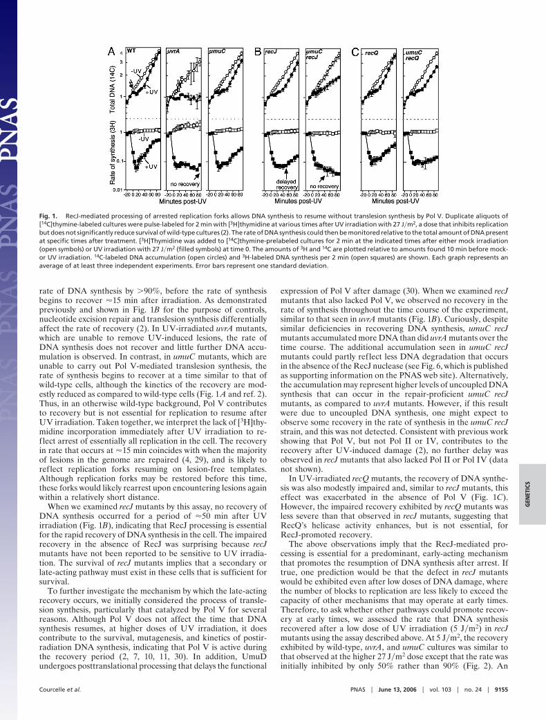
rate of DNA synthesis by �90%, before the rate of synthesisbegins to recover �15 min after irradiation. As demonstratedpreviously and shown in Fig. 1B for the purpose of controls,nucleotide excision repair and translesion synthesis differentiallyaffect the rate of recovery (2). In UV-irradiated uvrA mutants,which are unable to remove UV-induced lesions, the rate ofDNA synthesis does not recover and little further DNA accu-mulation is observed. In contrast, in umuC mutants, which areunable to carry out Pol V-mediated translesion synthesis, therate of synthesis begins to recover at a time similar to that ofwild-type cells, although the kinetics of the recovery are mod-estly reduced as compared to wild-type cells (Fig. 1 A and ref. 2).Thus, in an otherwise wild-type background, Pol V contributesto recovery but is not essential for replication to resume afterUV irradiation. Taken together, we interpret the lack of [3H]thy-midine incorporation immediately after UV irradiation to re-flect arrest of essentially all replication in the cell. The recoveryin rate that occurs at �15 min coincides with when the majorityof lesions in the genome are repaired (4, 29), and is likely toreflect replication forks resuming on lesion-free templates.Although replication forks may be restored before this time,these forks would likely rearrest upon encountering lesions againwithin a relatively short distance.
When we examined recJ mutants by this assay, no recovery ofDNA synthesis occurred for a period of �50 min after UVirradiation (Fig. 1B), indicating that RecJ processing is essentialfor the rapid recovery of DNA synthesis in the cell. The impairedrecovery in the absence of RecJ was surprising because recJmutants have not been reported to be sensitive to UV irradia-tion. The survival of recJ mutants implies that a secondary orlate-acting pathway must exist in these cells that is sufficient forsurvival.
To further investigate the mechanism by which the late-actingrecovery occurs, we initially considered the process of transle-sion synthesis, particularly that catalyzed by Pol V for severalreasons. Although Pol V does not affect the time that DNAsynthesis resumes, at higher doses of UV irradiation, it doescontribute to the survival, mutagenesis, and kinetics of postir-radiation DNA synthesis, indicating that Pol V is active duringthe recovery period (2, 7, 10, 11, 30). In addition, UmuDundergoes posttranslational processing that delays the functional
expression of Pol V after damage (30). When we examined recJmutants that also lacked Pol V, we observed no recovery in therate of synthesis throughout the time course of the experiment,similar to that seen in uvrA mutants (Fig. 1B). Curiously, despitesimilar deficiencies in recovering DNA synthesis, umuC recJmutants accumulated more DNA than did uvrA mutants over thetime course. The additional accumulation seen in umuC recJmutants could partly reflect less DNA degradation that occursin the absence of the RecJ nuclease (see Fig. 6, which is publishedas supporting information on the PNAS web site). Alternatively,the accumulation may represent higher levels of uncoupled DNAsynthesis that can occur in the repair-proficient umuC recJmutants, as compared to uvrA mutants. However, if this resultwere due to uncoupled DNA synthesis, one might expect toobserve some recovery in the rate of synthesis in the umuC recJstrain, and this was not detected. Consistent with previous workshowing that Pol V, but not Pol II or IV, contributes to therecovery after UV-induced damage (2), no further delay wasobserved in recJ mutants that also lacked Pol II or Pol IV (datanot shown).
In UV-irradiated recQ mutants, the recovery of DNA synthe-sis was also modestly impaired and, similar to recJ mutants, thiseffect was exacerbated in the absence of Pol V (Fig. 1C).However, the impaired recovery exhibited by recQ mutants wasless severe than that observed in recJ mutants, suggesting thatRecQ’s helicase activity enhances, but is not essential, forRecJ-promoted recovery.
The above observations imply that the RecJ-mediated pro-cessing is essential for a predominant, early-acting mechanismthat promotes the resumption of DNA synthesis after arrest. Iftrue, one prediction would be that the defect in recJ mutantswould be exhibited even after low doses of DNA damage, wherethe number of blocks to replication are less likely to exceed thecapacity of other mechanisms that may operate at early times.Therefore, to ask whether other pathways could promote recov-ery at early times, we assessed the rate that DNA synthesisrecovered after a low dose of UV irradiation (5 J�m2) in recJmutants using the assay described above. At 5 J�m2, the recoveryexhibited by wild-type, uvrA, and umuC cultures was similar tothat observed at the higher 27 J�m2 dose except that the rate wasinitially inhibited by only 50% rather than 90% (Fig. 2). An
Fig. 1. RecJ-mediated processing of arrested replication forks allows DNA synthesis to resume without translesion synthesis by Pol V. Duplicate aliquots of[14C]thymine-labeled cultures were pulse-labeled for 2 min with [3H]thymidine at various times after UV irradiation with 27 J�m2, a dose that inhibits replicationbut does not significantly reduce survival of wild-type cultures (2). The rate of DNA synthesis could then be monitored relative to the total amount of DNA presentat specific times after treatment. [3H]Thymidine was added to [14C]thymine-prelabeled cultures for 2 min at the indicated times after either mock irradiation(open symbols) or UV irradiation with 27 J�m2 (filled symbols) at time 0. The amounts of 3H and 14C are plotted relative to amounts found 10 min before mock-or UV irradiation. 14C-labeled DNA accumulation (open circles) and 3H-labeled DNA synthesis per 2 min (open squares) are shown. Each graph represents anaverage of at least three independent experiments. Error bars represent one standard deviation.
Courcelle et al. PNAS � June 13, 2006 � vol. 103 � no. 24 � 9155
GEN
ETIC
S

additional difference at the low dose was that more DNAaccumulated in the uvrA mutant, despite the fact that the rate ofsynthesis did not recover during the experiment. However, whenwe examined recJ mutants, we observed that the rate of DNAsynthesis failed to recover in a timely manner even at a UV doseof only 5 J�m2 (Fig. 2), indicating that RecJ processing isrequired at early times for DNA synthesis to resume.
In the Absence of RecJ-Mediated Processing, Survival Becomes De-pendent on Translesion Synthesis by Pol V and Results in HigherFrequencies of Mutagenesis. The idea that Pol V can promoterecovery when the predominant mechanism cannot occur wasalso supported by the survival of these mutants after exposureto UV. The absence of either RecJ or Pol V does not render cellsseverely sensitive to UV irradiation. However, we observed thatin the absence of RecJ, Pol V became critical to cell survival (Fig.3). Mutants lacking both recJ and umuC were approximately ashypersensitive to UV irradiation as recF mutants, which are
known to be defective in recovering replication after UV-induced arrest (14). The absence of RecQ also reduced thesurvival of umuC mutants, although, as seen in the previousassays, the effect was less severe than that of RecJ (Fig. 3). Noeffect on survival was observed in recJ mutants that lacked eitherPol II or Pol IV (Fig. 3A), consistent with several studies, bothin vitro and in vivo, that suggest Pol V, but not Pol II or Pol IV,is able to function at sites of UV-induced damage (2, 7, 11, 30,31). One study did observe a delay in the recovery of the polBstrain, STL1336, after UV-irradiation (32). However, previousstudies with this strain observed no Pol II-mediated translesionsynthesis on UV-damaged templates (31) and a propensity of thestrain to accumulate suppressor mutations that affect its re-sponse to DNA damage (33).
It is interesting to note that the survival curve of recJ mutantswas distinct from that of wild-type cultures. recJ mutants weremodestly sensitive to low doses of UV, whereas at higher doses,they were more resistant than wild-type cultures (Fig. 3 B and C).The hypersensitivity to low doses would be consistent with theidea that RecJ is required during the initial response to DNAdamage. The resistance of recJ mutants to high doses mayindicate that the delayed recovery is beneficial when high levelsof DNA damage are present. Previous studies have shown thatinhibiting replication by placing the culture in ‘‘liquid holding’’media that lacks a carbon source, similarly increases cell survivalafter DNA damage (34).
The survival of RecJ at higher doses also suggests thattranslesion synthesis by Pol V is capable of promoting nearwild-type levels of survival when required to function in this role.By contrast, umuC mutants were only hypersensitive at thehigher doses of UV irradiation (Fig. 3B), consistent with theinterpretation that Pol V becomes important either when lesionsare not repaired in a timely manner, or when lesions, due to theirchemical nature or sequence context, may be resistant to pro-cessing by repair enzymes.
The data are consistent with a model in which RecJ-mediatedprocessing serves to restore the lesion-containing site to a formthat is accessible to nucleotide excision repair enzymes, whichrequire double-stranded DNA to function (Fig. 4). In theabsence of the nascent DNA degradation by RecJ, the block toreplication remains until translesion synthesis by Pol V cansynthesize past the arresting lesion. A prediction of this model isthat in the absence of RecJ, the frequency of mutagenesis mightincrease, when the recovery depends on translesion synthesis byPol V. To examine this possibility, we determined the frequency
Fig. 2. Nascent DNA processing by RecJ is required to restore DNA synthesisat early times after low doses (5 J�m2) of UV irradiation. Data were obtainedand plotted as in Fig. 1. 14C-labeled DNA accumulation (open circle); 3H-labeled DNA synthesis per 2 min (open square). Each graph represents anaverage of at least three independent experiments. Error bars represent onestandard deviation.
Fig. 3. In the absence of RecJ, survival after UV-induced damage depends on translesion synthesis by Pol V. (A) Survival of parental (WT), recF, umuC, recJ, umuCrecJ, polB recJ, and dinB recJ strains after UV irradiation with the indicated dose. (B) The survival of parental (filled square), recJ (filled triangle), umuC (opensquare), umuC recJ (filled circle), recF (open triangle), recQ (inverted filled triangle), and umuC recQ (inverted open triangle) cultures are shown after UVirradiation at the indicated doses. (C) The survival of parental (filled square), recJ (filled triangle), umuC (open square), umuC recJ (filled circle), recQ (invertedfilled triangle), and umuC recQ (open inverted triangle) cultures are shown after UV irradiation between 0 and 30 J/m2.
9156 � www.pnas.org�cgi�doi�10.1073�pnas.0600785103 Courcelle et al.
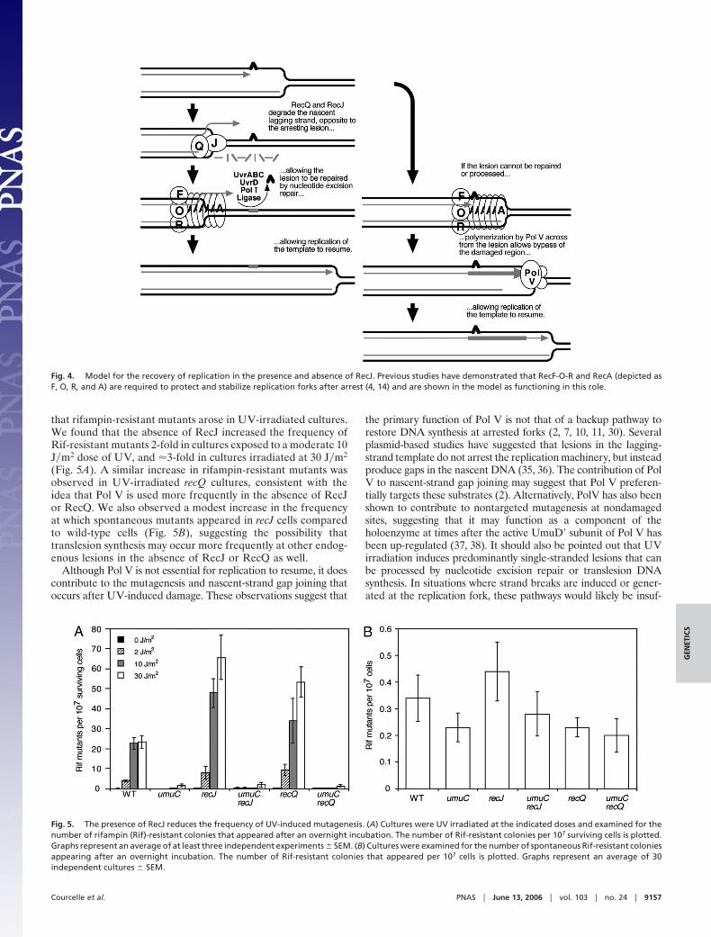
that rifampin-resistant mutants arose in UV-irradiated cultures.We found that the absence of RecJ increased the frequency ofRif-resistant mutants 2-fold in cultures exposed to a moderate 10J�m2 dose of UV, and �3-fold in cultures irradiated at 30 J�m2
(Fig. 5A). A similar increase in rifampin-resistant mutants wasobserved in UV-irradiated recQ cultures, consistent with theidea that Pol V is used more frequently in the absence of RecJor RecQ. We also observed a modest increase in the frequencyat which spontaneous mutants appeared in recJ cells comparedto wild-type cells (Fig. 5B), suggesting the possibility thattranslesion synthesis may occur more frequently at other endog-enous lesions in the absence of RecJ or RecQ as well.
Although Pol V is not essential for replication to resume, it doescontribute to the mutagenesis and nascent-strand gap joining thatoccurs after UV-induced damage. These observations suggest that
the primary function of Pol V is not that of a backup pathway torestore DNA synthesis at arrested forks (2, 7, 10, 11, 30). Severalplasmid-based studies have suggested that lesions in the lagging-strand template do not arrest the replication machinery, but insteadproduce gaps in the nascent DNA (35, 36). The contribution of PolV to nascent-strand gap joining may suggest that Pol V preferen-tially targets these substrates (2). Alternatively, PolV has also beenshown to contribute to nontargeted mutagenesis at nondamagedsites, suggesting that it may function as a component of theholoenzyme at times after the active UmuD� subunit of Pol V hasbeen up-regulated (37, 38). It should also be pointed out that UVirradiation induces predominantly single-stranded lesions that canbe processed by nucleotide excision repair or translesion DNAsynthesis. In situations where strand breaks are induced or gener-ated at the replication fork, these pathways would likely be insuf-
Fig. 4. Model for the recovery of replication in the presence and absence of RecJ. Previous studies have demonstrated that RecF-O-R and RecA (depicted asF, O, R, and A) are required to protect and stabilize replication forks after arrest (4, 14) and are shown in the model as functioning in this role.
Fig. 5. The presence of RecJ reduces the frequency of UV-induced mutagenesis. (A) Cultures were UV irradiated at the indicated doses and examined for thenumber of rifampin (Rif)-resistant colonies that appeared after an overnight incubation. The number of Rif-resistant colonies per 107 surviving cells is plotted.Graphs represent an average of at least three independent experiments � SEM. (B) Cultures were examined for the number of spontaneous Rif-resistant coloniesappearing after an overnight incubation. The number of Rif-resistant colonies that appeared per 107 cells is plotted. Graphs represent an average of 30independent cultures � SEM.
Courcelle et al. PNAS � June 13, 2006 � vol. 103 � no. 24 � 9157
GEN
ETIC
S
ficient, and recombinational mechanisms may play a more impor-tant role (39).
The absence of RecQ impaired recovery, reduced survival, andincreased mutagenesis after UV irradiation less severely than theabsence of RecJ in these assays, suggesting that RecQ’s helicaseactivity contributes to, but is not essential for, RecJ-promotedrecovery. Other studies have also noted that, in mutants thatdepend on the RecF pathway for recombination or survival, RecJplays a more critical role than RecQ (40). With respect to theiractivity at replication forks, the absence of either gene productdiminishes the nascent DNA degradation that can be detected atthe arrested replication fork (4, 15). Considering that in vitro, theRecJ exonuclease specifically acts on single-stranded DNA, weinitially proposed that RecQ would first be required to unwind thedouble-stranded DNA near the arrested replication fork before anyRecJ degradation could occur (4, 15, 18, 19). However, these resultsare more consistent with the idea that, in vivo, RecQ enhances theRecJ degradation, and that some nascent strand degradation occursindependently of RecQ. After the arrest of replication forks onplasmid substrates, it has been shown that the nascent DNA ispartially displaced spontaneously when the hyperwound DNAahead of the replication fork recoils, potentially providing a sub-strate for RecJ degradation (41). The presence of RecQ is clearlyrequired for much more extensive degradation to occur at thearrested replication fork and may serve a larger role in maintainingfork stability and suppressing illegitimate recombination events (20,21). Nevertheless, the presence of the RecQ helicase clearly en-hanced the RecJ-mediated recovery in each case, consistent withthe idea that these enzymes are acting cooperatively.
Related models to those presented in Fig. 4 for how DNAsynthesis resumes after disruption suggest that replication of theleading and lagging strands become uncoupled and is reestab-lished downstream from the site of disruption rather than at thedisrupting lesion (42, 43). Although the experiments presentedhere could be consistent with either model, the functionalrequirement for RecJ-mediated processing would essentiallyremain the same in each case. If disruption leads to reassemblydownstream of the arresting lesion, it is clear from this data thatRecJ-mediated processing is required before the efficient reas-sembly of the replication machinery can occur on the template.In the case of translesion synthesis, it is well established that PolV can efficiently synthesize past the UV-induced lesion itself (7,9). Thus, the observation that Pol V can restore DNA synthesissuggests that, at least in this situation, replication resumes fromthe original site of the disruption.
Materials and MethodsBacterial Strains. All bacterial strains are in a SR108 back-ground, a thyA36 deoC2 derivative of W3110. SR108, HL946(SR108 recF332::Tn3), HL944 (SR108 recQ1803::Tn3),HL924 (SR108 recJ284::Tn10), HL952 (SR108 uvrA::Tn10),CL579 (SR108 recF6206::tetr), CL575 (SR108 umuC122::Tn5),and CL632 (SR108 umuDC595::cat) have been described(2–4, 15). CL740 (SR108 umuDC595::cat uvrA::Tn10) wasconstructed by P1 transduction of the uvrA::Tn10 allelefrom HL952 into CL632. CL596 (SR108 umuC122::Tn5
recQ6215::cam) and CL766 (SR108 umuC122::Tn5recJ284::Tn10) were constructed by P1 transduction of therecQ6215::cam and recJ284::Tn10 alleles from TP648 (44) andHL924, respectively, into CL575. CL779 (SR108 dinB::kanr
recJ284::Tn10) and CL773 (SR108 polB::� Sm-SprecJ284::Tn10) were made by P1 transduction of recJ284::Tn10from HL924 into CL634 (2) and CL636 (2), respectively.Phenotypes were confirmed by antibiotic resistance and, whenappropriate, UV hypersensitivity. A complete list of strainsused in this study is also shown in Table 1, which is publishedas supporting information on the PNAS web site.
Recovery of DNA Synthesis. This approach is modified fromKhidhir et al. (45). Fresh overnight cultures were diluted 1:100and grown in DGCthy media supplemented with 0.1 �Ci�ml of[14C]thymine to an OD600 of precisely 0.3, at which point half ofthe culture received an incident dose of 5 or 27 J�m2, whereasthe other half of the culture was mock irradiated. At the timesindicated, duplicate 0.5-ml aliquots of culture were pulse labeledwith 1 �Ci�ml [3H]thymidine for 2 min at 37°C. The cells werethen lysed, the DNA was precipitated in cold 5% trichloroaceticacid (TCA) and filtered onto Millipore glass fiber filters, and theamount of 3H and 14C in each sample determined by liquidscintillation counting.
UV Survival Studies. Sylvania 15-watt germicidal lamp (254 nm) atan incident dose of 0.9 J�m2 per s (0.2 J�m2 per s for doses �20J�m2) was used for irradiations. Cells were grown in Davismedium supplemented with 0.4% glucose, 0.2% casamino acids,and 10 �g�ml thymine (DGCthy media). Cultures were inocu-lated from fresh overnight cultures and grown to an OD600between 0.4 and 0.5. Serial dilutions of each culture were platedin triplicate on Luria-Bertani plates supplemented with 10�g�ml thymine and UV irradiated at the indicated doses. Plateswere incubated overnight at 37°C, and colonies were counted thenext day.
UV-Induced Mutagenesis. Mutagenesis induced by UV was mea-sured by the appearance of rifampin-resistant colonies as a resultof UV exposure. At least 69 base substitutions within the rpoBgene have been identified that confer resistance to rifampin,allowing one to monitor numerous UV-induced mutation sitesin different sequence contexts (46). Overnight cultures werediluted 1:100 and grown in DGCthy media to an OD600 of 0.4,at which point the culture was split into four equal fractions andirradiated with an incident dose of 0, 2, 10, or 30 J�m2 UV. Afterovernight incubation at 37°C, the cultures were plated onLuria-Bertani plates containing 10 �g�ml thymine and 100�g�ml rifampin. The number of cells surviving after UV treat-ment was also determined at this time by plating three 10-�laliquots of serial 10-fold dilutions on Luria–Bertani platescontaining 10 �g�ml thymine. Rifampin-resistant colonies andsurviving cells were counted after overnight incubation at 37°C.
This work was supported by National Science Foundation CAREERAward MCB0551798. C.T.C. was supported by National Institutes ofHealth�National Institute of General Medical Sciences National Re-search Service Award F32 GM068566.
1. Setlow, R. B., Swenson, P. A. & Carrier, W. L. (1963) Science 142, 1464–1466.2. Courcelle, C. T., Belle, J. J. & Courcelle, J. (2005) J. Bacteriol. 187,
6953–6961.3. Courcelle, J., Crowley, D. J. & Hanawalt, P. C. (1999) J. Bacteriol. 181, 916–922.4. Courcelle, J., Donaldson, J. R., Chow, K. H. & Courcelle, C. T. (2003) Science
299, 1064–1067.5. Rupp, W. D. & Howard-Flanders, P. (1968) J. Mol. Biol. 31, 291–304.6. Sutton, M. D. & Walker, G. C. (2001) Proc. Natl. Acad. Sci. USA 98,
8342–8349.7. Napolitano, R., Janel-Bintz, R., Wagner, J. & Fuchs, R. P. (2000) EMBO J. 19,
6259–6265.
8. Reuven, N. B., Arad, G., Maor-Shoshani, A. & Livneh, Z. (1999) J. Biol. Chem.274, 31763–31766.
9. Tang, M., Shen, X., Frank, E. G., O’Donnell, M., Woodgate, R. & Goodman,M. F. (1999) Proc. Natl. Acad. Sci. USA 96, 8919–8924.
10. Bagg, A., Kenyon, C. J. & Walker, G. C. (1981) Proc. Natl. Acad. Sci. USA 78,5749–5753.
11. Kato, T. & Shinoura, Y. (1977) Mol. Gen. Genet. 156, 121–131.12. Wang, T. C. & Smith, K. C. (1985) Mutat. Res. 145, 107–112.13. Horii, Z. & Suzuki, K. (1968) Photochem. Photobiol. 8, 93–105.14. Courcelle, J., Carswell-Crumpton, C. & Hanawalt, P. C. (1997) Proc. Natl.
Acad. Sci. USA 94, 3714–3719.
9158 � www.pnas.org�cgi�doi�10.1073�pnas.0600785103 Courcelle et al.
15. Courcelle, J. & Hanawalt, P. C. (1999) Mol. Gen. Genet. 262, 543–551.16. Rangarajan, S., Woodgate, R. & Goodman, M. F. (2002) Mol. Microbiol. 43,
617–628.17. Lopes, M., Foiani, M. & Sogo, J. M. (2006) Mol. Cell 21, 15–27.18. Umezu, K., Nakayama, K. & Nakayama, H. (1990) Proc. Natl. Acad. Sci. USA
87, 5363–5367.19. Lovett, S. T. & Kolodner, R. D. (1989) Proc. Natl. Acad. Sci. USA 86, 2627–2631.20. Ukita, T. & Ikeda, H. (1996) J. Bacteriol. 178, 2362–2367.21. Hanada, K., Ukita, T., Kohno, Y., Saito, K., Kato, J. & Ikeda, H. (1997) Proc.
Natl. Acad. Sci. USA 94, 3860–3865.22. Kolodner, R., Fishel, R. A. & Howard, M. (1985) J. Bacteriol. 163, 1060–1066.23. Hanada, K., Iwasaki, M., Ihashi, S. & Ikeda, H. (2000) Proc. Natl. Acad. Sci.
USA 97, 5989–5994.24. Stewart, E., Chapman, C. R., Al-Khodairy, F., Carr, A. M. & Enoch, T. (1997)
EMBO J. 16, 2682–2692.25. Watt, P. M., Hickson, I. D., Borts, R. H. & Louis, E. J. (1996) Genetics 144,
935–945.26. Kim, Y. C., Lee, M. H., Ryu, S. S., Kim, J. H. & Koo, H. S. (2002) Genes Cells
7, 19–27.27. Bagherieh-Najjar, M. B., de Vries, O. M., Hille, J. & Dijkwel, P. P. (2005) Plant
J. 43, 789–798.28. Nakayama, H. (2002) Oncogene 21, 9008–9021.29. Mellon, I. & Hanawalt, P. C. (1989) Nature 342, 95–98.30. Opperman, T., Murli, S., Smith, B. T. & Walker, G. C. (1999) Proc. Natl. Acad.
Sci. USA 96, 9218–9223.31. Kow, Y. W., Faundez, G., Hays, S., Bonner, C. A., Goodman, M. F. & Wallace,
S. S. (1993) J. Bacteriol. 175, 561–564.
32. Rangarajan, S., Woodgate, R. & Goodman, M. F. (1999) Proc. Natl. Acad. Sci.USA 96, 9224–9229.
33. Escarceller, M., Hicks, J., Gudmundsson, G., Trump, G., Touati, D., Lovett, S.,Foster, P. L., McEntee, K. & Goodman, M. F. (1994) J. Bacteriol. 176,6221–6228.
34. Ganesan, A. K. & Smith, K. C. (1968) J. Bacteriol. 96, 365–373.35. Higuchi, K., Katayama, T., Iwai, S., Hidaka, M., Horiuchi, T. & Maki, H. (2003)
Genes Cells 8, 437–449.36. McInerney, P. & O’Donnell, M. (2004) J. Biol. Chem. 279, 21543–21551.37. Tang, M., Pham, P., Shen, X., Taylor, J. S., O’Donnell, M., Woodgate, R. &
Goodman, M. F. (2000) Nature 404, 1014–1018.38. Maor-Shoshani, A., Reuven, N. B., Tomer, G. & Livneh, Z. (2000) Proc. Natl.
Acad. Sci. USA 97, 565–570.39. Michel, B., Grompone, G., Flores, M. J. & Bidnenko, V. (2004) Proc. Natl.
Acad. Sci. USA 101, 12783–12788.40. Lovett, S. T., Luisi-DeLuca, C. & Kolodner, R. D. (1988) Genetics 120,
37–45.41. Postow, L., Ullsperger, C., Keller, R. W., Bustamante, C., Vologodskii, A. V.
& Cozzarelli, N. R. (2001) J. Biol. Chem. 276, 2790–2796.42. Heller, R. C. & Marians, K. J. (2005) J. Biol. Chem. 280, 34143–34151.43. Heller, R. C. & Marians, K. J. (2005) Mol. Cell 17, 733–743.44. Murphy, K. C., Campellone, K. G. & Poteete, A. R. (2000) Gene 246, 321–330.45. Khidhir, M. A., Casaregola, S. & Holland, I. B. (1985) Mol. Gen. Genet. 199,
133–140.46. Garibyan, L., Huang, T., Kim, M., Wolff, E., Nguyen, A., Nguyen, T., Diep, A.,
Hu, K., Iverson, A., Yang, H. & Miller, J. H. (2003) DNA Rep. 2, 593–608.
Courcelle et al. PNAS � June 13, 2006 � vol. 103 � no. 24 � 9159
GEN
ETIC
S
Copyright © 2022 FDOKUMEN