Nanoparticle-Induced Toxicities: an Update on the Role of ...
182
Oxidative Medicine and Cellular Longevity Nanoparticle-Induced Toxicities: an Update on the Role of Oxidative Stress Lead Guest Editor: Gvozden L. Rosić Guest Editors: Dragica Selakovic, Igor Jakovcevski, Miodrag Stojkovic, Sergey Bolevich, and Vladimir Jakovljevic
-
Upload
khangminh22 -
Category
Documents
-
view
0 -
download
0
Transcript of Nanoparticle-Induced Toxicities: an Update on the Role of ...

Oxidative Medicine and Cellular Longevity
Nanoparticle-Induced Toxicities:an Update on the Role of OxidativeStress
Lead Guest Editor: Gvozden L. RosićGuest Editors: Dragica Selakovic, Igor Jakovcevski, Miodrag Stojkovic, SergeyBolevich, and Vladimir Jakovljevic

Nanoparticle-Induced Toxicities: an Update onthe Role of Oxidative Stress

Oxidative Medicine and Cellular Longevity
Nanoparticle-Induced Toxicities: anUpdate on the Role of Oxidative Stress
Lead Guest Editor: Gvozden L. RosićGuest Editors: Dragica Selakovic, Igor Jakovcevski,Miodrag Stojkovic, Sergey Bolevich, and VladimirJakovljevic

Copyright © 2022 Hindawi Limited. All rights reserved.
is is a special issue published in “Oxidative Medicine and Cellular Longevity.” All articles are open access articles distributed under theCreative Commons Attribution License, which permits unrestricted use, distribution, and reproduction in any medium, provided theoriginal work is properly cited.

Chief EditorJeannette Vasquez-Vivar, USA
Editorial Board
Mohd Adnan, Saudi ArabiaIvanov Alexander, RussiaFabio Altieri, ItalySilvia Alvarez, ArgentinaFernanda Amicarelli, ItalyJosé P. Andrade, PortugalCristina Angeloni, ItalyDaniel Arcanjo, BrazilSandro Argüelles, SpainAntonio Ayala, SpainElena Azzini, ItalyPeter Backx, CanadaDamian Bailey, United KingdomJiaolin Bao, ChinaGeorge E. Barreto, ColombiaSander Bekeschus, GermanyJi C. Bihl, USAConsuelo Borrás, SpainNady Braidy, AustraliaRalf Braun, AustriaLaura Bravo, SpainMatt Brody, USAAmadou Camara, USAGianluca Carnevale, ItalyRoberto Carnevale, ItalyMarcio Carocho, PortugalAngel Catalá, ArgentinaPeter Celec, SlovakiaGiulio Ceolotto, ItalyGiselle Cerchiaro, BrazilShao-Yu Chen, USAYujie Chen, ChinaDeepak Chhangani, USAFerdinando Chiaradonna, ItalyZhao Zhong Chong, USAXinxin Ci, ChinaFabio Ciccarone, ItalyAlin Ciobica, RomaniaAna Cipak Gasparovic, CroatiaGiuseppe Cirillo, ItalyMaria R. Ciriolo, ItalyMassimo Collino, ItalyGraziamaria Corbi, ItalyManuela Corte-Real, Portugal
Mark Crabtree, United KingdomManuela Curcio, ItalyAndreas Daiber, GermanyFelipe Dal Pizzol, BrazilFrancesca Danesi, ItalyDomenico D'Arca, ItalySergio Davinelli, ItalyClaudio de Lucia, ItalyDamião de Sousa, BrazilEnrico Desideri, ItalyFrancesca Diomede, ItalyCinzia Domenicotti, ItalyRaul Dominguez-Perles, SpainDimitrios Draganidis, GreeceJoël R. Drevet, FranceGrégory Durand, FranceAlessandra Durazzo, ItalyAnne Eckert, SwitzerlandJavier Egea, SpainPablo A. Evelson, ArgentinaStefano Falone, ItalyIoannis G. Fatouros, GreeceQingping Feng, CanadaGianna Ferretti, ItalyGiuseppe Filomeni, ItalyPasquale Fino, ItalyOmidreza Firuzi, IranSwaran J. S. Flora, IndiaTeresa I. Fortoul, MexicoAnna Fracassi, USARodrigo Franco, USAJoaquin Gadea, SpainJuan Gambini, SpainJosé Luís García-Giménez, SpainGerardo García-Rivas, MexicoJanusz Gebicki, AustraliaAlexandros Georgakilas, GreeceHusam Ghanim, USAJayeeta Ghose, USARajeshwary Ghosh, USALucia Gimeno-Mallench, SpainEloisa Gitto, ItalyAnna M. Giudetti, ItalyDaniela Giustarini, Italy

José Rodrigo Godoy, USASaeid Golbidi, CanadaAldrin V. Gomes, USAArantxa González, SpainTilman Grune, GermanyChi Gu, China, ChinaNicoletta Guaragnella, ItalySolomon Habtemariam, United KingdomYing Han, ChinaEva-Maria Hanschmann, GermanyMd Saquib Hasnain, IndiaMd Hassan, IndiaTim Hofer, NorwayJohn D. Horowitz, AustraliaSilvana Hrelia, ItalyDragan Hrncic, SerbiaJuan Huang, ChinaZebo Huang, ChinaTarique Hussain, PakistanStephan Immenschuh, GermanyMaria Isaguliants, LatviaLuigi Iuliano, ItalyFRANCO J. L, BrazilVladimir Jakovljevic, Serbiasedat kacar, USAJason Karch, USAPeeter Karihtala, FinlandAndleeb Khan, Saudi ArabiaKum Kum Khanna, AustraliaNeelam Khaper, Canadaomas Kietzmann, FinlandRamoji Kosuru, USADemetrios Kouretas, GreeceAndrey V. Kozlov, AustriaEsra Küpeli Akkol, TurkeyDaniele La Russa, ItalyJean-Claude Lavoie, CanadaWing-Kee Lee, GermanySimon Lees, CanadaXin-Feng Li, ChinaQiangqiang Li, ChinaGaocai Li, ChinaJialiang Liang, ChinaChristopher Horst Lillig, GermanyPaloma B. Liton, USAAna Lloret, SpainLorenzo Loffredo, Italy
Camilo López-Alarcón, ChileDaniel Lopez-Malo, SpainAntonello Lorenzini, ItalyMassimo Lucarini, ItalyHai-Chun Ma, ChinaMateusz Maciejczyk, PolandNageswara Madamanchi, USAKenneth Maiese, USAMarco Malaguti, ItalyTullia Maraldi, ItalyReiko Matsui, USAJuan C. Mayo, SpainSteven McAnulty, USAAntonio Desmond McCarthy, ArgentinaSonia Medina-Escudero, SpainPedro Mena, ItalyVíctor M. Mendoza-Núñez, MexicoLidija Milkovic, CroatiaAlexandra Miller, USASanjay Misra, USASara Missaglia, ItalyPremysl Mladenka, Czech RepublicRaffaella Molteni, ItalyMaria U. Moreno, SpainSandra Moreno, ItalyTrevor A. Mori, AustraliaRyuichi Morishita, JapanFabiana Morroni, ItalyAnge Mouithys-Mickalad, BelgiumIordanis Mourouzis, GreeceDanina Muntean, RomaniaColin Murdoch, United KingdomRyoji Nagai, JapanAmit Kumar Nayak, IndiaDavid Nieman, USACristina Nocella, ItalySusana Novella, SpainHassan Obied, AustraliaJulio J. Ochoa, SpainPál Pacher, USAPasquale Pagliaro, ItalyDR DILIPKUMAR PAL, IndiaValentina Pallottini, ItalyRosalba Parenti, ItalyMayur Parmar, USAVassilis Paschalis, GreeceVisweswara Rao Pasupuleti, Malaysia

Keshav Raj Paudel, AustraliaIlaria Peluso, ItalyClaudia Penna, ItalySerafina Perrone, ItalyTiziana Persichini, ItalyShazib Pervaiz, SingaporeVincent Pialoux, FranceAlessandro Poggi, ItalyAda Popolo, ItalyAijuan Qu, ChinaJosé L. Quiles, SpainWalid Rachidi, FranceZsolt Radak, HungarySachchida Rai, IndiaNamakkal Soorappan Rajasekaran, USADario C. Ramirez, ArgentinaErika Ramos-Tovar, MexicoAbdur Rauf Rauf, PakistanSid D. Ray, USAMuneeb Rehman, Saudi ArabiaHamid Reza Rezvani, FranceAlessandra Ricelli, ItalyFrancisco J. Romero, SpainMariana G. Rosca, USAJoan Roselló-Catafau, SpainEsther Roselló-Lletí, SpainSubhadeep Roy, IndiaJosep V. Rubert, e NetherlandsH. P. Vasantha Rupasinghe, CanadaSumbal Saba, BrazilKunihiro Sakuma, JapanGabriele Saretzki, United KingdomAjinkya S. Sase, USALuciano Saso, ItalyNadja Schroder, BrazilSebastiano Sciarretta, ItalyRatanesh K. Seth, USAAnwen Shao, ChinaXiaolei Shi, ChinaCinzia Signorini, ItalyMithun Sinha, USAGiulia Sita, ItalyEduardo Sobarzo-Sánchez, ChileAdrian Sturza, RomaniaYi-Rui Sun, ChinaEisa Tahmasbpour Marzouni, IranCarla Tatone, Italy
Shane omas, AustraliaCarlo Gabriele Tocchetti, ItalyAngela Trovato Salinaro, ItalyPaolo Tucci, ItalyRosa Tundis, ItalyGiuseppe Valacchi, ItalyDaniele Vergara, ItalyVictor M. Victor, SpainLászló Virág, HungaryMin-qi Wang, ChinaKai Wang, ChinaNatalie Ward, AustraliaGrzegorz Wegrzyn, PolandPhilip Wenzel, GermanyJianbo Xiao, ChinaQiongming Xu, ChinaSho-ichi Yamagishi, JapanLiang-Jun Yan, USAGuillermo Zalba, SpainJunmin Zhang, ChinaZiwei Zhang, ChinaJia Zhang, First Affiliated Hospital of Xi’anJiaotong University, Xi’an, Shaanxi Province,China, ChinaJunli Zhao, USAYong Zhou, ChinaChen-he Zhou, ChinaMario Zoratti, Italy

Contents
Nanoparticle-Induced Toxicities: An Update on the Role of Oxidative StressGvozden Rosic and Dragica Selakovic
Editorial (2 pages), Article ID 9895061, Volume 2022 (2022)
Comparative In Vitro Cytotoxicity Study of Carbon Dot-Based Organometallic Nanoconjugates:Exploration of 'eir Cell Proliferation, Uptake, and Localization in Cancerous and Normal CellsEepsita Priyadarshini, Ramovatar Meena, Himadri B. Bohidar, Saurabh Kumar Sharma, Magda H.Abdellattif, Muthupandian Saravanan , and Paulraj Rajamani
Research Article (11 pages), Article ID 3483073, Volume 2022 (2022)
Exploring Dose-Dependent Cytotoxicity Profile of Gracilaria edulis-Mediated Green SynthesizedSilver Nanoparticles against MDA-MB-231 Breast CarcinomaYugal Kishore Mohanta , Awdhesh Kumar Mishra, Debasis Nayak, Biswajit Patra, Amra Bratovcic, SatyaKumar Avula, Tapan Kumar Mohanta, Kadarkarai Murugan, and Muthupandian Saravanan
Research Article (15 pages), Article ID 3863138, Volume 2022 (2022)
Effects of Metal Oxide Nanoparticles in ZebrafishMarta d’Amora , Tiziana Julia Nadjeschda Schmidt, Soultana Konstantinidou , Vittoria Raffa ,Francesco De Angelis , and Francesco Tantussi
Review Article (37 pages), Article ID 3313016, Volume 2022 (2022)
Evaluation of Zebrafish Toxicology and Biomedical Potential of Aeromonas hydrophila MediatedCopper Sulfide NanoparticlesS. Rajeshkumar , J. Santhoshkumar, M. Vanaja, P. Sivaperumal, M. Ponnanikajamideen, Daoud Ali, andKalirajan Arunachalam
Research Article (12 pages), Article ID 7969825, Volume 2022 (2022)
Exacerbation of 'rombotic Responses to Silver Nanoparticles in Hypertensive Mouse ModelZannatul Ferdous, Sumaya Beegam, Nur E. Zaaba, Ozaz Elzaki, Saeed Tariq, Yaser E. Greish, Badreldin H.Ali, and Abderrahim Nemmar
Research Article (10 pages), Article ID 2079630, Volume 2022 (2022)
An Overview of the Beneficial Role of Antioxidants in the Treatment of Nanoparticle-InducedToxicitiesVladimir Mihailovic , Jelena S. Katanic Stankovic , Dragica Selakovic , and Gvozden Rosic
Review Article (21 pages), Article ID 7244677, Volume 2021 (2021)
Combining Nanotechnology and Gas Plasma as an Emerging Platform for Cancer 'erapy:Mechanism and 'erapeutic ImplicationMilad Rasouli , Nadia Fallah , and Sander Bekeschus
Review Article (20 pages), Article ID 2990326, Volume 2021 (2021)

Phytoantioxidant Functionalized Nanoparticles: A Green Approach to Combat Nanoparticle-InducedOxidative StressAcharya Balkrishna, Ashwani Kumar , Vedpriya Arya, Akansha Rohela, Rachna Verma , EugenieNepovimova, Ondrej Krejcar , Dinesh Kumar, Naveen akur, and Kamil Kuca
Review Article (20 pages), Article ID 3155962, Volume 2021 (2021)
Anticancer, Enhanced Antibacterial, and Free Radical Scavenging Potential of Fucoidan- (Fucusvesiculosus Source) Mediated Silver NanoparticlesS. Rajeshkumar , Eman F. Aboelfetoh, Sri Renukadevi Balusamy, Daoud Ali, Mohammed H. A.Almarzoug , Jule Leta Tesfaye, and Ramaswamy Krishnaraj
Research Article (11 pages), Article ID 8511576, Volume 2021 (2021)
An Update Report on the Biosafety and Potential Toxicity of Fullerene-Based Nanomaterials towardAquatic AnimalsNemi Malhotra , Gilbert Audira , Agnes L. Castillo , Petrus Siregar , Johnsy Margotte S. Ruallo,Marri Jmelou Roldan , Jung-Ren Chen , Jiann-Shing Lee , Tzong-Rong Ger , and Chung-DerHsiao
Review Article (14 pages), Article ID 7995223, Volume 2021 (2021)

EditorialNanoparticle-Induced Toxicities: An Update on the Role ofOxidative Stress
Gvozden Rosic and Dragica Selakovic
Department of Physiology, Faculty of Medical Sciences, University of Kragujevac, Serbia
Correspondence should be addressed to Gvozden Rosic; [email protected]
Received 22 April 2022; Accepted 22 April 2022; Published 19 May 2022
Copyright © 2022 Gvozden Rosic and Dragica Selakovic. This is an open access article distributed under the Creative CommonsAttribution License, which permits unrestricted use, distribution, and reproduction in any medium, provided the original work isproperly cited.
The variety of nanoparticles has, unfortunately, becomecommon constituents of the growing pollution problem.Since there is a significant presence of nanoparticles in thefood industry, cosmetics, and other unavoidable products,better knowledge of their properties and undesirable effectsseems necessary to reduce their adverse effects. Althoughnanoparticles induce numerous toxicities, some commonpathways are harmful to human health, including oxidativedamage. Significant impact of nanoparticles has previouslybeen documented in clinical trials and preclinical investiga-tions. Therefore, the objective of this special issue is to allowa comprehensive insight based on both original research andreview articles that focus on the estimation of oxidativestress as the key point mechanism in many toxicitiesinduced by nanoparticles. At the same time, numerousinvestigations confirmed the beneficial role of nanoparticlesadministration for many medical indications, with the finaleffects strongly depending on the applied methodology.Thus, to enlighten the complex impact of nanoparticles ontargeting species, in this special issue, we are now offeringan update to the existing information that can help to revealour current knowledge from competent and reliable sources.
This special issue covers 10 articles focusing onnanoparticle-induced toxicities, highlighting the role of oxi-dative stress. The guest editors are pleased to present a com-pendium of these updates on nanoparticles’ effects in thepublished articles as follows:
Potential benefits of nanotechnology in the field of can-cer treatment were presented by Priyadarshini andcoworkers in the article “Comparative in vitro CytotoxicityStudy of Carbon Dot-Based Organometallic Nanoconju-
gates: Exploration of Their Cell Proliferation, Uptake, andLocalization in Cancerous and Normal Cells”. The resultsobtained in this in vitro study confirmed that carbon dot-based nanoconjugates with Ag had potent therapeuticpotential, signifying the effect of silver in cancer cell lines.At the same time, those nanoconjugates potentiated thenontoxic nature of human health cell line.
On the other hand, the adverse effects of specific nano-particles were elaborated in the article “Exacerbation ofthrombotic responses to silver nanoparticles in a hyperten-sive mouse model” by Ferdous and coworkers. Based onthe results of this original research, the population withhypertension is at higher risk of the toxicity of polyethyleneglycol-AgNPs. This conclusion appeared following theresults that confirm that polyethylene glycol-AgNPs canpotentially exacerbate the in vivo and in vitro procoagulatoryand oxidative stress effect in hypertensive mice.
Although there is a lot of evidence consideringnanoparticle-induced toxicities, potential benefits of theirclinical usage highlight the necessity for treatment of theiradverse effects, primarily by attenuation of this specifickind of iatrogenic oxidative damage. Therefore, the exten-sive overview of literature data presented in the article “Anoverview of the beneficial role of antioxidants in the treat-ment of nanoparticle-induced toxicities” by Mihailovic andcolleagues offers the confirmation that nanoparticles-induced oxidative stress may be attenuated by differentantioxidant substances. It is worth noting that naturallyoccurring antioxidants have an important role in theenhancement of the antioxidant defense systems in theprevention and mitigation of organism damage caused by
HindawiOxidative Medicine and Cellular LongevityVolume 2022, Article ID 9895061, 2 pageshttps://doi.org/10.1155/2022/9895061

nanoparticle-induced oxidative stress. Naturally occurringantioxidant protection was also elaborated in detail inthe insightful review article “Phyto-antioxidant functional-ized nanoparticles: A green approach to combatnanoparticles-induced oxidative stress” by Balkrishna andcolleagues. This review article, in turn, offers convincingevidence that nanoparticles may be useful in combatingthe oxidative damage of other origins. Namely, the major-ity of silver, gold, iron, zinc oxide, and copper nanoparti-cles produced utilizing various plant extracts were activefree radical scavengers. According to the authors, thispotential is linked to several surface-fabricated phytocon-stituents, such as flavonoids and phenols, which accentu-ated the potential of phyto-antioxidant functionalizednanoparticles to be a better alternative to nanoparticlesprepared by other existing approaches.
Interestingly, one of the most promising and state-of-the-art methodological approaches in the field of nanotech-nology, the green synthesis of nanoparticles, had been thesubject of several papers in this Issue. Mohanta and collabo-rators in the original article “Exploring dose dependent cyto-toxicityprofile of Gracilaria edulis mediated greensynthesized silver nanoparticles against MDA-MB-231breast carcinoma” showed that silver nanoparticles synthe-sized through the extensively elaborated green methodexpressed potential anticancer and antimicrobial activity.This finding allows potential utility in the food preservativefilm industry, as well as biomedical and pharmaceuticalindustries. Another intervention performed on silver nano-particles was presented in the original article “Anticancer,enhanced antibacterial and free radical scavenging potentialof Fucoidan (Fucus vesiculosus Source) mediated silvernanoparticles” by Rajeshkumar and coworkers. Based onthe results presented in this study, the activities of commer-cial antibiotics were enhanced by impregnation with thesynthesized silver nanoparticles, which led to the conclusionthat the utilization of environmentally synthesized silvernanoparticles offers numerous benefits of eco-friendlinessand compatibility for biomedical applications. Even morebeneficial impact of green methods in nanotechnology waspresented in the original research “Evaluation of zebra fishtoxicology and biomedical potential of Aeromonas hydro-philamediated copper sulfide nanoparticles” by Shanmugamand colleagues. The applied methodology resulted in theconfirmation that Aeromonashydrophila-mediated coppersulfide nanoparticles can be considered as a potential candi-date with therapeutic proficiencies as antibacterial, antioxi-dant, and anti-inflammatory agents. Innovative approachesin the application of nanotechnology were also the subjectof the review by Rasouli and colleagues. The overview ofdata obtained in clinical studies presented in the article“Combining nanotechnology and gas plasma as an emergingplatform for cancer therapy: mechanism and therapeuticimplication” had been summarized in the way that con-cluded that the convergence of plasma and nanotechnologyprovided a suitable strategy that may lead to the requiredtherapeutic outcomes in oncology research, and traditionalmethods remained improvable for many types of tumorentities.
A significant impact of different nanomaterials mani-fested with a variety of toxicities (including oxidative stress)that occurs in the aquatics was also presented in this Issue.Malhotra and colleagues in the review article “An UpdateReport on the Biosafety and Potential Toxicity ofFullerene-Based Nanomaterials toward Aquatic Animals”presented a lot of evidence that waterborne exposure tofullerene-based nanomaterials triggers toxicities at the cellu-lar, organic, and molecular, as well as neurobehavioral levels.Analyzing numerous original studies, the authors explainedthat the effects of fullerene-based nanomaterials stronglydepend on their chemical structure. Likewise, for the organicnanoparticles, the inorganic nanosized particles also inducednumerous toxicities in aquatic species, as presented byd’Amora and collaborators. In their comprehensive reviewarticle entitled “Effects of metal oxide nanoparticles in zebra-fish”, they offered a plethora of data that the use of metallicoxide nanoparticles leads to the possible toxicity in zebrafish(during both adulthood and growth stages). Thus, theunavoidable human exposure to this kind of pollution mayalso have an adverse effect, emphasizing the role of oxidativestress.
Conflicts of Interest
The editors declare that they have no conflicts of interestregarding the publication of this special issue.
Acknowledgments
We would like to thank the authors of the published articlesin this special issue. Their inspiring original research, as wellas the insightful critical update in review articles, made a sig-nificant improvement in the knowledge of these rather cur-rent research topics. We also emphasize the reviewers’extremely professional attitude that allowed the authors toachieve the highest standards of the journal. We are particu-larly expressing our deep and sincere gratitude to guest edi-tors Igor Jakovcevski, Miodrag Stojkovic, Sergey Bolevich,and Vladimir Jakovljevic for their excellent expert contribu-tion at all stages of the published papers mentoring. Finally,we highly appreciate the effort of the journal editorial andmanagement that accurately and competently supportedthe whole process and significantly improved the quality ofthis issue.
Gvozden RosicDragica Selakovic
2 Oxidative Medicine and Cellular Longevity

Research ArticleComparative In Vitro Cytotoxicity Study of Carbon Dot-BasedOrganometallic Nanoconjugates: Exploration of Their CellProliferation, Uptake, and Localization in Cancerous andNormal Cells
Eepsita Priyadarshini,1 Ramovatar Meena,1 Himadri B. Bohidar,2 Saurabh Kumar Sharma,3
Magda H. Abdellattif,4 Muthupandian Saravanan ,5,6 and Paulraj Rajamani 1
1School of Environmental Sciences, Jawaharlal Nehru University, New Delhi 110067, India2School of Physical Sciences, Jawaharlal Nehru University, New Delhi 110067, India3School of Computational and Integrative Sciences, Jawaharlal Nehru University, New Delhi 110067, India4Department of Chemistry, College of Science, Taif University, Al-Haweiah, P. O. Box 11099, Taif 21944, Saudi Arabia5Department of Medical Microbiology and Immunology, Division of Biomedical Sciences, School of Medicine, College ofHealth Sciences, Mekelle University, Tigray, Ethiopia6AMR and Nanomedicine Laboratory, Department of Pharmacology, Saveetha Dental College, Saveetha Institute of Medical andTechnical Sciences (SIMATS), Chennai 600 077, Chennai, India
Correspondence should be addressed to Muthupandian Saravanan; [email protected] Paulraj Rajamani; [email protected]
Received 15 October 2021; Revised 3 February 2022; Accepted 10 February 2022; Published 15 March 2022
Academic Editor: Dragica Selakovic
Copyright © 2022 Eepsita Priyadarshini et al. This is an open access article distributed under the Creative Commons AttributionLicense, which permits unrestricted use, distribution, and reproduction in any medium, provided the original work isproperly cited.
Organometallic nanoconjugates have raised great interest due to their bimodal properties and high stability. In the present study,we analyzed the cytotoxicity property of carbon dots (CDs) and a series of organometallic nanoconjugates including gold@carbondots (Au@CDs) and silver@carbon dots (Ag@CDs) synthesized via an aqueous mode. We aimed to divulge a comparative analysisof cell proliferation, uptake, and localization of the particles in HeLa and HEK293 cell lines. Our results showed dose-dependentcytotoxicity of Au@CDs, Ag@CDs, and CDs. However, Ag@CDs showed the highest inhibition through HeLa cells with an IC50value of around 50 ± 1:0 μg/mL. Confocal imaging signified the uptake of the particles suggested by blue fluorescence in theinterior region of HeLa cells. Furthermore, the TEM micrographs depicted that the particles are entrapped by endocytosisassisted through the cell microvilli. The CDs and Au@CDs were thus observed to be relatively safe up to a concentration of100 μg/mL and did not induce any morphological changes in the cells. Moreover, the cell proliferation assay of thesenanoconjugates against HEK 293 cells signified the nontoxic nature of the nanoconjugates. The results thus revealed two majorfacts: firstly, the Ag@CDs had potent therapeutic potential, signifying their potential as a promising anticancer drug, andsecondly, the CDs and Au@CDs at a defined dose could be used as probes for detection and also bioimaging agents.
1. Introduction
Engineered nanomaterials with multimodal properties havebeen of much focus recently, with particular emphasis onapplications related to the domain of biomedicine includingimaging, drug delivery, and biosensing probes [1–4]. The
small size of nanoparticles (NPs) allows their easy penetra-tion into the cells and interaction with the cellular systems[5]. Additionally, the intriguing physicochemical propertiesof NPs such as the size, shape, surface chemistry, and surfacecharge play a pivotal role in their uptake by the cells [6–9].Due to these properties, NPs have been widely analyzed for
HindawiOxidative Medicine and Cellular LongevityVolume 2022, Article ID 3483073, 11 pageshttps://doi.org/10.1155/2022/3483073

their potential in gene delivery, target-specific drug delivery,therapeutics, and tumor targeting [10–12].
In specific, metal oxide NPs are reported for their signif-icant biological applicability [13]. There is a plethora ofreports that suggest the application of silver and gold NPsin biomedicines [14–17]. Endosome-entrapped gold NPs inthe size ranging from 4 to 6nm have been reported for theirexcellent uptake and bioimaging potential by HeLa andMCF-7 cell lines [15]. Carbon dots (CDs) are novel zero-dimensional carbon-based nanomaterials with relativelystrong fluorescence characteristics. There has been a tremen-dous rise in the use of carbon dots (CDs) as fluorescentprobes for bioimaging applications [18]. The synthesismethods for CD production include techniques such as laserirradiation, electrochemical oxidation, strong acid oxidation,and ultrasonic synthesis [19–22]. But, these methods sufferfrom disadvantages of aqueous dispersibility, expensiveness,hazardous precursors, and complex instrumentation,thereby limiting their usage in biomedicines. Therefore,researchers are now focusing on the synthesis of nanoconju-gates that present the advantages of multifunctionality, tar-geted functionality, and superior physicochemicalproperties.
Regardless of the significant advances in the arena ofnanotechnology, not much is understood about their cellularuptake and the subsequent mode of action. Furthermore,most of the studies nowhere suggest the toxicity assessmentof these specific particles. A huge number of factors such asthe dose, distribution, period of treatment, and interactionwith specific biomolecule affect NP-based cellular response[23, 24]. In general, NPs are internalized by endocytic path-ways wherein the uptake efficiency and resultant toxicity arecorrelated with the route of administration. In addition tothe size and shape of the particles, charge density, cell type,stage of differentiation, and surface chemistry of NPs deter-mine the uptake route [10, 25–27].
The uncertainties of a mode of action and compatibilitybefore deciding its bioapplication are associated with theintroduction of any new composite nanomaterial. To ensurethe efficacious and harmless implementation of nanomateri-als, it is essential to completely elucidate the cellularresponse to the nanomaterial. To eliminate the risk of toxic-ity and undesired in vitro cellular response, many parame-ters (cell viability, dose of particles, cell type, number ofinternalized particles, and degradation product) requireinvestigation. Our prior studies report the synthesis of CDsfrom biocompatible precursors wherein the synthesized par-ticles offer the advantages of aqueous solubility, stability, andhigh quantum yield. Additionally, we have synthesized dual-mode nanoconjugates (Au@CDs and Ag@CDs) with bothwell-defined optical and fluorescent properties, thereby pre-senting promising usage in bioimaging.
Therefore, in the present study, we coveted to investigatethe toxic effects of the synthesized nanoconjugates (CDs,Ag@CDs, and Au@CDs) and understand the antiprolifera-tion, cellular uptake, and internalization pathway. The cellu-lar uptake and distribution of the Ag@CDs, Au@CDs, andCDs were analyzed in HeLa cell lines. Overall, our studyestablished the cellular response of HeLa cells on exposure
to CD-based nanoconjugates, fluorescence imaging poten-tial, and intracellular uptake efficiency.
2. Materials and Methods
2.1. Synthesis of Carbon Dots (CDs). CDs were synthesized asper our previous study [28]. PEG and citric acid were used asthe precursors, and synthesis was performed via microwave-assisted method.
2.2. Synthesis of Au@CD/Ag@CD Nanoconjugates. The syn-thesized CDs were appropriately diluted and used for gold@-carbon dots (Au@CDs) and silver@carbon dots (Ag@CDs)synthesis. Au@CDs were synthesized at HAuCl4 concentra-tion of 0.12mg/mL as per the protocol adopted by [28, 29].UV-visible and fluorescence spectral analysis was performedto ascertain the synthesis of the nanoconjugates. Averageparticle size and morphology were determined by dynamiclight scattering (DLS) and JEOL 2100F transmission electronmicroscope (TEM) operating at a voltage of 200 kV. Thehydrodynamic size (Rh) of particles was determined usingthe Stokes-Einstein equation [30] from the DLS data.
2.3. In Vitro Toxicity
2.3.1. Cell Culture. The human cervical cancer cell line(HeLa) and human healthy embryonic kidney cell line(HEK293) were procured from National Centre for Cell Sci-ence, Department of Biotechnology, Pune, India. The cellswere cultured in RPMI-1640 medium supplemented with10% (v/v) FBS and antibiotics (streptomycin 10μg/mL andpenicillin 100U/mL).
2.3.2. Cytotoxicity Assay. The cytotoxicity of the nanoconju-gates and CDs was determined by MTT assay against HeLaand HEK293 cell lines. Briefly, 5 × 10³ cells/well were seededin a 96-well plate and incubated for 24 h in an incubatormaintained at 37°C and 5% CO2. The old media werereplaced with a fresh medium containing various concentra-tions of nanoconjugates and CDs and incubated further foranother 24 hours. Thereafter, 30μL of 1mg/mL MTT (3-(4,5-dimethylthiazol-2-yl)-2,5-diphenyl tetrazolium bro-mide) and 70μL of the media were added to each well. After4 h of incubation, the media were replenished with 100μLDMSO and incubated for 10 minutes. Absorbance wasrecorded in ELISA plate reader at 570nm, and % viabilitywas calculated as per the below-mentioned formula.
Cell Viability ð%Þ =Mean of absorbance ðTreatedsamples/Untreated samplesÞ ∗ 100.
2.3.3. Determination of Reactive Oxygen Species (ROS). Intra-cellular ROS generated by incubating the cell lines withnanoconjugates was estimated by 2′,7′dichlorofluorescein-diacetate (DCFHDA) staining. The cell line was seeded at adensity of 5 × 10³ cells/well and incubated overnight at37°C at 5% CO2. The cells were then treated with varyingconcentrations of the nanoconjugates and carbon dots andleft for exposure for 24 h. The cells were thereafter washedwith phosphate-buffered saline (PBS), and 40μM DCFHDAwas added to each well and incubated for 30min at 37°C.
2 Oxidative Medicine and Cellular Longevity

The cells were then washed twice with PBS, and fluorescenceintensity was measured using 485 excitation and 520 nmemission filters using a fluorimeter (RF-5301 PC Shimadzuspectrofluorometer Nakagyo-Ku, Kyoto, Japan).
Furthermore, the ROS generated on the treatment ofHeLa cell line with nanoconjugates was determined by fluo-rescence imaging using DCFHDA dye. The HeLa cells at 5× 10⁵ cells/well were seeded over coverslip in six-well plates.The plate was incubated overnight that allows growth andattachment of cells. The nanoconjugates at varying concen-tration was added to the wells and incubated overnight at37°C and 5% CO2. The cells were washed with PBS and40μM DCFHDA and incubated for 30min. After incuba-tion, DCFHDA was removed, and the coverslips were sus-pended in PBS. The coverslips were removed andvisualized on a Nikon Eclipse Ti-E (Tokyo, Japan) fluores-cence microscope at 20x magnification.
2.3.4. Analysis of Cell Morphology. The changes in cellularmorphology of HeLa cells after treatment with nanoconju-gates were analyzed using phase-contrast microscope. Thecells were treated for 18 h, and any morphological variationswere observed using a microscope (Nikon Eclipse Ti-S,Tokyo, Japan).
2.3.5. Cellular Uptake and Bioimaging. To analyze the uptakeof the nanoconjugates by the HeLa cells, the cells weretreated with 50μg/mL of nanoconjugates and incubated for6 h. After completion of the incubation period, the cells weretrypsinized and centrifuged at 5000 rpm for 5min. The pelletobtained was washed and dissolved in 1mL of 0.1M PBS.Imaging was performed using an Olympus FluoView TMFV1000 laser confocal microscope.
2.3.6. TEM Analysis. Subcellular localization of nanoconju-gates in the HeLa cells was analyzed by treating the cells withnanoconjugates and subsequent incubation for 24 hours.The treated cells were trypsinized and fixed with 2.5% glu-
taraldehyde for 45min, postfixed using 1% osmic acid, and0.1M PBS was added to it. The cells were then dehydratedin ethanol, embedded in Epon 812, and sectioning was doneusing an ultramicrotome (Leica Ultracut-UCT). The sec-tions were observed under a JEOL-JEM-2100F transmissionelectron microscope at 200 kV after staining with uranylacetate.
3. Results
3.1. Characterization of Nanoconjugates. The CDs synthe-sized using PEG and citric acid were used as the reducingagent for subsequent synthesis of Au- and Ag-based nano-conjugates. A change in color from initial yellow to purpleand reddish brown provided initial evidence of Au@CDand Ag@CD formation, respectively. An evident surfaceplasmon resonance (SPR) peak was observed at around530 and 420nm for Au@CDs and Ag@CDs, respectively,in the UV-visible absorption spectra. Additionally, quench-ing of fluorescence intensity confirmed the formation ofnanoconjugates (Figures 1(a) and 1(b)).
The detailed physical characterization (TEM, HRTEM,and EDX spectra) and mechanism of CDs and Au@CDand Ag@CD synthesis have been already published, andthe data is available there in our previous study with multi-mode sensing application [28, 29], and hence, in this study,we explored the cell proliferation, uptake, and localizationin cancerous and normal cells. The crystalline nature of the
3000.0
0.5
1.0
1.5Ab
sorb
ance
(a.u
.)
2.0
400 500Wavelength (nm)
600 700
CDsAu@CDsAg@CDs
(a)
3500
200
400
600
Inte
nisty
(a.u
.)
800
400Wavelength (nm)
450 500 550
CDsAu@CDsAg@CDs
(b)
Figure 1: (a) UV-visible absorption spectra and (b) fluorescence spectra of synthesized CDs and nanoconjugates (Au@CDs and Ag@CDs).
Table 1: Physical parameters of the synthesized nanoconjugates.
SampleMaximum
absorbance (nm)Maximum
emission (nm)DLS Rh(nm)
CDs 305 454 13 ± 1
Au@CDs 520 454 47 ± 1
Ag@CDs 415 454 65 ± 2
3Oxidative Medicine and Cellular Longevity

particles was confirmed from XRD analysis. Table 1 summa-rizes the physical parameters of the synthesized nanoconju-gates and CDs.
3.2. Assessment of In Vitro Toxicity of SynthesizedNanoconjugates. The antiproliferative effect of the synthe-sized nanoconjugates and CDs was investigated againstHeLa cells via MTT assay. The cells were treated with thenanoconjugates in the concentration range of 25 to 200μg/mL for 24 hours (Figure 2). Among the particles, Ag@CDsshowed the highest inhibition of HeLa cells with an IC50value (concentration where 50% cell death is observed) ofaround 50 ± 1:0μg/mL, while CDs showed the least toxicity
with an IC50 value of around 180 ± 0:5μg/mL. Au@CDsshowed an IC50 value of 150 ± 0:08μg/mL, thus signifyingminimal toxicity of Au@CDs. The cytotoxicity studies indi-cated the antiproliferative effect of the Ag@CDs in a dose-dependent manner. The literature suggests the superior tox-icity of silver NPs, with an almost 60% decrease in cell viabil-ity at a mere concentration of 65μg/mL in L929 cells [31]. Inthe present case, the highest inactivation of cell proliferationwas observed in Ag@CDs, which can be attributed to theAg+ ions that form the integral core of the particles. In asimilar instance, gold NPs have been reported to inhibitthe proliferation of dalton lymphoma cells, with around40-50% viability at a concentration range of 80–100μg
50
20
40
60
80
100 150Nanoconjugate (𝜇g/ml)
Cel
l via
bilit
y (%
)
200
CDsAu@CDsAg@CDs
(a)
50
60
70
80
90
100 150Nanoconjugate (𝜇g/ml)
Cel
l via
bilit
y (%
)
200050
CDsAu@CDsAg@CDs
(b)
Figure 2: MTT assay: graph depicting the cell viability percentage as a function of varying nanoconjugate concentration: (a) HeLa and (b)HEK 293.
Figure 3: Morphological changes in HeLa cells after treatment with the nanoconjugates (100 μg/mL). No morphological alterations werefound in cells treated with CDs and control set. Images were captured at 20x magnification.
4 Oxidative Medicine and Cellular Longevity

[32]. In this study, the least toxicity of Au@CDs wasobserved which might be due to the CD shell over the goldparticles that renders them nontoxic. Comparatively, slightlyhigher toxicity of Au@CDs compared to CDs can be postu-lated to be the formation of efficient bonding between theAu ions and the cellular surface that allows superior interac-tion and penetration into the cells.
To observe the morphological changes induced by thenanoconjugates, images of the treated cells were taken undera microscope. The HeLa cells were treated with the nano-
conjugates at 100μg/mL concentration. Distinct changes inthe morphology as well as in the cell density were foundcompared to the control cells (Figure 3). While the controlset (untreated HeLa cells) showed intact morphological fea-tures, the cells treated with Ag@CDs showed disrupted cellorganization, cell shrinkage, and round cells. The cellsappeared to shrink, were irregular in shape, and becameround in shape. The dead cells or cells under stress showeda round morphology and get detached from the surface.Additionally, marked reductions in the number of surviving
(a)
00
5
10
15
20
25
30
100 200 300 400 500Nanoconjugate (𝜇g/ml)
Inte
nsity
(a.u
.)
600
CDs
Au@CDsAg@CDs
(b)
Figure 4: (a) Fluorescence microscopic images of DCFDA-stained cells including control cells, CD-treated cells, Ag@CD-treated cells, andAu@CD-treated cells. (b) ROS level in treated cells after incubation with the nanoconjugates as estimated by DCFDA.
5Oxidative Medicine and Cellular Longevity

CDs
10 𝜇m 10 𝜇m 10 𝜇m 10 𝜇m
Control Au@CDs Ag@CDs
Figure 5: Representative confocal images of nanoconjugates treated HeLa cells after 6 h of incubation. The first set represents the scatteringimages, and the second set is the corresponding merged images, after incubation with CDs, Au@CDs, and Ag@CDs.
(a) (b)
(c)
Figure 6: Low magnification TEM images showing intracellular localization of particles in HeLa cells. (a) untreated cells, (b) CD-treatedcells, and (c) Au@CD-treated cells. The arrows in (c) and (b) show electron-dense particles corresponding to CDs and Au@CDs,respectively. Scale bar corresponds to 2 μm.
6 Oxidative Medicine and Cellular Longevity

cells suggested the high toxicity and induction of apoptosisand necrosis at 100μg/mL Ag@CD concentrations. Further-more, cells treated with CDs appeared similar in morphologyto that of the control cells suggesting the nontoxicity of CDstowards HeLa cells. Likewise, the cells treated with Au@CDsshowed few round cells with intact morphology signifyingtheir lesser toxicity in comparison to Ag@CDs.
The literature suggests that the induction of toxicity byNPs is generally mediated by apoptosis, mitochondrial dam-age, metabolic inactivity, and oxidative stress [33–36]. Theseprocesses are assisted by the production of ROS. In thisstudy, we investigated ROS production in the HeLa cellsafter treatment with nanoconjugates. We used the fluores-cent dye DCFHDA for analysis of ROS generation, whereina direct correlation between the ROS amount and greenfluorescence intensity is found. We did not observe any fluo-rescence in the control cells, while the HeLa cells treatedwith CDs showed weakly and diffused green fluorescence.However, the HeLa cells treated with Ag@CDs showed ahigh intensity of green fluorescence. Simultaneously, adecrease in the number of cells was found which suggestedcell death due to high toxicity. ROS analysis thus stated thatAu@CDs were less toxic compared to Ag@CDs, signified by
comparatively lower fluorescence intensity (Figure 4(a)).Likewise, quantitative analysis of ROS estimation showed arelatively high intensity of DCF in Ag@CD-treated cellscompared to CDs and Au@CDs (Figure 4(b)).
3.3. Cellular Uptake and Internalization of Nanoconjugates.The uptake of nanoconjugates is important for analyzingthe internalization of particles in cells. To analyze thepotential of the synthesized nanoconjugates in live cellimaging studies and other biomedical applications, theuptake of CDs, Ag@CDs, and Au@CDs was assessed bytreating the cells with 50μg/mL nanoconjugate concentra-tions, which was almost half the concentration that wasobserved to be toxic to cells. Figure 5 shows the confocalimaging data signifying the cellular uptake of the nano-conjugates. The blue florescence signified the internaliza-tion of nanoconjugates inside the HeLa cells. The cellswithout any nanoconjugates treatment were taken as con-trol for adjusting the detector gain and baseline correction.The images (Figure 5) suggest that the scattering intensityis highest for Au@CD-treated particles. All the three parti-cles were internalized in the cells, signifying the ability ofthe particles to attach and be taken up by the cells. The
(a) (b)
(c)
Figure 7: TEM images of HeLa cells treated with CDs. Images (a) and (b) correspond to the different magnification of the same cell showingthe interaction of CDs with the cell microvilli. Image (c) represents the internalization of particles within cytoplasmic vesicles.
7Oxidative Medicine and Cellular Longevity

fluorescence intensity was maximum for Au@CDsfollowed by Ag@CDs and CDs, respectively.
The TEM further confirmed the cellular internalizationof particles. The uptake of CDs and Au@CDs was studiedby TEM. Due to the high toxicity of Ag@CDs, it was notpossible to perform the TEM study. Due to the rapid deathand subsequent detachment of cells from the adhered sur-face, the cell pellet could not be obtained. Figure 6(a) showsthe representative TEM images of control, CD-treated, andAu@CD-treated HeLa cells. In untreated HeLa cells, no vividmorphological changes were found, and the cell membranewas intact with an almost homogeneous cytoplasm and uni-form vesicles. However, in the treated group of cells, denseaggregates were observed within the vesicles that correspondto the internalized particles (Figures 6(b) and 6(c)).
In specific, both the particles are internalized by theHeLa cells, signified by the electron-dense aggregates, shownby the red arrows in Figures 7 and 8. In the HeLa cellstreated with CDs, the vesicles were quite large and were filledwith ample CD aggregates. Simultaneously, the endocytotic
vesicles were small and restricted to the cytoplasm, as indi-cated by the red arrows in Figures 7(a) and 7(c).
Additionally, as encircled in Figure 8(a), we observed theattachment of CDs to cell microvilli. The CD attachment tothe microvilli has been shown by an enlarged image inFigure 7(b). A large number of aggregated particles wereobserved attached to the external cell surface or the plasmamembrane. On the other hand, the red-encircled site inFigure 8(a) represents the localization of Au@CDs inHeLa cells.The magnified image has been shown in Figure 8(b). Notably,Figure 8(c) shows the interaction of particles on cell surfaceinterlacing between the microvilli and cytoplasm. The imagesthus suggested that the particles enter the cells via endocytosisassisted by the cell microvilli. Comparative analysis suggestedsignificantly more internalization of CDs into the cellular vesi-cles compared to Au@CDs, which was because of the minutesize of CDs that favor its uptake both by endocytosis and diffu-sion through the cell surface. However, both the particle type(CDs and Au@CDs) did not induce any change in cell mor-phology which was consistent with the cell viability data.
(a) (b)
(c)
Figure 8: TEM images of HeLa cells treated with Au@CDs. Image (a) shows the localization of nanoconjugates within the cells. The arrows indicatethe localization of Au@CDs, while image (b) corresponds to the magnified section of the same cell. Image (c) shows the interaction of particle at cellsurface.
8 Oxidative Medicine and Cellular Longevity

4. Discussion
In this study, a new series of organometallic nanoconjugatesincluding Au@CDs, Ag@CDs, and CDs were characterizedby UV-visible spectroscopy, fluorescence spectroscopy,TEM, SEM, and DLS and assessed for their antiproliferativeaction on the HeLa cells. Fluorescence analysis signified thehigh quantum yield of CDs. Additionally, the synthesizedAg@CDs and Au@CDs exhibited the dual properties of opti-cal as well as fluorescence detection [28]. Recently, quantumdots (QDs) specifically Cd-based QDs are reported for theiruse as in vivo contrast agents; however, the high toxicityoffered by leaching of Cd2+ ions into the solution limits theirapplications. With regard to this, the in-house synthesizednanoconjugates have superior properties (solubility, stabil-ity, and surface accessibility) that make them promising can-didates for in vivo applications. Cytotoxicity, in particularfor Ag, Au, and CD has already been described in differentcell types [37, 38]. In contrast, we analyzed the toxicity ofnovel dual property nanomaterials (optical and fluores-cence). The aqueous solubility, easy accessibility, high stabil-ity, and quantum yield efficiency make such dual-modenanoconjugates of superior interest in biomedical applica-tions. Therefore, assessing the biocompatibility of thesenanoconjugates is essential for determining the subsequentapplications. The toxicity of nanomaterials depends onmany factors such as rate of cellular uptake, particle size,and cell type [39, 40]. Cell viability assay suggested that ofthe three particles, Ag@CDs significantly inhibited thegrowth of the HeLa cells via dose-dependent manner. Mostof the mechanisms postulate the production of ROS to beone of the major factors contributing towardsnanomaterial-induced cell toxicity. The substrate used forsynthesis as well as the nature of surface modification playsan integral role in the uptake of particles and correspondingtoxicity. Consequently, we studied the generation of intracel-lular ROS in the HeLa cells as a response to internalized par-ticles, using the fluorescent probe DCFDA. Indeed, DCFDAin general measures the hydroxyl, peroxyl, and other reactiveoxygen species within the cell. The Ag@CD-treated HeLacells showed relatively high ROS intensity and vivid changesin cellular morphology; apoptotic and necrotic cells wereobserved. The probable leaching of Ag+ ions into the solu-tion and subsequent binding to the thiol groups of the innermitochondrial membrane results in the weakening of theantioxidant defense mechanism leading to ROS formation.Accumulation of ROS results in mitochondrial disruptionand release of Cyt C that in turn activates caspases ultimatelyresulting in cell death or DNA fragmentation [37, 38, 41].On the other hand, CDs and Au@CDs were found to be rel-atively nontoxic up to a high concentration of 100μg/mLand did not induce any morphological changes. The cellularuptake and internalization of CD and Au@CDs were studiedby confocal imaging and TEM. The analysis suggested thatboth the particles had a similar internalization processassisted by cell microvilli; however, the intracellular distribu-tion was different. Due to the small size of CDs, they easilypenetrated the cells by diffusion and were extensively accu-mulated within the cytosolic vesicles, while Au@CDs were
mostly localized in the cytoplasmic space. It thus impliesthat the cellular uptake of NPs depends on the nature ofmaterial, size, shape, and surface charge [42]. Bioimagingstudies demand the synthesis of nanomaterials that can eas-ily penetrate the cells, without affecting their morphologyand inducing cell death. The particles that are easily inter-nalized and evenly distributed within the cell serve as suit-able drug delivery and fluorescent markers. The resultsthus signify that the CDs and Au@CDs synthesized by thecurrent protocol may serve as superior probes for biomedi-cal and theranostic applications.
5. Conclusion
The Ag@CDs (50μg/mL) were found to be toxic to the HeLacells compared to CDs and Au@CDs, thus signifyingparticle-type-specific toxicity. While the CD and Au@CDparticles did not exhibit acute toxicity even at a high dose(100μg/mL), distinct interaction with the HeLa cells wasobserved. Both confocal and TEM analysis demonstratedthe uptake and subsequent internalization of these particleswithin the cytoplasmic space and vesicles. This thus sug-gested that CDs and Au@CDs could be taken up by cellswithout any toxic effect or induction of morphologicalchanges. Furthermore, Ag@CDs induced apoptosis in HeLacells probably through ROS-mediated apoptotic pathway. Insummary, the study divulges that cytotoxicity depends onparticle composition as well as surface modification. Simul-taneously, CDs and Au@CDs due to their aqueous solubility,nontoxicity, and fluorescence efficiency are suggested to beused for bioapplications, however with well-controlled con-centration as cytotoxicity varies with particle dose.
Data Availability
The data associated with the manuscript are available fromthe first and corresponding authors.
Conflicts of Interest
The authors declare that they have no conflict of interest.
Acknowledgments
EP is thankful to DST-SERB, Government of India, for thefellowship under the National Postdoctoral FellowshipScheme (Grant number PDF/2017/000024), and the workwas funded by UPE-II (project ID 357). The authors thankthe Advanced Instrument Research Facility of JawaharlalNehru University for the instrumental analysis. M.H.Athanks the Taif University research, Saudi Arabia, support-ing project TURSP2020/91.
References
[1] D. Medina-Cruz, E. Mostafavi, A. Vernet-Crua et al., “Greennanotechnology-based drug delivery systems for osteogenicdisorders,” Drug Delivery, vol. 17, no. 3, pp. 341–356, 2020.
[2] D.-E. Lee, H. Koo, I.-C. Sun, J. H. Ryu, K. Kim, and I. C. Kwon,“Multifunctional nanoparticles for multimodal imaging and
9Oxidative Medicine and Cellular Longevity

theragnosis,” Chemical Society Reviews, vol. 41, no. 7,pp. 2656–2672, 2012.
[3] T. L. Doane and C. Burda, “The unique role of nanoparticles innanomedicine: imaging, drug delivery and therapy,” ChemicalSociety Reviews, vol. 41, no. 7, pp. 2885–2911, 2012.
[4] K. Saha, S. S. Agasti, C. Kim, X. Li, and V. M. Rotello, “Goldnanoparticles in chemical and biological sensing,” ChemicalReviews, vol. 112, no. 5, pp. 2739–2779, 2012.
[5] A. Salvati, C. Åberg, T. dos Santos et al., “Experimental andtheoretical comparison of intracellular import of polymericnanoparticles and small molecules: toward models of uptakekinetics,” Nanomedicine: Nanotechnology, Biology and Medi-cine, vol. 7, no. 6, pp. 818–826, 2011.
[6] L. Treuel, X. Jiang, and G. U. Nienhaus, “New views on cellularuptake and trafficking of manufactured nanoparticles,” Jour-nal of the Royal Society Interface, vol. 10, article 20120939,2013.
[7] S. Salatin, S. Maleki Dizaj, and A. Yari Khosroushahi, “Effect ofthe surface modification, size, and shape on cellular uptake ofnanoparticles,” Cell Biology International, vol. 39, no. 8,pp. 881–890, 2015.
[8] J. A. Kim, C. Åberg, A. Salvati, and K. A. Dawson, “Role of cellcycle on the cellular uptake and dilution of nanoparticles in acell population,” Nature Nanotechnology, vol. 7, no. 1,pp. 62–68, 2012.
[9] E. Mostafavi, P. Soltantabar, and T. J. Webster, “Nanotechnol-ogy and picotechnology: a new arena for translational medi-cine,” in Biomaterials in Translational Medicine, pp. 191–212, Elsevier, 2019.
[10] C. A. Mirkin, T. J. Meade, S. H. Petrosko, and A. H. Stegh,“Nanotechnology-based precision tools for the detection andtreatment of cancer,” Springer, vol. 166, 2015.
[11] M. Pentenero, “Nanotechnology: a novel adjunctive aid tofight cancer,” Oral Diseases, vol. 23, no. 3, pp. 273–275, 2017.
[12] H. Maeda, H. Nakamura, and J. Fang, “The EPR effect for mac-romolecular drug delivery to solid tumors: improvement oftumor uptake, lowering of systemic toxicity, and distincttumor imaging in vivo,” Advanced Drug Delivery Reviews,vol. 65, no. 1, pp. 71–79, 2013.
[13] J. W. Rasmussen, E. Martinez, P. Louka, and D. G. Wingett,“Zinc oxide nanoparticles for selective destruction of tumorcells and potential for drug delivery applications,” Drug Deliv-ery, vol. 7, no. 9, pp. 1063–1077, 2010.
[14] A. Haider and I.-K. Kang, “Preparation of silver nanoparticlesand their industrial and biomedical applications: a compre-hensive review,” Advances in Materials Science and Engineer-ing, vol. 2015, 16 pages, 2015.
[15] C. S. Kim, X. Li, Y. Jiang et al., “Cellular imaging of endosomeentrapped small gold nanoparticles,” MethodsX, vol. 2,pp. 306–315, 2015.
[16] H. Amani, E. Mostafavi, M. R. Alebouyeh et al., “Would colloi-dal gold nanocarriers present an effective diagnosis or treat-ment for ischemic stroke?,” International Journal ofNanomedicine, vol. 14, pp. 8013–8031, 2019.
[17] K. Kalantari, E. Mostafavi, A. M. Afifi et al., “Wound dressingsfunctionalized with silver nanoparticles: promises and pit-falls,” Nanoscale, vol. 12, no. 4, pp. 2268–2291, 2020.
[18] Z. L. Wu, Z. X. Liu, and Y. H. Yuan, “Carbon dots: materials,synthesis, properties and approaches to long-wavelength andmulticolor emission,” Journal of Materials Chemistry, vol. 5,no. 21, pp. 3794–3809, 2017.
[19] S.-L. Hu, K.-Y. Niu, J. Sun, J. Yang, N.-Q. Zhao, and X.-W. Du,“One-step synthesis of fluorescent carbon nanoparticles bylaser irradiation,” Journal of Materials Chemistry, vol. 19,no. 4, pp. 484–488, 2009.
[20] S. Yang, H. Zeng, H. Zhao, H. Zhang, and W. Cai, “Lumines-cent hollow carbon shells and fullerene-like carbon spheresproduced by laser ablation with toluene,” Journal of MaterialsChemistry, vol. 21, no. 12, pp. 4432–4436, 2011.
[21] L. Zheng, Y. Chi, Y. Dong, J. Lin, and B. Wang, “Electrochemi-luminescence of water-soluble carbon nanocrystals releasedelectrochemically from graphite,” Journal of the AmericanChemical Society, vol. 131, no. 13, pp. 4564-4565, 2009.
[22] H. Li, X. He, Y. Liu et al., “One-step ultrasonic synthesis ofwater-soluble carbon nanoparticles with excellent photolumi-nescent properties,” Carbon, vol. 49, no. 2, pp. 605–609, 2011.
[23] S. Zhang, H. Gao, and G. Bao, “Physical principles of nanopar-ticle cellular endocytosis,” ACS Nano, vol. 9, no. 9, pp. 8655–8671, 2015.
[24] C. S. Paulo, R. P. das Neves, and L. S. Ferreira, “Nanoparticlesfor intracellular-targeted drug delivery,” Nanotechnology,vol. 22, no. 49, article 494002, 2011.
[25] G. Sahay, D. Y. Alakhova, and A. V. Kabanov, “Endocytosis ofnanomedicines,” Journal of Controlled Release, vol. 145, no. 3,pp. 182–195, 2010.
[26] E. Fröhlich, “The role of surface charge in cellular uptake andcytotoxicity of medical nanoparticles,” International Journal ofNanomedicine, vol. 7, p. 5577, 2012.
[27] A. E. Nel, L. Mädler, D. Velegol et al., “Understanding biophy-sicochemical interactions at the nano-bio interface,” NatureMaterials, vol. 8, no. 7, pp. 543–557, 2009.
[28] E. Priyadarshini and K. Rawat, “Au@ carbon dot nanoconju-gates as a dual mode enzyme-free sensing platform for choles-terol,” Journal of Materials Chemistry B, vol. 5, no. 27,pp. 5425–5432, 2017.
[29] E. Priyadarshini, K. Rawat, and H. B. Bohidar, “Multimodesensing of riboflavin via ag@ carbon dot conjugates,” AppliedNanoscience, vol. 10, no. 1, pp. 281–291, 2020.
[30] I. A. Mir, K. Das, K. Rawat, and H. B. Bohidar, “Hot injectionversus room temperature synthesis of CdSe quantum dots: adifferential spectroscopic and bioanalyte sensing efficacy eval-uation, colloids surf,” Colloids and Surfaces A: Physicochemicaland Engineering Aspects, vol. 494, pp. 162–169, 2016.
[31] S. Barua, R. Konwarh, S. S. Bhattacharya et al., “Non-hazard-ous anticancerous and antibacterial colloidal ‘green’silvernanoparticles, colloids surf,” Colloids and Surfaces B: Biointer-faces, vol. 105, pp. 37–42, 2013.
[32] P. Gautam, S. Kumar, M. Tomar, R. Singh, and A. Acharya,“Biologically synthesized gold nanoparticles using Ocimumsanctum (Tulsi leaf extract) induced anti-tumor response ina T cell daltons lymphoma,” Journal of Cell Science & Therapy,vol. 8, p. 2, 2017.
[33] J. J. Li, D. Hartono, C.-N. Ong, B.-H. Bay, and L.-Y. L. Yung,“Autophagy and oxidative stress associated with gold nano-particles,” Biomaterials, vol. 31, no. 23, pp. 5996–6003, 2010.
[34] K. A. Clark, C. O'Driscoll, C. A. Cooke et al., “Evaluation of theinteractions between multiwalled carbon nanotubes and Caco-2 cells,” Journal of Toxicology and Environmental Health, PartA: Current Issues, vol. 75, no. 1, pp. 25–35, 2012.
[35] J. S. Kim, K. S. Song, H. J. Joo, J. H. Lee, and I. J. Yu, “Determi-nation of cytotoxicity attributed to multiwall carbon nano-tubes (MWCNT) in normal human embryonic lung cell
10 Oxidative Medicine and Cellular Longevity

(WI-38) line,” Journal of Toxicology and EnvironmentalHealth, Part A: Current Issues, vol. 73, no. 21-22, pp. 1521–1529, 2010.
[36] Y. Pan, A. Leifert, D. Ruau et al., “Gold nanoparticles of diam-eter 1.4 nm trigger necrosis by oxidative stress and mitochon-drial damage,” Small, vol. 5, no. 18, pp. 2067–2076, 2009.
[37] H.-U. Simon, A. Haj-Yehia, and F. Levi-Schaffer, “Role of reac-tive oxygen species (ROS) in apoptosis induction,” Apoptosis,vol. 5, no. 5, pp. 415–418, 2000.
[38] J. Firdhouse and P. Lalitha, “Apoptotic efficacy of biogenic sil-ver nanoparticles on human breast cancer MCF-7 cell lines,”Progress in Biomaterials, vol. 4, no. 2-4, pp. 113–121, 2015.
[39] J. Zhu, L. Liao, L. Zhu et al., “Size-dependent cellular uptakeefficiency, mechanism, and cytotoxicity of silica nanoparticlestoward HeLa cells,” Talanta, vol. 107, pp. 408–415, 2013.
[40] K. Shapero, F. Fenaroli, I. Lynch, D. C. Cottell, A. Salvati, andK. A. Dawson, “Time and space resolved uptake study of silicananoparticles by human cells,” Molecular BioSystems, vol. 7,no. 2, pp. 371–378, 2011.
[41] I. I. Hejazi, R. Khanam, S. H. Mehdi et al., “New insights intothe antioxidant and apoptotic potential of _Glycyrrhiza gla-bra_ L. during hydrogen peroxide mediated oxidative stress:An _in vitro_ and _in silico_ evaluation,” Biomedicine & Phar-macotherapy, vol. 94, pp. 265–279, 2017.
[42] L. Damalakiene, V. Karabanovas, S. Bagdonas, M. Valius, andR. Rotomskis, “Intracellular distribution of nontargeted quan-tum dots after natural uptake and microinjection,” Interna-tional Journal of Nanomedicine, vol. 8, p. 555, 2013.
11Oxidative Medicine and Cellular Longevity

Research ArticleExploring Dose-Dependent Cytotoxicity Profile of Gracilariaedulis-Mediated Green Synthesized Silver Nanoparticles againstMDA-MB-231 Breast Carcinoma
Yugal Kishore Mohanta ,1 Awdhesh Kumar Mishra,2 Debasis Nayak,3 Biswajit Patra,4
Amra Bratovcic,5 Satya Kumar Avula,6 Tapan Kumar Mohanta,6 Kadarkarai Murugan,7
and Muthupandian Saravanan 8,9
1Department of Applied Biology, School of Biological Sciences, University of Science and Technology Meghalaya, Ri-Bhoi-793101,Meghalaya, India2Department of Biotechnology, Yeungnam University, Gyeongsan-38541, Gyeongsangbuk-do, Republic of Korea3Department of Wild Life and Biodiversity Conservation, Maharaja Sriram Chandra Bhanjadeo University, Baripada 757003, India4School of Life Sciences, Sambalpur University, Odisha, India5Department of Physical Chemistry and Electrochemistry, Faculty of Technology, University of Tuzla, Univerzitetska 8,75000 Tuzla, Bosnia and Herzegovina6Natural and Medical Sciences Research Centre, University of Nizwa, Nizwa 616, Oman7School of Biological Sciences, University of Science and Technology Meghalaya, Ri-Bhoi-793101, India8Department of Microbiology, Division of Biomedical Sciences, Mekelle University, Ethiopia9AMR and Nanotherapeutics Laboratory, Department of Pharmacology, Saveetha Dental College, Saveetha Institute of Medical andTechnical Sciences (SIMATS), Chennai, 600077 Chennai, India
Correspondence should be addressed to Yugal Kishore Mohanta; [email protected] Muthupandian Saravanan; [email protected]
Received 24 November 2021; Revised 18 January 2022; Accepted 5 February 2022; Published 24 February 2022
Academic Editor: Dragica Selakovic
Copyright © 2022 Yugal Kishore Mohanta et al. This is an open access article distributed under the Creative CommonsAttribution License, which permits unrestricted use, distribution, and reproduction in any medium, provided the original workis properly cited.
Green-based synthesis of metal nanoparticles using marine seaweeds is a rapidly growing technology that is finding a variety ofnew applications. In the present study, the aqueous extract of a marine seaweed, Gracilaria edulis, was employed for thesynthesis of metallic nanoparticles without using any reducing and stabilizing chemical agents. The visual color change andvalidation through UV-Vis spectroscopy provided an initial confirmation regarding the Gracilaria edulis-mediated greensynthesized silver nanoparticles. The dynamic light scattering studies and high-resolution transmission electron microscopypictographs exhibited that the synthesized Gracilaria edulis-derived silver nanoparticles were roughly spherical in shape havingan average size of 62:72 ± 0:25 nm and surface zeta potential of -15:6 ± 6:73mV. The structural motifs and chemicallyfunctional groups associated with the Gracilaria edulis-derived silver nanoparticles were observed through X-ray diffractionand attenuated total reflectance Fourier transform infrared spectroscopy. Further, the synthesized nanoparticles were furtherscreened for their antioxidant properties through DPPH, hydroxyl radical, ABTS, and nitric oxide radical scavenging assays.The phycosynthesized nanoparticles exhibited dose-dependent cytotoxicity against MDA-MB-231 breast carcinoma cellshaving IC50 value of 344:27 ± 2:56 μg/mL. Additionally, the nanoparticles also exhibited zone of inhibition against pathogenicstrains of Bacillus licheniformis (MTCC 7425), Salmonella typhimurium (MTCC 3216), Vibrio cholerae (MTCC 3904),Escherichia coli (MTCC 1098), Staphylococcus epidermidis (MTCC 3615), and Shigella dysenteriae (MTCC9543). Hence, thisinvestigation explores the reducing and stabilizing capabilities of marine sea weed Gracilaria edulis for synthesizing silvernanoparticles in a cost-effective approach with potential anticancer and antimicrobial activity. The nanoparticles synthesizedthrough green method may be explored for their potential utility in food preservative film industry, biomedical, andpharmaceutical industries.
HindawiOxidative Medicine and Cellular LongevityVolume 2022, Article ID 3863138, 15 pageshttps://doi.org/10.1155/2022/3863138

1. Introduction
Nanotechnology is a rapidly growing, dynamic, multidisci-plinary research area with potential health, environmental,and socioeconomic applications [1–4]. Natural, engineered,or chemically derived nanoparticles (NPs) are typically≤100nm in size and possess unique biophysicochemicalproperties, such as surface functionalization, an abundantsurface-to-volume ratio, target specificity, and controlledrelease in relative to similar bulk materials. The uniqueproperties of nanoparticles make them suitable for potentialapplications in cosmetics, biomedicines, and agriculture [5,6]. Notably, the synthesis of both metal and nonmetal nano-particles by the use of extracts from both plants and micro-organisms has become more prevalent than previouschemical and physical synthesis technologies. The majoradvantages of green-based synthesis of nanoparticles are (i)lower health and environmental toxicity due to the utiliza-tion of natural products during the synthesis of the nanopar-ticles; (ii) the superior attributes of the green-basednanoparticles that are based on their shape, size, composi-tion, and stability, all of which impact their bioactive proper-ties; (iii) the cost-effectiveness and eco-friendly nature ofgreen-based synthesis; and (iv) and the potential applicationand acceptability of green-based nanoparticles in food, cos-metics, and textile industries [1–4, 7].
Biofabricated metallic nanoparticles have recently beenrecognized as a valuable nanomaterial due to their widerange of antimicrobial, antioxidant, and anticancer proper-ties. Metallic nanoparticles (e.g., silver, gold, and platinum)have been utilized in bioelectronics, biosensors, medicine,and pharmaceuticals [8–10]. In particular, silver nanoparti-cles (AgNPs) have become one of the most commerciallyimportant nanoparticles due to their numerous potentialapplications [9, 11, 12]. Nanoparticles have been exploredin the pharmaceutical field for drug delivery, as antibacterialand anticancer drugs, wound dressing, and other applica-tions [13, 14]. Biological synthesis has been researched as apotential platform for the utilization of living organisms,such as plants, microbes, and their primary and secondarymetabolites to synthesize nanoparticles [15, 16]. Higherplants, algae, bacteria, fungi, and yeast have been used tosynthesize gold (Au), silver (Ag), Palladium (Pd), Platinum(Pt), and selenium (Se) nanoparticles. The derived nanopar-ticles have been shown to have antimicrobial, anticancer,anthelmintic, and larvicidal activities [11, 17]. The foremostasset of biological synthesis is that it only requires the use ofan extract from the host organism that contains chemicallyactive functional groups, a reducing agent, and a cappingagent, for the synthesis of nanoparticles, while in chemicalsynthesis, there is a need for extramural reducing and cap-ping agents [17].
The current trend in the “green synthesis” of nanoparti-cles is the utilization of algal species, including members ofthe Chlorophyceae, Phaeophyceae, Cyanophyceae, Rhodo-phyceae, and Diatoms [18]. This approach to the synthesisof metallic nanoparticles is growing rapidly because (i) it iseasy to handle and utilize algae, (ii) algae have a strong abil-ity to accumulate and/or absorb inorganic metallic ions, and
(iii) utilization of algae to synthesize nanoparticles repre-sents a natural, ecofriendly, fast, and cost-effective methodthat has low toxicity [18].
Algae are autotrophic and polyphyletic groups of photo-synthetic eukaryotic organisms that are classified as microal-gae (unicellular in nature, including diatoms) andmulticellular or macroalgae (such as seaweeds). These classi-fications are generally based on morphological features ofalgae residing in marine or freshwater habitats, or on thesurface of moist rocks [19]. Algae play a key role in aquaticecosystems; however, some species can form toxic blooms.While nanoparticles have been used to control algal blooms,it should be noted that these blooms represent a valuablebiomass source for various deriving compounds that canbe utilized in agriculture, pharmaceuticals, cosmetics, bioe-nergy, etc. [20, 21].
A broad spectrum of natural compounds have beenidentified in green, red, and brown algae that have a vari-ety of bioactive properties, including antimicrobial, antiox-idant, antiviral, anti-inflammatory, cytotoxic, antimitotic,antineoplastic, and antifouling activity [22]. Extracts frommarine algae also have the potential to synthesize inor-ganic metallic nanoparticles [23, 24]. Gracilaria edulis isan edible marine alga belonging to the class Gracilariaceae,found exclusively in India. It is a potential warehouse ofdocosahexaenoic acid (DHA) which is renowned as a vitaln-3 polyunsaturated fatty acid (PUFA) [25]. Along with it,the marine alga contains functionally significant aminoacids such as aspartic acid, alanine, glutamic acid, and glu-tamine and chemically important phytochemicals such aspolyphenols, phenols, terpenes, steroids, halogenatedketones, fucoxanthin, polyphloroglucinol, and bromophe-nols [26]. However, the edible marine alga Gracilaria edu-lis has not been explored for their potential in thesynthesis of metallic AgNPs. Hence, taking into accountthe rich phytochemical profile, anticancer efficacy of thecrude extract, the current study was designed to explorethe efficacy and prospective effect of the green synthesizedGracilaria edulis-derived silver nanoparticles (GE-AgNPs)against MDA-MB 231 breast carcinoma cells along withtheir antibacterial and antioxidant properties.
2. Results and Discussion
2.1. UV–Vis Spectrum of GE-AgNPs. The color change infer-ence is considered the preliminary optical inference for thesynthesis of AgNPs. Figure 1(a) shows the color changeinference of the primary transparent extract mixture withAgNO3 to a reddish-brown solution upon incubation. Toconfirm the color change inference, the reddish-brown solu-tion obtained was scanned through a UV-Vis spectropho-tometer, exhibiting a surface plasmon resonance (SPR)vibration band at 431nm confirming the synthesis of AgNPs(Figure 1(b)). The results obtained in our study are similar tothe previously reported absorption of AgNPs between 410and 450 nm and accredited to the SPR of AgNPs [27–29].Notably, use of a UV-Vis spectrophotometer is readily appli-cable for use in nanoparticle research.
2 Oxidative Medicine and Cellular Longevity
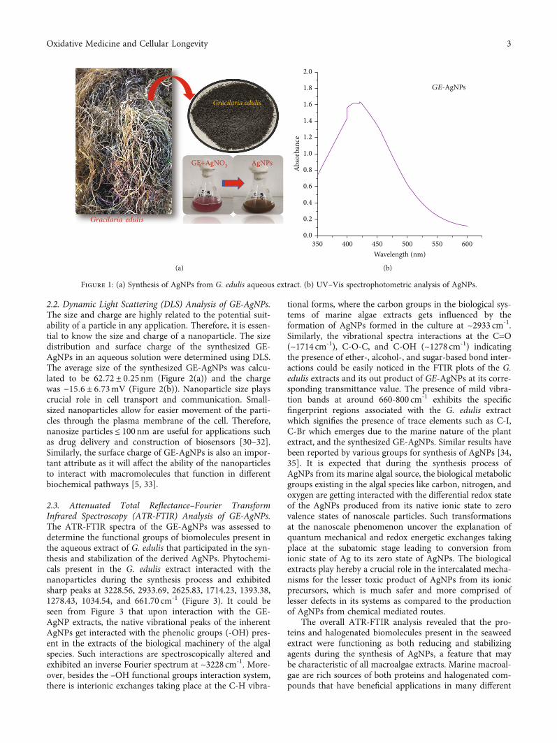
2.2. Dynamic Light Scattering (DLS) Analysis of GE-AgNPs.The size and charge are highly related to the potential suit-ability of a particle in any application. Therefore, it is essen-tial to know the size and charge of a nanoparticle. The sizedistribution and surface charge of the synthesized GE-AgNPs in an aqueous solution were determined using DLS.The average size of the synthesized GE-AgNPs was calcu-lated to be 62:72 ± 0:25nm (Figure 2(a)) and the chargewas −15:6 ± 6:73mV (Figure 2(b)). Nanoparticle size playscrucial role in cell transport and communication. Small-sized nanoparticles allow for easier movement of the parti-cles through the plasma membrane of the cell. Therefore,nanosize particles ≤ 100nm are useful for applications suchas drug delivery and construction of biosensors [30–32].Similarly, the surface charge of GE-AgNPs is also an impor-tant attribute as it will affect the ability of the nanoparticlesto interact with macromolecules that function in differentbiochemical pathways [5, 33].
2.3. Attenuated Total Reflectance–Fourier TransformInfrared Spectroscopy (ATR-FTIR) Analysis of GE-AgNPs.The ATR-FTIR spectra of the GE-AgNPs was assessed todetermine the functional groups of biomolecules present inthe aqueous extract of G. edulis that participated in the syn-thesis and stabilization of the derived AgNPs. Phytochemi-cals present in the G. edulis extract interacted with thenanoparticles during the synthesis process and exhibitedsharp peaks at 3228.56, 2933.69, 2625.83, 1714.23, 1393.38,1278.43, 1034.54, and 661.70 cm-1 (Figure 3). It could beseen from Figure 3 that upon interaction with the GE-AgNP extracts, the native vibrational peaks of the inherentAgNPs get interacted with the phenolic groups (-OH) pres-ent in the extracts of the biological machinery of the algalspecies. Such interactions are spectroscopically altered andexhibited an inverse Fourier spectrum at ~3228 cm-1. More-over, besides the –OH functional groups interaction system,there is interionic exchanges taking place at the C-H vibra-
tional forms, where the carbon groups in the biological sys-tems of marine algae extracts gets influenced by theformation of AgNPs formed in the culture at ~2933 cm-1.Similarly, the vibrational spectra interactions at the C=O(~1714 cm-1), C-O-C, and C-OH (~1278 cm-1) indicatingthe presence of ether-, alcohol-, and sugar-based bond inter-actions could be easily noticed in the FTIR plots of the G.edulis extracts and its out product of GE-AgNPs at its corre-sponding transmittance value. The presence of mild vibra-tion bands at around 660-800 cm-1 exhibits the specificfingerprint regions associated with the G. edulis extractwhich signifies the presence of trace elements such as C-I,C-Br which emerges due to the marine nature of the plantextract, and the synthesized GE-AgNPs. Similar results havebeen reported by various groups for synthesis of AgNPs [34,35]. It is expected that during the synthesis process ofAgNPs from its marine algal source, the biological metabolicgroups existing in the algal species like carbon, nitrogen, andoxygen are getting interacted with the differential redox stateof the AgNPs produced from its native ionic state to zerovalence states of nanoscale particles. Such transformationsat the nanoscale phenomenon uncover the explanation ofquantum mechanical and redox energetic exchanges takingplace at the subatomic stage leading to conversion fromionic state of Ag to its zero state of AgNPs. The biologicalextracts play hereby a crucial role in the intercalated mecha-nisms for the lesser toxic product of AgNPs from its ionicprecursors, which is much safer and more comprised oflesser defects in its systems as compared to the productionof AgNPs from chemical mediated routes.
The overall ATR-FTIR analysis revealed that the pro-teins and halogenated biomolecules present in the seaweedextract were functioning as both reducing and stabilizingagents during the synthesis of AgNPs, a feature that maybe characteristic of all macroalgae extracts. Marine macroal-gae are rich sources of both proteins and halogenated com-pounds that have beneficial applications in many different
Gracilaria edulis
AgNPsGE+AgNO3
Gracilaria edulis
(a)
3500.0
0.2
0.4
0.6
0.8
1.0
1.2
1.4
1.6
1.8
2.0
400 450 500 550Wavelength (nm)
Abso
rban
ce
600
GE-AgNPs
(b)
Figure 1: (a) Synthesis of AgNPs from G. edulis aqueous extract. (b) UV–Vis spectrophotometric analysis of AgNPs.
3Oxidative Medicine and Cellular Longevity

processes [36, 37]. Notably, the proteins present in the sea-weed could bind to the AgNPs via free amine groups, stabi-lizing clustered nanoparticles through surface-boundproteins [38]. The present results are also strongly supportedby the findings presented in previous studies [6, 16, 39].
2.4. X-Ray Diffraction (XRD) Analysis of GE-AgNPs. XRD isa rapid analytical technique primarily utilized for phaseidentification of a crystalline material and provides informa-tion on unit cell dimensions. Therefore, the analyzed mate-rial needs to be finely ground and homogenized todetermine its average bulk composition. The results of theXRD analysis of GE-AgNPs are presented in Figure 4. Thefigure represents a typical XRD diffractogram revealingBragg peaks predominantly at (angle 2θ) at 28.5, 33, 42,and 48.5 (in degree) for the AgNPs synthesized from G. edu-lis seaweed extract which corresponds to (100), (010), (200),and (002), respectively. Miller indices confirm the formationof crystalline elemental AgNPs with a face-centered cubic(FCC) lattice [40, 41]. Thus, the XRD pattern providesstrong evidence supporting the UV–Vis spectra and HR-TEM images of the GE-AgNPs.
2.5. HR-TEM Analysis of GE-AgNPs. HR-TEM micrographsconfirmed the spherical shape and polydisperse nature of the
GE-AgNPs and their attached biomoieties (Figure 5). TheHR-TEM pictographs exhibited that the GE-AgNPs wereregular and roughly spherical in shape, with blunt margins.The TEM images also revealed that the nanoparticles werenonagglomerated and freely scattered, making them a strongcandidate for biosensor development and drug delivery. TheDLS studies also support the properties of the GE-AgNPsrevealed in the TEM images, with approximately 80% ofthe DLS-scanned samples of the GE-AgNPs displaying a sizeof ~62 nm. Collectively, the dynamic light scattering studiesand HR-TEM micrographs confirm that the size of the GE-AgNPs is in the nanorange and that they possess a roughlyspherical morphology. This morphological shape and sizeindicate the potential efficiency of nanoparticles for drugconjugation and drug delivery [42–44].
2.6. Qualitative and Quantitative Phytochemical Analyses ofthe Seaweed Extract. The results of the qualitative and quan-titative phytochemical analyses of the aqueous G. edulis sea-weed extracts are summarized in Tables 1 and 2. Theanalysis revealed the presence of alkaloids, tannins, phenolic,flavonoids, and saponins, while glycosides, steroids, and ste-rols were absent. The identified compounds may representthe principal chemical ingredients that are involved in thebiosynthesis of AgNPs and define the potential of the nano-particles for different bioapplications [45–47]. Polyols, terpe-noids, phenols, flavones, and polysaccharides have beenpreviously reported to be the principle components in thebio reduction of silver and chloroaurate ions [48]. Impor-tantly, the potential absence of glycosides, steroids, and ste-rols in the G. edulis extracts in our study may be due tothe selective qualitative tests that were conducted and/orthe extraction procedures. The hypothetical mechanism ofthe synthesis of AgNPs may involve a cascade of complexantioxidant enzymes [49].
2.7. Antibacterial Activity of GE-AgNPs. Preliminary evalua-tion of the antibacterial activity of GE-AgNPs against sixpathogenic bacteria was conducted in an agar well diffusionassay (Table 1). Results of this assay indicated that the larg-est zone of inhibition was observed against Bacillus licheni-formis and the smallest against Salmonella typhimurium(Figure 6). Overall, significant antibacterial activity wasobserved against V. cholerae, E. coli, S. epidermidis, and S.dysenteriae. GE-AgNPs exhibited good bactericidal activityagainst both Gram-positive and Gram-negative bacteria. A
0.102468
10In
tens
ity (%
)
1 10 100 1000Size (d. nm)
Size distribution by intensity
10000
(a)
Tota
l cou
nts
–100 0 100 200Apparent zeta potential (mV)
Zeta potential distribution
0
100000
200000
300000
(b)
Figure 2: DLS analysis of AgNPs synthesized using GE extracts. (a) Average size distribution. (b) Surface charge.
4000
0.96
0.98
1.00
1.02
1.04
3000 2000 1000Wavenumber (cm–1)
Tran
smitt
ance
(%)
0
G. edulis extractGE-AgNPs
Figure 3: ATR-FTIR analysis of G. edulis extract and GE-AgNPs.
4 Oxidative Medicine and Cellular Longevity

microbroth dilution assay was also used to verify the anti-bacterial activity of GE-AgNPs and percent to determinepercent inhibition and the MICs for each of the pathogenicspecies of bacteria (Figure 7). The MICs was calculated ofsix pathogenic bacteria undertaken for the investigation suchas B. licheniformis (72:84 ± 1:54μg/mL), S. dysenteriae(130:67 ± 2:93μg/mL), E. coli (132:42 ± 3:08μg/mL), S.typhimurium (132:42 ± 3:08μg/mL), V. cholerae(65:58 ± 1:52μg/mL), and S. epidermidis (127:57 ± 3:08μg/mL). Significant growth inhibition (>94%) was observed inall six pathogenic bacterial species. Although the specificmechanism by which nanoparticles exhibit antibacterialactivity is not fully understood, different mechanisms ofaction have been reported in the literature. Structuralchanges in the bacterial membrane and ultimate cell deathas a result of penetration of nanoparticles into the cell walldue to their anchoring ability have been reported [39, 50,51]. Enzyme degradation, inactivation of structural proteins,and breakage of genetic materials by AgNPs have also beenproposed as mechanisms of action [38, 52, 53]. It has alsobeen suggested that several bacterial cellular enzymes areinactivated by the substantial attachment of Ag ions to –SH groups, a major chemical component of enzyme struc-ture present in the structure [42, 54, 55]. The intermittentinteraction of AgNPs with phosphorus and sulfur groups,interfering with the DNA replication processes and disman-tling the microbial nuclear system, has also been proposed[56–59]. In our study, the antibacterial results obtained withGE-AgNPs suggests that the seaweed extract-derived AgNPspossess an excellent antimicrobial with widespread potentialapplications.
2.8. Antioxidant Activity of GE-AgNPs. Abiotic stressinduces an overabundance of reactive oxygen species(ROS) that are highly toxic at high levels due to their strongoxidative properties. ROS can damage DNA and RNA, car-bohydrates, lipids, and proteins. Chronic oxidative stresscan also result in the induction of a variety of different dis-eases [60], and pharmaceuticals with antioxidant propertieshave been developed as an option to help minimize oxidativestress in humans. Notably, organisms have evolved an anti-oxidant system that can scavenge ROS and other free radi-cals. Delayed and poor absorption potential of exogenousantioxidants, difficulty in passing through cell membranes,and rapid degradation of antioxidants after their delivery,however, represent major challenges to the use of both nat-ural and synthetic antioxidant molecules. Unfortunately,the utilization of nanoparticles as an antioxidant has notbeen widely recognized and investigated and has been lim-ited to only a few types of nanomaterials [61]. Importantly,functional antioxidant AgNPs derived from the use of vari-ous natural extracts obtained from plant species appear torepresent a pivotal alternative due to their high stability, bio-compatibility, and targeted delivery [6]. Plants, algae, bacte-ria, and fungi possess a wide array of diverse phenoliccompounds and other secondary metabolites that are highlyuseful as reducing and stabilizing agents. In addition, theyalso possess excellent antioxidant properties. Therefore, theGE-AgNPs fabricated in the current study were extensively
30 40 502𝜃 (degree)
(211)
(002)(003)
(112)
(110)
(200)(010)
(100)
Inte
nsity
(arb
.u)
60 70
Figure 4: XRD analysis of GE-AgNPs.
1 𝜇m
Figure 5: HR-TEM micrograph of GE-AgNPs (indicated byarrows).
Table 1: Qualitative phytochemical analysis of an aqueous extractof G. edulis seaweed.
Phytoconstituent Observation
Alkaloids +
Tannins and phenolic compounds +
Glycoside -
Flavonoids +
Steroids and sterols -
Triterpenoids +
Saponins +
Notes: +: present; -: absent.
Table 2: Quantitative analysis of total phenolics and carbohydratesin an aqueous extract of G. edulis seaweed.
Phytochemical constituent mg/g dry weight (mean ± SD)TPC 7:27 ± 1:10TFC 4:70 ± 0:63
5Oxidative Medicine and Cellular Longevity

analyzed for their antioxidant properties (Figure 8). Theantioxidant activity of GE-AgNPs was evaluated using avariety of radical-scavenging assays against different typesof reactive radicals, including DPPH, hydroxyl ions, ABTS,and nitric oxide radicals (Figure 8). The DPPH radical-scavenging activity of GE-AgNPs was found to be dosedependent, displaying a maximum inhibition of 86.83% ata concentration of 50μg/mL. An IC50 value of 30:71 ± 0:22μg/mL was found to be significant, compared to the positivecontrol, ascorbic acid (IC50 value 10:33 ± 0:16μg/mL), thusdemonstrating the strong antioxidant property of GE-AgNPs. DPPH is commonly used to evaluate the antioxidantproperties of a compound. The radical scavenging activity ofGE-AgNPs was also tested against hydroxyl ion (OH-) radi-
cals, exhibiting a 94.20% scavenging capacity at 100μg/mLand an IC50 value of 43:85 ± 0:36μg/mL, compared to theascorbic acid standard which had an IC50 value of 20:43 ±0:03μg/mL. Hydroxyl ions readily disrupt disulfide bondsin proteins, resulting in unfolding and refolding into atypicalprotein structures [62]. Therefore, the current study pro-vides strong evidence for the potential use of GE-AgNPs asantioxidants in biological systems without adverse sideeffects.
ABTS is another free radical that is generally involved inoxidative damage to cells and polyphenols are capable ofminimizing the generation of ABTS. In the present study,the FT-IR analysis of the G. edulis extract identified the pres-ence of polyphenols in the synthesized GE-AgNPs. The
Bacillus licheniformis Shigella dysenteriae Salmonella typhimurium
Escherichia coli Vibrio cholerae Staphylococcus epidermidis
Figure 6: Antibacterial activity (Agar well method) of GE-AgNPs.
0.00
20.00
40.00
60.00
80.00
100.00
120.00
B. licheniformis S. dysenteriae E. coli S. typhimurium V. cholerae S. epidermidis
500 𝜇g/ml
250 𝜇g/ml
125 𝜇g/ml
62.5 𝜇g/ml
31.2 𝜇g/ml
Inhi
bitio
n (%
)
Antibacterial activity of GE-AgNPs (Micro broth dilution assay)
⁎⁎ ⁎
⁎
⁎
⁎
Figure 7: Antibacterial activity (microbroth dilution method) and MICs of GE-AgNPs. Error bar represents standard deviation of mean.∗p ≤ 0:05. Significant difference (p ≤ 0:05) within a parameter between two lines is denoted by asterisk.
6 Oxidative Medicine and Cellular Longevity

DPPH
Scav
engi
ng ac
tivity
(%)
0
20
40
60
80
100
10 𝜇g/mL 20 𝜇g/mL 30 𝜇g/mLConcentrations
40 𝜇g/mL 50 𝜇g/mL
GE-AgNPsAscorbic acid
⁎
⁎
⁎ ⁎ ⁎
(a)
Hydroxyl radical
Scav
engi
ng ac
tivity
(%)
0
20
40
60
80
100
20 𝜇g/mL 40 𝜇g/mL 60 𝜇g/mLConcentrations
80 𝜇g/mL 100 𝜇g/mL
GE-AgNPsAscorbic acid
⁎
⁎
⁎⁎⁎
(b)
Figure 8: Continued.
7Oxidative Medicine and Cellular Longevity

ABTS scavenging assay indicated 99.16% maximum scav-enging activity at 100μg/mL of GE-AgNPs and an IC50 valueof 64:77 ± 0:16μg/mL compared to an IC50 value of 16:31± 0:11μg/mL for ascorbic acid. Nitrite has harmful effectson human health due to its reaction with secondary aminesin cells, which forms toxic byproducts in human digestivesystems [62]. Thus, the activity of GE-AgNPs against nitricoxide radicals was evaluated. A maximum scavenging activ-ity of 62.43% was observed at 250μg/mL, and the IC50 valuewas determined to be 217:96 ± 1:42μg/mL; a value that isrepresentative of a moderately active antioxidant compoundagainst nitric oxide radicals. BHT was used as a positivestandard in the nitric oxide radical scavenging assay andwas determined to have an IC50 value of 51:74 ± 0:13μg/
mL. Oxidative stress is believed to play a crucial role indegenerative senescence. As a result, AgNPs with antioxi-dant capacity could provide a promising therapeutic forthe prevention of oxidative stress. G. edulis extracts havebeen previously reported to have antioxidant activity [63].The present results are also in good accordance to the resultsobtained from previous studies (Das et al., 2019; Kumaret al., 2020; Otunola and Afolayan, 2018; Ramamurthyet al., 2013b).
2.9. Biocompatibility and Cytotoxicity Analyses of GE-AgNPs.A methylthiazolyldiphenyl-tetrazolium bromide (MTT)assay was used to determine cell viability when evaluatingthe biocompatibility and cytotoxicity of GE-AgNPs. Normal
ABTS
Scav
engi
ng ac
tivity
(%)
0
20
40
60
80
100
10 𝜇g/mL 20 𝜇g/mLConcentrations
40 𝜇g/mL 60 𝜇g/mL 80 𝜇g/mL 100 𝜇g/mL
GE-AgNPsAscorbic acid
⁎⁎⁎
⁎
⁎
⁎
(c)
Nitric oxide
Scav
engi
ng ac
tivity
(%)
0
20
10
40
50
30
60
70
80
90
100
50 𝜇g/mL 100 𝜇g/mL 150 𝜇g/mLConcentrations
200 𝜇g/mL 250 𝜇g/mL
GE-AgNPsBHT
⁎⁎⁎
⁎
⁎
(d)
Figure 8: Antioxidant activity of GE-AgNPs in terms of radical scavenging activity: (a) DPPH, (b) hydroxyl ions, (c) ABTS, and (d) nitricoxide radicals. Error bar represents standard deviation of mean. ∗p ≤ 0:05. Significant difference (p ≤ 0:05) within a parameter between twolines is denoted by asterisk.
8 Oxidative Medicine and Cellular Longevity

fibroblast cells (L-929) were treated with GE-AgNPs in cul-ture for 24hour (h) at 37°C and cell viability was subse-quently assessed (Figure 9). Results indicated a dose-dependent effect of GE-AgNPs on L-929 cell viability. L-929 cells exposed to a 125μg/mL concentration of GE-AgNPs exhibited a 99.50% level of cell viability. However,the percent viability gradually reduced as the concentrationof GE-AgNPs increased with L-929 cell viabilitybeing74.38% when exposed to 1000μg/mL concentrationof GE-AgNPs. These results indicate that GE-AgNPs are rel-atively nontoxic to normal cells, even at a high concentra-tion. Consequently, these data demonstrate that GE-AgNPscan be potentially used for different biological applicationswithout detrimental effects on the health of cells. The bio-compatibility of GE-AgNPs with L-929 cells has been previ-ously reported [64–67].
In contrast to the biocompatibility of GE-AgNPs withnormal fibroblast cells, the viability assay of breast cancercells (MDA-MB-231) exposed to GE-AgNPs revealed signif-icantly higher levels of cytotoxicity (Figure 10). The breastcancer cells were exposed to ~1000μg/mL solution of GE-AgNPs for 24h, which reduced the cell viability to~21.23%. A time- and dose-dependent cytotoxicity forAgNPs derived from different biological sources againstMDA-MB-231 cells has been previously reported [68].
The percentage of viable cancer cells decreased as theconcentration of GE-AgNPs increased. A cell viability of63.81% and 94.06% was observed after exposure to a125μg/mL and 1.95μg/mL solution of GE-AgNPs, respec-tively. The MIC of GE-AgNPs was calculated as 344:27 ±2:56μg/mL against MDA-MB-231. These results indicatethat the MDA-MB-231 cell lines exhibit a concentration-dependent response with response to viability. The level ofcytotoxicity, however, did not appear to be time dependentas an identical percentage of viable cells was observed afterboth 24 and 48h exposure to the same concentration ofGE-AgNPs. The IC50 values for doxorubicin (used as a pos-itive control) were higher in MDA-MB-231 cells than in L-929 cells after 24 hr or 48 hr of exposure. In contrast, theIC50 values for GE-AgNP treatment were significantly lowerin MDA-MB-231 cells than in L-929 cells after exposure for24 or 48 h (Figure 10).
AgNPs induce cytotoxic effect due to their impact on dif-ferent metabolic pathways. Another study reported that thecytotoxicity of AgNPs results from an increase in ROS pro-duction [69]. Previous studies stated that the introductionof AgNPs into target cells could promote the overproductionof intracellular ROS, which activates apoptosis-associatedmetabolic pathways including p53, MAPK, and AKT apo-ptotic signaling pathways [70, 71]. Similar to other metalnanoparticles, AgNPs also promote oxidative stress in cellsby inducing the overproduction of ROS [70]. Mitochondriaare vital sources of apoptosis signals and the effect of Ag-NPs on mitochondrial membrane permeability results inthe loss of mitochondrial membrane integrity, leading tocaspase-dependent apoptotic cell death [72]. In addition to
0 200 400 600 800 1000GE-AgNPs concentrations (𝜇g/mL)
0102030405060708090
100110120
Cel
l via
bilit
y (%
)
L-929 24 hL-929 48 h
MDA-MB-231 24 hMDA-MB-231 48 h
Figure 9: Cell viability of MDA-MB-231 and L-929 after treatment with different concentrations of GE-AgNPs after 24 and 48 h.
0 200 400 600 800 1000
24 h
24 h
48 h
48 h
DO
XG
E-A
gNPs
DO
XG
E-A
gNPs
IC50 (𝜇g/mL ) values
MDA-MB 231L-929
Figure 10: Cell viability (IC50 value) of MDA-MB-231, L-929, aftertreatment with different nanoformulations (Dox: doxorubicin(control), GE-AgNPs) after 24 and 48 h. Error bar representsstandard deviation of mean. ∗p ≤ 0:05. Significant difference(p ≤ 0:05) within a parameter between two lines is denoted byasterisk.
9Oxidative Medicine and Cellular Longevity

AgNPs stimulating apoptosis in cells, it is more than likelythat future studies will reveal other mechanisms by whichAgNPs establish their cytotoxicity.
3. Materials and Methods
3.1. Collection and Preparation of Seaweed Extract. The redseaweed, G. edulis (Linnaeus), was collected from ChilikaLake, Odisha, India (19° 43′ 0″ N, 85° 19′ 0″ E) and trans-ported to a laboratory in a portable ice cooler. The harvestedseaweed was then thoroughly cleaned in running tap waterfollowed by distilled water to remove extraneous materialsand to substantially reduce the salt content, after which theseaweed was dried in a shady, open-air environment for 3–5 days. The dried seaweed was subsequently ground to a finepowder using a commercial-grade mixer grinder. Then, 5.0 gof the G. edulis seaweed powder was boiled in 50mL of ster-ilized Milli Q water for 20-30min and subsequently filteredthrough Whatman No. 1 filter paper. The filtered extractwas stored at -4°C until further use.
3.2. Synthesis of GE-AgNPs. A total of 10mL of seaweedextract was mixed with 90mL of a 1.0mM aqueous solutionof AgNO3 [45, 73] and incubated at room temperature on arotary shaker for 1 hr. A color change in the reaction solu-tion from light brick red to deep brown was noted by visualobservation and was used to confirm the completion of theAgNP synthesis. The synthesized AgNPs were pelleted bycentrifugation at 8000 rpm for 15min at 10°C. The obtainedAgNPs were dried and stored at 4°C for characterization andassessment of their bioactive properties.
3.3. Characterization of GE-AgNPs. The synthesis of theAgNPs using an aqueous extract of G. edulis was periodicallymonitored by UV-Vis spectrophotometer (Lambda 35R Per-kinElmer, USA) in the range of 350-600 nm. The UV-visiblespectra of the synthesis reaction solution were recorded as afunction of reaction time at a resolution of 1 nm at 25°C. Thesurface charge and average size of the AgNPs were analyzedusing a Zetasizer (ZS 90, Malvern, UK). The purified nano-particle samples were diluted tenfold in PBS (0.15M,pH7.2). Aliquots were sampled and placed in dynamic lightscattering (DLS) cuvettes and then evaluated for equivalentsize distribution, diameters, and zeta potential. Particlediameters were assessed at a scattering angle of 90° at 25°C.ATR-FTIR spectroscopy analysis of the, G. edulis aqueousextract and the synthesized GE-AgNPs was conducted tosubstantiate the potential role of the various functionalchemical groups present in the seaweed extracts on the mod-ification of the surface of the synthesized nanoparticles.ATR-FTIR was conducted on a Bruker ALPHA spectropho-tometer (Ettlinger, Germany) at a resolution of 4 cm−1. Thesamples were evaluated in the spectral region of 4000 to500 cm−1 by taking an average of 25 scans per sample. Forcontinuous observations, one drop of the sample was kepton the sample holder and the samples were scanned andthe obtained results were analyzed using OPUS software.The crystalline properties of the AgNPs were assessed usingan X-ray diffractometer (PANalytical X’Pert, Almelo, The
Netherlands) equipped with a Ni filter and a CuK(l = 1:54056Å) radiation source. The scanning rate was0.05°, while the diffraction angle varied from 20–80°. High-resolution transmission electron microscopy (Technai™F30 G2 STWIN, FEI, Lincoln, NE, USA) was used to observethe nanomorphology of the AgNPs. The synthesized AgNPswere placed on a coated copper grid with a 300 mesh sizeand observed at an accelerating voltage of 300 kV.
3.4. Qualitative and Quantitative Analyses of the SeaweedExtract. The qualitative phytochemical analysis of the G.edulis extract was performed following standard methods[63]. The obtained results were qualitatively expressed aspositive (+ve) or negative (-ve). The chemicals and reagentsused for the study were purchased from Sigma–Aldrich(India).
3.4.1. Total Phenol Content. Total phenol content (TPC) inthe seaweed extract was estimated using the Folin–Ciocalteumethod with slight modifications as described by Lim et al.(2007). The analysis was performed in triplicate. TPC wasexpressed as gallic acid equivalents (GAE) in mg/g sample.
The concentration of total phenolic compounds in theextract was determined using the following formula:
T = C ∗VM
, ð1Þ
where T is the total phenolic content mg/gm of seaweedsextract in GAE, C is the concentration of Gallic acid fromthe calibration curve in mg/mL, V is the volume of theextract in mL, and M is the Wt of the seaweeds extracts in g.
3.4.2. Total Flavonoid Content. Total flavonoid content(TFC) of the G. edulis extract was determined using an alu-minum chloride (AlCl3) colorimetric assay and expressed asmilligrams of quercetin equivalents per gram dry mass(mg·Q/g dw) [74]. The analysis was performed in triplicate.
3.5. Antibacterial Activity of the GE-AgNPs
3.5.1. Bacterial Strains. The six species of human pathogenicbacteria, Bacillus licheniformis (MTCC 7425), Salmonellatyphimurium (MTCC 3216), Vibrio cholerae (MTCC3904), Escherichia coli (MTCC 1098), Staphylococcus epider-midis (MTCC 3615), and Shigella dysenteriae (MTCC-9543)were used in the antibacterial assay. The bacterial strainswere purchased from MTCC, Pune.
3.5.2. Agar Well Diffusion and Microbroth Dilution Methods.A small colony of each targeted bacterial strain was inocu-lated from a stock agar slant into 2mL Muller Hinton(MH) broth medium (0.015% soluble starch, 0.2% beefextract, and 1.75% casamino acids) under proper asepticconditions. The inoculated tubes were incubated overnightat 37°C on a rotary shaker at 200 rpm.
The assessment of the antibacterial activity of the syn-thesized GE-AgNPs against the selected pathogenic bacteriawas conducted using a well diffusion assay with Muller Hin-ton Agar (MHA). Briefly, 100μL of each bacterium wasseeded over the prepared MHA plates. Test wells (5mm
10 Oxidative Medicine and Cellular Longevity

diameter and 3mm deep) were then made in the inoculatedagar medium using a sterile cork borer. Each well was thenfilled with 50μL of GE-AgNPs. Wells filled with 50μL of sil-ver nitrate (AgNO3) solution served as the control whilewells filled with the antibiotic, gentamicin, were used as apositive control. The inoculated plates were kept in an incu-bator at 37°C for 24 h. Following the period of incubation,the diameter of inhibition zones was measured and a zonediameter ≥ 8mm was recorded as a positive antibacterialactivity.
Antibacterial assessment was carried out using themicrobroth dilution method. The minimum inhibitory con-centration (MIC) of the GE-AgNPs on bacterial strains wasalso assessed [75]. Inhibition ≥ 90% in the microbroth dilu-tion assay was used as an indication of good antibacterialactivity, and additional experiments were carried out forMIC estimation. The test inoculum (190μL; A600 = 0:1) wereincubated in 10μL of different concentrations (500-31.25mg/mL; twofold dilution) of the GE-AgNPs until thelevel of inhibition was found to be <50%. The assays wereconducted in 96-well plates, and microbial growth wasdetermined in a microplate reader (Bio-Rad, USA) at600nm. The numerical MIC values were calculated usingIC50/IC90 Laboratory Excel Calculation formulas andexpressed as IC50. All of the assays were conducted in tripli-cate, and zones of inhibition were expressed in amean ± SD.
3.6. Antioxidant Activity of the GE-AgNPs. The antioxidantactivity of the GE-AgNPs was assessed by its radical scav-enging ability.
3.6.1. DPPH Radical Scavenging Activity. The radical scav-enging activity of GE-AgNPs was determined using the1,1-diphenyl-2-picryl-hydrazil (DPPH) assay with slightmodification (Arul Kumar et al., 2018; Lim et al., 2007). Dif-ferent concentrations (10, 20, 30, 40, and 50μg/mL) of GE-AgNPs were used in the assay. Ascorbic acid in equivalentconcentrations was used as a positive control, and resultswere expressed as percentage (%) radical scavenging activity.The MIC for DPPH radical scavenging activity was also cal-culated and expressed as an IC50.
3.6.2. Hydroxyl Radical Scavenging Activity. Hydroxyl (OH-)radical scavenging activity of GE-AgNPs was evaluated aspreviously described [76] using different concentrations(20, 40, 60, 80, and 100μg/mL) of GE-AgNPs. Ascorbic acidat equivalent concentrations was used as a positive control.The MIC for hydroxyl radical scavenging activity was alsocalculated and expressed as an IC50.
3.6.3. 2,2-Azino-bis(3-ethylbenzothiozoline-6-sulfonic acid)Diammonium Salt (ABTS) Radical Scavenging Activity.ABTS radical scavenging activity of the GE-AgNPs was alsodetermined using a radical cation decolorization assay aspreviously described [77] using different concentrations(20, 40, 60, and 80μg/mL) of GE-AgNPs. Ascorbic acid atequivalent concentrations was used as a positive control.The MIC for ABTS radical scavenging activity was also cal-culated and expressed as an IC50.
3.6.4. Nitric Oxide Radical (NO∗) Scavenging Activity. Nitricoxide radical scavenging activity of the GE-AgNPs was eval-uated using the method described by Garrat (1964) withslight modification. Briefly, sodium nitroprusside (Na2[Fe(CN)5NO]2H2O) in aqueous solution generates nitric oxidespontaneously at physiological pH, which immediatelyinteracts with oxygen to produce nitrite ions (NO2-), whichcan be determined by the Griess-Ilosvay reaction. A stan-dard method [63] to evaluate the NO∗ scavenging activitywas also used. Different concentrations (50, 100, 150, 200,and 250μg/mL) of GE-AgNPs were used in the assay, andBHT was used as a positive control. The MIC for NO2- rad-ical scavenging activity was also calculated and expressed asan IC50.
3.7. Biocompatibility and Cytotoxicity Analysis of GE-AgNPs
3.7.1. Cell Culture. A normal fibroblast cell line (L-929) anda breast cancer cell line (MDA-MB 231) were used in thebiocompatibility and cytotoxicity assays. Both cell lines wereseeded on Dulbecco’s modified Eagle’s medium and M-199medium supplemented with 10% fetal bovine serum (FBS),as well as streptomycin sulfate and benzyl antibiotics at afinal concentration of 100μg/mL and 100U/mL, respec-tively. Cell cultures were incubated at 37°C (5% CO2) for24 h, for the duration of the assays. The cells were trypsi-nized using 0.25% Trypsin-EDTA at a 70 to 80% confluence.Cells were counted and then placed in a 96-well enzyme-linked immunosorbent assay (ELISA) plate at a density of5 × 103 cells/well to conduct MTT assay. All cell culture che-micals are purchased from Sigma–Aldrich (India).
3.7.2. MTT Assay. Biocompatibility and cytotoxicity wereevaluated using a MTT colorimetric assay after 24 and48 h. incubation of the cell lines with the GE-AgNPs. Whenthe cells were at 90% confluency, the media was removedand the cells were exposed with fresh medium containingdifferent concentrations of GE-AgNPs (viz., 100, 200, 400,600, 800, and 1000μg/mL) and was further incubated for24 h. Doxorubicin was employed as a positive control. Simi-lar to the GE-AgNPs, various concentrations of DOX wasused to see its efficacy along with the synthesized GE-AgNPs. A stock solution of MTT (1mg/mL) in PBS was pre-pared immediately prior to use. A 500μL volume of theMTT solution (50μg/mL MTT in the culture medium) wasadded to each culture dish and left uncovered. Cells wereincubated for 3 hr, after which the reduced formazan wasextracted with 500μL of DMSO and absorbance was mea-sured at 595nm in a microtiter plate reader (Bio-Rad,USA). Cell viability was assessed as the percentage absorp-tion of treated cells relative to the untreated and controlcells.
3.8. Statistical Analysis. All assays in this study were per-formed in triplicate. The results of the antioxidant assaysare presented as a percentage inhibition, while the cytotoxic-ity results are presented as % viability, relative to the control.The antioxidant and cytotoxicity assay data for the differenttreatment groups vs. the controls were statistically evaluatedusing Student’s t-test (p ≤ 0:05).
11Oxidative Medicine and Cellular Longevity

4. Conclusion
Marine macroalgae or sea weed G. edulis has momentousattributes in the green synthesis process of metal nanoparti-cles, like other biological resources such as plant, bacteria,fungi, macrofungi or mushrooms, and yeast. Due to theencouraging involvement of algae in the nanotechnologyadvancement, the separate branch known as phyconano-technology is growing enormously to substantiate the differ-ent biomedical, agriculture, and environmental issues.Various studies on the biosynthesis of nanoparticles usingseaweed extracts have been conducted. In the current inves-tigations, the physiochemical characterization of the synthe-sized AgNPs demonstrated the stable synthesis of AgNPsthat can potentially be used in different applications. Theantibacterial, antioxidant, biocompatibility, and cytotoxicityof the AgNPs indicate their potential commercial utility inbiomedical and pharmaceutical industries. The use of sea-weed extract in nanoparticle biosynthesis is highly advanta-geous due to the presence of a variety of secondarymetabolites in the extract that affect the properties of thesynthesized nanoparticles and exhibit low cytotoxicity tohealthy cells. The use of “green-based” synthesis of nanopar-ticles is compatible with large-scale production and smoothdownstream processing. Further studies are warranted andnecessary to explore the use of seaweed extracts in nanotech-nological applications and to fully understand the propertiesof seaweed-fabricated metal nanoparticles, their mechanismof action, and their potential applications in food, health,and environmental industries. Comprehensively, the nano-biotechnology that utilizes the sources from algae andblue-green algae to synthesize nanomaterials is in the bud-ding stage, and further research and development arenecessary.
Data Availability
The data supporting the reported results are available uponrequest from the first and corresponding author.
Additional Points
Research Highlights: (1) Green synthesis of silver nanoparti-cles from marine sea weed Gracilaria edulis for efficient usein biomedical industry sector is highly demanding for itsecofriendly and less toxic byproduct production, (2) Theuse of marine sea weed for such green synthesis is highlyattractive to a combinatorial approach in addition of naturalcompounds with nanoparticles, (3) Silver nanoparticles alsoexhibited potential antibacterial activity against Bacilluslicheniformis, Shigella dysenteriae, Escherichia coli, Salmo-nella typhimurium, Vibrio cholerae, and Staphylococcus epi-dermidis, (4) AgNPs performed high efficacy of DPPH,hydroxyl radical, ABTS, and nitric oxide radical scavengingactivity, (5) AgNPs are highly biocompatible to normalfibroblast cell line L-929 and cytotoxic against breast cancer(MDA-MB-231) cells
Ethical Approval
Experimental research/field studies on plants comply withrelevant institutional, national, and international guidelinesand legislation.
Conflicts of Interest
The authors declare that they have no conflicts of interest.
Authors’ Contributions
Y.K.M and A.K.M have conceptualized, designed, and per-form experiments and wrote the manuscript. D.N andT.K.M characterized the samples and wrote the manuscript.S.K.A and B.P. analyzed the characterization data andrevised the manuscript. A.B, K.M, and S.M. helped in thereview and editing of the manuscript. All the authors readand approved the final manuscript. Yugal Kishore Mohantaand Awdhesh Kumar Mishra contributed equally to thiswork.
Acknowledgments
The authors are thankful to the University of Science andTechnology Meghalaya, India, University of Nizwa, Oman,and Saveetha Institute of Medical and Technical Sciences(SIMATS), Chennai, for providing necessary support to con-duct the research.
References
[1] A. Batool, F. Menaa, K. B. Ali, B. Uzair, and B. Menaa, “Prog-ress and prospects in translating nanobiotechnology in medi-cal theranostics,” Current Nanoscience, vol. 15, pp. 1–23, 2019.
[2] A. Sharma, S. Sharma, K. Sharma et al., “Algae as crucialorganisms in advancing nanotechnology: a systematic review,”Journal of Applied Phycology, vol. 28, no. 3, pp. 1759–1774,2016.
[3] F. Menaa, “When pharma meets nano or the emerging era ofnanopharmaceuticals,” Pharm. Anal. Acta., vol. 4, p. 223, 2013.
[4] B. Menaa, “The importance of nanotechnology in biomedicalsciences,” J. Biotechnol. Biomater, vol. 1, p. 105e, 2011.
[5] D. Nayak, A. P. Minz, S. Ashe et al., “Synergistic combinationof antioxidants, silver nanoparticles and chitosan in a nano-particle based formulation: characterization and cytotoxiceffect on MCF-7 breast cancer cell lines,” Journal of Colloidand Interface Science, vol. 470, pp. 142–152, 2016.
[6] P. Jakinala, N. Lingampally, B. H. Id, and R. Z. Sayyed, “Silvernanoparticles from insect wing extract: biosynthesis and eval-uation for antioxidant and antimicrobial potential,” PLoS One,vol. 16, no. 3, pp. 1–15, 2021.
[7] B. Uzair, A. Liaqat, H. Iqbal et al., “Green and cost-effectivesynthesis of metallic nanoparticles by algae: safe methods fortranslational medicine,” Bioengineering, vol. 7, no. 4, 2020.
[8] C. R. Patra, R. Bhattacharya, D. Mukhopadhyay, andP. Mukherjee, “Fabrication of gold nanoparticles for targetedtherapy in pancreatic cancer,” Advanced Drug DeliveryReviews, vol. 62, no. 3, pp. 346–361, 2010.
[9] P. D. Shankar, S. Shobana, I. Karuppusamy et al., “A review onthe biosynthesis of metallic nanoparticles (gold and silver)
12 Oxidative Medicine and Cellular Longevity
using bio-components of microalgae: formation mechanismand applications,” Enzyme and Microbial Technology, vol. 95,pp. 28–44, 2016.
[10] Y. Wang, X. Dong, L. Zhao et al., “Facile and green fabricationof carrageenan-silver nanoparticles for colorimetric determi-nation of Cu 2 + and S2−,” Nanomaterials, vol. 10, pp. 1–12,2020.
[11] S. R. Vijayan, P. Santhiyagu, R. Ramasamy et al., “Seaweeds: aresource for marine bionanotechnology,” Enzyme and Micro-bial Technology, vol. 95, pp. 45–57, 2016.
[12] S. Pugazhendhi, P. Sathya, P. K. Palanisamy, andR. Gopalakrishnan, “Synthesis of silver nanoparticles throughgreen approach using _Dioscorea alata_ and their characteri-zation on antibacterial activities and optical limiting behavior,”Journal of Photochemistry and Photobiology. B, vol. 159,pp. 155–160, 2016.
[13] S. Pugazhendhi, E. Kirubha, P. K. Palanisamy, andR. Gopalakrishnan, “Synthesis and characterization of silvernanoparticles from _Alpinia calcarata_ by Green approachand its applications in bactericidal and nonlinear optics,”Applied Surface Science, vol. 357, pp. 1801–1808, 2015.
[14] P. Singh, H. Singh, Y. J. Kim, R. Mathiyalagan, C. Wang, andD. C. Yang, “Extracellular synthesis of silver and gold nano-particles by _Sporosarcina koreensis_ DC4 and their biologicalapplications,” Enzyme and Microbial Technology, vol. 86,pp. 75–83, 2016.
[15] S. Iravani, “Green synthesis of metal nanoparticles usingplants,” Green Chemistry, vol. 13, no. 10, pp. 2638–2650, 2011.
[16] C. Vanlalveni, S. Lallianrawna, A. Biswas, M. Selvaraj,B. Changmai, and S. L. Rokhum, “Green synthesis of silvernanoparticles using plant extracts and their antimicrobialactivities: a review of recent literature,” RSC Advances,vol. 11, no. 5, pp. 2804–2837, 2021.
[17] K. B. Narayanan and N. Sakthivel, “Biological synthesis ofmetal nanoparticles by microbes,” Adv Colloid Interface Sci,vol. 156, no. 1-2, pp. 1–13, 2010.
[18] D. K. Tripathi, P. Ahmad, S. Sharma, D. K. Chauhan, and N. K.Dubey, Nanomaterials in Plants, Algae, and Microorganisms:Concepts and Controversies, Academic Press, Cambridge,MA, USA, 2017.
[19] D. Sharma, S. Kanchi, and K. Bisetty, “Biogenic synthesis ofnanoparticles: a review,” Arabian Journal of Chemistry,vol. 12, no. 8, pp. 3576–3600, 2019.
[20] H.-M. D. Wang, X.-C. Li, D.-J. Lee, and J.-S. Chang, “Potentialbiomedical applications of marine algae,” Bioresource Technol-ogy, vol. 244, pp. 1407–1415, 2017.
[21] M. I. Khan, J. H. Shin, and J. Kim, “The promising future ofmicroalgae: current status, challenges, and optimization of asustainable and renewable industry for biofuels, feed, andother products,” Microbial Cell Factories, vol. 17, no. 1, p. 36,2018.
[22] E. M. Cabral, M. Oliveira, J. R. M. Mondala, J. Curtin, B. K.Tiwari, and M. Garcia-Vaquero, “Antimicrobials from sea-weeds for food applications,” Marine Drugs, vol. 19, no. 4,p. 211, 2021.
[23] N. Asmathunisha and K. Kathiresan, “A review on biosynthe-sis of nanoparticles by marine organisms,” Colloids Surfaces BBiointerfaces., vol. 103, pp. 283–287, 2013.
[24] S. R. Vijayan, P. Santhiyagu, M. Singamuthu, N. Kumari Ahila,R. Jayaraman, and K. Ethiraj, “Synthesis and characterizationof silver and gold nanoparticles using aqueous extract of sea-
weed, Turbinaria conoides, and their antimicrofouling activ-ity,” Sci. World J, vol. 2014, article ???, pp. 1–10, 2014.
[25] E. Sokoła-Wysoczańska, T. Wysoczański, J. Wagner et al.,“Polyunsaturated fatty acids and their potential therapeuticrole in cardiovascular system disorders—a review,” Nutrients,vol. 10, no. 10, pp. 1–21, 2018.
[26] T. Rosemary, A. Arulkumar, S. Paramasivam, A. Mondragon-Portocarrero, and J. M. Miranda, “Biochemical, micronutrientand physicochemical properties of the dried red seaweeds <i>Gracilaria edulis</i> and <i>Gracilaria corticata</i>,”Mol-ecules, vol. 24, no. 12, pp. 1–14, 2019.
[27] J. Ashraf, M. Ansari, H. Khan, M. Alzohairy, and I. Choi,“Green synthesis of silver nanoparticles and characterizationof their inhibitory effects on AGEs formation using bio-physical techniques,” Scientific Reports, vol. 6, no. 1,p. 20414, 2016.
[28] Y. K. Mohanta, D. Nayak, K. Biswas et al., “Silver nanoparticlessynthesized using wild mushroom show potential antimicro-bial activities against food borne pathogens,” Molecules,vol. 23, pp. 1–18, 2018.
[29] Y. K. Mohanta, S. K. Panda, R. Jayabalan, N. Sharma, A. K.Bastia, and T. K. Mohanta, “Antimicrobial, antioxidant andcytotoxic activity of silver nanoparticles synthesized by leafextract of Erythrina suberosa (Roxb),” Frontiers in MolecularBiosciences, vol. 4, pp. 1–9, 2017.
[30] S. Mukherjee, D. Chowdhury, R. Kotcherlakota, S. Patra, andB. Vinothkumar, “Potential theranostics application of bio-synthesized silver nanoparticles (4-in-1 system),” Theranos-tics, vol. 4, no. 3, pp. 316–335, 2014.
[31] P. Tan, H. Li, J. Wang, and S. C. B. Gopinath, “Silver nanopar-ticle in biosensor and bioimaging : clinical perspectives,” Bio-technology and Applied Biochemistry, vol. 68, no. 6,pp. 1236–1242, 2021.
[32] K. V. Alex, P. T. Pavai, R. Rugmini, M. S. Prasad, K. Kamakshi,and K. C. Sekhar, “Green synthesized ag nanoparticles for bio-sensing and photocatalytic applications,” ACS Omega, vol. 5,no. 22, pp. 13123–13129, 2020.
[33] S. H. Lee and B. Jun, “Silver nanoparticles : synthesis andapplication for nanomedicine,” International Journal of Molec-ular Sciences, vol. 20, no. 4, pp. 1–24, 2019.
[34] S. P. Vinay, N. Chandrasekhar, and C. P. Chandrappa, “Eco-friendly approach for the green synthesis of silver nanoparti-cles using flower extracts of Sphagneticola trilobata and studyof antibacterial activity,” Int. J. Pharm. Biol. Sci., vol. 7,pp. 145–152, 2017.
[35] P. Devaraj, P. Kumari, C. Aarti, and A. Renganathan, “Synthe-sis and characterization of silver nanoparticles using cannon-ball leaves and their cytotoxic activity against MCF-7 cellline,” J. Nanotechnol., vol. 2013, pp. 1–5, 2013.
[36] A. Jesus, M. Correia-da-Silva, C. Afonso, M. Pinto, andH. Cidade, “Isolation and potential biological applications ofhaloaryl secondary metabolites from macroalgae,” MarineDrugs, vol. 17, no. 2, pp. 1–19, 2019.
[37] M. T. Cabrita, C. Vale, and A. P. Rauter, “Halogenated com-pounds from marine algae,” Marine Drugs, vol. 8, no. 8,pp. 2301–2317, 2010.
[38] G. Das, J. K. Patra, T. Debnath, A. Ansari, and H. S. Shin,“Investigation of antioxidant, antibacterial, antidiabetic, andcytotoxicity potential of silver nanoparticles synthesized usingthe outer peel extract of Ananas comosus (L.),” PLoS One,vol. 14, pp. 1–19, 2019.
13Oxidative Medicine and Cellular Longevity

[39] D. H. Nguyen, T. N. N. Vo, N. T. Nguyen, Y. C. Ching, andT. T. H. Thi, “Comparison of biogenic silver nanoparticlesformed by Momordica charantia and Psidium guajava leafextract and antifungal evaluation,” PLoS One, vol. 15, no. 9,p. e0239360, 2020.
[40] K. D. Arunachalam, S. Suhashani, and K. A. Sathesh, “Woundhealing and Antigenotoxic activities of Aegle marmelos withrelation to its antioxidant properties,” Journal of PharmacyResearch, vol. 5, pp. 1492–1502, 2012.
[41] C. Dipankar and S. Murugan, “The green synthesis, character-ization and evaluation of the biological activities of silvernanoparticles synthesized from _Iresine herbstii_ leaf aqueousextracts,” Colloids Surfaces B Biointerfaces., vol. 98, pp. 112–119, 2012.
[42] M. Yousefzadi, Z. Rahimi, and V. Ghafori, “The green synthe-sis, characterization and antimicrobial activities of silver nano-particles synthesized from green alga Enteromorpha flexuosa(wulfen),” J. Agardh, Mater. Lett, vol. 137, pp. 1–4, 2014.
[43] K. Biswas, Y. K. Mohanta, V. B. Kumar et al., “Nutritionalassessment study and role of green silver nanoparticles inshelf- life of coconut endosperm to develop as functionalfood,” Saudi J. Biol. Sci, vol. 27, no. 5, pp. 1280–1288, 2020.
[44] Y. Mohanta, K. Biswas, S. Jena, A. Hashem, E. A. Allah, andT. Mohanta, “Anti-biofilm and antibacterial activities of silvernanoparticles synthesized by the reducing activity of phyto-constituents present in the Indian medicinal plants,” Frontiersin Microbiology, vol. 11, pp. 1–15, 2020.
[45] V. Sri, A. Pugazhendhi, and K. Gopalakrishnan, “Biofabrica-tion and characterization of silver nanoparticles using aqueousextract of seaweed _Enteromorpha compressa_ and its bio-medical properties,” Biotechnol. Reports, vol. 14, pp. 1–7, 2017.
[46] P. Khanna, A. Kaur, and D. Goyal, “Algae-based metallicnanoparticles: Synthesis, characterization and applications,”Journal of Microbiological Methods, vol. 163, p. 105656, 2019.
[47] R. I. Priyadharshini, G. Prasannaraj, N. Geetha, andP. Venkatachalam, “Microwave-mediated extracellular syn-thesis of metallic silver and zinc oxide nanoparticles usingmacro-algae (Gracilaria edulis) extracts and its anticanceractivity against human PC3 cell lines,” Applied Biochemistryand Biotechnology, vol. 174, no. 8, pp. 2777–2790, 2014.
[48] J. Huang, Q. Li, D. Sun et al., “Biosynthesis of silver and goldnanoparticles by novel sundried Cinnamomum camphoraleaf,” Nanotechnology, vol. 18, no. 10, p. 105104, 2007.
[49] E. O. Mikhailova, “Silver nanoparticles: mechanism of actionand probable bio-application,” J. Funct. Biomater., vol. 11,no. 4, p. 84, 2020.
[50] I. Sondi and B. Salopek-Sondi, “Silver nanoparticles as antimi-crobial agent: a case study on E. coli as a model for Gram-negative bacteria,” J Colloid Interface Sci, vol. 275, 2004.
[51] M. S. Jabir, A. A. Hussien, G. M. Sulaiman et al., “Green syn-thesis of silver nanoparticles from Eriobotrya japonica extract:a promising approach against cancer cells proliferation,inflammation, allergic disorders and phagocytosis induction,”Artif. Cells, Nanomedicine Biotechnol, vol. 49, pp. 48–60, 2021.
[52] M. Guzman, J. Dille, and S. Godet, “Synthesis and antibacterialactivity of silver nanoparticles against gram- positive andgram-negative bacteria,”Nanomedicine: Nanotechnology, Biol-ogy and Medicine, vol. 8, no. 1, pp. 37–45, 2012.
[53] J. K. Patra and K.-H. Baek, “Antibacterial activity and syner-gistic antibacterial potential of biosynthesized silver nanopar-ticles against foodborne pathogenic bacteria along with its
anticandidal and antioxidant effects,” Frontiers in Microbiol-ogy, vol. 8, pp. 1–14, 2017.
[54] Y. Mohanta, S. Panda, K. Biswas et al., “Biogenic synthesis ofsilver nanoparticles from Cassia fistula (Linn.): In vitro assess-ment of their antioxidant, antimicrobial and cytotoxic activi-ties,” IET Nanobiotechnology., vol. 10, no. 6, pp. 438–444,2016.
[55] R. A. Ismail, G. M. Sulaiman, M. H. Mohsin, and A. H. Saa-doon, “Preparation of silver iodide nanoparticles using laserablation in liquid for antibacterial applications,” IET Nanobio-technology., vol. 12, no. 6, pp. 781–786, 2018.
[56] B. Singh, C. Prateeksha, A. Rao, D. Rawat, and B. Upreti,“Agricultural nanotechnologies: what are the current possibil-ities?,” Current Science, vol. 10, no. 2, pp. 124–127, 2015.
[57] P. Ramesh, T. Kokila, and D. Geetha, “Plant mediated greensynthesis and antibacterial activity of silver nanoparticlesusing _Emblica officinalis_ fruit extract,” Spectrochimica Acta.Part A, Molecular and Biomolecular Spectroscopy, vol. 142,pp. 339–343, 2015.
[58] Y. Dong, H. Zhu, Y. Shen, W. Zhang, and L. Zhang, “Antibac-terial activity of silver nanoparticles of different particle sizeagainst Vibrio natriegens,” PLoS One, vol. 14, no. 9, pp. 1–12,2019.
[59] M. Wypij, T. Jedrzejewski, J. Trzcin’ska-Wencel,M. Ostrowski, M. Rai, and P. Golinska, “Green synthesized sil-ver nanoparticles : antibacterial and anticancer activities , bio-compatibility, and analyses of surface-attached proteins,”Front. Microbiol, vol. 12, pp. 1–17, 2021.
[60] C. Ramamurthy, M. Padma, I. M. Samadanam et al., “Theextra cellular synthesis of gold and silver nanoparticles andtheir free radical scavenging and antibacterial properties,” Col-loids and Surfaces. B, Biointerfaces, vol. 102, pp. 808–815, 2013.
[61] H. Kumar, K. Bhardwaj, E. Nepovimova et al., “Antioxidantfunctionalized nanoparticles: a combat against oxidativestress,” Nanomaterials, vol. 10, no. 7, pp. 1–31, 2020.
[62] A. Phaniendra, D. B. Jestadi, and L. Periyasamy, “Free radicals:properties, sources, targets, and their implication in variousdiseases,” Indian Journal of Clinical Biochemistry, vol. 30,no. 1, pp. 11–26, 2015.
[63] A. Arulkumar, T. Rosemary, S. Paramasivam, andR. Rajendran, “Phytochemical composition, _in vitro_ antiox-idant, antibacterial potential and GC-MS analysis of red sea-weeds ( _Gracilaria corticata_ and _Gracilaria edulis_ ) fromPalk Bay, India,” Biocatalysis and Agricultural Biotechnology,vol. 15, pp. 63–71, 2018.
[64] M. Składanowski, P. Golinska, K. Rudnicka, H. Dahm, andM. Rai, “Evaluation of cytotoxicity, immune compatibilityand antibacterial activity of biogenic silver nanoparticles,”Medical Microbiology and Immunology, vol. 205, no. 6,pp. 603–613, 2016.
[65] M. Adabi, M. Naghibzadeh, M. Adabi et al., “Biocompatibilityand nanostructured materials: applications in nanomedicine,”Artif. Cells, Nanomedicine Biotechnol, vol. 45, pp. 833–842,2017.
[66] P. Mathur, S. Jha, S. Ramteke, and N. K. Jain, “Pharmaceuticalaspects of silver nanoparticles,” Artif. Cells, Nanomedicine Bio-technol, vol. 46, pp. 115–126, 2018.
[67] Y. K. Mohanta, S. K. Panda, A. K. Bastia, and T. K. Mohanta,“Biosynthesis of silver nanoparticles from <i>Protium serra-tum</i> and investigation of their potential impacts on foodsafety and control,” vol. 8, pp. 1–10, 2017.
14 Oxidative Medicine and Cellular Longevity

[68] M. Akter, M. T. Sikder, M. M. Rahman et al., “A systematicreview on silver nanoparticles-induced cytotoxicity: physico-chemical properties and perspectives,” Journal of AdvancedResearch, vol. 9, pp. 1–16, 2018.
[69] R. Foldbjerg, P. Olesen, M. Hougaard, D. Dang, H. Hoffmann,and H. Autrup, “PVP-coated silver nanoparticles and silverions induce reactive oxygen species, apoptosis and necrosisin THP-1 monocytes,” Toxicology Letters, vol. 190, no. 2,pp. 156–162, 2009.
[70] Y. Li, M. Guo, Z. Lin, T. Chen, and B. Zhu, “Polyethylenimine-functionalized silver nanoparticle-based co-delivery of pacli-taxel to induce HepG2 cell apoptosis,” International Journalof Nanomedicine, vol. Volume 11, pp. 6693–6702, 2016.
[71] M. S. Jabir, Y. M. Saleh, G. M. Sulaiman et al., “Green synthesisof silver nanoparticles using <i>Annona muricata</i> extractas an inducer of apoptosis in cancer cells and inhibitor forNLRP3 inflammasome via enhanced autophagy,” Nanomate-rials, vol. 11, no. 2, pp. 1–22, 2021.
[72] M. Jing, K. Ah, I. Kyung et al., “Silver nanoparticles induce oxi-dative cell damage in human liver cells through inhibition ofreduced glutathione and induction of mitochondria-involvedapoptosis,” Toxicology Letters, vol. 201, no. 1, pp. 92–100,2011.
[73] P. K. S. Senthamil and S. M. Govindaraju, “Seaweed-mediatedbiosynthesis of silver nanoparticles using Gracilaria corticatafor its antifungal activity against Candida spp,” AppliedNanoscience, vol. 3, no. 6, pp. 495–500, 2013.
[74] D. Marinova, F. Ribarova, and M. Atanasova, “Total phenolicsand flavonoids in Bulgarian fruits and vegetables,” J. Univ.Chem. Technol. Metall., vol. 40, pp. 255–260, 2005.
[75] S. K. Panda, Y. K. Mohanta, L. Padhi, andW. Luyten, “Antimi-crobial activity of select edible plants from Odisha, Indiaagainst food-borne pathogens,” LWT, vol. 113, p. 108246,2019.
[76] P. Tanamatayarat, “Antityrosinase, antioxidative activities,and brine shrimp lethality of ethanolic extracts from _Protiumserratum_ (Wall. ex Colebr.) Engl.,” Asian Pacific Journal ofTropical Biomedicine, vol. 6, no. 12, pp. 1050–1055, 2016.
[77] P. Vijayabaskar and V. Shiyamala, “Antioxidant properties ofseaweed polyphenol from _Turbinaria ornata_ (Turner) J.Agardh, 1848,” Asian Pac J Trop Biomed, vol. 2, no. 1,pp. S90–S98, 2012.
15Oxidative Medicine and Cellular Longevity

Review ArticleEffects of Metal Oxide Nanoparticles in Zebrafish
Marta d’Amora ,1,2 Tiziana Julia Nadjeschda Schmidt,2 Soultana Konstantinidou ,2
Vittoria Raffa ,2 Francesco De Angelis ,1 and Francesco Tantussi 1
1Istituto Italiano di Tecnologia, Via Morego 30, 16163 Genova, Italy2Department of Biology, University of Pisa, S.S. 12 Abetone e Brennero 4, 56127 Pisa, Italy
Correspondence should be addressed to Marta d’Amora; [email protected]
Received 17 December 2021; Accepted 18 January 2022; Published 4 February 2022
Academic Editor: Dragica Selakovic
Copyright © 2022 Marta d’Amora et al. This is an open access article distributed under the Creative Commons AttributionLicense, which permits unrestricted use, distribution, and reproduction in any medium, provided the original work isproperly cited.
Metal oxide nanoparticles (MO NPs) are increasingly employed in many fields with a wide range of applications from industriesto drug delivery. Due to their semiconducting properties, metal oxide nanoparticles are commonly used in the manufacturing ofseveral commercial products available in the market, including cosmetics, food additives, textile, paint, and antibacterialointments. The use of metallic oxide nanoparticles for medical and cosmetic purposes leads to unavoidable human exposure,requiring a proper knowledge of their potentially harmful effects. This review offers a comprehensive overview of the possibletoxicity of metallic oxide nanoparticles in zebrafish during both adulthood and growth stages, with an emphasis on the role ofoxidative stress.
1. Introduction
The field of engineered nanomaterials has gained increasingattention over the last years in human health science, opto-electronics, agriculture, food science, and in everyday useproducts [1]. Metal oxide nanoparticles (MO NPs) haveshown fascinating physical and chemical properties, suchas good sensitivity, catalytic and selective activity, unusualadsorptive behavior, and superparamagnetic state (Table 1)[2, 3]. Different studies focused on easy and efficient synthe-sis methods, a few of which implementing “green chemistryapproaches,” providing thus a variety of different strategiesto efficiently achieve the desired size, shape, structure, mor-phology, stabilization, and nonagglomeration. One of themost important advantages of MO NPs is the ease of theirsurface modification allowing for the functionalization ofnumerous molecules to improve their stability and biocom-patibility [4]. Hence, MO NPs serve as a promising tool forbiomedical applications. Metal oxide nanoparticles areknown for their antimicrobial properties [5, 6] and cytotoxiceffects [2]. The synthesis method of the nanoparticles plays acritical role in determining their properties, i.e., their biolog-ical and optical characteristics. For instance, it seems that the
smaller the nanoparticles are, the higher is the antibacterialactivity they exert [2, 7]. Moreover, due to their metalliccore, MO NPs can be used as plasmon resonance agents,in cancer therapeutics and theranostics (Table 1) [3, 8].
Different classes of MONPs are exploited in commerciallyavailable daily life products and biomedical applications(Table 1). The most commonly applied ones correspond tothree types of MO NPs, the titanium dioxide (TiO2), ironoxide (IO), and zinc oxide (ZnO) nanoparticles. The TiO2and ZnO nanoparticles are extensively used in sunscreensdue to their ability to attenuate UV radiation and as antimi-crobial reagents given their antibacterial properties. On theother hand, IO NPs are employed in several medical applica-tions, such as hyperthermia-based anticancer therapy andiron-deficient anemia treatment, as well as in magnetic reso-nance imaging (MRI).
The rising demand and use of nanotechnologies inevita-bly questions their impact on the environment. In thisframework, TiO2, IO NPs, and ZnO nanoparticles could bereleased i.e., via bathing and cause any toxic effects in theaquatic habitats [9]. Since metal oxide nanoparticles areexposed to humans and are extensively used in daily lifeand industrial content, their ecotoxicological profile should
HindawiOxidative Medicine and Cellular LongevityVolume 2022, Article ID 3313016, 37 pageshttps://doi.org/10.1155/2022/3313016

be evaluated [1]. Metal oxide nanoparticles present sometoxic defects as they [10] internalize in the cells and interactwith the DNA, proteins, and organelles. Here, they caninduce the formation of reactive oxidative species (ROS)and interfere with the antioxidant mechanisms. The exces-sive production of ROS, and accumulation in cells and tis-sues, leads to oxidative stress and subsequently to lipidperoxidation, DNA damage, inflammation, and cell death[11]. Undoubtedly, this along with the penetration abilitiesof the nanoparticles enhances their toxic effects in the cells[2]. The ROS usually include singlet oxygen (1O2), hydroxylradical (·OH), and superoxide radical (O2·
-) [12]. The excessof ROS can be detected by missregulation of antioxidantenzymes, either of their genes or of their activity. In this con-
text, the expression and activity of superoxide dismutase(SOD), catalase (CAT), glutathione peroxidase (GP), andglutathione S-transferase (GST) are most commonly evalu-ated. SOD catalyses the disproportionation of superoxideanions (O2-) into oxygen (O2) and hydrogen peroxide(H2O2), and CAT and GP reduce the hydrogen peroxidelevels. GST instead plays a role in detoxification by removingglutathione. Their normal regulation is critical for the sur-vival of the cells. On the other hand, to prevent the imbal-ance between production and catalysis of ROS, variouscytoprotective genes might be influenced. Nuclear factorerythroid 2-related factor 2 (Nrf2) is a transcription factorwith such a protective antioxidative role, targeting numerousredox cycling enzymes, including the ones named before
Table 1: Properties and applications of the most used metal oxide nanoparticles.
Metal oxidenanoparticles
Physical, chemicalproperties
Potential applications in medicine(tested in vitro/in vivo)
Biomedical and lifescience applications (in useand commercial products)
References
Aluminium oxide (Al2O3)
Catalyst, high thermal andmechanical stability, highcorrosion resistance, and
high melting point.
Drug delivery. — [18]
Copper oxide (CuO)Catalyst, high-temperature
superconductors.Anticancer treatment. Antimicrobial coating agents. [2, 11]
Iron oxide (α-Fe2O3,γ-Fe2O3, and Fe3O4)
Superparamagnetic andmagnetic hyperthermiaproperties, catalyst.
Antibacterial agent, drug delivery,anticancer treatment
(photothermal therapy,chemotherapy, and magnetichyperthermia therapy), andtheragnostic (near-infraredimaging, positron emissiontomography, single-photon
emission computed tomography,and ultrasound imaging).
Iron-deficient anemia treatment(Venofer®, Feraheme®, and
Rienso®).
[2, 19]
Solid tumor treatment(NanoTherm®).
Magnetic resonance imaging (inliver: Feridex I.V.®, Endorem®,and Resovist®; in gastrointestinal:Gastromark™ and Lumirem®;
and in blood pooling:Supravist®).
Magnesium oxide (MgO)High ionic character,
catalyst, andsemiconductor.
Antibacterial agent, anticancertreatment (hyperthermia
therapy), and tissue engineering.
Antimicrobial agents (in foodindustry).
[2, 18]
Nickel oxide (NiO)Catalyst, magneticproperties, and high
electrochemical stability.
Anticancer treatment (cytotoxicproperties).
— [11]
Silica dioxide (SiO2) Low density.Antibacterial agent, drug andgene delivery, anticancertreatment, and biosensor.
Additive in drugs, cosmetics.[2, 11, 13,
20]
Titanium oxide (TiO2)Semiconductor,
photocatalyst, and highchemical stability.
Anticancer treatment(photodynamic, photothermal,
sonodynamic therapy,chemodynamic therapy, andradiotherapy), theragnostic
(bioimaging), drug delivery, andtissue engineering.
UV-A, UV-B radiation filter (insunscreens, cosmetics).
[2, 8, 11,19, 20]
Antimicrobial agents (in foodpackaging, biomedical devices,and dentistry & orthopedic
implants).
Zinc oxide (ZnO)
Semiconductor,photocatalyst, high
chemical stability, largeexciton binding energy,
and high isoelectric point.
Anticancer treatment(photodynamic, photothermal,and sonodynamic therapy),
theragnostic (bioimaging), drugdelivery, and tissue engineering.
UV-A, UV-B radiation filter (insunscreens, cosmetics).
[2, 4, 9,11]
Antimicrobial agents (intoothpaste, dentistry implants,food packaging, and as food
additive).
2 Oxidative Medicine and Cellular Longevity

[13]. Upon transcription of Nfr2 target genes, the ROS levelis normalized, leading to detoxification and the reestablish-ment of homeostasis.
One of the main factors believed to be responsible of theMO NP-induced toxicity is the release of their appropriatemetal ions and the ions’ inherent toxic effects in thecells [11].
Hence, there is a not only a great need to fully under-stand the mechanisms underlying nanotoxicity but also todevelop innovative strategies allowing to mitigate this effectin order to fully exploit their potential [9] for our purposes.Most of the toxicological profiles of metal oxide nanoparti-cles have been studied in vitro, in suspensions of MO NPs,in several cell types, and in vivo in different invertebratesand vertebrate animal models. Zebrafish (Danio rerio) repre-sent a link between the in vitro cell culture studies andin vivo animal models. The zebrafish embryo toxicity test(ZET) known also as the early life stage test (ELS) is widelyaccepted as a valid model system for the evaluation ofecotoxicological effects and as a preclinical in vivo model[14, 15]. Zebrafish emerged as a model for in vivo toxicityscreening of nanoparticles due to several characteristics[16]. First of all, zebrafish and humans are highly geneticallyconserved. Additionally, zebrafish grow rapidly and aretransparent during early life stages, two very important char-acteristics that allow studying easily the development. Avariety of developmental endpoints have been alreadydescribed to evaluate toxicity during the embryonic stages,including the hatching timing, pericardial and yolk sacedema, spinal curvatures, tail malformations, swim bladderabnormalities, and mortality rates [4, 17, 18]. These pheno-types, together with other characteristic ones, are visible anddetectable up to the first five days after fertilization of theembryos, allowing thus zebrafish serve for fast screenings.Due to the small size of these fish, and the high number ofembryos that they produce, different parameters can betested simultaneously. The standardization of tests usingzebrafish to assess adverse effects induced by nanomaterialsallows gathering reproducible and reliable results. Doing sowould allow counteracting contradictory results obtainedin the past, implementing other model systems while intro-ducing a controllable amount of bias in the experimentalsetups. For instance, as it is evident in this review, it is crit-ical to test not only the metal oxide nanoparticles but alsothe appropriate metal ions they release. This is necessary toestimate the direct contribution of the dissolved ions in theestablishment of toxicity and to identify the potentiallyinvolved mechanisms [9].
In this framework, we focus on the toxicity studiesperformed on zebrafish embryos and adult zebrafish, statingthe effects of three different types of metal oxide nanoparti-cles: titanium dioxide (TiO2), iron oxide (IO), and zinc oxide(ZnO) nanoparticles. Different studies using either one typeor a combination of these analyzed the toxicokinetic behav-ior of the particles on the development of zebrafish and/oradult zebrafish. This review will provide thus an extendedoverview of the impact of metal oxide nanoparticle exposureon zebrafish while conferring a better understanding of thepotentially underlying toxicity mechanisms, such as the
induction of oxidative stress and apoptosis in Danio rerio(Figure 1).
2. Titanium Dioxide Nanoparticles
Titanium dioxide nanoparticles are one of the most com-monly employed manufactured nanoparticles in a widerange of applications, including building materials [21],medical treatments [22], and personal care and food prod-ucts [23]. Titanium oxide and zinc oxide are considered as“GRAS” (generally recognized as safe) by the US Food andDrug Administration (FDA) and by the InternationalAgency for Research on Cancer [24]. TiO2 is highly stable,biocompatible, and a semiconductor material. This increas-ing interest and use of TiO2 in our daily life and several appli-cations are due to their fascinating properties, such as goodoptical performance, electrical characteristics, durability,and corrosion resistance [25] [26–28]. In addition, sinceTiO2 NPs are excellent photocatalysts, they can produce per-oxide under ultraviolet (UV) illumination. Indeed, they areextensively used in photocatalytic applications [24]. TiO2NPs are nontoxic, and due to their optical and UV absorptionproperties, they are used in sunscreens, though there aremore restrictions in the EU than in the United States(EUR-Lex -32020R0217 - EN - EUR-Lex). One of the mainbiomedical applications of metal oxide nanoparticles is theiruse as drug carriers [24]. For instance, TiO2 NPs were func-tionalized with daunorubicin (DNR), an anticancer drug,for controllable release of the drug by lowering the pH from7.4 to 5. In this way, the side effects of DNR could be reduced,and the cytotoxicity of cancer cells augmented due to theimproved penetration of the drug in the cell [29]. Anotherexample showing the anticancer activities of TiO2 NPs comesfrom the work of Masoudi et al. who prepared TiO2 NPs withdoxorubicin hydrochloride (DOX) to induce cytotoxicity[30]. In addition, as other MO NPs, TiO2 NPs are used in tis-sue engineering and in antibacterial applications [31]. TiO2are also used as biosensors, such as in nanowires, to recognizebacteria Listeria monocytogenes in food with high specificity[32]. Metal oxide nanoparticles can induce the productionof reactive oxidative stress, an important characteristicemployed for cancer cytotoxicity. Considering all theabove-mentioned applications in medicine, it was critical tovalidate that the produced reactive oxygen species levels arenontoxic [33]. Anyway, the wide use of TiO2 NPs led to theirinevitable release in the aquatic environments, arising harm-ful threats for ecosystems and living organisms. For this rea-son, the adverse effects of TiO2 NPs need to be consideredand evaluated. In the past years, different toxicity studieshave elucidated the in vitro and in vivo behavior of TiO2NPs and their biointeractions with several cell lines and ani-mal models. In particular, several works have assessed thepotential harmful effects of TiO2 NPs both in embryos andin adults (Table 2).
2.1. Effects of TiO2 NPs during the Development. The firstresearch studies of titanium dioxide biointeraction with zeb-rafish have reported their nontoxicity [34–36]. Zhu et al.have assessed the impact of titanium dioxide nanoparticles
3Oxidative Medicine and Cellular Longevity

on zebrafish growth, reporting that the treatment of embryoswith high doses (up to 500mg/L) of TiO2 NPs did not leadto a significant decrease of the survival rate or delay in thehatching rate or presence of morphological abnormalities[34, 37]. However, larvae treated with low doses of nanopar-ticles presented behavioral alterations at 120 hours postfertilization (hpf). At doses of 0.1, 0.5, and 1mg/L TiO2NPs, larvae had a significantly lower velocity and higheractivity level compared to the samples of control, whilehigher concentrations of 5 and 10mg/L did not show anychanges. These perturbations can be attributed to physiolog-ical injuries or neurotoxicity induced by TiO2 NP treatment[37]. Moreover, this type of nonlinear concentration-reaction relationship was already previously shown [38, 39].This nonmonotonic behavior could be related to superimpo-sition of linear concentration interaction of constituent bio-logical counterbalances. Previous works in different aquaticspecies have shown that rainbow trout [40] and carp [41]treated with TiO2 NPs had gill injuries, including fusionand hyperplasia in filaments and lamellae and edema. Thesedamages can implicate a reduction of oxygen assumptionability and alter the activity. On the other hand, both treatedfish species presented oxidative stress in the brain [40, 41]that could cause neurotoxic effects [42]. To evaluate thepotential implication of oxidative stress in the perturbationsnoted in TiO2 NP-exposed zebrafish, the embryos werecotreated with an antioxidant compound, NAC, and/or anantioxidant suppressor, the inhibitor of GSH synthesis, thebuthionine sulfoximine (BSO) [37]. The used doses of BSOand NAC were 5 and 50μM, respectively, while the selectedconcentration of TiO2 was one of those implicated in behav-ioral changes (1mg/L). The cotreatment did not lead to per-turbations on the hatching or survival rate or malformations.Moreover, NAC or BSO did not modify the behavioralperturbations induced by titanium dioxide nanoparticles.
This observation indicated that, as well as oxidative stress,other processes can be implicated. The mentioned researchstudies indicated the nontoxicity of the tested TiO2 NPs.Nevertheless, the potential of titanium dioxide nanoparti-cles to generate reactive oxygen species under illuminationindicates that they can induce adverse effects in a photo-dependent manner. Moreover, the consequent oxidativestress can lead to lipids, proteins, or DNA injuries andultimately to cell death [43, 44]. To verify this assumption,Bar-Ilan et al. treated the zebrafish embryos with differentdoses of TiO2 NPs under a metal halide light [45]. First, asolution of TiO2 NPs illuminated under this source gener-ates an important amount of ROS. The survival rate oftreated and illuminated zebrafish with TiO2 NPs showeda lethal dose of 300μg/mL, while the embryos that wereexposed to nanoparticles but not illuminated had a valuesuperior to 1000μg/mL. By increasing the exposure time to8 days, all the illuminated larvae died at a dose of 100μg/mL. The combined exposure of light and nanoparticles ledalso to different malformations, affecting prevalently thehead, tail, yolk, and heart [45]. Moreover, the ROS generationled by TiO2 NPs in the treated embryos and larvae was dem-onstrated by using the dihydroethidium (DHE), an in vivofluorescent superoxide indicator. Samples treated with TiO2NPs and illuminated presented fluorescence, reporting thepresence of ROS. Moreover, the use of a transgenic zebrafish,Tg (are: eGFP), enables the observation of the oxidative stressresponse directly in the zebrafish. In particular, a dose of TiO2 NPs ≤ 1000μg/mL under illumination generates DNAdamage. The same authors treated the zebrafish embryoswith different concentrations (0.01 to 10 000ng/mL) of twodifferent batches of titanium dioxide nanoparticles for a lon-ger temporal window (over 23 days post fertilization, dpf) todetect subsequent and increasing effects due to ROS genera-tion, such as damages to macromolecules [46]. A significant
Perturbations on macroscopic effects: Activation gene expression
MONPs
Nrf2-target gene transcription(Gsts, Gclc, Nqo1, Ho-1)
(i) Survival rate (ii) Hatching rate (iii) Malformations
Increase ROS production
ROSexcess
SOD
GST
CAT
Oxidative stress (i) Lipid peroxidation (ii) DNA damage (iii) Apoptosis
ROSbalance
SOD
Cyto
prot
ectio
n
GST
CAT
(i) Prevent ROS generation (ii) Maintain redox balance (iii) Detoxification
Figure 1: Overview of the MO NP effects of in zebrafish, with an emphasis on oxidative stress.
4 Oxidative Medicine and Cellular Longevity

Table 2: Impact of TiO2 NPs on zebrafish.
Stage NPs diameterTreatmentduration
Testedconcentrations
General toxicityresponse
Specific ROSresponses
Reference
Embryos 30 nm 48 h Up to 10mg/L No toxic effects. — [35]
Embryos ≤20 nm 96 h1, 10, 50, 100, and
500mg/L
No significant differences insurvival, hatching, andmalformation rates.
— [34]
Embryos 27.7 nm 120 hpf0.1, 0.5, 1, 5, and
10mg/L
No significant differences insurvival and hatching rates;
reduction in averageswimming speed at 120 hpf atlow concentration; and nochanges after the coexposure
with NAC or BSO.
— [37]
Embryos 86 and 409 nm 96 hpf
170 ng/mL+40 μg/mLhydroxylatedfullerenes/
C60(OH) 24)
—
Downregulation of genesassociated with circadianrhythm, transport andvesicular trafficking, and
immune response.
[48]
Embryos 23.3 nm 120 hpf1, 10, 100, 500,and 1000 μg/mL
LC50= 300 μg/mL with nolight; LC50<1000μg/mL withlight; at 8 days all illuminatedlarvae died at 100 μg/mL; and
different malformations(head, tail, yolk, and heart).
ROS generation in presence oflight; oxidative stress responsein transgenic line; and DNA
damage with TiO2 NPs≤1000 μg/mL under
illumination.
[45]
Embryos 5, 10, and 21 nmOver 23days
0.01-10 000 ng/mL
Significant mortality rate(speed up with light);reduction in size,
deformations of craniofacialstructures and absence or
abnormal organization in thepigmentation; and swimbladder with a single lobe.
Significant oxidative stressand intracellular damages.
[46]
Embryos4, 10, 30, 50, and
134 nm48 h
50, 500, 5000,25000, and50000 μg/L
No effects on zebrafish with 4and 30 nm NPs; low impacton the mortality rate at 5000and 250000μg/L with 10 nm
and 134 nm NPs.
No necrotic cells or a lowamount of them for all the
different size and doses tested;normal expression of Mt2.
[59]
Embryos 21 nm 72 hpf 1mg/L
No effects on mortality rate;no significant incidence of
malformations; expression ofatho7 in the retina similar tocontrols; all the components
of the retina welldifferentiated; and no effects
on the neurogenesis.
— [51]
Embryos 7.04 nm 7 dpf0.1mg/L+BDE
(0.08 and0.38mg/L)
Similar survival and hatchingrates of the sample treatedwith BDE or BDE plus NPs;important increase in T4
values in cotreated samples;no difference in T3; importantupregulation in the expressionof the tg, tshβ, and dio2 genes;downregulations of α1-tubulinand mbd genes; perturbationsin the expression of the mbdprotein; and reduction in the
swimming speed.
— [50]
5Oxidative Medicine and Cellular Longevity

Table 2: Continued.
Stage NPs diameterTreatmentduration
Testedconcentrations
General toxicityresponse
Specific ROSresponses
Reference
Embryos ≤25 nm 96 hpf10 and 50mg/L+5 and 10mg/L
of BPA
TiO2 NPs: normal survivalrate; no important
malformations; and decreasedhatching rate at the highest
dose testedTiO2 NPs+BPA: significantdecrease dose-dependent of
survival rate, differentabnormalities (spinedeformation, weak
pigmentation, and pericardialedema).
— [52]
Embryos
NM-103/104:20 nm; P25:
21 nm; and micro-TiO2: 200 nm
8 dpf0.01, 0.1, and1mg/mL
No effects on survival,hatching, or deformities rates;
decrease in the length oflarvae at one dose ofmicrosized TiO2..
Decrease in SOD activity;perturbation in GSH levels;and highest levels of ROS inembryos treated with P25
NPs.
[54]
Embryos 25 nm 6 dpf0.1mg/L+PCP (3,10, and 30μg/L)
Similar survival and hatchingrates in samples treated with
PCP and PCP plusnanoparticles; incidence ofmalformations higher in
coexposed larvae.
Alterations in GSH content,SOD activity and MDA in
sample treated with only NPs;decrease in the SOD activity
and GSH content andimportant levels of MDA andROS in cotreated samples; andan important upregulationsod1 and nrf2 in cotreated
samples.
[49]
Embryos 6, 12, and 15 nm 120 hpf 0–1000 μg/mL
LC50 6 nm: 23μg/mL; LC50for 12 nm: 610μg/mLLC50 for 15 nm: not
detectable; several phenotypicabnormalities (opaque yolk,axial curvatures, craniofacial
defects, yolk sac, andpericardial edema).
High levels of hydroxyl radical(˙OH) and ROS; higher valuesfor 6 nm NPs in comparison
to 12 and 15 nm NPs.
[47]
Embryos
Anatase, TA<25 nm; anatase/rutile mixture,
TM, form, 25 nm
96 h1, 10, and100mg/L
5% of mortality only after96 hpf in the group treatedwith 100mg/L of TA underUV light; lower hatching ratein zebrafish treated with TAand under UV illuminations;
egg coagulation andperturbations in equilibriumin zebrafish treated with TM;and significant decrease ofsurvival and hatching rates
under UV light.
Under UV illuminationdecrease in the enzymatic
activity of AP, GST, and CAT;state of oxidative stress.
[68]
6 Oxidative Medicine and Cellular Longevity

Table 2: Continued.
Stage NPs diameterTreatmentduration
Testedconcentrations
General toxicityresponse
Specific ROSresponses
Reference
Embryos 7.02 nm 6 dpf0.1mg/L+Pb
(0, 5, 10, 20, and30 μg/mL)
Effects on organogenesis incoexposed larvae; decrease inT3 and T4 levels in zebrafishtreated with 30μg/mL of Pbalone or to all the doses of Pb
plus TiO2 NPs;downregulation of tg and TTRshha, gfap, α-tubulin, and mbpgenes; upregulation in tsβ
gene; and significantdecreased in the swimming
speed.
— [61]
Embryos 50-70 nm 96 hpf0.1, 1, and10 μgmL
No alteration in survival rate;decrease in hatching rate;significant incidence of
abnormalities (tail flexure andpericardial edema); decreasein total distance of swimming;and TiO2 NPs able to crossthe BBB, localized in the
larvae brain.
High ROS production withconsequent oxidative stress;
high apoptosis in thehypothalamus region;
upregulations of the genes α-syn, parkin, uchl1, and pink1;
and decrease in thedopaminergic neurons.
[60]
Embryos
Bulk TiO2:∼110 nm; 5 h TiO2NPs: 85 nm; 10 hTiO2 NPs: 62 nm;15 h TiO2 NPs: 46
96 h 10-250μg/mL
Significant decreased orincreased, respectively, in adose-dependent manner ofsurvival rates and hatchingrates; strongest effect for
embryos/larvae treated withTiO2 NPs milled for the
longer time (15 h).
ROS quenching; steatosis,lipid accumulation in dose-
dependent manner indifferent areas of the animal(tail, head, and notochord);
high number of apoptotic cellsin tail and head; perturbationof sod1 protein activity; andperturbation of protein tp53.
[56]
Adults <150 nm 5 days 1, 2, and 4mg/LStructural changes and
degeneration of the follicles.
Several vacuolizations in thecytoplasm; evident forms ofparaptosis; mitochondrialvesiculation and chromatincondensation; and swellingand mitotic catastrophe.
[69]
Embryos 5–25 nm 72 hpf500 and 1000mg/
LNo changes in the survival
rate for all the treated samples.
Perturbation of SOD2 mRNAlevel both under illuminationand in dark condition; normallevel of Pxmp2; and significantdifference in IF1 mRNA level
under illumination.
[65]
Embryos 20 and 30 nm 96 h1, 10, 50, and100μg/mL+10 μg/mL
TiO2 NPs: survival rate of85%: TiO2 NPs+HA: 95%. HAdecrease harmful effects of
TiO2 NPs.
— [53]
Embryos 40 nm 96 h10, 25, 50, 100,
250, and 500 μg/L
LC50 = 90μg/mL;enhancement of hatching rate
of embryos; and someabnormalities (both body and
organs).
Lower ROS production for theTiO2 NPs produced by HEBMmethod, compared to the bulk
one.
[56]
7Oxidative Medicine and Cellular Longevity

Table 2: Continued.
Stage NPs diameterTreatmentduration
Testedconcentrations
General toxicityresponse
Specific ROSresponses
Reference
Embryos 1-3 nm10, 100, and1000mg/L
100% mortality at the highestconcentrations; delay in
hatching rate at the middleand highest doses tested;several malformations
(aneurysm and pericardialedema) in embryos injected
with TiO2 USNPs; anyperturbations or vascular
toxicity in the ones injected inthe circulatory systems at
48 hpf; length reduction of theISVs in eggs treated bysoaking or injection with
100mg/L of TiO2 USNPs; andperturbation in Myo1c
expression.
— [58]
Embryos 21 nm34, 58, 82,106, and130 h
0.01, 10, and1000mg/mL
73% of embryos exposed tohighest dose hatched
prematurely between 34 and58 hours post exposure.
— [57]
Embryos 5 nm 2 days
100 μg/L) TiO2NPs+Pb (0, 10,20, and 40 μg/L);a subsequentdepuration(144 h)
Survival and hatching rates upto 85% for all the investigatedcases; significant perturbationin these biological parametersobserved only in at 40μg/L Pbplus TiO2 NPs; and reductionin the larvae swimming speed.
— [62]
EmbryosMicro-TiO2 1–
2 μmNano-TiO2 21 nm
6 dpf
0.01, 0.1, and1.0mg/L nano-TiO2 and 1.0mg/L micro-TiO2
No effects on survival andhatching rates; body weight
and length of larvae decreasedas well as rotation times and
the swimming speed;perturbation in the
neurogenesis and in the motorneuron axon length; and
perturbation in the expressionof genes α1-tubulin, mbp, and
gap43.
— [63]
Adults 20.5 48 h 1000 μg/L
No significant alterations ingill histopathology; importantchanges in the expression of
171 genes (111 genesdownregulated and 60
upregulated).
— [36]
Adults 21 nm 14 days 0.1 or 1.0mg/L
No behavioral abnormalitiesand no mortality; changes inthe number of white blood
cells at the last day ofexposure (14) for all the tested
doses of TiO2 NPs.
Normal Na+K+-ATPaseactivities in the liver, gill, andbrain; values of GSH in theliver, gill, and brain higher in
comparison to controls;histology of all these tissuesnormal; and absence ofintracellular oxidative
damage.
[67]
8 Oxidative Medicine and Cellular Longevity

Table 2: Continued.
Stage NPs diameterTreatmentduration
Testedconcentrations
General toxicityresponse
Specific ROSresponses
Reference
Adults 9.7 nm 90 days100 μg/L+0, 2 and
20μg/L BPA
Change in the intestinalmicrobial community after
cotreatment of TiO2 NPs andBPA.
Oxidative stress andinflammation dose-dependentand sex-dependent; oxidative
responses due to thecotreatment linked to a
different amount of Lawsoniaand Hyphomicrobium.
[72]
Adults <150 nm 5 days 1, 2, and 4mg/L
Swelling and loss of cristaeand degeneratedmitochondria in
spermatocytes and Sertolicells; high amount of necroticcells; and damages in thetesticular morphology andnegative impact on the
fertility.
— [70]
Adults 23.8 nm5, 7, 14, 21,and 28days
1 and 10μg/L —
Significant percentage ofDNA fragmentation withmaximum injuries after 14days; significant number of
apoptotic cells; and importantdecrease of genome stability(GTS%) at 14 days, and thenrecovered in part at 28 days.
[71]
Adults 240–360 nm 91 days 0.1, 1.0mg/L
After 9 weeks, decreasednumber of embryos; increasein mortality rate at 2 dpf ofembryos produced by the
exposed female; perturbationin the follicular stages, with ablock in the development; andimportant alteration of genesinvolved in the development
of oocytes.
— [66]
Embryosandadults
25 nm
Embryos:96 hpf
Adults: 7days
Embryos: 10, 50,and 100mg/LAdults: 10, 50,and 100mg/L
—
Embryos: no effects onhatching rate, no sign of
deformity.Adults: significant decrease ofactivities of GSTs, CAT, andSOD in the gills and liver;oxidative stress condition.
[64]
Adults 21 nm 21 days, 5 and 40mg/L
Increase of both bacteria (gut)in the water and animalmotility; Actinobacteria,
Bacteroidetes, andProteobacteria main
component of the flora of thegut.
— [73]
Abbreviations: AP: acid phosphatase; atho7: atonal homolog; BDE: polybrominated diphenyl ethers; BBB: blood-brain barrier; BPA: bisphenol A; BSO:buthionine sulfoximine; CAT: catalase; dio2: iodothyronine deiodinase 2; gap-43: growth-associated protein 43; gfap: glial fibrillary acidic protein; GSH:glutathione; GST: glutathione S-transferase; HEBM: high-energy ball milling; HIF1: hypoxia-inducible factor 1; HA: humic acid; hpf: hours postfertilization; ISVs: growing intersegmental vessels; LC50: 50% of lethal concentration; MDA: malondialdehyde; mbd: methyl-CpG-binding domain; Mt2:metalloprotein 2; Myo1c: Myosin IC; NAC: N-acetylcysteine (NAC); Nrf2: nuclear factor erythroid 2-related factor 2; PCP: pentachlorophenol; Pxmp2:peroxisomal membrane protein 2; ROS: reactive oxygen species; shha: hedgehog protein A precursor; SOD: superoxide dismutase; TA: anatase; tg:thyroglobulin; T3: triiodothyroxine; T4: thyroxine; TM: anatase/rutile mixture; tp53: tumor protein 53; tshβ: thyroid-stimulating hormone β; uchl1m:ubiquitin C-terminal hydrolase L1; USNPs: ultrasmall nanoparticles.
9Oxidative Medicine and Cellular Longevity

mortality rate was observed for all the tested doses in com-parison to control samples. In normal conditions, a certainnumber of zebrafish do not survive during the metamorpho-sis period, when they are especially vulnerable. Exposure tolight and TiO2 NPs speeded up the death of fish in this lifestage. In addition, the nanoparticles induced distinctive abnor-malities and perturbations in the growth. The embryos andlarvae showed reduced size, not developed fin rays, deforma-tions of craniofacial structures, and absence or abnormal orga-nization in the pigmentation. The larvae treated with 1000μg/mL presented a swim bladder with only a single lobe. More-over, an important increase of 8-hydroxy-2′-deoxyguanosine(8-OHdG) detected by ELISA revealed an indication of oxida-tive stress and intracellular damages. Another study exploredthe effects of size on the biointeractions of citrate-functionalized TiO2 NPs on zebrafish during the developmentunder illumination [47]. Zebrafish were exposed to 6, 12, or15nm sizes to citrate-TiO2 NPs for 120hpf. The smallestNPs (6nm) were the ones that presented the highest dose-dependent harmful effects, with a LC50 value of 23μg/mL;the LC50 for 12 and 15nm NPs were 610μg/mL and notdetectable, respectively. Moreover, the exposed larvae showedseveral phenotypic abnormalities, including the opaque yolk,axial curvatures, craniofacial defects, yolk sac, and pericardialedema. On the other hand, high levels of hydroxyl radical(˙OH) and ROS were detected by using specific indicators,the 3′-(p-aminophenyl) fluorescein (APF) and the acetyl esterof 5-(and 6-) chloromethyl-2′,7′-dichlorodihydrofuoresceindiacetate (CM-H2DCFDA). The detected values were higherfor 6nm NPs than those for the 12 and 15nm NPs [47].
Jovanovic et al. evaluated the potential neuroimmunolo-gical effects of TiO2 NPs injected together with hydroxylatedfullerenes in the otic vesicle of zebrafish [48]. To this end,the expression of different genes linked to the immune andnervous systems was analyzed. The coinjection caused thedownregulation of three clusters of genes, associated withthe circadian rhythm, transport, vesicular trafficking, andimmune response.
Due to the concomitant presence in the aquatic environ-ment of toxicants and nanoparticles, their combined effectswere tested in zebrafish. In particular, three different studiesevaluated the combined effects of pentachlorophenol (PCP),or deca-BDE (BDE-209), or bisphenol A with titanium diox-ide nanoparticles, assessing a possible effect impacting onDanio rerio growth [49, 50]. The study on the effects ofPCP and TiO2 NPs focused mainly on the genotoxicityand oxidative stress evaluation [49]. The values of survivaland hatching rates in samples treated with both PCP andPCP plus nanoparticles were similar, while the incidence ofmalformations was higher in the coexposed zebrafish larvaeexposed. Regarding oxidative stress, zebrafish treated onlywith nanoparticles presented an alteration in glutathionecontent, SOD activity, and malondialdehyde, while noincrease in ROS production was revealed in comparison tothe control groups. However, coexposure to PCP and nano-particles to fish led to a decrease in the SOD activity andGSH content when compared to the sample treated withPCP alone. Moreover, coexposure led to an increase inROS production and important levels of MDA in compari-
son to the single treatment. Similarly, the coexposure causedan important upregulation of two genes, implicated in theglutathione metabolism and oxidative damage, sod1 andnrf2 [49]. These findings indicate that titanium oxide nano-particles enhance the PCP metabolism, causing genotoxicityand oxidative stress in zebrafish during their development.In another study, the effects of BDE or BDE plus nanoparti-cles were investigated in the embryos for 7 dpf [50] withemphasis on the neurodevelopment and thyroid tissues.The survival and hatching rates of the samples treated withBDE or BDE plus nanoparticles had similar values over90% for both the biological parameters. Since a previouswork reported a thyroid endocrine disruption led by BDE-209 [50], the values of TH were noted. Samples cotreatedwith BDE and nanoparticles led to an important increasein the thyroxine (T4) values in comparison to the onesexposed only to the toxicant. No difference in the triio-dothyroxine (T3) levels was found. The analysis of differentgenes implicated in TH regulation, and metabolism reportedan important upregulation in the expression of the thyro-globulin (tg), thyroid-stimulating hormoneβ (tshβ), andiodothyronine deiodinase 2 (dio2) genes, in the cotreatedlarvae. The same control was performed on genes implicatedin zebrafish neurodevelopment. Downregulations of α1-tubulin and methyl-CpG-binding domain (mbd) genes weredetected, while the expression of the growth-associatedprotein 43 (gap-43) genes was normal in cotreated fish. Inaccordance with these results, also the expression of thembd protein was perturbed, while the one of α1-tubulinwas not affected. Coexposed larvae presented also a reduc-tion in the swimming speed [50]. These findings reportedthat TiO2 NPs enhance the metabolism of BDE. In addition,the exposure of zebrafish to BDE plus nanoparticles led toneurodevelopmental toxicity and thyroid endocrine pertur-bation. This study together with the one performed by Fanget al. demonstrated that TiO2 NPs can absorb toxicants, sug-gesting that toxicity assessments on contaminates shouldtake into account also the copresence of titanium dioxidenanoparticles. The same research groups investigated theimpact of TiO2 NPs on neurogenesis with emphasis on theretina in a parallel study [51]. The embryos treated with1mg/L of TiO2 NPs until 72 hpf showed a normal pheno-type, with no increase in the mortality rate or significantincidence of malformations. In addition, the expression ofthe atonal homolog 7 (atho7) in the retina of treated fishwas found to be similar to the control by using the in situhybridization. Moreover, the expression of different celltypes was investigated through immunostaining (Zn12,Zpr1, and Zpr3 antibodies), which allowed to further inves-tigate neuronal differentiation. At 3 dpf, all the componentsof the retina (cones, ganglion cells, and rods) were well-differentiated in all the samples, demonstrating the absenceof TiO2 NP-induced effects on the neurogenesis [51].Finally, the analysis of microglia migration revealed theabsence of perturbations in macrophage migration in theretina and the brain of the treated larvae. Another work eval-uated the toxicological profile of bisphenol A (4,4′-isopro-pylidenediphenol, BPA) and TiO2 NPs [52]. Fish treatedwith only TiO2 NPs showed a normal survival rate and
10 Oxidative Medicine and Cellular Longevity

presented no important malformations compared to thecontrols. On the other hand, after treatment with up to40mg/L of TiO2 NPs, the hatching rate was importantlydecreased. The combined exposure to BPA and TiO2 NPsled to a significant dose-dependent decrease of the survivalrate and induced different malformations in the larvae, suchas spine deformation, weak pigmentation, and pericardialedema. These abnormalities were much more intense inthe cotreated zebrafish compared to ones treated only withBPA. These findings, as in the case of the previously ana-lyzed toxicants, demonstrated that the effect of a chemicalis enhanced by the presence of TiO2 NPs. The combinedimpact, in all three cases (PCP, BDE, and BPA), caused apotentiation of the harmful effects.
Another study evaluated the toxicity of TiO2 NPs incombination with humic acid (HA) [53]. The presence ofHA led to a change in the survival rate of embryos. Indeed,the survival rate of eggs treated only with TiO2 NPs was85% and increased to 95% in the presence of HA. This indi-cated that the presence of HA mitigates the harmful effectsexerted by TiO2 NPs.
In the same year, Faria et al. evaluated the oxidativeeffects of three different titanium dioxide nanoparticleaggregates (NM TiO2) in the presence or absence of solarirradiation [54]. These aggregates were different in termsof crystal structure or coating: NM-103 and NM-104(89% TiO2, primary crystal size of 20nm), P25 (99.5%TiO2 and a primary size of 21nm), and microsized TiO2(98.5% TiO2 nontreated surface). The three aggregates didnot affect the survival and hatching rates of the treated larvaenor induce significant abnormalities in the embryos/larvae.Only one dose of microsized TiO2 caused a shortening of thelength of the larvae. In addition to a general decrease inSOD activity, glutathione levels were perturbed. However,the analysis of photo-oxidative stress indicated that the P25NPs produced aggregates that led to the highest levels of reac-tive oxygen species in comparison to the other NM TiO2.Taken together, titanium dioxide nanoparticle aggregates didnot cause strong toxicity or mortality to zebrafish during thedevelopment.
Another study assessed the potential toxicity of TiO2NPs produced with a particular technique, using the high-energy ball milling (HEBM) for 15h, in comparison to thebulk particles [55]. The determined value of LC50 was90μg/mL similar to the one of bulk NPs (95μg/mL). Sur-prisingly, the TiO2 NP exposure enhanced the hatching rateof embryos. The embryos and larvae presented some abnor-malities (both body and organs). Finally, the analysis of ROSshowed lower levels for the TiO2 NPs produced by theHEBM method, compared to the bulk one. The sameresearch group focused their attention again on TiO2 NPsproduced using the HEBM method by milling bulk TiO2particles for different times (5, 10, and 15h). The survivalrates and hatching rates of exposed embryos significantlydecreased or increased, respectively, in a dose-dependentmanner. In both cases, the strongest effect was found forembryos/larvae treated with TiO2 NPs milled for the longestperiod (15 h). As varying the milling times allows modifyingthe size and the charge of NPs, it was possible to assess
potential effects induced by these alterations. In particular,the evaluated biological parameter was found to be depen-dent on the NP milling time. Moreover, by using an in vivoand in silico computational approach, steatosis, apoptosis,and oxidative stress were assessed. Surprisingly, 5 h, 10 h,and 15 h milled TiO2 NPs led to ROS quenching. This partic-ular behavior of industrial TiO2 NPs could be probably dueto the production of oxygen vacancies during the HEBMapproach. In addition, the analysis of perturbations inneutral lipids allows determining the TiO2 NP-inducedsteatosis. Zebrafish treated with TiO2 NPs showed aconcentration-dependent accumulation of lipid in differentareas of the animal, including the tail, the head, and thenotochord. Moreover, acridine orange staining revealed ahigh number of apoptotic cells in the tail and head ofsamples treated with TiO2 NPs. Different computationalinvestigations were performed to reveal the interaction ofNPs with the sod1 gene, implicated in the ROS production,or the apoa1a61 (apo-lipoprotein), or docking the tumorprotein 53 (tp53) protein (apoptotic factor) with TiO2 NPs.These analyses allow understanding the key role of lipidaccumulation and ROS quenching in the TiO2 NPs toxicityin zebrafish during the development. In particular, the pro-duction of TiO2 NPs via the HEBM approach led to a changenot only in the zeta potential and size of the synthetizednanoparticles but importantly in the oxygen vacancies, caus-ing harmful effects. Moreover, the alteration of the activity ofsod1 causes a perturbation of tp53. The final pathway causedlipid alterations, apoptosis, and oxidative stress [56].
Even if different studies have already analyzed the effectsof TiO2 NPs on the most common toxicological endpoints(hatching, survival rates, and abnormalities), a deeper studywas performed to evaluate the hatching rate at different timepoints (34, 58, 82, 106, and 130hpf) by exposure of embryosto different TiO2 NPs doses (0.01,10, and 1000mg/mL) [57].The 73% of embryos exposed to the highest dose of TiO2NPs hatched prematurely between 34 and 58 hours postexposure (hpe) in comparison to the control group, exposedonly to normal medium (58-82 hpe). This indicates that thepresence of TiO2 NPs can induce premature hatching of theembryos.
The impact of ultrasmall TiO2 NPs (USNPs) (1-3 nm,10, 100, and 1000mg/L) with a focus on vascular toxicitywas studied in zebrafish during development [58]. Simplesoaking exposure to the highest concentration of TiO2USNPs (1000mg/L) induced 100% mortality and togetherwith the intermediate dose (100mg/L) delayed hatching.No vascular effects were noted at 120 hpf. On the otherhand, embryos injected a 0 hpf with TiO2 USNPs (1 ng/embryo) presented several malformations such as aneurysmand pericardial edema, while the ones injected in the circula-tory systems at 48 hpf did not present any perturbations orvascular toxicity. To assess the specific impact on angiogen-esis, eggs were treated by soaking or injected with 100mg/Lof TiO2 USNPs. In both cases, nanoparticles led to a reduc-tion in length of the growing intersegmental vessels (ISVs).To comprehend the mechanism related to the impact onangiogenesis, the expression of genes involved in vasculartoxicity was evaluated. Only the expression of Myosin IC
11Oxidative Medicine and Cellular Longevity

(Myo1c), involved in glomerular development, was affectedby TiO2 USNPs. These data demonstrated for the first timethe vascular effects of ultrasmall TiO2 on zebrafish duringdevelopment.
The effects of coating and size on the toxicity of TiO2NPs were evaluated by exposure of embryos to nanoparticleswith different sizes (4, 10, 30, and 134nm) prepared at 6 dif-ferent concentrations (50, 500, 5000, 50000, and 250000μg/L) [59]. TiO2 NPs of 4 and 30nm did not exert toxicity onzebrafish, while the 10 nm and 134nm had a low impacton the mortality rate at 5000 and 250000μg/L, respectively.Moreover, embryos treated with different sized NPs, andthe respective doses presented no necrotic cells or only alow amount of them. The expression of metalloprotein 2(Mt2) by in situ hybridization was found to be comparableto the control samples. These findings were in line with pre-vious studies, reporting the absence or low toxicity of TiO2NPs.
Also, the specific possible neurotoxicity of TiO2 NPs wasevaluated in zebrafish during the development [60]. Thetreated embryos/larva did not present an alteration in thesurvival rate in comparison to the control samples. Con-trarily, the hatching rate was decreased, and a significantincidence of abnormalities (tail flexure and pericardialedema) at 96 hpf was observed. In addition, the behavior oflarvae was affected by the treatment with nanoparticles, witha decrease in the total distance of swimming of the larvae at96 hpf compared to the controls. This indicates a toxic effectof TiO2 NPs, but without consequent mortality. TEM imagesshowed that once internalized in the embryos, TiO2 NPs cancross the blood-brain barrier (BBB) and localize in the larvaebrain. A high ROS production with consequent oxidativestress was detected in the treated larvae. On the other hand,histological analysis showed high apoptosis levels in thehypothalamus. Moreover, the analysis of the genes alpha-synuclein (α-syn), parkin, ubiquitin C-terminal hydrolaseL1 (uchl1m), and pink1, implicated in the Lewy body forma-tions, revealed their upregulation [60]. Finally, zebrafishlarvae presented a decrease in the dopaminergic neurons.All these findings underline that TiO2 NPs induced effectsthat are similar to symptoms of Parkinson’s disease (PD).
Since TiO2 NP can interface with heavy metals in theaquatic environment, few studies have assessed the effectson zebrafish of TiO2 NPs and Pb cotreatments [61, 62]. Tothis end, zebrafish embryos were coexposed to TiO2 NPs(0.1mg/L) and several doses of Pb (0, 5, 10, 20, and 30μg/mL) [61]. The hatching and survival rates were similar inthe samples that were cotreated or exposed only to one ofthe two compounds. However, adverse effects on organogen-esis were revealed only in the coexposed larvae. To evaluatethe potential impact of the cotreatment on the thyroid endo-crine system, the levels of T3 and T4 were determined. Adecrease in the T3 and T4 levels was observed when zebra-fish were treated with 30μg/mL of Pb alone or with all thedoses of Pb plus TiO2 NPs. On the other hand, no changeswere found in the embryos exposed only to NPs. In addition,the expressions of tg gene and transthyretin (TTR) genewere found to be downregulated, while the one of thethyroid-stimulating hormone (tsβ) resulted to be upregu-
lated, in treatments relying on both the compounds. More-over, the genes sonic hedgehog protein A precursor (shha),gfap, α-tubulin, and mbp, implicated in the development ofthe central nervous system (CNS), were downregulated incomparison to the samples exposed only to different dosesof Pb. Finally, larvae coexposed presented a significantdecrease in the swimming speed. All these perturbationsindicated that TiO2 NPs could induce toxicity in the thy-roid endocrine system and the development of the zebra-fish CNS [61]. In a similar study, embryos were treatedwith Pb or Pb plus TiO2 NP for 2 days with a subsequentdepuration (144 h) [62]. The uptake and complex formationbetween TiO2 NPs and Pb were assessed by transmissionelectron microscopy-energy dispersive spectrometry (TEM-EDS). The survival and hatching rates of treated embryos/larvae were up to 85% for all the investigated cases. A signif-icant perturbation in the two biological parameters wasobserved only in the case of 40μg/L Pb plus TiO2 NPs whencompared to the samples treated with Pb alone. Moreover,the coexposure led also to a reduction in the larval swimmingspeed. This perturbation in the locomotor behavior is in linewith the previous finding of Miao et al. Further, the expres-sions of genes implicated in brain formation and develop-ment and, specifically, those encoding for glial fibrillaryacidic protein (gfap), HuC (elavl3), and synapsin IIa (syn2a)were evaluated. A downregulation was observed in theexpression of all three genes. These results indicated thatthe presence of TiO2 NPs could enhance the neurotoxicityeffects of Pb in zebrafish during the development [62].
A recent study has deeply assessed the specific and poten-tial neurotoxic effects of TiO2 NPs on Danio rerio [63]. Zeb-rafish were treated until 6 dpf with 4 different doses ofnanoparticles (0.01, 0.1, and 1.0mg/L nano-TiO2 and1.0mg/L micro-TiO2). The survival and hatching rates werenot affected by any exposure, while the body weight andlength of larvae were decreased at 1.0mg/L nano-TiO2 as wellas rotation times and the swimming speed. The treatment ofthe transgenic line Tg (HuC-GFP) and Tg (hb9-GFP) withnano-TiO2 caused, respectively, perturbation in the neuro-genesis and the motor neuron axon length. Similarly, theexpression of genes α1-tubulin, mbp, and gap43 implicatedin the axonal growth, and the genes nrd and elavl3, involvedin the neurogenesis, were perturbed. It can be thus concludedthat nano-TiO2 induce neurotoxic effects in zebrafish, partic-ularly in the motor neuron axonal growth and neuronaldevelopment.
Tang et al. assessed the toxicity in embryos treatedchronically with high doses of TiO2 NPs (100mg/L) [64].However, no significant changes were observed in the hatch-ing, survival, or deformity rates.
Only one work evaluated the effects of a coating of theTiO2 NPs on zebrafish during development [65]. Eggs wereexposed to bare TiO2 NPs or TiO2 NPs with polyelectrolyteon the surface under illumination or darkness. In particular,nanoparticles were coated with poly(sodium 4-styrene sulfo-nate) sodium salt (PSS, anionic) (TiO2 NPs/PSS,) and poly-allylamine hydrochloride (cationic, PAH) (TiO2 NPs/PSS/PAH). No changes in the survival rate were observed forall the treated samples under both conditions. In addition,
12 Oxidative Medicine and Cellular Longevity

the gene expression of peroxisomal membrane protein 2(Pxmp2), a marker of hypoxia, hypoxia-inducible factor 1(HIF1), a marker for membrane function, and SOD2, amarker of oxidative stress, of samples exposed to bareTiO2 NPs, TiO2 NPs/PSS, and TiO2 NPs/PSS/PAH weremeasured. The level of SOD2 mRNA resulted to be per-turbed, under both illumination and dark conditions forall the different treatments. Pxmp2 expression was normalin all the cases. The mRNA level of HIF1 presented a sig-nificant alteration only when the experiments were per-formed under illumination. These findings demonstratedthat the toxicity of TiO2 NPs can be influenced by severalfactors, including the presence/absence of illumination andsurface coating.
2.2. Effects of Titanium Dioxide Nanoparticles on Adults.Studies on the effects of titanium dioxide nanoparticleson adult zebrafish are limited. Griffit et al. treated adultzebrafish females with titanium dioxide nanoparticles andanalyzed their possible effect on gills in terms of bothmorphological changes and perturbations in gene patterns.Titanium dioxide NPs did not alter significantly the gillhistopathology after 24 and 48 h of treatment [35]. More-over, the investigation of transcriptional activity revealedimportant changes in the expression of 171 genes after48 h of treatment, with 111 genes downregulated and 60upregulated. Interestingly, some of these genes are implica-ted in the function of ribosomes [35].
In 2011, Wang et al. performed a prolonged (91 days)and chronic treatment of zebrafish with titanium oxidenanoparticles, focusing on the potential impact on reproduc-tion [66]. After 9 weeks, females treated with TiO2 NPsstarted to produce a decreased number of eggs. In addition,the mortality rate of embryos produced by exposed femalespresented an increase in the mortality rate at 2 dpf. Thisobservation indicates that prolonged treatment with TiO2NPs impairs the survival and reproduction of zebrafish.Since the decreased number of eggs generated by femalescan be linked to a problem in folliculogenesis, histologicalanalysis of the ovaries was performed. TiO2 NPs caused aperturbation in the follicular stages, reporting a block inthe development probably due to nanoparticles interactingwith the follicles (Figure 2). The gene expression implicatedin the development of oocytes was evaluated by a microarrayof ovarian tissues. 0.1 and 1mg/mL of TiO2 NPs led to animportant alteration of several genes (1043 downregulated/2383 and 471 upregulated/2069), demonstrating a perturba-tion in the functionality and maturation of the ovary [66].
Ramsden and his research group investigated the bioin-teractions of TiO2 NPs on zebrafish of 14 dpf, focusing onthe reproduction and different physiological parameters,such as organ anatomy, hematology, and osmoregulation[67]. The treated adult did not present behavioral abnormal-ities or mortality. As the number of white blood cells chan-ged only on the last day of exposure (14) for all the testeddoses of TiO2 NPs, this observation can be neglected. Theamounts of trace metals and whole electrolytes were normalfor all the temporal windows of investigation. Also, Na+K+-ATPase activities in the liver, gills, and brain were found
similar to the control, demonstrating good osmoregulation.However, the values of GSH in the same tissues were higherin the treated adults in comparison with the control ones.The histological analysis of all these tissues did not revealany significant changes. Indeed, no aneurisms or edemawere detected in the gills, together with no parenchymaticchanges in the liver. The morphological structures of thebrain and the gonads resulted to be normal. The lack ofdamage revealed via the histological analysis in the investi-gated tissues suggested the absence of intracellular oxidativedamage [67]. These results are in agreement with the previ-ous study conducted by Chen et al. [37].
The toxicological profile of two different formulations ofTiO2 NPs (anatase, TA or an anatase/rutile mixture, TM,form) on zebrafish was assessed under different illuminationsettings (visible light or visible and ultraviolet light) [68]. Nomortality was detected in embryos treated with TA between4 and 72hpf in all the investigated samples. Five percentmortality was present only after 96 hpf in the group treatedwith 100mg/L of TA under UV light. On the other hand,zebrafish treated with TA under UV illumination showed alower hatching rate as well as shortening in terms of larvalbody length. Treatment with TM led to egg coagulationand perturbation of the larval equilibrium in all the samples,while the survival and hatching rates were significantlydecreased and increased, respectively, only under UV light.Under UV illumination, the analysis of biochemical markersrevealed a decrease in the enzymatic activity of acid phos-phatase, GST, and CAT [68]. These changes indicated a stateof oxidative stress.
Akbulut et al. focused their research on the potential andspecific effects of TiO2 NPs on ovaries. To this end, adultswere treated for 5 days with different doses of nanoparticles(1, 2, and 4mg/L). The analysis was performed by using bothhistological staining (hematoxylin and eosin on paraffinsections) and TEM [69]. Several toxic effects of TiO2 NPswere observed in the ovaries. Treated samples presentedstructural changes and degeneration of the follicles. Inparticular, several vacuolizations in the cytoplasm indicatedevident forms of specific cell death (paraptosis, type III).Further, the tissue showed mitochondrial vesiculation andchromatin condensation (Figure 2). In addition to this,mitochondria presented swelling and mitotic catastrophe.Hence, TiO2 NPs led to paraptosis in adult zebrafish andinhibited oogenesis. These findings are in line with the pre-vious work of Wang et al. related to zebrafish development[66] in which they reported perturbations in the femalereproduction, with evident defects in folliculogenesis.
The impact of TiO2 NPs on testis was further investi-gated by treatment of zebrafish with 1mg/L, 2mg/L, and4mg/L of nanoparticles, subsequent dissection, and fixationof the testis, and final analysis of sections by TEM [70]. TiO2NPs affected the testis in a dose-dependent manner, causingswelling and loss of cristae and degenerated mitochondria inspermatocytes and Sertoli cells (Figure 2). Zebrafish exposedto TiO2 NPs presented a high amount of necrotic cells. Asthe TiO2 NP-induced alterations in the Sertoli cells causeddamage in the testicular morphology, a concomitant possiblenegative impact on fertility cannot be excluded.
13Oxidative Medicine and Cellular Longevity

To assess potential genotoxic effects induced by TiO2NPs, adult zebrafish were exposed to NP doses similar tothe one present in the aquatic environment (1 and 10μg/L)for different time points (5, 7, 14, 21, and 28 days) [71].The genotoxicity was investigated by using three differentand complementary approaches. First, the level of DNAdamage was evaluated using the comet assay. A significantpercentage of DNA fragmentation in treated zebrafish wasdetected at a dose of 10μg/L of TiO2 NPs at 5 days whilereaching a maximum after 14 days in comparison to con-trols. In addition, the number of apoptotic cells in zebrafishexposed to the same dose of nanoparticles detected by diffu-sion assay was found to be significantly reduced after 10 daysof treatment, supporting the results obtained with the cometassay. Moreover, the DNA injuries were further analyzed bythe RAPD-PCR technique. This analysis showed a cleardeviation from the control in terms of DNA band patternof adults exposed to TiO2 NPs for 14 and 21 days, even ifafter 28 days this observation was partially mitigated. Thesame technique showed that the genome stability (GTS%)decreased notably at 14 days but then recovered partially
after 28 days. These data demonstrate clearly that the highesttested concentration (10μg/L) of TiO2 NPs caused genotoxiceffects in adult zebrafish after 14 and 21 days of expo-sure [71].
Tang et al. assessed the toxicity of TiO2 NPs both inembryos and adults. Here, they focused their attention onthe potential impact of NPs on the liver, gills, and intestinewith emphasis on oxidative stress [64]. The activities ofGSTs, CAT, and SOD were investigated in adults treatedwith different doses of nanoparticles. The enzymatic activityof all three investigated proteins was shown to be signifi-cantly decreased when compared to controls. Especially inthe gills and liver, these alterations are associated with theinduction of a condition of oxidative stress. Moreover, noimportant perturbations of their activities were detected inthe intestine. This observation could be attributed to thelow absorption of TiO2 NPs in the small intestine afteringestion. On the other hand, the expression of CAT, SOD,and GST genes was upregulated in all the investigatedorgans. It can be concluded that although TiO2 NPs induceupregulation of genes involved in the antioxidant
(a) (b)
(c) (d)
(e) (f)
Figure 2: Images of (a–d) ovaries and (e–f) testis tissues of adult zebrafish treated with TiO2 NPs. Reproduced with permissions from[66, 69, 70].
14 Oxidative Medicine and Cellular Longevity

machinery, the corresponding level of traduced proteins wasnot sufficient to counteract the production of ROS, causingthus oxidative stress in the liver and gill of adult zebrafish.
As in the case of zebrafish during the development, alsoadult fish were exposed both to TiO2 NPs (100μg/L) andBPA (0, 2, and 20μg/L) or their mixture for 90 days, tounderstand the possible effects on the gut microbiota [72].The cotreatment of TiO2 NPs and BPA caused a change inthe intestinal microbial community. In addition, the impactof TiO2 NPs on zebrafish development and in particular onthe intestine (oxidative stress and inflammation) was foundto be dose- and sex-dependent. The oxidative responsesdue to the cotreatment were linked to a different amountof Lawsonia and Hyphomicrobium. The treatment with amixture of TiO2 NPs and BPA had an impact on the gutmicrobiota, with consequent effects on the Danio rerio as ahost organism.
A subsequent work evaluated the mortality and injuryinduced by TiO2 NPs (5 and 40mg/L) [73]. The TiO2 NPexposure was connected with an increase of both bacteriain the water and animal motility. Moreover, the increase inbacteria was found in the gut and not in the caudal anddorsal fins. Actinobacteria, Bacteroidetes, and Proteobacteriawere found to be the main component of the flora of the gut,containing a high amount of bacteria present in zebrafishtreated with TiO2 NPs. These findings suggest a correlationbetween the zebrafish mortality caused by TiO2 NPs andbacterial infections.
3. Iron Oxide Nanoparticles (IO NPs)
Iron oxide nanoparticles (IO NPs) can be designed with awide range of physicochemical and biological properties,making them a useful platform for biological and medicalapplications. Due to their versatile characteristics, colloidalstability, and increased biocompatibility and degradability,they have been intensively studied and implemented inclinics over the past decades. As iron oxide is a naturallyoccurring mineral, it allows for ecofriendly nanoparticle(NPs) synthesis, without the need to rely on potentially toxicchemical procedures and costly reagents. Moreover, it isresponsible for the inherent magnetic properties that charac-terize these kinds of nanoparticles [74]. While IO NPs comein different shapes and sizes, generally, they share a basicdesign, composed of a magnetic iron core (mostly magnetite,maghemite, or γ-Fe2O3 for biological and environmentalapplications) comprised of one or multiple crystals [15, 75].Based on this crystalline core, IO NPs can be grouped intothree main categories: micron-sized magnetic iron oxideparticles (MP IO), superparamagnetic particles (SP IO) dis-playing a hydrodynamic diameter larger than 50nm, andultrasmall ones (USP IO), with less than 50nm [76]. It fol-lows that the response of IONPs to an external magnetic field(MF) is influenced by their composition, as well as by theirdimension [5]. Indeed, the coating does not only preventthe IO NPs from aggregating and protects them from envi-ronmental influences but importantly confers the basis forthe attachment of other biomolecules creating a plethora ofopportunities for possible applications [77]. Notably, IO
NPs are so far the only class of metallic nanoparticles thathave been approved for clinical use, i.e., in cancer bioima-ging, hyperthermia-based therapy, and the treatment ofiron deficiency [78]. As the particles’ size and surface coat-ing influence strongly its biodistribution, several medicalapplications have been developed accordingly [79]. Poly-meric coated-IO NPs are clinically proven and widelyapproved as magnetic resonance agents, where they can befurther implemented to display dysfunctional processes [76,79, 80]. Iron oxide-based nanoparticles hold thus greatpromise in theranostic applications and are extensivelyexploited as targeted drug delivery systems due to their capa-bility to undergo versatile functionalization processes [80,81]. As IONPs are generally of hydrophobic nature, a coatingof hydrophilic layers does not only improve their biocompat-ibility but allows further for subsequent attachment ofbiological molecules of interest [81, 82]. Here, a prominentexample is given by anemia therapy where IO NPs have beensuccessfully used as a remedy for many years [80, 82]. In con-trast, in cancer treatment, IO NPs have been implemented asin vivo cytotoxicity and apoptosis inducers, leading to asignificant reduction of the number of malignant cells [74,78]. Indeed, IO NPs are the only nanoparticles approvedfor hyperthermia-based therapy in humans, an approach thatexploits their magnetic properties for the generation of heatwhen exposed to an alternating magnetic field (AMF) [78,83]. A completely different approach is instead based on asimilar concept: it has been shown that the applicationof an MF to IO NP-labeled neuronal cells allows to stim-ulate them mechanically, inducing consequently a cellularresponse, transduced, e.g., in neurite outgrowth [84–86].Achieving controllable magnetic guidance of neuronscould contribute importantly to the understanding of neu-rodegenerative diseases and their treatment as knowledgein this field of research is scarce. Indeed, recently severalstudies tried to take advantage of the unique properties ofIO NPs for tissue engineering and regenerative medicine[82]. In addition, IO NPs can act as potent catalysts, due totheir particular physiochemical properties that cannot befound in their bulk counterparts, and as a consequence, theyhave been implemented successfully to address plenty of dif-ferent economic and environmental issues [87].
As demonstrated by their wide range of clinical appli-cations, IO NPs display without any doubt a high degreeof safety in living organisms. However, most studies asses-sing the cytotoxicity of IO NPs are performed in cell cul-ture model systems and lack in whole animal systems [86].While in vitro studies generally revealed an absence oftoxicity, it must be kept in mind that nanoparticles canbe still identified as invading non-self-components by theimmune system of living organisms. Here, they could triggerimmunogenetic responses, such as an allergic reaction,hypersensitivity, localized or systemic inflammation, immu-nosuppression, or a combination of all [75, 78]. The size,shape, and surface coating, but also the administrationmethod, the exposure condition, and the host itself, play arole in the induction of a potentially unexpected health effectto the IO NPs [15, 88, 89]. The type of interaction the IO NPsestablish with the immune system depends highly on their
15Oxidative Medicine and Cellular Longevity

Table3:Im
pact
ofIO
NPson
zebrafish.
Stage
NPdiam
eter
Treatment
time
Testedconcentrations
Generaltoxicity
respon
seSpecificROSrespon
ses
Reference
Embryos
22nm
144h
0.3;0.6;1.25;2.5;5;and
10mg/
LHighmortalityrate;cardiotoxicity(reduction
ofheartbeat
rate);andmorph
ologicalalteration
s.—
[3]
Embryos
6-12
nm120hp
f
SPIO
Ns,SPIO
N-D
X,SPIO
N-
CS,SP
ION-T,SPION-T-PEG,
SPIO
N@SiO2:0.125mM,
0.5mM,2.0mM,and
8.0mM
SPIO
N-C
S:redu
cedsurvivalrate,SPION-C
S,andSP
ION@SiO2delayin
hatching
rate;SP
ION-D
X,SPIO
N-T-PEG
andSP
ION-T:
slightly
prem
aturehatching;SPIO
N-C
Sand
SPION@SiO2:redu
ctionin
locomotor
activity;
andSP
ION-C
S,SP
ION-T-PEGSP
ION@SiO2
redu
ctionin
escape
behavior.
—[15]
Embryos
168hp
f0.1,0.5,1,5,10,5
0,and100mg/L
Mortalityconcentrationandexpo
sure
time
depend
ent;LC
50=53.35mg/L;
delayin
hatching
rate,L
C50
=36.06mg/L;
anddifferent
malform
ations
(pericardialedem
a,tissue
ulceration
,and
body
arcuation).
—]76]
Embryos
40nm
96h
Fe3O
4NPs:100-800μg/mL
bare
Cr@
Fe3O
4:5,150,300,
and
600mg/mL
Fe3O
4NPs:do
se-andtime-depend
entdelayin
hatching
rate;slight
decrease
inem
bryo
viability;C
r@Fe
3O4:NPshigh
mortalityin
2-week-oldlarvae;d
ose-depend
entaccumulation
indigestivetract.
—[93]
Embryos
100-250nm
168hp
f1,
5,10,50,and100mg/L
LC50
=10
mg/L;
delayin
thehatching
rate.
—[97]
Embryos
22-45nm
96hp
f10,2
0,40,60,
80,110,
120,and140pp
m
LC50
=60.17pp
m;d
elay
inhatching
rate;
redu
ctionin
heartbeat
rate;and
increased
teratogenicity.
Dose-depend
entdecrease
ofNa+
K+-A
TPase
activity;d
ose-depend
entincrease
ofAChE
;increasedlevelsof
lipid
peroxidation
ROS,PC,
andNO;increaseof
apop
toticbodies;and
decrease
ofantioxidantenzymes,C
AT,SOD,
andGpx.
[98]
Embryos/
adults
15nm
Embryos:
96hp
fAdu
lts:2
weeks
Embryos:1,10,100,and
1000
ppm
Adu
lts:1,10pp
m
Embryos:no
adverseeffectobserved
Adu
lts:reduced
locomotor
andexploration
activity,increased
anxiety,redu
cedsocial
interaction,
tightenedshoalin
gbehavior;
dysregulationof
circadianrhythm
locomotor
activity,reduction
ofshort-term
mem
ory
retention,
andredu
ctionof
serotoninanddo
pamine.
IncreasedCAT,cortisollevelin
thebrain;
redu
ctionof
AChE
activity.
[92]
16 Oxidative Medicine and Cellular Longevity

Table3:Con
tinu
ed.
Stage
NPdiam
eter
Treatment
time
Testedconcentrations
Generaltoxicity
respon
seSpecificROSrespon
ses
Reference
Adu
lts21
nm7days
100mg/L
BareIO
NPsaccumulate
mainlyin
thegills,coatedIO
NPs
intheliver.
Altered
expression
ofgenesinvolved
ininflam
mation,
immun
erespon
se,o
xidative
stress,antioxidant
respon
se,and
mitocho
ndria
inthegills
ofFe
3O4-treatedfish.U
pregulation
intheliver
ofgenesinvolved
inim
mun
eand
inflam
mationrespon
ses,anddo
wnregulationof
genesinvolved
inDNAdamageandrepairin
both
expo
sures;differentexpression
ofgenes
involved
inDNAdamage/repairandapop
tosis
(tp53)
forstarch-coatedNPs;up
regulation
ofcyp1a;
anddysregulationof
genesinvolved
inthemitocho
ndrialdysfun
ctionpathway.
[111]
Adu
ltsFe
2O3:80-90nm
Fe3O
4:140-
160nm
28days
4and10
mg/L
Shift
incoloration
,extravasated
blood,
andchronictoxicity
inthegut.
—[95]
Adu
lts23
nm48
h20,50,100.
140,
and200mg/kg
Reduction
ofAChE
activity;
impaired
swim
ming.
Increasedexpression
oftranscriptionaljun,
caspase-8,caspase-9,gclc,G
px1a,C
AT,gstp1,
andsod2.
[110]
Abbreviations:AChE
:acetylcholinesterase;ATP:adenosine-5′-triph
osph
ate;CAT:catalase;cyp1a:
cytochromeP4501A;gclc:glutam
ate-cysteine
ligase,catalyticsubu
nit;Gpx:glutathion
eperoxidase;GST
:glutathion
etransferase;HIF1:
hypo
xia-indu
ciblefactor
1;IO
NPs:iron
oxidenano
particles;LC
50:50%
oflethal
concentration;
NO:nitric
oxygen;PC:pyruvate
carboxylase;ROS:
reactivity
oxygen
species;
SOD:sup
eroxidedism
utase;tp53:tum
orprotein53.
17Oxidative Medicine and Cellular Longevity

characteristics as these govern ultimately their biodistribu-tion in the organisms. Indeed, several IO NP-based contrastagents have been withdrawn from the market in severalcountries after causing adverse side effects [72]. In this con-text, it is particularly noteworthy that several studies revealedthe immune reaction to IO NPs being either immune-stimulating or immune-suppressive [78, 89]. Especially whenpresent in a high concentration, IO NPs can favor the out-come of toxic side effects [90].
Over the past years, IO NPs have further gained growingattention in commercial and industrial applications whileincreasing consequently also their release in the environ-ment [15, 91]. Here, a particular concern is given to theaquatic environment, as alarming estimations regardingextensive sedimentary depositions of these nanopollutantsdemand an accurate evaluation of their ecotoxicologicalimpact on this niche, and thus ultimately on human health[15]. As a consequence of the globally increasing implemen-tation of IO NPs in several branches, the deposition of theseparticles in various life domains is inevitable. Several studieshave thus focused on the adverse effects induced by IO NPson aquatic organisms [14], with particular focus on zebra-fish. Different studies have elucidated the possible harmfuleffects of IO NPs both during the development and in adultorganisms (Table 3).
3.1. Effects of IO NPs on Zebrafish during Development.Although IO NPs have been widely accepted as nontoxic,care must be taken as different studies reveal contradictoryresults [92]. In general, it must be distinguished betweenprimary and secondary IO NP-dependent induced toxicity.The latter one is given, e.g., by the induction of an inflamma-tory status in response to the entry of NPs in the organismwith subsequent activation of several downstream responses,such as an increase in systemic levels of reactive oxygen spe-cies (ROS). A primary response instead requires the intracel-lular localization of the NPs and involves the responses thattake place at a cellular level [88]. Several studies showed thatNPs can interfere with the chorion by blocking its pores,limiting thus the exchange of nutrients and oxygen. How-ever, especially this factor is strongly influenced by the sizeof the IO NPs and their concentration [15, 89]. Usually,small IO NPs can pass the chorion without any disturbanceand do not induce any embryo toxicity if not exceeding inconcentration. In addition, the thickness of the chorioncan be altered due to NPs sticking to it and accumulatingon its inner/outer surface, especially when present in highconcentrations [15]. Together with the accumulation ofNPs on its surface, this could lead to a retard of embryogrowth and/or altered hatching due to hypoxia and theestablishment of ROS [15, 89]. In the study performed byPereira and colleagues, no deviation from normal hatchingbehavior was observed for any of the investigated doses,exposure conditions, and iron forms, indicating that thetreatments did not exhibit any adverse effect during the earlystages of development and presumably did not interfere neg-atively with the embryonic gene expression and/or chorionicsurface [15]. Being hatching the transition point from thedeveloping embryo to the free-living larvae, it is often evalu-
ated in toxicity tests, as it allows to assess the overall devel-opmental status [75]. However, a deviation of the hatchingtime point cannot be strictly associated with toxicity, as thehatched larvae might not display signs of underdevelopmentduring later life stages. Nevertheless, the absence of an alter-ation of hatching behavior can be indicative of the fact thatthe IO NPs that have surpassed the chorion accumulate inthe organs, displaying their potential toxic effects only atlater time points of the zebrafish development. Indeed, IONPs functionalized with citrate and their dissolved counter-part revealed a mild embryo toxic effect after an exposureperiod of 144 h [15]. Nevertheless, the absence of an alter-ation of hatching behavior can be indicative of the fact.However, this effect was shown to be dose- and exposure-type dependent. While the treatment of the larvae with γ-Fe2O3 NPs resulted in a high mortality rate after 144 h whensurpassing a certain concentration, the lethal dose wasdiverse according to the exposure type. In this context, staticexposure (0.6-10mg/L) appeared to be less toxic, as higherconcentrations were needed for the induction of death whencompared to the semistatic setup (0.3-10mg/L) [15].Regarding the iron ions, no difference was observed in termsof the type of exposure but resulted in a high mortality ratein all groups treated with doses >0.3mg/L. These observa-tions hint towards the fact that the exposure conditions ofthe IO NPs need to be considered in the establishment ofnanotoxicity and that the induced effect is partially indepen-dent of the presence of iron ions themselves. This comes asstatic exposure of IO NPs enables for treatment with ahigher dose without an increase in mortality when comparedto the same concentration of free iron. A slightly differenttrend was observed with regard to neurotoxicity, assessedby the SCF, a common marker for the potential neurotoxic-ity of substances. While the SCF of embryos treated with IONPs under static and semistatic conditions did not revealany neurotoxicity, this was not true for their dissolved coun-terpart. Here, only semistatic exposure to iron ions (5mg/L)led to a reduction in the spontaneous embryo contraction,when compared to lower doses (0.3, 0.6mg/L) and the con-trol group. The increase of toxic effects caused by the freeiron ions when compared to the same concentration of IONPs strengthens the assumption that proper surface coatingcan reduce the potentially adverse effects of IO NPs. Thishypothesis is in line with the finding that the coated γ-Fe2O3 NPs induced a low frequency of morphological alter-ations on zebrafish larvae and embryos in comparison totheir dissolved counterpart under static conditions [15].However, this was not true for semistatic exposure, wherea large number of malformations were observed for bothiron forms. In addition, the investigated concentrations ofIO NPs did not alter the morphometric parameters of zebra-fish exposed for 144 under static and semistatic conditions.Higher doses of free iron ions (1.25 and 2.5mg/L) inducedinstead notable physiological alterations (i.e., reduction ofthe area of the swim bladder, yolk sac, head height, and bodylength) under semistatic exposure. Nevertheless, by imple-menting uncoated γ-Fe2O3 NPs, this effect was reversed,with the embryos displaying pericardial edema, tissue ulcer-ation, and spinal curvature [15]. Taken together, the degree
18 Oxidative Medicine and Cellular Longevity

of toxicity induced by IO NPs is not only strongly influencedby the physicochemical composition of the surface of theNPs but also by the exposure condition itself that couldpotentiate the adverse effect notably.
Another recent study revealed a dose-dependent delay ofembryo hatching after incubation of 96 h with Congo red-labeled Fe3O4 (Cr@Fe3O4). The observed effect was bothdose- (>200μg/mL) and time-dependent. In particular, thehighest dose of 800μg/mL induced a reduction of around70% in the overall hatching behavior. However, as noadverse effect in terms of hatching was revealed for the bareNPs at any investigated concentration, it might be concludedthat the toxic impact is due to the presence of the dye [93].Indeed, another possibly cytotoxic effect to take into consid-eration for IO NPs is the one given by their surface structure.This is further demonstrated by the fact that proper surfacecoating of the IO NPs could ameliorate the observed adverseeffects to a certain degree underlying again the importanceof the physicochemical composition of nanoparticles in theinduction of toxicity and teratogenicity [89]. Indeed, it hasbeen shown that specific coatings, such as dextran or poly-ethylene, can significantly reduce the toxicity of IO NPs fora wide range of concentrations [77, 94].
To investigate further this aspect, Oliveira et al.reported the effect of different coatings in the elicitationof SP ION-induced toxicity [77]. In addition to the classicsublethal endpoints, they assessed also behavioral patternsafter 5 days of exposure (locomotion, thigmotaxis, andescape response). They evaluated the impact of differentcoatings: dextran (SP ION-DX), chitosan (SP ION-CS),carboxy-silane (SP ION-T), polyethylene glycol (SP ION-T-PEG), and silica (SP ION@SiO2). The animals wereevaluated daily for mortality, hatching rate, and malforma-tions using a stereomicroscope. Interestingly, only SP ION-CS led to a reduction in the survival of zebrafish embryoswhen administered in concentrations of >2mM. More indetail, the deadly effect was dose-dependent and was accen-tuated continuously after 2 days of exposure, leading to a100%mortality rate after 5 days. As reported in other studies,precipitation of high concentrated NPs might be at the basisof this adverse effect [14, 77, 95].
Next, the hatching behavior was evaluated between 48and 72 hpf. According to the specific surface coating, slightdifferences were revealed. In line with the increased mor-tality rate, animals treated with 8mM of SP ION-CS diedeven before hatching. While 2mM of SP ION-CS and SPION@SiO2 delayed the hatching, all other groups led tomild premature hatching at all investigated concentrations.The fact that none of the investigated IO NPs induced anymorphological malformations after 5 days of incubation,even at the highest investigated dose, supports the idea thatappropriate surface coating can favor the biocompatibilityof IO NPs when compared to the same dose of bare NPs[14]. However, as the animals treated with high concentra-tions of SP ION-CS deceased before analysis could take place,no conclusion about teratogenicity in this context could begiven. With regard to the behavioral evaluation, similar towhat has been observed for the hatching rate, only SPION-CS (0.125mM) and SP ION@SiO2 82mM) revealed
a deviation in their locomotor activity in comparison tothe controls—although these results were not completely con-vincing. For all investigated particles, an anxiogenic effectcould be excluded. However, in terms of the escape response,zebrafish treated with SP ION-CS, SP ION-T-PEG, and SPION@SiO2 showed a significant decrease in their performance[77]. The study performed by Oliveira et al. strongly affirmsthat surface coatings do mitigate potentially toxic effects ofIO NPs by reducing their reactivity while increasing theircolloidal stability. Nevertheless, this effect might be bilateral.Indeed, as demonstrated by Jurewicz et al., while IONPs func-tionalized with a certain compound did not impact negativelythe zebrafish development, the same compound inducedtoxicity when administered alone at the correspondingconcentration [93]. In this context, a previous study evaluatedthe effect of pure chitosan nanoparticles on zebrafish embryos[96]. Here, the authors showed that chitosan induced mortal-ity and hatching delay in a dose-dependent manner, togetherwith the induction of morphological alterations. At the basisof this toxicity were increased levels of ROS, apoptosis, andphysiological stress [96]. These findings could corroboratethe hypothesis that together with the characteristics of thenanoparticle, the surface coating must be evaluated carefullyin the assessment of potential toxicity, generated probably bya combination of both components.
To further increase the biocompatibility, Hafiz et al.synthetized IO NPs based on a green chemistry approachbased on spinach. Zebrafish embryos and larvae wereexposed to these 150-200 nm sized crystalline Fe2O3 particlesat different concentrations for several time points, from 8 to168 hpf. While concentrations ranging from 1 to 5mg/L didnot reveal any toxicity, higher doses of 50 and 100mg/Lhad a deleterious effect on the embryos (100% mortality),with an LC50 of 10mg/L, concomitant with a delay inhatching. The most sensitive stadium of development wasidentified to be 24 hpf, corresponding to the time pointof organogenesis [97]. Indeed, as reported in previousstudies, probably also in this case, high concentrations ofIO NPs favor their aggregation, especially in the case of pris-tine particles due to their intrinsic reduced stability [77]. Theidentified toxic effects are probably a consequence of theobstruction of the pores of the chorion by the presence ofIO NPs. However, the observed toxicity in the early stagesof the development is mostly attributable to altered gasexchange between the embryo and its environment ratherthan to the IO NP composition itself [97]. Usually, IO NPsof smaller dimensions are considered more toxic than theirbigger counterparts [98]. This is because smaller NPs presenta bigger reactive surface area and thus could generate theo-retically more ROS. In addition, smaller NPs are generallydegraded more rapidly, leading to fast iron accumula-tion [94].
Zhu et al. evaluated the effect of the exposure of differ-ent doses of uncoated Fe2O3 NPs (0.1, 0.5, 1, 5, 10, 50,and 100mg/L) on the early development of zebrafish[14]. For this purpose, they analyzed the embryos and lar-vae at different time points (6, 12, 24, 36, 48, 60, 72, 84,96, 120, 144, and 168h) via microscopy, emphasizing thesurvival, hatching rate, and morphological malformations.
19Oxidative Medicine and Cellular Longevity

First, they demonstrated a correlation between IO NP con-centration and hatching rate, where doses >10mg/L inducedhatching retardation and severe toxicity [14]. Indeed, thehigh concentration-induced increased adherence of IO NPsto the chorion is known to alter not only its thickness but alsoits physiological functions, as reported by other studies. Thedisturbance of the homeostasis of this important barrier,and especially the alteration of gas exchanges, could lead toROS accumulation and thus ultimately to the observed devel-opmental toxicity [14]. In particular, the establishment ofhypoxia is tightly associated with the onset of oxidative stress[98]. Moreover, Zhu et al. showed that similar concentrationsto the ones investigated by Hafiz et al. (0.1-10mg/L) of nakedFe2O3 NPs did not exhibit any toxicity to embryos or larvae,while again higher concentrations reduced significantly via-bility (75% 50mg/L and 45% 100mg/L) after 168 hpf, withan LC50 corresponding to 53.35mg/L. The survival of theembryos dropped importantly after 48 hpf, from 90% to25% at 168 hpf, indicating that also here the embryo toxiceffect exerted by the NPs is not only depending on the dosebut also on the exposure time itself [14, 15, 97]. Indeed, treat-ment period is a crucial point in the establishment of toxicityof metal oxide nanoparticles in aquatic organisms [95] More-over, serious malformations, such as pericardial edema,tissue ulceration, and body deformation, were observed fordoses >50mg/L. These effects were even more accentuatedfor the group treated with 100mg/L [14]. In extreme cases,the treated embryos were unable to hatch and died conse-quently. It must be kept in mind that the observed aggregateformation and precipitation of the naked IONPs after addingthem to the maintenance medium, and which is due to theircolloidal instability (especially observed for doses >10mg/L),possibly affects the effective concentration at which theembryos/larvae have been exposed [14, 94]. However, as zeb-rafish embryos/larvae are demersal, this effect might be lessimportant than previously stated, and the observed severetoxicity might be due to the IO NPs strong adherence tothe organisms surface concomitant with a localized increaseof released iron [14, 77, 97, 98]. Another important factorthat contributes importantly to IO NPs’ toxicity is the stateof the iron of the NP itself; Fe3+ present in Fe3O4 NPsrevealed a greater toxicity than Fe2+ stemming from Fe2O3NPs [94]. For example, in the lung cancer cell line A549, bareFe2O3 NPs (20-60 nm) did not reveal any toxicity for concen-trations under 200μg/mL [99]. Interestingly, while bareFe2O3 have been shown to be completely cleared by zebrafishafter a prolonged exposure time, Fe3O4 particles are stillretained in the organism after the same time span [95]. Thisobservation might indicate that while the first ones areexcreted through the digestive system, the latter ones mayreach other organs where they then accumulate.
Once internalized by an organism, many studies havedescribed the capture of IO NPs by cells belonging to theimmune system, and their subsequent degradation concom-itant with the release of iron ions [78, 88]. However, underphysiological conditions, tissue macrophages are the onesin charge of replenishing the organisms’ iron needs by clear-ance of senescent erythrocytes [100]. Under physiologicalconditions, free iron ions are sequestered under the form
of the redox-inactive Fe3+, while only a small fraction usedin the cellular metabolism is available as the redox-activeFe2+ [94]. Upon cell absorption, IO NPs accumulate usu-ally inside lysosomes or endosomes where they are metab-olized, causing the release of iron ions [94]. Although ironis involved in important biochemical processes, its intra-cellular levels must be monitored carefully [100, 101]. Itis thus clear that any disturbance of the iron homeostasismight affect cellular functions adversely. Importantly, ithas been shown that the accumulation of iron in livingorganisms is associated with the establishment of oxidativestress and pathological conditions [75, 102]. This is becausewhen the iron storing ability is exceeded, the free ions leadto the production of reactive oxygen species and/or reactivenitrogen species (RNS) [15, 103]. Interestingly, while2.5mg/L of pure iron ions induced a 100% mortality rate,this could not be observed for the same concentration ofinvestigated IO NPs. Generally speaking, the γ-Fe2O3NPs showed low toxicity when compared to their dis-solved counterpart [15]. In biological systems, the Fentonor the Haber-Weiss reaction is at the basis of the genera-tion of ROS molecules [100, 104]. Iron is a transitionmetal and consequently it can change easily its valence,providing or accepting an electron [98]. Fe2+ reacts withhydrogen peroxide under the release of radical OH [94].It follows that the accumulation of iron, and thus Fe2+,leads to the production of ROS inside the cytoplasm,which then could cause oxidative injury in cells. Thiswould be true especially for uncoated IO NPs, which onceendocytosed would release ions easily and thus elevate theintracellular iron concentration disturbing redox homeo-stasis [105]. Indeed, Pereira et al. showed that semistaticexposure to iron induced neurotoxicity in zebrafishembryos after 24 hours of incubation, while intact IONPs corresponding to the same concentration did notinduce this effect [15]. This comes as “increased free iron”favors the production of the highly reactive hydroxyl rad-icle, promoting lipid peroxidation, which in turn is furtheramplified in a self-sustained loop of cytotoxic events as itreacts directly with iron ions [101]. However, ROS is anormal byproduct of cellular metabolism, and as it carriesa key role as an intracellular signaling molecule, its con-centration is usually regulated by the antioxidativemachinery of the cell [106]. Specific enzymes involved inthis process ensure the maintenance of intracellular redoxhomeostasis [87]. It is known that exposure to xenobioticsincreases the production of ROS. Under certain condi-tions, such as IO NP-induced reduction of the mRNAlevels of genes involved in the antioxidant defense systemor the direct inhibition of their activity [89], ROS canthough accumulate and exert its toxicity, damaging bio-molecules and the DNA, leading eventually to cell death[87, 100]. Moreover, in this context, a concomitant ironaccumulation inside cells has been associated with cellulardeath due to oxidative injury [94]. As programmed celldeath is induced in response to adverse stimuli occurringduring embryogenic development, the onset of malforma-tions is a clear indication of toxicity. It has been reportedthat the increase of intracellular iron levels is correlated
20 Oxidative Medicine and Cellular Longevity

with the dose of administered IO NPs [94], explainingthus the aggravation of the toxic effects in the cited studiesimplementing higher concentrations.
Several studies revealed that increasing concentrations ofIO NPs induce in zebrafish larvae mainly cardiotoxic effects,such as pericardial edema, bradycardia, and cardiac bloodaccumulation [14, 15, 98]. IO NPs and iron accumulate pref-erentially in the heart as they display a high affinity for thisorgan, where they are known to induce several myocardialdeficits as shown in mammalian model systems [15]. Thecardiotoxic effects, observable in zebrafish embryos and/orlarvae after incubation with IO NPs starting from a fewhours post-fertilization, could be associated with the failureof the exposed cells to maintain normal physiological func-tions [15, 104]. Pereira et al. showed that citrate-coated γ-Fe2O3 NPs and iron ions reduced significantly the heartbeatrate after 48 h of semistatic exposure, with the latter onesinducing mortality when present at the highest dose(10mg/L). Static exposure to γ-Fe2O3 instead did not induceany effect when compared to the control groups. However,both exposure conditions to γ-Fe2O3 and iron induced anincrease of embryos displaying bradycardia. Higher concen-trations of free iron ions (5 and 10mg/L) resulted to be toxicfor the embryos under both exposure conditions. Interest-ingly the semistatic exposure had an overall negative effectnearly for all investigated conditions, highlighting theimportance of the surrounding circumstances in the estab-lishment of toxicity [15, 75]. The cardiotoxic effects observedby Pereira et al. and induced by these treatments should berelated to the accumulation of iron ions in this organ, knownto be able to induce inflammation, lipid peroxidation (LPO),and oxidative stress, associated with tissue degeneration andcell death. More in detail, iron accumulation in cardiomyo-cytes induces the production of the highly toxic hydroxylradicals [15]. Consequently, it is not surprising that theadministration of free iron ions induced a much more severeeffect on the treated embryos when compared to the IO NPs,although both iron forms display cardiotoxicity.
In addition, two recent studies focused on the sublethaleffects induced by low concentrations of maghemite NPs[77, 98]. Similar to what has been reported previously,higher amounts of IO NPs correlate directly with hatchingdelay and the induction of cardiac dysfunction [98]. Theheart is the first organ to function during embryogenesis,and cardiogenesis itself is one of the most sensitive processestaking place during development. In addition, after uptake,IO NPs are rapidly targeted to this organ thanks to bloodcirculation. It follows that environmental pollutants towhich an organism might be exposed during early life stageswould reveal their potentially hazardous effect, especially onthis organ, leading to cardiac defects in later life. In zebra-fish, cardiogenesis initiates 5 hpf [107], the time point atwhich most of the conducted experiments regarding IO NPexposure start. The most common targets implemented forthe assessment of cardiotoxicity induced by a compound inzebrafish are usually pericardial edema and altered heartrate, the first indicating a general status of dysfunction, thelatter instead pointing towards a defective cardiac func-tion [107].
A comparable observation, which could be in line withthis, was made by another research group [103]. Most ofthe research coping with IO NP-associated toxicity identifiedROS as the main player involved [81]. However, contrary towhat has been previously thought, the production of freeradicals is rather attributable to reactions that take place atthe surface of the IO NPs than to the dissolved iron oxideions. Voinov et al. demonstrated that the catalytic centerspresent on the surface of uncoated γ-Fe2O3 nanoparticlesare strongly responsible for the production of hydroxylradicals [105].
Thirumurthi and colleagues described in a recent studythat increased amounts of Fe3O4 nanoparticles induced mal-formations by triggering several pathways that are activatedby the accumulation of ROS in tissues and organs [98]. Tounderstand better the underlying phenomena, they firsttreated zebrafish embryos statically with several concentra-tions of bare IO NPs (10, 20, 40, 60, 80, 100, 120, and140 ppm) for 96 h. After individuating the LC50 = 60:17ppm, they choose to assess only the sublethal doses of 40and 60 ppm in further experiments. However, the obtainedresults are in sharp contrast with another study performedby Malhotra et al., in which concentrations up to1000 ppm of bare Fe2O3 NPs did not induce any mortalityin exposed embryos after 96 h exposure [92]. Thirumurthiet al. monitored the animals throughout the experimentalsetup for survival, hatching rate, and signs of teratogenicityvia microscopy. To evaluate the potential impact of ironlevels in the establishment of adverse effects, they furthermeasured the iron content in the groups using inductivelycoupled plasma mass spectrometry (ICP-MS) and SEMand assessed the number of dissolved iron ions in themedium and animals with atomic absorption spectroscopy(AAS). As the authors were interested in the potentialunderlying mechanisms triggered by the naked IO NPs, theyfocused their attention on important biomarkers involved inoxidative stress. Among these, alteration of the activities ofacetylcholinesterase (AChE), responsible for cholinergictransmission, and Na+K+-ATPase, involved in osmoregula-tion, are warning bells for xenobiotic toxicity. In addition,apoptosis, ROS, and NO levels, along with LPO and proteincarbonyls, hallmarks for protein oxidation were assessedamong the groups. As other studies showed that IO NPscould have an impact on the antioxidative response of thecell, the authors investigated further the status of importantantioxidant markers. As described in previous studies, theauthors revealed a dose- and time-dependent lethality anddelay in hatching (10% 40ppm and 38% 60ppm), concom-itant with colloidal instability of the bare IO NPs associableto high doses. Precipitation of aggregates of IO NPs to thebottom of the plate leads to an immediately increased inter-action with the embryos, while adhesion to the chorion andinterference with the pores is known to limit oxygenation.Regarding later life stages, it was shown that 40 and60 ppm led to a 20% and 40% augmentation in teratogenic-ity, respectively. In line with other studies, both investigateddoses induced a reduction in the heartbeat rate (24% 40ppmand 36% 60ppm). As Na+K+-ATPase in combination withROS is known to be involved in cardiotoxicity, they now
21Oxidative Medicine and Cellular Longevity

focused on the activity of this enzyme. They revealed a signif-icant dose-dependent decrease in the activity of the Na+K+-ATPase. The opposite effect was obtained for AChE, in whichprotein levels were increased in the treated groups. Thealtered activity of this enzyme is associated with developmen-tal neurotoxicity. In line with these alterations, Thirumurthiet al. showed that upon treatment with increasing doses ofFe3O4, the animals displayed a concomitant increase ofROS, LPO, PC, and NO levels. Consequent to the augmenta-tion of oxidants, larvae treated with 40 and 60 ppm revealedan elevated number of apoptotic bodies when compared tothe control group. Due to these first results, the groupfocused subsequently on the antioxidant machinery, whoseIONP-induced alteration could be connected to the observedeffects. Strikingly, a substantial decrease of SOD, CAT, andglutathione peroxidase (Gpx) activity was assessed in adose-dependent manner, explaining thus the failure of thecell to counteract the oxidative stress. As described earlier,the accumulation of iron ions is thought to be involved inthe establishment of oxidative stress. For this reason, theauthors evaluated the amount of iron in the treated larvae.Indeed, it was shown that the exposed animals presented aconcentration-dependent increase in iron. Taken together,the authors showed clearly that static exposure of zebrafishlarvae for 96 hpf with Fe2O3 NPs leads to a significantincrease of oxidative stress concomitant with a downregula-tion of the antioxidant machinery [98].
This observation was partially supported by a studyconducted in another aquatic organism, the microalgaeCoelastrella terrestris [108]. Here, they demonstrated thatprolonged incubation of uncoated iron oxide NPs didreduce in a dose-dependent manner (up to 50mg/L) via-bility and growth. They identified oxidative stress as akey element responsible for inducing the observed pheno-type. It has been suggested that in response to the tempo-rary increase of intracellular ROS levels induced by IONPs, the cellular antioxidative machinery is activated,including SOD, to reduce its harmful production [106].In line with this, SOD levels were augmented in cells ofCoelastrella terrestris accumulating IO NPs.
Jurewicz et al. investigated further the effect of differentconcentrations of naked and fluorescently labeled Fe3O4nanoparticles (Cr@Fe3O4) on two-week-old zebrafish larvae[93]. A significantly toxic effect (up to 40%) was revealed forall investigated concentrations above 200μg/mL after 72 h ofexposure, with the establishment of a saturation limit of tox-icity for doses >600μg/mL. One possible explanation of thisobservation is that higher doses did not lead to a highermetabolization rate of the IO NPs. On the other hand, a highconcentration-favored increase in agglomeration and pre-cipitation of the IO NPs could cause a reduction of the inter-action of the particles with the larvae, ameliorating thustoxicity. In line with this idea, and keeping in mind a poten-tial effect induced by the presence of the conjugated dye,naked NPs showed an effect only at the highest investigateddose of 800μg/mL. Indeed, administration of Congo redalone reduced the viability starting from concentrations of50μg/mL. The revealed highly life stage-dependent toxicityof the investigated IO NPs can be explained by an increased
oral uptake by the zebrafish larvae concomitant with anaccumulation of the IO NPs in the digestive tract. Indeed,larvae exposed to 100-800μg/mL of Cr@Fe3O4 revealed adose-dependent accumulation of particles mainly in theintestine [93]. Indeed, the digestive system is one of theprimary sites of iron absorption under physiological condi-tions and, together with the liver, usually one of the firstorgans to be affected by IO NP exposure in zebrafish [97,100]. In this context, the intestinal barrier is known to playa crucial role in the establishment of NP-induced toxicity[85]. According to the size of the implemented IO NPs, thebiodistribution can be limited to these organs, as crossingthe intestinal barriers can be hampered by the necessity torely on active transport mechanisms [91].
3.2. Effects of IO NPs on Adult Zebrafish. Although much lessresearch concerns the toxicological effect of substances onadult zebrafish, this model system proved important espe-cially in the evaluation of cardiotoxicity [107]. In a studyperformed by Chemello and colleagues, γ-Fe2O3 IO NPswere exploited as a drug carrier, to facilitate the absorptionof antibiotics in adult zebrafish. After 28 days of incubationwith oxytetracycline (OTC), tissue accumulation and toxi-cology markers were assessed for functionalized and nakedNPs [109]. In detail, relative quantification of the expressionof genes involved in fish stress response (hnf4a, hsp70.1,sod1, sod2, and gsta1) and growth (igf1, igf2a, and mstnb)was performed in addition to histological analysis of the liverand intestine to elucidate a potential effect. The resultsshowed that uncoated NPs did not induce any alteration inthe gene expression pattern concerning the control group.On the contrary, the presence of OCT, alone or on thesurface of the IO NPs, led in some cases to a deviation ofnormal gene expression. In addition, OCT-coated IO NPsshowed a reduction of hsp70 and sod1 expression concomi-tant with an increased expression of gsta1 in the liver oftreated fish. These observations are in line with previousstudies, revealing a potentially toxic effect of OCT [109].The authors reported further that the test group receivingthe nanocarrier showed a significant increase in drug accu-mulation, especially in the digestive system. This is in linewith previous studies conducted in zebrafish embryos,evidencing the strong ability of IO NPs to be internalizedvia oral routes and to be then targeted to the intestine tract.They did not reveal any morphological alterations for allanalyzed tissues, not for the conjugated nor for the uncoatedIO NPs [109], indicating the importance of the status of theorganism itself at the moment of administration in thedevelopment of teratogenicity. In addition, the fact that noincrease in stress response markers was detectable in thepresence of the IO NPs is indicative of the absence of anystress response in adults upon IO NP incubation.
A study conducted in 2015 analyzed the effect of staticexposure of Fe2O3 and Fe3O4 on adult fish in terms of ironaccumulation and elimination [95]. For this purpose, adultzebrafish were exposed to the IO NPs at two different con-centrations for 28 days and afterward moved to IO NP-free water for 24 days. Interestingly, adult fish exposed toFe2O3 NPs displayed a shift in their coloration, probably
22 Oxidative Medicine and Cellular Longevity
due to the direct accumulation of IO NPs onto the fish skin,or underlying [95]. In line with what has been described ear-lier for uncoated IO NPs, both types aggregated in the expo-sure medium. Since Fe2O3 precipitated less intensively, theiractual exposure to the fish was greater, potentially explainingwhy the researchers did not observe a shift in the color of thefish treated with Fe3O4. Independent of the two investigatedconcentrations (4 and 10mg/L), a similar amount of inter-nalized iron was revealed in the fish body after 28 days ofincubation. However, the amount of body iron did not reacha steady state but declined after reaching a maximum level,indicating that the chronic gut toxicity established as a con-sequence of the NP exposure induced a reduction of foodintake. Given that NPs are taken up by adult zebrafishmostly via ingestion, this led to a decrease in IO NP uptakeand thus whole-body iron levels [95].
Another study evaluated the toxicity of IO NPs in theadult zebrafish brain. For this purpose, different doses ofcross-linked aminated dextran-coated IO NPs were injectedintraperitoneally. Particular emphasis was given on theactivity of acetylcholinesterase at different time points afterexposure. Only at the highest investigated concentration(200mg/kg), the enzymatic activity was strongly reduced24 h posttreatment, concomitant with impaired swimmingbehavior, indicative of brain toxicity [110]. Although thiseffect was given only at this time point, no transcriptionalregulation seemed to be at its basis. In addition, under thesame condition, a significant accumulation of iron in thebrain, as well as the increased expression of genes involvedin oxidative stress (transcriptional factor AP-1), inflamma-tion (caspase-9), and apoptosis (caspase-8), was observed.Moreover, the oxidative stress markers gclc, Gpx1a, cat,gstb1, and sod2 were differentially expressed when comparedto the control groups. The obtained results are in line withprevious findings, where a localized accumulation of IONPs and thus iron induces oxidative stress, apoptosis, andproinflammatory signaling. In addition, it has been shownseveral times that mostly high concentrations of IO NPsdevelop toxicity, underlying the importance to establishcarefully the upper limit for each use.
In 2018, Zheng et al. evaluated the effect of 100mg/Lnaked and starch-coated Fe3O4 nanoparticle exposure of 7days on two different organs, the liver and the gills, knownto be a common target for IO NP accumulation [111]. Asthis concentration of bare IO NPs is known to induce cyto-toxic effects in a relatively short exposure time, the authorschose it to discern a possible effect of the coating. To preventparticle aggregation and improve biocompatibility, bare IONPs are often stabilized or coated with various solvents orchemicals. However, it is widely accepted that the nature ofthese coatings has an impact on the potential toxicity of IONPs [111]. Among these, starch coatings are widely imple-mented in different applications, but especially in the envi-ronmental remediation sector. To elucidate this potentialeffect of the coating on the IO NP-induced toxicity, Zhengand colleagues relied on the transcriptome sequencing(RNA-seq) technique. It was shown that naked NPs accu-mulated preferentially in the gills where they exerted theirtoxicity when compared to the coated ones, probably due
to higher aggregation and negative surface charge. Indeed,a total of 17 genes involved in immune response, inflamma-tion, oxidative stress, antioxidant response, and endoplasmicreticulum (ER = stress) were differentially expressed uponthe treatment. Strikingly, only 3 of these genes were alteredin the gills of fish exposed to starch-coated particles, stronglysuggesting that the presence of the coated ameliorated nota-bly the toxic effects of the IO NPs on this organ. Bare parti-cles displayed generally higher bioaccumulation in the wholezebrafish when compared to the coated IO NPs. As the gillsare one of the first external targets for MO NPs upon expo-sure, their accumulation in this organ seems logical. How-ever, after surpassing this first entry point, especially thestarch-coated IO NPs reach the liver, as their increased bio-compatibility renders them more transportable. This is inline with previous findings and further supported by the factthat the liver acts as a reservoir for excess iron and isinvolved in its excretion [111]. While Zheng and colleaguesdid not reveal any mortality upon treatment, they noted anoteworthy alteration of gene expression profiles in theinvestigated organs. More in detail, especially in the case ofbare IO NPs, an increase in the gene expression of genesinvolved in stress response and inflammation was observed.This finding is supported by a wide body of evidence, show-ing that exposure to IO NPs triggers stress responses andassociated toxicity in several in vitro and in vivo modelsystems [99, 106, 111]. However, the affected sets of genesidentified to be altered by IO NPs were only barely overlap-ping. This difference could be explained by the fact thataccording to the surface composition of the IO NPs, the inter-action with the target organ and thus the activation/inactiva-tion of downstream signaling might be of a different entity.While the presence of the starch coating mitigated the toxiceffects at the level of the gills, an increase was observed insteadin the liver. This observation is in line with the fact that thecoated IO NPs accumulated mainly in the latter one. Indeed,in the liver, both exposures led to an upregulation of genesinvolved in immune and inflammation responses, concomi-tant with a downregulation of genes involved in DNA damageand repair. Genes involved in DNA damage/repair and apo-ptosis (i.e., tp53) were differentially expressed especially inthe case of starch-coated NPs, indicating the possibility thatthe nature of the coating is involved in determining mainlythis alteration. Interestingly, in the same organ, both formsinduced a notable upregulation of the stress gene indicator,cytochrome P450 1 A (cyp1a), involved in the antioxidantdefense system. In addition, also tsc22d3, a marker for aninflammation stress condition, was significantly overex-pressed. In line with previous findings, several genes involvedin the mitochondrial dysfunction pathway were differentiallyexpressed after exposure to bare particles, further suggestingthe production of ROS. Despite the chemophysical propertiesof the IO NPs, the properties of the main target tissue must beevaluated to assess properly a potentially toxic effect. However,in the study conducted by Zheng et al., both types of IO NPsincreased the expression of biomarkers involved in the path-ways governing DNA damage, apoptosis, and oxidative stress,in both tissues, indicating that the treated zebrafish wereexposed to constant stress.
23Oxidative Medicine and Cellular Longevity

To assess further the potential ecotoxicity induced byIO NPs accumulating in the environment, another recentstudy instead investigated the effect of Fe3O4 MNPs interms of behavioral and biochemical alterations in zebra-fish adults [92]. More in detail, several tests that indicatepotential neurotoxicity, such as novel tank, mirror biting,social interaction, shoaling, circadian rhythm, and short-term memory, were performed after 96 h exposure. In par-ticular, the authors assessed two different concentrations, 1and 10 ppm, with the latter corresponding to the maximalconcentration of iron allowed to be present in the Taiwan-ese industrial waste effluents.
Diverse to what has been described in other studies,Malhotra et al. did not reveal a difference of iron contentin ROS levels of the brain, the liver, and the gills oftreated and control fish for the investigated concentrations,hinting towards the fact that these kinds of IO NPs areeither readily excreted from the adult fish or their uptakefrom the surrounding environment is reduced [95]. Conse-quently, it could be hypothesized that potentially associatedhazardous effects should be absent for the investigated con-centrations. For this purpose, the expression patterns of bio-markers involved in oxidative stress induction and defensewere assessed. In line with the absence of IO NP accumula-tion in this organ, no increase of local ROS levels wasobserved in the brain of treated fish. In addition, mRNAlevels of the enzyme CAT, importantly involved in the anti-oxidative stress response [106], were significantly increasedfor the 10ppm treated groups, indicating a state of stress. Itfollowed that cortisol and catecholamine levels were evalu-ated, and indeed, an increase of cortisol was revealed againfor the highest dose. In addition, hypoxia-inducible factor-1α (HIF-1α), adenosine-5′-triphosphate (ATP), and creatinekinase (CK) together with markers of DNA damage (ssDNA)were evaluated. Interestingly, only the DNA damage markerrevealed a significant upregulation in fish treated with10 ppm. Exposure to a high dose could thus be associatedwith the induction of stress response in zebrafish concomi-tant with elevated brain cortisol levels. Further, the authorsshowed that an especially high dosage of MNPs could induceseveral alterations in neurological behavior. For example, asignificant correlation was observed in terms of reducednovel tank exploration as well as a reduction in social behav-ior. Zebrafish are highly social animals, and especially underthreat, they tend to swim tightly together. This comportmentis known also as shoaling behavior and indicates the anxietystatus of the animals. Malhotra and colleagues were able todemonstrate that group formation was tightened in an IONP dose-dependent manner, indicating increased anxiety inthe treated fish. This is in line with the fact that IO NP-exposed fish showed reduced locomotion and exploratoryactivity for both investigated doses and tended to spend lesstime at the top of the tank. Furthermore, IO NP exposureled to a considerable reduction in social interaction amongthe treated fish in comparison to the control group. As nodifference was revealed during the mirror biting assay,treated fish did not seem to be more aggressive, althoughswimming speed was notably increased in the group thathas been treated with 10 ppm of IO NPs, underlying a dose-
dependent impact on the behavioral patterns. Interestingly,a high concentration of Fe3O4 NPs affected the circadianrhythm locomotor activity in both light and dark cycles,while 1 ppm induced an alteration only in the light cycle.
These observations were further supported by the factthat exposed fish showed a reduction in the neurotransmit-ter levels of serotonin, known to be associated with anxietyand depression-like behavior. In addition, the same groupsrevealed decreased dopamine levels, responsible for stressresponse, explaining the observed reduction in locomotoractivity and aggressiveness. An interesting point in this studyis that a high dosage of Fe3O4 MNPs is correlated to memorydeficiency and changes in the levels of the cholinergic neuro-transmitter. Indeed, the results revealed that a high dosage ofIO NPs had an adverse effect on short-term memory. Asreported previously, alteration of AChE is strongly relatedto neurotoxicity. In line with the behavioral observationsmade by Malhotra et al., fish treated with both doses of IONPs showed a significant decrease in AChE when comparedto controls. This finding is in line with the study performedby de Oliveira et al. [110], where treatment with high dosesof MNP induced a reduction in the enzymatic activity.
4. Zinc Oxide Nanoparticles
Zinc oxide is considered a safe material and is approved bythe Food and Drug Administration [112]. It is one of themost used metal oxides due to its unique physical and chem-ical properties, including semiconducting, photo- and sono-catalytic, and piezo- and pyroelectric properties [112, 113].For all these reasons, zinc oxide nanoparticles have gainedscientific interest and present a wide range of applications[114], including cosmetics, optoelectronics [115], ceramics,and pigments; they are implemented as catalysts as well aspain killers and for itch relief.
ZnO nanoparticles present strong antimicrobial proper-ties [5, 6, 116–118]. The antibacterial toxicity of ZnO NPshas been tested against different gram-positive and gram-negative bacteria, such as Vibrio fischeri, Staphylococcusaureus, E. coli, Salmonella typhimurium, and Klebsiella pneu-moniae, showing that higher concentrations of NPs are moretoxic [117]. Moreover, in the latest years, ZnO NPs haveemerged in the cancer nanomedicine field. As mentionedbefore, iron oxide nanoparticles are already in clinical usefor the hyperthermia treatment of cancer cells. Also, ZnONPs are implemented in cancer diagnosis and therapeuticsdue to their unique physicochemical properties and low tox-icity impact under certain circumstances [119]. For instance,an immunosensor was developed using ZnO NPs for theearly and accurate diagnosis of patients with hepatocellularcarcinoma detecting des-carboxy-prothrombin (DCP),which is a highly specific and sensitive biomarker for livercancer [119, 120]. Taking advantage of their photodynamicand sonodynamic properties, ZnO NPs could be used forexerting remotely cancer cytotoxicity upon an external stim-ulus, such as light [121], or a mechanical one, like ultra-sound [122]. ZnO nanoparticles are also used as targetedand pH-triggered drug delivery systems, as other MO NPs.Indeed, different sizes and shapes of ZnO NPs have been
24 Oxidative Medicine and Cellular Longevity

Table4:
Impact
ofZnO
NPson
zebrafish.
Stage
NPsdiam
eter
Treatment
time
Testedconcentrations
Generaltoxicity
respon
seSpecificROSrespon
ses
Reference
Embryos
20nm
96h
0.1,0.5,1,
5,10,and
50mg/L
Significant
decrease
ofsurvivalrate;d
elay
inhatching
rate
dose-dependent;96h
LC50
=1.793mg/L;
andseveralabn
ormalities
(bod
yaccusation
andpericardialedem
a).
—[34]
Embryos
20nm
96hp
f0.1,0.5,1,5,10,5
0,and
100mg/L
Decreaseof
survivalrate;d
elay
inhatching
rate;
andincidenceof
pericardialedem
ado
se-
depend
ent.
Increase
inROSprod
uction
;low
levelsof
Gstp2
andNqo1expression
s;anddo
wnfallin
coun
teractingtheROSby
oxidativestress
respon
ses.
[138]
Embryos
<100
nm144hp
f1,5,
10,20,50,and
100mg/L
Noeffectb
inthesurvivalrate;impo
rtantd
ecrease
inthehatching
rate;and
differentmalform
ations
(spinalcurvatureandhyperemia).
Impo
rtantelevationin
theSO
Dactivity
and
MDAlevelsin
ado
se-dependent
way;
decrease
inCATactivity;h
ighlevelsof
ROS;
DNAdamageon
lyat
thehighest
concentrationtested;and
impo
rtant
downregulationin
Bcl-2,N
qo1,
andGstp2
transcriptions
andup
regulation
inUcp-2
level.
[18]
Embryos
30nm
96hp
f1,5,
10,25,50,and
100mg/L
Decreasein
survivalrateandincrease
inhatching
rate
dose-dependent;severedecrease
inbody
length.
—[18]
Embryos
<100
nm96
hpf
1,5,
10,20,50,and
100mg/L
—
Increase
inthelip
idperoxidation
andSO
Dactivity;u
pregulationin
theexpression
ofthe
ppaα
andsod1;dow
nregulationof
cat;altered
expression
ofantiapop
toticgenes(bcl-2)and
proapo
ptotic(bax,pum
a,andapaf-1;
upregulation
ofp53gene,w
ith
overexpression
ofitsprotein;
andincrease
intheactivity
ofcaspase-3andcaspase-9.
[116]
Embryos
9.4nm
96hp
f0.2,1,
and5mg/L/
Dramaticdelayin
hatching.
Upregulationof
thecatandCu/Zn-sod
transcriptsin
embryosanddo
wnregulation
ineleuthero;
impo
rtantup
regulation
ofMt2<;
differentexpression
ofmRNAof
IL-
1β,T
NFα
, and
proinfl
ammatorycytokinesin
eleuthero-em
bryosin
comparisonto
embryos;alteration
inthejunproto-
oncogene
(c-jun
)em
bryostreatedwithhigh
concentration;
andperturbation
inantiviral
andim
mun
e-relatedgene
Myxovirus
resistance
A.
[131]
Embryos
50–70nm
144hp
f0.1,0.5,1,
5,and10
mg/L
Significant
delayin
hatching
forZnO
NPsandZn
ions;n
osignificant
difference
incotreatm
entw
ith
ROSgeneration
;cotreatmentwithBSO
:lower
prod
uction
ofGSH
.[132]
25Oxidative Medicine and Cellular Longevity

Table4:Con
tinu
ed.
Stage
NPsdiam
eter
Treatment
time
Testedconcentrations
Generaltoxicity
respon
seSpecificROSrespon
ses
Reference
ZnO
NPsandNAC;and
increasedratesof
delay
inhatching
incotreatm
entwithBSO
.
Embryos
Nanosph
eres:
27nm
;nano
sticks:
32×8
1nm
M;and
SMPs:202nm
120hp
f2,
4,8,16,and
32mgZn/L
LC50
forZn2
+=7.9mgZn/L,
LC50
ZnO
SMPs=
10.0mgZn/L
LC50
nano
sticks
=7.1Zn/L
LC50
nano
spheres=11.9mgZn/L,
respectively;
higher
toxicity
ofZnions
incomparisonto
the
differentshaped
NPs;anddecrease
ofhatching
rate
dose-dependent
intheem
bryostreatedwith
allthe
differentkind
ofnano
particlesandsulfate,
strongestdelayin
samples
expo
sedto
nano
sticks.
Decreasedo
se-dependent
ofsw
immingactivity;
nano
sticks
moretoxicthan
theotherNPs.
—[133]
Embryos
5,10,1
5,26,34,
62,and
70nm
120hp
f0.016to
250mg/L
Significant
mortalityat
24hp
fforallthe
coated
NPs;no
alteration
inmortalitywithbare
nano
particles.
—[134]
Embryos
20-30nm
96hp
f0.01,0.1,1,and
10mg/L
Highermortalityrate
byZnO
NPsthan
ZnS
O4;
LC25
forZnO
NPs=
2.64
mg/L;
LC25
for
ZnS
O4=7.75
mg/L;
andsignificant
embryonic
malform
ations
afterboth
treatm
ents(tail
malform
ation,
pericardialedem
a,andyolk
sac
edem
a).
Dow
nregulationof
ogfrl2
andintl2
transcripts;up
regulation
ofcyb5d1.
[17]
Embryos
<100
nm48
h10,30,60,90,
or120mg/L
Delay
inhatching,increased
heartrate,
pericardialedem
a,hyperemia,yolksacedem
a,spinalcurvature,taildeform
ities,andsw
imbladderabno
rmalities.
SOD
increasedactivity;sod1up
regulation
;CATdo
wnregulation;
increasedMDAlevels;
andincreasedprod
uction
ofROS.
[116]
Embryos
40nm
96hp
f
12.5,25,50
mg/L;PFO
S(0,0.4,
0.8,and1.6mg/L);and
PFO
Splus
ZnO
-NPs(0.4+12.5,0.8
+25,and
1.6+50
mg/L
—
Significant
increase
ofSO
Dactivity
aswellas
theactivity
ofglutathion
eperoxidase;
decrease
dose-dependent
ofCATactivity:
excess
ofROD;increaseof
MDAlevel;
upregulation
ofBax;dow
nregulationof
Bcl-2;
andno
changesin
theactivities
ofthe
caspase-3andcaspase-9(apartat
50mg/L).
[124]
Embryos
300nm
72h,
96h
10-100
ppm
Mitigateeffectson
thetoxicity
ofZnO
NPs
indu
cedorganicmatter.
—[4]
26 Oxidative Medicine and Cellular Longevity

Table4:Con
tinu
ed.
Stage
NPsdiam
eter
Treatment
time
Testedconcentrations
Generaltoxicity
respon
seSpecificROSrespon
ses
Reference
Embryos,
adults
PEG=2588
nmPVA=58
nmPVP=60
Bare=69
nm
Embryos:
72h
Adu
lts:
96h
Embryos:0.001-100mg/L
Embryos:morph
ologicaldefects(yolksacedem
a,no
tochordbend
ing,andeggcoagulation)
forbare
andthecapp
edNPs;ratesof
toxicity
after
treatm
entwith10
mg/Lwere38.67%
,28.49%,
95.46%
,and
89.32%
forPEG-,PVA-,andPVP-
capp
edandbare
ZnO
NPs,respectively;toxicity
ofbu
lkZnO
<ZnO
-PVA<ZnO
-PEG
<ZnO
NPs<
ZnO
-PVP.
Adu
lts:L
C50
ofbu
lkZnO
=3239
mg/L;
LC50
ZnO
-PEG=6.44
mg/L;
LC50
ZnO
-PVA=9.40
mg/L;
LC50
ZnO
-PVP=3.77
mg/L;
LC50
ZnO
NPs=20.72mg/L;
anddifferent
morph
ologicalalteration
s;severe
damages
tothe
gilltissues(secon
dary
lamellarstructure
alteration
s,necrosis,d
esqu
amation,
acutecellu
lar
swellin
g,aneurysm
,and
lamellar
disorganization).
—[16]
Adu
lts30
nm96
h2,5,
10,30,and50
mg/L
Toxicitydo
se-dependent;1
00%
ofmortalityat
30mg/mLof
ZnO
NPsandbu
lkZnO
;and
LC50
=4.92
mg/Land3.31
mg/L,
fortheZnO
NPsandbu
lkZnO
,respectively.
Increase
ofSO
Dactivity
SOD
inthegut;
redu
ctionof
CATactivity
intheliver;
increase
ofCATactivity
ingutand
gills
(only
slightly);decrease
ofGSH
intheliver;M
DA
levelshigher
inthecase
ofliver;and
injuries
inthegilltissueswithshrink
ageof
thecells,
loss
ofthecytoplasm,and
abno
rmalitiesin
thenu
cleishapes.
[9]
Abbreviations:Bax:BCL2
-associatedX,apop
tosisregulator;bcl2:B-celllymph
oma2;
BSO
:bu
thionine
sulfo
ximine;
CAT:catalase;cyb5d1:cytochromeb5
domaincontaining
1;GSH
:glutathion
e;Gstp2:
glutathion
eS-transferaseP2;hp
f:ho
urspo
stfertilization
;IL-1β
:interleuk
in-1β;intl2:Intelectin2;LC
50:50%
oflethalconcentration;
MDA:m
alon
dialdehyde;M
t2:m
etalloprotein2;NAC:N
-acetylcysteine;
Nqo1:
NAD(P)H
:quino
neoxidoreductase;ogfrl2:op
ioid
grow
thfactor
receptor-like;
PEG:po
lyethylene
glycol;PVA:po
lyvinylalcoho
l;PVP:po
lyvinylpyrrolid
one;
PFO
S:perfluo
rooctane
sulfo
nate;ROS:
reactive
oxygen
species;SM
Ps:subm
icronparticles;TNFα
:tum
ornecrosisfactor-α;U
cp-2:u
ncou
plingprotein2.
27Oxidative Medicine and Cellular Longevity

used for this reason, including mesoporous nanospheres anddandelion-like or hexagonal structures [112]. A lot of nano-materials have been employed in tissue engineering, which ispossible due to the easy functionalization methods of theirsurface with peptides, proteins, and other molecules [112].As mentioned previously for IO NPs, the biocompatibilityof ZnO NPs makes them a good candidate for several bio-medical purposes.
Since they can absorb UV radiation, they are commer-cially used in sunscreens and other products of personalcare. In addition to their increasing employment in therag-nostic and therapeutics, a lot of questions have been raisedon their impact on the aquatic systems [123] and the poten-tially negative and toxic effects in different organisms. Toaddress the toxicity of these nanoparticles, a lot of studieshave been performed on bacteria, plants [124, 125], cells[8, 126], and vertebrates [4, 127].
Understanding the toxicity induced by ZnO NPs turnedout to be a quite challenging task for the scientific commu-nity. This comes as a large number of parameters contributeto this, such as high experimental condition variability, NPformulation, size, and surface coating [128]. Each of themresults in diverse NP physicochemical characteristics thateventually affect the release of Zn2+ ions, the reactive oxygenspecies and photocatalytic ROS production, the pharmaco-kinetics and biodistribution, and the dynamic interactionswith cells [128, 129]. Nevertheless, given the huge potentialand advantages of the ZnO NP supersize, researchers focusintensively on approaches allowing to mitigate possible neg-ative aspects while investigating at the same time the optimalworking conditions [4, 130]. For this reason, several workshave evaluated the potential interactions of ZnO NPs withzebrafish (Table 4).
4.1. Effects of ZnO NPs during the Development. Differentstudies have revealed a dose-dependent toxicity of ZnONPs in zebrafish during the development [16, 18, 55, 131].The first work on the toxicological profile of ZnO NPs inzebrafish reported a significant decrease in the survival rateand a delay in the hatching rate, both concentration-depen-dent, with a value of the LC50 at 96h of 1.793mg/L. In addi-tion, larvae presented several abnormalities typical of metaloxide nanoparticle-induced toxicity, including body accusa-tion and pericardial edema [34]. One year later, the sameresearch group demonstrated that the concentration-dependent toxicity of zinc oxide nanoparticles was due tothe sedimentation and formation of nanoparticle aggregates(micron-sized) in the experimental plate during the ZnO NPexposure time. Moreover, by using a fluorogenic ROS indi-cator, an increase in ROS production was detected in treatedembryos and larvae. Concomitant with this expression anal-ysis of genes encoding for the oxidant metabolism enzymes,glutathione S-transferase P 2 (Gstp2) and NAD(P)H:qui-none oxidoreductase (Nqo1) revealed a downregulation,hinting thus towards a downfall in the oxidative stressresponse counteracting normally ROS. Indeed, the impair-ment of the antioxidant system is known to be associatedwith the establishment of oxidative stress and injuries. Sim-ilar behavior of the previously investigated biological
markers was also shown by a subsequent work performedby Bai et al., even if the treated larvae displayed only onemalformation, characterized by a severe reduction in thelarvae body length [18]. In 2013, Zhao et al. investigateddeeply the toxicological profile of ZnO NPs during zebrafishdevelopment, focusing also on the DNA damage and oxida-tive stress. The survival rate did not present any importantchanges for all the tested groups, while the hatching ratewas importantly decreased. In addition, they revealed differ-ent morphological malformations, such as tail deformity,spinal curvature, and hyperemia. The study of the antioxi-dant defense system showed an important elevation in theSOD activity in a ZnO NP concentration-dependent way.In line with this, also the MDA levels were significantlyincreased in embryos treated with ZnO NPs. CTA activitywas instead found to be lower in treated samples in compar-ison with control ones. Importantly, the level of reactive oxy-gen species in exposed zebrafish was significantly increasedfor all the treatments, while the DNA damage level was aug-mented only at a ZnO NP dose of 100mg/L [129]. Positivecorrelations were detected between ROS and DNA damagelevels, as well as between ROS and MDA. Moreover, thegene expression analysis of several genes of antioxidantproteins (Bcl-2, Nqo1, and Gstp2) revealed an importantdownregulation, as previously reported also by Bai et al.[18]. On the contrary, the transcriptome level of uncouplingprotein 2 (Ucp-2) was importantly upregulated in all thetreated groups. These findings underline the fact that ZnONPs cause adverse effects in zebrafish during the develop-ment, leading to an alteration in the expression of genesinvolved in the oxidation concomitant with oxidativestress [129].
A perturbation in the different toxicological endpoints ofzebrafish embryos/larvae treated with ZnO NPs was notedalso in other studies [4, 131]. Since Zn2+ ions can be releasedfrom the ZnO and subsequently transported and uptakenfrom the embryos, the effects of ZnO NPs and Zn2+ wereevaluated separately [131]. Zebrafish embryos treated bothwith ZnO NPs and Zn2+ presented a dramatic delay inhatching [131]. The expression analysis by RT-qPCR of spe-cific genes involved in oxidative stress in embryos treatedwith ZnO NPs and Zn2+ showed an upregulation of the catand Cu/Zn-sod transcripts at 2 dpf, and a downregulationat 3 dpf, respectively, at the highest investigated doses.Instead, eleuthero-embryos showed a downregulation at5 dpf. On the other hand, the expression of Mt2 was stronglyupregulated at 2 dpf and 4dpf in all the tested doses of bothZnO NPs and Zn2+. Moreover, mRNA levels of interleukin-1β (IL-1β), TNFα, and proinflammatory cytokines presenteda different expression pattern in eleuthero-embryos in com-parison to normal embryos. In eleuthero-embryos treatedwith Zn2+ or ZnO NPs, the TNF-α and the IL-1β were upreg-ulated, while they were downregulated in treated embryos.Furthermore, an alteration of the jun proto-oncogene (c-jun) was detected only in the case of embryos treated with ahigh concentration of Zn2+ and ZnO NPs. In addition, alsothe antiviral and immune-related gene Myxovirus resistanceA (MxA) was perturbed in the treatment, both with Zn2+ andZnO NPs. These results indicated that the perturbations
28 Oxidative Medicine and Cellular Longevity

induced by Zn2+ and ZnO NPs were stronger in the treatedembryos in comparison to eleuthero-embryo, indicating thatearly-stage embryos are more sensitive to nanoparticle expo-sure [131]. The effects of Zn2+ in comparison to ZnO NPswere evaluated also in other studies. Ultrafiltration andICP-OES allow to calculate the dissolved Zn ions [16]. Theconcentration of Zn ions keeps increasing over time and istransduced in an increase of pH [16, 18]. The presence ofreleased Zn2+ ions derived from the ZnO NPs could explainthe low hatching rates [4]. However, this is not yet clarifiedand there are different contradictory studies, some of whichsupport the same conclusion, while others claim that ionscontribute only partially to the low hatching rate. Chenet al. compared the adverse effects of ZnO NPs in zebrafishduring the development in comparison to Zn ions. Bothtreatments induced a hatching delay that was more severein the groups of embryos treated with ZnO NPs, rather thanin those exposed to Zn ions alone. However, as Zn ions didcause a delay in hatching, it can be concluded that the toxicityon hatching is probably caused by a combination of differentfactors. Indeed, the presence of released Zn2+ ions contrib-utes to this. The induced ROS generation and, consequently,the oxidative stress could be another reason [132]. To under-stand better the cause that leads to the hatching delay, Chenet al. coexposed the embryos to ZnO NPs and NAC orbuthionine BSO. In the groups cotreated with ZnO NPsand NAC, no significant difference was observed. However,treatment with BSO further increased the rates of delay inhatching. Moreover, when GSH was no longer synthetizeddue to the presence of BSO, further hatching delay wasobserved, suggesting that oxidative stress could be related tothe hatching delay along with Zn2+ ion release [132].
In the same year, another study evaluated the effects ofzinc oxide nanoparticles with different shapes, includingsubmicron particles, nanosticks, and nanospheres [133]and Zn(NO3)2. The LC50 values for Zn(NO3)2, ZnO SMPs,nanosticks, and nanospheres at 120 hpf were 7.9 (7.1–8.8)mg Zn/L, 10.0 (8.9–11.1) mg Zn/L, 7.1 (6.8–7.5) mg Zn/L,and 11.9 (10.3–13.7) mg Zn/L, respectively, reporting highertoxicity of Zn ions in comparison to the differently shapedNPs. The hatching rate showed a dose-dependent decreasein the embryos treated with all the different kinds of nano-particles and sulfate, with the strongest delay in samplesexposed to nanosticks. Besides this, the swimming activitydisplayed a dose-dependent decrease. The ZnO nanostickswere found to be more toxic in comparison to nanoparticleswith other shapes [133].
To determine the contribution of Zn2+ ions in the toxic-ity of ZnO NPs, ZnCl2 or ZnSO4 exposure is used to com-pare experimentally the toxic effects [16, 17]. Choi et al.performed toxicity experiments exposing the embryos toZnO NPs and ZnSO4 to compare eventual effects on zebra-fish development. As it is also shown in other studies, expo-sure to nanoparticles leads to a higher rate of mortality thanexposure to only ZnSO4. The LC25 values for ZnO NPs were2.64mg/L and 7.75mg/L for ZnSO4 [17]. However, embryosshowed significant embryonic malformations after bothtreatments, including tail malformation, pericardial edema,and yolk sac edema, indicating an adverse impact given by
the presence of nanoparticles and Zn ions (Figure 3) [17].In particular, embryos exposed to all the tested doses ofZnO NPs presented a yolk sac edema. After an extendedanalysis of differentially expressed genes (DEGs) in larvaezebrafish, it was shown that exposure to ZnO NPs andZnSO4 affects different molecular mechanisms and subse-quently causes distinct toxic effects. In particular, the treat-ment with ZnO NPs altered genes involved in the immunesystem inflammation. Indeed, the expression of ogfrl2 (opi-oid growth factor receptor-like 2 [ogfrl2]) and Intelectin(2intl2) was upregulated after treatment with ZnO NPs,while cytochrome b5 domain containing 1 (cyb5d1) wasupregulated. Together with cyb5d1, Ogfr12 and intl2 playan important role in developmental and transcriptional reg-ulation, and both genes were upregulated after treatmentwith Zn nanoparticles [17].
Wehmas et al. instead found that Zn ions caused thesame mortality rate and affected zebrafish development inthe same way as ZnO NPs. This result suggests that the tox-icity is mainly due to the presence of dissolved zinc ions[16]. But again, also in this case, not all the reported studiesarrive at the same conclusion. Indeed, Xiong et al. observedthat all the zebrafish embryos died at a concentration of30mg/L ZnO NPs or their bulk counterpart after 96 h ofexposure [9]. In addition, a high toxicity rate was observedin the group of embryos treated with Zn2+ ions. The LC50values were 4.92mg/L, 3.31mg/L, and 8.062mg/L for ZnONPs, ZnO bulk, and Zn2+ ions, respectively. As a result,the released Zn2+ ions could not be the main cause of toxic-ity, but it is rather the combination of nanoparticles withZnO [9], as in the case of other metallic oxide NPs.
As for other metal oxide nanoparticles, the influence ofcoating and size on the toxicity of zinc oxide nanoparticleswas evaluated [116]. To this end, zebrafish embryos weretreated with 17 different types of ZnO NPs, and they wereinvestigated in terms of 19 different toxicological end-points, including morphological and behavioral tests. Thebiological parameters that resulted to be more affected inall the tested nanoparticles were the mortality/survivalrate. In particular, all tested and differentially coated nano-particles induced significant mortality at 24 hpf, while thebare nanoparticles did not lead to important alterationsuntil 5 dpf. These findings indicate that the surface coat-ing, as in the case of other families of nanoparticles, is akey factor influencing the adverse effects in biological sys-tems [134]. Next, the effects of ZnO NPs functionalizedwith polymeric surface-modifying agents including polyvinylalcohol (PVA), polyethylene glycol (PEG), and polyvinylpyr-rolidone (PVP) were evaluated in zebrafish during the devel-opment [130]. The treatment with the bare or the cappednanoparticles caused morphological defects, such as yolksac edema, notochord bending, and egg coagulation. Indeed,the rates of toxicity reported after treatment with 10mg/Lwere 38.67%, 28.49%, 95.46%, and 89.32% for PEG-, PVA-,and PVP-capped and bare ZnO NPs, respectively [130]. Itwas demonstrated that the NP toxicity is a combination ofthe toxicity caused by dissolved Zn ions and aggregation ofnanoparticles on the eggs, shown by the increased toxicitycaused by capped or bare ZnO NPs in comparison to bulk
29Oxidative Medicine and Cellular Longevity

ZnO (bulk ZnO < ZnO − PVA < ZnO − PEG < ZnONPs <ZnO − PVP) [130].
The effects of size and surface charge were deeply inves-tigated by Verma et al. [135]. Here, they evaluated a poten-tial impact on the zebrafish development of different ZnOnanoparticles produced by the HEBM technique relying ona variety of milling times [55, 56]. They showed that decreas-ing the size and the charge influenced proportionally boththe survival and hatching rates of treated embryos. Thisobservation was confirmed also in the case of the heartbeatrate and incidence of malformations (Figure 3). In addition,ZnO NPs induced an increase in ROS production in zebra-fish larvae and embryos, as already observed in previousworks [134, 136].
Several studies have also reported that treatment withZnO NPs affects the expression of different genes which playa crucial role in oxidative stress and inflammation [116]. Theexpression and activity of CAT and SOD and the levels ofMDA were evaluated in zebrafish embryos exposed to differ-ent concentrations of ZnO NPs [116, 136]. Embryos treatedwith ZnO NPs in a concentration range of 30-120mg/Lshowed an increase in lipid peroxidation and in SOD activity
and revealed perturbations of genes involved in the antioxi-dant defense mechanism. In particular, an upregulation wasfound in the expression of ppaα and sod1, while cat wasdownregulated. To understand if such an increase in SODactivity was caused by the released Zn2+ ions, Zhao et al.treated zebrafish embryos with dissolved ions. They con-cluded that dissolved Zn2+ ions in concentrations below60mg/L do not lead to increased SOD activity. This meansthat the upregulation of SOD cannot be caused only by therelease of Zn2+ ions from the nanoparticles, but other factorscontribute to it [116]. Furthermore, the production of ROS,the basis of oxidative stress, was confirmed by an alteredexpression of antiapoptotic genes (Bcl-2, B-cell lymphoma2) and proapoptotic (bax, puma, and apaf-1) genes. In addi-tion, the transcription of the p53 gene was upregulated caus-ing an augmentation of p53 and cytochrome C proteinlevels. To determine the effect of ZnO NPs on apoptosis, alsothe expression levels of genes related to apoptosis (antiapop-totic and proapoptotic genes) were evaluated. Augmentingthe concentration of nanoparticles, a higher apoptotic ratiowas observed in a dose-dependent manner (10-120mg/L),corresponding to a significant increase in the activity of
HY
SC
Figure 3: Malformations induced by ZnO NPs in zebrafish during the development. Reproduced with permissions from [17, 135, 137].
30 Oxidative Medicine and Cellular Longevity

caspase-3 and caspase-9. As the expression of ROS isinvolved in the mitochondrial pathway responsible for theinduction of apoptosis, it is plausible that its accumulationleads to a concomitant increase in MDA levels. In thisframework, Du et al. treated zebrafish embryos with differ-ent doses of ZnO NPs and measured the activity of antioxi-dant enzymes at 96 hpf [136]. In agreement with theprevious results, the SOD activity was significantly increasedeven in embryos treated with the lowest concentration ofnanoparticles, as well as the activity of glutathione peroxi-dase. In contrast, the CAT activity was decreased in adose-dependent manner. However, despite this reduction,the expression of cat was not different from the control.The levels of intracellular ROS were analyzed by the cell-permeable dye DCFH-DA. ROS levels were highly increasedin a dose-dependent manner in the groups of zebrafishexposed to ZnO NPs [136]. Moreover, the cellular contentwas evaluated. As for the ROS, the MDA levels were signif-icantly increased after treatment with 25 and 50mg/L ofZnO. While the expression of BCL2-associated X apoptosisregulator (Bax) was significantly upregulated, Bcl-2 resultedto be downregulated after treatment with 50mg/L for bothconditions. These findings are in agreement with the studyconducted by Zhao et al. [116]. The only difference wasrelated to the caspase activity. In fact, Du et al. did not findany changes in the activities of the caspase-3 and caspase-9after exposure with ZnO NPs for 96h [136]. However, theexpression of caspase-3 was upregulated in the groupstreated with 25 and 50mg/L of ZnO NPs, with caspase-9resulting to be upregulated in the group treated with50mg/L. Both studies, as well as others, conclude that apo-ptotic cell death is mediated by oxidative stress [116, 136].
4.2. Effects of ZnO NPs on Adults. The first work performedon ZnO NPs and adult zebrafish has deeply investigated theadverse and oxidative effects of this class of nanoparticles incomparison to the titanium nanoparticles and their bulkcounterparts [9]. To this end, adult zebrafish were exposedto different concentrations of NPs and bulk materials for96 h. It was shown that the toxicity of ZnO NPs and bulkZnO was dose-dependent. Treatment with 30mg/mL ofZnO NPs and bulk ZnO led to a 100% of mortality. Thevalues of LC50 at 96 hpf were found to be 4.92mg/L and3.31mg/L, for the ZnO NPs and bulk ZnO, respectively.Zebrafish treated under light or dark conditions with thehighest dose of ZnO NPs and bulk ZnO presented a tempo-rary increase in liver activity of SOD in the gut, compared tothe controls. Interestingly, in the groups of zebrafish treatedwith bulk ZnO, the SOD activity was less than the control inboth tissues [9]. The CAT activity instead was reduced in theliver, whereas in the gut and gill (but only slightly), it wasshown to be increased. Glutathione was decreased in theliver tissue after 96 h of exposure, probably due to ROS thatneutralized it. However, in the gut tissue, an increase in GSHcontent was detected after exposure to the ZnO NPs, but notto bulk ZnO. Moreover, the MDA levels were two or threetimes higher in the case of the liver, while they were similarto the control in the case of gut and gills. As reported before,ZnO NPs can impair the maturation of gills and cause devel-
opmental defects [16, 130]. In addition, histological analysisperformed by using transmission electron microscopy(TEM) revealed injuries in the gill tissue after treatment withthe lowest doses tested of ZnO NPs and bulk ZnO. Here,cells displayed shrinkage, loss of the cytoplasm, and abnor-malities in the nuclei shapes. It is worth mentioning that asfor TiO2 NPs, ZnO NPs in suspension can generate OH ionsafter illumination with fluorescent light. Interestingly, thebulk ZnO or ZnO did not generate any OH in a dark envi-ronment. Moreover, at the concentration of 5mg/L, theamount of produced OH- was quite low. Due to this lowconcentration of·OH-, and the lack of significantly increasedlevels of oxidative indicators in the gills, the damage of thegill cells could not be caused by the induction of ROS andoxidative stress. Hence, Xiong et al. stated that a differentmechanism had to be at the basis of the gill tissue damage.Since zebrafish ingested the NPs mainly with the diet, theliver was the tissue mostly exposed to the nanoparticlesand consequently mainly affected [9].
As mentioned in the case of zebrafish embryos/larvae,different studies have been focused their attention on theimpact of surface modifications on the toxicity of ZnONPs. Kizhakkumpat et al. investigated these factors not onlyin embryos and larvae but also in adult zebrafish. Adult fishwere treated with ZnO-PEG, ZnO-PVA, and ZnO-PVP NPs.In embryos, the LC50 of bulk ZnO, ZnO-PEG, ZnO-PVA,ZnO-PVP, and ZnO NPs were found to be 520.9, 17.21,131, 0.6823, and 0.7579mg/L, respectively. However, theLC50 values in adults were 3239, 6.44, 9.40, 3.77, and20.72mg/L. In addition, capped ZnO nanoparticles weretaken up by the embryos at higher rates than bare ZnONPs, resulting in increased toxicity in later life stages. More-over, as ZnO-PVP NPs showed the highest uptake level,adult zebrafish had a lower survival rate when exposed tothis form of nanoparticles [130]. Furthermore, adult zebra-fish treated with these different kinds of nanoparticlesshowed different morphological alterations. In particular,the histopathological study revealed severe damage in the gilltissue. More specifically, secondary lamellar structure alter-ations, necrosis, desquamation, acute cellular swelling, aneu-rysm, and lamellar disorganization were observed [130].This specific effect was already noticed in the previous stud-ies elucidating the toxicity of ZnO NPs in zebrafish embryos.Larvae treated with ZnO NPs and Zn2+ presented specifi-cally tissue ulcerations and gill primordia. These findingsare in agreement with work of Kizhakkumpat et al. andclearly indicate that ZnO NPs cause diverse toxic effectsrelevant to the stages of zebrafish life, with later life stagesbeing more sensitive than the embryonic ones [16].
5. Conclusions
Current research suggests that exposure to metallic nanopar-ticles, especially when administered in high concentrations,causes adverse effects in zebrafish. Although TiO2 NPs, IONPs, and ZnO NPs are widely approved and considerednontoxic, they can indeed present some harmful properties.All the three nanoparticles types have in common that thecontribution given by their size (core and hydrodinamic),
31Oxidative Medicine and Cellular Longevity

coating, as well as by the experimental conditions themselves,need to be considered in the establishment of their toxicity.Importantly, several studies showed that the accumulationof highly concentrated metallic oxide nanoparticles, concom-itant with the release of their appropriate ions, is at the basisof the observed nanotoxicity. Especially in combination withlonger exposure times, this seems to play a crucial role in theinduction of ROS and the activation of related inflammatoryand/or immunogenetic mechanisms for all classes of investi-gated NPs. Particularly the exposure to the highly investi-gated TiO2 NPs has been revealed to affect these pathways.It has been shown that alteration of sod1 activity with conse-quent perturbation of tp53 impacts lipid homeostasis whilepromoting genotoxicity and apoptosis. Given their particularoptical properties, these effects can even deteriorate underillumination, demanding for an accurate evaluation of theirpotential toxicokinetics prior to their implementation. Simi-larly, ZnO NPs can cause an increase in reactive oxygenspecies in response to fluorescent light. Furthermore, theZnO NP-induced steep increase of ROS stimulates the apo-ptotic pathways regulated by caspases and mitochondria(Gstp2, Nqo1, Bcl-2, caspase-3, and caspase-9) causing exten-sive cellular dysfunction even at lower concentrations.Regarding IO NPs, which are associated with oxidative stressand the induction of redox-sensitive signal transductionpathways (AP), nanoparticle size and coating seem to bethe factors mostly contributing to the observed cellular dys-function. As iron ions are important components of manybiochemical reactions, its concentration must be tightly con-trolled although when administered as IO NPs. Despite theincreasing implementations of MO NPs, and the constantdevelopment of new variants, the obtained results regardingnanotoxicity are often contradictory. Taken as a whole,caution must be thus advised in the usage of all the indicatednanoparticles. This is even more important as MO NPs arewidely used in several daily life applications, leading inevita-ble to environmental and human exposure. It is clear thatfurther research is needed to fully unravel the mechanismsunderlying nanotoxicity in organisms upon MO NP expo-sure to mitigate as much as possible potentially occurringadverse effects. In addition, proper evaluation of their ecotox-icological profile demands strongly for the standardization ofthe experimental conditions.
Data Availability
All data are included in the manuscript.
Conflicts of Interest
The authors declare no conflict of interest.
Authors’ Contributions
M.d.A., T.J.N.S., S.K., V.R., F.D.A., and F.T. conceptualizedthe study; M.d.A., T.J.N.S., and S.K. wrote the original draftpreparation; V.R., F.D.A., and F.T. wrote, reviewed, and edi-ted the manuscript; V.R., F.D.A, and F.T carried out funding
acquisition. All authors have read and agreed to the pub-lished version of the manuscript.
Acknowledgments
The European Union’s Horizon 2020 research and innova-tion program under grant agreement 862714—I-GENE (Invivo Gene Editing by Nanotransducers) is greatly acknowl-edged for funding.
References
[1] E. Lombi, E. Donner, M. Dusinska, and F. Wickson, “A OneHealth approach to managing the applications and implica-tions of nanotechnologies in agriculture,” Nature Nanotech-nology, vol. 14, no. 6, pp. 523–531, 2019.
[2] S. Stankic, S. Suman, F. Haque, and J. Vidic, “Pure and multimetal oxide nanoparticles: synthesis, antibacterial and cyto-toxic properties,” Journal of Nanobiotechnology, vol. 14,no. 1, pp. 1–20, 2016.
[3] E. V. Soares and H. M. V. M. Soares, “Harmful effects ofmetal (loid) oxide nanoparticles,” Applied Microbiology andBiotechnology, vol. 105, no. 4, pp. 1379–1394, 2021.
[4] S. M. Kteeba, H. I. El-Adawi, O. A. El-Rayis et al., “Zinc oxidenanoparticle toxicity in embryonic zebrafish: mitigation withdifferent natural organic matter,” Environmental Pollution,vol. 230, pp. 1125–1140, 2017.
[5] Q. Li, S. Mahendra, D. Y. Lyon et al., “Antimicrobial nano-materials for water disinfection and microbial control: poten-tial applications and implications,” Water Research, vol. 42,no. 18, pp. 4591–4602, 2008.
[6] Y. Li, C. Liao, and S. C. Tjong, “Recent advances in zinc oxidenanostructures with antimicrobial activities,” InternationalJournal of Molecular Sciences, vol. 21, no. 22, p. 8836, 2020.
[7] T. Ohira, O. Yamamoto, Y. Iida, and Z. E. Nakagawa, “Anti-bacterial activity of ZnO powder with crystallographic orien-tation,” Journal of Materials Science: Materials in Medicine,vol. 19, no. 3, pp. 1407–1412, 2008.
[8] C. Liao, Y. Jin, Y. Li, and S. C. Tjong, “Interactions of zincoxide nanostructures with mammalian cells: cytotoxicityand photocatalytic toxicity,” International Journal of Molecu-lar Sciences, vol. 21, no. 17, p. 6305, 2020.
[9] D. Xiong, T. Fang, L. Yu, X. Sima, and W. Zhu, “Effects ofnano-scale TiO2, ZnO and their bulk counterparts on zebra-fish: acute toxicity, oxidative stress and oxidative damage,”Science of the Total Environment, vol. 409, no. 8, pp. 1444–1452, 2011.
[10] S. Attarilar, J. Yang, M. Ebrahimi et al., “The toxicity phe-nomenon and the related occurrence in metal and metaloxide nanoparticles: a brief review from the biomedical per-spective,” Frontiers in Bioengineering and Biotechnology,vol. 8, 2020.
[11] M. Horie, K. Fujita, H. Kato et al., “Association of the physicaland chemical properties and the cytotoxicity of metal oxidenanoparticles: metal ion release, adsorption ability and spe-cific surface area,” Metallomics, vol. 4, no. 4, pp. 350–360,2012.
[12] C. Peng, W. Zhang, H. Gao et al., “Behavior and potentialimpacts of metal-based engineered nanoparticles in aquaticenvironments,” Nanomaterials, vol. 7, no. 1, p. 21, 2017.
32 Oxidative Medicine and Cellular Longevity

[13] F. He, X. Ru, and T. Wen, “NRF2, a transcription factor forstress response and beyond,” International Journal of Molec-ular Sciences, vol. 21, no. 13, p. 4777, 2020.
[14] X. Zhu, S. Tian, and Z. Cai, “Toxicity assessment of iron oxidenanoparticles in zebrafish (Danio rerio) early life stages,”PLoS One, vol. 7, no. 9, article e46286, 2012.
[15] A. C. Pereira, B. B. Gonçalves, R. . S. Brito, L. G. Vieira,E. C. . O. Lima, and T. L. Rocha, “Comparative develop-mental toxicity of iron oxide nanoparticles and ferric chlo-ride to zebrafish (Danio rerio) after static and semi-staticexposure,” Chemosphere, vol. 254, article 126792, 2020.
[16] L. C. Wehmas, C. Anders, J. Chess et al., “Comparative metaloxide nanoparticle toxicity using embryonic zebrafish,” Tox-icology Reports, vol. 2, pp. 702–715, 2015.
[17] J. S. Choi, R.-O. Kim, S. Yoon, andW.-K. Kim, “Developmen-tal toxicity of zinc oxide nanoparticles to zebrafish (Daniorerio): a transcriptomic analysis,” PLoS One, vol. 11, no. 8,article e0160763, 2016.
[18] W. Bai, Z. Zhang,W. Tian et al., “Toxicity of zinc oxide nano-particles to zebrafish embryo: a physicochemical study of tox-icity mechanism,” Journal of Nanoparticle Research, vol. 12,no. 5, pp. 1645–1654, 2010.
[19] R. K. Kawassaki, M. Romano, N. Dietrich, and K. Araki,“Titanium and iron oxide nanoparticles for cancer therapy:surface chemistry and biological implications,” Frontiers inNanotechnology, vol. 3, p. 68, 2021.
[20] S. Jafari, B. Mahyad, H. Hashemzadeh, S. Janfaza,T. Gholikhani, and L. Tayebi, “Biomedical applications ofTiO2 nanostructures: recent advances,” International Journalof Nanomedicine, vol. 15, pp. 3447–3470, 2020.
[21] L. Bergamonti, I. Alfieri, M. Franzò et al., “Synthesis andcharacterization of nanocrystalline TiO2 with applicationas photoactive coating on stones,” Environmental Scienceand Pollution Research, vol. 21, no. 23, pp. 13264–13277,2014.
[22] R. Y. Pelgrift and A. J. Friedman, “Nanotechnology as a ther-apeutic tool to combat microbial resistance,” Advanced DrugDelivery Reviews, vol. 65, no. 13-14, pp. 1803–1815, 2013.
[23] A. Weir, P. Westerhoff, L. Fabricius, K. Hristovski, andN. Von Goetz, “Titanium dioxide nanoparticles in food andpersonal care products,” Environmental Science & Technol-ogy, vol. 46, no. 4, pp. 2242–2250, 2012.
[24] O. Długosz, K. Szostak, A. Staroń, J. Pulit-Prociak, andM. Banach, “Methods for reducing the toxicity of metal andmetal oxide NPs as biomedicine,” Materials, vol. 13, no. 2,p. 279, 2020.
[25] S. Kalathil, M. M. Khan, S. A. Ansari, J. Lee, and M. H. Cho,“Band gap narrowing of titanium dioxide (TiO 2) nanocrys-tals by electrochemically active biofilms and their visible lightactivity,” Nanoscale, vol. 5, no. 14, pp. 6323–6326, 2013.
[26] M. M. Khan, S. A. Ansari, M. I. Amal, J. Lee, and M. H. Cho,“Highly visible light active Ag@ TiO 2 nanocomposites syn-thesized using an electrochemically active biofilm: a novelbiogenic approach,” Nanoscale, vol. 5, no. 10, pp. 4427–4435, 2013.
[27] M. A Shaheed and F. H Hussein, “Preparation and applica-tions of titanium dioxide and zinc oxide nanoparticles,” Jour-nal of Environmental Analytical Chemistry, vol. 2, no. 1, 2014.
[28] M. Khan, S. F. Adil, and A. al-Mayouf, “Metal oxides asphotocatalysts,” Journal of Saudi Chemical Society, vol. 19,no. 5, pp. 462–464, 2015.
[29] B. Chen and H. Zhang, “Daunorubicin-TiO2 nanocompos-ites as a “smart” pH-responsive drug delivery system,” Inter-national Journal of Nanomedicine, vol. 7, p. 235, 2012.
[30] M. Masoudi, M. Mashreghi, E. Goharshadi, and A. Meshkini,“Multifunctional fluorescent titania nanoparticles: greenpreparation and applications as antibacterial and cancer the-ranostic agents,” Artificial Cells, Nanomedicine, and Biotech-nology, vol. 46, Supplement 2, pp. 248–259, 2018.
[31] M. P. Nikolova and M. S. Chavali, “Metal oxide nanoparticlesas biomedical materials,” Biomimetics, vol. 5, no. 2, p. 27,2020.
[32] R. Wang, C. Ruan, D. Kanayeva, K. Lassiter, and Y. Li, “TiO2nanowire bundle microelectrode based impedance immuno-sensor for rapid and sensitive detection of Listeria monocyto-genes,” Nano Letters, vol. 8, no. 9, pp. 2625–2631, 2008.
[33] T. Xia, M. Kovochich, J. Brant et al., “Comparison of the abil-ities of ambient and manufactured nanoparticles to inducecellular toxicity according to an oxidative stress paradigm,”Nano Letters, vol. 6, no. 8, pp. 1794–1807, 2006.
[34] X. Zhu, L. Zhu, Z. Duan, R. Qi, Y. Li, and Y. Lang, “Compar-ative toxicity of several metal oxide nanoparticle aqueous sus-pensions to zebrafish (Danio rerio) early developmentalstage,” Journal of Environmental Science and Health Part A,Toxic/Hazardous Substances & Environmental Engineering,vol. 43, no. 3, pp. 278–284, 2008.
[35] R. J. Griffitt, J. Luo, J. Gao, J. C. Bonzongo, and D. S. Barber,“Effects of particle composition and species on toxicity ofmetallic nanomaterials in aquatic organisms,” EnvironmentalToxicology and Chemistry, vol. 27, no. 9, pp. 1972–1978,2008.
[36] R. J. Griffitt, K. Hyndman, N. D. Denslow, and D. S. Barber,“Comparison of molecular and histological changes in zebra-fish gills exposed to metallic nanoparticles,” Toxicological Sci-ences, vol. 107, no. 2, pp. 404–415, 2009.
[37] T. H. Chen, C. Y. Lin, and M. C. Tseng, “Behavioral effects oftitanium dioxide nanoparticles on larval zebrafish (Daniorerio),” Marine Pollution Bulletin, vol. 63, no. 5-12,pp. 303–308, 2011.
[38] E. Calabrese and R. Blain, “The occurrence of hormetic doseresponses in the toxicological literature, the hormesis data-base: an overview,” Toxicology and Applied Pharmacology,vol. 202, no. 3, pp. 289–301, 2005.
[39] R. B. Conolly andW. K. Lutz, “Nonmonotonic dose-responserelationships: mechanistic basis, kinetic modeling, and impli-cations for risk assessment,” Toxicological Sciences, vol. 77,no. 1, pp. 151–157, 2004.
[40] G. Federici, B. Shaw, and R. Handy, “Toxicity of titaniumdioxide nanoparticles to rainbow trout (Oncorhynchusmykiss): Gill injury, oxidative stress, and other physiologi-cal effects,” Aquatic Toxicology, vol. 84, no. 4, pp. 415–430, 2007.
[41] L. Hao, Z. Wang, and B. Xing, “Effect of sub-acute exposureto TiO2 nanoparticles on oxidative stress and histopatholo-gical changes in juvenile carp (Cyprinus carpio),” Journal ofEnvironmental Sciences (China), vol. 21, no. 10, pp. 1459–1466, 2009.
[42] Y.-L. Hu and J.-Q. Gao, “Potential neurotoxicity of nanopar-ticles,” International Journal of Pharmaceutics, vol. 394,no. 1-2, pp. 115–121, 2010.
[43] I. M. Copple, C. E. Goldring, N. R. Kitteringham, and B. K.Park, “The Nrf2-Keap1 defence pathway: role in protection
33Oxidative Medicine and Cellular Longevity

against drug-induced toxicity,” Toxicology, vol. 246, no. 1,pp. 24–33, 2008.
[44] T. W. Kensler, N. Wakabayashi, and S. Biswal, “Cell survivalresponses to environmental stresses via the Keap1-Nrf2-AREpathway,” Annual Review of Pharmacology and Toxicology,vol. 47, no. 1, pp. 89–116, 2007.
[45] O. Bar-Ilan, K. M. Louis, S. P. Yang et al., “Titanium dioxidenanoparticles produce phototoxicity in the developing zebra-fish,” Nanotoxicology, vol. 6, no. 6, pp. 670–679, 2012.
[46] O. Bar-Ilan, C. C. Chuang, D. J. Schwahn et al., “TiO2 nano-particle exposure and illumination during zebrafish develop-ment: mortality at parts per billion concentrations,”Environmental Science & Technology, vol. 47, no. 9,pp. 4726–4733, 2013.
[47] M.-S. Kim, K. M. Louis, J. A. Pedersen, R. J. Hamers, R. E.Peterson, and W. Heideman, “Using citrate-functionalizedTiO 2 nanoparticles to study the effect of particle size on zeb-rafish embryo toxicity,” Analyst, vol. 139, no. 5, pp. 964–972,2014.
[48] B. Jovanović, T. Ji, and D. Palić, “Gene expression of zebrafishembryos exposed to titanium dioxide nanoparticles andhydroxylated fullerenes,” Ecotoxicology and EnvironmentalSafety, vol. 74, no. 6, pp. 1518–1525, 2011.
[49] Q. Fang, X. Shi, L. Zhang et al., “Effect of titanium dioxidenanoparticles on the bioavailability, metabolism, and toxicityof pentachlorophenol in zebrafish larvae,” Journal of Hazard-ous Materials, vol. 283, pp. 897–904, 2015.
[50] Q. Wang, Q. Chen, P. Zhou et al., “Bioconcentration andmetabolism of BDE-209 in the presence of titanium dioxidenanoparticles and impact on the thyroid endocrine systemand neuronal development in zebrafish larvae,” Nanotoxicol-ogy, vol. 8, no. sup1, pp. 196–207, 2014.
[51] Y.-J. Wang, Z.-Z. He, Y.-W. Fang et al., “Effect of titaniumdioxide nanoparticles on zebrafish embryos and developingretina,” International Journal of Ophthalmology, vol. 7,p. 917, 2014.
[52] J. Yan, B. Lin, C. Hu, H. Zhang, Z. Lin, and Z. Xi, “The com-bined toxicological effects of titanium dioxide nanoparticlesand bisphenol A on zebrafish embryos,” Nanoscale ResearchLetters, vol. 9, no. 1, pp. 1–9, 2014.
[53] G. S. Gupta, K. Kansara, H. Shah et al., “Impact of humic acidon the fate and toxicity of titanium dioxide nanoparticles inTetrahymena pyriformis and zebrafish embryos,” NanoscaleAdvances, vol. 1, no. 1, pp. 219–227, 2019.
[54] M. Faria, J. M. Navas, A. M. V. M. Soares, and C. Barata,“Oxidative stress effects of titanium dioxide nanoparticleaggregates in zebrafish embryos,” Science of the Total Envi-ronment, vol. 470-471, pp. 379–389, 2014.
[55] S. K. Verma, A. K. Mishra, M. Suar, and S. Parashar, “In vivoassessment of impact of titanium oxide nanoparticle on zeb-rafish embryo,” in Proceedings of the AIP Conference Proceed-ings, p. 040030, Bhubaneswar, Odisha, India, 2017.
[56] S. K. Verma, E. Jha, P. K. Panda et al., “Mechanistic insightinto ROS and neutral lipid alteration induced toxicity in thehuman model with fins (Danio rerio) by industrially synthe-sized titanium dioxide nanoparticles,” Toxicology Research,vol. 7, no. 2, pp. 244–257, 2018.
[57] S.-M. Samaee, S. Rabbani, B. Jovanović, M. R. Mohajeri-Teh-rani, and V. Haghpanah, “Efficacy of the hatching event inassessing the embryo toxicity of the nano-sized TiO2 parti-cles in zebrafish: a comparison between two different classes
of hatching-derived variables,” Ecotoxicology and Environ-mental Safety, vol. 116, pp. 121–128, 2015.
[58] N. Bayat, V. R. Lopes, J. Schölermann, L. D. Jensen, andS. Cristobal, “Vascular toxicity of ultra-small TiO2 nanoparti-cles and single walled carbon nanotubes in vitro and in vivo,”Biomaterials, vol. 63, pp. 1–13, 2015.
[59] O. J. Osborne, B. D. Johnston, J. Moger et al., “Effects of par-ticle size and coating on nanoscale Ag and TiO2 exposure inzebrafish (Danio rerio) embryos,” Nanotoxicology, vol. 7,no. 8, pp. 1315–1324, 2013.
[60] Q. Hu, F. Guo, F. Zhao, and Z. Fu, “Effects of titanium diox-ide nanoparticles exposure on parkinsonism in zebrafish lar-vae and PC12,” Chemosphere, vol. 173, pp. 373–379, 2017.
[61] W. Miao, B. Zhu, X. Xiao et al., “Effects of titanium dioxidenanoparticles on lead bioconcentration and toxicity on thy-roid endocrine system and neuronal development in zebra-fish larvae,” Aquatic Toxicology, vol. 161, pp. 117–126, 2015.
[62] S. Hu, J. Han, L. Yang et al., “Impact of co-exposure to tita-nium dioxide nanoparticles and Pb on zebrafish embryos,”Chemosphere, vol. 233, pp. 579–589, 2019.
[63] J. Gu, M. Guo, C. Huang et al., “Titanium dioxide nanoparti-cle affects motor behavior, neurodevelopment and axonalgrowth in zebrafish (Danio rerio) larvae,” Science of the TotalEnvironment, vol. 754, article 142315, 2021.
[64] T. Tang, Z. Zhang, and X. Zhu, “Toxic effects of TiO2 NPs onzebrafish,” International Journal of Environmental Researchand Public Health, vol. 16, no. 4, p. 523, 2019.
[65] Z. H. Arabeyyat, M. J. Al-Awady, G. M. Greenway, V. N. Pau-nov, and J. M. Rotchell, “Toxicity of polyelectrolyte-functionalized titania nanoparticles in zebrafish (Danio rerio)embryos,” SN Applied Sciences, vol. 2, pp. 1–12, 2020.
[66] J. Wang, X. Zhu, X. Zhang et al., “Disruption of zebrafish(Danio rerio) reproduction upon chronic exposure to TiO2nanoparticles,” Chemosphere, vol. 83, no. 4, pp. 461–467,2011.
[67] C. Ramsden, T. B. Henry, and R. D. Handy, “Sub-lethaleffects of titanium dioxide nanoparticles on the physiologyand reproduction of zebrafish,” Aquatic Toxicology,vol. 126, pp. 404–413, 2013.
[68] Z. Clemente, V. L. S. S. Castro, M. A. M. Moura, C. M.Jonsson, and L. F. Fraceto, “Toxicity assessment of TiO2nanoparticles in zebrafish embryos under different expo-sure conditions,” Aquatic Toxicology, vol. 147, pp. 129–139, 2014.
[69] C. Akbulut, T. Kotil, B. Öztürk, and N. D. Yön, “Exposure ofzebrafish (Danio rerio) to titanium dioxide nanoparticlecauses paraptosis: evaluation of ovarian follicle ultrastruc-ture,” Pakistan Journal of Zoology, vol. 49, no. 3, pp. 1077–1083, 2017.
[70] T. Kotil, C. Akbulut, and N. D. Yön, “The effects of titaniumdioxide nanoparticles on ultrastructure of zebrafish testis(Danio rerio),” Micron, vol. 100, pp. 38–44, 2017.
[71] L. Rocco, M. Santonastaso, F. Mottola et al., “Genotoxicityassessment of TiO2 nanoparticles in the teleost Danio rerio,”Ecotoxicology and Environmental Safety, vol. 113, pp. 223–230, 2015.
[72] L. Chen, Y. Guo, C. Hu, P. K. S. Lam, J. C. W. Lam, andB. Zhou, “Dysbiosis of gut microbiota by chronic coexposureto titanium dioxide nanoparticles and bisphenol A: implica-tions for host health in zebrafish,” Environmental Pollution,vol. 234, pp. 307–317, 2018.
34 Oxidative Medicine and Cellular Longevity

[73] C.-Y. Huang, W. S. Yu, G. C. Liu et al., “Opportunistic gillinfection is associated with TiO2 nanoparticle-induced mor-tality in zebrafish,” PLoS One, vol. 16, no. 7, article e0247859,2021.
[74] M. Jacinto, V. C. Silva, D. M. S. Valladão, and R. S. Souto,“Biosynthesis of magnetic iron oxide nanoparticles: a review,”Biotechnology Letters, vol. 43, no. 1, pp. 1–12, 2021.
[75] M. Geppert and M. Himly, “Iron oxide nanoparticles in bio-imaging–an immune perspective,” Frontiers in Immunology,vol. 12, 2021.
[76] S. Laurent, D. Forge, M. Port et al., “Magnetic iron oxidenanoparticles: synthesis, stabilization, vectorization, physico-chemical characterizations, and biological applications,”Chemical Reviews, vol. 108, no. 6, pp. 2064–2110, 2008.
[77] E. Oliveira, G. I. Selli, A. von Schmude et al., “Developmentaltoxicity of iron oxide nanoparticles with different coatings inzebrafish larvae,” Journal of Nanoparticle Research, vol. 22,no. 4, pp. 1–16, 2020.
[78] F. Soetaert, P. Korangath, D. Serantes, S. Fiering, andR. Ivkov, “Cancer therapy with iron oxide nanoparticles:agents of thermal and immune therapies,” Advanced DrugDelivery Reviews, vol. 163-164, pp. 65–83, 2020.
[79] S. M. Dadfar, K. Roemhild, N. I. Drude et al., “Iron oxidenanoparticles: diagnostic, therapeutic and theranostic appli-cations,” Advanced Drug Delivery Reviews, vol. 138,pp. 302–325, 2019.
[80] S. Laurent, J.-L. Bridot, L. V. Elst, and R. N. Muller, “Mag-netic iron oxide nanoparticles for biomedical applications,”Future Medicinal Chemistry, vol. 2, no. 3, pp. 427–449, 2010.
[81] U. S. Patil, S. Adireddy, A. Jaiswal, S. Mandava, B. Lee, andD. Chrisey, “In vitro/in vivo toxicity evaluation and quantifi-cation of iron oxide nanoparticles,” International Journal ofMolecular Sciences, vol. 16, no. 10, pp. 24417–24450, 2015.
[82] N. Elahi and M. Rizwan, “Progress and prospects of magneticiron oxide nanoparticles in biomedical applications: areview,” Artificial Organs, vol. 45, no. 11, pp. 1272–1299,2021.
[83] C. S. Kumar and F. Mohammad, “Magnetic nanomaterialsfor hyperthermia-based therapy and controlled drug deliv-ery,” Advanced Drug Delivery Reviews, vol. 63, no. 9,pp. 789–808, 2011.
[84] S. de Vincentiis, A. Falconieri, M. Mainardi et al., “Extremelylow forces induce extreme axon growth,” Journal of Neurosci-ence, vol. 40, no. 26, pp. 4997–5007, 2020.
[85] Y. Hu, D. Li, H. Wei et al., “Neurite extension and orien-tation of spiral ganglion neurons can be directed by super-paramagnetic iron oxide nanoparticles in a magnetic field,”International Journal of Nanomedicine, vol. 16, pp. 4515–4526, 2021.
[86] H. Schöneborn, F. Raudzus, E. Secret et al., “Novel toolstowards magnetic guidance of neurite growth:(I) Guidanceof magnetic nanoparticles into neurite extensions of inducedhuman neurons and in vitro functionalization with RAS reg-ulating proteins,” Journal of Functional Biomaterials, vol. 10,no. 3, p. 32, 2019.
[87] V. Parlak, “Evaluation of apoptosis, oxidative stressresponses, AChE activity and body malformations in zebra-fish (Danio rerio) embryos exposed to deltamethrin,” Chemo-sphere, vol. 207, pp. 397–403, 2018.
[88] V. Valdiglesias, N. Fernández-Bertólez, G. Kiliç et al., “Areiron oxide nanoparticles safe? Current knowledge and future
perspectives,” Journal of Trace Elements in Medicine andBiology, vol. 38, pp. 53–63, 2016.
[89] Z. Wang and Z. Wang, “Nanoparticles induced embryo–fetaltoxicity,” Toxicology and Industrial Health, vol. 36, no. 3,pp. 181–213, 2020.
[90] N. Lewinski, V. Colvin, and R. Drezek, “Cytotoxicity of nano-particles,” Small, vol. 4, no. 1, pp. 26–49, 2008.
[91] A. Ali, H. Zafar, M. Zia et al., “Synthesis, characterization,applications, and challenges of iron oxide nanoparticles,”Nanotechnology, Science and Applications, vol. 9, pp. 49–67,2016.
[92] N. Malhotra, J.-R. Chen, S. Sarasamma et al., “Ecotoxicityassessment of fe3o4 magnetic nanoparticle exposure in adultzebrafish at an environmental pertinent concentration bybehavioral and biochemical testing,” Nanomaterials, vol. 9,no. 6, p. 873, 2019.
[93] A. Jurewicz, S. Ilyas, J. K. Uppal, I. Ivandic, S. Korsching, andS. Mathur, “Evaluation of magnetite nanoparticle-based tox-icity on embryo–larvae stages of zebrafish (Danio rerio),”ACS Applied Nano Materials, vol. 3, no. 2, pp. 1621–1629,2020.
[94] Z. Yarjanli, K. Ghaedi, A. Esmaeili, S. Rahgozar, andA. Zarrabi, “Iron oxide nanoparticles may damage to the neu-ral tissue through iron accumulation, oxidative stress, andprotein aggregation,” BMC Neuroscience, vol. 18, no. 1,pp. 1–12, 2017.
[95] Y. Zhang, L. Zhu, Y. Zhou, and J. Chen, “Accumulation andelimination of iron oxide nanomaterials in zebrafish (Daniorerio) upon chronic aqueous exposure,” Journal of Environ-mental Sciences, vol. 30, pp. 223–230, 2015.
[96] Y.-L. Hu, W. Qi, F. Han, J. Z. Shao, and J. Q. Gao, “Toxicityevaluation of biodegradable chitosan nanoparticles using azebrafish embryo model,” International Journal of Nanomed-icine, vol. 6, pp. 3351–3359, 2011.
[97] S. Mohiuddin Hafiz, S. Sameer Kulkarni, and M. KapilThakur, “In-vivo toxicity assessment of biologically synthe-sized iron oxide nanoparticles in zebrafish (Danio rerio),”Biosciences Biotechnology Research Asia, vol. 15, no. 2,pp. 419–425, 2018.
[98] N. A. Thirumurthi, A. Raghunath, S. Balasubramanian,and E. Perumal, “Evaluation of maghemite nanoparticles–induced developmental toxicity and oxidative stress in zeb-rafish embryos/larvae,” Biological Trace Element Research,pp. 1–16, 2021.
[99] A. Miri, H. Najafzadeh, M. Darroudi, M. J. Miri, M. A. J.Kouhbanani, and M. Sarani, “Iron oxide nanoparticles:biosynthesis, magnetic behavior, cytotoxic effect,” Chemis-tryOpen, vol. 10, no. 3, pp. 327–333, 2021.
[100] D. Galaris, A. Barbouti, and K. Pantopoulos, “Iron homeosta-sis and oxidative stress: an intimate relationship,” Biochimicaet Biophysica Acta (BBA)-Molecular Cell Research, vol. 1866,no. 12, article 118535, 2019.
[101] J. Emerit, C. Beaumont, and F. Trivin, “Iron metabolism, freeradicals, and oxidative injury,” Biomedicine & Pharmacother-apy, vol. 55, no. 6, pp. 333–339, 2001.
[102] I. B. Afanas'ev, “Superoxide and nitric oxide in pathologicalconditions associated with iron overload. The effects of anti-oxidants and chelators,” Current Medicinal Chemistry,vol. 12, no. 23, pp. 2731–2739, 2005.
[103] R. E. Özel, R. S. J. Alkasir, K. Ray, K. N. Wallace, andS. Andreescu, “Comparative evaluation of intestinal nitric
35Oxidative Medicine and Cellular Longevity

oxide in embryonic zebrafish exposed to metal oxide nano-particles,” Small, vol. 9, no. 24, pp. 4250–4261, 2013.
[104] P. P. Fu, Q. Xia, H.-M. Hwang, P. C. Ray, and H. Yu,“Mechanisms of nanotoxicity: generation of reactive oxy-gen species,” Journal of Food and Drug Analysis, vol. 22,no. 1, pp. 64–75, 2014.
[105] M. A. Voinov, J. O. S. Pagán, E. Morrison, T. I. Smirnova, andA. I. Smirnov, “Surface-mediated production of hydroxylradicals as a mechanism of iron oxide nanoparticle biotoxi-city,” Journal of the American Chemical Society, vol. 133,no. 1, pp. 35–41, 2011.
[106] H. Wei, Y. Hu, J. Wang, X. Gao, X. Qian, and M. Tang,“Superparamagnetic iron oxide nanoparticles: cytotoxicity,metabolism, and cellular behavior in biomedicine applica-tions,” International Journal of Nanomedicine, vol. 16,pp. 6097–6113, 2021.
[107] R. K. Sharma, S. Dutta, S. Sharma, R. Zboril, R. S. Varma, andM. B. Gawande, “Fe 3 O 4 (iron oxide)-supported nanocata-lysts: synthesis, characterization and applications in couplingreactions,” Green Chemistry, vol. 18, no. 11, pp. 3184–3209,2016.
[108] P. Saxena, V. Sangela, and Harish, “Toxicity evaluation ofiron oxide nanoparticles and accumulation by microalgaeCoelastrella terrestris,” Environmental Science and PollutionResearch, vol. 27, no. 16, pp. 19650–19660, 2020.
[109] G. Chemello, C. Piccinetti, B. Randazzo et al., “Oxytetracy-cline delivery in adult female zebrafish by iron oxide nano-particles,” Zebrafish, vol. 13, no. 6, pp. 495–503, 2016.
[110] G. M. T. de Oliveira, L. W. Kist, T. C. B. Pereira et al., “Tran-sient modulation of acetylcholinesterase activity caused byexposure to dextran-coated iron oxide nanoparticles in brainof adult zebrafish,” Comparative Biochemistry and PhysiologyPart C: Toxicology & Pharmacology, vol. 162, pp. 77–84,2014.
[111] M. Zheng, J. Lu, and D. Zhao, “Effects of starch-coating ofmagnetite nanoparticles on cellular uptake, toxicity and geneexpression profiles in adult zebrafish,” Science of the TotalEnvironment, vol. 622-623, pp. 930–941, 2018.
[112] M. Carofiglio, S. Barui, V. Cauda, and M. Laurenti, “Dopedzinc oxide nanoparticles: synthesis, characterization andpotential use in nanomedicine,” Applied Sciences, vol. 10,no. 15, p. 5194, 2020.
[113] M. Laurenti and V. Cauda, “ZnO nanostructures for tissueengineering applications,” Nanomaterials, vol. 7, no. 11,p. 374, 2017.
[114] F. Piccinno, F. Gottschalk, S. Seeger, and B. Nowack, “Indus-trial production quantities and uses of ten engineered nano-materials in Europe and the world,” Journal of NanoparticleResearch, vol. 14, no. 9, pp. 1–11, 2012.
[115] S. Singh, P. Thiyagarajan, K. Mohan Kant et al., “Structure,microstructure and physical properties of ZnO based mate-rials in various forms: bulk, thin film and nano,” Journal ofPhysics D: Applied Physics, vol. 40, no. 20, pp. 6312–6327,2007.
[116] X. Zhao, X. Ren, R. Zhu, Z. Luo, and B. Ren, “Zinc oxidenanoparticles induce oxidative DNA damage and ROS-triggered mitochondria-mediated apoptosis in zebrafishembryos,” Aquatic Toxicology, vol. 180, pp. 56–70, 2016.
[117] K. S. Siddiqi, A. ur Rahman, and A. Husen, “Properties ofzinc oxide nanoparticles and their activity against microbes,”Nanoscale Research Letters, vol. 13, pp. 1–13, 2018.
[118] S.-E. Jin, J. E. Jin, W. Hwang, and S.W. Hong, “Photocatalyticantibacterial application of zinc oxide nanoparticles and self-assembled networks under dual UV irradiation for enhanceddisinfection,” International Journal of Nanomedicine, vol. 14,pp. 1737–1751, 2019.
[119] S. Anjum, M. Hashim, S. A. Malik et al., “Recent advances inzinc oxide nanoparticles (ZnONPs) for cancer diagnosis, tar-get drug delivery, and treatment,” Cancers, vol. 13, no. 18,p. 4570, 2021.
[120] Y. Mita, Y. Aoyagi, M. Yanagi, T. Suda, Y. Suzuki, andH. Asakura, “The usefulness of determining des-γ-carboxyprothrombin by sensitive enzyme immunoassay in the earlydiagnosis of patients with hepatocellular carcinoma,” Cancer:Interdisciplinary International Journal of the American Can-cer Society, vol. 82, no. 9, pp. 1643–1648, 1998.
[121] A. Ancona, B. Dumontel, N. Garino et al., “Lipid-coated zincoxide nanoparticles as innovative ROS-generators for photo-dynamic therapy in cancer cells,”Nanomaterials, vol. 8, no. 3,p. 143, 2018.
[122] L. Racca, T. Limongi, V. Vighetto et al., “Zinc oxide nano-crystals and high-energy shock waves: a new synergy for thetreatment of cancer cells,” Frontiers in Bioengineering andBiotechnology, vol. 8, p. 577, 2020.
[123] J. Tokarský, K. Mamulová Kutláková, R. Podlipná, andT. Vaněk, “Phytotoxicity of ZnO/kaolinite nanocomposi-te—is anchoring the right way to lower environmental risk?,”Environmental Science and Pollution Research, vol. 26, no. 21,pp. 22069–22081, 2019.
[124] C. García-Gómez, A. Obrador, D. González, M. Babín, andM. D. Fernández, “Comparative study of the phytotoxicityof ZnO nanoparticles and Zn accumulation in nine cropsgrown in a calcareous soil and an acidic soil,” Science ofthe Total Environment, vol. 644, pp. 770–780, 2018.
[125] N. Ruiz-Torres, A. Flores-Naveda, E. D. Barriga-Castro et al.,“Zinc oxide nanoparticles and zinc sulfate impact physiolog-ical parameters and boosts lipid peroxidation in soil growncoriander plants (Coriandrum sativum),” Molecules, vol. 26,no. 7, p. 1998, 2021.
[126] D. Cao, X. Shu, D. Zhu, S. Liang, M. Hasan, and S. Gong,“Lipid-coated ZnO nanoparticles synthesis, characterizationand cytotoxicity studies in cancer cell,” Nano Convergence,vol. 7, no. 1, pp. 1–18, 2020.
[127] H. M. el-Shorbagy, S. M. Eissa, S. Sabet, and A. A. el-Ghor,“Apoptosis and oxidative stress as relevant mechanisms ofantitumor activity and genotoxicity of ZnO-NPs alone andin combination with N-acetyl cysteine in tumor-bearingmice,” International Journal of Nanomedicine, vol. 14,pp. 3911–3928, 2019.
[128] M. Canta and V. Cauda, “The investigation of the param-eters affecting the ZnO nanoparticle cytotoxicity behaviour:a tutorial review,” Biomaterials Science, vol. 8, no. 22,pp. 6157–6174, 2020.
[129] X. Zhao, S. Wang, Y. Wu, H. You, and L. Lv, “Acute ZnOnanoparticles exposure induces developmental toxicity, oxi-dative stress and DNA damage in embryo-larval zebrafish,”Aquatic Toxicology, vol. 136-137, pp. 49–59, 2013.
[130] A. Kizhakkumpat, A. Syed, A. M. Elgorban, A. H. Bahkali,and S. S. Khan, “The toxicity analysis of PVP, PVA andPEG surface functionalized ZnO nanoparticles on embryonicas well as adult Danio rerio,” Environmental Monitoring andAssessment, vol. 193, no. 12, pp. 1–11, 2021.
36 Oxidative Medicine and Cellular Longevity

[131] N. R. Brun, M. Lenz, B. Wehrli, and K. Fent, “Comparativeeffects of zinc oxide nanoparticles and dissolved zinc onzebrafish embryos and eleuthero-embryos: importance ofzinc ions,” Science of the Total Environment, vol. 476,pp. 657–666, 2014.
[132] T.-H. Chen, C.-C. Lin, and P.-J. Meng, “Zinc oxide nanopar-ticles alter hatching and larval locomotor activity in zebrafish(Danio rerio),” Journal of Hazardous Materials, vol. 277,pp. 134–140, 2014.
[133] J. Hua, M. G. Vijver, M. K. Richardson, F. Ahmad, andW. J. G. M. Peijnenburg, “Particle-specific toxic effects ofdifferently shaped zinc oxide nanoparticles to zebrafishembryos (Danio rerio),” Environmental Toxicology andChemistry, vol. 33, no. 12, pp. 2859–2868, 2014.
[134] Z. Zhou, J. Son, B. Harper, Z. Zhou, and S. Harper, “Influenceof surface chemical properties on the toxicity of engineeredzinc oxide nanoparticles to embryonic zebrafish,” BeilsteinJournal of Nanotechnology, vol. 6, pp. 1568–1579, 2015.
[135] S. K. Verma, P. K. Panda, E. Jha, M. Suar, and S. K. S.Parashar, “Altered physiochemical properties in industri-ally synthesized ZnO nanoparticles regulate oxidativestress; induce in vivo cytotoxicity in embryonic zebrafishby apoptosis,” Scientific Reports, vol. 7, no. 1, p. 13909,2017.
[136] J. Du, J. Cai, S. Wang, and H. You, “Oxidative stress andapotosis to zebrafish (Danio rerio) embryos exposed toperfluorooctane sulfonate (PFOS) and ZnO nanoparticles,”International Journal of Occupational Medicine and Envi-ronmental Health, vol. 30, no. 2, pp. 213–229, 2017.
[137] D. M. Fasil, H. Hamdi, A. al-Barty, A. A. Zaid, S. K. S. Para-shar, and B. Das, “Selenium and zinc oxide multinutrientsupplementation enhanced growth performance in zebra fishby modulating oxidative stress and growth-related geneexpression,” Frontiers in Bioengineering and Biotechnology,vol. 9, 2021.
[138] X. Zhu, J. Wang, X. Zhang, Y. Chang, and Y. Chen, “Theimpact of ZnO nanoparticle aggregates on the embryonicdevelopment of zebrafish (Danio rerio),” Nanotechnology,vol. 20, no. 19, article 195103, 2009.
37Oxidative Medicine and Cellular Longevity

Research ArticleEvaluation of Zebrafish Toxicology and Biomedical Potential ofAeromonas hydrophila Mediated Copper Sulfide Nanoparticles
S. Rajeshkumar ,1 J. Santhoshkumar,2 M. Vanaja,3 P. Sivaperumal,1
M. Ponnanikajamideen,4 Daoud Ali,5 and Kalirajan Arunachalam 6
1Centre for Transdisciplinary Research, Nanobiomedicine Lab, Department of Pharmacology, Saveetha Dental College and Hospital,SIMATS, Chennai 600077, India2Department of Biotechnology, Saveetha School of Engineering, Saveetha Institute of Medical and Technical Science (SIMATS),602105, Chennai, India3SPKCES, Manonmaniam Sundaranar University, Alwarkurichi, 627410 Tamil Nadu, India4Department of Pharmacology and Toxicology, University of Mississippi Medical Centre, Jackson, Mississippi, USA5Department of Zoology, College of Science, King Saud University, PO Box 2455, Riyadh 11451, Saudi Arabia6Department of Science and Mathematics, School of Science, Engineering and Technology, Mulungushi University,Kabwe 80415, Zambia
Correspondence should be addressed to Kalirajan Arunachalam; [email protected]
Received 9 September 2021; Revised 27 December 2021; Accepted 7 January 2022; Published 28 January 2022
Academic Editor: Dragica Selakovic
Copyright © 2022 S. Rajeshkumar et al. This is an open access article distributed under the Creative Commons AttributionLicense, which permits unrestricted use, distribution, and reproduction in any medium, provided the original work isproperly cited.
The present study deals with extracellular synthesis and characterization of copper sulfide (CuS) nanoparticles using Aeromonashydrophila, and the biological applications of the synthesized CuS like antibacterial, anti-inflammatory, and antioxidant activitywere reported. Further, the toxicological effects of the CuS were evaluated using zebrafish as an animal model. The primarystep of the synthesis was carried out by adding the precursor copper sulfates to the culture supernatant of Aeromonashydrophila. The UV-visible spectrophotometer was used to characterize the synthesized nanoparticles, and the peak wasobtained at 307 nm through the reduction process. Fourier transform infrared spectroscopy (FTIR) was involved to find outthe functional groups (carboxylic acid, alcohols, alkanes, and nitro compounds) associated with copper sulfide nanoparticles(CuS-NPs). Atomic force microscopy (AFM) was used to characterize the CuS topographically, and a scanning electronmicroscope (SEM) revealed about 200 nm sized CuS nanoparticles with agglomerated structures. Overall, the characterizednanoparticles can be considered as a potential candidate with therapeutic proficiencies as antibacterial, antioxidant, and anti-inflammatory mediator/agents.
1. Introduction
Copper is a common element that exists naturally in theenvironment and distributes through anthropogenic activi-ties. It is soft, flexible metal with high thermal and electricalconductivity. Like iron, copper is a trace element requiredfor the formation of body tissues and red blood cells. Theeconomic value of combining crystalline copper and semi-conductor nanoparticles is used in various fields includingcatalysis, material science, solar cells, the environmentaspects, and medicine, owing to their unique properties [1,
2]. The size of semiconductor nanoparticles reduced intonanometer scale is due to quantum effects. As a result, size,shape, and quantum effects of such reduction play a criticalrole in determining the properties of semiconductor nano-particles. Copper sulfide nanoparticles have tremendousapplications, such as in solar cells [3–5], wastewater treat-ment, and sensors [6, 7], and also used in the manufacturingof other electronic components [8]. Different techniques,like physical, chemical, and biological methods, are used tosynthesize copper sulfide nanoparticles [9, 10]. The biologi-cal method has several advantages such as environmentally
HindawiOxidative Medicine and Cellular LongevityVolume 2022, Article ID 7969825, 12 pageshttps://doi.org/10.1155/2022/7969825

friendly, cost-effective, and usage of less toxic chemicals inthe synthesis process [11]. The described biological methodused the biological sources such as bacteria [12], fungi[13–15], algae, plants [16, 17], and other biological mate-rials. Here, the bacteria is used for nanoparticle synthesis,as it produces metal resistance and participates in effectivelyreducing the ions into nanoparticles. When bacteria wereexposed to low metal ion concentrations, they develop resis-tance and induce nanoparticle synthesis [18]. Bacterial syn-thesis of nanoparticles can be done either (i) intracellularor extracellular. Both methods are used to obtain the con-trolled size and shape of the nanoparticle during synthesisprocess. Bacterial culture supernatant was used for the extra-cellular synthesis of nanoparticles, and this method can bereproduced for the better size and shape with controllablesynthesis of nanoparticles than the intracellular one [19].There are plenty of reports on the synthesis of copper nano-particles using bacteria such as acidophilic sulfate-reducingbacteria [20], Escherichia coli [21], Pseudomonas sp. [22],Pseudomonas stutzeri [23, 24], Serratia sp. [25], Streptomycessp. [26], Morganella bacteria [27], Pseudomonas fluorescens[28], and Shewanella oneidensis MR-1 [12]; among them,extracellular synthesis of copper sulfide nanoparticles(CuS-NPs) using the bacteria Aeromonas hydrophila hasrarely been reported. The synthesized nanoparticles werecharacterized using UV-vis spectra, XRD, FTIR, AFM,SEM, and EDX analyses; biological applications such as anti-bacterial, antioxidant, and anti-inflammatory properties ofthe CuS-NPs were assessed to determine their biologicalrole. In addition, characterized nanoparticles and their bio-logical roles were assessed by antibacterial, anti-inflamma-tory, and antioxidant activities. Furthermore, atoxicological assay of CuS-NPs was performed in zebrafishembryos as an animal model.
2. Materials and Methods
2.1. Materials Used. The bacterial strain Aeromonas hydro-phila (7966) was purchased from the American Type Cul-ture Collection, Tamil Nadu, India. Nutrient broth,Mueller-Hinton agar, DPPH, and media were purchasedfrom Sigma-Aldrich, India, and the standard antibioticswere purchased from Hi-Media Laboratories, Mumbai,India. In addition, zebrafish (Danio rerio) embryos werepurchased from Tarun fish farm, Manimangalam, Chennai.
2.2. Culturing Aeromonas hydrophila and Synthesis of CuSNanoparticles. Aeromonas hydrophila is a rod-shaped,gram-negative bacterium commonly found in brackishwater and causes disease in fish. It is used in this experimentfor the synthesis of copper sulfide nanoparticles. The bacte-rial culture was grown in the nutrient broth with the pH7.2and incubated at 30°C for 24 hours in a shaking incubator at120 rpm. The culture was centrifuged at 10000 rpm for10min and collected the cell-free supernatant. The nanopar-ticle synthesis was attempted by following slightly modifiedprotocol [29] and related applications as well. The bacterialsupernatant was used for the extracellular synthesis of cop-per sulfide nanoparticles by adding the precursor material,
1mM copper sulfates thoroughly mixed and incubated forreduction process. After the addition of copper sulfate, thereaction mixture turns into greenish-blue from greenish-brown color. The color change indicates the synthesis ofCuS-NPs.
2.3. Characterization of Synthesized Copper SulfideNanoparticles. The crystalline character of the synthesizedcopper sulfide nanoparticles was investigated with the helpof powder XRD (XRD D8 ADVANCE BRUKER) analysis.The X-ray patterns were obtained in the 2 theta configura-tions in the range of 20°–80°. After drying off the purifiedCuS nanoparticles, the sample’s elemental composition wasanalyzed with energy dispersive analysis of X-ray spectros-copy (scanning electron microscope predicted ZEISS(EVD18)) and the morphology and size. Absorption spectrawere determined by a UV-vis spectrophotometer (SHI-MADZU UV-1280) with a frequency range from 300nmto 320nm.
3. Biomedical Applications
3.1. Antibacterial Activity of Copper Sulfide (CuS)Nanoparticles. Antibacterial activity was done by the agarwell diffusion method using various pathogenic bacteriasuch as Vibrio parahaemolyticus, Serratia marcescens, Pro-teus sp, E. coli, and Bacillus sp. Fresh bacterial inoculum ofpathogens was spread on sterile Mueller-Hinton agar plateusing sterile cotton swabs, respectively. About four wellswere made in each plate using a sterile gel puncture for add-ing different concentrations of CuS nanoparticles. Variousconcentrations (25 and 100μg/mL) of copper sulfide nano-particles were incorporated into each well, and negative con-trol was kept; a standard antibiotic (chloramphenicol)(25μg/mL) was used as positive control. All the plates wereincubated at 35°C for 24 hours and observed zone formationaround the well.
3.2. Antioxidant Activity. Antioxidant activity of biosynthe-sized CuS nanoparticles was performed by assaying the freeradical scavenging effect on DPPH (2-diphenyl-2-picrylhy-drazyl). For the present activity, 1mL of different concentra-tions of CuS nanoparticles (25, 50, 75, and 100μg/mL) wasmixed with 1mL of 1mM DPPH prepared using methanol.DPPH solution prepared in methanol without sample wasconsidered as control. Then, the reaction solutions weremixed thoroughly by vortexing and incubated at room tem-perature under dark conditions for up to 30min. After incu-bation, the discolorations of DPPH from purple to yellowwere observed and the absorbance of the DPPH scavengingby nanoparticles was recorded at 517 nm using a UV-visspectrophotometer.
The percentage of inhibition was calculated using thefollowing formula:
Inhibition%= ODof control −ODof test sample/OD of control ∗ 100:
ð1Þ
2 Oxidative Medicine and Cellular Longevity

3.3. Anti-Inflammatory Activity. The inflammation inhibi-tory effects of CuS nanoparticles were performed by mem-brane stabilizing activity in human red blood cells (RBCs).Fresh human blood (10mL) was collected and mixed with10mL of saline (pH7.2) solution. The mixed saline bloodwas centrifuged at 3000 rpm for 10 mins and washed withsaline solution. This process was repeated three times. Pres-ently, 2mL of CuS nanoparticles was taken in different con-centrations (25, 50, 75, and 100μg/mL) and mixed with1mL of RBCs (10%v/v), respectively. The standard drugwas prepared by mixing 2mL of diclofenac sodium (25mg)with 1mL of RBCs in saline. Distilled water instead of salineis considered as a control. All the mixtures were incubated at56°C for 30mins. After incubation, all the tubes were cooledand centrifuged at 2500 rpm for 5mins. The absorbance ofthe supernatants was read at 560nm.
The percentage of inhibition was calculated using thefollowing formula:
Inhibition%= ODof control −ODof test sample/OD of control ∗ 100:
ð2Þ
3.4. Toxicology Analysis of Synthesized Copper Nanoparticlesusing Zebrafish. The embryos of Zebrafish were incubated at26°C in culture water. Randomly selected embryos at 4 hourspostfertilization (sphere stage) were maintained with 10mLof zebrafish culture water. Healthy embryos were selectedand placed in 96-well culture plates containing 0.2mL ofculture water. To each well, 0.1mL of different concentra-tions of CuS nanoparticles (0 to 150μg/mL) was added,respectively. Three replicates were included, and theembryos in the culture medium were considered as control.Then, the plates were incubated at 26°C and observed thedevelopmental status of the embryos and zebrafish larvaeat different fertilizing periods. Hatching and mortality ratesin percentages were calculated at every 12 h from the totalnumber of survival embryos. Malfunction in embryoscaused by nanoparticles was observed using a microscope.
3.5. Statistical Analyses. All the experiments were performedin triplicate, and the obtained data were expressed as meanvalues ± standard error (SE). Data were interpreted usingGraphPad 6.1 software. Two-way ANOVA was performedusing the Bonferroni post hoc test to evaluate the significantdifferences between groups (standard drug and nanomate-rial sample). The significant level for different concentra-tions of standard drug and test samples was set top ≤ 0:05.
4. Results and Discussion
4.1. UV-Visible Spectroscopy. The optical properties and thebioreduction of nanoparticles have been studied using theUV-vis absorption spectrum. A color change from pale yel-low to green was primarily observed when the CuSO4 wasadded to the cell-free supernatant. After incubation, thecolor of the reaction mixture changed into greenish-brown(Figure 1). Several strong peaks for copper sulfide nanopar-
ticles were observed between 300 and 320nm. As the size ofthe CuSO4 to the bulk decreases the absorption shifts toshorter wavelengths (blue shift), which were observed inthe nanoregion, the maximum absorption spectrum thatproduced blue shift during synthesis was observed at307 nm, which implies the presence of copper synthesizedin high amounts.
4.2. Nature and Functional Group of CuS Nanoparticles.XRD result revealed the crystalline nature of the synthesizedcopper sulfide nanoparticles. The peaks obtained representthe presence of copper sulfide, and it is confirmed by theplanes (110) and (111), which are corresponding to the 2θdegree 31.18° and 43.81°, respectively (Figure 2). The averagesize of nanoparticles was found to be 20 nm which was cal-culated using the formula Debye-Scherrer equations D = Kλ/ðβCosθÞ. Here, D is the particle size, K is the Blanks con-stant, λ is the X-ray wavelength, β is the FWHM intensity,and θ is the Bragg angle.
The functional groups present in the supernatant of A.hydrophila were revealed through the frequency peaks atthe wavenumber of 3350.35 cm-1 pointed to the O-H stretch-ing vibration of carboxylic acid. Likely, two small peaks werefound at 3215.34 and 2856.58 cm-1 signifying the C-Hstretching vibration of alkanes and represented to be asym-metric. Similarly, two peaks at 1627.92 and 1408.04 cm-1
were assigned to N–O asymmetric stretch nitro compounds.The two wavenumbers at 1301.95 and 1037.70 cm-1 werecorresponding to the presence of C-O stretching alcohols,respectively. A frequency band at 736.81 cm-1 was due tothe vibrations of the CH3, and the peak implies the weakbond with C-C skeleton vibration (Figure 3).
Wavelength (nm)
Abs
orba
nce
3.5
3
2.5
2
1.5300 302 304 306 308 316310 312 314 318 320
0 h1 h2 h
3 h4 h
Figure 1: UV spectra of extracellularly synthesized copper sulfidenanoparticles recorded at various time intervals. Insert figure (“a”and “b”) shows color change of culture supernatant from yellowto green indicates synthesis of copper sulfide nanoparticles byextracellularly.
3Oxidative Medicine and Cellular Longevity

4.3. Surface and Shape of the CuS Nanoparticles. The AFManalysis confirmed the characteristic surface smoothnessand also portrayed the 3-dimensional structure of the nano-particles. Figure 4 shows the average size of 20.65 nm of cop-per sulfide nanoparticles. The smooth-surfaced andspherical-shaped nanoparticles were observed from AFM.Scanning electron microscopy analyzed the size and shapeof the synthesized CuS nanoparticles, which are shown inFigure 5. It reveals that the copper sulfide nanoparticles werein spherical and rod shaped with the size range of 20 to200nm. EDX graph shows the elemental composition ofbiosynthesized CuS nanoparticles (Figure 6). A strong signalwas received at 1 keV assigned to elemental copper, and aweak signal at 2.3 keV displays the sulfur component. Otherpeaks like carbon, oxygen, and sodium are associated withCuS nanoparticles.
4.4. Biomedical Applications
4.4.1. Antibacterial Activity. Antibacterial activity of coppersulfide nanoparticles was performed against gram-positiveand gram-negative bacteria. The bacterial pathogens,namely, E. coli, Vibrio harveyi, Vibrio parahaemolyticus,Bacillus sp., and Proteus sp., were used in this study. Fromthe antibacterial assay, CuS nanoparticles exhibited the high-est zone of inhibition in E. coli with 9:00 ± 0:35mm at thehighest concentration.
The results obtained for the other three concentrationsare 5:31 ± 0:20mm, 5:99 ± 0:34mm, and 7:99 ± 0:34mmfor 25, 50, and 75μg/mL, respectively. Copper sulfide NPsare more active against Proteus sp. recorded a maximumzone of inhibition with the size 12:11 ± 0:25mm in the con-centration of 100μg/mL, which confirmed potential antibac-terial activity against Proteus sp. than on other testedpathogens. Activity against Vibrio harveyi obtained 7:08 ±0:43mm, 7:32 ± 0:48mm, 10:77 ± 0:16mm, and 11:11 ±0:28mm for the tested concentrations. Similarly, activityagainst Vibrio parahaemolyticus was measured as 5:35 ±0:32mm, 7:48 ± 0:41mm, 9:43 ± 0:10mm, and 12:03 ± 0:07mm for the tested concentrations, respectively. For Bacillus
sp., the zone of inhibition was recorded as 5:08 ± 0:37mm,5:39 ± 0:21mm, 9:41 ± 0:32mm, and 10:58 ± 0:09mm. Theconcentration of nanoparticles increases in the treatmentagainst pathogenic bacteria; consequently, the zone of inhi-bition was increased (Figure 7).
4.4.2. Antioxidant Activity. Antioxidant activity of synthe-sized copper sulfide nanoparticles by DPPH free radicalscavenging assay showed increased activity, while increasingthe concentrations. The highest inhibition was exhibited byCuS nanoparticles at the maximum concentrations(100μg/mL). The result observed that increasing the con-centration of CuS nanoparticles increases the percentage ofantioxidant activity (Figure 8). Statistically, from the two-way ANOVA test, the experimental p value recorded hereis less than 0.05. In this connection, copper sulfide nanopar-ticles caused a significant increase in the antioxidant activityat the concentrations of 75μg/mL and 100μg/mL, whencompared to the standard drug, ascorbic acid. DPPH reduc-tion activity of nanoparticles showed the percentage of inhi-bition as 64:37 ± 1:11%, which is higher than the standarddrug (55:16 ± 1:28%) at the concentration of 75μg/mL(p < 0:01). Whereas at 100μg/mL concentration, copper sul-fide nanoparticles exhibited the most significant (p < 0:001)increase in the antioxidant activity (88:71 ± 0:98%) thanthe standard drug (72:50 ± 2:21%). Other concentrations(25μg/mL and 50μg/mL) did not show any significant dif-ferences between CuS nanoparticles and standard drugs.IC50 values of nanoparticles and commercial drug thatrequired for 50% reduction of DPPH radical were found tobe 55:43 ± 0:33 and 67:25 ± 1:62μg/mL, respectively.
4.4.3. Anti-Inflammatory Activity. Anti-inflammatory activ-ity was carried out by the membrane-stabilizing method,and the OD value of inhibition was recorded at 565 nm.The percentage of anti-inflammatory activity was calculated,and the graph of copper sulfide nanoparticles shows themaximum inhibition at 100μg/mL concentration whencompared to the other concentrations. The results haveshown that CuS nanoparticles actively inhibited the heat-induced hemolysis. Statistically, the two-way ANOVA showsthe experimental p value is less than 0.05. Therefore, it wasclear that the copper sulfide nanoparticles shows a signifi-cant increase in the anti-inflammatory activity at theincreasing concentrations compared to the standard drug.The percentage of inhibition produced by CuS nanoparticleswas found to be 68:68 ± 1:29%, which is highly significantthan standard drug (56:25 ± 2:34%) at the concentration of75μg/mL (p < 0:001). At 100μg/mL concentration, thenanoparticles exhibited the most significant increase(p < 0:001) in the activity (93:87 ± 0:80%) than standarddrug (79:55 ± 1:32%). Other concentrations did not showany significant differences between nanoparticles and stan-dard drug. The inhibition of hemolysis by the nanoparticlesat 50% inhibition concentration (IC50) was calculated andcompared with the standard drug. The IC50 values for theCuS nanoparticles and standard drugs were recorded as47:23 ± 0:33 and 62:72 ± 0:45μg/mL, respectively (Figure 9).
2 theta scale
Inte
nsity
coun
ts
(110)31.18
(111)43.81
1200
1000
800
600
400
200
0 20 40 60 80 100
Figure 2: XRD shows crystalline nature of copper sulfidenanoparticles.
4 Oxidative Medicine and Cellular Longevity

4000 3500 3000
3350.353215.34
2856.581627.92
1408.04
1301.95
1037.70
736.81
2500 2000Wavenumber (Cm-1)
Tran
smitt
ance
1500 1000
CuS NP
500
Figure 3: FTIR spectrum shows the functional molecules present in copper sulfide nanoparticles.
Figure 4: AFM image shows surface morphology of copper sulfide nanoparticles.
5Oxidative Medicine and Cellular Longevity

4.5. Toxicology Study of Copper Nanoparticles on theZebrafish Model. Embryos prior to 4 hpf (sphere stage) weretreated with different concentrations of copper sulfide nano-particles, and developmental abnormalities were observed inembryos treated at different concentrations (20-150μg/mL).Figure 10 represents the toxicity of untreated andnanoparticle-treated groups, which shows no significant(p > 0:05) effects at the concentrations of 20 and 60μg/mL,respectively, at all the exposure times. The increased concen-trations of 80 and 150μg/mL of CuS nanoparticles causedhighly significant effects (p < 0:001) on mortality inembryos. The mortality rate of the fish was gradually andsignificantly (p < 0:05) increased up to 96 hpf while increas-ing the concentration of nanoparticles. Both 100 and 150μg/mL of CuS nanoparticles caused 80 and 100% mortality offthe embryos, respectively, at the period of 96hpf. There isa significant effect noticed in 150μg/mL at the exposure timeof 72 and 96 hpf of the embryos. Noticeably, the 50% oflethal concentration (LC50) of CuS nanoparticles was foundto be 60μg/mL.
Likewise, the hatching rate of zebrafish embryos is alsoaffected by the increasing concentration of CuS nanoparti-cles. The untreated embryos have shown an 80 ± 2:9%hatching rate, whereas 60μg/mL CuS nanoparticle-treatedembryos showed 95 ± 1:7% hatching rates, respectively.There are no significant differences observed in the hatching
percentage at 20 and 40μg/mL copper sulfide nanoparticle-treated groups. The concentration of 40 and 60μg/mLexhibits p value as less than 0.01 (p < 0:01). This hatchingrate was found to be moderately significant (p < 0:05) anddecreased while increasing the concentration of nanoparti-cles up to 150μg/mL. Figure 11 shows significantly delayedhatching ability while increasing the concentration of CuS-NPs. Profoundly, the higher concentration of CuS nanopar-ticles caused developmental toxicity and growth retardationin zebrafish.
The untreated control zebrafish shows average growthwithout any delayed activity. The 60μg/mL concentrationdid not show any significant malfunctions or developmentaltoxicity up to 96hpf. Above 80μg/mL of CuS nanoparticlescaused tail and spinal cord flexure and truncation, yolk sacedema, and fin abnormalities. Axial bent and tail bend, peri-cardial edema was identified through a microscope, whichwas indicated by arrows, head and eye hypoplasia, and noswim bladder and reduced digestive gut were observed inCuS-NP-exposed embryos (Figure 12). The abnormalitywas observed in 80μg/mL CuS-NP-treated groups at24 hpf, 48 hpf, and 96 hpf.
5. Discussion
The formation of greenish-brown indicates the reduction ofCu2+ to zerovalent copper (Cu0) in the reaction mixture. Themechanism behind this reduction is copper sulfate which isdissociating into Cu2+ and sulfate initially. Further, theCu2+ is reduced into zerovalent copper sulfide nanoparticles(Cu0) through the biomolecules present in the culture super-natant [30]. Similarly, Pradhan et al. [31] have observed bluecolor, and later, it could be changed into brown by theinvolvement of lemon extract.
UV-vis spectrum showed the highest peak at 307 nm,which could be due to surface plasmon resonance effects ofcopper sulfide nanoparticles [32]. The peak intensity wasincreased by increasing the time of incubation of reactionmixture. The present results were in good accordance withRawat et al. [17] who observed the range from 220 to380 nm for copper nanoparticles during UV-vis spectrum
200 nm
(a)
1000 nm
(b)
Figure 5: SEM analysis of copper sulfide nanoparticle synthesized by Aeromonas hydrophila scanned at different magnification ranges: (a)scale bar: 2 μm; (b) scale bar: 200 nm.
Inte
nsity
coun
ts
0 5 10 15 20keV
Spectrum 1CuCONa
S
CuCu
Figure 6: EDX spectrum of CuS nanoparticles.
6 Oxidative Medicine and Cellular Longevity

analysis. Presently, some minor peaks were observed as fluc-tuations, which corresponded to the biomolecules associatedwith cell-free supernatant that may not be actively involvedin the synthesis [33].
XRD spectrum shows two distinct peaks at the 2θ values,31.18° and 44.81°, which are corresponding to the respective(hkl) planes of (110) and (111). These diffraction peaks arewell-matched with the pattern of the FCC (face-centredcubic) phase of copper sulfide nanoparticles (JCPDS 04-0836). The average size of nanoparticles observed in thispresent study (20 nm) using the Debye-Scherrer equation
was found to be proficient due to its size which implies moresurface area to volume ratio might act as a good candidate totarget drug delivery. Also, the present findings are in linewith Rosy et al. [34] who have synthesized that the coppernanoparticles in 56 ± 8nm sized from Cissus arnotianaproved significant biological properties.
The functional groups associated with the biosynthesizedcopper sulfide nanoparticles using cell-free supernatant of A.hydrophila were characterized. It was clear that the func-tional groups, N-O asymmetric stretch nitro compounds,and alcohols involved in the synthesis have been confirmed
(a)
(a)
(b)
(b)
(c)
(c)
(d)
(d)
(e)
(e)
Figure 7: Antibacterial activity of CuS nanoparticles against pathogenic bacteria: (a) V. harveyi; (b) V. parahaemolyticus; (c) E. coli; (d)Bacillus sp; (e) Proteus sp.
7Oxidative Medicine and Cellular Longevity

in the dried and purified copper nanoparticles by comparingthe earlier results of Rasouli et al. [35] who have stated thatcarboxylic acid from protein molecules in supernatant mightbe responsible for the synthesized nanoparticles. Theabsorption peak at 1627 cm-1 corresponds to protein link-ages that interact with nanoparticles and reduce copper ions(Cu2+) to copper nanoparticles (Cu0) which have been wellcoincided with Patel et al. [36], who reported the exact wave-number in their copper nanoparticle biosynthesis.
AFM study revealed the surface characters of synthe-sized nanoparticles which are showing the rough andsmooth surfaces. The average size of CuS nanoparticles is
20.65 nm, which is approximately equal to the XRD size cal-culation. The presently obtained sizes of the nanoparticlesare in accordance with the earlier findings of Karthik andSingh [37] who reported the average size of nanocopperfrom AFM images as 6.45 nm.
The morphological characters like size and shape ofnanoparticles were determined using SEM. The biosynthe-sized copper sulfide nanoparticles are homogenous and uni-formly dispersed. Some are observed as rod and sphericalwith the size ranging from 20 to 200nm. Copper and sulfidewere bonded to each other, and it appears clustered. Simi-larly, Ghidan et al. [38] and Khani et al. [39] reported the
100
⁎⁎
⁎⁎
⁎⁎⁎
⁎⁎⁎
Standard drug
CuS NP
80
60
40
20
25 50 75 100
0
Antioxidant activity
Concentration (𝜇g/mL)
% o
f inh
ibiti
on
Figure 8: Antioxidant activity of copper sulfide nanoparticles. The error bar values are expressed as mean ± SE. Significant differences wereexpressed as p < 0:001 (∗∗∗), p < 0:01 (∗∗), and p < 0:05 (∗), and all the experiments were performed in triplicate.
100
⁎⁎⁎
⁎⁎⁎
⁎⁎⁎
⁎⁎⁎
Standard drug
Anti-inflammatory activity
Concentration (𝜇g/mL)
% o
f inh
ibiti
on
CuS NP
80
60
40
20
25 50 75 100
0
Figure 9: Anti-inflammatory activity of synthesized copper sulfide nanoparticles. The error bar values are expressed as mean ± SE.Significant differences were expressed as p < 0:001 (∗∗∗), p < 0:01 (∗∗), and p < 0:05 (∗), and all the experiments were performed intriplicate.
8 Oxidative Medicine and Cellular Longevity

average size observed between 10 and 100nm using Punicagranatum and 5–20nm using Z. spina-christi supernatants’,respectively. Inspiringly, the CuS-NPs may elicit a good biolog-ical activity due to their different morphological characteristics.
EDX analysis showed the presence of different elementalcomposition of biosynthesized CuS nanoparticles. TheEDAX pattern confirmed respective peaks for copper andthe sulfur component as well as the other peaks called
100
⁎⁎⁎
⁎⁎⁎
⁎⁎⁎
⁎⁎⁎⁎⁎⁎
⁎⁎⁎⁎⁎⁎
⁎⁎⁎
0 24 48 72 96
80
60
Mor
talit
y (%
)40
20
0 μg/mL
Hours of post fertilizer (hpf)
20 μg/mL
40 μg/mL
60 μg/mL
80 μg/mL
100 μg/mL
150 μg/mL
0
Figure 10: Mortality rate of zebrafish embryos treated with copper sulfide nanoparticles. The error bar values are expressed as mean ± SE.Significant differences were expressed as p < 0:001 (∗∗∗), p < 0:01 (∗∗), and p < 0:05 (∗), and all the experiments were performed intriplicate.
100
0 μg/mL20 μg/mL
40 μg/mL
60 μg/mL
80 μg/mL
100 μg/mL
150 μg/mL
⁎⁎
⁎⁎
⁎⁎
⁎⁎
⁎⁎
⁎⁎
⁎⁎
⁎⁎
⁎⁎⁎
⁎⁎⁎
⁎⁎⁎
⁎⁎⁎
80
60
40
20
0 24 48 72 960
Hat
chin
g ra
te %
Hours of post fertilization (hpf)
Figure 11: Hatching rate of zebrafish embryos using copper sulfide nanoparticles. The error bar values are expressed as mean ± SE.Significant differences were expressed as p < 0:001 (∗∗∗), p < 0:01 (∗∗), and p < 0:05 (∗), and all the experiments were performed intriplicate.
9Oxidative Medicine and Cellular Longevity

biomoities, which may be evolved from the culture superna-tant of A. hydrophila [40].
Copper sulfide nanoparticles exhibited the preponderantinhibition activity against gram-negative pathogenic bacterialike E. coli, Vibrio harveyi, Vibrio parahaemolyticus, andProteus sp. The gram-positive Bacillus sp. resulted in a lowinhibition activity that might be due to integrity of cell wallwhere evasion of the nanoparticles is quite difficult, as theyhave thick and strong cell wall composition, and made upof complex peptidoglycan [41]. Whereas in the gram-negative bacteria, the nanoparticles may easily penetratethe cell and cause leakage of cell components. Sometimes,nanoparticles were altering either DNA or RNA and eventhus lead to cell death [41, 42].
Free radicals are highly uncharged and unstable mole-cules that contain one or more unpaired electrons whichare highly reactive to produce any toxic components. In vitroradical scavenging activity of biosynthesized copper sulfidenanoparticles has been evaluated against DPPH. Biosynthe-sized CuS nanoparticles exhibited significant scavengingactivity when compared to the standard drug, ascorbic acid.This difference could be achieved by NPs due to the presenceof carboxylic acid, alkanes, and alcohol. In vitro anti-inflammatory activity (inflammation inhibition) of biosynthe-sized CuS nanoparticles was determined by themembrane sta-bilizing method. The nanoparticles exhibited excellentpercentage of inhibition around 93.71%, when compared toa standard drug (79.43%). Comparably, the presently recordedinhibition is prominent as like the previous reports of Tiwariet al. [43] who obtained 92% of inflammation inhibition fromthe biosynthesized copper nanoparticles.
Toxicity assay of CuS nanoparticles carried out on zebra-fish embryos at different concentrations and exposure time.The mortality rate and hatching rate were affected with theincreased concentration of nanoparticles. BiosynthesizedCuS nanoparticles were found to be toxic to the zebrafishthat resulted above 80μg/mL. Diplomatically to the abovebiological activity, though the CuS-NPs are potentialenough, presently, the nontargeted toxic potentials havenot been favored in the acceptable dose of copper nanoparti-cles in various biomedical applications, i.e., below 80μg/mL.
On contrary to this, various abnormalities like axial bent, spi-nal cord curvature, tail bent, and yolk sac edema wereobserved at 80μg/mL concentration of CuS-NPs. Similarly,Rajendran et al. [44] reported the toxic effect of zirconiumnanoparticles on zebrafish. They obtained LC50 values for Zrnanoparticles to be 1μg/mL. Then, 2 to 5μg/mL showsdelayed hatching ability and increased concentration causedmalfunctions and developmental retardation in zebrafish.
6. Conclusion
One of the affordable and reproducible sources for biosyn-thesis, the bacteria (A. hydrophila), a very perceptiblemethod compared to physical- or chemical-based ones forsynthesizing metal nanoparticles was successfully achieved.Bacteria-mediated synthesis can also save time, can be acost-effective one, and can be grown large scale at an opti-mum condition. Hence, bacteria will be a best choice as animpending source for the improvement in nanotechnologyto synthesize nanoparticles for large-scale production. Extra-cellular synthesis using Aeromonas hydrophila and its bio-medical applications have been extensively studied in thispresent research. Nanoparticle synthesis was characterizedand confirmed by XRD, FTIR, UV, SEM, and AFM analyses.Applications such as antibacterial, anti-inflammatory, andantioxidant activity and the nontargeted toxicology effectsof copper nanoparticles using zebrafish were also been eval-uated. Future studies are warranted with further characteri-zation and field applications.
Data Availability
The data used to support the findings of this study areincluded within the article.
Conflicts of Interest
The authors declare that there is no conflict of interest.
(a)
PEYSE
(b)
PE SCAB
TB
(c)
Figure 12: Microscopic image of the zebrafish embryo recorded 80μg/mL of CuS nanoparticles at different time periods: (a) 24 hpf: notshown toxicity; (b) 48 hpf: shows PE (precardial edema) and YSE (yolk sac edema); (c) 96 hpf: shows SC (spinal cord curvature), AB(axial bent), and TB (tail bent).
10 Oxidative Medicine and Cellular Longevity

Authors’ Contributions
SR designed the research; SR, SJ, and VM carried outresearch; and SP, PM, DA, SR, and KA carried out statisticalanalysis and wrote and corrected the manuscript.
Acknowledgments
The authors did this research with their researcher fundprovided by their institutions. The authors would like toacknowledge Saveetha Dental College and Hospital,SIMATS, and this work was funded by Researchers Support-ing Project number (RSP-2021/165), King Saud University,Riyadh, Saudi Arabia.
References
[1] K. Iwahori, R. Takagi, N. Kishimoto, and I. Yamashita, “A sizecontrolled synthesis of CuS nano-particles in the protein cage,apoferritin,” Materials Letters, vol. 65, no. 21-22, pp. 3245–3247, 2011.
[2] Y. Lu, X. Meng, G. Yi, and J. Jia, “In situ growth of CuS thinfilms on functionalized self-assembled monolayers usingchemical bath deposition,” Journal of Colloid and InterfaceScience, vol. 356, no. 2, pp. 726–733, 2011.
[3] L. Isac, A. Duta, A. Kriza, S. Manolache, and M. Nanu, “Cop-per sulfides obtained by spray pyrolysis – Possible absorbers insolid- state solar cells,” Thin Solid Films, vol. 515, no. 15,pp. 5755–5758, 2007.
[4] U. K. Gautam and B. Mukherjee, “A simple synthesis andcharacterization of CuS nanocrystals,” Bulletin of MaterialsScience, vol. 29, no. 1, pp. 1–5, 2006.
[5] C. Jiang, W. Zhang, G. Zou, L. Xu, W. Yu, and Y. Qian,“Hydrothermal fabrication of copper sulfide nanocones andnanobelts,” Materials Letters, vol. 59, no. 8-9, pp. 1008–1011,2005.
[6] A. A. Sagade and R. Sharma, “Copper sulphide (Cu _x_ S)as an ammonia gas sensor working at room temperature,”Sensors and Actuators B: Chemical, vol. 133, no. 1,pp. 135–143, 2008.
[7] D. Manoj, R. Saravanan, J. Santhanalakshmi, S. Agarwal, V. K.Gupta, and R. Boukherroub, “Towards green synthesis ofmonodisperse Cu nanoparticles: an efficient and high sensitiveelectrochemical nitrite sensor,” Sensors and Actuators B:Chemical, vol. 266, pp. 873–882, 2018.
[8] Y.Wu, C.Wadia, W.Ma, B. Sadtler, and A. P. Alivisatos, “Syn-thesis and photovoltaic application of copper (I) sulfide nano-crystals,” Nano Letters, vol. 8, no. 8, pp. 2551–2555, 2008.
[9] T.-Y. Ding, M.-S. Wang, S.-P. Guo, G.-C. Guo, and J.-S. Huang, “CuS nanoflowers prepared by a polyol route andtheir photocatalytic property,” Materials Letters, vol. 62,no. 30, pp. 4529–4531, 2008.
[10] J. Santhoshkumar, S. Rajeshkumar, and S. Venkat Kumar,“Phyto-assisted synthesis, characterization and applicationsof gold nanoparticles - A review,” Biochemistry and BiophysicsReports, vol. 11, pp. 46–57, 2017.
[11] M. Schaffie and M. Hosseini, “Biological process for synthesisof semiconductor copper sulfide nanoparticle from minewastewaters,” Journal of Environmental Chemical Engineering,vol. 2, no. 1, pp. 386–391, 2014.
[12] N.-Q. Zhou, L. J. Tian, Y. C. Wang et al., “Extracellular biosyn-thesis of copper sulfide nanoparticles by Shewanella oneidensisMR-1 as a photothermal agent,” Enzyme and Microbial Tech-nology, vol. 95, pp. 230–235, 2016.
[13] M. Hosseini, M. Schaffie, M. Pazouki, E. Darezereshki,and M. Ranjbar, “Biologically synthesized copper sulfidenanoparticles: production and characterization,” MaterialsScience in Semiconductor Processing, vol. 15, no. 2,pp. 222–225, 2012.
[14] M. Hosseini, M. Schaffie, M. Pazouki, A. Schippers, andM. Ranjbar, “A novel electrically enhanced biosynthesis ofcopper sulfide nanoparticles,” Materials Science in Semicon-ductor Processing, vol. 16, no. 2, pp. 250–255, 2013.
[15] R. Sanghi and P. Verma, “A facile green extracellular biosyn-thesis of CdS nanoparticles by immobilized fungus,” ChemicalEngineering Journal, vol. 155, no. 3, pp. 886–891, 2009.
[16] P. Ananthi and S. M. J. Kala, “Plant extract mediated synthesisand characterization of copper nanoparticles and their phar-macological activities,” International Journal of Innovative Sci-ence Engineering and Technology, vol. 6, pp. 13455–13465,2017.
[17] P. Rawat, A. Nigam, and S. Kala, “Green synthesis of copperand copper sulfide nanoparticles,” AIP Conference Proceed-ings, vol. 2220, no. 1, article 020102, 2020.
[18] D. H. Nies, “Microbial heavy-metal resistance,” AppliedMicro-biology and Biotechnology, vol. 51, no. 6, pp. 730–750, 1999.
[19] P. Mohanpuria, N. K. Rana, and S. K. Yadav, “Biosynthesis ofnanoparticles: technological concepts and future applications,”Journal of Nanoparticle Research, vol. 10, no. 3, pp. 507–517,2008.
[20] C. Colipai, G. Southam, P. Oyarzún et al., “Synthesis of coppersulfide nanoparticles using biogenic H2S produced by a low-pH sulfidogenic bioreactor,”Minerals, vol. 8, no. 2, p. 35, 2018.
[21] A. V. Singh, R. Patil, A. Anand, P. Milani, and W. Gade, “Bio-logical synthesis of copper oxide nano particles using Escheri-chia coli,” Current Nanoscience, vol. 6, no. 4, pp. 365–369,2010.
[22] D. Majumder, “Bioremediation: copper nanoparticles fromelectronic-waste,” International Journal of Engineering Scienceand Technology, vol. 4, no. 10, 2012.
[23] R. Varshney, S. Bhadauria, M. S. Gaur, and R. Pasricha, “Char-acterization of copper nanoparticles synthesized by a novelmicrobiological method,” Jom, vol. 62, no. 12, pp. 102–104,2010.
[24] G. Shobha, V. Moses, and S. Ananda, “Biological synthesis ofcopper nanoparticles and its impact,” International Journalof Pharmaceutical Science Invention, vol. 3, no. 8, pp. 6–28,2014.
[25] S. Saif Hasan, S. Singh, R. Y. Parikh et al., “Bacterial synthesisof copper/copper oxide nanoparticles,” Journal of Nanoscienceand Nanotechnology, vol. 8, no. 6, pp. 3191–3196, 2008.
[26] R. Usha, E. Prabu, M. Palaniswamy, C. K. Venil, andR. Rajendran, “Synthesis of metal oxide nano particles byStreptomyces sp. for development of antimicrobial textiles,”Global Journal of Biochemistry and Biotechnology, vol. 5,no. 3, pp. 153–160, 2010.
[27] R. Ramanathan, S. K. Bhargava, and V. Bansal, “Biological syn-thesis of copper/copper oxide nanoparticles,” Chemeca, pp. 1–8, 2011.
[28] S. Shantkriti and P. Rani, “Biological synthesis of copper nano-particles using Pseudomonas fluorescens,” International
11Oxidative Medicine and Cellular Longevity

Journal of Current Microbiology and Applied Sciences, vol. 3,no. 9, pp. 374–383, 2014.
[29] S. Jayakodi and V. K. Shanmugam, “Green synthesis of CuOnanoparticles and its application on toxicology evaluation,”Biointerface Research in Applied Chemistry, vol. 10, no. 5,pp. 6343–6353, 2020.
[30] S. Rajeshkumar, M. Tharani, M. Jeevitha, andJ. Santhoshkumar, “Anticariogenic activity of FreshAloe Vera-Gel mediated copper oxide nanoparticles,” Indian Journal ofPublic Health Research & Development, vol. 10, no. 11,p. 3664, 2019.
[31] S. Pradhan, R. Shrestha, and K. Bhandari, “Effect of variousparameters on bio-synthesis of copper nanoparticles using Cit-rus medica Linn (lemon) extract and its antibacterial activity,”Amrit Research Journal, vol. 1, no. 1, pp. 51–58, 2020.
[32] K. B. Ayaz Ahmed and V. Anbazhagan, “Synthesis of coppersulfide nanoparticles and evaluation of in vitro antibacterialactivity and in vivo therapeutic effect in bacteria-infectedzebrafish,” RSC Advances, vol. 7, no. 58, pp. 36644–36652,2017.
[33] M. T. el-Saadony, M. E. Abd el-Hack, A. E. Taha et al., “Eco-friendly synthesis and insecticidal application of copper nano-particles against the storage pest Tribolium castaneum,”Nanomaterials, vol. 10, no. 3, p. 587, 2020.
[34] J. P. Rosy, V. Shanmugam, J. S. Jas, and S. Jayakodi, “Biomi-metic copper oxide nanoparticles and its validation throughin-silico approach on cardiac enzymes,” Current Nanoscience,vol. 17, no. 3, 2021.
[35] E. Rasouli, W. J. Basirun, M. R. Johan et al., “Facile and greenerhydrothermal honey-based synthesis of Fe3O4/Au core/shellnanoparticles for drug delivery applications,” Journal of Cellu-lar Biochemistry, vol. 120, no. 4, pp. 6624–6631, 2019.
[36] B. Patel, M. Channiwala, S. Chaudhari, and A. Mandot, “Bio-synthesis of copper nanoparticles; its characterization and effi-cacy against human pathogenic bacterium,” Journal ofEnvironmental Chemical Engineering, vol. 4, no. 2, pp. 2163–2169, 2016.
[37] P. Karthik and S. P. Singh, “Copper conductive inks: synthesisand utilization in flexible electronics,” RSC Advances, vol. 5,no. 79, pp. 63985–64030, 2015.
[38] A. Y. Ghidan, T. M. Al-Antary, and A.M. Awwad, “Green syn-thesis of copper oxide nanoparticles using _Punica granatum_peels extract: Effect on green peach Aphid,” EnvironmentalNanotechnology, Monitoring & Management, vol. 6, pp. 95–98, 2016.
[39] R. Khani, B. Roostaei, G. Bagherzade, and M. Moudi, “Greensynthesis of copper nanoparticles by fruit extract of _Ziziphusspina- christi_ (L.) Willd.: Application for adsorption of tri-phenylmethane dye and antibacterial assay,” Journal of Molec-ular Liquids, vol. 255, pp. 541–549, 2018.
[40] S. Jayakodi and V. K. Shanmugam, “Statistical optimization ofcopper oxide nanoparticles using response surface methodol-ogy and Box–Behnken design towards in vitro and in vivo tox-icity assessment,” Biointerface Research in Applied Chemistry,vol. 11, no. 3, pp. 10027–10039, 2021.
[41] P. Dibrov, J. Dzioba, K. K. Gosink, and C. C. Häse, “Chemios-motic mechanism of antimicrobial activity of Ag+inVibriocholerae,” Antimicrobial Agents and Chemotherapy, vol. 46,no. 8, pp. 2668–2670, 2002.
[42] S. Shrivastava, T. Bera, A. Roy, G. Singh, P. Ramachandrarao,and D. Dash, “Characterization of enhanced antibacterialeffects of novel silver nanoparticles,” Nanotechnology, vol. 18,no. 22, article 225103, 2007.
[43] M. Tiwari, K. Narayanan, M. B. Thakar, H. V. Jagani, andJ. Venkata Rao, “Biosynthesis and wound healing activity ofcopper nanoparticles,” IET Nanobiotechnology, vol. 8, no. 4,pp. 230–237, 2014.
[44] S. Rajendran, G. Annadurai, and S. Rajeshkumar, “Character-ization and toxicology evaluation of zirconium oxide nanopar-ticles on the embryonic development of zebrafish, Daniorerio,” Drug and Chemical Toxicology, vol. 42, no. 1, pp. 104–111, 2019.
12 Oxidative Medicine and Cellular Longevity

Research ArticleExacerbation of Thrombotic Responses to Silver Nanoparticles inHypertensive Mouse Model
Zannatul Ferdous,1 Sumaya Beegam,1 Nur E. Zaaba,1 Ozaz Elzaki,1 Saeed Tariq,2
Yaser E. Greish,3 Badreldin H. Ali,4 and Abderrahim Nemmar 1,5
1Department of Physiology, College of Medicine and Health Sciences, United Arab Emirates University, P.O. Box 17666,Al Ain, UAE2Department of Anatomy, College of Medicine and Health Science, United Arab Emirates University, P.O. Box 17666, Al Ain, UAE3Department of Chemistry, College of Sciences, United Arab Emirates University, P.O. Box 17666, Al Ain, UAE4Department of Pharmacology and Clinical Pharmacy, Sultan Qaboos University, P.O. Box 35, Muscat 123, Al-Khod, Oman5Zayed Center for Health Sciences, United Arab Emirates University, UAE
Correspondence should be addressed to Abderrahim Nemmar; [email protected]
Received 31 August 2021; Accepted 30 November 2021; Published 15 January 2022
Academic Editor: Vladimir Jakovljevic
Copyright © 2022 Zannatul Ferdous et al. This is an open access article distributed under the Creative Commons AttributionLicense, which permits unrestricted use, distribution, and reproduction in any medium, provided the original work isproperly cited.
With advent of nanotechnology, silver nanoparticles, AgNPs owing majorly to their antibacterial properties, are used widely infood industry and biomedical applications implying human exposure by various routes including inhalation. Several reportshave suggested AgNPs induced pathophysiological effects in a cardiovascular system. However, cardiovascular diseases such ashypertension may interfere with AgNPs-induced response, yet majority of them are understudied. The aim of this work was toevaluate the thrombotic complications in response to polyethylene glycol- (PEG-) coated AgNPs using an experimentalhypertensive (HT) mouse model. Saline (control) or PEG-AgNPs (0.5mg/kg) were intratracheally (i.t.) instilled four times, i.e.,on days 7, 14, 21, and 28 post-angiotensin II-induced HT, or vehicle (saline) infusion. On day 29, various parameters wereassessed including thrombosis in pial arterioles and venules, platelet aggregation in whole blood in vitro, plasma markers ofcoagulation, and fibrinolysis and systemic oxidative stress. Pulmonary exposure to PEG-AgNPs in HT mice induced anaggravation of in vivo thrombosis in pial arterioles and venules compared to normotensive (NT) mice exposed to PEG-AgNPsor HT mice given saline. The prothrombin time, activated partial thromboplastin time, and platelet aggregation in vitro wereexacerbated after exposure to PEG-AgNPs in HT mice compared with either NT mice exposed to nanoparticles or HT miceexposed to saline. Elevated concentrations of fibrinogen, plasminogen activator inhibitor-1, and von Willebrand factor wereseen after the exposure to PEG-AgNPs in HT mice compared with either PEG-AgNPs exposed NT mice or HT mice givenwith saline. Likewise, the plasma levels of superoxide dismutase and nitric oxide were augmented by PEG-AgNPs in HT micecompared with either NT mice exposed to nanoparticles or HT mice exposed to saline. Collectively, these results demonstratethat PEG-AgNPs can potentially exacerbate the in vivo and in vitro procoagulatory and oxidative stress effect in HT mice andsuggest that population with hypertension are at higher risk of the toxicity of PEG-AgNPs.
1. Introduction
Silver nanoparticles (AgNPs) became one of the most inves-tigated engineered nanomaterials during the past few years,given the fact that these nanomaterials proved to have inter-esting, challenging, and promising characteristics suitablefor various household and biomedical applications [1–3].
The widespread application in turn results in environmentalcontamination and human exposure raising serious concernabout their potential adverse effects and toxicity on humanhealth [2, 4]. Of all the various routes of exposure of nano-particles reported so far, pulmonary exposure provides amajor a potential route to aerosolized AgNPs used in healthsprays, nebulizers, deodorants, and disinfectants [5].
HindawiOxidative Medicine and Cellular LongevityVolume 2022, Article ID 2079630, 10 pageshttps://doi.org/10.1155/2022/2079630

Moreover, inhalation exposure to these particles is inescap-able to workers in nanosilver-manufacturing industries, par-ticularly during particle synthesis and handling of drypowders, as well as during the manufacture of AgNPs-containing products [6].
A collection of studies have previously addressed theeffects and applications of different kinds of AgNPs (shaped,sized, coated and functionalized) in several components ofthe cardiovascular system, such as endothelial cells, isolatedvessels, and organs as well as integrative animal models, try-ing to elucidate the underlying mechanisms involved in theirpathophysiological effect and hence to understand theirimplication in the field of biomedicine [7–11]. For instance,Sun et al. [12] demonstrated that AgNPs exposure signifi-cantly and dose-dependently decreased the cell viability,induced NADPH oxidase 4 and nuclear factor erythroid 2-related factor 2 (Nrf2) mediated oxidative stress, and led toearly apoptosis in human umbilical vein endothelial cells.Using Langendorff rat heart preparation, Ramirez-Lee andcolleagues evaluated direct actions of AgNPs (15 ± 4 nm)on coronary vascular tone and cardiac contractility [13].Similarly, we have recently demonstrated significant patho-physiological effect to pulmonary-exposed polyvinylpyrroli-done and citrate-coated AgNPs (10nm) on acardiovascular system particularly on the thrombotic events,oxidative stress, inflammatory markers, DNA damage, andapoptosis [14]. Our in vitro study further revealed the poten-tial of these same nanoparticles to cause significant erythro-cytic oxidative damage and eryptosis [10].
Although the number of evidence about the toxic effectsand mechanisms induced by AgNPs on heart is limited,there are much fewer investigations on the impeding effectsof these nanoparticles on populations with cardiovascularpathologies such as hypertension. In this regard, previousstudies have demonstrated that pulmonary exposure to engi-neered nanomaterials is capable of aggravating cardiovascu-lar dysfunction via mechanisms including systemicinflammation, coronary artery dysfunction, metabolicderangement, autonomic dysregulation, and oxidative stress[15–18]. Oxidative stress contributed by increased reactiveoxygen species level generates an imbalance between reactiveoxygen species generation and antioxidant defence mecha-nism such as catalase and superoxide dismutase [19]. Thisprocess also contributes to the development of a commoncardiovascular disorder, namely, hypertension [20]. In addi-tion, evidence for the prothrombotic and hypercoagulablestate in hypertension has been extensively reviewed[20–22]. In fact, some studies have reported abnormalitiesin the coagulation and fibrinolytic pathways, as well as inplatelets and the endothelium, among hypertensive experi-mental models and patients [23–25]. In this context, we haverecently shown significant oxidative stress and prothrombo-tic and inflammatory effects in mice exposed to 10 nmAgNPs via intratracheal instillation [14]. Nevertheless, influ-ence of hypertension on the latter effects has not been stud-ied so far.
AgNPs are coated with various natural or synthetic poly-mers in order to preserve their bioavailability, increase sta-bility, and reduce toxicity [2, 3, 26]. In this regard,
polyethylene glycol (PEG) has been widely applied as aneffective stabilizing agent in the fabrication of AgNPs andother metal nanoparticles. Consequently, the aim of thisstudy is to assess the effect of PEG-AgNPs, in a mouse modelof angiotensin (ANG) II-induced hypertension, on throm-botic events, coagulation profile, and oxidative stress bymeasuring thrombotic occlusion time in pial arterioles andvenules, prothrombin time (PT), activated partial thrombo-plastin time (aPTT), fibrinogen, plasminogen activatorinhibitor-1 (PAI-1), von Willebrand factor (vWF), superox-ide dismutase, and total nitric oxide.
2. Materials and Methods
2.1. Nanoparticles. Suspensions of polyethylene glycol-(PEG-) coated silver nanoparticles (PEG-AgNPs) of 40:6 ±3:8 nm (BioPure™) were purchased from NanoComposix(San Diego, CA, USA). The provided stock concentrationswere 1.0mg/ml with silver purity of 99.99% and endotoxinlevel < 2:5EU/ml. The nanoparticle surface area was13.8m2/g. PEG-AgNPs were suspended in sterile 0.9%NaCl. Silver acetate (AgAc), as the source of Ag+ ions, pur-chased from Sigma-Aldrich (#216674, St. Louis, MO,USA), was dissolved in sterile water to yield a stock concen-tration of 1mg/ml. In order to reduce nanoparticle aggrega-tion, the suspensions of PEG-AgNPs were constantlysonicated (Clifton Ultrasonic Bath, Clifton, NJ, USA) for10min and vortexed prior their dilution and intratracheal(i.t.) instillation.
2.2. Animal, Experimental Hypertensive Model, and Dosing.Both male and female BALB/C mice of age 8–10 weeks,weighing 20 to 25 g (Animal House of the College of Medi-cine and Health Sciences, United Arab Emirates University),were housed in light- (12 h light : 12 h dark cycle) andtemperature-controlled (22 ± 1°C) rooms. They had freeaccess to commercial laboratory chow and were providedtap water ad libitum.
A well-validated murine model of hypertension (HTmice) was utilized [21, 27, 28]. BALB/c (8-10 weeks old)mice were administered angiotensin II (ANG II, 0.75mg/kg/day in 0.15mol/l NaCl and 0.01N acetic acid) or vehicle(normotensive (NT), i.e., control mice) for the entire dura-tion of the experiments using an osmotic pump (Alzetosmotic pump model 2006, Durect Corporation, Cupertino,CA, USA). This treatment delivers ANG II plasma concen-tration equivalent to that observed in patients with renovas-cular hypertension [28, 29]. The systolic blood pressure(SBP) was measured using computerized noninvasive tailcuff manometry system (ADInstruments, Colorado Springs,USA). SBP was recorded prior to the measurement ofthrombosis or animal sacrifice for blood collection andanalysis.
Pulmonary exposure was achieved by intratracheal (i.t.)instillation [30, 31]. Mice were anesthetized with isofluraneand positioned supine with an extended neck on an angledboard. A Becton Dickinson 24 Gauge cannula was intro-duced via the mouth into the trachea. The PEG-AgNPs(0.5mg/kg) or saline (control) was instilled (100μl) via a
2 Oxidative Medicine and Cellular Longevity

sterile syringe, followed by an equal volume of air bolus. Thenanoparticles or saline were i.t. instilled four times, i.e., ondays 7, 14, 21, and 28 post-ANG II or vehicle (control) infu-sion. Another similar group of NT and HT mice receivedAgAc as a source of Ag+. On day 29, mice weights weretaken, and various cardiovascular parameters were assessed.
This study was reviewed and approved (approval #ERA_2019_5876) by the United Arab Emirates UniversityAnimal Ethics Committee, and experiments were performedin accordance with protocols approved by the committee.
2.3. Characterization of PEG-AgNPs. Transmission electronmicroscopy (TEM) of AgNPs was performed by a methoddescribed in our previous paper [10]. Briefly, the suspensionswere subjected to sonication at room temperature for 15minprior to processing for TEM. A drop of PEG-AgNPs suspen-sions was deposited on a 200-mesh Formvar/Carbon coatedcopper grid and allowed to dry for 1h at room temperature.Then, the grids were examined and photographed at differentmagnifications using Tecnai™ G2 Spirit transmission micro-scope (FEI Company, Hillsboro, OR, USA).
Regarding zeta potential analysis, the PEG-AgNPs sus-pensions were diluted to 10% by volume in absolute ethanol,vortexed for 10 minutes, and then were subjected to the sizedistribution and zeta potential measurements using Malvernzetasizer instrument (Malvern Panalytical, UK) and Zetasi-zer 7.11 software for the measurement and data processing.All measurements were carried out at room temperatureand were done in triplicate.
2.4. Experimental Pial Arteriole and Venule ThrombosisModel. In separate animals, in vivo thrombogenesis in thepial arterioles and venules was assessed in NT and HT miceafter saline or PEG-AgNPs or Ag+ ion exposure, accordingto a previously described technique [32]. Briefly, the animalwas anesthetized with urethane (1mg/g BW, i.p.), the tra-chea was intubated, and the right jugular vein was cannu-lated with a 2F venous catheter (Portex, Hythe, UK) forthe administration of fluorescein (Sigma-Aldrich, St. Louis,MO, USA). Thereafter, craniotomy was first performed onthe right temporoparietal cortex with a hand-held micro-drill, and the dura was stripped open. Only untraumatizedpreparations were used, and those showing trauma to eithermicrovessels or underlying brain tissue were discarded.Cerebral microcirculation was directly visualized using afluorescence microscope (Olympus, Melville, NY, USA) con-nected to a camera and DVD recorder. A heating pad wasused, and body temperature was raised to 37°C, as moni-tored by a rectal thermoprobe connected to a temperaturereader (Physitemp Instruments, NJ, USA). A field contain-ing arterioles and venules (15-20μm) in diameter was cho-sen. Such a field was taped prior to and during thephotochemical insult, which was carried out by injectingfluorescein (0.1ml/mouse of 5% solution) via the jugularvein, which was allowed to circulate for 30-40 sec. The cra-nial preparation was then exposed to stabilized mercurylight. The photochemically induced injury to arterioles andvenules, in turn, causes platelets to adhere at the site ofendothelial damage and aggregate. Platelet aggregates and
thrombus formation grow in size until complete vascularocclusion. The time from the injury until complete vascularocclusion (time to flow stop) in arterioles and venules wasmeasured in seconds. At the end of the experiments, the ani-mals were euthanized by an overdose of urethane.
2.5. Prothrombin Time (PT) and Activated PartialThromboplastin Time (aPTT) Measurements in Plasma.The PT and aPTT were measured in plasma collected fromtreated mice by using TEClot PT-S and TEClot aPTT-S kits(TECO GmbH, Dieselstr. 1, 84088, Neufahrn, NB, Ger-many), according to the manufacturer’s instruction. Briefly,the PT and aPTT were measured in platelet poor plasma(PPP), preincubated at 37°C for 3 minutes, followed by addi-tion of PT and aPTT reagent, using a Merlin coagulometer(MC 1 VET, Merlin, Lemgo, Germany).
2.6. In Vitro Platelet Aggregation in Mouse Whole Blood. Invitro platelet aggregation in whole blood collected from NTor HT mice after i.t. instillation of saline or PEG-AgNPs orAg+ ions was performed with slight modifications as previ-ously described [32]. After anesthesia, blood from untreatedmice was withdrawn from the inferior vena cava, placed in cit-rate (3.2%), and 0.1ml aliquots were added to the well of aMerlin coagulometer (MC 1 VET; Merlin, Lemgo, Germany).Blood samples were incubated at 37.2°C with ADP (0.1μM)for 3min and then stirred for another 3min. At the end of thisperiod, 25μl samples were removed and fixed on ice in 225mlcellFix (Becton Dickinson, Franklin Lakes, NJ). After fixation,single platelets were counted in a VET ABX Micros with amouse card (ABX). The occurrence of platelet aggregationinduced by ADP caused a decrease in the counted single plate-lets in the blood (fall in the number of single platelets counted)obtained from the four studied groups compared with eachother and with untreated (without ADP) whole bloodobtained from control (unexposed) mice.
2.7. Measurement of Systemic Markers of Coagulation,Fibrinolysis, and vWF. The concentrations of fibrinogen(Molecular Innovation, Southfield, MI, USA) and plasmino-gen activation inhibitor (PAI-1, Molecular Innovation,Southfield, USA) were determined using an ELISA Kit. Theplasma concentration of vWF (Molecular Innovation,Southfield, MI, USA) was measured using an ELISA kit.
2.8. Oxidative Stress Evaluation: Total NO2 and SuperoxideDismutase (SOD). The determination of NO was performedwith a total NO assay kit from R&D Systems (Minneapolis,MN, USA) which measures the more stable NO metabolitesNO2
- and NO3- [33]. SOD activity was measured as the conver-
sion of nitroblue tetrazolium (NBT) to NBT-diformazanaccording to the vendor’s protocol (Chemical Cayman, MI,USA). The extent of reduction in the appearance of NBT-formazan was used as a measure of SOD activity present inthe plasma.
2.9. Statistics. All data are presented as the mean ±standard error of the mean, and the statistical significancewas determined by one-way analysis of variance (ANOVA-1) followed by the Holm-Sidak post hoc test. P values less
3Oxidative Medicine and Cellular Longevity

than 0.05 were regarded as significant using GraphPadPrism Ver. 5.01 (GraphPad Software Inc., La Jolla, CA,USA).
3. Results
3.1. Characterization of PEG-AgNPs and Establishment ofHypertension. The morphology and particle size of PEG-AgNPs were determined by TEM are shown in Figure 1.TEM analysis of PEG-AgNPs revealed a homogenous parti-cle size of approximately 40 nm in diameter, and this corrob-orates the size provided by the manufacturer. Thenanoparticles were spherical in shape. The zeta potentialassessments of PEG-AgNPs revealed that they were electro-neutral (0.160mV).
Mice infused with Angiotensin II exhibited a significantincrease in SBP (P < 0:0001) compared with normotensivemice as shown in supplementary Figure S1.
3.2. Effect of PEG-AgNPs on Photochemically InducedThrombosis in Pial Arterioles and Venules of Mice In Vivo.The effect of PEG-AgNPs on thrombotic occlusion time isillustrated in Figure 2. PEG-AgNPs induced significantshortening of the thrombotic occlusion time (P < 0:0001)in both the arterioles (Figure 2(a)) and venules(Figure 2(b)) of HT mice compared to NT mice. Moreover,thrombotic occlusion time was significantly reduced in HTmice exposed to PEG-AgNPs (P < 0:0001) compared toHT mice exposed to saline.
Ag ions also caused significant shortening of thromboticocclusion time (P < 0:0001) in NT and HT mice comparedto controls receiving saline (data shown in supplementaryFigure S2).
3.3. Effect of PEG-AgNPs on PT and aPTT. Figure 3 repre-sents the PT and aPTT in PPP collected from NT and HTmice treated with either saline or PEG-AgNPs. Comparedto NT mice treated with PEG-AgNPs, a significant decreasein PT was observed in HT mice (P < 0:0001) exposed toPEG-AgNPs. Similar significant decrease was also observedwith aPTT in HT mice exposed to PEG-AgNPs comparedto NT mice given PEG-AgNPs. In addition, significantdecreases in PT and aPTT were observed in HT miceexposed to PEG-AgNPs (P < 0:0001) compared to salineexposed HT mice. Likewise, there was a significant shorten-ing of PT and aPTT in NT mice exposed to PEG-AgNPs(P < 0:0001) compared to NT mice treated with saline(Figure 3).
Similar significant reduction (P < 0:0001) in PT andaPTT was also observed in Ag+ ion-exposed NT and HTmice compared to their respective controls. Significantreduction (P < 0:0001) was also observed in Ag+ ion-exposed HT mice compared to NT mice (data shown in sup-plementary Figure S3).
3.4. Effect of PEG-AgNPs on Platelet Aggregation in WholeBlood In Vitro. Figure 4 illustrates the effect of e PEG-AgNPs on platelet aggregation in whole blood. The in vitroADP incubation of whole blood collected from HT miceexposed to PEG-AgNPs caused a significant platelet aggrega-
tion compared with HT mice exposed to saline (P < 0:0001)and NT mice exposed to nanoparticles (P < 0:0001).
Ag+ ions also caused significant platelet aggregation inHT mice compared to the saline-treated HT group(P < 0:0001) and also NT mice exposed to Ag+ ions(P < 0:0001) (data shown in supplementary Figure S4).
3.5. Effect of PEG-AgNPs on Fibrinogen, PAI-1, and vWF.After exposure to PEG-AgNPs, the concentrations of fibrin-ogen were significantly increased in HT mice compared toeither saline-exposed HT mice (P < 0:05) or PEG-AgNPs-exposed NT mice (P < 0:05) as represented in Figure 5(a).
The concentration of PAI-1 was significantly increasedin PEG-AgNPs-exposed HT mice compared to eithersaline-exposed HT mice (P < 0:01) or PEG-AgNPs-exposedNT mice (P < 0:01) as represented in Figure 5(b).
A significant increase in the concentration of vWF wasalso seen in PEG-AgNPs-exposed HT mice compared toeither saline-exposed HT mice (P < 0:0001) or PEG-AgNPs-exposed NT mice (P < 0:05) as represented inFigure 5(c).
In the Ag+ ions-exposed group, there was significantreduction in fibrinogen level in HT mice compared to eithersaline-exposed HT mice (P < 0:0001) and Ag+ ion-exposedNT mice (P < 0:0001). A significant reduction was alsoobserved in concentration of PAI-1 in Ag+ ion-exposedHT mice compared to either saline-exposed HT mice(P < 0:05) and Ag+ ion-exposed NT mice (P < 0:01) (datashown in supplementary Figure S5).
3.6. Effect of PEG-AgNPs on SOD and NO. Figure 6 illus-trates the effect of PEG-AgNPs on plasma levels of SODand NO. The activity of SOD in plasma was significantly ele-vated in PEG-AgNPs-exposed HT mice compared to saline-exposed HT mice (P < 0:05) and PEG-AgNPs-exposed NTmice (P < 0:01) as shown in Figure 6(a).
Likewise, the level of NO was significantly increased inPEG-AgNPs-exposed HT mice compared to either saline-exposed HT mice (P < 0:05) or PEG-AgNPs-exposed NTmice (P < 0:001) as shown in Figure 6(b).
Ag+ ions caused significant elevation of SOD (P < 0:05)in HT mice compared to NT mice. A significant increase
Figure 1: Transmission electron microscope (TEM) image ofpolyethylene glycol silver nanoparticles (PEG-AgNPs).
4 Oxidative Medicine and Cellular Longevity

of NO was also observed in this group in Ag+ ions-exposedHT mice compared to either saline-exposed HT mice(P < 0:0001) or Ag+ ions-exposed NT mice (P < 0:0001)(data shown in supplementary Figure S6).
4. Discussion
Application of AgNPs has been increasing immensely overthe last few years in various industries, given their strongantimicrobial properties [26, 34, 35]. Consequently, theirpotential exposure to environment and human health via
various routes including respiratory, oral, and dermal alsosoared, warranting intensive studies in order to understandthe safe application and effect of AgNPs on human health[36]. A considerable amount of studies has reported Agaccumulation and toxicity to local as well as distant organsfollowing AgNPs exposure [37–39]. To this regard, severaldata revealed the toxic effects of different kinds of AgNPs(shaped, sized, coated, and functionalized) on the cardiovas-cular system including systemic inflammatory response, oxi-dative stress, DNA damage, apoptosis, and thrombosis [14,15]. An aspect that has been overlooked is the nanotoxicity
NT+Saline
NT+PEG-AgN
Ps
HT+Saline
HT+PEG-AgN
Ps0
50
100
150
200
P<0.0001
P<0.0001
P<0.0001�
rom
botic
occ
lusio
n tim
e in
arte
riole
s (s)
P<0.0001
(a)
NT+Saline
NT+PEG-AgN
Ps
HT+Saline
HT+PEG-AgN
Ps
�ro
mbo
tic o
cclu
sion
time
in v
enul
es (s
)
0
50
100
150
P<0.0001
P<0.0001
P<0.0001
P<0.0001
(b)
Figure 2: Thrombotic occlusion time in pial arterioles (a) or venules (b) following intratracheal instillation of saline or polyethylene glycolsilver nanoparticles (PEG-AgNPs) in normotensive (NT) or hypertensive (HT) mice. Data are the mean ± SEM (n = 6–8 in each group).Statistical analysis by one-way ANOVA followed by Holm-Sidak’s multiple comparison test.
0
6
12
18
PT (s
)
NT+Saline
NT+PEG-AgN
Ps
HT+Saline
HT+PEG-AgN
Ps
P<0.0001
P<0.0001
P<0.0001
P<0.0001
(a)
0
10
20
30
40
aPTT
(s)
NT+Saline
NT+PEG-AgN
Ps
HT+Saline
HT+PEG-AgN
Ps
P<0.0001
P<0.0001
P<0.0001
P<0.0001
(b)
Figure 3: Prothrombin time (PT (a)) and activated partial thromboplastin time (aPTT (b)) measured following intratracheal instillation ofsaline or polyethylene glycol silver nanoparticles (PEG-AgNPs) in normotensive (NT) or hypertensive (HT) mice. Data are themean ± SEM(n = 6–8 in each group). Statistical analysis by one-way ANOVA followed by Holm-Sidak’s multiple comparison test.
5Oxidative Medicine and Cellular Longevity

effects on susceptible populations as majority of the studiesmainly focused on the risks to healthy adult population. Sus-ceptible populations, due to alterations in physiologicalstructure and functions, may suffer more damage and toxic-ity from the same exposure. A study by Holland et al. [16]revealed that pulmonary exposure to different size andcoated AgNPs (size 20 nm and 110nm, coating: polyvinyl-pyrrolidone and citrate) can induce exacerbation of cardio-vascular injury such as expansion of cardiac/ischemicreperfusion injury. Recently, another investigation of cardio-vascular responses to AgNPs (15 ± 4 nm) in spontaneouslyhypertensive rats suggested that hypertension intensifiedAgNPs cardiotoxicity [40].
Despite collection of studies reporting into how pulmo-nary exposure to AgNPs may impact cardiovascular toxicity,there are far fewer investigations on the impact of respira-tory exposures on populations with preexisting cardiovascu-lar conditions such as hypertension. In an effort to addressthese influences, we evaluated the effects of pulmonary-exposed AgNPs in healthy BALB/C mice and comparedthe same in a HT model. Our findings showed that PEG-AgNPs can induce acute dose-dependent cardiovasculareffects including thrombosis, oxidative stress, and coagula-tion, and these effects were significantly aggravated in the
animal model of hypertension. Due to potential of AgNPsto dissociate to Ag+ ions, we further studied the latter effectsin NT and HT mice exposed to Ag+ ions.
In our present study, we used PEG-AgNPs and charac-terized them using TEM which revealed a homogenous par-ticle size of 40nm, correlating with the size of themanufacturer. Similar to our previous studies, intratrachealinstillation was chosen to simulate pulmonary exposurewhich gave better control of the dose administered, giventhe fact that mice are obligate nose breathers and filter mostinhaled particles. The dose selected was comparable to thosein previous animal models of AgNPs exposure [41].
We have recently shown that single i.t. administration ofAgNPs induced a significant dose-dependent shortening ofthe thrombotic occlusion time in pial arterioles and venules,1 and 7 days after exposure, indicating that AgNPs possessprothrombotic effects [14]. Also, it is well known thatthrombogenesis is the basic pathophysiological processunderlying the major complications of hypertension [23,42]. Interestingly, our current data reveals a marked short-ening of thrombotic occlusion time in the HT mice com-pared to NT mice, confirming the prothrombotic effect ofPEG-AgNPs. To gain further insights, into the mechanismunderlying the latter effects of PEG-AgNPs, we investigatedin vitro platelet aggregation in whole blood and the coagula-tion pathways. In agreement with our previous study [14],the addition of ADP into the whole blood of PEG-AgNPs-exposed NT mice caused significant platelet aggregationin vitro, and much stronger effect was observed in the wholeblood of HT mice exposed to PEG-AgNPs. Furthermore, wedemonstrated enhanced activation of PT and aPTT in HTmice compared to NT mice in response to AgNPs exposure.These effects reflect aggravated hypercoagulability caused byPEG-AgNPs in the HT model. We further studied the effectsof PEG-AgNPs on haemostatic markers including fibrino-gen, PAI-1, and vWF. Fibrinogen is an acute-phase proteinthat increases blood viscosity and promotes thrombus for-mation. Our data shows significant increase in fibrinogenlevels in NT mice exposed to PEG-AgNPs, and the effect isaggravated in the HT mice. Likewise, an exacerbated effectwas also observed with PAI-1, a potent endogenous inhibitorof fibrinolysis in HT mice comparted to NT mice receivingAgNPs. vWF is a biomarker for endothelial damage, andan increase in its level is associated with hypertension andcardiovascular disease [43]. Interestingly, our data revealsthat this increase is further accelerated in PEG-AgNPs-exposed HT mice. Our results are in corroboration with pre-vious in vitro and in vivo studies demonstrating AgNPsincrease platelet aggregation, procoagulant activity, and con-sequently enhance thrombus formation [8, 14, 44, 45]. Inline with our data, a recent study evaluating effect of tita-nium oxide nanoparticles exhibited significant deteriorationof hemodynamic performance, demonstrated by an increasein the left ventricular end-diastolic pressure, decrement inthe maximal rate of left ventricular pressure rise and decline,and marked prolongation of isovolumic contraction time inspontaneously hypertensive rats [46]. Further, our resultscorroborate findings that showed that exposure to particu-late matter air pollution and nanoparticles is associated with
Control0
200
400
600
800
ADP (0.1 µM)No ADP
P<0.001P<0.0001
P<0.0001
P<0.0001
P<0.0001
P<0.05P<0.0001 P<0.0001
Plat
elet
aggr
egat
ion
in vi
tro[F
all s
ingl
e pla
tele
ts co
unte
d]
NT+Saline
NT+PEG-AgN
Ps
HT+Saline
HT+PEG-AgN
Ps
Figure 4: In vitro platelet aggregation in whole blood collectedfrom normotensive (NT) or hypertensive (HT) mice afterintratracheal (i.t.) instillation of saline or polyethylene glycolsilver nanoparticles (PEG-AgNPs). Blood samples obtained fromthe aforementioned groups were incubated at 37°C with ADP(0.1 μM) for 3min and stirred for another 3min, and singleplatelets were then counted. The degree of platelet aggregation inHT or NT mice exposed to PEG-AgNPs or saline was comparedwith each other and with that obtained in untreated (withoutADP) whole blood obtained from control (unexposed) mice. Dataare the mean ± SEM (n = 4). Statistical analysis by one-wayANOVA followed by Holm-Sidak’s multiple comparison test.
6 Oxidative Medicine and Cellular Longevity

0
20000Fibr
inog
en (n
g/m
l)
40000
60000
80000
P<0.05P<0.05
P<0.05
P<0.05
NT+Saline
NT+PEG-AgN
Ps
HT+Saline
HT+PEG-AgN
Ps
(a)
PAI-
1 (p
g/m
l)
0
1000
2000
3000
4000
5000 P<0.01
P<0.01
P<0.05
P<0.05
NT+Saline
NT+PEG-AgN
Ps
HT+Saline
HT+PEG-AgN
Ps
(b)
vWF
(ng/
ml)
0
20
40
60
80 P<0.0001
P<0.05
P<0.0001P<0.05
NT+Saline
NT+PEG-AgN
Ps
HT+Saline
HT+PEG-AgN
Ps
(c)
Figure 5: Fibrinogen (a), plasminogen activator inhibitor-1 (PAI-1 (b)), and von Willebrand factor (vWF (c)) concentrations in plasma,following intratracheal instillation of saline or polyethylene glycol silver nanoparticles (PEG-AgNPs) in normotensive (NT) orhypertensive (HT) mice. Data are the mean ± SEM (n = 6–8 in each group). Statistical analysis by one-way ANOVA followed by Holm-Sidak’s multiple comparison test.
0
2
4
6
8
10P<0.05
P<0.01
NT+Saline
NT+PEG-AgN
Ps
HT+Saline
HT+PEG-AgN
Ps
SOD
in p
lasm
a (U
/ml)
(a)
0
5
10
15
20P<0.05
P<0.001
P<0.05
NT+Saline
NT+PEG-AgN
Ps
HT+Saline
HT+PEG-AgN
Ps
Tota
l NO
in p
lasm
a (µM
)
(b)
Figure 6: Superoxide dismutase (SOD (a)), total nitric oxide (NO (b)) levels in plasma, following intratracheal instillation of saline orpolyethylene glycol silver nanoparticles (PEG-AgNPs) in normotensive (NT) or hypertensive (HT) mice. Data are the mean ± SEM(n = 6–8 in each group). Statistical analysis by one-way ANOVA followed by Holm-Sidak’s multiple comparison test.
7Oxidative Medicine and Cellular Longevity

changes in the global coagulation function suggesting a ten-dency toward hypercoagulability [21, 22, 47].
Similar to many nanoparticles, dissolution of PEG-AgNPs to Ag+ ions in vivo may also contribute to the causeof the observed effect discussed above. Hence, we repeatedthe identical tests on the Ag+ ion-exposed group. Interest-ingly, we found similar significant effects with regard tothrombotic occlusion time, PT, aPTT, and in vitro plateletaggregation. However, distinct results were obtained withthe level of fibrinogen and PAI-1 and no effects wereobserved on the level of vWF. This dissimilar pattern of dataobserved corroborates with our previous study indicatingAg+ ions may induce different pathophysiological effectcompared to the nanoparticle form [14, 48].
A central phenomenon associated with vascular struc-tural and functional changes in hypertension, leading to car-diovascular disease, stroke, and renal failure, is oxidativestress, generating an imbalance between the levels of reactiveoxygen species and antioxidants such as superoxide dismut-ase [20]. This condition also plays a key role in AgNPs-induced toxicity in living organisms [49–51]. In fact, thedata in our present study shows an increase of superoxidedismutase in AgNPs-exposed NT mice compared to saline-exposed NT mice, and this effect was significantly aggra-vated in HT mice exposed to AgNPs or Ag+. Likewise, sim-ilar results were also obtained with the level of total NO. Theincrease of SOD and NO in our study indicates the develop-ment of counterbalance system that in turn prevents thepotentially damaging activity of reactive oxygen species byantioxidant defence mechanisms [52, 53].
The limitations of the present work include the fact thatwe did not differentiate the observed effects in male andfemale mice separately. Given the fact that gender is animportant biological variable in biomedical research andhormonal variations can potentially influence vascular func-tion and circulating factors [54], additional studies arerequired to clarify this point. We evaluated the impact ofpulmonary-exposed PEG-AgNPs on the model of hyperten-sion. Nevertheless, it is equally important to understand theimpact of AgNPs on other susceptible populations, forinstance, diabetes, cancer, and pregnancy, before their wideruse. Hence, additional research is warranted to assess AgNPstoxicity among other populations of increased susceptibilityor disease model.
In conclusion, our present study provides a number ofassertions regarding the correlation of the biological impactof AgNPs on models of hypertension and their possiblemechanism. Our data reveals that pulmonary-exposedAgNPs can exacerbate procoagulatory and systemic oxida-tive stress in animals with preexisting hypertension. Thefindings of the present work have potential clinical signifi-cance. Actually, silver nanoparticles are widely used for ther-apeutic interventions and diagnosis in medical practice (e.g.,drug carriers, nanoprobes, bioimaging, and labeling agents).In this regard, susceptible populations, due to alterations inphysiological structure and functions, may suffer more dam-age and toxicity from the same exposure. Our data revealedthat the impact of pulmonary exposure to silver nanoparti-cles is more severe in hypertensive animals compared to
normotensive mice, indicating the importance of assessingthe toxicity of nanoparticles not only in healthy people butalso in populations with high risk factors or disease.
Data Availability
The data that support the findings of this study are availablefrom the corresponding author (Abderrahim Nemmar),upon reasonable request.
Conflicts of Interest
The authors declare that they have no conflicts of interest.
Acknowledgments
This work was supported by funds of the Sheikh HamdanFoundation for Medical Research (grant #MRG-157/2017-2018) and the College of Medicine and Health Sciences,United Arab Emirates University (grant #12M022).
Supplementary Materials
Supplementary 1. Figure S1: systolic blood pressure, mea-sured prior to sacrifice and analysis. Data are the mean ±SEM (n = 8 in each group). Statistical analysis by unpairedStudent’s t-test.
Supplementary 2. Figure S2: thrombotic occlusion time inpial arterioles (A) or venules (B) following intratrachealinstillation of saline or silver acetate (AgAc) in normotensive(NT) or hypertensive (HT) mice. Data are the mean ± SEM(n = 6–8 in each group). Statistical analysis by one-wayANOVA followed by Holm-Sidak’s multiple comparisontest.
Supplementary 3. Figure S3: prothrombin time (PT, A) andactivated partial thromboplastin time (aPTT, B) measuredfollowing intratracheal instillation of saline or silver acetate(AgAc) in normotensive (NT) or hypertensive (HT) mice.Data are themean ± SEM (n = 6–8 in each group). Statisticalanalysis by one-way ANOVA followed by Holm-Sidak’smultiple comparison test.
Supplementary 4. Figure S4: in vitro platelet aggregation inwhole blood collected from normotensive (NT) or hyperten-sive (HT) mice after intratracheal (i.t.) instillation of salineor silver acetate (AgAc). Blood samples obtained from theaforementioned groups were incubated at 37°C with ADP(0.1μM) for 3min and stirred for another 3min, and singleplatelets were then counted. The degree of platelet aggrega-tion in HT or NT mice exposed to AgAc or saline was com-pared with each other and with that obtained in untreated(without ADP) whole blood obtained from control (unex-posed) mice. Data are the mean ± SEM (n = 4). Statisticalanalysis by one-way ANOVA followed by Holm-Sidak’smultiple comparison test.
Supplementary 5. Figure S5: fibrinogen (A), plasminogenactivator inhibitor-1 (PAI-1, B), and von-Willebrand factor(vWF, C) concentrations in plasma, following intratrachealinstillation of saline or silver acetate (AgAc) in normotensive
8 Oxidative Medicine and Cellular Longevity

(NT) or hypertensive (HT) mice. Data are the mean ± SEM(n = 6–8 in each group). Statistical analysis by one-wayANOVA followed by Holm-Sidak’s multiple comparisontest.
Supplementary 6. Figure S6: superoxide dismutase (SOD, A)and total nitric oxide (NO, B) levels in plasma, followingintratracheal instillation of saline or silver acetate (AgAc)in normotensive (NT) or hypertensive (HT) mice. Data arethe mean ± SEM (n = 6–8 in each group). Statistical analysisby one-way ANOVA followed by Holm-Sidak’s multiplecomparison test.
References
[1] Q. H. Tran, V. Q. Nguyen, and A. T. le, “Silver nanoparticles:synthesis, properties, toxicology, applications and perspec-tives,” Advances in Natural Sciences: Nanoscience and Nano-technology, vol. 4, no. 3, 2013.
[2] J. Natsuki, T. Natsuki, and Y. Hashimoto, “A review of silvernanoparticles: synthesis methods, properties and applica-tions,” International Journal of Materials Science and Applica-tions, vol. 4, no. 5, pp. 325–332, 2015.
[3] A.-C. Burdușel, O. Gherasim, A. Grumezescu, L. Mogoantă,A. Ficai, and E. Andronescu, “Biomedical applications of silvernanoparticles: an up-to-date overview,” Nanomaterials, vol. 8,no. 9, p. 681, 2018.
[4] A. Nemmar, M. F. Hoylaerts, and B. Nemery, “Effects of par-ticulate air pollution on hemostasis,” Clinics in Occupationaland Environmental Medicine, vol. 5, no. 4, pp. 865–881, 2006.
[5] I. G. Theodorou, M. P. Ryan, T. D. Tetley, and A. E. Porter,“Inhalation of silver nanomaterials—seeing the risks,” Interna-tional Journal of Molecular Sciences, vol. 15, no. 12, pp. 23936–23974, 2014.
[6] J. H. Lee, J. Mun, J. D. Park, and I. J. Yu, “A health surveillancecase study on workers who manufacture silver nanomaterials,”Nanotoxicology, vol. 6, no. 6, pp. 667–669, 2012.
[7] P. V. Asharani, S. Sethu, S. Vadukumpully et al., “Investiga-tions on the structural damage in human erythrocytes exposedto silver, gold, and platinum nanoparticles,” Advanced Func-tional Materials, vol. 20, no. 8, pp. 1233–1242, 2010.
[8] Y. Bian, K. Kim, T. Ngo et al., “Silver nanoparticles promoteprocoagulant activity of red blood cells: a potential risk ofthrombosis in susceptible population,” Particle and Fibre Tox-icology, vol. 16, no. 1, p. 9, 2019.
[9] L. Q. Chen, L. Fang, J. Ling, C. Z. Ding, B. Kang, and C. Z. Huang,“Nanotoxicity of silver nanoparticles to red blood cells: sizedependent adsorption, uptake, and hemolytic activity,” ChemicalResearch in Toxicology, vol. 28, no. 3, pp. 501–509, 2015.
[10] Z. Ferdous, S. Beegam, S. Tariq, B. H. Ali, and A. Nemmar,“The in vitro effect of polyvinylpyrrolidone and citrate coatedsilver nanoparticles on erythrocytic oxidative damage anderyptosis,” Cellular Physiology and Biochemistry, vol. 49,no. 4, pp. 1577–1588, 2018.
[11] N. A. Holland, Intratracheal instillation of silver nanoparticlesexacerbates cardiac ischemia/reperfusion injury in maleSprague-Dawley rats, ProQuest Dissertations Publishing, 2014.
[12] X. Sun, Y. Yang, J. Shi, C.Wang, Z. Yu, and H. Zhang, “NOX4-and Nrf2-mediated oxidative stress induced by silver nanopar-ticles in vascular endothelial cells,” Journal of Applied Toxicol-ogy, vol. 37, no. 12, pp. 1428–1437, 2017.
[13] A. Ramirez-Lee Manuel, P. P. Martinez-Cuevas, H. Rosas-Hernandez et al., “Evaluation of vascular tone and cardiac con-tractility in response to silver nanoparticles, using Langendorffrat heart preparation,” Nanomedicine: Nanotechnology, Biol-ogy and Medicine, vol. 13, no. 4, pp. 1507–1518, 2017.
[14] Z. Ferdous, S. Al-Salam, Y. E. Greish, B. H. Ali, andA. Nemmar, “Pulmonary exposure to silver nanoparticlesimpairs cardiovascular homeostasis: effects of coating, doseand time,” Toxicology and Applied Pharmacology, vol. 367,pp. 36–50, 2019.
[15] N. A. Holland, L. C. Thompson, A. K. Vidanapathirana et al.,“Impact of pulmonary exposure to gold core silver nanoparti-cles of different size and capping agents on cardiovascularinjury,” Particle and Fibre Toxicology, vol. 13, no. 1, p. 48,2016.
[16] H. N. A. Becak DP and J. H. Shannahan, “Cardiac ischemiareperfusion injury following instillation of 20 nm citrate-capped nanosilver,” Journal of Nanomedicine & Nanotechnol-ogy, vol. s6, Suppl 6, 2015.
[17] V. C. Minarchick, P. A. Stapleton, D. W. Porter et al., “Pulmo-nary cerium dioxide nanoparticle exposure differentiallyimpairs coronary andmesenteric arteriolar reactivity,” Cardio-vascular Toxicology, vol. 13, no. 4, pp. 323–337, 2013.
[18] P. A. Stapleton, A. B. Abukabda, S. L. Hardy, and T. R. Nurkie-wicz, “xenobiotic pulmonary exposure and systemic cardio-vascular response via neurological links,” American Journalof Physiology Heart and Circulatory Physiology, vol. 309,no. 10, pp. H1609–H1620, 2015.
[19] R. Shrivastava, P. Kushwaha, Y. C. Bhutia, and S. J. S. Flora,“Oxidative stress following exposure to silver and gold nano-particles in mice,” Toxicology and Industrial Health, vol. 32,no. 8, pp. 1391–1404, 2016.
[20] H. N. Siti, Y. Kamisah, and J. Kamsiah, “The role of oxidativestress, antioxidants and vascular inflammation in cardiovascu-lar disease (a review),” Vascular Pharmacology, vol. 71, pp. 40–56, 2015.
[21] A. Nemmar, S. Zia, D. Subramaniyan, M. A. Fahim, and B. H.Ali, “Exacerbation of thrombotic events by diesel exhaust par-ticle in mouse model of hypertension,” Toxicology, vol. 285,no. 1-2, pp. 39–45, 2011.
[22] J. Duan, Y. Yu, Y. Li, Y. Wang, and Z. Sun, “Inflammatoryresponse and blood hypercoagulable state induced by low levelco- exposure with silica nanoparticles and benzo[a]pyrene inzebrafish (Danio rerio) embryos,” Chemosphere, vol. 151,pp. 152–162, 2016.
[23] C. Savoia, L. Sada, L. Zezza et al., “Vascular inflammation andendothelial dysfunction in experimental hypertension,” Inter-national Journal of Hypertension, vol. 2011, 8 pages, 2011.
[24] C. Catena, G. Colussi, M. Novello, V. Fagotto, and L. A. Sechi,“Intrarenal vascular resistance is associated with a prothrom-botic state in hypertensive patients,” Kidney and Blood Pres-sure Research, vol. 41, no. 6, pp. 929–936, 2016.
[25] C. Catena, G. Colussi, G. Brosolo, and L. A. Sechi, “A pro-thrombotic state is associated with early arterial damage inhypertensive patients,” Journal of Atherosclerosis and Throm-bosis, vol. 19, no. 5, pp. 471–478, 2012.
[26] A. Abbaszadegan, Y. Ghahramani, A. Gholami et al., “Theeffect of charge at the surface of silver nanoparticles on antimi-crobial activity against gram-positive and gram-negative bac-teria: a preliminary study,” Journal of Nanomaterials,vol. 2015, no. 1, pp. 1–8, 2015.
9Oxidative Medicine and Cellular Longevity

[27] D. Weiss, J. J. Kools, and W. R. Taylor, “Angiotensin II-induced hypertension accelerates the development of athero-sclerosis in apoE-deficient mice,” Circulation, vol. 103, no. 3,pp. 448–454, 2001.
[28] Z. Ying, P. Yue, X. Xu et al., “Air pollution and cardiac remod-eling: a role for RhoA/Rho-kinase,” American Journal of Phys-iology. Heart and Circulatory Physiology, vol. 296, no. 5,pp. H1540–H1550, 2009.
[29] Y. Higashi, S. Sasaki, K. Nakagawa, H. Matsuura, T. Oshima,and K. Chayama, “Endothelial function and oxidative stressin renovascular hypertension,” The New England Journal ofMedicine, vol. 346, no. 25, pp. 1954–1962, 2002.
[30] A. Nemmar, K. Melghit, S. al-Salam et al., “Acute respiratoryand systemic toxicity of pulmonary exposure to rutile Fe-doped TiO2 nanorods,” Toxicology, vol. 279, no. 1-3,pp. 167–175, 2011.
[31] A. Nemmar, S. Al-Salam, P. Yuvaraju, S. Beegam, and B. H.Ali, “Emodin mitigates diesel exhaust particles-inducedincrease in airway resistance, inflammation and oxidativestress in mice,” Respiratory Physiology & Neurobiology,vol. 215, pp. 51–57, 2015.
[32] A. Nemmar, S. Al-Salam, S. Beegam, P. Yuvaraju, and B. H.Ali, “The acute pulmonary and thrombotic effects of ceriumoxide nanoparticles after intratracheal instillation in mice,”International Journal of Nanomedicine, vol. Volume 12,pp. 2913–2922, 2017.
[33] A. Nemmar, S. Beegam, P. Yuvaraju, J. Yasin, A. Shahin, andB. H. Ali, “Interaction of amorphous silica nanoparticles witherythrocytes in vitro: role of oxidative stress,” Cellular Physiol-ogy and Biochemistry, vol. 34, no. 2, pp. 255–265, 2014.
[34] S. Ahmed, M. Ahmad, B. L. Swami, and S. Ikram, “A review onplants extract mediated synthesis of silver nanoparticles forantimicrobial applications: a green expertise,” Journal ofAdvanced Research, vol. 7, no. 1, pp. 17–28, 2016.
[35] S. K. Kailasa, T.-J. Park, J. V. Rohit, and J. R. Koduru, “Antimi-crobial activity of silver nanoparticles,” in Nanoparticles inPharmacotherapy, pp. 461–484, Elsevier, 2019.
[36] Z. Ferdous and A. Nemmar, “Health impact of silver nanopar-ticles: a review of the biodistribution and toxicity followingvarious routes of exposure,” International Journal of MolecularSciences, vol. 21, no. 7, p. 2375, 2020.
[37] C. Recordati, M. de Maglie, S. Bianchessi et al., “Tissue distri-bution and acute toxicity of silver after single intravenousadministration in mice: nano-specific and size-dependenteffects,” Particle and Fibre Toxicology, vol. 13, no. 1, p. 12,2015.
[38] C. A. Austin, G. K. Hinkley, A. R. Mishra et al., “Distributionand accumulation of 10 nm silver nanoparticles in maternaltissues and visceral yolk sac of pregnant mice, and a potentialeffect on embryo growth,” Nanotoxicology, vol. 10, no. 6,pp. 654–661, 2016.
[39] M. D. Boudreau, M. S. Imam, A. M. Paredes et al., “Differ-ential effects of silver nanoparticles and silver ions on tissueaccumulation, distribution, and toxicity in the SpragueDawley rat following daily oral gavage administration for13 weeks,” Toxicological Sciences, vol. 150, no. 1, pp. 131–160, 2016.
[40] M. A. Ramirez-Lee, P. Aguirre-Bañuelos, P. P. Martinez-Cue-vas et al., “Evaluation of cardiovascular responses to silvernanoparticles (AgNPs) in spontaneously hypertensive rats,”Nanomedicine: Nanotechnology, Biology and Medicine,vol. 14, no. 2, pp. 385–395, 2018.
[41] Y. Morimoto, H. Izumi, Y. Yoshiura et al., “Comparison ofpulmonary inflammatory responses following intratrachealinstillation and inhalation of nanoparticles,” Nanotoxicology,vol. 10, no. 5, pp. 607–618, 2016.
[42] W.-L. Song, E. Ricciotti, X. Liang, T. Grosser, G. R. Grant, andG. A. FitzGerald, “Lipocalin-Like Prostaglandin D Synthasebut not Hemopoietic Prostaglandin D Synthase deletioncauses hypertension and accelerates thrombogenesis in mice,”Journal of Pharmacology and Experimental Therapeutics,vol. 367, no. 3, pp. 425–432, 2018.
[43] M. C. Van Schie, J. E. Van Loon, M. P. M. De Maat, and F. W.G. Leebeek, “Genetic determinants of von Willebrand factorlevels and activity in relation to the risk of cardiovascular dis-ease: a review,” Journal of Thrombosis and Haemostasis, vol. 9,no. 5, pp. 899–908, 2011.
[44] E.-A. Jun, K. M. Lim, K. Y. Kim et al., “Silver nanoparticlesenhance thrombus formation through increased plateletaggregation and procoagulant activity,” Nanotoxicology,vol. 5, no. 2, pp. 157–167, 2011.
[45] J. Laloy, V. Minet, L. Alpan et al., “Impact of silver nanoparti-cles on haemolysis, platelet function and coagulation,” Nano,vol. 1, p. 4, 2014.
[46] S. Rossi, M. Savi, M. Mazzola et al., “Subchronic exposure totitanium dioxide nanoparticles modifies cardiac structureand performance in spontaneously hypertensive rats,” Particleand Fibre Toxicology, vol. 16, no. 1, p. 25, 2019.
[47] P. M. Mannucci, S. Harari, I. Martinelli, and M. Franchini,“Effects on health of air pollution: a narrative review,” Internaland Emergency Medicine, vol. 10, no. 6, pp. 657–662, 2015.
[48] Z. Ferdous, S. Al-Salam, P. Yuvaraju, B. H. Ali, and A. Nemmar,“Remote effects and biodistribution of pulmonary instilled silvernanoparticles in mice,”NanoImpact, vol. 22, article 100310, 2021.
[49] M. Ahamed, R. Posgai, T. J. Gorey, M. Nielsen, S. M. Hussain,and J. J. Rowe, “Silver nanoparticles induced heat shock protein70, oxidative stress and apoptosis in Drosophila melanogaster,”Toxicology and Applied Pharmacology, vol. 242, no. 3, pp. 263–269, 2010.
[50] J. Blanco, S. Tomás-Hernández, T. García et al., “Oral exposureto silver nanoparticles increases oxidative stress markers in theliver of male rats and deregulates the insulin signalling pathwayand p53 and cleaved caspase 3 protein expression,” Food andChemical Toxicology, vol. 115, pp. 398–404, 2018.
[51] H. Guo, J. Zhang, M. Boudreau et al., “Intravenous administra-tion of silver nanoparticles causes organ toxicity through intra-cellular ROS-related loss of inter-endothelial junction,”Particle and Fibre Toxicology, vol. 13, no. 1, p. 21, 2015.
[52] A. Nemmar, P. Yuvaraju, S. Beegam, J. Pathan, E. E. Kazzam, andB. H. Ali, “Oxidative stress, inflammation, and DNA damage inmultiple organs of mice acutely exposed to amorphous silicananoparticles,” International Journal of Nanomedicine, vol. 11,p. 919, 2016.
[53] A. Nemmar, P. Yuvaraju, S. Beegam, M. A. Fahim, and B. H. Ali,“Cerium oxide nanoparticles in lung acutely induce oxidativestress, inflammation, and DNA damage in various organs ofmice,” Oxidative Medicine and Cellular Longevity, vol. 2017, 12pages, 2017.
[54] P. A. Stapleton, C. R. McBride, J. Yi, A. B. Abukabda, and T. R.Nurkiewicz, “Estrous cycle-dependent modulation of in vivomicrovascular dysfunction after nanomaterial inhalation,”Reproductive Toxicology, vol. 78, pp. 20–28, 2018.
10 Oxidative Medicine and Cellular Longevity

Review ArticleAn Overview of the Beneficial Role of Antioxidants in theTreatment of Nanoparticle-Induced Toxicities
Vladimir Mihailovic ,1 Jelena S. Katanic Stankovic ,2 Dragica Selakovic ,3
and Gvozden Rosic 3
1University of Kragujevac, Faculty of Science, Department of Chemistry, Radoja Domanovica 12, 34000 Kragujevac, Serbia2University of Kragujevac, Institute for Information Technologies Kragujevac, Department of Science, Jovana Cvijica bb,34000 Kragujevac, Serbia3University of Kragujevac, Faculty of Medical Sciences, Department of Physiology, Svetozara Markovica 69,34000 Kragujevac, Serbia
Correspondence should be addressed to Dragica Selakovic; [email protected] and Gvozden Rosic; [email protected]
Received 13 September 2021; Accepted 26 October 2021; Published 15 November 2021
Academic Editor: Alessandra Durazzo
Copyright © 2021 Vladimir Mihailovic et al. This is an open access article distributed under the Creative Commons AttributionLicense, which permits unrestricted use, distribution, and reproduction in any medium, provided the original work isproperly cited.
Nanoparticles (NPs) are used in many products and materials for humans such as electronics, in medicine for drug delivery, asbiosensors, in biotechnology, and in agriculture, as ingredients in cosmetics and food supplements. Besides that, NPs maydisplay potentially hazardous properties on human health and the environment as a consequence of their abundant use in lifenowadays. Hence, there is increased interest of researchers to provide possible therapeutic agents or dietary supplements forthe amelioration of NP-induced toxicity. This review summarizes the new findings in the research of the use of antioxidants assupplements for the prevention and alleviation of harmful effects caused by exposure of organisms to NPs. Also, mechanismsinvolved in the formation of NP-induced oxidative stress and protective mechanisms using different antioxidant substanceshave also been elaborated. This review also highlights the potential of naturally occurring antioxidants for the enhancement ofthe antioxidant defense systems in the prevention and mitigation of organism damage caused by NP-induced oxidative stress.Based on the presented results of the most recent studies, it may be concluded that the role of antioxidants in the preventionand treatment of nanoparticle-induced toxicity is unimpeachable. This is particularly important in terms of oxidative stresssuppression.
1. Introduction
The “nano era” has emerged latterly in many different fieldsof science and industry. Nanotechnology refers to the devel-opment and use of small nanometer-sized objects based ontheir various properties. The European Commission definesnanomaterials (NM) as “Natural, incidental and manufac-tured materials that contain particles of which 50% or morehave one or more external dimensions in the size range 1–100 nm and/or their volume-specific surface area is largerthan 60 m2/cm3” [1]. There are three main classes of nano-materials (NPs): nanoparticles, nanofibers, and nanoplates[2]. All of them have valuable and diverse use, e.g., in the
electronics industry, in medicine for drug delivery, as biosen-sors, in biotechnology, and in agriculture, as ingredients incosmetics and food supplements [3, 4]. They are used inpaints, fillers, and filters for water purification, as catalysts,semiconductors, and opacifiers. Besides that, many nanoma-terials find their purpose in material science, for makingclothes, as well as in aerospace engineering [5–7]. The dis-tinctive physical, chemical, and optical properties of nano-materials enabled their use for a variety of purposes, but themost prominent is the progressive application in the field ofmedicine.
Because of their particularly interesting and uniqueproperties, like solubility, specific surface area, aggregation
HindawiOxidative Medicine and Cellular LongevityVolume 2021, Article ID 7244677, 21 pageshttps://doi.org/10.1155/2021/7244677

state, conductivity, and high tensile strength, the metal-based nanoparticles (NP) and carbon nanotubes (CNT) havegained most of the attention of science and industry [3]. Thefields of immense interest in different types of nanomateri-als, e.g., lipid- or polymer-based NM, metal, metal oxide,or carbon-based NM, are medicinal and biological sciences.Regarding their small size and potential to enter the bodyeasily, NPs have been used as drug delivery systems wherethey are capable to reach targeted organs or sites by cellularpathways [5]; thus, they are also used in cancer therapy, bioi-maging, and diagnostics [4, 6].
As a consequence of the abundant use of nanomaterialsin life nowadays, a new question has arisen concerning theirpotentially hazardous nature on human health and the envi-ronment in general [2, 3, 6]. In their interaction with cellmembranes, many key signaling pathways may be disrupted[8]. Numerous nanotoxicological studies reported thatautophagy, the main cellular process in the human organ-ism, is affected by NMs. The disruption in autophagy canlead to many ailments such as cancer and neurodegenerativediseases [6]. By entering the cell, NPs cause the excessive for-mation of reactive oxygen species (ROS) which can lead tooxidative stress. The process of oxidative stress lies in thebackground of NP-induced cell damage and destruction,cytotoxicity, and genotoxicity [3, 9].
In recent years, there are several review papers regardingthe nanoparticle-induced toxicities and their harmful mech-anism of action [6, 7, 10–13] but also the positive effects ofNPs synthesized using antioxidant compounds such as vita-mins, minerals, natural compounds, or plant extracts[14–16]. Nevertheless, there are no comprehensive studiesabout the effects of antioxidants on the prevention and mit-igation of severe toxicities induced by the application ofnanoparticles in therapies and everyday life. In that sense,in this review, our focus was to present the recent knowledgein the field of indicative application of antioxidants to com-bat the deleterious effects of nanoparticles in livingorganisms.
2. The Mechanism of Nanoparticle Toxicity
Nanoparticles may enter the human body via three mainpathways. The most common cause is the NP entrance byinhalation, then via the skin, and last and the most infre-quent though the digestion process that depends mostly ontheir physicochemical characteristics. These include theirhydrophilic and hydrophobic properties, particle size, shape,surface charge, and dispersity. The inhalation of NPs willtransfer them into the lungs and respiratory tract, and as aresult of the lower size of particles, there is an increasingconcern of NPs to get deeper into the respiratory systemquickly. In the dermal system, the nanoparticles will pene-trate through the process of absorption but only if the skinis deeply damaged or the size of particles is below 5nm.Ingestion of NPs rarely happens [17, 18]. After enteringthe body, NPs can be transferred via the bloodstreamthroughout the body and then accumulate and interact withvarious systems affecting many vital organs, such as thelungs, liver, kidneys, and reproductive organs [5, 18–21].
Additionally, the NPs can be transferred into the brain byaffecting the cells and disrupting the blood-brain barrier.They can cause severe neurotoxicity; nevertheless, the waythey pass through the membrane is still not sufficiently elu-cidated [8, 18, 22–25].
The interactions of NPs with cells lead to the disruptionof many cell barriers, NPs entering the cell and causingmitochondrial damage, affecting DNA via DNA methylationand histone modifications, the development of the state ofoxidative stress, and aftermost cell apoptosis. The high levelsof reactive oxygen species (ROS) produced in the cell gener-ally introduce the cell into the state of oxidative stress whereproteins, DNA, and lipid structures are damaged, leading tothe malfunction of the cells and severe toxicity. The oxida-tive stress is usually accompanied by increased expressionof proinflammatory genes and activation of neutrophilsand macrophages [17, 26, 27]. Nanoparticles may producevarious concentrations of ROS depending on their physico-chemical properties. The main properties of NPs that causeincreased production of ROS are the presence of prooxidantfunctional groups on the NP surface, particle-cell interac-tions, and the existence of active redox cycling on the surfaceof NPs (in transition metal-based NPs) [3]. Nevertheless, theclaims that oxidative stress is the most prominent factor inNP-induced toxicity have not been proven in all cases sincevarious NPs, which have an inactive surface or low solubil-ity, may induce toxicity without causing oxidative stress [2].
2.1. Nanoparticle-Induced Oxidative Stress. In most circum-stances, the excess production of ROS caused by the interac-tion with nanoparticles underlays the formation of oxidativestress [27–29]. Oxidative stress, by its definition, represents“an imbalance between ROS production and their elimina-tion in reaction with antioxidant defensive systems” [30].This imbalance in the prooxidant/antioxidant relation mayinduce severe damage of various biomolecules like proteins,lipids, and nucleic acids, thereby causing damage to the cellsand the whole organism. Although the synthesis of ROS inthe organism during mitochondrial respiration or phagocy-tosis is a normal process, the excess in their productioncan be caused by various elements. If antioxidant defensesystems of the organism, containing catalase (CAT), super-oxide dismutase (SOD), glutathione (GSH), etc., are notcapable to neutralize the increased concentration of ROS,this condition may lead to the development of severe dis-eases [31].
The physicochemical properties of nanoparticles signifi-cantly affect their interaction with the cell. The entrance ofNPs into the cell can occur via diffusion and endocytosis orinteracting with phospholipids in the cell membrane. In thephysicochemical interaction with the cell membrane surface,NPs can disrupt the membrane affecting the transport mech-anisms as well as induce oxidative stress by generating ions.NPs can also affect the function of cell organelles, primarilymitochondria and peroxisome, influencing the intracellulartransport and therefore inducing oxidative stress [2, 18].
In general, there are two types of NP-induced oxidativestress: (i) primary or direct and (ii) secondary or indirectoxidative stress (Figure 1). The first one refers to a direct
2 Oxidative Medicine and Cellular Longevity

reaction of the NP surface with cells inducing ROS genera-tion. Metal-based NPs are able to release the metal ion intothe cells that may also trigger an increase in ROS formation.The secondary oxidative stress may arise via indirect path-ways, mainly due to NPS-induced disruption in mitochon-drial function or the inability of the antioxidant defense toreinstate the redox balance. In this case, NPs are not directlyresponsible for the oxidative stress but affect mitochondriaand phagocytes, indirectly increasing the ROS level in thecell. For instance, NPs are interacting with phagocytes (mac-rophages and neutrophils) whose goal is to digest them, butsince NPs have often an inorganic part, the phagocytesbecome damaged due to their inability to neutralize the inor-ganic molecules. This ultimately results in an increase of theROS level in the cell and therefore the generation of oxida-tive stress. NPs may affect the levels of inflammatory factorslike TNF-α and interleukins causing mitochondrial disrup-tion and thereafter ER stress and DNA damage. All thiscan finally induce the activation of apoptotic response andcell death [2, 18]. When the level of oxidative stress sur-passes the ability of the organism to neutralize it, manysevere conditions may occur, like inflammation, fibrosis,genotoxicity, and cancer formation [3].
The cell disturbance caused by NPs was characterized bythe direct destruction of cell and organelle membranes, aswell as binding to biomacromolecules with an impact ontheir structure and function. In addition, the NP-inducedintracellular generation of ROS, also, modulates the struc-ture and function of lipids, DNA, proteins, and carbohy-
drates, as main cell constituents, leading to cellularorganelles and membrane damage. NP-induced toxicityinvolves complex mechanisms with the important role ofmitochondria, lysosomes, and endoplasmic reticulum (ER)in that process [32, 33]. The intensive ROS generation hasalso a role in several signal pathways causing cell apoptosis,inflammation, and autophagy process. The main conse-quences of these processes are mitochondrial dysfunction,lysosomal, and ER damage [32]. It has been shown that theincreased ROS production provoked by exposure to NPs,as well as some toxic xenobiotics, leads to mitochondrial res-piration disturbance and damage of mitochondrial mem-brane phospholipid bilayer. The lower adenosinetriphosphate (ATP) production and increased mitochon-drial membrane permeability initiate apoptotic cascade andcell death [34–36]. It was also shown that the toxic concen-tration of different NPs accompanied by oxidative stress maydisrupt the structure of the lysosomal membrane. The liber-ation of the lysosomal inner content to the cytosol, due to itsmembrane damage, could induce further damage of otherorganelles (especially the mitochondrial outer membrane)and further activate apoptosis [34, 37, 38]. In this regard,the use of antioxidants regulating ROS production seems apromising therapeutic strategy for NP-induced toxicity.
2.2. Toxicological Effects of Various Nanoparticles. In theglobal market of NPs, the alumina nanoparticles (Al2O3-NPs) are represented around 20% [17]. The purpose of theiruse is diverse, from application in medicine (for site-specific
OH.
-O2.
Endocytosis
Membrane transportproteins
Diffusion
Oxidative stress
NPs
DNA damage
Oxidation of proteins
Mitochondrialdysfunction
Inflammatory response
Inactivation ofantioxidant defenseMembranes damage
Apoptosis
OH.
-O2.
Figure 1: Nanoparticle-induced oxidative stress in the cell—an outline of main events.
3Oxidative Medicine and Cellular Longevity

drug delivery), orthopedic implants, cosmetics, food indus-try, chemical engineering, catalysis, resistant coatings, lith-ium batteries, and all the way to jet and rocket fuels [22,39, 40]. They also have been used in weapons, munitions,and explosives; in propeller shafts as surface coatings; alsoas scratch and abrasive-resistant coatings on sunglasses;and in the car industry [41]. However, their potentialadverse effects on humans, animals, and the environmentincrease due to many ways of exposure. The most commonmodus of Al2O3-NPs entering into the organism is via inha-lation, dermal exposure, food, and water. The small size ofAl2O3-NPs and high reactivity allow easier penetration intothe cells, transport via circulation, and thus the accumula-tion in multiple organs and tissues, e.g., the lungs, heart,spleen, testes, bone marrow, lymph nodes, and brain [22,39]. They can also easily cross the blood-brain barrier andenter the CNS causing severe neurotoxicity. Al2O3-NP accu-mulation in different parts of the brain may generate mem-ory dysfunction, depressive behavior, and neurodegenerativedisorders such as Alzheimer’s and Parkinson’s diseases [22,42]. Oxidative stress plays a key role in Al2O3-NP-inducedtoxicity in many organ systems. Recent findings showed thatAl2O3-NPs provoke high production of ROS, the elevationof the MnSOD level, high levels of markers of oxidativedamage (CAT, SOD, and GSH), activation of caspases,expression of endothelial cell adhesion molecules (VCAM-1, ICAM-1, and ELAM-1), and high levels of interleukinsin serum. Based on published data, it can be concluded thatthey are triggering many adverse reactions causing aninflammatory response, mitochondrial dysfunction, cytotox-icity, genotoxicity, carcinogenicity, and apoptosis [22, 39,42–44]. For instance, Park et al. [44] in their study relatedto the toxicity of aluminum NPs showed that their dailyadministration to mice for 28 days lead to the significantplatelet increase; decrease in white blood cells, neutrophils,lymphocytes, and monocytes; and high accumulation of Alin the lung, brain, and thymus in the group treated withthe highest dose. Besides, neurotoxicological effects havebeen observed leading to the formation of neurodegenerativeand immunosuppressive effects. Another in vivo study byShrivastava et al. [39] suggested that Al2O3-NPs induce ahigh level of oxidative stress followed by high ROS concen-tration, reduced levels of GSH, and low CAT and SOD activ-ities, in mice during 7 days of an oral application. Thehepatorenal toxicity of Al2O3-NPs and ZnO-NPs was mon-itored by Yousef et al. [43], showing that both NPs exertedsignificant toxicity but also synergistic toxicological effecton the liver and kidneys accompanied by systemic inflam-mation. Al2O3-NPs affected mitochondrial membranepotential, activation of caspases, and red blood cell dysfunc-tion and increased ROS formation. Neurotoxicity and braindamage have been primary adverse effects in the Al2O3-NPin vivo application. Abou-Zeid et al. [22] reported that Al2O3-NPs caused disrupted levels of oxidative stress markers, suchas MDA, 8-OHdG, GSH, CAT, and SOD; the expression ofGST, TNF-α, and caspase-3 genes in the brain; and IL-1βand IL-6 levels in serum of treated animals, pointing to severeoxidative stress, inflammatory reactions, and neurotoxicity.Since NPs can also be transferred through the placental barrier,
Zhang et al. [42] studied the effects of aluminum NP exposureto pregnant female mice that will influence the CNS develop-ment in the offspring. The concentration of Al in a newborn’shippocampus was significantly increased, and they showedstunted neurodevelopmental behaviors with high anxiety andimpaired learning and memory performance. Taking into con-sideration that aluminum has many questionable deleteriouseffects [45], the concern of the scientific community regardingthe further application of Al nanoparticles is justified.
Various calcium-containing nanoparticles (CaNPs), fre-quently used in composites, may also be the cause of devel-oping serious conditions in the organism. CaNPs such ashydroxyapatite, mono-, di-, tri-, and tetracalcium phos-phates as well as amorphous calcium phosphate werereported to provoke many adverse reactions in the organism.Accumulation of ROS, oxidative stress development, andcytotoxicity are just some of the consequences of CaNPuse. They can affect the structure and function of variousorgans, like the liver, kidneys, and testes [46] and influenceprodepressant behavior and cognitive impairment [23].
Cerium nanoparticles (CeO2-NPs or nanoceria) arewidely used metal oxide nanoparticles. They are mostlyapplied as a diesel fuel additive to enhance combustion, asabrasive agents, in solar cells, sunscreens as UV absorbent,and contact lenses [20, 47]. Their biomedical and pharmaco-logical application is based on their outstanding antioxidantproperties. Since CeO2-NPs contain a small amount of Ce3+
ions, the redox reactions between the Ce3+ and Ce4+ openthe possibility of nanoceria to react with free radicals likeO2
−• and •OH, therefore establishing a function similar toCAT and SOD. Based on these criteria, they can be used tocombat oxidative stress in the organism so their yearly pro-duction of around 10 000 t is not surprising [47–49]. TheCeO2-NP dermal and intestinal absorption is unlikely; there-fore, the main route of entering the organism is by inhala-tion into the respiratory tract [47]. Although it could beconcluded that the antioxidant effects of CeO2-NPs can onlybring benefits, the in vivo studies showed that inhalation ofCeO2-NPs can induce severe damages in the respiratory sys-tem, pulmonary tissues, and systematic toxicity. The investi-gation of Ma et al. [20] showed that due to the exposure ofrats with CeO2-NPs, the NO production was reduced butIL-12 production in alveolar macrophages increased leadingto the activation of caspases 3 and 9 and alveolar macro-phage apoptosis. Arginase-1 and osteopontin were elevatedin lung cells. CeO2-NPs induced significant lung inflamma-tion and damage of tissue that may cause fibrosis [20].Another in vivo research reported tissue distribution ofinhaled CeO2-NPs in rats after a 28-day exposure [50]. Ger-aets et al. came up with astonishing results that nanoceriaparticles were distributed in every monitored tissue (lung,liver, kidney, spleen, brain, testis, and epididymis) after asingle 6 h exposure. Moreover, repeated exposures lead to asignificant accumulation of CeO2-NPs in tissues. Besidessevere toxicity in the respiratory tract, hepatic, neural, anddermal toxicities of CeO2-NPs were also reported [47].
Titanium dioxide nanoparticles (TiO2-NPs) are in highuse in medicine, cosmetics, and industry. They are addedto sunscreen, toothpaste, food, and various paints and are
4 Oxidative Medicine and Cellular Longevity

also used for drug delivery and in wastewater treatment, dueto their photocatalytic, UV-protective, antibacterial, andself-cleaning properties [29, 51]. TiO2-NPs have the abilityto absorb photons after exposure to UV light, but photoex-cited TiO2-NPs can also induce high production of ROS,thus triggering a state of oxidative stress in live organisms[52]. The rising concern regarding human exposure toTiO2-NPs is more than justified. There are two crystallineforms of TiO2, anatase, and rutile. Anatase is a frequentlyused form in sunscreens (regulated by the United StatesFood and Drug Administration); thus, dermal exposure toTiO2-NPs can be quite high leading to possible keratinocytetoxicity and skin allergy responses [53]. TiO2-NP accumula-tion may cause severe problems in heart function, develop-ing oxidative stress, inflammation, and atherosclerosis. Thestudy of Hong et al. [51] showed significant TiO2-NP-induced cardiac lesions and pulmonary inflammation inmice, with high levels of oxidative stress parameters. Besidesthe skin and hearth tissue, TiO2-NPs were reported to accu-mulate in other vital organs, like the kidney, liver, lung,spleen, and brain, leading to apoptosis and organ failure[19, 29]. One of the most serious toxicities of TiO2-NPswas observed in the reproductive system. Because of theirphysicochemical properties and small size, TiO2-NPs caneasily go through the blood-testis barrier, accumulate anddamage testes tissue, and disrupt all vital functions [54].Gao and coworkers [19] reported that the application of alow dose of TiO2-NPs during a long period caused severetesticular tissue damage accompanied by sperm lesions andreduced spermatogenesis in mice. The expression of thegenes included in the process of spermatogenesis was alsodisrupted. Many similar results should raise awareness ofthe TiO2-NP negative effects on human health [19].
The iron oxide nanoparticles (IONPs) can be of varioustypes of oxides depending on the ferrous valence, such asmagnetite (Fe3O4), hematite (α-Fe2O3), and maghemite (γ-Fe2O3). The bioavailability of IONPs is very high, and theycan be located in certain tissues by the influence of an exter-nal magnetic field. In that sense, they find their applicationmostly in medicine (magnetite and maghemite), for variouspurposes like drug delivery, therapy of cancer and thermalablation, and magnetic resonance imaging (MRI). EvenFDA approved some of the IONPs, ferumoxytol and ferum-oxides, for use in MRI. Since IONPs are superparamagnetic,they can be used for medical imaging or magnetic drug tar-geting (MDT) [55, 56]. Various studies have been reportingthe discrepant results on IONP toxicological effects, someclaiming that there is no significant toxicity while othersreported severe consequences. A recent study dealt within vivo toxicity induced by ultrafine IONPs in rats [56].The results of 4 weeks of exposure to IONPs showed to bedecreasing in bone marrow-mononuclear cell proliferation,with high ROS levels, increased inflammatory response,and DNA changes leading to an apoptotic outcome.Although structural spleen tissue damage had not beennoticed, the level of oxidative stress markers in tissue wasextremely high, suggesting that high doses of IONPs maycause significant toxicity in the organism [56, 57]. One canbe exposed to IONPs also via inhalation, and by entering
the respiratory tract, these nanoparticles may becomeextremely deleterious, thereby causing pulmonary inflam-mation, tissue fibrosis, changes in pulmonary function, andimmunological response. Zhang and coworkers [58] showedthat the treatment with Fe3O4-NPs can induce high toxicityin the human bronchial epithelial cells by cumulative oxida-tive stress, whereby low GST, SOD, and CAT activities weredetected.
The application of copper oxide nanoparticles (CuO-NPs) is quite versatile, from industrial use as additives ininks, medical devices, and metallic coatings, up to medicinalpurposes due to their antibacterial, antifungal, and anti-inflammatory properties [59, 60]. Although the use ofCuO-NPs in nanomedicine showed many benefits, for drugdelivery, as a contrast agent, and in diagnostics, their overac-cumulation in the human body may lead to pronouncedconsequences, mainly via inducing oxidative stress [61]. Likethe abovementioned NPs, because of their size, CuO-NPscan easily cross biological barriers, therefore reacting withbiomolecules, inducing ROS synthesis and accumulation,which further evokes oxidative stress and damage on variouslevels [60]. They interact with biological membranes, DNA,and proteins, causing severe damage and inactivation, liverand kidney toxicities, brain dysfunction, and metabolic alka-losis [62].
One of the most important and the most used nanopar-ticles is zinc oxide nanoparticles (ZnO-NPs). ZnO-NPs havebeen listed as safe substances by the US FDA so that theiruse increased sharply in recent years [63]. They can be syn-thesized by various methods and used in different fields,such as the rubber, textile, electronics, electrotechnology,and food packaging industries, in concrete production, inphotocatalysis, and as pigments and coatings. ZnO-NPs arequite used in the cosmetic industry, in sunscreens, basedon their valuable UV absorption effects, but also in manyother products because of their remarkable antimicrobialproperties [63, 64]. Although these NPs are generally con-sidered to be safe, some aspects of their potential to inducetoxicity should be mentioned. ZnO-NPs can induce varioustoxicities accumulating in the human organism, but theexact mechanisms of their toxicity are still quite unknown[28, 65]. Pandurangan and Kim [64] explained the mostlikely mechanisms of ZnO-NP action in the cells causingsevere damage based on their high solubility. One is thatthe high extracellular concentration of these NPs may leadto an increase of the Zn2+ level inside the cells cutting downthe activity of the Zn-dependent enzymes and transcriptionfactors. Another mechanism of ZnO-NP toxicity can arisewhen they enter the cell where they can affect the structureof enzymes and transcription factors, and the last mecha-nism is via disrupting the pH level caused by dissolution ofZnO-NPs in the lysosomes. Cytotoxicity of ZnO-NPs wasdemonstrated in a study designed by Yu et al. [28] wherenormal skin cells were exposed to ZnO-NPs. It was shownthat ZnO-NPs induced the formation of ROS in high con-centrations, leading to oxidative stress development, autoph-agic vacuole accumulation, and mitochondria dysfunction.Cytotoxic effects and genotoxicity of ZnO-NPs were alsodemonstrated on human SHSY5Y neuronal cells [24].
5Oxidative Medicine and Cellular Longevity

Although zinc NPs did not enter the neuronal cells, theycaused cell death via various damages of the cell cycle,DNA, and cell structure. In vivo studies on ZnO-NPsreported similar findings and the possibility of developingserious disorders. ZnO-NPs, at concentrations of 200 or400mg/kg/day (for 90 days), induced a state of high oxida-tive stress in mice [65]. The level of liver injury was enor-mous, including tissue disruption, reduced concentrationof GSH, high levels of transaminases in serum, and endo-plasmic reticulum stress which lead to apoptosis. Similarresults were obtained by Yousef et al. [43] in the study onmale Wistar rats treated, not just with ZnO-NPs, but alsowith Al2O3-NPs. It was shown that the oral administrationof those NPs, alone and together, induced high toxicity inthe liver and kidneys with the loss of function, oxidativestress, tissue damage, and systemic inflammation, withhighly synergistic action.
Gold nanoparticles (AuNPs) are recognized and FDAapproved for their biomedical application, drug delivery,biosensing, cell imaging, gene therapy, and radiotherapybut also find use in the food industry, water purification,and alleviation of pollution [13]. Nevertheless, certain stud-ies revealed potential harmful effects of AuNPs on humansand the environment. After entering the organism, via previ-ously mentioned routes, AuNPs can induce inflammationand cytotoxicity, increasing levels of oxidative stress. Abdel-halim et al. [66] conducted an in vivo study in which maleWistar-Kyoto rats were intraperitoneally treated withAuNPs for 7 days. AuNPs significantly elevated the oxidativestress markers, but also the parameters of liver function,causing hepatotoxicity. Gold NPs also may affect the redblood cells (RBCs), causing hemoglobin deoxygenation [4].The same study reported similar activity of silver nanoparti-cles (AgNPs) on RBCs, additionally producing ROS andtherefore high oxidative stress levels and cell damage. Theconclusion was derived that changes in the structure ofhemoglobin were mainly due to pH shifting in the cyto-plasm [4].
Silver nanosized particles are used, to the greatest extent,for their immense antimicrobial properties, like silver itself.AgNPs proved their effects as antibacterial, antifungal, andantiviral agents [17] so their usage in biomedical purposesrelies on these properties. Since they are FDA approved forantibactericidal purposes, over four hundred products onthe market contain these NPs [67]. They are applied inwound dressings, but also as the coating of medical appli-ances, like surgical instruments or prosthetics [5]. AgNPshave also been applied, as many previously mentionedNPs, for drug delivery, molecular imaging, and even cancertherapy, but in the food and textile industry too [10, 17].Due to the widespread use of AgNPs, there is, again, reason-able concern whether these NPs can harm live organisms.The route of uptake of AgNPs does not differ much fromthe above stated. In the cells, AgNPs can accumulate andrelease Ag+ ions, therefore affecting the cell function by pro-voking oxidative stress, damage of the mitochondria andgenetic material, and, ultimately, apoptosis [3, 10, 67]. Theyare also able to readily transfer the blood-brain barrier, reachthe brain tissue, and provoke severe consequences [17].
Thus, AgNP-induced neurotoxicity was investigated by Yinet al. [25] on neonatal Sprague-Dawley rats and it was shownthat AgNPs induced significant alterations in neuronal tis-sue. Hepatic tissue can also be affected by AgNPs [5], wherethey induce high levels of oxidative stress (observed throughCAT, SOD, MDA, and GSH levels) and increase serummarkers of liver function (transaminases, alkaline phospha-tase, and proteins), accompanied with tissue changes andDNA damage. Besides the accumulation in organs, AgNPshave extensive toxicity on the human sperm. Wang et al.[27] reported a dose- and time-dependent change in spermviability and motility after treatment with AgNPs with highlevels of ROS and DNA damage. Treatment of freshwatersnail (Lymnaea luteola L.) with silver NPs lowered the levelsof GSH, glutathione-S-transferase, and glutathione peroxi-dase while lipid peroxidation was significantly elevated aswell as DNA damage in digestive gland cells [68].
3. Antioxidants
The antioxidants can be defined in different ways, but one ofthe most simple definitions is that they are molecules able toprotect the various section of biological systems against oxi-dative damage [69]. They are able to act in the prevention ofthe damage, to scavenge and neutralize free radicals andreactive oxygen and nitrogen species, and to repair new anti-oxidants, thus counteracting their action, inhibiting the oxi-dation of biologically important molecules [70]. In thatsense, antioxidants possess an important role in aerobic liv-ing organisms. Essentially, this group of different compo-nents in organisms possesses a high ability to preventoxidative stress (preventing reactions of free radicals withbiomolecules), terminate radical oxidation reactions, andrepair the damage induced by free radical reactions [71].The intensive production of free radical and reactive speciesin humans leads to an imbalance between the rate of theirformation and the antioxidant defense of the organism lead-ing to pathological processes called “oxidative stress.” Thisimbalance may be provoked by intense exposure of theorganism to exogenous harmful factors such as UV andradioactive irradiation, pollutants, xenobiotics, smoking,heavy metals, and extreme physical exertion. This may be acause of different tissue and organ damage, as well as differ-ent disease promotions [72]. The exogenous nonenzymaticantioxidants such as mineral elements, vitamins, dietarysupplements, or plant antioxidants represent an importantsource of compounds for support of the human antioxidantdefense system in the prevention and mitigation of organismdamage caused by oxidative stress [70, 73].
The most used antioxidants among the human popula-tion are vitamins, such as vitamin A, vitamin C, and vitaminE and, then, β-carotene, minerals (like Se), and plant poly-phenols. Regardless of their importance for human haltand vitality, they can cause adverse effects if consumed inmuch higher doses than those found in foodstuffs. Scientistshave reported that long-term consumption of high dosagesof antioxidant supplements (vitamins A, C, and E and β-car-otene) may be associated with an increased risk of some dis-orders in humans. Researchers reported that the most
6 Oxidative Medicine and Cellular Longevity

beneficial use of antioxidant supplements may be in the caseof their deficit for normalization of their levels [74, 75].
Despite the high efficacy and a high number of currentlyknown natural or synthetic antioxidants, there are some lim-itations in their specific applications in biomedicine, foodindustry, pharmaceutical, and cosmetic products. Some-times, the main problem for their application is possibletoxic effects, self-retention in the desired location, and sensi-tivity to atmospheric oxygen or enzyme degradation. Anti-oxidants in the form of nanoparticles have been recentlyproposed as an innovative solution for the improvement oftheir characteristics. Advancement in nanotechnology hasrevealed several nanoparticles consisting of biologically orig-inated molecules with antioxidant activities, such as lignin,melanin, coenzyme Q10, or polyphenol nanoparticles [38,76, 77]. Many antioxidant compounds are developed asnanoparticles functionalized with antioxidants. This type ofnanoparticle antioxidants may possess a core with a surfaceconsisting of covalently bound antioxidants (magneticnanoantioxidants) or nanoparticles as passive carriers ableto deliver and release antioxidants (e.g., nanoencapsulated,nanotubes, or mesoporous materials). There is a number offunctionalized nanoparticles, e.g., Fe3O4 or graphite-coatedcobalt magnetic NPs functionalized with different naturalor synthetic antioxidants, as well as nanoencapsulated anti-oxidants [76, 78, 79].
Considering the wide use of different NPs in many prod-ucts and materials for human use, as well as due to the peopleexposure risk workplaces and their existence in the environ-ment [29, 80], there is increased interest of researchers to pro-vide possible therapeutic agents or dietary supplements for theamelioration of nanoparticle-induced toxicity. In this context,the authors focused in this review, to summarize the knowl-edge about the use of antioxidants as supplements for preven-tion and alleviation of harmful effects caused by exposure oforganisms to NPs. The studies in this field were searched usingScopus, Google Scholar, Science Direct, and PubMed. Themost relevant publications were selected based on the follow-ing keywords: “nanoparticle-induced toxicity,” “preventionof nanoparticle-induced toxicity,” “effects of antioxidants onnanoparticle-induced toxicity,” “antioxidants and nanoparti-cles,” “plants and nanoparticle-induced toxicity,” “plantextracts and nanoparticle-induced toxicity,” and “bioactivecompounds in nanoparticle-induced toxicity.” The referencesfrom 2010 until 2021 are included in this review.
3.1. Vitamins and Dietary Supplements. Vitamin E (α-tocopherol) is one of the most important carotenoids withremarkable antioxidant properties. It is able to neutralizeROS and decrease the lipid peroxidation reactions in theorganism [81]. Therefore, the idea of its application to coun-teract the NP-induced oxidative stress is not surprising.Most of the recent in vivo studies used silver nanoparticlesfor inducing toxicity. For example, Hedayati et al. [82] usedthe zebrafish (Danio rerio) model for the evaluation of vita-min E protective effects towards AgNP-induced toxicity.Vitamin E was applied as a food supplement in three differ-ent doses. The results showed that AgNPs induced signifi-cant immunological impairments with inhibition of
lysozyme and ACH50 (alternative complement pathway)activity, cellular damage with increased LDH activity andcortisol levels, and high levels of oxidative and metabolicstress by lowering of inhibiting CAT and SOD activities.Higher doses of vitamin E were able to significantly protectthe organism from AgNP action, restoring all vital parame-ters [82]. The lipophilic nature of vitamin E grants its useas a neuroprotective agent, but the studies of its effect againstneurological impairment induced by NPs are rare. One ofthese is the study of Yin et al. [25] dealing with the AgNP-induced neurological toxicity in neonatal Sprague Dawleyrats. A series of deleterious neurotoxic effects of nasaladministration of AgNPs were reported, including structuraldisorders in the cerebellum, stress, and body weight loss.Vitamin E oral supplementation exerted strong neuropro-tective effects and was able to improve the bodyweight ofanimals and reduce the level of astrocyte activation or prolif-eration, but it was unable to significantly ameliorate AgNP-induced neurohistological changes [25]. Recently, anotherin vivo study showed valuable effects of vitamin E onAgNP-induced degeneration of filiform and circumvallatetongue papillae [83]. The albino rats were exposed to AgNPsand vitamin E for 28 days. The immunohistochemical andhistological examinations showed valuable protective effectsof vitamin E administration in terms of protecting both ton-gue papillae of AgNP toxic effects and apoptotic changes.The combination of lipophilic vitamin E and hydrophilicvitamin C proved to be efficient against toxicity induced byzinc oxide nanoparticles (ZnO-NPs) in fish species Nile tila-pia (Oreochromis niloticus) [84]. The oxidative stress param-eters, such as glutathione reductase (GR), glutathioneperoxidase (GPx), and glutathione-S-transferase (GST)activities and gene expression, the levels of glutathione(GSH) and lipid peroxidation, in the liver and gill of Niletilapia were monitored. It was shown that ZnO-NPs signifi-cantly altered all parameters, but the mixture of vitamins Eand C was able to reduce the levels of oxidative stress in Niletilapia by upgrading all parameters within normal limits.The synergistic effects of tocopherols with vitamin C, wherevitamin C is able to regenerate tocopherol activity, seem tobe a crucial factor for their use as a mixture [72].
Another lipophilic vitamin used as an antioxidant is vita-min A (retinol) which showed significant activity in differentcellular processes, and it is essential for the vision and repro-ductive system. Its activity, in mixture with vitamin E,regarding TiO2-NP-induced toxicity was monitored by sev-eral recent studies. Khanvirdiloo et al. [85] evaluated testic-ular changes induced by titanium dioxide nanoparticles(TiO2-NPs) and how vitamin A, vitamin E, and their combi-nation can alter those changes in male Wistar rats. TiO2-NPs caused severe damage to the spermatogenesis process;it decreased sperm count, motility, and viability, spermchromatin integrity was disturbed, and inflammation in tes-ticular tissue was observed. Nevertheless, the administrationof vitamins A and E, particularly their mixture, had pro-found effects on reducing the testicular toxicity of TiO2-NPs. Besides testicles, TiO2-NPs can be accumulated inmany other organs where they may provoke severe implica-tions. The spleen is very susceptible to TiO2-NP
7Oxidative Medicine and Cellular Longevity

accumulation and deleterious action. Afshari-Kaveh et al.[86] reported severe changes in the oxidative status of spleentissue of Wistar rats treated with TiO2-NPs. Nanoparticlesinduced significantly increased total oxidant status and lipidperoxidation levels. The total antioxidant capacity in spleentissue was decreased likewise SOD and GPx activities and theirgene expression. Nevertheless, the treatment with vitamins Aand E, separately and as a mixture, showed outstanding anti-oxidant properties in terms of reinstating the levels of antiox-idant parameters back to normal as well as protecting spleentissue from histological changes induced by TiO2-NPs.
Vitamin D, also called “the sunshine vitamin,” has a cru-cial role in promoting bone health in children and adults aswell as lowering the potential formation of chronic diseases,including cancer and cardiovascular disorders. It serves as amembrane antioxidant but also as a regulator of endogenousantioxidant defense systems. Generally, vitamin D exists inits inactive form, whether made in the skin or ingested, butbecomes activated by hydroxylation in the liver and kidneys[87]. Its protective role towards the liver and kidneys wasstudied in the state of oxidative stress induced by manganeseoxide-nanoparticles (MnO2-NPs) [88]. Although MnO2-NPs can affect environmental conditions, their possibilityof entering into the human organism, via previously men-tioned routes, is of great concern. They can be toxic on dif-ferent levels, wherever they accumulate, including thepossibility to penetrate the blood-brain barrier. In the BALBc mice, significant toxicity was developed after exposingthem to MnO2-NPs and the levels of liver and kidney func-tions were substantially lowered while serum bilirubin andglucose concentrations were much higher compared withthe control group. The intraperitoneal administration ofvitamin D for 50 consecutive days showed improvement inliver and kidney functions with a reduction of disruptedserum parameters. Taking that into account, vitamin Dexerted significant hepato- and nephroprotective effectsagainst MnO2-NP-induced toxicity [88].
Hydrophilic vitamin C (L-ascorbic acid) is known as avery potent antioxidant compound. Since it is easily solublein water, it can react directly with free radicals or its actionmay be indirect via reinstating the antioxidant activity ofliposoluble vitamin E, as mentioned previously [89]. Besidesits activity as a radical scavenger, it may also react as a che-lator of heavy metals. Vitamin C has many beneficial effectson human health, it prevents heart disease; improves thefunction of cartilage, joints, and skin; has profound effectson the immune system; increases nutrition absorption; andhas antigenotoxic and anticarcinogenic potential [90]. Inin vitro assays, in human lung carcinoma, A549 cells showedsignificant toxicity of ZnO-NPs. Exposure to vitamin C leadsto a decrease in intracellular ROS production which loweredthe inflammation level [91]. The proposed mechanism ofvitamin C action was based on its antioxidant activity andchelating reaction with Zn leading to the formation of a sta-ble complex. The in vivo fish model, common carp (Cypri-nus carpio), was used for the evaluation of vitamin Cprotective activity against TiO2-NP-induced toxicity [92].It was reported that TiO2-NPs significantly increased thelevel of oxidative stress in the organism, which can be seen
through increased levels of glucose and cortisol; higher activ-ity of ALT, AST, and ALP; and decreased immune parame-ters. Liver tissue damage was also observed in the grouptreated only with TiO2-NPs. Supplementation with vitaminC, at a concentration of 500 to 1000mg/kg of feed, decreasedthe level of tissue damage and mainly restored oxidativestress parameters preventing severe consequences whichmay have arisen due to exposure with TiO2-NPs. VitaminC has also proven itself in the protection of rats againstreproductive toxicities and oxidative stress induced by nickelnanoparticles (NiNPs) as reported by Kong et al. [93].NiNPs induced severe consequences in rats’ testicular tissuefunction; the levels of CAT, SOD, and gonad-stimulatinghormone (GSH) were disrupted, with increased levels ofROS, nitric oxide, and lipid peroxidation. Also, NiNPsaffected caspases 9, 8, and 3 and expression of Bcl-2-associated X protein (Bax) and apoptosis-inducing factor(AIF). Vitamin C upregulated all parameters and amelio-rated NiNP-induced reproductive toxicity mostly due to itsantioxidant properties [93].
Besides vitamins, many other compounds used as dietarysupplements can serve as natural antioxidant supplementa-tion to help combat NP-induced oxidative stress. One ofthose is selenium (Se), an essential trace element with valu-able antioxidant and anticancer activities [94]. In thein vivo study by Ansar et al. [95], sodium selenite was usedin the treatment of rats exposed to silver nanoparticles(AgNPs). AgNPs applied at a concentration of 5mg/kg/b.w.induced substantial oxidative stress in animal testes, byreducing GSH levels, GPx, SOD, and CAT activities and,on the other hand, increasing the levels of lipid peroxidationand expression of interleukins (IL-1β and IL-6) and tumornecrosis factor-alpha (TNF-α). Moreover, the testes tissuedamage induced by AgNPs was prominent and spermato-genesis was affected. Sodium selenite (0.2mg/kg/b.w.) wasable to improve all parameters of oxidative stress defenseand inflammatory markers, including testicular tissue mor-phology. Although the exact mechanism of Se antioxidanteffects against AgNP-induced toxicity is not known, its ben-eficial role in reestablishing endogenous antioxidant defensemechanisms should be acknowledged.
A sulfur-containing amino acid N-acetylcysteine (NAC)is known as an impressive free radical scavenger and antiox-idant. It serves as a contributor to L-cysteine, in relation towhich it has a more stable structure, as a precursor in gluta-thione synthesis, thus regulating the intracellular levels ofGSH. Antioxidant effects of NAC are realized throughreleasing of sulfhydryl groups to reduce ROS levels. NACcan react with various free radicals such as hydrogen perox-ide, superoxide, and peroxynitrite. It can also act on thereduction of the NF-κB pathway and secretion of inflamma-tory cytokines. In the state of oxidative stress, NAC plays acrucial role in preventing and reducing the damage thatmay arise [96, 97]. The effects of NAC on cobalt nanoparti-cle- (CoNP-) induced cytotoxicity in a mouse renal tubularepithelial cell model (TCMK-1 cell line) were monitoredin vitro [98]. The application of CoNPs induced a higher rateof cell apoptosis; increased the p-ERK, p-p38, and p-JNKexpression; and activated the MAPK pathway. NAC was able
8 Oxidative Medicine and Cellular Longevity

to reverse the cell death process and inhibited ROS-inducedp-ERK, p-p38, and p-JNK MAPK pathways. These findingssupport the fact that NAC has an exceptional antioxidantpotential which can find its application in NP-induced oxida-tive stress [98]. In that sense, an in vivo study conducted inmale albino rats used titanium dioxide nanoparticles (TiO2-NPs) for inducing the testicular toxicity [97]. As expected,TiO2-NPs caused severe histological changes in testes tissueaccompanied by positive TNF-α immunoreaction and DNAdamage. Lipid peroxidation in serum was highly elevatedwhile GSH and testosterone levels were reduced. The treat-ment with NAC had an impact on all parameters and lead totheir restoration, with minor antigenotoxic effects. Therefore,its antioxidant potential was significantly expressed in thisstate of TiO2-NP-induced oxidative stress.
Another amino acid, L-arginine (Arg), defined as a con-ditionally essential amino acid, can be implemented in thetreatment of NP-induced toxicities. Recently, Abdelhalimet al. [66] conducted an in vivo study using rats as a modelorganism. They were treated with gold nanoparticles(AuNPs), and the level of oxidative stress was monitoredvia estimation of crucial markers (ALP, ALT, GGT, totalprotein, MDA, and GSH). The AuNP administrationinduced significant hepatotoxicity and increased oxidativestress levels. The use of arginine proved to be very successfulin terms of alleviation of all oxidative stress parameters thusacting protectively against the influence of AuNPs.
α-Lipoic acid, also known as thioctic acid, is a naturallyoccurring organosulfur compound that can be synthesizedby plants, animals, and humans. It is often used as a dietarysupplement due to its remarkable bioactive properties, par-ticularly the antioxidant potential. α-Lipoic acid can act asa direct antioxidant by scavenging reactive oxygen andnitrogen species, or it may activate various antioxidantsand regulate other signaling pathways [99]. This supplementhas been used as additional therapy in the state of mesopo-rous silica nanoparticle- (MSiNP-) induced oxidative stress[100]. Primarily, Sun et al. designed in vitro experiment onthe human neuroblastoma SH-SY5Y cell line which showedthat MSiNPs were able to inhibit cellular proliferation viaROS generation that further entails impaired mitochondrialfunction and apoptosis activation. The in vivo part of thestudy was conducted on mice and showed high levels of oxi-dative stress and disrupted the brain function due to the easytransition of MSiNPs through the blood-brain barrier. α-Lipoic acid was used for modification of MSiNPs andshowed a reduction in the oxidative stress level, alleviationof the cytotoxicity both in vitro and in vivo, and reductionof NP toxicity due to its significant antioxidant effects[100]. The combination of α-lipoic acid and vitamin Eturned out to be great in the treatment of AuNP-inducednephrotoxicity in rats [101]. Since AuNPs caused severechanges in renal tissue and, again, high levels of oxidativestress, α-lipoic acid and vitamin E were able to reduce lipidperoxidation, inflammation, and toxicity by increasing anti-oxidant defense in the organism.
3.2. Plant-Based Antioxidants. The use of medicinal and edi-ble plants is intensively studied in the prevention and treat-
ment of oxidative stress ailments [73, 102, 103]. Thepotential use of plant extracts as antioxidant supplementsfor the mitigation of nanoparticle-induced toxicity hasintensively been researched in recent years.
The most extensive research about the amelioratedeffects of plant extracts or essential oils has been conductedon TiO2-NP- and AgNP-induced oxidative stress using dif-ferent model organisms (Table 1). The use of Tinospora cor-difolia ethanol extract in experiments with Nile tilapia(Oreochromis niloticus) fish showed that a standard fish dietsupplemented with this plan extracts can regulate antioxi-dant parameters in fish gill, liver, and kidney, as well asinflammation in the liver induced by TiO2-NPs [104]. Ros-marinus officinalis is reported as a plant that successfullyameliorated plasma antioxidant markers (CAT, SOD,MDA, and total antioxidant status (TAS)), the IL-6 level,and DNA damage in rats treated with TiO2-NPs [105].The modulation of hepatotoxicity induced by TiO2-NPs inrats showed grape seed standardized (based on proanthocya-nidins (95%)) extract [106] and cinnamon bark extract [107]enhancing oxidative parameters in liver tissue. Besides thehepatoprotective activity of cinnamon bark extract, anencapsulated cinnamon essential oil also possesses protectiveproperties of TiO2-NP-induced oxidative stress. It isobserved that the treatment of mice with maltodextrin-encapsulated cinnamon essential oil significantly reducedoxidative markers in the liver and kidney caused by TiO2-NPs. In the same study, cinnamon essential oil reducedserum cytokines levels, DNA fragmentation in the hepato-cytes, chromosomal aberrations in bone marrow cells, andsperm shape abnormalities of male mice treated with TiO2-NPs [108]. Abdou et al. [109] demonstrated that Moringaoleifera leaf extract possesses nephroprotective potential inrats treated with TiO2-NPs. This plant extract decreased oxi-dative stress in the kidneys induced by TiO2-NPs, as well asmodulated expression of NF-κB, Nrf2, and HSP-70 attrib-uted to increased oxidative stress. Another study alsoshowed thatM. oleifera seed extract displayed a similar effecton TiO2-NP-induced cerebral oxidative damage in rats[110]. Moringa oleifera seed extract was also applied in theprevention of copper nanoparticle- (CuNP-) induced toxic-ity in Cyprinus carpio fish, suggesting that this extract maysuccessfully normalized lipid peroxidation, GSH level, andCAT activity in gill and liver tissues of fish [60].
In vitro studies published about AgNP-induced toxicityshowed that studied NPs induced ROS formation leadingto DNA damage of human embryonic kidney (HEK 293)cells. Pretreatment of these cells with G. asclepiadea extractsshowed prevention of AgNP-induced DNA damage deter-mined in comet assay and reduction of oxidized base lesions(8-oxoG) compared with cells that were not pretreated withthe extracts [114, 115]. Experiments performed on Nile tila-pia fish demonstrated the beneficial role of diet supple-mented with pomegranate (Punica granatum) peel whenthe fish were exposed to sublethal levels of AgNPs (up to2.0mg/L) for six weeks. In this study, pomegranate peel sup-plementation significantly improved biochemical parame-ters in blood related with liver and kidney function,antioxidant markers in liver and kidney tissues, and
9Oxidative Medicine and Cellular Longevity

Table 1: Plant extracts, essential oils, and phytocompounds used in antioxidant supplementation of NP-induced toxicity.
Extract/compound Nanoparticles Action Reference
Extracts and essential oils
Basil essential oil(nanoencapsulated)
IONPs Hepatoprotection in ratsEl-Nekeety et al.
[111]
Beta vulgaris (beetroot) juice AgNPs Hepatoprotection in ratsAlbrahim andAlonazi [5]
Cinnamomum cassia extract TiO2-NPs Hepatoprotection in rats Shakeel et al. [107]
Cinnamomum cassia oilencapsulated with maltodextrin
TiO2-NPsIncrease antioxidant capacity in the liver and kidney andprevent genotoxicity and reproductive disturbances in male
miceSalman et al. [108]
Eruca sativa seedsHydroxyapatite
NPsCardioprotection in rats Alotaibi et al. [112]
Filipendula ulmaria CaNPsReduce oxidative stress in brain tissue of rats as well as in
liver, kidney, and testes tissues
Arsenijevic et al.[23]
Scepanovic et al.[46]
Foeniculum vulgare (fennel) andPimpinella anisum (anise) seeds
ZnO-NPs Hepatoprotection in rats Barakat [113]
Gentiana asclepiadea AgNPs In vitro DNA protectionHudecová et al.
[114, 115]
Ginkgo biloba AgNPsHepatoprotection in rats
Abd El-Maksoudet al. [116]
Improved neurotoxic side effects in rats Lebda et al. [117]
Ginseng SiO2-NPsReduce oxidative stress and apoptotic and inflammatory
processes in rat lungEl-Sayed et al. [118]
Grape seed extract TiO2-NPs Hepatoprotection in ratsMohammed andSafwat, [106]
Green tea extract CuNPs Hepatoprotection in rats Ibrahim et al. [59]
Moringa oleifera leaf extract TiO2-NPs Nephroprotection in rats Abdou et al. [109]
Moringa oleifera seed extractTiO2-NPs Cerebroprotective effect Kandeil et al. [110]
CuNPs Enhance gill and liver oxidative damage in Cyprinus carpio Noureen et al. [60]
Pistacia lentiscus essential oil NiO-NPsDecrease ROS generation in human lung epithelial (A549)
cellsMohamed et al.
[119]
Pomegranate peel AgNPsEnhance liver and kidney damage, oxidative stress, and
immunity biomarkers in Nile tilapia fishHamed et al. [120]
Pomegranate juiceCuO-NPs
Reduce oxidative stress manifestations in the brain throughregulation of HO-1 and Nrf2 gens
Hassanen et al.[121]
Antioxidant, anti-inflammatory, and antiapoptotic effects inthe liver and kidney of rats
Hassanen et al.[122]
AgNPs Hepatoprotection in mice Sallam et al. [123]
Zataria multiflora essential oil IONPs Hepatoprotection in rats Attaran et al. [124]
Pumpkin seed oil Al2O3-NPsAntioxidant protection of rats’ maternal and fetal hepatic
and brain tissuesHamdi et al. [125]
Rosmarinus officinalis extract TiO2-NPs Ameliorated plasma antioxidant markers in rets Grissa et al. [105]
Tinospora cordifolia extract TiO2-NPsEnhance gill, liver and kidney oxidative damage and
immunity biomarkers in Nile tilapia fishVineetha et al. [104]
Zataria multiflora essential oil IONPs Hepatoprotection in rats Attaran et al. [124]
Phytocompounds
Apigenin
NiO-NPs Hepatorenal protection in rats Ali et al. [126]
Mesoporous silicananoparticles
(MSNs)Nephroprotection in mice Wang et al. [127]
β-Carotene TiO2-NPs Cerebroprotective effect
10 Oxidative Medicine and Cellular Longevity

Table 1: Continued.
Extract/compound Nanoparticles Action Reference
Abdel-kareem andAyat Domouky
[128]
Crocetin CuO-NPs Oxidative stress protection in HT22 cells Niska et al. [129]
Curcumin
NiO-NPs Reduce oxidative stress in human HEp-2 and MCF-7 cells Siddiqui et al. [130]
TiO2-NPsReduction of ROS generation in Caenorhabditis elegans
wormsSonane et al. [131]
In vitro DNA protection in lymphocytes Ryu et al. [132]
ZnO-NPsReduction of ROS generation in Caenorhabditis elegans
wormsSonane et al. [131]
Curcumin nanoparticlesHydroxyapatitenanoparticles
Cardioprotection in rats Mosa et al. [133]
Ellagic acid IONPs Nephroprotection in ratsMohammed et al.
[57]
Eugenol TiO2-NPs Reduce oxidative stress in different organs of rats Wani et al. [134]
Epigallocatechin-3-gallate NiNPsReduce intracellular ROS generation and cell apoptosis in the
JB6 cell lineGu et al. [135]
Geraniol ZnO-NPs Neuroprotective effect in ratsFarokhcheh et al.
[136]
Glycyrrhizic acid TiO2-NPs Hepatoprotection in ratsOrazizadeh et al.
[137]
Hesperidin ZnO-NPs Hepatoprotection in rats Ansar et al. [138]
LycopeneTiO2-NPs Reduce oxidative stress in testicular tissue of rats Meng et al. [139]
ZnO-NPsEnhance gill, liver, and kidney oxidative damage in Nile
tilapia fishAbdel-Daim et al.
[140]
Morin TiO2-NPs Reduce oxidative stress in testicular tissue of rats Hussein et al. [141]
Pterostilbene AgNPs Prevent oxidative stress in zebrafish embryos Chen et al. [142]
Resveratrol
CuO-NPs Hepatorenal protection in rats Khalid et al. [62]
TiO2-NPsPrevent testicular damage
Solaiman et al.[143]
In vitro DNA protection in lymphocytes Ryu et al. [132]
ZnO-NPs
Prevent prooxidant mitochondrial damage and apoptoticand necrotic effects in zebrafish embryos
Giordo et al. [144]
Enhance gill, liver, and kidney oxidative damage in Niletilapia fish
Abdel-Daim et al.[140]
Rutin TiO2-NPs Reduce oxidative stress in testicular tissue of rats Hussein et al. [141]
Sesamol Al2O3-NPs Neuroprotective effect in ratsAbou-Zeid et al.
[22]
Silymarin AgNPs Hepatoprotection in Nile tilapia fish Veisi et al. [145]
SulforaphaneCuO-NPs Reduce oxidative stress in BALB C3T cells Akhtar et al. [146]
TiO2-NPs In vitro DNA protection in lymphocytes Ryu et al. [132]
Tannic acid AgNPs Hepatorenal protection in rats Mosa et al. [147]
Thymol TiO2-NPsProtection of testicular damage Jafari et al. [54]
Hepatoprotection in rats Jafari et al. [148]
Quercetin
AuNPsHepatoprotection in rats
Abdelhalim et al.[66]
Hepatoprotection in ratsAbdelhalim et al.
[149]
CuO-NPsReduce oxidative stress in the liver and antiapoptotic action
in rats’ liverAbdelazeim et al.
[150]
Hepatoprotection in rats Arafa et al. [151]
11Oxidative Medicine and Cellular Longevity

immunity biomarkers of the fish exposed to sublethal levelsof AgNPs [120]. The hepatoprotective properties of severalplant species were also studied in vivo against AgNP-induced hepatotoxicity. Ginkgo biloba aqueous extractshowed an important influence on liver function and theantioxidative status of rats treated with AgNPs (50mg/kgb.w.) upregulating PGC-1α, mtTFA, and Nrf2 mRNA mito-chondrial transcription factors [116]. In another study, stan-dardized G. biloba extract to 24% ginkgo flavonoidsimproved oxidative damage in the brain of rats treated withAgNPs, as well as significantly regulated proinflammatorycytokine gene expression in brain tissue [117]. Pomegranate[123] and beetroot [5] juices also provided significant hepa-toprotective activities against AgNP-induced toxicity in ani-mal experiments. Albrahim and Alonazi [5] showed thatbeetroot juice posttreatment has the potential to regulateapoptotic proteins p53 and Bcl-2 in liver tissue of ratstreated with AgNPs. Pomegranate juice also displayed thedecrease of the MDA level and the increase of the GSH levelin the liver and kidney of rats intoxicated with CuO-NPs,regulating caspase-3, Bcl-2 levels, and NF-κB disturbedexpression caused by overproduction of ROS [122]. Inanother publication, Hassanen et al. [121] showed thatpomegranate juice can reduce oxidative stress manifesta-tions in the brain of rats treated with CuO-NPs through reg-ulation of HO-1 and Nrf2 expression, important for cellularredox balance. Green tea extract also showed significantimprovement of hepatotoxic manifestation caused by CuO-NP application in rats, improving the oxidative status ofthe liver and regulating the expression of the caspase-3 andBax proteins [59].
Essential oils of Ocimum basilicum L. (basil) and Zatariamultiflora Boiss. were studied for hepatoprotective activityagainst IONP-induced toxicity. Both essential oils had theability to prevent hepatotoxicity of IONPs in rats regulatingantioxidant parameters in liver tissue of experimental ani-mals [111, 124]. A few published studies examined the appli-cation of plant extract or oils for the prevention of toxiceffects of lesser-extent-investigated NPs. Pumpkin seed oilwas applied in the study for the determination of its protec-tive effects against Al2O3-NP-induced toxicity in pregnantrats. The oil possessed the ability to enhance antioxidantparameters in maternal and fetal hepatic and brain tissuesof pregnant rats with developed Al2O3-NP-toxicity [125].Alotaibi et al. [112] used Eruca sativa L. seed extract forthe treatment of hydroxyapatite nanoparticle-induced toxic-ity concluding that this extract improved antioxidantparameters (SOD, CAT, GSH, and TBARS) in heart tissuesof hydroxyapatite NP-treated rats. Filipendula ulmaria
extract also showed a positive influence on CaNP- andhydroxyapatite NP-induced oxidative stress in brain tissueof rats [23], but also in liver, kidney, and testes tissues[46]. The essential oil of Pistacia lentiscus L. lowered ROSgeneration and stimulated SOD and CAT activities inhuman lung epithelial cells (A549) exposed to NiO-NPs[119]. SiO2-NPs-induced toxicity in rats described by El-Sayed et al. [118] was ameliorated using ginseng dried plantwhich could reduce oxidative stress, as well as apoptotic andinflammatory processes in rat lung. The mixture of Foenicu-lum vulgare (fennel) and Pimpinella anisum (anise) seedextracts showed hepatoprotective potential significantly low-ering oxidative stress in liver tissue of rats exposed to ZnO-NPs [113].
In terms of reducing NP toxicity, many plant extracts arefrequently used in the eco-friendly synthesis of NPs withlower toxic effects. NPs obtained in these processes usuallydisplay additional pharmacological properties comparedwith conventionally synthesized NPs. Besides, plants repre-sent a renewable, environment-friendly, and widely availablematerial for NP synthesis. This relatively new approach inNP synthesis is in the research focus in recent years [14,156, 157]. Hence, plants are also important for the develop-ment of new methods for the synthesis of less-toxic NPs aswell as for the suppression of NP toxicity. Analyzed litera-ture data about the use of plant extracts or essential oils inthe prevention and reduction of NP-induced oxidative stressshowed that some plants may be utilized as effective supple-ments in this type of oxidative damage. Most studies dealwith research about the use of aromatic and edible plantsin NP-induced oxidative stress that emphasizes and encour-ages the consumption of these plant species in the preven-tion and fight against oxidative stress. The highest numberof analyzed studies reported that examined plant productsexert hepatorenal protection in experimental animals report-ing essential results about the oxidative status of organ tis-sues. The significantly lower number of studies deals with aprofound analysis of protection mechanisms of NP-induced oxidative stress using plants. Hence, there is a needfor more research with comprehensive results about theapplication of plants in the suppression of NP-induced oxi-dative stress. All these results open the possibility for furtherresearch of plants in this field.
3.3. Phytochemicals. Different plant constituents are wellknown for their excellent antioxidant properties; amongthem, phenolic compounds and some components of essen-tial oils possess the most pronounced antioxidant properties[70, 157]. In this regard, phytochemicals with high
Table 1: Continued.
Extract/compound Nanoparticles Action Reference
TiO2-NPs
Hepatoprotection in rats Fadda et al. [152]
Nephroprotection in rats Alidadi et al. [153]
Protection of testicular damageKhorsandi et al.
[154]
ZnO-NPs Hepatoprotection in rats Lotfy et al. [155]
12 Oxidative Medicine and Cellular Longevity

antioxidant potential are frequently used in studies for NP-induced toxicity as protection agents. The most studied phy-tocompounds in such studies are phenolic compounds andflavonoids such as quercetin, resveratrol, or curcumin(Table 1).
Quercetin (Figure 2) is one of the most studied dietaryflavonoids present in many fruits, vegetables, and medicinalplants. Its antioxidant, anti-inflammatory, neuroprotective,chemopreventive, and cardioprotective properties are welldocumented. The bioactivity of quercetin is related to itshigh antioxidant and free radical scavenging activities [152,158]. The antioxidant potential of quercetin in the suppres-sion of NP-induced oxidative stress was examined using dif-ferent models exposed to TiO2-NPs, AuNPs, CuO-NPs, andZnO-NPs. Quercetin showed antioxidant protection of liver[152], kidney [153], and testicular [154] tissues in ratsexposed to an overdose of TiO2-NPs. The concentration ofTiO2-NPs used in these studies was in the range of 50 to1000mg/kg of body weight (b.w.) daily or once during theexperiment, while the dose of quercetin was 75 or 200mg/kgb.w. of tested animals daily. Quercetin displayed oxidativeprotection of liver, kidney, and testicular tissues loweringlipid peroxidation and improving antioxidant parametersof kidney and testicular tissues. Also, quercetin (75mg/kgb.w. daily) significantly reduced the apoptotic index in kid-neys [153] and testes [154], while mitigation of apoptoticmarker caspase 3 and DNA fragmentation in liver tissueusing 200mg/kg b.w. daily for 21 days was observed [152].The hepatoprotective activity of quercetin was also provenin AuNP-intoxicated rats reducing oxidative stress parame-ters in the liver [66, 149]. Quercetin was effective againstCuO-NP-induced hepatotoxicity in rats. It is repented thatcoadministration of 150μg/kg b.w. quercetin daily for 3weeks reduced significantly oxidative stress in liver tissue,serum levels of TNF-α, caspase-3 activity, and mRNA ofBax, while the significant elevation of the Bcl2 level wasobserved [150]. This research suggests that quercetin hasthe properties to inhibit some critical points of apoptosis.Similar antioxidant protection of quercetin against CuO-NP-induced hepatotoxicity in rats was observed in the studypublished by Arafa et al. [151], as well as against ZnO-NP-induced hepatotoxicity in rats [155].
Resveratrol (Figure 2), a stilbene, is also one of the com-monly used plant antioxidants in the prevention of NP-induced toxicity. It can be found in different fruits, berriesand medicinal and edible plants. Its antioxidant propertiesare the subject of many scientific publications, and resultssuggest that it possesses better antioxidant properties com-pared with vitamin E and C [143]. An in vitro study con-ducted by Ryu et al. [132] showed antioxidant effects ofresveratrol on DNA damage induced by TiO2-NPs in lym-phocytes. Resveratrol provoked significant decreases ofZnO-NP-induced prooxidant effects measured usingDCFDA fluorescence intensity, mitochondrial damage, andapoptotic and necrotic effects in zebrafish embryos [144].Pterostilbene, a stilbene chemically related to resveratrol,showed similar protection effects against AgNP-induced oxi-dative stress in zebrafish embryos [142]. The experimentswith Nile tilapia fish showed that a fish diet supplemented
with resveratrol enhances antioxidant protection parametersin fish gill, liver, and kidney disturbed by ZnO-NP applica-tion [140]. Resveratrol also displayed antioxidant protectionof CuO-NP-induced oxidative stress in rats increasing totalantioxidant capacity (TAC) and decreasing the total oxidantstatus (TOS) in serum [62]. Solaiman et al. [143] reportedthat resveratrol also has the ability to mitigate the increaseof the serum MDA level induced by TiO2-NPs in an exper-iment with rats.
Curcumin (Figure 2), the main phenolic compound inspice turmeric, is reported as a natural antioxidant supple-ment against NiO-NPs, TiO2-NPs, and ZnO-NPs. Siddiquiet al. [130] reported that curcumin reduces ROS and lipidperoxidation levels, as well as increases the GSH level inNiO-NP-induced toxicity in human airway epithelial(HEp-2) and breast cancer (MCF-7) cells. Another in vitrostudy showed antioxidant protective effects of curcumin onDNA damage induced by TiO2-NPs in lymphocytes [132].Curcumin also displayed a reduction in ROS generationmeasured using H2DCF-D dye on Caenorhabditis elegansworms exposed for 24 h to the LC50 concentration of TiO2-NPs and ZnO-NPs [131]. In vivo experiment on rats showedthat coadministration of curcumin nanoparticles improvedantioxidant parameters (TBARS, NO, GST, GPx, GSH,CAR, SOD, and total antioxidant capacity) and suppressedincreased levels of tumor suppressor P53, TNF-α, andinterleukin-6 in heart tissue of rats exposed to hydroxyapa-tite nanoparticles [133]. Apigenin is also one of the dietaryflavonoids that showed hepato- and nephroprotective activ-ities in animal models with NP-induced toxicity. Apigenincan protect the liver and kidneys against NiO-NP-inducedtoxicity [126] and kidneys against mesoporous silica nano-particle- (MSN-) induced toxicity [127]. Apigenin in bothmentioned studies significantly upregulated antioxidantparameters in analyzed tissues, while reducing the expres-sion of TNF-α and IL-6 in the kidney of mice with MSN-induced toxicity. Similar protection activity of ellagic acid,as described for apigenin nephroprotection of MSN-induced toxicity in mice, was observed in a study conductedby Mohammed et al. [57] for IONP-induced nephrotoxicityin Wistar rats.
Among examined phenolic compounds in the protectionof NP-induced oxidative stress, epigallocatechin-3-gallatewas proved to be effective in the inhibition of oxidative stressdeveloped with NiNPs in a mouse epidermal (JB6) cell line[135]. Epigallocatechin-3-gallate reduced intracellular ROSgeneration and cell apoptosis, significantly regulating theexpression levels of AP-1 and NF-κB and the MAPK signal-ing pathways disturbed by NiNP application. Nanoencapsu-lated silymarin and tannic acid showed potential toameliorate hepatotoxicity in Nile tilapia fish [145] and hepa-tonephrotoxicity induced in rats [147] with AgNP-inducedtoxicity, respectively. These phenolic components modu-lated disturbed antioxidant parameters in the liver or renaltissues. Also, similar effects, with the regulation of antioxi-dant parameters GSH, CAT, GPx, SOD, and MDA in livertissue, demonstrated coadministration of hesperidin parallelwith ZnO-NP-induced oxidative stress in rats [138]. Flavo-noids morin and rutin showed the same effects on testicular
13Oxidative Medicine and Cellular Longevity

tissue of rats treated with TiO2-NPs [141]. Sesamol, a phe-nolic lignan from sesame oil, exhibited significant antioxi-dant protection effects of brain tissue in Al2O3-NP-treatedrats [22].
Sulforaphane, an isothiocyanate compound found incruciferous vegetables, also manifested antioxidant protec-tion of CuO-NP-induced oxidative stress in BALB C3T cells[146], as well as DNA damage induced by TiO2-NPs in lym-phocytes [132]. Glycyrrhizic acid, a natural sweetener iso-lated from the root of Glycyrrhiza glabra, showed thepotential to reduce oxidative stress and the apoptotic processin the liver of rats treated with TiO2-NPs [137].
In addition to phenolic compounds, terpenoids andcomponents of plant essential oils are often the subjects ofNP-induced oxidative stress protection (Figure 3). β-Caro-tene showed potential to reduce the apoptotic index andincrease CAT and GPx activities in the cerebral tissues of
TiO2-NP-intoxicated rats [128]. A fish diet supplementedwith lycopene manifested an increase of antioxidant protec-tion parameters in Nile tilapia fish gill, liver, and kidney dis-turbed by ZnO-NP application [140]. Lycopene alsoenhances antioxidant protection and decreases cell apoptosisin testes of mice treated with TiO2-NPs [139]. CuO-NP-induced oxidative stress in the mouse hippocampal HT22cell was effectively protected with coadministration of croce-tin, a compound found in gardenia fruits and saffron [129].It is found that crocetin increases the activity of antioxidantenzymes SOD and CAT, as well as GSH, SOD mRNA, CATmRNA, and Bcl-2 mRNA levels in CuO-NP-intoxicatedHT22 cell. The same study described the crocetin potentialto reduce intracellular ROS and proapoptotic Bax mRNAlevels in HT22 cells treated with CuO-NPs. The commonconstituents of essential oils eugenol, thymol, and geraniolare proven as antioxidant compounds in the prevention of
HO O
OH
OH OApigenin
HOO
OHO
O OCurcumin
OOH
OHO
O
HO
HOO
Ellagic acid
OH
HO OOH
OHOH
OH
OHO
O
OHEpigallocatechin-3-gallate
OH
HO O
HO OH
OH OMorin
OH
O
O
Pterostilbene
O
OHOCH3
Rha Glc O
OH O
Hesperidin
HO
OH
OH
Resveratrol
O
OH
HOOH
OH O
Rutin
O Glc Rha
O OO
O
O
HOO O
OH
HO
OH
HO
OHHO
OH
OH
OH
OHTannic acid
OH
HO OOH
OH
OH O
Quercetin
HO O
O
Sesamol
Figure 2: Plant phenolic compounds used in supplementation of nanoparticle-induced oxidative stress.
14 Oxidative Medicine and Cellular Longevity

oxidative stress in different organs induced by various NPsin experiments performed on rats [54, 134, 136, 148].
Available literature data about the use of phytochemicalsas supplements in NP-induced oxidative stress showed thehigh potential of these compounds for application anddevelopment of new plant-based dietary supplements. Theinvestigated phytochemicals are common constituents ofaromatic, medicinal, and edible plants, and obtained resultsindicate a beneficial effect of the use of these plants orplant-based supplements in oxidative stress-related disor-ders. Also, these results are very useful for further researchin this field suggesting that most of the known antioxidantphytocompounds have not been investigated yet as potentialsupplements in the treatment of oxidative stress induced byNPs.
4. Conclusion
Considering the many benefits of antioxidants, includingvitamins, dietary supplements, plant products, and phyto-chemicals and their great potential in suppression of NP-induced oxidative stress, there is no doubt that the researchthat deals with the application of antioxidants in this fieldwill continue with more attention in the forthcoming years.Different natural occurring antioxidants have been compre-hensively reviewed in this paper. Although vitamins, plantextracts, and phytochemicals have shown great potential inNP-induced oxidative stress protection, understanding themechanisms involved in the modulation of NP-induced oxi-dative stress of these natural products is still not fully under-stood. Hence, it seems that achieved results in this field
present a good base for further research on protection mech-anisms of NP-induced oxidative stress using naturally occur-ring antioxidants, as well as for more research includingsome earlier not tested plants and their bioactive com-pounds. The overall goals of future studies are dominantlyfocused to give an insight into new perspectives of NP usagewith an imperative to decrease their toxicity using verified,safe, and validated antioxidant supplementary therapy thatmay be also beneficial for the living organism as a whole.Based on the analyzed results, one of the greatest potentialsfor further research is antioxidants of plant origin. A furtherstrategy for the advancement of this area of research couldbe the related investigation for the use of new plant-basedantioxidants as dietary supplements in the prevention oralleviation of oxidative stress symptoms associated withlong-term exposure to NPs or their high concentration.
Data Availability
All data are available upon request.
Conflicts of Interest
The authors declare no conflict of interest.
Authors’ Contributions
Vladimir Mihailovic and Jelena S. Katanic Stankovic con-tributed equally to this paper.
𝛽-CaroteneO
HOO
OH
Crocetin
HO
OEugenol
HOGeraniol
Lycopene
OH
Thymol
O
OHO
H
H
H
OOH
O OH
HO
O OH O OO
OH
OHOHGlycyrrhizic acid
Figure 3: Plant terpenoids used in supplementation of nanoparticle-induced oxidative stress.
15Oxidative Medicine and Cellular Longevity

Acknowledgments
This work was supported by the Faculty of Medical Sciences,University of Kragujevac (JP 01/19). VM and JKS thank theMinistry of Education, Science, and Technological Develop-ment of the Republic of Serbia for the research support (con-tract nos: 451-03-68/2021-14/200122 and 451-03-68/2021-14/200378).
References
[1] H. Rauscher, B. Sokull-Klüttgen, and H. Stamm, “The Euro-pean Commission’s recommendation on the definition ofnanomaterial makes an impact,” Nanotoxicology, vol. 7,no. 7, pp. 1195–1197, 2013.
[2] M. Horie and Y. Tabei, “Role of oxidative stress in nanopar-ticles toxicity,” Free Radical Research, vol. 55, no. 4,pp. 331–342, 2021.
[3] A. Manke, L. Wang, and Y. Rojanasakul, “Mechanisms ofnanoparticle-induced oxidative stress and toxicity,” BioMedResearch International, vol. 2013, 15 pages, 2013.
[4] S. Barkur, J. Lukose, and S. Chidangil, “Probing nanoparticle–cell interaction using micro-Raman spectroscopy: silver andgold nanoparticle-induced stress effects on optically trappedlive red blood cells,” ACS Omega, vol. 5, no. 3, pp. 1439–1447, 2013.
[5] T. Albrahim and M. A. Alonazi, “Role of beetroot (Beta vul-garis) juice on chronic nanotoxicity of silver nanoparticle-induced hepatotoxicity in male Rats,” International Journalof Nanomedicine, vol. Volume 15, pp. 3471–3482, 2020.
[6] K. Peynshaert, B. B. Manshian, F. Joris et al., “Exploitingintrinsic nanoparticle toxicity: the pros and cons ofnanoparticle-induced autophagy in biomedical research,”Chemical Reviews, vol. 114, no. 15, pp. 7581–7609, 2014.
[7] V. Srivastava, D. Gusain, and Y. C. Sharma, “critical reviewon the toxicity of some widely used engineered nanoparti-cles,” Industrial & Engineering Chemistry Research, vol. 54,no. 24, pp. 6209–6233, 2015.
[8] S. J. Hawkins, L. A. Crompton, A. Sood et al., “Nanoparticle-induced neuronal toxicity across placental barriers is medi-ated by autophagy and dependent on astrocytes,” NatureNanotechnology, vol. 13, no. 5, pp. 427–433, 2018.
[9] M. Mishra and M. Panda, “Reactive oxygen species: the rootcause of nanoparticle-induced toxicity inDrosophila melano-gaster,” Free Radical Research, ahead-of-print, vol. 55, no. 8,pp. 919–935, 2021.
[10] S. Kim and D. Y. Ryu, “Silver nanoparticle-induced oxidativestress, genotoxicity and apoptosis in cultured cells and animaltissues,” Journal of Applied Toxicology, vol. 33, no. 2, pp. 78–89, 2013.
[11] T. Sun, Y. Kang, J. Liu et al., “Nanomaterials and hepatic dis-ease: toxicokinetics, disease types, intrinsic mechanisms, liversusceptibility, and influencing factors,” Journal of Nanobio-technology, vol. 19, no. 1, p. 108, 2021.
[12] A. Scherzad, T. Meyer, N. Kleinsasser, and S. Hackenberg,“Molecular mechanisms of zinc oxide nanoparticle-inducedgenotoxicity,” Materials, vol. 10, no. 12, p. 1427, 2017.
[13] A. Sani, C. Cao, and D. Cui, “Toxicity of gold nanoparticles(AuNPs): a review,” Biochem Biophys Reports, vol. 26,p. 100991, 2021.
[14] C. Vanlalveni, S. Lallianrawna, A. Biswas, M. Selvaraj,B. Changmai, and S. L. Rokhum, “Green synthesis of silvernanoparticles using plant extracts and their antimicrobialactivities: a review of recent literature,” RSC Advances,vol. 11, no. 5, pp. 2804–2837, 2021.
[15] A. Eftekhari, S. M. Dizaj, L. Chodari et al., “The promisingfuture of nano-antioxidant therapy against environmentalpollutants induced-toxicities,” Biomedicine Pharmacother-apy, vol. 103, pp. 1018–1027, 2018.
[16] C. W. Li, L. L. Li, S. Chen, J. X. Zhang, andW. L. Lu, “Antiox-idant nanotherapies for the treatment of inflammatory diseases,”Frontiers in Bioengineering and Biotechnology, vol. 8, 2020.
[17] A. B. Sengul and E. Asmatulu, “Toxicity of metal and metaloxide nanoparticles: a review,” Environmental Chemistry Let-ters, vol. 18, no. 5, pp. 1659–1683, 2020.
[18] M. Ajdary, M. A. Moosavi, M. Rahmati et al., “Health con-cerns of various nanoparticles: a review of their in vitro andin vivo toxicity,” Nanomaterials, vol. 8, no. 9, p. 634, 2018.
[19] G. Gao, Y. Ze, X. Zhao et al., “Titanium dioxide nanoparticle-induced testicular damage, spermatogenesis suppression, andgene expression alterations in male mice,” Journal of Hazard-ous Materials, vol. 258-259, pp. 133–143, 2013.
[20] J. Y. Ma, H. Zhao, R. R. Mercer et al., “Cerium oxidenanoparticle-induced pulmonary inflammation and alveolarmacrophage functional change in rats,” Nanotoxicology,vol. 5, no. 3, pp. 312–325, 2011.
[21] F. Ravish, A. Kafil, H. M. Mobarak, and A. Riaz, “Chromiumoxide nanoparticle–induced biochemical and histopathologi-cal alterations in the kidneys and brain of Wistar rats,” Toxi-cology and Industrial Health, vol. 33, no. 12, pp. 911–921,2017.
[22] S. M. Abou-Zeid, B. A. Elkhadrawey, A. Anis et al., “Neuro-protective Effect of Sesamol against AluminumNanoparticle-Induced Toxicity in Rats,” Environmental Sci-ence and Pollution Research, vol. 28, no. 38, pp. 53767–53780, 2021.
[23] N. Arsenijevic, D. Selakovic, J. S. K. Stankovic et al., “The ben-eficial role of Filipendula ulmaria extract in prevention ofProdepressant effect and cognitive impairment induced bynanoparticles of calcium phosphates in rats,” Oxidative Med-icine and Cellular Longevity, vol. 2021, 12 pages, 2021.
[24] V. Valdiglesias, C. Costa, G. Kiliç et al., “Neuronal cytotoxic-ity and genotoxicity induced by zinc oxide nanoparticles,”Environment International, vol. 55, pp. 92–100, 2013.
[25] N. Yin, X. Yao, Q. Zhou, F. Faiola, and G. Jiang, “Vitamin Eattenuates silver nanoparticle-induced effects on body weightand neurotoxicity in rats,” Biochemical and BiophysicalResearch Communications, vol. 458, no. 2, pp. 405–410, 2015.
[26] M. Pogribna and G. Hammons, “Epigenetic effects of nano-materials and nanoparticles,” Journal of Nanobiotechnology,vol. 19, no. 1, p. 2, 2021.
[27] E. Wang, Y. Huang, Q. Du, and Y. Sun, “Silver nanoparticleinduced toxicity to human sperm by increasing ROS(reactiveoxygen species) production and DNA damage,” Environmen-tal Toxicology and Pharmacology, vol. 52, pp. 193–199, 2017.
[28] K.-N. Yu, T.-J. Yoon, A. Minai-Tehrani et al., “Zinc oxidenanoparticle induced autophagic cell death and mitochon-drial damage via reactive oxygen species generation,” Toxicol-ogy In Vitro, vol. 27, no. 4, pp. 1187–1195, 2013.
[29] B. Song, T. Zhou, W. Yang, J. Liu, and L. Shao, “Contributionof oxidative stress to TiO2 nanoparticle- induced toxicity,”
16 Oxidative Medicine and Cellular Longevity

Environmental Toxicology and Pharmacology, vol. 48,pp. 130–140, 2016.
[30] S. Reuter, S. C. Gupta, M. M. Chaturvedi, and B. B. Aggarwal,“Oxidative stress, inflammation, and cancer: how are theylinked?,” Free Radical Biology & Medicine, vol. 49, no. 11,pp. 1603–1616, 2010.
[31] A. Rahal, A. Kumar, V. Singh et al., “Oxidative stress, proox-idants, and antioxidants: the interplay,” BioMed ResearchInternational, vol. 2014, 19 pages, 2014.
[32] N. Liu and M. Tang, “Toxic effects and involved molecularpathways of nanoparticles on cells and subcellular organelles,”Journal of Applied Toxicology, vol. 40, no. 1, pp. 16–36, 2020.
[33] P. R. Suma, R. A. Padmanabhan, S. R. Telukutla et al., “Vana-dium pentoxide nanoparticle mediated perturbations in cel-lular redox balance and the paradigm of autophagy toapoptosis,” Free Radical Biology & Medicine, vol. 161,pp. 198–211, 2020.
[34] J. K. Fard, H. Hamzeiy, M. Sattari, A. Eftekhari, E. Ahmadian,and M. A. Eghbal, “Triazole rizatriptan induces liver toxicitythrough lysosomal/mitochondrial dysfunction,” DrugResearch, vol. 66, no. 9, pp. 470–478, 2016.
[35] S. Yan, L. Qiao, X. Dou et al., “Biogenic selenium nanoparti-cles byLactobacillus caseiATCC 393 alleviate the intestinalpermeability, mitochondrial dysfunction and mitophagyinduced by oxidative stress,” Food & Function, vol. 12,no. 15, pp. 7068–7080, 2021.
[36] D. Bou-Teen, N. Kaludercic, D. Weissman et al., “Mitochon-drial ROS and mitochondria-targeted antioxidants in theaged heart,” Free Radical Biology & Medicine, vol. 167,pp. 109–124, 2021.
[37] R. Jahangirnejad, M. Goudarzi, H. Kalantari, H. Najafzadeh,and M. Rezaei, “Subcellular organelle toxicity caused by arse-nic nanoparticles in isolated rat hepatocytes,” InternationalJournal of Occupational and Environmental Medicine,vol. 11, no. 1, pp. 41–52, 2020.
[38] A. Eftekhari, E. Ahmadian, A. Azami, M. Johari-Ahar, andM. A. Eghbalgerc, “Protective effects of coenzyme Q10 nano-particles on dichlorvos-induced hepatotoxicity and mito-chondrial/lysosomal injury,” Environmental Toxicology,vol. 33, no. 2, pp. 167–177, 2018.
[39] R. Shrivastava, R. Bhargava, and S. J. S. Flora, “Antioxidantactivity and free radical scavenging potential of alpha lipoicacid and quercetin against Al2O3 nanoparticle-induced tox-icity in mice,” Free Radicals and Antioxidants, vol. 4, no. 1,pp. 8–14, 2014.
[40] L. K. Braydich-Stolle, J. L. Speshock, A. Castle, M. Smith,R. C. Murdock, and S. M. Hussain, “Nanosized aluminumaltered immune function,” ACS Nano, vol. 4, no. 7,pp. 3661–3670, 2010.
[41] N. A. Monteiro-Riviere, S. J. Oldenburg, and A. O. Inman,“Interactions of aluminum nanoparticles with human epider-mal keratinocytes,” Journal of Applied Toxicology, vol. 30,no. 3, pp. 276–285, 2010.
[42] Q. Zhang, Y. Ding, K. He et al., “Exposure to alumina nano-particles in female mice during pregnancy induces neurode-velopmental toxicity in the offspring,” Frontiers inPharmacology, vol. 9, 2018.
[43] M. I. Yousef, T. F. Mutar, and M. A. E. N. Kamel, “Hepato-renal toxicity of oral sub-chronic exposure to aluminumoxide and/or zinc oxide nanoparticles in rats,” ToxicologyReports, vol. 6, pp. 336–346, 2019.
[44] E. J. Park, H. Kim, Y. Kim, and K. Choi, “Repeated-dose tox-icity attributed to aluminum nanoparticles following 28-dayoral administration, particularly on gene expression in mousebrain,” Toxicological & Environmental Chemistry, vol. 93,no. 1, pp. 120–133, 2011.
[45] C. A. Shaw and L. Tomljenovic, “Aluminum in the centralnervous system (CNS): toxicity in humans and animals, vac-cine adjuvants, and autoimmunity,” Immunologic Research,vol. 56, no. 2-3, pp. 304–316, 2013.
[46] R. Scepanovic, D. Selakovic, J. S. K. Stankovic et al., “The anti-oxidant supplementation with Filipendula ulmaria extractattenuates the systemic adverse effects of nanosized calciumphosphates in rats,” Oxidative Medicine and Cellular Longev-ity, vol. 2021, 16 pages, 2021.
[47] Z. M. Milani, F. Charbgoo, and M. Darroudi, “Impact ofphysicochemical properties of cerium oxide nanoparticleson their toxicity effects,” Ceramics International, vol. 43,no. 17, pp. 14572–14581, 2017.
[48] I. Celardo, J. Z. Pedersen, E. Traversa, and L. Ghibelli, “Phar-macological potential of cerium oxide nanoparticles,” Nano-scale, vol. 3, no. 4, pp. 1411–1420, 2011.
[49] E. Casals, M. Zeng, M. Parra-Robert et al., “Cerium oxidenanoparticles: advances in biodistribution, toxicity, andpreclinical exploration,” Small, vol. 16, no. 20,p. 1907322, 2020.
[50] L. Geraets, A. G. Oomen, J. D. Schroeter, V. A. Coleman, andF. R. Cassee, “Tissue distribution of inhaled micro- and nano-sized cerium oxide particles in rats: results from a 28-dayexposure study,” Toxicological Sciences, vol. 127, no. 2,pp. 463–473, 2012.
[51] F. Hong, L. Wang, X. Yu, Y. Zhou, J. Hong, and L. Sheng,“Toxicological effect of TiO2 nanoparticle-induced myocar-ditis in mice,” Nanoscale Research Letters, vol. 10, no. 1,p. 1029, 2015.
[52] M. Li, J.-J. Yin, W. G. Wamer, and Y. M. Lo, “Mechanisticcharacterization of titanium dioxide nanoparticle-inducedtoxicity using electron spin resonance,” Journal of Food andDrug Analysis, vol. 22, no. 1, pp. 76–85, 2014.
[53] B. C. Palmer and L. A. DeLouise, “Morphology-dependenttitanium dioxide nanoparticle-induced keratinocyte toxicityand exacerbation of allergic contact dermatitis,” CurrentResearch in Toxicology, vol. 4, no. 1, pp. 1–7, 2020.
[54] A. Jafari, M. Karimipour, M. R. Khaksar, andM. Ghasemnejad-Berenji, “Protective effects of orally admin-istered thymol against titanium dioxide nanoparticle–induced testicular damage,” Environmental Science and Pol-lution Research, vol. 27, no. 2, pp. 2353–2360, 2020.
[55] B. B. Gonçalves, F. C. Dias, N. S. de Souza Trigueiro et al.,“Chronic exposure to iron oxide nanoparticles (γ-Fe2O3)induces gonadal histopathology on male guppies (_Poeciliareticulata_),” Environmental Nanotechnology, Monitoring &Management, vol. 16, p. 100522, 2021.
[56] U. S. Gaharwar, S. Kumar, and P. Rajamani, “Iron oxidenanoparticle-induced hematopoietic and immunologicalresponse in rats,” RSC Advances, vol. 10, no. 59, pp. 35753–35764, 2020.
[57] E. T. Mohammed, K. S. Hashem, A. Z. Abdelazem, and F. A.M. A. Foda, “Prospective protective effect of ellagic acid as aSIRT1 activator in iron oxide nanoparticle-induced renaldamage in rats,” Biological Trace Element Research, vol. 198,no. 1, pp. 177–188, 2020.
17Oxidative Medicine and Cellular Longevity

[58] W. Zhang, J. Gao, L. Lu et al., “Intracellular GSH/GST antiox-idants system change as an earlier biomarker for toxicity eval-uation of iron oxide nanoparticles,” NanoImpact, vol. 23,p. 100338, 2021.
[59] M. A. Ibrahim, A. A. Khalaf, M. K. Galal, H. A. Ogaly, andA. H. M. Hassan, “Ameliorative influence of green tea extracton copper nanoparticle-induced hepatotoxicity in rats,”Nanoscale Research Letters, vol. 10, no. 1, p. 363, 2015.
[60] A. Noureen, F. Jabeen, T. A. Tabish et al., “Ameliorativeeffects of Moringa oleifera on copper nanoparticle inducedtoxicity in Cyprinus carpio assessed by histology and oxida-tive stress markers,” Nanotechnology, vol. 29, no. 46,p. 464003, 2018.
[61] S. Hu, J. Yang, M. Rao et al., “Copper nanoparticle-induceduterine injury in female rats,” Environmental Toxicology,vol. 34, 2018.
[62] S. Khalid, N. Afzal, J. A. Khan et al., “Antioxidant resveratrolprotects against copper oxide nanoparticle toxicity in vivo,”Naunyn-Schmiedeberg's Archives of Pharmacology, vol. 391,no. 10, pp. 1053–1062, 2018.
[63] J. Jiang, J. Pi, and J. Cai, “The advancing of zinc oxide nano-particles for biomedical applications,” Bioinorganic Chemis-try and Applications, vol. 2018, Article ID 1062562, 18pages, 2018.
[64] M. Pandurangan and D. H. Kim, “In vitro toxicity of zincoxide nanoparticles: a review,” Journal of NanoparticleResearch, vol. 17, no. 3, 2015.
[65] X. Yang, H. Shao,W. Liu et al., “Endoplasmic reticulum stressand oxidative stress are involved in ZnO nanoparticle-induced hepatotoxicity,” Toxicology Letters, vol. 234, no. 1,pp. 40–49, 2015.
[66] M. Abdelhalim, S. Moussa, and H. Qaid, “The protective roleof quercetin and arginine on gold nanoparticles induced hep-atotoxicity in rats,” International Journal of Nanomedicine,vol. 13, pp. 2821–2825, 2018.
[67] B.-H. Mao, J.-C. Tsai, C.-W. Chen, S.-J. Yan, and Y.-J. Wang,“Mechanisms of silver nanoparticle-induced toxicity andimportant role of autophagy,” Nanotoxicology, vol. 10,no. 8, pp. 1021–1040, 2016.
[68] D. Ali, P. G. Yadav, S. Kumar, H. Ali, S. Alarifi, and A. H.Harrath, “Sensitivity of freshwater pulmonate snail Lymnaealuteola L., to silver nanoparticles,” Chemosphere, vol. 104,pp. 134–140, 2014.
[69] B. Halliwell, “Free radicals and antioxidants - quo vadis?,”Trends in Pharmacological Sciences, vol. 32, no. 3, pp. 125–130, 2011.
[70] J. S. K. Stankovic, D. Selakovic, V. Mihailovic, and G. Rosic,“Antioxidant supplementation in the treatment of neurotox-icity induced by platinum-based chemotherapeutics—areview,” International Journal of Molecular Sciences, vol. 21,no. 20, 2020.
[71] I. Mirończuk-Chodakowska, A. M. Witkowska, and M. E.Zujko, “Endogenous non-enzymatic antioxidants in thehuman body,” Advances in Medical Sciences, vol. 63, no. 1,pp. 68–78, 2018.
[72] M. Carocho, P. Morales, and I. C. F. R. Ferreira, “Antioxi-dants: reviewing the chemistry, food applications, legislationand role as preservatives,” Trends in Food Science & Technol-ogy, vol. 71, pp. 107–120, 2018.
[73] A. M. Pisoschi, A. Pop, F. Iordache, L. Stanca, G. Predoi, andA. I. Serban, “Oxidative stress mitigation by antioxidants - An
overview on their chemistry and influences on health status,”European Journal of Medicinal Chemistry, vol. 209, p. 112891,2021.
[74] B. Salehi, M. Martorell, J. L. Arbiser et al., “Antioxidants: pos-itive or negative actors?,” Biomolecules, vol. 8, no. 4, 2018.
[75] A. Bast and G. R. M.M. Haenen, “The toxicity of antioxidantsand their metabolites,” Environmental Toxicology and Phar-macology, vol. 11, no. 3-4, pp. 251–258, 2002.
[76] A. Baschieri and R. Amorati, “Methods to determine chain-breaking antioxidant activity of nanomaterials beyondDPPH•. A review,” Antioxidants, vol. 10, no. 10, p. 1551,2021.
[77] T. Wang, Q. Fan, J. Hong et al., “Therapeutic nanoparticlesfrom grape seed for modulating oxidative stress,” Small, no. -article 2102485, 2021.
[78] L. Valgimigli, A. Baschieri, and R. Amorati, “Antioxidantactivity of nanomaterials,” Journal of Materials Chemistry B,vol. 6, no. 14, pp. 2036–2051, 2018.
[79] I. Khalil, W. A. Yehye, A. E. Etxeberria et al., “Nanoantioxi-dants: recent trends in antioxidant delivery applications,”Antioxidants, vol. 9, no. 1, p. 24, 2020.
[80] M. R. Wani and G. G. H. A. Shadab, “Coenzyme Q10 protectsisolated human blood cells from TiO2 nanoparticles inducedoxidative/antioxidative imbalance, hemolysis, cytotoxicity,DNA damage and mitochondrial impairment,” MolecularBiology Reports, vol. 48, no. 4, pp. 3367–3377, 2021.
[81] M. S. Brewer, “Natural antioxidants: sources, compounds,mechanisms of action, and potential applications,” Compre-hensive Reviews in Food Science and Food Safety, vol. 10,no. 4, pp. 221–247, 2011.
[82] S. A. Hedayati, H. G. Farsani, S. S. Naserabad, S. H. Hoseini-far, and H. Van Doan, “Protective effect of dietary vitamin Eon immunological and biochemical induction through silvernanoparticles (AgNPs) inclusion in diet and silver salt(AgNO3) exposure on Zebrafish (Danio rerio),” ComparativeBiochemistry and Physiology - Part C, vol. 222, pp. 100–107,2019.
[83] N. T. Zaki, M. M. A. Al, R. M. Amin, and A. M. Halawa, “Thepossible protective role of vitamin e on the induced silvernanoparticles toxicity on filiform and circumvallate tonguepapillae of albino rats histological and immunohistochemicalstudy,” Journal of Chemical Health Risks, vol. 11, no. 1,pp. 63–74, 2020.
[84] A. Alkaladi, “Vitamins E and C ameliorate the oxidativestresses induced by zinc oxide nanoparticles on liver and gillsof _Oreochromis niloticus_,” Saudi Journal of Biological Sci-ences, vol. 26, no. 2, pp. 357–362, 2019.
[85] S. Khanvirdiloo, N. Ziamajidi, A. Moradi et al., “Effects ofvitamins A and E on TiO2 nanoparticles-induced spermato-genesis defects in male Wistar rats,” Iranian Journal of Sci-ence and Technology, Transactions A, vol. 45, no. 4,pp. 1191–1200, 2021.
[86] M. Afshari-Kaveh, R. Abbasalipourkabir, A. Nourian, andN. Ziamajidi, “The protective effects of vitamins A and E ontitanium dioxide nanoparticles (nTiO2)-induced oxidativestress in the spleen tissues of male Wistar rats,” BiologicalTrace Element Research, vol. 199, no. 10, pp. 3677–3687,2021.
[87] M. F. Holick, “Vitamin D status: measurement, interpreta-tion, and clinical application,” Annals of Epidemiology,vol. 19, no. 2, pp. 73–78, 2009.
18 Oxidative Medicine and Cellular Longevity

[88] A. A. Hafez, P. Naserzadeh, K. Ashtari, A. M. Mortazavian,and A. Salimi, “Protection of manganese oxidenanoparticles-induced liver and kidney damage by vitaminD,” Regulatory Toxicology and Pharmacology, vol. 98,pp. 240–244, 2018.
[89] A. Bendich, L. J. Machlin, O. Scandurra, G. W. Burton, andD. D. M. Wayner, “The antioxidant role of vitamin C,”Advances in Free Radical Biology & Medicine, vol. 2, no. 2,pp. 419–444, 1986.
[90] P. Rajendran, N. Nandakumar, T. Rengarajan et al., “Antiox-idants and human diseases,” Clinica Chimica Acta, vol. 436,pp. 332–347, 2014.
[91] H. Fukui, H. Iwahashi, K. Nishio, Y. Hagihara, Y. Yoshida,and M. Horie, “Ascorbic acid prevents zinc oxide nanoparti-cle–induced intracellular oxidative stress and inflammatoryresponses,” Toxicology and Industrial Health, vol. 33, no. 9,pp. 687–695, 2017.
[92] S. Hajirezaee, G. Mohammadi, and S. S. Naserabad, “The pro-tective effects of vitamin C on common carp (Cyprinus car-pio) exposed to titanium oxide nanoparticles (TiO2-NPs),”Aquaculture, vol. 518, article 734734, 2020.
[93] L. Kong, W. Hu, C. Lu, K. Cheng, andM. Tang, “Mechanismsunderlying nickel nanoparticle induced reproductive toxicityand chemo-protective effects of vitamin C in male rats,” Che-mosphere, vol. 218, pp. 259–265, 2019.
[94] U. Tinggi, “Selenium: its role as antioxidant in humanhealth,” Environmental Health and Preventive Medicine,vol. 13, no. 2, pp. 102–108, 2008.
[95] S. Ansar, M. Abudawood, S. S. Hamed, and M. M. Aleem,“Sodium selenite protects against silver nanoparticle-induced testicular toxicity and inflammation,” BiologicalTrace Element Research, vol. 175, no. 1, pp. 161–168,2017.
[96] R. Vukovic, I. Kumburovic, J. Joksimovic Jovic et al., “N-acet-ylcysteine protects against the anxiogenic response to cis-platin in rats,” Biomolecules, vol. 9, no. 12, 2019.
[97] A. M. B. Elnagar, A. Ibrahim, and A. M. Soliman, “Histopath-ological effects of titanium dioxide nanoparticles and the pos-sible protective role of N-acetylcysteine on the testes of malealbino rats,” International Journal of Fertility and Sterility,vol. 12, no. 3, pp. 249–256, 2018.
[98] Y. Liu, H. Yang, M.Wang,W.Wang, F. Liu, and H. Yang, “N-acetylcysteine attenuates cobalt nanoparticle-induced cyto-toxic effects through inhibition of cell death, reactive oxygenspecies-related signaling and cytokines expression,” Ortho-paedic Surgery, vol. 8, no. 4, pp. 496–502, 2016.
[99] B. Salehi, Y. Berkay Yılmaz, G. Antika et al., “Insights on theUse of α-Lipoic Acid for Therapeutic Purposes,” Biomole-cules, vol. 9, no. 8, p. 356, 2019.
[100] A. Sun, D. Qian, Z. Wang et al., “Protective effect of lipoicacid modification on brain dysfunctions of mice induced bymesoporous silica nanoparticles,” Chemical Engineering Jour-nal, vol. 415, article 128957, 2021.
[101] M. A. K. Abdelhalim, H. A. Y. Qaid, Y. H. Al-Mohy, andM. M. Ghannam, “The protective roles of vitamin E and α-lipoic acid against nephrotoxicity, lipid peroxidation, andinflammatory damage induced by gold Nanoparticles,” Inter-national Journal of Nanomedicine, vol. 15, pp. 729–734, 2020.
[102] A. P. A. de Carvalho and C. A. Conte-Junior, “Health benefitsof phytochemicals from Brazilian native foods and plants:antioxidant, antimicrobial, anti-cancer, and risk factors of
metabolic/endocrine disorders control,” Trends in Food Sci-ence & Technology, vol. 111, pp. 534–548, 2021.
[103] N. Pap, M. Fidelis, L. Azevedo et al., “Berry polyphenols andhuman health: evidence of antioxidant, anti- inflammatory,microbiota modulation, and cell-protecting effects,” CurrentOpinion in Food Science, vol. 42, pp. 167–186, 2021.
[104] V. P. Vineetha, P. Devika, K. Prasitha, and T. V. Anilkumar,“Tinospora cordifolia ameliorated titanium dioxidenanoparticle-induced toxicity via regulating oxidativestress-activated MAPK and NRF2/Keap1 signaling pathwaysin Nile tilapia (Oreochromis niloticus),” Comparative Bio-chemistry and Physiology Part C: Toxicology & Pharmacology,vol. 240, article 108908, 2021.
[105] I. Grissa, L. Ezzi, S. Chakroun et al., “Rosmarinus officinalis L.ameliorates titanium dioxide nanoparticles and induced sometoxic effects in rats’ blood,” Environmental Science and Pollu-tion Research, vol. 24, no. 13, pp. 12474–12483, 2017.
[106] E. T. Mohammed and G. M. Safwat, “Grape seed proantho-cyanidin extract mitigates titanium dioxide nanoparticle(TiO2-NPs)–induced hepatotoxicity through TLR-4/NF-κBsignaling pathway,” Biological Trace Element Research,vol. 196, no. 2, pp. 579–589, 2020.
[107] M. Shakeel, F. Jabeen, R. Iqbal et al., “Assessment of titaniumdioxide nanoparticles (TiO2-NPs) induced hepatotoxicityand ameliorative effects of Cinnamomum cassia in Sprague-Dawley rats,” Biological Trace Element Research, vol. 182,no. 1, pp. 57–69, 2018.
[108] A. S. Salman, T. M. Al-Shaikh, Z. K. Hamza et al., “Matlodex-trin-cinnamon essential oil nanoformulation as a potent pro-tective against titanium nanoparticles-induced oxidativestress, genotoxicity, and reproductive disturbances in malemice,” Environmental Science and Pollution Research,vol. 28, no. 29, pp. 39035–39051, 2021.
[109] K. H. Abdou, W. A. Moselhy, H. M. Mohamed, E. S. El-Nahass, and A. G. Khalifa, “Moringa oleifera leaves extractprotects titanium dioxide nanoparticles-induced nephrotoxi-city via Nrf2/HO-1 signaling and amelioration of oxidativestress,” Biological Trace Element Research, vol. 187, no. 1,pp. 181–191, 2019.
[110] M. A. Kandeil, E. T. Mohammed, K. S. Hashem, L. Aleya, andM. M. Abdel-Daim, “Moringa seed extract alleviates titaniumoxide nanoparticles (TiO2-NPs)-induced cerebral oxidativedamage, and increases cerebral mitochondrial viability,”Environmental Science and Pollution Research, vol. 27,no. 16, pp. 19169–19184, 2020.
[111] A. A. El-Nekeety, M. E. Hassan, R. R. Hassanet al.Nanoencapsulation of basil essential oil alleviates the oxi-dative stress, genotoxicity and DNA damage in rats exposed tobiosynthesized iron nanoparticles,” Heliyon, vol. 7, no. 7, arti-cle 07537, 2021.
[112] B. Alotaibi, T. A. El-Masry, E. Tousson, S. J. Alarfaj, andA. Saleh, “Therapeutic effect of rocket seeds (Eruca sativaL.) against hydroxyapatite nanoparticles injection inducedcardiac toxicity in rats,” Pakistan Journal of PharmaceuticalSciences, vol. 33, no. 4, pp. 1839–1845, 2020.
[113] A. Barakat, “Ameliorating role of Foeniculum vulgare (fen-nel) and Pimpinella anisum (anise) against zinc oxide nano-particles induced hepatotoxicity in male albino rats,”Journal of Bioscience and Applied Research, vol. 5, no. 3,pp. 262–277, 2019.
[114] A. Hudecová, B. Kusznierewicz, K. Hašplová et al., “Gentianaasclepiadea exerts antioxidant activity and enhances DNA
19Oxidative Medicine and Cellular Longevity

repair of hydrogen peroxide- and silver nanoparticles-induced DNA damage,” Food and Chemical Toxicology,vol. 50, no. 9, pp. 3352–3359, 2012.
[115] A. Hudecová, B. Kusznierewicz, E. Rundén-Pran et al., “Silvernanoparticles induce premutagenic DNA oxidation that canbe prevented by phytochemicals from Gentiana asclepiadea,”Mutagenesis, vol. 27, no. 6, pp. 759–769, 2012.
[116] E. M. Abd el-Maksoud, M. A. Lebda, A. E. Hashem, N. M.Taha, and M. A. Kamel, “Ginkgo biloba mitigates silvernanoparticles-induced hepatotoxicity in Wistar rats viaimprovement of mitochondrial biogenesis and antioxidantstatus,” Environmental Science and Pollution Research,vol. 26, no. 25, pp. 25844–25854, 2019.
[117] M. A. Lebda, K. M. Sadek, H. G. Tohamy et al., “Potential roleof α-lipoic acid and Ginkgo biloba against silvernanoparticles-induced neuronal apoptosis and blood-brainbarrier impairments in rats,” Life Sciences, vol. 212,pp. 251–260, 2018.
[118] R. El-Sayed, F. El-Demerdash, and M. El-Magd, “Ginsengameliorates pulmonary toxicity induced by silicon dioxidenanoparticles in rats,” Asian Pacific Journal of Tropical Bio-medicine, vol. 11, no. 6, pp. 254–262, 2021.
[119] K. Mohamed, K. Zine, K. Fahima, E. Abdelfattah, S. M. Shar-ifudin, and K. Duduku, “NiO nanoparticles induce cytotoxic-ity mediated through ROS generation and impairing theantioxidant defense in the human lung epithelial cells(A549): Preventive effect of Pistacia lentiscus essential oil,”Toxicology Reports, vol. 5, pp. 480–488, 2018.
[120] H. S. Hamed and M. Abdel-Tawwab, “Dietary pomegranate(Punica granatum) peel mitigated the adverse effects of silvernanoparticles on the performance, haemato-biochemical,antioxidant, and immune responses of Nile tilapia finger-lings,” Aquaculture, vol. 540, article 736742, 2021.
[121] E. I. Hassanen, M. A. Ibrahim, A. M. Hassan, S. Mehanna,S. H. Aljuaydi, and M. Y. Issa, “Neuropathological and cogni-tive effects induced by CuO-NPs in rats and trials for preven-tion using pomegranate juice,” Neurochemical Research,vol. 46, no. 5, pp. 1264–1279, 2021.
[122] E. I. Hassanen, A. F. Tohamy, M. Y. Issa, M. A. Ibrahim, K. Y.Farroh, and A. M. Hassan, “Pomegranate juice diminishesthe mitochondria-dependent cell death and Nf-kB signalingpathway induced by copper oxide nanoparticles on liverand kidneys of Rats,” International Journal of Nanomedicine,vol. 14, pp. 8905–8922, 2019.
[123] A. A. Sallam, M. M. Ahmed, A. H. Abou Hadeed, and M. F.Abou el-Fotoh, “The protective effect of pomegranate juicein silver nanoparticles induced hepatotoxicity in mature malealbino mice,” Zagazig Veterinary Journal, vol. 45, pp. 289–295, 2017.
[124] H. R. Attaran, F. Fatemi, A. Rasooli et al., “Zataria multifloraessential oil prevent iron oxide nanoparticles-induced livertoxicity in rat model,” Journal of Medicinal plants and By-product, vol. 1, pp. 15–24, 2018.
[125] H. Hamdi and M. M. Hassan, “Maternal and developmentaltoxicity induced by nanoalumina administration in albinorats and the potential preventive role of the pumpkin seedoil,” Saudi Journal of Biological Sciences, vol. 28, no. 8,pp. 4778–4785, 2021.
[126] A. A. M. Ali, A. B. Mansour, and S. A. Attia, “The potentialprotective role of apigenin against oxidative damage inducedby nickel oxide nanoparticles in liver and kidney of maleWis-
tar rat, Rattus norvegicus,” Environmental Science and Pollu-tion Research, vol. 28, no. 22, pp. 27577–27592, 2021.
[127] T. Wang, Z. Zhang, M. Xie, S. Li, J. Zhang, and J. Zhou, “Api-genin Attenuates Mesoporous Silica Nanoparticles-InducedNephrotoxicity by Activating FOXO3a,” Biological Trace Ele-ment Research, 2021.
[128] R. H. Abdel-kareem and A. M. Domouky, “Role of Β-caro-tene against toxic effect of titanium dioxide nanoparticleson cerebral cortex of adult albino rat: histological and bio-chemical approach,” Egyptian Journal of Histology, vol. 43,no. 2, pp. 441–454, 2019.
[129] K. Niska, M. J. Santos-Martinez, M. W. Radomski, andI. Inkielewicz-Stepniak, “CuO nanoparticles induce apoptosisby impairing the antioxidant defense and detoxification sys-tems in the mouse hippocampal HT22 cell line: protectiveeffect of crocetin,” Toxicology In Vitro, vol. 29, no. 4,pp. 663–671, 2015.
[130] M. A. Siddiqui, M. Ahamed, J. Ahmad et al., “Nickel oxidenanoparticles induce cytotoxicity, oxidative stress and apo-ptosis in cultured human cells that is abrogated by the dietaryantioxidant curcumin,” Food and Chemical Toxicology,vol. 50, no. 3-4, pp. 641–647, 2012.
[131] M. Sonane, N. Moin, and A. Satish, “The role of antioxidantsin attenuation of Caenorhabditis elegans lethality on exposureto TiO2 and ZnO nanoparticles,” Chemosphere, vol. 187,pp. 240–247, 2017.
[132] J. H. Ryu and D. Kang, “Physicochemical properties, biolog-ical activity, health benefits, and general limitations of agedblack garlic: a review,” Molecules, vol. 22, no. 6, p. 919, 2017.
[133] I. F. Mosa, H. H. Abd, A. Abuzreda, A. B. Yousif, andN. Assaf, “Chitosan and curcumin nanoformulations againstpotential cardiac risks associated with hydroxyapatite nano-particles in Wistar male rats,” International Journal of Bio-materials, vol. 2021, Article ID 3394348, 19 pages, 2021.
[134] M. R. Wani, N. Maheshwari, and G. Shadab, “Eugenol atten-uates TiO2 nanoparticles-induced oxidative damage, bio-chemical toxicity and DNA damage in Wistar rats: an InVivo study,” Environmental Science and Pollution Research,vol. 28, no. 18, pp. 22664–22678, 2021.
[135] Y. Gu, Y. Wang, Q. Zhou et al., “Inhibition of nickelnanoparticles-induced toxicity by epigallocatechin-3-gallatein JB6 cells may be through down-regulation of the MAPKsignaling pathways,” PLoS One, vol. 11, no. 3, articlee0150954, 2016.
[136] M. Farokhcheh, L. Hejazian, Z. Akbarnejad et al., “Geraniolimproved memory impairment and neurotoxicity inducedby zinc oxide nanoparticles in male wistar rats through itsantioxidant effect,” Life Sciences, vol. 282, article 119823,2021.
[137] M. Orazizadeh, F. Fakhredini, E. Mansouri, andL. Khorsandi, “Effect of glycyrrhizic acid on titanium dioxidenanoparticles-induced hepatotoxicity in rats,” Chemico-Bio-logical Interactions, vol. 220, pp. 214–221, 2014.
[138] S. Ansar, M. Abudawood, A. S. A. Alaraj, and S. S. Hamed,“Hesperidin alleviates zinc oxide nanoparticle induced hepa-totoxicity and oxidative stress,” BMC Pharmacology and Tox-icology, vol. 19, no. 1, pp. 1–6, 2018.
[139] X. Meng, L. Li, H. An et al., “Lycopene Alleviates TitaniumDioxide Nanoparticle-Induced Testicular Toxicity by Inhi-biting Oxidative Stress and Apoptosis in Mice,” BiologicalTrace Element Research, 2021.
20 Oxidative Medicine and Cellular Longevity

[140] M. M. Abdel-Daim, I. A. M. Eissa, A. Abdeen et al., “Lyco-pene and resveratrol ameliorate zinc oxide nanoparticles-induced oxidative stress in Nile tilapia, Oreochromis niloti-cus,” Environmental Toxicology and Pharmacology, vol. 69,pp. 44–50, 2019.
[141] M. M. A. Hussein, E. Gad, M. M. Ahmed et al., “Ameliorationof titanium dioxide nanoparticle reprotoxicity by the antiox-idants morin and rutin,” Environmental Science and PollutionResearch, vol. 26, no. 28, pp. 29074–29084, 2019.
[142] R. J. Chen, C. C. Huang, R. Pranata et al., “Modulation ofinnate immune toxicity by silver nanoparticle exposure andthe preventive effects of pterostilbene,” International Journalof Molecular Sciences, vol. 22, no. 5, 2021.
[143] A. A. Solaiman, I. Nabil, H. S. Ramadan, and A. A. Eid, “His-tologic study of the possible protective effect of resveratrolversus resveratrol-loaded niosomes against titanium dioxidenanoparticles-induced toxicity on adult rat seminiferoustubules,” Egyptian Journal of Histology, vol. 43, no. 4,pp. 1143–1161, 2020.
[144] R. Giordo, G. K. Nasrallah, O. Al-Jamal, P. Paliogiannis, andG. Pintus, “Resveratrol inhibits oxidative stress and preventsmitochondrial damage induced by zinc oxide nanoparticlesin zebrafish (Danio rerio),” International Journal of Molecu-lar Sciences, vol. 21, no. 11, p. 3838, 2020.
[145] S. Veisi, S. A. Johari, C. R. Tyler, B. Mansouri, andM. Esmaeilbeigi, “Antioxidant properties of dietary supple-ments of free and nanoencapsulated silymarin and their ame-liorative effects on silver nanoparticles induced oxidativestress in Nile tilapia (Oreochromis niloticus),” EnvironmentalScience and Pollution Research, vol. 28, no. 20, pp. 26055–26063, 2021.
[146] M. J. Akhtar, M. Ahamed, M. Fareed, S. A. Alrokayan, andS. Kumar, “Protective effect of sulphoraphane against oxida-tive stress mediated toxicity induced by CuO nanoparticlesin mouse embryonic fibroblasts BALB 3T3,” The Journal ofToxicological Sciences, vol. 37, no. 1, pp. 139–148, 2012.
[147] I. F. Mosa, M. Youssef, T. Shalaby, and O. F. Mosa, “The pro-tective role of tannic acid against possible hepato-nephrotoxicity induced by silver nanoparticles on male rats,”Sanamed, vol. 14, no. 2, pp. 131–145, 2019.
[148] A. Jafari, Y. Rasmi, M. Hajaghazadeh, and M. Karimipour,“Hepatoprotective effect of thymol against subchronic toxic-ity of titanium dioxide nanoparticles: biochemical and histo-logical evidences,” Environmental Toxicology andPharmacology, vol. 58, pp. 29–36, 2018.
[149] M. A. K. Abdelhalim, S. A. A. Moussa, H. A. Y. Qaid, andM. al-Ayed, “Potential effects of different natural antioxidantson inflammatory damage and oxidative-mediated hepatotox-icity induced by gold nanoparticles,” International Journal ofNanomedicine, vol. 13, pp. 7931–7938, 2018.
[150] S. A. Abdelazeim, N. I. Shehata, H. F. Aly, and S. G. E. Shams,“Amelioration of oxidative stress-mediated apoptosis in cop-per oxide nanoparticles-induced liver injury in rats by potentantioxidants,” Scientific Repprts, vol. 10, no. 1, pp. 1–14, 2020.
[151] A. Arafa, H. Ghanem, M. Soliman, and E. EL-Meligy, “Mod-ulation effects of quercetin against copper oxidenanoparticles-induced liver toxicity in rats,” Egyptian Phar-maceutical Journal, vol. 16, no. 2, p. 78, 2017.
[152] L. M. Fadda, H. Hagar, A. M. Mohamed, and H. M. Ali,“Quercetin and idebenone ameliorate oxidative stress,inflammation, DNA damage, and apoptosis induced by tita-
nium dioxide nanoparticles in rat liver,” Dose-Response,vol. 16, no. 4, 2018.
[153] H. Alidadi, L. Khorsandi, and M. Shirani, “Effects of querce-tin on tubular cell apoptosis and kidney damage in ratsinduced by titanium dioxide nanoparticles,” Malaysian Jour-nal of Medical Sciences, vol. 25, no. 2, pp. 72–81, 2018.
[154] L. Khorsandi, M. Orazizadeh, N. Moradi-Gharibvand,M. Hemadi, and E. Mansouri, “Beneficial effects of quercetinon titanium dioxide nanoparticles induced spermatogenesisdefects in mice,” Environmental Science and PollutionResearch, vol. 24, no. 6, pp. 5595–5606, 2017.
[155] M. M. Lotfy, I. A. Ibrahim, S. Y. Saleh, M. A. Elbeltagy, andH. A. Ali, “Protective role of qurecitin against zinc oxidenanoparticles induced hepatotoxicity,” Biochemistry Letters,vol. 12, no. 1, pp. 30–39, 2017.
[156] Y. Kong, B. A. Paray, M. K. al-Sadoon, andM. Fahad Albeshr,“Novel green synthesis, chemical characterization, toxicity,colorectal carcinoma, antioxidant, anti-diabetic, and anticho-linergic properties of silver nanoparticles: a chemopharmaco-logical study,” Arabian Journal of Chemistry, vol. 14, no. 6,article 103193, 2021.
[157] M. Z. M. Salem, M. EL-Hefny, H. M. Ali et al., “Plants-derived bioactives: novel utilization as antimicrobial, antiox-idant and phytoreducing agents for the biosynthesis of metal-lic nanoparticles,” Microbial Pathogenesis, vol. 158,pp. 105107–105125, 2021.
[158] I. U. H. Bhat and R. Bhat, “Quercetin: A bioactive compoundimparting cardiovascular and neuroprotective benefits: scopefor exploring fresh produce, their wastes, and by-products,”Biology, vol. 10, no. 7, 2021.
21Oxidative Medicine and Cellular Longevity

Review ArticleCombining Nanotechnology and Gas Plasma as an EmergingPlatform for Cancer Therapy: Mechanism andTherapeutic Implication
Milad Rasouli ,1,2 Nadia Fallah ,3 and Sander Bekeschus 4
1Plasma Medicine Group, Endocrinology and Metabolism Research Center, Endocrinology and Metabolism ClinicalSciences Institute, Tehran University of Medical Sciences, Jalale-Al-Ahmad Ave, 1411713137 Tehran, Iran2Department of Physics and Institute for Plasma Research, Kharazmi University, 49 Dr. Mofatteh Ave, Tehran 15614, Iran3Department of Cell and Molecular Biology, Faculty of Biological Sciences, Kharazmi University, 49 Dr. Mofatteh Ave,31979-37551 Tehran, Iran4ZIK Plasmatis, Leibniz Institute for Plasma Science and Technology (INP), Felix-Hausdorff-Str. 2, 17489 Greifswald, Germany
Correspondence should be addressed to Sander Bekeschus; [email protected]
Received 29 July 2021; Revised 28 September 2021; Accepted 30 September 2021; Published 27 October 2021
Academic Editor: Dragica Selakovic
Copyright © 2021 Milad Rasouli et al. This is an open access article distributed under the Creative Commons Attribution License,which permits unrestricted use, distribution, and reproduction in any medium, provided the original work is properly cited.
Nanomedicine and plasmamedicine are innovative andmultidisciplinary research fields aiming to employ nanotechnology and gasplasma to improve health-related treatments. Especially cancer treatment has been in the focus of both approaches because clinicalresponse rates with traditional methods that remain improvable for many types of tumor entities. Here, we discuss the recentprogress of nanotechnology and gas plasma independently as well as in the concomitant modality of nanoplasma asmultimodal platforms with unique capabilities for addressing various therapeutic issues in oncological research. The mainfeatures, delivery vehicles, and nexus between reactivity and therapeutic outcomes of nanoparticles and the processes,efficacy, and mechanisms of gas plasma are examined. Especially that the unique feature of gas plasma technology, thelocal and temporally controlled deposition of a plethora of reactive oxygen, and nitrogen species released simultaneouslymight be a suitable additive treatment to the use of systemic nanotechnology therapy approaches. Finally, we focus on theconvergence of plasma and nanotechnology to provide a suitable strategy that may lead to the required therapeutic outcomes.
1. Introduction
Albeit progress continues, cancer remains a devastatingdisease in millions of patients worldwide. In 2020, over 19million new cancer cases are projected to occur globally [1].The standard treatments for cancer therapy include radio-therapy, chemotherapy, surgery, and immunotherapy. Thesetherapeutic strategies yield inadequate therapeutic efficacy insome patients or have unfavorable safety profiles [2].Another challenge of current treatment methods is thetherapy resistance related to tumor cells’ intrinsic or acquiredexit strategies to circumvent cytotoxic therapy effects [3]. Forinstance, tumors consistently comprise a mixture of drug-sensitive cells and stem cells, which leads to adaption anddrug resistance [4]. Hence, efforts have been dedicated to
exploiting multimodal, flexible, and multifunctional thera-peutic modalities. In combination with main treatment strat-egies, nanomedicine helps overcome numerous oncotherapyobstacles and might reduce side effects for enhancing treat-ment tolerability of conventional treatment [5].
Nanotechnology refers to different designs of matter innanoscale. This technology has emerged as a multidisciplin-ary scientific field, including physics, chemistry, engineering,and biology. In recent years, nanotechnology has gainedmuch attention, especially in medicine, known as nanomed-icine [6]. Nanoparticles (NPs) are utilized to treat, diagnose,image, and prevent disease spread in cancer. Different typesof NPs with several properties, such as drug delivery, havebeen created to complement current treatments. OrganicNPs (e.g., lipid-based and polymeric NPs) and inorganic
HindawiOxidative Medicine and Cellular LongevityVolume 2021, Article ID 2990326, 20 pageshttps://doi.org/10.1155/2021/2990326

NPs (e.g., silica NPs and quantum dots), or the combinationof them, indicate an efficient oncotherapy by targeting solidtumors [7]. Improved drug solubility, stability in thebloodstream, target delivery to tumors, control released,and reduction in toxicity are the outstanding features thatdistinguish this strategy from other therapies. Besides,enhancement in permeability and retention is accompa-nied by a high accumulation of NPs in tumors comparedto normal tissues [8].
Gas plasma, produced at body temperature by applyingan electric field to one or a set of electrodes, represents amultimodal environment of physical and chemical factors[9]. This technology has introduced an exciting applicationto modern medicine, ranging from wound healing, decon-tamination and antiviral action, and surface modificationto recently also cancer therapy [10, 11]. Plasma cancertherapy is one of the most investigated applications of thistechnology today by engaging multiple disciplines, includingengineering, physics, biology, and medicine, to achieve anovel oncotherapeutic approach. With effective targeting ofmultiple cancer hallmarks, gas plasmas provide a cocktailof physicochemical agents having great potential for transla-tional cancer medicine separately or in combinatorial usewith conventional therapeutic modalities [12]. Gas plasmatreatment is performed directly by bringing the target tissuein immediate contact with the plasma plume or indirectly byexposing liquids suitable for clinical practice [13]. At thelevel of preclinical studies, gas plasma treatment showed aselective antitumor action to some extent [14], improvescombination chemotherapy [15], and inhibits metastaticspread [16]. It is understood that these actions result fromthe multi-ROS/RNS (reactive oxygen species/reactive nitro-gen species) generation by gas plasmas [17]. Apart from this,gas plasma can be combined with conventional therapies[18] due to its adjustable and flexible properties [19]. Thisintroduces gas plasma as a promising modality in cancertreatment, separately or in combination with conventionalmethods and new technologies.
In pursuit of an innovative oncotherapeutic strategy, thecombination of nanoparticles and gas plasma with theirmain features is presented. Moreover, therapeutic outcomes,efficacy, and implication of each technology are being dis-cussed. To advance cancer treatment modality development,the convergence of plasma and nanotechnology in oncology,especially the nexus between reactivity and therapeuticimplications of these therapeutic modalities, is summarized.The future horizons with opportunities and challenges alsoare presented.
2. Nanotechnology as a Platform forOncotherapy: Types of Material andTargeting Systems
Playing a significant role in the COVID-19 vaccine develop-ment [20], nanotechnology was once again introduced as amultifunctional platform in resolving healthcare-relatedchallenges. Cancer nanomedicine, which utilized nanotech-nology for combating cancer, received significant attention
owing to the promising results. Here, we present the NPsused to treat cancer based on their main features. Further,with a particular focus on NPs delivery vehicles, the thera-peutic implications are described in detail.
2.1. Main Features of Appropriate Nanoparticles for CancerTreatment. By their tunable capacity for loading agents andthe facilitation and accuracy in drug delivery [21], nanocar-riers are proper candidates for experiments at the level ofin vitro and in vivo research and clinical trials [22]. Ingeneral, the use of NPs, due to their properties in variouscancers, might play an essential role in the effectiveness oftreatment across biological barriers. Charge, hydrophobicity,and surface cloaking are the surface properties of NPs, andshape, size, elasticity, and porosity are their physical features.Altering these physicochemical properties is the changes thesubsequent penetration and toxicity profiles of NPs [23].
The surface coating, shapes, size, and elasticity of NPsplay crucial roles in their biodistribution and pharmacoki-netics in clinical and preclinical experiments [24–26].Besides, the rate of internalization is linked to the shapeand size of NPs [27]. Spherical NPs are very common andgained trust during these years. However, nonsphericalproperties with their unique characteristics gained attentionin recent years [28]. It is interesting to note that the shape ofthe NPs is more important for the attraction of macrophagesand phagocytosis than their size [29]. Evidence suggests thedeviating hydrodynamic manner of nonspheroidal NPs; so,their circulation time in the blood is more extended thanspheroid NPs [30]. The aggregation of NPs at tumors sitesis regulated by their shape, too [31]. The size of NPs isdirectly related to their effectiveness and biological functionin experiments. Further, the formation of nanocarriers andagents is affected by the NP size [32]. Regarding elasticity,soft NPs represent higher permanence in blood circulationcompared to hard NPs. On the contrary, hard NPs demon-strate higher cellular uptake rates. Accordingly, soft andhard NPs, according to the type of organ, display varyingdistributions [33].
NPs can have a positive or negative surface charge basedon different components employed during their production.The surface charge has a significant effect on the stability,encapsulation capacity, and biodistribution of NPs. Forinstance, a slightly negative charge causes a better accumula-tion of NPs in tumor tissue [34]. Surface hydrophobicity hasan essential role in immune processing and phagocytosisthrough opsonization and quicker blood clearance. Nowa-days, using PEGylation (covalent or noncovalent attachmentof amalgamation of polyethylene-glycol for masking anagent to reduce antigenicity) and hiding surface charge andhydrophobicity enhances the durability of NPs in bloodcirculation [31, 35]. In addition to PEG, some other factorsfor NP coating include peptides and biological membranesfor concealing NPs and giving them unique properties [36,37]. Hence, the active targeting decrements toxic effects innonmalignant cells and enhances cellular uptake of NP-based drugs in tumors.
NPs in drug delivery systems for oncotherapy can becoated with different organic or inorganic substances
2 Oxidative Medicine and Cellular Longevity

containing, for instance, metals, polymers, carbon, lipids,and proteins. These NPs, based on their hydrophilic orhydrophobic properties, also can encapsulate differentagents. For example, liposomes with their hydrophilic coreare suitable for hydrophobic therapeutic compounds [38].Concerning polymeric NPs, different types of polymers (syn-thetic or natural) with biocompatible and biodegradableproperties are used for drug delivery. Emulsion polymeriza-tion, emulsion evaporation, emulsion diffusion, nanoprecipi-tation, salting-out, dialysis, and supercritical fluids are usedfor synthesizing polymeric NPs [39]. In recent decades,metal-based (inorganic) NPs made of gold, silver, superpara-magnetic iron oxide, and quantum dots have been utilized forexperimental therapy and especially tumor diagnosis [40, 41].
2.2. Nanoparticle Delivery Vehicles. NPs should have specificproperties for the successful delivery of therapeutic agents totumor tissue. First, NPs require a particular marker or anti-body targeted against tumor cells to reduce side effects tononmalignant tissues. All types of NPs, including micelles,liposomes, and polymeric NPs, can load antibodies on theirsurface to increase efficacy and improve clinical trials’ out-comes [42]. However, leakage of blood vessels and insuffi-cient lymphatic drainage often result in drugs not reachingtumor cells sufficiently. Hence, targeted NP therapy is a suit-able strategy to prevail these obstacles in tumor cells [8]. Forinstance, iron oxide NPs linked to anti-CD44 monoclonalantibodies are utilized for cancer cells with the high CD44expression [43]. Polymeric and magnetic NPs coated withanti-HER2 antibodies are used for HER2-receptor-positivecancers, especially ovarian and breast cancer [44, 45].Transferrin-coated liposomes are used against head andneck cancer [46] and glioblastoma [47].
At the same time, the immunological dimension ofcancer therapy is increasingly being recognized, as evidentby the advent and success of immunotherapies in the 21st
century [48–50]. Therefore, NPs have been heavily investi-gated in the past decade for their effects of providing andstimulating antitumor immunity in several types of cancer.Notably, the versatility of NPs lies in their tunable composi-tion and hence target penetration and delivery, as recentlysummarized for macrophage update [51]. As anotherexample, NPs were shown to perform targeted delivery ofmiR-200c and a CXCR-4 antagonistic peptide that led toimmunogenic cancer cell death (ICD), perpetuating antitu-mor immunity, decreasing immunosuppression, and abro-gating the expression of immune checkpoints in the tumormicroenvironment [52]. Primarily gold nanoparticles areenvisioned to perform a dual role as immune regulatorsand drug delivery into the tumor tissue [53]. NPs wererecently proposed as efficient vehicles for anticancer vac-cines, owing to their unique properties in targeted deliveryand tissue penetration [54]. Nevertheless, care must be takenthat NPs do not overstimulate immunity, leading to multipleorgan failures. Along those lines, other safety aspects need tobe considered, including NP reactions with proteins in theblood, nonphysiological activation of platelets leading tocoagulopathies, excessive cellular damage, and hemolysis[55]. By crossing biological barriers, some NPs can cause
adverse effects on various organs kidney, liver, brain, andreproductive systems. For instance, aggregation of NPs inthe reproductive system by toxicity inducing impair the cellsrelated to reproductive function. Although the exact molec-ular mechanisms and signaling are not clear, apoptosis,stress oxidative, and inflammation are among the responseof these organs to NP toxicity [56]. AgNPs are widely usedfor antimicrobial properties in medicine, but this kind ofNP can cause alteration in neurobehavioral and organ devel-opment in offspring after long-term exposure. AgNPspassing the blood-brain barrier (BBB) and disrupting devel-opment in the fetal brain can induce oxidative stress causingsensitivity against infection [57].
The last factor for drug delivery in oncotherapy is con-trolled drug release, which some of the elements used forNP generation can regulate. The purpose of the controlledrelease of drugs from NPs is to preserve the drug coatingduring the NP journey in the bloodstream and increase itstoxic effect once delivered to the purpose destination in thetumor microenvironment (TME) [58]. In general, it shouldbe noted that the optimal concentrations are achieved afteran appropriate dose is applied that allows maximum tumortoxicity while retaining acceptable levels of side effects [59].Stimuli-responsive NPs for drug release are categorized intotwo groups responsive to either internal and external stimuli.For example, pH, temperature, electric field, magnetic fields,and glutathione levels are used as stimuli [60–62]. Moreover,polymeric NPs can release agents by a hydrolytic or enzy-matic method called degradation-controlled release. In thisstrategy, bonds in the backbone of NPs are being destructedfor triggering drug release [63]. The solvent-controlledrelease, which works based on osmosis or swelling, isanother method for releasing drugs from NPs. Theosmosis-controlled release is suitable for NPs with semipe-netrable membranes [64], while swelling-controlled releaseoccurs in polymeric NPs with a glassy hydrophilic mem-brane [65]. In the latter, water can quickly enter the NPspresent in, for instance, hydrogels, and there is a direct rela-tion between the rate of water diffusion and drug release.
2.3. Therapeutic Outcomes of NPs in Oncology. One of theessential applications of NPs is their targeted delivery ofagents for oncotherapy engineered according to the type ofcancer and the therapeutic agents, as well as the uniqueproperties of the nanoparticles (Table 1). Overall, the useof NPs for the treatment or diagnosis of cancer is not limitedto preclinical experiments. There have been many successesin clinical trials in several cancer entities; albeit, approvalfor medical use still is awaited in many instances. Colorec-tal cancer, breast cancer, melanoma, and head and neckcancer are examples of using NPs in clinical trials [66].Gold nanoparticles (AuNPs) induce toxicity in tumor cells,and their size is directly associated with the rate of pene-trance, leading to toxicity effect by increasing ROS/RNSlevels and subsequently induce oxidative stress. AuNPsare also used for imaging and probing tumor tissues. Thisis facilitated by free electrons of gold atoms being exposedto light, which leads to collective oscillation, also known aslocalized surface plasmon resonance, and subsequent light
3Oxidative Medicine and Cellular Longevity

Table 1: Selected studies using nanoparticles in oncotherapy.
Tumor entity Particle type Main finding Ref.
Preclinical studies
Lung cancer
Polyurethane NPs, superparamagnetic iron oxideNPs coated with silica layers, mesoporous silicaNPs, zinc oxide NPs, triphenylphosphonium-
Pluronic F127 nanomicelles, cetuximab chitosanNPs, polymeric NPs, polyethyleneimine NPs
coated with bovine serum albumin
Reduction in cancer cell survival, apoptosisinduction (upregulating caspase-3, caspase-9,PARP, Bax), inhibition of lung tumor growth,pausing growth of cancerous cells, decrease in
tumor size, induction of DNA leakage from nucleiby ROS/RNS, inhibition of metastasis, cell cyclearrest at G2/M phase, prevention of autophagy
[180–187]
Breast cancer
Porous silicon NPs, mesoporous maghemite NPs,PCE NPs, metal-organic frameworks, polymericNPs (NVA-AA), porphyrin-based metal-organic
framework carrier
Inhibition of metastasis; prevention of tumorgrowth; decrement of cell viability; suppression ofcancer cell proliferation; reduction in tumor size;decrease in side effects; induction of apoptosis
(downregulating Bcl-2 and upregulating caspase-3, UBA52, TIAL1, and PPP1C); suppression ofcell motility and invasiveness; downregulating
proteins involved in vesicular trafficking
[188–193]
Ovarian cancer
Selenium NPs, poly (lactic-co-glycolic) acid NPswith inorganic molybdenum octahedral cluster,Fe2O3 NPs, PEGL NPs, chitosan copolymer-magnetic NPs, poly-ε-caprolactone NPs
Inhibition of cancer cell growth, cytotoxic effecton cancer cells, reduction of metastasis, decreaseof cancer cell viability and cytotoxicity, increasedthe intracellular ROS/RNS, diminution of tumor
volume
[194–199]
Colon cancer
Albumin NPs, chitosan NPs,perfluorooctylbromide- porphyrin grafted lipid
NPs, biosynthesized silver NPs,superparamagnetic iron oxide coated withmesenchymal stem cell, silver and gold NPs,mesoporous silica NPs coated with folic acidchitosan-glycine complex, hydroxyapatite NPscoated with gum Arabic, PLGA NPs co-loaded
with 5-fluorouracil and perfluorocarbon
Enhancement of cancer cells killing; improvedantitumor efficacy; prevention of tumor growthand metastasis; decrement of tumor volume;enhancement of photodynamic effects againstcancer cells (by increasing oxidative stress);induction of apoptosis (overexpression of
caspase-3, caspase-9, bid, and Bax); reduction ofimmune system response and systemic sideeffects; fragmentation of DNA in cancer cells;
increase in antimitotic effects
[200–207]
Glioblastoma
Silver NPs, lanthanum oxide NPs, transferrin-conjugated porous silicon NPs, high-Z metal NPs,PEI surface-functionalized mesoporous silicaNPs, PLGA NPs coated with polyvinyl alcoholand Poloxamer188, magnetic iron oxide NPsloaded trimethoxysilylpropyl-ethylenediamine
triacetic acid, polymerized human serum albuminNPs, PEI-PEG-magnetic iron oxide NPs
Immense antitumor effect, increase in caspaseactivity, increase intrinsic and extrinsic apoptosis,diminution tumor cell viability, induce DNAdamage and autophagic pathways, enhancing
ROS/RNS, pausing cancer cell migration, causinga rupture of the lysosomal membranes, inhibitionof cancer cell proliferation, downregulation ofcrucial enzymes for DNA repair and replicationin cancer cells, upregulation of tumor suppressors
[208–214]
Pancreatic cancer
Magnetic NPs, nitric oxide donor S-nitroso-N-acetylpenicillamine loaded liposomes, PLGA NPs,
polyanhydride NPs, solid lipid NPs, porouscoordination network-Fe (III) NPs
Tumor growth inhibition, efficient tumorretention, enhancement of cytotoxicity; decrease
of cell proliferation, reduction in cancermetastasis and progression, overexpression ofproapoptotic genes, induction of ROS/RNS,improvement of anticancer treatment efficacy
[215–220]
Bone cancer
Superparamagnetic γ-Fe2O3 iron oxide withSiO2-CaO shell NPs, zinc oxide NPs; Fe ions-releasing mesoporous NPs, NPs with magnetic
inner core and polymeric outer shell,alendronate-poly(amidoamine) NPs, metal-
organic framework NPs
Increase of cytotoxicity in cancer cells,suppression of cancer cell growth, induction of
apoptosis, exhibition of anticancer action,inhibition of the formation of osteoclasts,prevention of metastasis, induction of the
polarization of tumor-resident macrophages toM1 phenotype
[221–226]
Prostate cancer
Selenium NPs, PLGA-PEG NPs,superparamagnetic iron oxide NPs, human serumalbumin-coated NPs of (2) Ga, lipid-polymer
hybrid NPs, manganese oxide–mesoporous silica,hexagonal boron nitride NPs
High anticancer activity, induction of tumor celldeath via necrosis, increase of cytotoxicity, tumorregression, cell death induction, disruption of
lysosomal structure in cancer cells, attenuation oflysosomal protease activity, modulator of autophagy
[227–232]
4 Oxidative Medicine and Cellular Longevity

emission. Moreover, especially smaller AuNPs transmutelight to heat and, as a result, are suitable for photothermaltherapy [67, 68]. Due to their simple synthesis, suitablepharmacokinetics, and low toxicity profile, gold nanoparti-cles have drawn significant attention in the field of cancertherapy in recent years [69, 70].
Quantum dots are known as semiconductor NPs, andtheir characteristics originate from their ability to scatterfluorescent light from the visible to the infrared spectrumafter excitation [71]. Quantum dots can help image smalltumors in their initial stage that are otherwise difficult todiagnose [72]. Moreover, to better recognize tumor cells,they can be conjugate to different types of antibodies ontheir surface, which helps increase their utilization in clinicaltrials [73]. Polymeric-based NPs generally are made fromnaturally degradable materials such as polysaccharides, chi-tosan, hyaluronic acid, alginates, dextran, protein-basedpolymers, collagen, gelatin, and albumin, which do not causetoxic effects in the human body but can exert antitumoreffects based on their cargo [39]. As an example of polymericNPs, hyaluronic acid can affect tumor cell proliferation andangiogenesis, while albumin NPs can penetrate the blood-
brain barrier. Chitosan NPs, by their unique features, havean essential role in tumor growth inhibition and apoptosisinduction [74].
Lipid-based NPs consist of natural hydrocarbons or arebeing derived from plants and animal material. They canalso be composed of synthetic phospholipids, cholesterolfor membrane bilayer, and sphingolipids. For increasingtherapy efficacy, lipid-based NPs can be conjugate with poly-meric residues such as PEG and PEI (polyethyleneimine).Their form is usually spherical, and by active targeting, theyenhance the drug’s pharmacodynamics and pharmacoki-netic properties [75, 76]. Lipid-based NPs can inhibit migra-tion and invasion of tumor cells and improve theinternalization of anticancer drugs loaded on lipid-basedNPs compared to free drugs [77]. Mesoporous silica NPsare another widely used type of NPs, having a high capacityfor encapsulating therapeutic agents and showing adjustabledrug release. They are also utilized for optical imaging, ultra-sound and magnetic resonance imaging, and positron emis-sion tomography [78]. Furthermore, the alterable pore sizeof mesoporous silica NPs makes them a good option for pro-teins transfer [79]. Besides, they can easily be decorated with
Table 1: Continued.
Tumor entity Particle type Main finding Ref.
Liver cancer
Fe3O4-au nanoheterostructures,hydroxycamptothecin-based polyprodrug as theinner core, amphiphilic lipid-PEG as the outershell NPs, exonanoRNA NPs, chondroitin-
modified lipid NPs, glycogen NPs, rubber-likeRNA NPs, CoFe2O4@MnFe2O4 magnetic NPs,
mesoporous silica NPs
Significant cytotoxicity in cancerous cells,inhibition of tumor growth, induction of
apoptosis, reduction in cell proliferation, increaseof antitumor efficacy, inducing the enhanced
permeability and retention effect, increment therelease rate of the drug, reducing systemic side
effects
[233–239]
Clinical trials
Solid tumor inadvanced stage
CYT-6091 (consist of AuNPs-PEG and tumornecrosis factor-α)
Treatment was well-tolerated, and one partialresponse was observed among 29 patients in this
phase I study[240, 241]
Colorectal cancerCPX-1 (liposome-encapsulated formulation of
irinotecan and floxuridine)11 out of 13 patients showed disease control while
2 patients showed partial response[242]
Breast cancer, lungcancer, colorectalcancer
FCE28068 (anthracycline doxorubicin linked tocopolymers based on N-(2-hydroxypropyl)
methacrylamide)
Response in breast and lung cancer patients, noresponse in colorectal cancer patients
[243]
Stomach cancerMCC-465 (PEG immunoliposome-encapsulated
doxorubicin)
Acute reactions related to infusion observed, noantitumor response observed, stable disease (SD)
observed in 10 of 18 patients[244]
Adenocarcinoma of theesophagus andgastroesophagealjunction
SP1049C (doxorubicin in P-glycoprotein-targeting Pluronic)
9 out of 21 patients showed partial response, and8 patients had either a minor response or stable
disease[245]
Advanced pancreaticcancer
Rexin-G (retroviral vector expressing a cytocidalcyclin G1 construct)
No antitumor activity observed [246]
Pancreatic cancerNK105 (a paclitaxel-incorporating micellar
nanoparticle)
Partial response observed in 1 out of 11 patients,significant myelosuppression not observed up to80mg/m−2, pain or local toxicity in the area of theinjection not observed in any patient, and 10
patients did not experience any hypersensitivityduring the study
[247]
Pancreatic cancer Lipoplatin (liposomal cisplatin) and gemcitabinePartial response in 2/24 patients, disease stability
in 14 patients, clinical benefit in 8 patients[248]
5Oxidative Medicine and Cellular Longevity

different small molecules including folate, transferrin,VEGF, IGF, EGF, C-type lectin, mannose, asialoglycopro-tein, and monoclonal antibodies targeting, for instance,HER2, CD44, TLR9, and integrins as a marker to improvethe detection of cancer cells [80]. Ultimately, this can leadto decreased tumor volumes owing to enhanced cellularuptake of the NPs.
Themainstay of future clinical cancer treatment is combi-nation therapy between novel technologies and conventionalstrategies. Nanomedicine and gas plasma as documentedoncotherapeutic modalities have great potential to potentiallyimprove cancer treatment due to themultifunctional capacityof NPs and the multimodal nature of gas plasmas.
3. Plasma Oncology: Processes, Efficacy, andMechanisms of Action and Challenges
Medical gas plasma technology, also known as cold physicalplasma, is a partially ionized gas generated at atmosphericpressure and operated at body temperature. It is distin-guished for generating a complex physicochemical flux ofagents, including ions, electrons, mild thermal radiation,UV light, electric fields, and ROS/RNS [81]. The latter hasbeen identified as unique agents to deliver the biotherapeuticeffects [82]. While plasmas generate a mixture of ROS/RNSsimultaneously with defined spatiotemporal profiles [83, 84],the overall deposition of these redox agents can be controlledeither via the treatment time or energy in put [85]. Once closeto biological targets, the ROS/RNS react with different bio-molecules and partially oxidize, for instance, proteins [86],peptides [87], amino acids [88], lipids [89], and nuclei acids[90]. Accordingly, gas plasma-treated cells are potentiallychallenged by multiple ways, including diffusion of long-lived ROS such as hydrogen peroxide into the cytosol viaaquaporins [91], lipid peroxidation [92], uptake of proteinswith oxidative posttranslational modifications (PTMs) [86],and stresses through damage-associated pattern (DAMPs)being released into the microenvironment [93]. Due to theapolar nature of cell membranes, it is unlikely that the major-ity of species will enter the cytosol, as most ROS/RNS willfind plentiful reaction partners at cellular membranes andtheir immediate vicinity to react with [17].
3.1. Gas Plasma Generation and Delivery Technologies. Gasplasma is generated by electric discharges and represents apartly ionized gas, where all heavy particles except electronsremain cold. The collisions between surrounding air and gasplasma-derived species bring about a physicochemical envi-ronment, which comprises the reactive agents includingROS/RNS. Depending on the different device geometries(plasma jet, dielectric barrier discharge, and plasma torch)as well as device configurations and parameters along withindividual treatment procedures, different amounts of reac-tive compounds are being produced, leading to differentintensities of the effects observed [94].
Plasma treatment is the process of transferring a set ofphysical and chemical agents to the target. An importantconsideration, and perhaps downfall, of the field of plasmamedicine is the polypragmasia in the use of plasma devices.
Hundreds of different plasma sources for biomedical appli-cation have been published, and most work is not necessarilybuilding on top of previous knowledge but is instead repro-duced based on methods in physics, chemistry, and cellbiology. A clear scheme on optimal plasma source designconsiderations and technical parameters is not present.However, several sources have been developed in Germany;among them, the first true (cold) medical gas plasma devicesintended for medically accredited use in dermatology centersin Europe [95]. Notwithstanding, it is understood thatdespite different geometries and ROS/RNS profiles, gasplasma treatment overall produces similar effects, beingstimulating at low doses, treatment times, or energy input,and toxic at higher doses, treatment times, or energy inputas predicted by the concept of hormesis [82].
Direct plasma treatment and gas plasma-treated solution(PTS) are two very different plasma treatment procedures.Direct treatment transfers all physical and chemical agentsconcomitantly on target, especially the short-lived ROS/RNSunique to the gas plasma technology. When treating a liquid,some of the species can be retained in such liquid and storedfor later therapeutic use. This concept is called plasma-oxidized liquids (POL) that can be used for clinical applica-tion if using solutions certified as medical products such assodium chloride [13]. Alternative names for the conceptare plasma-treated liquids (PTL), plasma-treated solutionor saline (PTS), plasma-activated medium (PAM), andplasma-activated liquid (PAL), among others [96]. POSrecently has received significant attention in widespreadareas, especially where direct plasma treatment has facedchallenges. Several animal models have shown the versatilityof POL [97–99]. Current challenges include its large bulkliquid generation, storage, sterility, and the lack of animalstudies showing a benefit of such liquids overconcentration-matched hydrogen peroxide solutions.
3.2. Cocktail of Physical and Chemical Factors in GasPlasmas. ROS/RNS are produced in several stages based onplasma interaction with air, liquid, and matter and appearto play a vital role in the plasma therapy process [94]. Themost important aspect of plasma differentiation, along withthe diversity of physical and chemical factors and their com-bination, is their controlled and adjustable transfer to thebiological target. Thus, depending on the input factors(e.g., discharge voltage, external electric field, target capaci-tance above ground, gas flow rate, and quenching gas shield-ing), a specific concentration of ROS/RNS is generated,which is not achievable in any other conventional cancertreatment methods [17], including photodynamic therapy.At the same time, UV and microwave emissions’, positiveions, and electrons as main output parameters are highlyrelated to the input parameters; albeit, their individualcontribution to anticancer effects has not been studied sofar, primarily because of the lack of ability to separate suchfactors from the ubiquitous ROS/RNS being generatedsimultaneously. For more detail regarding the nexusbetween the inputs and output parameters, see [100].
Apart from identifying the chemistry being critical forbiomedical gas plasma effects, the short half-life of generated
6 Oxidative Medicine and Cellular Longevity

ROS/RNS [101] and the low penetration depth of species incells and tissues [102] remain a practical challenge in someapplications. For example, the half-life of hydrogen perox-ide, nitrite, nitrate, and ozone is on minutes to hours scale,depending on the temperature, whereas for other speciessuch as atomic oxygen, hydroxyl, and nitric oxide, it variesbetween nanoseconds to seconds [103–105]. Although ithas been reported that gas plasma triggers tissues effects incm ranges, it has to be kept in mind, however, that the pen-etration depth of the most reactive species is about a fewmicrometers only, which is not enough to penetrate thetissue and seems appropriate for superficial skin lesionstreatment. Notwithstanding, the signaling function of thesegas plasma-derived ROS/RNS seems to transport informa-tion deep into tissues, as demonstrated using hyperspectralimaging of murine gas plasma-treated skin and wounds[106–108]. Hence, the current model is that superficial layersare being oxidized by the gas plasma-generated ROS/RNS,subsequently leading to PTMs and oxPTMs (oxidativepost-translational modifications) on biomolecules, ulti-mately being sensed by cells and translated into differentialsignaling responses [109]. OxPTMs are increasingly recog-nized as signaling agents in, for example, neurodegenerativeand cardiovascular disease [110–112]. Oxidative distressoccurs at supraphysiological ROS/RNS concentrations, andcell and tissue damage may be induced directly [113]. Thebiological responses can then affect neighboring cells viaparacrine routes via soluble factors or communication viajunctional proteins to deeper layers of the tissue [107, 114].
The other physiochemical parameters of gas plasmaare thought to play a minor role. UV radiation is presentbut relatively weak [115]. Electric fields are moderate withdielectric barrier discharges [116] and helium plasma jets[117] and weak for the clinically relevant argon plasmajet kINPen, but the fields on their own cannot recapitulatethe plasma effect.
3.3. Anticancer Effects and Mechanisms of Gas PlasmaTherapy. Even though significant progress has been achievedin recent years, the exact dose definition and optimization ofplasma devices remain a debate due to the variety of plasmadevices, different therapeutic procedures, and input factorsaffecting the composition of the produced plasma. Primarily,the concentration of produced ROS/RNS is considered theplasma dose, and based on that, the effect of gas plasma oncancer cells is classified in the majority of cases as pro-grammed cell death as evident in vitro [17], in vivo [93],in ovo [118], and ex vivo in human patient samples [119].
At low doses, gas plasma exposure causes autophagy,senescence, and cell cycle arrest. Concomitant modality ofgas plasma and silymarin nanoemulsion (SN) resulted inautophagy activation in human melanoma cells (G-361)[120]. Besides, it was reported that the cell viability of AMECand HEC50 cells, relevant to endometrial cancer, wasdecreased through POL treatment, and this was related tothe induction of autophagic cell death [121]. Furthermore,short gas plasma exposure led to a senescence phenotypein the adipose-derived stromal cells (ASC) and dermal fibro-blasts [122]. Senescence induction was also found in mela-
noma cells following gas plasma exposure [123]. This wasfound to be related to calcium influx [124]. Simultaneously,several studies showed that gas plasma treatment inducescell cycle arrest. Lung adenocarcinoma (A549 cells), epider-mal papilloma (308 cells), glioblastoma (U87MG cells),epidermal carcinoma (PAM212 cells), and wild-type kerati-nocytes are among the reported cell line that gas plasma ableto induce cell cycle arrest in them, especially at G2/M andG1/S and checkpoints [15, 125, 126].
Regardless of the various affected signaling, apoptosis isthe most documented type of cell death that has been evalu-ated following gas plasma treatment. It can be claimed thatthe induction of apoptosis has been shown in the majorityof cancer types that have been studied yet by gas plasmaand POL. For example, we recently indicated that POL withhigh selectivity induces intrinsic apoptosis in chemotherapy-resistant ovarian cancer cells accompanied by high expres-sion of p53, Bax, and caspase-3 [127]. Overall, in moderateconcentrations of ROS/RNS, apoptosis is induced by gasplasma exposure.
Interestingly, gas plasma can induce ICD, a type of celldeath eliciting an immune response that is highly importantin progress on plasma oncology [128]. To overcome thepenetrating depth challenges of gas plasmas-generatedROS/RNS into tumors, inducing ICD by gas plasma is themilestone of this multidisciplinary technology to introducegas plasma as an emerging approach to complement tradi-tional and novel oncotherapeutic modalities such as immu-notherapies. This was previously shown in a vaccinationmodel in mice [129] and in a model of elevating proteinimmunogenicity in a melanoma model [86]. The immune-stimulating effects of gas plasma were very recently shownto be dramatic, showing direct evidence of abscopal effectsin a syngenic breast cancer tumor model in vivo [93]. Sucheffects are observed at high treatment energies or long expo-sure times, while low energy and short treatment times werealso documented to be beneficial for tissue regeneration,including proangiogenic, and wound healing effects [130].
Further, nonprogrammed cell death might occur under ahigh dose of ROS/RNS so that both normal and cancer cellsare affected and might cause undesirable hallmark effectsand tissue damage. Therefore, the concentration ofROS/RNS should be adjusted for acquiring a unique envi-ronment for oncotherapy through gas plasma. Besides theabovementioned therapeutic efficacies, selectivity towardscancer and normal cells has been described in some reports[131–133]. The selectivity mechanism has been ascribed tothe chemistry of gas plasma and the fundamental differencebetween cancer and healthy cells. Briefly, the high ROS/RNSbaseline, more abundant aquaporins in cell membranes, andthe lower cholesterol content in cancer cells versus healthycounterparts form the basis of selectivity [134–137].
It is important to note that several end-stage head andneck cancer patients have benefited from clinical gas plasmatreatment using the medically accredited atmospheric pres-sure argon plasma jet kINPen MED [138]. Tissue analysisrevealed induction of apoptosis but not severe side effects[139, 140]. Based on the results and responding vs. nonre-sponding patients, it was hypothesized that the immune
7Oxidative Medicine and Cellular Longevity

system might have contributed to the therapeutic effectsobserved [141].
4. Future Horizons: Convergence of Plasma andNanotechnology in Oncology
Although the combination of nanotechnology and gasplasma is still in preclinical settings, the results show a newstrategy that in the future might be able to advance the effi-cacy of conventional therapies. Here, we emphasize the com-plementary role of plasma and nanotechnology technologiesin improving each other’s performance and highlight theirmain features that led to promising results. In addition, thenexus between reactivity and their therapeutic implicationsand potential challenges for translating into clinical usesare presented as the basis for a future innovative trend incancer treatment.
4.1. How Nanotechnology and Gas Plasma Complement EachOther. Considering the multifunctional and practical prop-erties of nanotechnology platforms and gas plasmas inaddressing various cancer hallmarks, the combination orconcomitant modality of these technologies arises an emerg-ing strategy towards personalized medicine for cancerpatients [142]. Albeit advances in nanomedicine are morethan plasma oncotherapy, gas plasmas with promisingoutcomes led to the emergence of multimodal, safe, and con-trollable therapy for cancer treatment. While the complexityof tumor morphology on the one hand and the toxicity ofsome NPs on the other are the biggest challenges of nano-medicine in cancer therapy, the low penetration of gasplasma-produced ROS/RNS and the complexity of control-ling and determining gas plasmas’ dose are the essentialtroubles in plasma cancer therapy [143, 144].
The proposed synergy of gas plasma and NPs is suchthat in addition to improving each other’s strengths, theyalso cover each other’s limitations (Figure 1). NPs have greatpotential to combine locally with gas plasma-generatedROS/RNS [145]. Moreover, the combinational use of gasplasma with NPs might reduce the minimum NPs concen-tration required, decreasing NPs toxicity as a novel strategy.Gas plasma improves the delivery of NPs and increasesROS/RNS in the target tissue. Such mechanisms have beenpreviously shown in gas plasma-treated murine and humanskin [107, 146–148]. Moreover, one of the most promisingapplications of these two technologies is the combinationwith chemotherapy drugs [149–151]. The potential combi-natory routes are numerous and include, for instance,extrinsic and intrinsic apoptosis, enhanced drug transporteractivity, DNA damage, oxidative stress, mitochondrial mem-brane collapse, growth factor deprival, and enhancedimmune cell activity. Interestingly, in dermal applications,plasma appears to facilitate the penetration of NPs into theupper layers of the skin, and one hypothesis is that this isbased on the plasma-generated electric fields [152]. Inparticular, transdermal delivery is an exciting field forcombining plasma and nanotechnology, where the plasma-derived electric field is the most crucial factor for the effi-
cient transfer of biological materials such as proteins, NPs,dextrans, and liposomes [153–155].
Due to the peroxynitrite production, gas plasma reducesthe pH of tissue fluid or tissue in a rapid and reversibleprocess and creates the acidic conditions required for thedelivery of NPs [156, 157]. Another mechanism affectingthe delivery NPs is the localized variation of temperatureas adjuvant treatment in preclinical studies [158]. We haverecently observed that the combination of hyperthermiawith gas plasma leads to encouraging results for melanomacancer treatment (unpublished observation). Therefore, thecombination of hyperthermia, gas plasma, and nanomedi-cine seems to lead to an innovative combination therapyby increasing membrane fluidity, reducing tissue pH, andtargeted transfer. Furthermore, the electric fields generatedby the plasma possibly improve the magnetic NPs’ perfor-mance for cancer therapy.
4.2. Relationship between Reactivity and TherapeuticImplications of These Therapeutic Modalities. Regardless ofthe types of cancer, NPs, and plasma devices, the combina-tion of NPs and gas plasmas led to encouraging results.Regarding the mechanisms and effectiveness, currentresearch is directed to the production of ROS/RNS andincrease of NPs uptake. The currently available studies oncombining NPs and gas plasma treatment in vitro andin vivo are summarized in Table 2.
Glioblastoma is the most studied tumor with a combina-tion of gas plasmas and AuNPs, and numerous studies haveemphasized the efficacy of concomitant treatments of thesetechnologies compared to each of them. Increased cancer celldeath, activation of tumor suppressors, inhibition of tumorgrowth, reduction of migration and invasion in cancer cells,increased induction of apoptosis, increased E-cadherin intreated tissues, and decreased tumor volume have been pre-sented as a collection of the main findings. The action mech-anisms were related to the production of ROS/RNS andenhancement of the uptake of NPs [154, 159–162].
Similar results for melanoma were obtained when gasplasmas and NPs were used together. The increase ofROS/RNS resulting from the combination of different config-urations of gas plasma with FAK antibody conjugated-AuNPs, silica, silver, iron oxide, cerium oxide, titanium oxide,iron-doped titanium oxide NPs, and Anti-EGFR-AuNPsleads to a significant increase of early apoptosis and secondarynecrosis, reduction in G2/M levels, increase in the sub-G1fraction, FAS externalization, caspase-8 activation, increaseof selective cancer cell death, inhibition the viability of cancercells, and reduction of growth pattern [163–167].
In addition to the antiproliferative effects and inductionof cytotoxic effects, decreased metastatic gene expressionand increased cellular internalization of NPs have previouslybeen revealed, where fluorouracil-loaded PLGA NPs and gasplasmas concomitantly are utilized as novel solutions forbreast cancer oncotherapy [149]. Further, combined use ofiron NPs and plasma jet exposure reduces cell proliferationand induces apoptosis and DNA fragmentation in breastcancer [168].
8 Oxidative Medicine and Cellular Longevity

Iron oxide-based magnetic NPs and plasma jet treatmenthave previously been used to cause cell cycle arrest at theG0/G1 phase, apoptosis induction, condensation of nuclei,restraining tumor growth, intensive necrosis, and reductionof tumor size in lung cancer considering the high-levelgeneration of ROS/RNS [169].
4.3. Challenges to Achieving Clinical Success and FutureNeeds. To avoid undesirable effects and target incurabletumors effectively, the mainstay strategy is combinationtherapies, which aims for cotreatments and integrating novelmodalities with traditional methods. To this end, nanomed-icine is combined with gas plasma as a multimodal andencouraging platform, as seen in the promising outcomesof preclinical studies. Based on these studies and the proper-ties of gas plasma and NPs, the synergy of these two technol-ogies can become an anticancer treatment strategy in thefuture. In particular, it is hoped that the synthesis of NPs withgas plasma or processing and subsequent plasma treatmentwill improve NPs in terms of preventing the degradation ofconjugated drugs, delivery of optimum concentration andfluxes in desirable time, and improving the pharmacokineticsof the drug, which is consequently aimed to lead to enhancedcancer cell death and immunogenicity. Importantly, multi-functional and multimodal natures of NPs and gas plasmacreate a unique environment for cancer treatment.
Despite the increasing number of studies on cancernanomedicine, there is a striking imbalance between preclin-ical and clinical applications, and the number of approved
NPs, which are using for the clinical settings, is relativelylimited [170]. Regarding plasma medicine, several deviceshave so far received accreditation as medical device classIIa in Europe [130]. However, plasma application in cancerpatients has been mostly performed within exploratory stud-ies [138, 171–175], and a guideline-based indication ofplasma devices in cancer treatment is not given as of now.State-of-the-art NP synthesis methods, which can addresssome challenges of traditional bulk techniques, have notbeen considered in most clinical trials. Besides, a gapbetween preclinical and clinical studies indicates the needfor additional clinical trials, especially given the acceptablesafety profiles of some of the approaches (Table 1). As forthe challenges facing plasma, since most research groupsuse self-made devices, the most critical issue is the lackof a specific framework for standardizing plasma devices.In addition, the multiplicity of factors involved in plasmaprocessing and treatment causes obstacles to the definitionof plasma dose. Therefore, more studies with the sametreatment process and device are needed in addition toefforts to standardize and optimize these two technologies.An exciting option to combine both technologies would beto treat molecules or substances, including NPs, with gasplasma while the treated target has been modified to storethe chemical energy of the short-lived plasma-derivedROS/RNS. At the delivery site in the TME, the highlyreactive ROS/RNS modification can then perform theoxidative action to bring the gas plasma to the point ofcare via a detour.
Fas L
FADD
Caspase - 8
Caspase - 3
Apoptosis
Bid
Caspase - 9
Ca 2+
Ca 2 +
DNA fragmentation
Cell cycle arrest
UV
NO3−
H2O2
H2O
OH
O3
N2
O2
NO
NO2−
EM
Liposome
Quantum dots
Mesoporous NPs
Gold NPs
Polymeric NPs
Drug delivery
Plasma jet
E-cadherin
Figure 1: Combinational use of nanoparticles and gas plasma for cancer treatment, where reactive oxygen and nitrogen species along withthe gas plasma-derived electromagnetic field and UV radiation affect tumor cells through the membrane. Reducing the pH acts as acomplementary agent for improving nanoparticles efficacy and reducing toxicity. In addition, nanoparticles as carriers facilitatetransferring gas plasma-generated reactive species to deep biological targets and may moderate the gas plasma irradiation time in highlyselective ranges.
9Oxidative Medicine and Cellular Longevity

Table 2: Studies on combining nanoparticle and gas plasma treatment in vitro and in vivo.
Particle and gas plasma type Main finding Tumor entity Ref.
AuNPs and helium-based plasma jet
(i) Enhancement of the intracellularformation of superoxide andhydroxyl radical(ii) Decrease in intracellular glutathione
(iii) Increase early apoptosis and secondarynecrosis
(iv) Caused a significant increase in the sub-G1 fraction
(v) Reduction in G2/M levels(vi) FAS externalization and caspase-8
activation
Melanoma [163]
AuNPs and plasma jet(i) Increase of cell death
(ii) Production of ROS/RNS(iii) decrease of cancer cells viability
Glioblastoma [154]
Anti-NEU AuNPs and surface type air plasma(i) Reduction of proliferation rate
(ii) Increase of selective cancer cell deathMelanoma [164]
PEG-coated AuNPs and surface DBD air plasma
(i) Decrease cancer cells viability(ii) Inhibiting tumor cell proliferation(iii) ROS/RNS-mediated apoptosis(iv) Activation of tumor suppressors
(v) Inhibition of tumor growth(vi) Reduction of migration and invasion in
cancer cells(vii) Decrease of tumor volume
(viii) Increase of E-cadherin in treated tissues
Glioblastoma [159]
AuNPs and DBD plasma
(i) Augmentation of anti-cancer cytotoxicity(ii) Increasing AuNP endocytosis andtrafficking to lysosomes in cancer cells(iii) Enhancement of AuNP uptake
Glioblastoma [160]
AuNPs and plasma jet
(i) Decrease of cell viability(ii) Improvement of NPs uptake rate into cells(iii) Increment of ROS/RNS intensity in the
cancer cells
Glioblastoma [161]
FAK antibody conjugated-AuNPs and DBD plasma
(i) Inhibition of the viability of cancer cells(ii) Induction of apoptosis
(iii) Decrease of cell cycle phase in G1(iv) Increase of the number of apoptotic cells
Melanoma [165]
Fluorouracil loaded PLGA NPs and plasma jet
(i) Induction of cytotoxic effects(ii) Decrease of metastatic gene expression(iii) Enhancement of anti-cancer effects(iv) Exhibited anti-proliferative effects
(v) Increase of cellular internalization of NPs
Breast cancer [149]
Iron NPs and plasma jet
(i) Reduction in cell proliferation(ii) Induction apoptotic process(iii) Showed DNA fragmentation(iv) Increment of cancer cell death
Breast cancer [168]
Epidermal growth factor conjugated AuNPs and DBD plasma(i) Increase in cytotoxicity
(ii) Enhancement of the apoptotic responseLung cancer [249]
Silica, silver, iron oxide, cerium oxide, titanium oxide, and iron-doped titanium oxide NPs, and plasma jet
(i) Reduction of growth pattern(ii) Increased cytotoxic effects(iii) ROS/RNS generation
Melanoma [166]
AuNPs and plasma jet(i) Increase of apoptotic cell death
(ii) Induction of nuclear condensation andDNA fragmentation
Colorectalcancer
[250]
10 Oxidative Medicine and Cellular Longevity

Finally, there are future challenges for NP drug discoveryand development in oncology. First, standardized proce-dures in production for research and commercial applica-tions will help accelerate results translated from bench tobedside [176]. Second, organo-typic 3D cultures much moreresemble the clinical pathology of cancer tissues than 2Dcultures while being faster and more ethical animal models,potentially allowing for screening many different types offunctionalized NPs, which will eventually allow propellingthe field to clinically relevant approaches at higher speeds[177, 178]. Third, novel functionalization techniques such
as genetically engineered cell membranes may aid the tar-geted delivery of NPs’ cargo [179]. Last, preclinical researchwould vastly benefit from adhering to standardized proto-cols on studying NPs and assessing their drug delivery anddistribution, as proposed by the National Cancer Institute(NCI) [179].
5. Conclusions
Nanomedicine and gas plasmas have been consideredappropriate options for future oncotherapy due to the
Table 2: Continued.
Particle and gas plasma type Main finding Tumor entity Ref.
Iron oxide-based magnetic NPs and plasma jet
(i) Decrease of cell viability(ii) Indication of high levels of ROS/RNS(iii) G0/G1 Phase cell cycle arrest and
condensation of nuclei(iv) Inhibitory effect on cell migration and
invasion(v) Indicating intensive necrosis and apoptosis(vi) Inhibition of cancer cells proliferation
(vii) Restraining tumor growth and reductionof tumor size
Lung cancer [169]
PLGA-magnetic iron oxide NPs and plasma jet
(i) Inhibition of cancer cells proliferation(ii) Enhancement of cytotoxicity
(iii) Induction of necrosis and apoptosis(iv) Increase of intracellular ROS/RNS levels
Lung cancer [142]
Production of AuNPs by gas plasma
(i) Reduction of invasive cancer cellproliferation
(ii) Induction of cancer cell apoptosis(iii) Impairment of cell migration
Breast cancer [251]
Platinum NPs and plasma jet
(i) Decrease in the viability of cancer cells(ii) Enhancement the percentage of apoptosis
cells(iii) Increment in the percentage of DNA
fragmentation(iv) Decrease of cells in the G1 cell cycle
phases(v) Induction of ROS/RNS production(vi) Augment of intracellular Ca2+ levels
Lymphoma [252]
AuNPs and gas plasma
(i) Gas plasma-stimulated AuNP uptake(ii) Constant production of ROS/RNS(especially H2O2, NO2
-, and NO3-)
(iii) Gas plasma-induced lipid peroxidation(iv) Increase of AuNPs uptake through
endocytosis
Glioblastoma [162]
Anti-EGFR-AuNPs and air plasma(i) Increment of death rate and proliferation
(ii) Increase necrosisMelanoma andoral cancer
[167]
PEG-AuNPs and plasma jet(i) Production of singlet oxygen
(ii) Hot electrons cause gold−PEG bond[253]
Curcumin loaded on triphosphate chitosan NPs by plasma jet
(i) Decrease of cell viability(ii) Induction of sub-G1; arrest of G2/M
(iii) Upregulation of TP53 mRNA expressionas a tumor suppressor
(iv) Increase in the percentage of apoptoticcells
Breast cancer [254]
11Oxidative Medicine and Cellular Longevity

promising preliminary results. To improve nanomedicine’sefficacy, combination with novel therapeutic modalities suchas gas plasmas should be taken into account. Cocktail ofROS/RNS and electric fields of gas plasma and the NPs’ abil-ity to precisely targeting and penetrating tissues creates aputative oncotherapeutic platform for cancer treatment byenhancing selectivity and targeting chemotherapy resistance.Even though synergistic efficacy of NPs and gas plasmas arereported, more studies are essential to elucidate underlyingmechanisms and impact on aggressive cancers.
Abbreviations
NPs: NanoparticlesROS: Reactive oxygen speciesRNS: Reactive nitrogen speciesICD: Immunogenic cell deathTME: Tumor microenvironmentAuNPs: Gold nanoparticlesPTMs: Posttranslational modificationsPTS: Plasma-treated salineTLR: Toll-like receptorPEG: PolyethylenglycolASC: Adipose-derived stromal cellsPEI: Polyethyleneimine.
Conflicts of Interest
The authors declare no conflict of interest.
Authors’ Contributions
Milad Rasouli and Nadia Fallah contributed equally to thispaper as first authors.
Acknowledgments
SB has acquired and received funding from the GermanFederal Ministry of Education and Research (BMBF, grantnumber 03Z22DN11).
References
[1] H. Sung, J. Ferlay, R. L. Siegel et al., “Global cancer statistics2020: Globocan estimates of incidence and mortality world-wide for 36 cancers in 185 countries,” CA: a Cancer Journalfor Clinicians, vol. 71, pp. 209–249, 2021.
[2] K. Fitzner, F. Oteng-Mensah, P. Donley, and E. A. F.Heckinger, “Safety of cancer therapies: at what cost?,” Pop-ulation Health Management, vol. 20, pp. 318–328, 2017.
[3] Y. Sun, “Tumor microenvironment and cancer therapy resis-tance,” Cancer Letters, vol. 380, pp. 205–215, 2016.
[4] H. C. Zheng, “The molecular mechanisms of chemoresis-tance in cancers,” Oncotarget, vol. 8, 2017.
[5] G. Housman, S. Byler, S. Heerboth et al., “Drug resistance incancer: an overview,” Cancers (Basel), vol. 6, no. 3, pp. 1769–1792, 2014.
[6] S. E. McNeil, “Nanotechnology for the biologist,” Journal ofLeukocyte Biology, vol. 78, pp. 585–594, 2005.
[7] L. Salvioni, M. A. Rizzuto, J. A. Bertolini, L. Pandolfi,M. Colombo, and D. Prosperi, “Thirty years of cancer nano-medicine: success, frustration, and hope,” Cancers (Basel),vol. 11, 2019.
[8] D. Peer, J. M. Karp, S. Hong, O. C. Farokhzad, R. Margalit,and R. Langer, “Nanocarriers as an emerging platform forcancer therapy,” Nature Nanotechnology, vol. 2, pp. 751–760, 2007.
[9] T. von Woedtke, S. Reuter, K. Masur, and K. D. Weltmann,“Plasmas for medicine,” Physics Reports, vol. 530, pp. 291–320, 2013.
[10] D. B. Graves, “Low temperature plasma biomedicine: a tuto-rial review,” Physics of Plasmas, vol. 21, 2014.
[11] S. Bekeschus, A. Kramer, E. Suffredini, T. von Woedtke, andV. Colombo, “Gas plasma technology-an asset to healthcareduring viral pandemics such as the covid-19 crisis?,” IEEETrans Radiat Plasma Med Sci, vol. 4, pp. 391–399, 2020.
[12] X. Dai, K. Bazaka, E. W. Thompson, and K. K. Ostrikov,“Cold atmospheric plasma: a promising controller of cancercell states,” Cancers (Basel), vol. 12, 2020.
[13] E. Freund and S. Bekeschus, “Gas plasma-oxidized liquids forcancer treatment: pre-clinical relevance, immuno-oncology,and clinical obstacles,” IEEE Transactions on Radiation andPlasma Medical Sciences, pp. 1–1, 2020.
[14] S. Bekeschus, G. Liebelt, J. Menz et al., “Tumor cell metabo-lism correlates with resistance to gas plasma treatment: theevaluation of three dogmas,” Free Radical Biology & Medi-cine, vol. 167, pp. 12–28, 2021.
[15] J. Koritzer, V. Boxhammer, A. Schafer et al., “Restoration ofsensitivity in chemo-resistant glioma cells by cold atmo-spheric plasma,” PLoS One, vol. 8, article e64498, 2013.
[16] S. Hasse, T. Meder, E. Freund, T. von Woedtke, andS. Bekeschus, “Plasma treatment limits human melanomaspheroid growth and metastasis independent of the ambientgas composition,” Cancers (Basel), vol. 12, p. 2570, 2020.
[17] A. Privat-Maldonado, A. Schmidt, A. Lin et al., “Ros fromphysical plasmas: redox chemistry for biomedical therapy,”Oxidative Medicine and Cellular Longevity, vol. 2019, ArticleID 9062098, 2019.
[18] G. Pasqual-Melo, R. K. Gandhirajan, I. Stoffels, andS. Bekeschus, “Targeting malignant melanoma with physicalplasmas,” Clinical Plasma Medicine, vol. 10, pp. 1–8, 2018.
[19] S. Bekeschus, A. Schmidt, F. Niessner, T. Gerling, K. D.Weltmann, and K. Wende, “Basic research in plasma med-icine - a throughput approach from liquids to cells,” Jour-nal of Visualized Experiments, vol. e56331, 2017.
[20] A. Khurana, P. Allawadhi, I. Khurana et al., “Role of nano-technology behind the success of mrna vaccines for covid-19,” Nano Today, vol. 38, article 101142, 2021.
[21] S. Wilhelm, A. J. Tavares, Q. Dai et al., “Analysis of nanopar-ticle delivery to tumours,” Nature Reviews Materials, vol. 1,2016.
[22] V. Sanna and M. Sechi, “Therapeutic potential of targetednanoparticles and perspective on nanotherapies,” ACSMedicinal Chemistry Letters, vol. 11, pp. 1069–1073, 2020.
[23] Z. Zhao, A. Ukidve, V. Krishnan, and S. Mitragotri, “Effect ofphysicochemical and surface properties on in vivo fate ofdrug nanocarriers,” Advanced Drug Delivery Reviews,vol. 143, pp. 3–21, 2019.
[24] J. A. Champion, Y. K. Katare, and S. Mitragotri, “Particleshape: a new design parameter for micro- and nanoscale drug
12 Oxidative Medicine and Cellular Longevity

delivery carriers,” Journal of Controlled Release, vol. 121,pp. 3–9, 2007.
[25] P. Aggarwal, J. B. Hall, C. B. McLeland, M. A. Dobrovolskaia,and S. E. McNeil, “Nanoparticle interaction with plasma pro-teins as it relates to particle biodistribution, biocompatibilityand therapeutic efficacy,” Advanced Drug Delivery Reviews,vol. 61, pp. 428–437, 2009.
[26] S. Mitragotri and J. Lahann, “Physical approaches to bioma-terial design,” Nature Materials, vol. 8, pp. 15–23, 2009.
[27] S. Muro, C. Garnacho, J. A. Champion et al., “Control ofendothelial targeting and intracellular delivery of therapeuticenzymes by modulating the size and shape of icam-1-targeted carriers,” Molecular Therapy, vol. 16, pp. 1450–1458, 2008.
[28] Y. Y. Khine and M. H. Stenzel, “Surface modified cellulosenanomaterials: a source of non-spherical nanoparticles fordrug delivery,” Materials Horizons, vol. 7, pp. 1727–1758,2020.
[29] G. Sharma, D. T. Valenta, Y. Altman et al., “Polymer particleshape independently influences binding and internalizationby macrophages,” Journal of Controlled Release, vol. 147,pp. 408–412, 2010.
[30] Y. Geng, P. Dalhaimer, S. Cai et al., “Shape effects of filamentsversus spherical particles in flow and drug delivery,” NatureNanotechnology, vol. 2, pp. 249–255, 2007.
[31] X. Duan and Y. Li, “Physicochemical characteristics of nano-particles affect circulation, biodistribution, cellular internali-zation, and trafficking,” Small, vol. 9, pp. 1521–1532, 2013.
[32] V. H. Nguyen and B. J. Lee, “Protein corona: a new approachfor nanomedicine design,” International Journal of Nano-medicine, vol. 12, pp. 3137–3151, 2017.
[33] A. C. Anselmo, M. Zhang, S. Kumar et al., “Elasticity of nano-particles influences their blood circulation, phagocytosis,endocytosis, and targeting,” ACS Nano, vol. 9, pp. 3169–3177, 2015.
[34] C. He, Y. Hu, L. Yin, C. Tang, and C. Yin, “Effects of particlesize and surface charge on cellular uptake and biodistributionof polymeric nanoparticles,” Biomaterials, vol. 31, pp. 3657–3666, 2010.
[35] J. V. Jokerst, T. Lobovkina, R. N. Zare, and S. S. Gambhir,“Nanoparticle pegylation for imaging and therapy,” Nano-medicine (London, England), vol. 6, pp. 715–728, 2011.
[36] Q. Zhang, D. Dehaini, Y. Zhang et al., “Neutrophilmembrane-coated nanoparticles inhibit synovial inflamma-tion and alleviate joint damage in inflammatory arthritis,”Nature Nanotechnology, vol. 13, pp. 1182–1190, 2018.
[37] J. Y. Oh, H. S. Kim, L. Palanikumar et al., “Cloaking nanopar-ticles with protein corona shield for targeted drug delivery,”Nature Communications, vol. 9, p. 4548, 2018.
[38] P. Y. Liyanage, S. D. Hettiarachchi, Y. Zhou et al., “Nanopar-ticle-mediated targeted drug delivery for breast cancer treat-ment,” Biochimica Et Biophysica Acta. Reviews on Cancer,vol. 1871, pp. 419–433, 2019.
[39] P. Abasian, S. Ghanavati, S. Rahebi, S. Nouri Khorasani, andS. Khalili, “Polymeric nanocarriers in targeted drug deliverysystems: a review,” Polymers for Advanced Technologies,vol. 31, pp. 2939–2954, 2020.
[40] X. Huang, P. K. Jain, I. H. El-Sayed, and M. A. El-Sayed,“Gold nanoparticles: interesting optical properties and recentapplications in cancer diagnostics and therapy,” Nanomedi-cine (London, England), vol. 2, pp. 681–693, 2007.
[41] X. Guo, Z. Wu, W. Li et al., “Appropriate size of magneticnanoparticles for various bioapplications in cancer diagnos-tics and therapy,” ACS Applied Materials & Interfaces,vol. 8, pp. 3092–3106, 2016.
[42] R. van der Meel, E. Sulheim, Y. Shi, F. Kiessling, W. J. M.Mulder, and T. Lammers, “Smart cancer nanomedicine,”Nature Nanotechnology, vol. 14, pp. 1007–1017, 2019.
[43] A. Aires, S. M. Ocampo, B. M. Simoes et al., “Multifunctiona-lized iron oxide nanoparticles for selective drug delivery tocd44-positive cancer cells,” Nanotechnology, vol. 27, article065103, 2016.
[44] R. Dominguez-Rios, D. R. Sanchez-Ramirez, K. Ruiz-Sarayet al., “Cisplatin-loaded plga nanoparticles for her2 targetedovarian cancer therapy,” Colloids and Surfaces. B, Biointer-faces, vol. 178, pp. 199–207, 2019.
[45] N. L. Adolphi, K. S. Butler, D. M. Lovato et al., “Imaging ofher2-targeted magnetic nanoparticles for breast cancer detec-tion: comparison of squid-detected magnetic relaxometryand mri,” Contrast Media & Molecular Imaging, vol. 7,pp. 308–319, 2012.
[46] L. Xu, K. F. Pirollo, W. H. Tang, A. Rait, and E. H. Chang,“Transferrin-liposome-mediated systemic p53 gene therapyin combination with radiation results in regression of humanhead and neck cancer xenografts,” Human Gene Therapy,vol. 10, pp. 2941–2952, 1999.
[47] L. Han, Y. Ren, L. Long et al., “Inhibition of c6 glioma in vivoby combination chemotherapy of implantation of polymerwafer and intracarotid perfusion of transferrin-decoratednanoparticles,” Oncology Reports, vol. 27, pp. 121–128, 2012.
[48] S. A. Weiss, J. D. Wolchok, and M. Sznol, “Immunotherapyof melanoma: facts and hopes,” Clinical Cancer Research,vol. 25, no. 17, pp. 5191–5201, 2019.
[49] K. Ganesh, Z. K. Stadler, A. Cercek et al., “Immunotherapy incolorectal cancer: rationale, challenges and potential,” NatureReviews. Gastroenterology & Hepatology, vol. 16, pp. 361–375, 2019.
[50] S. Y. Gun, S. W. L. Lee, J. L. Sieow, and S. C. Wong, “Target-ing immune cells for cancer therapy,” Redox Biology, vol. 25,article 101174, 2019.
[51] R. M. Pallares, P. Choo, L. E. Cole, C. A. Mirkin, A. Lee, andT. W. Odom, “Manipulating immune activation of macro-phages by tuning the oligonucleotide composition of goldnanoparticles,” Bioconjugate Chemistry, vol. 30, pp. 2032–2037, 2019.
[52] H. T. Nguyen, C. D. Phung, T. H. Tran et al., “Manipulatingimmune system using nanoparticles for an effective cancertreatment: combination of targeted therapy and checkpointblockage mirna,” Journal of Controlled Release, vol. 329,pp. 524–537, 2021.
[53] J. S. He, S. J. Liu, Y. R. Zhang et al., “The application of andstrategy for gold nanoparticles in cancer immunotherapy,”Frontiers in Pharmacology, vol. 12, article 687399, 2021.
[54] M. E. Aikins, C. Xu, and J. J. Moon, “Engineered nanoparti-cles for cancer vaccination and immunotherapy,” Accountsof Chemical Research, vol. 53, pp. 2094–2105, 2020.
[55] M. A. Dobrovolskaia, M. Shurin, and A. A. Shvedova, “Cur-rent understanding of interactions between nanoparticlesand the immune system,” Toxicology and Applied Pharmacol-ogy, vol. 299, pp. 78–89, 2016.
[56] R. Wang, B. Song, J. Wu, Y. Zhang, A. Chen, and L. Shao,“Potential adverse effects of nanoparticles on the
13Oxidative Medicine and Cellular Longevity

reproductive system,” International Journal of Nanomedi-cine, vol. Volume 13, pp. 8487–8506, 2018.
[57] Z. Lyu, S. Ghoshdastidar, K. R. Rekha et al., “Developmentalexposure to silver nanoparticles leads to long term gut dys-biosis and neurobehavioral alterations,” Scientific Reports,vol. 11, p. 6558, 2021.
[58] H. Mekaru, J. Lu, and F. Tamanoi, “Development of mesopo-rous silica-based nanoparticles with controlled release capa-bility for cancer therapy,” Advanced Drug Delivery Reviews,vol. 95, pp. 40–49, 2015.
[59] R. A. Siegel and M. J. Rathbone, Overview of ControlledRelease Mechanisms, Springer, In Fundamentals and applica-tions of controlled release drug delivery, 2012.
[60] C. W. Song, R. Griffin, and H. J. Park, Influence of Tumor Phon Therapeutic Response, Springer, In Cancer drug resistance,2006.
[61] D. C. Manatunga, R. M. de Silva, K. M. N. de Silva et al.,“Ph responsive controlled release of anti-cancer hydropho-bic drugs from sodium alginate and hydroxyapatite bi-coated iron oxide nanoparticles,” European Journal ofPharmaceutics and Biopharmaceutics, vol. 117, pp. 29–38,2017.
[62] Z. Xu, S. Liu, Y. Kang, and M. Wang, “Glutathione- and ph-responsive nonporous silica prodrug nanoparticles for con-trolled release and cancer therapy,” Nanoscale, vol. 7,pp. 5859–5868, 2015.
[63] H. J. Lee, S. E. Kim, I. K. Kwon et al., “Spatially mineralizedself-assembled polymeric nanocarriers with enhancedrobustness and controlled drug-releasing property,” Chem.Commun. (Camb.), vol. 46, pp. 377–379, 2010.
[64] S. Herrlich, S. Spieth, S. Messner, and R. Zengerle, “Osmoticmicropumps for drug delivery,” Advanced Drug DeliveryReviews, vol. 64, pp. 1617–1627, 2012.
[65] N. A. Peppas, P. Bures, W. Leobandung, and H. Ichikawa,“Hydrogels in pharmaceutical formulations,” European Jour-nal of Pharmaceutics and Biopharmaceutics, vol. 50, pp. 27–46, 2000.
[66] L. Cabeza, G. Perazzoli, C. Mesas et al., “Nanoparticles incolorectal cancer therapy: latest in vivo assays, clinical trials,and patents,” AAPS PharmSciTech, vol. 21, p. 178, 2020.
[67] M. D'Acunto, P. Cioni, E. Gabellieri, and G. Presciuttini,“Exploiting gold nanoparticles for diagnosis and cancer treat-ments,” Nanotechnology, vol. 32, article 192001, 2021.
[68] S. Siddique and J. C. L. Chow, “Gold nanoparticles for drugdelivery and cancer therapy,” Applied Sciences-Basel, vol. 10,p. 3824, 2020.
[69] B. Pang, X. Yang, and Y. Xia, “Putting gold nanocages towork for optical imaging, controlled release and cancer ther-anostics,” Nanomedicine (London, England), vol. 11,pp. 1715–1728, 2016.
[70] A. Shakeri-Zadeh, H. Zareyi, R. Sheervalilou, S. Laurent,H. Ghaznavi, and H. Samadian, “Gold nanoparticle-mediated bubbles in cancer nanotechnology,” Journal of Con-trolled Release, vol. 330, pp. 49–60, 2021.
[71] L. J. Desmond, A. N. Phan, and P. Gentile, “Critical overviewon the green synthesis of carbon quantum dots and theirapplication for cancer therapy,” Environmental Science:Nano, vol. 8, pp. 848–862, 2021.
[72] A. Alaghmandfard, O. Sedighi, N. Tabatabaei Rezaei et al.,“Recent advances in the modification of carbon-based quan-tum dots for biomedical applications,” Materials Science &
Engineering. C, Materials for Biological Applications,vol. 120, article 111756, 2021.
[73] H. Zhang, D. Yee, and C. Wang, “Quantum dots for cancerdiagnosis and therapy: biological and clinical perspectives,”Nanomedicine (London, England), vol. 3, pp. 83–91, 2008.
[74] K. H. Wong, A. Lu, X. Chen, and Z. Yang, “Naturalingredient-based polymeric nanoparticles for cancer treat-ment,” Molecules, vol. 25, p. 3620, 2020.
[75] J. Meng, F. Q. Guo, H. Y. Xu, W. Liang, C. Wang, and X. D.Yang, “Combination therapy using co-encapsulated resvera-trol and paclitaxel in liposomes for drug resistance reversalin breast cancer cells in vivo,” Scientific Reports, vol. 6,pp. 1–11, 2016.
[76] P. Yingchoncharoen, D. S. Kalinowski, and D. R. Richardson,“Lipid-based drug delivery systems in cancer therapy: what isavailable and what is yet to come,” Pharmacological Reviews,vol. 68, pp. 701–787, 2016.
[77] A. Rahiminejad, R. Dinarvand, B. Johari et al., “Preparationand investigation of indirubin-loaded sln nanoparticles andtheir anti-cancer effects on human glioblastoma u87mgcells,” Cell Biology International, vol. 43, pp. 2–11, 2019.
[78] F. Dilnawaz, “Multifunctional mesoporous silica nanoparti-cles for cancer therapy and imaging,” Current MedicinalChemistry, vol. 26, pp. 5745–5763, 2019.
[79] C. Xu, C. Lei, and C. Yu, “Mesoporous silica nanoparticles forprotein protection and delivery,” Frontiers in Chemistry,vol. 7, p. 290, 2019.
[80] S. Barui and V. Cauda, “Multimodal decorations of mesopo-rous silica nanoparticles for improved cancer therapy,” Phar-maceutics, vol. 12, p. 527, 2020.
[81] K. D. Weltmann and T. von Woedtke, “Plasma medicine-current state of research and medical application,” PlasmaPhysics and Controlled Fusion, vol. 59, article 014031, 2017.
[82] T. von Woedtke, A. Schmidt, S. Bekeschus, K. Wende, andK. D. Weltmann, “Plasma medicine: a field of applied redoxbiology,” In Vivo, vol. 33, no. 4, pp. 1011–1026, 2019.
[83] N. Y. Babaeva and M. J. Kushner, “Reactive fluxes deliveredby dielectric barrier discharge filaments to slightly woundedskin,” Journal of Physics D: Applied Physics, vol. 46, article025401, 2013.
[84] A. Schmidt-Bleker, S. A. Norberg, J. Winter et al., “Propaga-tion mechanisms of guided streamers in plasma jets: theinfluence of electronegativity of the surrounding gas,” PlasmaSources Sci. T., vol. 24, 2015.
[85] S. Bekeschus, A. Lin, A. Fridman, K.Wende, K.-D.Weltmann,and V. Miller, “A comparison of floating-electrode dbd andkinpen jet: plasma parameters to achieve similar growthreduction in colon cancer cells under standardized condi-tions,” Plasma Chemistry and Plasma Processing, vol. 38,pp. 1–12, 2017.
[86] R. Clemen, E. Freund, D. Mrochen et al., “Gas plasma tech-nology augments ovalbumin immunogenicity and ot-ii t cellactivation conferring tumor protection in mice,” Adv Sci(Weinh), vol. 8, p. 2003395, 2021.
[87] S. Wenske, J. W. Lackmann, L. M. Busch, S. Bekeschus,T. von Woedtke, and K. Wende, “Reactive species drivenoxidative modifications of peptides-tracing physical plasmaliquid chemistry,” Journal of Applied Physics, vol. 129,2021.
[88] T. Heusler, G. Bruno, S. Bekeschus, J.-W. Lackmann, T. vonWoedtke, and K. Wende, “Can the effect of cold physical
14 Oxidative Medicine and Cellular Longevity

plasma-derived oxidants be transported via thiol group oxi-dation?,” Clinical Plasma Medicine, vol. 14, 2019.
[89] J. Striesow, J. W. Lackmann, Z. Ni et al., “Oxidative modifica-tion of skin lipids by cold atmospheric plasma (cap): a stan-dardizable approach using rp-lc/ms(2) and di-esi/ms(2),”Chemistry and Physics of Lipids, vol. 226, article 104786, 2020.
[90] L. Guo, Y. Zhao, D. Liu et al., “Cold atmospheric-pressureplasma induces DNA-protein crosslinks through proteinoxidation,” Free Radical Research, vol. 52, pp. 783–798,2018.
[91] G. P. Bienert and F. Chaumont, “Aquaporin-facilitated trans-membrane diffusion of hydrogen peroxide,” Biochimica etBiophysica Acta, vol. 1840, pp. 1596–1604, 2014.
[92] C. M.Wolff, J. F. Kolb, K. D.Weltmann, T. vonWoedtke, andS. Bekeschus, “Combination treatment with cold physicalplasma and pulsed electric fields augments ros productionand cytotoxicity in lymphoma,” Cancers (Basel), vol. 12,p. 845, 2020.
[93] H. Mahdikia, F. Saadati, E. Freund et al., “Gas plasma irradi-ation of breast cancers promotes immunogenicity, tumorreduction, and an abscopal effect in vivo,” Oncoimmunology,vol. 10, p. 1859731, 2021.
[94] X. Lu, G. V. Naidis, M. Laroussi, S. Reuter, D. B. Graves, andK. Ostrikov, “Reactive species in non-equilibriumatmospheric-pressure plasmas: generation, transport, andbiological effects,” Physics Reports-Review Section of PhysicsLetters, vol. 630, pp. 1–84, 2016.
[95] S. Bekeschus, A. Schmidt, K.-D. Weltmann, and T. vonWoedtke, “The plasma jet kinpen – a powerful tool forwound healing,” Clinical Plasma Medicine, vol. 4, pp. 19–28, 2016.
[96] H. Tanaka, S. Bekeschus, D. Yan, M. Hori, M. Keidar, andM. Laroussi, “Plasma-treated solutions (pts) in cancer ther-apy,” Cancers (Basel), vol. 13, p. 1737, 2021.
[97] E. Freund, K. R. Liedtke, J. van der Linde et al., “Physicalplasma-treated saline promotes an immunogenic phenotypein ct26 colon cancer cells in vitro and in vivo,” ScientificReports, vol. 9, p. 634, 2019.
[98] E. Freund, L. Miebach, R. Clemen et al., “Large volume sparkdischarge and plasma jet-technology for generating plasma-oxidized saline targeting colon cancer in vitro and in vivo,”Journal of Applied Physics, vol. 129, 2021.
[99] S. Takeda, S. Yamada, N. Hattori et al., “Intraperitonealadministration of plasma-activated medium: proposal of anovel treatment option for peritoneal metastasis from gastriccancer,” Annals of Surgical Oncology, vol. 24, pp. 1188–1194,2017.
[100] L. Lin and M. Keidar, “Amap of control for cold atmosphericplasma jets: from physical mechanisms to optimizations.Applied,” Physics Reviews, vol. 8, 2021.
[101] K. Wende, T. von Woedtke, K. D. Weltmann, andS. Bekeschus, “Chemistry and biochemistry of cold physicalplasma derived reactive species in liquids,” Biological Chemis-try, vol. 400, pp. 19–38, 2018.
[102] L. I. Partecke, K. Evert, J. Haugk et al., “Tissue tolerableplasma (ttp) induces apoptosis in pancreatic cancer cellsin vitro and in vivo,” BMC Cancer, vol. 12, 2012.
[103] D. B. Graves, “The emerging role of reactive oxygen andnitrogen species in redox biology and some implications forplasma applications to medicine and biology,” Journal ofPhysics D: Applied Physics, vol. 45, article 263001, 2012.
[104] J. Benedikt, M. Mokhtar Hefny, A. Shaw et al., “The fate ofplasma-generated oxygen atoms in aqueous solutions: non-equilibrium atmospheric pressure plasmas as an efficientsource of atomic o(aq),” Physical Chemistry Chemical Physics,vol. 20, pp. 12037–12042, 2018.
[105] S. Mohades, N. Barekzi, H. Razavi, V. Maruthamuthu, andM. Laroussi, “Temporal evaluation of the anti-tumor effi-ciency of plasma-activated media,” Plasma Processes andPolymers, vol. 13, pp. 1206–1211, 2016.
[106] A. Schmidt, G. Liebelt, F. Niessner, T. von Woedtke, andS. Bekeschus, “Gas plasma-spurred wound healing is accompa-nied by regulation of focal adhesion, matrix remodeling, and tis-sue oxygenation,” Redox Biology, vol. 38, article 101809, 2021.
[107] A. Schmidt, G. Liebelt, J. Striesow et al., “The molecular andphysiological consequences of cold plasma treatment inmurine skin and its barrier function,” Free Radical Biology& Medicine, vol. 161, pp. 32–49, 2020.
[108] A. Schmidt, F. Niesner, T. von Woedtke, and S. Bekeschus,“Hyperspectral imaging of wounds reveals augmented tissueoxygenation following cold physical plasma treatmentin vivo,” IEEE Transactions on Radiation and PlasmaMedicalSciences, vol. 5, pp. 412–419, 2021.
[109] A. Schmidt, S. Dietrich, A. Steuer et al., “Non-thermal plasmaactivates human keratinocytes by stimulation of antioxidantand phase ii pathways,” The Journal of Biological Chemistry,vol. 290, pp. 6731–6750, 2015.
[110] Y. M. Lee, W. He, and Y. C. Liou, “The redox language inneurodegenerative diseases: oxidative post-translationalmodifications by hydrogen peroxide,” Cell Death & Disease,vol. 12, p. 58, 2021.
[111] S. I. Bibli and I. Fleming, “Oxidative post translational mod-ifications: a focus on cysteine s-sulfhydration and the regula-tion of endothelial fitness,” Antioxidants & Redox Signaling,2021.
[112] A. Lermant and C. E. Murdoch, “Cysteine glutathionylationacts as a redox switch in endothelial cells,” Antioxidants(Basel), vol. 8, 2019.
[113] H. Sies, “On the history of oxidative stress: concept and someaspects of current development,” Current Opinion in Toxicol-ogy, vol. 7, pp. 122–126, 2018.
[114] H. Y. Lee, J. H. Choi, J. W. Hong, G. C. Kim, and H. J. Lee,“Comparative study of the ar and he atmospheric pressureplasmas on e-cadherin protein regulation for plasma-mediated transdermal drug delivery,” Journal of Physics D:Applied Physics, vol. 51, 2018.
[115] R. Bussiahn, N. Lembke, R. Gesche, T. von Woedtke, andK.-D. Weltmann, “Plasma sources for biomedical applica-tions,” Hyg. Med., vol. 38, pp. 212–216, 2013.
[116] N. Y. Babaeva and M. J. Kushner, “Intracellular electric fieldsproduced by dielectric barrier discharge treatment of skin,”Journal of Physics D: Applied Physics, vol. 43, article 185206,2010.
[117] T. Darny, J. M. Pouvesle, V. Puech, C. Douat, S. Dozias, andE. Robert, “Analysis of conductive target influence in plasmajet experiments through helium metastable and electric fieldmeasurements,” Plasma Sources Sci. T., vol. 26, article045008, 2017.
[118] K. R. Liedtke, S. Diedrich, O. Pati et al., “Cold physical plasmaselectively elicits apoptosis in murine pancreatic cancer Cell-sIn VitroandIn ovo,” Anticancer Research, vol. 38, no. 10,pp. 5655–5663, 2018.
15Oxidative Medicine and Cellular Longevity

[119] S. Bekeschus, J. Moritz, I. Helfrich et al., “Ex vivo exposure ofhuman melanoma tissue to cold physical plasma elicits apo-ptosis and modulates inflammation,” Applied Sciences,vol. 10, 2020.
[120] M. Adhikari, N. Kaushik, B. Ghimire et al., “Cold atmo-spheric plasma and silymarin nanoemulsion synergisticallyinhibits human melanoma tumorigenesis via targetinghgf/c-met downstream pathway,” Cell Communication andSignaling: CCS, vol. 17, p. 52, 2019.
[121] N. Yoshikawa, W. Liu, K. Nakamura et al., “Plasma-activatedmedium promotes autophagic cell death along with alterationof the mtor pathway,” Scientific Reports, vol. 10, p. 1614, 2020.
[122] M. Bourdens, Y. Jeanson, M. Taurand et al., “Short exposureto cold atmospheric plasma induces senescence in humanskin fibroblasts and adipose mesenchymal stromal cells,” Sci-entific Reports, vol. 9, p. 8671, 2019.
[123] S. Arndt, E. Wacker, Y. F. Li et al., “Cold atmospheric plasma,a new strategy to induce senescence in melanoma cells,”Experimental Dermatology, vol. 22, pp. 284–289, 2013.
[124] C. Schneider, L. Gebhardt, S. Arndt et al., “Cold atmosphericplasma causes a calcium influx in melanoma cells triggeringcap-induced senescence,” Scientific Reports, vol. 8, p. 10048,2018.
[125] S. B. Karki, T. T. Gupta, E. Yildirim-Ayan, K. M. Eisenmann,and H. Ayan, “Miniature non-thermal plasma induced cellcycle arrest and apoptosis in lung carcinoma cells,” PlasmaChemistry and Plasma Processing, vol. 40, pp. 99–117, 2019.
[126] D. Yan, A. Talbot, N. Nourmohammadi et al., “Principles ofusing cold atmospheric plasma stimulated media for cancertreatment,” Scientific Reports, vol. 5, p. 18339, 2015.
[127] M. Rasouli, H. Mehdian, K. Hajisharifi, E. Amini,K. Ostrikov, and E. Robert, “Plasma-activated mediuminduces apoptosis in chemotherapy-resistant ovarian cancercells: high selectivity and synergy with carboplatin,” PlasmaProcesses and Polymers, vol. 18, no. 9, p. 2100074, 2021.
[128] M. Khalili, L. Daniels, A. Lin et al., “Non-thermal plasma-induced immunogenic cell death in cancer: a topical review,”Journal of Physics D: Applied Physics, vol. 52, 2019.
[129] S. Bekeschus, R. Clemen, F. Niessner, S. K. Sagwal, E. Freund,and A. Schmidt, “Medical gas plasma jet technology targetsmurine melanoma in an immunogenic fashion,” Adv Sci(Weinh), vol. 7, p. 1903438, 2020.
[130] S. Bekeschus, T. von Woedtke, S. Emmert, and A. Schmidt,“Medical gas plasma-stimulated wound healing: evidenceand mechanisms,” Redox Biology, vol. 46, article 102116,2021.
[131] A. Bisag, C. Bucci, S. Coluccelli et al., “Plasma-activatedringer’s lactate solution displays a selective cytotoxic effecton ovarian cancer cells,” Cancers (Basel), vol. 12, 2020.
[132] C. Canal, R. Fontelo, I. Hamouda, J. Guillem-Marti,U. Cvelbar, and M. P. Ginebra, “Plasma-induced selectivityin bone cancer cells death,” Free Radical Biology & Medicine,vol. 110, pp. 72–80, 2017.
[133] B. S. Kwon, E. H. Choi, B. Chang, J. H. Choi, K. S. Kim, andH. K. Park, “Selective cytotoxic effect of non-thermalmicro-dbd plasma,” Physical Biology, vol. 13, article 056001,2016.
[134] D. Yan, H. Xiao, W. Zhu et al., “The role of aquaporins inthe anti-glioblastoma capacity of the cold plasma-stimulated medium,” Journal of Physics D: Applied Physics,vol. 50, 2017.
[135] M. Yusupov, D. Y. Yan, R. M. Cordeiro, and A. Bogaerts,“Atomic scale simulation of h2o2 permeation through aqua-porin: toward the understanding of plasma cancer treat-ment,” Journal of Physics D: Applied Physics, vol. 51, 2018.
[136] F. de Meyer and B. Smit, “Effect of cholesterol on the struc-ture of a phospholipid bilayer,” Proceedings of the NationalAcademy of Sciences of the United States of America,vol. 106, pp. 3654–3658, 2009.
[137] D. Trachootham, J. Alexandre, and P. Huang, “Targetingcancer cells by ros-mediated mechanisms: a radical therapeu-tic approach?,” Nature Reviews Drug Discovery, vol. 8,pp. 579–591, 2009.
[138] H.-R. Metelmann, C. Seebauer, V. Miller et al., “Clinical expe-rience with cold plasma in the treatment of locally advancedhead and neck cancer,” Clinical Plasma Medicine, vol. 9,pp. 6–13, 2018.
[139] M. Schuster, R. Rutkowski, A. Hauschild et al., “Side effects incold plasma treatment of advanced oral cancer—clinical dataand biological interpretation,” Clinical Plasma Medicine,vol. 10, pp. 9–15, 2018.
[140] M. Schuster, C. Seebauer, R. Rutkowski et al., “Visible tumorsurface response to physical plasma and apoptotic cell kill inhead and neck cancer,” Journal of Cranio-Maxillo-Facial Sur-gery, vol. 44, pp. 1445–1452, 2016.
[141] K.Witzke, C. Seebauer, K. Jesse et al., “Plasma medical oncol-ogy: immunological interpretation of head and neck squa-mous cell carcinoma,” Plasma Processes and Polymers,vol. 17, article e1900258, 2020.
[142] H. Yu, Y. Wang, S. Wang et al., “Paclitaxel-loaded core–shellmagnetic nanoparticles and cold atmospheric plasma inhibitnon-small cell lung cancer growth,” ACS Applied Materials &Interfaces, vol. 10, pp. 43462–43471, 2018.
[143] X. Dai, Z. Zhang, J. Zhang, and K. Ostrikov, “Dosing: the keyto precision plasma oncology,” Plasma Processes and Poly-mers, vol. 17, no. 10, p. 1900178, 2020.
[144] A. Praetorius, R. Arvidsson, S. Molander, and M. Scheringer,“Facing complexity through informed simplifications: aresearch agenda for aquatic exposure assessment of nanopar-ticles,” Environmental Science-Processes & Impacts, vol. 15,pp. 161–168, 2013.
[145] M. G. Kong, M. Keidar, and K. Ostrikov, “Plasmas meetnanoparticles-where synergies can advance the frontier ofmedicine,” Journal of Physics D: Applied Physics, vol. 44, arti-cle 174018, 2011.
[146] J. Lademann, A. Patzelt, H. Richter et al., “Nanocapsules fordrug delivery through the skin barrier by tissue-tolerableplasma,” Laser Physics Letters, vol. 10, article 083001, 2013.
[147] O. Lademann, H. Richter, A. Kramer et al., “Stimulation ofthe penetration of particles into the skin by plasma tissueinteraction,” Laser Physics Letters, vol. 8, pp. 758–764,2011.
[148] O. Lademann, H. Richter, M. C. Meinke et al., “Drug deliverythrough the skin barrier enhanced by treatment with tissue-tolerable plasma,” Experimental Dermatology, vol. 20,pp. 488–490, 2011.
[149] W. Zhu, S. J. Lee, N. J. Castro, D. Yan, M. Keidar, and L. G.Zhang, “Synergistic effect of cold atmospheric plasma anddrug loaded core-shell nanoparticles on inhibiting breast can-cer cell growth,” Scientific Reports, vol. 6, p. 21974, 2016.
[150] S. K. Sagwal, G. Pasqual-Melo, Y. Bodnar, R. K. Gandhirajan,and S. Bekeschus, “Combination of chemotherapy and
16 Oxidative Medicine and Cellular Longevity

physical plasma elicits melanoma cell death via upregulationof slc22a16,” Cell Death & Disease, vol. 9, p. 1179, 2018.
[151] K. R. Liedtke, E. Freund, M. Hermes et al., “Gas plasma-conditioned ringer's lactate enhances the cytotoxic activityof cisplatin and gemcitabine in pancreatic cancer in vitroand in ovo,” Cancers (Basel), vol. 12, p. 123, 2020.
[152] G. Busco, E. Robert, N. Chettouh-Hammas, J. M. Pouvesle,and C. Grillon, “The emerging potential of cold atmosphericplasma in skin biology,” Free Radical Biology & Medicine,vol. 161, pp. 290–304, 2020.
[153] T. Kaneko, S. Sasaki, Y. Hokari, S. Horiuchi, R. Honda, andM. Kanzaki, “Improvement of cell membrane permeabilityusing a cell-solution electrode for generating atmospheric-pressure plasma,” Biointerphases, vol. 10, article 029521,2015.
[154] X. Q. Cheng, W. Murphy, N. Recek et al., “Synergistic effectof gold nanoparticles and cold plasma on glioblastoma cancertherapy,” Journal of Physics D: Applied Physics, vol. 47, article335402, 2014.
[155] P. Shaw, N. Kumar, D. Hammerschmid, A. Privat-Maldonado,S. Dewilde, and A. Bogaerts, “Synergistic effects of melittin andplasma treatment: a promising approach for cancer therapy,”Cancers (Basel), vol. 11, 2019.
[156] P. Lukes, E. Dolezalova, I. Sisrova, and M. Clupek, “Aqueous-phase chemistry and bactericidal effects from an air dischargeplasma in contact with water: evidence for the formation ofperoxynitrite through a pseudo-second-order post-discharge reaction of h2o2and hno2,” Plasma Sources Sci.T., vol. 23, article 015019, 2014.
[157] Y. Dai, C. Xu, X. Sun, and X. Chen, “Nanoparticle designstrategies for enhanced anticancer therapy by exploiting thetumour microenvironment,” Chemical Society Reviews,vol. 46, pp. 3830–3852, 2017.
[158] G. Taneja, A. Sud, N. Pendse, B. Panigrahi, A. Kumar, andA. K. Sharma, “Nano-medicine and vascular endothelial dys-function: options and delivery strategies,” CardiovascularToxicology, vol. 19, pp. 1–12, 2019.
[159] N. K. Kaushik, N. Kaushik, K. C. Yoo et al., “Low doses ofpeg-coated gold nanoparticles sensitize solid tumors to coldplasma by blocking the pi3k/akt-driven signaling axis to sup-press cellular transformation by inhibiting growth and emt,”Biomaterials, vol. 87, pp. 118–130, 2016.
[160] Z. He, K. Liu, E. Manaloto et al., “Cold atmospheric plasmainduces atp-dependent endocytosis of nanoparticles and syn-ergistic u373mg cancer cell death,” Scientific Reports, vol. 8,p. 5298, 2018.
[161] X. Q. Cheng, K. Rajjoub, J. Sherman et al., “Cold plasmaaccelerates the uptake of gold nanoparticles into glioblastomacells,” Plasma Processes and Polymers, vol. 12, pp. 1364–1369,2015.
[162] G. C. Kim, G. J. Kim, S. R. Park et al., “Air plasma coupledwith antibody-conjugated nanoparticles: a new weaponagainst cancer,” Journal of Physics D: Applied Physics,vol. 42, article 032005, 2009.
[163] P. Jawaid, M. U. Rehman, Q. L. Zhao et al., “Small size goldnanoparticles enhance apoptosis-induced by cold atmo-spheric plasma via depletion of intracellular gsh and modifi-cation of oxidative stress,” Cell Death Discovery, vol. 6, p. 83,2020.
[164] B. B. Choi, M. S. Kim, U. K. Kim, J. W. Hong, H. J. Lee, andG. C. Kim, “Targeting neu protein in melanoma cells with
non-thermal atmospheric pressure plasma and gold nano-particles,” Journal of Biomedical Nanotechnology, vol. 11,pp. 900–905, 2015.
[165] B. B. R. Choi, J. H. Choi, J. W. Hong et al., “Selective killing ofmelanoma cells with non-thermal atmospheric pressureplasma and p-fak antibody conjugated gold nanoparticles,”International Journal of Medical Sciences, vol. 14, pp. 1101–1109, 2017.
[166] S. Bekeschus, “Combined toxicity of gas plasma treatmentand nanoparticles exposure in melanoma cells in vitro,”Nanomaterials (Basel), vol. 11, 2021.
[167] Z. He, K. Liu, L. Scally et al., “Cold atmospheric plasma stim-ulates clathrin-dependent endocytosis to repair oxidisedmembrane and enhance uptake of nanomaterial in glioblas-toma multiforme cells,” Scientific Reports, vol. 10, p. 6985,2020.
[168] A. Jalili, S. Irani, and R. Mirfakhraie, “Combination of coldatmospheric plasma and iron nanoparticles in breast cancer:gene expression and apoptosis study,” Oncotargets and Ther-apy, vol. 9, pp. 5911–5917, 2016.
[169] W. Li, H. Yu, D. Ding et al., “Cold atmospheric plasma andiron oxide-based magnetic nanoparticles for synergetic lungcancer therapy,” Free Radical Biology & Medicine, vol. 130,pp. 71–81, 2019.
[170] K. W. Witwer and J. Wolfram, “Extracellular vesicles versussynthetic nanoparticles for drug delivery,” Nature ReviewsMaterials, vol. 6, pp. 103–106, 2021.
[171] J. Canady, S. Gordon, T. Zhuang et al., “Cold atmosphericplasma (cap) combined with chemo-radiation and cytore-ductive surgery: the first clinical experience for stage ivmetastatic colon cancer,” in Comprehensive Clinical PlasmaMedicine: Cold Physical Plasma for Medical Application,H.-R. Metelmann, T. Woedtke, and K.-D. Weltmann,Eds., pp. 163–183, Springer International Publishing,Cham, 2018.
[172] M. Arisi, S. Soglia, E. Guasco Pisani et al., “Cold atmosphericplasma (cap) for the treatment of actinic keratosis and skinfield cancerization: clinical and high-frequency ultrasoundevaluation,” Dermatology and Therapy, vol. 11, pp. 855–866,2021.
[173] G. Daeschlein, A. Arnold, S. Lutze et al., “Treatment of recal-citrant actinic keratosis (ak) of the scalp by cold atmosphericplasma,” Cogent Medicine, vol. 4, p. 1412903, 2017.
[174] P. C. Friedman, V. Miller, G. Fridman, A. Lin, andA. Fridman, “Successful treatment of actinic keratoses usingnonthermal atmospheric pressure plasma: a case series,”Journal of the American Academy of Dermatology, vol. 76,pp. 349-350, 2017.
[175] M. Wirtz, I. Stoffels, J. Dissemond, D. Schadendorf, andA. Roesch, “Actinic keratoses treated with cold atmosphericplasma,” Journal of the European Academy of Dermatologyand Venereology, vol. 32, pp. e37–e39, 2018.
[176] J. Shi, P. W. Kantoff, R. Wooster, and O. C. Farokhzad, “Can-cer nanomedicine: Progress, challenges and opportunities,”Nature Reviews. Cancer, vol. 17, pp. 20–37, 2017.
[177] N. Bertrand, J. Wu, X. Xu, N. Kamaly, and O. C. Farokhzad,“Cancer nanotechnology: the impact of passive and activetargeting in the era of modern cancer biology,” AdvancedDrug Delivery Reviews, vol. 66, pp. 2–25, 2014.
[178] A. Albanese, A. K. Lam, E. A. Sykes, J. V. Rocheleau, andW. C. Chan, “Tumour-on-a-chip provides an optical window
17Oxidative Medicine and Cellular Longevity

into nanoparticle tissue transport,” Nature Communications,vol. 4, p. 2718, 2013.
[179] M. J. Mitchell, M. M. Billingsley, R. M. Haley, M. E.Wechsler,N. A. Peppas, and R. Langer, “Engineering precision nano-particles for drug delivery,” Nature Reviews. Drug Discovery,vol. 20, pp. 101–124, 2021.
[180] R. Iyer, T. Nguyen, D. Padanilam et al., “Glutathione-respon-sive biodegradable polyurethane nanoparticles for lung can-cer treatment,” Journal of Controlled Release, vol. 321,pp. 363–371, 2020.
[181] K. Reczynska, M. Marszalek, A. Zarzycki et al., “Superpara-magnetic iron oxide nanoparticles modified with silica layersas potential agents for lung cancer treatment,”Nanomaterials(Basel), vol. 10, p. 1076, 2020.
[182] Y. Song, B. Zhou, X. Du et al., “Folic acid (fa)-conjugatedmesoporous silica nanoparticles combined with mrp-1 sirnaimproves the suppressive effects of myricetin on non-smallcell lung cancer (nsclc),” Biomedicine & Pharmacotherapy,vol. 125, article 109561, 2020.
[183] R. Tanino, Y. Amano, X. Tong et al., “Anticancer activity ofzno nanoparticles against human small-cell lung cancer inan orthotopic mouse model,”Molecular Cancer Therapeutics,vol. 19, pp. 502–512, 2020.
[184] H. Wang, F. K. Zhang, H. Y. Wen et al., “Tumor- andmitochondria-targeted nanoparticles eradicate drug resistantlung cancer through mitochondrial pathway of apoptosis,”Journal of Nanobiotechnology, vol. 18, pp. 1–21, 2020.
[185] Y. Wang, H. Yu, S. Wang et al., “Targeted delivery of querce-tin by nanoparticles based on chitosan sensitizing paclitaxel-resistant lung cancer cells to paclitaxel,” Materials Science &Engineering. C, Materials for Biological Applications,vol. 119, article 111442, 2021.
[186] X. Wang, V. Parvathaneni, S. K. Shukla et al., “Inhalableresveratrol-cyclodextrin complex loaded biodegradablenanoparticles for enhanced efficacy against non-small celllung cancer,” International Journal of Biological Macromole-cules, vol. 164, pp. 638–650, 2020.
[187] B. Vaidya, N. S. Kulkarni, S. K. Shukla et al., “Development ofinhalable quinacrine loaded bovine serum albumin modifiedcationic nanoparticles: repurposing quinacrine for lung can-cer therapeutics,” International Journal of Pharmaceutics,vol. 577, article 118995, 2020.
[188] M. Landgraf, C. A. Lahr, I. Kaur et al., “Targeted camptothe-cin delivery via silicon nanoparticles reduces breast cancermetastasis,” Biomaterials, vol. 240, article 119791, 2020.
[189] M. Sharifi, S. Jafari, A. Hasan et al., “Antimetastatic activity oflactoferrin-coated mesoporous maghemite nanoparticles inbreast cancer enabled by combination therapy,” ACS Bioma-terials Science & Engineering, vol. 6, pp. 3574–3584, 2020.
[190] K. Xiong, Y. Zhang, Q. Wen et al., “Co-delivery of paclitaxeland curcumin by biodegradable polymeric nanoparticles forbreast cancer chemotherapy,” International Journal of Phar-maceutics, vol. 589, article 119875, 2020.
[191] J. Lei, H. Wang, D. Zhu, Y. Wan, and L. Yin, “Combinedeffects of avasimibe immunotherapy, doxorubicin chemo-therapy, and metal-organic frameworks nanoparticles onbreast cancer,” Journal of Cellular Physiology, vol. 235,pp. 4814–4823, 2020.
[192] M.Mughees, S.Wajid, andM. Samim, “Cytotoxic potential ofartemisia absinthium extract loaded polymeric nanoparticlesagainst breast cancer cells: insight into the protein targets,”
International Journal of Pharmaceutics, vol. 586, article119583, 2020.
[193] Q. Jiang, M. Zhang, Q. Sun, D. Yin, Z. Xuan, and Y. Yang,“Enhancing the antitumor effect of doxorubicin with photo-sensitive metal-organic framework nanoparticles againstbreast cancer,” Molecular Pharmaceutics, vol. 18, no. 8,pp. 3026–3036, 2021.
[194] B. Toubhans, S. A. Gazze, C. Bissardon et al., “Seleniumnanoparticles trigger alterations in ovarian cancer cell biome-chanics,” Nanomedicine, vol. 29, article 102258, 2020.
[195] N. Brandhonneur, Y. Boucaud, A. Verger et al., “Molybde-num cluster loaded plga nanoparticles as efficient toolsagainst epithelial ovarian cancer,” International Journal ofPharmaceutics, vol. 592, article 120079, 2021.
[196] V. Ramalingam, M. Harshavardhan, S. Dinesh Kumar, andS. Malathi devi, “Wet chemical mediated hematite α-fe2o3nanoparticles synthesis: preparation, characterization andanticancer activity against human metastatic ovarian cancer,”Journal of Alloys and Compounds, vol. 834, article 155118,2020.
[197] A. I. Fraguas-Sanchez, A. I. Torres-Suarez, M. Cohen et al.,“Plga nanoparticles for the intraperitoneal administration ofcbd in the treatment of ovarian cancer: in vitro and in ovoassessment,” Pharmaceutics, vol. 12, 2020.
[198] M. Fathi, J. Barar, H. Erfan-Niya, and Y. Omidi, “Methotrex-ate-conjugated chitosan-grafted ph- and thermo-responsivemagnetic nanoparticles for targeted therapy of ovarian can-cer,” International Journal of Biological Macromolecules,vol. 154, pp. 1175–1184, 2020.
[199] Q. Pan, J. Tian, H. Zhu et al., “Tumor-targeting polycaprolac-tone nanoparticles with codelivery of paclitaxel and ir780 forcombinational therapy of drug-resistant ovarian cancer,”ACS Biomaterials Science & Engineering, vol. 6, pp. 2175–2185, 2020.
[200] Z. Yu, X. Li, J. Duan, and X. D. Yang, “Targeted treatment ofcolon cancer with aptamer-guided albumin nanoparticlesloaded with docetaxel,” International Journal of Nanomedi-cine, vol. 15, pp. 6737–6748, 2020.
[201] G. Chen, Y. Zhao, Y. Xu, C. Zhu, T. Liu, and K. Wang, “Chi-tosan nanoparticles for oral photothermally enhanced photo-dynamic therapy of colon cancer,” International Journal ofPharmaceutics, vol. 589, article 119763, 2020.
[202] X. L. Liang, M. Chen, P. Bhattarai, S. Hameed, and Z. F. Dai,“Perfluorocarbon@porphyrin nanoparticles for tumor hyp-oxia relief to enhance photodynamic therapy against livermetastasis of colon cancer,” ACS Nano, vol. 14, pp. 13569–13583, 2020.
[203] D. Acharya, S. Satapathy, P. Somu, U. K. Parida, andG. Mishra, “Apoptotic effect and anticancer activity of bio-synthesized silver nanoparticles from marine algae chaeto-morpha linum extract against human colon cancer cellhct-116,” Biological Trace Element Research, vol. 199,pp. 1812–1822, 2021.
[204] Y. Liu, J. T. Zhao, J. L. Jiang, F. F. Chen, and X. D. Fang,“Doxorubicin delivered using nanoparticles camouflagedwith mesenchymal stem cell membranes to treat colon can-cer,” International Journal of Nanomedicine, vol. 15,pp. 2873–2884, 2020.
[205] V. K. Chaturvedi, N. Yadav, N. K. Rai et al., “Pleurotus sajor-caju-mediated synthesis of silver and gold nanoparticlesactive against colon cancer cell lines: a new era of herbonano-ceutics,” Molecules, vol. 25, p. 3091, 2020.
18 Oxidative Medicine and Cellular Longevity

[206] K. AbouAitah, A. Stefanek, I. M. Higazy et al., “Effective tar-geting of colon cancer cells with piperine natural anticancerprodrug using functionalized clusters of hydroxyapatitenanoparticles,” Pharmaceutics, vol. 12, p. 70, 2020.
[207] P. P. Wu, Q. Zhou, H. Y. Zhu, Y. Zhuang, and J. Bao,“Enhanced antitumor efficacy in colon cancer using egffunctionalized plga nanoparticles loaded with 5-fluorouracil and perfluorocarbon,” BMC Cancer, vol. 20,pp. 1–10, 2020.
[208] I. Sur-Erdem, K. Muslu, N. Pinarbasi et al., “Trail-conjugatedsilver nanoparticles sensitize glioblastoma cells to trail by reg-ulating chk1 in the DNA repair pathway,” NeurologicalResearch, vol. 42, pp. 1061–1069, 2020.
[209] V. M. Lu, T. R. Jue, and K. L. McDonald, “Cytotoxic lantha-num oxide nanoparticles sensitize glioblastoma cells to radi-ation therapy and temozolomide: an in vitro rationale fortranslational studies,” Scientific Reports, vol. 10, p. 18156,2020.
[210] S. Sheykhzadeh, M. Luo, B. Peng et al., “Transferrin-targetedporous silicon nanoparticles reduce glioblastoma cell migra-tion across tight extracellular space,” Scientific Reports,vol. 10, p. 2320, 2020.
[211] N. Prabhakar, J. Merisaari, V. Le Joncour et al., “Circumvent-ing drug treatment? Intrinsic lethal effects of polyethylenei-mine (pei)-functionalized nanoparticles on glioblastomacells cultured in stem cell conditions,” Cancers (Basel),vol. 13, p. 2631, 2021.
[212] M. Norouzi, V. Yathindranath, J. A. Thliveris, B. M. Kopec,T. J. Siahaan, and D. W. Miller, “Doxorubicin-loaded ironoxide nanoparticles for glioblastoma therapy: a combina-tional approach for enhanced delivery of nanoparticles,” Sci-entific Reports, vol. 10, p. 11292, 2020.
[213] J. V. Gregory, P. Kadiyala, R. Doherty et al., “Systemic braintumor delivery of synthetic protein nanoparticles for glioblas-toma therapy,” Nature Communications, vol. 11, p. 5687,2020.
[214] M. Norouzi, V. Yathindranath, J. A. Thliveris, and D. W.Miller, “Salinomycin-loaded iron oxide nanoparticles forglioblastoma therapy,” Nanomaterials (Basel), vol. 10,p. 477, 2020.
[215] H. Han, Y. Hou, X. Chen et al., “Metformin-induced stromaldepletion to enhance the penetration of gemcitabine-loadedmagnetic nanoparticles for pancreatic cancer targeted ther-apy,” Journal of the American Chemical Society, vol. 142,pp. 4944–4954, 2020.
[216] X. Chen, F. Jia, Y. Li et al., “Nitric oxide-induced stromaldepletion for improved nanoparticle penetration in pancre-atic cancer treatment,” Biomaterials, vol. 246, article119999, 2020.
[217] S. Bao, H. Zheng, J. Ye et al., “Dual targeting egfr and stat3with erlotinib and alantolactone co-loaded plga nanoparticlesfor pancreatic cancer treatment,” Frontiers in Pharmacology,vol. 12, article 625084, 2021.
[218] L. Liu, P. Kshirsagar, J. Christiansen et al., “Polyanhydridenanoparticles stabilize pancreatic cancer antigen muc4beta,”Journal of Biomedical Materials Research. Part A, vol. 109,pp. 893–902, 2021.
[219] K. O. Affram, T. Smith, E. Ofori et al., “Cytotoxic effects ofgemcitabine-loaded solid lipid nanoparticles in pancreaticcancer cells,” Journal of Drug Delivery Science and Technol-ogy, vol. 55, article 101374, 2020.
[220] T. Zhang, Z. Q. Jiang, L. B. Chen et al., “Pcn-fe(iii)-ptx nano-particles for mri guided high efficiency chemo-photodynamictherapy in pancreatic cancer through alleviating tumor hyp-oxia,” Nano Research, vol. 13, pp. 273–281, 2020.
[221] X. Kesse, A. Adam, S. Begin-Colin et al., “Elaboration ofsuperparamagnetic and bioactive multicore-shell nanoparti-cles (gamma-fe2o3@sio2-cao): a promising material for bonecancer treatment,” ACS Applied Materials & Interfaces,vol. 12, pp. 47820–47830, 2020.
[222] J. Cheng, X. Wang, L. Qiu et al., “Green synthesized zincoxide nanoparticles regulates the apoptotic expression inbone cancer cells mg-63 cells,” Journal of Photochemistryand Photobiology. B, vol. 202, article 111644, 2020.
[223] A. El-Fiqi and H. W. Kim, “Iron ions-releasing mesoporousbioactive glass ultrasmall nanoparticles designed asferroptosis-based bone cancer nanotherapeutics: ultrasonic-coupled sol-gel synthesis, properties and iron ions release,”Materials Letters, vol. 294, article 129759, 2021.
[224] D. Ahmadi, M. Zarei, M. Rahimi et al., “Preparation and in-vitro evaluation of ph-responsive cationic cyclodextrincoated magnetic nanoparticles for delivery of methotrexateto the saos-2 bone cancer cells,” Journal of Drug Delivery Sci-ence and Technology, vol. 57, article 101584, 2020.
[225] S. B. Bai, Y. Cheng, D. Z. Liu et al., “Bone-targeted pamamnanoparticle to treat bone metastases of lung cancer,” Nano-medicine (London, England), vol. 15, pp. 833–849, 2020.
[226] Y. Pang, Y. Fu, C. Li et al., “Metal-organic framework nano-particles for ameliorating breast cancer-associated osteoly-sis,” Nano Letters, vol. 20, pp. 829–840, 2020.
[227] P. Sonkusre, “Specificity of biogenic selenium nanoparticlesfor prostate cancer therapy with reduced risk of toxicity: anin vitro and in vivo study,” Frontiers in Oncology, vol. 9,p. 1541, 2019.
[228] Y. Q. Fang, S. X. Lin, F. Yang, J. Situ, S. D. Lin, and Y. Luo,“Aptamer-conjugated multifunctional polymeric nanoparti-cles as cancer-targeted, mri-ultrasensitive drug delivery sys-tems for treatment of castration-resistant prostate cancer,”BioMed Research International, vol. 2020, 2020.
[229] M. Soll, Q. C. Chen, B. Zhitomirsky et al., “Protein-coatedcorrole nanoparticles for the treatment of prostate cancercells,” Cell Death Discovery, vol. 6, p. 67, 2020.
[230] Y. Chen, Y. Deng, C. Zhu, and C. Xiang, “Anti prostate cancertherapy: aptamer-functionalized, curcumin and cabazitaxelco-delivered, tumor targeted lipid-polymer hybrid nanoparti-cles,” Biomedicine & Pharmacotherapy, vol. 127, article110181, 2020.
[231] D. Du, H. J. Fu, W. W. Ren, X. L. Li, and L. H. Guo, “Psa tar-geted dual-modality manganese oxide-mesoporous silicananoparticles for prostate cancer imaging,” Biomedicine &Pharmacotherapy, vol. 121, article 109614, 2020.
[232] M. Emanet Ciofani, Ö. Şen, and M. Çulha, “Hexagonal boronnitride nanoparticles for prostate cancer treatment,” ACSApplied Nano Materials, vol. 3, pp. 2364–2372, 2020.
[233] E. Kozenkova, K. Levada, M. V. Efremova et al., “Multifunc-tional fe3o4-au nanoparticles for the mri diagnosis andpotential treatment of liver cancer,” Nanomaterials (Basel),vol. 10, p. 1646, 2020.
[234] S. Li, P. E. Saw, C. Lin et al., “Redox-responsive polypro-drug nanoparticles for targeted sirna delivery and synergis-tic liver cancer therapy,” Biomaterials, vol. 234, article119760, 2020.
19Oxidative Medicine and Cellular Longevity

[235] K. Ishiguro, I. K. Yan, L. Lewis-Tuffin, and T. Patel, “Target-ing liver cancer stem cells using engineered biological nano-particles for the treatment of hepatocellular cancer,”Hepatology Communications, vol. 4, pp. 298–313, 2020.
[236] J. Luo, T. Gong, and L. Ma, “Chondroitin-modified lipidnanoparticles target the golgi to degrade extracellular matrixfor liver cancer management,” Carbohydrate Polymers,vol. 249, article 116887, 2020.
[237] Y. N. Han, B. Hu, M. Y. Wang et al., “Ph-sensitive tumor-targeted hyperbranched system based on glycogen nanoparti-cles for liver cancer therapy,” Applied Materials Today,vol. 18, article 100521, 2020.
[238] M. Suleman and S. Riaz, “In silico study of hyperthermiatreatment of liver cancer using core-shell cofe2o4@mnfe2o4magnetic nanoparticles,” Journal of Magnetism and MagneticMaterials, vol. 498, article 166143, 2020.
[239] Z. Ding, D.Wang,W. Shi et al., “In vivo targeting of liver can-cer with tissue- and nuclei-specific mesoporous silicananoparticle-based nanocarriers in mice,” International Jour-nal of Nanomedicine, vol. 15, pp. 8383–8400, 2020.
[240] S. K. Libutti, G. F. Paciotti, L. Myer et al., “Results of a com-pleted phase i clinical trial of cyt-6091: a pegylated colloidalgold-tnf nanomedicine,” Journal of Clinical Oncology,vol. 27, pp. 3586–3586, 2009.
[241] S. K. Libutti, G. F. Paciotti, A. A. Byrnes et al., “Phase i andpharmacokinetic studies of cyt-6091, a novel pegylated colloi-dal gold-rhtnf nanomedicine,” Clinical Cancer Research,vol. 16, pp. 6139–6149, 2010.
[242] G. Batist, K. A. Gelmon, K. N. Chi et al., “Safety, pharmaco-kinetics, and efficacy of cpx-1 liposome injection in patientswith advanced solid tumors,” Clinical Cancer Research,vol. 15, pp. 692–700, 2009.
[243] L. W. Seymour, “Phase ii studies of polymer-doxorubicin(pk1, fce28068) in the treatment of breast, lung and colorectalcancer,” International Journal of Oncology, vol. 34, pp. 1629–1636, 2009.
[244] Y. Matsumura, M. Gotoh, K. Muro et al., “Phase i and phar-macokinetic study of mcc-465, a doxorubicin (dxr) encapsu-lated in peg immunoliposome, in patients with metastaticstomach cancer,” Annals of Oncology, vol. 15, pp. 517–525,2004.
[245] J. W. Valle, A. Armstrong, C. Newman et al., “A phase 2 studyof sp1049c, doxorubicin in p-glycoprotein-targeting pluro-nics, in patients with advanced adenocarcinoma of the esoph-agus and gastroesophageal junction,” Investigational NewDrugs, vol. 29, pp. 1029–1037, 2011.
[246] E. Galanis, S. K. Carlson, N. R. Foster et al., “Phase i trial of apathotropic retroviral vector expressing a cytocidal cyclin g1construct (rexin-g) in patients with advanced pancreatic can-cer,” Molecular Therapy, vol. 16, pp. 979–984, 2008.
[247] T. Hamaguchi, K. Kato, H. Yasui et al., “A phase i and phar-macokinetic study of nk105, a paclitaxel-incorporating micel-lar nanoparticle formulation,” British Journal of Cancer,vol. 97, pp. 170–176, 2007.
[248] G. Stathopoulos, T. Boulikas, M. Vougiouka, S. Rigatos, andJ. Stathopoulos, “Liposomal cisplatin combined with gemcit-abine in pretreated advanced pancreatic cancer patients: aphase i-ii study,” Oncology Reports, vol. 15, 2006.
[249] W. Kim, K. Y. Na, K. H. Lee, H. W. Lee, J. K. Lee, and K. T.Kim, “Selective uptake of epidermal growth factor-conjugated gold nanoparticle (egf-gnp) facilitates non-
thermal plasma (ntp)-mediated cell death,” Scientific Reports,vol. 7, p. 10971, 2017.
[250] S. Irani, Z. Shahmirani, S. M. Atyabi, and S. Mirpoor, “Induc-tion of growth arrest in colorectal cancer cells by cold plasmaand gold nanoparticles,” Archives of Medical Science, vol. 11,pp. 1286–1295, 2015.
[251] A. Dzimitrowicz, A. Bielawska-Pohl, P. Pohl et al., “Applica-tion of oil-in-water nanoemulsion carrying size-defined goldnanoparticles synthesized by non-thermal plasma for thehuman breast cancer cell lines migration and apoptosis,”Plasma Chemistry and Plasma Processing, vol. 40, pp. 1037–1062, 2020.
[252] P. Jawaid, M. U. Rehman, Q. L. Zhao et al., “Helium-basedcold atmospheric plasma-induced reactive oxygen species-mediated apoptotic pathway attenuated by platinum nano-particles,” Journal of Cellular and Molecular Medicine,vol. 20, pp. 1737–1748, 2016.
[253] G. Kim, S. R. Park, G. C. Kim, and J. K. Lee, “Targeted cancertreatment using anti-egfr and -tfr antibody-conjugated goldnanoparticles stimulated by nonthermal air plasma,” PlasmaMedicine, vol. 1, pp. 45–54, 2011.
[254] D. Sun, J. McLaughlan, L. Zhang et al., “Atmospheric pres-sure plasma-synthesized gold nanoparticle/carbon nanotubehybrids for photothermal conversion,” Langmuir, vol. 35,pp. 4577–4588, 2019.
20 Oxidative Medicine and Cellular Longevity

Review ArticlePhytoantioxidant Functionalized Nanoparticles: A GreenApproach to Combat Nanoparticle-Induced Oxidative Stress
Acharya Balkrishna,1,2 Ashwani Kumar ,1 Vedpriya Arya,1,2 Akansha Rohela,1
Rachna Verma ,3 Eugenie Nepovimova,4 Ondrej Krejcar ,5,6 Dinesh Kumar,7
Naveen Thakur,8 and Kamil Kuca 4,9
1Patanjali Herbal Research Department, Patanjali Research Institute, Haridwar 249405, India2Department of Allied Sciences, University of Patanjali, Haridwar 249405, India3School of Biological and Environmental Sciences, Shoolini University of Biotechnology and Management Sciences,Solan 173229, India4Department of Chemistry, Faculty of Science, University of Hradec Kralove, Hradec Kralove 50003, Czech Republic5Center for Basic and Applied Science, Faculty of Informatics and Management, University of Hradec Kralove,50003 Hradec Kralove, Czech Republic6Malaysia Japan International Institute of Technology (MJIIT), Universiti Teknologi Malaysia, Jalan Sultan Yahya Petra,Kuala Lumpur 54100, Malaysia7School of Bioengineering and Food Technology, Shoolini University of Biotechnology and Management Sciences,Solan 173229, India8Department of Physics, Career Point University, Hamirpur 177001, India9Biomedical Research Center, University Hospital in Hradec Kralove, Sokolska 581, Hradec Kralove 50005, Czech Republic
Correspondence should be addressed to Ashwani Kumar; [email protected], Rachna Verma; [email protected],and Kamil Kuca; [email protected]
Received 30 July 2021; Revised 19 September 2021; Accepted 4 October 2021; Published 26 October 2021
Academic Editor: Dragica Selakovic
Copyright © 2021 Acharya Balkrishna et al. This is an open access article distributed under the Creative Commons AttributionLicense, which permits unrestricted use, distribution, and reproduction in any medium, provided the original work isproperly cited.
Nanotechnology is gaining significant attention, with numerous biomedical applications. Silver in wound dressings, copper oxideand silver in antibacterial preparations, and zinc oxide nanoparticles as a food and cosmetic ingredient are common examples.However, adverse effects of nanoparticles in humans and the environment from extended exposure at varied concentrationshave yet to be established. One of the drawbacks of employing nanoparticles is their tendency to cause oxidative stress, asignificant public health concern with life-threatening consequences. Cardiovascular, renal, and respiratory problems anddiabetes are among the oxidative stress-related disorders. In this context, phytoantioxidant functionalized nanoparticles couldbe a novel and effective alternative. In addition to performing their intended function, they can protect against oxidativedamage. This review was designed by searching through various websites, books, and articles found in PubMed, Science Direct,and Google Scholar. To begin with, oxidative stress, its related diseases, and the mechanistic basis of oxidative damage causedby nanoparticles are discussed. One of the main mechanisms of action of nanoparticles was unearthed to be oxidative stress,which limits their use in humans. Secondly, the role of phytoantioxidant functionalized nanoparticles in oxidative damageprevention is critically discussed. The parameters for the characterization of nanoparticles were also discussed. The majority ofsilver, gold, iron, zinc oxide, and copper nanoparticles produced utilizing various plant extracts were active free radicalscavengers. This potential is linked to several surface fabricated phytoconstituents, such as flavonoids and phenols. Thesephytoantioxidant functionalized nanoparticles could be a better alternative to nanoparticles prepared by other existingapproaches.
HindawiOxidative Medicine and Cellular LongevityVolume 2021, Article ID 3155962, 20 pageshttps://doi.org/10.1155/2021/3155962

1. Introduction
Nanotechnology is being designated the “next industrial rev-olution” since it is rapidly developing with the introductionof nanomaterial-based consumer goods [1, 2]. Nanoparticles(10-9m) are small objects that function as a whole unit intheir transport and characteristics [3]. Nanoparticles (NPs)have diverse applications in disciplines like agriculture,healthcare, diagnosis, drug delivery, imaging, cosmetics,sunscreens, food, paints, catalysis, biolabeling, sensors, elec-tronics, fiber optics, and other areas [2, 4–7]. Interestingly,most nanoparticles, such as Ag, Au, MgO, CuO, Al, CdS,and TiO2, are potent antibacterial agents [3, 8–10]. Copperoxide (CuO) NPs are used in antimicrobial products, cos-metics, heat transfer liquids, and semiconductors [11–13].On the other hand, iron nanoparticles are used in biologicalmaterial labeling and magnetic separation, drug delivery,and anticancer hyperthermia therapy [14]. Zinc oxide nano-particles (ZnO NPs) are used in sunscreens, cosmetics, foodadditives, and packaging purposes [15–17]. Silver nanoparti-cles (Ag NPs) are popular due to their broad-spectrum anti-bacterial action [18–20]. Their therapeutic applicationranged from silver-impregnated catheters to wound dress-ings [21, 22]. The silver-Acticoat™ dressing containing AgNPs is superior to silver nitrate and silver sulfadiazine inwound healing [23, 24]. Ferric oxide (Fe2O3) NPs are usedas catalysts and in the manufacture of pigments [25]. TiO2NPs are believed to be the most valuable materials for cos-metics, food colorants, paper inks, pharmaceuticals, andprotecting skin from UV rays [26–29]. Further, they havebeen used to prevent the spread of many infectious diseases[30, 31]. Furthermore, NPs including SiO2, TiO2, Bi2O3,Ag2O, FeO, MnO2, and Al2O3 play important roles in a vari-ety of medicinal applications [32, 33]. In addition, AgS-,CuS-, FeS-, Zn-, and Cu-based metal organic frameworksare frequently utilized in drug delivery and antibacterial for-mulations [34].
Despite significant advances in nanomedicine, the long-term implications of NP exposure on human health andthe environment are unknown [35]. When NPs enter theenvironment, they affect water, soil, and the air, where theymight persist for a longer duration or be gobbled up by liv-ing organisms. They may biodegrade or bioaccumulate inthe food chain, posing a hazardous risk [36–38]. The mem-brane injury, inflammatory response, DNA damage, andapoptosis have all been harmful consequences of ZnO NPsin mammalian cells [39–41]. Although Ag NPs are highlytoxic to cancer cells, their use is limited since they are alsohazardous to normal cells [35]. In continuation, when thetoxicity of Ag NPs (10μg/mL) was investigated in humanmesenchymal stem cells, DNA damage, impaired functioning,and cell death were observed [42]. Subsequently, ZnO NPs(300mg/kg) caused oxidative stress in mice, which resultedin DNA damage [43]. Furthermore, intratracheal instillationof TiO2 NPs in mice resulted in accumulation of ROS, lipidperoxidation, and decreased antioxidant capacity [44].Metal-derived NPs (copper, iron, cadmium, and silver) pro-duce reactive oxygen species (ROS), which causes oxidativestress, restricting their wide-ranging application [45–50].
As evidenced by several pieces of research, oxidativestress is the key factor ascribed to the biological potentialof nanoparticles [2, 35, 51]. ROS-induced oxidative stressdestroys lipids, proteins, and DNA, and long-term exposureleads to neurological disorders, diabetes, rheumatoid arthri-tis, cancer, cardiovascular problems, and other diseases[52–56]. In the current situation, one interesting possibilityfor combating nanoparticle-induced oxidative stress couldbe the phytoantioxidant functionalized nanoparticles syn-thesized using plant extract-mediated green approach. Sincethese NPs contain a variety of surface-attached bioactivecompounds from plants, they are referred to as phytoan-tioxidant functionalized NPs. The sonochemical, thermaldecomposition, microwave aided, electrochemical, chemicalreduction, and green synthesis are some of the chemicaland physical methods used to synthesize NPs [57–62].Unfortunately, many of these technologies employ hazardouschemicals, necessitate much energy, and produce poisonousby-products [63]. Green synthesis, which encompasses syn-thesis using plants, bacteria, fungus, algae, actinomycetes,and other organisms, is environmentally friendly, cost-effec-tive, biocompatible, and safe [64–70]. Furthermore, phyto-mediated synthesis is preferable to a microbial method, whichnecessitates time-consuming and expensive downstream pro-cessing [71, 72].
Keeping in view the problem of nanoparticle-inducedoxidative stress, in this review, we attempted to put somelight on the antioxidant potential of phytoantioxidant func-tionalized NPs. Firstly, we have compiled a brief overview ofoxidative stress-associated disorders and oxidative stress-mediated toxicity of nanoparticles. Secondly, a brief over-view of green synthesis using plant extracts and the protec-tive role of phytoantioxidant functionalized NPs as atherapeutic for oxidative stress is highlighted along with itsmechanistic approaches.
2. Search and Inclusion Criteria
Science Direct, PubMed, and Google Scholar databases havebeen searched in this review with various keywords like oxida-tive stress, its related disorders, reactive oxygen and nitrogenspecies, mechanism of action, the toxicity of nanoparticles,oxidative stress-induced toxicity of nanoparticles, green syn-thesis, and antioxidant activity of nanoparticles. The literaturereview took place between May 1, 2021, and July 2, 2021. Thisreview only considered full-length, original, and English-language papers from Web of Science, Scopus-indexed, andpeer-reviewed journals.
3. Oxidative Stress and Its Related Disorders: ABrief Overview
Free radicals are strongly reactive atoms or molecules withunpaired electrons in their exterior shell and can be gener-ated when oxygen reacts with specific molecules [73]. Freeradicals, such as reactive oxygen species (ROS) and reactivenitrogen species (RNS), are produced continuously through-out cellular metabolism and play an important part in vari-ous cell signaling pathways [74–78]. Various endogenous
2 Oxidative Medicine and Cellular Longevity

and external activities generate ROS and RNS, and antioxi-dant defenses mitigate their harmful effects. Superoxide rad-icals (O2
⋅−), hydroxyl for (⋅OH), hydrogen peroxide radicals(H2O2), and singlet oxygen are generally defined ROS[79–82]. RNS comprises nitric oxide, peroxynitrite(ONOO-), and their reaction products [83].
The protein phosphorylation, transcription factor activa-tion, immunity, apoptosis, and differentiation are all relianton adequate ROS production within cells, which must bemaintained at a minimum level [84]. The ROS generationoccurs mainly in the mitochondria, cell membranes, andcytoplasm. Even though these organelles have an inherentpotential to scavenge ROS [85], it is noteworthy that this isinsufficient to fulfill the cellular demand to eliminate thequantity of ROS generated by mitochondria [86].
The disparities between ROS and RNS generation andantioxidant defenses cause oxidative stress. Furthermore,when the formation of ROS rises, they begin to haveadverse effects on essential cellular components (lipids, pro-teins, and nucleic acids) [77, 78, 87–89]. A substantial bodyof evidence suggests that oxidative stress has a role in thegenesis and overall progress of various diseases, includingdiabetes; metabolic disorders; atherosclerosis; neurological,cardiovascular, and respiratory disorders; and cancer[90–92]. In Figure 1, several disorders linked to oxidativestress are presented.
Furthermore, as the level of antioxidant enzymes, gluta-thione peroxidase (GPx), catalase (CAT), and superoxidedismutase (SOD), diminishes with age, the age-related func-tional losses are attributed to ROS- and RNS-mediated lipid,protein, and DNA damage [93, 94]. Oxidative stress has arole in vascular endothelial dysfunction with age [95]. Sev-eral reports (both in vivo and ex vivo) shred the threads ofevidence against oxidative stress-induced atherosclerosis,hypertension, ischemic heart disease, cardiomyopathy, andcongestive heart failure [96–99]. Importantly, oxidative
stress causes cardiovascular complications in type 2 diabetes(T2D) subjects by promoting prothrombotic reactions [100].ROS are also associated with cardiac arrest by cardiac hyper-trophy development, ischemia-reperfusion injury, and myo-cyte apoptosis [101–103].
There are reports that asthma and chronic obstructivepulmonary disease (COPD) are linked to ROS-inducedoxidative stress [104–106]. Choudhury and MacNee [107]reported increased levels of oxidative stress biomarkers(8-hydroxydeoxyguanosine, protein carbonyl, 3-nitrotyro-sine, F2-isoprostanes, and advanced glycation end products)in COPD patients. Subsequently, oxidative stress contributesto chronic kidney disease (CKD) pathogenesis via glomerulardamage, renal ischemia, inflammation, and endothelial dys-function [97, 108, 109]. In CKD patients, polymorphonu-clear leukocytes (PMNs) and monocytes are activated,resulting in increased release of nicotinamide adenine dinu-cleotide phosphate (NADPH) oxidase and myeloperoxidase(MPO), which promotes the formation of ROS [110].
On the other hand, oxidative stress disrupts neuronaland cellular processes and has been related to several neuro-logical illnesses, including amyotrophic lateral sclerosis,depression, amnesia, and Parkinson’s and Alzheimer’s dis-ease [56, 111, 112]. The lipid membrane is one of the mostimpacted structures in the brain due to redox imbalance[113]. Moreover, due to higher glucose, insulin blood levels,fatty acids, and impaired glutathione synthesis, diabetesindividuals have substantial ROS levels [114]. Increased pro-duction of ROS causes a mutation in an oncogene, leading tocancer [115]. ROS disrupt the Akt/PI3K/ERK cell signalingpathway, diminishing proapoptotic proteins while boostingantiapoptotic genes [116, 117]. Subsequently, ROS has beenlinked to acute liver injury pathogenesis for a long time[118]. Similarly, multiple reports have shown that oxidativestress has a role in the etiology of inflammatory disorderslike rheumatoid arthritis [119–121].
Neurodegenerativedisorders
Inflammation
Cardiovasculardisorders
Renal disordersHepatic disorders
Diabetes
Cancer
Oxidativestress(free
radicals)
Figure 1: Various indications associated with the generation of oxidative stress.
3Oxidative Medicine and Cellular Longevity

4. Oxidative Stress-MediatedToxicity of Nanoparticles
Nanotechnology has found applications in a range of fields,including the environment, energy, food, and medicine.Nanoparticles are employed in biomedical applicationsbecause they have several advantages over bulk materials,including a higher surface-to-volume ratio, improved mag-netic characteristics, thermal stability, and improved opticaland mechanical properties. Despite the widespread use ofNPs stated in Introduction, there is still a lack of ampleknowledge about NP-mediated toxicity. However, there arereports that NPs induce toxicity via increasing intracellularROS levels. Nanoparticle toxicity due to oxidative stress iswell documented, restricting their usage in human patients.
In this context, using carboxy-2′,7′-dichlorofluoresceindiacetate (H2DCFDDA) assay, the function of oxidativestress in the toxicity of iron oxide NPs against murine mac-rophage (J774) cells was examined. It was shown that expos-ing cells to a higher concentration of NPs (500μg/mL)enhanced the generation of ROS, resulting in cellular dam-age and death [122]. Subsequently, when human microvas-cular endothelial cells were exposed to iron NPs, theyshowed an increase in permeability, ascribed to ROS gener-ation. Furthermore, when cells are exposed to iron NPs, ROSis proven to be a significant factor in modulating Akt/GSK-3-mediated cell permeability [123].
Ahamed et al. [51] investigated the CuO NP-inducedgenotoxic reaction in human pulmonary epithelial cells(A549) via the p53 pathway. CuO NPs increased the cellcycle checkpoint protein p53 and the DNA damage repairproteins Rad51 and MSH2. In a dose-dependent manner,CuO NPs also triggered oxidative stress (10, 25, and50μg/mL), as evidenced by glutathione, CAT, and SODdepletion and the stimulation of lipid peroxidation. Thesefindings show that CuO NPs exerted genotoxicity in A549cells, which could be due to oxidative stress. Likewise, usingthe Alamar blue assay, the cytotoxicity of CuO, silicon oxide,and ferric oxide NPs against human laryngeal epithelial cells(HEp-2) was examined. CuO exhibited cytotoxicity; how-ever, HEp-2 cells were unaffected by silicon oxide or ferricoxide even at high doses (400μg/cm2). CuO-induced oxida-tive stress was suggested by a considerable rise in amount of8-isoprostanes and the ratio of oxidized glutathione to totalglutathione [124]. In human liver cells (HepG2), the apoptoticand genotoxic capacity of ZnO NPs was investigated. Theircellular toxicity was also examined at the molecular level.HepG2 viability was reduced on exposure to ZnO NPs (14-20μg/mL) for 12h, and the cell death that occurred was apo-ptosis. They also triggered DNA damage, as demonstrated byan upsurge in formamidopyrimidine DNA glycosylase- (Fpg-)sensitive regions mediated by oxidative stress. ROS led to adecline in mitochondria membrane capacity and a rise in theBax/Bcl2 ratio, resulting in a mitochondria-mediated apopto-sis pathway [2].
Similarly, the impact of oxidative stress in the toxicity ofZnO NPs against human skin melanoma (A375) cells wasstudied. ZnO NPs were reported to cause oxidative stress,
as evidenced by lipid peroxidation and the depletion of anti-oxidant enzymes. In cells exposed to the ZnO NPs, DNAdamage was seen, which could be mediated by oxidativestress [125]. The occurrence of oxidative damage in lipidsand proteins of MRC-5 human lung fibroblasts after expo-sure to Au NPs was investigated in vitro by Li et al. [126].In addition, Au NP-treated cells produced considerablyhigher lipid hydroperoxides, indicating lipid peroxidation.Furthermore, oxidative damage was confirmed by verifyingmalondialdehyde (MDA) protein adducts using westernblot study.
The impact of oxidative stress on the cytotoxic and gen-otoxic potential of Ag NPs was investigated against humanlung fibroblasts (IMR-90) and the human glioma (U251) celllines. The findings revealed mitochondrial malfunction andROS generation by Ag NPs, which resulted in DNA damageand chromosomal abnormalities. The mitochondrial respi-ratory chain disruption by Ag NPs is thought to cause thegeneration of ROS and the cessation of ATP synthesis,which leads to DNA damage [35]. Chairuangkitti et al.[127] evaluated the in vitro mechanisms of Ag NP(<100nm) toxicity in connection to the ROS generation inA549 cells. Surprisingly, both ROS-dependent (cytotoxicity)and ROS-independent (cell cycle arrest) mechanisms areinvolved in Ag NP toxicity in A549 cells. The oxidativestress-dependent activity of NPs is depicted in Figure 2.
Several investigations using various human cells haveadded to our understanding of the underlying mechanism ofNPs concerning ROS production. To a large extent, ROS for-mation causes cytotoxicity, genotoxicity, and signaling andinflammatory response activation, revealing the mutagenicand carcinogenic properties of NPs [51, 128, 129]. IncreasedROS production is highly linked to the size and shape ofNPs [130, 131]. However, because research reports vary, it isdifficult to draw broad conclusions about shape and size.
In the event of NP exposure to cells, ROS generation isenhanced and leads to hyperoxidation of cell organelles, dis-ruption of mitochondrial activity, endoplasmic reticulum(ER) stress, and unfolded protein response [129, 132–137].Mitochondrial and ER stresses have cumulative effects oncell ROS production and apoptotic cell death, referred toas cytotoxicity [35, 132]. Furthermore, NPs in the nucleuscause oxidative base damages (8-oxoguanine), strand break-age, and mutations in DNA, resulting in genotoxicity[138–141]. Furthermore, NPs can mediate oxidative-sensitive activation of signaling cascades such as mitogen-activated protein kinase (MAPK), epidermal growth factorreceptor, transcription factor activator protein-1, andnuclear factor-kappa B (NF-κB), as well as activate theinflammatory response, which plays a role in mammaliangrowth and proliferative and developmental processes [97,129, 142]. Subsequently, when phagocytes, such as neutro-phils and macrophages, fail to phagocytose NPs completely,the NADPH-oxidase enzyme system produces ROS. Thestimulation of cell signaling pathways such as MAPK, NF-κB, Akt, and RTK by NP-induced ROS promotes an inflam-matory cascade of chemokine and cytokine expression. Themajority of the subsequent adverse effects elicited by NPsare due to ROS [129].
4 Oxidative Medicine and Cellular Longevity

For instance, Chen and Schluesener [143] demonstratedthat, to the human primary organ system, silver is relativelynontoxic and nonmutagenic. In contrast to antimicrobialmetallic NPs (Au, Pt, Cu, Zn, Ti, and so on), silver is recog-nized to have the most potent antibacterial activity. Ag NPs’powerful antibacterial, antifungal, and antiviral propertiesare related to their potential to produce H2O2, O2
⋅−, ⋅OH,and hypochlorous acid (HOCl) singlet oxygen [20, 144–147].Furthermore, free radicals induced by Ag NPs reduce glutathi-one to glutathione disulfide that leads to oxidative stress, apo-ptosis, and stimulation of oxidative signaling pathways [51, 90,128, 129, 148].
The cytotoxicity, genotoxicity, and inflammatoryresponse of Ag NPs in cells have raised concerns about theirunintended human exposure [149]. Ag NPs’ cytotoxic, gen-otoxic, apoptotic, and antiproliferative effects, on the otherhand, can be employed to treat glioblastoma [150, 151].Dakal et al. [152] reported that Ag NP-induced ROS produc-tion and increased oxidative stress are linked to antimicro-bial effects, with cytotoxic and genotoxic consequences.The most devastating and undeniable issue with using silveror any other nanoparticles in humans is their biosafety andbiocompatibility.
5. Green Synthesis of NPs: A Brief Overview
Several methods for the synthesis of NPs have been devel-oped, but their use in biomedical applications is limiteddue to the use of toxic compounds, the high energy require-ments, and the formation of toxic by-products. The choice ofa solvent medium, an environmentally friendly reducingagent, and a nontoxic substance for NP stabilization are all
important components to consider during the NP prepara-tion process [153]. In this context, green synthesis, whichencompasses synthesis through plants, bacteria, fungi, algae,and others, is an effective way for generating NPs [64, 154]as shown in Figure 3(a). The ability of numerous biologicalentities, such as those indicated above, to generate metalnanoparticles for diverse pharmacological applications hasbeen extensively researched. In general, plant extracts andmicroorganisms are used in the green, environmentallyacceptable synthesis of NPs [65, 66].
Plant-derived NPs overwhelm microorganism-derivedNPs, owing to the former’s single-step, nonhazardous fabri-cating process [72]. Furthermore, phyto-mediated synthesisis preferable to microbial synthesis, which requires time-consuming and expensive downstream processing [71].The plant extract is combined with a metal salt solution inthe green synthesis of metal nanoparticles. The electrochem-ical potential of a metal ion and the pH of the reaction mix-ture, temperature, concentration, and reaction time are allcritical aspects to consider. The phytoconstituents promotemetal ion reduction to zero-valent state, followed by nucle-ation and growth to generate metal NPs [72, 154, 155] asdepicted in Figure 3(b).
The plant-mediated synthesis is attributed to protein,phenols, terpenoids, ascorbic acid, and flavonoids that arecapable of reducing the ions to nanosize and capping ofnanoparticles [156, 157]. This method has several advantages,including energy savings due to the lack of high energy andpressure, use of biological entities that work as both reducingand stabilizing agents, environmental friendliness, lower costs,and the capacity to be employed on a large scale [64, 68, 69,154]. Several NPs such as TiO2 using Azadirachta indica [9],
NPs
Cell membrane
ROS and RNS (oxidative stress)
Misfolded protein
OS
OSOS
OS
OS
NPs induced OS
Mitochondrial dysfunction
Oxidative outburstin phagocytes
Multinucleiformation
Cell cycle arrest(MAPK, JNK, ERK)
DNA damage(strand break and mutations)
Lipidperoxidation
G1
G2
MS
X
X
Figure 2: Mechanistic aspects of oxidative stress-mediated nanoparticle-induced toxicity. NPs: nanoparticles; ROS: reactive oxygen species;OS: oxidative stress; RNS: reactive nitrogen species.
5Oxidative Medicine and Cellular Longevity

ZnO using Ocimum tenuiflorum[158], CuO using Ocimumtenuiflorum[159], and ZnO using Aloe vera[160] have beensuccessfully synthesized using the green approach.
6. Antioxidant Potential of PhytoantioxidantFunctionalized Nanoparticles
Green synthesis is an innovative method of synthesizingphytoantioxidant functionalized NPs using plant extracts.It is gaining popularity as a result of its cost-effective, envi-ronmentally friendly, and large-scale production capabilities.As the importance of green synthesis using plant extracts isalready highlighted in Section 5, in Table 1, the antioxidantpotential of phytoantioxidant functionalized nanoparticlesis shown. Hibiscus rosa-sinensis demonstrated excellent abil-ity to synthesize copper NPs at optimal temperatures. TheseNPs showed good antioxidant potential in ferric-reducing
antioxidant power (FRAP) and hydrogen peroxide (H2O2)assays [161]. Cu NPs synthesized using Dioscorea bulbiferatubers (DBTE) showed 40:81 ± 1:44, 79:06 ± 1:02, and48:39 ± 1:46% scavenging against 1,1-diphenyl-2-picryl-hydrazyl (DPPH), nitric oxide (NO), and superoxide radi-cals (O2
⋅−), respectively; this demonstrated its role in the pre-vention of oxidative stress, which is a significant factor in theprogression of a variety of diseases. The action of DBTE isbelieved to be due to its substantial ascorbic acid con-tent [162].
The aqueous fruit extract of Couroupita guianensis (CG),a promising bioreductant for reducing Au3+ ions into theirnanoscale analogs, was used to produce gold nanoparticles(Au NPs) in a smaller duration of time. Au NPs have excep-tional antioxidant characteristics; DPPH radical scavengingat 100μg/mL was 70.6%, compared to 96.28% for ascorbicacid. Further, they showed dose-dependent ferric ion
Roots Flowers
Precursorsused for green
synthesis
Fruits
Honey
Enzymes
AlgaeFungi
Bacterialstrains
Leaves
Starch
(a)
OptimizationpHTemperatureInclubation timeConcentration of extract
and metal ion sourceElectrochemical potential
of metal ions
Metal saltsoultion
Plantextract
Phytoconstituents
Reduction Stabilization
ReductionNucleation and
growth FormulationMn+ M0
M0
M0
M0
M0
H
H
H
H
H
HH
H
H
H
H
H
H
H
H
O
OOOOO
O
O
O
OO
O
O
O
O
OO
OO
OO
O
••••
•
Synthesizednanoparticles
(b)
Figure 3: (a) Various precursors used for green synthesis of NPs; (b) mechanistic insight into plant-mediated green synthesis of metallicNPs. Reproduced from Kumar et al. [154] and Bhardwaj et al. [72] under Creative Commons Attribution (CC BY) license (http://creativecommons.org/licenses/by/4.0/).
6 Oxidative Medicine and Cellular Longevity

Table1:Characterizationandantioxidantprofi
leof
phytoantioxidant
function
alized
nano
particles.
Nanop
article
type
Plant
(partused)
Reactiontime
(tem
p.)
Characterization
metho
dsSize
(nm)
Shape
Storage
stability
Antioxidant
assayandmajor
find
ings
(IC50μg/mL)
Reference
standard
inantioxidantassay
(IC50μg/mL)
Data
source
Cop
per
Hibiscusrosa-sinensis
(leaves)
48h(RT)
TEM,F
T-IR,U
V-V
isNM
NM
n.d.
H2O
2:68.5%
at500μg/mL
FRAP:O
D:1.2at
1000
μg/mL
DNS
[161]
Cop
per
Dioscorea
bulbifera
(tub
ers)
5h(40°C)
TEM,E
DX,X
RD,
DLS,F
T-IR,U
V-V
is86-126
Spherical
n.d.
(%at
100μg/mL)
DPPH:4
0.81%
NO:79.06%
O2⋅−:4
8.39%
AA(100
μg/mL)
51.42%
68.37%
14.11%
[162]
Gold
Couroup
itaguianensis
(fruits)
1h(70°C)
TEM,E
DX,X
RD,
DLS,F
T-IR,U
V-V
is,
zeta
potential
26
Spherical,
triangular,
and
hexagonal
45days
(RT)
(%andOD
at100μg/mL)
DPPH:7
0.6%
O2⋅−:8
9.8%
Reducingpo
wer:O
D:0.3
⋅ OH
(IC50):36
AA
96.28%
90%
OD:>
0.7
<20μg/mL
[67]
Gold
Rhu
scoriaria
(who
leplant)
40min
(40°C)
followed
by1h(RT)
UV-V
is,X
RD,T
EM,
FT-IR,zetapo
tential
15-25
Spherical
n.d.
(%at
800μM)
DPPH:8
5.73%
ABTS:96.83%
Glutathione
(100%)
[163]
Silver
Hippophae
rham
noides
(leaves)
24h(RT)
TEM,U
V-V
is,E
DX,
FT-IR,D
LS,zeta
potential
10-40
Spherical
1year
DPPH:>
80%
at20
μg/mL
AA
DNS
[164]
Silver
Costusafer
(leaves)
2h(90°C)
SEM,T
EM,U
V-V
is,
EDX,F
T-IR
20Spherical
n.d.
DPPH
(IC50):<5
0Similarto
AA
[165]
Silver
Taraxacum
officina
le(leaves)
15min
(RT)
TEM,X
RD
UV-V
is,
FT-IR
5-30
Spherical
4mon
ths
(RT)
ABTS(IC50):45.6
DPPH
(IC50):56.1
NO
(IC50):56.1
AAandcatechol
(IC50):
40-60
[166]
Silver
Erythrinasuberosa
(leaves)
Overnight
(RT)
DLS,T
EM,U
V-V
is,
FT-IR
12-115
Spherical
n.d.
DPPH
(IC50):30.04
BHT(D
NS)
[167]
Silver
Cestrum
nocturnu
m(leaves)
1week(RT)
SEM,T
EM,X
RD,
UV-V
is,F
T-IR
20Spherical
n.d.
DPPH:2
9.55%
H2O
2:45.41%
⋅ OH:2
0%O2⋅−:8%
AA
24.28%
65.63%
9.47%
32%
[168]
Silver
Cassiaan
gustifo
lia(flow
ers)
NM
(27±
1°C)
UV-V
is,SEM,E
DX,
XRD,F
T-IR,D
LS,
zeta
potential
10-80
Spherical
n.d.
DPPH
(IC50):47.24
H2O
2(IC50):78.10
FRAP(IC50):63.21
AA(IC50)
>60in
alltestedassays
[169]
Silver
Cam
ellia
sinensis(leaves),
Allium
sativum
(bulb),
Curcumalonga(rhizome)
2h(60°C)
SEM,T
EM,U
V-V
is,
EDX,F
T-IR
8Spherical
n.d.
IC50
between5.02
and22.93
(ABTS,DPPH,⋅OH,O
2⋅−,
H2O
2)
AAandrutoside
(IC50)
7:14
±1:02
to14:17
±0:24
[170]
7Oxidative Medicine and Cellular Longevity

Table1:Con
tinu
ed.
Nanop
article
type
Plant
(partused)
Reactiontime
(tem
p.)
Characterization
metho
dsSize
(nm)
Shape
Storage
stability
Antioxidant
assayandmajor
find
ings
(IC50μg/mL)
Reference
standard
inantioxidantassay
(IC50μg/mL)
Data
source
Silver
Spiceblend
24h(N
M)
UV-V
is,E
DX,SEM,
XRD,T
EM,F
T-IR
6-28
Spherical
n.d.
DPPH
(IC50):<3
1.2
ABTS(IC50):68.75
Com
parableto
rutoside
[171]
Silver
Psidium
guajava(leaves)
30min
(RT)
SEM,T
EM,X
RD,
UV-V
is,E
DX,F
T-IR,
zeta
potential
20-35
Spherical
n.d.
DPPH
(IC50):52.53
ABTS(IC50):55.10
AA(IC50)
<40
[172]
Zincoxide
Berberisaristata
(leaves)
DNS
SEM,X
RD,U
V-V
is,
FT-IR,E
DX,D
LS5-40
Needle-like
n.d.
DPPH
(IC50):3.55
AA(IC50)
1.69
[173]
Silver
Ana
nascomosus
(fruitpeel)
20-30min
(100
° C)
UV-V
is,SEM,E
DX,
XRD,F
T-IR
NM
Spherical
n.d.
(%andOD
at100μg/mL)
ABTS:10-20%
DPPH:<
50%
NO:<
30%
Reducingpo
wer:O
D<0:1
BHT(100
μg/mL)
>90%
80%
>70%
OD>3:0
[174]
Silver
Prosopisfarcta
(fruits)
25-45min
(50-70
° C)
UV-V
is,X
RD,T
EM
10-15
Spherical
n.d.
DPPH
(IC50):0:70
±0:08
FRAP:>
25mmol
Fe(II)/m
gextract
AA(IC50)(0:26±
0:09)
NM
[175]
Gold
Vitex
negund
o(leaves)
Overnight
(RT)
SEM,T
EM,X
RD,
UV-V
is,F
T-IR,E
DX
20-70
Spherical
n.d.
DPPH
(IC50):62.18
NO
(IC50):70.45
DNS
[176]
Silver
Morus
alba
(leaves)
10min
(NM)
SEM,T
EM,X
RD,
UV-V
is,E
DX,D
LS,
FT-IR
12-39
Spherical
n.d.
IC50
between25.9and97.2
(DPPH,A
BTS,O2⋅−,N
O,
metalchelating)
AA(IC50)
10-50
[177]
Silver
Lavand
ulastoechas
(aerialparts)
30min
(80°C)
UV-V
is,SEM,X
RD,
TEM,F
T-IR
20-50
Spherical
n.d.
(%at
25mg/mL)
DPPH:75%
AA(25mg/mL)
100%
[178]
Silver
Nothapodytesfoetida
(leaves)
NM
(80°C)
UV-V
is,T
EM
20-50
Spherical
n.d.
DPPH
(IC50):22.56
ABTS(IC50):41.47
Com
parableto
BHT
[179]
Silver
Brassicaoleracea
(leaves)
10min
(RT)
TEM,E
DX,F
T-IR,
UV-V
is,zetapo
tential
20Spherical
n.d.
(%at
200μg/mL)
DPPH:80%
NO:80%
O2⋅−:6
0-80%
⋅ OH:6
0-80%
AA(200
μg/mL)
>80%
inalltestedassays
[180]
Silver
Blighiasapida
(leaves)
2h(30±
2°C)
UV-V
is,F
T-IR,SEM
50-70
Spherical
n.d.
(%at
150μg/mL)
DPPH:7
5.42%
Reducingpo
wer:53.52%
AA(150
μg/mL)
>80%
70.19%
[181]
Iron
Asphodelusaestivus
(aerialparts)
20min
(50-60
° C)
SEM,T
EM,X
RD,
UV-V
is,F
T-IR,E
DX,
TGA,zetapo
tential
20-25
NM
n.d.
DPPH
(IC50):3.48
NM
[182]
8 Oxidative Medicine and Cellular Longevity

Table1:Con
tinu
ed.
Nanop
article
type
Plant
(partused)
Reactiontime
(tem
p.)
Characterization
metho
dsSize
(nm)
Shape
Storage
stability
Antioxidant
assayandmajor
find
ings
(IC50μg/mL)
Reference
standard
inantioxidantassay
(IC50μg/mL)
Data
source
Silver
Atropaacum
inata(leaves)
30min
(60°C)
TEM,X
RD,E
DX,
DLS,zetapo
tential,
UV-V
is,F
T-IR
5-20
Spherical
n.d.
DPPH
(IC50):16.08
H2O
2(IC50):25.4
O2⋅−
(IC50):21.12
AA(IC50):27.68
GA(IC50):28.31
AA(IC50):30.48
[183]
Titanium
dioxide
Psidium
guajava(leaves)
24h(RT)
FT-IR,SEM,E
DX,
XRD
32.58
Spherical
n.d.
(%at
500μg/mL)
DPPH:>
85%
TAA:>
90%
AA(500
μg/mL)
>90%
<50%
[184]
Titanium
dioxide
Colanitida
(leaves,po
ds,
seeds,andseed
shell)
1h(RT)
UV-V
is,F
T-IR,T
EM,
XRD,E
DX
25-191
Spherical
n.d.
(%at
10-80μg/mL)
DPPH:32.61-62.06%
H2O
2:78.45-99.23%
NM
[185]
Note:n.d.:not
determ
ined;N
M:not
mention
ed;D
NS:datano
tsho
wn;AA:ascorbicacid;B
HT:butylated
hydroxytoluene;G
A:gallic
acid;IC50:half-maxim
alinhibitory
concentration;%:percent
scavenging;R
T:
room
temperature;nm
:nano
meter;O2⋅−:superoxide
radical;DPPH:DPPH
radicalscavenging
activity;H
2O2:hydrogen
peroxide
radicalscavenging
activity;ABTS:
ABTSradicalscavenging
activity;⋅ OH:
hydroxyl
radicalscavenging
activity;NO:nitric
oxideradicalscavenging
activity;FR
AP:ferric-reducingantioxidantpo
wer;UV-V
is:ultravioletandvisibleabsorption
spectroscopy;SE
M:scanning
electron
microscop
y;TEM:transm
ission
electron
microscop
y;FT
-IR:Fo
uriertransform
infrared
spectroscopy;XRD:X-ray
diffraction
analysis;EDX:energy-dispersiveX-ray
spectroscopy;DLS:dynamic
light
scattering;T
GA:therm
algravim
etricanalysis;T
AA:totalantioxidantactivity.
9Oxidative Medicine and Cellular Longevity

reduction activity and hydroxyl (⋅OH) radical scavengingability, which is primarily due to the presence of antioxidantresidues, such as phenolics from the CG on its surface,which have been recognized, using various analytical tech-niques [67]. Subsequently, Au NPs were synthesized usingRhus coriaria, which is used as a reducing and cappingagent. The plant polyphenols may play a role in reducinggold ions, as evident from FT-IR analysis. In vitro, antiox-idant activity studies showed that DPPH (85.73% at800μM) and ABTS activities (96.83% at 800μM) increasedin a dose-dependent manner (25-800μM) which is relatedto the adsorption of phytocompounds on the surface ofthe Au NPs [163].
Hippophae rhamnoides leaves were utilized by Kalaiyar-asan et al. [164] for the biosynthesis of silver NPs (AgNPs). As the sample concentrations (5-25μg/mL) began torise, the DPPH radical scavenging abilities increased, indi-cating that the antioxidant capabilities of the samples aredosage-dependent. The activity of Ag NPs has enhanced bymore than tenfold when compared to that of the plantextract alone, which can be attributable to the presence ofplant phytochemicals, including flavonoids. Due to these fla-vonoids and silver ions, antioxidant activity may occur via asingle electron transfer mode [186, 187]. Similarly, Ag NPsdeveloped using Costus after leaves were more effectiveDPPH scavengers than the leaf extract alone, and their anti-oxidant activity was comparable to those of ascorbic acidwith IC50 value < 50 μg/mL[165]. Ag NPs fabricated usingTaraxacum officinale leaves demonstrated substantial anti-oxidant capability against ABTS (IC50 45.6μg/mL), DPPH(IC50 56.1μg/mL), and NO (IC50 55.2μg/mL). Catecholand ascorbic acid were utilized as controls, with IC50 40 to60μg/mL [166]. Ag NPs synthesized using Erythrina suberosaleaves demonstrated significant antioxidant activity in aDPPH radical scavenging experiment, with IC50 30.04μg/mL.The BHT was used as a standard. The findings significantlysupport the use of Ag NPs as natural antioxidants against oxi-dative stress-linked degenerative disorders [167].
Ag NPs synthesized using Cestrum nocturnum leaves,when evaluated for antioxidant activity, were found moreactive scavengers of DPPH (29.55% at 100μg/mL) as com-pared to ascorbic acid at a similar concentration (24.28%).Further, Ag NPs showed 45.41 and 20% scavenging ofH2O2 and ⋅OH as compared to ascorbic acid (65.63 and9.47% at 250μg/mL, respectively). However, negligible activ-ity was reported in the O2
⋅− scavenging assay [168].Subsequently, DPPH, H2O2, and FRAP assays were used
to estimate the antioxidant activity of Ag NPs preparedusing Cassia angustifolia flowers. Ag NPs were found to haveFRAP and DPPH IC50 values of 63:21 ± 0:75 and 47:24 ± 0:5μg/mL, respectively. On the other hand, in the H2O2 assay,the IC50 value was 78:10 ± 1:2μg/mL [169]. The H2O2 scav-enging is well related to the presence of phenolic compo-nents in the samples [188]. A compound mixturecontaining Camellia sinensis leaves, Allium sativum bulbs,and Curcuma longa rhizome mediated Ag NPs which werefound highly active scavengers of ABTS, DPPH, ⋅OH, O2
⋅−,and H2O2 radicals with IC50 ranging between 5:02 ± 1:11and 22:93 ± 0:34μg/mL in comparison to rutoside and
ascorbic acid (IC50 between 7:14 ± 1:02 and 14:17 ± 0:24μg/mL) [170]. Interestingly, Ag NPs synthesized using aspice blend exhibited IC50 < 31:25 and 68.75μg/mL againstDPPH and ABTS, respectively, and the findings are compa-rable to standard rutoside. The different functional groups ofspice blends present on the surface of Ag NPs could beresponsible for the activity [171]. Psidium guajava leaveswere utilized by Wang et al. [172] for the successful synthesisof Ag NPs, which were found to be highly active in scaveng-ing free radicals with IC5052:53 ± 0:31μg/mL (DPPH) and55:10 ± 0:29μg/mL (ABTS). In contrast, standard ascorbicacid showed IC50 < 40μg/mL against both DPPH and ABTSradicals. On the other hand, Berberis aristata leaf-mediatedZnO NPs, when evaluated for antioxidant activity usingDPPH, showed 61.63% scavenging at 5μg/mL, lower thanascorbic acid (87.76%) at the same concentration [173]. Onthe other hand, Ag NPs synthesized using Ananas comosusfruit peel exhibited a moderate ABTS and DPPH andreduced power and nitric oxide (NO) scavenging activityas compared to standard BHT [174]. The action is due tothe involvement of numerous plant functional groupsattached to the surface of Ag NPs [189].
The antioxidant activity of Ag NPs fabricated using Pro-sopis farcta (PF) fruits was evaluated using DPPH and FRAPassay. Ag NPs had a scavenging activity of 43 to 63% at dosesof 0.2-1mg/mL; however, the effect was lower (74% at1mg/mL) than that of standard ascorbic acid. Ag NPs werealso found active with FRAP activity > 25mmol Fe(II)/mgextract at 1mg/mL [175]. The primary phytochemicalsresponsible for the antioxidant capacity are phenolics andflavonoids, abundant in PF [190]. Subsequently, Vitexnegundo leaf-mediated Au NPs exhibited IC50 values of62.18 and 70.45μg/mL in DPPH and nitric oxide assays,respectively [176]. Moreover, Morus alba leaves were uti-lized by Das et al. [177] for synthesizing Ag NPs. Whencompared to ascorbic acid (10-50μg/mL), antioxidant activ-ity of produced NPs against DPPH, ABTS, superoxide, andnitric oxide was dose-dependent with IC50 ranging between25.9 and 97.2μg/mL. The lowest IC50 (25.9μg/mL) of AgNPs was observed in the ABTS assay.
The DPPH radical scavenging activity of Lavandula stoe-chas aerial part-mediated Ag NPs (75% scavenging at 25mg/mL) is attributed to phytochemical compounds such asphenol and terpenoid flavonoids that are involved in thereduction of Ag+ to Ag0[178]. Nothapodytes foetida leaf-mediated Ag NPs (100μg/mL) exhibited strong antioxidantpotential with 93.80% inhibition of DPPH and compara-tively lower ABTS radical scavenging activity (84.59% atthe same conc.); however, results are comparable to standardBHT [179].
Likewise, Ansar et al. [180] utilized Brassica oleracealeaves for Ag NP synthesis. Ag NPs revealed a strong antiox-idant potential against DPPH, NO, superoxide (O2
⋅−), andhydroxyl radical (⋅OH) with percent scavenging rangedbetween 60 and 80% at 200μg/mL as compared to standardascorbic acid (>80% at 200μg/mL). The antioxidant capacityof these nanoparticles could be attributed to the abundanceof surface fabricated flavonoids and phenolics as cappingagents [180]. In another study, Akintola et al. [181]
10 Oxidative Medicine and Cellular Longevity

investigated the antioxidant effects of Ag NPs (synthesizedusing Blighia sapida leaves) using DPPH and reducing powerassay. AgNPs at different concentrations (50, 75, 100, 125, and150μg/mL) scavenged DPPH by 58.10, 59.26, 62.33, 71.24,and 75.42%, respectively. These effects, however, are less pro-nounced than those of ascorbic acid (>80% at 150μg/mL). AgNPs had a maximum reduction capability of 53.52% at150μg/mL compared to ascorbic acid (70.19%).
Similarly, iron NPs produced using Asphodelus aestivusaerial parts scavenged DPPH with IC50 3.48μg/mL [182].In addition, Rajput et al. [183] evaluated the antioxidantpotential of Ag NPs (Atropa acuminata leaf mediated) usingDPPH, H2O2, and O2
⋅− assay with IC50 16.08, 25.4, and
21.12μg/mL, which is lower than standard ascorbic and gal-lic acid (IC50 27.68-30.48μg/mL). In the DPPH and totalantioxidant activity (TAA) assays, TiO2 NPs made from Psi-dium guajava leaves displayed strong antioxidant capability,with >85 and >90% scavenging at 500μg/mL, respectively.When compared to standard ascorbic acid, the action ofNPs in TAA is more prominent [184]. On the other hand,different parts (leaves, pods, seeds, and seed shell) of Colanitida (10-80μg/mL) exhibited 32.61-62.06% scavenging ofDPPH. In addition, the scavenging ranged between 78.45and 99.23% in H2O2 assay [185]. The antioxidant activityof phytoantioxidant functionalized NPs is related to the bio-active composition of the plant. The substantial body of
30
10
20
0
14
21
18 17
8 8
2225
Freq
uenc
y (in
num
ber)
TEM
TGA
UV-
Vis
FT-I
RZPDLS
EDX
XRD
SEM
Methods used for characterization of NPs
1
(a)
25
DPP
H
TAA
RPA
MCA. O
H
NO
ABT
S
O2. −
Methods used in antioxidant studies
H2O
2
113 3
866
7
30
10
20
0
Freq
uenc
y (in
num
ber)
(b)
Mixture
Seed shell
Pods
Seeds
Fruits
Leaves
Part
s use
d
Aerial parts
Tuber
Bulb
Rhizome
Whole plant
0 5 10 15 20
1
11
11
1
1
1
3
2
17
Frequency (in number)
(c)
Freq
uenc
y (in
num
ber)
0
5
10
15
20
1122
3
17
Zine
oxi
de
Iron
Cop
per
Gol
d
Silv
er
Tita
nium
dio
xide
NPs
(d)
Figure 4: Frequency of methods used for (a) NP characterization, (b) antioxidant studies, (c) plant part used, and (d) types of NPs. Note: O2⋅
−: superoxide radical; DPPH: DPPH radical scavenging activity; H2O2: hydrogen peroxide radical scavenging activity; ABTS: ABTS radicalscavenging activity; ⋅OH: hydroxyl radical scavenging activity; NO: nitric oxide radical scavenging activity; FRAP: ferric-reducingantioxidant power; UV-Vis: ultraviolet and visible absorption spectroscopy; SEM: scanning electron microscopy; TEM: transmissionelectron microscopy; FT-IR: Fourier transform infrared spectroscopy; XRD: X-ray diffraction analysis; EDX: energy-dispersive X-rayspectroscopy; DLS: dynamic light scattering; TGA: thermal gravimetric analysis; TAA: total antioxidant activity.
11Oxidative Medicine and Cellular Longevity

research, including those selected in this study, did not gointo extensive depth about plant selection. However, theselection of antioxidant-rich plants is the most importantfactor in the activity of phytoantioxidant functionalized NPs.
The frequency of methodologies utilized for characteri-zation of NPs, their significance, methods used for assessingantioxidant activity, plant parts used in green synthesis, andchoice of NPs were all examined critically. The most com-monly used strategy for antioxidant investigations wasobserved to be DPPH, followed by ABTS, superoxide, nitricoxide, and others. Surprisingly, all of the free radicals weresuccessfully scavenged by the tested NPs. On the other hand,almost all plant parts have been used, but leaves are primar-ily harvested to synthesize NPs (Figure 4 and Table 1). AgNPs are frequently employed in practice due to their inclu-sion in various formulations; the majority of researchersare currently focusing on these NPs, with silver toppingthe list of studies, followed by gold, copper, iron, and zincoxide NPs.
The most common approach for measuring surface plas-mon resonance and investigating the optical characteristicsof produced NPs is UV-visible absorption spectroscopy,followed by FT-IR analysis to identify functional groups cor-responding to surface-attached bioactive compoundsresponsible for reduction and stabilization. In 14, 21, and18 investigations included in this study (N = 26), the shapeand size of produced NPs were analyzed using scanningelectron microscopy, transmission electron microscopy,and X-ray diffraction analysis. Dynamic light scatteringanalysis was also used to determine hydrodynamic size and
surface charge. Zeta potential measurements were carriedout to assess NP stability; a relatively high zeta potentialvalue shows that the surface has a substantial electric charge,demonstrating its stability. The thermal stability of NPs wasevaluated using thermal gravimetric analysis (TGA) in a sin-gle study (Figure 4 and Table 1). Furthermore, elementalanalysis was carried out using energy-dispersive X-ray anal-ysis (EDX), which is used to detect impurities [67, 163, 166,169, 177, 182, 183].
7. Mechanistic Basis of Oxidative StressManagement by PhytoantioxidantFunctionalized NPs
Antioxidant defenses mitigate the detrimental effects of ROSand RNS, which are generated by a variety of endogenousand external mechanisms [88]. NADPH oxidase, lipoxygen-ase, angiotensin II, and myeloperoxidase (MPO) are allendogenous producers of ROS and RNS [191]. NADPH oxi-dase produces superoxide radical (O2
⋅−), which in turn is dis-sociated into the H2O2 by SOD [192]. Glutathioneperoxidase (GPx), SOD, and CAT aid in the breakdown offree radicals into safe and less active molecules (H2O2/alco-hol, and O2) [2, 193–198]. The phytoconstituents upregulatethe level of antioxidant enzymes, and SOD is a significantforce in radical neutralization [198]. As previously stated,SOD converts O2
⋅− to H2O2, which is then degraded byCAT and GPx into water and oxygen, preventing the forma-tion of ⋅OH [199, 200]. Glutathione-S-transferase and
Endo
cyto
sisPhytoantioxidant
functionalized NPs
Cell m
embr
ane
Endosome
Released NPs
Reduced ROS and RNSROS and RNS
(oxidative stress)
Flavonoids, phenols,ascorbic acid, others
Mechanistic insightsNADPH oxidase+
arginine
GPxGPxCAT
SOD
ONOO−
.OH
O2.−
ONOOCO2−
X
NO
X
CO2
Fentonreaction
Lipid membrane
H2O+O2H2O2
LR⁎
Figure 5: Role of phytoantioxidant functionalized nanoparticles in ameliorating oxidative stress. Note: GPx: glutathione peroxidase; CAT:catalase; O2
⋅−: superoxide radical; H2O2: hydrogen peroxide; LR∗: lipid radical; H2O: water; O2: oxygen; SOD: superoxide dismutase; ⋅OH:hydroxyl radical; NO: nitric oxide; CO2: carbon dioxide; NADPH oxidase: nicotinamide adenine dinucleotide phosphate oxidase;ONOOCO2
−: nitrosoperoxycarbonate; ONOO−: peroxynitrate.
12 Oxidative Medicine and Cellular Longevity

glucose-6-phosphate dehydrogenase are two other antioxidantenzymes [200]. The inhibition of ⋅OH generation contributedto lipid radical (LR∗) inhibition, which is generated due tothe interplay between ⋅OH and lipid membrane [198, 199].GPx inhibits the peroxynitrite anion produced by numerousinteractions, such as when combined with carbon dioxide tomake nitrosoperoxycarbonate, which disintegrates over timeto form nitrogen dioxide and carbonate radicals [198, 201].
On the other hand, nonenzymatic antioxidants interactwith ROS and RNS to stop the free radical chain. In contin-uation, blood contains α-tocopherol, bilirubin, and β-caro-tene whereas albumin and uric acid make about 85% ofantioxidant defense in plasma [89]. The phytoconstituentson nanoparticle surfaces reduce oxidative stress [72]. InFigure 5, a mechanistic approach to the protective impactof phytoantioxidant functionalized nanoparticles in the reg-ulation of oxidative stress is highlighted.
The abundance of terpenoids, ascorbic acid, flavonoids,phenols, and other bioactive phytoconstituents on the sur-face of NPs is strongly correlated with their antioxidantactivity [178, 180]. The phytoantioxidant functionalizednanoparticles would upregulate the antioxidant enzymes,and nonenzymatic components such as ascorbic acid onthe surface of these NPs would also neutralize the adverseeffects of free radicals.
8. Conclusion and Perspectives
In conclusion, the evidence of oxidative stress caused bynanoparticle exposure raises concerns about their use inhumans. Although the antioxidant potential of phytoantiox-idant functionalized NPs is well documented, the majority ofthe researches have been conducted in vitro. The bioactivesubstances like flavonoids and phenols are correlated to theantioxidant action of these NPs. The stability of nanoparti-cles is an important aspect, but only three articles haveinvestigated the storage stability of these NPs. Most studieslack a zeta potential measurement, which is an indicator ofstability. The data compiled in this review is expected toserve as a roadmap for researchers to fulfill various gaps.Because of their widespread use in a variety of industries,Ag NPs are the most investigated NPs. It is suggested thatthe plant utilized for green synthesis should be selected care-fully, as antioxidant action is linked to phytoconstituents. Asan alternative to NPs, phytoantioxidant functionalized NPscould be employed; however, high-quality toxicity studiesare necessary. Nanotechnology is rapidly expanding, butresearch into the nanoparticles’ toxicological impacts onhuman health and the environment is still in its early stages.
Data Availability
All data are included in the manuscript.
Conflicts of Interest
The authors declare no conflict of interest.
Authors’ Contributions
A.B. supervised the first draft. A.K. and V.A. wrote the firstdraft of the manuscript. A.R. and A.K. contributed in figuresand in providing literature. R.V., D.K., N.T., E.N., O.K., andK.K. critically reviewed the first draft. A.K. and V.A.improved the first draft. The final submitted version of themanuscript has been seen and approved by all contributors.
Acknowledgments
The authors (A.B., V.A., A.K., and A.R.) are grateful toRevered Swami Ramdev and Patanjali Herbal ResearchDepartment colleagues for their support, as well as thePatanjali Research Foundation Trust for providing all essen-tial facilities. This work was supported by the ExcellenceProject PrF UHK 2011/2021-2022 and MH CZ-DRO(UHHK, 00179906).
Supplementary Materials
Graphical abstract. (Supplementary Materials)
References
[1] C. Gerber and H. P. Lang, “How the doors to the nanoworldwere opened,” Nature Nanotechnology, vol. 1, no. 1, pp. 3–5,2006.
[2] V. Sharma, D. Anderson, and A. Dhawan, “Zinc oxide nano-particles induce oxidative DNA damage and ROS-triggeredmitochondria mediated apoptosis in human liver cells(HepG2),” Apoptosis, vol. 17, no. 8, pp. 852–870, 2012.
[3] J. Jeevanandam, A. Barhoum, Y. S. Chan, A. Dufresne, andM. K. Danquah, “Review on nanoparticles and nanostruc-tured materials: history, sources, toxicity and regulations,”Beilstein Journal of Nanotechnology, vol. 9, no. 1, pp. 1050–1074, 2018.
[4] W. B. Tan, S. Jiang, and Y. Zhang, “Quantum-dot basednanoparticles for targeted silencing of HER2/neu gene viaRNA interference,” Biomaterials, vol. 28, no. 8, pp. 1565–1571, 2007.
[5] K. Y. Yoon, J. Hoon Byeon, J. H. Park, and J. Hwang, “Suscep-tibility constants of Escherichia coli and Bacillus subtilis tosilver and copper nanoparticles,” Science of the Total Environ-ment, vol. 373, no. 2-3, pp. 572–575, 2007.
[6] J. Kreuter and S. Gelperina, “Use of nanoparticles for cerebralcancer,” Tumori Journal, vol. 94, no. 2, pp. 271–277, 2008.
[7] H. A. Salam, P. Rajiv, M. Kamaraj, P. Jagadeeswaran,S. Gunalan, and R. Sivaraj, “Plants: green route for nanopar-ticle synthesis,” International Journal of Biological Sciences,vol. 1, pp. 85–90, 2012.
[8] A. Kumar, S. Singh, and D. Kumar, “Evaluation of antimicro-bial potential of cadmium sulphide nanoparticles against bac-terial pathogens,” International Journal of PharmaceuticalSciences and Research, vol. 24, pp. 202–207, 2014.
[9] B. K. Thakur, A. Kumar, and D. Kumar, “Green synthesis oftitanium dioxide nanoparticles using _Azadirachta indica_leaf extract and evaluation of their antibacterial activity,”South African Journal of Botany, vol. 124, pp. 223–227, 2019.
[10] N. Thakur, K. K. Anu, A. Kumar, and A. Kumar, “Effect of(Ag, Zn) co-doping on structural, optical and bactericidal
13Oxidative Medicine and Cellular Longevity

properties of CuO nanoparticles synthesized by a microwave-assisted method,” Dalton Transactions, vol. 50, no. 18,pp. 6188–6203, 2021.
[11] H. Chang, C. S. Jwo, C. H. Lo, T. T. Tsung, M. J. Kao, andH. M. Lin, “Rheology of CuO nanoparticle suspension pre-pared by ASNSS,” Reviews on Advanced Materials Science,vol. 10, pp. 128–132, 2005.
[12] K. Zhou, R. Wang, B. Xu, and Y. Li, “Synthesis, characteriza-tion and catalytic properties of CuO nanocrystals with vari-ous shapes,” Nanotechnology, vol. 17, no. 15, pp. 3939–3943, 2006.
[13] V. Aruoja, H. C. Dubourguier, K. Kasemets, and A. Kahru,“Toxicity of nanoparticles of CuO, ZnO and TiO2 tomicroalgae _Pseudokirchneriella subcapitata_,” Science ofthe Total Environment, vol. 407, no. 4, pp. 1461–1468,2009.
[14] D. L. Huber, “Synthesis, properties, and applications of ironnanoparticles,” Small, vol. 1, no. 5, pp. 482–501, 2005.
[15] K. Gerloff, C. Albrecht, A. W. Boots, I. Forster, and R. P. F.Schins, “Cytotoxicity and oxidative DNA damage by nano-particles in human intestinal Caco-2 cells,” Nanotoxicology,vol. 3, no. 4, pp. 355–364, 2009.
[16] T. Jin, D. Sun, J. Y. Su, H. Zhang, and H. J. Sue, “Antimicro-bial efficacy of zinc oxide quantum dots against Listeriamonocytogenes, Salmonella enteritidis, and Escherichia coliO157:H7,” Journal of Food Science, vol. 74, no. 1, pp. 46–52,2009.
[17] K. Schilling, B. Bradford, D. Castelli et al., “Human safetyreview of nano titanium dioxide and zinc oxide,” Photochem-ical and Photobiological Sciences, vol. 9, no. 4, pp. 495–509,2010.
[18] C. N. Lok, C. M. Ho, R. Chen et al., “Proteomic analysis of themode of antibacterial action of silver nanoparticles,” Journalof Proteome Research, vol. 5, no. 4, pp. 916–924, 2006.
[19] S. K. Gogoi, P. Gopinath, A. Paul et al., “Green FluorescentProtein-ExpressingEscherichiacolias a model system forinvestigating the antimicrobial activities of silver nanoparti-cles,” Langmuir, vol. 22, no. 22, pp. 9322–9328, 2006.
[20] J. S. Kim, E. Kuk, K. N. Yu et al., “Antimicrobial effects of sil-ver nanoparticles,” Nanomedicine, vol. 3, no. 1, pp. 95–101,2007.
[21] U. Samuel and J. P. Guggenbichler, “Prevention of catheter-related infections: the potential of a new nano-silver impreg-nated catheter,” International Journal of AntimicrobialAgents, vol. 23, pp. 75–78, 2004.
[22] J. Chen, C. M. Han, X. W. Lin, Z. J. Tang, and S. J. Su,“Effect of silver nanoparticle dressing on second degreeburn wound,” Zhonghua Wai Ke Za Zhi, vol. 44, pp. 50–52, 2006.
[23] K. Dunn and V. Edwards-Jones, “The role of Acticoat™ withnanocrystalline silver in the management of burns,” Burns,vol. 30, pp. S1–S9, 2004.
[24] V. R. Pasupuleti, T. N. V. Prasad, R. A. Shiekh et al., “Biogenicsilver nanoparticles using Rhinacanthus nasutus leaf extract:synthesis, spectral analysis, and antimicrobial studies,” Inter-national Journal of Nanomedicine, vol. 8, pp. 3355–3364,2013.
[25] G. Montes-Hernandez, J. Pironon, and F. Villieras, “Synthesisof a red iron oxide/montmorillonite pigment in a CO2-richbrine solution,” Journal of Colloid and Interface Science,vol. 303, no. 2, pp. 472–476, 2006.
[26] M. R. Hoffmann, S. T. Martin, W. Y. Choi, and D. W. Bahne-mann, “Environmental applications of semiconductor photo-catalysis,” Chemical Reviews, vol. 95, no. 1, pp. 69–96, 1995.
[27] A. Sinha and S. K. Khare, “Mercury bioaccumulation andsimultaneous nanoparticle synthesis by Enterobacter sp.cells,” Bioresource Technology, vol. 102, no. 5, pp. 4281–4284, 2011.
[28] C. Gélis, S. Girard, A. Mavon, M. Delverdier, N. Paillous, andP. Vicendo, “Assessment of the skin photoprotective capaci-ties of an organo-mineral broad-spectrum sunblock ontwoex vivoskin models,” Photodermatology Photoimmunol-ogy and Photomedicine, vol. 19, no. 5, pp. 242–253, 2003.
[29] B. Trouiller, R. Reliene, A. Westbrook, P. Solaimani, andR. H. Schiestl, “Titanium dioxide nanoparticles induceDNA damage and genetic instability in vivo in mice,” CancerResearch, vol. 69, no. 22, pp. 8784–8789, 2009.
[30] O. Janson, S. Gururaj, S. Pujari-Palmer et al., “Titanium sur-face modification to enhance antibacterial and bioactiveproperties while retaining biocompatibility,” Materials Sci-ence and Engineering, C, vol. 96, pp. 272–279, 2019.
[31] C. L. C. Mora, A. Mueller, and A. P. G. Janssen, “Antibacterialmedical product and method for producing same,” 2018, USPatent 10,143,196.
[32] I. Passagne, M. Morille, M. Rousset, I. Pujalte, and B. L’Azou,“Implication of oxidative stress in size-dependent toxicity ofsilica nanoparticles in kidney cells,” Toxicology, vol. 299,no. 2-3, pp. 112–124, 2012.
[33] A. A. Yaqoob, H. Ahmad, T. Parveen et al., “Recent advancesin metal decorated nanomaterials and their various biologicalapplications: a review,” Frontiers in Chemistry, vol. 8, pp. 1–23, 2020.
[34] A. A. Yaqoob, T. Parveen, K. Umar, and M. N. M. Ibrahim,“Role of nanomaterials in the treatment of wastewater: areview,” Water, vol. 12, pp. 1–30, 2020.
[35] P. V. AshaRani, G. L. K. Mun, M. P. Hande, andS. Valiyaveettil, “Cytotoxicity and genotoxicity of silver nano-particles in human cells,” ACS Nano, vol. 24, pp. 279–290,2009.
[36] B. Nowack and T. D. Bucheli, “Occurrence, behavior andeffects of nanoparticles in the environment,” EnvironmentalPollution, vol. 150, no. 1, pp. 5–22, 2007.
[37] A. Dhawan and V. Sharma, “Toxicity assessment of nanoma-terials: methods and challenges,” Analytical and BioanalyticalChemistry, vol. 398, no. 2, pp. 589–605, 2010.
[38] B. A. Jamuna and R. V. Ravishankar, “Environmental risk,human health and toxic effects of nanoparticles,” inNanoma-terials for Environmental Protection, B. I. Kharisov, O. V.Kharissova, and H. V. Rasika Dias, Eds., pp. 523–535, JohnWiley & Sons, New Jersey, 2014.
[39] A. Gojova, B. Guo, R. S. Kota, J. C. Rutledge, I. M. Kennedy,and A. I. Barakat, “Induction of inflammation in vascularendothelial cells by metal oxide nanoparticles: effect of parti-cle composition,” Environmental Health Perspectives,vol. 115, no. 3, pp. 403–409, 2007.
[40] H. Yang, C. Liu, D. Yang, H. Zhang, and Z. Xi, “Comparativestudy of cytotoxicity, oxidative stress and genotoxicityinduced by four typical nanomaterials: the role of particlesize, shape and composition,” Journal of Applied Toxicology,vol. 29, no. 1, pp. 69–78, 2009.
[41] I. F. Osman, A. Baumgartner, E. Cemeli, J. N. Fletcher, andD. Anderson, “Genotoxicity and cytotoxicity of zinc oxide
14 Oxidative Medicine and Cellular Longevity

and titanium dioxide in HEp-2 cells,” Nanomedicine, vol. 5,no. 8, pp. 1193–1203, 2010.
[42] S. A. Hackenberg, A. Scherzed, M. Kessler et al., “Silver nano-particles: evaluation of DNA damage, toxicity and functionalimpairment in human mesenchymal stem cells,” ToxicologyLetters, vol. 201, no. 1, pp. 27–33, 2011.
[43] V. Sharma, P. Singh, A. K. Pandey, and A. Dhawan, “Induc-tion of oxidative stress, DNA damage and apoptosis in mouseliver after sub-acute oral exposure to zinc oxide nanoparti-cles,” Mutation Research, vol. 745, no. 1-2, pp. 84–91, 2012.
[44] Q. Sun, D. Tan, Y. Ze et al., “Pulmotoxicological effectscaused by long-term titanium dioxide nanoparticles exposurein mice,” Journal of Hazardous Materials, vol. 235, pp. 47–53,2012.
[45] W. MacNee and K. Donaldson, “Mechanism of lung injurycaused by PM10 and ultrafine particles with special referenceto COPD,” European Respiratory Journal, vol. 21, no. 40,pp. 47–51, 2003.
[46] H. Y. Jia, Y. Liu, X. J. Zhang et al., “Potential oxidative stressof gold nanoparticles by induced-NO releasing in serum,”Journal of the American Chemical Society, vol. 131, no. 1,pp. 40-41, 2009.
[47] S. Durocher, A. Rezaee, C. Hamm, C. Rangan, S. Mittler, andB. Mutus, “Disulfide-linked, gold nanoparticle based reagentfor detecting small molecular weight thiols,” Journal of theAmerican Chemical Society, vol. 131, no. 7, pp. 2475–2477,2009.
[48] C. Kirchner, T. Liedl, S. Kudera et al., “Cytotoxicity of colloi-dal CdSe and CdSe/ZnS nanoparticles,” Nano Letters, vol. 5,no. 2, pp. 331–338, 2005.
[49] H. Moriwaki, M. R. Osborne, and D. H. Phillips, “Effects ofmixing metal ions on oxidative DNA damage mediated by aFenton-type reduction,” Toxicology in Vitro, vol. 22, no. 1,pp. 36–44, 2008.
[50] S. Arora, J. Jain, J. M. Rajwade, and K. M. Paknikar, “Cellularresponses induced by silver nanoparticles: _In vitro_ studies,”Toxicology Letters, vol. 179, no. 2, pp. 93–100, 2008.
[51] M. Ahamed, M. A. Siddiqui, M. J. Akhtar, I. Ahmad, A. B.Pant, and H. A. Alhadlaq, “Genotoxic potential of copperoxide nanoparticles in human lung epithelial cells,” Biochem-ical and Biophysical Research Communications, vol. 396,no. 2, pp. 578–583, 2010.
[52] F. He and L. Zuo, “Redox roles of reactive oxygen species incardiovascular diseases,” International Journal of MolecularSciences, vol. 16, no. 11, pp. 27770–27780, 2015.
[53] V. Dias, E. Junn, and M. M. Mouradian, “The role of oxida-tive stress in Parkinson’s disease,” Journal of Parkinson's Dis-ease, vol. 3, no. 4, pp. 461–491, 2013.
[54] L. Zuo, T. Zhou, B. K. Pannell, A. C. Ziegler, and T. M. Best,“Biological and physiological role of reactive oxygen species-the good, the bad and the ugly,” Acta Physiologica, vol. 214,no. 3, pp. 329–348, 2015.
[55] B. L. Tan, “Water extract of brewers’ rice induces apoptosis inhuman colorectal cancer cells via activation of caspase-3 andcaspase-8 and downregulates the Wnt/β-catenin down-stream signaling pathway in brewers’ rice-treated rats withazoxymethane-induced colon carcinogenesis,” BMC Comple-mentary Medicine and Therapies, vol. 15, pp. 1–14, 2015.
[56] Z. Liu, T. Zhou, A. C. Ziegler, P. Dimitrion, and L. Zuo, “Oxi-dative stress in neurodegenerative diseases: from molecularmechanisms to clinical applications,” Oxidative Medicine
and Cellular Longevity, vol. 2017, Article ID 2525967, 11pages, 2017.
[57] L. Rodriguez-Sanchez, M. C. Blanco, and M. A. Lopez-Quin-tela, “Electrochemical synthesis of silver nanoparticles,” TheJournal of Physical Chemistry B, vol. 104, no. 41, pp. 9683–9688, 2000.
[58] F. Mafuné, J. Y. Kohno, Y. Takeda, T. Kondow, andH. Sawabe, “Structure and stability of silver nanoparticlesin aqueous solution produced by laser ablation,” The Jour-nal of Physical Chemistry B, vol. 104, no. 35, pp. 8333–8337, 2000.
[59] M. Salavati-Niasari, F. Davar, M. Mazaheri, andM. Shaterian,“Preparation of cobalt nanoparticles from [bis(salicylidene)-cobalt(II)]-oleylamine complex by thermal decomposition,”Journal of Magnetism and Magnetic Materials, vol. 320,no. 3-4, pp. 575–578, 2008.
[60] K. Okitsu, M. Ashokkumar, and F. Grieser, “Sonochemicalsynthesis of gold nanoparticles: effects of ultrasound fre-quency,” The Journal of Physical Chemistry B, vol. 109,no. 44, pp. 20673–20675, 2005.
[61] C. Gutiérrez-Wing, R. Esparza, C. Vargas-Hernández, M. E.Fernández García, and M. José-Yacamán, “Microwave-assisted synthesis of gold nanoparticles self-assembled intoself-supported superstructures,” Nanoscale, vol. 4, no. 7,pp. 2281–2287, 2012.
[62] S. Iravani, “Green synthesis of metal nanoparticles usingplants,” Green Chemistry, vol. 13, no. 10, pp. 2638–2650,2011.
[63] F. Arockiya Aarthi Rajathi, R. Arumugam, S. Saravanan, andP. Anantharaman, “Phytofabrication of gold nanoparticlesassisted by leaves of _Suaeda monoica_ and its free radicalscavenging property,” Journal of Photochemistry and Photo-biology B: Biology, vol. 135, pp. 75–80, 2014.
[64] A. A. Zahir, I. S. Chauhan, A. Bagavan et al., “Synthesis ofnanoparticles using Euphorbia prostrata extract reveals ashift from apoptosis to G0/G1 arrest in Leishmania dono-vani,” Journal of Nanomedicine and Nanotechnology, vol. 5,pp. 1–12, 2014.
[65] K. S. Kavitha, S. Baker, D. Rakshith et al., “Plants as greensource towards synthesis of nanoparticles,” InternationalResearch Journal of Biological Sciences, vol. 2, pp. 66–76,2013.
[66] M. S. Akhtar, J. Panwar, and Y. S. Yun, “Biogenic synthesis ofmetallic nanoparticles by plant extracts,” ACS SustainableChemistry and Engineering, vol. 1, no. 6, pp. 591–602, 2013.
[67] G. Sathishkumar, P. K. Jha, V. Vignesh et al., “Cannonballfruit (Couroupita guianensis, Aubl.) extract mediated synthe-sis of gold nanoparticles and evaluation of its antioxidantactivity,” Journal of Molecular Liquids, vol. 215, pp. 229–236, 2016.
[68] J. Singh, T. Dutta, K. H. Kim, M. Rawat, P. Samddar, andP. Kumar, “Green synthesis of metals and their oxide nano-particles: applications for environmental remediation,” Jour-nal of Nanobiotechnology, vol. 16, pp. 1–24, 2018.
[69] R. Kumar Bachheti, A. Fikadu, A. Bachheti, and A. Husen,“Biogenic fabrication of nanomaterials from flower-basedchemical compounds, characterization and their variousapplications: a review,” Saudi Journal of Biological Sciences,vol. 27, no. 10, pp. 2551–2562, 2020.
[70] H. Kumar, K. Bhardwaj, D. S. Dhanjal et al., “Fruit extractmediated green synthesis of metallic nanoparticles: a new
15Oxidative Medicine and Cellular Longevity

avenue in pomology applications,” International Journal ofMolecular Sciences, vol. 21, no. 22, 2020.
[71] K. B. Narayanan and N. Sakthivel, “Green synthesis of bio-genic metal nanoparticles by terrestrial and aquatic photo-trophic and heterotrophic eukaryotes and biocompatibleagents,” Advances in Colloid and Interface Science, vol. 169,no. 2, pp. 59–79, 2011.
[72] K. Bhardwaj, D. S. Dhanjal, A. Sharma et al., “Conifer-derived metallic nanoparticles: green synthesis and biologicalapplications,” International Journal of Molecular Sciences,vol. 21, no. 23, pp. 1–22, 2020.
[73] A. Chandrasekaran, M. D. P. S. Idelchik, and J. A. Melendez,“Redox control of senescence and age-related disease,” RedoxBiology, vol. 11, pp. 91–102, 2017.
[74] Y. Liu and J. A. Imlay, “Cell death from antibiotics withoutthe involvement of reactive oxygen species,” Science,vol. 339, no. 6124, pp. 1210–1213, 2013.
[75] L. A. Sena and N. S. Chandel, “Physiological roles of mito-chondrial reactive oxygen species,” Molecular Cell, vol. 48,no. 2, pp. 158–167, 2012.
[76] G. S. Shadel and T. L. Horvath, “Mitochondrial ROS signalingin organismal homeostasis,” Cell, vol. 163, no. 3, pp. 560–569,2015.
[77] M. J. Smallwood, A. Nissim, A. R. Knight, M. Whiteman,R. Haigh, and P. G. Winyard, “Oxidative stress in autoim-mune rheumatic diseases,” Free Radical Biology and Medi-cine, vol. 125, pp. 3–14, 2018.
[78] M. Karagülle, S. Kardeş, O. Karagülle et al., “Effect of spatherapy with saline balneotherapy on oxidant/antioxidantstatus in patients with rheumatoid arthritis: a single-blindrandomized controlled trial,” International Journal of Biome-teorology, vol. 61, no. 1, pp. 169–180, 2017.
[79] H. Sato, H. Shibata, T. Shimizu et al., “Differential cellularlocalization of antioxidant enzymes in the trigeminal gan-glion,” Neuroscience, vol. 248, pp. 345–358, 2013.
[80] J. Navarro-Yepes, L. Zavala-Flores, A. Anandhan et al.,“Antioxidant gene therapy against neuronal cell death,”Pharmacology and Therapeutics, vol. 142, no. 2, pp. 206–230, 2014.
[81] I. Keren, Y. Wu, J. Inocencio, L. R. Mulcahy, and K. Lewis,“Killing by bactericidal antibiotics does not depend on reac-tive oxygen species,” Science, vol. 339, no. 6124, pp. 1213–1216, 2013.
[82] J. Boonstra and J. A. Post, “Molecular events associated withreactive oxygen species and cell cycle progression in mamma-lian cells,” Gene, vol. 337, pp. 1–13, 2004.
[83] S. di Meo, T. T. Reed, P. Venditti, and V. M. Victor, “Role ofROS and RNS sources in physiological and pathological condi-tions,” Oxidative Medicine and Cellular Longevity, vol. 2016,Article ID 1245049, 44 pages, 2016.
[84] P. Rajendran, N. Nandakumar, T. Rengarajan et al., “Antiox-idants and human diseases,” Clinica Chimica Acta, vol. 436,pp. 332–347, 2014.
[85] J. M. Hansen, Y. M. Go, and D. P. Jones, “Nuclear and mito-chondrial compartmentation of oxidative stress and redoxsignaling,” Annual Review of Pharmacology and Toxicology,vol. 46, no. 1, pp. 215–234, 2006.
[86] A. Glasauer and N. S. Chandel, “Targeting antioxidants forcancer therapy,” Biochemical Pharmacology, vol. 92, no. 1,pp. 90–101, 2014.
[87] V. Lobo, A. Patil, A. Phatak, and N. Chandra, “Free radicals,antioxidants and functional foods: impact on human health,”Pharmacognosy Reviews, vol. 4, no. 8, pp. 118–126, 2010.
[88] I. Liguori, G. Russo, F. Curcio et al., “Oxidative stress, aging,and diseases,” Clinical Interventions in Aging, vol. 13,pp. 757–772, 2018.
[89] J. Q. Wu, T. R. Kosten, and X. Y. Zhang, “Free radicals, anti-oxidant defense systems, and schizophrenia,” Progress inNeuro-Psychopharmacology and Biological Psychiatry,vol. 46, pp. 200–206, 2013.
[90] K. Rahman, “Studies on free radicals, antioxidants, and co-factors,” Clinical Interventions in Aging, vol. 2, no. 2,pp. 219–236, 2007.
[91] Y. Taniyama and K. K. Griendling, “Reactive oxygen speciesin the vasculature,” Hypertension, vol. 42, no. 6, pp. 1075–1081, 2003.
[92] G. Pizzino, N. Irrera, M. Cucinotta et al., “Oxidative stress:harms and benefits for human health,” Oxidative Medicineand Cellular Longevity, vol. 2017, Article ID 8416763, 13pages, 2017.
[93] P. Abete, C. Napoli, G. Santoro et al., “Age-related decrease incardiac tolerance to oxidative stress,” Journal of Molecularand Cellular Cardiology, vol. 31, no. 1, pp. 227–236, 1999.
[94] K. B. Beckman and B. N. Ames, “The free radical theory ofaging matures,” Physiological Reviews, vol. 78, no. 2,pp. 547–581, 1998.
[95] A. J. Donato, I. Eskurza, A. E. Silver et al., “Direct evidence ofendothelial oxidative stress with aging in Humans,” Circula-tion Research, vol. 100, no. 11, pp. 1659–1666, 2007.
[96] T. Bahorun, M. A. Soobrattee, V. Luximon-Ramma, and O. I.Aruoma, “Free radicals and antioxidants in cardiovascularhealth and disease,” Internet Journal of Medical Update,vol. 1, pp. 1–17, 2007.
[97] W. Droge, “Free radicals in the physiological control of cellfunction,” Physiological Reviews, vol. 82, no. 1, pp. 47–95,2002.
[98] M. Chatterjee, R. Saluja, S. Kanneganti, S. Chinta, andM. Dikshit, “Biochemical and molecular evaluation of neu-trophil NOS in spontaneously hypertensive rats,” Cellularand Molecular Biology, vol. 53, pp. 84–93, 2007.
[99] A. Ceriello, “Possible role of oxidative stress in the pathogen-esis of hypertension,” Diabetes Care, vol. 31, Supplement 2,pp. S181–S184, 2008.
[100] R. de Cristofaro, B. Rocca, E. Vitacolonna et al., “Lipid andprotein oxidation contribute to a prothrombotic state inpatients with type 2 diabetes mellitus,” Journal of Thrombosisand Haemostasis, vol. 1, no. 2, pp. 250–256, 2003.
[101] C. M. Sag, C. X. Santos, and A. M. Shah, “Redox regulation ofcardiac hypertrophy,” Journal of Molecular and Cellular Car-diology, vol. 73, pp. 103–111, 2014.
[102] T. Zhou, E. R. Prather, D. E. Garrison, and L. Zuo, “Interplaybetween ROS and antioxidants during ischemia-reperfusioninjuries in cardiac and skeletal muscle,” International Journalof Molecular Sciences, vol. 19, pp. 1–20, 2018.
[103] A. Van der Pol, W. H. Van Gilst, A. A. Voors, and P. Van derMeer, “Treating oxidative stress in heart failure: past, presentand future,” European Journal of Heart Failure, vol. 21,pp. 425–435, 2019.
[104] G. Caramori and A. Papi, “Oxidants and asthma,” Thorax,vol. 59, no. 2, pp. 170–173, 2004.
16 Oxidative Medicine and Cellular Longevity

[105] Y. Hoshino and M. Mishima, “Redox-based therapeutics forlung diseases,” Antioxidants and Redox Signaling, vol. 10,no. 4, pp. 701–704, 2008.
[106] R. K. Thimmulappa, I. Chattopadhyay, and S. Rajasekaran,“Oxidative stress mechanisms in the pathogenesis of environ-mental lung diseases,” in Oxidative Stress in Lung Diseases,pp. 103–137, Springer, Singapore, 2020.
[107] G. Choudhury and W. MacNee, “Role of inflammation andoxidative stress in the pathology of ageing in COPD: potentialtherapeutic interventions,” Journal of Chronic ObstructivePulmonary Disease, vol. 14, no. 1, pp. 122–135, 2017.
[108] J. Galle, “Oxidative stress in chronic renal failure,” Nephrol-ogy, Dialysis, Transplantation, vol. 16, no. 11, pp. 2135–2137, 2001.
[109] S. Balasubramanian, “Progression of chronic kidney disease:mechanisms and interventions in retardation,” Apollo Medi-cine, vol. 10, no. 1, pp. 19–28, 2013.
[110] A. Y. Putri andM. Thaha, “Role of oxidative stress on chronickidney disease progression,” Acta Medica Indonesiana,vol. 46, no. 3, pp. 244–252, 2014.
[111] B. Halliwell, “Role of free radicals in the neurodegenerativeDiseases,” Drugs and Aging, vol. 18, no. 9, pp. 685–716, 2001.
[112] R. P. Singh, S. Sharad, and S. Kapur, “Free radicals and oxida-tive stress in neurodegenerative diseases: relevance of dietaryantioxidants,” Journal, Indian Academy of Clinical Medicine,vol. 5, pp. 218–225, 2004.
[113] A. V. Rao and B. Balachandran, “Role of oxidative stress andantioxidants in neurodegenerative diseases,” NutritionalNeuroscience, vol. 5, no. 5, pp. 291–309, 2002.
[114] S. C. Lee and J. C. Chan, “Evidence for DNA damage as a bio-logical link between diabetes and cancer,” Chinese MedicalJournal, vol. 128, no. 11, pp. 1543–1548, 2015.
[115] Q. Qian, W. Chen, Y. Cao et al., “Targeting reactive oxygenspecies in cancer via Chinese herbal medicine,” OxidativeMedicine and Cellular Longevity, vol. 2019, Article ID9240426, 23 pages, 2019.
[116] M. Redza-Dutordoir and D. A. Averill-Bates, “Activation ofapoptosis signalling pathways by reactive oxygen species,”Biochimica et Biophysica Acta, vol. 1863, no. 12, pp. 2977–2992, 2016.
[117] M. Mehta, D. S. Dhanjal, K. R. Paudel et al., “Cellular signal-ling pathways mediating the pathogenesis of chronic inflam-matory respiratory diseases: an update,”Inflammopharmacology, vol. 28, no. 4, pp. 795–817, 2020.
[118] A. Ramachandran and H. Jaeschke, “Oxidative stress andacute hepatic injury,” Current Opinion in Toxicology, vol. 7,pp. 17–21, 2018.
[119] C. M. Quiñonez-Flores, S. A. González-Chávez, D. del RíoNájera, and C. Pacheco-Tena, “Oxidative stress relevance inthe pathogenesis of the rheumatoid arthritis: a systematicreview,” BioMed Research International, vol. 2016, ArticleID 6097417, 14 pages, 2016.
[120] A. M. M. Attia, F. A. A. Ibrahim, N. A. Abd el-Latif et al.,“Therapeutic antioxidant and anti-inflammatory effects oflaser acupuncture on patients with rheumatoid arthritis,”Lasers in Surgery and Medicine, vol. 48, no. 5, pp. 490–497,2016.
[121] S. Jaswal, H. C. Mehta, A. K. Sood, and J. Kaur, “Antioxidantstatus in rheumatoid arthritis and role of antioxidant ther-apy,” Clinica Chimica Acta, vol. 338, no. 1-2, pp. 123–129,2003.
[122] S. Naqvi, Naqvi, M. Samim et al., “Concentration-dependenttoxicity of iron oxide nanoparticles mediated by increasedoxidative stress,” International Journal of Nanomedicine,vol. 5, no. 1, pp. 983–989, 2010.
[123] P. L. Apopa, Y. Qian, R. Shao et al., “Iron oxide nanoparticlesinduce human microvascular endothelial cell permeabilitythrough reactive oxygen species production and microtubuleremodeling,” Particle and Fibre Toxicology, vol. 6, no. 1, 2009.
[124] B. Fahmy and S. A. Cormier, “Copper oxide nanoparticlesinduce oxidative stress and cytotoxicity in airway epithelialcells,” Toxicology in Vitro, vol. 23, no. 7, pp. 1365–1371, 2009.
[125] D. A. Saud Alarifi, S. Alkahtani, A. Verma, M. Ahamed,M. Ahmed, and H. A. Alhadlaq, “Induction of oxidativestress, DNA damage, and apoptosis in a malignant humanskin melanoma cell line after exposure to zinc oxide nanopar-ticles,” International Journal of Nanomedicine, vol. 8,pp. 983–993, 2013.
[126] J. J. Li, D. Hartono, C. Ong, B. Bay, and L. L. Yung, “Autoph-agy and oxidative stress associated with gold nanoparticles,”Biomaterials, vol. 31, no. 23, pp. 5996–6003, 2010.
[127] P. Chairuangkitti, S. Lawanprasert, S. Roytrakul et al., “Silvernanoparticles induce toxicity in A549 cells via ROS-dependent and ROS- independent pathways,” Toxicology inVitro, vol. 27, no. 1, pp. 330–338, 2013.
[128] H. J. Johnston, G. Hutchison, F. Christensen, M. Peters,S. Hankin, and S. Stone, “A review of the in vivo andin vitro toxicity of silver and gold particulates: particle attri-butes and biological mechanisms responsible for the observedtoxicity,” Critical Reviews in Toxicology, vol. 4, pp. 328–346,2010.
[129] A. Manke, L. Wang, and Y. Rojanasakul, “Mechanisms ofnanoparticle-induced oxidative stress and toxicity,” BioMedResearch International, vol. 2013, Article ID 942916, 15pages, 2013.
[130] T. Huang, J. A. Holden, D. E. Heath, N. M. O'Brien-Simpson,and A. J. O'Connor, “Engineering highly effective antimicro-bial selenium nanoparticles through control of particle size,”Nanoscale, vol. 11, no. 31, pp. 14937–14951, 2019.
[131] S. Cho, B. Lee, W. Park, X. Huang, and D. H. Kim, “Photope-riodic flower mimicking metallic nanoparticles for image-guided medicine applications,” ACS Applied Materials andInterfaces, vol. 10, no. 33, pp. 27570–27577, 2018.
[132] P. Menu, A. Mayor, R. Zhou et al., “ER stress activates theNLRP3 inflammasome via an UPR-independent pathway,”Cell Death and Disease, vol. 3, pp. 1–6, 2012.
[133] M. Akter, M. T. Sikder, M. M. Rahman et al., “A systematicreview on silver nanoparticles-induced cytotoxicity: Physico-chemical properties and perspectives,” Journal of AdvancedResearch, vol. 9, pp. 1–16, 2018.
[134] A. R. Lee, S. J. Lee, M. Lee et al., “Editor’s highlight: a genome-wide screening of target genes against silver nanoparticles infission yeast,” Toxicological Sciences, vol. 161, no. 1,pp. 171–185, 2018.
[135] L. M. Gaetke and C. Kuang, “Copper toxicity, oxidative stress,and antioxidant nutrients,” Toxicology, vol. 189, no. 1-2,pp. 147–163, 2003.
[136] P. AshaRani, M. P. Hande, and S. Valiyaveettil, “Anti-prolif-erative activity of silver nanoparticles,” BMC Cell Biology,vol. 10, no. 1, pp. 1–14, 2009.
[137] R. Zhang, M. J. Piao, K. C. Kim et al., “Endoplasmic reticulumstress signaling is involved in silver nanoparticles- induced
17Oxidative Medicine and Cellular Longevity

apoptosis,” The International Journal of Biochemistry andCell Biology, vol. 44, no. 1, pp. 224–232, 2012.
[138] M. Ahamed, M. Karns, M. Goodson et al., “DNA damageresponse to different surface chemistry of silver nanoparticlesin mammalian cells,” Toxicology and Applied Pharmacology,vol. 233, no. 3, pp. 404–410, 2008.
[139] R. Foldbjerg, P. Olesen, M. Hougaard, D. A. Dang, H. J. Hoff-mann, and H. Autrup, “PVP-coated silver nanoparticles andsilver ions induce reactive oxygen species, apoptosis andnecrosis in THP-1 monocytes,” Toxicology Letters, vol. 190,no. 2, pp. 156–162, 2009.
[140] S. Kim, J. E. Choi, J. Choi et al., “Oxidative stress-dependent toxicity of silver nanoparticles in human hepa-toma cells,” Toxicology in Vitro, vol. 23, no. 6, pp. 1076–1084, 2009.
[141] A. Hudecová, B. Kusznierewicz, E. Rundén-Pran et al., “Silvernanoparticles induce premutagenic DNA oxidation that canbe prevented by phytochemicals from Gentiana asclepiadea,”Mutagenesis, vol. 27, no. 6, pp. 759–769, 2012.
[142] K. M. Holmstrom and T. Finkel, “Cellular mechanisms andphysiological consequences of redox-dependent signalling,”Nature Reviews Molecular Cell Biology, vol. 15, no. 6,pp. 411–421, 2014.
[143] X. Chen and H. J. Schluesener, “Nanosilver: a nanoproduct inmedical application,” Toxicology and Applied Pharmacology,vol. 176, pp. 1–12, 2008.
[144] C. Pellieux, A. Dewilde, C. Pierlot, and J. M. Aubry, “[18]Bactericidal and virucidal activities of singlet oxygen gener-ated by thermolysis of naphthalene endoperoxides,”Methodsin Enzymology, vol. 319, pp. 197–207, 2000.
[145] S. H. Kim, H. S. Lee, D. S. Ryu, S. J. Choi, and D. S. Lee,“Antibacterial activity of silver-nanoparticles against Staph-ylococcus aureus and Escherichia coli,” Korean Journal ofMicrobiology and Biotechnology, vol. 39, pp. 77–85, 2011.
[146] D. Wu, W. Fan, A. Kishen, J. L. Gutmann, and B. Fan, “Eval-uation of the Antibacterial Efficacy of Silver Nanoparticlesagainst _Enterococcus faecalis_ Biofilm,” Journal of End-odontics, vol. 40, no. 2, pp. 285–290, 2014.
[147] J. Y. Kim, K. Sungeun, J. Kim, L. Jongchan, and J. Yoon, “Thebiocidal activity of nano-sized silver particles comparing withsilver ion,” Journal of Korean Society of Environmental Engi-neers, vol. 27, pp. 771–776, 2005.
[148] I. Fenoglio, I. Corazzari, C. Francia, S. Bodoardo, andB. Fubini, “The oxidation of glutathione by cobalt/tungstencarbide contributes to hard metal-induced oxidative stress,”Free Radical Research, vol. 42, no. 8, pp. 437–745, 2008.
[149] I. Chopra, “The increasing use of silver-based products asantimicrobial agents: a useful development or a cause forconcern,” Journal of Antimicrobial Chemotherapy, vol. 59,no. 4, pp. 587–590, 2007.
[150] P. Gopinath, S. K. Gogoi, A. Chattopadhyay, and S. S. Ghosh,“Implications of silver nanoparticle induced cell apoptosisforin vitrogene therapy,” Nanotechnolgy, vol. 19, no. 7, article075104, 2008.
[151] K. Urbańska, B. Pająk, A. Orzechowski et al., “The effect ofsilver nanoparticles (AgNPs) on proliferation and apoptosisof in ovo cultured glioblastoma multiforme (GBM) cells,”Nanoscale Research Letters, vol. 10, pp. 1–11, 2015.
[152] T. C. Dakal, A. Kumar, R. S. Majumdar, and V. Yadav,“Mechanistic basis of antimicrobial actions of silver nanopar-ticles,” Frontiers in Microbiology, vol. 7, pp. 1–17, 2016.
[153] B. Baruwati and R. S. Varma, “High value products fromwaste: grape pomace Extract-A three-in-one package for thesynthesis of metal nanoparticles,” ChemSusChem, vol. 2,no. 11, pp. 1041–1044, 2009.
[154] H. Kumar, K. Bhardwaj, K. Kuča et al., “Flower-based greensynthesis of metallic nanoparticles: applications beyond fra-grance,” Nanomaterials, vol. 10, no. 4, p. 766, 2020.
[155] M. Kandiah and K. N. Chandrasekaran, “Green synthesis ofsilver nanoparticles using Catharanthus roseus flowerextracts and the determination of their antioxidant, antimi-crobial, and photocatalytic activity,” Journal of Nanotechnol-ogy, vol. 2021, 18 pages, 2021.
[156] P. Kuppusamy, M. Yusoff, G. Maniam, and N. Govindan,“Biosynthesis of metallic nanoparticles using plant deriva-tives and their new avenues in pharmacological applications- An updated report,” Saudi Pharmaceutical Journal,vol. 24, no. 4, pp. 473–484, 2016.
[157] S. Jain and M. S. Mehata, “Medicinal plant leaf extract andpure flavonoid mediated green synthesis of silver nanoparti-cles and their enhanced antibacterial property,” ScientificReports, vol. 7, no. 1, p. 15867, 2017.
[158] S. Sharma, K. Kumar, N. Thakur, and M. S. Chauhan, “Oci-mum tenuiflorum leaf extract as a green mediator for the syn-thesis of ZnO nanocapsules inactivating bacterial pathogens,”Chemical Papers, vol. 74, no. 10, pp. 3431–3444, 2020.
[159] S. Sharma, K. Kumar, N. Thakur, S. Chauhan, and M. S.Chauhan, “Eco-friendly _Ocimum tenuiflorum_ green routesynthesis of CuO nanoparticles: Characterizations on photo-catalytic and antibacterial activities,” Journal of Environmen-tal Chemical Engineering, vol. 9, no. 4, p. 105395, 2021.
[160] S. Sharma, K. Kumar, N. Thakur, S. Chauhan, and M. S.Chauhan, “The effect of shape and size of ZnO nanoparticleson their antimicrobial and photocatalytic activities: a greenapproach,” Bulletin of Materials Science, vol. 43, no. 1, 2020.
[161] R. Subbaiya and M. M. Selvam, “Green synthesis of coppernanoparticles from Hibicus rosasinensis and their antimicro-bial, antioxidant activities,” Research Journal of Pharmaceuti-cal, Biological and Chemical Sciences, vol. 6, pp. 1183–1190,2015.
[162] S. Ghosh, P. More, R. Nitnavare et al., “Antidiabetic and anti-oxidant properties of copper nanoparticles synthesized bymedicinal plant Dioscorea bulbifera,” Journal of Nanomedi-cine and Nanotechnology, vol. s6, 2015.
[163] H. Shabestarian, M. Homayouni-Tabrizi, M. Soltani et al.,“Green synthesis of gold nanoparticles using sumac aqueousextract and their antioxidant activity,” Materials Research,vol. 20, pp. 264–270, 2017.
[164] T. Kalaiyarasan, V. K. Bharti, and O. P. Chaurasia, “One potgreen preparation ofSeabuckthornsilver nanoparticles(SBT@AgNPs) featuring high stability and longevity, anti-bacterial, antioxidant potential: a nano disinfectant futureperspective,” RSC Advances, vol. 7, no. 81, pp. 51130–51141, 2017.
[165] E. E. Elemike, O. E. Fayemi, A. C. Ekennia, D. C. Onwudiwe,and E. E. Ebenso, “Silver nanoparticles mediated by Costusafer leaf Extract: Synthesis, Antibacterial, Antioxidant andelectrochemical properties,” Molecules, vol. 22, no. 5, p. 701,2017.
[166] R. G. Saratale, G. Benelli, G. Kumar, D. S. Kim, and G. D.Saratale, “Bio-fabrication of silver nanoparticles using theleaf extract of an ancient herbal medicine, dandelion (Tarax-acum officinale), evaluation of their antioxidant, anticancer
18 Oxidative Medicine and Cellular Longevity

potential, and antimicrobial activity against phytopatho-gens,” Environmental Science and Pollution Research,vol. 25, no. 11, pp. 10392–10406, 2018.
[167] Y. K. Mohanta, S. K. Panda, R. Jayabalan, N. Sharma, A. K.Bastia, and T. K. Mohanta, “Antimicrobial, antioxidant andcytotoxic activity of silver nanoparticles synthesized by leafextract of Erythrina suberosa (Roxb.),” Frontiers in MolecularBiosciences, vol. 4, 2017.
[168] A. K. Keshari, R. Srivastava, P. Singh, V. B. Yadav, andG. Nath, “Antioxidant and antibacterial activity of silvernanoparticles synthesized by _Cestrum nocturnum_,” Jour-nal of Ayurveda and Integrative Medicine, vol. 11, no. 1,pp. 37–44, 2020.
[169] D. Bharathi and V. Bhuvaneshwari, “Evaluation of the cyto-toxic and antioxidant activity of phyto-synthesized silvernanoparticles using Cassia angustifolia flowers,” BioNa-noScience, vol. 9, no. 1, pp. 155–163, 2019.
[170] D. A. Selvan, D. Mahendiran, R. S. Kumar, and A. K. Rahi-man, “Garlic, green tea and turmeric extracts-mediated greensynthesis of silver nanoparticles: Phytochemical, antioxidantand _in vitro_ cytotoxicity studies,” Journal of Photochemis-try and Photobiology B: Biology, vol. 180, pp. 243–252, 2018.
[171] G. A. Otunola and A. J. Afolayan, “In vitroantibacterial, anti-oxidant and toxicity profile of silver nanoparticles green-synthesized and characterized from aqueous extract of a spiceblend formulation,” Biotechnology and BiotechnologicalEquipment, vol. 32, no. 3, pp. 724–733, 2018.
[172] L. Wang, Y. Wu, J. Xie, S. Wu, and Z. Wu, “Characterization,antioxidant and antimicrobial activities of green synthesizedsilver nanoparticles from Psidium guajava L. leaf aqueousextracts,”Materials Science and Engineering: C, vol. 86, 2018.
[173] H. Chandra, D. Patel, P. Kumari, J. S. Jangwan, and S. Yadav,“Phyto-mediated synthesis of zinc oxide nanoparticles of_Berberis aristata_ : Characterization, antioxidant activityand antibacterial activity with special reference to urinarytract pathogens,” Materials Science and Engineering C-Materials for Biological Applications, vol. 102, pp. 212–220,2019.
[174] G. Das, J. K. Patra, T. Debnath, A. Ansari, and H. S. Shin,“Investigation of antioxidant, antibacterial, antidiabetic, andcytotoxicity potential of silver nanoparticles synthesizedusing the outer peel extract of Ananas comosus (L.),” PloSOne, vol. 14, no. 8, p. e0220950, 2019.
[175] S. Salari, S. E. Bahabadi, A. Samzadeh-Kermani, andF. Yosefzaei, “In-vitro evaluation of antioxidant and antibac-terial potential of GreenSynthesized silver nanoparticlesusing Prosopis farcta fruit extract,” Iranian Journal of Phar-maceutical Research, vol. 18, no. 1, pp. 430–455, 2019.
[176] S. Veena, T. Devasena, S. S. M. Sathak, M. Yasasve, and L. A.Vishal, “Green synthesis of gold nanoparticles from Vitexnegundo leaf extract: characterization and in vitro evaluationof antioxidant-antibacterial activity,” Journal of Cluster Sci-ence, vol. 30, no. 6, pp. 1591–1597, 2019.
[177] D. Das, R. Ghosh, and P. Mandal, “Biogenic synthesis of sil-ver nanoparticles using S1 genotype of Morus alba leafextract: characterization, antimicrobial and antioxidantpotential assessment,” SN Applied Sciences, vol. 1, no. 5,pp. 1–16, 2019.
[178] R. Mahmoudi, S. Aghaei, Z. Salehpour et al., “Antibacterialand antioxidant properties of phyto-synthesized silver nano-particles usingLavandula stoechasextract,” Applied Organo-metallic Chemistry, vol. 34, no. 2, 2020.
[179] K. D. Datkhile, S. R. Patil, P. P. Durgavale, M. N. Patil, N. J.Jagdale, and V. N. Deshmukh, “Studies on antioxidant andantimicrobial potential of biogenic silver nanoparticles syn-thesized using Nothapodytes foetida leaf extract (Wight)Sleumer,” Biomedical and Pharmacology Journal, vol. 13,no. 1, pp. 441–448, 2020.
[180] S. Ansar, H. Tabassum, N. S. Aladwan et al., “Eco friendly sil-ver nanoparticles synthesis by _Brassica oleracea_ and itsantibacterial, anticancer and antioxidant properties,” Scien-tific Reports, vol. 10, no. 1, pp. 1–12, 2020.
[181] A. O. Akintola, B. D. Kehinde, P. B. Ayoola et al., “Antioxi-dant properties of silver nanoparticles biosynthesized frommethanolic leaf extract ofBlighia sapida,” In IOP ConferenceSeries: Materials Science and Engineering, vol. 805, 2020.
[182] B. S. Tuzun, T. Fafal, P. Tastan et al., “Structural characteriza-tion, antioxidant and cytotoxic effects of iron nanoparticlessynthesized using Asphodelus aestivus Brot. aqueousextract,” Green Processing and Synthesis, vol. 9, no. 1,pp. 153–163, 2020.
[183] S. Rajput, D. Kumar, and V. Agrawal, “Green synthesis of sil-ver nanoparticles using Indian Belladonna extract and theirpotential antioxidant, anti-inflammatory, anticancer and lar-vicidal activities,” Plant Cell Reports, vol. 39, no. 7, pp. 921–939, 2020.
[184] T. Santhoshkumar, A. A. Rahuman, C. Jayaseelan et al.,“Green synthesis of titanium dioxide nanoparticles using_Psidium guajava_ extract and its antibacterial and antioxi-dant properties,” Asian Pacific Journal of Tropical Medicine,vol. 7, no. 12, pp. 968–976, 2014.
[185] P. O. Akinola, A. Lateef, T. B. Asafa, L. S. Beukes, A. S.Hakeem, and H. M. Irshad, “Multifunctional titanium diox-ide nanoparticles biofabricated via phytosynthetic routeusing extracts of _Cola nitida_ : antimicrobial, dye degrada-tion, antioxidant and anticoagulant activities,” Heliyon,vol. 6, no. 8, p. e04610, 2020.
[186] N. K. Upadhyay, M. S. Y. Kumar, and A. Gupta, “Antioxi-dant, cytoprotective and antibacterial effects of Sea buckthorn(_Hippophae rhamnoides_ L.) leaves,” Food and ChemicalToxicology, vol. 48, no. 12, pp. 3443–3448, 2010.
[187] G. Clarke, K. N. Ting, C. Wiart, and J. Fry, “High correlationof 2, 2-diphenyl-1-picrylhydrazyl (DPPH) radical scaveng-ing, ferric reducing activity potential and total phenolics con-tent indicates redundancy in use of all three assays to screenfor antioxidant activity of extracts of plants from the Malay-sian rainforest,” Antioxidants, vol. 2, 2013.
[188] S. A. Gaddam, V. S. Kotakadi, D. S. Gopal, Y. S. Rao, andA. V. Reddy, “Efficient and robust biofabrication of silvernanoparticles by Cassia alata leaf extract and their antimicro-bial activity,” Journal of Nanostructure in Chemistry, vol. 4,no. 1, 2014.
[189] A. A. Adedapo, F. O. Jimoh, A. J. Afolayan, and P. J. Masika,“Antioxidant activities and phenolic contents of the metha-nol extracts of the stems of Acokanthera oppositifolia andAdenia gummifera,” BMC Complementary and AlternativeMedicine, vol. 8, no. 1, 2008.
[190] S. Saumya and P. Basha, “Antioxidant effect of Lagerstroemiaspeciosa Pers (Banaba) leaf extract in streptozotocin-induceddiabetic mice,” Indian Journal of Experimental Biology,vol. 49, no. 2, pp. 125–131, 2011.
[191] D. Salisbury and U. Bronas, “Reactive oxygen and nitrogenspecies: impact on endothelial dysfunction,” NursingResearch, vol. 64, no. 1, pp. 53–66, 2015.
19Oxidative Medicine and Cellular Longevity

[192] M. Genestra, “Oxyl radicals, redox-sensitive signalling cas-cades and antioxidants,” Cell Signal, vol. 19, no. 9,pp. 1807–1819, 2007.
[193] R. Dhalaria, R. Verma, D. Kumar et al., “Bioactive com-pounds of edible fruits with their anti-aging properties: acomprehensive review to prolong human life,” Antioxidants,vol. 9, no. 11, pp. 1123–1138, 2020.
[194] D. M. Nagmoti, D. K. Khatri, P. R. Juvekar, and A. R. Juvekar,“Antioxidant activity free radical-scavenging potential ofPithecellobium dulce Benth. seed extracts,” Free radical andAntioxidant, vol. 2, no. 2, pp. 37–43, 2012.
[195] F. Boora, E. Chirisa, and S. Mukanganyama, “Evaluation ofnitrite radical scavenging properties of selected Zimbabweanplant extracts and their phytoconstituents,” Journal of FoodProcessing, vol. 2014, 7 pages, 2014.
[196] H. S. Tehrani and A. A. Moosavi-Movahedi, “Catalase and itsmysteries,” Progress in Biophysics and Molecular Biology,vol. 140, pp. 5–12, 2018.
[197] M. Rakotoarisoa, B. Angelov, S. Espinoza, K. Khakurel,T. Bizien, and A. Angelova, “Cubic liquid crystalline nano-structures involving catalase and curcumin: BioSAXS studyand catalase peroxidatic function after cubosomal nanoparti-cle treatment of differentiated SH-SY5Y cells,” Molecules,vol. 24, no. 17, pp. 1–21, 2019.
[198] A. Balkrishna, A. Rohela, A. Kumar et al., “Mechanisticinsight into antimicrobial and antioxidant potential of Jasmi-num species: a herbal approach for disease management,”Plants, vol. 10, no. 6, pp. 1–25, 2021.
[199] E. D. Wills, “Effects of lipid peroxidation on membrane-bound enzymes of the endoplasmic reticulum,” BiochemicalJournal, vol. 123, pp. 983–991, 1971.
[200] E. Birben, U. M. Sahiner, C. Sackesen, S. Erzurum, andO. Kalayci, “Oxidative stress and antioxidant defense,”WorldAllergy Organization Journal, vol. 5, no. 1, pp. 9–19, 2012.
[201] C. Szabó, H. Ischiropoulos, and R. Radi, “Peroxynitrite: bio-chemistry, pathophysiology and development of therapeu-tics,” Nature Reviews Drug Discovery, vol. 6, no. 8, pp. 662–680, 2007.
20 Oxidative Medicine and Cellular Longevity

Research ArticleAnticancer, Enhanced Antibacterial, and Free Radical ScavengingPotential of Fucoidan- (Fucus vesiculosus Source) MediatedSilver Nanoparticles
S. Rajeshkumar ,1 Eman F. Aboelfetoh,2 Sri Renukadevi Balusamy,3 Daoud Ali,4
Mohammed H. A. Almarzoug ,4 Jule Leta Tesfaye,5,6 and Ramaswamy Krishnaraj 5,7
1Department of Pharmacology, Saveetha Dental College and Hospital, SIMATS, Chennai, 600077 Tamil Nadu, India2Chemistry Department, Faculty of Science, Tanta University, Tanta 31527, Egypt3Department of Food Science and Biotechnology, Sejong University, Gwangjin-gu, Seoul 05006, Republic of Korea4Department of Zoology, College of Science, King Saud University, PO Box 2455, Riyadh 11451, Saudi Arabia5Centre for Excellence-Indigenous Knowledge, Innovative Technology Transfer and Entrepreneurship,Dambi Dollo University, Ethiopia6Department of Physics, College of Natural and Computational Science, Dambi Dollo University, Ethiopia7Department of Mechanical Engineering, Dambi Dollo University, Ethiopia
Correspondence should be addressed to Ramaswamy Krishnaraj; [email protected]
Received 16 July 2021; Revised 5 August 2021; Accepted 26 August 2021; Published 30 September 2021
Academic Editor: Dragica Selakovic
Copyright © 2021 S. Rajeshkumar et al. This is an open access article distributed under the Creative Commons AttributionLicense, which permits unrestricted use, distribution, and reproduction in any medium, provided the original work isproperly cited.
The present research displays the green synthesis of stable silver nanoparticles (AgNPs). The aqueous solution of Fucoidan fromFucus vesiculosus source (brown marine algae) is used as a reducing and capping agent. UV-Vis spectroscopy, XRD, FT-IR, SEM,EDX, and TEM with selected area electron diffraction are used to characterize the synthesized silver nanoparticles (AgNPs). Thesynthesized AgNPs exhibit a surface plasmon resonance at 430 nm after 24 h. The characterization results showed that AgNPs arecrystalline in nature and exhibit mostly spherical shapes with an average diameter of 4-45 nm. Silver nanoparticles showedeffective antibacterial activity against representative pathogens of bacteria. The activities of commercial antibiotics wereenhanced by impregnation with the synthesized AgNPs. It also shows good fungicidal and anticancer activity against liver andlung cell lines and shows significant antioxidant efficacy (84%) at 10 μg/ml AgNP concentration against DPPH. The utilizationof environmentally synthesized AgNPs offers numerous benefits of ecofriendliness and compatibility for biomedical applications.
1. Introduction
The noble metal nanoparticles have gained great interest in anumber of studies due to their potential applications inmedical, optical, electronic devices, and water treatment.The most prominent challenge is how to control their sizesand shapes. For this purpose, a large number of reports havebeen published for the synthesis of metal nanoparticles ofdiverse structures [1]. The silver nanoparticles have becomea comprehensive research point owing to their broad rangeof applications as disinfectant agents, catalyst, biosensor,and water treatment [2]. Various approaches were made
for synthesis of AgNPs such as chemical reduction, electro-chemical techniques, photochemical reduction, sonochem-ical, microwave, and radiation-assisted process [3]. Amongthese methods, the chemical reduction method is the mostfrequently used but remains costly and employs riskysubstances, such as organic solvents and harmful reducingagents, e.g., sodium borohydride, hydrazine, and N,N-dimethyl formamide. Furthermore, surface passivation andcapping agents are generally added to the reaction systemto avoid aggregates formation of the nanoparticles [3]. Thestate-of-the-art studies have focused on the green synthesisapproaches to avoid utilization of highly toxic materials.
HindawiOxidative Medicine and Cellular LongevityVolume 2021, Article ID 8511576, 11 pageshttps://doi.org/10.1155/2021/8511576

These approaches emphasize on the utilization of eco-friendly, cost-effective, and biocompatible reducing agentsfor the synthesis of AgNPs, which gives the synthesizedAgNPs sufficient stability in the strong electrolytic and pHconditions for therapeutic application [4]. Various biologicalorganisms have emerged as simple and viable substitutes toobtain AgNPs such as bacteria, yeast, fungi, algae, andplants [4–7]. Polysaccharides acquired from marine algaeinclude fucoidan, alginate, ascophyllan, agar, and carra-
geenan as phycocolloids have been used for decades inmedicine and pharmacy [8–15]. Fucoidan (sulfated fucan)is one of nonstarch polysaccharide which is soluble inwater. It has complicated chemical structure and foundin brown sugars such as galactose, xylose, and mannose.Fucoidan is derived from marine brown algae, containinglarge quantities of L-fucose and sulfated including Fucusvesiculosus [15–20]. Due to the wide spectrum of activityof fucoidan in biological systems including antitumor,
(a) (b)
Figure 1: Fucoidan- (F. vesiculosus) mediated AgNPs. (a) Initial reaction and (b) after 24 h final color change reaction.
0
0.5
1
1.5
2
2.5
3
350 400 450 500 550 600 650
Abs
orba
nce (
a.u)
Wavelength (nm)
2 h8 h24 h48 h
72 h96 h120 h
Figure 2: UV-Vis spectra of fucoidan-stabilized AgNPs at different time intervals. X-axis indicates the wavelength in nm and Y-axisindicates the absorbance of nanoparticles.
2 Oxidative Medicine and Cellular Longevity

immunomodulatory, antibacterial, antiviral, anti-inflamma-tory, anticoagulant, and antithrombotic effects [20–25], theaim of the present study is to synthesize AgNPs using thecommercially available fucoidan source of Fucus vesiculo-sus (Fv-fucoidan) as a reducing and capping agent andto explore the potential antimicrobial, antioxidant, andanticancer activities of the synthesized AgNPs.
2. Materials and Methods
Fucoidan from Fucus vesiculosus source (brown marinealgae) and silver nitrate was purchased from Sigma Aldrich,
India, and the standard antibiotics and media were pur-chased from HiMedia laboratories, Mumbai, India. Thebacterial strains used in the present study were obtainedfrom Micro Labs, Tamil Nadu, India (Bacillus sp., Serratiapnematodiphila, Streptococcus sp., and Klebsiella pneumo-niae), and Micro Labs, Chandigarh, India (Bacillus subtilisand Klebsiella planticola).
2.1. Synthesis and Characterization of AgNPs. ColloidalAgNPs were synthesized by reducing the silver nitrate inan aqueous solution of fucoidan from Fucus vesiculosussource (brown marine algae). Briefly, fucoidan-stabilized
10 20 30 40 50 60 70 80 900
200
400
600
800
1000
1200
1400
1600
1800
Inte
nsity
(a.u
)
(200)
(111)
(220)(311)
(222)
2 theta scale
Figure 3: XRD pattern of the synthesized AgNPs. X-axis shows the 2-theta scale, and Y-axis indicates the intensity of the silvernanoparticles.
50 nm
(a)
(111)
(200)
(220)(311)
21 nm
(b)
Figure 4: (a) TEM micrograph of AgNPs and (b) SAED pattern.
3Oxidative Medicine and Cellular Longevity

AgNPs were synthesized by mixing aqueous solutions offucoidan (10ml) and silver nitrate (100ml, 1mM) in anErlenmeyer flask at room temperature. The flask was keptin the orbital shaker at 300 rpm to homogenize the resultingsolution for 20min. The reduction of Ag+ to AgNPs wasregularly checked by the UV-visible spectra. The analysiswas done using UV-visible spectrophotometer (PerkinElmer) in the wavelength range 350–650nm.
The synthesized AgNPs were purified using deionizedwater with continuous centrifugation, collected, and driedin hot air oven at 80°C for 2 hours. The dried AgNPs werecharacterized by various techniques. The crystalline naturewas studied by XRD (Bruker, Karlsruhe, Germany), andthe functional groups responsible for reduction of silver ionsand stabilization of the formed AgNPs were studied byFourier transform infrared (FT-IR) spectroscopy (PerkinElmer). To examine the size and shape of AgNPs, the trans-
mission electron microscope (TEM) (Hitachi S-4500) andscanning electron microscope (SEM) (Philip model CM200) were used. The elemental analysis was determined byenergy-dispersive X-ray spectroscopy (EDS) attached to theSEM [26–28].
2.2. Antibacterial Assessment. The antibacterial activity offucoidan-stabilized AgNPs was examined against six bacte-rial strains (Bacillus sp., Serratia pnematodiphila, Streptococ-cus sp., Klebsiella pneumoniae, Bacillus subtilis, and Klebsiellaplanticola). The standard antibiotic disks were purchasedfrom HiMedia laboratories (Mumbai, India). Two differentdiffusion methods were applied [27–30] to assess the antibac-terial activity.
2.3. Agar Well Diffusion Method. The antibacterial activityof the synthesized AgNPs were explored against Gram-
00
1000
2000
3000
Ag
Ag
1 2 3 4 5 6 7 8 9 10
OH
C
Kev
Figure 5: EDX spectrum of the synthesized AgNPs.
4000 3600 3200 2800 2400 2000 1600 1200 800 400
Wavenumber (cm–1)
80
81
82
83
84
85
86
87
88
89
90
3773
3068 2655 23252050
15601363
994702
490
Tran
smitt
ance
(%)
Figure 6: FT-IR spectrum of AgNPs synthesized using Fv-fucoidan, X-axis shows the wavenumber in cm-1, and Y-axis indicates thetransmittance of the silver nanoparticles.
4 Oxidative Medicine and Cellular Longevity

negative (Serratia pnematodiphila,Klebsiella pneumonia, andKlebsiella planticola) and Gram-positive (Streptococcus sp.,Bacillus subtilis, and Bacillus sp.) bacteria by adopting theagar well diffusion method [10]. Roughly, 20ml of sterilizedand cooledMueller-Hinton agar medium was filled with ster-ile Petri dishes and permitted to solidify at room tempera-ture. The overnight growth test organisms were spread overthe agar medium by a sterile cotton swab for each test, andthen, the wells were made using a sterile polystyrene tip.Diverse concentrations of AgNPs (25, 50, and 75μl) wereadded to the wells. The AgNP-inoculated plates were incu-bated for 24 h at 37°C. After that, the inhibition zone aroundthe well was calculated and recorded. The tests were done intriplicates [31–34].
2.4. Disk Diffusion Method. Disk diffusion method was usedto evaluate the in vitro enhanced antibacterial activity ofdiverse antibiotics (ampicillin, tetracycline, novobiocin, pen-icillin, kanamycin, gentamicin, chloramphenicol, streptomy-cin, and ciprofloxacin) against the clinical isolates of bacteria(such as B. subtilis, Bacillus sp., S. nematodiphila, K. planti-cola, K. pneumoniae, and Streptococcus sp.). To determinethe mutual effect, each standard antibiotic disks was furtherimpregnated with 25μl of the freshly prepared AgNPs. ThePetri dishes containing 20ml Mueller-Hinton agar (MHA)were swabbed with 24h culture of bacterial strains. Standardsterile antibiotic disks are known as positive control, and
Table 2: Antibacterial activity of diverse antibiotics and AgNPs impregnated with diverse antibiotics (blend).
Bacterial isolatesBacillussubtilis
Bacillus sp. K. planticola K. pneumoniaeSerratia
nematodiphilaStreptococcus
sp.
Zone of inhibition (mm)
Antibiotics Ab Blend Ab Blend Ab Blend Ab Blend Ab Blend Ab Blend
Tetracycline 15 18 18 21 21 25 29 31 13 12 15 18
Novobiocin 15 15 16 18 18 23 18 20 0 6 15 17
Gentamicin 11 14 18 23 19 24 25 29 17 21 19 20
Kanamycin 18 20 19 20 21 28 21 30 19 19 18 20
Streptomycin 15 19 15 20 16 20 16 22 13 14 16 20
Penicillin 0 5 0 8 20 19 36 38 0 6 0 8
Chloramphenicol 25 28 21 25 32 38 34 37 27 30 24 21
Ampicillin 0 11 8 11 20 23 38 43 15 22 0 9
Ciprofloxacin 27 22 27 25 34 39 31 33 40 41 27 25
Antibiotics
Tetracycline
Streptomycin
Chloramphenicol
Ciprofloxacin
Ampicillin
Penicillin
Novabiocin
Gentamycin
Kanamycin
0.5
1
1.5
2
2.5
Bacillus subtilisBacillus sp K. Planticola
K. Pneumoniae
Streptococcus sp S. Nematodiphila
Figure 7: Increased fold area of fucoidan-stabilized AgNPs forenhanced antibacterial activity.
Table 1: Antibacterial activity of fucoidan-stabilized AgNPs.
AgNPs conc. 25μl 50μl 75 μl
Bacterial isolates Zone of inhibition (mm)
Bacillus subtilis 09:03 ± 0:033 10:17 ± 0:088 11:17 ± 0:088Bacillus sp. 09:27 ± 0:146 09:97 ± 0:033 11:03 ± 0:033K. planticola 09:07 ± 0:120 10:07 ± 0:120 11:77 ± 0:394K. pneumoniae 10:23 ± 0:186 11:00 ± 0:000 15:53 ± 0:318Serratia nematodiphila 10:20 ± 0:116 11:00 ± 0:000 14:00 ± 0:000Streptococcus sp. 09:06 ± 0:177 09:67 ± 0:334 10:23 ± 0:234
5Oxidative Medicine and Cellular Longevity

antibiotics disks impregnated with AgNPs were placed ontothe MHA medium inoculated with pathogenic bacterial iso-lates. The inoculated plates were then incubated at roomtemperature for 24 h. After the incubation, the zone of inhi-bition was measured, and the assays were performed in trip-licates. The enhancement in the fold area in the zone ofinhibition was evaluated by calculating the mean surfacearea of the inhibition zone generated by an antibiotic (a)and AgNPs impregnated with an antibiotic (b). The foldincrease area was calculated by the following equation.
b2 − a2� �
a2, ð1Þ
where a refers to the inhibition zones for antibiotic aloneand b refers to the AgNPs impregnated with antibiotic,respectively [11].
2.5. Antifungal Susceptibility by the Well Diffusion Method.Several pathogenic impacts of fungi have been stated inplants and animals, as well as humans. The opportunisticinfections are caused by fungi such as Aspergillus niger,Aspergillus fumigatus, Candida sp., and Aspergillus flavus.Inoculum suspensions were prepared by scratching the sur-face of the colonies via an antiseptic needle, and the fungal
spores were blended with 10ml sterilized distilled water.Each fungal suspension was swabbed consistently using ster-ile cotton swabs on sterilized Potato Dextrose Agar (PDA)plates. With the help of a sterilized polystyrene tip, about 3wells (5mm diameter) were prepared. Diverse concentra-tions of AgNPs solution (50μl, 100μl, and 150μl) wereadded to each well on all plates. Then, the plates were incu-bated at 37°C for 48-78 h. A clear inhibition zone around thewells was detected. For each organism, the inhibition zonediameter was measured (in millimeter).
2.6. Anticancer Activity of AgNPs against HepG2 and A549Cell Lines
2.6.1. Cell Viability Test. The in vitro cytotoxic effect of thesynthesized AgNPs on HepG2 and A549 cell lines wereevaluated by MTT assay [11]. Briefly, the cell lines wereplated separately in 96 well plates (1 × 10⁴ cells/well) andincubated for 24 h at 37°C in 5% CO2. Afterwards, the cellswere washed twofold with 100μl of serum-free mediumand starved for 1 h in CO2 incubator. Subsequently, the cellswere treated with various concentrations of AgNPs in therange of 1-100μg/ml and again warmed at 37°C in CO2incubator. After 24 h incubation, MTT (0.05mg/ml) wasadded in every well and again incubated for 4 h. The MTT-
TE
NV
GEN
K
S
P
C
Antibiotics
AMP
CIP
Bacterial pathogens
B. subttilisK.
planticolaK.
pneumoniaeS.
nematodiphilaBacillus spstreptococcus
sp
Figure 8: Enhanced antibacterial activity of the synthesized AgNPs impregnated with diverse antibiotics in the Y-axis the antibiotics such asTE (tetracycline), NV (novamycin), GEN (gentamycin), K (kanamycin), S (streptomycin), P (penicillin), C (cephalexin), AMP (ampicillin),CIP (ciprofloxacin) against the bacterial strains in X-axis B. subtilis, Bacillus sp., K. planticola, K. pneumoniae, S. pnematodiphila, andStreptococcus sp.
6 Oxidative Medicine and Cellular Longevity

30 𝜇L
60 𝜇L
90 𝜇L
(a)
30 𝜇L
60 𝜇L
90 𝜇L
(b)
30 𝜇L 60 𝜇L
90 𝜇L
(c)
30 𝜇L
60 𝜇L
90 𝜇L
(d)
30 𝜇L
60 𝜇L
90 𝜇L
(e)
Figure 9: Fungicidal activity of the synthesized AgNPs against the selected fungi. (a) C. albicans, (b) Fusarium sp., (c) A. fumigatus, (d) A.niger, and (e) A. flavus.
7Oxidative Medicine and Cellular Longevity

containing medium was thrown away and washed with200μl phosphate buffer saline solutions. Then, the crystalswere dissolved by adding 100μl of DMSO and mixed well.The appearance of purple blue formazan dye was measuredin a microplate reader (570 nm). The cytotoxicity of AgNPsis analyzed using the GraphPad Prism 5 software.
2.7. Antioxidant Activity of AgNPs (DPPH Radical ScavengingActivity). The formation of free radicals or the deficientremoval of reactive oxygen species can develop oxidativedamage to biomolecules. These damage leads to numeroussicknesses for human, such as tumor, atherosclerosis, diabe-tes, maturing, and other degenerative disorders [12]. DPPH(2,2-diphenyl-2-picrylhydrazyl) is a stable free radical thataccepts an electron or hydrogen radical from the antioxi-dant compound and gets reduced to a stable diamagneticmolecule. The reduction of DPPH is associated with colorchange from pink to yellow. The scavenging ability ofDPPH free radicals by fucoidan extract-mediated AgNPsand vitamin C was used as standard as mentioned in theprevious study [13]. The percentage of inhibition was calcu-lated by utilizing the following equation:
%Inhibition
= absorbance of control − absorbance of test sampleabsorbance of control
� �× 100:
ð2Þ
3. Results and Discussion
3.1. Visual Inspection and UV-Visible Spectroscopy of AgNPs.The formation of AgNPs by aqueous solution of fucoidan atroom temperature was affirmed by visual examination. Asappeared in Figure 1, the color of the reaction mixturechange from yellow to brown which shows the generationof silver nanoparticles, due to the reduction of Ag ions intoAgNPs through the active molecules present in the fucoidanextract. This color is credited due to the excitation of surfaceplasmon spectra (SPR). Moreover, the formation of AgNPswas followed by measuring the UV-visible absorbance atvarious time intervals in the range of 350–650nm (Figure 2).
3.2. X-Ray Diffraction Analysis. The crystalline nature ofAgNPs was confirmed by XRD (Figure 3). The diffractionpatterns showed four distinct peaks at 2θ = 38:15°, 44:30°,64:53°, and 76:96°. These peaks can be indexed to the(111), (200), (220), and (311) reflection planes which pre-dicts the face centered cubic structure (fcc) of AgNPs inagreement with the previous study.
3.3. Transmission Electron Microscopy. The TEM image ofAgNPs showed monodisperse nanoparticles with sphericalshape in the size ranges from 4 to 45 nm (Figure 4(a)). More-over, the crystalline nature of the nanoparticles is evidencedby the selected area electron diffraction (SAED) patternswith bright circular spots matching to (111), (200), (220),and (311) planes. The SAED pattern results stay in concor-dant good agreement with the previous study [14].
3.4. Energy-Dispersive X-Rays (EDX). The elemental outlineof the synthesized AgNPs (Figure 5) showed higher propor-tion of silver at 3 keV. Mostly, metallic silver nanoparticlesdisplay distinctive optical absorption peak almost at 3 keVdue to their surface plasmon resonance.
3.5. Fourier Transform Infrared Spectroscopy. The inorganicbiomolecules in the fucoidans responsible for the synthesisof silver nanoparticles were analyzed by using the FT-IRanalysis (Figure 6). Silver nanoparticle-synthesized Fv-fucoidan had the absorption band at 3068 cm-1, showingC-H stretching groups of aromatics, and the low absorptionband shows at 2655 and 2325 cm-1, indicating the presenceof O-H stretching carboxylic groups. The band at 1560 cm-1
corresponds to amides of N-H bending group, 1363 cm-1
shows aliphatic nitro groups, and 994 cm-1 shows =C-Hbending of alkenes groups. The small bands at 702 and490 cm-1 indicate the presence of alkyl halides.
3.6. Antibacterial Activities of Fucoidan-Mediated SilverNanoparticles. The result demonstrated that AgNPs displaygreat bactericidal action against Gram-negative and Gram-positive microscopic organisms. The Gram-negative bacteria(K. planticola, K. pneumoniae, and Serratia nematodiphila)indicated bigger inhibition zones than the Gram-positivebacteria (Bacillus Subtilis, Bacillus sp., and Streptococcussp.) (Table 1).
This might be due to the variety in the composition oftheir cell walls [14, 15]. The bactericidal action increases byincreasing the AgNP concentration. Interestingly, the com-bination of AgNPs and diverse antibiotics demonstrated asynergistic impact. It was observed that AgNPs impregnated
0
5
10
15
20
25
30 𝜇l 60 𝜇l 90 𝜇l
Zone
of I
nhib
ition
(mm
)
Fungal pathogens
Aspergillus nigerAspergillus flavusAspergillus fumigatus
Candida albicansFusarium sps
Figure 10: The fungicidal activity of AgNPs against different fungalpathogens. X-axis indicates the different concentration of silvernanoparticles, and Y-axis indicates the zone of inhibition in mm
8 Oxidative Medicine and Cellular Longevity

with novobiocin and penicillin disks showed a great inhibi-tion zone (Table 2 and Figures 7 and 8) compared to thenovobiocin and penicillin antibiotic-treated S. nematodi-phila (6mm), penicillin-treated B. subtilis (5mm), Bacillussp. (8mm), and Streptococcus sp. (8mm), and ampicillin-treated B. subtilis (11mm) and Streptococcus sp. (9mm).
3.7. Antifungal Activity of Fucoidan-Mediated SilverNanoparticles. The synthesized AgNPs showed excellentfungicidal activity against all chosen clinical isolates(Figures 9 and 10). The inhibition zone increases in a spe-cific order ranging from Fusarium sp. (22:43 ± 0:296) > C.albicans (20:07 ± 0:067) > A. niger (17:87 ± 0:241) > A.fumigatus (17:50 ± 0:501) > A. flavus (12:97 ± 0:261). Theminimal fungicidal action was noted against A. flavus.
3.8. Anticancer Activity of AgNPs against Liver and LungCancer Cell Lines. The liver and lung cancers are the mostcommon cancers causing a lot of death globally. The resultsof anticancer action imply the high cytotoxic activity of thesynthesized AgNPs against the tumor cells (HepG2 andA549) when compared with the standard cyclophospha-mide. The cytotoxic effect of fucoidan-mediated silver nano-particles was higher in A549 cell line than HepG2. As theconcentration of AgNPs increases, the cytotoxicity alsoincreases as predicted in Table 3.
3.9. DPPH Radical Scavenging Activity. The free radicalscavenging activity by the DPPH (2,2-diphenyl-2-picrylhy-drazyl) method showed higher activity in the green synthe-sized AgNPs when compared with fucoidan and vitamin C(standard). The antioxidant activity of AgNPs displayedsignificant dosage-dependent inhibition (Figure 11). Themaximum inhibition percentage of AgNPs at 10μg/ml con-centration was recorded to be 84% for DPPH scavenging.
4. Discussion
Noble metals are known to exhibit unique optical propertiesdue to the excitation of SPR. A distinctive surface plasmonresonance (SPR) band was observed at 430nm after 24 hwhich is characteristic for AgNPs. As the incubation timewas increased, the absorbance of SPR band increased toowithout band shift. This clearly reflects the development ofAgNPs. The stability of the formed AgNPs was evidencedby measuring its UV-Vis absorbance after two monthswhere no alteration in absorbance or shapes of bands wasrecognized. In the XRD results, the intensity of these peaks(111), (200), (220), and (311) reflects the high degree of crys-talline nature of the AgNPs [11].
The EDX spectrum additionally shows that the weak sig-nals for oxygen, nitrogen, and carbon were observed. Thiscould be due to the biochemical molecules of Fv-fucoidanresponsible for AgNP synthesis and stabilization [14]. Inconcurrence with previous studies, the AgNPs impregnatedwith antibiotics showed fine antibacterial action when com-pared with AgNPs and antibiotics separately [16, 17]. Theseare essential results to prompt a diminishment in theamount of medications important to treat illnesses, subse-quently reducing side effects and improvement of antibacte-rial action against drug-resistant bacterial strains.
Despite the fact that it is more pathogenic, it also hasmore noteworthy harmfulness and produces mycotoxins.Moreover, the increase in AgNPs concentration reducesthe antifungal action in a dose-dependent manner [35].The cytotoxic effect of AgNPs is due to the physicochemicalinteraction of silver atoms with the functional groups ofintracellular proteins, as well as with the phosphate group’snitrogen bases in DNA [2]. Some of the approved chemo-therapeutic agents were identified to cause side effects.Accordingly, there is an imperative need to create alternativedrugs against these deadly diseases. Thus, the green synthe-sized AgNPs from fucoidan extract act as a powerful freeradical scavenger and thus establish their therapeuticimportance [11, 35].
Table 3: Anticancer activity of AgNPs against liver (HepG2) and lung (A549) cancer cell lines.
AgNPs conc. (μg) 1 10 25 50 100
HepG2% cell viability (treatment) 90.48 74.23 59.25 47.56 32.22
% cell viability (cyclophosphamide) 87.81 73.40 23.21 1.93 1.48
A549% cell viability (treatment) 98.27 89.26 75.20 68.25 50.14
% cell viability (cyclophosphamide) 85.57 68.64 37.90 19.30 5.40
0
20
40
60
80
100
2 4 6 8 10%
inhi
bitio
n
Concentration (𝜇g/ml)
FucoidanStandardSilver nanoparticles
Figure 11: DPPH radical scavenging activity of fucoidan, fucoidan-stabilized AgNPs, and vitamin C as standard X-axis indicates thedifferent concentrations of silver nanoparticles and Y-axisindicates the percentage of inhibition.
9Oxidative Medicine and Cellular Longevity

5. Conclusion
In the current study, we have recognized an ecofriendly,cost-effective, and facile method for the synthesizing AgNPsusing fucoidan extract from Fucus vesiculosus source (brownmarine algae) at ambient temperature as an effective greenreducing and stabilizing agent. The spectroscopic character-ization methods display the formation of stable, crystalline,and spherical shape of AgNPs with size ranges from 4 to45 nm. The synthesized AgNPs show good antimicrobialactivity against the selected six pathogenic microorganisms.They also showed a synergistic effect on the antimicrobialactivity of the standard antibiotics. Moreover, AgNPs inhibitthe cell viability of liver cancer cells lines (HepG2) and lungcancer cell lines (A549). In vitro antioxidant assays illus-trated that AgNPs have the potential scavenging activityagainst DPPH. The conventional chemical methods for syn-thesizing nanoparticles are costly and time consuming andpose a great threat when disposed in environment, and con-tact with these chemically synthesized nanoparticles mightresult in major diseases like skin cancer and lung cancer.Therefore, this green synthesis approach appears to be an eco-friendly alternative to the conventional chemical methods andstands as an effective alternative drug that can be used forbiomedical applications in the future.
Data Availability
The authors confirm that the data supporting the findings ofthis study are available within the article.
Conflicts of Interest
The authors declare no conflicts of interest.
Authors’ Contributions
This work was carried out in collaboration among allauthors. Author 1 designed the study and performed theresearch. Author 2 wrote the first draft of the manuscript.Authors 3 and 4 managed the statistical analysis of the study,analyses, and revision. Author 5 managed the literaturesearches and manuscript revision. All authors read andapproved the final manuscript.
Acknowledgments
This work was funded by the Researchers Supporting Pro-ject (RSP-2021/165), King Saud University, Riyadh, SaudiArabia.
References
[1] P. J. Rivero, J. Goicoechea, A. Urrutia, and F. J. Arregui, “Effectof both protective and reducing agents in the synthesis ofmulticolor silver nanoparticles,” Nanoscale Research Letters,vol. 8, no. 1, pp. 1–9, 2013.
[2] A. Haider and I. K. Kang, “Preparation of silver nanoparticlesand their industrial and biomedical applications: a compre-
hensive review,” Advances in Materials Science and Engineer-ing, vol. 2015, Article ID 165257, 16 pages, 2015.
[3] N. L. Pacioni, C. D. Borsarelli, V. Rey, and A. V. Veglia,“Synthetic routes for the preparation of silver nanoparticles,”in Silver nanoparticle applications, pp. 13–46, Springer, Cham,2015.
[4] S. Ahmed, M. Ahmad, B. L. Swami, and S. Ikram, “A review onplants extract mediated synthesis of silver nanoparticles forantimicrobial applications: a green expertise,” Journal ofAdvanced Research, vol. 7, no. 1, pp. 17–28, 2016.
[5] J. G. Fernandez, M. A. Fernandez-Baldo, E. Berni et al., “Pro-duction of silver nanoparticles using yeasts and evaluation oftheir antifungal activity against phytopathogenic fungi,” Pro-cess Biochemistry, vol. 51, no. 9, pp. 1306–1313, 2016.
[6] B. Xue, D. He, S. Gao, D. Wang, K. Yokoyama, and L. Wang,“Biosynthesis of silver nanoparticles by the fungus Arthro-derma fulvum and its antifungal activity against genera ofCandida, Aspergillus and Fusarium,” International Journal ofNanomedicine, vol. 11, p. 1899, 2016.
[7] T. N. J. I. Edison, R. Atchudan, C. Kamal, and Y. R. Lee, “Cau-lerpa racemosa: a marine green alga for eco-friendly synthesisof silver nanoparticles and its catalytic degradation of methy-lene blue,” Bioprocess and Biosystems Engineering, vol. 39,no. 9, pp. 1401–1408, 2016.
[8] S. Mohsin, R. Mahadevan, A. S. Sumayya, and G. M.Kurup, “Bifunctional effect of fucoidan from Padina tetra-stromatica against human pathogenic microbes and freeradicals,” Journal of Medicinal Herbs and Ethnomedicine,vol. 2, pp. 1–10, 2016.
[9] T. C. Y. Leung, C. K. Wong, and Y. Xie, “Green synthesis ofsilver nanoparticles using biopolymers, carboxymethylated-curdlan and fucoidan,” Materials Chemistry and Physics,vol. 121, no. 3, pp. 402–405, 2010.
[10] M. Balouiri, M. Sadiki, and S. K. Ibnsouda, “Methods for_in vitro_ evaluating antimicrobial activity: A review,” Journalof pharmaceutical analysis, vol. 6, no. 2, pp. 71–79, 2016.
[11] S. Rajeshkumar, C. Malarkodi, M. Vanaja, and G. Annadurai,“Anticancer and enhanced antimicrobial activity of biosynthe-sizd silver nanoparticles against clinical pathogens,” Journal ofMolecular Structure, vol. 1116, pp. 165–173, 2016.
[12] B. Halliwell, “Reactive species and antioxidants. Redox biologyis a fundamental theme of aerobic life,” Plant Physiology,vol. 141, no. 2, pp. 312–322, 2006.
[13] J. P. Abraham, B. Plourde, L. Vallez, J. Stark, and K. R. Diller,“Estimating the time and temperature relationship for causa-tion of deep-partial thickness skin burns,” Burns, vol. 41,no. 8, pp. 1741–1747, 2015.
[14] S. Shrivastava, T. Bera, A. Roy, G. Singh, P. Ramachandrarao,and D. Dash, “Characterization of enhanced antibacterialeffects of novel silver nanoparticles,” Nanotechnology, vol. 18,no. 22, p. 225103, 2007.
[15] M. R. Shah, S. Ali, M. Ateeq et al., “Morphological analysis ofthe antimicrobial action of silver and gold nanoparticles stabi-lized with ceftriaxone on Escherichia coli using atomic forcemicroscopy,” New Journal of Chemistry, vol. 38, no. 11,pp. 5633–5640, 2014.
[16] I. Akpinar, M. Unal, and T. Sar, “Potential antifungal effects ofsilver nanoparticles (AgNPs) of different sizes against phyto-pathogenic fusarium oxysporum f. sp. radicis-lycopersici(FORL) strains,” SN Applied Sciences, vol. 3, no. 4, pp. 1–9,2021.
10 Oxidative Medicine and Cellular Longevity

[17] S. Jebril, R. Khanfir Ben Jenana, and C. Dridi, “Green synthesisof silver nanoparticles using _Melia azedarach_ leaf extractand their antifungal activities: _In vitro_ and _in vivo_,”Mate-rials Chemistry and Physics, vol. 248, p. 122898, 2020.
[18] S. V. Kumar, T. R. Sri, N. Prakash, and E. Muthusankar, “Prep-aration and evaluation of silver nanoparticles embedded inMuntingia calabura leaf extract to cure White Piedra,” Journalof Pharmaceutical Innovation, vol. 135, pp. 1–12, 2021.
[19] S. Hari, “Biosynthesis of nanoparticles from microorganisms,”Research Journal of Pharmacy and Technology, vol. 13, no. 4,pp. 2024–2028, 2020.
[20] A. Sidhu, A. Bala, H. Singh, R. Ahuja, and A. Kumar, “Devel-opment of MgO-sepoilite nanocomposites against phytopath-ogenic fungi of rice (Oryzae sativa): a green approach,” ACSOmega, vol. 5, no. 23, pp. 13557–13565, 2020.
[21] M. Gośliński, D. Nowak, and L. Kłębukowska, “Antioxidantproperties and antimicrobial activity of manuka honey versusPolish honeys,” Journal of Food Science and Technology,vol. 57, no. 4, pp. 1269–1277, 2020.
[22] S. Walia, S. Mukhia, V. Bhatt, R. Kumar, and R. Kumar, “Var-iability in chemical composition and antimicrobial activity ofTagetes minuta L. essential oil collected from different loca-tions of Himalaya,” Industrial Crops and Products, vol. 150,article 112449, 2020.
[23] R. Kalarani, M. Sankarganesh, G. V. Kumar, and M. Kalanithi,“Synthesis, spectral, DFT calculation, sensor, antimicrobialand DNA binding studies of Co(II), Cu(II) and Zn(II) metalcomplexes with 2-amino benzimidazole Schiff base,” Journalof Molecular Structure, vol. 1206, p. 127725, 2020.
[24] S. A. Akintelu, B. Yao, and A. S. Folorunso, “Green synthesis,characterization, and antibacterial investigation of synthesizedgold nanoparticles (AuNPs) from Garcinia kola pulp extract,”Plasmonics, vol. 16, no. 1, pp. 157–165, 2021.
[25] D. Bismarck, A. Dusold, A. Heusinger, and E. Müller, “Anti-fungal in vitro activity of essential oils against clinical isolatesof Malassezia pachydermatis from canine ears: a report froma practice laboratory,” Complementary medicine research,vol. 27, no. 3, pp. 143–154, 2020.
[26] A. S. Márquez-Rodríguez, S. Nevárez-Baca, J. C. Lerma-Her-nández et al., “In vitro antibacterial activity of Hibiscus sabdar-iffa L. phenolic extract and its in situ application on shelf-life ofbeef meat,” Foods, vol. 9, no. 8, 2020.
[27] A. Habib, M. A. Iqbal, H. N. Bhatti, A. Kamal, and S. Kamal,“Synthesis of alkyl/aryl linked binuclear silver(I)- _N_ -Hetero-cyclic carbene complexes and evaluation of their antimicrobial,hemolytic and thrombolytic potential,” Inorganic ChemistryCommunications, vol. 111, p. 107670, 2020.
[28] A. Salama, M. Hasanin, and P. Hesemann, “Synthesis and anti-microbial properties of new chitosan derivatives containingguanidinium groups,” Carbohydrate Polymers, vol. 241,p. 116363, 2020.
[29] A. A. Menazea and N. S. Awwad, “Antibacterial activity ofTiO2 doped ZnO composite synthesized via laser ablationroute for antimicrobial application,” Journal of MaterialsResearch and Technology, vol. 9, no. 4, pp. 9434–9441,2020.
[30] J. Jin, T. T. H. Nguyen, S. Humayun et al., “Characteristics ofsourdough bread fermented with _Pediococcus pentosaceus_and _Saccharomyces cerevisiae_ and its bio-preservative effectagainst _Aspergillus flavus_,” Food Chemistry, vol. 345,p. 128787, 2021.
[31] A. Günsel, A. T. Bilgiçli, C. Kandemir, R. Sancak, G. Arabaci,and M. Nilüfer Yarasir, “Comparison of novel tetra-substituted phthalocyanines with their quaternized derivatives:antioxidant and antibacterial properties,” Synthetic Metals,vol. 260, p. 116288, 2020.
[32] L. Tesfaye Jule, K. Ramaswamy, N. Nagaprasad,V. Shanmugam, and V. Vignesh, “Design and analysis of serialdrilled hole in composite material,”Materials Today: Proceed-ings, vol. 45, no. 6, pp. 5759–5763, 2021.
[33] L. T. Jule, R. Krishnaraj, B. Bekele, A. Saka, and N. Nagaprasad,“Experimental investigation on the impacts of annealing tem-peratures on titanium dioxide nanoparticles structure, size andoptical properties synthesized through sol-gel methods,”Materials Today Proceeding, vol. 45, no. 6, pp. 5752–5758,2021.
[34] N. Nagaprasad, B. Stalin, V. Vignesh, M. Ravichandran,N. Rajini, and O. Ismail, “Applicability of cellulosic-basedPolyalthia longigolia seed filler reinforced vinyl ester biocom-posites on tribological performance,” Polymer Composites,vol. 42, no. 2, pp. 791–804, 2021.
[35] K. C. Hembram, R. Kumar, L. Kandha, P. K. Parhi, C. N.Kundu, and B. K. Bindhani, “Therapeutic prospective ofplant-induced silver nanoparticles: application as antimicro-bial and anticancer agent,” Artificial Cells, Nanomedicine,and Biotechnology, vol. 46, supplement 3, pp. S38–S51, 2018.
11Oxidative Medicine and Cellular Longevity

Review ArticleAn Update Report on the Biosafety and Potential Toxicity ofFullerene-Based Nanomaterials toward Aquatic Animals
Nemi Malhotra ,1 Gilbert Audira ,2,3 Agnes L. Castillo ,4 Petrus Siregar ,2,3
Johnsy Margotte S. Ruallo,5 Marri Jmelou Roldan ,6 Jung-Ren Chen ,7
Jiann-Shing Lee ,8 Tzong-Rong Ger ,1,9 and Chung-Der Hsiao 2,3,9,10
1Department of Biomedical Engineering, Chung Yuan Christian University, Chung-Li 320314, Taiwan2Department of Chemistry, Chung Yuan Christian University, Chung-Li 320314, Taiwan3Department of Bioscience Technology, Chung Yuan Christian University, Chung-Li 320314, Taiwan4Faculty of Pharmacy, The Graduate School and Research Center for the Natural and Applied Sciences, University of Santo Tomas,Manila 1008, Philippines5The Graduate School, University of Santo Tomas, Manila 1015, Philippines6Faculty of Pharmacy and Graduate School, University of Santo Tomas, Manila 1008, Philippines7Department of Biological Science & Technology, College of Medicine, I-Shou University, Kaohsiung 82445, Taiwan8Department of Applied Physics, National Pingtung University, Pingtung 900391, Taiwan9Center for Nanotechnology, Chung Yuan Christian University, Chung-Li 320314, Taiwan10Research Center for Aquatic Toxicology and Pharmacology, Chung Yuan Christian University, Chung-Li 320314, Taiwan
Correspondence should be addressed to Jiann-Shing Lee; [email protected], Tzong-Rong Ger; [email protected],and Chung-Der Hsiao; [email protected]
Received 30 May 2021; Accepted 26 June 2021; Published 19 July 2021
Academic Editor: Dragica Selakovic
Copyright © 2021 Nemi Malhotra et al. This is an open access article distributed under the Creative Commons Attribution License,which permits unrestricted use, distribution, and reproduction in any medium, provided the original work is properly cited.
Fullerene molecules are composed of carbon in the form of a hollow sphere, tube, or ellipsoid. Since their discovery in 1985, theyhave gained a lot of attention in many science fields. The unique carbon cage structure of fullerene provides immense scope forderivatization, rendering potential for various industrial applications. Thus, the prospective applications of fullerenes have led toassorted fullerene derivatives. In addition, their unique chemical structure also eases them to be synthesized through variouskinds of conjugating techniques, where fullerene can be located either on the backbone or the branch chain. In this review, wehave compiled the toxicity and biosafety aspects of fullerene in aquatic organisms since the frequent use of fullerene is likely tocome in contact and interact with the aquatic environment and aquatic organisms. According to the current understanding,waterborne exposure to fullerene-based nanomaterials indeed triggers toxicities at cellular, organic, molecular, andneurobehavioral levels.
1. Introduction and Application of Fullerene
1.1. Introduction of Fullerene. Carbon is known to be found intwo allotropes, namely, diamond and graphite. In 1985, Kroto,Curl, and Smalley discovered another allotropic form of car-bon, which was fullerene. Because of the discovery and theirpioneering efforts, they received the Nobel Prize in 1996 [1].Fullerenes are regarded as three-dimensional analogs of ben-zene and composed of carbon atoms that are joined by single
and double bonds. Together with fused rings of five to sevencarbon atoms, these bonds form a closed or partially closedmesh. Carbon atoms in a fullerene molecule can be found ina variety of sizes and shapes, including hollow spheres, ellip-soids, and tubes [2, 3]. The discovered fullerenes consist of var-ious n numbers of carbon atoms, which obeyed a specific rule.
C60, one of the most well-known and abundant forms offullerenes, is composed of n = 60 carbon atoms, which arearranged in a spherical cage structure of about 7Å in diameter.
HindawiOxidative Medicine and Cellular LongevityVolume 2021, Article ID 7995223, 14 pageshttps://doi.org/10.1155/2021/7995223

Furthermore, to honor the inventor of geodesic domes in the1960s, it also goes by the name of buckminsterfullerene [2].Informally, C60 fullerenes are also known as buckyballs sincetheir shape resembles a soccer ball shape. In C60 fullerenes,there are two types of C-C bonds with distinct lengths:C5-C5 single bonds in the pentagons and C5-C6 doublebonds in the hexagons with 1.45Å and 1.40Å in distance,respectively [4]. The C60 has a low specific gravity relative tothe diamond (1.65 compared to 3.51). Chemically, the mole-cule is very stable owing to the fact that destruction of thecages requires temperatures above 1000°C [4, 5]. In addition,besides C60, other fullerenes can consist of numerous carbonatoms, ranging from 30 to 980, creating various structureswith different characteristics and applications.
1.2. Applications of Fullerene. These new forms of carbon’sdistinctive physical and chemical properties have takennumerous scientists to propose several technological applica-tions. Nowadays, C60 molecules have attracted a lot of atten-tion since they have a high electron affinity and antioxidantand radical scavenging properties, which can absorb manyfree radicals responsible for aging the skins and grafting sur-factants or hydrophilic polymers in aqueous environments[6–8]. Fullerenes can be integrated into shafts and framesfor strengthening composite materials with very thin-walled,lightweight, robust carbon structures that make them cur-rently be applied in cosmetic products and sporting goodsindustries [9]. Recently, several investigations suggested thata lot of the proposed fullerenes are applicable in many areas,including information technology devices, lubricants, cata-lysts, diagnostics, pharmaceuticals, polymer modifications,energy applications, and environmental fields [2, 3, 6, 10].Furthermore, the unique cage structure of fullerenes, coupledwith their immense scope for derivatization, opens up the
potential of fullerenes to be a therapeutic agent. In addition,it has also been extensively used in many biomedical applica-tions, including MRI contrast agents, X-ray imaging contrastagents, anti-HIV drugs, targeted drug delivery systems, andphotodynamic therapy [11–13]. Therefore, the productionand usage of fullerenes are expected to escalate in the future.However, most fullerenes are nonbiodegradable moleculeswhose potential toxicity has not been thoroughly investigatedso far. The increased demand for fullerenes and their massproduction have raised biosafety and environmental con-cerns. In vivo toxicity testing provides intact systems for theprediction of biological responses. Aquatic invertebratesand vertebrates provide cost, labor, and time-effective plat-forms for these studies. These in vivo study models provideresults on many cellular, anatomical, and physiological char-acteristics. Moreover, their small size, speedy development,and short life cycle make them attractive for evaluating nano-material’s toxicological effects. Various methods of toxicityassessments have been developed at different levels of thesein vivo model systems, such as morphological alterationsassessed under a microscope, cellular alterations, gene/-protein expression alteration, biochemical analysis, andbehavior alterations. The applications of fullerene, aquaticinvertebrate, and vertebrate model systems used to assessthe toxicity and methods of toxicity analysis of fullerene arecompiled in Figure 1.
2. Types and ChemicalComposition of Fullerene
2.1. Types of Fullerenes. Fullerenes occur in nature and have asimilar structure to graphite, except that they may containpentagonal rings [14]. Small quantities of fullerene in theform of C60, C70, C76, and C84 might be found hidden in
Polymermodifications
Informationtechnology devices
Lubricants
Aquatic invertebrate and vertebrate models
Morphologicalalterations
Cellularalterations
Gene/protein expressionalterations
Behavioralalterations
Catalysts Diagnostics Drug delivery
Figure 1: Summary of applications, animal models, and methods of fullerene toxicity assessment in aquatic species. The industrial andbiomedical applications of fullerene were compiled in the upper panel (pink color). The invertebrate and vertebrate animal models used toperform fullerene toxicity assessment were compiled in the middle panel (blue color). The various methods used to detect fullerene-induced changes at either morphological, cellular, gene/protein expressional, or behavioral levels were summarized in the bottom panel(green color).
2 Oxidative Medicine and Cellular Longevity

carbon soot [15]. In 2010, by using Spitzer infrared telescope,NASA also found C60 and C70 in a cloud of cosmic dust sur-rounding a star [16]. Generally, the synthesis of these fuller-enes starts by forming fullerene-rich soot. The originalprocess generates an electric arc between two graphite rodsin an inert atmosphere resulting in a vaporized carbon thatis subsequently cooled into sooty residue [1, 17]. In addition,laser ablation of graphite targets or laser pyrolysis of aromatichydrocarbons can also form carbon soot [17]. By contrast,combustion is the most effective method to produce com-mercial fullerenes in high-temperature, low-pressure pre-mixed flat flames [18]. Later, through these chemicalprocesses, a solid mixture of various fullerenes and other car-bons is formed. Afterward, by using suitable organic solvents,small amounts of fullerenes are extracted from the soot andseparated by liquid chromatography [17].
The closed buckyballs and cylindrical carbon nanotubesare two major fullerene classes with distinct properties andapplications, although some hybrid structures also existbesides these families, including carbon nanobuds. Theseclasses have unique properties due to their carbon atomarrangement. Each carbon atom in the closed fullerenes,especially C60, bonds to three others and is sp2 hybridized.These delocalized electrons on the surface of a three-dimensional structure stabilize the spheroid structure of C60by resonance [19]. Distorted buckyballs with n = 24, 28, 32,36, and 50 were also obtained, but they are predicted to beunstable. Other relatively common clusters with n = 70, 72,74, 76, 80, 82, and 84 exist but are less abundant in the exper-imentally produced carbon soot [20]. A general rule isobserved that the chemical reactivity significantly decreaseswith the increasing size of the fullerene molecule. The closedbuckyballs, in contrast to graphite, are not electrically con-ductive. Due to their spherical shape, buckyballs are wellknown as suitable lubricants. Moreover, buckyballs mightbe useful in medicine deliveries in the future because of theirunique and hollow cage. On the other hand, cylindrical ful-lerenes, another major family of fullerenes, are known as car-bon nanotubes or buckytubes. Every nanotube is a singlemolecule consisting of millions of carbon atoms. Generally,this molecule’s width is only a few nanometers; however,the length ranges from less than a micrometer to several mil-limeters [21]. Most of the time, the nanotubes have closedends; however, it is also possible to be open-ended. Carbonnanotubes exhibit higher tensile strength, flexibility, elastic-ity, and high thermal conductivity [22, 23]. They are oftenutilized to reinforce composite materials with improvedmechanical, electrical, and thermal properties. In addition,these nanotubes may act electrically as either a metal or asemiconductor depending on the hexagonal units’ orienta-tion in the tube wall with the tube axis [24].
2.2. Chemical Composition of Fullerene. Since every fullerenepossesses abundant cyclohexanes, they are very aromatic andhave stable and inert carbon bonds. The insolubility in aque-ous media and poor miscibility of fullerenes limit the biolog-ical applications, and its strong tendency to form self-aggregate also leads to phase separation problems [25, 26].Since pristine fullerenes and carbon nanotubes lack hydrogen
atoms or other groups on their surface, they cannot undergosubstitution reactions. Because of this reason, they need tocarry out surface modification in order to promote the func-tionalization on their surface [26]. It is worth noting that ful-lerenes are the only known allotropic carbons that are solublein common organic solvents (for example, toluene) despite alimited solubility in most solvents [4, 26–28]. Once fullerenesare dissolved in organic solvents, various chemical reactionstend to proceed in solution, and thus, numerous fullerenederivatives are formed. In producing these derivatives, fuller-enes can undergo various chemical reactions, e.g., oxidation,reduction, nucleophilic substitutions, halogenations, hydro-genations, radical additions, transition-metal-complex for-mations, and regioselective functionalization reactions [2].By attaching active groups to their surface and modifyingtheir basic properties to be adjusted to specific functions,the increment in fullerenes reactivity has been demonstrated.
Functionalized buckyballs are primarily divided intotwo classes: endohedral and exohedral fullerenes. Exohe-dral fullerenes are formed with substituents outside theircages, while endohedral fullerenes are formed with trappedatomes or molecules inside their cages [27]. These endohe-dral and exohedral derivatives have been shown to exhibitattractive photonic, electronic, superconducting, lubrica-tion, biomedical, and magnetic properties due to theirunique structures [25]. Similar studies have shown thatthe carbon nanotubes require chemical modifications byattaching the functional groups to improve the compatibil-ity, processing, and solubility with host materials in theengineering of multifunctional materials [29, 30]. Accord-ingly, the chemical modifications maintain the interestingpristine fullerenes electrochemical, chemical, and physicalproperties and make them more applicable and reactivefor many applications [31].
3. Interaction of Fullerene with AquaticInvertebrates in reference to Bioavailability,Toxicity, and Biosafety
The invertebrates are among the target groups for nanoparti-cle (NP) ecotoxicity due to their highly developed cellularinternalization processes, such as phagocytosis and endocy-tosis, of nano- and microscale particles that are essential forphysiological functions (cellular immunity and intracellulardigestion). The biologic, ecologic, and toxicologic character-istics of invertebrates render them a suitable model to detectchemicals and pollutants in typical habitats, primarily viabioaccumulation potential. In addition, invertebrates alsooffer an advantage to evaluate the individual effect of expo-sure to the tested chemicals. Assumptions have also beenmade that the invertebrate model can be used to predict theeffects of some toxicants at population and community levels.Thus, they can early indicate the ecosystem function andstructure’s deterioration or restoration [32]. We are goingto discuss the effects of fullerene in aquatic invertebrateorganisms in the current section.
First, the chronic toxicity of fullerene C60 was tested onChironomus riparius, also known as the harlequin fly, and
3Oxidative Medicine and Cellular Longevity

hypothesized that higher food concentration could reducethe toxic response. This 10-day test was performed by usingUrtica sp. as food in two different concentrations, which were0.5 and 0.8%. The test was conducted in sediment that con-tained fullerene with masses of 0.36 to 0.55mg/cm2. Theresults demonstrated that at 0.5% food treatment, a signifi-cant difference in growth-related endpoints was found,whereas little effect was observed for the higher food concen-trations than the control group. Furthermore, although theyfound agglomerates of fullerene in the gut, the microvilli weredamaged. Taken together, this finding demonstrated the ful-lerene’s potential toxicity to C. riparius in terms of morpho-logical changes and larval growth inhibition [33]. Next, asimilar study on the chronic effects of C60 on different lifestages of C. riparius in 10-day growth (larvae) and 42-day(adult midges) emergence tests was conducted. The resultsshowed a decrease in body length at a concentration of0.0025-20mg/kg C60, but effects disappeared at higher con-centrations. The study stated that small fullerene agglomer-ates more significantly affected C. riparius than the largeones, as shown with high doses of C60. Further, a bell-shaped dose-response relationship was observed in the C60exposure results, which might be caused by the relativegrowth pattern. This dose-response relationship makes eco-logical risk assessment of C60 more difficult since severaleffects occur at low concentrations [34].
Several prior studies had assessed the toxicity of C60 inMytilus. First, the toxicity of C60 at 1, 5, and 10μg/ml concen-trations was investigated in marine bivalveMytilus. From theresults, the C60 expressed no significant effect in lysosomalmembrane stability, which indicated an absence of a majortoxicity effect. However, C60 suspension led to the release oflysozyme and extracellular oxyradical and nitric oxide pro-duction in a concentration-dependent manner. Therefore,the results supported the speculation about the bivalveimmune system as a key target of NPs [35]. Forward, in asimilar study with Mytilus sp., a marine mussel, they wereexposed for three days to either 0.10-1mg/l of C60 and 32-100μg/l of polycyclic aromatic hydrocarbon (PAH) fluoran-thene or the combination of both compounds. The observedresults depicted a concentration-dependent increase in DNAstrand breaks caused by C60 and fluoranthene individually;however, their combined exposure enhanced the level ofDNA strand breaks together with a twofold increment intotal glutathione (GSH) content, indicating oxidative stress.The work concluded the generation of toxic response anddamage with additive effects. Afterward, the research groupsuggested further analysis of C60 and fluoranthene alone orin combination for a longer duration to establish concretetoxicity results [36]. Further, Mytilus galloprovincialis Lam.were also exposed to C60 at 0.01, 0.1, and 1mg/l concentra-tion for 72h. Later, the accumulation of C60 in the digestivegland was demonstrated to induce dephosphorylation ofmTOR with no oxidative stress for cellular distribution ofC60 at 0.01mg/l concentration. The study suggested thatmussels’most affected functions were related to the organiza-tion of the cytoskeleton, energy metabolism, and lysosomalactivity [36, 37]. Lastly, in another report on Mytilus gallo-provincialis, the mussels were exposed to C60 (0.01, 0.1, and
1mg/l), B[a]P (5, 50, and 100μg/l), and B[a]P (5, 50, and100μg/l) in combination with C60 (1mg/l). Afterward, theuptake of each treatment group was measured by a differentset of chromatography techniques. The study supports thehypothesis of an interaction between these two compoundsand demonstrated an antagonistic relationship at the geno-toxic and proteome expression level. However, as this effectis observed on a single concentration, the research group fur-ther suggested that other dosage concentrations should alsobe investigated [37, 38].
Further, ecotoxicities of several fullerenes (C60, C70, andC60-phenyl-C61-butyric acid methyl ester (PCBM)) on Lum-briculus variegatus (California blackworm), a benthic organ-ism, were assessed. The results indicated that 25 to 150mg/kgof C60 reduce the population growth of L. variegatus, eventhough no effect on the organism’s growth or weight wasobserved after treated with 25mg/kg of C70 [38, 39]. Simi-larly, in another study, 10 and 50mg/kg dry mass of C60 wereexposed to L. variegatus for 28 days. Later, it was found thatthe survival rate or reproduction of L. variegatus was notaffected by C60. However, the impairment of feeding activityobserved in the study indicated the C60’s disruptive effect onworm feeding. In addition, electron and light microscopyalso detected C60 agglomerates in fecal pellets, and they werenot absorbed in gut epithelial cells. This study also reportedthat through feeding and egestion, fullerene transfer fromsediment-to-sediment surface occurred in L. variegatus. Thisphenomenon might increase the bioavailability of C60 to epi-benthic organisms and facilitate the C60 transfer in the foodweb [39, 40].
The acute toxicity of C60 was also assessed on Daphniamagna, Hyalella Azteca, and Copepods. The 21-day exposureof C60 in different concentrations (2.5 and 5ppm) generateda significant delay in molting and a reduction in offspringproduction, producing impacts at a population level [40,41]. Further toxicity tests were performed in Daphnia magnaand Moina macrocopa with 4 h/d sunlight exposure, testingthe photo-toxicity of fullerene by the environmental level ofultraviolet light. The neonates were exposed acutely toaqu/nC60 filtered through 0.2μm (0, 0.462, 0.925, 1.85, 3.70,and 7.40mg/l) and filtered through 0.45μm (0, 0.703, 1.40,2.81, 5.62, and 11.2mg/l) at 21 ± 1°C, and observations wererecorded at 24, 48, 72, and 96 hours. From the results, antiox-idant enzyme activities were observed to be increased bycoexposure to C60 aqueous suspensions and sunlight. Theresults demonstrated that fullerene led to oxidative damageto D. magna, aggravated by natural sunlight [41, 42]. Next,in another study, C60 NPs were prepared at different concen-trations by sonication (0.5, 1, 2, 3, 4, 5, 6, 7, 8, 9, and 10ppm)and ultrafiltration (40, 180, 260, 350, 440, 510, 700, and880 ppb) before exposure to Artemia salina for 1, 6, 12, 24,36, 48, and 96 h for acute toxicity testing. The results showedthat sonicated C60 caused varied mortalities in different cho-sen stages of this brine shrimp at 15-24 h, 24-48 h, 72-96 h, 6to 7 days old, and adult. In contrast, filtered solution showedincreased mortality with increased C60 NP concentrations[42, 43]. Next, a prior study analyzed the antioxidant andoxidative damage responses in the several regions of Lophio-toma acuta (marbled turrid) and a bacterium (feeding on
4 Oxidative Medicine and Cellular Longevity

mucus produced by L. acuta) after C60 exposure for 24h.After 1.0mg of C60/l was administered, low levels of antioxi-dant capacity and lipid peroxidation were displayed in theanterior region of L. acuta, indicating that complex interac-tions between estuarine organisms and associated bacteriacould be compromised by nanomaterials, which is C60 in thiscase. This finding highlights the importance of proper evalu-ation of C60 usage, considering the ecological consequences[43, 44]. Next, C60 was tested in Daphnia magna for 48 hacute toxicity tests. From the results, C60 increased the mor-tality rate with an increased concentration of C60 and causedeven more severe toxicity effects at lower C60 concentrations[44, 45]. Similarly, the amount of C60 stored in the body at aparticular time due to the exposure of C60 was also evaluatedon Daphnia. After being treated with 1mg/l of C60, C60 wastaken with a body burden of 413μg/g wet weight. C60 wasobserved to be accumulated significantly in the gut ofDaphnia. Moreover, gut impairments, reduced digestionand filtration rates, and inhibited several digestive enzymes,such as β-galactosidase, trypsin, cellulose, and amylase, wereshown after the exposure. The research work provides evi-dence for limitation in energy acquisition and increases inoxidative damage in Daphnia that might be associated withthe C60 bioaccumulation, which causes immobility and mor-tality later [45, 46]. In another similar study with the sameanimal model, the toxicity of C60 in artificial freshwater wasinvestigated. After 2mg/l of fullerene solution was adminis-tered for 24 hours, the wet weight of the organism’s wet masswas 4:5 ± 0:7 g/kg. However, after depuration for 24 and 48 hin clean water, 46% and 74% of accumulated fullerene,respectively, were eliminated. Also, large aggregates of fuller-enes were found in the gut of Daphnids. Taken together, thestudy suggested that D. magna might have contributed tocarrying fullerene from one trophic level to another becauseof their significant uptake of fullerene and relatively slowdepuration [46, 47]. Further, another behavior study in D.magna also discovered that chronic exposure of nC60(21 days) in different concentrations (0.1 or 1mg/l) yieldeda reduction in their feeding ability, hopping, and heartbeatfrequencies. Later, the transcriptome analysis depicted thisphenomenon which is possibly due to alterations in someunderlying physiological functions, including protein degra-dation, reproduction, energy metabolism, and cell structurerepair [37, 38]. Similarly, C60 and PCBM (is a solubilized ver-sion of the C60) were not found to be acutely toxic whentested at 5, 10, 25, and 50mg/l for 21 days, while C70 had sig-nificant acute toxic effects. However, C60, C70, and PCBMdepicted heart rate elevation over time [47, 48]. In addition,another study demonstrated that accumulated C60 in D.magna could be transferred to zebrafish through dietaryexposure and accumulated mostly in the intestines; however,no magnification was found [48, 49].
Taken together, we envisage that when exposed toDaphnia magna, the suspended C60 nanoparticles revealedprotection against short-term UV and fluoranthene photo-induced toxicity, although it caused cellular damage. The cel-lular components, such as microvilli, mitochondria, andbasal unfolding, are protected by C60, which evidenced bytransmission electron microscopy in organisms after being
shortly exposed to UV and fluoranthene photo-toxicity,while longer exposure time (21 days) of C60 at low concentra-tion led to significant cellular damage in the alimentary canalof Daphnia magna [49, 50]. Further, when C60 was analyzedin Perinereis gualpensis (a Polychaete species), there were nooxidative damage, GSH (glutathione), and GCL (glutamatecysteine ligase) observed in all tested concentrations, eventhough after 2 and 7 days, the antioxidant capacity was foundto be elevated in the treated group, suggesting a possibility forfullerene acting as an antioxidant [50, 51]. The summary ofthe toxicity of fullerene-based nanomaterial to aquatic inver-tebrates is described in Table 1.
4. Interaction of Fullerene with AquaticVertebrates in reference to Bioavailability,Toxicity, and Biosafety
Identification of nanomaterial toxicity is challenging sincethe potential usage of nanomaterial exposes them to the envi-ronment and eventually harms human health. The assess-ment of cytotoxicity of nanomaterials is important tounderstand the actual interaction and interpretation in a bio-logical organism. The quality of NPs depends on the disper-sion medium used for their suspension, which may add totheir cytotoxicity potential. The main concern of fullerenetesting is to identify the associated risk factors. In addition,the defined conditions of laboratories play a large part inunderstanding the toxicology profile. The comprehensionof nanomaterial interaction inside the biological body isessential to understand all aspects of cells, organs, and bloodsystems. The vertebrate system plays a significant part in pro-viding cheap, easy, and time-efficient animal models to assesstoxicity rapidly. In a prior study, fullerenes (100-500 ppb ofC60 and C70 and 500-5000 ppb of C60(OH)24) were adminis-tered 24-96 hours of postfertilization (hpf) zebrafish embryo.The results showed that while C60 alone led to apoptotic andnecrotic cell death, both C60 and C70 were demonstrated tocause mortality, malformations, and pericardial edema inthe embryos. On the other hand, even though C60(OH)24exposure increased embryonic cellular death, it did not leadto apoptosis. Therefore, the study suggested less toxicity ofC60(OH)24 compared to C60 [52]. Furthermore, in a similarstudy in the embryonic zebrafish model conducted by Henryet al., the depletion of C60 from exposure medium andembryonic zebrafish uptake was evaluated [53]. Later, itwas found that around 90% of C60 could be recovered fromzebrafish embryo extracts. The toxicological assay revealedthat sorption to test vials caused the loss. After 6 hours, thisabsorption already resulted a decrease of exposure solutionto less than 50% of the initial dose. The embryo uptake ofC60 increased throughout the 12 h exposure. The study sug-gested that it is necessary to measure the time course of theC60 dose to determine the range of concentrations to whichthe organism will be exposed. Furthermore, in a prior exper-iment done by Zhu et al., the Danio rerio embryo wasexposed to nC60 to analyze the developmental toxicity in a96 h exposure [54]. The results demonstrated that 1.5mg/lof nC60 delayed the development of the zebrafish embryo
5Oxidative Medicine and Cellular Longevity

Table1:Fu
llerene-based
nano
materialtoxicityin
aquaticinvertebrates.
Fullerene
Mod
elorganism
Dosageandtime
Toxiceffect
LC50
Reference
C60
Chironomus
riparius
10gwetartificialsedimentand40
ml
C60,foodsource
0.5and0.8%
Urticasp.
10days
Morph
ologicalchangesand
inhibiting
larvalgrow
th.
Agglomerationin
gutand
damageof
microvilli.
NA
[33]
C60
Chironomus
riparius
Artificialsediment0.0004-80mg/kg
dryweight
10days
and42
days
C60resultedin
abell-shaped
dose-
respon
serelation
ship
inview
ofthe
relative
grow
thpatterns.
NA
[34]
C60
Mytilu
sgalloprovincialis
Lam.
1,5,and10
ppm
30minutes
to4ho
urs
Con
centration
-dependent
lysozyme
release,extracellularoxyradical,and
nitricoxideprod
uction
.NA
[35]
C60
Mytilu
sgalloprovincialis
Lam.
0.01,0.1,and
1pp
m72
h
C60accumulated
inthedigestive
gland-indu
ceddeph
osph
orylationof
mTOR.
NA
[36,37]
C60andfluo
ranthene
alon
eandcombination
Mytilu
ssp.
0.10–1
ppm
32–100
ppb
3days
C60andfluo
ranthene
evoketoxic
respon
sesandgeneticdamage.The
combinedexpo
sure
prod
uced
enhanced
damagewithadditive
rather
than
synergisticeffects.
NA
[36,51]
C60,C
70,and
C60-PCBM
Lumbriculus
variegatus
0,10,25,100,150pp
m28
days
C60canaffectthe
popu
lation
grow
thof
L.variegatus
butC60-PCBM
and
C70effectsarelower
incomparison.
NA
[38,39]
C60
Lumbriculus
variegatus
10and50
ppm
28days
Impairmentof
feedingactivity
and
C60aggregatepresence
infeces.
NA
[39,40]
C60
Daphn
iamagna
,Hyalella
azteca,copepods
30pp
m5days
7pp
m48-96h
0,3.75,7.5,15,and22.5pp
m96
h
C6021-day
Daphn
iaexpo
sure
resultedin
asignificant
delayin
molting
andredu
cedoff
spring
prod
uction
at2.5and5pp
m.
NA
[40,41]
C60
Daphn
iamagna
and
Moina
macrocopa
4hr/d
sunlight
C60filtered0.2μm
(0,0.462,0.925,1.85,
3.70,and
7.40
ppm)and0.45
μm
(0,0.703,
1.40,2.81,5.62,and
11.2pp
m)
24,48,72,and
96h
Fullerene
leadsto
oxidativedamage
toD.m
agna
anditwas
aggravated
bynaturalsun
light.
0.2μm—96
hLC
50—5.95
ppm
0.45
μm—96
hLC
50;>
11.2pp
m(outdo
or)
0.2μm—96
hLC
501.35
ppm
0.45
μm—96
hLC
501.58
ppm
(outdo
or)
[41,42]
C60
Artem
iasalin
a
FilteredC6040,180,260,350,440,510,700,
and880pp
bSonicatedC600.5,1,2,3,4,5,6,7,8,9,and
10pp
mfor1,6,12,24,36,48,and96
h
Exposureto
sonicatednano
particles
show
svaried
mortalitiesin
different
stages
ofA.salina,whereas
filtered
solution
sshow
edincreased
mortalitywiththeincrease
inconcentration.
SonicatedC60,theadultLC
50valuewas
3.17
ppm,w
hereas
itwas
617pp
bforthefiltered
solution
.
[42,43]
6 Oxidative Medicine and Cellular Longevity

Table1:Con
tinu
ed.
Fullerene
Mod
elorganism
Dosageandtime
Toxiceffect
LC50
Reference
C60
Laeonereisacuta
0.01,0.10,or
1.00
ppm
24h
L.acutaanterior
region
presented
lower
antioxidantcapacity
andlip
idperoxidation
afterexpo
sure
to1.0mgC60/l.
NA
[43,44]
C60
Daphn
iamagna
FilteredC60
40,180,260,350,440,510,700,and
880pp
bSonicatedC60
0.2,0.45,0.9,2.25,4.5,5.4,7.2,and9pp
m48
h
C60caused
anincrease
inmortality
withan
increase
inconcentration
andhigher
levelsof
toxicity
atlower
concentrations.
Filtered
C60460pp
bSonicatedC607.9pp
m[44,45]
C60
Daphn
iamagna
1,5,10,20,and40
ppm
72h
C60expo
sure
restricted-energy
acqu
isitionandindu
cedoxidative
damage,which
might
bethe
mechanism
sun
derlying
the
observed
acutetoxicity
ofC60to
daph
nia.
16:3±0:8p
pm[45,46]
C60
Daphn
iamagna
Accum
ulation0,0.2,2,7,15,30,and50
ppm
24h
Depuration48
h
D.m
agna
may
play
aroleas
acarrier
offullerenesfrom
onetrop
hiclevel
toanother.
NA
[46,47]
C60
Daphn
iamagna
Shortterm
22pp
mandlong
term
1pp
m10
and21
days
C60protectedcellu
larcompo
nents
inorganism
sexpo
sedto
UVand
fluo
ranthene
photo-toxicity
inshort-term
expo
sure,w
hereas
long-
term
expo
sure
(21days)of
low-level
C60caused
significant
cellu
lar
damagein
Daphn
iamagna
alim
entary
canal.
NA
[49,50]
C60
Perinereisgualpensis
200g14
days
The
data
indicatedan
absenceof
toxicrespon
sesmediatedby
oxidativestress
inestuarineworms
expo
sedto
C60mixed
insediments.
NA
[50,51]
NA:n
otavailable.
7Oxidative Medicine and Cellular Longevity

and larvae, reduced the hatching and survival rates, andcaused pericardial edema, whereas 50mg/l of fullerol hydrox-ylated C60 derivative did not affect the zebrafish embryos.The study also showed that the addition of an antioxidant(glutathione) mitigated the toxicity, suggesting the develop-mental toxicities are regulated by a free radical-inducedmechanism or another form of oxidative stress. Furthermore,in the following study reported by Henry et al., C60 in two dif-ferent forms (C60-water) and tetrahydrofuran (THF-C60)were exposed to larval zebrafish to assess the changes in sur-vival and gene expression [55]. The results demonstrated thatthe zebrafish larval survivability was compromised in THF-C60 and THF-water. However, this phenomenon was notobserved in the C60-water treatment group. In addition, interms of gene expression, the biggest difference was displayedin the THF-C60 group. The research indicated that toxiceffects found in this study might be associated with the prod-ucts of THF degradation rather than C60. Additionally, thisalso may explain the C60 toxicity found in other findings.
In another in vivo study conducted by Sarasamma et al.[56], waterborne C60 was exposed to adult zebrafish for 12days at 1 and 2ppm concentrations, respectively, and fish’sbehavioral alterations were measured by phenomicsapproach. The results showed the alteration in fish’s locomo-tor activity, response to a novel environment, aggressionlevel, shoal formation, and color preference. Moreover, thefish also displayed dysregulation in the circadian rhythmlocomotor activity. The corroboratory biochemical testresults showed the induction of oxidative stress and DNAdamage, followed by a significant reduction in antioxidativecapacity and ATP level. The research group concluded lowconcentration of C60-induced multiple behavioral abnormal-ities in adult zebrafish. Similarly, in another prior study alsoconducted by Sarasamma et al. [57], the potential adverseeffects of fullerene C70 exposure were assessed on adult zebra-fish. Two different doses, 0.5 ppm and 1.5 ppm, were exposedto adult zebrafish for two weeks. Similar to C60 results, theresults showed abnormalities in locomotion, explorativebehavior, aggressiveness level, conspecific social interactionbehavior, shoal formation, anxiety elevation, and circadianrhythm locomotor activity. Also, biochemical marker testsrevealed a significant increase in superoxide dismutase(SOD), reactive oxygen species (ROS), cortisol, Hif 1-α,ssDNA, TNF-α, and IL-1ꞵ in brain and muscle tissue, con-cluding several and similar toxic effects in altering the neuro-behavior parameters of zebrafish after exposed to fullerene-based nanomaterials [57]. Taken together, the studies per-formed on Danio rerio in the embryos and adult stages indi-cate the importance of concentration range and exposuretime in assessing the toxicity effect of fullerene over verte-brate model since different toxicological effects are observedin different studies under different concentrations and testsperformed. To establish a concrete result of the toxicologicaleffect of fullerene, it is necessary to accumulate more data ondifferent parameters.
Next, in a previous study done by Oberdörster, when0.5 ppm of uncoated nC60 was exposed to largemouth bass,Micropterus salmoides, for 48 hours, it resulted in significantlipid peroxidation in the brain [58]. In addition, GSH was
observed to be marginally decreased in the fish’s gills withan increase in water clarity. Further, in another experimenton Anabas testudineus, a freshwater fish, C60 was demon-strated to induce toxicity, specifically on reproductive param-eters, after being exposed for 60 days at 5mg/l and 10mg/l.The results showed a reduction in gonadal steroidogenesiswith a decrease in steroidogenic enzymes, 3β- and 17β-hydroxysteroid dehydrogenase. Furthermore, a significantdecrement was also observed in the serum testosterone andestradiol in male and female fish, respectively, in concentra-tion- and time-dependent manners. Thus, the study sug-gested stress induced by administration of C60 leads toreproductive toxicity in Anabas testudineus [59]. In a similarstudy protocol on Anabas testudineus, the toxic effect of C60was evaluated on their behavior and hematology levels at 5and 10mg/l for 96 h and 60 days. The decline in acetylcholin-esterase (AChE) enzyme activity in brain tissue showedprominent changes in fish behavior. Furthermore, the hema-tological parameter showed a significant reduction in bloodcells with increased alanine and aspartate aminotransferasein serum. The results concluded that the sublethal concentra-tion of C60 generates toxicity by affecting the normal physiol-ogy of A. testudineus, which might affect the ecosystem’shealth status [59, 60].
Forward, in another prior study by Sumi and Chitra andBlickley and McClellan-Green., the role of C60 was evaluatedon the role of the brain antioxidant system of cichlid fish,Pseudetroplus maculatus [60, 61]. In their study, 0.1mg/l ofC60 was administered to the fish for 96 hours. The resultsdemonstrated no significant alterations in terms of the brainweight, whereas the notable reduction in antioxidantenzymes (like catalase, SOD, and GSH) and a significantincrease in hydrogen peroxide and lipid peroxidation werefound after 48 h C60 treatment. Also, acetylcholinesterase(AChE), a marker enzyme for the brain, showed a significantreduction after exposure to C60 at the end of 48-96 h. Thus,the work showed that the administration of C60 has adverseeffects on the fish brain. The aqueous suspensions of C60aggregates were studied on marine teleost Fundulus heterocli-tus of an embryo, larvae, and adult stages. In natural seawa-ter, the aggregates of C60 are mixed and precipitated inbottom water after 24 hours, resulting in very low mortality.No median lethal doses could be calculated at a concentra-tion below 10mg/l. In addition, even though the C60 aggre-gates were attached to chorion, no effect in thedevelopment of the embryos on hatching success wasobserved. With higher exposure levels, the movement ofC60 from chorion into the embryo tended to increasetogether with a dose-dependent increase in GSH and adecrease in lipid peroxidation (LPO) [61]. Further, the C60exposed to Cyprinus carpio (common carp) demonstratedno effect on viability, whereas hampered growth occurredafter 3 h of exposure to several concentrations of C60, whichwere 0.1, 1, and 10mg/l. Also, higher antioxidant competenceto peroxyl radicals was observed in this fullerene than inother reactive colonies [62, 63].
On the contrary, another study exposed three differentchemical toxins, which were 6-hydroxydopamine, gentami-cin, and cisplatinum, to a whole animal system in order to
8 Oxidative Medicine and Cellular Longevity

investigate the fullerenes’ ability to protect the animal fromthe toxins. This model is useful to predict the toxicity andefficacy of this fullerene in mammals. When water-solublefullerenes in both positive and negative charges were exposedto the zebrafish embryos at 1 and 500μM for 24-120 hpf, theresults indicated that the fullerenes could give protectionagainst the toxins, which can induce apoptotic cell death ina vertebrate. Furthermore, this work suggested that the rela-tive potential for these compounds’ pharmacologic use variessignificantly with respect to stability [63, 64]. Hence, morestudies on different parameters of concentration, time, envi-ronmental factors, and vertebrate model are necessary tounderstand the toxic as well as beneficial properties of fuller-ene nanomaterials. We have compiled the results of differenttoxicity studies in Table 2.
5. Biodistribution and Fate of Fullerene afterIngestion by Aquatic Organisms
The biodistribution of fullerene on ingestion by aquaticorganisms has been seldom addressed in the literature. More-over, the concentration of fullerenes in many environmentalmatrices is still unknown, while quantification methods areunder development [33, 65, 66]. However, NPs within cellsmay cause alterations in the cytoskeletal network [67, 68].Waissi-Leinonen and colleagues depicted the agglomerationof fullerene C60 in the gut area and damaged the microvilliof C. riparius [33]. Furthermore, in another study by Sforziniet al. and Barranger et al., C60 was reported to be distributedin mussel digestive gland cells [36, 37]. In addition, the inhi-bition of mTORmight also be involved in pathophysiologicalperturbation induced by nanoparticle accumulation. Further,they stated that autophagic induction by fullerene C60 mightreflect the degradation of unrecognized materials attemptedby lysosomes. These materials might be identified by cellsas damaged intracellular proteins and membranes or patho-gens. The statement was made based on observations, whereC60 accumulation in the lysosomal-vacuolar system of theepithelial cells is in the major digestive gland. Overall, thereport suggested that dysregulation of mTOR 1 and 2 mightinhibit the growth and reproduction of cells and organisms.Also, in a study, Pakarinen et al. and Oberdörster et al.reported that fullerene concentration in feces of L. variegatuswas high in comparison to the bulk sediments [39, 40]. Thestudy suggested that high fullerene concentration in the formof pellets might partially stem fromworm’s consumption andabsorption of some sediment fraction for nutritional pur-poses, whereas the fullerenes and other particles are excreted.Further, Tao et.al and Rouse et al. exposed D. magna to 0,0.01, 0.02, 0.04, 0.06, and 1.0mg/l for seven days and theresults showed an increment in body burden of C60 alongwith a higher dose and bigger particle size [69, 70]. Thus,the various papers and results suggest that more research isneeded to understand the risks that fullerene may pose insediments and organisms’ bodies in different doses and con-centrations under specific environmental conditions. There-fore, the limited information in the fullerene biodistributionis an important topic for future research.
6. Discussion Based on Current Understanding
The manufacturing of fullerenes to fit in for a specific taskdemands changes in their surface chemistries and properties.With the improvement according to demand, the fullerenesacquire novel physicochemical properties to be assessed forpotential toxicological behavior compared to the naturalones. The impact of fullerenes via direct contact with watercontaining the amount of fullerene through skin and inhala-tion and via an indirect consumption of aquatic organismsexposure might pose a serious threat to human health inthe long run [65, 67, 71, 72]. Until now, the real effects asso-ciated with the interaction of fullerenes with the aquaticorganisms are still lacking and remain challenging to analyzein the absence of relevant data.
The methods to evaluate the toxicity of fullerenes areevolving in recent years. The current understanding of thetoxicity of fullerene must acknowledge that data compilationlimitation serves as a barrier to understanding the interactionof fullerene inside the organism’s body to interpret the resultsfirmly. In reviewing the emerging environmental problem, itis highlighted that all significant effects on environmentalfate, transport, and bioavailability of cocontaminants play acrucial part in understanding fullerene’s toxicology.
The toxicity of fullerenes is, to date, poorly understoodand contradictory in some cases. However, experimentationon fullerene toxicity testing has demonstrated that fullereneis toxic in some forms. Studies have shown that ROS and freeradical production are among the main mechanisms of nano-toxicity; in turn, they might lead to inflammation, oxidativestress, and consequent damage to proteins, membranes, andDNA [69, 73]. In a study, Oberdörster showed that fullerenescaused damage in fish brains [58]. Also, Fortner et al. andHoward demonstrated that fullerenes killed water fleas andshowed bactericidal properties [66, 71]. Furthermore, Sayeset al. and Daughton stated that toxicity is a sensitive functionof surface derivatization [68, 72], while Rouse et al. and Nelet al. reported that the extent of aggregation, emulsion bases,and different solvents are important variables in the forma-tion of aggregates [70, 73].
The studies discussed here with fullerene toxicityresponse on invertebrate and vertebrate models depictedcontradictory results. Although fullerene C60 has been shownto cause mortality, ROS production, aggregation, and lipidperoxidation on a large basis, some studies have reportedconflicting results. Waissi-Leinonen et al. demonstrated theeffect of C60 in relative growth patterns of C. riparius in abell-shaped dose-response manner [34]. Rajasree et al. andMarques et al. showed that exposure of sonicated C60 resultedin varied mortality in different stages ofA. salina, whereas thefiltered solution of C60 upon exposure revealed increasingmortality with an increase in concentration [42, 43]. Simi-larly, Blickley and McClellan-Green and Letts et al. also sug-gested the nontoxic effect of water-stirred suspensions of C60at a concentration up to 10mg/l in H. heteroclitus at differentlife stages [61, 62].
Next, in another study, Tervonen et al. and Wang et al.mentioned the role of D. magna in carrying fullerene fromone trophic level to another [46, 47]. Similarly, Pakarinen
9Oxidative Medicine and Cellular Longevity

Table2:Fu
llerene-based
nano
materialtoxicityin
aquaticvertebrates.
Fullerene
Mod
elorganism
sDosageandtime
Toxiceffect
LC50
Reference
C60,C
70,C
60(O
H) 24
Dan
iorerio(embryo)
100and500pp
bforC60andC70and500to
5000
ppbforC60(O
H) 24
Exposureto
C60indu
cedboth
necrotic
andapop
toticcelldeaths
intheem
bryo,
whileC60(O
H) 24indu
cedan
increase
inem
bryoniccellu
lardeath.Resultsobtained
suggestC60(O
H) 24issignificantlyless
toxicthan
C60.
C60/C
70—200pp
bC60(O
H) 24—
4000
ppb
[52]
C60
Dan
iorerio(embryo)
100,200,and400pp
b2,6,12
h
Con
centration
sof
C60decreasedto
levels
notassociated
withmortality,<5
0μg/l,
100%
mortalityresults
whentheem
bryos
wereexpo
sedto
concentrations
from
250
to130pp
b.
130pp
b[53]
nC60,fullerol
Dan
iorerio(embryos)
nC60—1.5pp
mC60(O
H) 16–18—50
ppm
96hp
f
nC60at
1.5pp
mdelayedthezebrafish
embryo
andlarvaldevelopm
ent,
decreasedthesurvivalandhatching
rates,
andcaused
pericardialedema,whereas
fullerolh
ydroxylatedC60derivative
at50
ppm
didno
texertto
thezebrafish
embryos.
NA
[54]
Water-solub
lefullerenes(1-12)
Dan
iorerio(embryos)
1,10,100,and
250μM
for24
hours
Positivelychargedwater-solub
lefullerenestend
toexhibitgreatertoxicity
than
negativelychargedfullereneswith
similarstructures;toxicityvaries
considerablyam
ongnegativelycharged
fullerenesfrom
very
lowto
mod
erate,
depend
ingon
structuralfeatures.
Cationicfullerenes—
120μM
Anion
icfullerenes—
500μM
[63,64]
C60
Dan
iorerio(adu
lt)1and2pp
mfor1day
C60expo
sure
toadultzebrafish
atlow
concentrationindu
cesmultiplebehavioral
abno
rmalities.
NA
[56]
C70
Dan
iorerio(adu
lt)0.5and1.5pp
mfor2weeks
Toxicityandthealteration
swereobserved
inseveraln
eurobehavior
parametersafter
zebrafish
expo
sure
toenvironm
entally
relevant
amou
ntsof
C70.
NA
[57]
nC60
Micropterus
salm
oides
(juveniles)
0.5pp
mand1pp
mfor48
hIncreaseinlip
idperoxidation
inthebrains
at0.5pp
mandmarginald
epletion
ofglutathion
e(G
SH)in
thegills.
NA
[58]
C60
Ana
bastestud
ineus
5and10
ppm
for60
days
Stress
indu
cedby
fullerene
C60expo
sure
provoked
reprod
uctive
toxicityin
thefish,
Ana
bastestud
ineus.
96hLC
50—50
ppm
[59,64]
C60
Ana
bastestud
ineus
5and10
ppm
for96
hand60
days
Sublethalcon
centration
sof
fullerene
C60
have
atoxicim
pacton
fish
A.testudineus
byaffecting
norm
alph
ysiology.
96hLC
50—50
ppm
[59,60]
10 Oxidative Medicine and Cellular Longevity

Table2:Con
tinu
ed.
Fullerene
Mod
elorganism
sDosageandtime
Toxiceffect
LC50
Reference
C60
Pseudetroplusmaculatus
0.1pp
mfor96
hDecreasein
SOD,C
AT,G
SHredu
ctase,
AChE
.Increasein
hydrogen
peroxide
and
lipid
peroxidation
.NA
[60,61]
C60
Fund
ulus
heteroclitus
0,1,2.5,and10
ppm
for96
h
Water-stirred
suspension
sof
nC60areno
ttoxicto
embryonic,larval,oradultstages
ofF.
heteroclitus
atconcentrations
upto
10pp
m.
NA
[61,62]
C60
Cyprinu
scarpio
0.1,1,and10
ppm
for48
hThe
results
indicatedthat
C60affects
bacterialcom
mun
itiesthat
livein
mucus
secretions
ofcommon
carp.
NA
[62,63]
NA:n
otavailable.
11Oxidative Medicine and Cellular Longevity

et al. and Oberdörster et al. demonstrated the fullerenestransfer process from sediment to sediment surface byL. variegatus through feeding and egestion [39, 40]. Thisobservation indicated that this transfer might potentiallyincrease the fullerenes’ bioavailability to epibenthic organ-isms, which might be more sensitive to the exposures andmight further assist the transfer process in the food chain.Beuerle et al. and Sumi and Chitra stated that positivelycharged fullerene exhibited greater toxicity in the vertebratemodel compared to negatively charged fullerene, whichshowed a dependency behavior on the structural features[63, 64].
7. Conclusions of Future Directions
Taken together, the toxicity of fullerenes to aquatic animalswas clearly demonstrated. Since it has great medical implica-tion potentials, it should be studied further in the future. Wepropose that techniques such as behavioral assay by phe-nomics, tissue distribution analysis by MASS spectrum, orisotope labeling for in vivo tracking should be used to studyfullerene-induced toxicity in aquatic animals (i.e., specificbiomarkers study, biodistribution, and interaction withinan organism’s body) to accumulate relevant data requiredto achieve a clear picture about fullerene-induced toxicity.
Data Availability
Data is available upon request to authors.
Disclosure
This paper has been submitted as a preprint in ResearchSquare in the below link: https://www.preprints.org/manuscript/202009.0376/v1.
Conflicts of Interest
The authors declare no conflicts of interest.
Authors’ Contributions
N.M. and C.-D.H. are responsible for the conceptualization.T.-R.G., J-S.L., and C.-D.H. are responsible for the fundingacquisition. T.-R.G., J-S.L., and C.-D.H. are responsible forthe investigation. C.-D.H. is responsible for the projectadministration. A.L.C., G.A., P.S., J.M.S.R., and M.J.R. areresponsible for the resources. T.-R.G. and C.-D.H. areresponsible for the supervision. N.M. and C.-D.H. areresponsible for the visualization. N.M., T.-R.G., J.-S.L., J.-R.C., and C.-D.H are responsible for writing the originaldraft. All authors have read and agreed to the published ver-sion of the manuscript. Nemi Malhotra and Gilbert Audiraare equal contribution authors.
Acknowledgments
This studywas funded by the grants sponsored by theMinistryof ScienceandTechnology (MOST107-2622-B-033-001-CC2,MOST108-2622-B-033-001-CC2, and MOST108-2313-B-
033-001-MY3 to C.-D.H., MOST109-2112-M153-002 toJ.-S.L., and MOST108-2221-E033-017-MY3 to T.-R.G.).
References
[1] H. Kroto, J. R. Heath, S. C. O’Brien, R. F. Curl, and R. E.Smalley, “C60: buckminsterfullerene,” Nature, vol. 318,no. 6042, pp. 162-163, 1985.
[2] P. Bhakta and B. Barthunia, “Fullerene and its applications: areview,” Journal of Indian Academy of Oral Medicine andRadiology, vol. 32, no. 2, p. 159, 2020.
[3] R. Bakry, R. M. Vallant, M. Najam-ul-Haq et al., “Medicinalapplications of fullerenes,” International Journal of Nanomedi-cine, vol. 2, no. 4, pp. 639–649, 2007.
[4] S. Thakral and R. Mehta, “Fullerenes: an introduction andoverview of their biological properties,” Indian Journal ofPharmaceutical Sciences, vol. 68, no. 1, p. 13, 2006.
[5] V. Georgakilas, J. A. Perman, J. Tucek, and R. Zboril, “Broadfamily of carbon nanoallotropes: classification, chemistry,and applications of fullerenes, carbon dots, nanotubes, gra-phene, nanodiamonds, and combined superstructures,” Chem-ical Reviews, vol. 115, no. 11, pp. 4744–4822, 2015.
[6] K. Aschberger, H. J. Johnston, V. Stone et al., “Review of fuller-ene toxicity and exposure - appraisal of a human health riskassessment, based on open literature,” Regulatory Toxicologyand Pharmacology, vol. 58, no. 3, pp. 455–473, 2010.
[7] P. J. Krusic, E. Wasserman, P. N. Keizer, J. R. Morton, and K. F.Preston, “Radical reactions of C60,” Science, vol. 254, no. 5035,pp. 1183–1185, 1991.
[8] J. Tam, J. Liu, and Z. Yao, “Effect of microstructure on the anti-oxidant properties of fullerene polymer solutions,” RSCAdvances, vol. 3, no. 14, pp. 4622–4627, 2013.
[9] P. Prasanthi, G. S. Rao, and B. U. Gowd, “Mechanical behaviorof fullerene reinforced fiber composites with interface defectsthrough homogenization approach and finite elementmethod,” International Journal of Advanced Science and Tech-nology, vol. 78, pp. 67–82, 2015.
[10] F. Giacalone and N. Martin, “Fullerene polymers: synthesisand properties,” Chemical Reviews, vol. 106, no. 12,pp. 5136–5190, 2006.
[11] Y.-Y. Huang, S. K. Sharma, R. Yin, T. Agrawal, L. Y. Chiang,and M. R. Hamblin, “Functionalized fullerenes in photody-namic therapy,” Journal of Biomedical Nanotechnology,vol. 10, no. 9, pp. 1918–1936, 2014.
[12] G. Lalwani and B. Sitharaman, “Multifunctional fullerene-andmetallofullerene-based nanobiomaterials,” Nano Life, vol. 3,no. 3, p. 1342003, 2013.
[13] H. Kazemzadeh and M. Mozafari, “Fullerene-based deliverysystems,” Drug Discovery Today, vol. 24, no. 3, pp. 898–905,2019.
[14] P. R. Buseck, S. J. Tsipursky, and R. Hettich, “Fullerenes fromthe geological environment,” Science, vol. 257, no. 5067,pp. 215–217, 1992.
[15] F. Mollaamin, T. T. Pham, D. M. T. Dang, M. Monajjemi, andC. M. Dang, “Modelling and Controlling of ion transport rateefficiency in Proton exchange membrane (PEMFC), alkaline(AFC), direct methanol (DMFC), phosphoric acid (PAFC),direct forming acid (DFAFC) and direct carbon (DCFC) fuelcells,” Biointerface Research in Applied Chemistry, vol. 9,no. 4, pp. 4050–4059, 2019.
12 Oxidative Medicine and Cellular Longevity

[16] J. Cami, J. Bernard-Salas, E. Peeters, and S. E. Malek, “Detec-tion of C60 and C70 in a young planetary nebula,” Science,vol. 329, no. 5996, pp. 1180–1182, 2010.
[17] A. Nimibofa, E. A. Newton, A. Y. Cyprain, and W. Donbebe,“Fullerenes: synthesis and applications,” Journal of MaterialsScience, vol. 7, pp. 22–33, 2018.
[18] R. Taylor, “Lecture Notes on Fullerene Chemistry,” inAHand-book for Chemists, pp. 1–288, World Scientific, 1999.
[19] R. Haddon, L. Brus, and K. Raghavachari, “Rehybridizationand π-orbital alignment: the key to the existence of spheroidalcarbon clusters,” Chemical Physics Letters, vol. 131, no. 3,pp. 165–169, 1986.
[20] I. Obodovskiy, “Radiation,” in Fundamentals, applications,risks, and safety, Elsevier, 2019.
[21] S. Sweeney, D. Berhanu, S. K. Misra, A. J. Thorley, E. Valsami-Jones, and T. D. Tetley, “Multi-walled carbon nanotube lengthas a critical determinant of bioreactivity with primary humanpulmonary alveolar cells,” Carbon, vol. 78, pp. 26–37, 2014.
[22] N. Anzar, R. Hasan, M. Tyagi, N. Yadav, and J. Narang, “Car-bon nanotube - A review on Synthesis, Properties and plethoraof applications in the field of biomedical science,” SensorsInternational, vol. 1, 2020.
[23] A. S. Wu and T.-W. Chou, “Carbon nanotube fibers foradvanced composites,” Materials Today, vol. 15, no. 7-8,pp. 302–310, 2012.
[24] N. Saifuddin, A. Raziah, and A. Junizah, “Carbon nanotubes: areview on structure and their interaction with proteins,” Jour-nal of Chemistry, vol. 2013, Article ID 676815, 18 pages, 2013.
[25] Y. Kanbur and Z. Küçükyavuz, “Synthesis and characterizationof surface modified fullerene,” Fullerenes, Nanotubes, and Car-bon Nanostructures, vol. 20, no. 2, pp. 119–126, 2012.
[26] R. Taylor and D. R. Walton, “The chemistry of fullerenes,”Nature, vol. 363, no. 6431, pp. 685–693, 1993.
[27] K. N. Semenov, N. A. Charykov, V. A. Keskinov, A. K.Piartman, A. A. Blokhin, and A. A. Kopyrin, “Solubility oflight fullerenes in organic solvents,” Journal of Chemical &Engineering Data, vol. 55, no. 1, pp. 13–36, 2010.
[28] L. Y. Chiang, R. B. Upasani, and J. W. Swirczewski, “Versatilenitronium chemistry for C60 fullerene functionalization,”Journal of the American Chemical Society, vol. 114, no. 26,pp. 10154–10157, 1992.
[29] Z. Špitalský, L. Matějka, M. Šlouf et al., “Modification of car-bon nanotubes and its effect on properties of carbon nanotu-be/epoxy nanocomposites,” Polymer Composites, vol. 30,no. 10, pp. 1378–1387, 2009.
[30] V. N. Khabashesku and M. X. Pulikkathara, “Chemical modi-fication of carbon nanotubes,” Mendeleev Communications,vol. 16, no. 2, pp. 61–66, 2006.
[31] M. Maggini, G. Scorrano, and M. Prato, “Addition of azo-methine ylides to C60: synthesis, characterization, and functio-nalization of fullerene pyrrolidines,” Journal of the AmericanChemical Society, vol. 115, no. 21, pp. 9798-9799, 1993.
[32] L. Lagadic and T. Caquet, “Invertebrates in testing of environ-mental chemicals: are they alternatives?,” EnvironmentalHealth Perspectives, vol. 106, suppl 2, pp. 593–611, 1998.
[33] G. C. Waissi-Leinonen, E. J. Petersen, K. Pakarinen,J. Akkanen, M. T. Leppänen, and J. V. K. Kukkonen, “Toxicityof fullerene (C60) to sediment-dwelling invertebrate Chirono-mus riparius larvae,” Environmental Toxicology and Chemis-try, vol. 31, no. 9, pp. 2108–2116, 2012.
[34] G. C. Waissi-Leinonen, I. Nybom, K. Pakarinen, J. Akkanen,M. T. Leppänen, and J. V. K. Kukkonen, “Fullerenes(nC60)affect the growth and development of the sediment-dwellinginvertebrate Chironomus riparius larvae,” Environmental Pol-lution, vol. 206, pp. 17–23, 2015.
[35] L. Canesi, C. Ciacci, D. Vallotto, G. Gallo, A. Marcomini, andG. Pojana, “In vitro effects of suspensions of selected nanopar-ticles (C60 fullerene, TiO2, SiO2) on Mytilus hemocytes,”Aquatic Toxicology, vol. 96, no. 2, pp. 151–158, 2010.
[36] S. Sforzini, C. Oliveri, A. Barranger et al., “Effects of fullereneC60 in blue mussels: role of mTOR in autophagy related cellu-lar/tissue alterations,” Chemosphere, vol. 246, p. 125707, 2020.
[37] A. Barranger, L. M. Langan, V. Sharma et al., “Antagonisticinteractions between benzo [a] pyrene and fullerene (C60) intoxicological response of marine mussels,” Nanomaterials,vol. 9, no. 7, p. 987, 2019.
[38] S. Ponte, E. A. Moore, C. T. Border, C. W. Babbitt, and A. C.Tyler, “Fullerene toxicity in the benthos with implications forfreshwater ecosystem services,” Science of the Total Environ-ment, vol. 687, pp. 451–459, 2019.
[39] K. Pakarinen, E. J. Petersen, M. T. Leppänen, J. Akkanen, andJ. V. K. Kukkonen, “Adverse effects of fullerenes (nC60) spikedto sediments on Lumbriculus variegatus (Oligochaeta),” Envi-ronmental Pollution, vol. 159, no. 12, pp. 3750–3756, 2011.
[40] E. Oberdörster, S. Zhu, T. M. Blickley, P. McClellan-Green,and M. L. Haasch, “Ecotoxicology of carbon-based engineerednanoparticles: effects of fullerene (C60) on aquatic organisms,”Carbon, vol. 44, no. 6, pp. 1112–1120, 2006.
[41] K. H. Ji, J. K. Kim, and K. H. Choi, “Sunlight enhances toxicityof fullerene (C60) to freshwater invertebrates Daphnia magnaand Moina macrocopa,” Journal of Health Sciences, vol. 51,no. 1, pp. 35–45, 2014.
[42] S. R. R. Rajasree, V. G. Kumar, L. S. Abraham, andN. Manoharan, “Assessment on the toxicity of engineerednanoparticles on the lifestages of marine aquatic invertebrateArtemia salina,” International Journal of Nanoscience,vol. 10, pp. 1153–1159, 2011.
[43] B. F. Marques, L. F. Cordeiro, L. W. Kist et al., “Toxicologicaleffects induced by the nanomaterials fullerene and nanosilverin the polychaeta Laeonereis acuta (Nereididae) and in thebacteria communities living at their surface,”Marine Environ-mental Research, vol. 89, pp. 53–62, 2013.
[44] S. B. Lovern and R. Klaper, “Daphnia magna mortality whenexposed to titanium dioxide and fullerene (C60) nanoparti-cles,” Environmental Toxicology and Chemistry, vol. 25, no. 4,pp. 1132–1137, 2006.
[45] X. Lv, B. Huang, X. Zhu et al., “Mechanisms underlying theacute toxicity of fullerene to Daphnia magna: energy acquisi-tion restriction and oxidative stress,” Water Research,vol. 123, pp. 696–703, 2017.
[46] K. Tervonen, G. Waissi, E. J. Petersen, J. Akkanen, and J. V.Kukkonen, “Analysis of fullerene-C60 and kinetic measure-ments for its accumulation and depuration in Daphniamagna,” Environmental Toxicology and Chemistry, vol. 29,no. 5, pp. 1072–1078, 2010.
[47] P. Wang, B. Huang, Z. Chen et al., “Behavioural and chronictoxicity of fullerene to Daphnia magna: mechanisms revealedby transcriptomic analysis,” Environmental Pollution,vol. 255, p. 113181, 2019.
[48] E. A. Moore, C. W. Babbitt, S. J. Connelly, A. C. Tyler, andG. Rogalskyj, “Cascading ecological impacts of fullerenes in
13Oxidative Medicine and Cellular Longevity

freshwater ecosystems,” Environmental Toxicology and Chem-istry, vol. 38, no. 8, pp. 1714–1723, 2019.
[49] X. Yang, R. Edelmann, and J. Oris, “Suspended C60 nanoparti-cles protect against short-term UV and fluoranthene photo-induced toxicity, but cause long-term cellular damage inDaphnia magna,” Aquatic Toxicology, vol. 100, no. 2,pp. 202–210, 2010.
[50] M. Díaz-Jaramillo, J. Ribas, A. M. da Rocha et al., “Antioxidantresponses in the polychaetePerinereis gualpensis(Nereididae)exposed to the carbon nanomaterial fullerene (C60),” Chemis-try and Ecology, vol. 27, no. 1, pp. 43–48, 2011.
[51] S. N. al-Subiai, V. M. Arlt, P. E. Frickers et al., “Merging nano-genotoxicology with eco-genotoxicology: An integratedapproach to determine interactive genotoxic and sub-lethaltoxic effects of C60 fullerenes and fluoranthene in marine mus-sels, Mytilus sp.,” Mutation Research/Genetic Toxicology andEnvironmental Mutagenesis, vol. 745, no. 1-2, pp. 92–103,2012.
[52] C. Y. Usenko, S. L. Harper, and R. L. Tanguay, “In vivo evalu-ation of carbon fullerene toxicity using embryonic zebrafish,”Carbon, vol. 45, no. 9, pp. 1891–1898, 2007.
[53] T. B. Henry, E. J. Petersen, and R. N. Compton, “Aqueous ful-lerene aggregates (nC60) generate minimal reactive oxygenspecies and are of low toxicity in fish: a revision of previousreports,” Current Opinion in Biotechnology, vol. 22, no. 4,pp. 533–537, 2011.
[54] X. Zhu, L. Zhu, Y. Li, Z. Duan, W. Chen, and P. J. J. Alvarez,“Developmental toxicity in zebrafish (Danio rerio) embryosafter exposure to manufactured nanomaterials: buckminster-fullerene aggregates (nC60) and fullerol,” Environmental Tox-icology and Chemistry, vol. 26, no. 5, pp. 976–979, 2007.
[55] T. B. Henry, F. M. Menn, J. T. Fleming, J. Wilgus, R. N. Comp-ton, and G. S. Sayler, “Attributing effects of aqueous C60 nano-aggregates to tetrahydrofuran decomposition products in lar-val zebrafish by assessment of gene expression,” Environmen-tal Health Perspectives, vol. 115, no. 7, pp. 1059–1065, 2007.
[56] S. Sarasamma, G. Audira, S. Juniardi et al., “Evaluation of theeffects of carbon 60 nanoparticle exposure to adult zebrafish:a behavioral and biochemical approach to elucidate the mech-anism of toxicity,” International Journal of Molecular Sciences,vol. 19, no. 12, p. 3853, 2018.
[57] S. Sarasamma, G. Audira, P. Samikannu et al., “Behavioralimpairments and oxidative stress in the brain, muscle, and gillcaused by chronic exposure of C70 nanoparticles on adult zeb-rafish,” International Journal of Molecular Sciences, vol. 20,no. 22, p. 5795, 2019.
[58] E. Oberdörster, “Manufactured nanomaterials (fullerenes,C60) induce oxidative stress in the brain of juvenile large-mouth bass,” Environmental Health Perspectives, vol. 112,no. 10, pp. 1058–1062, 2004.
[59] N. Sumi and K. Chitra, “Impact of fullerene C 60 on behavioraland hematological changes in the freshwater fish, Anabas tes-tudineus (Bloch, 1792),” Applied Nanoscience, vol. 9, no. 8,pp. 2147–2167, 2019.
[60] N. Sumi and K. Chitra, “Fullerene (C60) induced alteration inthe brain antioxidant system of the cichlid fish, Pseudetroplusmaculatus (Bloch, 1795),” Journal of Global Biosciences, vol. 6,no. 4, pp. 4908–4917, 2017.
[61] T. M. Blickley and P. McClellan-Green, “Toxicity of aqueousfullerene in adult and larval Fundulus heteroclitus,” Environ-mental Toxicology and Chemisty, vol. 27, no. 9, pp. 1964–1971, 2008.
[62] R. E. Letts, T. C. B. Pereira, M. R. Bogo, and J. M. Monserrat,“Biologic responses of bacteria communities living at themucus secretion of common carp (Cyprinus carpio) afterexposure to the carbon nanomaterial fullerene (C 60),”Archives of Environmental Contamination and Toxicology,vol. 61, no. 2, pp. 311–317, 2011.
[63] F. Beuerle, P. Witte, U. Hartnagel, R. Lebovitz, C. Parng, andA. Hirsch, “Cytoprotective activities of water-soluble fuller-enes in zebrafish models,” Journal of ExperimentalNanoscience, vol. 2, no. 3, pp. 147–170, 2007.
[64] N. Sumi and K. C. Chitra, “Possible role of C 60 fullerene in theinduction of reproductive toxicity in the freshwater fish, Ana-bas testudineus (Bloch, 1792),” Environmental Science andPollution Research, vol. 27, no. 16, pp. 19603–19615, 2020.
[65] C. W. Isaacson, M. Kleber, and J. A. Field, “Quantitative anal-ysis of fullerene nanomaterials in environmental systems: acritical review,” Environmental Science & Technology, vol. 43,no. 17, pp. 6463–6474, 2009.
[66] J. Fortner, D. Y. Lyon, C. M. Sayes et al., “C60 in water: nano-crystal formation and microbial response,” Environmental Sci-ence & Technology, vol. 39, no. 11, pp. 4307–4316, 2005.
[67] O. Ispanixtlahuatl-Meráz, R. P. Schins, and Y. I. Chirino, “Celltype specific cytoskeleton disruption induced by engineerednanoparticles,” Environmental Science: Nano, vol. 5, no. 2,pp. 228–245, 2018.
[68] C. M. Sayes, J. D. Fortner, W. Guo et al., “The differential cyto-toxicity of water-soluble fullerenes,” Nano Letters, vol. 4,no. 10, pp. 1881–1887, 2004.
[69] X. Tao, Y. He, B. Zhang, Y. Chen, and J. B. Hughes, “Effects ofstable aqueous fullerene nanocrystal (nC60) on Daphniamagna: evaluation of hop frequency and accumulations underdifferent conditions,” Journal of Environmental Sciences,vol. 23, no. 2, pp. 322–329, 2011.
[70] J. G. Rouse, J. Yang, A. R. Barron, and N. A. Monteiro-Riviere,“Fullerene-based amino acid nanoparticle interactions withhuman epidermal keratinocytes,” Toxicology In Vitro, vol. 20,no. 8, pp. 1313–1320, 2006.
[71] C. Howard, “Small particles–big problems,” International Lab-oratory News, vol. 34, no. 2, pp. 28-29, 2004.
[72] C. G. Daughton, “Non-regulated water contaminants: emerg-ing research,” Environmental Impact Assessment Review,vol. 24, no. 7-8, pp. 711–732, 2004.
[73] A. Nel, T. Xia, L. Mädler, and N. Li, “Toxic potential of mate-rials at the nanolevel,” Science, vol. 311, no. 5761, pp. 622–627,2006.
14 Oxidative Medicine and Cellular Longevity