Performances of continuous dryer with inert medium fluidized bed
© 2015 Harsha et al. This work is published by Dove Medical Press Limited, and licensed under Creative Commons Attribution – Non Commercial (unported, v3.0) License. The full terms of the License are available at http://creativecommons.org/licenses/by-nc/3.0/. Non-commercial uses of the work are permitted without any further
permission from Dove Medical Press Limited, provided the work is properly attributed. Permissions beyond the scope of the License are administered by Dove Medical Press Limited. Information on how to request permission may be found at: http://www.dovepress.com/permissions.php
Drug Design, Development and Therapy 2015:9 273–282
Drug Design, Development and Therapy Dovepress
submit your manuscript | www.dovepress.com
Dovepress 273
O R I G I N A L R E S E A R C H
open access to scientific and medical research
Open Access Full Text Article
http://dx.doi.org/10.2147/DDDT.S66654
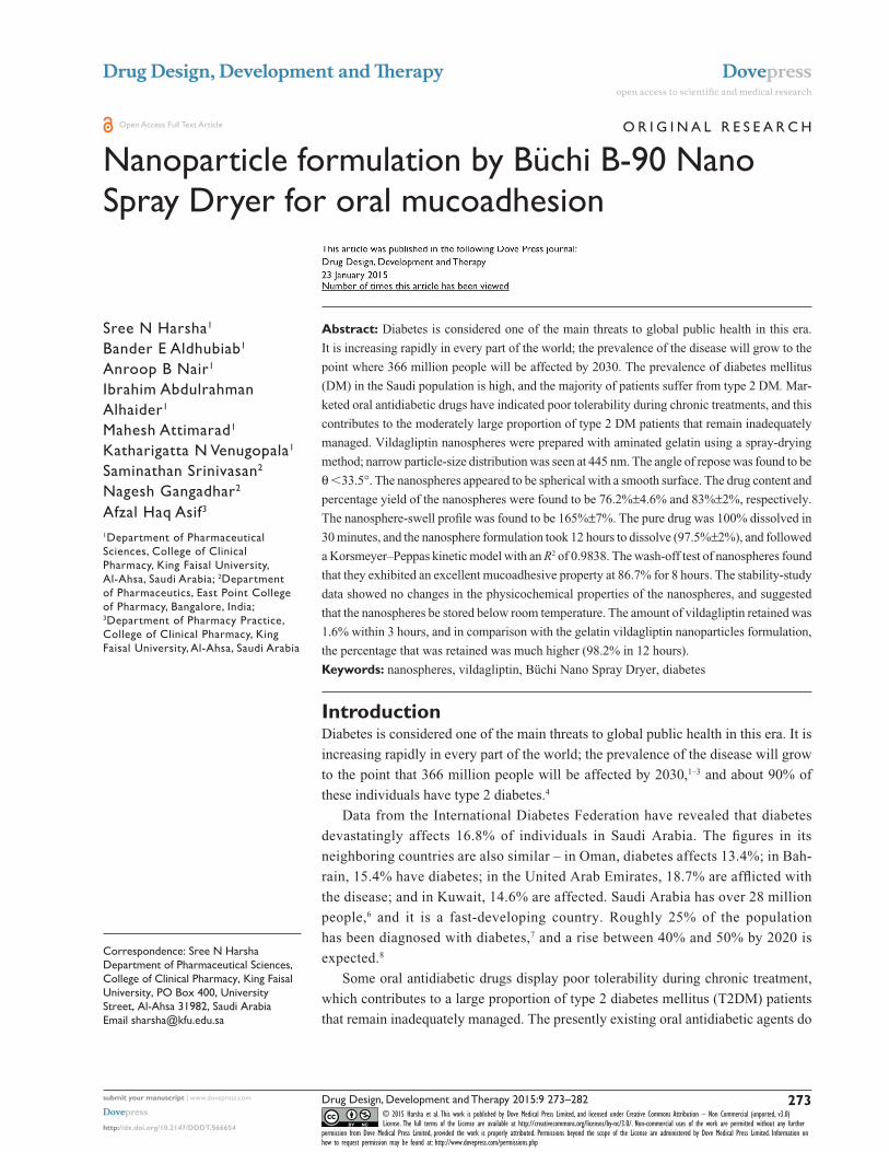
Nanoparticle formulation by Büchi B-90 Nano Spray Dryer for oral mucoadhesion
Sree N Harsha1
Bander E Aldhubiab1
Anroop B Nair1
Ibrahim Abdulrahman Alhaider1
Mahesh Attimarad1
Katharigatta N Venugopala1
Saminathan Srinivasan2
Nagesh Gangadhar2
Afzal Haq Asif3
1Department of Pharmaceutical Sciences, College of Clinical Pharmacy, King Faisal University, Al-Ahsa, Saudi Arabia; 2Department of Pharmaceutics, East Point College of Pharmacy, Bangalore, India; 3Department of Pharmacy Practice, College of Clinical Pharmacy, King Faisal University, Al-Ahsa, Saudi Arabia
Abstract: Diabetes is considered one of the main threats to global public health in this era.
It is increasing rapidly in every part of the world; the prevalence of the disease will grow to the
point where 366 million people will be affected by 2030. The prevalence of diabetes mellitus
(DM) in the Saudi population is high, and the majority of patients suffer from type 2 DM. Mar-
keted oral antidiabetic drugs have indicated poor tolerability during chronic treatments, and this
contributes to the moderately large proportion of type 2 DM patients that remain inadequately
managed. Vildagliptin nanospheres were prepared with aminated gelatin using a spray-drying
method; narrow particle-size distribution was seen at 445 nm. The angle of repose was found to be
θ 33.5°. The nanospheres appeared to be spherical with a smooth surface. The drug content and
percentage yield of the nanospheres were found to be 76.2%±4.6% and 83%±2%, respectively.
The nanosphere-swell profile was found to be 165%±7%. The pure drug was 100% dissolved in
30 minutes, and the nanosphere formulation took 12 hours to dissolve (97.5%±2%), and followed
a Korsmeyer–Peppas kinetic model with an R2 of 0.9838. The wash-off test of nanospheres found
that they exhibited an excellent mucoadhesive property at 86.7% for 8 hours. The stability-study
data showed no changes in the physicochemical properties of the nanospheres, and suggested
that the nanospheres be stored below room temperature. The amount of vildagliptin retained was
1.6% within 3 hours, and in comparison with the gelatin vildagliptin nanoparticles formulation,
the percentage that was retained was much higher (98.2% in 12 hours).
Keywords: nanospheres, vildagliptin, Büchi Nano Spray Dryer, diabetes
IntroductionDiabetes is considered one of the main threats to global public health in this era. It is
increasing rapidly in every part of the world; the prevalence of the disease will grow
to the point that 366 million people will be affected by 2030,1–3 and about 90% of
these individuals have type 2 diabetes.4
Data from the International Diabetes Federation have revealed that diabetes
devastatingly affects 16.8% of individuals in Saudi Arabia. The figures in its
neighboring countries are also similar – in Oman, diabetes affects 13.4%; in Bah-
rain, 15.4% have diabetes; in the United Arab Emirates, 18.7% are afflicted with
the disease; and in Kuwait, 14.6% are affected. Saudi Arabia has over 28 million
people,6 and it is a fast-developing country. Roughly 25% of the population
has been diagnosed with diabetes,7 and a rise between 40% and 50% by 2020 is
expected.8
Some oral antidiabetic drugs display poor tolerability during chronic treatment,
which contributes to a large proportion of type 2 diabetes mellitus (T2DM) patients
that remain inadequately managed. The presently existing oral antidiabetic agents do
Correspondence: Sree N HarshaDepartment of Pharmaceutical Sciences, College of Clinical Pharmacy, King Faisal University, PO Box 400, University Street, Al-Ahsa 31982, Saudi ArabiaEmail [email protected]
Journal name: Drug Design, Development and TherapyArticle Designation: ORIGINAL RESEARCHYear: 2015Volume: 9Running head verso: Harsha et alRunning head recto: NP formulation by Büchi B-90 Nano Spray DryerDOI: http://dx.doi.org/10.2147/DDDT.S66654
Drug Design, Development and Therapy 2015:9submit your manuscript | www.dovepress.com
Dovepress
Dovepress
274
Harsha et al
not directly restore the complex secretory function of the islet
cells, and they are also limited by side effects. Therefore,
there has been much interest in recognizing newer agents that
are capable of restoring the complex secretory dysfunction
of patients with T2DM.9
Dipeptidyl peptidase (DPP)-4 inhibitors are the most
common accompaniments to the therapeutic armamentarium
offered for treating T2DM. The source of the thera-
peutic effects of DPP-4 inhibitors basically increases
the intact circulating levels of the incretin hormones –
glucose-dependent insulinotropic polypeptide and glucagon
like peptide 1 – and they achieve their insulinotropic effects
by adhering to the specific receptor. In addition, they have
a short half-life because of rapid enzyme inactivation. With
respect to the mode of action, vildagliptin is a DPP-4 inhibi-
tor; a detailed study has been carried out in clinical studies,
and considerable developments have been made toward
understanding the molecular interaction between vildagliptin
and the DPP-4 enzyme in biological activity.10,11
Gelatin is a denatured, biodegradable polymer obtained
from collagen and the best material for clinical and medicinal
use. It has been widely used as an excipient for drugs and
controls the release for many formulations.12–14 The safety
of gelatin has been confirmed through its extended clinical
usage as a plasma expander.15 Mucoadhesion is a complex
process, and many hypotheses clarify the mechanisms,
including wetting theory, electric theory, diffusion theory,
and adsorption theory. Although no hypothesis alone can
make clear this process, it is generally accepted that the
first step of mucoadhesion relates to the intimate contact
between the mucoadhesive materials and the mucus surface.
Consequently, mucoadhesive agents would interact with
mucin on the mucus layer and adhere with secondary chemi-
cal bonds, such as van der Waals and hydrogen bonds. Based
on this consideration, polymers with hydrophilic groups,
such as OH, NH2, and COOH, were considered to be crucial
for successful mucoadhesion.16,17 The literature suggests
aminated gelatin demonstrates a sustained release with con-
siderable gastric mucoadhesive properties.12,18
Disappointingly, one of the major therapeutic drawbacks
of using vildagliptin is its rapid metabolism and short elimi-
nation half-life of 1.32–2.43 hours.19 Due to the shorter half-
life of the therapeutic agent, it is recommended that patients
receiving the medical treatment need to adhere meticulously
to the interval between doses, and the drug should be taken at
a dose of 50 mg twice a day.20 Mucoadhesive drug-delivery
systems offer abundant advantages when compared to other
conventional/controlled-release systems. These dosage forms
adhere to the target site of the gastrointestinal tract (GIT),
and they release the drug by maintaining the therapeutic
concentration for prolonged periods of time. Therefore, the
uptake and consequent bioavailability of the therapeutic agent
may be enhanced. In addition, the rate of recurrence of the
dosing reduces when patient compliance is improved.21–23
Even with the current exciting drug delivery, more
discoveries are still required to progress further the effec-
tiveness of existing drugs. An attempt has been made to
develop a mucoadhesive nanoparticle of vildagliptin using
a spray-drying method. Several attempts have been devel-
oped in formulating a mucoadhesive drug-delivery system,
and an additional formulation for vildagliptin has been
attempted by preparing nanoparticles. Numerous methods,
such as direct polymerization, salting out, supercritical fluid
technology, solvent evaporation, nanoprecipitations, and
various polymerization techniques have been developed for
formulating nanoparticles.24–26 On the other hand, these tech-
niques typically engage the use of organic solvents, which
may serve as a potential reservoir of particle contamination
and cause toxicity.
Furthermore, these techniques are inappropriate for mass
production, because they do not produce a high yield or a
reproducible quantity. At present, spray-drying is used in
pharmaceutical industries. It is an entrenched drying process
that is conventionally used to exchange liquid solutions to a
powder form. Spray-drying is widely used for the microencap-
sulation of many therapeutic agents, due to its reproducibility,
consistency, and control of particle-size distribution and drug
release.27 The Nano Spray Dryer B-90 from Büchi Labortech-
nik AG (Flawil, Switzerland) offers patented technology,
such as the production of small particles from a minimum
sample, and it provides high yields and reduces research and
development costs.28 In addition, spray-drying is a continuous
single process; it is a one-step process, is simple to scale up,
and shows minor variations in terms of feed flow, as well as
the concentration of polymer and the temperature needed to
fabricate the preferred particle-size distribution.
To formulate nanospheres by spray-drying, gelatin may
be used as an effectual option, given that it is a wall material
due to its favorable film formation, natural biodegradable
polymer properties, edibility, emulsification ability, and
water-solubility.29
The aim of this research was to formulate mucoadhesive
nanospheres for the delivery of vildagliptin and to evaluate
the GIT drug distribution in an in vivo rat model. Several
studies have established the potential of gelatin as an
excellent bioadhesive, and its controlled-release properties
Drug Design, Development and Therapy 2015:9 submit your manuscript | www.dovepress.com
Dovepress
Dovepress
275
NP formulation by Büchi B-90 Nano Spray Dryer
have been used extensively to enhance the delivery of active
ingredients to various mucous membranes.30,31 To the best
of our knowledge, based on a literature survey, this is the
first mucoadhesive drug-delivery system to be designed for
the formulation of a vildagliptin–gelatin nanospheres via a
Büchi (Büchi Labortechnik AG) spray-dryer.
Materials and methodsVildagliptin was purchased from Biokemix, London, UK.
Gelatin Bloom 250 was a gift sample provided from Shree
Ram Industry. All of the other chemicals used were of
analytical reagent grade.
Preparation of aminated gelatinAminated gelatin was prepared by reaction between gelatin
and 1,2-ethylenediamine. An excess amount of 1-ethyl-3-
(3-dimethylaminopropyl)-carbodiimide hydrochloride was
added to gelatin solution, which had been previously dis-
solved in phosphate buffer (pH 5.3). At 37°C, the resultant
solution was allowed to react for 1 hour, and was dialyzed
against purified water for 2 days.18
Formulation of the vildagliptin nanospheresGelatin-loaded vildagliptin nanospheres were prepared via
a spray-drying technique (B-90; Büchi Labortechnik AG)
with a nanospray-dryer. This spray-dryer has a pulsating
casing in the spray nozzle to atomize the feed, and the par-
ticles were gathered by an electrostatic particle accumulator.
A 4 µm spray nozzle was used; the flow rate was set to about
100–110 L/minute, and the relative spray rate was fixed to
100%. The inlet temperature and outlet temperature were
established at 120°C and 27°C, respectively. A solution
of gelatin (1 g of both vildagliptin and gelatin in 200 mL
of water) was sprayed. The viscosity of the solution was
6.2 cP. The parameters had been previously optimized in our
laboratory. The solution was filtered prior to the spray-drying
process to avoid nozzle blockage. The dried particles were
collected from the particle chamber using a powder scraper,
and they were then kept in a desiccator at a temperature of
25°C for further analysis.31
Characterization of nanospheresScanning electron microscopyThe uncoated vildagliptin nanospheres were analyzed by
scanning electron microscopy (JSM-6390LA analytical scan-
ning microscope; JEOL, Tokyo, Japan) at 20 kV using differ-
ent magnifications (between 1,000× and 95,000×). Different
batches were examined after they had been platinum-
sputtered (JEOL JFC-1600).32,33
Analysis of the particle size of nanospheresThe particle-size distribution of the nanospheres was studied
by laser light-diffraction techniques.34 The nanospheres were
diluted tenfold with deionized water using a Zetasizer Nano
ZS (Malvern Instruments, Malvern, UK).
Drug contentDrug content was determined according to our previous
report.35 Briefly, the weighed quantity of gelatin vildagliptin
nanoparticles (GVN) was dissolved in methanol. The sample
solution was further diluted with a mobile phase, and 10 µL
was injected into a liquid chromatography mass spectrometry
(LC-MS) system to determine the concentration of vildagliptin.
The reported chromatographic method36 was modified slightly
and used for the examination of vildagliptin in formulation and
tissue extracts. The LC-MS system (1200 series; Agilent Tech-
nologies, Santa Clara, CA, USA) consisted of a binary pump,
a 6120 mass (MS) detector, an autoinjector, and a Chem data
module. Chromatographic separation of the active ingredients
was accomplished using a Zorbax Eclipse C18
(150×4.6 mm
internal diameter, 5 µm) reverse-phase analytical column at
room temperature (approximately 25°C). The mobile phase
consisted of 40% mobile phase A (ammonium acetate [10 mM],
pH 8 adjusted with ammonia) and 60% mobile phase B (ace-
tonitrile–methanol 10:90), and it was filtered through a 0.45 µm
nylon filter before use. LC-MS was performed isocratically at
a flow rate of 0.5 mL/minute, and the elute was continuously
monitored by the MS detector in a positive-ion single-ion moni-
tory mode at (M++1) mass to charge ratio 304.3. The tryouts
were piloted in triplicate. The drug loading was calculated as
the ratio between the quantity of vildagliptin loaded and the
theoretical amount of vildagliptin loaded for each prepara-
tion. The production yield was calculated from the quotient
of the average weight of the dried nanospheres (N1) that were
recovered from batches to the sum of the first dry weight of
the preliminary materials (N2). The average value of the drug
content was represented as the drug loading.35,37,38
Drugcontent
Weight of vildagliptin nanospheres
Total weight=
of nanospheres×100.
(1)
Production recoveryNanosphere recovery was calculated as the relationship of
the achieved weight of the nanospheres related to the entire
amount of the drug and polymer initially used in the prepara-
tion process.32,37
Drug Design, Development and Therapy 2015:9submit your manuscript | www.dovepress.com
Dovepress
Dovepress
276
Harsha et al
Recovery = Weight of vilagliptin nanosphere
Total weight of polymer ++100.
drug×
(2)
Determination of powder-flow propertiesPowder flow is a prerequisite in pharmaceutical industries.
The flowability of powder is carried out by a fixed-funnel
method. The angle of repose is the angle between the hori-
zontal surface and the slope of the pile of powder fall from
a specific height. Briefly, a funnel with the end of its stem
cut at right angles to the axis of symmetry is secured with
its tip at a 2 cm height, H, above a graph sheet positioned
on a flat, level surface. The nanospheres were cautiously
shifted through the funnel until reaching the peak of the
conical heap.39 The mean diameter, 2R, of H at the base of
the powder cone was determined, and the tangent of the angle
of repose was given by:
∝ =
H
R. (3)
Interaction studiesFourier-transform infrared spectroscopy (FTIR) spectra of GVN,
vildagliptin, and aminated gelatin were analyzed independently
after mixing with potassium bromide (1:10) and pressed to
develop a thin suitable disk by applying pressure using a Perkin-
Elmer model 883 spectroscope (range 400–4,000 cm-1).40
Measuring the swelling behavior of polymeric nanospheresMeasurement of the swelling of nanospheres was conducted
in simulated gastric fluid. The sizes of the dried powder
(GVN) and those after incubation in methanol for 15 minutes,
30 minutes, 1 hour, 3 hours, 6 hours, 10 hours, and 12 hours
were calculated using a microscopic method. The percentage
of swelling at the different time intervals was determined
by calculating the difference between the diameter of nano-
spheres at time “t” (Vt) and the initial time (t =0 [V
0]). This
was calculated as follows41:
Swelling% = Vt - V
0/V
0 ×100. (4)
In vitro mucoadhesive wash-off methodThe mucoadhesive properties of the nanospheres were deter-
mined with a slight modification of the method reported by
Mankala et al.42 A small piece (2×2 cm) of rat stomach mucosa
was mounted onto a glass slide that was 7.62×2.54 cm and
secured with elastic bands. A known amount of nanospheres
was placed on a mucosal wet surface; thereafter, the
support was hung onto the arm of a US Pharmacopeia
tablet-disintegrating apparatus (ZT 304 disintegration tester;
Erweka, Heusenstamm, Germany). The disintegrator basket
containing the mucosa was adjusted for regular up and down
movement in a beaker containing phosphate buffer with a pH
of 6.8 at 37°C. The drug content was estimated spectrophoto-
metrically at different time intervals (1 hour, 3 hours, 5 hours,
and 8 hours) to ascertain the remaining drug-adherence
levels to the mucosa. The total amount of nanospheres that
corresponded to the difference in the amount of drug was
calculated. The adhered nanosphere amounts were estimated
from the difference between the amount of the applied nano-
spheres and the nanoparticle-flow amount. The percentage of
the nanospheres that adhered to the applied nanospheres was
computed as the percentage of mucoadhesion.
Drug-release study of vildagliptinIn vitro drug release was carried out using a dialysis tech-
nique. Vildagliptin nanospheres were positioned in a dialysis
bag and dialyzed against 250 mL of phosphate-buffered
saline (PBS). The PBS was prepared to 250 mL with water.
Then, 5 mL of the samples at 15 minutes, 30 minutes, 1 hour,
2 hours, 3 hours, 4 hours, 6 hours, 8 hours, 10 hours, and
12 hours were withdrawn and replaced with a fresh sample of
5 mL of PBS. During the dissolution process, the temperature
was maintained at 37°C±1°C, and the sink conditions were
maintained throughout the course of the study. The drug
content was analyzed by the LC-MS method, as described
previously.35,43
In vitro mucoadhesiveness measurementMale Sprague Dawley rats weighing 200±50 g were fasted
for 24 hours before commencing the experiment. Water was
provided ad libitum. Five groups of animals were employed,
with each group containing 15 rats. The first group was orally
administered an aqueous solution of vildagliptin. The second
to fifth groups received vildagliptin nanospheres. The oral
administration of the nanospheres was accomplished by
suspending a 20 mg sample of the nanospheres in 1.0 mL
of normal saline, and the nanoparticles were administered
via a rubber tube under nonanesthetic conditions. Three
rats were killed at each time point after 1 hour, 2 hours,
4 hours, 8 hours, 10 hours, and 12 hours of administration.
The stomach and the entire length of the small intestine
were isolated.41 The entire GIT was cut open to expose the
inner mucosal surface. All nanospheres located in each part
were collected by scratching the mucosa with a spatula.
Drug Design, Development and Therapy 2015:9 submit your manuscript | www.dovepress.com
Dovepress
Dovepress
277
NP formulation by Büchi B-90 Nano Spray Dryer
The mixture was mashed using a homogenizer to extract the
vildagliptin, and the sample was kept for 24 hours. After
centrifugation at 805 g for 20 minutes, the supernatant was
determined spectrophotometrically.
LC-MS methodThe respective GIT tissue samples were has revolutionized,
and vildagliptin was extracted with double-distilled water
five times. The collected extractions were diluted to 15 mL
and then centrifuged at 12,000 rpm for 15 minutes. For
protein precipitation, 500 µL of acetonitrile was added to
2 mL of the supernatant, and the mixture was vortexed for
1 minute and centrifuged at 8,000 rpm for 10 minutes. Then,
the 10 µL supernatant was injected into the LC-MS system
to determine the vildagliptin concentration, while adopting
the method described in “Drug content”.
The stability of GVNThe formulation was kept in a glass bottle and stored for
12 months at 3°C–5°C, 15°C–25°C, and 37°C, respectively
(Binder KMF 115; Tuttlingen, Germany). The surface exami-
nation and active pharmaceutical ingredient (API) content
were evaluated periodically.35
Results and discussionThe Büchi Nano Spray Dryer B-90 has revolutionized today’s
pharmaceutical industries, and it offers a variety of particle-
engineering prospects. Additionally, it is capable of produc-
ing high levels of drug loading and efficient spray processes
for the smallest amount at a high yield.31,44
The vildagliptin loading and the yield percentage were
76.2%±4.6% and 83%±2%, respectively. The high drug-
encapsulation efficiency from 50% up to 100% is classical
for spray-drying. The rationale for this is that the drug
cannot separate into a continuous phase, as is the case with
the water-in-oil emulsion method or solvent evaporation.45
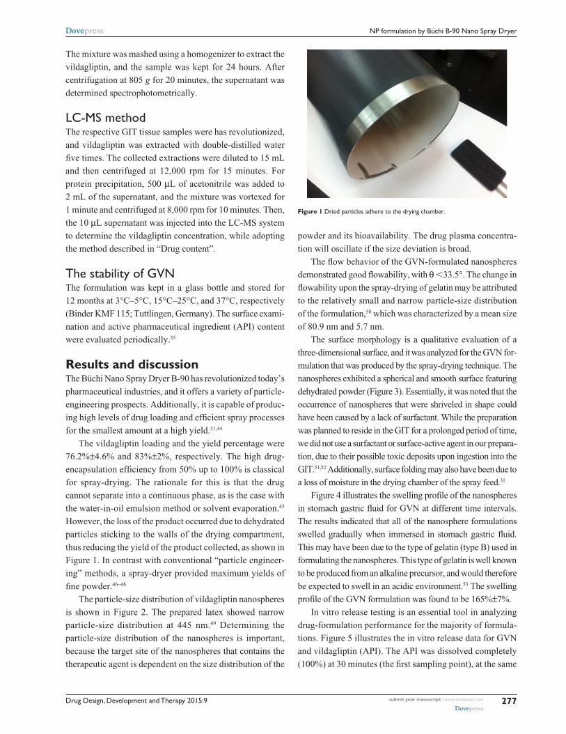
However, the loss of the product occurred due to dehydrated
particles sticking to the walls of the drying compartment,
thus reducing the yield of the product collected, as shown in
Figure 1. In contrast with conventional “particle engineer-
ing” methods, a spray-dryer provided maximum yields of
fine powder.46–48
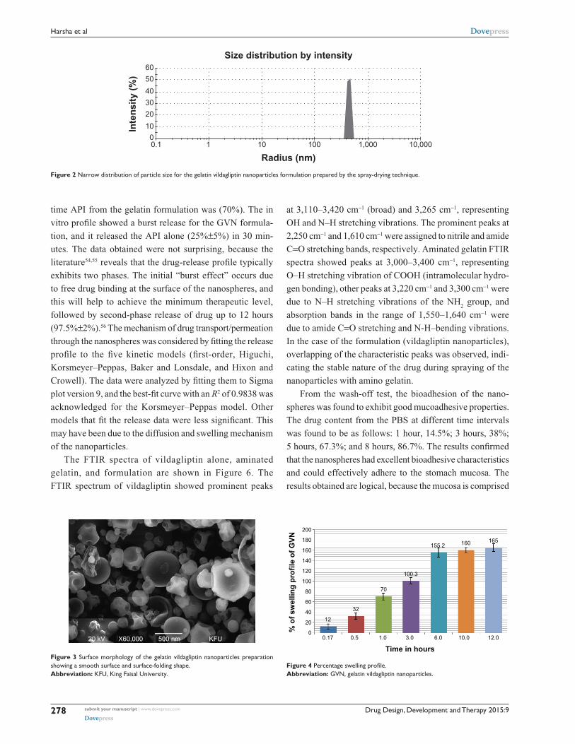
The particle-size distribution of vildagliptin nanospheres
is shown in Figure 2. The prepared latex showed narrow
particle-size distribution at 445 nm.49 Determining the
particle-size distribution of the nanospheres is important,
because the target site of the nanospheres that contains the
therapeutic agent is dependent on the size distribution of the
powder and its bioavailability. The drug plasma concentra-
tion will oscillate if the size deviation is broad.
The flow behavior of the GVN-formulated nanospheres
demonstrated good flowability, with θ 33.5°. The change in
flowability upon the spray-drying of gelatin may be attributed
to the relatively small and narrow particle-size distribution
of the formulation,50 which was characterized by a mean size
of 80.9 nm and 5.7 nm.
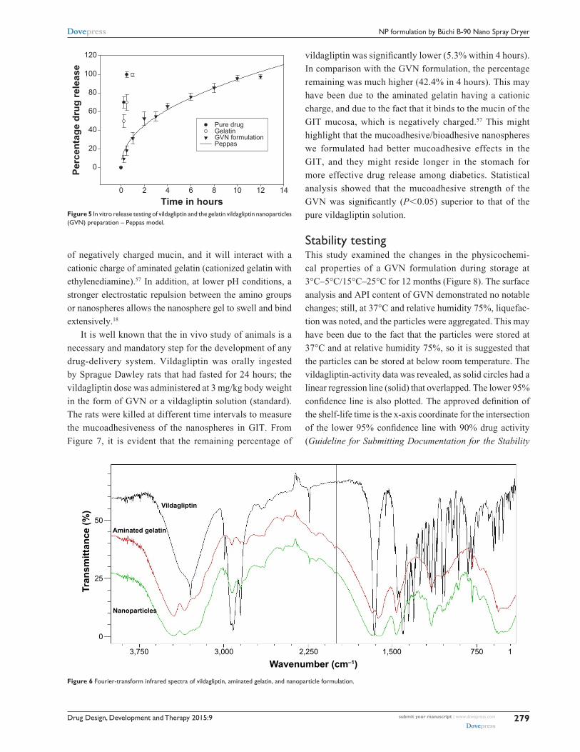
The surface morphology is a qualitative evaluation of a
three-dimensional surface, and it was analyzed for the GVN for-
mulation that was produced by the spray-drying technique. The
nanospheres exhibited a spherical and smooth surface featuring
dehydrated powder (Figure 3). Essentially, it was noted that the
occurrence of nanospheres that were shriveled in shape could
have been caused by a lack of surfactant. While the preparation
was planned to reside in the GIT for a prolonged period of time,
we did not use a surfactant or surface-active agent in our prepara-
tion, due to their possible toxic deposits upon ingestion into the
GIT.51,52 Additionally, surface folding may also have been due to
a loss of moisture in the drying chamber of the spray feed.31
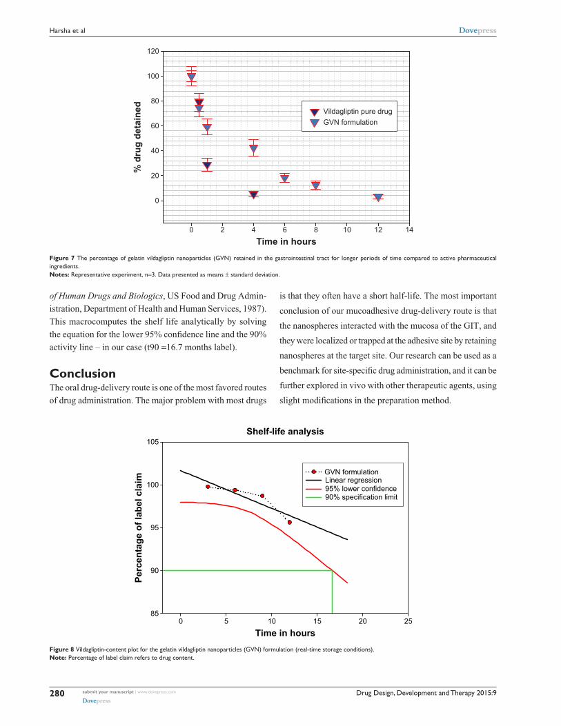
Figure 4 illustrates the swelling profile of the nanospheres
in stomach gastric fluid for GVN at different time intervals.
The results indicated that all of the nanosphere formulations
swelled gradually when immersed in stomach gastric fluid.
This may have been due to the type of gelatin (type B) used in
formulating the nanospheres. This type of gelatin is well known
to be produced from an alkaline precursor, and would therefore
be expected to swell in an acidic environment.53 The swelling
profile of the GVN formulation was found to be 165%±7%.
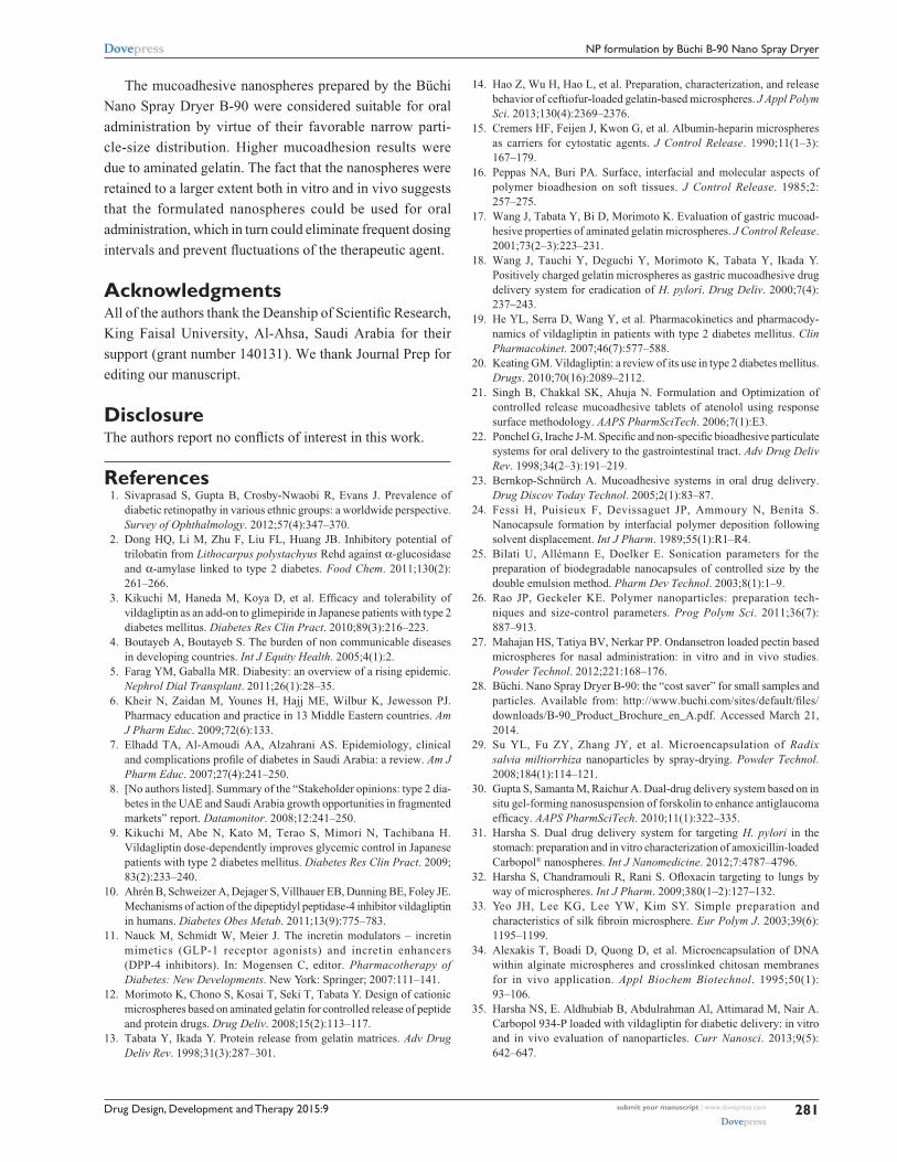
In vitro release testing is an essential tool in analyzing
drug-formulation performance for the majority of formula-
tions. Figure 5 illustrates the in vitro release data for GVN
and vildagliptin (API). The API was dissolved completely
(100%) at 30 minutes (the first sampling point), at the same
Figure 1 Dried particles adhere to the drying chamber.
Drug Design, Development and Therapy 2015:9submit your manuscript | www.dovepress.com
Dovepress
Dovepress
278
Harsha et al
time API from the gelatin formulation was (70%). The in
vitro profile showed a burst release for the GVN formula-
tion, and it released the API alone (25%±5%) in 30 min-
utes. The data obtained were not surprising, because the
literature54,55 reveals that the drug-release profile typically
exhibits two phases. The initial “burst effect” occurs due
to free drug binding at the surface of the nanospheres, and
this will help to achieve the minimum therapeutic level,
followed by second-phase release of drug up to 12 hours
(97.5%±2%).56 The mechanism of drug transport/permeation
through the nanospheres was considered by fitting the release
profile to the five kinetic models (first-order, Higuchi,
Korsmeyer–Peppas, Baker and Lonsdale, and Hixon and
Crowell). The data were analyzed by fitting them to Sigma
plot version 9, and the best-fit curve with an R2 of 0.9838 was
acknowledged for the Korsmeyer–Peppas model. Other
models that fit the release data were less significant. This
may have been due to the diffusion and swelling mechanism
of the nanoparticles.
The FTIR spectra of vildagliptin alone, aminated
gelatin, and formulation are shown in Figure 6. The
FTIR spectrum of vildagliptin showed prominent peaks
at 3,110–3,420 cm-1 (broad) and 3,265 cm-1, representing
OH and N–H stretching vibrations. The prominent peaks at
2,250 cm-1 and 1,610 cm-1 were assigned to nitrile and amide
C=O stretching bands, respectively. Aminated gelatin FTIR
spectra showed peaks at 3,000–3,400 cm-1, representing
O–H stretching vibration of COOH (intramolecular hydro-
gen bonding), other peaks at 3,220 cm-1 and 3,300 cm-1 were
due to N–H stretching vibrations of the NH2 group, and
absorption bands in the range of 1,550–1,640 cm-1 were
due to amide C=O stretching and N-H–bending vibrations.
In the case of the formulation (vildagliptin nanoparticles),
overlapping of the characteristic peaks was observed, indi-
cating the stable nature of the drug during spraying of the
nanoparticles with amino gelatin.
From the wash-off test, the bioadhesion of the nano-
spheres was found to exhibit good mucoadhesive properties.
The drug content from the PBS at different time intervals
was found to be as follows: 1 hour, 14.5%; 3 hours, 38%;
5 hours, 67.3%; and 8 hours, 86.7%. The results confirmed
that the nanospheres had excellent bioadhesive characteristics
and could effectively adhere to the stomach mucosa. The
results obtained are logical, because the mucosa is comprised
20 kV X60,000 500 nm KFU
Figure 3 Surface morphology of the gelatin vildagliptin nanoparticles preparation showing a smooth surface and surface-folding shape.Abbreviation: KFU, King Faisal University.
% o
f sw
ellin
g pr
ofile
of G
VN
Time in hours
200
180
160
140
120
100
80
60
40
20
00.17
12
32
70
100.3
155.2 160 165
0.5 1.0 3.0 6.0 10.0 12.0
Figure 4 Percentage swelling profile.Abbreviation: GVN, gelatin vildagliptin nanoparticles.
0.1 1 10 100 1,000 10,000
100
2030405060
Inte
nsity
(%)
Radius (nm)
Size distribution by intensity
Figure 2 Narrow distribution of particle size for the gelatin vildagliptin nanoparticles formulation prepared by the spray-drying technique.
Drug Design, Development and Therapy 2015:9 submit your manuscript | www.dovepress.com
Dovepress
Dovepress
279
NP formulation by Büchi B-90 Nano Spray Dryer
of negatively charged mucin, and it will interact with a
cationic charge of aminated gelatin (cationized gelatin with
ethylenediamine).57 In addition, at lower pH conditions, a
stronger electrostatic repulsion between the amino groups
or nanospheres allows the nanosphere gel to swell and bind
extensively.18
It is well known that the in vivo study of animals is a
necessary and mandatory step for the development of any
drug-delivery system. Vildagliptin was orally ingested
by Sprague Dawley rats that had fasted for 24 hours; the
vildagliptin dose was administered at 3 mg/kg body weight
in the form of GVN or a vildagliptin solution (standard).
The rats were killed at different time intervals to measure
the mucoadhesiveness of the nanospheres in GIT. From
Figure 7, it is evident that the remaining percentage of
vildagliptin was significantly lower (5.3% within 4 hours).
In comparison with the GVN formulation, the percentage
remaining was much higher (42.4% in 4 hours). This may
have been due to the aminated gelatin having a cationic
charge, and due to the fact that it binds to the mucin of the
GIT mucosa, which is negatively charged.57 This might
highlight that the mucoadhesive/bioadhesive nanospheres
we formulated had better mucoadhesive effects in the
GIT, and they might reside longer in the stomach for
more effective drug release among diabetics. Statistical
analysis showed that the mucoadhesive strength of the
GVN was significantly (P0.05) superior to that of the
pure vildagliptin solution.
Stability testingThis study examined the changes in the physicochemi-
cal properties of a GVN formulation during storage at
3°C–5°C/15°C–25°C for 12 months (Figure 8). The surface
analysis and API content of GVN demonstrated no notable
changes; still, at 37°C and relative humidity 75%, liquefac-
tion was noted, and the particles were aggregated. This may
have been due to the fact that the particles were stored at
37°C and at relative humidity 75%, so it is suggested that
the particles can be stored at below room temperature. The
vildagliptin-activity data was revealed, as solid circles had a
linear regression line (solid) that overlapped. The lower 95%
confidence line is also plotted. The approved definition of
the shelf-life time is the x-axis coordinate for the intersection
of the lower 95% confidence line with 90% drug activity
(Guideline for Submitting Documentation for the Stability
Time in hours
Perc
enta
ge d
rug
rele
ase
0
0 2 4 6 8 10 12 14
20
40
60
80
100
120
Pure drugGelatinGVN formulationPeppas
Figure 5 In vitro release testing of vildagliptin and the gelatin vildagliptin nanoparticles (GVN) preparation – Peppas model.
50Aminated gelatin
Vildagliptin
Nanoparticles
25
Tran
smitt
ance
(%)
Wavenumber (cm–1)
0
3,750 3,000 2,250 1,500 750 1
Figure 6 Fourier-transform infrared spectra of vildagliptin, aminated gelatin, and nanoparticle formulation.
Drug Design, Development and Therapy 2015:9submit your manuscript | www.dovepress.com
Dovepress
Dovepress
280
Harsha et al
of Human Drugs and Biologics, US Food and Drug Admin-
istration, Department of Health and Human Services, 1987).
This macrocomputes the shelf life analytically by solving
the equation for the lower 95% confidence line and the 90%
activity line – in our case (t90 =16.7 months label).
ConclusionThe oral drug-delivery route is one of the most favored routes
of drug administration. The major problem with most drugs
is that they often have a short half-life. The most important
conclusion of our mucoadhesive drug-delivery route is that
the nanospheres interacted with the mucosa of the GIT, and
they were localized or trapped at the adhesive site by retaining
nanospheres at the target site. Our research can be used as a
benchmark for site-specific drug administration, and it can be
further explored in vivo with other therapeutic agents, using
slight modifications in the preparation method.
Time in hours
% d
rug
deta
ined
0
0 2 4 6 8 10 12 14
20
40
60
80
100
120
Vildagliptin pure drugGVN formulation
Figure 7 The percentage of gelatin vildagliptin nanoparticles (GVN) retained in the gastrointestinal tract for longer periods of time compared to active pharmaceutical ingredients.Notes: Representative experiment, n=3. Data presented as means ± standard deviation.
Shelf-life analysis
Time in hours
Perc
enta
ge o
f lab
el c
laim
850 5 10 15 20 25
90
95
100
105
GVN formulationLinear regression95% lower confidence90% specification limit
Figure 8 Vildagliptin-content plot for the gelatin vildagliptin nanoparticles (GVN) formulation (real-time storage conditions).Note: Percentage of label claim refers to drug content.
Drug Design, Development and Therapy 2015:9 submit your manuscript | www.dovepress.com
Dovepress
Dovepress
281
NP formulation by Büchi B-90 Nano Spray Dryer
The mucoadhesive nanospheres prepared by the Büchi
Nano Spray Dryer B-90 were considered suitable for oral
administration by virtue of their favorable narrow parti-
cle-size distribution. Higher mucoadhesion results were
due to aminated gelatin. The fact that the nanospheres were
retained to a larger extent both in vitro and in vivo suggests
that the formulated nanospheres could be used for oral
administration, which in turn could eliminate frequent dosing
intervals and prevent fluctuations of the therapeutic agent.
AcknowledgmentsAll of the authors thank the Deanship of Scientific Research,
King Faisal University, Al-Ahsa, Saudi Arabia for their
support (grant number 140131). We thank Journal Prep for
editing our manuscript.
DisclosureThe authors report no conflicts of interest in this work.
References 1. Sivaprasad S, Gupta B, Crosby-Nwaobi R, Evans J. Prevalence of
diabetic retinopathy in various ethnic groups: a worldwide perspective. Survey of Ophthalmology. 2012;57(4):347–370.
2. Dong HQ, Li M, Zhu F, Liu FL, Huang JB. Inhibitory potential of trilobatin from Lithocarpus polystachyus Rehd against α-glucosidase and α-amylase linked to type 2 diabetes. Food Chem. 2011;130(2): 261–266.
3. Kikuchi M, Haneda M, Koya D, et al. Efficacy and tolerability of vildagliptin as an add-on to glimepiride in Japanese patients with type 2 diabetes mellitus. Diabetes Res Clin Pract. 2010;89(3):216–223.
4. Boutayeb A, Boutayeb S. The burden of non communicable diseases in developing countries. Int J Equity Health. 2005;4(1):2.
5. Farag YM, Gaballa MR. Diabesity: an overview of a rising epidemic. Nephrol Dial Transplant. 2011;26(1):28–35.
6. Kheir N, Zaidan M, Younes H, Hajj ME, Wilbur K, Jewesson PJ. Pharmacy education and practice in 13 Middle Eastern countries. Am J Pharm Educ. 2009;72(6):133.
7. Elhadd TA, Al-Amoudi AA, Alzahrani AS. Epidemiology, clinical and complications profile of diabetes in Saudi Arabia: a review. Am J Pharm Educ. 2007;27(4):241–250.
8. [No authors listed]. Summary of the “Stakeholder opinions: type 2 dia-betes in the UAE and Saudi Arabia growth opportunities in fragmented markets” report. Datamonitor. 2008;12:241–250.
9. Kikuchi M, Abe N, Kato M, Terao S, Mimori N, Tachibana H. Vildagliptin dose-dependently improves glycemic control in Japanese patients with type 2 diabetes mellitus. Diabetes Res Clin Pract. 2009; 83(2):233–240.
10. Ahrén B, Schweizer A, Dejager S, Villhauer EB, Dunning BE, Foley JE. Mechanisms of action of the dipeptidyl peptidase-4 inhibitor vildagliptin in humans. Diabetes Obes Metab. 2011;13(9):775–783.
11. Nauck M, Schmidt W, Meier J. The incretin modulators – incretin mimetics (GLP-1 receptor agonists) and incretin enhancers (DPP-4 inhibitors). In: Mogensen C, editor. Pharmacotherapy of Diabetes: New Developments. New York: Springer; 2007:111–141.
12. Morimoto K, Chono S, Kosai T, Seki T, Tabata Y. Design of cationic microspheres based on aminated gelatin for controlled release of peptide and protein drugs. Drug Deliv. 2008;15(2):113–117.
13. Tabata Y, Ikada Y. Protein release from gelatin matrices. Adv Drug Deliv Rev. 1998;31(3):287–301.
14. Hao Z, Wu H, Hao L, et al. Preparation, characterization, and release behavior of ceftiofur-loaded gelatin-based microspheres. J Appl Polym Sci. 2013;130(4):2369–2376.
15. Cremers HF, Feijen J, Kwon G, et al. Albumin-heparin microspheres as carriers for cytostatic agents. J Control Release. 1990;11(1–3): 167–179.
16. Peppas NA, Buri PA. Surface, interfacial and molecular aspects of polymer bioadhesion on soft tissues. J Control Release. 1985;2: 257–275.
17. Wang J, Tabata Y, Bi D, Morimoto K. Evaluation of gastric mucoad-hesive properties of aminated gelatin microspheres. J Control Release. 2001;73(2–3):223–231.
18. Wang J, Tauchi Y, Deguchi Y, Morimoto K, Tabata Y, Ikada Y. Positively charged gelatin microspheres as gastric mucoadhesive drug delivery system for eradication of H. pylori. Drug Deliv. 2000;7(4): 237–243.
19. He YL, Serra D, Wang Y, et al. Pharmacokinetics and pharmacody-namics of vildagliptin in patients with type 2 diabetes mellitus. Clin Pharmacokinet. 2007;46(7):577–588.
20. Keating GM. Vildagliptin: a review of its use in type 2 diabetes mellitus. Drugs. 2010;70(16):2089–2112.
21. Singh B, Chakkal SK, Ahuja N. Formulation and Optimization of controlled release mucoadhesive tablets of atenolol using response surface methodology. AAPS PharmSciTech. 2006;7(1):E3.
22. Ponchel G, Irache J-M. Specific and non-specific bioadhesive particulate systems for oral delivery to the gastrointestinal tract. Adv Drug Deliv Rev. 1998;34(2–3):191–219.
23. Bernkop-Schnürch A. Mucoadhesive systems in oral drug delivery. Drug Discov Today Technol. 2005;2(1):83–87.
24. Fessi H, Puisieux F, Devissaguet JP, Ammoury N, Benita S. Nanocapsule formation by interfacial polymer deposition following solvent displacement. Int J Pharm. 1989;55(1):R1–R4.
25. Bilati U, Allémann E, Doelker E. Sonication parameters for the preparation of biodegradable nanocapsules of controlled size by the double emulsion method. Pharm Dev Technol. 2003;8(1):1–9.
26. Rao JP, Geckeler KE. Polymer nanoparticles: preparation tech-niques and size-control parameters. Prog Polym Sci. 2011;36(7): 887–913.
27. Mahajan HS, Tatiya BV, Nerkar PP. Ondansetron loaded pectin based microspheres for nasal administration: in vitro and in vivo studies. Powder Technol. 2012;221:168–176.
28. Büchi. Nano Spray Dryer B-90: the “cost saver” for small samples and particles. Available from: http://www.buchi.com/sites/default/files/downloads/B-90_Product_Brochure_en_A.pdf. Accessed March 21, 2014.
29. Su YL, Fu ZY, Zhang JY, et al. Microencapsulation of Radix salvia miltiorrhiza nanoparticles by spray-drying. Powder Technol. 2008;184(1):114–121.
30. Gupta S, Samanta M, Raichur A. Dual-drug delivery system based on in situ gel-forming nanosuspension of forskolin to enhance antiglaucoma efficacy. AAPS PharmSciTech. 2010;11(1):322–335.
31. Harsha S. Dual drug delivery system for targeting H. pylori in the stomach: preparation and in vitro characterization of amoxicillin-loaded Carbopol® nanospheres. Int J Nanomedicine. 2012;7:4787–4796.
32. Harsha S, Chandramouli R, Rani S. Ofloxacin targeting to lungs by way of microspheres. Int J Pharm. 2009;380(1–2):127–132.
33. Yeo JH, Lee KG, Lee YW, Kim SY. Simple preparation and characteristics of silk fibroin microsphere. Eur Polym J. 2003;39(6): 1195–1199.
34. Alexakis T, Boadi D, Quong D, et al. Microencapsulation of DNA within alginate microspheres and crosslinked chitosan membranes for in vivo application. Appl Biochem Biotechnol. 1995;50(1): 93–106.
35. Harsha NS, E. Aldhubiab B, Abdulrahman Al, Attimarad M, Nair A. Carbopol 934-P loaded with vildagliptin for diabetic delivery: in vitro and in vivo evaluation of nanoparticles. Curr Nanosci. 2013;9(5): 642–647.
Drug Design, Development and Therapy
Publish your work in this journal
Submit your manuscript here: http://www.dovepress.com/drug-design-development-and-therapy-journal
Drug Design, Development and Therapy is an international, peer-reviewed open-access journal that spans the spectrum of drug design and development through to clinical applications. Clinical outcomes, patient safety, and programs for the development and effective, safe, and sustained use of medicines are a feature of the journal, which
has also been accepted for indexing on PubMed Central. The manu-script management system is completely online and includes a very quick and fair peer-review system, which is all easy to use. Visit http://www.dovepress.com/testimonials.php to read real quotes from published authors.
Drug Design, Development and Therapy 2015:9submit your manuscript | www.dovepress.com
Dovepress
Dovepress
Dovepress
282
Harsha et al
36. He YL, Sabo R, Riviere GJ, et al. Effect of the novel oral dipeptidyl peptidase IV inhibitor vildagliptin on the pharmacokinetics and pharmacodynamics of warfarin in healthy subjects. Curr Med Res Opin. 2007;23(5):1131–1138.
37. Lu B, Zhang JQ, Yang H. Lung-targeting microspheres of carboplatin. Int J Pharm. 2003;265(1–2):1–11.
38. Radha GV, Rani TS, Sarvani B. A review on proniosomal drug deliv-ery system for targeted drug action. J Basic Clin Pharm. 2013;4(2): 42–48.
39. Bayomi MA, al-Suwayeh SA, el-Helw AM, Mesnad AF. Preparation of casein-chitosan microspheres containing diltiazem hydrochloride by an aqueous coacervation technique. Pharm Acta Helv. 1998;73(4): 187–192.
40. Tayel SA, El-Nabarawi MA, Tadros MI, Abd-Elsalam WH. Positively charged polymeric nanoparticle reservoirs of terbinafine hydrochloride: preclinical implications for controlled drug delivery in the aqueous humor of rabbits. AAPS PharmSciTech. 2013;14(2):782–793.
41. Dhaliwal S, Jain S, Singh HP, Tiwary AK. Mucoadhesive microspheres for gastroretentive delivery of acyclovir: in vitro and in vivo evaluation. AAPS J. 2004;10(2):322–330.
42. Mankala SK, Korla AC, Gade S. Development and evaluation of aceclofenac-loaded mucoadhesive microcapsules. J Adv Pharm Technol Res. 2011;2(4):245–254.
43. Harsha NS, Rani RH. Drug targeting to lungs by way of microspheres. Arch Pharm Res. 2006;29(7):598–604.
44. Harsha S, Attimarad M, Khan T, et al. Design and formulation of mucoadhesive microspheres of sitagliptin. J Microencapsul. 2013;30(3):257–264.
45. Cordin A, Nina S. Spray dried biodegradable polymers as target material for controlled drug delivery. Best@buchi. 2007;46.
46. Atuah KN, Walter E, Merkle HP, Alpar HO. Encapsulation of plasmid DNA in PLGA-stearylamine microspheres: a comparison of solvent evaporation and spray-drying methods. J Microencapsul. 2003;20(3):387–399.
47. Burrki K, Jeon I, Arpagaus C, Betz G. New insights into respirable protein powder preparation using a nano spray dryer. Int J Pharm. 2011; 408(1–2):248–256.
48. Schafroth N, Arpagaus C, Jadhav UY, Makne S, Douroumis D. Nano and microparticle engineering of water insoluble drugs using a novel spray-drying process. Colloids Surf B Biointerfaces. 2012;90:8–15.
49. Paulal HC, Sombral FM, Abreu FO, de Paul RC. Lippia sidoides essential oil encapsulation by angico gum/chitosan nanoparticles. J Braz Chem Soc. 2010;21(12):2359–2366.
50. Samy W, Elgindy N, El-Gowelli HM. Biopolymeric nifedipine powder for acceleration of wound healing. Int J Pharm. 2012;422(1–2): 323–331.
51. Coowanitwong I, Arya V, Kulvanich P, Hochhaus G. Slow release formulations of inhaled rifampin. AAPS J. 2008;10(2):342–348.
52. Patil P, Joshi P, Paradkar A. Effect of formulation variables on preparation and evaluation of gelled self-emulsifying drug delivery system (SEDDS) of ketoprofen. AAPS PharmSciTech. 2004;5(3):43–50.
53. Adikwu MU. Biopolymers in Drug Delivery: Recent Advances and Challenges. Sharjah: Bentham Science; 2009.
54. Pavanetto F, Genta I, Giunchedi P, Conti B. Evaluation of spray drying as a method for polylactide and polylactide-co-glycolide microsphere preparation. J Microencapsul. 1993;10(4):487–497.
55. Fu YJ, Mi FL, Wong TB, Shyu SS. Characteristic and controlled release of anticancer drug loaded poly(D,L-lactide) microparticles prepared by spray drying technique. J Microencapsul. 2001;18(6):733–747.
56. Jadhav NR, Tone JS, Irny PV, Nadaf SJ. Development and characterization of gelatin based nanoparticles for targeted delivery of zidovudine. Int J Pharm Investig. 2013;3(3):126–130.
57. Van Der Walle C. Peptide and Protein Delivery. Amsterdam: Elsevier Science; 2011.
Copyright © 2022 FDOKUMEN