Multiscale characterization of acrylic bone cement modified with functionalized mesoporous silica...
12
www.elsevier.com/locate/jmbbm Available online at www.sciencedirect.com Research Paper Multiscale characterization of acrylic bone cement modified with functionalized mesoporous silica nanoparticles Josh Slane a,c,n , Juan Vivanco b,e , Donna Ebenstein d , Matthew Squire c , Heidi-Lynn Ploeg a,b a Materials Science Program, University of Wisconsin-Madison, Madison, WI, USA b Department of Mechanical Engineering, University of Wisconsin-Madison, Madison, WI, USA c Department of Orthopedics and Rehabilitation, University of Wisconsin-Madison, Madison, WI, USA d Department of Biomedical Engineering, Bucknell University, Lewisburg, PA, USA e Facultad de Ingeniería y Ciencias, Universidad Adolfo Ibañez, Viña del Mar, Chile article info Article history: Received 22 April 2014 Accepted 15 May 2014 Available online 24 May 2014 Keywords: Bone cement Mechanical properties Nanoindentation Reinforced polymer Orthopedics abstract Acrylic bone cement is widely used to anchor orthopedic implants to bone and mechanical failure of the cement mantle surrounding an implant can contribute to aseptic loosening. In an effort to enhance the mechanical properties of bone cement, a variety of nanopar- ticles and fibers can be incorporated into the cement matrix. Mesoporous silica nanopar- ticles (MSNs) are a class of particles that display high potential for use as reinforcement within bone cement. Therefore, the purpose of this study was to quantify the impact of modifying an acrylic cement with various low-loadings of mesoporous silica. Three types of MSNs (one plain variety and two modified with functional groups) at two loading ratios (0.1 and 0.2 wt/wt) were incorporated into a commercially available bone cement. The mechanical properties were characterized using four-point bending, microindentation and nanoindentation (static, stress relaxation, and creep) while material properties were assessed through dynamic mechanical analysis, differential scanning calorimetry, thermo- gravimetric analysis, FTIR spectroscopy, and scanning electron microscopy. Four-point flexural testing and nanoindentation revealed minimal impact on the properties of the cements, except for several changes in the nano-level static mechanical properties. Conversely, microindentation testing demonstrated that the addition of MSNs significantly increased the microhardness. The stress relaxation and creep properties of the cements measured with nanoindentation displayed no effect resulting from the addition of MSNs. The measured material properties were consistent among all cements. Analysis of scanning electron micrographs images revealed that surface functionalization enhanced http://dx.doi.org/10.1016/j.jmbbm.2014.05.015 1751-6161/& 2014 Elsevier Ltd. All rights reserved. n Coreespondence to: Materials Science Program, University of Wisconsin-Madison, 1513 University Ave, Room 3046, Madison, WI, 53706, USA. Tel.: þ1 608 263 6692; fax: þ1 608 2652316. E-mail address: [email protected] (J. Slane). journal of the mechanical behavior of biomedical materials 37(2014)141–152
Transcript of Multiscale characterization of acrylic bone cement modified with functionalized mesoporous silica...

Available online at www.sciencedirect.com
www.elsevier.com/locate/jmbbm
j o u r n a l o f t h e m e c h a n i c a l b e h a v i o r o f b i o m e d i c a l m a t e r i a l s 3 7 ( 2 0 1 4 ) 1 4 1 – 1 5 2
http://dx.doi.org/101751-6161/& 2014 El
nCoreespondence53706, USA. Tel.: þ1
E-mail address: j
Research Paper
Multiscale characterization of acrylic bone cementmodified with functionalized mesoporoussilica nanoparticles
Josh Slanea,c,n, Juan Vivancob,e, Donna Ebensteind, Matthew Squirec,Heidi-Lynn Ploega,b
aMaterials Science Program, University of Wisconsin-Madison, Madison, WI, USAbDepartment of Mechanical Engineering, University of Wisconsin-Madison, Madison, WI, USAcDepartment of Orthopedics and Rehabilitation, University of Wisconsin-Madison, Madison, WI, USAdDepartment of Biomedical Engineering, Bucknell University, Lewisburg, PA, USAeFacultad de Ingeniería y Ciencias, Universidad Adolfo Ibañez, Viña del Mar, Chile
a r t i c l e i n f o
Article history:
Received 22 April 2014
Accepted 15 May 2014
Available online 24 May 2014
Keywords:
Bone cement
Mechanical properties
Nanoindentation
Reinforced polymer
Orthopedics
.1016/j.jmbbm.2014.05.015sevier Ltd. All rights rese
to: Materials Science Pr608 263 6692; fax: þ1 608
[email protected] (J. Slane
a b s t r a c t
Acrylic bone cement is widely used to anchor orthopedic implants to bone and mechanical
failure of the cement mantle surrounding an implant can contribute to aseptic loosening.
In an effort to enhance the mechanical properties of bone cement, a variety of nanopar-
ticles and fibers can be incorporated into the cement matrix. Mesoporous silica nanopar-
ticles (MSNs) are a class of particles that display high potential for use as reinforcement
within bone cement. Therefore, the purpose of this study was to quantify the impact of
modifying an acrylic cement with various low-loadings of mesoporous silica. Three types
of MSNs (one plain variety and two modified with functional groups) at two loading ratios
(0.1 and 0.2 wt/wt) were incorporated into a commercially available bone cement.
The mechanical properties were characterized using four-point bending, microindentation
and nanoindentation (static, stress relaxation, and creep) while material properties were
assessed through dynamic mechanical analysis, differential scanning calorimetry, thermo-
gravimetric analysis, FTIR spectroscopy, and scanning electron microscopy. Four-point
flexural testing and nanoindentation revealed minimal impact on the properties of the
cements, except for several changes in the nano-level static mechanical properties.
Conversely, microindentation testing demonstrated that the addition of MSNs significantly
increased the microhardness. The stress relaxation and creep properties of the cements
measured with nanoindentation displayed no effect resulting from the addition of MSNs.
The measured material properties were consistent among all cements. Analysis of
scanning electron micrographs images revealed that surface functionalization enhanced
rved.
ogram, University of Wisconsin-Madison, 1513 University Ave, Room 3046, Madison, WI,2652316.).

j o u r n a l o f t h e m e c h a n i c a l b e h a v i o r o f b i o m e d i c a l m a t e r i a l s 3 7 ( 2 0 1 4 ) 1 4 1 – 1 5 2142
particle dispersion within the cement matrix and resulted in fewer particle agglomerates.
These results suggest that the loading ratios of mesoporous silica used in this study were
not an effective reinforcement material. Future work should be conducted to determine the
impact of higher MSN loading ratios and alternative functional groups.
& 2014 Elsevier Ltd. All rights reserved.
1. Introduction
Proper implant fixation is vital to ensure the long-termsuccess of an orthopedic implant. Two primary methods areused to achieve fixation: press-fit/biological fixation wherebone growth onto the implant's surface provides anchorageor with acrylic bone cement that acts as a grouting materialbetween the implant and bone (Jayasuriya et al., 2013;Khanuja et al., 2011). With respect to data obtained fromlong-term joint registry databases, the use of bone cement isconsidered the ‘gold standard’ of implant fixation (Haileret al., 2010). Despite this, aseptic loosening remains theprimary cause of revision arthroplasty regardless of thechosen fixation technique (Adelani et al., 2013; Sonntaget al., 2012). One of the leading factors contributing to thedevelopment of aseptic loosening is mechanical failure of thecement mantle (Jeffers et al., 2007), which can occur at one ormore of the three ‘weak zones’: the cement-implant inter-face, within the cement mantle itself or the cement-boneinterface (Lewis, 2003).
In an effort to enhance the static and dynamic mechanicalproperties of bone cement, reinforcement materials can beincorporated within the powder or monomer componentprior to mixing. A wide variety of materials have beeninvestigated such as carbon nanotubes (Marrs et al., 2006;Ormsby et al., 2010b, 2012), titanium oxide fibers (Khaledet al., 2011), zirconia fibers (Kane et al., 2010), and polyethy-lene terephthalate fibers (Kumar and Cooke, 2006), along withothers. Issues regarding interfacial adhesion, high stiffnessand poor handling characteristics have generally preventedthese composite cements from transitioning from the benchtop to clinical practice, despite several encouraging in vitroresults (Lennon, 2008).
Mesoporous silica nanoparticles (MSNs) show high poten-tial for use as a polymer reinforcement due to their smallparticle size, large surface area, high pore volume andhomogeneous structure (Izquierdo-Barba et al., 2008; Zhanget al., 2010). The high surface area of MSNs indicates thatsmall loading ratios can be used to provide significantmechanical reinforcement, similar to that observed withcarbon nanotubes (Ormsby et al., 2010a). Additionally, thespherical nature of MSNs offers an advantage over otherreinforcement materials such as carbon nanotubes, whichare often difficult to disperse due to strong Van der Waalsforces and physical entanglements (Pegel et al., 2008), whichcan severely limit their usefulness (Ania et al., 2006). Silica/polymer nanocomposites have been shown to possess super-ior mechanical properties relative to neat polymers (Ji et al.,2003; Lach et al., 2006), however, the weight percentage ratiostypically employed are high (10–20% wt/wt). While this is
acceptable for industrial applications that can utilize specia-lized homogenization and mixing techniques, high particle-loading ratios are difficult to disperse within bone cementsince the cement must be prepared immediately at the timeof surgery. It is important therefore that the addition ofparticles does not alter the mixing and handling character-istics of cement, otherwise the clinical usefulness may becompromised.
A wide variety of testing methods spanning multiplelength scales can be used to characterize particle-reinforcedpolymers. Indentation techniques, such as micro and nanoin-dentation, can provide details on reinforcement mechanismssince they operate at small force/displacement scales allow-ing for individual components to be analyzed which maythus otherwise be difficult to quantity with traditional bulktesting methods (Beake et al., 2002; Dhakal et al., 2006). Withrespect to acrylic cements, microindentation has been usedto examine the change in mechanical properties resultingfrom the addition of antibiotics (Musib et al., 2012),particulate-fillers (Chung et al., 2005), and silica-fused whis-kers (Xu et al., 2002) and changes induced following implan-tation in patients (Chaplin et al., 2006). Similarly, nanoin-dentation has been used to characterize the fracture proper-ties (Ayatollahi and Karimzadeh, 2012), elastic modulus, andhardness (Karimzadeh and Ayatollahi, 2012) of acrylic bonecement. Additionally, Arun et al. used nanoindentation toevaluate the mechanical properties of bone cement modifiedwith functionalized single-walled carbon nanotubes anddetermined the optimal loading ratio for peak modulus andhardness to be 0.15% wt/wt (Arun et al., 2014).
Previously, we reported on the static and fatigue properties ofacrylic bone cement modified with various loadings of MSNs(0.5, 2 and 5%wt/wt) and found a general decrease in severalmechanical properties with increasing MSN content (Slane et al.,2014). These results were attributed to inadequate dispersionand poor interfacial adhesion between the particle and polymermatrix, compounded by the relatively high loading ratios used.A potential method to overcome these shortcomings is to usesurface modified nanoparticles, where various functional groupsare linked to the particle surface. Surface functionalization canact to stabilize nanoparticles while enhancing their dispersionand compatibility with the polymer matrix (Guo et al., 2006;Kordás et al., 2013). Therefore, the aim of the current study wasto investigate the influence of low-loadings of surface-modifiedmesoporous silica on the mechanical and material properties ofacrylic bone cement. A commercially available acrylic bonecement was modified with several different loading ratios ofunmodified MSNs (as a control) and two types of surfacefunctionalized MSNs. A multiscale approach was used to char-acterize the cement's mechanical properties and the macro,

j o u r n a l o f t h e m e c h a n i c a l b e h a v i o r o f b i o m e d i c a l m a t e r i a l s 3 7 ( 2 0 1 4 ) 1 4 1 – 1 5 2 143
micro and nano-level properties were quantified. Additionally,the effects of MSN inclusion and surface functionalization onthe dynamic, thermal and material properties of the cementswere assessed.
2. Materials and methods
2.1. Cement preparation
A commercially available bone cement was used in this study(Palacos R, Heraeus Medical GmbH, Wehrheim, Germany).The powder component contained of 33.6 g poly(methylmethacrylate), 0.3 g benzoyl peroxide, 6.1 g zirconium dioxide(a radiopacifier), and scant chlorophyll while the liquidmonomer contained 18.4 g methyl methacrylate (r30 ppmhydroquinone used as an inhibitor) and 0.4 g N,N-dimethyl-p-toluidine. For all testing, a 2:1 powder-to-monomer ratiowas used.
Mesoporous silica nanoparticles, with an average particlediameter of 200 nm, were obtained from a chemical supplycompany (Sigma Aldrich, St. Louis, MO, USA). Three types ofMSNs were used in loading ratios of 0.1 and 0.2 wt/wt relativeto the powder: plain unmodified (SiO2), propylamine functio-nalized (NH2) and propylcarboxylic acid functionalized(COOH). MSNs had a surface area of 720 m2/g, average porediameter of 4.5 nm, and a total pore volume of 0.82 cm3/g, asdetermined with nitrogen adsorption (Autosorb-1, Quanta-chrome, Boynton Beach, FL, USA). In total, there were sixexperimental groups containing MSNs (three particle types,two loading ratios) and the plain cement was a control.
MSNs were dispersed within the liquid monomer using anultrasonic homogenizer (150VT, Biologics Inc, Manassas, VA,USA) with a mixing time of 3 min. The monomer/MSNmixture was sonicated within a jacketed reaction vessel withcold water continuously circulated through the vessel. Addi-tionally, the homogenizer was operated in a pulsed powermode to mitigate monomer heating. For the configuredmixing setup, the delivered acoustic power was calculatedto be 15 W, which was determined using the calorimetricmethod (Taurozzi et al., 2011). In an effort to coax themonomer to flow into the pores of the MSNs and degas anyentrapped air, the mixture was immediately transferred to avacuum chamber following sonication and subjected to avacuum pressure of �800 mbar for 3 min. To ensure consis-tency among all samples, the control cement was preparedusing the same technique.
The monomer and powder were combined by hand in apolymer mixing bowl for 30 s, spaulated into aluminummolds, and allowed to cure for 30 min. Although not mea-sured, on a qualitative level the handling characteristics ofthe cements were not altered by the addition of MSNs.Following removal from the mold, samples were wet groundwith 400 grit silicon carbide paper. To ensure completecuring, samples were stored for one week at ambient condi-tions (21 1C, 22% humidity) prior to performing any testing.Samples for microindenation and nanoindentation wereembedded in epoxy, ground with silicon carbide paper(240, 400, 600, 800, and 1200 grit), and polished to a uniform
surface roughness with polycrystalline diamond suspensions(6, 1, and 0.25 mm).
2.2. Mechanical testing
2.2.1. Macro levelAn electromechanical materials testing frame equipped witha 250 N load cell was used to conduct all static testing(Criterion C43.104, MTS Systems, Eden Prairie, MN, USA).Force and displacement data were collected at 100 Hz. Four-point flexural tests were conducted at a displacement rate of5 mm/min using an outer span of 60 mm and an inner spanof 20 mm. Testing was conducted until failure using flat beamsamples with a length, width and, thickness 7570.01,9.9670.01 and 3.3070.02 mm, respectively. A minimum ofseven samples per experimental group were tested. Flexuralmodulus, E, was determined using Eq. 1 (Malzbender andSteinbrech, 2004):
E¼ að3Lx�3x2�a2Þ12I
ΔFΔd
ð1Þ
where a is the distance between inner supports, L is thedistance between outer supports, x the position at whichdeflection is measured, I the area moment of inertia, andΔF/Δd the slope of the linear portion of the force–displace-ment curve. In this study, specimen displacement wasmeasured at the point of load application (i.e. x¼a). Flexuralstrength, sF, was determined from:
sF ¼3Fa
bh2 ð2Þ
where F is the applied load at failure, b the sample width, andh the sample thickness.
2.2.2. MicroindentationMicroindentation testing was conducted using a commer-cially available indentation system (Tukon 1202, Buehler,Lake Bluff, IL, USA). A diamond-tipped Vickers indenter wasused to apply a peak load of 9.8 N with a dwell time of 10 s.For each cement group, two samples were tested with 10indents performed per sample (i.e. 20 indents per cementgroup). The diagonals of the residual indent were immedi-ately measured with the internal optics of the system.Indents were placed a minimum of three diagonals apart toensure no interaction. Hardness, H, was calculated using:
H¼ 1:854UF
d2
� �U9:807 ð3Þ
where F is the applied indentation load and d the averagediagonal of the indent. Collected hardness data were furtheranalyzed with the linearized two-parameter Weibull distribu-tion (Padmanabhan et al., 2010):
ln ln1
1�Pi
� �� �¼m ln H�m ln Hc ð4Þ
where m is the Weibull modulus (a measure of data scatter),Hc the scale parameter or characteristic hardness (where theprobability of occurrence is 63.2%), and Pi the cumulativeprobability (Lewis and Janna, 2003).
j o u r n a l o f t h e m e c h a n i c a l b e h a v i o r o f b i o m e d i c a l m a t e r i a l s 3 7 ( 2 0 1 4 ) 1 4 1 – 1 5 2144
2.2.3. NanoindentationA Hysitron TI 950 TriboIndenter (Hysitron Inc, Eden PrairieMN, USA) equipped with a diamond-tipped Berkovich probewas used to perform nanoindentation. Prior to the start oftesting, the machine compliance was determined by perform-ing a series of indents on a fused silica standard. Addition-ally, a probe calibration procedure was completed to ensurereliable contact area measurements. However, as indentdepths were much greater than the maximum calibratedprobe depth (�200 nm), the idealized Berkovich shape func-tion (A(hc)¼24.5hc
2) was utilized for modulus and hardnesscalculations All testing was conducted enclosed within theTriboIndenter's housing at ambient laboratory conditions(�23 1C). Static indentation testing was performed using atrapezoidal loading function with a peak displacement of750 nm, a loading/unloading rate of 25 nm/s, and a dwell timeof 15 s. Load and displacement data were recorded at 200 Hz.For each sample, a minimum of 38 indents were performedwith a spacing of 250 mm between each indent. The obtainedload–displacement data were analyzed with the Oliver–Pharrmethod (Oliver and Pharr, 2004). Additionally, the plasticityindex, ψ, of the cements was calculated from:
ψ ¼ hmax� hf
hmaxð5Þ
where hmax is the maximum penetration depth during loadingand hf the final unloading depth. The plasticity index variesfrom 0 to 1, where 0 represents fully-elastic behavior and 1represents fully plastic material behavior (Karimzadeh andAyatollahi, 2012).
The time-dependent properties of the cements weredetermined using stress relaxation and creep tests. For stressrelaxation, a minimum of 18 indents were performed persample. The same parameters and loading profile as used instatic testing were utilized with the exception that the dwelltime was increased to 60 s. The relaxation modulus, G(t), wasdetermined from (Schiffmann, 2006):
GðtÞ ¼ 2ð1�υ2Þ tan βPðtÞA
� �ð6Þ
where υ is Poisson's ratio (0.3), β the half angle of indenter tip(65.351), P(t) the applied load at any time t, and A the contactarea determined from the Berkovich shape function. In astress relaxation test, the contact area is constant since theindentation depth does not change throughout the test. It isimportant to note that during nanoindentation, sampledeformation is not pure shear but rather a complex state ofmulti-axial compression and shear. Despite this, the relation-ship between stress and strain should be independent of thedeformation mechanism (Schiffmann, 2006).
Creep testing was performed with a trapezoidal loadingfunction with a peak load of 2500 mN, a loading/unloadingrate of 250 mN/s, and a dwell time of 100 s. A minimum of sixindents were performed per sample. The creep behavior ofthe cements were modeled using a combined Maxwell–Voigtfour element model (also known as a burger model) (Fischer-Cripps, 2011):
h2ðtÞ ¼ π
2Po cot α
1E1
þ 1E2
1�e� tE2c
� �þ 1
ηt
� �ð7Þ
where h is the penetration depth as a function of time, t, Pothe applied load, α the equivalent cone angle of the Berkovichtip (70.31), E1 and E2 the moduli (GPa), η the long-term creepviscosity (GPa s), and c/E2 the creep time constant (s). Valuesof E1, E2, η, and c were estimated by performing nonlinearleast-squares curve fitting of the experimentally obtaineddeformation (indenter penetration) data to Eq. (7) (MATLAB,Mathworks, Natick, MA).
2.3. Material characterization
2.3.1. MorphologyThe microstructural morphology of the failure surface offour-point bending samples was investigated with scanningelectron microscopy (SEM). Samples were mounted onaluminum stubs and sputter coated with gold for 30 s. Imageswere then obtained with a LEO DSM 1530 field emission SEM(Zeiss-LEO, Oberkochen, Germany) using an accelerationvoltage of 5 kV and working distance of 5 mm.
2.3.2. Dynamic propertiesDynamic mechanical analysis (DMA) was used to assess theviscoelastic properties of the cements. Flat beam samples(3 mm thickness, 9.92 mm width) were subjected to dynamicstrain sweeps from 0.005% to 0.08% at 37 1C using a three-point bending configuration with a 40 mm span (RSA III, TAInstruments, New Castle, DE, USA). Loading frequencies of 1,10 and 40 Hz were used and a constant static force of 0.085 Nwas applied to the samples to ensure continuous contactwith the bending fixture throughout testing. The higherloading frequencies used (10 and 40 Hz) approach the rangeof traumatic impact loading (Park et al., 2004) while the strainlevels are within the range seen within the cement mantlesurrounding a femoral prosthesis during the single-limbstance phase of gait (O'Connor et al., 1996).
2.3.3. Chemical analysisStructural changes in the cement caused by the inclusion ofMSNs were monitored with Fourier transform infrared spec-troscopy (Equinox 55, Bruker, Billerica, MA, USA) between4000 and 750 cm�1 at a resolution of 2 cm�1. Cement crosssections were scanned in attenuated total reflectance (ATR)mode at three random sections on each sample, with 32scans taken at each location. All collected spectra wereaveraged to obtain one representative spectra per cement.
2.3.4. Thermal characteristicsThe thermal degradation properties of the cement compo-sites were investigated using thermogravimetric analysis(Q500, TA Instruments, New Castle, DE, USA). Thermogramswere obtained from 30 to 600 1C in a nitrogen environmentusing a linear heating rate of 20 1C/min. The initial thermaldecomposition temperature, T10, was taken as the pointwhere 10% of the material had decomposed while the mid-point decomposition, T50, the point where 50% of the materialhad decomposed.
The glass transition temperatures, Tg, of the cements weredetermined using a differential scanning calorimeter (Q100,TA Instruments, New Castle, DE, USA). Samples between 4and 6 mg were sealed in aluminum pans and subjected to
j o u r n a l o f t h e m e c h a n i c a l b e h a v i o r o f b i o m e d i c a l m a t e r i a l s 3 7 ( 2 0 1 4 ) 1 4 1 – 1 5 2 145
heating/cooling/heating cycles between 10 and 160 1C at10 1C/min. The Tg was determined from the second heatingcycle using the method described in ASTM D3418 (ASTMInternational, 2012).
2.4. Statistics
Statistical analysis was conducted with commercially avail-able software (Minitab Inc, State College, PA, USA) and thesignificance level was set at 0.05 for all observations. Microand nanoindentation data were found to exhibit non-normaldistributions using the Anderson–Darling test, therefore, non-parametric analyses were employed. These data were eval-uated with the Kruskal–Wallis test and the Mann–WhitneyU-test with Bonferroni correction was used for post-hoc ana-lysis. All other collected data were processed using analysisof variance (ANOVA) with Tukey's HSD post-hoc analysis.Where appropriate, data are presented as mean7standarddeviation.
Table 2 – Results (mean7SD) obtained from microinden-tation testing. H – hardness, m – Weibull modulus, HC –characteristic hardness.
Sample H (MPa) m HC (MPa)
Palacos 172.873.2 62.1 174.30.1% SiO2 179.271.7a 117.9 180.10.2% SiO2 187.772.5a 81.1 188.90.1% NH2 182.671.9a 108.8 183.50.2% NH2 186.672.3a 92.1 187.70.1% COOH 185.473.7a 57.6 187.20.2% COOH 182.673.7a 32.5 180.8
a po0.001, relative to Palacos.
3. Results
3.1. Mechanical testing
3.1.1. Macro levelISO 5833 (International Organization for Standardization,2002) establishes benchmark values for the flexural proper-ties of acrylic bone cement of 50 MPa and 1.8 GPa for theflexural strength and modulus, respectively. All cementstested in this study vastly exceed these requirements. Itshould be noted that samples in this study were cured forone week following fabrication, while in the ISO standard itcalls for 24 h. While this increase in curing time can enhancethe mechanical properties, the relative change from one toseven days is not drastic (Nottrott et al., 2007).
The inclusion of MSNs had no significant effect on eitherthe flexural modulus or flexural strength relative to standardPalacos bone cement (Table 1). Additionally, results wereconsistent across all MSN groups indicating there was nosignificant effect resulting from surface functionalization.The flexural properties slightly decreased with the additionof nanoparticles, however, these changes were minimal witha peak percent difference of 4.4% for modulus and 3.0% forstrength.
Table 1 – Results (mean7SD) obtained from staticmechanical testing. Ef – flexural modulus and rf – flexuralstrength.
Sample Ef (GPa) rf (MPa)
Palacos 2.8170.09 72.0773.380.1% SiO2 2.7870.06 70.5372.910.2% SiO2 2.7770.08 69.9373.990.1% NH2 2.7770.04 72.0171.670.2% NH2 2.7170.07 70.3872.570.1% COOH 2.6970.05 70.6172.170.2% COOH 2.7970.02 70.0173.67
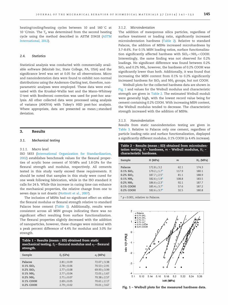
3.1.2. MicroindentationThe addition of mesoporous silica particles, regardless ofsurface treatment or loading ratio, significantly increasedmicroindentation hardness (Table 2). Relative to standardPalacos, the addition of MSNs increased microhardness by3.7–8.6%. For 0.1% MSN loading ratios, surface functionaliza-tion significantly affected hardness with SiO2oNH2oCOOH.Interestingly, the same finding was not observed for 0.2%loadings. No significant difference was found between 0.2%SiO2 and 0.2% NH2, however, the hardness of 0.2% COOH wassignificantly lower than both. Additionally, it was found thatincreasing the MSN content from 0.1% to 0.2% significantlyincreased hardness for SiO2 and NH2 groups, but not COOH.
Weibull plots for the collected hardness data are shown inFig. 1 and values for the Weibull modulus and characteristicstrength are given in Table 2. The estimated Weibull moduliwere generally high, with the lowest record value being forcement containing 0.2% COOH. With increasing MSN content,the Weibull modulus tended to decrease. The characteristicstrength increased with the addition of MSNs.
3.1.3. NanoindentationResults from static nanoindentation testing are given inTable 3. Relative to Palacos only one cement, regardless ofparticle loading ratio and surface functionalization, displayeda significantly different modulus, 0.1% COOH (a 4.4% increase).
Fig. 1 – Weibull plots for the measured hardness data.

Table 3 – Results (mean7SD) obtained from nanoindentation testing. Es – modulus, H – hardness, ψ – plasticity index, andrelaxation modulus G(t) evaluated at 1 and 10 s.
Sample Es (GPa) H (MPa) ψ G (t¼1 s) (GPa) G (t¼10 s) (GPa)
Palacos 3.8370.24 195.7731.3 0.37670.064 0.8770.09 0.7370.090.1% SiO2 3.9670.26 207.4730.6 0.39870.047 0.8970.09 0.7570.090.2% SiO2 3.6970.37 179.2723.4a 0.32970.070a 0.8470.11 0.7070.100.1% NH2 3.8670.24 203.2731.8 0.38870.057 0.9070.11 0.7670.110.2% NH2 3.9270.14 217.8734.7a 0.37870.033 0.9470.11 0.7970.110.1% COOH 4.0070.21a 217.0722.9a 0.36770.033 0.9070.09 0.7870.100.2% COOH 3.8170.27 203.2732.8 0.37470.061 0.8770.10 0.7370.10
a po0.05, relative to Palacos.
Fig. 2 – Plots of the calculated relaxation moduli, G(t) (left). Plots of the burger model fit for each cement. For clarity, only thecurve fit of the burger model is shown, and not the experimental data (right). The same legend applies to both figures.
Table 4 – The estimated parameters (mean7SD) of the burger model fit to the collected creep data. E1 and E2 are moduli andn is the long-term creep viscosity.
Cement E1 (GPa) E2 (GPa) n (103 GPa s) Creep Constant (s)
Palacos 123.279.3 18.871.5 3.270.5 10.170.10.1% SiO2 121.7713.0 18.772.1 3.570.8 10.170.30.2% SiO2 115.3717.0 17.672.4 3.270.6 10.370.50.1% NH2 123.9713.7 18.472.1 4.972.8 10.270.40.2% NH2 124.0716.5 18.972.2 3.270.5 10.170.30.1% COOH 120.6713.2 18.372.1 4.171.6 10.370.30.2% COOH 105.2716.5 16.572.4 2.870.6 10.570.4
j o u r n a l o f t h e m e c h a n i c a l b e h a v i o r o f b i o m e d i c a l m a t e r i a l s 3 7 ( 2 0 1 4 ) 1 4 1 – 1 5 2146
For cements containing 0.2% loading ratios, modulus wassignificantly influenced by surface functionalization with NH2
and COOH cements displaying higher moduli than SiO2. Thesame trend was not observed for 0.1% loading ratios. Hardnesswas found to significantly decrease for 0.2% SiO2 and increasefor 0.1% COOH and 0.2% NH2, relative to Palacos. Plasticityindex was found to be consistent with the only significantdifference being 0.2% SiO2.
No significant difference in relaxation moduli was foundand the magnitude of G(t) was generally consistent amongstall cements (Table 3). The profile of G(t) vs. time changeddrastically after one second of relaxation time and began todegrade at a faster rate, as seen in Fig. 2. All cementsdisplayed a uniform profile in G(t) vs. time, except for 0.1%
COOH, which interestingly had the lowest record modulus instatic testing. No significant differences were observed in thecreep behavior of the cements (Fig. 2). The collected data werewell described by the burger model (adjusted R2 of at least0.995 for all cements), however, there were no significantdifferences in the estimated parameters (Table 4).
3.2. Material characterization
3.2.1. MorphologyScanning electron micrographs revealed no apparent differ-ence in the fracture surface of the cements (Fig. 3). Smallpores between 50 and 100 mm were observed on the fracturesurfaces of all cements and were not influenced by MSN
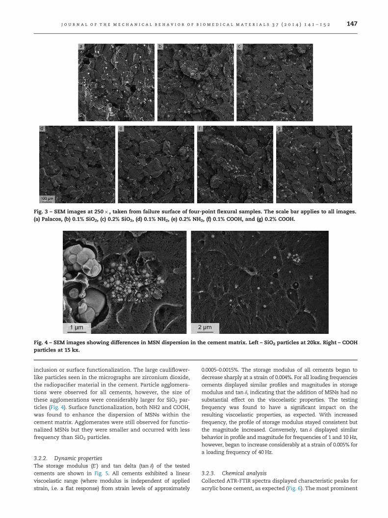
Fig. 3 – SEM images at 250� , taken from failure surface of four-point flexural samples. The scale bar applies to all images.(a) Palacos, (b) 0.1% SiO2, (c) 0.2% SiO2, (d) 0.1% NH2, (e) 0.2% NH2, (f) 0.1% COOH, and (g) 0.2% COOH.
Fig. 4 – SEM images showing differences in MSN dispersion in the cement matrix. Left – SiO2 particles at 20kx. Right – COOHparticles at 15 kx.
j o u r n a l o f t h e m e c h a n i c a l b e h a v i o r o f b i o m e d i c a l m a t e r i a l s 3 7 ( 2 0 1 4 ) 1 4 1 – 1 5 2 147
inclusion or surface functionalization. The large cauliflower-like particles seen in the micrographs are zirconium dioxide,the radiopacifier material in the cement. Particle agglomera-tions were observed for all cements, however, the size ofthese agglomerations were considerably larger for SiO2 par-ticles (Fig. 4). Surface functionalization, both NH2 and COOH,was found to enhance the dispersion of MSNs within thecement matrix. Agglomerates were still observed for functio-nalized MSNs but they were smaller and occurred with lessfrequency than SiO2 particles.
3.2.2. Dynamic propertiesThe storage modulus (E0) and tan delta (tan δ) of the testedcements are shown in Fig. 5. All cements exhibited a linearviscoelastic range (where modulus is independent of appliedstrain, i.e. a flat response) from strain levels of approximately
0.0005–0.0015%. The storage modulus of all cements began todecrease sharply at a strain of 0.004%. For all loading frequenciescements displayed similar profiles and magnitudes in storagemodulus and tan δ, indicating that the addition of MSNs had nosubstantial effect on the viscoelastic properties. The testingfrequency was found to have a significant impact on theresulting viscoelastic properties, as expected. With increasedfrequency, the profile of storage modulus stayed consistent butthe magnitude increased. Conversely, tan δ displayed similarbehavior in profile andmagnitude for frequencies of 1 and 10 Hz,however, began to increase considerably at a strain of 0.005% fora loading frequency of 40 Hz.
3.2.3. Chemical analysisCollected ATR-FTIR spectra displayed characteristic peaks foracrylic bone cement, as expected (Fig. 6). The most prominent

Fig. 5 – Results from DMA strain sweeps. Solid lines – 1 Hz, dashed lines – 40 Hz. For clarity purposes, data from 10 Hz isnot shown.
Fig. 6 – ATR-FTIR spectra collected for each cement.
Table 5 – The initial thermal decomposition temperature(T10), midpoint decomposition (T50), and glass transition(Tg) obtained from TGA and DSC testing.
Sample T10 (1C) T50 (1C) Tg (1C)
Palacos 340.11 384.08 99.690.1% SiO2 336.20 381.43 100.440.2% SiO2 326.09 380.79 99.290.1% NH2 323.96 380.73 102.300.2% NH2 325.02 381.59 100.850.1% COOH 323.22 381.76 100.290.2% COOH 320.70 379.94 99.39
j o u r n a l o f t h e m e c h a n i c a l b e h a v i o r o f b i o m e d i c a l m a t e r i a l s 3 7 ( 2 0 1 4 ) 1 4 1 – 1 5 2148
peaks occurred at 1730 cm�1 and 1140 cm�1 which corre-spond to a C¼O stretch and O–C–C stretch, respectively.Analysis of ATR-FTIR spectra showed no differences in theposition of intensity peaks between the different cements,indicating that there was no formation of new chemicalgroups. However, peak intensities did increase with theaddition of MSNs except for 0.1% SiO2.
3.2.4. Thermal characteristicsThe initial decomposition temperature of the cements wasfound to decrease with the addition of MSNs and surfacefunctionalization increased this negative effect (Table 5).
Midpoint decomposition temperatures tended to equalizebetween all cements. This is potentially attributable todegradation of functional groups on the MSN surface. Theglass transition temperature was not influenced by theaddition of MSNs.
4. Discussion
In this study, we extend upon our previous work withnanoparticle-reinforced bone cement (Slane et al., 2014) inan effort to enhance particle dispersion and interfacial adhe-sion using low loadings of functionalized MSNs. Two func-tional groups (and plain MSNs as a positive control) at loadingratios of 0.1% and 0.2% wt/wt were incorporated into acommercially available acrylic bone cement. The functionalgroups used, propylamine and propylcarboxylic acid, werechosen based upon previous research on bone cement mod-ified with functionalized carbon nanotubes (Ormsby et al.,2010b). Mixed results were observed for the mechanicalproperties of the cements. Four-point flexural testing andnanoindentation revealed minimal impact on the propertiesof the cements, except for several changes in the nano-level

j o u r n a l o f t h e m e c h a n i c a l b e h a v i o r o f b i o m e d i c a l m a t e r i a l s 3 7 ( 2 0 1 4 ) 1 4 1 – 1 5 2 149
mechanical properties. Conversely, microindentation testingdemonstrated that the addition of MSNs significantly alteredthe properties of the cements. This finding suggests thepresence of different reinforcement mechanisms, which willbe addressed in greater depth later in the discussion. To theauthors' current knowledge, this is the first study to utilize amultiscale approach to characterize the influence of nano-particle reinforcement on acrylic bone cement.
The concept of adding reinforcement materials to enhancethe mechanical properties of acrylic bone cement is notnovel; in the mid 1970s researchers experimented with bonecements modified with graphite and carbon fibers (Knoellet al., 1975; Pilliar et al., 1976). Despite the long historyassociated with reinforced bone cements, a variety of pro-blems persist which limit their clinical applicability such aspoor material dispersion, poor fiber/particle interfacial adhe-sion with the cement matrix and diminished mixing char-acteristics. Regardless of the chosen reinforcement material,one of the primary difficulties faced with polymer compositesis achieving a uniform dispersion of the reinforcement. If thisis not achieved, the agglomerations of fibers/particles canform stress concentration sites within the polymer matrixleading to premature failure with the application of load(Ormsby et al., 2010a). Surface functionalization, a processwhere chemical moieties are covalently linked to a particle/fibers surface, is a widely implemented technique that canassist in particle dispersion and interfacial adhesion (Maet al., 2010). Previous work with bone cement demonstratedthat functionalized carbon nanotubes could enhance severaldifferent mechanical properties (Ormsby et al., 2010b).
Mesoporous silica nanoparticles have been shown toenhance the mechanical properties of polymer composites.Samuel et al. modified a dental resin with MSNs and found anincrease in the flexural modulus and no effect on the flexuralstrength with increasing MSN content(Samuel et al., 2009)while Ji et al. demonstrated drastic improvements in thetensile properties of a polymer composite loaded with MSNs(Ji et al., 2003). Similarly, Park and Pinnavaia showedenhanced tensile properties and toughness with an epoxyresin modified with mesoporous silica foam particles (Parkand Pinnavaia, 2007). However, all of these studies usedloading ratios considerably higher than in the current study.Our previous work on MSN-modified bone cements usedloading ratios of 0.5%, 2%, and 5% wt/wt. While severalencouraging results were found, we wanted to determine iflow-loading ratios could enhance the properties of cementwhile minimizing the impact on the handling characteristics.In theory, the high surface area of the MSNs used in thisstudy (720 m2/g) suggest an efficient stress transfer mechan-ism between the particle and polymer matrix can develop(Fu et al., 2008).
The flexural properties measured in this study are in closeagreement with previously published values. For example,Dunne reported a flexural strength and modulus for Palacos R(cured in air for 24 h) of 75.67 MPa and 2.76 GPa, respectively(Dunne, 2008), compared to 72.07 MPa and 2.81 GPa in thisstudy. The flexural strength and modulus of the cementswere not influenced by the addition of MSNs and all cementsdisplayed properties above those outlined in ISO 5833. This islikely attributable to the low loadings used, which were
insufficient to induce changes on the macro level. SEManalysis of the fracture surfaces of bending samples alsodisplayed no apparent differences, indicating that the failuremechanism was consistent across all groups. Several pre-vious studies have examined the use of nanomaterials toreinforce acrylic bone cement, however, the results aresomewhat conflicting. For example, Ormsby et al. incorpo-rated various loadings of functionalized carbon nanotubes(f-CNTs) into Colacryl bone cement. They found a 4.1%decrease in the flexural strength using 0.1 wt/wt propylamineCNTs and a 21.9% increase using 0.1 wt/wt propylcarboxylicacid CNTs (Ormsby et al., 2010b). In contrast, Gonçalves et al.observed an �25% decrease in the flexural strength of anacrylic cement modified with 0.1 wt/wt f-CNTs, which theyattributed to the CNTs acting as free-radical scavengers,resulting in polymerization retardation and inhibition(Gonçalves et al., 2012). Based on the glass transition tem-peratures determined in this study, which were consistentacross all groups, we do not believe that the MSNs inhibitedthe polymerization of the cement.
Nanoindentation testing revealed only slight differencesin measures of hardness and modulus with only severalsignificant variations detected. In contrast, microindentationshowed that the inclusion of MSNs significantly enhancedthe hardness of all cements. This finding highlights thedifferences between micro and nanoindentation. The inden-tation data collected generally agrees with that previouslypublished for bone cement. Karimzadeh and Ayatollahiperformed nanoindentation testing on Cemex RX cementand found a modulus of 5.56 GPa and hardness of 290 MPa(Karimzadeh and Ayatollahi, 2012). These values are slightlyhigher than those found in this study, however, they used apeak indentation depth of 210 nm which is substantiallysmaller than that used in this study, and due to the indenta-tion size effect the measured properties are expected to behigher. Lewis et al. obtained samples of Palacos R fromretrieved cement mantles and found a modulus of 3.78 GPaand hardness of 169 MPa for cement that was implanted for11 months using nanoindentation (Lewis et al., 2006). Lelovicsand Liptakova performed microindentation on SmartSetcement and found a microhardness of 199 MPa (Lelovicsand Liptakova, 2010), which generally agrees with the valueof 172 MPa determined in this study.
Compared to the size of the particles used in this study(200 nm), nanoindentation and microindentation can probedifferent heterogeneities within the cement since the localmechanical properties are directly linked to the size of thecontact. The relative size of the indentation surface/volumevaries substantially between the two techniques withnanoindentation being hundreds of nanometers (�750 nmdepth,o5 mm diagonal) and microindentation hundreds ofmicrons (�300–400 mm diagonal). The hardness of a materialrepresents its ability to resist surface deformation to anapplied load. In a material such as PMMA, non-recoverabledeformation of the surface possibly results from rupture ofthe localized polymer matrix network at the site of loadapplication or slippage between the matrix and any presentreinforcement material (Shen et al., 2006). At the micro level,substantially more MSNs are within the indented cementvolume, thus leading to an increase in the measured
j o u r n a l o f t h e m e c h a n i c a l b e h a v i o r o f b i o m e d i c a l m a t e r i a l s 3 7 ( 2 0 1 4 ) 1 4 1 – 1 5 2150
hardness. For 0.1% MSN loadings, it was found that functio-nalization significantly increased microhardness, however,this is likely due to enhanced dispersion and not the resultof new bond formation between the particle and matrix(as confirmed with ATR-FTIR). Additionally, hardness mea-sured with nanoindentation was typically higher than thecalculated microhardness. This finding is due to differences inhow contact area is measured between the techniques (Qianet al., 2005) and the confined polymer mobility of the inter-facial region adjacent to the indenter tip (Tehrani et al., 2011).
Particle dispersion within the cement matrix was signifi-cantly influenced by particle surface functionalization. Asverified with SEM (Figs. 3 and 4), SiO2 particles tended to formlarge agglomerates often times with interfacial gaps betweenthe particle and the matrix. In contrast, functionalizedparticles were more evenly dispersed throughout the matrix.Agglomerations still occurred with functionalized particles,however, they generally were less frequent and smaller insize. It appears that the sonication process used to disperseMSNs was adequate for functionalized particles but not forSiO2. While ultrasonication is a powerful tool to disruptparticle agglomerations, care must be taken since in somesituations (e.g. extended sonication times) the applied acous-tic energy can actually induce the formation of new agglom-erates or damage the particle/fiber being dispersed (such ascarbon nanotube scission) (Taurozzi et al., 2011).
Bone cement acts as a grouting material between theimplant and the patient's bone, providing immediate implantfixation while distributing forces from the implant to thesurrounding bone bed. Therefore, it must possess propertiesthat are the intermediately of the prosthesis (typically tita-nium or cobalt chrome) and bone. Dynamic mechanicalanalysis conducted in this study confirms that this is thecase for all cements tested. At a frequency of 1 Hz and strainof 0.01%, the storage modulus of cements ranged from 3.1 to3.4 GPa while tan δ varied between 0.07 and 0.08. In contrast,cancellous bone has a storage modulus of 0.2 GPa and tan δ of0.09–0.1 (cortical bone 0.04) while metals used for implantstypically have a storage modulus of exceeding 100 GPa and atan δ of 10�4 (De Santis et al., 2003). It was found that theaddition of MSNs had minimal impact on the viscoelasticproperties of the cements and generally, values for storagemodulus and tan δ were consistent. Interestingly, at a fre-quency of 40 Hz tan δ values significantly increased. This is inmarked contrast to skeletal tissue where tan δ decreases withincreasing loading frequency, indicating the material acts asan elastic solid and transmits rather than dissipates force.It has been suggested that this increase in tan δ with higherfrequencies enable acrylic cements to survive the loadingpatterns they are exposed to during gait, despite the fact thatcements fail in a brittle fashion during static testing (Danielset al., 2005).
Several limitations of the current study are acknowledged.Firstly, only a single type of mesoporous silica with a specificsize and structure was used. A variety of MSNs can befabricated with different pores sizes, surface areas, and porevolumes. These factors could potentially influence the find-ings from this study. One exceptionally attractive alternativeis mesoporous silica foam, which has a very large porevolume (e.g. 2.4 cm3/g) and an open structure that could
allow for strong bonding with polymer chains (Park andPinnavaia, 2007). Secondly, only a single variety of acrylicbone cement was used although many other formulationsexist. Different cements with alternative chemical composi-tions and viscosities may respond differently to the inclusionof MSNs. As mentioned previously, the cement used in thisstudy was chosen since it is one of the most widely used inthe North American and European markets. Thirdly, only asingle type of static testing was conducted. Other methodscommonly used to test bone cement such as tension andcompression could potentially demonstrate enhancements,or detrimental, effects on the properties of the cement. Four-point bending was used since it includes both tensile andcompression components and is thought to be a realistictesting method for bone cement (Kuehn et al., 2005). Fourthly,micro and nanoindentation testing were performed on dif-ferent samples, although samples were prepared at the sametime from the same batch of cement. The observed differ-ences in micro and nanoindentation findings could poten-tially result from sample inhomogeneity. Finally, fatiguetesting of the cements was not performed, although it is wellknown that mechanical failure of bone cement can resultfrom fatigue failure (Lewis, 2003). It was deemed beyond thescope of this project to conduct fatigue testing.
Despite these limitations, this is the first study (to theauthors' current knowledge) that has utilized a multiscaleapproach to characterize the mechanical properties ofnanoparticle-reinforced bone cement. Contrary to our pre-vious work with MSN-modified cements, in this study theaddition of mesoporous silica did not have a detrimentalinfluence on the properties of the cements. Future workshould include examining alternative functionalizationgroups that can create the formation of interfacial bondsbetween the particle and PMMA matrix. One potentialmethod would be to alter the hydrophilicity of the particle'ssurface since it has previously been demonstrated thathydrophobic mesoporous silica can aid in dispersion andincrease adhesion (Bento et al., 2013). Higher loading ratios ofMSNs should be considered, however, with increased loadingthe potential for diminished handling characteristics andaltered polymerization characteristics increase. Therefore, itwould be required to quantify the influence of higher MSNsloadings on the rheological behavior and setting polymeriza-tion behavior of the cement. Finally, one potential benefit ofMSN-modified cement not explored in this study is the abilityof MSNs to reduce volumetric shrinkage during polymeriza-tion (Samuel et al., 2009), which can be as high as 6–7%. It isknown that the shrinkage of bone cement can induce resi-dual stresses as high as 10 MPa in the cement, potentiallycausing cracks that would act as failure initiation sites(Roques et al., 2004).
5. Conclusion
In this study, a commercially available acrylic bone cementwas modified with low-loadings of functionalized mesopor-ous silica nanoparticles and the resulting impact on thecement's mechanical and material properties were quantifiedover different length scales. Results from this study indicate

j o u r n a l o f t h e m e c h a n i c a l b e h a v i o r o f b i o m e d i c a l m a t e r i a l s 3 7 ( 2 0 1 4 ) 1 4 1 – 1 5 2 151
that the loading ratios of MSNs used were insufficient toinfluence the macro level properties of the cement. Functio-nalization aided in particle dispersion but did not appear toresult in enhanced interfacial adhesion between the particleand the matrix. These results suggest that that loading ratiosof mesoporous silica used are not an effective reinforcementmaterial in acrylic bone cement. Additionally, since non-functionalized MSNs were found to form large agglomerates,their use in bone cement is not recommended regardless ofthe loading ratio. Future work should be conducted todetermine the impact of higher MSN loading ratios andalternative functional groups.
Acknowledgments
The authors thank Christopher Besaw for his work on themicroindentation testing. The authors gratefully acknowl-edge use of facilities and instrumentation supported by theNSF-funded University of Wisconsin Materials ResearchScience and Engineering Center (DMR-1121288).
r e f e r e n c e s
Adelani, M.A., Keeney, J.A., Palisch, A., Fowler, S.A., Clohisy, J.C.,2013. Has total hip arthroplasty in patients 30 years oryounger improved? A systematic review. Clin. Orthop Relat.Res. 471, 2595–2601.
Ania, F., Broza, G., Mina, M.F., Schulte, K., Roslaniec, Z., Balta-Calleja, F.J., 2006. Micromechanical properties of poly(butyleneterephthalate) nanocomposites with single- and multi-walledcarbon nanotubes. Compos. Interface 13, 33–45.
Arun, S., Rama Sreekanth, P.S., Kanagaraj, S., 2014. Mechanicalcharacterisation of PMMA/SWNTs bone cement usingnanoindenter. Mater. Technol. 29, B4–B9.
ASTM International, 2012. Standard Test Method for TransitionTemperatures and Enthalpies of Fusion and Crystallization ofPolymers by Differential Scanning Calorimetry. ASTMInternational West Conshohocken, PA.
Ayatollahi, M., Karimzadeh, A., 2012. Determination of fracturetoughness of bone cement by nano-indentation test. Int. J.Fract. 175, 193–198.
Beake, B.D., Chen, S., Hull, J.B., Gao, F., 2002. Nanoindentationbehavior of clay/poly(ethylene oxide) nanocomposites. J.Nanosci. Nanotechnol. 2, 73–79.
Bento, A., Lourenco, J.P., Fernandes, A., Cerrada, M.L., RosarioRibeiro, M., 2013. Functionalization of mesoporous MCM-41(nano)particles: preparation methodologies, role on catalyticfeatures, and dispersion within polyethylenenanocomposites. ChemCatChem 5, 966–976.
Chaplin, R.P., Lee, A.J., Hooper, R.M., Clarke, M., 2006. Themechanical properties of recovered PMMA bone cement: apreliminary study. J. Mater. Sci. Mater. Med. 17, 1433–1448.
Chung, S.M., Yap, A.U., Tsai, K.T., Yap, F.L., 2005. Elastic modulusof resin-based dental restorative materials: amicroindentation approach. J. Biomed. Mater. Res. B Appl.Biomater. 72, 246–253.
Daniels, A., Wirz, D., Morscher, E., 2005. Properties of bonecement: extreme differences in properties of successful bonecements. In: Breusch, S., Malchau, H. (Eds.), The Well-Cemented Total Hip Arthroplasty. Springer, Berlin.
De Santis, R., Mollica, F., Ambrosio, L., Nicolais, L., Ronca, D., 2003.Dynamic mechanical behavior of PMMA based bone cementsin wet environment. J. Mater. Sci. Mater. Med. 14, 583–594.
Dhakal, H.N., Zhang, Z.Y., Richardson, M.O.W., 2006.Nanoindentation behaviour of layered silicate reinforcedunsaturated polyester nanocomposites. Polym. Test. 25,846–852.
Dunne, N., 2008. Mechanical properties of bone cement. In: Deb,S. (Ed.), Orthopaedic Bone Cements. Woodhead PublishingLimited, Cambridge, pp. 233–264.
Fischer-Cripps, A., 2011. In: Nanoindentation. Springer,Dordrecht.
Fu, S., Feng, X., Lauke, B., Mai, Y., 2008. Effects of particle size,particle/matrix interface adhesion and particle loading onmechanical properties of particulate–polymer composites.Compos. Part B-Eng. 39, 933–961.
Goncalves, G., Cruz, S.M., Ramalho, A., Gracio, J., Marques, P.A.,2012. Graphene oxide versus functionalized carbon nanotubesas a reinforcing agent in a PMMA/HA bone cement. Nanoscale4, 2937–2945.
Guo, Z., Pereira, T., Choi, O., Wang, Y., Hahn, H.T., 2006. Surfacefunctionalized alumina nanoparticle filled polymericnanocomposites with enhanced mechanical properties. J.Mater. Chem. 16, 2800–2808.
Hailer, Garellick, N.P., Karrholm, J., G., 2010. Uncemented andcemented primary total hip arthroplasty in the Swedish HipArthroplasty Register. Acta Orthop. 81, 34–41.
International Organization for Standardization, 2002. Implantsfor Surgery – Acrylic Resin Cements. ISO, Geneva, Switzerland.
Izquierdo-Barba, I., Colilla, M., Vallet-Regi, M., 2008.Nanostructured mesoporous silicas for bone tissueregeneration. J. Nanomater, 1–14.
Jayasuriya, R.L., Buckley, S.C., Hamer, A.J., Kerry, R.M., Stockley, I.,Tomouk, M.W., Wilkinson, J.M., 2013. Effect of sliding-tapercompared with composite-beam cemented femoral prosthesisloading regime on proximal femoral bone remodeling: arandomized clinical trial. J. Bone Joint Surg. Am. 95, 19–27.
Jeffers, J.R., Browne, M., Lennon, A.B., Prendergast, P.J., Taylor, M.,2007. Cement mantle fatigue failure in total hip replacement:experimental and computational testing. J. Biomech. 40,1525–1533.
Ji, X., Hampsey, J.E., Hu, Q., He, J., Yang, Z., Lu, Y., 2003.Mesoporous silica-reinforced polymer nanocomposites.Chem. Mater. 15, 3656–3662.
Kane, R.J., Yue, W., Mason, J.J., Roeder, R.K., 2010. Improvedfatigue life of acrylic bone cements reinforced with zirconiafibers. J. Mech. Behav. Biomed. 3, 504–511.
Karimzadeh, A., Ayatollahi, M.R., 2012. Investigation ofmechanical and tribological properties of bone cement bynano-indentation and nano-scratch experiments. Polym. Test.31, 828–833.
Khaled, S.M., Charpentier, P.A., Rizkalla, A.S., 2011. Physical andmechanical properties of PMMA bone cement reinforced withnano-sized titania fibers. J. Biomater. Appl. 25, 515–537.
Khanuja, H.S., Vakil, J.J., Goddard, M.S., Mont, M.A., 2011.Cementless femoral fixation in total hip arthroplasty. J. BoneJoint Surg. Am. 93, 500–509.
Knoell, A., Maxwell, H., Bechtol, C., 1975. Graphite fiber reinforcedbone cement. An experimental feasibility investigation. Ann.Biomed. Eng. 3, 225–229.
Kordas, K., Kukkola, J., Toth, G., Jantunen, H., Szabo, M., Sapi, A.,Kukovecz, A., Konya, Z., Mikkola, J., 2013. Nanoparticledispersions. In: Vajtai, R. (Ed.), Springer Handbook ofNanomaterials. Springer-Verlag, Berlin, pp. 729–775.
Kuehn, K.D., Ege, W., Gopp, U., 2005. Acrylic bone cements:mechanical and physical properties. Orthop. Clin. N. Am. 36,29–39 (v–vi).
Kumar, B., Cooke, F.W., 2006. Fatigue behaviour of fiber reinforcedbone cement. In: Gdoutos, E.E. (Ed.), Fracture of Nano andEngineering Materials and Structures. Springer, Netherlands,pp. 1023–1024.

j o u r n a l o f t h e m e c h a n i c a l b e h a v i o r o f b i o m e d i c a l m a t e r i a l s 3 7 ( 2 0 1 4 ) 1 4 1 – 1 5 2152
Lach, R., Kim, G.-M., Michler, G.H., Grellmann, W., Albrecht, K.,2006. Indentation fracture mechanics for toughnessassessment of PMMA/SiO2 nanocomposites. Macromol. Mater.Eng. 291, 263–271.
Lelovics, H., Liptakova, T., 2010. Time and mixing technique-dependent changes in bone cement SmartSet(R) HV. ActaBioeng. Biomech./Wroclaw Univ. Technol. 12, 63–67.
Lennon, A.B., 2008. Fracture toughness and fatigue characteristicsof bone cements. In: Deb, S. (Ed.), Orthopaedic Bone Cements.Woodhead Publishing Limited, Cambridge, pp. 265–310.
Lewis, G., 2003. Fatigue testing and performance of acrylic bone-cement materials: state-of-the-art review. J. Biomed. Mater.Res. B Appl. Biomater. 66, 457–486.
Lewis, G., Janna, S., 2003. Effect of test specimen cross-sectionalshape on the in vitro fatigue life of acrylic bone cement.Biomaterials 24, 4315–4321.
Lewis, G., Xu, J., Dunne, N., Daly, C., Orr, J., 2006. Criticalcomparison of two methods for the determination ofnanomechanical properties of a material: application tosynthetic and natural biomaterials. J. Biomed. Mater. Res. PartB, Appl. Biomater. 78, 312–317.
Ma, P.-C., Siddiqui, N.A., Marom, G., Kim, J.-K., 2010. Dispersionand functionalization of carbon nanotubes for polymer-basednanocomposites: a review. Compos. Part A – Appl. Sci. 41,1345–1367.
Malzbender, J., Steinbrech, R.W., 2004. Mechanical properties ofcoated materials and multi-layered composites determinedusing bending methods. Surf. Coat. Technol. 176, 165–172.
Marrs, B., Andrews, R., Rantell, T., Pienkowski, D., 2006.Augmentation of acrylic bone cement with multiwall carbonnanotubes. J. Biomed. Mater. Res. Part A 77, 269–276.
Musib, M., Jones, J., Chakote, K., Hayes, W., Saha, S., 2012.Microhardness of bi-antibiotic-eluting bone cement scaffolds.Prog. Biomater. 1, 1–7.
Nottrott, M., Molster, A.O., Gjerdet, N.R., 2007. Time dependentmechanical properties of bone cement. An in vitro study overone year. J. Biomed. Mater. Res. B Appl. Biomater. 83, 416–421.
O’Connor, D.O., Burke, D.W., Jasty, M., Sedlacek, R.C., Harris, W.H.,1996. In vitro measurement of strain in the bone cementsurrounding the femoral component of total hip replacementsduring simulated gait and stair-climbing. J. Orthop. Res.: Off.Publ. Orthop. Res. Soc. 14, 769–777.
Oliver, W.C., Pharr, G.M., 2004. Measurement of hardness andelastic modulus by instrumented indentation: advances inunderstanding and refinements to methodology. J. Mater. Res.19, 3–20.
Ormsby, R., McNally, T., Mitchell, C., Dunne, N., 2010a.Incorporation of multiwalled carbon nanotubes to acrylicbased bone cements: effects on mechanical and thermalproperties. J. Mech. Behav. Biomed. 3, 136–145.
Ormsby, R., McNally, T., Mitchell, C., Dunne, N., 2010b. Influenceof multiwall carbon nanotube functionality and loading onmechanical properties of PMMA/MWCNT bone cements. J.Mater. Sci. Mater. Med. 21, 2287–2292.
Ormsby, R., McNally, T., O’Hare, P., Burke, G., Mitchell, C., Dunne,N., 2012. Fatigue and biocompatibility properties of a poly(methyl methacrylate) bone cement with multi-walled carbonnanotubes. Acta Biomater. 8, 1169–1179.
Padmanabhan, S.K., Balakrishnan, A., Chu, M.C., Kim, T.N., Cho,S.J., 2010. Micro-indentation fracture behavior of humanenamel. Dent. Mater. 26, 100–104.
Park, I., Pinnavaia, T.J., 2007. Mesocellular silica foam as an epoxypolymer reinforcing agent. Adv. Funct. Mater. 17, 2835–2841.
Park, S., Hung, C.T., Ateshian, G.A., 2004. Mechanical response ofbovine articular cartilage under dynamic unconfinedcompression loading at physiological stress levels. Osteoarthr.Cartil. 12, 65–73.
Pegel, S., Potschke, P., Petzold, G., Alig, I., Dudkin, S.M., Lellinger,D., 2008. Dispersion, agglomeration, and network formation ofmultiwalled carbon nanotubes in polycarbonate melts.Polymer 49, 974–984.
Pilliar, R.M., Blackwell, R., Macnab, I., Cameron, H.U., 1976.Carbon fiber-reinforced bone cement in orthopedic surgery. J.Biomed. Mater. Res. B Appl. Biomater. 10, 893–906.
Qian, L., Li, M., Zhou, Z., Yang, H., Shi, X., 2005. Comparison ofnano-indentation hardness to microhardness. Surf. Coat.Technol. 195, 264–271.
Roques, A., Browne, M., Taylor, A., New, A., Baker, D., 2004.Quantitative measurement of the stresses induced duringpolymerisation of bone cement. Biomaterials 25, 4415–4424.
Samuel, S.P., Li, S., Mukherjee, I., Guo, Y., Patel, A.C., Baran, G.,Wei, Y., 2009. Mechanical properties of experimental dentalcomposites containing a combination of mesoporous andnonporous spherical silica as fillers. Dent. Mater. 25, 296–301.
Schiffmann, K.I., 2006. Nanoindentation creep and stressrelaxation tests of polycarbonate: analysis of viscoelasticproperties by different rheological models. Int. J. Mater. Res.97, 1199–1211.
Shen, L., Wang, L., Liu, T., He, C., 2006. Nanoindentation andmorphological studies of epoxy nanocomposites. Macromol.Mater. Eng. 291, 1358–1366.
Slane, J., Vivanco, J., Meyer, J., Ploeg, H.L., Squire, M., 2014.Modification of acrylic bone cement with mesoporous silicananoparticles: effects on mechanical, fatigue and absorptionproperties. J. Mech. Behav. Biomed. Mater. 29, 451–461.
Sonntag, R., Reinders, J., Kretzer, J.P., 2012. What’s next?Alternative materials for articulation in total jointreplacement. Acta Biomater. 8, 2434–2441.
Taurozzi, J.S., Hackley, V.A., Wiesner, M.R., 2011. Ultrasonicdispersion of nanoparticles for environmental, health andsafety assessment – issues and recommendations.Nanotoxicology 5, 711–729.
Tehrani, M., Safdari, M., Al-Haik, M.S., 2011.Nanocharacterization of creep behavior of multiwall carbonnanotubes/epoxy nanocomposite. Int. J. Plast. 27, 887–901.
Xu, H.H.K., Quinn, J.B., Smith, D.T., Antonucci, J.M., Schumacher,G.E., Eichmiller, F.C., 2002. Dental resin composites containingsilica-fused whiskers – effects of whisker-to-silica ratio onfracture toughness and indentation properties. Biomaterials23, 735–742.
Zhang, F.-A., Lee, D.-K., Pinnavaia, T.J., 2010. PMMA/mesoporoussilica nanocomposites: effect of framework structure and poresize on thermomechanical properties. Polym. Chem. 1,107–113.