Multiple species of CPP32 and Mch2 are the major active caspases present in apoptotic cells
11
The EMBO Journal Vol.16 No.9 pp.2271–2281, 1997 Multiple species of CPP32 and Mch2 are the major active caspases present in apoptotic cells residue, a very unusual substrate specificity for eukaryotic Lavina Faleiro 1,2 , Ryuji Kobayashi 1 , proteases, although not every aspartate provides a cleavage Howard Fearnhead 1 and Yuri Lazebnik 1 site. In fact, only about a dozen proteins cleaved by 1 Cold Spring Harbor Laboratory, 1 Bungtown Road, caspases in vivo have been identified. Cold Spring Harbor, NY 11724 and 2 Graduate Program in Molecular The key observation linking caspases to apoptosis was and Cellular Biology, State University of New York at Stony Brook, the finding that CED-3, a protein essential for apoptosis Stony Brook, NY 11794, USA in Caenorhabditis elegans, is a caspase (Xue and Horvitz, 1995; Yuan et al., 1993). This finding prompted the search The activity of ICE-like proteases or caspases is essen- for the mammalian equivalent of CED-3. The observation tial for apoptosis. Multiple caspases participate in that poly(ADP-ribose) polymerase (PARP) is cleaved apoptosis in mammalian cells but how many caspases during apoptosis (Kaufmann et al., 1993) by a caspase are involved and what is their relative contribution to activity (Lazebnik et al., 1994) led to the identification of cell death is poorly understood. To identify caspases this activity as CPP32 (Nicholson et al., 1995; Tewari activated in apoptotic cells, we developed an approach et al., 1995), a caspase cloned previously (Fernandes- to simultaneously detect multiple active caspases. Using Alnemri et al., 1994). Since CPP32 could cleave PARP tumor cells as a model, we have found that CPP32 and because inhibitors of CPP32 blocked apoptosis, it (caspase 3) and Mch2 (caspase 6) are the major active was concluded that CPP32 is the mammalian CED-3 caspases in apoptotic cells, and are activated in response equivalent. to distinct apoptosis-inducing stimuli and in all cell However, while only one caspase is known in C.elegans, lines analyzed. Both CPP32 and Mch2 are present in 10 caspases have been identified in humans (Alnemri apoptotic cells as multiple active species. In a given et al., 1996). Of these only ICE, which processes cytokines, cell line these species remained the same irrespective has a well established function (Thornberry, 1994; Gu of the apoptotic stimulus used. However, the species of et al., 1997). The diversity of human caspases raised the CPP32 and Mch2 detected varied between cell lines, question of whether CPP32 is the only apoptotic caspase indicating differences in caspase processing. The or if other caspases are also required. The answer to this strategy described here is widely applicable to identify question is critical both for understanding apoptosis and for active caspases involved in apoptosis. developing therapeutic strategies that target this process. Keywords: affinity labels/apoptosis/cancer therapy/ Several lines of evidence strongly suggest that in caspases (ICE-like proteases)/proteases mammalian cells multiple caspases are involved. First, deficiency in CPP32 prevented apoptosis in some cell types while it had no effect in others, indicating that Introduction CPP32 is redundant in many cell types (Kuida et al., 1996). This redundancy was consistent with the finding Apoptosis is a fundamental biological process critical for that caspases other than CPP32 can cleave PARP and the development of organisms and for maintaining tissue are sensitive to CPP32 inhibitors (Gu et al., 1995). homeostasis. Consequently, deregulation of apoptosis con- Furthermore, studies in a cell-free system provided evid- tributes to diseases such as cancer and neurodegenerative ence that nuclear changes of apoptosis require more than disorders (Thompson, 1995). The role of apoptosis in one caspase (Lazebnik et al., 1995; Takahashi et al., disease makes the apoptotic machinery a promising target 1996). In addition, several caspases were found to be for therapeutic intervention. However, such therapy processed in apoptotic cells (Chinnaiyan and Dixit, 1996), appears to be a distant goal since the knowledge of suggesting that their activation is linked to apoptosis. apoptosis is still fragmentary. Finally, FLICE/MACH (caspase 8) was identified as a The emerging view of apoptosis is that diverse regu- part of the receptor complexes that mediate apoptosis, latory pathways activate a conserved execution machinery implying that some caspases act as an interface between which carries out cell disassembly. Although this execution signaling pathways while others are involved in cell machinery is poorly understood, it appears that an essential execution (Boldin et al., 1996; Muzio et al., 1996). Thus, component is caspases, a family of cysteine proteases while there is little doubt that multiple caspases are (Martin and Green, 1995b; Chinnaiyan and Dixit, 1996). implicated in apoptosis, the number and the roles in cell Caspases are expressed as precursors that must be proteo- death remain poorly understood. lytically processed to become active enzymes (Thornberry, In this study, we used peptide inhibitors to identify 1994). This processing yields three polypeptides, a pro- directly the pool of caspases activated in apoptotic cells. domain, a small subunit and a large subunit which contains Peptide inhibitors that mimic caspase substrates can block the catalytic cysteine. The two subunits associate to form apoptosis. However the identity of the caspases inhibited the active protease and both contribute to the catalytic activity. Caspases cleave their substrates after an aspartate remained elusive since peptide inhibitors have broad © Oxford University Press 2271
-
Upload
independent -
Category
Documents
-
view
8 -
download
0
Transcript of Multiple species of CPP32 and Mch2 are the major active caspases present in apoptotic cells
The EMBO Journal Vol.16 No.9 pp.2271–2281, 1997
Multiple species of CPP32 and Mch2 are the majoractive caspases present in apoptotic cells
residue, a very unusual substrate specificity for eukaryoticLavina Faleiro1,2, Ryuji Kobayashi1,proteases, although not every aspartate provides a cleavageHoward Fearnhead1 and Yuri Lazebnik1
site. In fact, only about a dozen proteins cleaved by1Cold Spring Harbor Laboratory, 1 Bungtown Road, caspasesin vivo have been identified.Cold Spring Harbor, NY 11724 and2Graduate Program in Molecular The key observation linking caspases to apoptosis wasand Cellular Biology, State University of New York at Stony Brook,
the finding that CED-3, a protein essential for apoptosisStony Brook, NY 11794, USAin Caenorhabditis elegans, is a caspase (Xue and Horvitz,1995; Yuanet al., 1993). This finding prompted the searchThe activity of ICE-like proteases or caspases is essen-for the mammalian equivalent of CED-3. The observationtial for apoptosis. Multiple caspases participate inthat poly(ADP-ribose) polymerase (PARP) is cleavedapoptosis in mammalian cells but how many caspasesduring apoptosis (Kaufmannet al., 1993) by a caspaseare involved and what is their relative contribution toactivity (Lazebniket al., 1994) led to the identification ofcell death is poorly understood. To identify caspasesthis activity as CPP32 (Nicholsonet al., 1995; Tewariactivated in apoptotic cells, we developed an approachet al., 1995), a caspase cloned previously (Fernandes-to simultaneously detect multiple active caspases. UsingAlnemri et al., 1994). Since CPP32 could cleave PARPtumor cells as a model, we have found that CPP32and because inhibitors of CPP32 blocked apoptosis, it(caspase 3) and Mch2 (caspase 6) are the major activewas concluded that CPP32 is the mammalian CED-3caspases in apoptotic cells, and are activated in responseequivalent.to distinct apoptosis-inducing stimuli and in all cell
However, while only one caspase is known inC.elegans,lines analyzed. Both CPP32 and Mch2 are present in10 caspases have been identified in humans (Alnemriapoptotic cells as multiple active species. In a givenet al., 1996). Of these only ICE, which processes cytokines,cell line these species remained the same irrespectivehas a well established function (Thornberry, 1994; Guof the apoptotic stimulus used. However, the species ofet al., 1997). The diversity of human caspases raised theCPP32 and Mch2 detected varied between cell lines,question of whether CPP32 is the only apoptotic caspaseindicating differences in caspase processing. Theor if other caspases are also required. The answer to thisstrategy described here is widely applicable to identifyquestion is critical both for understanding apoptosis and foractive caspases involved in apoptosis.developing therapeutic strategies that target this process.Keywords: affinity labels/apoptosis/cancer therapy/
Several lines of evidence strongly suggest that incaspases (ICE-like proteases)/proteasesmammalian cells multiple caspases are involved. First,deficiency in CPP32 prevented apoptosis in some celltypes while it had no effect in others, indicating that
Introduction CPP32 is redundant in many cell types (Kuidaet al.,1996). This redundancy was consistent with the findingApoptosis is a fundamental biological process critical forthat caspases other than CPP32 can cleave PARP andthe development of organisms and for maintaining tissueare sensitive to CPP32 inhibitors (Guet al., 1995).homeostasis. Consequently, deregulation of apoptosis con-Furthermore, studies in a cell-free system provided evid-tributes to diseases such as cancer and neurodegenerativeence that nuclear changes of apoptosis require more thandisorders (Thompson, 1995). The role of apoptosis inone caspase (Lazebniket al., 1995; Takahashiet al.,disease makes the apoptotic machinery a promising target1996). In addition, several caspases were found to befor therapeutic intervention. However, such therapyprocessed in apoptotic cells (Chinnaiyan and Dixit, 1996),appears to be a distant goal since the knowledge ofsuggesting that their activation is linked to apoptosis.apoptosis is still fragmentary.Finally, FLICE/MACH (caspase 8) was identified as aThe emerging view of apoptosis is that diverse regu-part of the receptor complexes that mediate apoptosis,latory pathways activate a conserved execution machineryimplying that some caspases act as an interface betweenwhich carries out cell disassembly. Although this executionsignaling pathways while others are involved in cellmachinery is poorly understood, it appears that an essentialexecution (Boldinet al., 1996; Muzioet al., 1996). Thus,component is caspases, a family of cysteine proteaseswhile there is little doubt that multiple caspases are(Martin and Green, 1995b; Chinnaiyan and Dixit, 1996).implicated in apoptosis, the number and the roles in cellCaspases are expressed as precursors that must be proteo-death remain poorly understood.lytically processed to become active enzymes (Thornberry,
In this study, we used peptide inhibitors to identify1994). This processing yields three polypeptides, a pro-directly the pool of caspases activated in apoptotic cells.domain, a small subunit and a large subunit which containsPeptide inhibitors that mimic caspase substrates can blockthe catalytic cysteine. The two subunits associate to formapoptosis. However the identity of the caspases inhibitedthe active protease and both contribute to the catalytic
activity. Caspases cleave their substrates after an aspartate remained elusive since peptide inhibitors have broad
© Oxford University Press 2271

L.Faleiro et al.
specificity and can inactivate multiple caspases. To over- enhanced chemiluminescence (ECL). We will refer to suchblots as affinity blots.come this problem, we have used irreversible peptide
inhibitors tagged with biotin as affinity labels (Thornberry Several polypeptides were labeled with 10µM biotin–YVAD–amk in etoposide-treated cells (Figure 1A). Severalet al., 1994; Nicholsonet al., 1995) and developed an
approach to simultaneously detect and identify multiple observations suggested that these proteins are caspasesactivated during apoptosis. First, they had apparent molecu-labeled caspases by their mobility in two-dimensional
polyacrylamide gels. This approach provides a snapshot lar weights between 17 and 21 kDa, a range consistent withthe sizes of large caspase subunits. Second, the labeledthat identifies the caspases activated in apoptotic cells,
estimates their relative abundance and indicates their post- proteins were detected only in cells treated with etoposide,an apoptosis-inducing agent. Finally, these proteins weretranslational modifications.
The approach that we developed relies solely on the only revealed in the presence of the affinity label. Labelingwith biotin–DEVD–cmk and biotin–VAD–cmk revealed abiochemical activity of caspases and does not depend on
caspase primary sequence, cell type or the signal inducing set of polypeptides similar to those detected with biotin–YVAD–amk. However, at concentrations.1 µM, bothapoptosis. Using this approach we found that in all cell
lines tested two major active caspases were CPP32 and biotin–DEVD–cmk and biotin–VAD–cmk also labeled awide range of proteins in untreated cells (data not shown).Mch2. Intriguingly, both caspases were present as multiple
species which varied with the cell line, perhaps indicating This apparent non-specific binding may be explained by thechemical reactivity of chloromethyl ketones, which reactdifferences in post-translational modification. We also
found that CPP32 and Mch2 were consistently activated in with a wide range of proteins (Grammer and Blenis, 1996).In contrast, acyloxymethyl ketones are quiescent in solutionresponse to distinct apoptosis-inducing stimuli, suggesting
that distinct stimuli ultimately activate the same set of and are reactive only when bound to a cysteine protease(Krantz, 1994) although we found that increasing the con-caspases.centration of biotin–YVAD–amk to 100µM resulted inincreased background labeling (data not shown). Thus, forResultsfurther studies, we chose to use 10µM biotin–YVAD–amkas the affinity label.Affinity labeling of caspases activated in apoptotic
cells Since large subunits of caspases have similar molecularweights, we reasoned that one-dimensional (1D) gelThe key point of our approach was to make no assumptions
about the substrate specificity of the apoptotic caspases. electrophoresis may not resolve all labeled caspases.Therefore, we used two-dimensional (2D) gel electrophor-Although all caspases cleave after an aspartate, their
specificity varies. For example, CPP32 cleaves preferen- esis. A broad pH range (3–10) was used for isoelectro-focusing to accommodate the labeled subunits of all knowntially after a DxxD↓x site while Mch2 cleaves at a
VxxD↓x site and ICE at YxxD↓x. As peptide inhibitors caspases with the possible exception of TY (Faucheuet al., 1996) which has a calculated isoelectric point (pI)of caspases mimic a cleavage site, any inhibitor would
display some selectivity between caspases. However, our of 9.5. Proteins resolved by 2D electrophoresis weretransferred to a membrane which was processed as angoal was to use an inhibitor to label all caspases activated
in apoptotic cells. Therefore, to decrease the selectivity affinity blot. This 2D affinity blot (Figure 1B) indeedrevealed more putative caspases than the 1D affinity blotof the inhibitor we use the following strategy. First, we
used the observation that selectivity of caspase inhibitors (Figure 1A).To confirm that the labeled proteins were related todecreases with increasing concentration (Nicholson, 1996).
Second, we used an irreversible inhibitor and extended apoptosis rather than to etoposide treatment, we used 293cells which are resistant to apoptosis induced by etoposidelabeling times which improves labeling of caspases with
low affinity for the peptide moiety of the inhibitor. The (Figure 1C). We treated both Jurkat and 293 cells withetoposide, labeled active caspases with biotin–YVAD–key requirement for such an approach is low non-specific
binding at a high concentration of the label. Thus, we amk and obtained affinity blots (Figure 1C). While putativecaspases were labeled in Jurkat cells which underwenttested several available affinity labels: biotin–YVAD–
acyloxymethyl ketone (biotin–YVAD–amk); biotin–VAD– apoptosis after the treatment, no proteins were labeled in293 cells which did not undergo apoptosis. Thus, thechloromethyl ketone (biotin–VAD–cmk); and biotin–
DEVD–chloromethyl ketone (biotin–DEVD–cmk). labeled proteins were linked to apoptosis rather than toetoposide treatment.Each label was tested for its ability to reveal active
caspases present in apoptotic Jurkat cells and for non- To determine whether the proteins labeled with biotin–YVAD–amk were required for apoptosis, we set up a cell-specific labeling. Jurkat cells were treated with etoposide,
an anti-cancer drug, to induce apoptosis or left untreated. free system. In such systems, isolated nuclei added toextracts from apoptotic cells undergo morphological andCells were harvested and snap-frozen 6 h after treatment
when ~60% of the treated cells had characteristic apoptotic biochemical changes characteristic of apoptosis (Lazebniket al., 1993). We and others have demonstrated that thesemorphology. To label active caspases, cells were lysed by
freezing and thawing in a buffer that contained one of the changes depend on caspase activity and can be inhibitedby peptide inhibitors of caspases, including YVAD–cmkthree affinity labels at concentrations from 0.1 to 10µM.
The lysates were incubated for 15 min at 37°C, clarified by (Lazebniket al., 1994; Martin et al., 1995). Here weprepared extracts from apoptotic Jurkat cells that werecentrifugation, subjected to SDS–PAGE and the separated
proteins were transferred to a membrane. The blot was lysed either in the absence (mock labeling) or presenceof 10 µM biotin–YVAD–amk, a concentration used forprobed with avidin followed by biotin–horseradish per-
oxidase and the labeled proteins were visualized by the affinity blots. Nuclei added to the mock labeled extract
2272

Revealing apoptotic caspases by affinity labeling
Fig. 1. Affinity labeling reveals putative active caspases in apoptotic cells. (A) Jurkat cells were either treated with etoposide or left untreated. Cellswere then lysed either in the presence of biotin–YVAD–amk or in dilution buffer and labeled proteins were visualized by 1D affinity blot. Thepercent of apoptotic cells is indicated below the blot. Arrows indicate labeled proteins. (B) 2D affinity blot of Jurkat cells treated with etoposide andlabeled with biotin–YVAD–amk as in (A). (C) Putative caspases are not labeled in 293 cells, a cell line resistant to etoposide-induced apoptosis. Thepercent of apoptotic cells is indicated below the blot. (D) Biotin–YVAD–amk abolishes the activity required for apoptosis in the cell-free system.Jurkat cells were treated with etoposide and then lysed either with or without biotin–YVAD–amk (10µM). HeLa nuclei (13105) were incubatedwith an aliquot of each lysate (10µl). After incubation for 60 min at 37°C nuclei were fixed, stained with DAPI and examined by fluorescencemicroscopy. Shown are nuclei representative for each treatment.
underwent apoptotic changes whereas nuclei added to the to the resin. Bound proteins were eluted by boiling in 1%SDS (Figure 2A), resolved by 2D electrophoresis andlabeled extract did not (Figure 1D). Thus, at least one of
the proteins labeled and, therefore, inactivated by biotin– visualized by Coomassie-staining (Figure 2B, 3). Analiquot of the purified proteins was used to obtain aYVAD–amk was required for the nuclear apoptotic
changes in the cell-free system. corresponding 2D affinity blot (Figure 2B, 4). We alsoobtained a Coomassie-stained 2D gel of total cell lysate(Figure 2B, 1) and the corresponding affinity blot (FigurePurification of caspases
To identify the labeled putative caspases, we decided to 2B, 2). The sets of affinity-labeled proteins in cell lysate(Figure 2B, 2) and in purified sample (Figure 2B, 4) werepurify them and to obtain their protein sequence. Sixty-
six liters of Jurkat cells were treated with etoposide and similar, indicating that all detectable, labeled proteinswere recovered by purification, although the efficiency ofcells were harvested when 60–70% of the cells acquired
characteristic apoptotic morphology. Active caspases were purification varied slightly between the proteins.labeled by lysing the cells in the presence of biotin–YVAD–amk. In preliminary experiments we determined Multiple species of CPP32 and Mch2 are activated
in apoptotic Jurkat cellsthat the labeled proteins were soluble by comparing 2Daffinity blots of the pellet and supernatant obtained from By comparing the 2D Coomassie-stained gel to the corres-
ponding 2D affinity blot (Figure 3A), we identified eightthe labeled cell lysate (data not shown). Therefore, lysateswere clarified by centrifugation and the lysate was incub- spots that contained labeled proteins. These spots were
excised from the Coomassie-stained gel, the proteins wereated with avidin agarose to bind labeled proteins. Nobiotinylated proteins remained unbound (Figure 2A), sug- digested, and the resulting peptides were sequenced. We
obtained partial peptide sequences for six out of eightgesting that all detectable, labeled caspases were bound
2273

L.Faleiro et al.
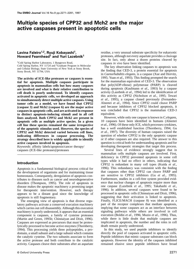
Asp28/Ser29 cleavage site, indicating that these specieswere processed differently from the p17 subunit. However,some species of CPP32 (spots 1 and 2) and Mch2 (spots3 and 4) had similar apparent molecular weight butdiffered in their pIs. These results indicated that bothCPP32 and Mch2 are present in apoptotic cells as multipleactive species which have different post-translationalmodification.
To determine whether either of the two unidentifiedproteins (Figure 3A, spots 7 and 8) was CPP32, weprobed a 2D affinity blot of apoptotic Jurkat cells with amonoclonal anti-CPP32 antibody (Transduction Labora-tories). This antibody recognized the protein in spot 7,indicating that it is CPP32 (Figure 3C). The antibody alsorecognized another CPP32 species (Figure 3A and C, spot2) previously identified by sequencing, but not the p17subunit of CPP32 (Figure 3A and C, spot 5), indicatingthat the epitope for this antibody is located upstream ofAsp28. Another CPP32 species (Figure 3A and C, spot1) was also not detected with this antibody, perhaps dueto low abundance of the CPP32 species. The lack ofreliable antibodies to other caspases prevented us fromusing immunoblots to identify the protein in spot 8. Thus,by using peptide sequencing and immunoblotting weidentified that the major active caspases in apoptotic Jurkatcells are multiple species of CPP32 and Mch2.
CPP32 and Mch2 are activated early in apoptosisHaving determined the position of CPP32 and Mch2species on a 2D affinity blot, we used this caspase mapto answer the following questions: when during apoptosisare CPP32 and Mch2 activated? Is the same set of caspases
Fig. 2. Purification of labeled caspases from Jurkat cell lysate. Cells activated in response to distinct cytotoxic agents? Are thewere treated with etoposide and lysed in the presence of biotin– caspases that are activated during apoptosis conservedYVAD–amk as described in Material and methods. (A) Affinity
between cell lines?chromatography of lysates on avidin–agarose. Shown is an affinity blotTo determine when CPP32 and Mch2 are activatedof input lysate (input), lysate incubated without the resin (mock-
depletion), lysate after incubation with avidin–agarose (unbound) and during apoptosis, we treated Jurkat cells with etoposidethe eluate from avidin–agarose (eluate). (B) 2D gel electrophoresis. and harvested the cells at different times during theCoomassie-stained 2D gel (1) and the matching affinity blot (3) of
treatment (Figure 4). For each sample, we determined theinput lysate. Coomassie-stained 2D gel (2) and the matching affinityproportion of cells that had apoptotic nuclear morphologyblot (4) of proteins eluted from avidin–agarose. Dashed boxes indicate
location of affinity labeled proteins selected for sequencing. (Figure 4A), the extent of PARP cleavage, a marker ofcaspase activity (Figure 4B), and the presence of activecaspases, as revealed by labeling with biotin–YVAD–amk(Figure 4C and D). Consistent with previous reportsproteins. Peptides derived from proteins contained in spots
1, 2, 5 and 6 matched the sequence of the caspase CPP32 (Kaufmannet al., 1993) we observed a delay betweenaddition of etoposide and an increase in the proportion ofwhile peptides derived from spots 3 and 4 matched the
sequence of the caspase Mch2 (Figure 3B). We were apoptotic cells (Figure 4A). This increase was precededby the appearance of the characteristic 85 kDa fragmentunable to obtain sequences from proteins in spots 7 and
8 because of insufficient amount of recovered peptides. of PARP (Kaufmannet al., 1993; Lazebniket al., 1994)(Figure 4B). As revealed by the 1D affinity blot (FigureThe identified species of CPP32 and Mch2 differed in
both molecular weight and pI. The apparent molecular 4C), caspases were activated concurrently with the cleav-age of PARP (Figure 4C). The increase in the intensity ofweights of CPP32 species from spots 1 and 2 differed
from those in spots 5 and 6 (Figure 3A), a difference that labeling correlated with both the amount of cleaved PARPand the proportion of apoptotic cells. As revealed by 2Dcould be attributed to proteolytic processing. In fact, a
peptide sequence obtained from spot 5 matched the affinity blots (Figure 4D), most abundant labeled proteinswere CPP32 and Mch2. Other weakly labeled proteinssequence of CPP32 from Ser29 to Lys39 (Figure 3B).
Since the residue preceding Ser29 in CPP32 is an aspartate with apparent molecular weights similar to that of largecaspase subunits were also detected (indicated by arrowsand the protease used for protein digestion cleaves only
after a lysine, Ser29 should be the N-terminal residue of in Figure 4D) transiently during etoposide treatment.Whether these labeled proteins are transiently activatedthe CPP32 species recovered from spot 5. Such a species
was described previously and is referred to as p17 CPP32 caspases (Enariet al., 1996) or are processing inter-mediates remains to be established. Labeled CPP32 wassubunit (Nicholsonet al., 1995). Peptides derived from
two other CPP32 species (spots 1 and 2) spanned the detected earlier than Mch2 (Figure 4D) although this could
2274
Revealing apoptotic caspases by affinity labeling
Fig. 3. Identification of affinity labeled caspases purified from apoptotic Jurkat cells. (A) Coomassie-stained 2D gel and the matching 2D affinity blotof purified proteins labeled with biotin–YVAD–amk. Shown are regions indicated in Figure 2B by dashed boxes. Numbers 1–8 indicate the spots thatwere excised for peptide sequencing. (B) Partial peptide sequences obtained from proteins extracted from spots 1 to 6. Sequences are indicated bythe line under the matched protein sequence. The number below the line corresponds to the number of the spot in (A). The protease used fordigestion of excised proteins cleaves after lysine residues which are highlighted in bold. (C) A 2D affinity blot (top) of lysate from apoptotic Jurkatcells was re-probed with an anti-CPP32 monoclonal antibody (bottom). Arrows indicate species of CPP32 detected by the antibody. (D) 2D map ofaffinity labeled caspases in Jurkat cells as identified by both sequencing (spots 1–6) and immunoblotting (spots 2 and 7). Positions of identifiedcaspases are shown by circles. Unidentified putative caspases are indicated by a square.
be attributed to greater abundance of CPP32 rather than arbitrary standard. In addition, for each tested agent, weobtained two 2D affinity blots: one of the sample preparedthe sequence of activation. Thus, these experiments
demonstrated that both CPP32 and Mch2 were activated from the cells treated with the agent (Figure 5B, 2 foranti-Fas and Figure 5B, 3 for staurosporine), and anotherearly during apoptosis.of the mixture of this sample with the sample preparedfrom etoposide treated cells (Figure 5B, 4 for anti-FasApoptosis induced by distinct agents involves
activation of CPP32 and Mch2 treated cells and Figure 5B, 5 for staurosporine treatedcells). If the same caspases were activated by both theWhether apoptosis induced by distinct stimuli is mediated
by the same set of caspases is a question fundamental to tested agent and etoposide, then the patterns of caspaselabeling in all three 2D affinity blots should be identicalunderstanding apoptosis and to the use of caspases as
targets for therapeutics. To address this question, we whereas caspases specific for one agent will be detectedon only two of the three blots. We found not only thattreated Jurkat cells with staurosporine, a kinase inhibitor
(Jacobsonet al., 1993), anti-Fas antibody that activates CPP32 and Mch2 were activated by all three treatmentsbut that the ratio between CPP32 and Mch2 species wasthe Fas pathway (Trauthet al., 1989; Itohet al., 1991)
and etoposide. When 60–70% of cells were apoptotic, similar regardless of the agent used. Hence, we concludedthat at least three distinct signaling pathways activate theactive caspases were labeled with biotin–YVAD–amk to
obtain 1D and 2D affinity blots. Both 1D and 2D affinity same set of CPP32 and Mch2 species in Jurkat cells.blots revealed no difference in activated caspases amongcells killed by the three agents (Figure 5). To rule out Sets of active CPP32 and Mch2 species vary
between cell lineserrors caused by gel-to-gel differences in caspase mobilitywe used the following approach. The 2D affinity blot of Whether the same set of caspases mediates apoptosis in
various cell types is also a critical question both toetoposide-treated cells (Figure 5B, 1) was used as an
2275

L.Faleiro et al.
Fig. 4. Activation of CPP32 and Mch2 in Jurkat cells correlates with cleavage of PARP and the onset of apoptosis. Apoptosis in Jurkat cells wasinduced by etoposide and aliquots of cells were collected at different times during the treatment. The cells were scored for apoptotic nuclearmorphology (A), and the cell lysates were analyzed by immunoblotting for cleavage of PARP (B), and by 1D (C) and 2D (D) affinity blotting forcaspase activation. Arrows in (C) indicate active caspases. Arrows in (D) indicate putative caspases that appeared transiently during etoposidetreatment. (D) Bottom: 2D map of active caspases as identified in Figure 3. Positions of identified caspases are shown by circles. Unidentifiedputative caspases detected transiently are indicated by squares.
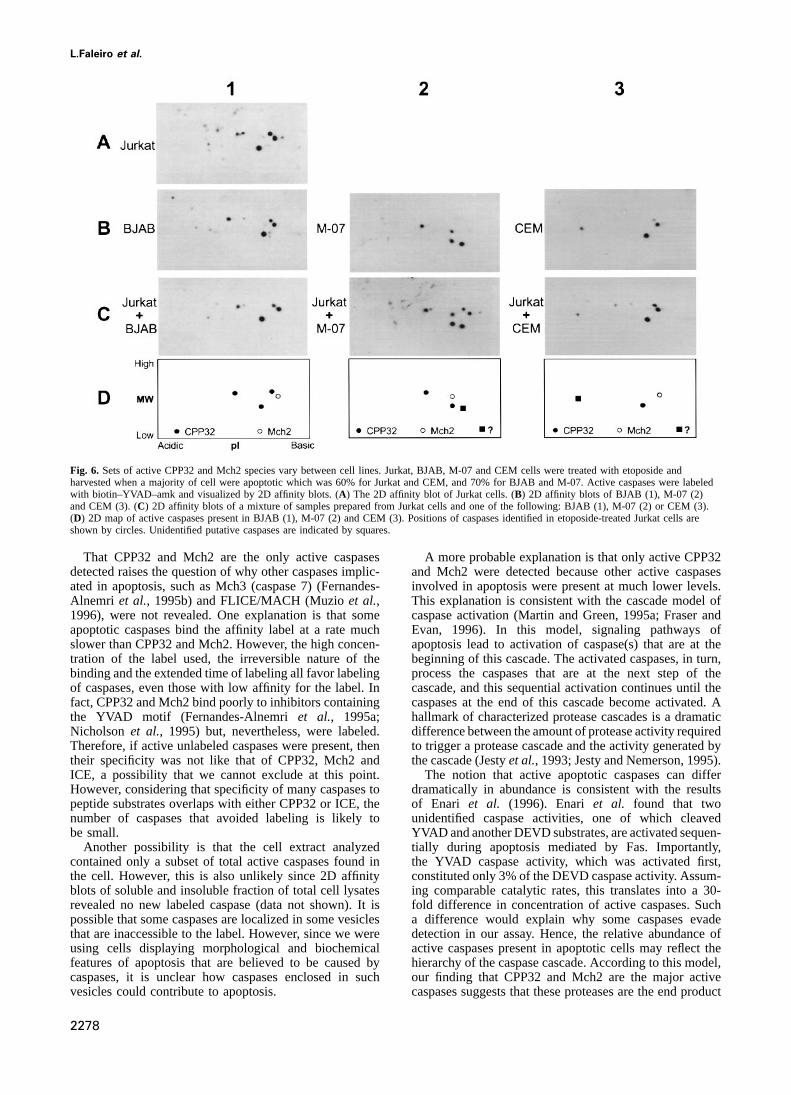
understanding apoptosis and to evaluating the possibility most abundant labeled proteins. Third, the sets of activeCPP32andMch2speciesvariedbetweencell lines (compareof using caspases as target for therapeutics. In particular,
since activation of caspases is a critical step in cell death Figure 6B parts 1, 2 and 3). While the sets of CPP32 andMch2 in BJAB, Namalwa, Molt-4 and HL-60 were similarinduced by anti-cancer drugs, it is important to determine
whether the set of caspases induced by a drug varies to that of Jurkat (Figure 6, 1, and data not shown), the setsdetected in M-07 and CEM were different (Figure 6, parts 2among tumor cells. To address this question, we induced
apoptosis with etoposide in the following cells: BJAB and and 3). In addition, we detected unidentified labeled proteinsthat could be caspases since they had molecular weightsNamalwa (Burkitt’s lymphomas), CEM and Molt-4 (adult
lymphocytic leukemias, ALL), HL-60 (promyeloid leuke- similar to that of large caspase subunits. These putativecaspases were prominent in both M-07 and CEM cellsmia) and M-07 (granulocytic leukemia). To compare sets
of labeled caspases activated in these cell lines, we used (Figure 6B, parts 2 and 3) and were minor in other cell lines(data not shown). Although these putative caspases remainthe same approach that we used to compare caspases
activated by distinct agents. We compared 2D affinity unidentified, we noticed that the location of the putativecaspase from M-07 cells on 2D affinity blots appears toblots of the tested cells, of Jurkat cells (standard) and of
a mixture of these two samples (Figure 6). coincide with that of a minor CPP32 species detected in thepool of caspases purified from Jurkat cells (Figure 3A andThis analysis provided several observations. First, active
CPP32 and Mch2 were detected in all cell lines (Figure 6 B, spot 6). Thus, we found that active species of CPP32 andMch2 vary with the cell line.and data not shown). Second, CPP32 and Mch2 were the
2276

Revealing apoptotic caspases by affinity labeling
Fig. 5. CPP32 and Mch2 are activated in Jurkat cells treated with distinct apoptotic stimuli. Jurkat cells were treated with etoposide, an anti-Fasantibody or staurosporine to induce apoptosis, or left untreated. Caspases were labeled with biotin–YVAD–amk and visualized by 1D (A) and 2D(B, 1–3) affinity blots. (B, 4) is a 2D affinity blot of a mixture of samples used to obtain blots (B, 1) and (B, 2). (B, 5) is a 2D affinity blot of amixture of samples used to obtain blots (B, 1) and (B, 3). (B, 6) is a map of active caspases detected by 2D affinity blots in etoposide-treatedJurkat cells.
Discussion only CPP32 a chance to be labeled. Second, the use ofirreversible inhibitors in this study allows identification
Although there is little doubt that caspases are a critical of the labeled caspases by affinity blotting.element of the apoptotic machinery, which of the human The approach described here is simple and widelycaspases are involved is not clear. To address this question
applicable. The large scale purification of labeled caspases,we developed an approach that directly identifies caspasesa costly step that we used to identify labeled caspases,activated in apoptotic cells. This approach generates snap-can be avoided, by using reliable antibodies when theyshots that simultaneously identify multiple labeled casp-become available. Alternatively, caspases can be identifiedases, estimate their relative abundance and indicate theirby their co-localization with known caspases on a 2Dpost-translational modifications. Using tumor cell lines asaffinity blot. The latter approach requires as little as 1–a model, we found that the major active caspases in10 µg of total cell protein or ~33105 apoptotic cells,apoptotic cells are CPP32 and Mch2. We also demonstratedan amount affordable in many experimental systems.that these caspases are present as multiple active species.Furthermore, using multi-layer avidin–biotin complexesIn a given cell line these species remain the same irrespect-may improve the sensitivity further. However, identifica-ive of the apoptotic stimulus used, suggesting that distincttion of labeled proteins that neither react with availablesignaling pathways ultimately activate the same set ofantibodies nor co-migrate with known labeled caspasescaspases. However, these species vary among cell lines,will require purification and sequencing. Considering thereflecting differences in their post-translational modi-progress in protein sequencing (Muzioet al., 1996) thefications.amount of protein required to obtain peptide sequence canOur approach relies only on the biochemical activity ofbe significantly less than that used in this study. Othercaspases and does not depend on cell type, species or onaffinity labels can be used as they become available, inthe primary caspase sequence. Therefore, this study laysparticular those that have low selectivity between caspases.the groundwork for identification of caspases activated inIt is important to keep in mind, however, that the 2D mapapoptotic cells irrespective of the species or type ofof affinity labeled caspases may change with a label.apoptotic stimulus. Our approach, however, does not
Since one molecule of a caspase binds only one moleculeprovide information about what caspases do to bring aboutof an affinity label, affinity labeling can be used to quantifycell death, but rather directly identifies the pool of caspasesamounts of active caspases in the extract, given that theactivated in apoptotic cells, providing candidates to focuscaspases are saturated with the label. Although in thisfunctional studies on.study we did not attempt to quantify labeled caspases inPreviously, the reversible peptide inhibitor DEVD-cell extracts, we established by using recombinant ICEaldehyde was used to purify CPP32 from a cell lysateand CPP32 that the amount of caspase revealed by affinity(Nicholson et al., 1995). The affinity labeling approachlabeling is proportional to the caspase concentration in adescribed here is distinct from that of Nicholsonet al. insample, suggesting that the described approach may betwo major aspects. First, our approach was designed to
provide every caspase activated in apoptotic cells and not used to evaluate relative abundance of active caspases.
2277
L.Faleiro et al.
Fig. 6. Sets of active CPP32 and Mch2 species vary between cell lines. Jurkat, BJAB, M-07 and CEM cells were treated with etoposide andharvested when a majority of cell were apoptotic which was 60% for Jurkat and CEM, and 70% for BJAB and M-07. Active caspases were labeledwith biotin–YVAD–amk and visualized by 2D affinity blots. (A) The 2D affinity blot of Jurkat cells. (B) 2D affinity blots of BJAB (1), M-07 (2)and CEM (3). (C) 2D affinity blots of a mixture of samples prepared from Jurkat cells and one of the following: BJAB (1), M-07 (2) or CEM (3).(D) 2D map of active caspases present in BJAB (1), M-07 (2) and CEM (3). Positions of caspases identified in etoposide-treated Jurkat cells areshown by circles. Unidentified putative caspases are indicated by squares.
That CPP32 and Mch2 are the only active caspases A more probable explanation is that only active CPP32and Mch2 were detected because other active caspasesdetected raises the question of why other caspases implic-
ated in apoptosis, such as Mch3 (caspase 7) (Fernandes- involved in apoptosis were present at much lower levels.This explanation is consistent with the cascade model ofAlnemri et al., 1995b) and FLICE/MACH (Muzioet al.,
1996), were not revealed. One explanation is that some caspase activation (Martin and Green, 1995a; Fraser andEvan, 1996). In this model, signaling pathways ofapoptotic caspases bind the affinity label at a rate much
slower than CPP32 and Mch2. However, the high concen- apoptosis lead to activation of caspase(s) that are at thebeginning of this cascade. The activated caspases, in turn,tration of the label used, the irreversible nature of the
binding and the extended time of labeling all favor labeling process the caspases that are at the next step of thecascade, and this sequential activation continues until theof caspases, even those with low affinity for the label. In
fact, CPP32 and Mch2 bind poorly to inhibitors containing caspases at the end of this cascade become activated. Ahallmark of characterized protease cascades is a dramaticthe YVAD motif (Fernandes-Alnemriet al., 1995a;
Nicholson et al., 1995) but, nevertheless, were labeled. difference between the amount of protease activity requiredto trigger a protease cascade and the activity generated byTherefore, if active unlabeled caspases were present, then
their specificity was not like that of CPP32, Mch2 and the cascade (Jestyet al., 1993; Jesty and Nemerson, 1995).The notion that active apoptotic caspases can differICE, a possibility that we cannot exclude at this point.
However, considering that specificity of many caspases to dramatically in abundance is consistent with the resultsof Enari et al. (1996). Enari et al. found that twopeptide substrates overlaps with either CPP32 or ICE, the
number of caspases that avoided labeling is likely to unidentified caspase activities, one of which cleavedYVAD and another DEVD substrates, are activated sequen-be small.
Another possibility is that the cell extract analyzed tially during apoptosis mediated by Fas. Importantly,the YVAD caspase activity, which was activated first,contained only a subset of total active caspases found in
the cell. However, this is also unlikely since 2D affinity constituted only 3% of the DEVD caspase activity. Assum-ing comparable catalytic rates, this translates into a 30-blots of soluble and insoluble fraction of total cell lysates
revealed no new labeled caspase (data not shown). It is fold difference in concentration of active caspases. Sucha difference would explain why some caspases evadepossible that some caspases are localized in some vesicles
that are inaccessible to the label. However, since we were detection in our assay. Hence, the relative abundance ofactive caspases present in apoptotic cells may reflect theusing cells displaying morphological and biochemical
features of apoptosis that are believed to be caused by hierarchy of the caspase cascade. According to this model,our finding that CPP32 and Mch2 are the major activecaspases, it is unclear how caspases enclosed in such
vesicles could contribute to apoptosis. caspases suggests that these proteases are the end product
2278

Revealing apoptotic caspases by affinity labeling
of the caspase cascade and that other caspases involved inMaterials and methodsapoptosis may contribute to activation of CPP32 and Mch2.
Cell cultureThe end product of a protease cascade, such as thrombinAll cells except 293 were maintained in RPMI tissue culture medium
in blood clotting, often carries out the main function of (Gibco-BRL) supplemented with 10% fetal bovine serum. 293 cells weremaintained in DMEM tissue culture medium supplemented with 10%the whole protease system, suggesting that the mostfetal bovine serum. M-07 cells were provided by Nick Carpino (Coldabundant active caspases in apoptotic cells are directlySpring Harbor Laboratory).
involved in cell destruction. This argument is consistentwith the predicted function of Mch2, which is thought to Induction of apoptosiscleave lamins during apoptosis (Orthet al., 1996; Apoptosis in cells (5–103105 cells/ml) was induced with one of the
following agents: 50µM etoposide (Sigma), 1µM staurosporine (Sigma)Takahashiet al., 1996) thereby causing the destruction ofor 100 ng/ml CH-11 anti-Fas antibody (Kamiya). The proportion ofthe nuclear lamina and the collapse of the nucleus.apoptotic cells was measured as described previously (Lazebniket al.,
Although putative CPP32 substrates are known, how their 1993). Briefly, cells were fixed in 4% paraformaldehyde (Ted Pella),permeabilized with 0.1% Triton-100 (Sigma) and stained with 1µg/mlcleavage contributes to cell death is not clear. NeverthelessDAPI. Nuclear morphology was examined using fluorescence microscopya recent study with CPP32-deficient mice indicated thatand cells with characteristic chromatin condensation were scored asCPP32 is required for developmental apoptosis in neuronsapoptotic.
although it is dispensable for apoptosis in other tested celltypes (Kuidaet al., 1996). SDS–PAGE and 2D gel electrophoresis
For 1D SDS–PAGE, cells lysates were diluted to 2 mg/ml with SDSOur study demonstrated that active CPP32 and Mch2sample buffer, boiled for 5 min and stored at –20°C. 10µg of proteinare present in apoptotic cells as multiple species. Somefrom each sample was separated on a 15% SDS–PAGE gel. For 2D
of the CPP32 species are cleaved at distinct sites andgels, cell lysates were diluted to 1 mg/ml in dSDS buffer (0.3% SDS,differ in their molecular weight, suggesting alternative 1% β-mercaptoethanol, 0.05 M Tris, pH 8), heated to 100°C for 5 min
and cooled on ice. One-tenth volume of DNase/RNase solution (1 mg/mlor multistep proteolytic processing of these caspases.DNase, 0.5 mg/ml RNase, 0.5 M Tris, 0.5 M MgCl2, pH 7.0) was addedHowever, other species differed only in their isoelectricto the samples which were then snap-frozen, lyophilized and resuspended
points, suggesting post-translational modifications other in urea sample buffer [9.5 M urea, 2% NP40, 5%β-mercaptoethanol,than proteolysis. These species may have different sub-2% ampholites 1.34% pH 3.5–10 (Pharmacia), 0.44% pH 7–9 (Pharmacia)
and 0.22% pH 5–6 (Serva)]. The samples (either 10 or 50µg) werestrate specificity, intracellular localization or other proper-separated in a 3% acrylamide-IEF gel for 19 000 Vh. The IEF gel wasties relevant to their function in apoptosis.then loaded onto a 20 cm324 cm 15% polyacrylamide gel that was run
Intriguingly, the sets of active CPP32 and Mch2 species at 60 W for 3.5 h.varied between cell lines. This variability may reflect the
Labeling of caspases and affinity blottingdifferences in how CPP32 and Mch2 were activated. OneAll affinity labels were obtained from Biosyn (Ireland) and stored atpossibility is that the caspases which process CPP32 or–70°C as a 10 mM stock solution in DMSO. The stock solution wasMch2 vary between cell types and, therefore, cleave diluted before use into MDB buffer (50 mM NaCl, 2 mM MgCl2, 5 mM
CPP32 or Mch2 precursors at different sites. Another EGTA, 10 mM HEPES, 1 mM DTT, pH 7) to prepare a 20µM solution.Cells were resuspended at 13107 cells/ml in KPM buffer (50 mM KCl,explanation is that the processing caspase is the same but50 mM PIPES, 10 mM EGTA, 1.92 mM MgCl2, pH 7, 1 mM DTT,the access of this protease to cleavage sites is regulated0.1 mM PMSF, 10µg/ml of cytochalasin B and 2µg/ml of the following:differently. For example, regulation may be achieved by chymostatin, pepstatin, leupeptin, antipain), pelleted and the pellet was
non-proteolytic, post-translational modifications that either snap-frozen in liquid nitrogen. An equal volume of 20µM biotin–YVAD–amk was added to the cell pellet and cells were lysed by threechange the conformation of the precursor or modify acycles of freezing–thawing. The lysates were incubated at 37°C for 15residue next to a cleavage site rendering the site inaccess-min, clarified at 4°C for 20 min at 16 000g, and mixed with either
ible to the processing caspase. Such post-translationalSDS–PAGE or 2D electrophoresis buffer.modifications could also provide a link between signaling The labeled proteins were separated by 1D SDS–PAGE or 2D
electrophoresis and transferred to PVDF Immobilon PSQ membranepathways and the caspase cascade involved in apoptosis.(Millipore) for 1 h at 100 V (1Dgels) or 4 h at 45 V (2Dgels) usingThe observation that induction of apoptosis by the samethe standard transfer buffer (25 mM Tris, 192 mM glycine and 20%
anti-cancer drug resulted in different pattern of caspase methanol). Membranes were then soaked in methanol and dried at roomactivation is intriguing and prompts the speculation that temperature overnight or at 80°C for 15 min. Dried membranes were
incubated in avidin-Neutralite (Molecular Probes) at 1µg/ml in PTBalterations in caspase processing may be related to cell(20 mM Tris, 150 mM NaCl, 0.02% Tween-20) supplemented with 1%transformation. Further characterization of caspase activa-BSA (PBT–BSA). Membranes were washed and then incubated in
tion in untransformed and transformed cells would provide biotinylated horseradish peroxidase (Molecular Probes) at 25 ng/ml inPBT–BSA. The labeled proteins were visualized by ECL (Amersham).an insight as to whether this hypothesis is true.Using recombinant ICE as a standard, we estimated that this assayIn summary, this study describes a novel strategydetects as little as 0.1 ng of an active caspase, the amount present inthat directly identifies the pool of caspases activated in 1 µg of a lysate prepared from apoptotic Jurkat cells.
apoptotic cells. Using tumor cell lines as a model wefound that CPP32 and Mch2 are the major active caspasesPurification of labeled caspases
Five batches (total of 66 l) of Jurkat cells grown to 6–73105 cells/mlpresent in apoptotic cells. We suggest that other caspaseswere treated with etoposide (50µM) to induce apoptosis. Cells wereif involved in apoptosis play a regulatory role by contribut-harvested 6–7 h after treatment when greater than 60–70% of the cell
ing to activation of CPP32 or Mch2. Our finding that population had apoptotic nuclear morphology. Cells were collected byactive CPP32 and Mch2 are present as various multiple centrifugation, lysed in the presence of 10µM biotin–YVAD–amk to
label caspases and the lysates were clarified by centrifugation at 100 000gspecies suggests that pathways that activate these enzymesfor 1 h at 4°C. The protein concentration of the pooled supernatant wasvary with cell type. The methodology described here is8.9 mg/ml with total of 68 ml. To remove unbound probe, the lysate
widely applicable to experimental systems of apoptosis was dialyzed against four changes of MDB buffer (50 mM NaCl, 2 mMMgCl2, 5 mM EGTA, 10 mM HEPES, 1 mM DTT, pH 7) supplementedand should facilitate the understanding of this process.
2279

L.Faleiro et al.
with 2 µg/ml of each chymostatin, leupeptin, antipain and pepstatin, and Fernandes-Alnemri,T., Litwack,G. and Alnemri,E.S. (1994) CPP32, a0.1 mM PMSF. 68 ml of dialyzed extract was incubated with 6 ml of novel human apoptotic protein with homology toCaenorhabditisImmunopure Immobilized Avidin (Pierce) for 1 h. The resin was washed eleganscell death protein Ced-3 and mammalian interleukin-1 beta-with 1% SDS and bound proteins were eluted by boiling in the same converting enzyme.J. Biol. Chem., 269, 30761–30764.buffer. The eluted proteins were precipitated by adding 0.25 volume of Fernandes-Alnemri,T., Litwack,G. and Alnemri,E.S. (1995a) Mch2, aTCA solution (100% trichloroacetic acid, 0.4% sodium deoxycholate) new member of the apoptotic ced-3/ICE cysteine protease family.to the eluate, washed once with 1 mM HCl in acetone and once with Cancer Res., 55, 2737–2742.pure acetone. The precipitate was resuspended in 150µl of the urea Fernandes-Alnemri,T.et al. (1995b) Mch3, a novel human apoptoticsample buffer. 0.2µl of the sample were saved and the rest was resolved cysteine protease highly related to cpp32.Cancer Res., 55, 6045–6052.by 2D electrophoresis. The gel was stained for 1 h with 0.05% Coomassie Fraser,A. and Evan,G. (1996) A license to kill.Cell, 85, 781–784.Blue (Sigma) in 5% acetic acid containing 10% methanol and then Grammer,T.C. and Blenis,J. (1996) The serine protease inhibitors,destained in 5% acetic acid containing 10% methanol. To obtain the tosylphenylalanine chloromethyl ketone and tosyllysine chloromethylmatching affinity blot, the saved sample (0.2µl) was resolved by 2D ketone, potently inhibit pp70(s6k) activation.J. Biol. Chem., 271,electrophoresis and the gel was used to obtain the affinity blot. The labeled 23650–23652.proteins were identified on the Coomassie-stained gel by comparing the Gu,Y., Sarnecki,C., Aldape,R.A., Livingston,D.J. and Su,M.S. (1995)gel with the matching affinity blot. The identified spots were excised Cleavage of poly(ADP-ribose) polymerase by interleukin-1 betaand used to obtain the protein sequence. converting enzyme and its homologs TX and Nedd-2.J. Biol. Chem.,
270, 18715–18718.Sequencing of purified caspases Gu,Y. et al. (1997) Activation of interferon-γ inducing factor mediatedThe excised gel fragments were washed twice with 50% methanol and by interleukin-1b converting enzyme.Science, 275, 206–208.treated with Achromobacter protease I (Waco Chemical) in (50 mM Itoh,N., Yonehara,S., Ishii,A., Yonehara,M., Mizushima,S.,Tris–HCl, 0.05% Tween 20, pH 9.0) for 20 h at 30°C to digest the Sameshima,M., Hase,A., Seto,Y. and Nagata,S. (1991) The polypeptideproteins. Peptide fragments were extracted in a solution of 50% aceto- encoded by the cDNA for human cell surface antigen Fas can mediatenitrile containing 0.065% trifluoroacetic acid and separated by HPLC apoptosis.Cell, 66, 233–243.(Hewlett-Packard, model 1090) using a Vydac C-18 column (1.03 Jacobson,M.D., Burne,J.F., King,M.P., Miyashita,T., Reed,J.C. and250 mm/100µM, 300 Å). Separated peptides were sequenced using an Raff,M.C. (1993) Bcl-2 blocks apoptosis in cells lacking mitochondrialautomated protein sequencer (Applied Biosystems, model 494). DNA. Nature, 361, 365–368.
Jesty,J. and Nemerson,Y. (1995) In Beutker,E., Lichtman,M.a.,Immunoblotting Coller,B.S. and Kipps,T.J. (eds),The Pathway of Blood Coagulation.Immunoblotting with monoclonal antibody to PARP (gift of Dr Guy McGraw-Hill, New York, pp. 1227–1238.Poirier) was carried out as described previously (Kaufmannet al., 1993). Jesty,J., Beltrami,E. and Willems,G. (1993) Mathematical analysis of aProbing of an affinity blot with monoclonal anti-CPP32 antibody proteolytic positive-feedback loop: dependence of lag time and enzyme(Transduction Lab) was carried out after the blot was soaked in methanol yields on the initial conditions and kinetic parameters.Biochemistry,to inactivate the horseradish peroxidase bound to avidin. 32, 6266–6274.
Kaufmann,S.H., Desnoyers,S., Ottaviano,Y., Davidson,N.E. andCell-free system of apoptosis Poirier,G.G. (1993) Specific proteolytic cleavage of poly(ADP-ribose)The cell-free assays were carried out as described (Lazebniket al., polymerase: an early marker of chemotherapy-induced apoptosis.1993). Briefly, purified HeLa nuclei were added to a Jurkat cell extract Cancer Res., 53, 3976–3985.(10 µl) supplemented with ATP regeneration system (2 mM ATP, 10 mM Krantz,A. (1994) Peptidyl (acyloxy)methanes as quiescent affinity labelscreatine phosphate and 50 mg/ml creatine kinase) and incubated for 60 for cysteine proteinases.Methods Enzymol., 244, 656–671.min at 37°C. To observe nuclear apoptotic changes nuclei were fixed Kuida,K., Zheng,T.S., Na,S.Q., Kuan,C.Y., Yang,D., Karasuyama,H.,with 4% paraformaldehyde, stained with DAPI (1µg/ml) and examined Rakic,P. and Flavell,R.A. (1996) Decreased apoptosis in the brain andby fluorescence microscopy. Images were acquired using a Photometrics premature lethality in CPP32-deficient mice.Nature, 384, 368–372.PXL CCD camera (Photometrics Ltd) controlled by Oncor Image Lazebnik,Y.A., Cole,S., Cooke,C.A., Nelson,W.G. and Earnshaw,W.C.software (Oncor Inc.) and figures prepared using Adobe Photoshop (1993) Nuclear events of apoptosisin vitro in cell-free mitotic extracts:software. a model system for analysis of the active phase of apoptosis.J. Cell
Biol., 123, 7–22.Lazebnik,Y.A., Kaufmann,S.H., Desnoyers,S., Poirier,G.G. andAcknowledgements
Earnshaw,W.E. (1994) Cleavage of poly(ADP-ribose) polymerase byWe thank our colleagues at CSHL and Guy Salvesen from the Burnham a proteinase with properties like ICE.Nature, 371, 346–347.Institute for helpful comments on the manuscript. We also thank Neena Lazebnik,Y.A., Takahashi,A., Moir,R.D., Goldman,R.D., Poirier,G.G.,Sareen and Nick Bizios from the 2D gel facility at CSHL for the state Kaufmann,S.H. and Earnshaw,W.C. (1995) Studies of the laminof the art 2D gels and Jeanne Wiggins for growing tons of cells. We proteinase reveal multiple parallel biochemical pathways duringthank Nora Poppito and Camille Walker for help with protein sequencing. apoptotic execution.Proc. Natl Acad. Sci. USA, 92, 9042–9046.The authors are grateful to Nancy Thornberry for the kind gift of Martin,S. and Green,D. (1995) Protease activation during apoptosis:recombinant ICE and to Guy Porier for providing an anti-PARP antibody. death by a thousand cuts?Cell, 82, 349–352.This work was supported by the NIH grant CA13106-25 to Y.L. and Martin,S.J., Newmeyer,D.D., Mathias,S., Farschon,D.M., Wang,H.G.,R.K. Y.L. is a Pew Scholar. Reed,J.C., Kolesnick,R.N. and Green,D.R. (1995) Cell-free
reconstitution of Fas-, UV radiation- and ceramide-induced apoptosis.EMBO J., 14, 5191–5200.
References Muzio,M. et al. (1996) FLICE, a novel FADD-homologous ICE/CED-3-like protease, is recruited to the CD95 (Fas/APO-1) death-inducingAlnemri,E.S., Livingston,D.J., Nicholson,D.W., Salvesen,G., Thornberry,signaling complex.Cell, 85, 817–827.N.A., Wong,W.W. and Yuan,J.Y. (1996) Human ICE/CED-3 protease
Nicholson,D.W. (1996) ICE/CED3-like proteases as therapeutic targetsnomenclature.Cell, 87, 171.for the control of inappropriate apoptosis.Nature Biotechnol., 14,Boldin,M., Goncharov,T., Goltsev,Y. and Wallach,D. (1996) Involvement297–301.of MACH, a novel MORT1/FADD-Interacting Protease, in Fas/Apo-1
Nicholson,D.W.et al. (1995) Identification and inhibition of the ICE/and TNF receptor-induced cell death.Cell, 85, 803–815.CED-3 protease necessary for mammalian apoptosis.Nature, 376,Chinnaiyan,A.M. and Dixit,V.M. (1996) The cell-death machine.Curr.37–43.Biol., 6, 555–562.
Orth,K., Chinnaiyan,A.M., Garg,M., Froelich,C.J. and Dixit,V.M. (1996)Enari,M., Talanian,R.V., Wong,W.W. and Nagata,S. (1996) SequentialThe CED-3/ICE-like protease Mch2 is activated during apoptosis andactivation of ICE-like and CPP32-like proteases during Fas-mediatedcleaves the death substrate lamin A.J. Biol. Chem., 271, 16443–16446.apoptosis.Nature, 380, 723–726.
Takahashi,A.et al. (1996) Cleavage of lamin A by Mch2 alpha butFaucheu,C., Blanchet,A.M., Collarddutilleul,V., Lalanne,J.L. andnot CPP32: Multiple interleukin 1 beta-converting enzyme-relatedDiuhercend,A. (1996) Identification of a cysteine protease closelyproteases with distinct substrate recognition properties are active inrelated to interleukin-1 beta-converting enzyme.Eur. J. Biochem.,
236, 207–213. apoptosis.Proc. Natl Acad. Sci. USA, 93, 8395–8400.
2280

Revealing apoptotic caspases by affinity labeling
Tewari,M., Quan,L.T., O’Rourke,K., Desnoyers,S., Zeng,Z., Beidler,D.R., Poirier,G.G., Salvesen,G.S. and Dixit,V.M. (1995) Yama/CPP32beta, a mammalian homolog of CED-3, is a CrmA-inhibitable proteasethat cleaves the death substrate poly(ADP-ribose) polymerase.Cell,81, 801–809.
Thompson,C. (1995) Apoptosis in the pathogenesis and treatment ofdisease.Science, 267, 1456–1462.
Thornberry,N.A. (1994) Interleukin-1 beta converting enzyme.MethodsEnzymol., 244, 615–631.
Thornberry,N.A., Peterson,E.P., Zhao,J.J., Howard,A.D., Griffin,P.R. andChapman,K.T. (1994) Inactivation of interleukin-1 beta convertingenzyme by peptide (acyloxy)methyl ketones.Biochemistry, 33,3934–3940.
Trauth,B.C., Klas,C., Peters,A.M.J., Matzku,S., Moller,P., Falk,W.,Debatin,K.-M. and Krammer,P.H. (1989) Monoclonal antibody-mediated tumor regression by induction of apoptosis.Science, 245,301–305.
Xue,D. and Horvitz,H.R. (1995) Inhibition of the Caenorhabditis eleganscell-death protease CED-3 by a CED-3 cleavage site in baculovirusp35 protein.Nature, 377, 248–251.
Yuan,J., Shaham,S., Ledoux,S., Ellis,H.M. and Horvitz,H.R. (1993) TheC.elegans cell death geneced-3 encodes a protein similar tomammalian interleukin-1β-converting enzyme.Cell, 641–652.
Received on November 29, 1996; revised on February 5, 1997
2281