Sonic Hedgehog Is Required for Progenitor Cell Maintenance in Telencephalic Stem Cell Niches
Upload
independentCategory
view
2download
0
Multiple Lineages of Human Breast Cancer Stem/Progenitor Cells Identified by Profiling with Stem CellMarkersWendy W. Hwang-Verslues1, Wen-Hung Kuo2, Po-Hao Chang1, Chi-Chun Pan1, Hsing-Hui Wang1, Sheng-
Ta Tsai1, Yung-Ming Jeng3, Jin-Yu Shew1, John T. Kung4, Chung-Hsuan Chen1, Eva Y.-H. P. Lee1,5,6*, King-
Jen Chang2*, Wen-Hwa Lee1,6*
1 Genomics Research Center, Academia Sinica, Taipei, Taiwan, 2 Department of Surgery, National Taiwan University Hospital, Taipei, Taiwan, 3 Department of Pathology, National
Taiwan University Hospital, Taipei, Taiwan, 4 Institute of Molecular Biology, Academia Sinica, Taipei, Taiwan, 5 Department of Development and Cell Biology, University of
California Irvine, Irvine, California, United States of America, 6 Department of Biological Chemistry, University of California Irvine, Irvine, California, United States of America
Abstract
Heterogeneity of cancer stem/progenitor cells that give rise to different forms of cancer has been well demonstrated forleukemia. However, this fundamental concept has yet to be established for solid tumors including breast cancer. In thiscommunication, we analyzed solid tumor cancer stem cell markers in human breast cancer cell lines and primary specimensusing flow cytometry. The stem/progenitor cell properties of different marker expressing-cell populations were furtherassessed by in vitro soft agar colony formation assay and the ability to form tumors in NOD/SCID mice. We found that theexpression of stem cell markers varied greatly among breast cancer cell lines. In MDA-MB-231 cells, PROCR and ESA, insteadof the widely used breast cancer stem cell markers CD44+/CD24-/low and ALDH, could be used to highly enrich cancer stem/progenitor cell populations which exhibited the ability to self renew and divide asymmetrically. Furthermore, the PROCR+/ESA+ cells expressed epithelial-mesenchymal transition markers. PROCR could also be used to enrich cells with colonyforming ability from MB-361 cells. Moreover, consistent with the marker profiling using cell lines, the expression of stem cellmarkers differed greatly among primary tumors. There was an association between metastasis status and a high prevalenceof certain markers including CD44+/CD242/low, ESA+, CD133+, CXCR4+ and PROCR+ in primary tumor cells. Taken together,these results suggest that similar to leukemia, several stem/progenitor cell-like subpopulations can exist in breast cancer.
Citation: Hwang-Verslues WW, Kuo W-H, Chang P-H, Pan C-C, Wang H-H, et al. (2009) Multiple Lineages of Human Breast Cancer Stem/Progenitor Cells Identifiedby Profiling with Stem Cell Markers. PLoS ONE 4(12): e8377. doi:10.1371/journal.pone.0008377
Editor: Mikhail V. Blagosklonny, Roswell Park Cancer Institute, United States of America
Received September 17, 2009; Accepted November 18, 2009; Published December 21, 2009
Copyright: � 2009 Hwang-Verslues et al. This is an open-access article distributed under the terms of the Creative Commons Attribution License, which permitsunrestricted use, distribution, and reproduction in any medium, provided the original author and source are credited.
Funding: This work was supported by Academia Sinica Peak Project (grant number 2371, 4012, http://www.sinica.edu.tw) and an Academia Sinica DistinguishedPostdoctoral Fellowship (http://aao.sinica.edu.tw/pfjob-e.html) to WWHV. The funders had no role in study design, data collection and analysis, decision topublish, or preparation of the manuscript.
Competing Interests: Based on UC Irvine policy, WHL and EL declare that they have financial interests of a biotech company, GeneTex, Inc, which distributesresearch reagents globally and owns stock from Schering-Plough, a publicly-traded pharmaceutical company, like any stockholder. WHL is on the board ofdirectors and EL is a consultant. GeneTex did not support this work in anyway. These data and material will be distributed freely following the policies of PLoSONE. It is their recognition that these financial interests could not generate any bias toward this work, and do not alter their adherence to all the PLoS ONEpolicies on sharing data and materials.
* E-mail: [email protected] (EY-HPL); [email protected] (K-JC); [email protected] (W-HL)
Introduction
The recently emerged concept of cancer stem cells has led to new
hypotheses about tumor progression. Cancer stem cells can divide
asymmetrically to self-renew and generate transient-amplifying
tumor cells that cause tumor formation and subsequent metastasis.
Thus, within the population of cancer cells, cancer stem cells are the
ones which can form new tumors and their asymmetric division
contributes to tumor heterogeneity. It has been reported that cancer
stem cells are present in acute myelogenous leukemia (AML) [1] as
well as many solid tumors [2–9] including breast tumors [10]. It has
been demonstrated that leukemia stem cells are heterogeneous in
terms of their origins [11] and different leukemia stem cells can give
rise to different types of leukemia [12,13]. However, it is not fully
known whether heterogeneous cancer stem cells exist in the many
types of solid tumors and how this heterogeneity may affect
treatment response of these cancers.
Of the many types of breast cancers, approximately 80 percent are
invasive ductal carcinomas, and 10–15 percent are invasive lobular
carcinomas. Additional rare types constitute less than 5–10 percent of
breast cancers. Gene expression profiling can further classify invasive
ductal carcinomas into five subtypes: luminal A, luminal B, ERBB2
(human epidermal growth factor receptor 2, HER2), basal and
normal-like [14–17]. One fundamental question that needs to be
addressed is whether these different subtypes of breast cancers are
derived from different lineage origins. Differing cancer stem cells in
each type may explain why they differ in degree of metastasis and
invasion, as well as prognosis outcome and treatment response. It is
thus essential to identify and characterize these cancer stem cell
populations in order to establish the origin and optimal treatment
strategy of each breast cancer subtype (see [18] for review).
Breast cancer stem cells have been isolated from human breast
tumors or breast cancer-derived pleural effusions using flow
cytometry to find subpopulations of cells with a specific pattern of
PLoS ONE | www.plosone.org 1 December 2009 | Volume 4 | Issue 12 | e8377

cell surface markers (CD44+, CD242/low, ESA+ (epithelial specific
antigen)) but lacking expression of specific lineage markers (Lin2)
[10]. These cells expressed epithelial-mesenchymal transition
(EMT) markers [19] and had higher tumorigenic potential than
bulk tumor cells after transplantation in nonobese diabetic/severe
combined immunodeficient (NOD/SCID) mice [10,19]. It has also
been shown that single cell suspensions of CD44+CD242/lowLin2
cells from human breast cancers were able to proliferate extensively
and form clonal nonadherent mammospheres in a low attachment
in vitro culture system [20]. These mammospheres were more
tumorigenic than established breast cancer-derived cell lines
including MCF-7 and B3R [20].
Additional markers useful in characterizing breast cancer stem
cells were recently reported [21–23]. PROCR, identified using
gene expression profiling of primary breast cancers [22], is also a
known marker of hematopoietic, neural, and embryonic stem cells
[24]. An additional marker, CD133, was identified for breast
cancer stem cells isolated from cell lines generated from
Brca12exon11/p53+/2 mouse mammary tumors [23] and is a
known marker of cancer stem cells in several organs including
brain, blood, liver and prostate [2,3,25,26]. A more recent study
showed that aldehyde dehydrogenase (ALDH) was increased in a
subpopulation of both normal and cancerous human mammary
epithelial cells that exhibit stem/progenitor cell properties. This
subpopulation is tumorigenic, capable of self-renewal, and able to
generate tumors that had the heterogeneity of the parental tumor
[21]. Other surface markers such as CXCR4 and ABCG2 may be
associated with cancer stem cell characteristics. CXCR4 is a G-
coupled heptahelical receptor contributing to metastasis in breast
cancers [27]. ABCG2 is one of the ABC transporters which has
been detected in known stem/progenitor cells such as hemato-
poietic stem cells [28], nestin-positive islet-derived progenitors [29]
and neural stem cells [30]. Although each of these markers has
been studied individually, they have not been used together to
determine the overall marker profile of individual tumors and the
possible heterogeneous origins in tumors.
To address this fundamental question, we first performed cancer
stem cell marker profile analysis using human breast cancer cell
lines and specimens to further define and characterize different
stem cell populations by flow cytometry, along with in vitro and in
vivo assays to verify the stem cell properties of different cell isolates.
Our results showed that: 1. the expression of stem cell markers
differed greatly among breast cancer cell lines as well as primary
tumors, 2. the previously recognized markers for breast cancer
stem cells may not be the optimal or universal markers for
identifying cancer stem cell populations, and 3. a highly
tumorigenic subpopulation expressing PROCR+/ESA+ was iden-
tified. Furthermore, this subpopulation was able to divide
asymmetrically both in vitro and in vivo, and expressed EMT
markers. These results suggest existence of multiple subpopula-
tions of breast cancer stem cells, as in the case for leukemia.
Results
Breast Cancer Cell Lines Heterogeneously ExpressedStem Cell Markers
To examine the expression profile of cancer stem cell markers in
breast cancer cell lines, we selected eight known stem cell markers
and performed FACS analysis in eight human breast cancer cell
lines (Table 1). Among those markers, CD44 was expressed mostly
in basal-like cell lines including MDA-MB-468, MDA-MB-231,
and HCC1937, while CD24 was expressed in luminal-like cell
lines such as T47D, MCF-7, ZR-75, and SKBR-3. ALDH
expression was observed in most of the cell lines and not associated
with specific cell types. Although it has been reported that HER2
overexpression could drive tumorigenesis and ALDH expression
[31], our cell line data did not support such a relationship. This
was consistent with the human specimen data described below
which showed no strong relationship between HER2 and ALDH
expression. Specifically, SKBR-3 and MDA-MB361 both ex-
pressed HER2 but differed in ALDH expression. PROCR was
expressed only in mesenchymal-like MDA-MB-231 and luminal-
like MDA-MB-361 cells. While ABCG2 was not expressed in all
cell lines except a few MCF-7 cells, high level of CD133 was
detected in almost all MDA-MB-468 cells, but not in other cell
lines. Furthermore, only a small portion of ZR-75 cells, but not
others, expressed CXCR4, while ESA was expressed in all the cell
lines, but the level was relatively low in MDA-MB231 (Table 1).
These data suggest that human breast cancer cell lines express
stem cell markers heterogeneously and likely contain different
types of cancer stem/progenitor cells.
CD44+/CD242/low and ALDH Are Not the UniversalMarkers for Isolation of Cancer Stem Cells with HighEfficiency of Colony Forming Ability
The hallmark of cancer stem cells is that one or very few cells
are capable of forming tumor in animal assay. To accelerate the
selection procedure for identification of stem cells, we sought to
use anchorage-independent growth in soft agar culture since it best
mimics tumorigenic ability in animal. Although mammosphere
formation has served as an in vitro stem cell criteria, not every
breast cancer cell line posses such ability [32,33]. Based on cell
surface markers listed in Table 1, the bulk cells were labeled with
antibodies specifically against the selected stem cell markers, sorted
into two separate groups using fluorescence activated cell sorting
(FACS), and subjected to soft agar colony formation assays
(Figure 1). If the selected markers effectively identified cancer stem
cells, it would be expected that the marker-expressing subpopu-
Table 1. Different human breast cancer cell lines expresseddifferent known solid cancer stem cell markers.
Marker/Cell line MB468 MB231 HCC1937 T47D MCF7 ZR75 SKBR3 MB361
ER - - - + + + - +
PR - - - + + - - -
HER2 o.e. o.e.
CD44 +++ +++ +++ ++ ++ + + +++
CD24 +++ - ++ +++ +++ +++ +++ -
CD44+/CD24-/low
- +++ ++ - - - - +++
CD133 +++ - - - - - - -
PROCR - ++ - -/+ - - - ++
ABCG2 - - - - + - - -
CXCR4 - - - - - + - -
ESA +++ ++ +++ +++ +++ +++ +++ +++
ALDH + + + - + + ++ -
The ER/PR (+/2) and HER2 overexpression (o.e.) status were adapted from Neveet al (2006)[44].2, not detectable.+, ,5%.++, 5–70% of the cells express the marker indicated.+++, .70% of the cells express the marker indicated.doi:10.1371/journal.pone.0008377.t001
Cancer StemCells Heterogeneity
PLoS ONE | www.plosone.org 2 December 2009 | Volume 4 | Issue 12 | e8377

lation would have higher colony forming efficiency than cells not
expressing the markers.
Based on this premise, we have analyzed five cell lines including
ZR-75, HCC1937, SKBR-3, MDA-MB-231 and MDA-MB-361.
Consistent with previous reports [21,34], ZR-75 cells sorted by
ALDH+ were negatively associated with ER expression (Figure S1,
Method S1), but exhibited higher colony forming ability than the
bulk cells or ALDH2 cells (Figure 1A). However, if sorted by
CXCR4+, the subpopulation exhibited less efficiency in colony
forming ability when compared with the ALDH+ subpopulation
(Figure S2, Method S1), suggesting that the ALDH+ subpopulation
may contain more cancer stem/progenitor cells of the ZR-75 cell
line. The subpopulation of HCC1937 cells sorted by CD44+/
CD242/low formed more colonies than the bulk cells (Figure 1B,
bulk vs. CD44+/CD242). However, the remaining subpopulation,
which was not CD44+/CD242/low, also formed colonies more
than the bulk cells and showed a better efficiency than the CD44+/
CD242/low cells (Figure 1B). This suggested the existence of
another stem cell population, which was not CD44+/CD242/low.
Further selection with ALDH marker from HCC1937 cells failed
to enrich a subpopulation with higher colony forming ability
(Figure S2, Method S1). Similarly, the subpopulation sorted by
ALDH+ from SKBR-3 cells showed less efficiency in colony
formation than the bulk and the ALDH2 cells (Figure 1C). Taken
together, these results suggested that CD44+/CD242/low and
ALDH may not be universal markers to identify and enrich highly
tumorigenic stem cells from all breast cancers. Instead, there may
be other types of breast cancer stem cells that are defined by other
markers.
PROCR+/ESA+ Enriched a Subpopulation of Breast CancerCells with High Colony Forming Efficiency in Soft Agar
It was noted that in MDA-MB-231 and MDA-MB-361 cell
lines, almost all cells ($90%) were CD44+/CD242/low. However,
less than five and twelve percent of the cells were able to form
colonies in soft agar, respectively (Figure 1D and 1E, bulk cells). In
MDA-MB-231, the sorted PROCR+/ESA+ cells showed a two-
fold and nine-fold increase in colony forming efficiency when
compared with that of the bulk cells and PROCR2/ESA2 cells,
respectively (Figure 1D), suggesting that the PROCR+/ESA+
subpopulation may be comprised of a higher number of stem/
progenitor cells than the PROCR2/ESA2 cells.
Since more than 85–90% of MDA-MB-361 cells expressed
CD44 and ESA (Table 1), PROCR alone was used to evaluate
whether a high tumorigenic subpopulation can be enriched in this
cell line. A two-fold higher colony forming efficiency was observed
in PROCR+ cells when compared with the bulk cells or the
Figure 1. Soft agar colony forming efficiency of bulk cells or marker expressing or nonexpressing subpopulations from breastcancer cell lines. Soft agar colony formation assay of cells isolated from ZR-75 (A), HCC1937 (B), SKBR-3 (C), MDA-MB-231 (D) and MDA-MB361 (E)cells with the indicated cell surface markers expressed. The unsorted cells (Bulk) were used as control. Shown is the percentage of colony formation.Results are means 6 SD of triplicate samples from one representative experiment.doi:10.1371/journal.pone.0008377.g001
Cancer StemCells Heterogeneity
PLoS ONE | www.plosone.org 3 December 2009 | Volume 4 | Issue 12 | e8377
PROCR2 cells (Figure 1E). However, no difference in colony
forming efficiency was observed between the bulk cells and
PROCR2 cells, suggesting that the PROCR marker alone may
not be the optimal selection of the stem/progenitor cells from this
cell line.
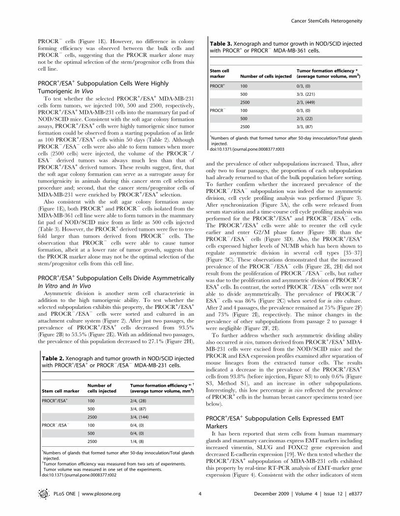
PROCR+/ESA+ Subpopulation Cells Were HighlyTumorigenic In Vivo
To test whether the selected PROCR+/ESA+ MDA-MB-231
cells form tumors, we injected 100, 500 and 2500, respectively,
PROCR+/ESA+ MDA-MB-231 cells into the mammary fat pad of
NOD/SCID mice. Consistent with the soft agar colony formation
assays, PROCR+/ESA+ cells were highly tumorigenic since tumor
formation could be observed from a starting population of as little
as 100 PROCR+/ESA+ cells within 50 days (Table 2). Although
PROCR2/ESA2 cells were also able to form tumors when more
cells (2500 cells) were injected, the volume of the PROCR2/
ESA2 derived tumors was always much less than that of
PROCR+/ESA+ derived tumors. These results suggest, first, that
the soft agar colony formation can serve as a surrogate assay for
tumorigenicity in animals during this cancer stem cell selection
procedure and; second, that the cancer stem/progenitor cells of
MDA-MB-231 were enriched by PROCR+/ESA+ selection.
Also consistent with the soft agar colony formation assay
(Figure 1E), both PROCR+ and PROCR2 cells isolated from the
MDA-MB-361 cell line were able to form tumors in the mammary
fat pad of NOD/SCID mice from as little as 500 cells injected
(Table 3). However, the PROCR+ derived tumors were five to ten-
fold larger than tumors derived from PROCR2 cells. The
observation that PROCR2 cells were able to cause tumor
formation, albeit at a lower rate of tumor growth, suggests that
the PROCR marker alone may not be the optimal selection of the
stem/progenitor cells from this cell line.
PROCR+/ESA+ Subpopulation Cells Divide AsymmetricallyIn Vitro and In Vivo
Asymmetric division is another stem cell characteristic in
addition to the high tumorigenic ability. To test whether the
selected subpopulation exhibits this property, the PROCR+/ESA+
and PROCR2/ESA2 cells were sorted and cultured in an
attachment culture system (Figure 2). After just two passages, the
prevalence of PROCR+/ESA+ cells decreased from 93.5%
(Figure 2B) to 53.5% (Figure 2E). With an additional two passages,
the prevalence of this population decreased to 27.1% (Figure 2H),
and the prevalence of other subpopulations increased. Thus, after
only two to four passages, the proportion of each subpopulation
had already returned to that of the bulk population before sorting.
To further confirm whether the increased prevalence of the
PROCR2/ESA2 subpopulation was indeed due to asymmetric
division, cell cycle profiling analysis was performed (Figure 3).
After synchronization (Figure 3A), the cells were released from
serum starvation and a time-course cell cycle profiling analysis was
performed for the PROCR+/ESA+ and PROCR2/ESA2 cells.
The PROCR+/ESA+ cells were able to reenter the cell cycle
earlier and enter G2/M phase faster (Figure 3B) than the
PROCR2/ESA2 cells (Figure 3D). Also, the PROCR+/ESA+
cells expressed higher levels of NUMB which has been shown to
regulate asymmetric division in several cell types [35–37]
(Figure 3C). These observations demonstrated that the increased
prevalence of the PROCR2/ESA2 cells (Figure 2E, 2H) did not
result from the proliferation of PROCR2/ESA2 cells, but rather
was due to the proliferation and asymmetric division of PROCR+/
ESA+ cells. In contrast, the sorted PROCR2/ESA2 cells were not
able to divide asymmetrically. The prevalence of PROCR2/
ESA2 cells was 86% (Figure 2C) when sorted for in vitro culture.
After 2 and 4 passages, the prevalence remained at 75% (Figure 2F)
and 73% (Figure 2I), respectively. The minor changes in the
prevalence of other subpopulations from passage 2 to passage 4
were negligible (Figure 2F, 2I).
To further address whether such asymmetric dividing ability
also occurred in vivo, tumors derived from PROCR+/ESA+ MDA-
MB-231 cells were excised from the NOD/SCID mice and the
PROCR and ESA expression profiles examined after separation of
mouse lineages from the extracted tumor cells. The results
indicated a decrease in the prevalence of the PROCR+/ESA+
cells from 93.8% (before injection, Figure S3) to only 0.6% (Figure
S3, Method S1), and an increase in other subpopulations.
Interestingly, this low percentage in vivo reflected the prevalence
of PROCR+ cells in the human breast cancer specimens tested (see
below).
PROCR+/ESA+ Subpopulation Cells Expressed EMTMarkers
It has been reported that stem cells from human mammary
glands and mammary carcinomas express EMT markers including
increased vimentin, SLUG and FOXC2 gene expression and
decreased E-cadherin expression [19]. We then tested whether the
PROCR+/ESA+ subpopulation of MDA-MB-231 cells exhibited
this property by real-time RT-PCR analysis of EMT-marker gene
expression (Figure 4). Consistent with the other indicators of stem
Table 2. Xenograph and tumor growth in NOD/SCID injectedwith PROCR+/ESA+ or PROCR2/ESA2 MDA-MB-231 cells.
Stem cell markerNumber ofcells injected
Tumor formation efficiency *, {
(average tumor volume, mm3)
PROCR+/ESA+ 100 2/4, (28)
500 3/4, (87)
2500 3/4, (144)
PROCR2/ESA2 100 0/4, (0)
500 0/4, (0)
2500 1/4, (8)
*Numbers of glands that formed tumor after 50-day innoculation/Total glandsinjected.{Tumor formation efficiency was measured from two sets of experiments.Tumor volume was measured in one set of the experiments.
doi:10.1371/journal.pone.0008377.t002
Table 3. Xenograph and tumor growth in NOD/SCID injectedwith PROCR+ or PROCR2 MDA-MB-361 cells.
Stem cellmarker Number of cells injected
Tumor formation efficiency *(average tumor volume, mm3)
PROCR+ 100 0/3, (0)
500 3/3. (221)
2500 2/3, (449)
PROCR2 100 0/3, (0)
500 2/3, (22)
2500 3/3, (87)
*Numbers of glands that formed tumor after 50-day innoculation/Total glandsinjected.
doi:10.1371/journal.pone.0008377.t003
Cancer StemCells Heterogeneity
PLoS ONE | www.plosone.org 4 December 2009 | Volume 4 | Issue 12 | e8377

cell properties described above, we found that expression of
vimentin, SLUG and FOXC2 was increased in PROCR+/ESA+
cells relative to PROCR2/ESA2 cells, while expression of E-
cadherin was lower. Taking these results together, the PROCR+/
ESA+ subpopulation of MDA-MB-231 cells was indeed comprised
of cancer stem cells.
Breast Cancer Cells Freshly Prepared from ClinicalSpecimens Express Different Stem Cell Markers
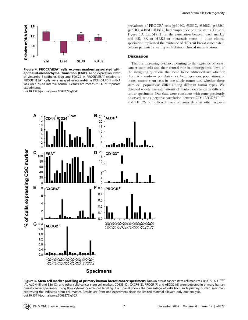
To investigate whether breast cancer cells freshly derived from
clinical specimens express heterogeneous stem cell markers as
observed in human breast cancer cell lines, we performed stem cell
marker profiling by FACS analysis of nineteen specimens obtained
from National Taiwan University Hospital. All specimens
examined contained CD44+/CD242/low cells; however, the
prevalence of these cells ranged from 1 to 12.4 percent
(Figure 5A) and that of ALDH+ cells ranged from 1.4 to 19.8
percent (Figure 5B). The percentage of cells that expressed
CD133, CXCR4 and ESA varied even more among specimens.
Of the nineteen specimens, all specimens contained ESA+ cells
(Figure 5C); however, only eleven specimens contained a
detectable number of CD133+ cells (Figure 5D) and fifteen
contained CXCR4+ cells (Figure 5E). In specimens where
expression was detectable, the percentages of the cells that
expressed these markers ranged from 2.1 to 99 percent, 0.1 to
18.5 percent and 0.1 to 7.6 percent for ESA, CD133 and CXCR4,
respectively. PROCR and ABCG2 expressing cells were rare: only
0.1 to 0.4 percent of the cells from ten specimens had detectable
PROCR expression (Figure 5F) and 0.1 to 2.2 percent of the cells
from seventeen specimens had detectable ABCG2 expression
(Figure 5G). It was noted that all specimens were collected prior to
chemotherapy or hormone therapy. Thus, the expression of
ABCG2 may not be resulted from these treatments. Consistent
with the cell line data described above, these primary tumor cells
expressed heterogeneous stem cell markers, suggesting the
existence of different types of cancer stem/progenitor cells.
To search for any potential clinical correlation with those
marker expressing profiles, pathology data of each patient was
obtained from NTU Hospital (Table 4) and analyzed by statistics.
Of the nineteen patients, twelve were estrogen receptor (ER)
Figure 2. PROCR/ESA marker profiling of PROCR+/ESA+ and PROCR2/ESA2 MDA-MB-231 cells in vitro to evaluate their asymmetricdivision. PROCR+/ESA+ (B) and PROCR2/ESA2 (C) cells were sorted and cultured for four passages in an attachable culture system. The PROCR/ESAmarker profile was evaluated at passage 2 (E, F) and 4 (H, I). Isotype control cells were assayed at each time point as indicated (A, D, G). The PROCR+/ESA+ subpopulation was able to asymmetrically divide into other subpopulations in vitro; however, the PROCR2/ESA2 subpopulation was notcapable of asymmetric division.doi:10.1371/journal.pone.0008377.g002
Cancer StemCells Heterogeneity
PLoS ONE | www.plosone.org 5 December 2009 | Volume 4 | Issue 12 | e8377

positive, nine were progesterone receptor (PR) positive, and three
were HER2 overexpressed. Ten of the patients had metastasis to
lymph nodes. Despite the limited sample size, the data revealed an
overall trend that the cancer specimens with higher numbers of
CD44+/CD242/low cells express low level of HER2 (Table 4,
Figure 5A). However, a positive association between the
prevalence of CD44+/CD242/low cells and ER status was not
found (Table 4, Figure 5A). These results were consistent with the
data obtained from breast cancer cell lines (Table 1) and similar to
the previously reported [38].
In contrast, there was no strong association between the
prevalence of ALDH+ cells and ER expression (Table 4,
Figure 5B), which differed from other reports [34]. Among the
specimens, we found that specimen #362C was the only one
where high ALDH activity was associated with ER negative status
(Table 4, Figure 5B). Furthermore, this specimen, and specimens
#394C and #405C, were the only three with HER2 over-
expression (Table 4). The high prevalence of ALDH+ cells in these
three HER2-overexpression specimens provides a possibility that
HER2 overexpression may drive tumorigenesis as well as ALDH
expression as proposed [31]. However, such correlation was not in
complete concordance since some specimens with a high
prevalence of ALDH+ cells expressed low level of HER2
(Figure 5B).
In addition, we also observed a possible association between
lymph node status and the prevalence of multiple markers.
Specimens with a prevalence of CD133+ and CXCR4+ cells
(#337C, #343C, #366C, #382C, #431C) or with a high
Figure 3. Cell cycle profiling and NUMB expression of PROCR+/ESA+ and PROCR2/ESA2 MDA-MB-231 cells. Bulk cells (A) weresynchronized by serum starvation using DMEM supplemented with 0.5% FBS for 48 h. At 20, 24 and 48 h after release from serum starvation,PROCR+/ESA+ (B) and PROCR2/ESA2 (D) cells stained with propidium iodide and the cell cycle profile analyzed. C. The expression level of NUMB inPROCR+/ESA+ relative to PROCR2/ESA2 cells were assayed using real-time PCR. GAPDH was used as an internal control. Results are means 6 SD oftriplicate experiments.doi:10.1371/journal.pone.0008377.g003
Cancer StemCells Heterogeneity
PLoS ONE | www.plosone.org 6 December 2009 | Volume 4 | Issue 12 | e8377
prevalence of PROCR+ cells (#303C, #366C, #368C, #382C,
#394C, #405C, #431C) had lymph node positive status (Table 4,
Figure 5D, 5E, 5F). Thus, the association between each marker
and ER, PR or HER2 or metastasis status in those clinical
specimens implicated the existence of different breast cancer stem
cells in patients reflecting with distinct clinical manifestation.
Discussion
There is increasing evidence pointing to the existence of breast
cancer stem cells and their central role in tumorigenesis. Two of
the intriguing questions that need to be addressed are whether
there is a uniform population or heterogeneous populations of
breast cancer stem cells in one single tumor and whether these
stem cell populations differ among different tumor types. We
detected widely varying patterns of marker expression in different
tumor specimens. Our data were consistent with some previously
observed trends (negative correlation between CD44+/CD242/low
and HER2) but differed from previous data in other regards
Figure 4. PROCR+/ESA+ cells express markers associated withepithelial-mesenchymal transition (EMT). Gene expression levelsof vimentin, E-cadherin, Slug and FOXC2 in PROCR+/ESA+ relative toPROCR2/ESA2 cells were assayed using real-time PCR. GAPDH mRNAwas used as an internal control. Results are means 6 SD of triplicateexperiments.doi:10.1371/journal.pone.0008377.g004
Figure 5. Stem cell marker profiling of primary human breast cancer specimens. Known breast cancer stem cell markers CD44+/CD242/low
(A), ALDH (B) and ESA (C), and other solid cancer stem cell markers CD133 (D), CXCR4 (E), PROCR (F) and ABCG2 (G) were detected in primary humanbreast cancer specimens using flow cytometry after cell labeling. Each panel shows the percentage of cells from each primary human specimenexpressing the indicated stem cell marker. Results are from one experiment since the limited material allowed only one analysis.doi:10.1371/journal.pone.0008377.g005
Cancer StemCells Heterogeneity
PLoS ONE | www.plosone.org 7 December 2009 | Volume 4 | Issue 12 | e8377

(relationship of ALDH status and HER2 or ER). The widely
varying marker expression in the tumor specimens strongly
suggested the existence of more types of breast cancer stem cells
than have been previously described. Experiments where we
performed functional assays of tumorigenesis and in vivo tumor
forming ability on marker expressing populations isolated from
breast cancer cell lines also strongly supported this hypothesis. In
particular, subpopulations of cells expressing markers PROCR
and ESA were highly tumorigenic in both in vitro and in vivo assays.
Thus PROCR and ESA expression defines as yet uncharacterized
types of breast cancer stem cells and our data from both cell lines
and primary specimens also suggest the existence of still more
types of highly tumorigenic cells. These observations bring up two
central questions. The first is whether the generally recognized
markers that have been emphasized in previous studies of breast
cancer stem cells actually identify the most highly tumorigenic
cells. The second is whether there are different lineages of breast
cancer stem cells which lead to different types of breast cancers
and whether these different lineages may sometimes even coexist
within the same tumor.
Most current studies have emphasized the identification of
cancer stem cell subpopulations from breast tumors using CD44
and CD24. It has been reported that CD44+/CD242/low cells
were more common in basal-like tumors and strongly associated
with BRCA1 hereditary breast cancer; however, not every basal
breast tumor contains CD44+/CD242/low cells [38]. Other recent
studies have also shown that the presence of CD44+/CD242/low
cells in breast tumors did not correlate with clinical outcome
including tumor size, lymph node status or S-phase fraction
[22,39]. More recent data suggests that the newly identified
marker ALDH could be more effective in identifying the most
tumorigenic breast cancer stem cells [21]. However, only 30% of
tumors contained ALDH+ cells. Although the ALDH phenotype
correlates with clinical outcome such as tumor grade, no
association with a particular molecular subtype of breast cancer
was observed [21]. These findings raise the possibility that there
might be cancer stem cells other than CD44+/CD242/low or
ALDH+ that drive breast tumorigenesis. It has been reported that
use of the surface marker CD133 can isolate a group of breast
cancer stem cells that does not overlap with CD44+/CD242 cells
[23], which is consistent with our data that CD44+/CD242 cells
may not have the highest tumorigenic ability. In HCC1937, cells
that lacked CD44+/CD242 marker expression were capable of
forming more colonies on soft agar than the CD44+/CD242 cells
(Figure 1B). These findings further suggest that CD44+/CD242
can only enrich a subtype of breast cancer stem cell populations.
On the other hand, we observed that cells with positive ALDH
were not consistently correlated with their tumorigenicity.
Although the ALDH+ ZR-75 cells exhibited higher tumorigenicity
than bulk cells (Figure 1A), the ALDH marker failed to enrich
higher tumorigenic cancer stem cells from the SKBR3 (Figure 1C)
and HCC1937 cell lines (Figure S2, Method S1). These
observations further demonstrated that CD44+/CD242/low and
ALDH cannot serve as universal markers for cancer stem cell
identification and isolation. Apparently, additional marker-identi-
fication study is needed to further define new cancer stem cell
populations.
Different human breast cancer cell lines are known to have
different abilities to form tumors in vivo. It is possible that this
difference in tumorigenicity is due to the presence of different
types of cancer stem cells. Similar to primary tumor specimens,
different breast cancer cell lines had different cancer stem cell
marker profiles as demonstrated in this study (Table 1). For each
cell line, the subpopulations expressing cancer stem cell markers
exhibited tumorigenic ability different from that of cells without
marker expression. Using PROCR and ESA markers, we were
able to isolate a subset of highly tumorigenic cells from the MDA-
MB-231 cell line (the prevalence of CD44+/CD242/low in the bulk
cell population was more than 90%) (Figure 1D and Table 2).
Although it has been reported that CD44+ and PROCR+ cells
were similar to each other and were enriched for genes involved in
cell motility, chemotaxis, hemostasis, and angiogenesis as well as
stem cell-specific genes [22], our data suggested that PROCR+/
ESA+ allowed further enrichment of highly tumorigenic cancer
stem cells from the CD44+/CD242/low breast cancer cells. In
addition, our preliminary proteome analysis suggests that
PROCR+/ESA+ cells exhibit a cancer stem cell molecular
signature and that PROCR+/ESA+ and PROCR2/ESA2
MDA-MB-231 cells differ in expression of a number of proteins
(WWHV et al unpublished data). For example, PROCR+/ESA+
cells showed a higher expression of casein kinase 2 which is
involved in the Wnt signaling pathway [40] known to be highly
activated in breast cancer stem cells. Such additional character-
ization of the tumorigenic cell populations has potential to clearly
define the molecular signature of the small populations with highly
tumorigenic activity.
It was a significant challenge to conduct experiments using
primary tumor cells due to limited materials. To overcome this
difficulty, in vitro culture and in animal xenograft tumor
transplantations are required to expand cancer cells for assessing
functional stem cell properties in primary tumors, albeit, the
Table 4. Pathology data of the human breast cancerspecimens tested.
Specimen Age Size Lymph node ER (%) PR (%) HER2
303C 43 2.9 3/17 (+) 90 30–40 1 (L)
331C 84 3.5 0/21 (2) .90 40 1 (L)
333C 51 4.5 0/11 (2) 0 0 1–2 (M)
335C 57 2.5 0/10 (2) 0 0 1 (L)
337C 42 1.8 1/4 (+) .90 20–50 1 (L)
341C 56 3 0/25 (2) 90 ,5 0 (ND)
343C 51 3.5 7/8 (+) 95 0 0 (ND)
345C 57 5 0/1 (2) 0 0 1 (L)
362C 44 NA NA 0 0 3 (H)
363C 53 5 0/3 (2) 90 90 0 (ND)
366C 43 4.5 12/29 (+) 60 30 1 (L)
368C 47 5 3/30 (+) 90 0 0 (ND)
382C 86 5.6 6/8 (+) .90 5–10 0 (ND)
394C 52 7.3 26/35 (+) 0 0 3 (H)
398C 65 2.5 7/24 (+) 70 0 1–3 (M)
405C 36 8 35/49 (+) 20 2 3 (H)
419C 76 3.3 0/9 (2) 0 0 0 (ND)
431C 44 2.8 2/22 (+) 0 0 0 (ND)
439C 63 1.5 0/34 (2) 100 20 0 (ND)
Information includes the age of the patient, the size of the tumor (mm), theexpression percentage of estrogen receptor (ER), progesterone receptor (PR)and HER2, and the metastasis to the lymph node.Lymph node status is indicated as (+) or (2); HER2 expression levels aregrouped into four categories according to the pathological data: 3, high oroverexpression (H) of HER2; 1–2, medium (M) expression; 1, low (L) expression,and 0 indicates that the expression was not detectable (ND).doi:10.1371/journal.pone.0008377.t004
Cancer StemCells Heterogeneity
PLoS ONE | www.plosone.org 8 December 2009 | Volume 4 | Issue 12 | e8377

prevalence of marker expressing cells could potentially change
under such selection. Overall, we processed 131 primary
specimens through the course of this study; however, most of the
tumors were too small for the marker expression profiling. We
have amplified the tumor cells by inoculating them in NOD/
SCID mice. However, only seven transplants with tumor
outgrowth were observed, and only one of the seven was large
enough for sorting by stem cell markers. Interestingly, selection
with CD44+/CD242/low from this transplant enriched a subpop-
ulation with higher soft agar colony forming ability (Figure S4,
Method S1). Continued parallel analysis of primary tumor
specimens will reveal which of these stem cell populations are of
greatest importance in clinical outcome. Similar to the recent
novel therapeutic strategies specifically targeting different cancer
stem cells in different leukemias [41], further characterization of
these breast cancer stem cells and the signaling pathways
underlying their phenotypes will allow us to design tailored
therapy to treat different types of breast cancers.
Materials and Methods
Human Breast Cancer Tissue Dissociation and CellPreparation
Ethics statement. Human samples were collected after
obtaining informed written consent from all participants. The
samples were encoded to protect patient confidentiality and used
under protocols approved by the Institutional Review Board of
Human Subjects Research Ethics Committee of Academia Sinica
(AS-IRB02-98042) and National Taiwan University, Taipei,
Taiwan (#200902001R).
Human breast cancer tissue obtained from National Taiwan
University Hospital was dissociated enzymatically and mechanically
in a manner similar to previous reports but with modification [42]. In
brief, tissue was minced into 2–3 mm3 pieces with sterile scalpels and
enzymetically dissociated for 15–16 hours at 37uC in DMEM
(Gibco/Invitrogen) supplemented with 150 units/mL collagenase
(Sigma), 50 units/mL hyaluronidase and antibiotics/antimycotics.
After centrifugation at 300 6g for 5 min, red blood cells were
removed. A single cell suspension was obtained by mechanical
disaggregation in 0.25% trypsin for 5 min, followed by digestion in
200 mg/mL DNase1 for 1 min, and passage through a 40-mm cell
strainer. Single cells were then transferred into mammary epithelium
basal medium (MEBM) (Cambrex) supplemented with B27 (Invitro-
gen), 20 ng/mL EGF, 10 ng/mL FGF (Sigma), 4 mg/mL insulin
(Invitrogen) and antibiotics/antimycotics. Hematopoietic and endo-
thelial cells were removed using Dynabeads coated with antibodies
against CD45, CD14, CD15, CD19, and CD31 (Invitrogen)
following the manufacturers instructions. Cells were then subjected
to stem cell marker profiling analysis.
Cell CultureHuman breast cancer cell lines MCF-7, MDA-MB-231, MDA-
MB-361, MDA-MB-468, T47D, ZR-75, SK-BR-3 and HCC-
1937 were obtained from the American Type Culture Collection
(ATCC) and routinely maintained in DMEM supplemented with
10% FBS and antibiotics/antimycotics in a humidified 37uCincubator supplemented with 5% CO2.
Stem Cell Marker Profiling: Cell Labeling, Aldefluor Assayand Flow Cytometry
Cell labeling was done by staining with antibodies in buffer
composed of PBS supplemented with 0.1% sodium azide, 1% FBS
and 2 mM EDTA. Cells were first suspended and blocked in ice cold
staining buffer at a concentration of 2.56106 cells/mL for 10 min,
and stained with antibodies (using antibody titration suggested by the
supplier) for 30 min on ice in the dark. After centrifugation at 3006g
for 5 min at 4uC, cells were then washed 1–2 times with cold staining
buffer before being subjected to flow cytometry. Antibodies used in
this study were mouse(m)-anti(a)-human(h)-CD44-allophycocyanin
(APC), mahCD24-phycoerythrin (PE), rat-ahPROCR-PE,
mahCXCR4-APC (BD Bioscience), mahABCG2-APC (R&D),
mahESA-647 (eBioscience), and m-ahCD133-APC (Miltenyi Biotec
J&H Technology). Proper isotype controls were used for each cell
labeling experiment. Aldefluor assay was performed following the
manufacturer instruction using an Aldefluor kit (Stemcell Technol-
ogies). The stem cell marker profiling analysis was performed using a
BD FACS Canto II. Live cell sorting was done using a BD FACS Aria
with 100 mm nozzle following the manufacturer instructions. Sorted
cells were washed with DMEM/10%FBS/antibiotics/antimycotics
three times before being cultured in MEBM/B27 media supple-
mented with 20 ng/mL EGF, 10 ng/mL FGF and 4 mg/mL insulin.
Cells were allowed to recover in ultra low attachment surface plates
overnight in a humidified 37uC incubator before further analyses.
The percentage of cells in different marker populations was evaluated
using BD FACSDiva software.
Soft Agar Colony Formation AssaySoft agar colony formation assay was performed by seeding cells
in a layer of 0.35% agar DMEM/FBS over a layer of 0.5% agar/
DMED/FBS. Additional MEBM/B27/EGF/FGF/insulin media
was added every 5 days to continuously supply growth
supplements to the cells. Cultures were maintained in a humidified
37uC incubator. On day 14 or day 21 after seeding, cells were
fixed with pure ethanol containing 0.05% crystal violet and colony
forming efficiency quantified by light microscopy.
Mouse Tumorigenicity AssayNOD/SCID mice were used to evaluate the stem cell properties
of sorted cells expressing potential stem cell markers from the
human breast cancer cell lines. Animal care and experiments were
approved by the Institutional Animal Care and Utilization
Committee of Academia Sinica (IACUC#080085). The animal
model was adapted and modified from Kuperwasser et al [43].
NOD/SCID fat pads were injected with sorted cancer cells mixed
with human breast cancer associated fibroblasts (CAF) (1:1) and
Matrigel (BD bioscience) (1:1). Tumor volumes were evaluated
every five days after initial detection. The tumor formation
efficiency was determined on day 50 after cell injection.
Cell Cycle ProfilingMDA-MB-231 cells were synchronized by culturing in serum
starvation condition (DMEM supplemented with 0.5% FBS) for
48 hours. After synchronization, the growth medium was replaced
with DMEM supplemented with 10%FBS. Cells were then labeled
with rat-ahPROCR-PE and mahESA-647 antibodies and sub-
jected to cell sorting to collect PROCR+/ESA+ and PROCR2/
ESA2 subpopulations at 20, 24 and 48 h after released from
starvation. Cells were washed twice with PBS and then fixed in ice-
cold 70% ethanol overnight before stained with 10 mg/ml
propidium iodide (PI). The PI stained cells were subjected to cell
cycle profiling analysis on BD FACS Canto II reading at 488 nm.
Real-Time RT-PCRQuantitative real-time RT-PCR was performed using SYBR-
Green master mix (Applied Biosystems) according to the
manufacturer’s instruction and analyzed on an ABI 7300 Real-
Time PCR system. GAPDH mRNA was used as an internal
Cancer StemCells Heterogeneity
PLoS ONE | www.plosone.org 9 December 2009 | Volume 4 | Issue 12 | e8377

control to normalize RNA inputs and expression levels were
calculated according to the relative DCt method. Primers used in
this analysis were designed as previously described [19] and primer
sequences are given in Table S1.
Supporting Information
Figure S1 Estrogen receptor (ER) expression levels in bulk,
ALDH+ and ALDH2 ZR-75 cells. A. ALDH+ and ALDH2 ZR-
75 cells were collected for immunoblot analysis. Different
expression levels of ERa were detected in the bulk, ALDH+ and
ALDH2 cells. The expression levels of ERa in ALDH2 cells were
8-fold higher than the bulk cells, and 10-fold higher than the
ALDH+ cells. B. Shown is the relative ERa expression levels in the
bulk, ALDH+ and ALDH2 cells using the average from three
independent immunoblotting assays (means6 SE).
Found at: doi:10.1371/journal.pone.0008377.s001 (0.03 MB
PDF)
Figure S2 Soft agar colony forming efficiency of marker
expressing or nonexpressing subpopulations from HCC1937 and
ZR-75 cells. A. ALDH+ and CXCR4+ identified two distinct
subpopulations of ZR-75 cells which did not overlap with each
other. B. Both ALDH+ and CXCR4+ ZR-75 cells formed more
colonies than the cells not expressing these markers. The ALDH+
cells were more tumorigenic than the CXCR4+ cells. Shown is the
relative fold increase of the colony forming efficiency normalized
to the colony forming efficiency of the bulk population (means 6
SD). C. Selection of ALDH+ cells failed to enrich the tumorigenic
potential of HCC1937 cells. Shown is the percentage of colony
formation (means 6 SD).
Found at: doi:10.1371/journal.pone.0008377.s002 (0.02 MB
PDF)
Figure S3 PROCR+/ESA+ MDA-MB-231 cells asymmetrically
divide in vivo. A. PROCR+/ESA+ MDA-MB-231 cells with 93.8
percent purity were collected for in vivo inoculation in NOD/SCID
mice. B. The marker profile of the cells derived from the tumor
showed that the PROCR+/ESA+ cells retained at a small
percentage (0.6%) and asymmetrically divided into PROCR2/
ESA2 and PROCR2/ESA+ cells in vivo.
Found at: doi:10.1371/journal.pone.0008377.s003 (0.02 MB
PDF)
Figure S4 Stem cell marker profiling of primary breast cancer
specimen #235C and the mammosphere and soft agar colony
forming efficiency of CD44+/CD242/low cells compared to other
cells from #235C. A. The prevalence of CD44+/CD242/low,
PROCR+, ESA+, ABCG2+, CXCR4+, CD133+ and ALDH+ cells
were determined using flow cytometry. B. The mammosphere
forming efficiency in CD44+/CD242/low cells was 4-fold higher
than other cells. C. The soft agar colony forming efficiency in
CD44+/CD242/low cells was 10-fold higher than other cells.
Found at: doi:10.1371/journal.pone.0008377.s004 (0.02 MB
PDF)
Table S1 Primer sequences.
Found at: doi:10.1371/journal.pone.0008377.s005 (0.03 MB
DOC)
Method S1
Found at: doi:10.1371/journal.pone.0008377.s006 (0.04 MB
DOC)
Acknowledgments
We thank Dr. Marc Lippman for his suggestion to use soft agar colony
formation assay to trace the cancer stem cell property in culture, Dr. Paul
E. Verslues (Institute of Plant and Microbial Biology, Academia Sinica) for
proofreading the manuscript and Ms. Meng-Han Wang for her kind
assistance through this study.
Author Contributions
Conceived and designed the experiments: WWHV EYHPL WHL.
Performed the experiments: WWHV PHC CCP HHW STT. Analyzed
the data: WWHV YMJ. Contributed reagents/materials/analysis tools:
WHK JYS JTK CHC KJC. Wrote the paper: WWHV EYHPL WHL.
References
1. Bonnet D, Dick JE (1997) Human acute myeloid leukemia is organized as a
hierarchy that originates from a primitive hematopoietic cell. Nat Med 3:730–737.
2. Singh SK, Clarke ID, Hide T, Dirks PB (2004) Cancer stem cells in nervous
system tumors. Oncogene 23: 7267–7273.
3. Collins AT, Berry PA, Hyde C, Stower MJ, Maitland NJ (2005) Prospective
identification of tumorigenic prostate cancer stem cells. Cancer Res 65:10946–10951.
4. Fang D, Nguyen TK, Leishear K, Finko R, Kulp AN, et al. (2005) Atumorigenic subpopulation with stem cell properties in melanomas. Cancer Res
65: 9328–9337.
5. O’Brien CA, Pollett A, Gallinger S, Dick JE (2007) A human colon cancer cell
capable of initiating tumour growth in immunodeficient mice. Nature 445:106–110.
6. Ricci-Vitiani L, Lombardi DG, Pilozzi E, Biffoni M, Todaro M, et al. (2007)
Identification and expansion of human colon-cancer-initiating cells. Nature 445:
111–115.
7. Matsui W, Huff CA, Wang Q, Malehorn MT, Barber J, et al. (2004)Characterization of clonogenic multiple myeloma cells. Blood 103: 2332–2336.
8. Li C, Heidt DG, Dalerba P, Burant CF, Zhang L, et al. (2007) Identification ofpancreatic cancer stem cells. Cancer Res 67: 1030–1037.
9. Kim CF, Jackson EL, Woolfenden AE, Lawrence S, Babar I, et al. (2005)
Identification of bronchioalveolar stem cells in normal lung and lung cancer.Cell 121: 823–835.
10. Al-Hajj M, Wicha MS, Benito-Hernandez A, Morrison SJ, Clarke MF (2003)Prospective identification of tumorigenic breast cancer cells. Proc Natl Acad
Sci U S A 100: 3983–3988.
11. Passegue E, Jamieson CH, Ailles LE, Weissman IL (2003) Normal and leukemic
hematopoiesis: are leukemias a stem cell disorder or a reacquisition of stem cellcharacteristics? Proc Natl Acad Sci U S A 100 Suppl 1: 11842–11849.
12. Jamieson CH, Ailles LE, Dylla SJ, Muijtjens M, Jones C, et al. (2004)
Granulocyte-macrophage progenitors as candidate leukemic stem cells in blast-
crisis CML. N Engl J Med 351: 657–667.
13. Passegue E, Wagner EF, Weissman IL (2004) JunB deficiency leads to a
myeloproliferative disorder arising from hematopoietic stem cells. Cell 119:431–443.
14. Perou CM, Sorlie T, Eisen MB, van de Rijn M, Jeffrey SS, et al. (2000)
Molecular portraits of human breast tumours. Nature 406: 747–752.
15. Sorlie T, Perou CM, Tibshirani R, Aas T, Geisler S, et al. (2001) Gene
expression patterns of breast carcinomas distinguish tumor subclasses withclinical implications. Proc Natl Acad Sci U S A 98: 10869–10874.
16. Sorlie T, Tibshirani R, Parker J, Hastie T, Marron JS, et al. (2003) Repeatedobservation of breast tumor subtypes in independent gene expression data sets.
Proc Natl Acad Sci U S A 100: 8418–8423.
17. Sotiriou C, Neo SY, McShane LM, Korn EL, Long PM, et al. (2003) Breast
cancer classification and prognosis based on gene expression profiles from apopulation-based study. Proc Natl Acad Sci U S A 100: 10393–10398.
18. Hwang-Verslues WW, Chang KJ, Lee EY, Lee WH (2008) Breast cancer stem
cells and tumor suppressor genes. J Formos Med Assoc 107: 751–766.
19. Mani SA, Guo W, Liao MJ, Eaton EN, Ayyanan A, et al. (2008) The epithelial-
mesenchymal transition generates cells with properties of stem cells. Cell 133:704–715.
20. Ponti D, Costa A, Zaffaroni N, Pratesi G, Petrangolini G, et al. (2005) Isolationand in vitro propagation of tumorigenic breast cancer cells with stem/progenitor
cell properties. Cancer Res 65: 5506–5511.
21. Ginestier C, Hur MH, Charafe-Jauffret E, Monville F, Dutcher J, et al. (2007)ALDH1 Is a Marker of Normal and Malignant Human Mammary Stem Cells
and a Predictor of Poor Clinical Outcome. Cell Stem Cell 1: 555–567.
22. Shipitsin M, Campbell LL, Argani P, Weremowicz S, Bloushtain-Qimron N,
et al. (2007) Molecular definition of breast tumor heterogeneity. Cancer Cell 11:259–273.
23. Wright MH, Calcagno AM, Salcido CD, Carlson MD, Ambudkar SV, et al.(2008) Brca1 breast tumors contain distinct CD44+/CD24- and CD133+ cells
with cancer stem cell characteristics. Breast Cancer Res 10: R10.
24. Ivanova NB, Dimos JT, Schaniel C, Hackney JA, Moore KA, et al. (2002) A
stem cell molecular signature. Science 298: 601–604.
Cancer StemCells Heterogeneity
PLoS ONE | www.plosone.org 10 December 2009 | Volume 4 | Issue 12 | e8377

25. Vercauteren SM, Sutherland HJ (2001) CD133 (AC133) expression on AML
cells and progenitors. Cytotherapy 3: 449–459.26. Yin S, Li J, Hu C, Chen X, Yao M, et al. (2007) CD133 positive hepatocellular
carcinoma cells possess high capacity for tumorigenicity. Int J Cancer 120:
1444–1450.27. Kang Y, Siegel PM, Shu W, Drobnjak M, Kakonen SM, et al. (2003) A
multigenic program mediating breast cancer metastasis to bone. Cancer Cell 3:537–549.
28. Zhou S, Schuetz JD, Bunting KD, Colapietro AM, Sampath J, et al. (2001) The
ABC transporter Bcrp1/ABCG2 is expressed in a wide variety of stem cells andis a molecular determinant of the side-population phenotype. Nat Med 7:
1028–1034.29. Lechner A, Leech CA, Abraham EJ, Nolan AL, Habener JF (2002) Nestin-
positive progenitor cells derived from adult human pancreatic islets ofLangerhans contain side population (SP) cells defined by expression of the
ABCG2 (BCRP1) ATP-binding cassette transporter. Biochem Biophys Res
Commun 293: 670–674.30. Cai J, Cheng A, Luo Y, Lu C, Mattson MP, et al. (2004) Membrane properties
of rat embryonic multipotent neural stem cells. J Neurochem 88: 212–226.31. Korkaya H, Paulson A, Iovino F, Wicha MS (2008) HER2 regulates the
mammary stem/progenitor cell population driving tumorigenesis and invasion.
Oncogene. .32. Dittmer A, Schunke D, Dittmer J (2008) PTHrP promotes homotypic
aggregation of breast cancer cells in three-dimensional cultures. Cancer Lett260: 56–61.
33. Grimshaw MJ, Cooper L, Papazisis K, Coleman JA, Bohnenkamp HR, et al.(2008) Mammosphere culture of metastatic breast cancer cells enriches for
tumorigenic breast cancer cells. Breast Cancer Res 10: R52.
34. Liu S, Ginestier C, Charafe-Jauffret E, Foco H, Kleer CG, et al. (2008) BRCA1
regulates human mammary stem/progenitor cell fate. Proc Natl Acad Sci U S A105: 1680–1685.
35. Cayouette M, Raff M (2002) Asymmetric segregation of Numb: a mechanism for
neural specification from Drosophila to mammals. Nat Neurosci 5: 1265–1269.36. Chang JT, Palanivel VR, Kinjyo I, Schambach F, Intlekofer AM, et al. (2007)
Asymmetric T lymphocyte division in the initiation of adaptive immuneresponses. Science 315: 1687–1691.
37. Jan YN, Jan LY (1998) Asymmetric cell division. Nature 392: 775–778.
38. Honeth G, Bendahl PO, Ringner M, Saal LH, Gruvberger-Saal SK, et al. (2008)The CD44+/CD24- phenotype is enriched in basal-like breast tumors. Breast
Cancer Res 10: R53.39. Abraham BK, Fritz P, McClellan M, Hauptvogel P, Athelogou M, et al. (2005)
Prevalence of CD44+/CD242/low cells in breast cancer may not be associatedwith clinical outcome but may favor distant metastasis. Clin Cancer Res 11:
1154–1159.
40. Wang S, Jones KA (2006) CK2 controls the recruitment of Wnt regulators totarget genes in vivo. Curr Biol 16: 2239–2244.
41. Krause DS, Van Etten RA (2007) Right on target: eradicating leukemic stemcells. Trends Mol Med 13: 470–481.
42. Stingl J, Raouf A, Emerman JT, Eaves CJ (2005) Epithelial progenitors in the
normal human mammary gland. J Mammary Gland Biol Neoplasia 10: 49–59.43. Kuperwasser C, Chavarria T, Wu M, Magrane G, Gray JW, et al. (2004)
Reconstruction of functionally normal and malignant human breast tissues inmice. Proc Natl Acad Sci U S A 101: 4966–4971.
44. Neve RM, Chin K, Fridlyand J, Yeh J, Baehner FL, et al. (2006) A collection ofbreast cancer cell lines for the study of functionally distinct cancer subtypes.
Cancer Cell 10: 515–527.
Cancer StemCells Heterogeneity
PLoS ONE | www.plosone.org 11 December 2009 | Volume 4 | Issue 12 | e8377
Copyright © 2022 FDOKUMEN