Multicenter Study of Brain Volume Abnormalities in Children and Adolescent-Onset Psychosis
11
Multicenter Study of Brain Volume Abnormalities in Children and Adolescent-Onset Psychosis Santiago Reig *,1 , Mara Parellada 2 , Josefina Castro-Fornieles 3 , Joost Janssen 1 , Dolores Moreno 2 , Inmaculada Baeza 4 , Nuria Bargallo ´ 5 , Ana Gonza ´lez-Pinto 6 , Montserrat Graell 7 , Felipe Ortun ˜o 8 , Soraya Otero 9 , Celso Arango 2 , and Manuel Desco 1 1 Unidad de Medicina y Cirugı ´a Experimental, Hospital General Universitario Gregorio Maran ˜o ´ n, Centro de Investigacio ´ n Biome ´dica en Red de Salud Mental, CIBERSAM, Madrid, Spain; 2 Unidad de Adolescentes, Departamento de Psiquiatrı ´a, Hospital General Universitario Gregorio Maran ˜o ´ n, Centro de Investigacio ´ n Biome ´dica en Red de Salud Mental, CIBERSAM, Madrid, Spain; 3 Servicio de Psiquiatrı ´a y Psicologı ´a Infantil y Juvenil, Universidad de Barcelona, IDIBAPS (Institut d’Investigacions Biome `diques August Pi I Sunyer), Hospital Clı ´nic Universitari de Barcelona, Centro de Investigacio ´ n Biome ´dica en Red de Salud Mental, CIBERSAM, Barcelona, Spain; 4 Servicio de Psiquiatrı ´a y Psicologı ´a Infantil y Juvenil, Institut de Neurocie `ncies, Hospital Clı ´nic i Universitari, Centro de Investigacio ´ n Biome ´dica en Red de Salud Mental, CIBERSAM, Barcelona, Spain; 5 Departamento de Radiologı ´a, Centro de Diagno ´ stico por la Imagen, Hospital Clı ´nico, Barcelona, Spain; 6 Stanley Institute International Mood-Disorders Research Center, 03-RC-003, Hospital Santiago Apo ´ stol, Centro de Investigacio ´ n Biome ´dica en Red de Salud Mental, CIBERSAM, Vitoria, Spain; 7 Servicio de Psiquiatrı ´a y Psicologı ´a Infantil y Juvenil, Hospital Infantil Universitario Nin ˜ o Jesu ´ s, Madrid, Spain; 8 Departamento de Psiquiatrı ´a y Psicologı ´a Me ´dica, Clı ´nica Universitaria de Navarra, Pamplona, Navarra, Spain; 9 Servicio de Psiquiatrı ´a y Psicologı ´a Infantil y Juvenil, Departamento de Psiquiatrı ´a, Hospital Universitario Marque ´s de Valdecilla, Centro de Investigacio ´ n Biome ´dica en Red de Salud Mental, CIBERSAM, Santander, Cantabria, Spain *To whom correspondence should be addressed; tel: þ34-91-586-6678, fax: þ34-91-426-5108; e-mail: [email protected]. The goal of the study is to determine the extent of structural brain abnormalities in a multicenter sample of children and adolescents with a recent-onset first episode of psychosis (FEP), compared with a sample of healthy controls. Total brain and lobar volumes and those of gray matter (GM), white matter, and cerebrospinal fluid (CSF) were measured in 92 patients with a FEP and in 94 controls, matched for age, gender, and years of education. Male patients (n 5 64) showed several significant differences when compared with controls (n 5 61). GM volume in male patients was re- duced in the whole brain and in frontal and parietal lobes compared with controls. Total CSF volume and frontal, temporal, and right parietal CSF volumes were also in- creased in male patients. Within patients, those with a further diagnosis of ‘‘schizophrenia’’ or ‘‘other psychosis’’ showed a pattern similar to the group of all patients relative to con- trols. However, bipolar patients showed fewer differences relative to controls. In female patients, only the schizophre- nia group showed differences relative to controls, in frontal CSF. GM deficit in male patients with a first episode corre- lated with negative symptoms. Our study suggests that at least part of the GM deficit in children and adolescent-onset schizophrenia and in other psychosis occurs before onset of the first positive symptoms and that, contrary to what has been shown in children-onset schizophrenia, frontal GM deficits are probably present from the first appearance of positive symptoms in children and adolescents. Key words: MRI/first-episode psychosis/early-onset psychosis/brain morphometrics Introduction Patients with first-episode early-onset psychosis (first ep- isode before 18 years of age with an onset of less than 6 months of positive symptoms before baseline assessment) may constitute a quite homogenous group to study, re- garding the absence of course-related effects, such as du- ration and treatment. Cross-sectional magnetic resonance imaging (MRI) studies have demonstrated that child- hood-onset schizophrenia is associated with smaller whole-brain and total gray matter (GM) volume, thinner cortical thickness, and larger ventricular volume than controls. 1–4 Longitudinal studies have shown a back-to- front wave of GM loss in children with schizophrenia re- sembling the normal development pattern but in an exag- gerated manner. 5,6 In particular, parietal and motor cortex deficits have been shown at baseline while prefron- tal, supplementary motor, sensorimotor, parietal, and temporal deficits appeared or increased the deficit over time. 5 The few studies that have scanned adolescent-onset schizophrenia patients show decreased frontal (but not parietal or temporal) GM volumes and increased total and frontal cerebrospinal fluid (CSF) at onset. 7–9 Schizophrenia Bulletin doi:10.1093/schbul/sbq044 Ó The Author 2010. Published by Oxford University Press on behalf of the Maryland Psychiatric Research Center. All rights reserved. For permissions, please email: [email protected]. 1 Schizophrenia Bulletin Advance Access published May 16, 2010 at Universidad de Navarra. Servicio de Bibliotecas on October 20, 2010 schizophreniabulletin.oxfordjournals.org Downloaded from
-
Upload
independent -
Category
Documents
-
view
1 -
download
0
Transcript of Multicenter Study of Brain Volume Abnormalities in Children and Adolescent-Onset Psychosis
Multicenter Study of Brain Volume Abnormalities in Children and Adolescent-OnsetPsychosis
Santiago Reig*,1, Mara Parellada2, Josefina Castro-Fornieles3, Joost Janssen1, Dolores Moreno2, Inmaculada Baeza4,Nuria Bargallo5, Ana Gonzalez-Pinto6, Montserrat Graell7, Felipe Ortuno8, Soraya Otero9, Celso Arango2, andManuel Desco1
1Unidad de Medicina y Cirugıa Experimental, Hospital General Universitario Gregorio Maranon, Centro de Investigacion Biomedica enReddeSaludMental,CIBERSAM,Madrid, Spain; 2UnidaddeAdolescentes,DepartamentodePsiquiatrıa,HospitalGeneralUniversitarioGregorio Maranon, Centro de Investigacion Biomedica en Red de Salud Mental, CIBERSAM, Madrid, Spain; 3Servicio de Psiquiatrıa yPsicologıa Infantil y Juvenil, Universidad de Barcelona, IDIBAPS (Institut d’Investigacions Biomediques August Pi I Sunyer), HospitalClınic Universitari de Barcelona, Centro de Investigacion Biomedica en Red de SaludMental, CIBERSAM, Barcelona, Spain; 4Servicio dePsiquiatrıayPsicologıa Infantil y Juvenil, InstitutdeNeurociencies,HospitalClınic iUniversitari,Centrode InvestigacionBiomedicaenRedde Salud Mental, CIBERSAM, Barcelona, Spain; 5Departamento de Radiologıa, Centro de Diagnostico por la Imagen, Hospital Clınico,Barcelona, Spain; 6Stanley Institute International Mood-Disorders Research Center, 03-RC-003, Hospital Santiago Apostol, Centro deInvestigacion Biomedica en Red de Salud Mental, CIBERSAM, Vitoria, Spain; 7Servicio de Psiquiatrıa y Psicologıa Infantil y Juvenil,Hospital Infantil Universitario Nino Jesus, Madrid, Spain; 8Departamento de Psiquiatrıa y Psicologıa Medica, Clınica Universitaria deNavarra, Pamplona, Navarra, Spain; 9Servicio de Psiquiatrıa y Psicologıa Infantil y Juvenil, Departamento de Psiquiatrıa, HospitalUniversitarioMarquesdeValdecilla,CentrodeInvestigacionBiomedicaenReddeSaludMental,CIBERSAM,Santander,Cantabria,Spain
*To whom correspondence should be addressed; tel: þ34-91-586-6678, fax: þ34-91-426-5108; e-mail: [email protected].
The goal of the study is to determine the extent of structuralbrain abnormalities in a multicenter sample of children andadolescents with a recent-onset first episode of psychosis(FEP), compared with a sample of healthy controls. Totalbrain and lobar volumes and those of gray matter (GM),white matter, and cerebrospinal fluid (CSF) were measuredin 92 patients with a FEP and in 94 controls, matched forage, gender, and years of education. Male patients (n 564) showed several significant differences when comparedwith controls (n5 61). GM volume in male patients was re-duced in the whole brain and in frontal and parietal lobescompared with controls. Total CSF volume and frontal,temporal, and right parietal CSF volumes were also in-creasedinmalepatients.Withinpatients, thosewithafurtherdiagnosis of ‘‘schizophrenia’’ or ‘‘other psychosis’’ showeda pattern similar to the group of all patients relative to con-trols. However, bipolar patients showed fewer differencesrelative to controls. In female patients, only the schizophre-nia group showed differences relative to controls, in frontalCSF. GM deficit in male patients with a first episode corre-lated with negative symptoms. Our study suggests that atleast part of the GM deficit in children and adolescent-onsetschizophrenia and in other psychosis occurs before onset ofthe first positive symptoms and that, contrary to what hasbeen shown in children-onset schizophrenia, frontal GMdeficits are probably present from the first appearance ofpositive symptoms in children and adolescents.
Key words: MRI/first-episode psychosis/early-onsetpsychosis/brain morphometrics
Introduction
Patients with first-episode early-onset psychosis (first ep-isode before 18 years of age with an onset of less than 6months of positive symptoms before baseline assessment)may constitute a quite homogenous group to study, re-garding the absence of course-related effects, such as du-ration and treatment. Cross-sectional magnetic resonanceimaging (MRI) studies have demonstrated that child-hood-onset schizophrenia is associated with smallerwhole-brain and total gray matter (GM) volume, thinnercortical thickness, and larger ventricular volume thancontrols.1–4 Longitudinal studies have shown a back-to-front wave of GM loss in children with schizophrenia re-sembling the normal development pattern but in an exag-gerated manner.5,6 In particular, parietal and motorcortex deficits have been shown at baseline while prefron-tal, supplementary motor, sensorimotor, parietal, andtemporal deficits appeared or increased the deficit overtime.5 The few studies that have scanned adolescent-onsetschizophrenia patients show decreased frontal (but notparietal or temporal) GM volumes and increased totaland frontal cerebrospinal fluid (CSF) at onset.7–9
Schizophrenia Bulletindoi:10.1093/schbul/sbq044
� The Author 2010. Published by Oxford University Press on behalf of the Maryland Psychiatric Research Center. All rights reserved.For permissions, please email: [email protected].
1
Schizophrenia Bulletin Advance Access published May 16, 2010 at U
niversidad de Navarra. S
ervicio de Bibliotecas on O
ctober 20, 2010schizophreniabulletin.oxfordjournals.org
Dow
nloaded from

The specificity of these findings for schizophrenia hasnot been thoroughly investigated. Bipolar disorderpatients frequently share many of the psychotic symp-toms associated with schizophrenia. Thus, analysis ofstructural abnormalities in bipolar patients with psy-chotic symptoms and schizophrenia patients will shedlight on the specificity of brain changes in the contextof pathological processes related to psychosis.
The present study builds on earlier reports demonstratingbrain abnormalities in adolescent-onset psychosis patientswith a recent-onset first episode of psychosis (FEP).7,10 Theobjective was now to extend the study to a much larger, in-dependent, and representative sample by recruiting subjectsfrom 5 psychiatric units. Our sample includes a matchedcomparison group of healthy children and adolescentsfrom each participating center to control for potential de-mographic factors known to affect brain morphometry.The main hypothesis of the study was that children andadolescents with first psychotic episodes will show volumet-ric differences inGMandCSFof themain brain lobes, par-ticularly in the frontal. Furthermore, the goal of this articleusing a larger data set is to allow for subdivision of patientsinto diagnostic groups within psychosis.
Methods
Subjects
The sample used in this study consisted of 92 patients (28females) and 94 controls (33 females), participating in theChild and Adolescent First-Episode Psychosis Study(CAFEPS).11 CAFEPS included patients from 6 differentpsychiatric units.
The inclusion criteria for patients were age between 7and 17 years at the time of first evaluation and positivepsychotic symptoms (within a psychotic episode) such asdelusions or hallucinations of less than 6-month dura-tion. Exclusion criteria were a concomitant Axis I disor-der at the time of evaluation that might account for thepsychotic symptoms, mental retardation as per the Diag-nastic and Statistical Manual of Mental Disordes (FourthEdition) (DSM-IV) criteria, pervasive developmental dis-order, neurological disorders, history of head traumawith loss of consciousness, pregnancy, and drug-inducedpsychosis. When drugs were positive in the urine baselineassessment, we included the patient in the study if symp-toms persisted 2 weeks after a negative urine test. Eightpatients and 1 control fulfilled DSM-IV criteria for can-nabis abuse. All the patients maintained psychotic symp-toms 15 days after a negative urine test and did not fulfillcriteria for drug-induced psychosis. Only 1 patient at re-cruitment abused cocaine, 1 abused alcohol, and 1 abusedamphetamines. None of them fulfilled criteria for drug-induced psychosis. A detailed description of recruitmentcriteria, clinical and demographic characteristics, andmethodology has been provided elsewhere.11
Diagnosis was confirmed according to the DSM-IVcriteria using the Kiddie Schedule for Affective Disordersand Schizophrenia, Present and Lifetime version(K-SADS-PL), Spanish adaptation.12 DSM-IV criteriawere applied also at 1-year follow-up. Psychopathologywas assessed with the Positive and Negative SyndromeScale (PANSS), and duration of untreated psychosis wascalculated at the first interview by asking about the firstappearance ever of positive psychotic symptoms withina psychotic episode. At 1 year of follow up, 3 main diag-nostic categories were established in the FEP group: schizo-phrenia, psychotic bipolar disorder, and other psychoses(including schizoaffective disorder: 7, 16.3%; psychotic dis-order not otherwise specified: 15, 34.9%; depressive disor-der with psychotic symptoms: 6, 14.0%; schizophreniformdisorder: 6, 14.0%; brief psychotic disorder: 3, 7.0%; obses-sive compulsive disorder with psychotic symptoms:2, 4.7%; 4, 9.35% without specific diagnostic because the1-year reassessment could not be performed.Parental socioeconomic status was assessed with the
Hollingshead-Redlich scale. The study was approvedby the Ethics and Clinical Research Boards of all hospi-tals involved in the study. After complete description ofthe study to the subjects, written informed consent wasobtained. All controls and patients met MRI safetycriteria.
MRI Acquisition
Five different scanning facilities contributed data to thestudy: 2 Siemens Symphony, 2 General Electric Signa,and 1 Philips ACS Gyroscan, all 1.5-T scanners. Onesite (first site in table 2) received patients and controlsfrom 2 Psychiatric Units that were located in the samecity. Data were collected from each center and processedat one site.13 Two MRI sequences were acquired in axialorientation for each subject, a T1-weighted 3D gradientecho (voxel size 1 3 1 3 1.5 mm) and a T2-weightedTurbo-Spin-Echo (voxel size 1 3 1 3 3.5 mm). Full detailsabout the acquisition parameters at each site, comparabil-ity between machines for this study, and the limitationsinvolved in the analysis of multicenter data are providedin Reig et al.13 To minimize the effect of the multicenterdesign, the sample of patients and controls were matchedwithin each of the 5 contributing institutions. The numberof subjects scanned at each site are provided in table 1.
Segmentation and Region of Interest Definition
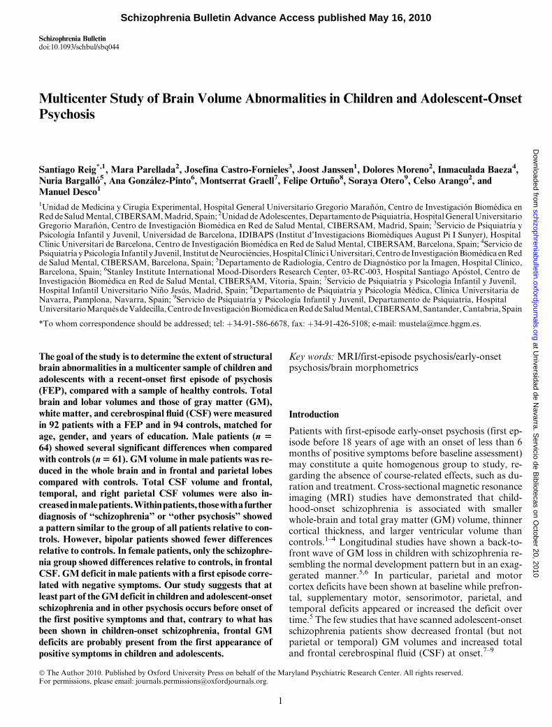
MRI images were processed using locally developed soft-ware incorporating a variety of image processing andquantification tools.13,14 To obtain volume measure-ments of themain brain lobes, we used amethod for semi-automated segmentation of the brain based on theTalairach proportional grid system, performed in nativespace.15,16 Basically, a 2-step procedure was followed.14
First, an initial segmentation of cerebral tissues into
S. Reig et al.
2
at Universidad de N
avarra. Servicio de B
ibliotecas on October 20, 2010
schizophreniabulletin.oxfordjournals.orgD
ownloaded from
GM, white matter (WM), and CSF was obtained usingSPM2 (Statistical Parametric Mapping; Wellcome TrustCentre forNeuroimaging, London,UK; http://www.fil.ion.ucl.ac.uk/spm) routines for multimodal (T1 and T2) seg-mentation. Because of the multicenter setup, multimodaltissue segmentation is more robust than single modality(T1 only) with regards to image contrast–related differen-ces between sites. This assumption was corroborated ina previous study in which the repeatability of multimodal(T1 and T2) and T1-only segmentation was comparedamong the 5 scanners using the same methodology as inthis study.13 The SPM algorithm for tissue segmentationincludes a method to eliminate the effect of radio fre-quency field inhomogeneities.17 In a second step, theTalairach grid was built on the edited brain MRI by man-ually selecting the position of the anterior and posteriorcommissures (AC and PC) and a third point in the mid-sagittal plane. The coordinates of these points serve to cal-culate the transformation (rigid rotation) required tocomply with the Talairach orientation: to set the AC-PC line in the axial horizontal plane and the interhemi-spheric plane in the vertical orientation.18 Then, our soft-ware application automatically finds the outer brain limitsin Talairach orientation, and 3D grids are built for eachbrain. The Talairach grid obtained in this way representsa piecewise linear transformation and a tessellation of thebrain into a 3D grid of 1056 cells representing homologous
brain regions across subjects.18 The region of interest(ROI) measurements were obtained by superimposingthe 3D tissue masks corresponding to GM, WM, andCSF onto each subject’s Talairach grid, where the ROIswere defined as sets of Talairach grid cells (figure1).13,15,16 Volume for each tissue type was measured onthis MRI by summing up the data from the Talairachgrid cells associated with each ROI.14 The validity ofthe Talairach-based procedure as an automated segmen-tation and quantification tool suitable for volumetric stud-ies has already been proven,15,16 and it has been used inother multicenter studies.19 In our implementation, allmanual procedures were performed by a single operatorblind to the origin of each scan, thus avoiding any poten-tial interrater variability. The error due to manual inter-vention in our segmentation procedures was highest forCSF measurements (around 4%), whereas for GM wasless than 2% in all the ROIs studied.13
Measures
We chose to study the brain regions most likely to showvolume changes, as previously observed in first-episodepatients.7–9 The ROIs included in the analysis were thefrontal, parietal, and temporal lobes, defined using theboundaries previously described for the Talairachmethod.15 Whole-brain GM and CSF were also
Table 1. Sociodemographic and Clinical Characteristics of Patients and Controls
Controls (n = 94) Patients (n = 92)
Age (y) (mean, range, SD) 15.4, 19–9, 1.4 15.7, 18–9, 1.7 NS
Gender (male/female) 61/33 64/28 NS
Education (y) (mean, SD) 8.8, 1.4 8.3, 2.0 NS
Parental socioeconomic status (1/2/3/4/5) 10/23/24/11/26 19/29/21/11/12 NS
Handedness: right/left/mixed 87/5/2 85/7/0 NS
Race/ethnicity (Caucasian/Hispanic/Black-African/Other)
86/6/2/0 82/5/1/4 NS
Duration of illness (mo) (mean, SD) 2.1, 1.7
Diagnosis (schizophrenia/bipolar/otherpsychoses)
31/18/43
Duration of treatment (wk) (mean,range, SD)
4.8, 65.0–0, 9.5
Treatment:quetiapine/olanzapine/risperidone(mg, mean, SD)
21/23/48, 211.0/196.2/10.7, 5.0/3.4/1.6
Subjects per site 46/28/9/7/4 48/21/5/12/6 NS
Note: NS: nonsignificant differences.
3
Multicenter Study in Children and Adolescent-Onset Psychosis
at Universidad de N
avarra. Servicio de B
ibliotecas on October 20, 2010
schizophreniabulletin.oxfordjournals.orgD
ownloaded from

measured. Intracranial volume (ICV) was obtained byadding the total GM, WM, and CSF volumes, includingthe cerebellum. For each ROI, we obtained volumes foreach hemisphere and forGMandCSF. The occipital lobewas not included in the study because of its relatively highmeasurement error in multicenter data, which was 5.7%in GM and 17% in CSF values.13 Subcortical and cere-bellar regions were not measured because in our multi-center setup, their segmentation using automatedmethods would require a thorough study of compatibilityand validation. Multicentre reproducibility of WM wasnearly twice lower than of GM,13 thus volume data forWM were not included in the study, also to reduce thedimensionality of the analysis. In addition to these tech-nical problems, a structural study of WM would havebeen incomplete and inconclusive without data fromother sequences such as diffusion-weighted imagingthat provide functional information about connectivity.
Statistical Analysis
Differences between patients and controls in demo-graphic data were assessed by Student’s t and chi-squaretests, according to the type of the variable.
Because age and total cranial size are known factorsaffecting regional cerebral volumes, their effect was re-moved using the residuals from the regression modelsobtained from the group of healthy controls, separatelyfor each gender, following the procedure of Pfefferbaumet al.20 After this correction, volume variables wereexpressed as deviations (regression residuals) from theexpected volumes in healthy individuals of the sameage (in months) and brain size (ICV) as the patient.Thus, negative residuals could be seen as volume deficitsrelative to controls and vice versa. The comparative anal-ysis of volume differences between groups was done usingthese regression residuals for each variable, which isequivalent to using brain size and age as covariates inan ANCOVA model.The age and ICV regression residuals obtained in the
previous step were used to test for volume differences be-tween patients and controls by anANCOVAmodel usingscanner site as a factor of no interest in the analysis.13
Because of known differences in brainmorphometrics be-tween males and females21 and also because we havefound significant group 3 gender interactions for somevariables, we did the comparison between patients andcontrols separately for each gender. We discarded using
Table 2. Volume Measurements (in cm3) of Controls and Patients
Controls Patients
Controls/Patients(Males)a
Females (n = 33) Males (n = 61) Females (n = 28) Males (n = 64)
Mean SD Mean SD Mean SD Mean SD
Age (y) 15.4 1.8 15.4 1.9 15.3 1.9 15.8 1.9
Intracranial volume 1432.8 103.0 1540.3 141 1366.8 114 1563.6 133Total GM 769.8 71.3 847.9 87.9 724.8 88.0 832.2 81.3 ***Total WM 379.9 39.3 410.9 53.2 376.8 67.0 417.1 47.4Total CSF 283.0 45.4 281.4 50.3 264.5 63.3 314.1 60.2 ***
GMFrontal, left 70.8 8.9 80.2 10.3 67.9 8.1 75.8 9.2 ***Frontal, right 73.6 9.0 83.7 10.8 70.9 9.8 77.9 9.9 ***Parietal, left 58.5 7.9 64.2 8.5 54.8 8.8 62.4 8.7 *Parietal, right 59.0 7.7 64.5 8.9 55.4 10.0 62.0 8.0 ***Temporal, left 77.1 7.3 84.4 8.4 72.1 10.1 84.9 9.1Temporal, right 76.9 7.5 83.1 8.5 72.1 10.2 84.7 9.1
CSFFrontal, left 28.2 5.9 27.5 7.0 26.6 9.0 31.9 8.3 ***Frontal, right 29.9 6.2 28.5 6.8 28.0 9.3 33.6 8.6 ***Parietal, left 24.9 6.3 23.4 6.7 22.4 7.2 26.0 6.1Parietal, right 22.7 5.5 22.3 6.5 21.0 8.2 25.5 6.5 **Temporal, left 23.2 3.9 23.0 4.0 21.7 5.1 25.8 5.2 ***Temporal, right 21.9 3.8 21.4 4.4 20.1 4.5 24.3 5.7 ***
Note: GM, gray matter; WM, white matter; CSF, cerebrospinal fluid.aANCOVA of male controls vs patients using residuals after correction for ICV, age, and scanner factors. No differences observed infemales.***P < .001; **P < .01; *P < .05.
4
S. Reig et al.
at Universidad de N
avarra. Servicio de B
ibliotecas on October 20, 2010
schizophreniabulletin.oxfordjournals.orgD
ownloaded from
a full factorial design including the interaction group 3
gender on the model because of the different samplesize for males and females, which makes an unbalancedmodel that compromises the interpretation of factoreffects, specially for the interaction term. Another com-parative analysis between patients and controls was alsodone by subdividing the patient sample into 3 diagnosticgroups: schizophrenia, bipolar, and other psychosis. Dif-ferences between these groups were tested using post hocprocedures (Dunnett’s test) whenever the ANCOVAmodel revealed significant differences between groups.In variables showing significant differences betweengroup means, Cohen’s effect size (ES) were calculatedto provide an index of the magnitude of differences.The association between symptom scores (PANSS posi-tive, negative, and general scales) and volumetric varia-bles (age and ICV residuals) was studied usingPearson’s correlation. Normality of the distributionsand homoscedasticity of variance among groups waschecked prior to the analysis. Statistical analyses weredone using SAS 9.0 (Statistical Analysis System, Cary,North Carolina), and a 2-tailed P-value lower than .05was considered statistically significant.
Results
Sociodemographic and Clinical Characteristics
Patients and controls were not significantly different interms of age, sex, education, years of education, race,or handedness (table 1). Duration of illness prior to theMRI scan was very short (mean 2 months; table 1). Nodifferences were observed between male and femalepatients regarding PANSS scores in total positive(mean, SD: 14.0, 6.2 and 15.8, 6.5, respectively, for femalesand males) or negative (mean, SD: 17.5, 6.9 and 17.4, 6.3,respectively, for females and males) subscales.
Differences Between Patients and Controls
Results of the ANCOVA of age and ICV residuals usingscanner as factor of no interest suggest that only malepatients showed significant differences relative to con-trols (table 2). Male patients showed smaller whole-brainGM volume (F1,123 = 16.3; P < .0001; ES = 0.77) andlarger CSF volume (F1,123 = 20.3; P < .0001; ES =0.81). GM volumes of male patients were smaller thancontrols in the frontal lobe in both right (F1,123 = 28.2;P < .0001; ES = 0.98) and left hemispheres
Fig. 1. Segmentation andQuantificationMethod Based on the Talairach Proportional Grid System. The figure shows a triplanar view of theTalairach grid built in a particular brain. The colored set of grid cells define the frontal (red), parietal (pink), and temporal (blue) regions ofinterest of the right hemisphere. Green color overlaid on the magnetic resonance imaging shows the segmentation of gray matter tissue.
5
Multicenter Study in Children and Adolescent-Onset Psychosis
at Universidad de N
avarra. Servicio de B
ibliotecas on October 20, 2010
schizophreniabulletin.oxfordjournals.orgD
ownloaded from
(F1,123 = 17.5; P < .0001; ES= 0.79) and in the parietallobe (right: F1,123 = 10.9, P = .0013, ES = 0.61; left:F1,123 = 5.3, P = .0230, ES = 0.42). Male patientsalso showed larger CSF volumes in the frontal (right:F1,123 = 17.6, P < .0001, ES = 0.63; left: F1,123 = 11.4,P = .0012, ES = 0.84), temporal (right: F1,123 = 20.3,P < .0001, ES = 0.79; left: F1,123 = 21.8, P < .0001,ES = 0.77), and right parietal (F1,123 = 8.3; P = .0047;
ES = 0.56) lobes (table 2). The statistical significanceof most of these differences persist even after a stringentBonferroni correction considering a total of 30 tests(P = .0017).
Differences Between Patients Subgroups and Controls
Results of the post hoc Dunnett’s test revealed that maleschizophrenia patients showed significantly smaller
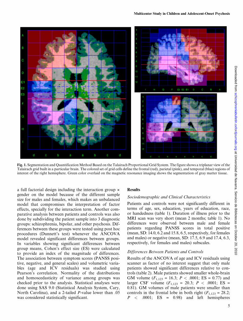
Table 3. Volume Measurements (in cm3) of Patients in Each Diagnostic Group, by Gender
Schizophrenia Bipolar Other Psychosis
Mean SD Mean SD Mean SD Schizophreniaa BipolaraOtherPsychosisa
Males (n) 24 12 28Age (y) 15.8 2.1 16.2 2.0 15.7 1.6Intracranial volume 1577.8 151.6 1489.1 63.4 1582.0 128.0Total GM 835.3 88.5 800.5 63.7 842.7 80.6 *** ***Total WM 422.3 51.7 395.5 40.9 421.4 44.8Total CSF 319.9 78.7 293.0 46.2 317.7 43.6 ** * ***
GMFrontal, left 76.1 10.5 74.7 8.7 76.0 8.5 *** ***Frontal, right 77.4 10.7 77.4 11.3 78.6 8.8 *** ***Parietal, left 62.9 8.7 60.2 8.7 62.9 8.9Parietal, right 62.2 8.1 60.1 8.5 62.6 8.0 ** **Temporal, left 86.4 10.0 79.5 6.2 85.7 8.6Temporal, right 85.3 9.6 79.8 6.7 86.1 9.1
CSFFrontal, left 32.2 9.6 30.5 7.9 32.3 7.5 * * **Frontal, right 32.6 9.5 31.9 7.0 35.3 8.3 * ** ***Parietal, left 27.2 7.8 24.3 5.4 25.8 4.5Parietal, right 25.4 8.1 24.9 5.5 25.9 5.1 * *Temporal, left 26.9 6.7 23.0 3.9 26.0 3.7 *** ***Temporal, right 24.9 7.4 21.6 4.0 24.8 4.0 *** ***
Females (n) 7 6 15Age (y) 15.2 1.9 15.2 2.4 15.3 1.9Intracranial volume 1437.1 144.0 1303.3 133.9 1358.9 84.3Total GM 766.0 105.6 687.3 111.2 720.9 71.2Total WM 377.8 41.2 365.6 77.7 380.1 75.1Total CSF 293.2 53.1 250.3 72.3 257.7 64.7
GMFrontal, left 69.9 7.5 64.7 10.0 68.2 7.9Frontal, right 74.5 9.5 69.2 13.3 70.1 9.0Parietal, left 56.1 10.8 50.6 11.3 55.6 7.3Parietal, right 56.3 9.1 52.6 15.9 56.0 8.5Temporal, left 77.8 10.9 66.3 11.3 71.7 8.7Temporal, right 77.4 13.2 68.5 10.4 71.1 8.6
CSFFrontal, left 33.8 11.0 21.4 7.2 25.5 7.2 *Frontal, right 35.7 11.4 25.4 7.9 25.8 7.6 **Parietal, left 25.9 6.5 22.2 7.8 21.1 7.3Parietal, right 22.3 7.1 23.3 13.6 19.7 6.8Temporal, left 23.9 2.9 18.2 4.0 22.0 5.7Temporal, right 20.9 2.0 18.9 5.4 20.2 5.0
Abbreviations are explained in the first footnote to table 2.aPost hoc Dunnett’s tests comparing each patient group vs controls using residuals after correction for intracranial volume, age, andscanner factors.***P < .001; **P < .01; *P < .05.
6
S. Reig et al.
at Universidad de N
avarra. Servicio de B
ibliotecas on October 20, 2010
schizophreniabulletin.oxfordjournals.orgD
ownloaded from

whole-brainGM (P = .0008) and larger CSF volumes (P =.0027) (table 3; figure 2) than controls. Male schizophre-nia patients also showed smaller GM volumes in the fron-tal (right:P< .0001; left:P = .0009) and right parietal lobe(P = .0061) and larger CSF volumes in the temporal (right:
P = .0005; left: P = .0001) and frontal lobes (right:P = .0261; left: P = .00147). The males in the other psy-chosis group showed differences relative to controls inthe same ROIs as male schizophrenia patients (table 3;figure 2), smaller whole-brain GM (P = .0008), larger
Fig. 2.ScatterPlotsShowingIndividualDataPoints IllustratingDistributionofGrayMatter (GM)andCerebrospinalFluid (CSF)Variablesin Male Subjects Showing the Most Significant Differences Relative to Controls (table 3). Volume data are expressed as residuals aftercorrection for intracranial volume, age, and scanner factors (see ‘‘Methods’’ for details). Dashed lines mark the mean for each group (þ,controls; O, patients).
7
Multicenter Study in Children and Adolescent-Onset Psychosis
at Universidad de N
avarra. Servicio de B
ibliotecas on October 20, 2010
schizophreniabulletin.oxfordjournals.orgD
ownloaded from
CSF volume (P = .0001), smaller GM volume in the fron-tal (right and left: P < .0001) and parietal (right: P =.0045; left: P = .0187) lobes, and larger CSF volumes inthe frontal (right:P< .0001; left: P = .0047), right parietal(P = .0111), and temporal lobes (right and left: P< .0001)(table 3; figure 2). Bipolar male patients showed signifi-cant differences from controls only for CSF values;whole-brain (P = .0131), frontal (right P = .0017; leftP = .0134), and right parietal (P = .0110) (table 3; figure 2)lobes. The same ANCOVA model testing for differencesbetween the 3 clinical subtypes revealed no significant dif-ferences.
In female patients, only the schizophrenia groupshowed significantly larger CSF volumes in the frontallobe (right: P = .0085; left: P = .0207), relative to controls(table 3). However, these differences should be taken withcaution because of the small sample size of the patientgroup (n = 7).
Association Between Volume and Symptom Scores
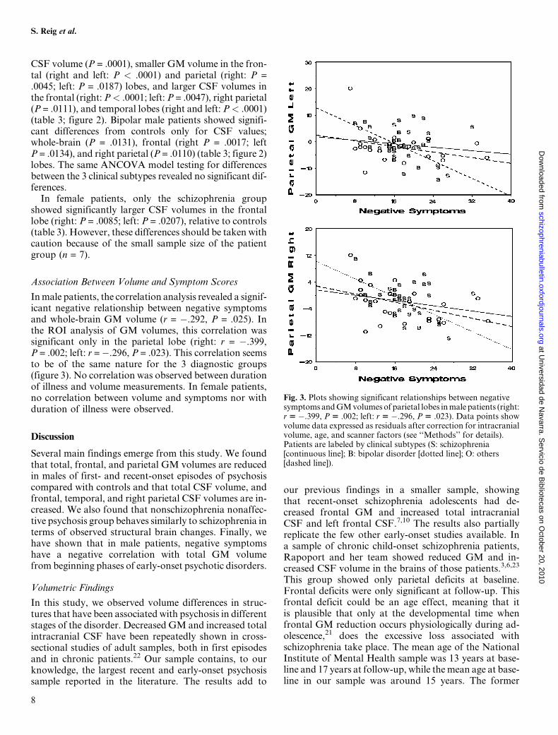
Inmale patients, the correlation analysis revealed a signif-icant negative relationship between negative symptomsand whole-brain GM volume (r = �.292, P = .025). Inthe ROI analysis of GM volumes, this correlation wassignificant only in the parietal lobe (right: r = �.399,P = .002; left: r =�.296, P = .023). This correlation seemsto be of the same nature for the 3 diagnostic groups(figure 3). No correlation was observed between durationof illness and volume measurements. In female patients,no correlation between volume and symptoms nor withduration of illness were observed.
Discussion
Several main findings emerge from this study. We foundthat total, frontal, and parietal GM volumes are reducedin males of first- and recent-onset episodes of psychosiscompared with controls and that total CSF volume, andfrontal, temporal, and right parietal CSF volumes are in-creased. We also found that nonschizophrenia nonaffec-tive psychosis group behaves similarly to schizophrenia interms of observed structural brain changes. Finally, wehave shown that in male patients, negative symptomshave a negative correlation with total GM volumefrom beginning phases of early-onset psychotic disorders.
Volumetric Findings
In this study, we observed volume differences in struc-tures that have been associated with psychosis in differentstages of the disorder. Decreased GM and increased totalintracranial CSF have been repeatedly shown in cross-sectional studies of adult samples, both in first episodesand in chronic patients.22 Our sample contains, to ourknowledge, the largest recent and early-onset psychosissample reported in the literature. The results add to
our previous findings in a smaller sample, showingthat recent-onset schizophrenia adolescents had de-creased frontal GM and increased total intracranialCSF and left frontal CSF.7,10 The results also partiallyreplicate the few other early-onset studies available. Ina sample of chronic child-onset schizophrenia patients,Rapoport and her team showed reduced GM and in-creased CSF volume in the brains of those patients.3,6,23
This group showed only parietal deficits at baseline.Frontal deficits were only significant at follow-up. Thisfrontal deficit could be an age effect, meaning that itis plausible that only at the developmental time whenfrontal GM reduction occurs physiologically during ad-olescence,21 does the excessive loss associated withschizophrenia take place. The mean age of the NationalInstitute of Mental Health sample was 13 years at base-line and 17 years at follow-up, while themean age at base-line in our sample was around 15 years. The former
Fig. 3. Plots showing significant relationships between negativesymptomsandGMvolumes of parietal lobes inmale patients (right:r 5 �.399, P 5 .002; left: r 5 �.296, P 5 .023). Data points showvolume data expressed as residuals after correction for intracranialvolume, age, and scanner factors (see ‘‘Methods’’ for details).Patients are labeled by clinical subtypes (S: schizophrenia[continuous line]; B: bipolar disorder [dotted line]; O: others[dashed line]).
8
S. Reig et al.
at Universidad de N
avarra. Servicio de B
ibliotecas on October 20, 2010
schizophreniabulletin.oxfordjournals.orgD
ownloaded from

sample had a duration of illness before baseline of around3 years, and ours was a very recent-onset sample, with lessthan 3-month duration of illness, which argues againstfrontal deficit being just an effect of chronicity. The Ox-ford group reporting on a sample of 16 adolescents withfirst episodes of schizophrenia, with a duration of illnessof around 18 months, showed only larger ventricular vol-umes and changes in left temporal lobe that were presentfrom the beginning of the disorder.8,9 Relative to theseother studies, our sample of patients is of particular in-terest because of the large sample size and the short pe-riod between the appearance of positive symptoms andMRI scanning.
Timing of GM Reduction and CSF Increase
Results from the literature regarding CSF enlargementare conflicting in their interpretation. CSF enlargementhas been shown from the beginning of illness and atall ages of onset. It has usually been considered suggestiveof degeneration.4 However, some authors argue that thedifferences between the brains of schizophrenia patientsat the beginning of the illness and those of controls reflectabnormal neurodevelopment.24 Trying to determinewhen the brain reduction occurs, Woods developed a se-ries of formulas that are based on the assumptions thatmaximum cranial volume is driven by brain growth andthat, once the cranium reaches its peak size, this is notreduced. Using this approach, the authors found a quan-titatively similar reduction of volume before and after thebrain reaches its maximum capacity.25 The timing of theGM reduction cannot be concluded from a cross-sec-tional study such as ours, but we hereby show that theGM reduction is present from the first stages of the dis-order, which is in agreement with other studies.7,10 Lon-gitudinal studies suggest that the rate of cortical loss seenin child-onset schizophrenia during adolescence reachesa plateau during early adulthood.26 In conclusion, severalstudies, including ours, concur that it is during preado-lescence or adolescence when exaggerated GM loss takesplace in the frontal lobe.The consideration of GM loss as degenerative or
developmental is to some extent conceptual. Somematurational processes continue well after infancy or ad-olescence. Early insults (including prenatal) can havea pruning or regressive effect, as in late maturational pro-cesses. Some authors explicitly call neurodegenerativethose changes occurring in adolescence in which a greaterthan expected disruption in the process of synaptic loss isfound,27 while others usually call developmental anyevent occurring before maturation is completed.24
Diagnostic-Related Differences
In our sample, both reduced GM and enlarged sulcal CSFwere found in schizophrenia and ‘‘other psychosis’’patients, but these differences were smaller in bipolar
patients. This finding supports previous findings showingthe relative specificity of these 2 markers to schizophrenia28
in comparison with bipolar disorders. Two studies haveshown that in severe bipolar patients ventricular enlarge-ment may also be present.29 In fact, an association betweennumber of affective episodes and ventricular volume hasbeen shown in 2 different studies30,31 and may underliethe ventricular enlargement shown in those studies.The fact that the ‘‘other psychosis’’ group shows find-
ings similar to the schizophrenia group probably reflectsthe heterogeneity of this subgroup, comprising 6 differentdiagnoses (see ‘‘Methods’’). In addition, the stability ofthe nonaffective and nonschizophrenia psychotic disor-ders in adolescence, contrary to the schizophrenia and bi-polar disorder diagnosis, is in fact very low.32,33 We werevery restrictive in delimiting the schizophrenia and bipolargroups, and it is expected that some psychotic depressionsin the ‘‘others’’ group will subsequently change to the bi-polar group and that schizophreniform and schizoaffec-tive patients will change to the schizophrenia group.Early age of onset could also underlie and be responsible
for the similarities in our findings between different diag-nostic groups. Decreased GM can be an indicator of un-specific developmental deviations, which could cause anygenetically driven disorder to be expressed earlier in life.Vulnerable brains may express psychotic disorders earlier.
Gender Issues
We found no differences in female patients in generalcompared with female controls. Within females, onlythe schizophrenia group showed minor differences inCSF of the frontal lobe relative to controls. Studieshave systematically found more anomalies in malesthan in females. This could be because the male brainis more vulnerable than the female brain or it may justreflect false negatives in the analysis of females relatedto sample size. However, in this study, our sample shouldbe large enough to show differences, if they were in thesame range as those found in males. Although the liter-ature shows gender differences in clinical features such asearlier age at onset, poorer premorbid adjustment, morebrain abnormalities, and poorer outcome in males, thefindings have been considered consistent with normalsexual dimorphism in brain development and gender-assigned social roles and do not invoke the need fora sex-specific etiological factor.34 Factors related to gen-der and pubertal status in the evolution of the illness andhormonal status at the time of MRI evaluation should beconsidered. In fact, physiological changes might explaindifferences as large as 10% in certain CSF volumes infemales.35 This, however, needs further research.The assumption that brain or brain region size or vol-
ume is associated with integrity, function, or level of mat-uration is not to be taken for granted. Many examplesundermine this assumption. In fact, longitudinal studies
9
Multicenter Study in Children and Adolescent-Onset Psychosis
at Universidad de N
avarra. Servicio de B
ibliotecas on October 20, 2010
schizophreniabulletin.oxfordjournals.orgD
ownloaded from

have been contradictory in correlating GM loss withprognosis.5,6,36,37
Psychopathology and Brain Volumes
The finding that negative symptoms negatively correlatewith total GM and CSF volume is consistent with previ-ous findings in early-onset3,6,38 and adult samples.39 How-ever, this association between morphometric and clinicalvariables should be taken with caution because the overallstrength of these correlations was low. Only the right pa-rietal lobe would persist after a Bonferroni correction formultiple comparisons. This result converges with the lit-erature findings that associate parietal deficits with thedeficit syndrome in schizophrenia studies.40,41
Limitations
This study has several limitations. The first is the multi-center nature of study. Although this made it possible toobtain data froma large number of patients, the use of dif-ferent MRI systems adds an additional source of error inthe analysis. This error due to scanner effect was assessedin a prior study13 and has been explicitly addressed byincluding scanner site as a covariate in the models tested.Also due to this limitation, data from the occipital lobeswere excluded from the analysis because of the high errorobserved in our multicenter reliability study.13 The sec-ond main limitation is the small sample size of clinicalsubtypes within patients for each gender, which limitedthe results of the post hoc analyses. This limitationhad a stronger effect in the case of female sample. How-ever, the statistical power of the female sample in the re-gional GM values was higher than 80% (mean and SDdata from table 2, assuming an alpha error level of20%). Finally, the findings on the group of bipolars in-cluded may not be generalized to nonpsychotic bipolars.
Despite these limitations, within the neuroimaging lit-erature, our study includes a relatively large sample offirst episodes of early-onset psychosis and providesfurther data supporting frontal and parietal GM deficitswithin the FEP.
Funding
Ministerio de Sanidad y Consumo; Instituto de SaludCarlos III (Red de Investigacion G03/032, RETICSRD06/0011 [REM-TAP Network], FIS PI052271); Con-sorcio Para El Desarrollo De Tecnologıas Avanzadas DeImagen En Medicina Programa Consorcios EstrategicosNacionales en Investigacion Tecnica, Ministerio deIndustria; Fundacion Alicia Koplowitz; Centro de Inves-tigacion Biomedica en Red de Salud Mental, Instituto deSalud Carlos III, Ministerio de Ciencia e Innovacion.Support also given by the Generalitat de Catalunya tothe Child Psychiatry and Psychology Group (2009SGR 1119).
Acknowledgments
All authors report no biomedical financial interests orpotential conflicts of interest.
References
1. Greenstein D, Lerch J, Shaw P, et al. Childhood onset schizo-phrenia: cortical brain abnormalities as young adults. J ChildPsychol Psychiatry. 2006;47:1003–1012.
2. Sowell ER, Toga AW, Asarnow R. Brain abnormalities ob-served in childhood-onset schizophrenia: a review of thestructural magnetic resonance imaging literature. Ment Re-tard Dev Disabil Res Rev. 2000;6:180–185.
3. Rapoport JL, Giedd J, Kumra S, et al. Childhood-onsetschizophrenia. Progressive ventricular change during adoles-cence. Arch Gen Psychiatry. 1997;54:897–903.
4. Arango C, McMahon RP, Lefkowitz DM, Pearlson G,Kirkpatrick B, Buchanan RW. Patterns of cranial, brain andsulcal CSF volumes in male and female deficit and nondeficitpatients with schizophrenia. Psychiatry Res. 2008;162:91–100.
5. Thompson PM, Vidal C, Giedd JN, et al. Mapping adoles-cent brain change reveals dynamic wave of accelerated graymatter loss in very early-onset schizophrenia. Proc NatlAcad Sci U S A. 2001;98:11650–11655.
6. Sporn AL, Greenstein DK, Gogtay N, et al. Progressive brainvolume loss during adolescence in childhood-onset schizo-phrenia. Am J Psychiatry. 2003;160:2181–2189.
7. Moreno D, Burdalo M, Reig S, et al. Structural neuroimagingin adolescents with a first psychotic episode. J Am Acad ChildAdolesc Psychiatry. 2005;44:1151–1157.
8. James AC, Javaloyes A, James S, Smith DM. Evidence fornon-progressive changes in adolescent-onset schizophrenia:follow-up magnetic resonance imaging study. Br J Psychiatry.2002;180:339–344.
9. James AC, James S, Smith DM, Javaloyes A. Cerebellar, pre-frontal cortex, and thalamic volumes over two time points inadolescent-onset schizophrenia. Am J Psychiatry. 2004;161:1023–1029.
10. Reig S, Moreno C, Moreno D, et al. Progression of brain vol-ume changes in adolescent-onset psychosis. Schizophr Bull.2009;35:233–243.
11. Castro-Fornieles J, Parellada M, Gonzalez-Pinto A, et al. Thechild and adolescent first-episode psychosis study (CAFEPS):design and baseline results. Schizophr Res. 2007;91:226–237.
12. Soutullo CA. Schedule for Affective Disorders and Schizo-phrenia for School-Age Children (K-SADS), adapted toSpanish fromSpain. http://www.cun.es/la-clinica/departamentos-y-servicios-medicos/psiquiatria-y-psicologia-medica/mas-sobre-el-departamento/unidades/psiquiatria-infantil-y-adolescente.Accessed July 2008.
13. Reig S, Sanchez-Gonzalez J, Arango C, et al. Assessment ofthe increase in variability when combining volumetric datafrom different scanners. Hum Brain Mapp. 2009;30:355–368.
14. Desco M, Pascau J, Reig S, et al. Multimodality image quan-tification using Talairach grid. Paper presented at: Proceedingsof SPIE Medical Imaging. February 2001; San Diego, CA.
15. Andreasen NC, Rajarethinam R, Cizadlo T, et al. Automaticatlas-based volume estimation of human brain regions fromMR images. J Comput Assist Tomogr. 1996;20:98–106.
16. Kates WR, Warsofsky IS, Patwardhan A, et al. AutomatedTalairach atlas-based parcellation and measurement of cere-bral lobes in children. Psychiatry Res. 1999;91:11–30.
10
S. Reig et al.
at Universidad de N
avarra. Servicio de B
ibliotecas on October 20, 2010
schizophreniabulletin.oxfordjournals.orgD
ownloaded from
17. Ashburner J, Friston KJ. Multimodal image coregistrationand partitioning—a unified framework. Neuroimage. 1997;6:209–217.
18. Talairach J, Tournoux P. Co-planar Stereotaxic Atlas of theHuman Brain. New York, NY: Thieme Medical; 1988.
19. Patwardhan AJ, Eliez S, Warsofsky IS, et al. Effects of imageorientation on the comparability of pediatric brain volumesusing three-dimensional MR data. J Comput Assist Tomogr.2001;25:452–457.
20. Pfefferbaum A, Lim KO, Zipursky RB, et al. Brain gray andwhite matter volume loss accelerates with aging in chronicalcoholics: a quantitative MRI study. Alcohol Clin Exp Res.1992;16:1078–1089.
21. Lenroot RK, Gogtay N, Greenstein DK, et al. Sexual dimor-phism of brain developmental trajectories during childhoodand adolescence. Neuroimage. 2007;36:1065–1073.
22. Hulshoff Pol HE, Kahn RS. What happens after the firstepisode? A review of progressive brain changes in chronicallyill patients with schizophrenia. Schizophr Bull. 2008;34:354–366.
23. Gogtay N, Sporn A, Clasen LS, et al. Comparison of progres-sive cortical gray matter loss in childhood-onset schizophre-nia with that in childhood-onset atypical psychoses. ArchGen Psychiatry. 2004;61:17–22.
24. Weinberger DR, Marenco S. Schizophrenia as a neurodeve-lopmental disorder. In: Weinberger D, ed. Schizophrenia.2nd ed. Malden, MA: Blackwell Publishing; 2003:326–348.
25. Narr KL, Toga AW, Szeszko P, et al. Cortical thinning in cin-gulate and occipital cortices in first episode schizophrenia.Biol Psychiatry. 2005;58:32–40.
26. Arango C, Kahn R. Progressive brain changes in schizophre-nia. Schizophr Bull. 2008;34:310–311.
27. Woods BT. Is schizophrenia a progressive neurodevelopmen-tal disorder? Toward a unitary pathogenetic mechanism. AmJ Psychiatry. 1998;155:1661–1670.
28. McDonald C, Marshall N, Sham PC, et al. Regional brainmorphometry in patients with schizophrenia or bipolar disor-der and their unaffected relatives. Am J Psychiatry.2006;163:478–487.
29. Kempton MJ, Geddes JR, Ettinger U, Williams SC,Grasby PM. Meta-analysis, database, and meta-regressionof 98 structural imaging studies in bipolar disorder. ArchGen Psychiatry. 2008;65:1017–1032.
30. Brambilla, Harenski K, Nicoletti M, et al. MRI study of pos-terior fossa structures and brain ventricles in bipolar patients.J Psychiatr Res. 2001;35:313–322.
31. Strakowski SM, DelBello MP, Zimmerman ME, et al. Ven-tricular and periventricular structural volumes in first- versusmultiple-episode bipolar disorder. Am J Psychiatry.2002;159:1841–1847.
32. Fraguas D, de Castro MJ, Medina O, et al. Does diagnosticclassification of early-onset psychosis change over follow-up? Child Psychiatry Hum Dev. 2008;39:137–145.
33. Hollis C. Adult outcomes of child- and adolescent-onsetschizophrenia: diagnostic stability and predictive validity.Am J Psychiatry. 2000;157:1652–1659.
34. Todd J, Michie PT, Jablensky AV. Association between re-duced duration mismatch negativity (MMN) and raised tem-poral discrimination thresholds in schizophrenia. ClinNeurophysiol. 2003;114:2061–2070.
35. Teasdale GM, Grant R, Condon B, et al. Intracranial CSF vol-umes: natural variations and physiological changes measuredby MRI. Acta Neurochir Suppl (Wien). 1988;42:230–235.
36. van Haren NE, Pol HE, Schnack HG, et al. Progressive brainvolume loss in schizophrenia over the course of the illness: ev-idence of maturational abnormalities in early adulthood. BiolPsychiatry. 2008;63:106–113.
37. Gur RE, Cowell P, Turetsky BI, et al. A follow-up magneticresonance imaging study of schizophrenia. Relationship ofneuroanatomical changes to clinical and neurobehavioralmeasures. Arch Gen Psychiatry. 1998;55:145–152.
38. Alaghband Rad J, McKenna K, Gordon CT, et al. Childhood-onset schizophrenia: the severity of premorbid course. J AmAcad Child Adolesc Psychiatry. 1995;34:1273–1283.
39. Gur RE, Mozley PD, Shtasel DL, et al. Clinical subtypes ofschizophrenia: differences in brain and CSF volume. Am JPsychiatry. 1994;151:343–350.
40. Lahti AC, Weiler MA, Holcomb HH, Tamminga CA,Carpenter WT, McMahon R. Correlations between rCBFand symptoms in two independent cohorts of drug-freepatients with schizophrenia. Neuropsychopharmacology.2006;31:221–230.
41. Tamminga CA, Thaker GK, Buchanan R, et al. Limbic sys-tem abnormalities identified in schizophrenia using positronemission tomography with fluorodeoxyglucose and neocorti-cal alterations with deficit syndrome. Arch Gen Psychiatry.1992;49:522–530.
11
Multicenter Study in Children and Adolescent-Onset Psychosis
at Universidad de N
avarra. Servicio de B
ibliotecas on October 20, 2010
schizophreniabulletin.oxfordjournals.orgD
ownloaded from