Multi-Scale Deep Networks and Regression Forests for Direct Bi-ventricular Volume Estimation
16
Accepted Manuscript Multi-Scale Deep Networks and Regression Forests for Direct Bi-ventricular Volume Estimation Xiantong Zhen, Zhijie Wang, Ali Islam, Mousumi Bhaduri, Ian Chan, Shuo Li PII: S1361-8415(15)00102-4 DOI: 10.1016/j.media.2015.07.003 Reference: MEDIMA 1026 To appear in: Medical Image Analysis Received date: 8 September 2014 Revised date: 29 March 2015 Accepted date: 11 July 2015 Please cite this article as: Xiantong Zhen, Zhijie Wang, Ali Islam, Mousumi Bhaduri, Ian Chan, Shuo Li, Multi-Scale Deep Networks and Regression Forests for Direct Bi-ventricular Volume Estimation, Med- ical Image Analysis (2015), doi: 10.1016/j.media.2015.07.003 This is a PDF file of an unedited manuscript that has been accepted for publication. As a service to our customers we are providing this early version of the manuscript. The manuscript will undergo copyediting, typesetting, and review of the resulting proof before it is published in its final form. Please note that during the production process errors may be discovered which could affect the content, and all legal disclaimers that apply to the journal pertain.
Transcript of Multi-Scale Deep Networks and Regression Forests for Direct Bi-ventricular Volume Estimation

Accepted Manuscript
Multi-Scale Deep Networks and Regression Forests for DirectBi-ventricular Volume Estimation
Xiantong Zhen, Zhijie Wang, Ali Islam, Mousumi Bhaduri, Ian Chan,Shuo Li
PII: S1361-8415(15)00102-4DOI: 10.1016/j.media.2015.07.003Reference: MEDIMA 1026
To appear in: Medical Image Analysis
Received date: 8 September 2014Revised date: 29 March 2015Accepted date: 11 July 2015
Please cite this article as: Xiantong Zhen, Zhijie Wang, Ali Islam, Mousumi Bhaduri, Ian Chan, Shuo Li,Multi-Scale Deep Networks and Regression Forests for Direct Bi-ventricular Volume Estimation, Med-ical Image Analysis (2015), doi: 10.1016/j.media.2015.07.003
This is a PDF file of an unedited manuscript that has been accepted for publication. As a serviceto our customers we are providing this early version of the manuscript. The manuscript will undergocopyediting, typesetting, and review of the resulting proof before it is published in its final form. Pleasenote that during the production process errors may be discovered which could affect the content, andall legal disclaimers that apply to the journal pertain.

ACCEPTED MANUSCRIPT
ACCEPTED MANUSCRIP
T
Highlights
• We propose a general, fully learning-based frameworkfor direct estimation of cardiac bi-ventricular volumes bycombining the strengths of both generative and discrimi-nant learning.
• A novel multi-scale convolutional deep network is pro-posed for unsupervised cardiac image representationlearning from unlabeled data.
• Bi-ventricular volume estimation is formulated as a regres-sion problem and random forests are employed for effi-cient volume estimation.
1
ACCEPTED MANUSCRIPT
ACCEPTED MANUSCRIP
T
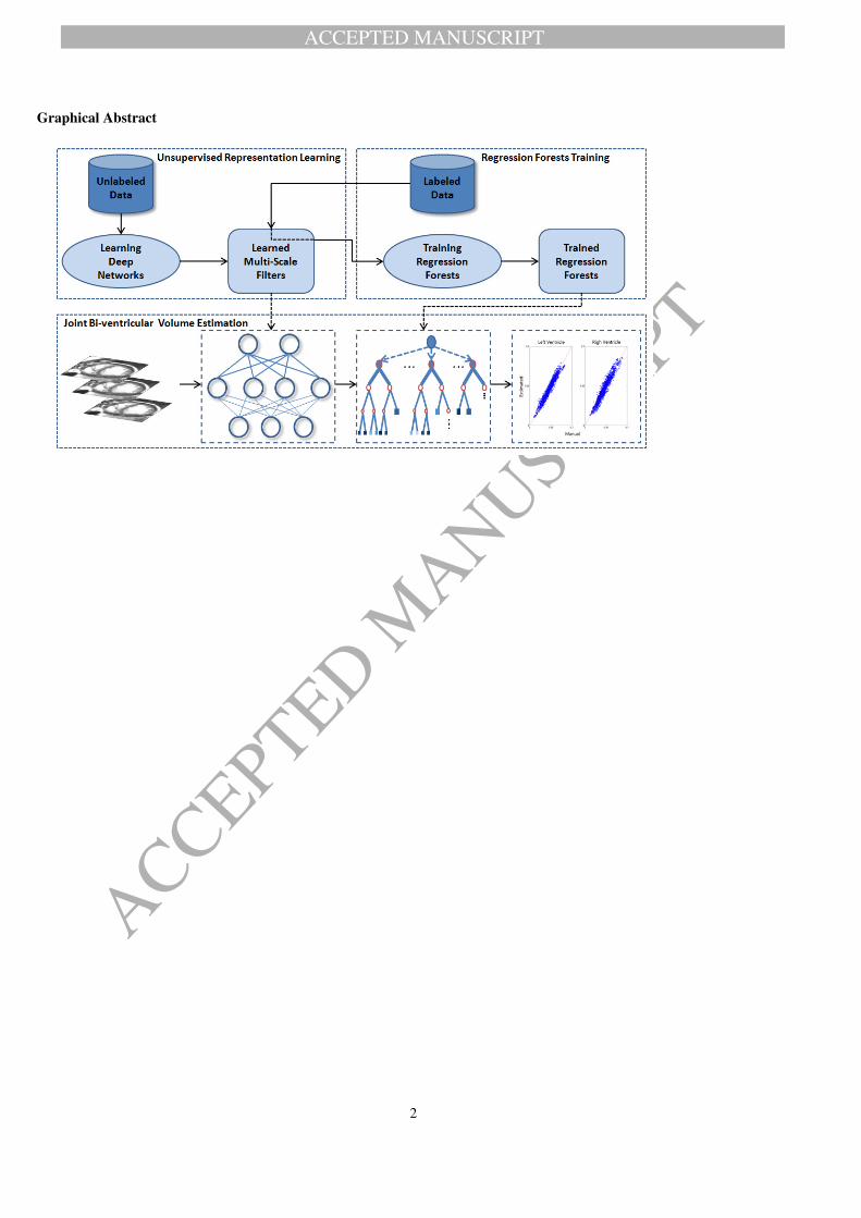
Graphical Abstract
2

ACCEPTED MANUSCRIPT
ACCEPTED MANUSCRIP
T
Multi-Scale Deep Networks and Regression Forests for Direct Bi-ventricular VolumeEstimation
Xiantong Zhena,b, Zhijie Wangc, Ali Islamd, Mousumi Bhadurie, Ian Chane, and Shuo Lia,b,c∗
aThe University of Western Ontario, London, ON, Canada.bDigital Image Group (DIG), London, ON, Canada.
cGE Healthcare, London, ON, Canada.dSt. Joseph’s Health Care, London, ON, Canada.
eLondon Healthcare Sciences Centre, London, ON, Canada.
Abstract
Direct estimation of cardiac ventricular volumes has become increasingly popular and important in cardiac function analysis due toits effectiveness and efficiency by avoiding an intermediate segmentation step. However, existing methods rely on either intensiveuser inputs or problematic assumptions. To realize the full capacities of direct estimation, this paper presents a general, fullylearning-based framework for direct bi-ventricular volume estimation, which removes user inputs and unreliable assumptions. Weformulate bi-ventricular volume estimation as a general regression framework which consists of two main full learning stages:unsupervised cardiac image representation learning by multi-scale deep networks and direct bi-ventricular volume estimation byrandom forests.
By leveraging strengths of generative and discriminant learning, the proposed method produces high correlations of around 0.92with ground truth by human experts for both the left and right ventricles using a leave-one-subject-out cross validation, and largelyoutperforms existing direct methods on a larger dataset of 100 subjects including both healthy and diseased cases with twice thenumber of subjects used in previous methods. More importantly, the proposed method can not only be practically used in clinicalcardiac function analysis but also be easily extended to other organ volume estimation tasks.
Keywords: Direct volume estimation, multi-scale deep networks, random forests, regression.
1. Introduction
Accurate and automatic assessment of cardiac functionsplays an increasingly important role in diagnosis and prognosisof heart diseases, one of the leading causes of death (Wang andAmini, 2012). Cardiac ventricular volumes have been widelyused as measurements of cardiac abnormalities and functions,e.g., ejection fraction (EF) and stroke volume (Wang et al.,2009; Punithakumar et al., 2013; Marchesseau et al., 2013).Conventional volume estimation methods usually rely on anintermediate step of segmentation. However, segmentation it-self is an extremely challenging problem which in clinical prac-tise physicians are not interested in. Direct estimation methodswhich remove the segmentation step become attractive in car-diac function diagnosis and ventricular estimation due to its ef-ficiency and clinical significance (Afshin et al., 2012a,b; Wanget al., 2014; Afshin et al., 2014; Zettinig et al., 2014; Zhenet al., 2014d; Wang et al., 2013; Zhen et al., 2015a). A com-prehensive study of methods for cardiac ventricular volume es-timation has been conducted in (Zhen et al., 2014c) showingthat direct estimation methods provide more accurate estima-tion than segmentation-based methods for both left and right
∗Corresponding author ([email protected]).
ventricles (LV and RV). More importantly, direct estimation al-lows us to leverage both the state-of-the-art machine learningtechniques and increasingly large amount of labeled and unla-beled imaging data. In many applications, the performance ofmachine learning-based automatic detection and diagnosis sys-tems have shown to be comparable to that of well-trained andexperienced radiologists (Wang and Summers, 2012). More-over, direct methods also enable us to richly explore data statis-tics which therefore helps provide more meaningful and clini-cally significant volume estimation.
However, existing direct methods suffer from many draw-backs, e.g., dependence on user inputs and unreliable assump-tions, which largely restrict their applications. In this paper,towards the full capacity of direct estimation, we propose ageneral framework for bi-ventricular volume estimation. A pre-liminary conference version of this work appeared in MICCAI2014 (Zhen et al., 2014d). In this journal version we extendin two main aspects: 1) we replace the handcrafted features in(Zhen et al., 2014d) by feature engineering with representationlearned by our multi-scale deep networks (Bengio et al., 2013).In contrast to feature engineering, the unsupervised learning bydeep networks allows us to both make full use of plenty of un-labeled data which is largely available in medical imaging andfaithfully detect data-driven features for informative image rep-resentations in specific tasks; 2) we provide a wider investiga-
Preprint submitted to Medical Image Analysis July 22, 2015

ACCEPTED MANUSCRIPT
ACCEPTED MANUSCRIP
T
tion on a larger dataset of 100 subjects with 6000 images thanpreviously used. Such as a large dataset encompasses the hugeinter-subject variabilities and therefore provides a more com-prehensive and clinically meaningful validation.
1.1. Direct methodsDirect diagnosis has proven to be effective and efficient in
assessment of cardiac function abnormality automatically with-out intermediate steps. Afshin et al. (2014) present a directmethod for regional assessment of the LV myocardial functionvia classification. Without delineating regional boundaries ofthe LV, they obtain an abnormality assessment of each standardcardiac segment in real-time with more accurate results thansegmentation-based methods. A set of statistical MRI features,i.e., Bhattacharyya coefficients (Afshin et al., 2014) based ona measure of similarity between image distributions, are builtfor all the regional segments and all subsequent frames. Thestatistical features are related to the proportion of blood withineach segment and can characterize segmental contraction. Un-fortunately, the method requires user inputs for one frame ofeach subject and is restricted to the LV, which limits its furtherapplication in clinical use.
In addition to MR imaging, direct methods have also been de-veloped in other modalities such as electrocardiograms (ECG)for diagnosis of cardiac function abnormalities. Recently,Zettinig et al. (2014) proposed a data-driven estimation of car-diac electrical diffusivity from 12-lead ECG for diagnosis andtreatment of dilated cardiomyopathy (DCM). Instead of solv-ing an inverse problem to find patient-specific parameters ofelectrophysiology (EP) model, they propose to learn the inversefunction by formulating as a polynomial regression problem todirectly estimate model parameters for specific patients. TheECG features are taken as the input of the regressor with modelparameters being the output.
Direct estimation of cardiac volumes has started to gener-ate increasing interest due to the avoidance of intermediate seg-mentation (Afshin et al., 2012a; Wang et al., 2014; Zhen et al.,2014d). To directly estimate the ejection fraction of the LV,global image statistics are used to calculate LV volumes in (Af-shin et al., 2012a). A key limitation is that intensive user in-puts including two boxes, i.e., one inside the LV cavity andone enclosing the cavity, are required. Moreover, the methodis restricted to the LV due to the strong assumption of corre-lation between the considered statistics and LV cavity areas,and therefore can not be directly generalized to the RV or bi-ventricles, i.e., the LV and RV.
Joint analysis of cardiac bi-ventricles within a single frame-work is of great significance to cardiac function assessmentand disease diagnosis (Lotjonen et al., 2004; Hu et al., 2005;Fritz et al., 2006; Lu et al., 2011; Wang et al., 2013, 2014;Zhen et al., 2014d), while posing great challenge for tradi-tional segmentation based techniques (Cocosco et al., 2008).The first attempt to direct bi-ventricular volume estimation wasprovided in (Wang et al., 2014) which employs a Bayesianmodel. Given an input MR image with bi-ventricles, the modelsearches similar images in a set of templates with manually seg-mented LV/RV, and the similarity is measured by computing
Figure 1: The illustration of MR images with bi-ventricles.
the distance on simple handcrafted features. Cardiac ventric-ular volumes in the input image are then simply calculated asthe weighted average over the templates. The method did notfully model the statistical relationship between image featuresand cardiac ventricular volumes and is far from a capable toolof direct estimation.
The method suffers from several severe drawbacks. It 1) re-lies on a simplified assumption to model the relationship be-tween volumes of the LV and RV in a cardiac cycle by an em-pirical linear function: Vol(RV) = aVol(LV) + b, 2) does notgeneralize well on more diverse datasets with large number ofsubjects, and 3) is computationally expensive to match a testpoint against all templates which are all the training samples,and typically requires uniformly-sampled training data for ac-curate results (Huang et al., 2011).
To deal with the above-mentioned issues, we proposed usingrandom forests (Breiman, 2001) for direct bi-ventricular vol-ume estimation in our conference version (Zhen et al., 2014d),in which bi-ventricular volume estimation is formulated as aregression problem. Multiple complementary features includ-ing pyramid Gabor features (PGF) (Zhen and Shao, 2013), his-togram of gradients (HOG) (Dalal and Triggs, 2005) and in-tensity are carefully designed to represent cardiac MR images.Like (Afshin et al., 2012a; Wang et al., 2014), the method stillrelies on handcrafted feature representation by feature engineer-ing (Bengio et al., 2013), which cannot be adapted to learn op-timal representation from data and are unable to extract the dis-criminative information related to the specific domain (Bengioet al., 2013).
In general, existing direct methods (Afshin et al., 2012a;Wang et al., 2014) are still far from being a satisfactory toolfor cardiac volume estimation. They might perform well ondatasets of a small number of subjects due to overfitting whilethe performance cannot be guaranteed on datasets of a largenumber of subjects which present greater variability of imagestherefore raise more challenges (Petitjean and Dacher, 2011).
Towards full capacities of direct estimation, we propose ageneral fully learning-based framework for joint bi-ventricularvolume estimation which removes user inputs and unnecessaryassumptions. The framework consists of two main stages: car-diac image representation by multi-scale convolutional deepnetworks and joint bi-ventricular volume estimation by ran-dom forests. The multi-scale convolutional deep networks takeadvantages of deep learning as a powerful tool for unsuper-vised representation learning and leverage the abundant unla-beled data. Random forests as discriminative learning can ef-fectively capture the relationship between image appearanceand bi-ventricular volumes, and more importantly they are able
4

ACCEPTED MANUSCRIPT
ACCEPTED MANUSCRIP
T
to automatically extract the most discriminate features for eachventricle due to the innate nature of feature selection.
1.2. Cardiac image representation learningCardiac image representation serves as a fundamental role
in cardiac function analysis and is typically obtained by featureengineering in the existing methods (Montillo et al., 2004; Qianet al., 2006; Zheng et al., 2008; Chen et al., 2010; Garcia-Barneset al., 2010; Afshin et al., 2012a; Punithakumar et al., 2013;Wang et al., 2014; Zhen et al., 2014d). Handcrafted featuresby feature engineering are labor-intensive and suffers from theweakness of being unable to extract and organize representa-tive information from the data (Bengio et al., 2013; Tang et al.,2014). Bi-ventricles exhibit great variability of cardiac imagesfrom gray levels to structure shapes as shown in Fig. 1. Theshape of the two ventricles varies across patients, over time andalong the long axis, which makes it extremely difficult for ac-curate analysis of bi-ventricular volumes. Since bi-ventricularvolume estimation poses more challenge than previous tasks(Afshin et al., 2012a), these handcrafted image representationsare not able to capture the variability of bi-ventricles due toignoring the specific domain knowledge in data, especially ondataset of larger number of subjects.
A data-driven representation directly learned from unlabeleddata is highly desirable and can capture underlying explanatoryfactors (Bengio et al., 2013; Zhen et al., 2014b). As highlightedin (Bengio et al., 2013) that being less dependant on feature en-gineering, data-driven representation learning is able to capturehigh variability of images, especially for cardiac images withbi-ventricles presenting combinatorial variations.
As a powerful state-of-the-art unsupervised feature learningtechnique, deep learning algorithms suit medical applicationswell due to the availability of abundant sample images withoutlabels and have recently started to generate increasing atten-tions in medical image analysis (Ciresan et al., 2013; Carneiroand Nascimento, 2013; Prasoon et al., 2013), Convolutionaldeep learning networks (Krizhevsky et al., 2012; Turaga et al.,2010; LeCun et al., 1998; Lee et al., 2009; Petersen et al., 2014),one of the most representative deep learning algorithms, are ef-fective techniques to retain topological structure, e.g., 2D layoutof pixels, and can be used to capture the anatomical structure ofbi-ventricles in cardiac images.
To handle the great variability of bi-ventricles, we pro-pose using a multi-scale convolutional deep belief network(MCDBN) to learn cardiac image representations (Shin et al.,2013; Petersen et al., 2014). By combining the strengths ofboth multi-scale analysis (Burt and Adelson, 1983; Zhen et al.,2013; Shao et al., 2014; Zhen et al., 2014a) and deep learn-ing, the MCDBN fits well for cardiac image representation withbi-ventricles. The MCDBN benefits representation learning ofcardiac images in three folds: It 1) can, to a large extent, retainstructural layouts in cardiac images which are the most impor-tant features of bi-ventricles; 2) can be efficiently trained dueto the weight sharing which also allows us to use a large set ofunlabeled data; 3) can detect sufficient complementary featuresby multi-scale filtering which provides a rich and effective rep-resentation of bi-ventricles.The MCDBN leverages the strength
of convolutional DBN in unsupervised representation learningand more importantly allows us to use a large amount of un-labeled data which is abundantly available in medical imageanalysis.
1.3. Bi-ventricular volume estimation
The challenges that arise from the complex functional andgeometrical interference and interdependency between the LVand RV lead to a high-dimensional representation. Regressionforests (Breiman, 2001) are capable of modeling complex rela-tionships between high-dimensional input features and contin-uous outputs. They have been successfully applied to variouscomputer vision tasks (Shotton et al., 2013; Gall et al., 2011;Zhen et al., 2015b), and recently started to attract interest inmedical image analysis (Criminisi et al., 2011). We chooserandom forests to work on top of image representations fromMCDBN to fulfill bi-ventricular volume estimation due to thestrong ability of feature selection and efficient implementation.Random forests are well-suited to direct and joint bi-ventricularvolume estimation, which has been shown in our preliminarywork (Zhen et al., 2014d).
Random forests (Breiman, 2001) are an ensemble of decisiontrees which combine the ideas of bagging and the random fea-ture selection which benefit our learning-based bi-ventricularvolume estimation in three folds: They 1) effectively deal withthe high-dimensional representation due to innate ability to se-lect discriminative features (Criminisi and Shotton, 2013); 2)avoid overfitting while providing accurate prediction by inject-ing randomness (Biau, 2012); 3) are specifically fit for bi-ventricular volume estimation due to the intrinsic nature offeature selection (Breiman, 2001). With the above properties,random forests offer a prime regressor for direct and joint bi-ventricular volume estimation.
1.4. Contributions
We propose a general, fully learning-based framework to re-alize the full capacities of direct estimation of cardiac ventric-ular volumes. The proposed framework combines the strengthsof both generative (for representation) and discriminant (for re-gression) learning. Specifically, 1) we propose using multi-scale convolutional deep networks for unsupervised cardiac im-age representations learning from unlabeled data; and 2) we for-mulate bi-ventricular volume estimation as a regression prob-lem and adopt random forests for efficient volume estimation.Our method provides a new framework from the perspective ofregression for cardiac ventricular volume estimation which canalso be used for other organ volume estimation and extensivemodel parameter estimation problems, e.g., model personaliza-tion (Marchesseau et al., 2013; Zettinig et al., 2014).
2. Methodology
The flowchart of the proposed general estimation frameworkis illustrated in Fig. 2. In the left block, the multi-scale convolu-tional deep belief networks learn a set of multi-scale data-drivenfeature detectors from totally unlabeled data for cardiac image
5

ACCEPTED MANUSCRIPT
ACCEPTED MANUSCRIP
T
Figure 2: The flowchart of the proposed unsupervised feature learning and random forest regression. [Left block]: Unsupervised cardiac image representationlearning by multi-scale deep networks from a unlabeled dataset. [Right block]: Training regression forests and on labeled data. [Bottom block]: Joint bi-ventricularvolume estimation with the trained regressors.
representations. In the right block, random forests are trainedon labeled data for bi-ventricular volume estimation. In the bot-tom block, test images go through learned feature detectors andtrained regressors successively and bi-ventricular volumes arejointly estimated.
2.1. Representation learning by multi-scale deep networks
The proposed multi-scale convolutional deep belief network(MCDBN) is a three-layer deep network composed of a multi-scale convolutional restricted Boltzmann machine (MCRBM)and an RBM as shown in Fig. 3. Our MCDBN is inspiredby convolutional deep belief nets (CDBNs) introduced by Leeet al. (Lee et al., 2009). By combining convolutional filterswith deep belief nets, CDBNs can encode local structures ofimages and therefore achieve more descriptive representations.However, only a single scale of filters are learned in CDBNs,which limits the ability of feature detection. By learning multi-scale filters, our MCDBN further enhances the effectiveness ofrepresentation learning especially for cardiac images with bi-ventricles that exhibit complex geometrical variations.
2.1.1. Restricted Boltzmann machine (RBM)
A restricted Boltzmann machine (RBM) is a two-layer, bi-partite, undirected graphical model. RBMs are fully connectedwith a group of binary hidden nodes h, a group of visible nodes,which are either binary or real-valued, and symmetric connec-tions between h and v represented by a weight matrix W. Theweight W and biases b and c of an RBM can be learned bycontrastive convergence (CD) (Hinton, 2002).
The RBMs can be stacked to form a deep belief network(DBN), a generative model with multiple layers, which demon-strates high ability of representation learning. In DBN, two ad-jacent layers are fully connected and no nodes in the same layerare connected. The DBN can be trained in a greedy layerwiseway by treating each layer as an RBM (Hinton et al., 2006; Ben-gio et al., 2007), which works well in practice. Unlike DBNswhich treat all the pixels in an image equally, the CDBNs modelthe topology of images by operating convolutional kernels onlocal neighborhoods and can naturally preserve image topolog-ical information (LeCun et al., 1998) which is extremely impor-tant especially for medical images with anatomical structures.
2.1.2. Multi-scale CDBNs
A convolutional RBM (CRBM) proposed by Lee et al. (2009)is composed of two layers, but the weights of the connectionsbetween visual and hidden layers are shared among all the lo-cations in an image. The input visual layer consists of an arrayof NV × NV which in this work is real-valued. The hidden layeris composed of K groups each of which is a binary array ofNH × NH . Each of the K group in the hidden layer is associatedwith an NW × NW filter.
The energy function for a CRBM is defined as
E(v,h) =−K∑
k=1
NH∑
i, j=1
NW∑
r,s=1
hki jW
krsvi+r−1, j+s−1 (1)
−K∑
k=1
bk
NH∑
i, j=1
hki j − c
NV∑
i, j=1
vi j, (2)
6

ACCEPTED MANUSCRIPT
ACCEPTED MANUSCRIP
T
Figure 3: The schematic diagram of unsupervised feature learning with the proposed multi-scale deep networks. The three blocks from bottom to top are the inputMR images, a multi-scale convolutional RBM and an RBM.
7

ACCEPTED MANUSCRIPT
ACCEPTED MANUSCRIP
TFigure 4: Illustration of the filters (feature detectors) of different sizes asso-ciated with the feature maps. In the top row are the leaned filters and in thebottom row are the corresponding feature maps.
where bk is the bias for each group and c is the bias shared by allvisible nodes. The energy function can be represented in termsof convolution as
E(v,h) =
K∑
k=1
hk • (W ∗ v) −K∑
k=1
bk
∑
i, j
hki, j − c
∑
i, j
vi j (3)
In contrast to the original CRBM, we propose multi-scaleCRBM (MCRBM) with filters of different sizes, which meanswe have S × K filters with S the number of scales.
By stacking an RBM on top of the proposed MCRBM, weobtain a three-layer network, i.e., the multi-scale convolutionaldeep belief network (MCDBN). Totally unlabeled cardiac MRimages are fed into the MCRBM to learn a set of multi-scalefilters, i.e., feature detectors. The feature maps from CRBMgo further through an RBM to obtain more compact represen-tations.
2.1.3. Learning multi-scale filtersThe proposed MCDBN provides an effective representation
learning of cardiac images, which creates a solid basis for sub-sequent bi-ventricular volume estimation. The complexities ofbi-ventricles residing in different scales can be effectively cap-tured by the multi-scale filters learned by the proposed deepnetworks. Larger scales of filters extract the simplex structure
of the LV, while finer filters with smaller sizes are capable ofdetecting the sophisticated crescent-shaped of the RV which ismuch more complex than the LV.
Specifically, we learn 2 filters for each of 3 scales: 17×17,13×13 and 9×9. Fig. 4 illustrates the learned multi-scale fil-ters (in top row) and the corresponding feature maps (in thebottom row) outputted from the filters. As can be seen thatthe learned filters have successfully detected oriented and lo-calized edges (Poultney et al., 2006; Lee et al., 2009) whichare the most representative features of cardiac ventricles. Thelearned feature maps intensify the main shapes and contoursof bi-ventricles while removing the unrelated regions such asbackground, which provides an informative and discriminativerepresentation, especially for images with bi-ventricles.
2.2. Regression forestsMathematically, given a multivariate input v which in our
case is the feature vector extracted from an image, our aim isto associate with a continuous multi-variate label y, i.e., the bi-ventricular cavity areas in images.
2.2.1. TrainingWe build decision trees using the adapted algorithm from
(Breiman, 2001). Each internal node j of a tree is associatedwith a split function. The training process is to construct eachtree with a randomly selected training subset. Note that only asubset of features are used for training each decision tree, whichare fixed for prediction.
The split function at a split node j is formulated as a functionwith binary outputs
h(v, θ j) : Rd × T → {0, 1} (4)
where v is the input feature vector, T represents the spaceof all split parameters (Criminisi and Shotton, 2013), and θ j
is the function parameter associated with the j-th node andcan be trained by minimizing a least-squares error function I(Breiman, 2001) at the j-th split node:
θ j = arg maxθ∈T
I(S j, θ) (5)
where S j is a subset of training samples associated with the j-thnode. The data point v arriving at the split node is sent to its leftor right child node according to the result of the split function.An example of trained random forests is shown in Fig. 5 whichwill be used for prediction of new input images.
2.2.2. PredictionAs shown in Fig. 5, we pass a test image v through each
tree starting from the root of each decision tree Ti, send to theleft/right child by applying the split function, and stop when vreaches a leaf node of the tree. The simple comparison opera-tion on each split node makes the prediction extremely fast andefficient. Given the t-th tree in a forest, the associated leaf out-put takes the form of a density probability function pt(y|v). Theforest output is the average over all tree outputs
p(y|v) =1T
T∑
t=1
pt(y|v) (6)
8

ACCEPTED MANUSCRIPT
ACCEPTED MANUSCRIP
T
Figure 5: Illustrated are the random forests comprised of n decision trees {T1, . . . ,Ti, . . . ,Tn} learned from the training set. Test images can be quickly predictedby several simple comparison operations. X# indicates the #-th feature that are selected for the associated splitting notes. Test images go through each tree in theforests and the results outputted from all the trees are combined as the final prediction.
where y is the continuous multi-variate label and T is the num-ber of trees in the forest.
2.2.3. Feature selectionRandom forests have the innate ability to select the most rep-
resentative features closely related to cardiac volumes. The fea-ture selection can be reflected by the feature importance rankedby random forests in the training stage (Breiman, 2001). Fea-tures are assigned with different values of importance accordingto their discriminative ability which is measured by regressionerrors. Features with larger values are ranked as more impor-tant than features with small values. Our experiments have in-dicated that random forests have a strong capacity of featureselection for the estimation of the LV and RV volumes. Theresults are shown in Fig. 6 from which we can easily observethat regions that are closely related to the LV and RV, respec-tively are successfully detected with high importance by ran-dom forests, while insignificant regions are largely removed.More importantly, the selected features mostly fall on the keyregions such as edges, boundaries of the ventricles that can dis-tinguish different cardiac ventricular volumes.
2.2.4. Tree numbersThe effects of different numbers of trees in random forests
are illustrated in Fig 7. The performance keeps going up withthe number of trees from 100 to 500. The computational costwill also increase with the number of trees. In our experiments,we use 500 trees as the final setting to keep the balance betweenperformance and computational burden.
3. Experiments and results
3.1. Datasets and settings
In our experiments, two sets of subjects with 2D short-axis cine MR images are used including 2820 unlabeled im-ages from 47 subjects for unsupervised feature learning and6000 labeled images from 100 subjects for the validation ofbi-ventricular volume estimation. The subjects are collectedfrom 3 hospitals affiliated with two health care centers (Lon-don Healthcare Centre and St. Joseph’s HealthCare) and 2 ven-dors (GE and Simens) including both health and diseased cases.
Figure 6: Illustration of feature importance learned by random forests. Themagnitudes at corresponding locations indicate the importance of the featuresfor volume estimation of the left and right ventricles, respectively. Featureswith large values of magnitudes are ranked as more important than featureswith small values.
100 150 200 250 300 350 400 450 5000.8
0.82
0.84
0.86
0.88
0.9
0.92
0.94
0.96
0.98
1
Number of Tress
Cor
rela
tion
Coe
ffici
ents
The performance of random forests varying with the number of trees
Left VentricleRight Ventricle
Figure 7: The performance of random forests with different numbers of trees.
9

ACCEPTED MANUSCRIPT
ACCEPTED MANUSCRIP
T
The pathologies are extremely diverse including regional wallmotion abnormalities, myocardial hypertrophy, mildly dilatedRV, atrial septal defect, LV dysfunction, mildly enlarged LV,decreased ejection fraction in most cases, etc. Each subjectcontains 20 frames throughout a cardiac cycle. In each frame,three representative slices, i.e., apical, mid-cavity and basal, areselected following the standard AHA prescriptions (Cerqueiraet al., 2002) for validation, and their manual segmentations areused as the benchmark.
To benchmark with existing direct methods (Wang et al.,2014; Zhen et al., 2014d), we estimate cavity areas of the LVand RV in MR images, and the volumes are computed by inte-grating LV/RV cavity areas along the sagittal direction perpen-dicular to the short axis. A single cropped region of interest(ROI) rather than two individual ones is placed to enclose theLV and RV in an MR image, which can be obtained automat-ically (Petitjean and Dacher, 2011). The cropped images arethen resized into 60 × 60 pixels as the inputs.
The MCDBN has been conducted on the unlabeled dataset of47 subjects to obtain filters which are used to create feature rep-resentations of all images. For volume estimation, we employ aleave-one subject-out cross-validation approach, i.e., 100-foldcross validation on the labeled dataset of 100 subjects. Theperformance is evaluated by comparing with the golden stan-dard manual segmentation using absolute estimation errors andcorrelation coefficients. The correlation coefficient is used tomeasure the linear correlation between ground truth and directestimation.
3.2. Implementation details
For the MCDBN, we follow the original work (Lee et al.,2008, 2009; Hinton et al., 2006; Hinton, 2010) using a learningrate of 0.0001 for RBMs and sparsity of 0.01 for CRBMs. Assuggested in (Hinton, 2010), we start with a momentum of 0.5and once the large initial progress in the reduction of the recon-struction error has settled down to gentle progress, increase themomentum to 0.9. We use 2 filters of each scale to create fea-tures maps as the inputs to the RBMs. The final feature vectoras the image representation is of 3000 dimensions which is fedinto regression forests for bi-ventricular volume estimation.
For random forests, we set the number of features used insplitting note as 500, and since the number of trees in forests isa key parameter which determines the regression performance,we experiment with different values to investigate its effects.
3.3. Estimation results
We apply the proposed methods to cardiac bi-ventricular vol-ume estimation and calculation of ejection fractions (EF) withthe estimated volumes. The EF is an important cardiac func-tional parameter and predictor of prognosis and widely used inclinical analysis. The EF is computed by
EF =Vd − Vs
Vd, (7)
where Vd and Vs denote the largest (end-diastolic) and thesmallest (end-systolic) volumes of a ventricle in a cardiac cycle,respectively.
0 0.35 0.70
0.35
0.7
Manual
Est
imat
ed
Left Ventricle
0 0.35 0.70
0.35
0.7
Manual
Est
imat
ed
Right Ventricle
Figure 8: The correlation coefficients between the volumes obtained by theproposed method and manual segmentation for the LV and RV, respectively.
Table 1: The correlation coefficients with different sizes of learned filters.
Filter sizes 17 × 17 13 × 13 9 × 9 Multiple scales
LV 0.899 0.885 0.873 0.921RV 0.869 0.875 0.889 0.908
3.3.1. Bi-ventricular volumes
The effectiveness of the proposed method is demonstrated bythe outstanding performance on a large dataset with 6000 MRimages from 100 subjects for both LV and RV. The estimatedvolumes by the proposed method are measured by comparingwith those by manual segmentation using a leave-one-subject-out cross validation. The correlations between estimated andmanually obtained volumes are depicted in Fig. 8 for the LVand RV, respectively. Despite of the challenges in joint estima-tion of bi-ventricular volumes, the proposed method achieves acorrelation coefficient of 0.921 for the LV, and can yield 0.908for the RV which has much greater geometrical complexity thanthe LV. The high estimation accuracies with low volume esti-mation errors, i.e., 0.010 ± 0.013 (LV) and 0.014 ± 0.012 (RV),indicate that the proposed method can be practically used inclinical cardiac function analysis.
3.3.2. Ejection fraction
As shown in Fig. 9, we compare EFs obtained from estimatedvolumes with those from manual segmentation by human ex-perts. The EFs from the estimated volumes provide very closeapproximations for both the LV and RV with a large propor-tion equal to their counterparts obtained manually. Comparedto segmentation based methods, the estimation errors of EFsare relatively low: 0.0387 ± 0.0330 and 0.0455 ± 0.0347 forthe LV and RV, respectively. The high estimation accuracies onboth the LV and RV will be extremely important for assessmentof cardiac functions, which indicates the potential use of theproposed method for cardiac disease diagnosis.
10

ACCEPTED MANUSCRIPT
ACCEPTED MANUSCRIP
T
Subject0 10 20 30 40 50 60 70 80 90 100
Eje
ctio
n F
ract
ion
0
0.2
0.4
0.6
0.8
1Left Ventricle
ManualEstimated
Subject0 10 20 30 40 50 60 70 80 90 100
Eje
ctio
n F
ract
ion
0
0.2
0.4
0.6
0.8
1Right Ventricle
ManualEstimated
Figure 9: Comparison of EFs obtained by manual segmentation (blue) and the proposed method (red) for the LV (upper) and RV (bottom), respectively.
3.4. Parameter evaluation
To look further into the proposed method, we have also in-vestigated the performance of the learned filters with differentscales used in our experiments.
3.4.1. Multi-scale filtersThe advantage of the proposed multi-scale convolutional
deep network is validated by comparing with results from threedifferent sizes of filters as shown in Table 1. More importantly,our results further show the complementary properties of dif-ferent sizes of filters in the cardiac image representation. Onthe LV, larger scales of filters show to be superior over smallerones, while on the RV, smaller scales of filters perform better.This is reasonable since structure of the RV is more complexthan the LV, smaller scales of filters are more able to capturethese localized complex features on the RV. The multi-scale fil-ters outperform each of single scale filters. The results confirmthat representation learning by the proposed multi-scale con-volutional deep networks overcomes the great variability andgeometrical complexity of bi-ventricles, and validate the effec-tiveness of multi-scale convolutional deep networks for cardiacimage representation learning in bi-ventricular volume estima-tion.
3.5. Comparison
The performance of the proposed method can be furtherdemonstrated by the comparison with existing direct estimationmethods and segmentation-based methods. Both bi-ventricular
volumes and ejection fraction are used as the measurements inthe comparison.
3.5.1. Comparison with existing direct methodsThe advantage of the proposed method is further shown
by comparing with existing direct estimation methods: theBayesian model (Wang et al., 2014), and multiple features(Zhen et al., 2014d) under the unified experimental settings. Asshown in Table 2, the proposed method largely outperforms theBaysian model and multiple features both for the LV and RV byup to 0.085 in terms of correlation coefficients and the volumeestimation errors are much -up to 37.5%- lower than both ofthem. As shown in Table 3, the proposed methods produceslower estimation errors of ejection fractions for bi-ventriclesthan both the Bayesian model and multiple features.
Despite of that the RV is extremely challenging due to itscomplex geometrical structures, the proposed method performsmuch better than the methods in (Wang et al., 2014; Zhen et al.,2014d) using handcrafted features, which shows the effective-ness of the proposed multi-scale deep networks for cardiac im-age representation learning. The better performance than theBayesian model in (Wang et al., 2014) shows the advantagesof the proposed method by formulating volume estimation as aregression problem which incorporates learning stages. The su-perb performance over multiple features in (Zhen et al., 2014d)demonstrates the effectiveness of unsupervised representationlearning by the proposed multi-scale deep networks. More-over, the outstanding performance indicates that the proposedmethod can be practically used in clinical cardiac diagnosis.
11

ACCEPTED MANUSCRIPT
ACCEPTED MANUSCRIP
T
Note that the Bayesian model has achieved high accuracy prob-ably because of using a relative small size of dataset with only56 subjects which can not capture the huge variability of car-diac ventricles. In addition, the Bayesian model relies on theunproven assumption that LV and RV volumes are linearly cor-related during a cardiac cycle, which however does not al-ways hold in diseased cases. Our dataset contains 100 subjects(nearly twice sizes of their dataset) exhibit sufficiently inter-and intra-subject variabilities, especially due to the presence ofdiverse pathologies.
3.5.2. Comparison with segmentation-based methodsTo demonstrate the superiority of the proposed direct esti-
mation method over conventional segmentation-based methods,following the work (Wang et al., 2013), we conduct a compar-ison with two representative segmentation methods includinglevel set (Ayed et al., 2009a) and graph cut (Ayed et al., 2009b).As shown in Table 2 and 3, on our dataset the proposed methodlargely outperforms graph cut and level set in terms of both bi-ventricular volumes and ejections fractions. Moreover, neitherlevel set or graph cut are applicable to the RV for volume es-timation. Our method can be flexibly used for either a singleventricle or joint bi-ventricles.
4. Discussion
Direct estimation, in general, replaces tedious and unreliablesegmentation, and focuses on the ultimate goal of volume esti-mation. The proposed direct estimation method not only solvesa dual estimation problem but also for the first time formulatesit as a regression framework. This new framework substan-tially outperforms existing segmentation based and direct meth-ods, and more importantly offers a more compact and exquisitemathematical formulation of regression, which is flexible andeasily extendable to other applications.
The effectiveness of the proposed method stems from thetwo key incorporated components: unsupervised representationlearning and random forests regression which are suited wellto cardiac ventricular volume estimation and medical imageanalysis. Since unlabeled imaging data is always abundantlyavailable in medical image analysis, unsupervised representa-tion learning allows us to use a large of amount of unlabeleddata to obtain more faithful and informative data-driven repre-sentations. Random forests provide an effective tool for cardiacbi-ventricular volume estimation due to its innate ability of fea-ture selection mechanism and computational efficiency by sub-sampling and boosting.
The proposed method shows advantages over both conven-tional segmentation based methods and other recently-proposeddirect estimation methods.
4.1. Proposed method vs. segmentation based methods
The proposed method possesses attractive advantages overprevious cardiac ventricular volume estimation using segmen-tation based methods. Cardiac ventricular volume estimation
has been a challenging task. Most of conventional segmenta-tion based methods rely on the unreliable assumption that car-diac ventricles are supported by edges and region intensity ho-mogeneity. However, edges of cardiac ventricles are not alwaysconsistently visible along the entire contour due to overlappingof anatomical structures and noise, etc., and the homogeneityis severely violated due to the complex image textures and ap-pearances, especially with the presence of pathology. In addi-tion, existing automatic segmentation methods are mostly re-stricted to the LV. RV segmentation remains an unsolved prob-lem not to mention multiple ventricles, e.g., bi-ventricles andfour chambers. Our direct estimation discovers the relationshipbetween image appearances and cardiac volumes by statisticallearning from annotated data, which significantly outperformstwo typical segmentation methods: level set and graph cut asindicated in experimental results. In contrast to segmentationbased methods, our method as direct estimation 1) removes theintermediate and always tedious segmentation by either manualor automatic methods, achieving more accurate and efficient es-timation 2) can naturally handle the cases without consistentlystrong edge and of region inhomogeneity, guaranteeing the ap-plicability in clinical practise; and 3) is able to flexibly deal withboth single and multiple ventricles, showing great generality toother organ volume estimation.
4.2. Proposed method vs. existing direct methodsThe proposed method also demonstrates more merits in con-
trast to other recently-proposed direct estimation methods. Ourmethod is a fully automatic without relying on any assumptions,user inputs and initialization. By leveraging advanced machinelearning techniques, i.e., unsupervised representation learningand random forests regression, our method achieves the fullycapacity of direct estimation, showing much better performanceon the largest datset as indicated in experimental results. 1) In-stead of using ineffective handcrafted features in previous meth-ods, our method learns data-driven representations which caneffectively capture the characteristics of objects, e.g., cardiacventricles, to achieve optimal representations for volume esti-mation; 2) By formulating bi-ventricular volume estimation as aregression problem, our method achieves more accurate estima-tion than previous methods of no learning stages, e.g., templatematching (Wang et al., 2014); and 3) By using powerful randomforests for regression, our method can effectively handle large-scale datasets without overfitting and promises the practical useof our method in clinical applications.
4.3. Critical analysisWhile the proposed method has been validated on a large
dataset with highly diverse cases, the pros and cons of the pro-posed method come from the same fact that no explicit contouris provided. Without relying on contouring, the proposed di-rect estimation method is able to effectively handle challengesin conventional segmentation-based methods. Meanwhile, itwould pose a learning curve to those who are used to contouringfor validation which, however, can be easily shortened by pro-viding more measurement through relating to human percep-tion and intuition: 1) Due to the temporal coherence, plotting
12

ACCEPTED MANUSCRIPT
ACCEPTED MANUSCRIP
T
Table 2: The comparison of estimation errors for bi-ventricular volumes.
Correlation coefficients Volume estimation errorsMethods LV RV LV RV
Our method 0.921 0.908 0.010 ± 0.011 0.014 ± 0.012Baysian model (Wang et al., 2014) 0.861 0.823 0.016 ± 0.019 0.018 ± 0.013
Multiple features (Zhen et al., 2014d) 0.870 0.853 0.012 ± 0.011 0.017 ± 0.016Level set (Ayed et al., 2009a) 0.803 - 0.036 ± 0.025 -Graph cut (Ayed et al., 2009b) 0.835 - 0.029 ± 0.027 -
Table 3: The comparison of estimation errors for ejection fraction.
Methods Level set Graph cut Baysian model Multiple features Our method
LV 0.110 0.097 0.055 0.047 0.0387RV - - 0.071 0.059 0.0455
the volume variation cross a cardiac cycle can provide directmutual validation of volumes in different frames; and 2) Dueto the anatomical constraints between adjacent slices along thelong axis, plotting the area distributions of all slices can alsohelp check the results.
5. Conclusion
In this paper, we have presented a fully learning basedmethod for direct estimation of cardiac bi-ventricular volumes.Our method takes advantages of both generative and discrimi-nant learning, i.e., unsupervised representation learning and su-pervised volume estimation. We have evaluated it on a large setof dataset with 6000 images from 100 subjects using a leave-one-subject-out cross validation. The proposed method pro-duces high correlations with ground truth and outperforms ex-isting direct estimation methods, showing its effectiveness forcardiac ventricular volume estimation. The comparison withsegmentation based methods demonstrates the superiority ofour direct method for cardiac bi-ventricular volume estimation.
More importantly, discriminant learning via random forestsregression allows to deploy advanced machine learning tech-niques to facilitate cardiac functional analysis, which providesan effective tool to automate analysis of medical imagining dataand therefore enables accurate and efficient diagnosis in clini-cal practise (Wang and Summers, 2012). The proposed esti-mation framework can be easily extended to other applicationsfor volume estimation, such as estimating the volume of a tu-mor (Bolte et al., 2007; Heckel et al., 2014) which is still de-pendent on segmentation, and can also be used for extensivemodel parameter estimation problems, e.g., model personaliza-tion (Zettinig et al., 2014; Marchesseau et al., 2013).
Acknowledgment
Computations were performed using the data analytics Cloudat SHARCNET (www.sharcnet.ca) provided through the South-ern Ontario Smart Computing Innovation Platform (SOSCIP);
the SOSCIP consortium is funded by the Ontario Governmentand the Federal Economic Development Agency for SouthernOntario. The authors also wish to thank Dr. Jinhui Qin forassistance with the computing environment.
References
Afshin, M., Ayed, I. B., Islam, A., Goela, A., Peters, T. M., Li, S., 2012a. Globalassessment of cardiac function using image statistics in mri. In: MedicalImage Computing and Computer-Assisted Intervention–MICCAI 2012. pp.535–543.
Afshin, M., Ben Ayed, I., Punithakumar, K., Law, M., Islam, A., Goela, A.,Peters, T., Li, S., 2014. Regional assessment of cardiac left ventricular my-ocardial function via mri statistical features. IEEE Transactions on MedicalImaging.
Afshin, M., Ben Ayed, I., Reza Sadeghi Neshat, H., Punithakumar, K., Goela,A., Islam, A., Peters, T., Li, S., 2012b. Automated assessment of regionalleft ventricular function for cardiac mri with minimal user interaction. In:Annual Meeting - Radiological Society of North America (RSNA).
Ayed, B. I., Li, S., Ross, I., 2009a. Embedding overlap priors in variational leftventricle tracking. IEEE Transactions on Medical Imaging 28 (12), 1902–1913.
Ayed, B. I., Punithakumar, K., Li, S., Islam, A., Chong, J., 2009b. Left ven-tricle segmentation via graph cut distribution matching. In: Medical ImageComputing and Computer-Assisted Intervention–MICCAI 2009. Springer,pp. 901–909.
Bengio, Y., Courville, A., Vincent, P., 2013. Representation learning: A reviewand new perspectives. IEEE Transactions on Pattern Analysis and MachineIntellegnence 35, 1798–1828.
Bengio, Y., Lamblin, P., Popovici, D., Larochelle, H., et al., 2007. Greedylayer-wise training of deep networks. In: Advances in neural informationprocessing systems. Vol. 19. p. 153.
Biau, G., 2012. Analysis of a random forests model. The Journal of MachineLearning Research 98888, 1063–1095.
Bolte, H., Jahnke, T., Schafer, F., Wenke, R., Hoffmann, B., Freitag-Wolf, S.,Dicken, V., Kuhnigk, J., Lohmann, J., Voss, S., et al., 2007. Interobserver-variability of lung nodule volumetry considering different segmentation al-gorithms and observer training levels. European journal of radiology 64 (2),285–295.
Breiman, L., 2001. Random forests. Machine learning 45 (1), 5–32.Burt, P. J., Adelson, E. H., 1983. The laplacian pyramid as a compact image
code. IEEE Transactions on Communications 31 (4), 532–540.Carneiro, G., Nascimento, J., 2013. Combining multiple dynamic models and
deep learning architectures for tracking the left ventricle endocardium inultrasound data. IEEE Transactions on Pattern Analysis and Machine Intel-legnence.
13

ACCEPTED MANUSCRIPT
ACCEPTED MANUSCRIP
T
Cerqueira, M. D., Weissman, N. J., Dilsizian, V., Jacobs, A. K., Kaul, S.,Laskey, W. K., Pennell, D. J., Rumberger, J. A., Ryan, T., Verani, M. S.,et al., 2002. Standardized myocardial segmentation and nomenclature fortomographic imaging of the heart a statement for healthcare professionalsfrom the cardiac imaging committee of the council on clinical cardiology ofthe american heart association. Circulation 105 (4), 539–542.
Chen, T., Wang, X., Chung, S., Metaxas, D., Axel, L., 2010. Automated 3d mo-tion tracking using gabor filter bank, robust point matching, and deformablemodels. IEEE Transactions on Medical Imaging 29 (1), 1–11.
Ciresan, D. C., Giusti, A., Gambardella, L. M., Schmidhuber, J., 2013. Mitosisdetection in breast cancer histology images with deep neural networks. In:Medical Image Computing and Computer-Assisted Intervention–MICCAI2013. Springer, pp. 411–418.
Cocosco, C. A., Niessen, W. J., Netsch, T., Vonken, E.-j., Lund, G., Stork, A.,Viergever, M. A., 2008. Automatic image-driven segmentation of the ven-tricles in cardiac cine mri. Journal of Magnetic Resonance Imaging 28 (2),366–374.
Criminisi, A., Shotton, J., 2013. Decision Forests for Computer Vision andMedical Image Analysis. Springer Publishing Company, Incorporated.
Criminisi, A., Shotton, J., Robertson, D., Konukoglu, E., 2011. Regressionforests for efficient anatomy detection and localization in ct studies. In:MICCAI Workshop on Medical Computer Vision. pp. 106–117.
Dalal, N., Triggs, B., 2005. Histograms of oriented gradients for human de-tection. In: IEEE Computer Society Conference on Computer Vision andPattern Recognition. Vol. 1. pp. 886–893.
Fritz, D., Rinck, D., Dillmann, R., Scheuering, M., 2006. Segmentation ofthe left and right cardiac ventricle using a combined bi-temporal statisticalmodel. In: Medical Imaging. International Society for Optics and Photonics,pp. 614121–614121.
Gall, J., Yao, A., Razavi, N., Van Gool, L., Lempitsky, V., 2011. Hough forestsfor object detection, tracking, and action recognition. IEEE Transactions onPattern Analysis and Machine Intelligence 33 (11), 2188–2202.
Garcia-Barnes, J., Gil, D., Badiella, L., Hernandez-Sabate, A., Carreras, F., Pu-jades, S., Martı, E., 2010. A normalized framework for the design of featurespaces assessing the left ventricular function. IEEE Transactions on MedicalImaging 29 (3), 733–745.
Heckel, F., Meine, H., Moltz, J., Kuhnigk, J., Heverhagen, J., Kiebling, A.,Buerke, B., Hahn, H., 2014. Segmentation-based partial volume correctionfor volume estimation of solid lesions in ct. IEEE Transactions on MedicalImaging 22, 462 – 480.
Hinton, G., 2010. A practical guide to training restricted boltzmann machines.Momentum 9 (1), 926.
Hinton, G. E., 2002. Training products of experts by minimizing contrastivedivergence. Neural computation 14 (8), 1771–1800.
Hinton, G. E., Osindero, S., Teh, Y.-W., 2006. A fast learning algorithm fordeep belief nets. Neural computation 18 (7), 1527–1554.
Hu, Z., Metaxas, D., Axel, L., 2005. Computational modeling and simulationof heart ventricular mechanics from tagged mri. Functional Imaging andModeling of the Heart, 881–883.
Huang, D., Storer, M., De la Torre, F., Bischof, H., 2011. Supervised local sub-space learning for continuous head pose estimation. In: IEEE Conference onComputer Vision and Pattern Recognition (CVPR). IEEE, pp. 2921–2928.
Krizhevsky, A., Sutskever, I., Hinton, G. E., 2012. Imagenet classification withdeep convolutional neural networks. In: Advances in neural information pro-cessing systems.
LeCun, Y., Bottou, L., Bengio, Y., Haffner, P., 1998. Gradient-based learningapplied to document recognition. Proceedings of the IEEE 86 (11), 2278–2324.
Lee, H., Ekanadham, C., Ng, A. Y., 2008. Sparse deep belief net model forvisual area v2. In: Advances in neural information processing systems. pp.873–880.
Lee, H., Grosse, R., Ranganath, R., Ng, A. Y., 2009. Convolutional deep beliefnetworks for scalable unsupervised learning of hierarchical representations.In: Proceedings of the 26th Annual International Conference on MachineLearning. ACM, pp. 609–616.
Lotjonen, J., Kivisto, S., Koikkalainen, J., Smutek, D., Lauerma, K., 2004.Statistical shape model of atria, ventricles and epicardium from short-andlong-axis mr images. Medical image analysis 8 (3), 371–386.
Lu, X., Wang, Y., Georgescu, B., Littman, A., Comaniciu, D., 2011. Auto-matic delineation of left and right ventricles in cardiac mri sequences usinga joint ventricular model. In: Functional Imaging and Modeling of the Heart.
Springer, pp. 250–258.Marchesseau, S., Delingette, H., Sermesant, M., Cabrera-Lozoya, R., Tobon-
Gomez, C., Moireau, P., Figueras i Ventura, R., Lekadir, K., Hernandez,A., Garreau, M., et al., 2013. Personalization of a cardiac electromechanicalmodel using reduced order unscented kalman filtering from regional vol-umes. Medical image analysis 17 (7), 816–829.
Montillo, A., Metaxas, D., Axel, L., 2004. Extracting tissue deformation us-ing gabor filter banks. In: Medical Imaging 2004. International Society forOptics and Photonics, pp. 1–9.
Petersen, K., Nielsen, M., Diao, P., Karssemeijer, N., Lillholm, M., 2014.Breast tissue segmentation and mammographic risk scoring using deeplearning. In: Breast Imaging. Springer, pp. 88–94.
Petitjean, C., Dacher, J.-N., 2011. A review of segmentation methods in shortaxis cardiac mr images. Medical Image Analysis 15 (2), 169–184.
Poultney, C., Chopra, S., Cun, Y. L., et al., 2006. Efficient learning of sparserepresentations with an energy-based model. In: Advances in neural infor-mation processing systems. pp. 1137–1144.
Prasoon, A., Petersen, K., Igel, C., Lauze, F., Dam, E., Nielsen, M., 2013.Deep feature learning for knee cartilage segmentation using a triplanar con-volutional neural network. In: Medical Image Computing and Computer-Assisted Intervention–MICCAI 2013. Springer, pp. 246–253.
Punithakumar, K., Ben Ayed, I., Islam, A., Goela, A., Ross, I. G., Chong, J.,Li, S., 2013. Regional heart motion abnormality detection: An informationtheoretic approach. Medical image analysis 17 (3), 311–324.
Qian, Z., Metaxas, D. N., Axel, L., 2006. Extraction and tracking of mri tag-ging sheets using a 3d gabor filter bank. In: Engineering in Medicine andBiology Society, 2006. EMBS’06. 28th Annual International Conference ofthe IEEE. IEEE, pp. 711–714.
Shao, L., Zhen, X., Tao, D., Li, X., 2014. Spatio-temporal laplacian pyramidcoding for action recognition. IEEE Transactions on Cybernetics 44 (6),817–827.
Shin, H.-C., Orton, M. R., Collins, D. J., Doran, S. J., Leach, M. O., 2013.Stacked autoencoders for unsupervised feature learning and multiple organdetection in a pilot study using 4d patient data. IEEE Transactions on PatternAnalysis and Machine Intelligence 35 (8), 1930–1943.
Shotton, J., Sharp, T., Kipman, A., Fitzgibbon, A., Finocchio, M., Blake, A.,Cook, M., Moore, R., 2013. Real-time human pose recognition in parts fromsingle depth images. Communications of the ACM 56 (1), 116–124.
Tang, J., Shao, L., Zhen, X., 2014. Robust point pattern matching based onspectral context. Pattern Recognition 47 (3), 1469–1484.
Turaga, S. C., Murray, J. F., Jain, V., Roth, F., Helmstaedter, M., Briggman, K.,Denk, W., Seung, H. S., 2010. Convolutional networks can learn to generateaffinity graphs for image segmentation. Neural Computation 22 (2), 511–538.
Wang, H., Amini, A. A., 2012. Cardiac motion and deformation recovery frommri: a review. IEEE Transactions on Medical Imaging 31 (2), 487–503.
Wang, S., Summers, R. M., 2012. Machine learning and radiology. Medicalimage analysis 16 (5), 933–951.
Wang, V. Y., Lam, H., Ennis, D. B., Cowan, B. R., Young, A. A., Nash, M. P.,2009. Modelling passive diastolic mechanics with quantitative mri of cardiacstructure and function. Medical image analysis 13 (5), 773–784.
Wang, Z., Ben Salah, M., Gu, B., Islam, A., Goela, A., Li, S., 2014. Directestimation of cardiac bi-ventricular volumes with an adapted bayesian for-mulation. IEEE Transactions on Biomedical Engineering, 1251–1260.
Wang, Z., Salah, M., Ayed, I., Islam, A., Goela, A., Li, S., 2013. Bi-ventricularvolume estimation for cardiac functional assessment. In: Annual Meeting -Radiological Society of North America (RSNA).
Zettinig, O., Mansi, T., Neumann, D., Georgescu, B., Rapaka, S., Seegerer,P., Kayvanpour, E., Sedaghat-Hamedani, F., Amr, A., Haas, J., et al., 2014.Data-driven estimation of cardiac electrical diffusivity from 12-lead ecg sig-nals. Medical image analysis.
Zhen, X., Shao, L., 2013. A local descriptor based on laplacian pyramid codingfor action recognition. Pattern Recognition Letters 34 (15), 1899–1905.
Zhen, X., Shao, L., Li, X., 2014a. Action recognition by spatio-temporal ori-ented energies. Information Sciences 281, 295–309.
Zhen, X., Shao, L., Tao, D., Li, X., 2013. Embedding motion and structurefeatures for action recognition. IEEE Transactions on Circuits and Systemsfor Video Technology 23 (7), 1182–1190.
Zhen, X., Shao, L., Zheng, F., 2014b. Discriminative embedding via image-to-class distances. In: British Machine Vision Conference.
Zhen, X., Wang, Z., Islam, A., Bhaduri, M., Chan, I., Li, S., 2015a. Direct
14

ACCEPTED MANUSCRIPT
ACCEPTED MANUSCRIP
T
volume estimation without segmentation. In: SPIE Medical Imaging. Inter-national Society for Optics and Photonics, pp. 94132G–94132G.
Zhen, X., Wang, Z., Islam, A., Chan, I., Li, S., 2014c. A comparative studyof methods for cardiac ventricular volume estimation. In: Annual Meeting -Radiological Society of North America (RSNA).
Zhen, X., Wang, Z., Islam, A., Chan, I., Li, S., 2014d. Direct estimation of car-diac bi-ventricular volumes with regression forests. In: Accepted by MedicalImage Computing and Computer-Assisted Intervention–MICCAI 2014.
Zhen, X., Wang, Z., Yu, M., Li, S., June 2015b. Supervised descriptor learningfor multi-output regression. In: The IEEE Conference on Computer Visionand Pattern Recognition (CVPR).
Zheng, Y., Barbu, A., Georgescu, B., Scheuering, M., Comaniciu, D., 2008.Four-chamber heart modeling and automatic segmentation for 3-d cardiac ctvolumes using marginal space learning and steerable features. IEEE Trans-actions on Medical Imaging 27 (11), 1668–1681.
15