Simplified Root Architectural Models Using Continuous Deformable Domains
Upload
independentCategory
view
2download
0
Motion Compensated Magnetic Resonance ReconstructionUsing Inverse-Consistent Deformable Registration: Applicationto Real-Time Cine Imaging*
Hui Xue1, Yu Ding2, Christoph Guetter1, Marie-Pierre Jolly1, Jens Guehring4, SvenZuehlsdorff3, and Orlando P. Simonetti2Hui Xue: [email protected] Analytics and Informatics, Siemens Corporate Research, Princeton, NJ, USA2Davis Heart and Lung Research Institute, The Ohio State University, OH, USA3CMR R&D, Siemens Medical Solutions USA, Inc., Chicago, IL, USA4Imaging & IT Division, Siemens AG, Healthcare Sector, Erlangen, Germany
AbstractPatient motion is a major limitation for magnetic resonance imaging. Recent theoretical advancesincorporate explicit rigid and non-rigid motion compensation into conventional imagereconstruction for multi-shot acquisitions and recover motion-free images by solving a generalmatrix inversion problem. Although the theory has been established, applications are rare due tothe challenges of estimating motion field for every pixel of every shot. In this paper we propose amethod to overcome this difficulty using the inverse-consistent deformable registration supplyingboth forward and backward deformations for matrix inversion. We further extend this frameworkfor multi-coil motion compensated image reconstruction using the eigen-mode analysis. Bothsimulations and in vivo studies demonstrate the effectiveness of our approach.
1 IntroductionPatient motion during magnetic resonance imaging (MRI) causes blurring or ghosting thatdegrades image quality. The undesired effects of patient motion are introduced during theacquisition of Fourier domain or k-space data. Any inconsistencies in k-space can stronglyinfluence every pixel in the image domain. Even slight motion will cause blurring in linearlyacquired k-space or ghosting artifacts in interleaved k-space acquisitions. Significantmovement may create artificial structures that may interfere with diagnostic interpretation ofthe image (Fig. 1).
Motion compensation in MRI is an active research area [1]. Published methods can beclassified as either prospective or retrospective. Prospective approaches such as breath-holding, ECG synchronization or respiratory gating require additional clinical set-up, rely onpatient cooperation, regular breathing and/or cardiac rhythm, and can lengthen the scan time.The majority of retrospective methods deals with rigid motion, and may be insufficient forapplications like cardiac or liver imaging where significant deformation of organs couldoccur in addition to rigid motion.
*This work was supported in part by the NIH grant RO1 HL102450.
© Springer-Verlag Berlin Heidelberg 2011
NIH Public AccessAuthor ManuscriptMed Image Comput Comput Assist Interv. Author manuscript; available in PMC 2012 May 08.
Published in final edited form as:Med Image Comput Comput Assist Interv. 2011 ; 14(Pt 1): 564–572.
NIH
-PA Author Manuscript
NIH
-PA Author Manuscript
NIH
-PA Author Manuscript

Recently proposed methods aim to extend conventional MR reconstruction to incorporateexplicit motion compensation for non-rigid deformations. Recent theoretical advances [2]elucidate that if a) the MR data acquisition is performed in a multi-shot or multi-segmentmanner, which is often the case for many cardiac and 3D imaging applications, and b) themotion between every shot is known, motion compensation can be achieved by solving ageneral matrix inversion problem [2]. A ‘shot’ here is defined as a subset of k-space, but canbe extended to include the k-space data for an entire image; i.e., in dynamic single-shot orreal-time acquisition. The assumption in [2] is that all k-space points within a shot areacquired during a sufficiently short period to be considered motion-free. Although the theoryhas been established, this methodology is rarely applied mainly due to the difficulty ofestimating dense deformation fields for every shot. An attempt to bypass this problem waspublished in [3], where a motion model within the field of view is assumed andparameterized as a linear combination of selected input basis signals, e.g. navigator echoesor respiratory belts or signals derived from ECG. The optimization process is extended tointerleave the estimation of a motion-free image and a motion model by consecutivelysolving two large linear systems. Although this approach avoids the estimation ofdeformation fields, it requires a more complicated matrix representation and couples motionestimation and compensation. Significant computational cost is thus incurred to converge toa solution.
With the aim to correct non-rigid motion, we propose an efficient algorithm as an extensionof the motion compensation framework based on matrix inversion. The key feature is toestimate the pixel-wise forward and backward deformation fields using the inverse-consistent non-rigid registration algorithm, and to perform multi-coil reconstruction usingeigen-mode analysis. In this way, the motion field can be interpreted as a prior, simplifyingthe solver and reducing total reconstruction times to the order of seconds. The algorithm wassuccessfully applied to cardiac real-time cine imaging showing suppression of ghostingartifacts caused by chest-wall motion.
2 Methods2.1 General Motion Compensation Framework
Suppose the MR acquisition consists of ks shots, presenting a n1 × n2 image. The k-spacesampling pattern for every shot is defined by the sampling matrix As(n1n2 × n1n2). Themotion-free image S0(n1n2 × 1) is corrupted by the motion field us(n1n2 × n1n2). Then thefinal image with motion artifacts S(n1n2 × 1) can be expressed by the following matrixequation, as proposed in [3]:
(1)
Here F(n1n2 × n1n2) is the Fourier transform and FH is the inverse of F (hermitianconjugate). us is the image transform, corresponding to the deformation for shot s. Weemphasize that us · S0 represents the transformed image intensities and can be efficientlycomputed by image interpolation operation. For every pixel in S0, its spatial position iscalculated after applying the transform. The intensity at this position is estimated byperforming an image interpolation. This process is repeated for all pixels in S0 and theresulting transformed image is us · S0. The task here is to estimate the motion-free image S0,given the measured k-space data S, sampling pattern As and deformation field us. While theEq. 1 is linear, it is fully capable to represent both rigid and non-rigid motion, because onlytransformed image intensities, not the motion itself, are needed.
Xue et al. Page 2
Med Image Comput Comput Assist Interv. Author manuscript; available in PMC 2012 May 08.
NIH
-PA Author Manuscript
NIH
-PA Author Manuscript
NIH
-PA Author Manuscript

To estimate the motion-free image S0, the inversion of ghosting matrix g (here ‘ghosting’means motion artifacts are introduced after applying this matrix to motion-free images) isnecessary. Despite the large size of matrix g, the standard conjugate gradient solver such asLSQR only requires the computation of matrix-vector product g · S and gH · S, as pointedout in [2]. These matrix-vector products can be efficiently computed using image pixel-wiseoperations, such as FFT and image interpolation.
Given the matrix description of motion compensation and its fast solver, the deformation
field us for every shot is still missing. Besides, is neededsince gH · S is required by the LSQR solver. One way to get gH is to explicitly compute g.But this will disable the fast algorithm using image based operations. Fortunately, as
suggested in [2], if the inverse deformation is available in the sense of inverse
consistency , the can be replaced by . Then, fast pixel-wise operationscan be applied to computing g · S and gH · S.
2.2 Inverse-Consistent Non-rigid RegistrationIncorporating motion compensation into MR reconstruction requires the availability of both
forward and backward deformation fields us and for every shot. While most non-rigidalgorithms neither supply the inverse deformation field as the output nor maintain theinverse consistency, there are some researches in this topic [4,5]. To estimate both forwardand backward deformation, we propose to utilize an inverse consistent non-rigid registrationalgorithm [6] which estimates both deformation fields using an interleaved optimizationscheme and maintaining the symmetry and inverse-consistency of image alignment. In thisoptimization scheme, a symmetric energy functional is descended by alternating theregistration direction after each iteration and enforcing the inverse consistency. This inverse-consistency optimization is added on top of a variational registration framework [6].Although current algorithm is selected because of its great efficiency and ability to producethe pixel-wise deformation field which is required by the matrix description of motioncompensation, other inverse consistent registration algorithms can be applied.
2.3 Motion Compensated MR Reconstruction with Multiple CoilsThe usage of multiple phased array receiver coils has become essential in contemporary MRsystems due to the success of parallel imaging. It is thus necessary to utilize the proposedtechnique in the context of multi-coil imaging using parallel image acquisition.
There are two approaches that extend the above-mentioned technique for multi-coil imaging.Firstly, one could repeat the solver to correct motion independently for every coil with thesame deformation fields. After all coils are corrected, they can be combined to generate thefinal image. To estimate the deformation fields, non-rigid registration needs to be applied tothe magnitude image of every shot. There are many possible ways to achieve this. Indynamic imaging, an initial round of parallel imaging reconstruction can be applied togenerating images needed to estimate the deformation fields. In multi-segment 3D imaging,images for every shot could be computed by a rough re-gridding reconstruction. In 2Dimaging, those images can be obtained by a simple SENSE reconstruction. Otheralternatives include performing the registration on low-resolution pre-scan before runningthe intended protocol.
An apparent drawback of performing the solver on every coil is the increased computationalcost since coils with 32 channels or more are commonly used in clinical settings. To avoidthis, suppose a total of nr coils are used for imaging, the ghosting matrix formula can be
Xue et al. Page 3
Med Image Comput Comput Assist Interv. Author manuscript; available in PMC 2012 May 08.
NIH
-PA Author Manuscript
NIH
-PA Author Manuscript
NIH
-PA Author Manuscript

listed for every coil: where Cr is the coilsensitivity for the channel r. The above matrix equation can be repeated for every coil andstacked together except S0, ending up solving a linear system of nrn1n2 × n1n2. The pixel-wise image operation can still be applied for efficiency. The main disadvantage here is thatthe coil sensitivity must be known beforehand, which can be achieved if a reference scan isfeasible. However, in many applications a precise estimation of coil sensitivity isproblematic due to motion of the chest-wall or abdomen that alters the coil sensitivitybetween the pre-scan and subsequent image acquisitions.
We propose to employ the so-called ‘eigen-coil’ method to reduce computational costwithout affecting reconstruction accuracy. The eigen-coil images are computed byperforming a Karhunen-Loeve Transform or principal component analysis on the multi-coilimages [7]. Suppose a set of nr coils, each coil acquires an image of n1 · n2 pixels at thesame time. These nr images can be represented by a nr × n1n2 data matrix D. As the sameobject is imaged by all coils and there are overlaps between coil sensitivities, images fromevery coil bear redundancy. Therefore, the empirical covariance matrix of D, defined asDDH/n1n2, has maximal nr non-zero eigenvalues. The eigen-coil images are computed bymultiplying the data matrix by corresponding eigenvectors. If we sort the eigenvalues by itsmagnitude and the first few eigen-coil images will occupy most image information, whilethose corresponding to small eigenvalues are basically representing noise. Therefore, it isadequate to only perform the motion compensation on the first few eigen-coil imageswithout discernibly jeopardizing the accuracy. In this way, the computational cost can belargely reduced and coil sensitivity is not required.
2.4 Motion Compensated Real-Time Cine ImagingReal-time cine imaging is a technique that captures cardiac motion without the need forbreath-holding and regular cardiac rhythm; these are requirements of segmented multi-shotacquisition strategies. Temporal GRAPPA, i.e. TGRAPPA [8], and temporal SENSE, i.e.TSENSE [9] are state-of-the-art dynamic parallel imaging methods that acquire time-interleaved, undersampled k-space data and fuse information from adjacent frames in orderto estimate the coil sensitivity for TSENSE or the autocalibrating signals (ACS) forTGRAPPA. This fusion of information from k-space data acquired in an interleaved fashioncan lead to ghosting artifacts in the estimated coil sensitivity that might corrupt thereconstructed image. This can be especially problematic in the real-time stress imaging,where patient heart-rate and respiratory motion are at extremes, and there may be severemismatches between the estimated coil sensitivity and the acquired image data. We proposehere to apply the presented technique to correct the chest-wall motion between adjacentframes and suppress artifacts in the coil sensitivity estimation, which leads to an improvedreconstruction. Fig. 2 illustrates the workflow of this reconstruction. In this scheme, aninitial reconstruction was performed using standard TGRAPPA and the frame-to-framedeformation fields were estimated from the magnitude images. To reconstruct frame f, everyR neighboring k-spaces around f were treated as R shots from a MR acquisition and amotion-compensated reference image was computed by solving the general matrix inversionwith the deformation fields as inputs. This reference was fed into the GRAPPA computationto reconstruct frame f.
3 ResultsProposed algorithm was implemented using Matlab (MathWorks, Natick, Massachusetts)and the non-rigid registration was programmed in C++. All computations were performedon a dual-core desktop with 3.00GHz CPU and 6GB RAM without utilizing multi-threading.Typical computation time needed for non-rigid registration is less than 0.1ms (320×80
Xue et al. Page 4
Med Image Comput Comput Assist Interv. Author manuscript; available in PMC 2012 May 08.
NIH
-PA Author Manuscript
NIH
-PA Author Manuscript
NIH
-PA Author Manuscript
pixels). The conjugate gradient solver costs ~1s for every eigen-coil image. In all followingtests, the solver was fixed to iterate 15 times due to the observation that more iterations didnot result in better results.
3.1 SimulationTo test feasibility, two simulations were performed. The first was designed to apply a knownnon-rigid deformation to a complex 32 channel cardiac cine image. Four artificialdeformation fields were applied to warp this image to simulate a continuous chest-wallmotion. K-space sub-sampling was restricted to the phase-encoding direction (horizontalaxis in this case) and a regular sampling pattern with 4 times reduction was used and leads to4 shots for every coil. After contaminating these 4 shots with corresponding deformationfields, the motion compensation solver was performed on each coil independently and thusrepeated 32 times. Simulation results are shown in Fig. 3, presenting an accurate correctionthat is virtually identical to the ground truth. The second simulation was designed to performmotion compensation on the eigen-coil images. Fig. 4 shows the motion compensationresults on eigen images where we empirically selected 0.05 as the cutoff of accumulatedeigenvalue and kept 10 modes out of 32. Motion correction was applied to those kept andresults were transformed back to original image space by multiplying the hermitiantranspose of eigenvector matrix. As 95% of the total image content was actuallycompensated for motion, the corrected image was indiscernible compared to previoussimulation with ~70% of processing time reduced.
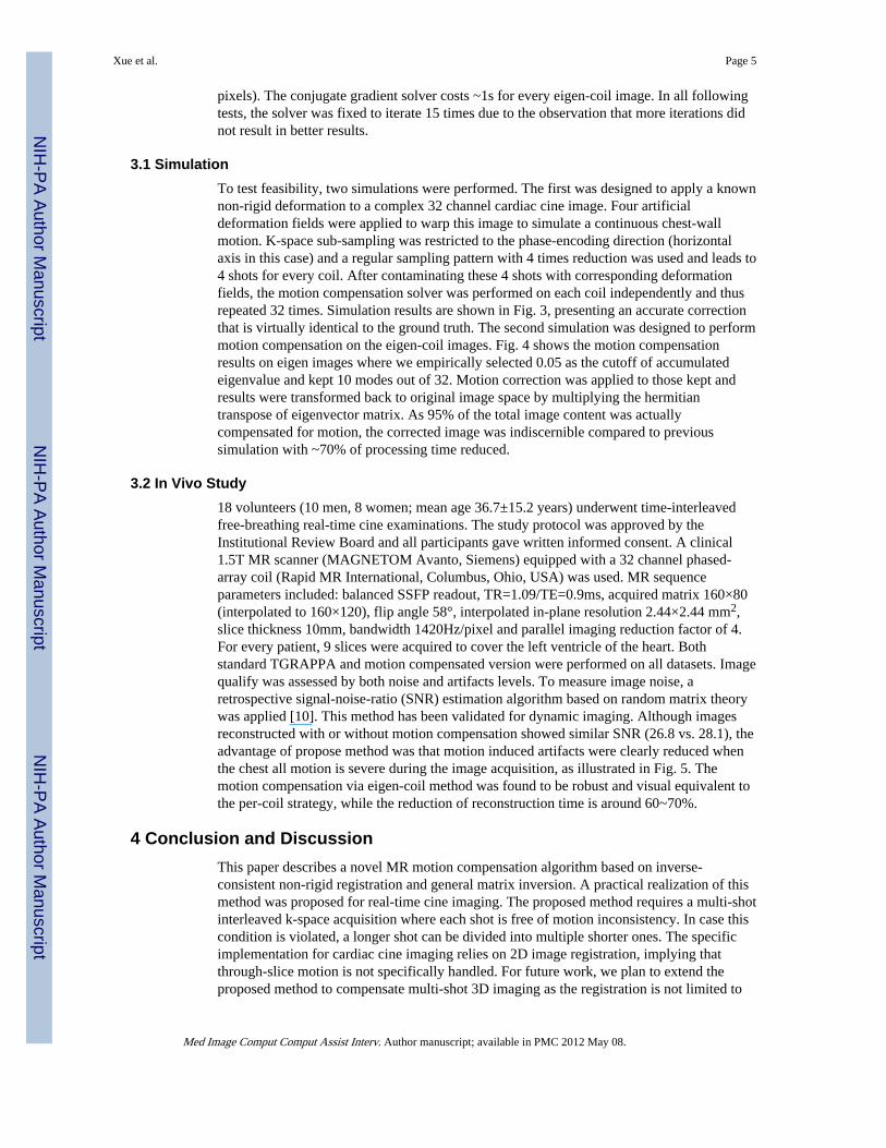
3.2 In Vivo Study18 volunteers (10 men, 8 women; mean age 36.7±15.2 years) underwent time-interleavedfree-breathing real-time cine examinations. The study protocol was approved by theInstitutional Review Board and all participants gave written informed consent. A clinical1.5T MR scanner (MAGNETOM Avanto, Siemens) equipped with a 32 channel phased-array coil (Rapid MR International, Columbus, Ohio, USA) was used. MR sequenceparameters included: balanced SSFP readout, TR=1.09/TE=0.9ms, acquired matrix 160×80(interpolated to 160×120), flip angle 58°, interpolated in-plane resolution 2.44×2.44 mm2,slice thickness 10mm, bandwidth 1420Hz/pixel and parallel imaging reduction factor of 4.For every patient, 9 slices were acquired to cover the left ventricle of the heart. Bothstandard TGRAPPA and motion compensated version were performed on all datasets. Imagequalify was assessed by both noise and artifacts levels. To measure image noise, aretrospective signal-noise-ratio (SNR) estimation algorithm based on random matrix theorywas applied [10]. This method has been validated for dynamic imaging. Although imagesreconstructed with or without motion compensation showed similar SNR (26.8 vs. 28.1), theadvantage of propose method was that motion induced artifacts were clearly reduced whenthe chest all motion is severe during the image acquisition, as illustrated in Fig. 5. Themotion compensation via eigen-coil method was found to be robust and visual equivalent tothe per-coil strategy, while the reduction of reconstruction time is around 60~70%.
4 Conclusion and DiscussionThis paper describes a novel MR motion compensation algorithm based on inverse-consistent non-rigid registration and general matrix inversion. A practical realization of thismethod was proposed for real-time cine imaging. The proposed method requires a multi-shotinterleaved k-space acquisition where each shot is free of motion inconsistency. In case thiscondition is violated, a longer shot can be divided into multiple shorter ones. The specificimplementation for cardiac cine imaging relies on 2D image registration, implying thatthrough-slice motion is not specifically handled. For future work, we plan to extend theproposed method to compensate multi-shot 3D imaging as the registration is not limited to
Xue et al. Page 5
Med Image Comput Comput Assist Interv. Author manuscript; available in PMC 2012 May 08.
NIH
-PA Author Manuscript
NIH
-PA Author Manuscript
NIH
-PA Author Manuscript
2D. Further validation studies are being pursued with emphasis on the clinical benefits ofour technique.
AcknowledgmentsAuthors would like to thank Dr Philip Batchlor (King’s College London) for discussion and providing generalmatrix inversion program.
References1. Atkinson, D. Sunrise Course: Image Reconstruction. ISMRM; 2011. Motion Correction.
2. Batchelor PG, Atkinson D, Irarrazaval P, Hill D, Hajnal J, Larkman D. Matrix description of generalmotion correction applied to multishot images. MRM. 2005; 54(5):1273–1280.
3. Odille F, Vuissoz P, Marie P, Felblinger J. Generalized Reconstruction by Inversion of CoupledSystems (GRICS) applied to free-breathing MRI. MRM. 2008; 60(1):146–157.
4. Christensen GE, Johnson HJ. Consistent image registration. TMI. 2001; 20(7):568–582.
5. Hernandez M, Bossa M, Olmos S. Registration of anatomical images using paths ofdiffeomorphisms parameterized with stationary vector field flows. IJCV. 2009; 85:291–306.
6. Guetter, C.; Xue, H.; Chefd’hotel, C.; Guehring, J. IEEE ISBI. 2011. Efficient symmetric andinverse-consistent deformable registration through interleaved optimization.
7. Jolliffe, I. Principal component analysis. Springer; New York: 2002.
8. Breuer F, Kellman P, Griswold M, Jakob P. Dynamic Autocalibrated parallel imaging usingTemporal GRAPPA (TGRAPPA). MRM. 2005; 53:981–985.
9. Kellman P, Epstein F, McVeigh E. Adaptive sensitivity encoding incorporating temporal filtering(TSENSE). MRM. 2001; 45(5):846–852.
10. Ding Y, Chung Y, Simonetti OP. A method to assess spatially variant noise in dynamic MR imageseries. MRM. 2010; 63:782–789.
Xue et al. Page 6
Med Image Comput Comput Assist Interv. Author manuscript; available in PMC 2012 May 08.
NIH
-PA Author Manuscript
NIH
-PA Author Manuscript
NIH
-PA Author Manuscript
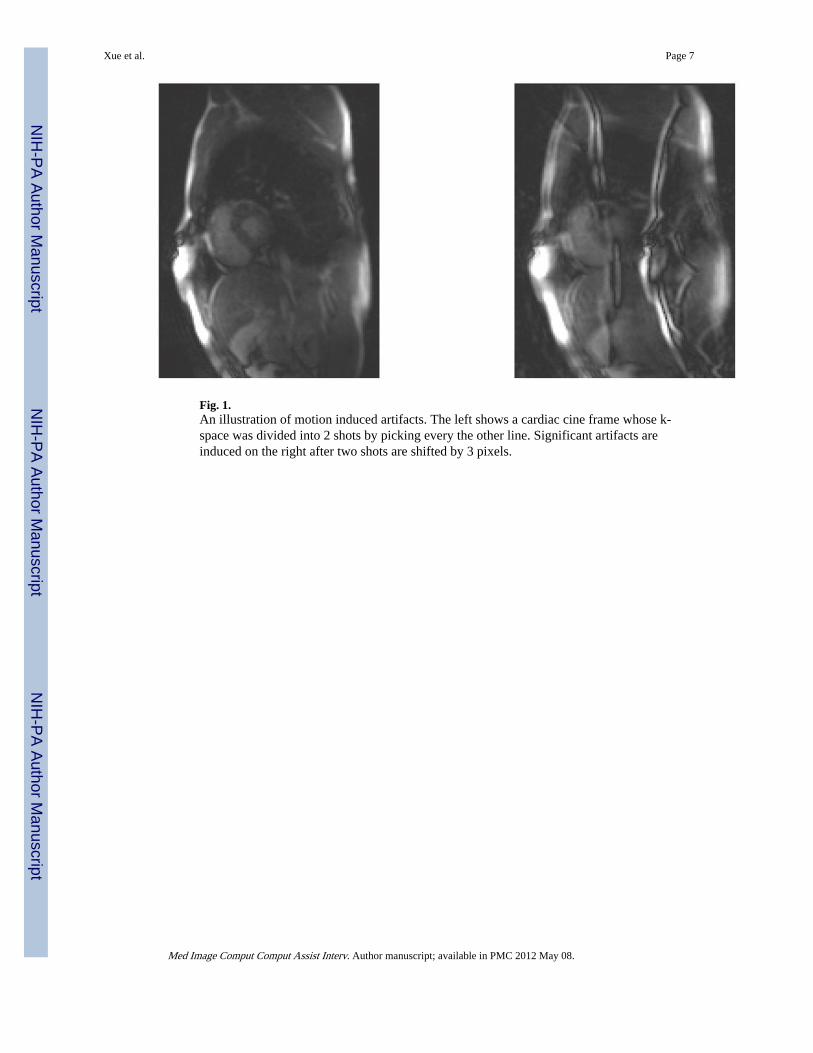
Fig. 1.An illustration of motion induced artifacts. The left shows a cardiac cine frame whose k-space was divided into 2 shots by picking every the other line. Significant artifacts areinduced on the right after two shots are shifted by 3 pixels.
Xue et al. Page 7
Med Image Comput Comput Assist Interv. Author manuscript; available in PMC 2012 May 08.
NIH
-PA Author Manuscript
NIH
-PA Author Manuscript
NIH
-PA Author Manuscript

Fig. 2.Motion compensation reconstruction for time-interleaved real-time cine imaging
Xue et al. Page 8
Med Image Comput Comput Assist Interv. Author manuscript; available in PMC 2012 May 08.
NIH
-PA Author Manuscript
NIH
-PA Author Manuscript
NIH
-PA Author Manuscript

Fig. 3.Simulation of non-rigid motion compensation for multi-coil complex image. K-space wasdivided into 4 shots and deformation fields were applied to mimic the chest wall motion. (a)Original image of the first coil; (b) Ghosting image of first coil due to non-rigid chest wallmotion. (c) After motion compensation, the image content is completely recovered. (e–g)Sum-of-square original image, its ghosting version and recovered result. (d,h) Effect of chestwall motion.
Xue et al. Page 9
Med Image Comput Comput Assist Interv. Author manuscript; available in PMC 2012 May 08.
NIH
-PA Author Manuscript
NIH
-PA Author Manuscript
NIH
-PA Author Manuscript

Fig. 4.Simulation of motion compensation using eigen-coil. (a) Ghosting artifacts appear on everycoil; (b) All eigen modes bear ghosting artifacts, but only those corresponding to largeeigenvalues need correction. (c) Eigen images after correction show good artifact removal.(d) Sum-of-square image after correcting 10 modes and leaving other 22 unchanged.Difference compared to Fig. 3(f) where all coils are processed is indiscernible with ~70%time-saving. The curve shows the accumulated eigen-values for all 32 channels.
Xue et al. Page 10
Med Image Comput Comput Assist Interv. Author manuscript; available in PMC 2012 May 08.
NIH
-PA Author Manuscript
NIH
-PA Author Manuscript
NIH
-PA Author Manuscript

Fig. 5.Example images generated using three different methods to estimate ACS signals forTGRAPPA reconstruction. (a,d) Averaging all undersampled k-space; (b,e) Movingaveraging every 4 consecutive frames; (c,f) Moving averaging every 4 frames with motioncompensation, where artifacts induced by chest wall motion were better suppressed.
Xue et al. Page 11
Med Image Comput Comput Assist Interv. Author manuscript; available in PMC 2012 May 08.
NIH
-PA Author Manuscript
NIH
-PA Author Manuscript
NIH
-PA Author Manuscript
Copyright © 2022 FDOKUMEN