Morphological, molecular and MALDI-TOF MS identification of ...
20
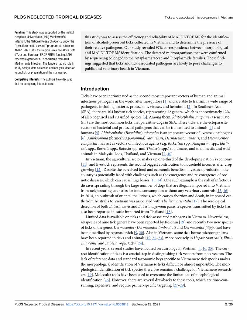
RESEARCH ARTICLE Morphological, molecular and MALDI-TOF MS identification of ticks and tick-associated pathogens in Vietnam Ly Na Huynh 1,2,3 , Adama Zan Diarra 1,2 , Quang Luan Pham 3 , Nhiem Le-Viet 4 , Jean- Michel Berenger 1,2 , Van Hoang Ho 3 , Xuan Quang Nguyen 3 , Philippe Parola ID 1,2 * 1 Aix Marseille Univ, IRD, AP-HM, SSA, VITROME, Marseille, France, 2 IHU-Me ´ diterrane ´ e Infection, Marseille, France, 3 Institute of Malariology, Parasitology and Entomology, Quy Nhon (IMPE-QN), Vietnam, 4 School of Medicine and Pharmacy, The University of Da Nang (UD), Da Nang, Vietnam * [email protected] Abstract Matrix-assisted laser desorption/ionization time-of-flight mass spectrometry (MALDI-TOF MS) has been reported as a promising and reliable tool for arthropod identification, including the identification of alcohol-preserved ticks based on extracted leg protein spectra. In this study, the legs of 361 ticks collected in Vietnam, including 251 Rhiphicephalus sanguineus s.l, 99 Rhipicephalus (Boophilus) microplus, two Amblyomma varanensis, seven Derma- centor auratus, one Dermacentor compactus and one Amblyomma sp. were submitted for MALDI-TOF MS analyses. Spectral analysis showed intra-species reproducibility and inter- species specificity and the spectra of 329 (91%) specimens were of excellent quality. The blind test of 310 spectra remaining after updating the database with 19 spectra revealed that all were correctly identified with log score values (LSV) ranging from 1.7 to 2.396 with a mean of 1.982 ± 0.142 and a median of 1.971. The DNA of several microorganisms includ- ing Anaplasma platys, Anaplasma phagocytophilum, Anaplasma marginale, Ehrlichia rus- tica, Babesia vogeli, Theileria sinensis, and Theileria orientalis were detected in 25 ticks. Co-infection by A. phagocytophilum and T. sinensis was found in one Rh.(B) microplus. Author summary Ticks are one of the important vectors and reservoirs of multiple pathogens infecting humans and animals such as bacteria, protozoans, viruses, and helminths. Nevertheless, studies on ticks and tick-borne infections remain limited in Vietnam. That said, serologi- cal and molecular evidence of tick infections in animals and humans have been reported on several occasions in Vietnam and Southeast Asia in recent decades. The identification of ticks and tick-associated diseases has an important role to play in epidemiological investigation and in assessing the risks of disease transmission to humans and animals. Recently, MALDI-TOF MS has been used as an innovative tool for the rapid and accurate identification of alcohol-preserved ticks based on proteins from extracted legs. This proce- dure represents a time-cost saving and does not require expert knowledge. This goal of PLOS NEGLECTED TROPICAL DISEASES PLOS Neglected Tropical Diseases | https://doi.org/10.1371/journal.pntd.0009813 September 28, 2021 1 / 20 a1111111111 a1111111111 a1111111111 a1111111111 a1111111111 OPEN ACCESS Citation: Huynh LN, Diarra AZ, Pham QL, Le-Viet N, Berenger J-M, Ho VH, et al. (2021) Morphological, molecular and MALDI-TOF MS identification of ticks and tick-associated pathogens in Vietnam. PLoS Negl Trop Dis 15(9): e0009813. https://doi.org/10.1371/journal. pntd.0009813 Editor: Lynn Soong, University of Texas Medical Branch, UNITED STATES Received: May 11, 2021 Accepted: September 13, 2021 Published: September 28, 2021 Peer Review History: PLOS recognizes the benefits of transparency in the peer review process; therefore, we enable the publication of all of the content of peer review and author responses alongside final, published articles. The editorial history of this article is available here: https://doi.org/10.1371/journal.pntd.0009813 Copyright: © 2021 Huynh et al. This is an open access article distributed under the terms of the Creative Commons Attribution License, which permits unrestricted use, distribution, and reproduction in any medium, provided the original author and source are credited. Data Availability Statement: All relevant data are within the manuscript and its Supporting Information files.
-
Upload
khangminh22 -
Category
Documents
-
view
3 -
download
0
Transcript of Morphological, molecular and MALDI-TOF MS identification of ...

RESEARCH ARTICLE
Morphological, molecular and MALDI-TOF MS
identification of ticks and tick-associated
pathogens in Vietnam
Ly Na Huynh1,2,3, Adama Zan Diarra1,2, Quang Luan Pham3, Nhiem Le-Viet4, Jean-
Michel Berenger1,2, Van Hoang Ho3, Xuan Quang Nguyen3, Philippe ParolaID1,2*
1 Aix Marseille Univ, IRD, AP-HM, SSA, VITROME, Marseille, France, 2 IHU-Mediterranee Infection,
Marseille, France, 3 Institute of Malariology, Parasitology and Entomology, Quy Nhon (IMPE-QN), Vietnam,
4 School of Medicine and Pharmacy, The University of Da Nang (UD), Da Nang, Vietnam
Abstract
Matrix-assisted laser desorption/ionization time-of-flight mass spectrometry (MALDI-TOF
MS) has been reported as a promising and reliable tool for arthropod identification, including
the identification of alcohol-preserved ticks based on extracted leg protein spectra. In this
study, the legs of 361 ticks collected in Vietnam, including 251 Rhiphicephalus sanguineus
s.l, 99 Rhipicephalus (Boophilus) microplus, two Amblyomma varanensis, seven Derma-
centor auratus, one Dermacentor compactus and one Amblyomma sp. were submitted for
MALDI-TOF MS analyses. Spectral analysis showed intra-species reproducibility and inter-
species specificity and the spectra of 329 (91%) specimens were of excellent quality. The
blind test of 310 spectra remaining after updating the database with 19 spectra revealed that
all were correctly identified with log score values (LSV) ranging from 1.7 to 2.396 with a
mean of 1.982 ± 0.142 and a median of 1.971. The DNA of several microorganisms includ-
ing Anaplasma platys, Anaplasma phagocytophilum, Anaplasma marginale, Ehrlichia rus-
tica, Babesia vogeli, Theileria sinensis, and Theileria orientalis were detected in 25 ticks.
Co-infection by A. phagocytophilum and T. sinensis was found in one Rh. (B) microplus.
Author summary
Ticks are one of the important vectors and reservoirs of multiple pathogens infecting
humans and animals such as bacteria, protozoans, viruses, and helminths. Nevertheless,
studies on ticks and tick-borne infections remain limited in Vietnam. That said, serologi-
cal and molecular evidence of tick infections in animals and humans have been reported
on several occasions in Vietnam and Southeast Asia in recent decades. The identification
of ticks and tick-associated diseases has an important role to play in epidemiological
investigation and in assessing the risks of disease transmission to humans and animals.
Recently, MALDI-TOF MS has been used as an innovative tool for the rapid and accurate
identification of alcohol-preserved ticks based on proteins from extracted legs. This proce-
dure represents a time-cost saving and does not require expert knowledge. This goal of
PLOS NEGLECTED TROPICAL DISEASES
PLOS Neglected Tropical Diseases | https://doi.org/10.1371/journal.pntd.0009813 September 28, 2021 1 / 20
a1111111111
a1111111111
a1111111111
a1111111111
a1111111111
OPEN ACCESS
Citation: Huynh LN, Diarra AZ, Pham QL, Le-Viet
N, Berenger J-M, Ho VH, et al. (2021)
Morphological, molecular and MALDI-TOF MS
identification of ticks and tick-associated
pathogens in Vietnam. PLoS Negl Trop Dis 15(9):
e0009813. https://doi.org/10.1371/journal.
pntd.0009813
Editor: Lynn Soong, University of Texas Medical
Branch, UNITED STATES
Received: May 11, 2021
Accepted: September 13, 2021
Published: September 28, 2021
Peer Review History: PLOS recognizes the
benefits of transparency in the peer review
process; therefore, we enable the publication of
all of the content of peer review and author
responses alongside final, published articles. The
editorial history of this article is available here:
https://doi.org/10.1371/journal.pntd.0009813
Copyright: © 2021 Huynh et al. This is an open
access article distributed under the terms of the
Creative Commons Attribution License, which
permits unrestricted use, distribution, and
reproduction in any medium, provided the original
author and source are credited.
Data Availability Statement: All relevant data are
within the manuscript and its Supporting
Information files.
this study was to assess the efficiency and reliability of MALDI-TOF MS for the identifica-
tion of alcohol-preserved ticks collected in Vietnam and to determine the presence of
their relative pathogens. Our study revealed 97% correspondence between morphological
and MALDI-TOF MS identification. The detected microorganisms that were confirmed
by sequencing belonged to the Anaplasmataceae and Piroplasmida families. These find-
ings suggested that ticks and tick-associated pathogens are likely to pose challenges to
public and veterinary health in Vietnam.
Introduction
Ticks have been incriminated as the second most important vectors of human and animal
infectious pathogens in the world after mosquitoes [1] and are able to transmit a wide range of
pathogens, including bacteria, protozoans, viruses, and helminths [2]. In Southeast Asia
(SEA), there are 104 known tick species, representing 12 genera, which is approximately 12%
of all recognised and classified species [3]. Among them, Rhipicephalus sanguineus sensu lato
(s.l.) are the most common ticks that parasitise dogs in SEA. These ticks are the ectoparasite
vectors of bacterial and protozoal pathogens that can be transmitted to animals [4] and
humans [5]. Rhipicephalus (Boophilus) microplus is an important vector of livestock pathogens
[6]. Amblyomma (formerly Aponomma) varanensis, Dermacentor auratus, and Dermacentorcompactus may act as vectors of infectious agents (e.g. Rickettsia spp., Anaplasma spp., Ehrli-chia spp., Borrelia spp., Babesia spp. and Theileria spp.) to humans, and to domestic and wild
animals in Malaysia, Laos, Thailand, and Vietnam [7–10].
In Vietnam, the agricultural sector makes up one-third of the developing nation’s economy
[11], and livestock represents the second biggest contribution to household incomes after crop
growing [12]. Despite the perceived food and economic benefits of livestock production, the
country is potentially faced with challenges such as the emergence and re-emergence of zoo-
notic diseases, which can cause huge losses [13, 14]. One such example is the risk of infectious
diseases spreading through the large number of dogs that are illegally imported into Vietnam
from neighbouring countries for food consumption without any veterinary controls [15, 16].
In 2014, an outbreak of oriental theileriosis, which causes abortion and death, in imported cat-
tle from Australia to Vietnam was associated with Theileria orientalis [17]. The serological
detection of both Babesia bovis and Babesia bigemina parasite species transmitted by ticks has
also been reported in cattle imported from Thailand [18].
Limited data is available on ticks and tick-associated pathogens in Vietnam. Nevertheless,
48 species of nine tick genera have been reported by Kolonin [19] and recently two new species
of ticks of the genus Dermacentor (Dermacentor limbooliati and Dermacentor filippovae) have
been described by Apanaskevich [9, 20]. Also in Vietnam, some tick-borne microorganisms
have been reported in ticks and animals [19, 21–23], more precisely in Hepatozoon canis, Ehrli-chia canis, and Babesia vogeli ticks [24].
In recent years, several studies have focused on acarology in Vietnam [4, 10, 25]. The cor-
rect identification of ticks is a crucial step in distinguishing tick vectors from non-vectors. The
lack of reference data and standard taxonomic keys specific to Vietnamese tick species makes
the morphological identification of Vietnamese ticks difficult or almost impossible. The mor-
phological identification of tick species therefore remains a challenge for Vietnamese research-
ers [19]. Molecular tools have been used to overcome the limitations of morphological
identification [26]. However, there are several drawbacks to these tools, which are time-con-
suming, expensive, and require primer-specific targeting [27–29].
PLOS NEGLECTED TROPICAL DISEASES Ticks and associated microorganisms in Vietnam
PLOS Neglected Tropical Diseases | https://doi.org/10.1371/journal.pntd.0009813 September 28, 2021 2 / 20
Funding: This study was supported by the Institut
Hospitalo-Universitaire (IHU) Mediterranee
Infection, the National Research Agency under the
“Investissements d’avenir” programme, reference
ANR-10-IAHU-03, the Region Provence Alpes Cote
d’Azur and European ERDF PRIMI funding. LNH
received a grant of PhD scholarship from IHU
Mediterranee Infection. The funders had no role in
study design, data collection and analysis, decision
to publish, or preparation of the manuscript.
Competing interests: The authors have declared
that no competing interests exist.

Recently, the MALDI-TOF MS method has been proposed as an alternative and innovative
tool to overcome the limitations of the above two methods in arthropod identification [30].
Since then, studies in several laboratories have demonstrated that MALDI-TOF MS is a
remarkably robust tool for identifying many species of arthropod vectors and non-vectors
[30]. The aim of this study was to identify tick species collected from domestic and wild ani-
mals in Vietnam and their associated pathogens using morphological, MALDI-TOF MS and
molecular tools.
Materials and methods
Ethics statement
Ethical approval was obtained from the Institute of Malariology, Parasitology, and Entomol-
ogy, Quy Nhon (IMPE-QN) on behalf of the Vietnamese Ministry of Health (approval no:
401/VSR-CT-2010, 333/CT-VSR-2018). Permission was obtained from the communal author-
ities for wild animals that were not listed in the Red Data Book of Vietnam, and agreement
was obtained from the owners of cows, goats, and dogs.
Tick collection and morphological identification
Ticks were collected in four provinces: Quynh Luu (19o13’ N; 105o60’ E) District, Nghe An
Province; Nam Giang (15o65’ N; 107o50’ E) District, Quang Nam Province; Van Canh (13o37’
N; 108o59’ E) District, Binh Dinh Province; and Khanh Vinh (12o16’ N; 108o53’ E) District,
Khanh Hoa Province in Vietnam in September 2010, between April and September 2018. The
map of Vietnam showing the collection sites was made with QGIS version 3.10 and the Viet-
namese layers were downloaded from DIVA-GIS at the following link: https://www.diva-gis.
org/datadown (Fig 1A). All engorged and non-engorged ticks were collected from the skin of
domestic animals (cows, goats, and dogs) and wild animals (pangolins, wild pigs) using for-
ceps. Ticks from wild animals were collected in a collaborative manner by rangers and trained
care personnel from the Wildlife Rescue, Conservation and Development Center. Ticks were
morphologically identified first at species level using dichotomous keys [9, 31] by an entomo-
logical team from the Institute of Malariology, Parasitology and Entomology, Quy Nhon,
Fig 1. Map of Vietnam showing tick collection sites realised with QGIS version 3.10, the layers have been uploaded to
the DIVA-GIS website: https://www.diva-gis.org/datadown (A); Morphologically, the 70% alcohol tick-preserved
species were collected in Vietnam over a period of 10 years: Amblyomma varanensis [♀: a, b]; Amblyomma sp. [♀: c,
d]; Dermacentor auratus [♂: e, f]; Dermacentor compactus [♂: g, h]; and approximately 2 years: Rhipicephalus (B)
microplus [♀: I, k]; Rhipicephalus sanguineus s.l [♂: l, m] (B).
https://doi.org/10.1371/journal.pntd.0009813.g001
PLOS NEGLECTED TROPICAL DISEASES Ticks and associated microorganisms in Vietnam
PLOS Neglected Tropical Diseases | https://doi.org/10.1371/journal.pntd.0009813 September 28, 2021 3 / 20

Vietnam (IMPE-QN). Ticks from the same host were counted and placed in the same tube
containing 70% v/v alcohol, before being sent to the Institut Hospitalo-Universitaire (IHU)
Mediterranee Infection in Marseille, France for MALDI-TOF MS and molecular analysis. In
Marseille, the morphological identification of ticks was verified by two specialists in morpho-
logical identification of ticks using a magnifying glass (Zeiss Axio Zoom.V16, Zeiss, Marly le
Roi, France) and dichotomous keys. Morphological identification was carried out only if all
discriminating characters had been observed.
Tick dissection and sample preparation
Ticks were individually removed from the alcohol and were rinsed and dissected with a sterile
surgical blade, as previously described [32]. The four legs of each tick and the half part without
legs were submitted for MALDI-TOF MS and molecular biology analysis, respectively. The
remaining parts with legs were frozen and stored as samples for any further research.
DNA extraction and molecular identification of ticks
DNA from each half-tick or legs (for ticks from which we did not obtain sequences with half-
tick DNA) was individually extracted using an EZ1 DNA tissue kit (Qiagen), according to the
manufacturer’s recommendations, as previously described [33]. DNA was monitored with
Nanodrop 1000 Spectrophotometer (Thermo Fisher Scientific, Wilmington, USA) and either
immediately used or stored at -20˚C until use.
DNA from ticks was submitted to standard PCR in an automated DNA thermal cycle to
amplify a 465-base pair (bp) fragment of the mitochondrial 16S DNA gene, as described previ-
ously [34]. The 12S tick gene, amplifying about 405-bp of the mitochondrial DNA fragment,
was used for all specimens for which we did not have a sequence with the 16S gene. DNA from
Rh. sanguineus s.l., reared in our laboratory, was used as a positive control. Purified PCR prod-
ucts were sequenced as previously described [34]. The obtained sequences were assembled and
analysed using the ChromasPro software (version 1.7.7) (Technelysium Pty. Ltd., Tewantin,
Australia), and were then blasted against the reference sequences available in GenBank (http://
blast.ncbi.nlm.nih.gov/).
MALDI-TOF MS analysis
Sample preparation. The four legs of each tick were first put into an Eppendorf tube and
dried overnight at 37˚C and then put into an Eppendorf tube with 40 μL of high-performance
liquid chromatography (HPLC) grade water and incubated overnight at 37˚C. The legs were
then crushed in a mix of 20 μL of 70% (v/v) formic acid (Sigma) and 20 μL of 50% (v/v) aceto-
nitrile (Fluka, Buchs, Switzerland), with glass beads (Sigma, Lyon, France), as described previ-
ously [35]. The crushed legs were centrifuged and 1 μL of the supernatant of each sample was
deposited in quadruplicate onto a MALDI-TOF MS steel plate (Bruker Daltonics, Wissem-
bourg, France). After drying at room temperature, 1μL of matrix solution composed of a satu-
rated solution of α-cyano-4-hydroxycynnamic acid (Sigma, Lyon, France), 50% acetonitrile
(v/v), 2. 5% trifluoroacetic acid (v/v) (Aldrich, Dorset, United Kingdom), and high perfor-
mance liquid chromatography (HPLC) grade water was added [36]. The target plate was air-
dried one more at room temperature before being introduced into the Microflex LT MALDI-
TOF Mass Spectrometer (Bruker Daltonics, Germany) for analysis. The quality of the matrix,
sample loading, and performance of the MALDI-TOF MS device were controlled using the
legs of a Rh. sanguineus s.l. reared in our laboratory as a positive control.
MALDI-TOF MS parameters, spectral analysis and reference database creation. The
spectral profiles obtained from the tick legs were visualised using a Microflex LT MALDI-TOF
PLOS NEGLECTED TROPICAL DISEASES Ticks and associated microorganisms in Vietnam
PLOS Neglected Tropical Diseases | https://doi.org/10.1371/journal.pntd.0009813 September 28, 2021 4 / 20

mass spectrometer with FlexControl software (version 3.3, Bruker Daltonics). The setting
parameters of the MALDI-TOF MS apparatus were identical to those previously used [32].
The FlexAnalysis v.3.3 software was used to evaluate spectral quality (smoothing, baseline
subtraction, peak intensities). MS spectra reproducibility was assessed by comparing the aver-
age spectral profiles (MSP, main spectrum profile) obtained from the four spots of each tick
leg, according to species, using MALDI-Biotyper v3.0 software (Bruker Daltonics) [37]. MS
spectra reproducibility and specificity were assessed based on a principal component analysis
(PCA) and cluster analysis (MSP dendrogram). PCA was performed using ClinProTools v2.2
with the manufacturer’s default settings. Cluster analysis was performed based on a compari-
son of the MSP given by MALDI-Biotyper v3.0. software with clustering according to protein
mass profile (i.e., their mass signals and intensities) [37].
Based on the morphological identification, eight and seven reference spectra of Rh. sangui-neus and Rh. (B) microplus, respectively, were added to our MALDI-TOF MS database. How-
ever, two, one, and one spectra of D. auratus, Am. varanensis, D. compactus, respectively,
which were only identified morphologically by three tick identification specialists, were also
added to our MALDI-TOF MS database. To create a database, reference spectra (MSP, Main
Spectrum Profile) were created by combining the results of spectra from specimens of each
species using the automated function of the MALDI-Biotyper v3.0 software (Bruker Dal-
tonics). MSPs were created based on an unbiased algorithm using peak position, intensity, and
frequency data [38]. Four tick species that could not be identified by molecular biology were
temporarily added into the MS reference database to identify the remaining specimens from
the same species.
Blind test for tick identification. A blind test was performed with the remaining tick
specimens not included in our MALDI-TOF MS database after the database had been
upgraded with 19 MS spectra from specimens of the five tick species to determine their identi-
fication. The reliability of tick species identification was estimated using the log score values
(LSVs) obtained from the MALDI-Biotyper software, which ranged from 0 to 3. These LSVs
correspond to the degree of similarity between the MS reference spectra in the database and
those submitted to blind tests. An LSV was obtained for each spectrum of the samples tested.
According to one previous study [37], an LSV of at least 1.8 should be obtained to be consid-
ered reliable for species identification.
Detection of microorganisms. Quantitative PCR (qPCR) was performed for screening
microorganisms using specific primers and probes targeting Anaplasmataceae, Piroplasmida,
Borrelia spp., Bartonella spp., Coxiella burnetii, and Rickettsia spp. PCR reactions were per-
formed according to the manufacturer’s instructions, using a CFX96 Touch detection system
(Bio-Rad). qPCR amplification was performed using the thermal profile described previously
[39]. The DNA of Rickettsia montanensis, Bartonella elizabethae, Anaplasma phagocytophilum,
Coxiella burnetii, Borrelia crocidurae, and Babesia vogeli were used as a positive control and
DNA from Rh. sanguineus s.l from our laboratory, which were free of bacteria, were used as
negative controls. The samples were considered to be positive when the cycle threshold (Ct)
was strictly less than 36 [40].
All samples that were positive following qPCR were submitted to standard PCR and
sequencing to identify the microorganism species. For the Rickettsia sp. positive sample, we
first used a primer targeting a 630-bp fragment of the OmpA gene [35] and then another tar-
geting a 401-bp fragment of the gltA gene [33]. Samples which were Anaplasmataceae positive
following qPCR were subjected to amplifying and sequencing of a 520-bp fragment of the 23SrRNA gene [33]. Samples which were Piroplasmidae positive following qPCR were subjected
to amplifying and sequencing of a 969-bp fragment of the 18S rRNA [41]. Samples which were
Borrelia sp. positive following qPCR was subjected to amplifying and sequencing of a 344-bp
PLOS NEGLECTED TROPICAL DISEASES Ticks and associated microorganisms in Vietnam
PLOS Neglected Tropical Diseases | https://doi.org/10.1371/journal.pntd.0009813 September 28, 2021 5 / 20

fragment of the flaB gene [42]. The primers and probes used in this study are listed in Table 1.
The obtained sequences were assembled and analysed using the ChromasPro software (version
1.7.7) (Technelysium Pty. Ltd., Tewantin, Australia), and were then blasted against the refer-
ence sequences available in GenBank (http://blast.ncbi.nlm.nih.gov/). The method used for
phylogenetic tree analysis was the neighbour-joining (NJ) method with 1,000 replicates. DNA
sequences were aligned using MEGA software version 7.0 (https://www.megasoftware.net/).
The various statistical analyses were performed using R software version 3.4 (R Development
Core Team, R Foundation for Statistical Computing, Vienna, Austria) and ggplot packages
were used to perform the graphics.
Results
Tick collection and morphological identification
A total of 1120 ticks including 334 (30%) engorged ticks were collected in four provinces of
Vietnam: Nghe An, Quang Nam, Binh Dinh, and Khanh Hoa. Morphologically, ticks were
identified as belonging to six species (Fig 1A), including 935 (83.5%) Rh. sanguineus s.l. col-
lected from dogs, 174 (15.5%) Rh. (B) microplus) from cows and goats, seven (0.6%) D. auratus
Table 1. Target amplified and used for qPCR and standard PCR.
Microorganisms Targeted sequence Primers (5’-3’) and Probes (Used for qPCR Screening or Sequencing) References
Anaplasmataceae 23S f_TGACAGCGTACCTTTTGCAT
r_GTAACAGGTTCGGTCCTCCA
p_6FAM-GGATTAGACCCGAAACCAAG
[108]
23S (520-bp) f_ATAAGCTGCGGGGAATTGTC
r_TGCAAAAGGTACGCTGTCAC
Piroplasmida 5.8S f_AYYKTYAGCGRTGGATGTC
r_TCGCAGRAGTCTKCAAGTC
p_FAM-TTYGCTGCGTCCTTCATCGTTGT-MGB
[39]
18S (969-bp) f1_GCGAATGGCTCATTAIAACA
f4_CACATCTAAGGAAGGCAGCA
f3_GTAGGGTATTGGCCTACCG�
r4_AGGACTACGACGGTATCTGA�
Rickettsia spp. gltA(RKND03) f_GTGAATGAAAGATTACACTATTTAT
r_GTATCTTAGCAATCATTCTAATAGC
p_6FAM-CTATTATGCTTGCGGCTGTCGGTTC
[109]
gltA (401-bp) f_ATGACCAATGAAAATAATAAT
r_CTTATACTCTCTATGTACA
[110]
OmpA (630-bp) 70_ATGGCGAATATTTCTCCAAAA
701_GTTCCGTTAATGGCAGCATCT
180_GCAGCGATAATGCTGAGTA�
[1]
Borrelia spp. ITS4 f_GGCTTCGGGTCTACCACATCTA
r_CCGGGAGGGGAGTGAAATAG
p_TGCAAAAGGCACGCCATCACC
[111]
flaB (344-bp) f_TGGTATGGGAGTTTCTGG
r_ TAAGCTGACTAATACTAATTACCC
Bartonella spp. ITS2 f_GATGCCGGGGAAGGTTTTC
r_GCCTGGGAGGACTTGAACCT
p_GCGCGCGCTTGATAAGCGTG
[112]
Coxiellia burnetii IS30A f_CGCTGACCTACAGAAATATGTCC
r_GGGGTAAGTAAATAATACCTTCTGG
p_CATGAAGCGATTTATCAATACGTGTATG
[113]
Abbreviation
�, used for sequencing only.
https://doi.org/10.1371/journal.pntd.0009813.t001
PLOS NEGLECTED TROPICAL DISEASES Ticks and associated microorganisms in Vietnam
PLOS Neglected Tropical Diseases | https://doi.org/10.1371/journal.pntd.0009813 September 28, 2021 6 / 20

from pangolins, two (0.2%) Am. varanensis from wild pigs, and one (0.1%) D. compactus and
one (0.1%) Amblyomma sp. from a pangolin (Table 2). Rhipicephalus sanguineus s.l. and Rh.
(B) microplus were collected between April and September 2018. The other ticks were collected
in September 2010. The different specimens that could not be identified by molecular biology
are shown in the pictures in Fig 1B that we took using a magnifying glass (Zeiss Axio Zoom.
V16, Zeiss, Marly le Roi, France).
Molecular identification of ticks
To confirm our morphological identification, 25 tick specimens were submitted to molecular
analysis using the 16S rDNA gene, including eight specimens of Rh. sanguineus s.l., seven Rh.
(B) microplus, seven D. auratus, one Am. varanensis, one D. compactus and one Amblyomma
Table 2. The number of tick species used for MALDI-TOF MS analysis, creation of the MS reference spectra creation, and molecular biology confirmation.
Morphological
identification
Number submitted
for molecular ID�Molecular ID� (%identity;
GenBank accession number)
Number of good
spectra/ tested
Number of spectra
added to DB$MADI-TOF MS ID�
(number identified)
LSVs &
[Low-High]
Rhipicephalussanguineus s.l
8 Rh. sanguineus s.l (99.75–100%;
MG651947, MG793434,
KX632154)
241/251 8 Rh. sanguineus s.l (233) [1.7–2.351]
Rhipicephalus (B)
microplus7 Rh. (B) microplus (100%;
MN880401, MT462222,
EU918187)
78/99 7 Rh. (B) microplus (71) [1.705–
2.346]
Amblyommavaranensis
1 not identified 1/2 1 NA NA
Amblyomma sp. 1 not identified 1/1 0 Am.varanensis (1) 1.857
Dermacentorauratus
7 not identified 7/7 2 D. auratus (5) [1.949–
2.396]
Dermacentorcompactus
1 not identified 1/1 1 NA NA
Total 25 329/361 19 310
�Identification& Range of log score values$ Database.
https://doi.org/10.1371/journal.pntd.0009813.t002
Fig 2. Comparison of MALDI-TOF MS spectra from the legs of six tick species collected in Vietnam. The MS
spectra revealed intra-species reproducibility and inter-species specificity (A); The MS spectra were compared by
Principal Component Analysis (B); a.u., arbitrary units; m/z, mass-to-charge ratio.
https://doi.org/10.1371/journal.pntd.0009813.g002
PLOS NEGLECTED TROPICAL DISEASES Ticks and associated microorganisms in Vietnam
PLOS Neglected Tropical Diseases | https://doi.org/10.1371/journal.pntd.0009813 September 28, 2021 7 / 20

sp. Sequences were obtained only for the specimens of Rh. sanguineus s.l. and Rh. (B) micro-plus. BLAST analysis indicated that obtained sequences from Rh. sanguineus s.l. were 99.75 to
100% identical to the corresponding sequences of Rh. sanguineus s.l. (Genbank: MG651947,
MG793434, KX632154) and those obtained from Rh. (B) microplus were 100% identical to the
corresponding sequences of Rh. (B) microplus (Genbank: MN880401, MT462222, EU918187).
Unfortunately, for the specimens morphologically identified as D. auratus, Am. varanensis,Amblyomma sp. and D. compactus, we were unable to amplify any DNA from the half-tick or
legs of these tick species with PCR targeting part of the two genes (16S and 12S rDNA), despite
the fact that the nanodrop had indicated that the amount of DNA contained in these samples
was 7.8 to 19.4 ng/μl.
MS reference spectra analysis
The legs of 361 specimens, including 251 morphologically identified as Rh. sanguineus s.l., 99
Rh. (B) microplus, seven D. auratus, two Am. varanensis, one Amblyomma sp. and one D. com-pactus were randomly selected and subjected to MALDI-TOF MS analysis. Visualisation of
MS spectra from all specimens using FlexAnalysis v.3.3 software showed that 91% (329) of
specimens had excellent quality spectra (peak intensity > 3,000 a.u., no background noise and
baseline subtraction correct) (Figs 2A and S1 and Table 2). The MS spectra of different speci-
mens showed intra-species reproducibility and inter-species specificity, as confirmed by PCA
(Figs 2B and 3B) and dendrogram (Fig 3A) analysis. PCA and dendrogram analysis showed
that all specimens of the same species were grouped together or were on the same branches.
Additionally, at the genus level, all specimens from the same genus were also gathered in the
same part of dendrogram (Fig 3A).
MALDI-TOF MS tick identification by blind test
The 310 MS remaining spectra of excellent quality, including 233 Rh. sanguineus s.l, 71 Rh. (B)
microplus, five D. auratus and one Amblyomma sp. were queried against our reference spectra
database upgraded with eight Rh. sanguineus s.l. and seven Rh. (B) microplus which were mor-
phologically and molecularly identified, and two D. auratus, one Am. varanensis and one
D. compactus identified only morphologically. The spectra of the ticks introduced in the
Fig 3. Comparison of MALDI-TOF MS spectra from the legs of six alcohol-preserved tick species collected in
Vietnam and stored for different periods of time. The dendrogram was built using between one and eight
representative MS spectra from six distinct tick species (A). The MS spectra of different specimens showed intra-
species reproducibility and inter-species specificity as confirmed by PCA (B).
https://doi.org/10.1371/journal.pntd.0009813.g003
PLOS NEGLECTED TROPICAL DISEASES Ticks and associated microorganisms in Vietnam
PLOS Neglected Tropical Diseases | https://doi.org/10.1371/journal.pntd.0009813 September 28, 2021 8 / 20

MALDI-TOF MS database have been deposited on the website of the University Hospital
Institute (UHI) under the following DOI: https://doi.org/10.35088/rbqp-g648. The blind test
revealed that 100% (233) of Rh. sanguineus s.l. specimens were correctly identified as Rh. san-guineus s.l. with LSVs ranging from 1.7–2.351 with a mean of 1.976 ± 0.137, 100% (71) of Rh.
(B) microplus identified with LSVs ranging from 1.705–2.346 with a mean of 1.989 ± 0.148 and
100% (five) D. auratus with LSVs of 1.949–2.396 with a mean of 2.164 ± 0.149 (Table 2). The
tick identified morphologically as Amblyomma sp. was identified by MALDI-TOF MS as Am.
varanensis (LSV = 1.857) (Table 2). All our specimens were identified with LSVs ranging from
1.7–2.396 with a mean of 1.982 ± 0.142 and a median of 1.971, and 97% (301) had LSVs >1.8,
which is considered the threshold for identification (Fig 3B). No blind test was performed for
D. compactus because of the low number of specimens.
Detection of microorganisms in ticks
A total of 361 ticks, including 260 (72%) non-engorged and 101 (28%) engorged ticks, were
examined for the DNA of six microorganisms using qPCR. Thirty-nine (10.8%) were positive
for at least one of the microorganisms, including Anaplasmataceae, Rickettsia spp, Borreliaspp. and Piroplasmida (Table 3). Notably, two Rh. (B) microplus specimens were co-infected
with both Anaplasmataceae and Piroplasmida. No samples were positive for C. burnetii or Bar-tonella spp.
DNA from bacteria of the Anaplasmataceae family were detected in 18/361 (5%) of ticks by
qPCR. The DNA of bacteria belonging to the Anaplasmataceae family was found in 13 (72%)
Rh. (B) microplus and five (28%) Rh. sanguineus s.l. We successfully obtained seven (40%)
sequences all from Rh. (B) microplus by standard PCR and sequencing using the 23S Anaplas-
mataceae gene amplifying a 520-pb fragment of rRNA (Table 3). A BLAST analysis showed
that four of the sequences obtained were 100% identical to the corresponding sequence of
Anaplasma marginale (Genbank: CP023731), one of sequences obtained was 100% identical to
the corresponding sequence of Ehrlichia rustica (Genbank: KT364330), one was 99.13% identi-
cal to the corresponding sequence of Anaplasma phagocytophilum (Genbank: CP015376) and
one was 100% identical to the corresponding sequence of Anaplasma platys (Genbank:
CP046391).
DNA of Piroplasmida was detected in 19/361 (5.3%) of ticks by qPCR using the 5.8S rRNA
gene. Of these, ten (53%) were found in Rh. sanguineus s.l. and nine (47%) were found in Rh.
Table 3. Microorganisms detected using molecular biology tools in ticks collected in Vietnam.
Microorganisms tested Tick species
Rh. sanguineus Rh. (Bo) microplus Amblyomma sp. Total
Anaplasmataceae 2% (5/251) 13.1% (13/99) - 5% (18/361)
Anaplasma phagocytophilum - 1% (1/99) - 0.3% (1/361)
Anaplasma platys 0.4% (1/251) - - 0.3% (1/361)
Anaplasma marginale 1.2% (3/251) 1% (1/99) - 1.1% (4/361)
Ehrlichia rustica - 1% (1/99) - 0.3% (1/361)
Piroplasmida 4% (10/251) 10.1% (9/99) - 5.3% (19/361)
Babesia vogeli 3.6% (9/251) - - 2.5% (9/361)
Theileria sinensis - 6.1% (6/99) - 1.7% 96/361)
Theileria orientalis - 3% (3/99) - 0.8% (3/361)
Rickettsia sp. - - 100% (1/1) 0.3% (1/361)
Borrelia sp. 0.4% (1/251) - - 0.3% (1/361)
https://doi.org/10.1371/journal.pntd.0009813.t003
PLOS NEGLECTED TROPICAL DISEASES Ticks and associated microorganisms in Vietnam
PLOS Neglected Tropical Diseases | https://doi.org/10.1371/journal.pntd.0009813 September 28, 2021 9 / 20

(B) microplus. We successfully obtained 18 (95%) sequences by standard PCR and sequencing
using the 18S rRNA gene amplifying a 969-pb fragment of rRNA. The BLAST analysis of nine
sequences obtained from Rh. sanguineus s.l. revealed that they were between 99.75% and 100%
identical to the corresponding sequence of Babesia vogeli (GenBank: MN067709), six
sequences obtained from Rh. (B) microplus were between 99.82% and 100% identical to the
corresponding sequences of Theileria sinensis (GenBank: KF559355, MT271911, AB000270)
and three sequences obtained from Rh. (B) microplus were between 99.88 and 100% identical
to the corresponding sequences of Theileria orientalis (GenBank: MG599099) (Table 3).
Rickettsia and Borrelia sp. were detected by qPCR in one tick of Amblyomma sp. and one of
Rh. sanguineus s.l., respectively. However, all the standard PCR procedures for the identifica-
tion of Rickettsia and Borrelia species failed. Of the 25 ticks for which we obtained sequences
of microorganisms, 16 (64%) came from engorged ticks and one tick (4%) was co-infected
with A. phagocytophilum and T. sinensis. The species of microorganism, the species of tick and
the state of engorgement of the ticks in which the microorganisms were detected are listed in
S1 Table.
Two phylogenetic trees of Anaplasmataceae and Piroplasmida were built from the 23SrRNA and 18S rRNA genes sequences of our amplicons, respectively. These phylogenetic trees
showed that the microorganisms detected in this study are close to their homologues available
in GenBank (Fig 4A and 4B).
Discussion
The correct identification of tick species and associated pathogens can contribute to improving
vector control efforts adapted to the surveillance and prevention of outbreaks of tick-borne
diseases. In this study, our ticks were identified using traditional methods (morphological)
and then confirmed by molecular methods and MALDI-TOF MS, and the associated patho-
gens were researched using molecular tools. In this study, we combined these three tools to
identify ticks and to search for microorganisms associated with these ticks collected in
Vietnam.
In this study, the morphological identification of ticks collected in Vietnam revealed six
species, including Rh. sanguineus s.l., Rh. (B) microplus, Am. varanensis, Amblyomma sp., D.
Fig 4. 23S rRNA gene-based phylogenetic analysis of strains identified in this study. Phylogenetic tree highlighting
the position of A. phagocytophilum, A. marginale, A. platys, and E. rustica identified in our study are close to their
homologues available in GenBank (A). 18S rRNA gene-based phylogenetic analysis of strains identified in the present
study. Phylogenetic tree highlighting the position of B. vogeli, T. sinensis, and T. orientalis relative to their
correspondence available in GenBank (B).
https://doi.org/10.1371/journal.pntd.0009813.g004
PLOS NEGLECTED TROPICAL DISEASES Ticks and associated microorganisms in Vietnam
PLOS Neglected Tropical Diseases | https://doi.org/10.1371/journal.pntd.0009813 September 28, 2021 10 / 20

auratus and D. compactus. All these species had already been reported in Vietnam [3, 19, 25]
and neighbouring countries including Laos, Malaysia, Cambodia, and Thailand [3, 23, 43].
Among the Rh. sanguineus s.l. were the species most commonly found on dogs in Vietnam.
This tick species is the most widely distributed worldwide and is known to be a vector of sev-
eral pathogens such as Anaplasma, Rickettsia, Ehrlichia, and Babesia spp. [44, 45]. Rhipicepha-lus (Boophilus) microplus was collected from both cows and goats and is responsible for the
transmission of livestock pathogens [6, 24]. There have been several reports of tick-borne live-
stock pathogens such as Anaplasma spp., Ehrlichia ruminantium, Babesia bigemina, Babesiabovis, and Theileria spp. [46–48]. However, this tick rarely bites humans [22]. Other tick spe-
cies were collected from wild animals (pangolins and pigs). Several species of ticks of the genus
Amblyomma have been collected from almost all species of pangolins [49, 50] and are vectors
of Rickettsia, Ehrlichia spp. [51]. Recently, several studies reported Amblyomma javanensedetected from pangolins in Singapore [52] and China [53], and Amblyomma compressum ticks
on pangolins from Congo [54]. Our study is the first to observe Am. varanensis, Amblyommasp. on pangolins from Vietnam. Dermacentor auratus, D. compactus are widely distributed
across Sri Lanka, Bangladesh, India, and SEA including Vietnam [55, 56], and are well known
vectors of Rickettsia, Coxiella burnetii, Borrelia, and Anaplasma spp. [57, 58].
Molecular techniques were used to confirm our morphological identification of tick species
by amplifying a portion sequence of a 465-bp fragment 16S rRNA gene. The choice of the 16SrRNA gene was based on previous studies that reported that this gene was a reliable tool for
tick identification [29, 59]. Interrogating the GenBank database with 16S rDNA sequences
from Rh. sanguineus s.l and Rh. (B) microplus showed similarity with the reference sequences
available in Genbank for these species that were stored in 70% alcohol for approximately two
years. Conversely, we were unable to obtain sequences for all specimens that had been pre-
served for more than 10 years in alcohol (i.e., Am. varanensis, Amblyomma sp., D. auratus, and
D. compactus) with the 16S and 12S rDNA genes. This might be due to the fact that the alcohol
was not completely eliminated during extraction [60] and/or to the fact that these ticks con-
tained blood from their host, which includes several factors that can inhibit the PCR reaction,
as already reported [61].
In this study, MALDI-TOF MS was used to identify ticks collected in Vietnam from domes-
tic and wild animals. Among the spectra of tick legs that were subjected to MS analysis, the cor-
rect identification rates (LSVs>1.8) were 97%, almost identical to the identification rate
reported in other studies [32, 33, 62]. Interestingly, specimens that were not able to be identified
by molecular biology were identified by MALDI-TOF MS. This confirms that the tool is reliable
and accurate for the identification of ticks. Despite these numerous advantages, this technique
is limited by the high cost of the device, although it can be used for clinical microbiology and
mycology in addition to entomology, with no additional cost. Maintenance may be another lim-
itation but this can be compensated for by the low cost of reagents once the device is acquired
[30]. Secondly, the development of protocols, the choice of the arthropod compartment to be
used, the spectra for the creation of the database and, finally, the methods and time of conserva-
tion of the arthropods can influence the performance of MALDI-TOF MS [30, 37, 63].
In this study, 10.8% of the ticks were positive for at least one of the microorganisms by
qPCR, of which 16/25 (64%) of the ticks carrying DNA of microorganisms by sequencing were
engorged ticks. The detection of microorganisms in engorged ticks doesn’t have the same epi-
demiological meaning as when detected in a questing or non-engorged attached tick. Such
ticks may potentially have fed on hosts with bacteraemia, thus biasing the estimate of the actual
rate of tick infestation.
The microorganisms detected in this study and confirmed by sequencing belong to the
Anaplasmataceae family (A. phagocytophilum, A. marginale, A. platys, and E. rustica), which
PLOS NEGLECTED TROPICAL DISEASES Ticks and associated microorganisms in Vietnam
PLOS Neglected Tropical Diseases | https://doi.org/10.1371/journal.pntd.0009813 September 28, 2021 11 / 20
are known aetiologies of zoonotic diseases [8, 13, 64, 65]. The Piroplasmida family (B. vogeli,T. sinensis, and T. orientalis) was mainly known as the potential zoonotic pathogens [66].
Anaplasma marginale is responsible for bovine anaplasmosis and is an intracellular bacte-
rium transmitted by tick species mainly belonging to the Rhipicephalus and Dermacentor gen-
era [67]. The DNA and specific antibodies against A. marginale were previously reported in
the blood of cattle and cows from Vietnam [23, 24]. This study is the first report of A. margin-ale in Rh. (B) microplus and Rh. sanguineus s.l ticks collected in Vietnam. However, A. margin-ale had previously been reported in cattle and cattle Rh. (B) microplus ticks in China [68], the
Philippines [69] which is a neighbouring country to Vietnam, in cattle and cattle ticks in
Malaysia [70], and many African countries [71].
Anaplasma platys, the aetiological agent of infectious canine cyclic thrombocytopenia and
which can be transmitted by Rh. sanguineus s.l., A. platys has been recorded in China [48],
Colombia [72], and detected on various ectoparasites such as Rh. (B) microplus [48] and Hya-lomma dromedarii [73]. Anaplasma platys is one of the most significant tick-borne zoonotic
pathogens [24, 74] and several cases of human infections have been described in Venezuela
[75], Chicago [76], and South Africa [77]. Anaplasma platys has already been detected from
blood specimens of cattle and dogs in Vietnam [24], but it was the first discovery in Rh. sangui-neus s.l. ticks from Vietnam in our study. It had been previously detected in Rh. sanguineus s.l.
in SEA [25], including in the Philippines [78], Thailand, and Malaysia [79, 80].
The pathogen A. phagocytophilum is the causative agent of human granulocytic anaplasmo-
sis (HGA) and tick-borne fever in ruminants [81]. It is rarely found in Rh. (B) microplus and is
known to be transmitted by the Ixodes tick genus [82]. Of the detected tick-borne diseases, A.
phagocytophilum is the most important bacterium due to its wide distribution across Europe,
Asia, and North America [83, 84], with several reports of human infections [85, 86]. This is the
first study reporting the detection of A. phagocytophilum in Rh. (B) microplus ticks using the
molecular method in Vietnam. It has also been described in the same tick species in China
[87] and Malaysia [70].
We found Candidatus Ehrlichia rustica in the Ehrlichia chaffeensis group, the agent of
human monocytic ehrlichiosis [88]. Canine ehrlichiosis was first recorded in a serological
study in US military dogs serving in the Vietnam war [89]. The vectors of this pathogen are
Rhipicephalus, Amblyomma, Dermacentor spp. [90]. Another study from 2003 reported that
Ehrlichia spp., which gathered with E. chaffeensis, was also discovered in other species, such as
Haemaphysalis hystricis from wild pigs in Vietnam [22], and Ixodes sinensis in China [91].
Babesia vogeli, the agent of canine babesiosis in North and South America, is transmitted
by Rh. sanguineus s.l. and is the less pathogenic species. It is a protozoan found mainly in tropi-
cal or subtropical areas of northern, eastern and southern Africa, Asia, and northern and cen-
tral Australia [92]. In SEA, B. vogeli has been described in Malaysia [93] and in the Philippines
[94]. The molecular evidence of B. vogeli in Rh. sanguineus s.l. collected from dogs has been
reported in Vietnam [4] and in ticks collected from East and Southeast Asia [25]. The DNA of
B. vogeli was detected in this study in Rh. sanguineus s.l. ticks, confirming the presence of the
protozoan in Vietnam.
Theileria sinensis, the causative agent of bovine theileriosis, causes economic losses and
threats to the cattle industry. Theileria sinensis is primarily distributed throughout Asia
(including China, the Korean Peninsula, Japan, and Malaysia [95–97]. It was identified in Hae-maphysalis qinghaiensis ticks collected from cattle and yaks in China [98]. Theileria spp. were
then detected in Haemaphysalis longicornis, Hyalomma (i.e., Hy. detritum, Hy. dromedarii, Hy.
a. anatolicum, Hy.a asiaticum, Hy. rufipes), and Rhipicephalus sp. [99, 100]. Besides ticks, Thei-leria spp. were also detected in sheep, goat, and ruminant blood samples [101]. This is the first
report of T. sinensis DNA in Rh. (B) microplus in Vietnam.
PLOS NEGLECTED TROPICAL DISEASES Ticks and associated microorganisms in Vietnam
PLOS Neglected Tropical Diseases | https://doi.org/10.1371/journal.pntd.0009813 September 28, 2021 12 / 20

Similarly, Theileria orientalis, the causative agent of oriental theileriosis, is an economically
significant protozoan which infects cattle [95]. Theileria orientalis is widely distributed in
countries such as Japan [102], China [103], Indonesia [104], Australia [105], and New Zealand
[95]. The Theileria orientalis species has been identified in Vietnam from blood samples from
cattle, water buffalo, sheep, goats and Rh. (B) microplus ticks collected from these hosts [46].
Here, we showed the presence of 3% T. orientalis in Rh. (B) microplus collected from cows.
Although Rh. (B) microplus is not recorded as a vector of T. orientalis, none of the common
vectors Amblyomma, Dermacentor, and Haemaphysalis spp. [106] were detected in our work.
Rickettsia spp. and Borrelia spp. detected by qPCR in this study were not amplified and
sequenced to confirm their species. As previously reported, this could be caused by the higher
sensitivity of qPCR than standard PCR [107].
Co-infections in ticks usually occur after a blood meal from a host co-infected with different
microorganisms. In this study, we reported for the first time the co-infection by A. phagocyto-philum and T. sinensis in Rh. (B) microplus ticks. The coinfection rate of 0.3% (1/361) in this
study is lower those that have been reported in the Cote d’Ivoire [71], and in Mali [33].
Conclusion
Our work indicates that MALDI-TOF MS is a useful and reliable tool for the identification of
alcohol-preserved tick species which have undergone different storage periods collected in
Vietnam. Our database demonstrates, for the first time, the prevalence of A. platys, A. phagocy-tophilum, A. marginale, E. rustica, and T. sinensis pathogens in ticks collected in Vietnam. Our
finding should prompt further investigation to evaluate the potential risks of ticks and tick-
associated pathogens in Vietnam. Furthermore, it shows that MALDI-TOF MS may be used as
an alternative tool for identifying ticks infected or uninfected by pathogens in future studies.
Supporting information
S1 Fig. Flow diagram of tick specimens which were included and analysed using
MALDI-TOF MS and molecular tools.
(TIF)
S1 Table. The number of microorganisms were detecetd in engorged/non-engored ticks.�: Tick was co-infections by two microoganisms.
(DOCX)
Acknowledgments
We are sincerely grateful to the staff of the Entomological Department of the Institute of
Malariology, Parasitology and Entomology, Quy Nhon (IMPE-QN), Vietnam, especially to the
staff in the experimental entomology group, for their support with specimen collection and
transportation. We would also thank the insectarium team of the Institut Hospitalo-Universi-
taire Mediterranee Infection, Marseille, France, for their assistance and the provision of the
MALDI-TOF MS and molecular biology materials.
Author Contributions
Conceptualization: Ly Na Huynh, Nhiem Le-Viet, Philippe Parola.
Data curation: Ly Na Huynh, Adama Zan Diarra, Philippe Parola.
Formal analysis: Ly Na Huynh, Adama Zan Diarra, Philippe Parola.
PLOS NEGLECTED TROPICAL DISEASES Ticks and associated microorganisms in Vietnam
PLOS Neglected Tropical Diseases | https://doi.org/10.1371/journal.pntd.0009813 September 28, 2021 13 / 20
http://journals.plos.org/plosntds/article/asset?unique&id=info:doi/10.1371/journal.pntd.0009813.s001

Funding acquisition: Philippe Parola.
Investigation: Ly Na Huynh, Quang Luan Pham, Nhiem Le-Viet, Van Hoang Ho.
Methodology: Ly Na Huynh, Adama Zan Diarra, Xuan Quang Nguyen, Philippe Parola.
Project administration: Xuan Quang Nguyen, Philippe Parola.
Resources: Ly Na Huynh, Adama Zan Diarra, Jean-Michel Berenger, Philippe Parola.
Software: Ly Na Huynh, Adama Zan Diarra.
Supervision: Adama Zan Diarra, Philippe Parola.
Validation: Ly Na Huynh, Adama Zan Diarra, Jean-Michel Berenger, Philippe Parola.
Visualization: Ly Na Huynh, Adama Zan Diarra, Philippe Parola.
Writing – original draft: Ly Na Huynh, Philippe Parola.
Writing – review & editing: Ly Na Huynh, Adama Zan Diarra, Quang Luan Pham, Nhiem
Le-Viet, Jean-Michel Berenger, Van Hoang Ho, Xuan Quang Nguyen, Philippe Parola.
References1. Parola P, Raoult D. Ticks and tickborne bacterial diseases in humans: an emerging infectious threat.
Clin Infect Dis. 2001; 32(6): 897–928. https://doi.org/10.1086/319347 PMID: 11247714
2. Otranto D, Dantas-Torres F, Breitschwerdt EB. Managing canine vector-borne diseases of zoonotic
concern: part one. Trends Parasitol. 2009; 25(4): 157–63. https://doi.org/10.1016/j.pt.2009.01.003
PMID: 19269898
3. Petney TN, Kolonin GV, Robbins RG. Southeast Asian ticks (Acari: Ixodida): a historical perspective.
Parasitol Res. 2007; 101: S201–05. https://doi.org/10.1007/s00436-007-0687-4 PMID: 17823829
4. Nguyen VL, Colella V, Iatta R, Bui KL, Dantas-Torres F, Otranto D. Ticks and associated pathogens
from dogs in northern Vietnam. Parasitol Res. 2019; 118(1): 139–142. https://doi.org/10.1007/
s00436-018-6138-6 PMID: 30421349
5. Dantas-Torres F. The brown dog tick, Rhipicephalus sanguineus (Latreille, 1806) (Acari: Ixodidae):
from taxonomy to control. Vet Parasitol. 2008; 152(3–4): 173–185. https://doi.org/10.1016/j.vetpar.
2007.12.030 PMID: 18280045
6. Sungirai M, Moyo DZ, Clercq PD, Madder M, Vanwambeke SO, Clercq EMD. Modelling the distribu-
tion of Rhipicephalus microplus and R. decoloratus in Zimbabwe. Vet Parasitol Reg Stud Reports.
2018; 14: 41–49. https://doi.org/10.1016/j.vprsr.2018.08.006 PMID: 31014735
7. Kaewmongkol G, Lukkana N, Yangtara S, Sirinarumitr T, Jittapalapong S, Fenwick SG, et al. Associa-
tion of Ehrlichia canis, Hemotropic Mycoplasma spp. and Anaplasma platys and severe anemia in
dogs in Thailand. Vet Microbiol. 2017; 201: 195–200. https://doi.org/10.1016/j.vetmic.2017.01.022
PMID: 28284610
8. Koh FX, Panchadcharam C, Sitam FT, Tay ST. Molecular investigation of Anaplasma spp. in domestic
and wildlife animals in Peninsular Malaysia. Vet Parasitol Reg Stud Reports. 2018; 13: 141–147.
https://doi.org/10.1016/j.vprsr.2018.05.006 PMID: 31014863
9. Apanaskevich MA, Apanaskevich DA. Description of New Dermacentor (Acari: Ixodidae) Species
from Malaysia and Vietnam. J Med Entomol. 2015; 52(2): 156–62. https://doi.org/10.1093/jme/tjv001
PMID: 26336300
10. Petney T, Saijuntha W, Boulanger N, Muders SV, Petney DA, Robbins RG, et al. Ticks (Argasidae,
Ixodidae) and tick-borne diseases of continental Southeast Asia. Zootaxa. 2019; 4558:1. https://doi.
org/10.11646/zootaxa.4558.1.1 PMID: 30790915
11. Statista. Agriculture in Vietnam—Statistics & Facts.[cited 23 2021]. Available from: https://www.
statista.com/topics/5653/agriculture-in-vietnam.
12. FAO. Small family farms country factsheet,” 2018. [cited 2 Oct 2020]. Available from: http://www.fao.
org/3/I8358EN/i8358en.pdf.
13. Dantas-Torres F, Chomel BB, Otranto D. Ticks and tick-borne diseases: a One Health perspective.
Trends Parasitol. 2012; 28(10): 437–46. https://doi.org/10.1016/j.pt.2012.07.003 PMID: 22902521
PLOS NEGLECTED TROPICAL DISEASES Ticks and associated microorganisms in Vietnam
PLOS Neglected Tropical Diseases | https://doi.org/10.1371/journal.pntd.0009813 September 28, 2021 14 / 20

14. Kwaghe A, Teru V, Ndahi M, Usman J, Abubakar A, Iwar V. Veterinary Services as a Panacea for Agri-
cultural Development and Increase in Nigeria’s Gross Domestic Product (GDP): A Review. Interna-
tional Journal of Life Sciences. 2015; 4(2): 134–46.
15. Chin S. Thriving dog meat trade | The ASEAN Post. [cited 21 Mar 2021]. Available from: https://
theaseanpost.com/article/thriving-dog-meat-trade.
16. Ngo TC, Nguyen DT, Tran HH, Morita K, Nguyen TH, Ehara M, et al. Imported Dogs as Possible Vehi-
cles of Vibrio Cholerae O1 Causing Cholera Outbreaks in Northern Vietnam. The Open Infectious Dis-
eases Journal. 2011; 5: 127–34. https://doi.org/10.2174/1874279301105010127
17. Gebrekidan H, Nelson LG. Smith RB. Gasser, Jabbar A. An outbreak of oriental theileriosis in dairy
cattle imported to Vietnam from Australia. Parasitology. 2017; 144(6): 738–46. https://doi.org/10.
1017/S0031182016002328 PMID: 27938442
18. Sivakumar T, Dinh TBL, Phung TL, Keisuke S, Ikuo I, Naoaki Y, et al. Serological and molecular sur-
veys of Babesia bovis and Babesia bigemina among native cattle and cattle imported from Thailand in
Hue, Vietnam. J Vet Med Sci. 2018; 80(2): 333–6. https://doi.org/10.1292/jvms.17-0549 PMID:
29249730
19. Kolonin GV. Review of the Ixodid tick fauna (Acari: Ixodidae) of Vietnam. J Med Entomol. 1995; 32(3):
276–82. https://doi.org/10.1093/jmedent/32.3.276 PMID: 7616517
20. Apanaskevich DA, Apanaskevich MA. Description of a New Dermacentor (Acari: Ixodidae) Species
from Thailand and Vietnam. J Med Entomol. 2015b; 52(5): 806–12. https://doi.org/10.1093/jme/tjv067
PMID: 26336207
21. Petney TN, Keirans JE. Ticks of the genera Boophilus, Dermacentor, Nosomma and Rhipicephalus
(Acari: Ixodidae) in South-east Asia. Tropical Biomedicine. 1996a; 13: 73–84. [cited 30 Mar 2021].
Available from: https://www.scienceopen.com/document?vid=e77f85d6-9946-4217-87e4-
0dc7e1c850dd.
22. Parola P, Cornet JP, Sanogo YO, Raoult D, Telford SR III, Wongsrichanalai C, et al. Detection of Ehrli-
chia spp., Anaplasma spp., Rickettsia spp., and other eubacteria in ticks from the Thai-Myanmar bor-
der and Vietnam. J Clin Microbiol. 2003; 4(4): 1600–08. https://doi.org/10.1128/jcm.41.4.1600–1608.
2003
23. Geurden T, Somers R, Thanh NTG, Dorny P, Giao HK, Vercruysse J, et al. Parasitic infections in dairy
cattle around Hanoi, northern Vietnam. Vet Parasitol. 2008; 153(3–4): 384–88. https://doi.org/10.
1016/j.vetpar.2008.01.031 PMID: 18328629
24. Chien NTH, Nguyen TL, Bui KL, Nguyen VT, Le TH. Anaplasma marginale and A. platys Character-
ized from Dairy and Indigenous Cattle and Dogs in Northern Vietnam. Korean J Parasitol. 2019; 57
(1): 43–47. https://doi.org/10.3347/kjp.2019.57.1.43 PMID: 30840799
25. Nguyen VL, Colella V, Greco G, Hadi UK, Venturina V, Tong KBY, et al. Molecular detection of patho-
gens in ticks and fleas collected from companion dogs and cats in East and Southeast Asia. Parasit
Vectors. 2020; 13(1): 420. https://doi.org/10.1186/s13071-020-04288-8 PMID: 32799914
26. Mediannikov O and Fenollar F. Looking in ticks for human bacterial pathogens. Microb Pathog. 2014;
77: 142–8. https://doi.org/10.1016/j.micpath.2014.09.008 PMID: 25229617
27. Lauri A, Mariani PO. Potentials and limitations of molecular diagnostic methods in food safety. Genes
Nutr. 2009; 4(1): 1–12. https://doi.org/10.1007/s12263-008-0106-1 PMID: 19067016
28. Favrot C. Polymerase chain reaction: advantages and drawbacks. The Zurich Open Repository and
Archive, University of Zurich. 2015. [cited 18 Dec 2015]. Available from: https://doi.org/https%3A//doi.
org/10.5167/uzh-116536
29. Yssouf A, Almeras L, Raoult D, Parola P. Emerging tools for identification of arthropod vectors. Future
Microbiol. 2016; 11(4): 549–66. https://doi.org/10.2217/fmb.16.5 PMID: 27070074
30. Sevestre J, Diarra AZ, Laroche M, Almeras L, Parola P. Matrix-assisted laser desorption/ionization
time-of-flight mass spectrometry: an emerging tool for studying the vectors of human infectious dis-
eases. Future Microbiol. 2021; 16: 323–40. https://doi.org/10.2217/fmb-2020-0145 PMID: 33733821
31. Berry CM. Resolution of the taxonomic status of Rhipicephalus (Boophilus) microplus. Institute of Bio-
diversity, Animal Health and Comparative Medicine College of Medical, Veterinary and Life Sciences
University of Glasgow. 2017; 268.
32. Boyer PH, Almeras L, Plantard O, McCoy K, Jaulhac B, Boulanger N, et al. Identification of closely
related Ixodes species by protein profiling with MALDI-TOF mass spectrometry. PLoS One. 2019; 14
(10): e0223735. https://doi.org/10.1371/journal.pone.0223735 PMID: 31622384
33. Diarra AZ, Almeras L, Laroche M, Doumbo O, Raoult D, Parola P, et al. Molecular and MALDI-TOF
identification of ticks and tick-associated bacteria in Mali. PLoS Negl Trop Dis. 2017; 11(7).
e0005762. https://doi.org/10.1371/journal.pntd.0005762 PMID: 28742123
PLOS NEGLECTED TROPICAL DISEASES Ticks and associated microorganisms in Vietnam
PLOS Neglected Tropical Diseases | https://doi.org/10.1371/journal.pntd.0009813 September 28, 2021 15 / 20

34. Kumsa B, Laroche M, Almeras L, Mediannikov O, Raoult D, Parola P. Morphological, molecular and
MALDI-TOF mass spectrometry identification of ixodid tick species collected in Oromia, Ethiopia.
Parasitol. Res. 2016; 115(11): 4199–210. https://doi.org/10.1007/s00436-016-5197-9 PMID:
27469536
35. Boucheikhchoukh M, Laroche M, Aouadi A, Benakhla A, Raoult D, Parola P, et al. MALDI-TOF MS
identification of ticks of domestic and wild animals in Algeria and molecular detection of associated
microorganisms. Comp Immunol Microbiol Infect Dis. 2018; 57: 39–49. https://doi.org/10.1016/j.
cimid.2018.05.002 PMID: 30017077
36. Yssouf A, Socolovschi C, Flaudrops C, Sokhna CS, Raoult D, Parola P, et al. Matrix-Assisted Laser
Desorption Ionization—Time of Flight Mass Spectrometry: An Emerging Tool for the Rapid Identifica-
tion of Mosquito Vectors. PLoS One. 2013; 8(8). https://doi.org/10.1371/journal.pone.0072380 PMID:
23977292
37. Nebbak A, Willcox AC, Bitam I, Raoult D, Parola P, Almeras L Standardization of sample homogeniza-
tion for mosquito identification using an innovative proteomic tool based on protein profiling. Proteo-
mics. 2016; 16(24): 3148–60. https://doi.org/10.1002/pmic.201600287 PMID: 27862981
38. Yssouf A, Parola P, Lindstrom A, Berenger JM, Raoult D, Almeras L, et al. Identification of European
mosquito species by MALDI-TOF MS. Parasitol. Res. 2014; 113(6): 2375–78. https://doi.org/10.1007/
s00436-014-3876-y PMID: 24737398
39. Dahmana H, Amanzougaghene N, Davoust B, Chik M, Fenollar F, Mediannikov O, et al. Great diver-
sity of Piroplasmida in Equidae in Africa and Europe, including potential new species. Vet Parasitol
Reg Stud Reports. 2019; 18: 100332. https://doi.org/10.1016/j.vprsr.2019.100332 PMID: 31796173
40. Lafri I, Hamzaoui BE, Bitam I, Karakellah M, Raoult D, Parola P, et al. Detection of relapsing fever Bor-
relia spp., Bartonella spp. and Anaplasmataceae bacteria in argasid ticks in Algeria. PLoS Negl Trop
Dis. 2017; 11(11): e0006064. https://doi.org/10.1371/journal.pntd.0006064 PMID: 29145396
41. Dahmani M, Loudahi A, Mediannikov O, Fenollar F, Raoult D, Davoust B. Molecular detection of Ana-
plasma platys and Ehrlichia canis in dogs from Kabylie, Algeria. Ticks Tick Borne Dis. 2015; 6(2):
198–203. https://doi.org/10.1016/j.ttbdis.2014.12.007 PMID: 25583345
42. Vial L, Diatta G, Tall A, Rogier C, Renaud F, Trape JF, et al. Incidence of tick-borne relapsing fever in
west Africa: longitudinal study. Lancet. 2006; 368(9529): 37–43. https://doi.org/10.1016/S0140-6736
(06)68968-X PMID: 16815378
43. Vongphayloth K, Hertz JC, Lakeomany K, Robbins RG, Sutherland IW, Brey PT, et al. The Genus Der-
macentor (Acari: Ixodidae) in Laos: A Review and Update of Species Records. J Med Entomol. 2018;
55(4): 1047–50. https://doi.org/10.1093/jme/tjy041 PMID: 29590396
44. Parola P, Paddock CD, Raoult D. Tick-borne rickettsioses around the world: emerging diseases chal-
lenging old concepts. Clin Microbiol Rev. 2005; 18(4): 719–56. https://doi.org/10.1128/CMR.18.4.
719-756.2005 PMID: 16223955
45. Dantas-Torres F, Otranto D. Further thoughts on the taxonomy and vector role of Rhipicephalus san-
guineus group ticks. Vet Parasitol. 2015; 208(1–2): 9–13. https://doi.org/10.1016/j.vetpar.2014.12.
014 PMID: 25579394
46. Khukhuu A, Dinh TBL, Phung TL, Igarashi I, Xuan X, Yokoyama N, et al. Molecular epidemiological
survey of Theileria orientalis in Thua Thien Hue Province, Vietnam. J Vet Med Sci. 2011; 73(5): 701–
5. https://doi.org/10.1292/jvms.10-0472 PMID: 21187678
47. Adjou Moumouni PF, Aplogan GL, Katahira H,Wang G, Mingming Liu M, Xuan X, et al. Prevalence,
risk factors, and genetic diversity of veterinary important tick-borne pathogens in cattle from Rhipice-
phalus microplus-invaded and non-invaded areas of Benin. Ticks Tick Borne Dis. 2018; 9(3): 450–64.
https://doi.org/10.1016/j.ttbdis.2017.12.015 PMID: 29307783
48. Guo WP, Zhang B, Wang YH, Xu G, Wang X, Ni X, et al. Molecular identification and characterization
of Anaplasma capra and Anaplasma platys-like in Rhipicephalus microplus in Ankang, Northwest
China. BMC Infect Dis. 2019; 19(1): 434. https://doi.org/10.1186/s12879-019-4075-3 PMID:
31101084
49. Hassan M, Sulaiman MH, Lian CJ. The prevalence and intensity of Amblyomma javanense infestation
on Malayan pangolins (Manis javanica Desmarest) from Peninsular Malaysia. Acta Trop. 2013; 126
(2): 142–5. https://doi.org/10.1016/j.actatropica.2013.02.001 PMID: 23416121
50. Khatri-Chhetri R, Wang HC, Chen CC, Khatri-Chhetri N, Wu HY, Pei KJC, et al. Surveillance of ticks
and associated pathogens in free-ranging Formosan pangolins (Manis pentadactyla pentadactyla).
Ticks Tick Borne Dis. 2016; 7(6): 1238–44. https://doi.org/10.1016/j.ttbdis.2016.07.007 PMID:
27426438
51. Qiu Y, Kidera N, Hayashi M, Fujishima K, Tamura H. Rickettsia spp. and Ehrlichia spp. in Amblyomma
ticks parasitizing wild amphibious sea kraits and yellow-margined box turtles in Okinawa, Japan. Ticks
Tick Borne Dis. 2021; 12(2): 101636. https://doi.org/10.1016/j.ttbdis.2020.101636 PMID: 33360921
PLOS NEGLECTED TROPICAL DISEASES Ticks and associated microorganisms in Vietnam
PLOS Neglected Tropical Diseases | https://doi.org/10.1371/journal.pntd.0009813 September 28, 2021 16 / 20

52. Kwak ML, Hsu CD, Douay G, Ahmad AA. The first authenticated record of the pangolin tick
Amblyomma javanense (Acari: Ixodidae) in Singapore, with notes on its biology and conservation. Exp
Appl Acarol. 2018; 76(4): 551–7. https://doi.org/10.1007/s10493-018-0310-7 PMID: 30298232
53. Jabin G, Dewan Y, Khatri H, Singh SK, Chandra K, Thakur M. Identifying the tick Amblyomma java-
nense (Acari: Ixodidae) from Chinese pangolin: generating species barcode, phylogenetic status and
its implication in wildlife forensics. Exp Appl Acarol. 2019; 78(3): 461–7. https://doi.org/10.1007/
s10493-019-00393-1 PMID: 31168752
54. Mediannikov O, Davoust B, Socolovschi C, Tshilolo L, Raoult D, Parola P. Spotted fever group rickett-
siae in ticks and fleas from the Democratic Republic of the Congo. Ticks Tick Borne Dis. 2012; 3(5–6):
371–3. https://doi.org/10.1016/j.ttbdis.2012.10.015 PMID: 23137572
55. Hoogstraal G, Wassef HY. Dermacentor (Indocentor) auratus (Acari: Ixodoidea: Ixodidae): hosts, dis-
tribution, and medical importance in tropical Asia. J Med Entomol. 1985; 22(2): 170–7. https://doi.org/
10.1093/jmedent/22.2.170 PMID: 3838555
56. Chen Z, Yang X, Bu F, Yang X, Yang X, Liu J. Ticks (acari: ixodoidea: argasidae, ixodidae) of China.
Exp Appl Acarol. 2010; 51(4): 393–404. https://doi.org/10.1007/s10493-010-9335-2 PMID: 20101443
57. Sumrandee C, Baimai V, Trinachartvanit W, Ahantarig A. Molecular detection of Rickettsia, Ana-
plasma, Coxiella and Francisella bacteria in ticks collected from Artiodactyla in Thailand. Ticks Tick
Borne Dis. 2016; 7(5): 678–89. https://doi.org/10.1016/j.ttbdis.2016.02.015 PMID: 26934997
58. Nooroong P, Trinachartvanit W, Baimai V, Ahantarig A. Phylogenetic studies of bacteria (Rickettsia,
Coxiella, and Anaplasma) in Amblyomma and Dermacentor ticks in Thailand and their co-infection.
Ticks Tick Borne Dis. 2018; 9(4): 963–71. https://doi.org/10.1016/j.ttbdis.2018.03.027 PMID:
29610046
59. Dantas-Torres F, Latrofa MS, Annoscia G, Giannelli A, Parisi A, Otranto D. Morphological and genetic
diversity of Rhipicephalus sanguineus sensu lato from the New and Old Worlds. Parasit Vectors.
2013; 6: 213. https://doi.org/10.1186/1756-3305-6-213 PMID: 23880226
60. Schrader C, Schielke A, Ellerbroek L, Johne R. PCR inhibitors—occurrence, properties and removal.
J Appl Microbiol. 2012; 113(5): 1014–26. https://doi.org/10.1111/j.1365-2672.2012.05384.x PMID:
22747964
61. Rodrıguez Gonzalez I, Fraga J, Noda AA, Duarte Y, Echevarria E, Fernandez C, et al. An Alternative
and Rapid Method for the Extraction of Nucleic Acids from Ixodid Ticks by Potassium Acetate Proce-
dure. Braz. Arch. Biol. Technol. 2014; 57: 542–7. https://doi.org/10.1590/S1982-
88372014000100011
62. Tran NHB, Nguyen HH. Tinh hinh nhiem ngoai ky sinh trung tren cho tai thanh pho Can Tho. Can Tho
Junior of Science. 2014; 2: 69–73.
63. Nebbak A, Hamzaoui BE, Berenger JM, Raoult D, Almeras L, Parola P, et al. Comparative analysis of
storage conditions and homogenization methods for tick and flea species for identification by MALDI-
TOF MS. Med Vet Entomol. 2017; 31(4): 438–48. https://doi.org/10.1111/mve.12250 PMID:
28722283
64. Dumler JS, Barbet AF, Bekker CP, Ray SC, Rikihisa Y, Rurangirwa FR, et al. Reorganization of gen-
era in the families Rickettsiaceae and Anaplasmataceae in the order Rickettsiales: unification of some
species of Ehrlichia with Anaplasma, Cowdria with Ehrlichia and Ehrlichia with Neorickettsia, descrip-
tions of six new species combinations and designation of Ehrlichia equi and ‘HGE agent’ as subjective
synonyms of Ehrlichia phagocytophila. Int J Syst Evol Microbiol. 2001; 51(Pt 6): 2145–65. https://doi.
org/10.1099/00207713-51-6-2145 PMID: 11760958
65. Heppner DG, Wongsrichanalai C, Walsh DS, Eamsila C, Hanson B, Paxton H, et al. Human ehrlichio-
sis in Thailand. Lancet. 1997; 350(9080): 785–6. https://doi.org/10.1016/S0140-6736(05)62571-8
PMID: 9298007
66. Schnittger L, Rodriguez AE, Florin-Christensen M, Morrison DA. Babesia: a world emerging. Infect
Genet Evol. 2012; 12(8): 1788–809. https://doi.org/10.1016/j.meegid.2012.07.004 PMID: 22871652
67. Battilani M, Balboni DAS A, Dondi F. Genetic diversity and molecular epidemiology of Anaplasma.
Infect Genet Evol. 2017; 49: 195–211. https://doi.org/10.1016/j.meegid.2017.01.021 PMID:
28122249
68. Wen B, Jian R, Zhang Y, Chen R. Simultaneous detection of Anaplasma marginale and a new Ehrli-
chia species closely related to Ehrlichia chaffeensis by sequence analyses of 16S ribosomal DNA in
Boophilus microplus ticks from Tibet. J Clin Microbiol. 2002; 40(9): 3286–90. https://doi.org/10.1128/
JCM.40.9.3286-3290.2002 PMID: 12202567
69. Ybañez AP, Sivakumar T, Ybañez RHD, Matsumoto K, Yokoyama N, Inokuma H, et al. First molecular
characterization of Anaplasma marginale in cattle and Rhipicephalus (Boophilus) microplus ticks in
Cebu, Philippines. J Vet Med Sci. 2013; 75(1): 27–36. https://doi.org/10.1292/jvms.12-0268 PMID:
22878542
PLOS NEGLECTED TROPICAL DISEASES Ticks and associated microorganisms in Vietnam
PLOS Neglected Tropical Diseases | https://doi.org/10.1371/journal.pntd.0009813 September 28, 2021 17 / 20

70. Tay ST, Koh FX, Kho KL, Ong BL. Molecular survey and sequence analysis of Anaplasma spp. in cat-
tle and ticks in a Malaysian farm. Trop Biomed. 2014; 31(4): 769–76. PMID: 25776603
71. Ehounoud CB, Yao KP, Dahmani M, Raoult D, Fenollar F, Mediannikov O, et al. Multiple Pathogens
Including Potential New Species in Tick Vectors in Cote d’Ivoire. PLoS Negl Trop Dis. 2016; 10(1):
e0004367. https://doi.org/10.1371/journal.pntd.0004367 PMID: 26771308
72. Pesapane R, Foley J, Thomas R, Castro LR. Molecular detection and characterization of Anaplasma
platys and Ehrlichia canis in dogs from northern Colombia. Vet Microbiol. 2019; 233: 184–9. https://
doi.org/10.1016/j.vetmic.2019.05.002 PMID: 31176406
73. Selmi R, Ben Said M, Dhibi M, Ben Yahia H, Messadi L. Improving specific detection and updating
phylogenetic data related to Anaplasma platys-like strains infecting camels (Camelus dromedarius)
and their ticks. Ticks Tick Borne Dis. 2019; 10(6): 101260. https://doi.org/10.1016/j.ttbdis.2019.07.
004 PMID: 31327747
74. Wei W, Li J, Wang YW, Cui XM, Li LF, Yuan TT, et al. Anaplasma platys-Like Infection in Goats, Bei-
jing, China. Vector Borne Zoonotic Dis. 2020; 20(10): 755–62. https://doi.org/10.1089/vbz.2019.2597
PMID: 32679008
75. Arraga-Alvarado CM, Qurollo BA, Parra OC, Berrueta MA, Hegarty BC, and Breitschwerdt EB. Molec-
ular Evidence of Anaplasma platys Infection in Two Women from Venezuela. The American Journal of
Tropical Medicine and Hygiene. 2014; 91(6): 1161–65. https://doi.org/10.4269/ajtmh.14-0372 PMID:
25266347
76. Breitschwerdt EB, Hegarty BC, Qurollo BA, Maggi RG, Blanton LS, Bouyer DH, et al. Intravascular
persistence of Anaplasma platys, Ehrlichia chaffeensis, and Ehrlichia ewingii DNA in the blood of a
dog and two family members. Parasites Vectors. 2014; 7(1): 298. https://doi.org/10.1186/1756-3305-
7-298 PMID: 24984562
77. Maggi RG, Mascarelli PE, Havenga LN, Naidoo V, Breitschwerdt EB. Co-infection with Anaplasma
platys, Bartonella henselae and Candidatus Mycoplasma haematoparvum in a veterinarian. Parasit
Vectors. 2013; 6: 103. https://doi.org/10.1186/1756-3305-6-103 PMID: 23587235
78. Ybanez A. First report on Anaplasma platys infection in a dog in the Philippines. Israel Journal of Veter-
inary Medicine. 2013; 7: 227–231.
79. Low VL, Prakash BK, Lim YAL, Vinnie-Siow WY, Sofian-Azirun M, AbuBakar S, et al. Detection of
Anaplasmataceae agents and co-infection with other tick-borne protozoa in dogs and Rhipicephalus
sanguineus sensu lato ticks. Exp Appl Acarol. 2018; 75(4): 429–35. https://doi.org/10.1007/s10493-
018-0280-9 PMID: 30073430
80. Piratae S, Senawong P, Chalermchat P, Harnarsa W, Sae-chue B. Molecular evidence of Ehrlichia
canis and Anaplasma platys and the association of infections with hematological responses in natu-
rally infected dogs in Kalasin, Thailand. Vet World. 2019; 12(1): 131–5. https://doi.org/10.14202/
vetworld.2019.131-135 PMID: 30936666
81. Stuen S, Granquist EG, Silaghi C. Anaplasma phagocytophilum—a widespread multi-host pathogen
with highly adaptive strategies. Front Cell Infect Microbiol. 2013; 3(31). https://doi.org/10.3389/fcimb.
2013.00031 PMID: 23885337
82. Ekner A, Dudek K, Sajkowska Z, Majlathova V, Majlath I, Tryjanowski P. Anaplasmataceae and Borre-
lia burgdorferi sensu lato in the sand lizard Lacerta agilis and co-infection of these bacteria in hosted
Ixodes ricinus ticks. Parasit Vectors. 2011; 4: 182. https://doi.org/10.1186/1756-3305-4-182 PMID:
21933412
83. Silaghi C, Santos AS, Gomes J, Oteo JA, Fuente JDL, Dumler JS, et al. Guidelines for the Direct
Detection of Anaplasma spp. in Diagnosis and Epidemiological Studies. Vector Borne Zoonotic Dis.
2017; 17(1): 12–22. https://doi.org/10.1089/vbz.2016.1960 PMID: 28055579
84. Mukhacheva TA, Shaikhova DR, Kovalev SY. Asian isolates of Anaplasma phagocytophilum: Multilo-
cus sequence typing. Ticks Tick Borne Dis. 2019; 10(4): 775–80. https://doi.org/10.1016/j.ttbdis.2019.
03.011 PMID: 30904539
85. Dumler JS, Choi KS, Garcia-Garcia JC, Garyu JW, Grab DJ, Bakken JS, et al. Human Granulocytic
Anaplasmosis and Anaplasma phagocytophilum. Emerg Infect Dis. 2005; 11(12): 1828–34. https://
doi.org/10.3201/eid1112.050898 PMID: 16485466
86. Jin H, Wei F, Liu Q, Qian J. Epidemiology and control of human granulocytic anaplasmosis: a system-
atic review. Vector Borne Zoonotic Dis. 2012; 12(4): 269–74. https://doi.org/10.1089/vbz.2011.0753
PMID: 22217177
87. Zhang L, Liu H, Xu B, Fan D, Li G, Jin Y, et al. Anaplasma phagocytophilum infection in domestic ani-
mals in ten provinces/cities of China. Am J Trop Med Hyg. 2012; 87(1): 185–9. https://doi.org/10.
4269/ajtmh.2012.12-0005 PMID: 22764312
PLOS NEGLECTED TROPICAL DISEASES Ticks and associated microorganisms in Vietnam
PLOS Neglected Tropical Diseases | https://doi.org/10.1371/journal.pntd.0009813 September 28, 2021 18 / 20

88. Rar V, Golovljova I. Anaplasma, Ehrlichia, and ‘Candidatus Neoehrlichia’ bacteria: pathogenicity, bio-
diversity, and molecular genetic characteristics, a review. Infect Genet Evol. 2011; 11(8): 1842–61.
https://doi.org/10.1016/j.meegid.2011.09.019 PMID: 21983560
89. Kelch WJ. The canine ehrlichiosis (tropical canine pancytopenia) epizootic in Vietnam and its implica-
tions for the veterinary care of military working dogs. Mil Med. 1984; 149(6): 327–31. PMID: 6429572
90. Fuente J, Estrada-Pena A, Venzal JM, Kocan KM, Sonenshine DE. Overview: Ticks as vectors of
pathogens that cause disease in humans and animals. Front Biosci. 2008; 13: 6938–46, May 2008,
https://doi.org/10.2741/3200 PMID: 18508706
91. Sun J, Liu Q, Lu L, Li G, Liu J, Lin H, et al. Coinfection with four genera of bacteria (Borrelia, Bartonella,
Anaplasma, and Ehrlichia) in Haemaphysalis longicornis and Ixodes sinensis ticks from China. Vector
Borne Zoonotic Dis. 2008; 8(6): 791–5. https://doi.org/10.1089/vbz.2008.0005 PMID: 18637722
92. Beugnet F, Moreau Y. Babesiosis. Rev Sci Tech. 2015; 34(2): 627–39. https://doi.org/10.20506/rst.
34.2.2385 PMID: 26601462
93. Prakash BK, Low VL, Vinnie-Siow WY, Morvarid AR, AbuBakar S, Sofian-Azirun M, et al. Detection of
Babesia spp. in Dogs and Their Ticks From Peninsular Malaysia: Emphasis on Babesia gibsoni and
Babesia vogeli Infections in Rhipicephalus sanguineus sensu lato (Acari: Ixodidae). J Med Entomol.
2018; 55(5): 1337–40. https://doi.org/10.1093/jme/tjy072 PMID: 29762747
94. Ybañez AP, Ybañez RHD, Talle MG, Liu M, Moumouni PFA, Xuan X. First report on Babesia vogeli
infection in dogs in the Philippines. Parasitol Int. 2017; 66(1): 813–5. https://doi.org/10.1016/j.parint.
2016.10.001 PMID: 27713098
95. Watts JG, Playford MC, Hickey KL. Theileria orientalis: A review. New Zealand veterinary journal.
2015; 64(1): 1–21. https://doi.org/10.1080/00480169.2016.1090898 [cited 18 Apr 2021]. Available
from: https://www.researchgate.net/publication/279864314. PMID: 26440501
96. Fujisaki K, Ito Y, Kamio T, Kitaoka S. The presence of Theileria sergenti in Haemaphysalis longicornis
overwintering in pasture in Japan. Ann Trop Med Parasitol. 1985; 79(5): 519–24. https://doi.org/10.
1080/00034983.1985.11811957 PMID: 3936424
97. Kho KL, Amarajothi ADG, Koh FX, Panchadcharam C, Hassan Nizam QN, Tay ST, “The first molecu-
lar survey of theileriosis in Malaysian cattle, sheep and goats. Vet Parasitol Reg Stud Reports. 2017;
10: 149–53. https://doi.org/10.1016/j.vprsr.2017.08.003 PMID: 31014589
98. Bai Q, Liu G, Yin H, Liu D, Ren J, Li X, et al. Theileria sinensis sp nov: a new species of Bovine Thei-
leria-classical taxonomic studies. Acta Veterinaria et Zootechnica Sinica. 2002a; 33(2): 73–7.
99. Tian Z, Du J, Yang J, Liu X, Liu G, Yin H, et al. A PCR-RFLP Assay targeting RPS8 gene for the dis-
crimination between bovine Babesia and Theileria species in China. Parasit Vectors. 2015; 8. https://
doi.org/10.1186/s13071-015-1085-x PMID: 26382041
100. Li Y, Li X, Liu J, Liu G, Luo J, Yin H, el at. First Report of Theileria Infection of Bactrian Camels (Came-
lus bactrianus) in Xinjiang, China. Acta Parasitol. 2019; 64(4): 923–6. https://doi.org/10.2478/s11686-
019-00086-0 PMID: 31165983
101. Yin H, Luo J, Guan G, Lu C, Yuan Z, Guo S, et al. Transmission of an unidentified Theileria species to
small ruminants by Haemaphysalis qinghaiensis ticks collected in the field. Parasitol Res. 2002; 88
(13): S25–27. https://doi.org/10.1007/s00436-001-0565-4 PMID: 12051602
102. Kim JY, Naoaki Y, Sanjay K, Sachiko S, Kozo F, Chihiro S, et al. Molecular epidemiological survey of
benign Theileria parasites of cattle in Japan: detection of a new type of major piroplasm surface protein
gene. J Vet Med Sci. 2004; 66(3):251–6. https://doi.org/10.1292/jvms.66.251 PMID: 15107552
103. Liu A, Guan G, Liu Z, Gao J, Ma M, Niu Q, et al. Detecting and differentiating Theileria sergenti and
Theileria sinensis in cattle and yaks by PCR based on major piroplasm surface protein (MPSP). Exp
Parasitol. 2010; 126(4): 476–81. https://doi.org/10.1016/j.exppara.2010.05.024 PMID: 20685208
104. Govaerts M, Verhaert P, Jongejan F, Goddeeris BM. Characterisation of the 33kDa piroplasm surface
antigen of Theileria orientalis/sergenti/buffeli isolates from West Java, Indonesia. Vet Parasitol. 2002;
104(2): 103–17. https://doi.org/10.1016/s0304-4017(01)00621-5 PMID: 11809330
105. Izzo MM, Poe I, Horadagoda N, De Vos AJ, and House JK. Haemolytic anaemia in cattle in NSW asso-
ciated with Theileria infections. Aust Vet J. 2010; 88(1–2): 45–51. https://doi.org/10.1111/j.1751-
0813.2009.00540.x PMID: 20148827
106. Sugimoto C, Fujisaki K. Non-Transforming Theileria Parasites of Ruminants. Springer US. 2002; 3:
93–106.
107. Kidd L, Maggi R, Diniz PPVP, Hegarty B, Tucker M, Breitschwerdt E. Evaluation of conventional and
real-time PCR assays for detection and differentiation of Spotted Fever Group Rickettsia in dog blood.
Vet Microbiol. 2008; 129(3–4): 294–303. https://doi.org/10.1016/j.vetmic.2007.11.035 PMID:
18226476
PLOS NEGLECTED TROPICAL DISEASES Ticks and associated microorganisms in Vietnam
PLOS Neglected Tropical Diseases | https://doi.org/10.1371/journal.pntd.0009813 September 28, 2021 19 / 20

108. Djiba ML Mediannikov O, Mbengue M, Fenollar F, Raoult D, Ndiaye M, et al. Survey of Anaplasmata-
ceae bacteria in sheep from Senegal. Trop Anim Health Prod. 2013; 45(7): 1557–61. https://doi.org/
10.1007/s11250-013-0399-y PMID: 23553260
109. Rolain JM, Stuhl L, Maurin M, Raoult D. Evaluation of antibiotic susceptibilities of three rickettsial spe-
cies including Rickettsia felis by a quantitative PCR DNA assay. Antimicrob Agents Chemother. 2002;
46(9): 2747–51. https://doi.org/10.1128/AAC.46.9.2747-2751.2002 PMID: 12183224
110. Mura Aet al. Molecular detection of spotted fever group rickettsiae in ticks from Ethiopia and Chad.
Trans R Soc Trop Med Hyg. 2008; 102(9): 945–9. https://doi.org/10.1016/j.trstmh.2008.03.015 PMID:
18440576
111. Mediannikov O, Trape JF, Diatta G, Parola P, Fournier PE, Raoult D. Rickettsia africae, Western
Africa. Emerg Infect Dis. 2010; 16(3): 571–3. https://doi.org/10.3201/eid1603.090346 PMID:
20202453
112. Rolain JM, Franc M, Davoust B, Raoult D. Molecular Detection of Bartonella quintana, B. koehlerae,
B. henselae, B. clarridgeiae, Rickettsia felis, and Wolbachia pipientis in Cat Fleas, France. Emerg
Infect Dis. 2003; 9(3): 339–42. https://doi.org/10.3201/eid0903.020278 PMID: 12643829
113. Rolain JM, Raoult D. Molecular detection of Coxiella burnetii in blood and sera during Q fever. QJM.
2005; 98(8): 615–7. https://doi.org/10.1093/qjmed/hci099 PMID: 16027172
PLOS NEGLECTED TROPICAL DISEASES Ticks and associated microorganisms in Vietnam
PLOS Neglected Tropical Diseases | https://doi.org/10.1371/journal.pntd.0009813 September 28, 2021 20 / 20