Morphological impairments in retinal neurons of the scotopic ...
13
Morphological Impairments in Retinal Neurons of the Scotopic Visual Pathway in a Monkey Model of Parkinson’s Disease NICOLA ´ S CUENCA, 1 * MARI ´ A-TRINIDAD HERRERO, 2 ANTONIA ANGULO, 3 EMILIO DE JUAN, 4 GEMA C. MARTI ´ NEZ-NAVARRETE, 1 SALVADOR LO ´ PEZ, 1 CARLOS BARCIA, 2 AND JOSE ´ MARTI ´ N-NIETO 4 1 Departamento de Biotecnologı ´a, Facultad de Ciencias, Universidad de Alicante, Campus San Vicente del Raspeig, E-03080 Alicante, Spain 2 Departamento de Anatomı ´a Humana y Psicobiologı ´a, Facultad de Medicina, Universidad de Murcia, Campus Espinardo, E-30071 Murcia, Spain 3 Departamento Interuniversitario de O ´ ptica, Escuela Universitaria de O ´ ptica y Optometrı ´a, Universidad de Alicante, Campus San Vicente del Raspeig, E-03080 Alicante, Spain 4 Departamento de Fisiologı ´a, Gene ´tica y Microbiologı ´a, Facultad de Ciencias, Universidad de Alicante, Campus San Vicente del Raspeig, E-03080 Alicante, Spain ABSTRACT Physiological abnormalities resulting from death of dopaminergic neurons of the central nervous system in Parkinson’s disease also extend to the retina, resulting in impaired visual functions. In both parkinsonian patients and animal models, low levels of dopamine and loss of dopaminergic cells in the retina have been reported. However, the morphology and connectivity of their postsynaptic neurons, the amacrine cells, have not been analyzed. Here we report, with macaques chronically treated with 1-methyl-4-phenyl-1,2,3,6- tetrahydropyridine (MPTP) as a model of Parkinson’s disease, that morphological impair- ments in dopaminergic retinal neurons and their plexus in the inner retina are accompanied by an immunoreactivity decrease in -aminobutyric acidergic and glycinergic amacrine cells. Especially deteriorated were AII amacrine cells, the main neuronal subtype postsynaptic to dopaminergic cells, which exhibited a marked loss of lobular appendages and dendritic processes. Concomitantly, electrical synapses among AII cells, as well as chemical synapses between these and rod bipolar cells, were highly deteriorated in parkinsonian monkeys. These results highlight that the scotopic visual pathway is severely impaired in the parkin- sonian condition and provide a morphological basis for a number of abnormalities found in electrophysiological and psychophysical trials in Parkinson’s disease patients and animal models. J. Comp. Neurol. 493:261–273, 2005. © 2005 Wiley-Liss, Inc. Indexing terms: parkinsonian monkeys; retina; dopaminergic neurons; AII amacrines; connexin-36 Parkinson’s disease is a common neurodegenerative dis- order characterized by the degeneration of mesencephalic dopaminergic neurons in the substantia nigra and dopa- mine depletion in the striatum. In this disease, not only neuropsychological but also perceptual defects can be rec- ognized, and a growing body of evidence has accumulated concerning the alterations of visual functions (Bodis- Wollner, 2003). Thus, numerous studies have identified deficiencies in absolute sensitivity, spatial and temporal contrast sensitivity, color perception, and dark adaptation in Parkinson’s disease patients, including impairment in motion detection (Bodis-Wollner, 1990; Masson et al., 1993; Djamgoz et al., 1997; Rodnitzky, 1998; Witkovsky, Grant sponsor: Ministerio de Ciencia y Tecnologı ´a, Spain; Grant num- ber: BFI2003-01404 (to N.C.); Grant sponsor: Generalitat Valenciana, Spain; Grant number: CTIDIB/2002/146 (to N.C.); Grant number: GV04B/ 452 (to J.M.-N.). *Correspondence to: Nicola ´ s Cuenca, Departamento de Biotecnologı ´a, Universidad de Alicante, Apartado 99, E-03080 Alicante, Spain. E-mail: [email protected] Received 18 August 2004; Revised 11 January 2005; Accepted 7 July 2005 DOI 10.1002/cne.20761 Published online in Wiley InterScience (www.interscience.wiley.com). THE JOURNAL OF COMPARATIVE NEUROLOGY 493:261–273 (2005) © 2005 WILEY-LISS, INC.
-
Upload
khangminh22 -
Category
Documents
-
view
0 -
download
0
Transcript of Morphological impairments in retinal neurons of the scotopic ...
Morphological Impairments in RetinalNeurons of the Scotopic Visual Pathway in
a Monkey Model of Parkinson’s Disease
NICOLAS CUENCA,1* MARIA-TRINIDAD HERRERO,2 ANTONIA ANGULO,3
EMILIO DE JUAN,4 GEMA C. MARTINEZ-NAVARRETE,1 SALVADOR LOPEZ,1
CARLOS BARCIA,2AND JOSE MARTIN-NIETO4
1Departamento de Biotecnologıa, Facultad de Ciencias, Universidad de Alicante, CampusSan Vicente del Raspeig, E-03080 Alicante, Spain
2Departamento de Anatomıa Humana y Psicobiologıa, Facultad de Medicina, Universidadde Murcia, Campus Espinardo, E-30071 Murcia, Spain
3Departamento Interuniversitario de Optica, Escuela Universitaria de Optica y Optometrıa,Universidad de Alicante, Campus San Vicente del Raspeig, E-03080 Alicante, Spain
4Departamento de Fisiologıa, Genetica y Microbiologıa, Facultad de Ciencias, Universidad deAlicante, Campus San Vicente del Raspeig, E-03080 Alicante, Spain
ABSTRACTPhysiological abnormalities resulting from death of dopaminergic neurons of the central
nervous system in Parkinson’s disease also extend to the retina, resulting in impaired visualfunctions. In both parkinsonian patients and animal models, low levels of dopamine and lossof dopaminergic cells in the retina have been reported. However, the morphology andconnectivity of their postsynaptic neurons, the amacrine cells, have not been analyzed. Herewe report, with macaques chronically treated with 1-methyl-4-phenyl-1,2,3,6-tetrahydropyridine (MPTP) as a model of Parkinson’s disease, that morphological impair-ments in dopaminergic retinal neurons and their plexus in the inner retina are accompaniedby an immunoreactivity decrease in �-aminobutyric acidergic and glycinergic amacrine cells.Especially deteriorated were AII amacrine cells, the main neuronal subtype postsynaptic todopaminergic cells, which exhibited a marked loss of lobular appendages and dendriticprocesses. Concomitantly, electrical synapses among AII cells, as well as chemical synapsesbetween these and rod bipolar cells, were highly deteriorated in parkinsonian monkeys.These results highlight that the scotopic visual pathway is severely impaired in the parkin-sonian condition and provide a morphological basis for a number of abnormalities found inelectrophysiological and psychophysical trials in Parkinson’s disease patients and animalmodels. J. Comp. Neurol. 493:261–273, 2005. © 2005 Wiley-Liss, Inc.
Indexing terms: parkinsonian monkeys; retina; dopaminergic neurons; AII amacrines; connexin-36
Parkinson’s disease is a common neurodegenerative dis-order characterized by the degeneration of mesencephalicdopaminergic neurons in the substantia nigra and dopa-mine depletion in the striatum. In this disease, not onlyneuropsychological but also perceptual defects can be rec-ognized, and a growing body of evidence has accumulatedconcerning the alterations of visual functions (Bodis-Wollner, 2003). Thus, numerous studies have identifieddeficiencies in absolute sensitivity, spatial and temporalcontrast sensitivity, color perception, and dark adaptationin Parkinson’s disease patients, including impairment inmotion detection (Bodis-Wollner, 1990; Masson et al.,1993; Djamgoz et al., 1997; Rodnitzky, 1998; Witkovsky,
Grant sponsor: Ministerio de Ciencia y Tecnologıa, Spain; Grant num-ber: BFI2003-01404 (to N.C.); Grant sponsor: Generalitat Valenciana,Spain; Grant number: CTIDIB/2002/146 (to N.C.); Grant number: GV04B/452 (to J.M.-N.).
*Correspondence to: Nicolas Cuenca, Departamento de Biotecnologıa,Universidad de Alicante, Apartado 99, E-03080 Alicante, Spain.E-mail: [email protected]
Received 18 August 2004; Revised 11 January 2005; Accepted 7 July2005
DOI 10.1002/cne.20761Published online in Wiley InterScience (www.interscience.wiley.com).
THE JOURNAL OF COMPARATIVE NEUROLOGY 493:261–273 (2005)
© 2005 WILEY-LISS, INC.

2004). As with other Parkinson’s disease symptoms, thesevisual deficits are mostly reversed by treatment with thedopamine precursor L-DOPA. These dysfunctions can bedetected as abnormalities in electroretinograms (ERGs),visual evoked potentials (VEPs), and psychophysical testsand have also been reported in experimental parkinsonianmonkeys (Bodis-Wollner, 1990; Rodnitzky, 1998) andother mammals (Wong et al., 1985) treated with theneurotoxin 1-methyl-4-phenyl-1,2,3,6-tetrahydropyridine(MPTP). This drug causes a systemic and selective de-struction of dopaminergic neurons by virtue of its uptakeby the dopamine transporter, thereby mimicking most ofthe clinical and biochemical symptoms of Parkinson’s dis-ease. In these animal models (Wong et al., 1985; Marianiet al., 1986; Ghilardi et al., 1988b; Chen et al., 2003) andin parkinsonian humans (Nguyen-Legros, 1988; Harnoisand di Paolo, 1990; Nguyen-Legros et al., 1993), the inad-equate processing of visual information has been corre-lated to decreased retinal levels of dopamine and of itsrate-limiting synthesizing enzyme tyrosine hydroxylase(TH) in dopaminergic processes. In this context, a sig-nificant decrease in the number of retinal TH-immunoreactive (IR) neurons has been found in MPTP-treated mammals, such as mice (Mariani et al., 1986;Tatton et al., 1990) and rabbits (Wong et al., 1985).
In the retina, the light captured by photoreceptors(cones and rods) is vertically transmitted to bipolar neu-rons, which make synapses onto ganglion cells whose ax-ons constitute the optic nerve (Bloomfield and Dacheux,2001; Linberg et al., 2001; Marshak, 2001). In the outerplexiform layer (OPL) of the retina, horizontal cells inter-connect photoreceptor axons and integrate the visual in-formation to be transmitted to bipolar cells. In the innerplexiform layer (IPL), amacrine cells make synaptic con-tacts with bipolar neurons as well as with other amacrinesand ganglion cells. The major neurotransmitters of ama-crine cells are �-aminobutyric acid (GABA) and glycine(Ehinger, 1983; Marshak, 2001). Whereas in the mamma-lian retina bright-light (photopic), cone signals are sent tothe brain directly through the cone bipolar to ganglion cellpathway, nighttime (scotopic), rod signals follow an indi-rect pathway in which a glycinergic amacrine cell subtype,the so-called AII cell (Bloomfield, 2001; Bloomfield andDacheux, 2001; Linberg et al., 2001; Witkovsky, 2004),receives excitatory input from ON-type rod bipolars andfeeds rod signals into the cone pathway by means of gapjunctions with ON-type cone bipolars. The central nervoussystem-specific connexin-36 (Cx36) has been identified atgap junctions established both among AII cells and be-tween these and ON cone bipolar neurons (Feigenspan etal., 2001; Mills et al., 2001; Deans et al., 2002).
Dopaminergic retinal neurons are two subtypes of am-acrine cells, I and II, of which the main, subtype I, isconstituted by GABAergic neurons (Wassle and Chun,1988; Kolb et al., 1991; Contini and Raviola, 2003) forminga rich dendritic plexus stratifying in the distal IPL, wheretheir processes make chemical synaptic contacts with AIIneurons (Ehinger, 1983; Marshak, 2001) and release do-pamine at the inner and outer retinal layers, where thisneuromodulator possibly acts in a paracrine fashion(Bodis-Wollner, 1990; Djamgoz et al., 1997; Bloomfield,2001; Bloomfield and Dacheux, 2001; Witkovsky, 2004).Dopaminergic amacrines thereby modulate both rod- andcone-mediated vertical pathways, with dopamine playinga crucial role as a chemical messenger in light-to-dark
adaptation (Ehinger, 1983; Masson et al., 1993; Marshak,2001; Witkovsky, 2004).
Alterations in retinal neurons other than dopaminergiccells have not been studied in parkinsonian mammals.The question can thus be raised of whether the set ofvisual dysfunctions associated with parkinsonism can beexplained on the basis of dopaminergic neuronal degener-ation only or whether other retinal cell types are affectedas well. Also, despite the strong morphological and phys-iological similarities existing between the simian and thehuman retinas, a close examination at the structural levelof the retina of parkinsonian monkeys has not been car-ried out to date. These considerations, together with theMPTP-treated monkey constituting a good model for hu-man Parkinson’s disease at the behavioral and pharma-cological levels (Burns et al., 1983), prompted us to ana-lyze the effect of dopamine depletion on the morphologyand connectivity of retinal neurons in parkinsonian pri-mates. We found that, in addition to the loss of mesence-phalic dopaminergic cells and development of parkinso-nian motor disabilities, these monkeys exhibited adecrease in the number of cell bodies, dendritic processes,and branching complexity of dopaminergic neurons in theretina. These morphological alterations extended to theirpostsynaptic, GABAergic and glycinergic amacrine cells,and, among the latter, AII neurons were found to displaysevere deficiencies in both morphology and synaptic con-nectivity among them and with their presynaptic rod bi-polar cells. This set of structural alterations in dopami-nergic cells and their postsynaptic neurons provides amorphological support for many of the functional visualimpairments detected by electrophysiological and psycho-physical methods in parkinsonian patients and experi-mental animals.
MATERIALS AND METHODS
Parkinsonian monkeys
Long-tailed cynomolgous monkeys (Macaca fascicu-laris) from both sexes, 4–6 kg weight, were imported asadults at the age of 3 years from the Biomedical PrimateResearch Centre (Ryswyk, The Netherlands) and kept asdescribed elsewhere (Barcia et al., 2003). All animal han-dling was carried out in accordance with the InternationalPrimate Society and the National Institutes of Health,and experiments were performed in compliance with theinstitutional guidelines set by the authors’ universities.Three monkeys were treated with systemic injections ofMPTP (0.3 mg/kg each, i.v.) for �2 years and were neveradministered L-DOPA or dopaminergic agonists. Thenumber of MPTP doses given during this period rangedbetween 4 and 12, according to each individual’s suscep-tibility to the neurotoxin, in order to reach a stable par-kinsonian syndrome. Three uninjected monkeys housedunder the same conditions served as control subjects.
Assessment of motor disabilities
The parkinsonian syndrome was independently ana-lyzed by several scientists accustomed to studying motordisabilities in monkeys. Motor performance was evaluatedaccording to a disability score that ranged from 0 (normal)to 25 (maximal severity), as previously described (Luquinet al., 1992; Herrero et al., 1993). This score compriseseight items: spontaneous activity (0–5), bradykinesia (0–
262 N. CUENCA ET AL.
3), tremor duration (0–3), tremor intensity (0–3), posture(0–3), balance (0–2), freezing (0–3), and feeding (0–3). Ineach case, the score data were noted during a 10-minuteobservation period with respect to control, uninjectedmonkeys.
Immunohistochemical analysis ofsubstantia nigra
One year after the last MPTP dose, the monkeys wereadministered a lethal pentobarbital injection (50 mg/kg,i.p.) after ketamine anesthesia (10 mg/kg, i.m.). Thebrains were then removed and divided into blocks of tis-sue, which were fixed for 3 days with formaldehyde in 0.1M phosphate buffer, pH 7.4, depolymerized from 4% para-formaldehyde as described elsewhere (Pow et al., 1995).Thereafter, the mesencephalon was cut into 40-�m-thickserial sections with an HM400 microtome (Microm, Wall-dorf, Germany). To evaluate the degree of neuronal dopa-minergic loss in the substantia nigra pars compacta at thelevel of the mesencephalic third cranial nerve (Paxinos etal., 1999), sections regularly spaced at intervals of 1,440�m were immunostained with rabbit anti-TH polyclonalantibodies (Institut Jacques Boy, Reims, France; referenceNo. P-40101) at a 1:500 dilution, followed by incubationwith biotin-labeled secondary antibodies. Developmentwas carried out with 3,3-diaminobenzidine by using theVectastain ABC kit from Vector (Burlingame, CA), andTH-IR neurons were visualized and photographed under aLeica DMR light microscope (Leica Microsystems, Wet-zlar, Germany). Sections from MPTP-treated and controlanimals were processed simultaneously under the sameexperimental conditions.
Retinal immunohistochemistry
The eyeballs were enucleated following death 1 yearafter the last MPTP dose, fixed with formaldehyde, andcryoprotected with sucrose (Cuenca et al., 2003). Retinalcryostat sections (16 �m thick) and whole mounts wereobtained and processed for immunohistochemistry as de-scribed previously (Kolb et al., 2002; Cuenca et al., 2003).For objective comparison, retinas from control and MPTP-treated monkeys were fully processed in parallel. Withthis purpose, they were cryosected together, mountedpairwise adjacent on the same slide, and stained underthe same drop of primary and secondary antibody dilu-tions. Primary antibodies have been utilized in severalprevious studies and are well characterized by us and byother authors regarding specific-cell-type immunostain-ing. Rabbit anti-TH (Chemicon, Temecula, CA; referenceNo. AB151, lot No. 21070072) and goat anti-calretinin(SWant, Bellinzona, Switzerland; reference No. CG1, lotno. 1§.1) polyclonal antisera were used at a 1:500 dilution.Rat polyclonal antisera against formaldehyde conjugatesof GABA and glycine (Pow et al., 1995) were obtained fromD.V. Pow (University of Queensland, Brisbane, Australia)and applied at a 1:500 dilution. Rabbit polyclonal IgG toCx36 (Zymed, South San Francisco, CA; reference No.51-6200, lot No. 10564949) and mouse monoclonal IgG toprotein kinase C � isoform (PKC�; Sigma Chemical, St.Louis, MO; reference No. P5704, clone MC5) were used ata 1:100 dilution. The corresponding, species-specific sec-ondary donkey antibodies to IgG conjugated to fluoresceinor tetramethyl-rhodamine (Jackson Immunoresearch,West Grove, PA) were applied at a 1:100 dilution. Controlslides in which primary antibodies were omitted were
processed in parallel, with no immunoreactivity found inany case. Fluorescence was detected with a Leica TCS SP2confocal microscope imaging system equipped with anAr-Kr laser, using a pinhole diameter of 77 �m, giving anoptical slice thickness �0.9 �m. Images were obtainedsequentially from the 488-nm (fluorescein) and 568-nm(rhodamine) channels with the same capture parametersfor untreated and MPTP-treated samples and were storedas TIFF files. Final images from control and experimentalsubjects were processed in parallel in Adobe Photoshop7.0. Immunoreactivity differences in vertical retinal sec-tions were analyzed from their corresponding gray inten-sity (range 0–256) profile plots obtained with the NIHImageJ software. Cell body counts were carried out onretinal whole mounts as described elsewhere (Kolb et al.,2002).
RESULTS
Motor disabilities and mesencephalicalterations
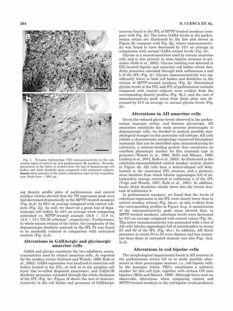
We used in this work three long-term MPTP-treatedmacaques who developed a moderate-to-severe, stableparkinsonian syndrome as judged from their motor dis-abilities, and three uninjected monkeys were used as con-trols. Generally, the degree of motor impairment in-creased after each MPTP injection and then fell until,after several doses, it became stable. Parkinsonian mon-keys displayed bradykinesia, akinesia, freezing phenom-ena, action tremor, paradoxical kinesias, and vertical andhorizontal saccadic ocular movements. These animalsshowed as well postural disturbances (trunk and limbflexion and tail rigidity), balance alterations, and occa-sional falls. The susceptibility of individual monkeys tothe neurotoxin differed; two of them showed a moderateparkinsonian syndrome after administration of four orseven MPTP doses, respectively (motor score rate for both13/25), and the third monkey underwent severe parkin-sonism after 12 MPTP doses (motor score rate 17/25).Immunohistochemical labeling revealed TH-IR, dopami-nergic neurons in the mesencephalon substantia nigra. Adramatic loss of TH immunoreactivity in these neuronswas evident when comparing sections from control sub-jects (Fig. 1a) and MPTP-treated monkeys (Fig. 1b).
Alterations in dopaminergic amacrine cells
After Ramon y Cajal (1892), in vertical retinal sections,the IPL is commonly subdivided into five, equally thickstrata grouped into two sublaminas, of which the mostscleral (sublamina a) comprises strata S1 and S2 and themost vitreal (sublamina b) comprises strata S3, S4, and S5(Famiglietti and Kolb, 1976). In the monkey retina, mostdopaminergic neurons (subtype I) exhibit large cell bodieslocated in the inner nuclear layer (INL), and emit a radi-ate array of dendrites running as an intricate plexus highin the IPL and establishing synapses with amacrine cells(Ehinger, 1983; Mariani et al., 1984; Mills and Massey,1999; Marshak, 2001). As described, we observed in nor-mal monkeys dopaminergic, TH-IR processes to stratifymainly in the outermost IPL stratum, S1 (Figs. 2a, 3a).However, in vertical sections of MPTP-treated monkeyretinas, there were significantly fewer dopaminergic pro-cesses in this sublayer, as illustrated by the representa-tive pictures shown in Figures 2b and 3b. The correspond-
263RETINAL DEGENERATION IN PARKINSON’S DISEASE
ing density profile plots of parkinsonian and controlmonkey retinas showed that the TH expression peak areahad decreased dramatically in the MPTP-treated monkeys(Fig. 2c,d), by 65% on average compared with control sub-jects (Fig. 2q). As well, we observed a great loss of dopa-minergic cell bodies, by 44% on average when comparinguntreated vs. MPTP-treated animals (24.9 � 11.9 vs.14.0 � 13.1 TH-IR cells/mm2, respectively). Furthermore,in whole-mount retinas of the latter, the complexity of thedopaminergic dendritic network in the IPL S1 was foundto be markedly reduced in comparison with untreatedcontrols (Fig. 3c,d).
Alterations in GABAergic and glycinergicamacrine cells
GABA and glycine constitute the two inhibitory neuro-transmitters used by retinal amacrine cells. As reportedfor the monkey retina (Grunert and Wassle, 1990; Kolb etal., 2002), GABA expression was localized to amacrine cellbodies located in the INL, as well as in the ganglion celllayer (the so-called displaced amacrines), and GABA-IRdendritic processes extended through the whole thicknessof the IPL (Fig. 2e). Figure 2f shows the loss of immuno-reactivity in the cell bodies and processes of GABAergic
neurons found in the IPL of MPTP-treated monkeys (com-pare with Fig. 2e). The lower GABA levels in the parkin-sonian retina are illustrated by the line plot shown inFigure 2h (compare with Fig. 2g), where immunoreactiv-ity was found to have decreased by 31% on average incomparison with normal GABA retinal levels (Fig. 2r).
Glycine is a neurotransmitter used by certain amacrinecells and is also present in some bipolar neurons in pri-mates (Kolb et al., 2002). Glycine labeling was detected inINL-located bipolar and amacrine cell bodies whose den-dritic processes extended through both sublaminas a andb of the IPL (Fig. 2i). Glycine immunoreactivity was sig-nificantly lower in both cell bodies and dendrites in theretinas of MPTP-treated monkeys (Fig. 2j). Diminishedglycine levels in the INL and IPL of parkinsonian animalscompared with control subjects were evident from thecorresponding density profiles (Fig. 2k,l), and the sum ofimmunoreactivity peak areas from these plots was de-creased by 31% on average vs. normal glycine levels (Fig.2s).
Alterations in AII amacrine cells
Given the reduced glycine levels observed in the parkin-sonian macaque retina, and because glycinergic, AIIamacrines constitute the main neurons postsynaptic todopaminergic cells, we decided to analyze possible mor-phological changes in this particular cell subtype. AII cellsexhibit a characteristic morphology conserved throughoutmammals that can be identified upon immunostaining forcalretinin, a calcium-binding protein that constitutes anexcellent phenotypic marker for this neuronal type inprimates (Wassle et al., 1995; Mills and Massey, 1999;Linberg et al., 2001; Kolb et al., 2002). As illustrated in thecalretinin-immunolabeled control monkey section shownin Figure 4b, AII cells bear a mitral-shaped cell body,located in the innermost INL stratum, and a primary,stout dendrite from which lobular appendages full of mi-tochondria emerge restricted to sublamina a of the IPL(Voigt and Wassle, 1987; Kolb et al., 1991). In addition,bushy distal dendrites ramify down into the lowest stra-tum of sublamina b.
In parkinsonian monkeys, we found that the levels ofcalretinin expression in the IPL were clearly lower than incontrol monkey retinas (Fig. 2m,n), as also evident fromthe corresponding profiles in Figure 2o,p. A quantitationof the immunoreactivity peak areas showed that, inMPTP-treated monkeys, calretinin levels were decreasedby 33% on average compared with control values (Fig. 2t).This lower immunoreactivity was associated with a loss ofAII cells’ lobular appendages full of mitochondria in strataS1 and S2 of the IPL (Fig. 4b,c). In addition, AII distalprocesses in strata S3 to S5 were thinner and less numer-ous than those in untreated animals (see also Figs. 3a,b,5c,d).
Alterations in rod bipolar cells
The morphological impairments found in AII neurons inthe parkinsonian retina led us to study possible alter-ations in their presynaptic neurons, i.e., rod bipolar cells.In the macaque retina, PKC� constitutes a selectivemarker for this cell type, together with certain ON conebipolars (Mills and Massey, 1999). Although there were noobservable alterations when comparing control andMPTP-treated monkeys in the rod bipolar ovoid perikarya
Fig. 1. Tyrosine hydroxylase (TH) immunoreactivity in the sub-stantia nigra of control (a) and parkinsonian (b) monkeys. Neurode-generation in the latter is evident from the loss of dopaminergic cellbodies and their dendritic plexi compared with untreated subjects.Insets show pictures of the entire substantia nigra at low magnifica-tion. Scale bars � 200 �m.
264 N. CUENCA ET AL.

located in the INL, or in their axons descending into theIPL (Fig. 5a,b), there were marked changes in PKC�-IRrod bipolar axon terminals in stratum S5 of the IPL. In theparkinsonian retina, these appeared thicker and some-what more swollen, with loss of the lateral varicositiesnormally found in their synaptic endings in the controlmonkey retina (Fig. 5b).
Impairments in synaptic connectivity
Dopaminergic dendrites in the monkey retina form ring-like structures embracing the cell bodies of amacrine neu-rons, especially AII cells (Mariani et al., 1984; Mills andMassey, 1999; Marshak, 2001). These rings constitutesites of chemical synaptic contacts with AII cell bodies,
Fig. 2. Vertical monkey retinal sections were immunostained fortyrosine hydroxylase (TH; a,b), GABA (e,f), glycine (GLY; i,j) andcalretinin (CR; m,n). Each pair of control (a,e,i,m) plus MPTP-treated(b,f,j,n) retinal sections was photographed together into a single pic-ture, and a representative image pair of the immunoreactivity patternobserved is shown. The profile plots of average gray intensity for eachhorizontal line are shown to the right of their corresponding pictures
for untreated (c,g,k,o) and treated (d,h,l,p) subjects. The gray valuesobtained after scanning 15–20 optic fields from each monkey set areplotted as the mean � SD for TH (q), GABA (r), GLY (s), and CR (t)immunoreactivities. In each case, the sum of the peak areas from apair of control plus MPTP-treated monkey retinas on the same slidewas taken as 100%. Scale bars � 80 �m in b (applies to a,b), f (appliesto e,f), j (applies to i,j), n (applies to m,n).
265RETINAL DEGENERATION IN PARKINSON’S DISEASE
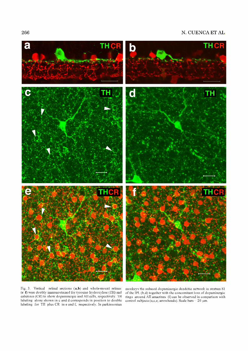
primary dendrites, and lobular appendages. A controlmonkey whole-mount retina, doubly immunostained withTH and calretinin to identify dopaminergic neurons andtheir meshwork of fine, varicose dendrites encircling AIIcell bodies, is shown in Figure 3e. The same view of anMPTP-treated monkey retina showed a marked loss of therings formed by dopaminergic dendrites around AII cells(Fig. 3f), which translated into a greatly diminished syn-aptic connectivity between these two cell types in theparkinsonian animal model.
AII amacrine distal dendrites are known to be intercon-nected by gap junctions consisting of Cx36 in the rabbitand rodent retinas (Feigenspan et al., 2001; Mills et al.,2001; Deans et al., 2002). The distribution of this connexinhas not been studied in the primate retina to date, whichprompted us to analyze its expression pattern in normaland MPTP-treated monkeys. Cx36 was immunolocatedprimarily in the IPL in the control monkey retina, whereit displayed a characteristic punctate distribution pattern(as expected for the labeling of gap junctional structures)that was much denser in sublamina b than in sublaminaa (Fig. 4a). A fainter Cx36 staining was also observed inthe OPL of the control animals, but there was no differ-ence in Cx36 content in the OPL of MPTP-treated ma-caques (data not shown). However, the number of Cx36-IRpuncta in the IPL was found to be significantly decreasedin parkinsonian monkeys compared with controls, espe-cially in sublamina a (Fig. 4d,e).
Double immunolabeling for Cx36 and calretinin illus-trated that in parkinsonian macaques the numbers both ofCx36-IR puncta in sublamina a of the IPL and of Cx36-IRcalretinin-IR puncta in sublamina b stratum S5 were de-creased (Fig. 4b–e). These changes, together with the lossof lobular appendages and decreased dendritic arboriza-tion from AII cells described above, were suggestive of animpaired electrical coupling between these cells in parkin-sonian monkeys. Additionally, it cannot be ruled out thatgap junctional connectivity between AII amacrines andON cone bipolar cells in the IPL was impaired as well (seeDiscussion).
We also investigated interconnections between rod bi-polars and AII cells in parkinsonian animals by perform-ing double immunostaining of retinal sections for PKC�and calretinin. In the monkey retina, AII-cellcalretinin-IR processes cross the IPL spiraling downaround descending rod bipolar axons, and receive gluta-matergic, excitatory input from these cells at ribbon syn-apses located in the IPL S5 (Wassle et al., 1995; Mills andMassey, 1999). As shown in Figure 5c, in the normalmonkey retina, AII dendritic processes were adjacent torod bipolar terminals in this stratum. In contrast, MPTP-treated monkeys showed far fewer PKC�-IR tocalretinin-IR contact regions, which was suggestive of areduction in the number of chemical synapses establishedbetween these two neuronal types (Fig. 5d).
DISCUSSION
For Parkinson’s disease patients, a diminished retinaldopamine content and reduced dopaminergic innervationhave been reported (Nguyen-Legros, 1988; Harnois and diPaolo, 1990; Nguyen-Legros et al., 1993). MPTP is a neu-rotoxic drug that elicits a selective destruction of mesen-cephalic dopaminergic neurons and hence is widely usedin the generation of animal models of Parkinson’s disease
(Burns et al., 1983). Although in MPTP-treated monkeys(Ghilardi et al., 1988b) and other mammals (Wong et al.,1985; Mariani et al., 1986) a decrease of retinal dopaminehas been found, morphological changes in the retina ofparkinsonian monkeys have not been studied to date. Inthis work, we made use of macaques who were treatedwith systemic doses of MPTP for 2 years. The impair-ments found at post-mortem examination 1 year after thelast MPTP injection are thus part of a chronic, stableparkinsonian syndrome, as evidenced by the persistentmotor symptoms and neurodegeneration at the level of thesubstantia nigra and are not ascribable to direct, short-term effects of the neurotoxin. We observed in these ani-mals a loss of immunoreactivity in retinal dopaminergicneurons and a reduction of their dendritic plexus in theIPL. These observations are in line with previous findingsin the mouse (Mariani et al., 1986; Tatton et al., 1990;Chen et al., 2003) and rabbit (Wong et al., 1985), in whichMPTP-induced loss of dopaminergic cell bodies in the INLand/or degeneration of their dendritic processes in the IPLhave been described.
Other than dopaminergic neurons, no retinal cell typeshave been examined so far in parkinsonian experimentalmammals. However, it must be considered that retinaldopaminergic cells receive inhibitory synaptic input fromamacrine cells in the IPL and in turn are presynapticmostly to a number of glycinergic and GABAergic ama-crine cells and to other dopaminergic neurons (Ehinger,1983; Marshak, 2001). In this context we found, accompa-nying dopaminergic innervation decrease, GABAergic andglycinergic amacrines to have reduced neurotransmitterlevels in both the INL and the IPL in parkinsonian mon-keys. Although a series of defined amacrine cell subtypesis known to be targeted by dopaminergic cells (Kolb et al.,1991), the main neuron postsynaptic to the latter is theAII amacrine, a glycinergic cell specialized in transmittingrod-driven signals to OFF- and ON-type cone bipolars,thereby allowing the transfer of scotopic visual informa-tion to the photopic pathway (Bloomfield, 2001; Bloomfieldand Dacheux, 2001; Linberg et al., 2001; Marshak, 2001).This led us to examine the presumptive morphologicaldeficiencies in this amacrine neuronal subtype. We ob-served MPTP-treated monkeys to exhibit a loss of thecharacteristic rings formed by dopaminergic neuronsaround the AII amacrine cell bodies, where synaptic con-tacts are normally established between these two celltypes (Voigt and Wassle, 1987; Kolb et al., 1991; Marshak,2001; Contini and Raviola, 2003; Witkovsky, 2004). Thisimpaired connectivity correlated with a loss of AII lobularappendages in the IPL sublamina a and to anomalouslythin AII dendritic processes in sublamina b.
Two explanations can be offered for AII neuron deteri-oration under deficient dopamine influence. First,dopamine-containing amacrine cells also immunolabel toGABA in cat (Wassle and Chun, 1988; Kolb et al., 1991)and rat (Contini and Raviola, 2003) retinas, and it hasbeen proposed for rodents that, at dendrosomatic syn-apses between dopaminergic and AII neurons, not onlydopamine is released but also the inhibitory neurotrans-mitter GABA, which probably acts on GABAA receptorslocated at the surface of the cell bodies of AII amacrines toreduce their light response (Contini and Raviola, 2003). Itcan be thus postulated that dopamine and/or GABA syn-aptic input might be necessary for the maintenance of AIIcell normal morphology and function. Thus, in the same
267RETINAL DEGENERATION IN PARKINSON’S DISEASE


fashion that molecular mechanisms depending on neuralactivity (e.g., neurotrophin secretion) are crucial for theregulation of structure, function, and connections of neu-rons during development (Krubitzer and Kahn, 2003), thedeprivation of a neuron’s specific environmental context(i.e., dopamine/GABA influx in our case) could lead to itsmorphological deterioration and loss of synaptic connec-tivity, for instance, by loss of proper trophic supportand/or because of its dependence for viability on a certainlevel of ongoing electrical activity. In this context, anincreasing body of evidence has been obtained on multipletrophic roles of dopamine in retinal function (Witkovsky,2004). Second, light-dependent release of dopamine isknown to promote closure of gap junctions between hori-zontal cells at the OPL and between AII amacrine neuronsat the IPL, with ensuing inhibition of electrical and chem-ical communication (Djamgoz et al., 1997; Bloomfield,2001; Bloomfield and Dacheux, 2001; Witkovsky, 2004). Ithas been shown as well that AII amacrines exhibit D1-subtype dopamine receptors (Veruki and Wassle, 1996). Itfollows that loss of negative modulation by dopamine inour parkinsonian monkeys could lead to an abnormal gapjunctional coupling state among AII cells (Djamgoz et al.,1997) as well as to morphologically impaired AII lobularappendages and normal dendritic meshwork appearancein the IPL. Similar results have been reported for thebrain of Parkinson’s disease patients, where deafferentia-tion is known to induce transynaptic atrophy of striatalneurons postsynaptic to dopaminergic cells. In this con-text, neurons in the putamen and caudate nucleus of thesepatients show truncated dendrites, with few dendriticspines and irregular bulbous swellings (McNeill et al.,1988; Lach et al., 1992).
The main component of gap junctions established be-tween AII-cell dendrites in the IPL S5 has been shown inrabbits and rodents to be Cx36. Additionally, AII ama-crines establish Cx36-containing electrical synapses withON cone bipolar neurons in the S3–S5 strata of the IPL(Feigenspan et al., 2001; Mills et al., 2001; Deans et al.,2002). With regard to parkinsonian monkeys, we foundCx36 punctate immunostaining to be considerably re-duced throughout the IPL compared with that in controlsubjects, especially in sublamina a. As a difference withother mammalian species, in this sublayer Cx36 immuno-reactivity labeled hemichannel(s) of unidentified celltype(s). Cx36 puncta were in most cases adjacent to AIIamacrine cells. However, a few Cx36-IR puncta in sub-lamina a were not associated with AII cells, correspondingto electrical synapses established between unidentifiedcells (amacrines, bipolars, and/or ganglion cells), as pro-posed for the rabbit and murine retinas (Feigenspan et al.,2001; Mills et al., 2001). In the case of sublamina b,Cx36-IR calretinin-IR puncta, which we found to be sig-nificantly diminished in the MPTP-treated animals, havedefinitely been shown to represent gap junctional contactsbetween neighboring AII distal dendrites, whereas someCx36-IR puncta may reflect gap junctions between AIIneurons and ON cone bipolars (Feigenspan et al., 2001;Mills et al., 2001). These conclusions are strongly sup-ported by the lack of electrical coupling between the twogap junction types in Cx36 knockout mice (Guldenagel etal., 2001; Deans et al., 2002). The diminished Cx36 ex-pression we observed in the IPL in parkinsonian monkeys,together with the loss of AII dendrites in sublamina b,must therefore reflect a severe reduction in the number of
gap junctional contacts, and thus electrical connections,between AII amacrines and possibly also between theseand their postsynaptic ON cone bipolar cells.
In the case of rod bipolar neurons, we found them to bemorphologically deteriorated as well, especially by the lossof lateral varicosities at their axon terminals. Given thatthese represent the regions of excitatory synaptic input toAII cells (Wassle et al., 1995; Mills and Massey, 1999;Bloomfield and Dacheux, 2001), their alteration shouldtranslate into an impaired chemical transmission betweenthese two neuronal cell types. It remains to be studiedwhether ON cone bipolar neurons, postsynaptic to AIIcells, are also structurally altered.
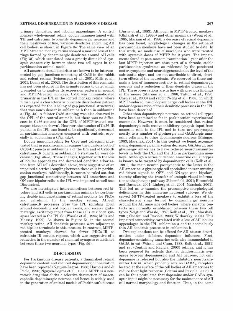
Taken together, these results highlight that not only doretinal dopaminergic neurons undergo deterioration inthe parkinsonian condition but also cells postsynaptic tothem, i.e., GABAergic and glycinergic amacrines. Amongthe latter, the alteration of AII cells occurred in associa-tion with morphological changes in neurons presynaptic tothem, i.e., rod bipolar cells. These findings are summa-rized in Figure 6, in which chronic morphological andsynaptic connectivity deficiencies exhibited by parkinso-nian monkeys in comparison with normal subjects aredepicted. Given that both Cx36-mediated synapses androd bipolar-AII connections are essential for the transmis-sion of rod visual signals in the mammalian retina(Bloomfield and Dacheux, 2001; Guldenagel et al., 2001;Linberg et al., 2001; Deans et al., 2002), these observa-tions provide strong morphological support to the notionthat the rod pathway of scotopic light transmission isseverely impaired in parkinsonism.
In fact, parkinsonian symptomatology in humans andexperimental animals includes a variety of electrophysi-ologically and psychophysically detectable impairments invisual functions (Bodis-Wollner, 1990, 2003; Masson et al.,1993; Djamgoz et al., 1997; Rodnitzky, 1998; Witkovsky,2004). Since in most cases these can be partially or totallyreversed upon L-DOPA treatment (Ellis and Ikeda, 1988;Ghilardi et al., 1988a; Pierelli et al., 1988), such dysfunc-tions have been interpreted as being due mainly to thedecrease of retinal dopamine levels. Our results, however,allow us to extend the relationship between deficiencies invisual functions and retinal morphological deteriorationto neurons other than dopaminergic amacrine cells. First,both flash- and pattern-elicited ERG amplitudes, reflect-ing the activity of outer and inner retinal layers, respec-tively, have been found to be significantly decreased inParkinson’s patients, indicating a loss of absolute sensi-tivity (Djamgoz et al., 1997; Langheinrich et al., 2000;Witkovsky, 2004). These impairments are especially evi-dent in the b-wave ERG component in humans (Ellis andIkeda, 1988; Ikeda et al., 1994) and have also been seen inMPTP-treated monkeys (Ghilardi et al., 1988a) and othermammals (Wong et al., 1985). Second, an extended latency(i.e., onset delay) in flash and pattern VEPs, representinga loss of temporal sensitivity, has also been reported inparkinsonian humans (Ellis and Ikeda, 1988; Pierelli etal., 1988), monkeys (Ghilardi et al., 1988a,b), and rabbits(Wong et al., 1985). VEPs represent electroencephalo-graphic changes time-locked to a visual stimulus, thoughchanges in the normal waveform are taken to reflect le-sions in the retina rather than in the brain visual cortex(Ghilardi et al., 1988a; Langheinrich et al., 2000). Asconcerns ERGs, the b wave recorded under scotopic lightconditions is attributable primarily to the activity of de-
270 N. CUENCA ET AL.
polarizing rod bipolars (Wong et al., 1985; Saszik et al.,2002; Dong and Hare, 2003). In Cx36 knockout mice, thelack of gap junctions between AII cells and between AIIamacrines and ON cone bipolar cells is accompanied bothby a decrease in the amplitude of the scotopic ERG b waveand by a delayed VEP response (Guldenagel et al., 2001).Therefore, the loss of rod bipolar-AII and AII-AII synapticconnectivity, together with the deterioration we found inthese two cell types (albeit without discarding that otheramacrine subtypes are as well affected), provides an ex-planation at the morphological level for abnormalities re-ported in ERGs and VEPs in the parkinsonian condition.
Additionally, a loss of spatiotemporal contrast sensitiv-ity, with elevated contrast detection and discriminationthresholds, has also been detected both by pattern ERGand by psychophysical trials in parkinsonian human(Bodis-Wollner, 1990; Djamgoz et al., 1997; Langheinrichet al., 2000) and nonhuman (Ghilardi et al., 1988a) pri-mates. This attenuation of contrast sensitivity is thoughtto be likely to lead to the impaired visual sensitivity tomotion perception found in parkinsonism (Bodis-Wollner,1990) and to derive from a strong lateral coupling betweenhorizontal cells in the OPL and/or between amacrine cellsin the IPL (Pierelli et al., 1988; Djamgoz et al., 1997). Ourresults suggest that the diminished ability of retinal spa-tial signaling associated with parkinsonism could be duepurely to a loss of electrical connectivity among AII cells,secondary to the deficient controlling influence of the de-teriorated dopaminergic amacrines. Given that dopaminenegative modulation (and GABA inhibitory chemical in-put) is likely impaired, a physiological condition equiva-
lent to that of a permanently coupled state of AII neuronsmay exist, related or not to AII dendritic degeneration.This may account, at least in part, for the difficulty inlight-to-dark adaptation and the loss of absolute andscotopic-contrast sensitivities exhibited by parkinsonianpatients.
CONCLUSIONS
The results presented here show the existence of a set ofstructural and morphological deficiencies previously un-described in retinal circuitries of the scotopic visual path-way in primates and allow us to explain many of the visualdysfunctions exhibited by Parkinson’s disease patientsand experimental mammals. These impairments affectnot only dopaminergic cells but also the neurons postsyn-aptic to them, i.e., GABAergic and glycinergic amacrines.Among the latter, AII cells of the rod pathway through theretina exhibited severe deficiencies in their morphologyand the connectivity among them and with rod bipolarcells. Extrapolation of these findings to the midbrain sug-gests that impairments in the structure and connectivityof specific neurons postsynaptic to dopaminergic cells arelikely to occur also in the brain of parkinsonian patients.Indeed, the atrophy of the dendritic arbor and transynap-tic degeneration of neurons observed in the striatum ofthese patients (McNeill et al., 1988; Lach et al., 1992)could explain the declining efficacy of chronic L-DOPAtherapy in advanced Parkinson’s disease. Such a possibil-ity has also been proposed to account for the dyskinesiasand motor fluctuations frequently found in humans on
Fig. 6. Scheme of the structural neuronal lesions found in theretina of parkinsonian monkeys compared with normal controls. Ar-rows indicate normal electrical and chemical synapses, and crossed-out arrows indicate impaired synapses. Note morphologically deteri-
orated dopaminergic (DA) cell dendrites, rod bipolar axon terminals,and AII-cell lobular appendages and dendritic terminals in the MPTP-treated monkey retina. Decreased connexin-36 expression at gap junc-tions is also indicated by crossed-out arrows.
271RETINAL DEGENERATION IN PARKINSON’S DISEASE

prolonged treatment with L-DOPA (Colosimo and deMichele, 1999; van Laar, 2003). Further study of thesecontentions may be warranted.
ACKNOWLEDGMENTS
We thank Drs. Helga Kolb and Eduardo Solessio forcritical revision of the manuscript and Dr. David Pow forhis kind gift of polyclonal antisera.
LITERATURE CITED
Barcia C, Bautista V, Sanchez-Bahillo A, Fernandez-Villalba E, Navarro-Ruis J-M, Fernandez Barreiro A, Poza y Poza M, Herrero M-T. 2003.Circadian determinations of cortisol, prolactin, and melatonin inchronic MPTP-treated monkeys. Neuroendocrinology 78:118–128.
Bloomfield SA. 2001. Plasticity of AII amacrine cell circuitry in the mam-malian retina. Prog Brain Res 131:185–200.
Bloomfield SA, Dacheux RF. 2001. Rod vision: pathways and processing inthe mammalian retina. Prog Ret Eye Res 20:351–384.
Bodis-Wollner I. 1990. Visual deficits related to dopamine deficiency inexperimental animals and Parkinson’s disease patients. Trends Neu-rosci 13:296–302.
Bodis-Wollner I. 2003. Neuropsychological and perceptual defects in Par-kinson’s disease. Parkinsonism Rel Disord 9:S83–S89.
Burns RS, Chiueh CC, Markey SP, Ebert MH, Jacobowitz DM, Kopin IJ.1983. A primate model of parkinsonism: selective destruction of dopa-minergic neurons in the pars compacta of the substantia nigra byN-methyl-4-phenyl-1,2,3,6-tetrahydropyridine. Proc Natl Acad SciU S A 80:4546–4550.
Chen ST, Hsu JR, Hsu PC, Chuang JI. 2003. The retina as a novel in vivomodel for studying the role of molecules of the Bcl-2 family in relationto MPTP neurotoxicity. Neurochem Res 28:805–814.
Colosimo C, de Michele M. 1999. Motor fluctuations in Parkinson’s disease:pathophysiology and treatment. Eur J Neurol 6:1–21.
Contini M, Raviola E. 2003. GABAergic synapses made by a retinal dopa-minergic neuron. Proc Natl Acad Sci U S A 100:1358–1363.
Cuenca N, Deng P, Linberg KA, Fisher SK, Kolb H. 2003. Choline acetyl-transferase is expressed by non-starburst amacrine cells in the groundsquirrel retina. Brain Res 964:21–30.
Deans MR, Volgyi B, Goodenough DA, Bloomfield SA, Paul DL. 2002.Connexin36 is essential for transmission of rod-mediated visual signalsin the mammalian retina. Neuron 36:703–712.
Djamgoz MBA, Hankins MW, Hirano J, Archer SN. 1997. Neurobiology ofretinal dopamine in relation to degenerative states of the tissue. VisRes 37:3509–3529.
Dong CJ, Hare WA. 2003. Temporal modulation of scotopic visual signalsby A17 amacrine cells in mammalian retina in vivo. J Neurophysiol89:2159–2166.
Ehinger B. 1983. Functional role of dopamine in the retina. In: OsborneNN, Chader GJ, editors. Progress in retinal research, vol 2. Oxford,United Kingdom: Pergamon Press. p 213–232.
Ellis CJK, Ikeda H. 1988. Evidence for retinal dopamine deficiency inParkinson’s disease. In: Bodis-Wollner I, Piccolino M, editors. Dopami-nergic mechanisms in vision. New York: Alan R Liss, Inc. p 239–251.
Famiglietti EV Jr, Kolb H. 1976. Structural basis for ON- and OFF-centerresponses in retinal ganglion cells. Science 194:193–195.
Feigenspan A, Teubner B, Willecke K, Weiler R. 2001. Expression ofneuronal connexin36 in AII amacrine cells of the mammalian retina.J Neurosci 21:230–239.
Ghilardi MF, Bodis-Wollner I, Onofrj MC, Marx MS, Glover A. 1988a.Spatial frequency-dependent abnormalities of the pattern electroreti-nogram and visual evoked potentials in a parkinsonian monkey model.Brain 111:131–149.
Ghilardi MF, Chung E, Bodis-Wollner I, Dvorzniak M, Glover A, Onofrj M.1988b. Systemic 1-methyl,4-phenyl,1-2-3-6-tetrahydropyridine (MPTP)administration decreases retinal dopamine content in primates. Life Sci43:255–262.
Grunert U, Wassle H. 1990. GABA-like immunoreactivity in the macaquemonkey retina: a light and electron microscopic study. J Comp Neurol297:509–524.
Guldenagel M, Ammermuller J, Feigenspan A, Teubner B, Degen J, Sohl
G, Willecke K, Weiler R. 2001. Visual transmission deficits in mice withtargeted disruption of the gap junction gene connexin36. J Neurosci21:6036–6044.
Harnois C, di Paolo T. 1990. Decreased dopamine in the retinas of patientswith Parkinson’s disease. Invest Ophthalmol Vis Sci 31:2473–2475.
Herrero M-T, Perez-Otano I, Oset C, Kastner A, Hirsch EC, Agid Y, LuquinMR, Obeso JA, del Rıo J. 1993. GM-1 ganglioside promotes the recoveryof surviving midbrain dopaminergic neurons in MPTP-treated mon-keys. Neuroscience 56:965–972.
Ikeda H, Head GM, Ellis CJK. 1994. Electrophysiological signs of retinaldopamine deficiency in recently diagnosed Parkinson’s disease and afollow up study. Vis Res 34:2629–2638.
Kolb H, Cuenca N, Dekorver L. 1991. Postembedding immunocytochemis-try for GABA and glycine reveals the synaptic relationships of thedopaminergic amacrine cell of the cat retina. J Comp Neurol 310:267–284.
Kolb H, Zhang L, Dekorver L, Cuenca N. 2002. A new look at calretinin-immunoreactive amacrine cell types in the monkey retina. J CompNeurol 453:168–184.
Krubitzer L, Kahn DM. 2003. Nature versus nurture revisited: an old ideawith a new twist. Prog Neurobiol 70:33–52.
Lach B, Grimes D, Benoit B, Minkiewicz-Janda A. 1992. Caudate nucleuspathology in Parkinson’s disease: ultrastructural and biochemical find-ings in biopsy material. Acta Neuropathol 83:352–360.
Langheinrich T, van Elst LT, Lagreze WA, Bach M, Lucking CH, GreenleeMW. 2000. Visual contrast response functions in Parkinson’s disease:evidence from electroretinograms, visually evoked potentials and psy-chophysics. Clin Neurophysiol 111:66–74.
Linberg K, Cuenca N, Ahnelt P, Fisher S, Kolb H. 2001. Comparativeanatomy of major retinal pathways in the eyes of nocturnal and diurnalmammals. Prog Brain Res 131:27–52.
Luquin MR, Laguna J, Obeso JA. 1992. Selective D2 receptor stimulationinduces dyskinesia in parkinsonian monkeys. Ann Neurol 31:551–554.
Mariani AP, Kolb H, Nelson R. 1984. Dopamine-containing amacrine cellsof rhesus monkey retina parallel rods in spatial distribution. Brain Res322:1–7.
Mariani AP, Neff NH, Hadjiconstantinou M. 1986. 1-Methyl-4-phenyl-1,2,3,6-tetrahydropyridine (MPTP) treatment decreases dopamine andincreases lipofuscin in mouse retina. Neurosci Lett 72:221–226.
Marshak DW. 2001. Synaptic inputs to dopaminergic neurons in mamma-lian retinas. Prog Brain Res 131:83–91.
Masson G, Mestre D, Blin O. 1993. Dopaminergic modulation of visualsensitivity in man. Fund Clin Pharmacol 7:449–463.
McNeill TH, Brown SA, Rafols JA, Shoulson I. 1988. Atrophy of mediumspiny I striatal dendrites in advanced Parkinson’s disease. Brain Res455:148–152.
Mills SL, Massey SC. 1999. AII amacrine cells limit scotopic acuity incentral macaque retina: a confocal analysis of calretinin labeling.J Comp Neurol 411:19–34.
Mills SL, O’Brien JJ, Li W, O’Brien J, Massey SC. 2001. Rod pathways inthe mammalian retina use connexin 36. J Comp Neurol 436:336–350.
Nguyen-Legros J. 1988. Functional neuroarchitecture of the retina: hy-pothesis on the dysfunction of retinal dopaminergic circuitry in Par-kinson’s disease. Surg Radiol Anat 10:137–144.
Nguyen-Legros J, Harnois C, di Paolo I, Simon A. 1993. The retinaldopamine system in Parkinson’s disease. Clin Vis Sci 8:1–12.
Paxinos G, Huang X-F, Toga AW. 1999. The rhesus monkey brain instereotaxic coordinates. San Diego: Academic Press.
Pierelli F, Stanzione P, Peppe A, Stefano E, Rizzo PA, Bernardi G, Moro-cutti C. 1988. Electrophysiological (PERG,VEP) abnormalities in Par-kinson disease are reversed by L-DOPA. In: Bodis-Wollner I, PiccolinoM, editors. Dopaminergic mechanisms in vision. New York: Alan RLiss, Inc. p 253–265.
Pow DV, Wright LL, Vaney DI. 1995. The immunocytochemical detection ofamino-acid neurotransmitters in paraformaldehyde-fixed tissues.J Neurosci Methods 56:115–123.
Ramon y Cajal S. 1892. The structure of the retina. Thorpe SA, GlicksteinM, translators. 1972. Springfield, IL: Charles C. Thomas Publisher.
Rodnitzky RL. 1998. Visual dysfunction in Parkinson’s disease. Clin Neu-rosci 5:102–106.
Saszik SM, Robson JG, Frishman LJ. 2002. The scotopic threshold re-sponse of the dark-adapted electroretinogram of the mouse. J Physiol543:899–916.
Tatton WG, Kwan MM, Verrier MC, Seniuk NA, Theriault E. 1990. MPTP
272 N. CUENCA ET AL.

produces reversible disappearance of tyrosine hydroxylase-containingretinal amacrine cells. Brain Res 527:21–31.
Van Laar T. 2003. Levodopa-induced response fluctuations in patients withParkinson’s disease: strategies for management. CNS Drugs 17:475–489.
Veruki ML, Wassle H. 1996. Immunohistochemical localization of dopa-mine D1 receptors in rat retina. Eur J Neurosci 8:2286–2297.
Voigt T, Wassle H. 1987. Dopaminergic innervation of A II amacrine cellsin mammalian retina. J Neurosci 7:4115–4128.
Wassle H, Chun MH. 1988. Dopaminergic and indoleamine-accumulating
amacrine cells express GABA-like immunoreactivity in the cat retina.J Neurosci 8:3383–3394.
Wassle H, Grunert U, Chun MH, Boycott BB. 1995. The rod pathway of themacaque monkey retina: identification of AII-amacrine cells with an-tibodies against calretinin. J Comp Neurol 361:537–551.
Witkovsky P. 2004. Dopamine and retinal function. Doc Ophthalmol 108:17–40.
Wong C, Ishibashi T, Tucker G, Hamasaki D. 1985. Responses of thepigmented rabbit retina to NMPTP, a chemical inducer of parkinson-ism. Exp Eye Res 40:509–519.
273RETINAL DEGENERATION IN PARKINSON’S DISEASE