Morphological, Gene, and Hormonal Changes in Gonads and ...
15
Citation: Lettieri, G.; Carusone, N.; Notariale, R.; Prisco, M.; Ambrosino, A.; Perrella, S.; Manna, C.; Piscopo, M. Morphological, Gene, and Hormonal Changes in Gonads and In-Creased Micrococcal Nuclease Accessibility of Sperm Chromatin Induced by Mercury. Biomolecules 2022, 12, 87. https://doi.org/10.3390/ biom12010087 Academic Editor: Yves Ménézo Received: 30 November 2021 Accepted: 4 January 2022 Published: 6 January 2022 Publisher’s Note: MDPI stays neutral with regard to jurisdictional claims in published maps and institutional affil- iations. Copyright: © 2022 by the authors. Licensee MDPI, Basel, Switzerland. This article is an open access article distributed under the terms and conditions of the Creative Commons Attribution (CC BY) license (https:// creativecommons.org/licenses/by/ 4.0/). biomolecules Article Morphological, Gene, and Hormonal Changes in Gonads and In-Creased Micrococcal Nuclease Accessibility of Sperm Chromatin Induced by Mercury Gennaro Lettieri 1,† , Nadia Carusone 1,† , Rosaria Notariale 2,† , Marina Prisco 1 , Alessia Ambrosino 1 , Shana Perrella 1 , Caterina Manna 2 and Marina Piscopo 1, * 1 Department of Biology, University of Naples Federico II, Via Cinthia, 21, 80126 Naples, Italy; [email protected] (G.L.); [email protected] (N.C.); [email protected] (M.P.); [email protected] (A.A.); [email protected] (S.P.) 2 Department of Precision Medicine, School of Medicine, University of Campania “Luigi Vanvitelli”, Via Luigi de Crecchio, 80138 Naples, Italy; [email protected] (R.N.); [email protected] (C.M.) * Correspondence: [email protected] † These authors contributed equally to this work. Abstract: Mercury is one of the most dangerous environmental pollutants. In this work, we analysed the effects of exposure of Mytilus galloprovincialis to 1, 10 and 100 pM HgCl 2 for 24 h on the gonadal morphology and on the expression level of three stress genes: mt10, hsp70 and πgst. In this tissue we also evaluated the level of steroidogenic enzymes 3β-HSD and 17β-HSD and the expression of PL protein genes. Finally, we determined difference in sperm chromatin accessibility to micrococcal nuclease. We found alterations in gonadal morphology especially after exposure to 10 and 100 pM HgCl 2 and hypo-expression of the three stress genes, particularly for hsp70. Furthermore, decreased labelling with both 3β-HSD and 17β-HSD antibodies was observed following exposure to 1 and 10 pM HgCl 2 and complete absence at 100 pM HgCl 2 exposure. Gonads of mussels exposed to all HgCl 2 doses showed decreased expression of PL protein genes especially for PLIII. Finally, micrococcal nuclease digestions showed that all doses of HgCl 2 exposure resulted in increased sperm chromatin accessibility to this enzyme, indicative of improper sperm chromatin structure. All of these changes provide preliminary data of the potential toxicity of mercury on the reproductive health of this mussel. Keywords: mercury; Mytilus galloprovincialis; protamine-like proteins genes; gonad; spermatozoa; micrococcal nuclease digestion; hormones; morphology 1. Introduction The increasing worldwide levels of pollutants in the marine environment require control and monitoring strategies. Mercury is an extremely toxic heavy metal in the marine environment and its effects on marine organisms are complex and often unpredictable. The United Nations Environment Programme (UNEP)’s “Global Mercury Assessment” estimated that anthropogenic activities have increased current atmospheric mercury concen- trations by approximately 450% above natural levels, resulting in a substantial increase in seawater [1]. In marine environments, mercury is subject to bioaccumulation and biomagni- fication, and its seawater contamination is posing a serious health problem for humans [2,3] and marine organisms [4,5]. In environmental biomonitoring programs, the use of bioindicator organisms rep- resents one of the most suitable methods for the assessment of trace metal toxicity [6]. Bivalves are excellent organisms as bioindicators of heavy metal pollution due to their characteristics including worldwide distribution, sedentary filter-feeding, easy sampling and reasonable size [7]. Biomolecules 2022, 12, 87. https://doi.org/10.3390/biom12010087 https://www.mdpi.com/journal/biomolecules
-
Upload
khangminh22 -
Category
Documents
-
view
0 -
download
0
Transcript of Morphological, Gene, and Hormonal Changes in Gonads and ...

�����������������
Citation: Lettieri, G.; Carusone, N.;
Notariale, R.; Prisco, M.; Ambrosino,
A.; Perrella, S.; Manna, C.; Piscopo, M.
Morphological, Gene, and Hormonal
Changes in Gonads and In-Creased
Micrococcal Nuclease Accessibility of
Sperm Chromatin Induced by
Mercury. Biomolecules 2022, 12, 87.
https://doi.org/10.3390/
biom12010087
Academic Editor: Yves Ménézo
Received: 30 November 2021
Accepted: 4 January 2022
Published: 6 January 2022
Publisher’s Note: MDPI stays neutral
with regard to jurisdictional claims in
published maps and institutional affil-
iations.
Copyright: © 2022 by the authors.
Licensee MDPI, Basel, Switzerland.
This article is an open access article
distributed under the terms and
conditions of the Creative Commons
Attribution (CC BY) license (https://
creativecommons.org/licenses/by/
4.0/).
biomolecules
Article
Morphological, Gene, and Hormonal Changes in Gonads andIn-Creased Micrococcal Nuclease Accessibility of SpermChromatin Induced by MercuryGennaro Lettieri 1,† , Nadia Carusone 1,†, Rosaria Notariale 2,†, Marina Prisco 1, Alessia Ambrosino 1,Shana Perrella 1, Caterina Manna 2 and Marina Piscopo 1,*
1 Department of Biology, University of Naples Federico II, Via Cinthia, 21, 80126 Naples, Italy;[email protected] (G.L.); [email protected] (N.C.); [email protected] (M.P.);[email protected] (A.A.); [email protected] (S.P.)
2 Department of Precision Medicine, School of Medicine, University of Campania “Luigi Vanvitelli”,Via Luigi de Crecchio, 80138 Naples, Italy; [email protected] (R.N.);[email protected] (C.M.)
* Correspondence: [email protected]† These authors contributed equally to this work.
Abstract: Mercury is one of the most dangerous environmental pollutants. In this work, we analysedthe effects of exposure of Mytilus galloprovincialis to 1, 10 and 100 pM HgCl2 for 24 h on the gonadalmorphology and on the expression level of three stress genes: mt10, hsp70 and πgst. In this tissuewe also evaluated the level of steroidogenic enzymes 3β-HSD and 17β-HSD and the expression ofPL protein genes. Finally, we determined difference in sperm chromatin accessibility to micrococcalnuclease. We found alterations in gonadal morphology especially after exposure to 10 and 100 pMHgCl2 and hypo-expression of the three stress genes, particularly for hsp70. Furthermore, decreasedlabelling with both 3β-HSD and 17β-HSD antibodies was observed following exposure to 1 and10 pM HgCl2 and complete absence at 100 pM HgCl2 exposure. Gonads of mussels exposed toall HgCl2 doses showed decreased expression of PL protein genes especially for PLIII. Finally,micrococcal nuclease digestions showed that all doses of HgCl2 exposure resulted in increased spermchromatin accessibility to this enzyme, indicative of improper sperm chromatin structure. All of thesechanges provide preliminary data of the potential toxicity of mercury on the reproductive health ofthis mussel.
Keywords: mercury; Mytilus galloprovincialis; protamine-like proteins genes; gonad; spermatozoa;micrococcal nuclease digestion; hormones; morphology
1. Introduction
The increasing worldwide levels of pollutants in the marine environment requirecontrol and monitoring strategies. Mercury is an extremely toxic heavy metal in the marineenvironment and its effects on marine organisms are complex and often unpredictable.The United Nations Environment Programme (UNEP)’s “Global Mercury Assessment”estimated that anthropogenic activities have increased current atmospheric mercury concen-trations by approximately 450% above natural levels, resulting in a substantial increase inseawater [1]. In marine environments, mercury is subject to bioaccumulation and biomagni-fication, and its seawater contamination is posing a serious health problem for humans [2,3]and marine organisms [4,5].
In environmental biomonitoring programs, the use of bioindicator organisms rep-resents one of the most suitable methods for the assessment of trace metal toxicity [6].Bivalves are excellent organisms as bioindicators of heavy metal pollution due to theircharacteristics including worldwide distribution, sedentary filter-feeding, easy samplingand reasonable size [7].
Biomolecules 2022, 12, 87. https://doi.org/10.3390/biom12010087 https://www.mdpi.com/journal/biomolecules

Biomolecules 2022, 12, 87 2 of 15
Mytilus galloprovincialis (M. galloprovincialis) is a readily available organism and widelydistributed in the Mediterranean. This bivalve mollusc accumulates contaminants in itstissues mainly in the hepatopancreas, kidney, gills and gonads [8]. This organism respondsquickly and effectively to the presence of many environmental pollutants, including heavymetals [9]. Exposure of M. galloprovincialis to various heavy metals causes alterations in itsreproductive system [9–14]. In the sperm chromatin of M. galloprovincialis the main basicprotein component is represented by the Protamine-like (PL) proteins [15]. In previousworks we have found that the exposure to subtoxic doses of copper causes an increasein the binding affinity of PL-proteins to DNA and alterations in the expression of sper-matozoa mt10 gene [12]. Finally, alterations, such as a reduced DNA binding affinity ofPL-proteins [9], and an increased mt20 gene expression [14] following exposure to subtoxicdoses of cadmium, have been reported. Mytilus spp. have shown a greater capacity formercury (Hg) accumulation than other bivalve species [16], and recently, we evaluated theeffects of exposure to mercury doses (1, 10 and 100 pM HgCl2), similar to those found in theMediterranean basin and North Atlantic oceans, on M. galloprovincialis male reproductivesystem [17]. In particular, we showed bioaccumulation of mercury in the male gonadsand alterations in the expression of spermatozoa mt10 and hsp70 stress genes. We alsofound several changes in the PL-proteins, such as a reduced ability to bind DNA andan inability to protect DNA from oxidative damage [9]. Furthermore, at these doses ofHgCl2 exposure, the formation of PL protein aggregates and alterations in their structuralconformation was shown to result in changes in both their binding to DNA and their releasefrom sperm nuclei [10].
In order to obtain further useful information on the effects of these HgCl2 exposuredoses on the male reproductive system of M. galloprovincialis, in the present work weevaluated possible morphological changes in the gonads by hematoxylin and eosin (H&E)staining and the levels of steroidogenic enzymes 3β-HSD and 17β-HSD by immunohis-tochemistry. In addition, by quantitative reverse transcription polymerase chain reaction(RT-qPCR), gene expression of mt10, hsp70, πgst, and genes encoding PL proteins were alsomeasured in the gonads. Finally, the possible difference in sperm chromatin accessibilityto micrococcal nuclease (MNase) in mussels unexposed and exposed to these doses ofmercuric chloride was investigated.
2. Materials and Methods2.1. Ethics Statement
This research was performed on the marine invertebrate M. galloprovincialis (Lamarck,1819), which is not protected by any environmental agency in Italy. This study wasconducted in strict accordance with European (Directive 2010/63) and Italian (LegislativeDecree n. 116/1992) legislation on the care and use of animals for scientific purposes.
2.2. Mussels Sampling and Exposure to HgCl2Adult mussels of M. galloprovincialis, medium size of the shell length 4.93 ± 0.17 cm,
were supplied by Eurofish Napoli S.R.L. Baia, in Naples and used in this study. Fifteenmussels of unknown sex were exposed to 1, 10 and 100 pM HgCl2, as previously describedfor the other heavy metals [18] in laboratory plastic tanks (36 cm × 22 cm × 22 cm)containing 6 L of 33‰ artificial sea water (ASW) with the following composition for1 L: NaCl 29.2 g, KCl 0.60 g, MgCl2 1.2 g, NaHCO3 0.20 g and CaCl2 1.08 g. In each tank,mussels were exposed to a single dose of HgCl2. The exposure was performed at 18 ± 1 ◦C,for 24 h changing water and metal salts every 12 h during treatment and recording dissolvedoxygen and temperature at predetermined time intervals. The experiments were conductedin January of the current year. Tanks containing only ASW were used as a control fornonexposed mussels.

Biomolecules 2022, 12, 87 3 of 15
2.3. Gonad Sampling and PL Proteins Extraction
After 24 h of exposure to the three HgCl2 doses, gonads were collected from malespecimens. Mussels were opened forcing the valves with a knife, being care not to cutsoft tissues. Mussels’ sex was initially identified observing the colour of mantle andconfirmed by light microscope observation of gonads smear. Briefly, the piece of gonadwas smeared on the microscope slide and as soon as it was dry, it was observed under thelight microscope. Each identified gonad was stocked and stored at −80 ◦C.
Sperm-filled gonads were used for acid extraction of PL proteins. Briefly, one gonadfrom each condition was mechanically pestled in distilled water. Subsequently, centrifuga-tion was performed at 1000× g for 2 min at 4 ◦C to remove debris, and from the obtained su-pernatant, PLs were extracted by 5% perchloric acid as described in Piscopo et al. 2017 [14].
2.4. RNA Extraction, cDNA Synthesis and RT-qPCR
Total RNA was purified from gonads of nonexposed mussels (control) and exposedto 1, 10, 100 pM HgCl2 using Trizol reagent (Invitrogen, Carlsbad, CA, USA) accordingto the manufacturer’s instructions. The RNA obtained was quantified by a UV-Vis spec-trophotometer (NanoDropH ND-1000, Waltham, MA, USA). Moreover, electrophoreticanalysis was performed on 1% agarose gels under denaturing conditions to assess itsquality. Genomic DNA was removed from the samples with an Ambion (Austin, TX, USA)DNA-free kit. cDNA was synthesized from 1 µg of RNA from each samples using M-MLVreverse transcriptase (ImpProm II kit, Promega, Madison, WI, USA). The RT-qPCR wascarried out as described in Lettieri et al. [11]. In particular, to determine the gene expressionwas used 100 ng of the cDNA with 10 µM of each forward and reverse primers in a finalvolume of 50 µL using SYBRGreen PCR Master Mix Kit (Applied Biosystems, Foster City,CA, USA) with the 7500 Real Time PCR System (Applied Biosystems, Foster City, CA, USA).Each PCR reaction was conducted for 40 cycles as described in Lettieri et al., 2019 [19]maintaining the following conditions: Denaturation at 95 ◦C for 15 min; annealing andelongation at 60 ◦C for 1 min. The primer used for the reaction was engineered usingthe open-source software Primer3, following the sequences and the accession numbersreported in Table 1. The results were exported into Microsoft Excel (Redmond, WA, USA,ver. 2009—build 13231.20262) from the ViiA™-7 Software (Foster City, CA, USA). Therelative quantification of gene expression was evaluated using the the ∆∆Ct method asreported in Livak et al. [20]. The expression of mt10, hsp70, πgst, PL-III and PL-II/IV geneswas compared to those obtained from control mussels. In Table 1 was reported the list ofprimers used.
Table 1. List of forward and reverse primers used for amplification of each gene analysed and for thereference housekeeping GAPDH gene.
Gene F-Primer F-Primer Length R-Primer R-Primer Length AccessionNumber
Gapdh CTGCACCACCAACTGCTT 18 TTCTGGGTGGCAGTGATG 18 SY171038758-018/019
Hsp70 CGCGATGCCAAACTAGACAA 20 TCACCTGACAAAATGGCTGC 20 AY861684
Mt10 GCCTGCACCTTGTAACTGTAT 21 CTGTACACCCTGCTTCACAC 20 AY566248
Gst AGTTAGAGGCCGAGCTGAA 19 TGGAAACCGTCATCATCTG 19 SY140930374-050/051
Pl III CACCCAACAAGAAGGATGCC 20 CCTTGCCCTTTTCTTTCCCC 20 SY140930274
Pl II/IV AAGCCCAAGTAGACGTTCCA 20 TCCGAGGTGTGATGTGTTGA 20 SY140930274
2.5. Sperm Nuclei Preparation and MNase Assay
For the preparation of sperm nuclei the procedure reported in Olivares and Ruiz1991 [21] was followed. In brief, sperm nuclei were obtained starting from spermatozoapellet of mussels exposed to different HgCl2 doses. Spermatozoa pellets were obtainedcentrifuging semen for 10 min at 1900× g at 4 ◦C. Each pellet was then resuspended in1 ml of a solution (solution 1) with the following composition. -NaCl 0.15 M- EDTA pH8 25 mM PMSF 1 mMF and the sample was centrifuged at 4 ◦C for 10 min at 1900× g.

Biomolecules 2022, 12, 87 4 of 15
Subsequently, the obtained pellet was resuspended in the following buffer (solution 2):0.25 M sucrose- MgCl2 5 mM- Tris-HCl pH 8 10 mM- PMSF 1 mM- Triton X-100 0.38%and then centrifuged at 4 ◦C for 10 min at 1900× g. The pellet was recovered and washedtwice with the previous buffer in the absence of Triton. The prepared sperm nuclei wereresuspended in the following buffer (solution 3)-NaCl 0.15 M- Tris HCl 10 mM pH 8- CaCl20.5 mM and subjected to MNase assay. Digestion was carried out with 10 units of enzymeat 37 ◦C for 1 mg/mL DNA concentration (A260 = 20) and the reaction stopped at differenttimes (5′, 10′, 15′, 30′ and 60′) adding 2 mM EDTA pH 8 at ice. The digestion products werecentrifuged at 1900× g for 10 min at 4 ◦C. This produced a pellet (P) and a supernatant (S). Pwas used for DNA extraction following a common extraction phenol/chloroform/isoamylalcohol. Briefly, 1 M NaCl and 0.5% SDS was added to P and incubated 30 min at RT.Equal volume of phenol/chloroform/isoamyl alcohol (25:24:1) was added, samples werevortexed and centrifuged at 4 ◦C for 5 min at 13,000× g. Successively, 0.3 M NaCl and2.5 volumes of ethanol 100% was added to the aqueous phase to precipitate the DNA at−20 ◦C for 1 h.
2.6. Morphological Analyses of Gonad
For light microscope investigations, samples 0.5 cm × 0.5 cm were taken from thecentral portion of one hemi-mantle. Specimens were fixed in Bouin’s solution for 24 h,dehydrated in alcohol and embedded in paraffin wax. Five µm thick sections were cut andstained with hematoxylin and eosin stain.
2.7. Immunohistochemistry Analyses
For immunohistochemistry, 5 µm thick sections of Bouin fixed specimen on poly-l-lysine slides were deparaffinized and rehydrated in xylene and a series of graded alcohols.The sections were treated with 10 mM citrate buffer pH 6.0 in the microwave for antigenretrieval and then incubated in 2.5% H2O2 in methanol for endogenous peroxidase blocking.Non-specific background was reduced with the incubation in 3% BSA in PBS buffer for1 h at room temperature. Sections were then treated overnight at 4 ◦C with the primaryrabbit antibodies diluted in 1% BSA in PBS buffer: (1) anti-human 3β-HSD (1:50), (2) anti-mouse 17β-HSD (1:50). In the first instance, antibodies specificity was tested by making animmunoblot analysis on Mytilus galloprovincialis gonadal proteins as previously reportedby Prisco et al. (2017) [22]. The reaction was revealed with a peroxidase-conjugated goatanti-rabbit secondary antibody, using DAB (Roche) as chromogen. Negative controlswere carried out by omitting primary antibodies. An immunohistochemical signal wasobserved using a Zeiss Axioskop microscope; images were acquired by using AxioVision4.7 software (Zeiss).
2.8. Statistical Analysis
The ANOVA test followed Tukey’s post-hoc test was performed for gene expressionanalysis. The statistical analyses were performed by GraphPad Prism 9 (ver. 9.1.2 (226))(GraphPad Software, San Diego, CA, USA).
3. Results3.1. Gonadal mt10, πgst and hsp70 Expression
The RT-qPCR was used to assess the expression of mt10, πgst and hsp70 as a responseto possible stress in male gonads after mussel exposure to 1, 10 and 100 pM HgCl2. Allgenes were downregulated after exposure of mussels to all doses of HgCl2 except for mt10gene after exposure to 1 pM HgCl2 in which an overexpression, approximately of 2-fold, ofthis gene, was observed (Figure 1c).
Biomolecules 2022, 12, 87 5 of 15
Biomolecules 2022, 12, x FOR PEER REVIEW 5 of 16
2.8. Statistical Analysis The ANOVA test followed Tukey’s post-hoc test was performed for gene expression
analysis. The statistical analyses were performed by GraphPad Prism 9 (ver. 9.1.2 (226)) (GraphPad Software, San Diego, CA, USA).
3. Results 3.1. Gonadal mt10, πgst and hsp70 Expression
The RT-qPCR was used to assess the expression of mt10, πgst and hsp70 as a response to possible stress in male gonads after mussel exposure to 1, 10 and 100 pM HgCl2. All genes were downregulated after exposure of mussels to all doses of HgCl2 except for mt10 gene after exposure to 1 pM HgCl2 in which an overexpression, approximately of 2-fold, of this gene, was observed (Figure 1c).
Hsp70 gene showed a similar decrease of expression for all exposure doses, a little more pronounced after exposure to 10 pM HgCl2, where the reduction was of about 2.2 times compared to the control condition (Figure 1a). Downregulations were also found both for the πgst gene (Figure 1b) and for the mt10 gene (Figure 1c) but to a lesser extent (1–1.5-fold).
Figure 1. RT-qPCR expression analysis of M. galloprovincialis gonadal hsp70, πgst and mt10. In the figure, the change in expression, respect to the control condition (nonexposed mussels), of hsp70 (a), πgst (b) and mt10 (c) is reported under the three HgCl2 exposure conditions. Expression was deter-mined with respect to the GAPDH housekeeping gene. Values are presented as mean ± S.D. (n = 6); asterisks indicate a statistically significant difference from unexposed mussels: * = p < 0.05.
Figure 1. RT-qPCR expression analysis of M. galloprovincialis gonadal hsp70, πgst and mt10. Inthe figure, the change in expression, respect to the control condition (nonexposed mussels), ofhsp70 (a), πgst (b) and mt10 (c) is reported under the three HgCl2 exposure conditions. Expressionwas determined with respect to the GAPDH housekeeping gene. Values are presented as mean ± S.D.(n = 6); asterisks indicate a statistically significant difference from unexposed mussels: * = p < 0.05.
Hsp70 gene showed a similar decrease of expression for all exposure doses, a little morepronounced after exposure to 10 pM HgCl2, where the reduction was of about 2.2 timescompared to the control condition (Figure 1a). Downregulations were also found both forthe πgst gene (Figure 1b) and for the mt10 gene (Figure 1c) but to a lesser extent (1–1.5-fold).
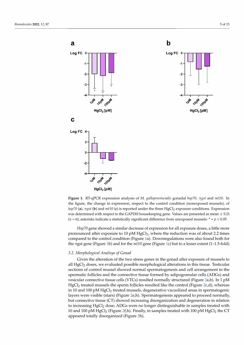
3.2. Morphological Analisys of Gonad
Given the alteration of the two stress genes in the gonad after exposure of mussels toall HgCl2 doses, we evaluated possible morphological alterations in this tissue. Testicularsections of control mussel showed normal spermatogenesis and cell arrangement in thespermatic follicles and the connective tissue formed by adipogranular cells (ADGs) andvesicular connective tissue cells (VTCs) resulted normally structured (Figure 2a,b). In 1 pMHgCl2 treated mussels the sperm follicles resulted like the control (Figure 2c,d), whereasin 10 and 100 pM HgCl2 treated mussels, degenerative vacuolized areas in spermatogeniclayers were visible (stars) (Figure 2e,h). Spermatogenesis appeared to proceed normally,but connective tissue (CT) showed increasing disorganization and degeneration in relationto increasing HgCl2 dose; ADGs were no longer distinguishable in samples treated with10 and 100 pM HgCl2 (Figure 2f,h). Finally, in samples treated with 100 pM HgCl2 the CTappeared totally disorganized (Figure 2h).

Biomolecules 2022, 12, 87 6 of 15
Biomolecules 2022, 12, x FOR PEER REVIEW 6 of 16
3.2. Morphological Analisys of Gonad Given the alteration of the two stress genes in the gonad after exposure of mussels to
all HgCl2 doses, we evaluated possible morphological alterations in this tissue. Testicular sections of control mussel showed normal spermatogenesis and cell arrangement in the spermatic follicles and the connective tissue formed by adipogranular cells (ADGs) and vesicular connective tissue cells (VTCs) resulted normally structured (Figure 2a,b). In 1 pM HgCl2 treated mussels the sperm follicles resulted like the control (Figure 2c,d), whereas in 10 and 100 pM HgCl2 treated mussels, degenerative vacuolized areas in sper-matogenic layers were visible (stars) (Figure 2e,h). Spermatogenesis appeared to proceed normally, but connective tissue (CT) showed increasing disorganization and degeneration in relation to increasing HgCl2 dose; ADGs were no longer distinguishable in samples treated with 10 and 100 pM HgCl2 (Figure 2f,h). Finally, in samples treated with 100 pM HgCl2 the CT appeared totally disorganized (Figure 2h).
Figure 2. Staining of M. galloprovincialis gonad with hematoxylin and eosin. Gonad morphology of nonexposed (a,b) and exposed mussels to 1 (c,d), 10 (e,f) and 100 pM (g,h) HgCl2, respectively. CT: connective tissue; VTCs: vesicular connective tissue cells; ADGs: adipogranular cells; * (star): de-generative vacuolized areas.
Figure 2. Staining of M. galloprovincialis gonad with hematoxylin and eosin. Gonad morphol-ogy of nonexposed (a,b) and exposed mussels to 1 (c,d), 10 (e,f) and 100 pM (g,h) HgCl2, respec-tively. CT: connective tissue; VTCs: vesicular connective tissue cells; ADGs: adipogranular cells;* (star): degenerative vacuolized areas.
3.3. Immunohistochemistry
Anti-3β-HSD and anti-17β-HSD antibodies showed the presence of both enzymes inthe gonad, in overlapping manner, so we present in the figure the localization of 3β-HSDonly. The enzymes are present in connective tissue and in germ cells (GC) of controlspecimens (Figure 3a). In the specimens treated with 1 pM HgCl2 (Figure 3b), the labellingon connective tissue (CT) was still present, decreasing instead in germ cells. Antibodypositivity decreased even more in specimens treated with 10 pM HgCl2 (Figure 3c) andcompletely disappeared in specimens treated with 100 pM HgCl2 (Figure 3d).

Biomolecules 2022, 12, 87 7 of 15
Biomolecules 2022, 12, x FOR PEER REVIEW 7 of 16
3.3. Immunohistochemistry Anti-3β-HSD and anti-17β-HSD antibodies showed the presence of both enzymes in
the gonad, in overlapping manner, so we present in the figure the localization of 3β-HSD only. The enzymes are present in connective tissue and in germ cells (GC) of control spec-imens (Figure 3a). In the specimens treated with 1 pM HgCl2 (Figure 3b), the labelling on connective tissue (CT) was still present, decreasing instead in germ cells. Antibody posi-tivity decreased even more in specimens treated with 10 pM HgCl2 (Figure 3c) and com-pletely disappeared in specimens treated with 100 pM HgCl2 (Figure 3d).
Figure 3. Immunolabeling of M. galloprovincialis male gonad with anti-3β-HSD antibody. Brown coloration indicates the enzyme presence. Nonexposed (a) and exposed mussels to 1 (b), 10 (c) and 100 pM (d) HgCl2, respectively. Germ cells (GC) are positive to antibody in control and 1 and 10 pM treated samples (a–c); the positivity disappears almost completely in 100 pM treated samples (d) In the connective cells (CT, connective tissue) the 3β-HSD enzyme is recognizable in control and 1 pM treated animals (a,b); in 10 and 100 pM treated samples the antibody positivity is poor (c) or totally disappeared (d).
3.4. Gonadal PL-Proteins Genes Expression Considering the morphological changes and enzymes changes involved in hormones
synthesis found in the gonad of mussels exposed to these doses of HgCl2, we also evalu-ated in this tissue the expression levels of genes encoding PL-proteins, as these are the major basic protein component of the sperm chromatin of this organism. In gonads, the PLII/IV was found to be slightly downregulated under all exposure conditions. A more marked downregulation was instead detected for the PL-III gene, under all exposure con-ditions but particularly at 10 pM HgCl2, where an approximately 2-fold decrease was measured (Figure 4).
Figure 3. Immunolabeling of M. galloprovincialis male gonad with anti-3β-HSD antibody. Browncoloration indicates the enzyme presence. Nonexposed (a) and exposed mussels to 1 (b), 10 (c) and100 pM (d) HgCl2, respectively. Germ cells (GC) are positive to antibody in control and 1 and 10 pMtreated samples (a–c); the positivity disappears almost completely in 100 pM treated samples (d)In the connective cells (CT, connective tissue) the 3β-HSD enzyme is recognizable in control and1 pM treated animals (a,b); in 10 and 100 pM treated samples the antibody positivity is poor (c) ortotally disappeared (d).
3.4. Gonadal PL-Proteins Genes Expression
Considering the morphological changes and enzymes changes involved in hormonessynthesis found in the gonad of mussels exposed to these doses of HgCl2, we also evaluatedin this tissue the expression levels of genes encoding PL-proteins, as these are the majorbasic protein component of the sperm chromatin of this organism. In gonads, the PLII/IVwas found to be slightly downregulated under all exposure conditions. A more markeddownregulation was instead detected for the PL-III gene, under all exposure conditionsbut particularly at 10 pM HgCl2, where an approximately 2-fold decrease was measured(Figure 4).
Biomolecules 2022, 12, x FOR PEER REVIEW 8 of 16
Figure 4. RT-qPCR expression analysis of M. galloprovincialis gonadal PLIII and PLII/IV genes. In the figure, the change in gene expression is reported under the three HgCl2 exposure conditions compared to the control condition (nonexposed mussels: CTR). Expression was determined with respect to the GAPDH housekeeping gene. Values are presented as mean ± S.D. (n = 6).
3.5. MNase Digestion Pattern of M. Galloprovincialis’sperm Nuclei Figure 5 shows the MNase digestion time course of M. galloprovincialis’ sperm chro-
matin for 5, 15, 30, and 60 min. The analyses were conducted on sperm chromatin of mus-sels nonexposed and exposed to the three HgCl2 exposure doses. In this approach, DNA contents of the fractions obtained at different times of MNase digestion were analysed. The results show that in unexposed mussels (Figure 5a) MNase digestion of sperm chro-matin results in the electrophoretic pattern typical of sperm chromatin of this organism. Chromatin appears mainly as a smear, which is indicative of the absence of a typical nu-cleosomal organization. In contrast, following exposure to all doses of HgCl2, the sperm chromatin DNA appears fully degraded upon digestion with Mnase as early as the lowest digestion time (5′), indicative of an improper sperm chromatin structure (Figure 5a,b).
Figure 5. Micrococcal nuclease (MNase) digestion time course of Mytilus galloprovincialis’ sperm chromatin. Analyses of DNA by electrophoresis on agarose gel: unexposed and exposed mussels to 1 pM HgCl2 (Panel (a)), exposed mussels to 10 and 100 pM HgCl2 (Panel (b)). n.d. = undigested sperm genomic DNA. 1Kb and 100 bp: molecular weight markers.
4. Discussion
Figure 4. RT-qPCR expression analysis of M. galloprovincialis gonadal PLIII and PLII/IV genes. Inthe figure, the change in gene expression is reported under the three HgCl2 exposure conditionscompared to the control condition (nonexposed mussels: CTR). Expression was determined withrespect to the GAPDH housekeeping gene. Values are presented as mean ± S.D. (n = 6).

Biomolecules 2022, 12, 87 8 of 15
3.5. MNase Digestion Pattern of M. Galloprovincialis’sperm Nuclei
Figure 5 shows the MNase digestion time course of M. galloprovincialis’ sperm chro-matin for 5, 15, 30, and 60 min. The analyses were conducted on sperm chromatin ofmussels nonexposed and exposed to the three HgCl2 exposure doses. In this approach,DNA contents of the fractions obtained at different times of MNase digestion were anal-ysed. The results show that in unexposed mussels (Figure 5a) MNase digestion of spermchromatin results in the electrophoretic pattern typical of sperm chromatin of this organ-ism. Chromatin appears mainly as a smear, which is indicative of the absence of a typicalnucleosomal organization. In contrast, following exposure to all doses of HgCl2, the spermchromatin DNA appears fully degraded upon digestion with Mnase as early as the lowestdigestion time (5′), indicative of an improper sperm chromatin structure (Figure 5a,b).
Biomolecules 2022, 12, x FOR PEER REVIEW 8 of 16
Figure 4. RT-qPCR expression analysis of M. galloprovincialis gonadal PLIII and PLII/IV genes. In the figure, the change in gene expression is reported under the three HgCl2 exposure conditions compared to the control condition (nonexposed mussels: CTR). Expression was determined with respect to the GAPDH housekeeping gene. Values are presented as mean ± S.D. (n = 6).
3.5. MNase Digestion Pattern of M. Galloprovincialis’sperm Nuclei Figure 5 shows the MNase digestion time course of M. galloprovincialis’ sperm chro-
matin for 5, 15, 30, and 60 min. The analyses were conducted on sperm chromatin of mus-sels nonexposed and exposed to the three HgCl2 exposure doses. In this approach, DNA contents of the fractions obtained at different times of MNase digestion were analysed. The results show that in unexposed mussels (Figure 5a) MNase digestion of sperm chro-matin results in the electrophoretic pattern typical of sperm chromatin of this organism. Chromatin appears mainly as a smear, which is indicative of the absence of a typical nu-cleosomal organization. In contrast, following exposure to all doses of HgCl2, the sperm chromatin DNA appears fully degraded upon digestion with Mnase as early as the lowest digestion time (5′), indicative of an improper sperm chromatin structure (Figure 5a,b).
Figure 5. Micrococcal nuclease (MNase) digestion time course of Mytilus galloprovincialis’ sperm chromatin. Analyses of DNA by electrophoresis on agarose gel: unexposed and exposed mussels to 1 pM HgCl2 (Panel (a)), exposed mussels to 10 and 100 pM HgCl2 (Panel (b)). n.d. = undigested sperm genomic DNA. 1Kb and 100 bp: molecular weight markers.
4. Discussion
Figure 5. Micrococcal nuclease (MNase) digestion time course of Mytilus galloprovincialis’ spermchromatin. Analyses of DNA by electrophoresis on agarose gel: unexposed and exposed musselsto 1 pM HgCl2 (Panel (a)), exposed mussels to 10 and 100 pM HgCl2 (Panel (b)). n.d. = undigestedsperm genomic DNA. 1Kb and 100 bp: molecular weight markers.
4. Discussion
Mercury (Hg) is a global environmental pollutant that affects ecosystem health andmarine organisms [23,24], due to its significant implications on several biological processes,including growth, sexual maturation, and reproductive success [25]. Contamination of toxicsubstances in the environment, such as mercury, has been known to have socio-economicconsequences. These effects have led to a limitation of Hg emissions from large-scale hu-man activities. Bivalves are important species in coastal ecosystems, and play an importantrole particularly in food webs (as suspension feeders) and represent a significant fractionof fishery resources. In recent years, mussels have become a commercially importantseafood species worldwide. Although the consumption of mussels is considered a safefood, bioaccumulation of heavy metals remains a problem for mussel consumers and thishas an impact not only on human health but also on the economy [26]. Mussels, in fact,have strong interactions with the environment, water, and sediments and are consideredgood bioindicator species. In particular, M. galloprovincialis is an important food source,widespread throughout the world with high ability to bioaccumulate pollutants. So, theingestion of drug-contaminated bivalves can pose a health risk to the human popula-tion [27], e.g., potential antibiotic resistance [28]. Thus, many pollutants can reach humansthrough mussels. These include: carbamazepine (CBZ), an antiepileptic drug used in thetreatment of epilepsy, neuropathic pain, and psychiatric disorders [29], microplastics [30],and heavy metals, such as mercury (Hg), lead (Pb), chromium (Cr), and cadmium (Cd).Long-term exposure to these substances can produce neurological, physiological, andphysical problems [31].
Mussels have also shown a greater capacity for Hg accumulation than other bivalvespecies [16], and the gonads have a particular affinity for mercury like other endocrine organs [32].
For these reasons and considering that the combination of morphological changesand transcriptional responses is recognized as valuable and promising tools for the studyof ecotoxicological effects [33], in the present work, we first investigated changes in male

Biomolecules 2022, 12, 87 9 of 15
gonadal morphology and the level of steroidogenic enzymes after exposure of M. gallo-provincialis for 24 h to 1, 10, 100 pM HgCl2. In addition, we assessed both the expressionlevels of three stress genes and the PL protein-coding genes and determined the differencein the accessibility of sperm chromatin to micrococcal nuclease at these doses of exposureto HgCl2.
First of all, we registered gonadal stress because RT-qPCR analyses showed alteredexpression of the stress genes, mt10, π-gst and hsp70. As reported by several authors [34,35]Mytilus species are able to synthesize metallothionenins, encoded by mt genes, in order toeliminate accumulated metals. Metallothionenins (MTs) are expressed in tissue, cell, andisoform specific modes depending on the type of metal they are exposed, including essentialmetals like copper and zinc and nonessential metals like cadmium and mercury. BanniM. et al., 2007 [36], based on laboratory exposures indicated that copper, zinc, mercuryand cadmium, are able to induce the expression of this stress gene in mussels. Literaturehas reported work indicating alterations in M. galloprovincialis mt10 gene expression afteraccumulation of various toxic metals, both essential, such as copper and zinc, and non-essential, such as cadmium and mercury but prevalently in digestive gland [37] in which,generally an increase of expression of these genes has been observed. However, there is adistinction between zinc and copper because these two metals act differently. First of all,Zn is a DNA stabilizer involved in transcription. It does not generate free radicals unlikeCu which is necessary for some redox reactions. Moreover, it can generate like Fe somefree radicals chain reactions. Zn can displace Hg from interacting with proteins. In fact, itis sometimes administered in mammals as an anti Hg poisoning. Moreover, Hg uptake isdecreased in the presence of Zn and recently have been reported new insight on the geneticbasis of Zn and Cu accumulation in molluscs [38,39]. Zinc is an essential trace element forspermatogenesis. In mammals the Cu/Zn ratio is a marker of oxidative stress and severalstudies have suggested that later germ cells are less tolerant to ROS than early-type germcells, mainly because of their limited reserve of antioxidant enzymes. This is due to the factthat zinc levels decrease during spermatogenesis. Zinc, in fact, plays an important role as aDNA stabilizer, being essential for several DNA repair enzymes that are important duringearly embryogenesis [40] and modulates SOD activity [41].
In the gonad of M. galloprovincialis, increased gene expression of mt20 and hsp70 wasreported after exposure to cadmium in [13,14] whereas in our present study, we observed adecrease in the expression of both mt10 and hsp70 after mercury exposure, consistent withwhat was achieved after exposure of M. galloprovincialis to copper [12].Also π-gst geneswas hypo-expressed after exposure of mussels to all doses of HgCl2.
In our present study, after 1 pM HgCl2 exposure, there was an approximately2-fold increase in expression of mt10 gene, while after 10 and 100 pM HgCl2 exposure, weobserved a hypo-expression of this gene which resulted of 1.2- and 1.5-fold, respectively,compared to the control condition. To our knowledge, there is no direct correlation betweenmt10, 3β-HSD and 17β-HSD expression. A possible relationship, at the transcriptionallevel, between these genes has never been reported in literature. However, it cannot beexcluded that mercury, at this dose, may similarly affect the transcription factors regulat-ing the expression of these genes. A hypoexpression was also observed for mt20 gene inmussel digestive gland after exposure to copper (Cu) [36]. In spermatozoa, however, asdemonstrated in Piscopo et al., 2021 [11], mt10 gene did not show significant differencesafter 1 pM HgCl2 exposure, while an increase in expression levels after mussels exposureto 10 and 100 pM HgCl2 was found, in particular an increase of about three times in theexpression of this gene.
The different response found in mussels spermatozoa and gonads exposed to HgCl2could suggest higher effectiveness of spermatozoa in the response to mercury, comparedwith gonads. A different response from spermatozoa and gonads was previously observedin response to subtoxic concentrations of copper [10].
Heat shock proteins (HSPs) are a group of ubiquitous proteins witch expression isactivated by thermal stress [42]. Other stimuli that influence cell protein structure and

Biomolecules 2022, 12, 87 10 of 15
function, including oxidative stress and heavy metals, can induce heat shock genes [43]. Inunstressed cells, some members of the HSP family are produced constitutively and operateas molecular chaperones, assisting protein maturation steps such as folding, unfolding, andtranslocation across membranes [44]. HSPs are classified based on their molecular weight.The stress-inducible hsp70 and constitutively expressed hsc70 proteins are well-knownmembers of the HSP70 (70 kDa) multigene family. HSP70 acts as a cell damage defencesystem, whereas HSC70 acts as a molecular chaperone. In our present research stress-inducible hsp70 gene showed a hypo-expression in all exposure doses, more pronounced at10 pM HgCl2 exposure condition and in the order of approximately 2.2-fold compared tothe control condition. A hypo-expression of hsp70 gene was also find in M. galloprovincialisspermatozoa after 1 and 10 pM HgCl2 exposure [11]. To our knowledge, this is the first timethat has been demonstrated a downregulation of the hsp70 gene in gonads under metallicstress at low concentrations and within short acute exposure, as well as in spermatozoa [11].However, this is not the first time that downregulation of this gene has been observedin response to a xenobiotic. In fact, a decrease in the expression of hsp70 gene has beenobserved in response to paracetamol in blue mussel Mytilus edulis [45]. Hsp70 preservesprotein integrity and suppresses apoptosis directly, whereas thermal and oxidative stressdamage protein structure. Hsp70 is implicated in inducible stress and is involved innumerous biological processes: cancer, autophagy [46], apoptosis [47] and necrosis [48].This implies that when HSP70 is downregulated in mussels exposed to this elements, agreater risk of apoptosis and cell death is induced [45].
Hypo-expression was also found for the π-gst gene, the main glutathione s- transferaseisoform expressed in mussel tissues [49]. Also in this case, no linearity was observed withrespect to the three exposure doses. The most marked hypo-expression was obtained after10 pM HgCl2 exposure, and was of the order of 1.5-fold compared to control condition. Onepossible explanation for down-regulation of π-gst gene could reside in a potent activationof the lysosomal-vacuolar system and, subsequently, in the enhancement of autophagy [50],a condition that may mask anabolic processes such as increased gene transcription, inagreement with the hypothesis of Dondero et al., 2006 [51].
The gonadal stress responses observed by RT-qPCR prompted us to investigate on thepossible damages mercury-induced on this tissue. We found that the treatment with 1 pMHgCl2 did not give morphological changes in the gonad respect to the control, while thetwo other doses, 10 and 100 pM HgCl2, caused vacuolized areas between the germ cellsand the complete disorganization of the connective tissue. We used gonads at the maturestage, on mussels obtained in January and vacuolized areas were observed only in musselsexposed to HgCl2 and not in control condition. The observed morphological changes ofseminiferous follicles, as vacuolization and decrease of germ cell compartment thickness,were reported also in fish [52] and in mammal testis [53,54] after HgCl2 treatment. Agrowing number of studies show that sex steroids are widespread in molluscs [55]. Initially,they were thought to be taken through the diet, since many plant species contain sexsteroids similar to those in vertebrates [56]. Nevertheless, numerous studies have shownthat the major classes of molluscs, i.e., cephalopods, gastropods, and bivalves, are capableof synthesizing sex steroids from precursors such as cholesterol or pregnenolone [57–59].Actually, most of the steroidogenic pathways described for vertebrates have been shown tooccur in molluscs.
In literature, it has been already demonstrated the negative impact of mercury onspermatogenesis and also steroidogenesis: in male catfish exposed to organic and inor-ganic mercury the activity of 3β-HSD was inhibited completely [60], as we observed inMytilus treated with the higher HgCl2 doses. 3β-HSD is implicated in the conversionof dehydroepiandrosterone (DHEA) in androstenedione. The 3-HSD family of enzymesalso catalyzes the synthesis and/or degradation of 5-androstanes and 5-pregnanes. Asa result, 3β -HSD regulates key steroid hormone-related responses in the adrenal cortex,gonads, placenta, liver, and other peripheral target organs. In general, all steroid hormones,including glucocorticoids, mineralocorticoids, progesterone, androgens, and oestrogens,

Biomolecules 2022, 12, 87 11 of 15
require 3β-HSD for production [61]. 17β-HSD, on the other hand, mediates the conversionof androstenedione (ASD) to testosterone (T). However, 17β-HSD have a wide range offunctions, such as the regulation of not only steroids but also fatty acid and bile concentra-tions [62]. Our results showed a positive reaction after labelling with both anti-3β-HSDand anti-17β-HSD antibodies at the level of germ cells and connective tissue in the controlspecimen as previously reported [22], germ cells are positive to antibody in 1 pM and 10 pMtreated samples also, but the positivity disappears almost completely in 100 pM treatedsamples. In the connective tissue the steroidogenic enzymes are recognizable in control and1 pM treated animal, whereas in 10 and 100 pM treated samples the antibody positivity isvery poor. A decrease in spermatogenesis, 17β-HSD, 3β-HSD and also testosterone level inplasma was shown in male mice after exposure to arsenic trioxide [32]. In the light of theseresults, we examined on gonads the expression of genes encoding PL-proteins, as theseare the major component of the basic proteins that constitute the sperm chromatin of thisorganism [63]. A decrease of expression was found in the case of the PLIII gene in exposedmussels. The downregulation was observed at all exposure conditions but in particular afterexposure to 10 pM HgCl2. In addition, a slight hypo-expression of PLII-PLIV was detected.These results highlights the negative impact of mercury also on gonadal protamine-like (PL)genes, proteins essential for correct DNA compaction in M. galloprovincialis sperm heads. Asimilar result was found after mussels exposure to sub-toxic concentrations of copper [12].These alterations in the PL gene could be associated with changes in 3β-HSD and 17β-HSDexpression that we observed by immunolabelling as mercury has already been shown todisrupt hormones, including testosterone [64]. Both enzymes play an important role inhormone regulation, in particular for testosterone production and its involvement in sper-matogenesis [22]. These results are in line with those observed in experiments previouslyconducted following exposure to sub-toxic copper concentrations [12] and those relatingto the accumulation of Polycyclic Aromatic Hydrocarbons (PAH) and heavy metals in themale gonad of M. galloprovincialis showing that these pollutants disrupts spermatogenesisand produce alterations in somatic and germ cells [65]. The testicular toxicities of mercuri-als, including impaired spermatogenesis and/or steroidogenesis, have been demonstratedin a number of laboratory animal species: fish [50,66–69], fowls [67]. Furthermore, this hasalso been demonstrated in the mouse [66,70].
The morphological and hormonal alterations observed in the gonad and the changesfound for both stress genes and those encoding PLs prompted us to investigate possiblealterations in the sperm chromatin structure of mussels exposed to the three doses ofHgCl2. Results obtained by digestion with MNase show substantial differences betweencontrol and treated at all three doses of HgCl2. In fact, the DNA of the sperm nucleiwas totally hydrolyzed after digestion with MNase in mussels exposed to all exposureconditions, contrary to what was observed in unexposed mussels in which, despite there isno nucleosomal organization, and then a smear is obtained after sperm chromatin digestionof spermatozoa with MNase but not a total degradation of DNA. These results are inline with the alterations we had highlighted in our previous works showing a differencein PL conformation upon exposure of mussels to HgCl2 and their different binding toDNA [10,11]. The release of PL proteins from sperm nuclei was also altered especially forPLII and PLIII at all doses of HgCl2 exposure [10]. Far more significant was the evidence, wehave already provided, concerning the effects of the addition of PL from mussels exposedto all three HgCl2 doses to a plasmid DNA under pro-oxidant conditions. The resultshowed the breakage of DNA and a non-protection of the latter unlike what happenedwith PL from mussels not exposed [10,71,72]. Thus, the result we obtained with MNasedigestion are quite predictable as it indicates that in mussels exposed to these doses ofHgCl2 there might be an improper structure of the sperm chromatin. This could indicate apossible risk in the fertilizing capacity of spermatozoa from mussels exposed to these dosesof HgCl2. After all, in literature, mercury is recognized as a male reproductive toxicant.In fact, studies performed in vitro have shown that this metal induces DNA breakage inspermatozoa and causes a decrease in sperm motility. Our previous work provides insights

Biomolecules 2022, 12, 87 12 of 15
into the mechanisms of mercury toxicity on the reproductive system of M. galloprovincialis.Specifically, the alteration of PL-proteins binding to DNA induced by exposure to theseHgCl2 doses [10]. This is in line with the results obtained with MNase digestion and couldindicate an alteration in the structure of sperm chromatin, which is known to be critical forthe swimming ability of spermatozoa and their ability to fertilize. In any case, however,our results are currently only descriptive as fertilization tests with spermatozoa of musselsexposed to these doses of HgCl2 have not yet been conducted. Therefore, it cannot bepredicted at this time whether this may have a negative impact from an economic point ofview. Further study will allow to understand if the genotoxic effect of HgCl2 in musselscould become an environmental problem since our ultimate goal is to develop genotoxicitytests based on sperm chromatin status.
In conclusion, the data obtained in this work could be an important step in understand-ing the mechanisms of mercury toxicity and reveal responses in a tissue such as the gonadthat is generally not considered for ecotoxicological studies. In addition, our results againemphasize the importance of using M. galloprovincialis as a sentinel organism and suggest apossible risk on the fertilizing capacity of spermatozoa in mussels exposed to these dosesof HgCl2 as revealed by the results of experiments conducted with MNase. However, theseresults will need to be further investigated with additional studies to understand how thesemechanisms impact on the reproductive health of M. galloprovincialis.
Moreover, heavy metal contamination can induce an ecological imbalance in thereceiving environment and on the diversity of aquatic organisms. Pollutants accumulate inthe food chain and are responsible for harmful effects on the occurrence of various diseases,such as Minamata disease (organic mercury poisoning). Therefore, preventive measuresare needed to reduce the intensity of heavy metal pollution in the aquatic environment [73].
Author Contributions: Conceptualization, M.P. (Marina Piscopo) and G.L.; methodology, M.P.(Marina Piscopo), G.L. and R.N.; software, G.L.; validation, M.P. (Marina Piscopo) and C.M.; formalanalysis, G.L.; investigation, G.L., R.N., N.C., M.P. (Marina Prisco), A.A. and S.P.; Data curation, G.L.,R.N., N.C.; writing—original draft preparation, M.P., G.L., N.C., R.N., M.P. (Marina Prisco), A.A. andS.P.; writing—review and editing, M.P. (Marina Piscopo), G.L., N.C. visualization, G.L.; supervision,M.P. (Marina Piscopo); project administration, M.P. (Marina Piscopo). All authors have read andagreed to the published version of the manuscript.
Funding: This research received no external funding.
Institutional Review Board Statement: Not applicable.
Informed Consent Statement: Not applicable.
Data Availability Statement: Not applicable.
Conflicts of Interest: The authors declare no conflict of interest.
Abbreviations
CT Connective tissueVTC Vesicular connective tissue cellsADGs Adipogranular cells17β-HSD 17β-Hydroxysteroid dehydrogenases3β-HSD β-Hydroxysteroid dehydrogenase/∆5−4 isomerasePL- proteins Protamine-LikeH&E Hematoxylin and EosinASD AndrostenedioneT TestosteroneDHEA Dehydroepiandrosteronen.d.MNase UndigestMicrococcal nuclease

Biomolecules 2022, 12, 87 13 of 15
References1. Outridge, P.M.; Mason, R.P.; Wang, F.; Guerrero, S.; Heimbürger-Boavida, L.E. Updated Global and Oceanic Mercury Budgets for
the United Nations Global Mercury Assessment 2018. Environ. Sci. Technol. 2018, 52, 11466–11477. [CrossRef] [PubMed]2. Tortora, F.; Notariale, R.; Maresca, V.; Good, K.V.; Sorbo, S.; Basile, A.; Piscopo, M.; Manna, C. Phenol-Rich Feijoa Sellowiana
(Pineapple guava) Extracts Protect Human Red Blood Cells from Mercury-Induced Cellular Toxicity. Antioxidants 2019, 8, 220.[CrossRef] [PubMed]
3. Piscopo, M.; Notariale, R.; Tortora, F.; Lettieri, G.; Palumbo, G.; Manna, C. Novel Insights into Mercury Effects on Hemoglobinand Membrane Proteins in Human Erythrocytes. Molecules 2020, 25, 3278. [CrossRef]
4. Officioso, A.; Alzoubi, K.; Lang, F.; Manna, C. Hydroxytyrosol Inhibits Phosphatidylserine Exposure and Suicidal Death Inducedby Mercury in Human Erythrocytes: Possible Involvement of the Glutathione Pathway. Food Chem. Toxicol. 2016, 89, 47–53.[CrossRef] [PubMed]
5. Fathallah, S.; Medhioub, M.N.; Medhioub, A.; Kraiem, M.M. Toxicity of Hg, Cu and Zn on Early Developmental Stages of theEuropean Clam (Ruditapes decussatus) with Potential Application in Marine Water Quality Assessment. Environ. Monit. Assess.2010, 171, 661–669. [CrossRef]
6. Depledge, M.H.; Aagaard, A.; Györkös, P. Assessment of Trace Metal Toxicity Using Molecular, Physiological and BehaviouralBiomarkers. Mar. Pollut. Bull. 1995, 31, 19–27. [CrossRef]
7. Bresler, V.; Bissinger, V.; Abelson, A.; Dizer, H.; Sturm, A.; Kratke, R.; Fishelson, L.; Hansen, P.-D. Marine Molluscs and Fishas Biomarkers of Pollution Stress in Littoral Regions of the Red Sea, Mediterranean Sea and North Sea. Helgol. Mar. Res. 1999,53, 219–243. [CrossRef]
8. Viarengo, A.; Canesi, L. Mussels as Biological Indicators of Pollution. Aquaculture 1991, 94, 225–243. [CrossRef]9. Piscopo, M. Seasonal Dependence of Cadmium Molecular Effects on Mytilus galloprovincialis (Lamarck, 1819) Protamine-like
Protein Properties. Mol. Reprod. Dev. 2019, 86, 1418–1429. [CrossRef]10. Lettieri, G.; Notariale, R.; Carusone, N.; Giarra, A.; Trifuoggi, M.; Manna, C.; Piscopo, M. New Insights into Alterations in
PL Proteins Affecting Their Binding to DNA after Exposure of Mytilus galloprovincialis to Mercury-A Possible Risk to SpermChromatin Structure? Int. J. Mol. Sci. 2021, 22, 5893. [CrossRef]
11. Lettieri, G.; Notariale, R.; Ambrosino, A.; Di Bonito, A.; Giarra, A.; Trifuoggi, M.; Manna, C.; Piscopo, M. SpermatozoaTranscriptional Response and Alterations in PL Proteins Properties after Exposure of Mytilus galloprovincialis to Mercury. Int. J.Mol. Sci. 2021, 22, 1618. [CrossRef]
12. Lettieri, G.; Mollo, V.; Ambrosino, A.; Caccavale, F.; Troisi, J.; Febbraio, F.; Piscopo, M. Molecular Effects of Copper on theReproductive System of Mytilus galloprovincialis. Mol. Reprod. Dev. 2019, 86, 1357–1368. [CrossRef]
13. Piscopo, M.; Notariale, R.; Rabbito, D.; Ausió, J.; Olanrewaju, O.S.; Guerriero, G. Mytilus galloprovincialis (Lamarck, 1819)Spermatozoa: Hsp70 Expression and Protamine-like Protein Property Studies. Environ. Sci. Pollut. Res. Int. 2018, 25, 12957–12966.[CrossRef]
14. Piscopo, M.; Trifuoggi, M.; Notariale, R.; Labar, S.; Troisi, J.; Giarra, A.; Rabbito, D.; Puoti, R.; de Benedictis, D.; Brundo, M.V.; et al.Protamine-like Proteins’ Analysis as an Emerging Biotechnique for Cadmium Impact Assessment on Male Mollusk Mytilusgalloprovincialis (Lamarck 1819). Acta Biochim. Pol. 2018, 65, 259–267. [CrossRef]
15. Vassalli, Q.A.; Caccavale, F.; Avagnano, S.; Murolo, A.; Guerriero, G.; Fucci, L.; Ausió, J.; Piscopo, M. New Insights into Protamine-like Component Organization in Mytilus galloprovincialis’ Sperm Chromatin. DNA Cell Biol. 2015, 34, 162–169. [CrossRef]
16. Briant, N.; Chouvelon, T.; Martinez, L.; Brach-Papa, C.; Chiffoleau, J.; Savoye, N.; Sonke, J.; Knoery, J. Spatial and TemporalDistribution of Mercury and Methylmercury in Bivalves from the French Coastline. Mar. Pollut. Bull. 2017, 114, 1096–1102.[CrossRef]
17. Cinnirella, S.; Bruno, D.E.; Pirrone, N.; Horvat, M.; Živkovic, I.; Evers, D.C.; Johnson, S.; Sunderland, E.M. Mercury Concentrationsin Biota in the Mediterranean Sea, a Compilation of 40 Years of Surveys. Sci. Data 2019, 6, 205. [CrossRef]
18. Piscopo, M.; Ricciardiello, M.; Palumbo, G.; Troisi, J. Selectivity of Metal Bioaccumulation and Its Relationship with GlutathioneS-Transferase Levels in Gonadal and Gill Tissues of Mytilus galloprovincialis Exposed to Ni (II), Cu (II) and Cd (II). Rend. Fis. Acc.Lincei 2016, 27, 737–748. [CrossRef]
19. Lettieri, G.; Maione, M.; Ranauda, M.A.; Mele, E.; Piscopo, M. Molecular Effects on Spermatozoa of Mytilus galloprovincialisExposed to Hyposaline Conditions. Mol. Reprod. Dev. 2019, 86, 650–660. [CrossRef] [PubMed]
20. Livak, K.J.; Schmittgen, T.D. Analysis of Relative Gene Expression Data Using Real-Time Quantitative PCR and the 2(-Delta DeltaC(T)) Method. Methods 2001, 25, 402–408. [CrossRef] [PubMed]
21. Olivares, C.; Ruiz, S. Nucleosomal Organization of Chromatin in Sperm Nuclei of the Bivalve Mollusc Aulacomya Ater. Mol. CellBiochem. 1991, 101, 93–99. [CrossRef] [PubMed]
22. Prisco, M.; Agnese, M.; De Marino, A.; Andreuccetti, P.; Rosati, L. Spermatogenic Cycle and Steroidogenic Control of Spermatoge-nesis in Mytilus galloprovincialis Collected in the Bay of Naples. Anat. Rec. 2017, 300, 1881–1894. [CrossRef] [PubMed]
23. al-Hashimi, A.H.; al-Zorba, M.A. Mercury in Some Commercial Fish from Kuwait: A Pilot Study. Sci. Total Environ. 1991,106, 71–82. [CrossRef]
24. Ganguli, P.M.; Conaway, C.H.; Swarzenski, P.W.; Izbicki, J.A.; Flegal, A.R. Mercury Speciation and Transport via SubmarineGroundwater Discharge at a Southern California Coastal Lagoon System. Environ. Sci. Technol. 2012, 46, 1480–1488. [CrossRef][PubMed]

Biomolecules 2022, 12, 87 14 of 15
25. Thain, J.E. Effects of Mercury on the Prosobranch Mollusc Crepidula fornicata: Acute Lethal Toxicity and Effects on Growth andReproduction of Chronic Exposure. Mar. Environ. Res. 1984, 12, 285–309. [CrossRef]
26. Stankovic, S.; Jovic, M. Health Risks of Heavy Metals in the Mediterranean Mussels as Seafood. Environ. Chem. Lett. 2012,10, 119–130. [CrossRef]
27. Almeida, Â.; Esteves, V.I.; Soares, A.M.V.M.; Freitas, R. Effects of Carbamazepine in Bivalves: A Review. In Reviews of EnvironmentalContamination and Toxicology Volume 254; de Voogt, P., Ed.; Reviews of Environmental Contamination and Toxicology; SpringerInternational Publishing: Cham, Switzerland, 2021; pp. 163–181. ISBN 978-3-030-68530-0.
28. Rodrigues, J.; Albino, S.; Silva, S.; Cravo, A.; Cardoso, V.V.; Benoliel, M.J.; Almeida, C.M.M. Development of a MultiresidueMethod for the Determination of 24 Pharmaceuticals in Clams by QuEChERS and Liquid Chromatography-Triple QuadrupoleTandem Mass Spectrometry. Food Anal. Methods 2019, 12, 838–851. [CrossRef]
29. Ambrósio, A.F.; Soares-da-Silva, P.; Carvalho, C.M.; Carvalho, A.P. Mechanisms of Action of Carbamazepine and Its Derivatives,Oxcarbazepine, BIA 2-093, and BIA 2-024. Neurochem. Res. 2002, 27, 121–130. [CrossRef]
30. Li, J.; Lusher, A.L.; Rotchell, J.M.; Deudero, S.; Turra, A.; Bråte, I.L.N.; Sun, C.; Shahadat Hossain, M.; Li, Q.; Kolandhasamy, P.; et al.Using Mussel as a Global Bioindicator of Coastal Microplastic Pollution. Environ. Pollut. 2019, 244, 522–533. [CrossRef]
31. Zaynab, M.; Al-Yahyai, R.; Ameen, A.; Sharif, Y.; Ali, L.; Fatima, M.; Khan, K.A.; Li, S. Health and Environmental Effects of HeavyMetals. J. King Saud Univ. Sci. 2022, 34, 101653. [CrossRef]
32. Khayat, A.; Dencker, L. Organ and Cellular Distribution of Inhaled Metallic Mercury in the Rat and Marmoset Monkey (Callithrixjacchus): Influence of Ethyl Alcohol Pretreatment. Acta Pharmacol. Toxicol. 1984, 55, 145–152. [CrossRef]
33. Viganò, L.; Casatta, N.; Farkas, A.; Mascolo, G.; Roscioli, C.; Stefani, F.; Vitelli, M.; Olivo, F.; Clerici, L.; Robles, P.; et al.Embryo/Larval Toxicity and Transcriptional Effects in Zebrafish (Danio rerio) Exposed to Endocrine Active Riverbed Sediments.Environ. Sci. Pollut. Res. Int. 2020, 27, 10729–10747. [CrossRef]
34. Köhler, K.; Riisgård, H.U. Formation of Metallothioneins in Relation to Accumulation of Cadmium in the Common MusselMytilus Edulis. Mar. Biol. 1982, 66, 53–58. [CrossRef]
35. Ceratto, N.; Dondero, F.; van de Loo, J.W.; Burlando, B.; Viarengo, A. Cloning and Sequencing of a Novel Metallothionein Gene inMytilus galloprovincialis Lam. Comp. Biochem. Physiol. C Toxicol. Pharmacol. 2002, 131, 217–222. [CrossRef]
36. Banni, M.; Dondero, F.; Jebali, J.; Guerbej, H.; Boussetta, H.; Viarengo, A. Assessment of Heavy Metal Contamination UsingReal-Time PCR Analysis of Mussel Metallothionein Mt10 and Mt20 Expression: A Validation along the Tunisian Coast. Biomarkers2007, 12, 369–383. [CrossRef]
37. Dondero, F.; Piacentini, L.; Banni, M.; Rebelo, M.; Burlando, B.; Viarengo, A. Quantitative PCR Analysis of Two MolluscanMetallothionein Genes Unveils Differential Expression and Regulation. Gene 2005, 345, 259–270. [CrossRef] [PubMed]
38. Wang, W.-X.; Rainbow, P.S. Influence of Metal Exposure History on Trace Metal Uptake and Accumulation by Marine Invertebrates.Ecotoxicol. Environ. Saf. 2005, 61, 145–159. [CrossRef]
39. Meng, J.; Wang, W.; Shi, R.; Song, K.; Li, L.; Que, H.; Zhang, G. Identification of SNPs Involved in Zn and Cu Accumulation in thePacific Oyster (Crassostrea gigas) by Genome-Wide Association Analysis. Ecotoxicol. Environ. Saf. 2020, 192, 110208. [CrossRef][PubMed]
40. Menezo, Y.; Russo, G.; Tosti, E.; El Mouatassim, S.; Benkhalifa, M. Expression Profile of Genes Coding for DNA Repair in HumanOocytes Using Pangenomic Microarrays, with a Special Focus on ROS Linked Decays. J. Assist. Reprod. Genet. 2007, 24, 513–520.[CrossRef] [PubMed]
41. Aitken, A. Protein Consensus Sequence Motifs. Mol. Biotechnol. 1999, 12, 13. [CrossRef]42. Nover, L.; Scharf, K.D. Heat Stress Proteins and Transcription Factors. Cell Mol. Life Sci. 1997, 53, 80–103. [CrossRef] [PubMed]43. Feder, M.E.; Hofmann, G.E. Heat-Shock Proteins, Molecular Chaperones, and the Stress Response: Evolutionary and Ecological
Physiology. Annu. Rev. Physiol. 1999, 61, 243–282. [CrossRef] [PubMed]44. Franzellitti, S.; Fabbri, E. Differential HSP70 Gene Expression in the Mediterranean Mussel Exposed to Various Stressors. Biochem.
Biophys. Res. Commun. 2005, 336, 1157–1163. [CrossRef] [PubMed]45. Koagouw, W.; Ciocan, C. Effects of Short-Term Exposure of Paracetamol in the Gonads of Blue Mussels Mytilus Edulis. Environ.
Sci. Pollut. Res. Int. 2020, 27, 30933–30944. [CrossRef]46. Albakova, Z.; Armeev, G.A.; Kanevskiy, L.M.; Kovalenko, E.I.; Sapozhnikov, A.M. HSP70 Multi-Functionality in Cancer. Cells
2020, 9, 587. [CrossRef] [PubMed]47. Gao, Y.; Han, C.; Huang, H.; Xin, Y.; Xu, Y.; Luo, L.; Yin, Z. Heat Shock Protein 70 Together with Its Co-Chaperone CHIP Inhibits
TNF-Alpha Induced Apoptosis by Promoting Proteasomal Degradation of Apoptosis Signal-Regulating Kinase1. Apoptosis 2010,15, 822–833. [CrossRef]
48. Guo, F.; Sigua, C.; Bali, P.; George, P.; Fiskus, W.; Scuto, A.; Annavarapu, S.; Mouttaki, A.; Sondarva, G.; Wei, S.; et al. MechanisticRole of Heat Shock Protein 70 in Bcr-Abl–Mediated Resistance to Apoptosis in Human Acute Leukemia Cells. Blood 2005,105, 1246–1255. [CrossRef] [PubMed]
49. Hoarau, P.; Damiens, G.; Roméo, M.; Gnassia-Barelli, M.; Bebianno, M.J. Cloning and Expression of a GST-Pi Gene in MytilusGalloprovincialis. Attempt to Use the GST-Pi Transcript as a Biomarker of Pollution. Comp. Biochem. Physiol. C Toxicol. Pharmacol.2006, 143, 196–203. [CrossRef]
50. Maretta, M.; Marettová, E.; Skrobánek, P.; Ledec, M. Effect of Mercury on the Seminiferous Epithelium of the Fowl Testis. Acta Vet.Hung. 1995, 43, 153–161.

Biomolecules 2022, 12, 87 15 of 15
51. Dondero, F.; Piacentini, L.; Marsano, F.; Rebelo, M.; Vergani, L.; Venier, P.; Viarengo, A. Gene Transcription Profiling in PollutantExposed Mussels (Mytilus spp.) Using a New Low-Density Oligonucleotide Microarray. Gene 2006, 376, 24–36. [CrossRef]
52. Vergílio, C.S.; Moreira, R.V.; Carvalho, C.E.V.; Melo, E.J.T. Effects of in Vitro Exposure to Mercury on Male Gonads and SpermStructure of the Tropical Fish Tuvira Gymnotus carapo (L.). J. Fish Dis. 2014, 37, 543–551. [CrossRef]
53. Nagar, R.N.; Bhattacharya, L. Effect of Mercuric Chloride on Testicular Activities in Mice, Musculus Albinus. J. Environ. Biol.2001, 22, 15–18. [PubMed]
54. Boujbiha, M.A.; Hamden, K.; Guermazi, F.; Bouslama, A.; Omezzine, A.; Kammoun, A.; El Feki, A. Testicular Toxicity in MercuricChloride Treated Rats: Association with Oxidative Stress. Reprod. Toxicol. 2009, 28, 81–89. [CrossRef] [PubMed]
55. Janer, G.; Porte, C. Sex Steroids and Potential Mechanisms of Non-Genomic Endocrine Disruption in Invertebrates. Ecotoxicology2007, 16, 145–160. [CrossRef] [PubMed]
56. Fernandes, D.; Loi, B.; Porte, C. Biosynthesis and Metabolism of Steroids in Molluscs. J. Steroid. Biochem. Mol. Biol. 2011, 127,189–195. [CrossRef] [PubMed]
57. Lehoux, J.G.; Sandor, T. The Occurrence of Steroids and Steroid Metabolizing Enzyme Systems in Invertebrates. A Review.Steroids 1970, 16, 141–171. [CrossRef]
58. Lafont, R. Reverse Endocrinology, or “Hormones” Seeking Functions. Insect Biochem. 1991, 21, 697–721. [CrossRef]59. Lafont, R.; Mathieu, M. Steroids in Aquatic Invertebrates. Ecotoxicology 2007, 16, 109–130. [CrossRef] [PubMed]60. Crump, K.L.; Trudeau, V.L. Mercury-Induced Reproductive Impairment in Fish. Environ. Toxicol. Chem. 2009, 28, 895–907.
[CrossRef]61. Rasmussen, M.K.; Ekstrand, B.; Zamaratskaia, G. Regulation of 3β-Hydroxysteroid Dehydrogenase/∆5-∆4 Isomerase: A Review.
Int. J. Mol. Sci. 2013, 14, 17926–17942. [CrossRef]62. Mindnich, R.; Möller, G.; Adamski, J. The Role of 17 Beta-Hydroxysteroid Dehydrogenases. Mol. Cell Endocrinol. 2004, 218, 7–20.
[CrossRef] [PubMed]63. De Guglielmo, V.; Puoti, R.; Notariale, R.; Maresca, V.; Ausió, J.; Troisi, J.; Verrillo, M.; Basile, A.; Febbraio, F.; Piscopo, M.
Alterations in the Properties of Sperm Protamine-like II Protein after Exposure of Mytilus galloprovincialis (Lamarck 1819) toSub-Toxic Doses of Cadmium. Ecotoxicol. Environ. Saf. 2019, 169, 600–606. [CrossRef]
64. Hedayati, A.; Hosseini, A.R. Endocrine Disruptions Induced by Artificial Induction of Mercury Chloride on Sea Bream. Comp.Clin. Pathol. 2013, 22, 679–684. [CrossRef]
65. Alonso, A.; Suárez, P.; Ruiz, Y.; Dobal, V.; San Juan, F. Gonadal Histopathological Disorders in Mytilus Galloprovincialis MaleExposed to Tars Used in Mussel Farms. Front. Mar. Sci. 2019, 6. [CrossRef]
66. Freeman, H.C.; Sangalang, G.B. A Study of the Effects of Methyl Mercury, Cadmium, Arsenic, Selenium, and a PCB, (Aroclor1254) on Adrenal and Testicular Steroidogeneses in Vitro, by the Gray Seal Halichoerus grypus. Arch. Environ. Contam. Toxicol.1977, 5, 369–383. [CrossRef] [PubMed]
67. McNeil, S.I.; Bhatnagar, M.K. Ultrastructure of the Testis of Pekin Ducks Fed Methyl Mercury Chloride: Seminiferous Epithelium.Am. J. Vet. Res. 1985, 46, 2019–2025. [PubMed]
68. Burton, G.V.; Meikle, A.W. Acute and Chronic Methyl Mercury Poisoning Impairs Rat Adrenal and Testicular Function. J. Toxicol.Environ. Health 1980, 6, 597–606. [CrossRef]
69. Kirubagaran, R.; Joy, K.P. Inhibition of Testicular 3 Beta-Hydroxy-Delta 5-Steroid Dehydrogenase (3 Beta-HSD) Activity in CatfishClarias batrachus (L.) by Mercurials. Indian J. Exp. Biol. 1988, 26, 907–908. [PubMed]
70. Ng, T.B.; Liu, W.K. Toxic Effect of Heavy Metals on Cells Isolated from the Rat Adrenal and Testis. In Vitro Cell Dev. Biol. 1990,26, 24–28. [CrossRef] [PubMed]
71. Lettieri, G.; D’Agostino, G.; Mele, E.; Cardito, C.; Esposito, R.; Cimmino, A.; Giarra, A.; Trifuoggi, M.; Raimondo, S.;Notari, T.; et al. Discovery of the Involvement in DNA Oxidative Damage of Human Sperm Nuclear Basic Proteins of HealthyYoung Men Living in Polluted Areas. Int. J. Mol. Sci. 2020, 21, 4198. [CrossRef]
72. Lettieri, G.; Marra, F.; Moriello, C.; Prisco, M.; Notari, T.; Trifuoggi, M.; Giarra, A.; Bosco, L.; Montano, L.; Piscopo, M. MolecularAlterations in Spermatozoa of a Family Case Living in the Land of Fires. A First Look at Possible Transgenerational Effects ofPollutants. Int. J. Mol. Sci. 2020, 21, 6710. [CrossRef] [PubMed]
73. Baby, J.; Raj, J.; Biby, E.; Sankarganesh, P.; Jeevitha, M.; Ajisha, S.; Rajan, S. Toxic Effect of Heavy Metals on Aquatic Environment.Int. J. Biol. Chem. Sci. 2011, 4. [CrossRef]