Optimal relay functionality for SNR maximization in memoryless relay networks

...........................................................................................................................
Mitochondrial functionality inreproduction: from gonads andgametes to embryos and embryonicstem cellsJoao Ramalho-Santos1, Sandra Varum, Sandra Amaral, Paula C. Mota,Ana Paula Sousa, and Alexandra AmaralCenter for Neuroscience and Cell Biology, Department of Zoology, Faculty of Sciences and Technology, University of Coimbra 3004-517,Coimbra, Portugal
1Correspondence address. Tel: þ351-239-855-760; Fax: þ351-239-855-789; E-mail: [email protected]
table of contents
† Introduction† Search method† Testis mitochondria† Sperm mitochondria† Oocyte/embryo mitochondria† Mitochondria in embryonic stem cells† Conclusions
background: Mitochondria are multitasking organelles involved in ATP synthesis, reactive oxygen species (ROS) production, calciumsignalling and apoptosis; and mitochondrial defects are known to cause physiological dysfunction, including infertility. The goal of this reviewwas to identify and discuss common themes in mitochondrial function related to mammalian reproduction.
methods: The scientific literature was searched for studies reporting on the several aspects of mitochondrial activity in mammalian testis,sperm, oocytes, early embryos and embryonic stem cells.
results: ATP synthesis and ROS production are the most discussed aspects of mitochondrial function. Metabolic shifts frommitochondria-produced ATP to glycolysis occur at several stages, notably during gametogenesis and early embryo development, eitherreflecting developmental switches or substrate availability. The exact role of sperm mitochondria is especially controversial. Mitochon-dria-generated ROS function in signalling but are mostly described when produced under pathological conditions. Mitochondria-basedcalcium signalling is primarily important in embryo activation and embryonic stem cell differentiation. Besides pathologically triggeredapoptosis, mitochondria participate in apoptotic events related to the regulation of spermatogonial cell number, as well as gamete,embryo and embryonic stem cell quality. Interestingly, data from knock-out (KO) mice is not always straightforward in terms of expectedphenotypes. Finally, recent data suggests that mitochondrial activity can modulate embryonic stem cell pluripotency as well as differentiationinto distinct cellular fates.
conclusions: Mitochondria-based events regulate different aspects of reproductive function, but these are not uniform throughout theseveral systems reviewed. Low mitochondrial activity seems a feature of ‘stemness’, being described in spermatogonia, early embryo, innercell mass cells and embryonic stem cells.
Key words: mitochondria / testis / sperm / oocyte / embryonic stem cells
& The Author 2009. Published by Oxford University Press on behalf of the European Society of Human Reproduction and Embryology. All rights reserved.For Permissions, please email: [email protected]
Human Reproduction Update, Vol.15, No.5 pp. 553–572, 2009
Advanced Access publication on May 4, 2009 doi:10.1093/humupd/dmp016

IntroductionMitochondria are double-membrane organelles that play a fundamentalrole in the cell, and mitochondrial dysfunction has been linked withseveral pathologies, including infertility (reviewed in Wallace, 1999).Although sharing a general pattern, mitochondria can have distinct fea-tures, based on inner membrane invaginations (cristae) and matrix struc-ture. Depending on the cell type and functional status, mitochondriapresent an extensive range of morphologies, are functionally hetero-geneous (Collins et al., 2002), and vary in number (reviewed in Meinhardtet al., 1999). ATP synthesis by oxidative phosphorylation (OXPHOS) isthe primary function associated with mitochondria. During this processelectrons derived from the oxidation of substrates are led throughredox carriers of the inner membrane electron transfer chain (ETC) tothe final acceptor, molecular oxygen (O2). Electron transfer is associatedwith proton pumping into the intermembrane space at complexes I, III andIV. This establishes an Hþ-based electric and chemical gradient (Dc andDpH, respectively), then used to drive ATP synthesis via complex V(ATP synthase) (Fig. 1) (Darley-Usmar et al., 1987; Harris, 1995).OXPHOS is the most efficient way to oxidize substrates; however,
glycolytic enzymes are evolutionarily older and have reached catalytic per-fection, thus suggesting that glycolysis may be the pathway of choice if gly-colytic substrates are plentiful and oxygen is low.
ATP production can vary to match energy demands, and mitochon-dria are heterogeneous in different tissues. Long-term adaptations tovarious rates of ATP utilization can be achieved by modifying thenumber, morphology and location of mitochondria, as well as the pro-portions of certain ETC constituents (Meinhardt et al., 1999; Nogueiraet al., 2001). Two morphologic states have been defined in mitochon-dria: in the orthodox state cristae tend to be tubes or short flat lamel-lae with few junctions in the peripheral region of the inner membrane,although condensed mitochondria have larger internal compartmentswith multiple tubular connections (reviewed in Mannella, 2006a;Fig. 2). These states are interchangeable, and seem to be related tomitochondrial status, with mitochondria displaying condensed confor-mation when ADP is in excess but reverting to the orthodox statewhen ADP is limiting (Mannella, 2006b). However, changes in mito-chondrial architecture may also merely reflect osmotic changes inthe local environment (Mannella, 2008).
Mitochondria are also characterized by having their own, maternallyinherited genome (Giles et al., 1980), mitochondrial DNA (mtDNA).Human mtDNA is a double stranded circular molecule encoding 13polypeptides that are essential subunits of ETC complexes, 22tRNAs and 2 rRNAs, that are necessary for their translation(Anderson et al., 1981). The maintenance and expression of themitochondrial genome is controlled by nuclear-encoded factors thatare translocated to the mitochondria (St John et al., 2007).
All in all, 85–90% of a cell’s oxygen is consumed by mitochondria inOXPHOS. However, this comes with an undesirable extra, the pro-duction of potentially harmful reactive oxygen species (ROS). Mito-chondria are the major ROS generator, with 0.2–2% of the oxygentaken up by the cells converted to ROS by mitochondria (reviewedin Harper et al., 2004; Orrenius et al., 2007). At several sites alongthe ETC (namely complexes I and III) electrons can react directlywith oxygen or other electron acceptors, and generate free radicals(Muller et al., 2004; Grivennikova and Vinogradov, 2006; Fig. 1). Asa result, mitochondria need continuous protection, provided byvarious low-molecular-weight antioxidants, as well as by multiple enzy-matic defence systems (for review see Ott et al., 2007). However,recent evidence also highlights a specific role of ROS in cell signalling(reviewed in Orrenius et al., 2007). Nonetheless, mitochondrialROS production is very sensitive to the proton-motive force. Astheir name indicates, UCPs uncouple proton flux through the innermitochondrial membranes and ATP synthesis, providing a route forproton re-entry (Mattiasson and Sullivan, 2006) (Fig. 1), loweringthe proton-motive force and attenuating mitochondrial ROS pro-duction (Brand and Esteves, 2005).
Another major role is mitochondrial participation in apoptosis(namely via the ‘intrinsic’ apoptosis pathway). A wide range of stressstimuli can be transduced to mitochondria, resulting in increased per-meability to mitochondrial proteins such as cytochrome c, which playsa prominent role in promoting the caspase cascade of cell execution(reviewed in Khosravi-Far and Esposti, 2004). Once released to thecytoplasm, cytochrome c triggers a series of events leading to the pro-teolytic activation of executioner caspases 3, 6 and 7 (reviewed inOrrenius et al., 2007). Several factors can regulate mitochondria-mediated apoptosis. Notably, Bcl-2 family members may either
Figure 1 Roles of mitochondria.
(A) The electron transfer chain (ETC), of the inner mitochondrial membraneinvolved in oxidative phosphorylation (OXPHOS) and reactive oxygen special(ROS) production, including ETC complexes (I, II, III, IV and V), electron car-riers ubiquinone (Q) and cytochrome c (cyct) and uncoupling proteins (UCP).(B) Integration of mitochondrial functions, including ATP and ROS production,activation of apoptosis and effects of oxidative stress.
554 Ramalho-Santos et al.

promote cell survival (Bcl-2, Bcl-xL, Bcl-w, Mcl-1, A1/Bfl-1) or lead tocell death (Bax, Bak, Bcl-Xs, Bad, Bid, Bik, Hrk, Bok) interacting toform homo- and heterodimers, their relative abundance being thedeterminant of the threshold of apoptosis (Zamzami et al.,1998).
The assessment of mitochondrial functionality can be carried outusing oxygen and TPPþ (tetraphenyl phosphonium) electrodes toanalyse respiration and electric membrane potential, respectively.The fluorescent probe Calcium Green can be used to accesscalcium buffering capacity (Amaral et al., 2009), and a range of cationicfluorescent probes, which accumulate in either isolated or cellularmitochondria depending on mitochondrial membrane potential(MMP or DC), although controls must always be carried out toensure proper functional measurements (Ramalho-Santos et al.,2007). Loss of mitochondrial function can be mimicked in vitro bymeans of specific drugs that either inhibit the ETC or eliminate themandatory link between the respiratory chain and the phosphorylationsystem (OXPHOS uncouplers) (Table I).
Search methodA computerized literature search was conducted using Medline andWeb of Knowledge for aspects of mitochondrial function in testis,sperm, oocytes, early embryos and embryonic stem cells (ESCs). Mito-chondrial morphology, mitochondrial localization, membrane potential,electron transfer complex activity, ATP synthesis, ROS production,antioxidant defences, calcium signalling and apoptosis were theaspects searched for. Given the controversial issue concerning the
origin of ATP for different steps of gametogenesis, sperm motilityand embryo development, other metabolic pathways were also inves-tigated in these cases. mtDNA was not included as a primary search
Figure 2 Testis mitochondria.
(A) Different types of mitochondria present in male germ cells. See text for discussion. (B) Electron microscopy of rat testicular mitochondria highlighting the hetero-geneous mix of mitochondrial types shown in A. Arrow highlights a cross-section typical of the sperm axoneme microtubule organization.
........................................................................................
Table I Mitochondrial inhibitors and their effects
Effect Drug Mode of action
ETC inhibiton Amytal, Rotenone Blocks Complex IAtpenins, TIFA Blocks Complex IIAntimycin A,Myxothiazol
Blocks Complex III
Azide, Carbonmonoxide, Cyanide
Blocks Complex IV
Oligomycin, DCCD Blocks Complex V
OXPHOSuncoupling
CCCP, FCCP,Dinitrophenol
Hydrophobic proton carriers
Valinomycin Kþ ionophoreNigericin Kþ/Hþ mobile carrier
Krebs cycleinhibition
Malonate Succinate analog; Inhibitssuccinate dehydrogenase(Complex II)
Transportinhibiton
Atractyloside Blocks ANT
Ca2þ uptakeinhibition
Ruthenium red Inhibits the mitochondrial Ca2þ
uniporter
CCCP, Carbonyl cyanide m-chloro phenyl hydrazone; DCCD,Diclychlohexylcarbodiimide; FCCP, Carbonyl cyanide-p-trifluoromethoxyphenolhydrozone; TIFA, 2-Thenoyltrifluoroacetate; ANT, Adenine nucleotide translocator.
Mitochondrial activity in reproduction 555
criteria, given that it merits a lengthy separate discussion, beyond thescope of the current review, although some aspects are briefly includedwhere they pertain to other aspects of mitochondrial function. Relevantliterature was selected to determine common themes throughout thereproductive system. Given the volume of literature obtained reviewarticles are cited for general or less controversial topics.
Testis mitochondria
MorphologyMorphology, localization and energy metabolism of testicular mito-chondria change markedly during spermatogenesis, and three typesof mitochondria are recognizable: orthodox-type mitochondria inSertoli cells, spermatogonia and preleptotene and leptotene sperma-tocytes; the intermediate form in zygotene spermatocytes; and thecondensed form in pachytene and secondary spermatocytes andearly spermatids, a conformation that shifts back to the intermediateform in late spermatids and spermatozoa (De Martino et al., 1979)(Fig. 2). An association between germ cell mitochondrial morphologyand metabolic status during spermatogenesis was postulated, in whichthe condensed form presents higher efficiency (De Martino et al.,
1979). These morphological changes may be supported/induced byfactors released by Sertoli cells. In fact, Activin A was described asan inducer of the condensed form, which may be one of the factorscontributing to the regulation of germ cell differentiation by Sertolicells (Meinhardt et al., 2000).
Leydig cell mitochondria present lamellar cristae in close associ-ation, with a gap between apposing lamellae of �4 nm, a uniquefeature of steroid-producing cells. Although the functional significanceof these structures is unknown, it has been suggested that they are notinvolved in ATP production since the close apposition of membranesdoes not allow for the presence of ATP synthase (Prince, 2002).
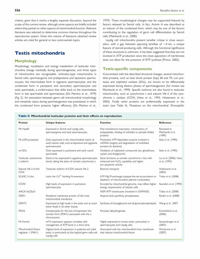
Testis-specific componentsConcomitant with the described structural changes, several mitochon-drial proteins, such as heat shock protein (hsp) 60 and 70, Lon pro-tease and sulphidryl oxidase (SOx), are known to be differentiallyexpressed during distinct phases of spermatogenesis (for review seeMeinhardt et al., 1999). Specific isoforms are also found in testicularmitochondria, such as cytochrome c and subunit VIb-2 of the cyto-chrome c oxidase (COX) (Hess et al., 1993; Huttemann et al.,2003). Finally other proteins are preferentially expressed in thetestis (see Table II). Mutations on the mitochondrial ‘Drosophila’
.............................................................................................................................................................................................
Table II Mitochondrial testicular proteins and their effects on reproduction
Protein Unique features Function Reference
Mt-Hsp60 Expressed in Sertoli and Leydig cells,spermatogonia and early spermatocytes
Post translational maturation, translocation ofpolypeptides, binding of unfolded or partially foldedproteins
Reviewed inMeinhardt et al.(1999)
Mt LON proteases Only expressed in the mitochondrial matrix ofearly meiotic cells, such as leptotene and zygotenespermatocytes
Proteolytic ATP-dependent enzyme involved inmtDNA integrity and degradation of misfoldedproteins (in bacteria)
Seitz et al. (1995)
mt SOx Only expressed in pachytene and early roundspermatids
Oxidation of sulphydryl compounds (ex: glutathione,cystein and thioglycerol)
Seitz et al. (1995)
Testicular cytochromec isoform
Starts to be expressed in zygotene spermatocytesslowly taking the place of somatic cytochrome c
Same functions as somatic cytochrome c but withenhanced anti H2O2 capability and higherpro-apoptotic activity
Liu et al. (2006), Hesset al. (1993)
Subunit Vib-2-of theCOX
Testicular isoform of COX subunit Vib-2 Electron transport Huttemann et al.(2003)
SCaMC-3-Like Lacks the Ca2þ-binding N-extension ATP-Mg/Pi exchange (catalyze the net accumulation ordepletion of mitochondrial adenine nucleotides)
Traba et al. (2008)
COXII High levels of expression in pachytenespermatocytes
Encoded by mitochondrial genome, may reflect higherenergy requirements of meiotic cells
Saunders et al. (1993)
AACA/slc25a31 ADP/ATP translocases (involved in OXPHOS) Traba et al. (2008)
DSP21 Peripheral membrane protein of the innermitochondrial membrane
Atypical dual specificity phosphatases Rardin et al. (2008)
GPAT2 Expressed at high levels in the testis and at muchlower levels in all other tissues
Synthesis of triacylglycerol and all glycerophospholipids Wang et al., 2007
PDH2 Compensates for the loss of expression thesomatic form (PDH1) associated with the xchromosome
Pyruvate dehydrogenase Korotchkina et al.(2006)
Mt Ferritin mf Ft expression appears correlate withmanagement of ATP burst in a short time
Highly expressed in human testis, particularly inspermatocytes and Leydig cells
Santambrogio et al.(2007)
Mitochondrial fissionregulator 1 (Mtfr1)
Highest level of expression in pubertal and adulttestis, in particularly by the haploid germ cells andLeydig cells
Associated with the mitochondrial inner membranethat induces mitochondrial fission
Monticone et al.(2007)
556 Ramalho-Santos et al.

protein Merlin, common to somatic cells (ortholog in humans is‘Neurofibromatosis’), produces viable but sterile males, indicatingthat the induced deregulation on mitochondrial function, althoughnot affecting somatic cells, has profound implications on germ celldifferentiation (Dorogova et al., 2008). However, this is certainly nota general effect, as mice lacking a testis-specific translocase (Tom34b) are normal and fertile (Terada et al., 2003).
Leydig cell steroidogenesisLeydig cell steroidogenesis is reliant on mitochondrial functionality asdemonstrated using the MA 10 Leydig cell line. Results clearlyshowed that Dc, ATP synthesis, DpH and mitochondrial [Ca2þ] areall required for steroid biosynthesis (Hales et al., 2005). Myxothiazol(a complex III blocker) inhibits LH-stimulated testosterone productionin multiple sites along the steroidogenic pathway (Midzak et al., 2007).In addition Leydig cell mitochondria are influenced by ROS, notablyduring the cholesterol transfer step (Stocco et al., 1993). TheseROS are also used by macrophages as mediators to modulateLeydig cell activity (Hales, 2002).
Testicular energy metabolismTesticular mitochondria have different bioenergetic parameters whencompared with mitochondria harvested from other tissues. Specifi-cally, testis mitochondria are shown to consume less oxygen to gen-erate approximately the same maximum electric potential as othertissues, and show an age-related modification in phosphorylative effi-ciency with young animals presenting less efficient phosphorylation(Mota et al., 2009; Amaral et al., 2009). These observations suggestthat, contrary to previous studies, testicular mitochondria should beconsidered as the primary mitochondrial toxicological model to testthe effect of distinct substances on male gametogenesis (Tavareset al., 2009).
Testis-specific morphogenetic events suggest that male gonads havea higher energy requirement than ovaries, starting early at the time ofSry activation (Matoba et al., 2008). Because spermatogonial stem cells(SSCs) are slow-dividing, it is expected that low MMP might be ashared characteristic with other stem cells (see below). Theneonate (0–5 days post-partum (dpp)) rat testis cell fraction withthe highest concentration of SSCs includes gonocytes, and exhibitslow DC, although stem cells in rat pup (8–14 dpp) testes appear tohave more active mitochondria than their gonocyte precursors,which might reflect increased proliferative activity as this populationexpands to fill the rapidly increasing number of SSC niches (Ryuet al., 2004).
In the adult testis the survival of germ cells is dependent on carbo-hydrate metabolism, including both anaerobic (glycolysis) and aerobic(OXPHOS) pathways. However, different cell types differ in their pre-ferred substrates (Robinson and Fritz, 1981; Grootegoed et al., 1984;Nakamura et al., 1984; Bajpai et al., 1998; Meinhardt et al., 1999). Infact, the formation of the blood-testis barrier and the alteration in thesurrounding medium, cause a considerable change in the energymetabolism of germ cells. Spermatogonia in the basal compartmentare supplied exclusively by blood components. However, afterpassage to the luminal compartment germ cells rely on the breakdownof lactate and pyruvate provided by Sertoli cells (reviewed inBoussouar and Benhamed, 2004). Therefore, spermatogonia, mature
sperm and the somatic Sertoli cells exhibit high glycolytic activity,whereas spermatocytes and spermatids produce ATP mainly byOXPHOS (Robinson and Fritz, 1981; Grootegoed et al., 1984;Nakamura et al., 1984; Bajpai et al., 1998; Meinhardt et al., 1999).
This could also be a matter of opportunity: since seminiferoustubule fluid is rich in lactate and poor in glucose, it is hypothesizedthat, even though spermatocytes have the machinery to produceenergy through glycolysis, they rely mostly on lactate (Bajpai et al.,1998). Nonetheless, there are incongruities between availability andusability. Blood vessels, located exclusively between tubules, supplythe oxygen needed to perform OXPHOS that only reaches thelumen of the seminiferous tubules by diffusion (Wenger andKatschinski, 2005). The facilitated access of spermatogonia tooxygen would lead us to expect the use of OXPHOS, instead of gly-colysis. Similarly, having less access to oxygen, spermatocytes wereexpected to perform glycolysis. However, the substrate availabilityimposed by seminiferous tubules compartmentalization, togetherwith ATP demand, may prime the cell to different adaptations. It isalso possible that stem cells maintain a low metabolism to avoid ROS-related damage.
Mitochondria-related apoptosis in the testisIn normal spermatogenesis not all germ cells reach maturity, and thenormal physiological death of germ cells via apoptosis seems to be aconstant event which can be potentiated by various stimuli (reviewedin Sinha-Hikim et al., 2003). Caspases are not only effectors of theapoptotic process but can induce its activation through the mitochon-drial pathway. Caspase 2 expression is increased in 16 dpp rat testis,when germ cell apoptosis also peaks. The increased amounts of acti-vated caspase 2 in mitochondria was matched by an increased level ofcytochrome c release. Specific inhibitors of caspase 2 mitigate theincreased cytochrome c release, indicating an important upstreamrole of mitochondria in germ cell apoptosis during the first wave ofspermatogenesis (Zheng et al., 2006).
Bcl-2 family members are also crucial during the first spermatogenicwave (Rodriguez et al., 1997). Bax is required to induce germ celldeath in dividing spermatogonia, at the time point at which theirnumber is regulated in a density-dependent manner (Russell et al.,2002), as shown by infertile Bax KO mice that present accumulationof atypical premeiotic germ cells but no mature haploid sperm(Knudson et al., 1995). In contrast, male bcl-w-deficient mice displaynormal testicular development before puberty, although afterpuberty Sertoli and germ cells of all types are severely reduced innumber, and seminiferous tubules contain many apoptotic cells andno mature sperm (Yan et al., 2000). In a different study using cynomo-logus monkeys (Macaca fascicularis), hormone deprivation, heat, orboth, led to an increase in Bcl-2 levels in testicular lysates andincreased cytochrome c and Smac/Diablo release (Jia et al., 2007).In mice and humans, hormone deprivation and heat-induced malegerm cell death, also induced the mitochondria-dependent apoptoticpathway (Vera et al., 2004, 2006). However, recent researchdescribed activation of both intrinsic and extrinsic (Fas-mediated)apoptotic pathways in situations of FSH and testosterone depletionin rats and mice (Pareek et al., 2007).
Interestingly, inhibition of ATP synthase decreased ATP levels andsuppressed cell death, an effect not seen with inhibition of glycolysis,
Mitochondrial activity in reproduction 557
indicating that mitochondrial ATP production plays a role in regulatingmale germ cell apoptosis (Erkkila et al., 2006). In a different study,apoptosis was triggered by incubating segments of seminiferoustubules without survival factors (i.e. serum or hormones), and apop-tosis in spermatocytes and spermatids was significantly repressed atlow levels of oxygen or by inhibitors of mitochondrial transitory per-meability, revealing another aspect of mitochondrial function in apop-tosis (Erkkila et al., 1999). Finally, germ cell mitochondria present aunique feature in apoptosis: testicular cytochrome c isoform showsa 3–5-fold greater apoptotic activity than the somatic isoform (Liuet al., 2006). Interestingly, testicular cytochrome c KO mice arefertile but present with highly atrophied testes, with a reducednumber of spermatocytes, spermatids and spermatozoa (Narisawaet al., 2002).
Testicular ROS and antioxidant defencesBecause mitochondria are the major source of intracellular ROS theyneed constant protection from these species, accomplished through awide network of mitochondrial non-enzymatic and enzymatic antiox-idant defences (reviewed in Ott et al., 2007). Thioredoxin glutathionereductase, a redox enzyme, accumulates in testes after puberty, pri-marily in elongating spermatids at the site of the mitochondrialsheath formation (Su et al., 2005). Previously, the same pattern of dis-tribution had been described for glutathione-peroxidase 4 (GPX4)(Roveri et al., 1992). Interestingly, prolonged expression of thisenzyme during the spermatogenic cycle (observed in transgenic micebearing the rat GPX4) revealed a number of spermatogenic defectsincluding primary spermatocyte apoptosis, haploid cell loss, seminifer-ous epithelium disorganization and reduced fertility (Puglisi et al.,2007). In agreement with this result, high levels of manganese super-oxide dismutase (SOD) were associated with small testis, male infer-tility, and decreased female fertility (Raineri et al., 2001). Resultsobtained in these studies suggest that any deregulation, or eitherloss/overexpression of mitochondrial antioxidant enzymes, disruptnormal homeostasis of the seminiferous epithelium resulting inreduced fertility.
Pathological effects of aging on testicularmitochondriaCellular ageing has been linked to increased ROS production andmitochondrial dysfunction (Harman, 1983; Jonhson et al., 1999).Analysis of testis mitochondria has shown differences in ETC com-plexes and membrane fatty acid composition throughout developmentand ageing. In fact, the activity of ETC complexes follows a commonpattern, with an increase during mitosis and first meiosis of germcells, and a decrease with ageing. These changes were correlatedwith a drop in polyunsaturated fatty acid content, increased pro-duction of superoxide and reduced SOD activity (Vazquez-Memijeet al., 2005, 2008). In fact, the balance between pro and antioxidantagents in the ageing testis is shifted towards pro-oxidation (Rebrinet al., 2003). Using domestic cat testicular mitochondria it was recentlyshown that membrane potential reached a maximum in animals withfully active reproductive function, after a pubertal period of increasingvalues of mitochondrial electric potential (Mota et al., 2009). In the rat,there was a peak of functionality in adult animals, a decrease with age,was coupled with an increase in expression and activity of uncoupling
protein 2 (UCP-2), suggesting that proton leakage may have a protec-tive role in managing age-dependent mitochondrial dysfunction(Amaral et al., 2008). Mitochondria from Leydig cells also presentalterations with age, consistent with the proposal that mitochondria-derived ROS may play a role in the decline in testosterone production(reviewed in Zirkin and Chen, 2000).
Sperm mitochondriaDuring the differentiation of spermatids into sperm (spermiogenesis),some mitochondria, like much of the cytoplasm, are lost in theso-called residual bodies, whilst those remaining rearrange inelongated tubular structures (Ho and Wey, 2007) and are packed heli-cally around the anterior portion of the flagellum. Concomitantly, thenumber of mtDNA molecules per haploid genome is reduced (Hechtet al., 1984), which is probably mediated by the down-regulation ofmitochondrial transcription factor A (TFAM) (Larsson et al., 1996,1997). Once the process is concluded mature mammalian spermpossess 22–75 mitochondria arranged end to end in the midpiece(Otani et al., 1988). The anchorage of the mitochondrial sheath tothe axoneme is supported by the sub-mitochondrial reticulum, acomplex of filaments that seems to sustain mitochondrial organization(Olson and Winfrey, 1990). Furthermore, the outer membranes ofsperm mitochondria are enclosed in a keratinous structure, the mito-chondrial capsule, formed by disulfide bonds between cysteine- andproline-rich selenoproteins, including the sperm-specific phospholipidhydroperoxidase glutathione peroxidase (Ursini et al., 1999). Thisstructure appears to confer mechanical stability, and is responsiblefor some distinctive features of sperm mitochondria, namely the resist-ance to hypo-osmotic stress, and the unfeasibility of completely isolat-ing these organelles. The fact that some mitochondria areevolutionarily retained in a very specialized sperm region suggeststhat these organelles fulfil a crucial role in sperm function. However,their physiological significance is still unclear.
Mitochondrial activity and sperm functionMitochondria may supply sperm with energy for several purposes,including sperm motility. Electron microscopy revealed that spermfrom asthenozoospermic samples have disordered mitochondria, sig-nificantly shorter midpieces and fewer mitochondrial gyres than nor-mozoospermic counterparts, whereas midpiece widths and taillengths were similar (Mundy et al., 1995). Moreover, enzymatic activi-ties of ETC complexes are positively correlated with human spermquality, particularly motility (Ruiz-Pesini et al., 1998, 2000b). Likewise,the expression of ETC subunits is associated with human spermquality (Amaral et al., 2007).
The relevance of mitochondrial activity in sperm function has beenstudied at the gene level, namely the significance of sperm mtDNAintegrity in male (in)fertility. Although some conflicting results havebeen published on the effects of specific mitochondrial point mutationsand large-scale deletions (for review see St John et al., 2007), it seemsconsensual that the accumulation of multiple mtDNA rearrangements isassociated with loss of sperm function (St. John et al., 2001). Similarly,both the number and size of mtDNA deletions in either testicular or eja-culated sperm are negatively correlated with ICSI pregnancy outcomes(Lewis et al., 2004), clearly showing the deleterious effect of mtDNA
558 Ramalho-Santos et al.

rearrangements, even when low motility is bypassed. These outcomeshave also been supported by transmitochondrial mice models, wherethe accumulation of pathogenic mtDNA-derived ETC defects wasresponsible for male infertility (Nakada et al., 2006). Furthermore,certain mtDNA haplogroups (groups of specific mtDNA types) havebeen suggested to predispose for reduced sperm motility (Ruiz-Pesiniet al., 2000a; Montiel-Sosa et al., 2006), although this has been contra-dicted by others (Pereira et al., 2005). On the other hand, a number ofreports have suggested that low quality sperm present abnormalmtDNA content (May-Panloup et al., 2003; Amaral et al., 2007), andthe expression of TFAM and of the catalytic subunit of DNA polymerasegamma (POLG), both of which are implicated in the regulation ofmtDNA copy number, are both lower in poorer quality sperm(Amaral et al., 2007). The relevance of POLG in male infertility hasalso been confirmed using mouse models: homozygous knock-in miceexpressing a deficient POLG presented increased levels of mtDNAmutations and deletions, and showed reduced lifespan and prematureonset of aging-related phenotypes, including reduced fertility (Trifunovicet al., 2004). In addition, polymorphisms in the CAG-repeat region ofPOLG are possibly associated with sperm quality, although their truesignificance in male infertility is questionable (Amaral et al., 2007).Overall, and despite the controversies on the exact effect of a particularmtDNA point mutation, deletion or haplogroup in human sperm, thereis clear evidence that alterations in the mitochondrial genome can com-promise sperm motility and function.
The link between mitochondria-generated ATP and sperm motility/fertilization competence has been illustrated by Dc evaluation (Fig. 3).Several studies confirmed that motility is related to mitochondrialfunctional status in humans (Troiano et al., 1998), equines(Loveet al., 2003), rats (Gravance et al., 2001), boars (Spinaci et al.,2005) and rams (Martinez-Pastor et al., 2004). Additionally, at leastin humans, sperm fertilization potential (measured as fertilizationrates after IVF) is strongly related to Dc and thus to mitochondrialfunctionality (Kasai et al., 2002; Marchetti et al., 2002, 2004b). Onthe other hand, Dc seems to negatively correlate with both DNA frag-mentation and the generation of ROS (Wang et al., 2003). Mirroringthe importance of mitochondria to sperm function, subpopulationsof mitochondria with high Dc are enriched in cells with elevated
fertilization capabilities (Gallon et al., 2006). Interestingly, it hasrecently been proposed that the ultimate cause for the negativeeffect of endocannabinoids in male reproduction may be the reductionof sperm mitochondrial activity (Rossato, 2008). These cumulativeoutcomes strongly suggest Dc as an indicator of sperm functionalstatus.
The role of sperm mitochondrial metabolismis controversialSperm require ATP mainly for motility, as well as for the cellular eventsinvolved in hyperactivation, capacitation and the acrosome reaction.The provenance of the ATP that fuels these events, especially motility,has been discussed for decades: is it derived from OXPHOS, or purelyglycolytic? The debate has been confounded by possible species-specific discrepancies, as well as by differences in experimental con-ditions. The usual approach is to analyse motility and/or ATP levelsafter incubating sperm in media containing different substrates, inthe presence or absence of inhibitors. However, media composition,temperatures and times of incubation, as well as the inhibitors andtheir concentrations, vary from study to study.
The compartmentalization of mitochondria in the sperm midpiece maylimit the availability of OXPHOS-derived ATP for the dynein-ATPaseslocated in the principal piece. This is especially true in species with longsperm tails, such as rodents, as it is doubtful that sufficient ATP candiffuse to the distal end of the flagellum. This problem could be solvedvia ATP shuttles, but their significance in mammals needs further confir-mation (Ford, 2006). Alternatively, ATP can be produced in the principalpiece through the glycolytic pathway. Indeed, glycolytic enzymes seem tobe compartmentalized in the fibrous sheath, a cytoskeletal element of theprincipal piece (Krisfalusi et al., 2006; Kim et al., 2007).
Elegant work was done using hypotonically treated cells, i.e. withdisrupted membranes, to solve the difficulty in isolating sperm mito-chondria and then analysing oxygen consumption and respirationrates with various substrates (Keyhani and Storey, 1973; Storey,1978; Carey et al., 1981). Results were species-specific, i.e. spermfrom distinct species have dissimilar capabilities to metabolize differentsubstrates, suggesting that mitochondrial activity in sperm of a givenspecies is adjusted to the substrate content of the female tract(Storey, 1980).
Human sperm motility and ATP content can be maintained in thepresence of a glycolyzable substrate, but decline rapidly in itsabsence, even in the presence of oxidizable substrates (Peterson andFreund, 1970; Williams and Ford, 2001). On the other hand, the pre-viously stated fact that mutations in the mitochondrial genome areassociated with decreased sperm quality suggests that OXPHOS isalso relevant. Interesting outcomes were obtained from a patient har-bouring a mtDNA point mutation causing reduced activity of the ETCcomplex I, and whose sperm presented low-motility. Supplementationwith succinate, a substrate for complex II, circumventing the effect ofthe mutation, resulted in an increase in sperm motility (Folgero et al.,1993). Furthermore, incubation of human sperm with distinct ETCinhibitors in media containing glucose, resulted in rapid decreases insperm motility (Ruiz-Pesini et al., 2000a; St John et al., 2005a, b).
Experiments in rat sperm have shown that 6-chloro-6-deoxysugarsprevent glucose metabolism, and have contraceptive action (Fordet al., 1981a, b). Sperm became immotile and presented low ATP
Figure 3 Mitochondria in human sperm.
(A) Mitochondrial membrane potential (MMP) detection in live human spermusing Mitotracker Red (red), nuclear DNA is counterstained with SYBR 14(green). (B) Immunodetection of a subunit of COX, part of the electron trans-fer chain (ETC) (complex IV), in the sperm midpiece (green), nuclear DNA iscounterstained with DAPI (blue).
Mitochondrial activity in reproduction 559
levels when glucose was the only substrate available, but no detrimen-tal effects were observed in the presence of pyruvate and lactate (Fordand Harrison, 1981a, b). Likewise, sperm from control rats presentedhigher motility and ATP levels in medium with pyruvate and lactate.a-Chlorohydrin has also been used to inhibit glycolysis, in epididymalram and ejaculated boar sperm, resulting in decreased motility andATP concentration, but only in the presence of glucose (Ford andHarrison, 1985, 1986). Taken together, these outcomes may indicatea role of OXPHOS in these species, although a contribution ofglycolysis is also evident. The results of experiments in mouse epididy-mal sperm also seem to imply that both glycolysis and OXPHOS areable to sustain sperm motility, although with glycolysis in a predomi-nant role (Mukai and Okuno, 2004).
In testis-specific cytochrome c homozygous knockout mice, maleswere fertile, although presenting early testicular atrophy, as discussedearlier. Moreover, when compared with wild type, their sperm wereless motile, presented lower ATP content and were less successfulin in vitro fertilization (Narisawa et al., 2002). On the other hand,the disruption of the spermatogenic cell-specific glycolytic enzyme gly-ceraldehyde 3-phosphate dehydrogenase-S (GAPDH-S) has alsoresulted in some remarkable outcomes (Miki et al., 2004). Male homo-zygous KO mice were infertile (but females were fertile) and theirsperm showed both decreased motility and ATP levels, although mito-chondrial activity was unchanged. These results have been interpretedas proof that glycolysis is essential for sperm motility. However, thisview has been challenged (Ford and Harrison, 2006; Ruiz-Pesiniet al., 2007; Storey, 2008), the reasoning being that the inactivationof GAPDH-S (similarly to 6-chloro-6-deoxysugars, DOG anda-chlorohydrin) block the glycolytic net synthesis of ATP, but the gly-colytic ATP-consuming phase still operates. Often neglected is boththat glycolysis is usually a prerequisite for OXPHOS and that, unlikeOXPHOS, it actually requires ATP to initiate the process, a fact thatmust always be taken into consideration. Nevertheless, disruption ofthe glycolytic enzyme lactate dehydrogenase-C4 (LDH-C4), normallyexpressed in spermatogenic cells and also in minor amounts inoocytes, resulted in decreased sperm function and impairment ofmale fertility in mice (Odet et al., 2008). Recently, a new approachhas been developed to assess sperm motility, which relies on theuse of laser tweezers to measure swimming speeds and force(Nascimento et al., 2006). Unexpectedly, no relationship was foundbetween either human or dog sperm motility and Dc, also suggestinga predominant role of glycolysis (Nascimento et al., 2008).
The role of OXPHOS versus glycolysis in other events leading tofertilization is believed to be species-specific. To this extent, glucoseseems necessary for the acquisition of hyperactivated motility, capaci-tation and acrosome reaction in both humans and mice (Hoppe, 1976;Fraser and Quinn, 1981; Rogers and Perreault, 1990), and may partici-pate in the protein phosphorylation events occurring in rhesusmacaque sperm capacitation (Hung et al., 2008). On the otherhand, glucose seems to inhibit capacitation in bull and guinea pig(Rogers and Yanagimachi, 1975; Rogers et al., 1979; Parrish et al.,1989), where oxidizable substrates are required. Also noteworthyare two recent findings using proteomic approaches. The first studyconcerns sperm epididymal maturation in rodent species (Aitkenet al., 2007). Apparently, caput epipydimal sperm possess silent mito-chondria, although caudal sperm have polarized mitochondria, andtherefore epididymal maturation may involve the activation of sperm
mitochondria, and mitochondria-generated ATP may facilitate thetyrosine phosphorylation events associated with capacitation. Thesecond study involves the identification of proteomic differences inasthenozoospermic samples (Martinez-Heredia et al., 2008). Theauthors found 17 proteins expressed at different levels in astheno-zoospermic samples compared with controls. Interestingly, the list ofproteins includes decreased expression of the ETC enzymeCOXVIb. In contrast, none of the glycolityc enzymes were affected.
These cumulative reports seem to demonstrate that in the few daysit can spend in the female reproductive tract mammalian sperm mightbe able to utilize both glycolysis and OXPHOS to produce ATP fordifferent purposes. The balance between these two metabolic path-ways may vary between species, according to the substrates availableduring the sperm’s route.
Sperm mitochondria and ROSAlthough seminal leukocytes were thought to be the only ROS-generators in an ejaculate, it is now well established that sperm arealso responsible for some ROS production (Aitken et al., 1996).Accordingly, both seminal plasma and sperm possess a number ofantioxidant strategies to protect the male gametes against ROSdamage. These include enzymes such as SOD, catalase, the gluta-thione peroxidase/reductase system and non-enzymatic substances,such as ascorbic acid, glutathione and a-tocopherol, among others(reviewed by Tremellen, 2008).
Low and regulated levels of ROS have been implicated in spermcapacitation, acquisition of hyperactivated motility, acrosome reactionand oocyte interaction (for review see de Lamirande et al., 1997; Ford,2004). When the physiological equilibrium between ROS productionand scavenging is perturbed, sperm function may be compromisedand indeed oxidative stress is implicated in male infertility.Increased ROS levels have been associated with sperm lipoperoxida-tion damage, decreased motility, DNA fragmentation and increasedapoptosis (Agarwal et al., 2008). Importantly, it has been recentlyproposed that mitochondria are a major contributor to the oxidativestress experienced by defective human sperm (Koppers et al., 2008).
Mitochondria-related apoptosis in spermAlthough it is well established that a fraction of sperm in any ejaculatepresents apoptotic markers (e.g. Varum et al., 2007), the mechanismsinvolved in putative sperm apoptosis are not completely characterized.However, and among other mechanisms, apoptosis can be triggeredvia the mitochondrial pathway. To this extent, the presence of acti-vated caspase 3 in ejaculated human sperm midpiece was clearlydemonstrated (Weng et al., 2002), and poorer quality samples exhib-ited higher levels of active caspase 3 positive-sperm. Furthermore, theactivation of caspase 3 is correlated with the externalization of phos-phatidylserine (Kotwicka et al., 2008). Similar results were obtainedfor caspase 9 which, when expressed, is also localized in the midpiece(Paasch et al., 2004a). Additionally, the treatment of sperm with betu-linic acid, an inducer of the intrinsic apoptotic pathway, resulted in lossof Dc and activation of caspases 3 and 9, with a concomitant decreasein sperm motility (Paasch et al., 2004b; Grunewald et al., 2005).Indeed, caspase 3 activity seems to be negatively correlated withsperm Dc (Marchetti et al., 2004a). The transcript for the apoptoticmarker Bcl-2 is also present in ejaculated sperm, with higher levels
560 Ramalho-Santos et al.
in infertile men compared with controls (Steger et al., 2008). Futurestudies will be needed to better determine the role of mitochondriain triggering sperm apoptosis, and its relevance in sperm (dys)function.
Other possible roles of sperm mitochondriaThe involvement of Ca2þ signalling in the regulation of several aspectsof mammalian sperm function is very well documented (Felix, 2005;Publicover et al., 2008). On the other hand, the ability of mitochondriato store Ca2þ has been demonstrated in sperm from diverse species,but its role is unclear (Publicover et al., 2007). Thus, although mamma-lian sperm mitochondria are able to function as intracellular Ca2þ
stores, a clear role in signalling has not been demonstrated.Another issue relates to protein synthesis. It is generally accepted
that gene expression in mature sperm is restricted to the mitochon-dria. In fact, mammalian sperm seem to be able to synthesize bothmitochondria-encoded RNAs (MacLaughlin and Terner, 1973; Hechtand Williams, 1978; Alcivar et al., 1989) and proteins (Ahmed et al.,1984; Twaina-Bechor and Bartoov, 1994). More recently, however,and in contrast with previous data (Diez-Sanchez et al., 2003), mam-malian sperm were suggested to synthesize nuclear-encoded proteins,at least during capacitation (Gur and Breitbart, 2006). The results areparticularly odd, and certainly require independent confirmation, notonly because they seem to contradict the dogma that sperm are trans-lationally silent cells (at least for nuclear-encoded proteins), but alsobecause they also suggest that translation of nuclear-encoded proteinsoccurs in mitochondria-type ribosomes localized either inside oroutside mitochondria, but with no involvement of the cytoplasmictranslation machinery, an event with no equivalent in any othercell type.
Oocyte/embryo mitochondriaMitochondria are the most abundant and prominent organelle in theearly embryo (Motta et al., 2000; Sathananthan and Trounson,2000) and are thought to be exclusively derived from the oocyte(Cummins, 2000). Contradicting a notion still found in a number oftextbooks, in mammals the entire sperm enters the oocyte at fertiliza-tion (Ankel-Simons and Cummins, 1996), however, sperm mitochon-dria are diluted beyond detectable levels or destroyed inside theembryo (Sutovsky et al., 1999).
Oocyte mitochondrial dysfunction, expressed as declined cell res-piration and electron transport, may contribute to diminished fertility,and be the cause of development retardation and arrest in human pre-implantation embryos (Fissore et al., 2002; Ramalho-Santos et al.,2004; Thouas et al., 2004). Intracytoplasmatic injection of ‘normal’mitochondria can overcome mitochondrial dysfunctions (Nagai et al.,2004) and inhibit oocyte fragmentation (Perez et al., 2000), stressingthe importance of mitochondria in cell death (Fissore et al., 2002).In contrast, injection of abnormal mitochondria induces oocyte apop-tosis (Perez et al., 2007).
Mitochondrial number and structurein the oocyte and early embryoOocyte mitochondria must support early embryo development untilthe resumption of mitochondrial replication, which only occurs
post-implantation (reviewed in Dumollard et al., 2006). Dependingon the species, a mammalian oocyte contains around 105–108 mito-chondria, with 105 in human (Chen et al., 1995; Jansen and de Boer,1998). Mitochondria propagate from a restricted founder populationpresent in the primordial germ cell (PGC) (Cummins, 2000; Jansen,2000), ensuring that mitochondria in the mature oocyte (and there-fore in dividing blastomeres) are homogeneous. The mtDNA bottle-neck theory (Hauswirth and Laipis, 1982) suggests a restriction inthe number of mtDNA molecules to be transmitted from themother to the offspring, followed by a strong amplification inoocytes (Reviewed in May-Panloup et al., 2007). The bottleneckoccurs in order to maintain mtDNA homoplasmy and minimize het-eroplasmy (Cummins, 2001) Therefore, a selection of a group ofmtDNA molecules to repopulate the next generation takes place,and deleterious mutations tend to be eliminated so as not to be trans-mitted to the offspring. The nature of the mtDNA bottleneck hasbeen recently discussed and different mechanisms have been pro-posed (Cao et al., 2007; Cree et al., 2008; Khrapko, 2008).However, further investigations are needed to clarify exactly whenand how this phenomenon occurs.
During oogenesis there is an amplification in mitochondrial numberin parallel with cytoplasmatic volume increase. Pre-migratory PGChave less than 10 mitochondria, 100 mitochondria are present inovarian PGCs and 200 in oogonia. Primordial follicle oocytes have10 000 mitochondria, a number which ultimately increases 10-fold.In the mature oocyte, each of the 105 mitochondria possesses asingle copy of mtDNA (reviewed in Jansen and de Boer, 1998).
The increase in mitochondrial number during oocyte growth isaccompanied by changes in their ultrastructure (Wassarman andJosefowicz, 1978; Motta et al., 2000; Au et al., 2005). Small oocytescontain mitochondria with numerous transversely-oriented cristae,although growing oocytes present round and oval-shaped mitochon-dria, with columnar-shaped arched cristae. At ovulation, mitochondriahave a spherical immature structure, are highly vacuolated, with adense matrix and only few cristae. Between the zygote and the2-cell stage, mitochondria assume a dumb-bell shape and presentconcentrically located cristae. From the 4-cell to the morulastage, mitochondria have a more elongated structure with transversecristae.
The total number of mitochondria in a normal human blastocyst isabout 14 000, and the average number of mitochondria per cell isabout 150 (Van Blerkom, 2008). However, studies in mouse andhamster models show that the average mitochondria per cell ishigher in the trophectoderm (TE), which will give rise to the placenta,than in the inner cell mass (ICM), which will give rise to the embryoproper (Barnett et al., 1996; Van Blerkom, 2008). There is some con-troversy regarding the morphological homogeneity of the mitochon-dria found at the blastocyst stage. Although some authors claim thatmitochondria in mouse and human blastocysts are homogenous andelongated elements (Sathananthan and Trounson, 2000), the existenceof two types of mitochondria in the mouse blastocyst has beenreported: spherical mitochondria in the ICM and elongated mitochon-dria in the TE. In both types mitochondrial cristae are transverselyoriented and their matrix is less dense than the mitochondrialmatrix found in earlier developmental stages (Stern et al., 1971). Inter-estingly, although the ICM cells have low Dc and are almost quiescent,the TE cells are highly polarized and very active producing more ATP
Mitochondrial activity in reproduction 561
and consuming more oxygen (Barnett et al., 1996; Houghton, 2006;Van Blerkom et al., 2006).
Localization and distribution of mitochondriain the oocyte and early embryo is highlyregulatedDuring oocyte maturation, and in early embryos, mitochondria arerelocated to different regions, probably in response to localizedenergy demands (reviewed in Bavister and Squirrell, 2000). Through-out maturation mitochondria are mainly found in clusters in closeproximity to endoplasmic reticulum membranes (Jansen, 2000),suggesting a possible interaction between the two organelles(Dumollard et al., 2006). In fully-grown germinal vesicle (GV) stageoocytes mitochondria are concentrated in clusters surrounding thenucleus (Jansen, 2000; Sun et al., 2001) and migrate to the peripheryof the oocyte after GV breakdown. In metaphase-II (MII) arrestedoocytes, mitochondria are mainly present around the meioticspindle and at the oocyte centre (Dumollard et al., 2004, 2006),accumulating around pronuclei following fertilization, and maintainingclose nuclear association through the morula stage. Impaired redistri-bution of mitochondria may compromise fertilization and embryodevelopment (Au et al., 2005), and blastomeres that receive aninsufficient amount of mitochondria remain undivided and undergofragmentation (Van Blerkom et al., 2000).
In addition, mitochondrial populations present heterogeneity interms of DC. Two populations of mitochondria are present: onewith low DC, which is more abundant, and a smaller amount withhigh polarization. Clusters of highly-polarized mitochondria are loca-lized in the subplasmalemmal/pericortical cytoplasm in oocytes andearly blastomeres (Van Blerkom et al., 2002). Loss of these mitochon-drial domains affects division (Van Blerkom and Davis, 2006), whichmay be associated with the focal ionic and metabolic regulation(Van Blerkom et al., 2003) involved in oocyte activation and earlydevelopment (Van Blerkom et al., 2002; Van Blerkom and Davis,2007).
Energy metabolism in the oocyteand early embryoDuring oocyte development, a combination of metabolic pathways isfound. Pyruvate and glucose are used by primordial follicles, suggestingthat both OXPHOS and glycolysis are involved (Biggers et al., 1967;Boland et al., 1993; Wycherley et al., 2005). Furthermore, glucoseused by the cumulus cells may lead to pyruvate production that is uti-lized by the oocyte (Jansen and Burton, 2004).
Between the primary and pre-ovulatory stages, pyruvate uptakeincreases 2-fold (Harris et al., 2008), accompanied by an increase inO2 consumption. However, the average level of ATP seems constantbetween the GV and MII stages (Van Blerkom et al., 1995). Themature oocyte displays a high ATP turnover, supplied by mitochon-drial respiration (Dumollard et al., 2004) and by the uptake of pyru-vate (Leese, 1995), which is also the main substrate used by zygotes(Biggers et al., 1967; Leese and Barton, 1984). At fertilization,where higher ATP levels are required to support cortical granules exo-cytosis, chromosome dysjunction, polar body extrusion and Ca2þ
homeostasis (Van Blerkom et al., 1995), there is an increase in O2
use (Magnusson et al., 1977). Therefore, pyruvate is essential for
meiotic maturation and to support the first cleavage division (Biggerset al., 1967).
From zygote to morula, the levels of ATP and O2 used remain basi-cally constant, and it is essentially substrates for OXPHOS that aremetabolized (Slotte et al., 1990; Van Blerkom et al., 1995). In laterstages the pattern of energy metabolism for the cleaving embryochanges (reviewed in Dumollard et al., 2007). At the morula stage,mitochondrial and metabolic changes occur gradually, and a shift inATP production to glycolysis is evident (Leese, 1995; Van Blerkomet al., 1995; Thompson et al., 1996). Glucose is the predominant sub-strate that supports later embryo development (Biggers et al., 1967;Gardner and Leese, 1986; Hardy et al., 1989; Gott et al., 1990), butthe increase in glucose uptake at the blastocyst stage is accompaniedby a substantial increment in ATP generation and O2 consumption(Houghton and Leese, 2004), suggesting OXPHOS also takes place(Dumollard et al., 2007). After implantation, levels of O2 use decreaseto those found in pre-blastocyst stages (Houghton and Leese, 2004).
In summary, the uptake of pyruvate is high in the mature oocyte,drops just after fertilization and then peaks before declining again atthe morula stage (Gardner and Leese, 1986). Interestingly, if one sim-ultaneously considers independent data for sperm and oocyte, thebuilding consensus is that the male gamete predominantly uses glycoly-sis to reach the oocyte, although at the same time the female gameteis seemingly more reliant on OXPHOS, despite the same substrateand oxygen availability.
Importantly, intra- and inter-individual variations in oocyte ATPcontent have been described, and there is a close associationbetween oocyte ATP concentration and developmental competenceof the resulting embryo (Van Blerkom et al., 1995). Furthermore, pyr-uvate and glucose uptake are lower in arrested embryos (Hardy et al.,1989; Gott et al., 1990), which are also unable to switch to a glucose-based metabolism when necessary (Gott et al., 1990). Additionally,blastocysts that implant and develop to term have a significantlyhigher glucose uptake prior to transfer than those that fail todevelop (Gardner and Leese, 1987). As in the case of sperm, it isalso possible that these constant changes are simply adjustments tothe substrates available in distinct region of the female reproductivetract (Jansen and Burton, 2004). Taking cellular volume intoaccount, Harris and coworkers (2008) found that metabolism ishigher in primary follicles, indicating that energy demands aregreater. On the other hand, a relatively low metabolism is found inembryos, which seems associated with embryo vitality (Lane andGardner, 1996; Leese, 2002).
The relevance of mitochondrial activity in terms of mtDNA hasbeen also studied. A correlation between mutations in the catalyticsubunit of POLG and premature ovarian failure has been noted andis probably due to an accumulation of mtDNA deletions as hasbeen observed for other tissues (Luoma et al., 2004). Female micethat carry a proofreading-deficient POLG have reduced fertility(Trifunovic et al., 2004). Furthermore, a relationship betweenoocyte mtDNA copy number and oocyte quality/fertility wasobserved (Yesodi et al., 2002; Almeida-Santos et al., 2006), and ferti-lized oocytes present a higher mtDNA copy number than unfertilizedoocytes (Almeida-Santos et al., 2006). This may suggest that it is notprimarily OXPHOS dysfunction that contributes to diminished fertility,but rather reduced mitochondria/mtDNA copy number that leads tothe OXPHOS dysfunction observed and subsequently to poor quality
562 Ramalho-Santos et al.
oocytes or reduced fertility (Jacobs et al., 2006). However, contrary to thesituation in males, females that carry mtDNA with pathogenic mutationare fertile. These females produce oocytes with a predominant amountof the mutant mtDNA, oocytes that survive despite severe OXPHOSdefects. The mutant mtDNA is maternally transmitted to the F1–F3 pro-genies, which present mitochondrial dysfunction in various tissues andhave a shorter lifespan due to the consequent pathologies that are unre-lated to fertility itself (Inoue et al., 2000).
Mitochondria in Ca21 signallingOocyte mitochondria have an important role in the regulation ofsperm-triggered Ca2þ waves essential for zygote activation (reviewedin Dumollard et al., 2006, 2007), probably acting as a Ca2þ store andparticipating in the generation of the intracellular [Ca2þ] oscillations(Tesarik and Sousa, 1996; Liu et al., 2001). Sperm-triggered Ca2þ
oscillations stimulate mitochondrial energy production at fertilization,leading to an increase in O2 consumption that is maximal duringCa2þ release (Dumollard et al., 2003), and can be primarily initiatedby an influx of Ca2þ into the mitochondria (Dumollard et al., 2006).Accordingly, the Ca2þ signal is directly transmitted to the mitochondrialmatrix, leading to the up-regulation of OXPHOS, which is, in turn, necess-ary for the maintenance of [Ca2þ]i oscillations (Dumollard et al., 2004).Thus, Ca2þ links ATP supply and demand, allowing for the maintenanceof a low-level of OXPHOS, which increases only when ATP is neededto support post-fertilization events, stimulated by Ca2þ waves. The pro-duction of ROS by the ETC is therefore minimized, ensuring that mito-chondria are exposed to low levels of oxidative stress. Furthermore,mitochondria are also important for calcium clearance, i.e. in the mainten-ance of low levels of cytosolic [Ca2þ].
Mitochondrial ROS and apoptosis in oocytesand embryosAccumulating evidence has shown that ROS play important roles infemale reproduction (reviewed in Agarwal et al., 2005). However,the mammalian oocyte and embryo are very sensitive to oxidativestress (Liu et al., 2000) and if physiological levels of ROS are beneficial,higher levels can disrupt oocyte maturation and embryo development(Harvey et al., 2002), and promote embryo fragmentation (Johnsonand Nasr-Esfahani, 1994; Yang et al., 1998).
Indeed, oxidative stress can induce apoptosis of the oocyte andearly embryo (Liu et al., 2000). Mitochondria-dependent apoptosisseems to be responsible for the post-natal decline in the femalegerm cell population (Reynaud and Driancourt, 2000; Tilly, 2001)and for follicular atresia (Kim and Tilly, 2004), as well as in oocytesand early embryos (Liu et al., 2000). Mammalian oocytes expressseveral anti- and pro-apoptotic members of the Bcl-2 family and it isthe balance between these factors that determines oocyte survival(Liu et al., 2000; reviewed in Jurisicova and Acton, 2004). Further-more, and mirroring the importance of mitochondria in oocyte apop-tosis, the apoptotic inducer hydrogen peroxide induces cytochrome crelease from oocyte mitochondria, associated with a decrease in DC
(Liu et al., 2000).In essence, mature oocytes and early embryos maintain an overall
low-level (i.e. ‘quiet’) metabolism, thus minimizing oxidative stress,but generating the necessary ATP to fulfil cellular functions (Leese,2002; Leese et al., 2007).
Physiological alterations associatedwith agingA negative correlation has been described between maternal age andmitochondrial activity (Jansen and de Boer, 1998). Oocytes from olderwomen present declining mitochondrial function, which can contributeto declining fertility, and may be associated with lower embryo devel-opment and pregnancy rates (Wilding et al., 2001). Ocytes from olderwomen often present aberrant spindle formation, abnormal chromo-somal alignment, and consequently, a high occurence of aneuploidy(Battaglia et al., 1996; reviewed in Eichenlaub-Ritter et al., 2004). Ithas been suggested that these abnormalities can be due to aninadequate capacity to generate sufficient ATP levels to supportthese events (Gaulden, 1992). In accordance, oocytes from olderwomen present an accumulation of mtDNA point mutations (Barrittet al., 2000) and higher levels of mtDNA deletions (Keefe et al.,1995), factors that can ultimately be responsible for aneuploidy andpoor implantation rates (Bartmann et al., 2004), ensuring that onlymetabolically intact embryos develop to term (Dumollard et al.,2006). These abnormalities can be corrected by injecting cytoplasmfrom younger oocytes (discussed in Klein and Sauer, 2001). Thedonor cytoplasm may thus ‘rescue’ spindle misalignments, due, atleast in part, to the mitochondria of the younger oocytes. Moreover,oocytes from older women present a higher volume fraction of mito-chondria, indicating compensatory mechanisms (Muller-Hocker et al.,1996). In the same vein, mitochondrial dysfunction seems to also playan important role in fragmentation in postovulatory aged oocytes.Indeed, aged oocytes present a lower uptake of pyruvate (Hardyet al., 1989). It was also found that, in these oocytes, [Ca2þ] oscil-lations fail to trigger ATP production and instead induce apoptosis(Gordo et al., 2002). A decrease in fertility potential, in both maternalaging and postovulatory aged oocytes, seems to be primarily due todamages in mitochondria caused by oxidative stress (Fissore et al.,2002), leading to apoptosis (Perez et al., 1999).
Mitochondria in embryonic stemcellsESCs, are derived from the ICM of the blastocyst stage embryo priorto implantation and can be maintained in vitro in colonies for prolongedperiods without losing the abilities of indefinite self-renewal or differ-entiation into tissues from all three germ layers (Evans and Kaufman,1981; Martin, 1981; Thomson et al., 1998; Amit et al., 2000; Panand Thomson, 2007). This can be assessed in vitro (through embryoidbody formation) or in vivo (by teratoma formation). Because of thesebiological properties, human embryonic stem cells (hESCs) have anenormous potential as models to study cell differentiation and forpossible replacement cell therapies. Indeed, several groups haveshown that under specific culture conditions hESCs can differentiateinto various somatic cell types (for review see Gepstein, 2002;Dhara and Stice, 2008; Raikwar and Zavazava, 2009).
Mitochondria number and morphology in ESCAs ESCs are derived from the ICM one should expect that they sharemetabolic and morphologic features. Indeed, and although there areline-specific differences, it has been shown that undifferentiatedhESCs have few mitochondria arranged in small perinuclear clusters,
Mitochondrial activity in reproduction 563
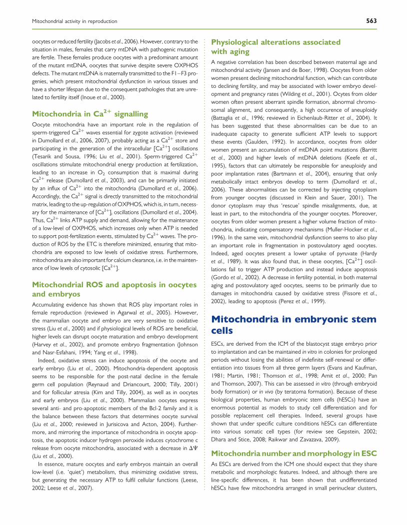
and immature morphology, evidenced by the presence of few cristaeand low electron lucid matrix (Oh et al., 2005; St John et al., 2005a, b;Cho et al., 2006) (Fig. 4). hESCs colonies are characterized by highnuclear cytoplasmatic ratios and cells tightly packed within colonies.
Although there seems to be a paucity of intracellular organization(sometimes discussed as a ‘stemness’ attribute), this could also be areflection of reduced cytoplasm. Furthermore, it is well acceptedthat cells in the periphery of the colony are among the first cells toundergo spontaneous differentiation during in vitro culture. Interest-ingly these cells have higher levels of mitochondria (Cho et al., 2006).
MMP and metabolism in ESCThere is some controversy regarding the polarization of mitochondriain undifferentiated versus differentiated ESC. Undifferentiated mouseESC has been reported to have highly polarized mitochondria,which decreases upon differentiation to cardiomyocytes (Chunget al., 2007). On the other hand, no differences in Dc between undif-ferentiated hESCs and differentiated hESCs were reported (Saretzkiet al., 2008). The controversy might be due to the fact that thesedata come from ESCs of different species, mouse and human, respect-ively. In addition, mouse ESC were specifically differentiated into car-diomyocytes, whereas with hESCs spontaneous differentiation wasstudied, and as consequence a mixture of cell lineages would bepresent. Several studies have differentiated ESCs in vitro and observedchanges in mitochondrial dynamics during differentiation. As ESCsdifferentiate the total number of mitochondria increases, as do thenumber of mitochondria with a more mature morphology (St Johnet al., 2005a, b; Cho et al., 2006), similar to that described forSSCs. Concomitantly with an increase of mitochondrial numberduring ESC differentiation, the rates of O2 consumption and ATP pro-duction in the cell increase, whereas lactate production decreases(Chung et al., 2007). These results suggest that during ESC differen-tiation there is a switch in energy metabolism from glycolysis toOXPHOS. Similar results have been reported in adult stem cells(Piccoli et al., 2005; Chen et al., 2008).
It seems logical to assume that an increase in the number of mito-chondria and OXPHOS in differentiated cells leads to an increase inROS production, and several authors have shown that that is indeedthe case (Cho et al., 2006; Saretzki et al., 2008). Interestingly,reports regarding antioxidant defences during the process of differen-tiation differ. Although a decrease in the expression of antioxidants,namely SOD2 and GPX2, has been reported (Saretzki et al., 2008),increased expression of GPx1, Cu/Zn SOD, Prx1 and Prx2 wasalso described (Cho et al., 2006). The contradictory results may bedue to the fact that these studies looked at different antioxidants,and it is possible that during differentiation there is an increase incertain antioxidants to the detriment of others.
Mitochondrial role in ESC differentiationGiven the distinct mitochondrial properties in undifferentiated versusdifferentiated ESCs it is logical to assume a role for mitochondriain differentiation. Hypoxic environment prevents spontaneous hESCdifferentiation (Ezashi et al., 2005). In addition, several groups haveshown that functional mitochondria are necessary for differentiation.For example, inhibition of mitochondrial respiratory chain complexesI and III, by Rotenone and Antimycin A, respectively, results inreduced cardiomyocyte differentiation, due to an impairment ofOXPHOS (Chung et al., 2007). Furthermore, glycolytic metabolism issufficient for maintaining mouse ESC homeostasis; however, in orderfor cells to differentiate there must be a switch from glycolysis to the
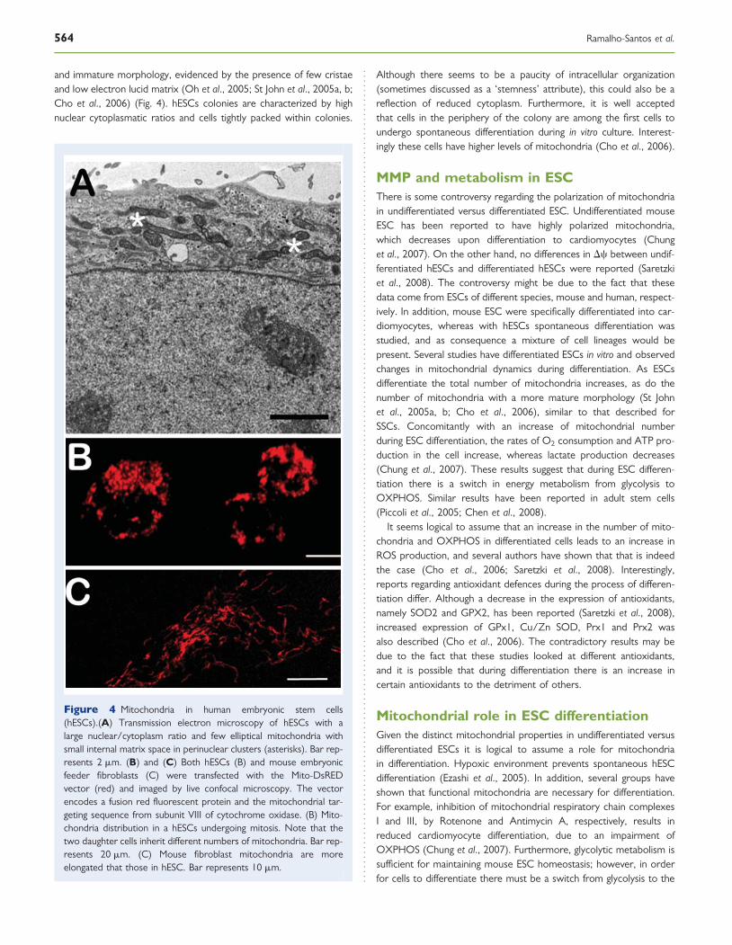
Figure 4 Mitochondria in human embryonic stem cells(hESCs).(A) Transmission electron microscopy of hESCs with alarge nuclear/cytoplasm ratio and few elliptical mitochondria withsmall internal matrix space in perinuclear clusters (asterisks). Bar rep-resents 2 mm. (B) and (C) Both hESCs (B) and mouse embryonicfeeder fibroblasts (C) were transfected with the Mito-DsREDvector (red) and imaged by live confocal microscopy. The vectorencodes a fusion red fluorescent protein and the mitochondrial tar-geting sequence from subunit VIII of cytochrome oxidase. (B) Mito-chondria distribution in a hESCs undergoing mitosis. Note that thetwo daughter cells inherit different numbers of mitochondria. Bar rep-resents 20 mm. (C) Mouse fibroblast mitochondria are moreelongated that those in hESC. Bar represents 10 mm.
564 Ramalho-Santos et al.

more efficient OXPHOS (Chung et al., 2007). In addition, inhibition ofthe complex III of mitochondrial respiratory chain by Antimycin Areduced the spontaneous appearance of beating foci formed bydifferentiating ESCs, probably due to inhibition in calcium signalling(Spitkovsky et al., 2004). The same inhibitor has been shown to alsoboost undifferentiated hESC pluripotency (Varum et al., submitted).Again, several authors have reported a similar role for mitochondriain adult stem cell differentiation (Carriere et al., 2004; Chen et al.,2008). Recently, a correlation between Dc, metabolic rate and thedifferentiation of mouse ESCs has been described, with cells withlower MMP showing more efficient mesodermal differentiation (butlow ability to form teratomas), although a population with highermembrane potential behaved in the opposite fashion, although bothpopulations were indistinguishable in terms of pluripotency markers(Schieke et al., 2008).
Mitochondria and ESC apoptosisSeveral authors have reported that mitochondrial apoptotic pathwaysplay a role in modulating ESC homeostasis and differentiation.Upon oxidative stress silent mating type information regulation 2homolog 1 (SIRT1), a deacetylase that catalyzes deacetylation ofacetyllysine residues of proteins such as p53, allows mouse ESCs tomaintain self-renewal by eliminating the cells that were exposed toendogenous ROS (Vaziri et al., 2001; Han et al., 2008). Under ROSexposure SIRT1 blocks translocation of the tumor suppressor p53to the nucleus and induces it’s accumulation in the mitochondria ofmouse ESCs. p53 can then induce mitochondria-mediated cell deathby inducing the release of cytocrome c, SMAC/Diablo and apoptosisinducing factor (Mihara et al., 2003; Leu et al., 2004; Moll et al., 2006).Furthermore, p53 suppresses expression of the key regulator ofpluripotency, Nanog, in hESCs (Chambers et al., 2003; Mitsui et al.,2003; Quin et al., 2007). These elegant studies suggest that SIRT1maintains mouse ESC self-renewal under stress by inhibition ofp53-mediated suppression of Nanog and by inducing apoptosis ofcells that were exposed to endogenous ROS.
Although not necessarily related to mitochondrial function, it hasbeen recently reported that caspase 3 mediates both ESC and hema-topoietic stem cell differentiation (Fujita et al., 2008; Janzen et al.,2008). Another component of the apoptotic machinery that wasreferred as a mediator of stem cell differentiation is Bcl-2, andmouse ESCs overexpressing this anti-apoptotic protein maintainedpluripotency in serum- and feeder-free conditions (Yamane et al.,2005). Overall these studies suggest that the mitochondrial apoptoticmachinery, besides it’s canonical role in apoptosis, is an importantmediator of stem cell differentiation.
In summary, although this remains a promising and novel area forresearch, overall results indicate that modulation of mitochondrialactivity may be a useful tool to maintain ESCs in a pluripotent state,or drive differentiation towards a specific lineage. It remains to beestablished if the same is true of the more recently characterizedinduced pluripotent (iPS) cells, in which a pluripotent ESC-like stateis induced in somatic cells (Takahashi et al., 2007; Yu et al., 2007;Yamanaka, 2008). Indeed, and although much further research is war-ranted, both at the basic and applied levels, in all likelihood these iPScells will essentially replace current hESC lines in much of the researchrelated to pluripotency, differentiation, maintenance of a cell state (i.e.also relevant for putative cell dedifferentiation during cancer),
inasmuch as they also represent a technology with the true potentialfor the generation of embryo and oocyte-free patient-specific celllines for putative cell replacement therapies.
ConclusionsMitochondria-based events regulate different aspects of reproductivefunction, but these are not uniform throughout the several systemsreviewed. Reversible switches in mitochondrial activity occur through-out the reproductive system and could reflect changes in substrateavailability or signal profound changes that could be used to modulatedifferent processes, such as gamete and embryo quality and cell differ-entiation. Low(er) mitochondrial activity seems a feature of ‘stem-ness’, being described in spermatogonia, early embryo, inner cellmass cells and ESCs. Thus, not only do mitochondria mirror, butalso affect cellular state, suggesting the mitochondrial manipulationaffects the differentiation of ESCs (and possibly also iPS cells). Further-more, recent studies showing unexpected and non-canonical relation-ships between transcription factors and mitochondrial activity(Wegrzyn et al., 2009) suggest heretofore unacknowleged levels ofcomplexity in global cell regulation involving the joint coordination ofsignalling, gene expression and metabolism. Future work shouldembrace these organelles as targets or indicators of changesinvoked by manipulation, toxic injury and other conditioning factors.
AcknowledgementsAll lab members are acknowledged for many fruitful discussions, par-ticularly Sandra Gamboa (Agricultural School of Coimbra, Portugal).Antonio Moreno, Paula I. Moreira, Paulo J. Oliveira, M. SanchaSantos, Teresa Almeida-Santos (University of Coimbra, Portugal),Bayard Storey (University of Pennsylvania, USA), Christopher Navara(University of Texas, San Antonio, USA), Gerald Schatten (Universityof Pittsburgh, USA), Justin St. John (University of Warwick, UK), OlgaGenbacev and Susan J. Fisher (University of California, San Francisco,USA) and Stefan Schlatt (University of Muenster, Germany) are alsogratefully thanked for their input on different aspects discussed in thismanuscript.
FundingS.V., S.A., P.C.M., A.P.S. and A.A. were supported in part by Ph.D.Fellowships from Fundacao para a Ciencia e Tecnologia (FCT), Portu-gal. J.R.-S. was supported by a Fulbright Fellowship and by a Sabbaticalfellowship from FCT, Portugal.
ReferencesAgarwal A, Gupta S, Sharma RK. Role of oxidative stress in female
reproduction. Reprod Biol Endocrinol 2005;3:28.Agarwal A, Makker K, Sharma R. Clinical relevance of oxidative stress in
male factor infertility: an update. Am J Reprod Immunol 2008;59:2–11.Ahmed NA, Salem MH, El-Oksh HA, Pursel VG. Effect of incubation
conditions, inhibitors and seminal plasma on protein synthesis in ramspermatozoa. J Reprod Fertil 1984;71:213–219.
Aitken RJ, Buckingham DW, West K, Brindle J. On the use ofparamagnetic beads and ferrofluids to assess and eliminate the
Mitochondrial activity in reproduction 565

leukocytic contribution to oxygen radical generation by human spermsuspensions. Am J Reprod Immunol 1996;35:541–551.
Aitken RJ, Nixon B, Lin M, Koppers AJ, Lee YH, Baker MA. Proteomicchanges in mammalian spermatozoa during epididymal maturation.Asian J Androl 2007;9:554–564.
Alcivar AA, Hake LE, Millette CF, Trasler JM, Hecht NB. Mitochondrialgene expression in male germ cells of the mouse. Dev Biol 1989;135:263–271.
Almeida-Santos T, El Shourbagy S, St John JC. Mitochondrial contentreflects oocyte variability and fertilization outcome. Fertil Steril 2006;85:584–591.
Amaral S, Mota PC, Lacerda B, Alves M, Pereira MD, Oliveira PJ,Ramalho-Santos J. Testicular mitochondrial alterations in untreatedstreptozotocin-induced diabetic rats. Mitochondrion 2009;9:41–50.
Amaral S, Mota P, Rodrigues AS, Martins L, Oliveira PJ, Ramalho-Santos J.Testicular aging involves mitochondrial dysfunction as well as an increasein UCP2 levels and proton leak. FEBS Lett 2008;582:4191–4196.
Amaral A, Ramalho-Santos J, St John JC. The expression of polymerasegamma and mitochondrial transcription factor A and the regulation ofmitochondrial DNA content in mature human sperm. Hum Reprod2007;22:1585–1596.
Amit M, Carpenter MK, Inokuma MS, Chiu CP, Harris CP, Waknitz MA,Itskovitz-Eldor J, Thomson JA. Clonally derived human embryonic stemcell lines maintain pluripotency and proliferative potential for prolongedperiods of culture. Dev Biol 2000;227:271–278.
Anderson S, Bankier AT, Barrell BG, de Bruijn MH, Coulson AR, Drouin J,Eperon IC, Nierlich DP, Roe BA, Sanger F et al. Sequence andorganization of the human mitochondrial genome. Nature 1981;290:457–465.
Ankel-Simons F, Cummins JM. Misconceptions about mitochondria andmammalian fertilization: implications for theories on human evolution.Proc Natl Acad Sci USA 1996;93:13859–13863.
Au HK, Yeh TS, Kao SH, Tzeng CR, Hsieh RH. Abnormal mitochondrialstructure in human unfertilized oocytes and arrested embryos. Ann NY Acad Sci 2005;1042:177–185.
Bajpai M, Gupta G, Setty BS. Changes in carbohydrate metabolism oftesticular germ cells during meiosis in the rat. Eur J Endocrinol 1998;138:322–327.
Barnett DK, Kimura J, Bavister BD. Translocation of active mitochondriaduring hamster preimplantation embryo development studied byconfocal laser scanning microscopy. Dev Dyn 1996;205:64–72.
Barritt JA, Cohen J, Brenner CA. Mitochondrial DNA point mutation inhuman oocytes is associated with maternal age. Reprod Biomed Online2000;1:96–100.
Bartmann AK, Romao GS, Ramos Eda S, Ferriani RA. Why do olderwomen have poor implantation rates? A possible role of themitochondria. J Assist Reprod Genet 2004;21:79–83.
Battaglia DE, Goodwin P, Klein NA, Soules MR. Influence of maternal ageon meiotic spindle assembly in oocytes from naturally cycling women.Hum Reprod 1996;11:2217–2222.
Bavister BD, Squirrell JM. Mitochondrial distribution and function inoocytes and early embryos. Hum Reprod 2000;15:189–198.
Biggers JD, Whittingham DG, Donahue RP. The pattern of energymetabolism in the mouse oocyte and zygote. Proc Natl Acad Sci USA1967;58:560–567.
Boland NI, Humpherson PG, Leese HJ, Gosden RG. Pattern of lactateproduction and steroidogenesis during growth and maturation ofmouse ovarian follicles in vitro. Biol Reprod 1993;48:798–806.
Boussouar F, Benahmed M. Lactate and energy metabolism in male germcells. Trends Endocrinol Metab 2004;15:345–350.
Brand MD, Esteves TC. Physiological functions of the mitochondrialuncoupling proteins UCP2 and UCP3. Cell Metab 2005;2:85–93.
Cao L, Shitara H, Horii T, Nagao Y, Imai H, Abe K, Hara T, Hayashi J,Yonekawa H. The mitochondrial bottleneck occurs without reductionof mtDNA content in female mouse germ cells. Nat Genet 2007;39:386–390.
Carey JE, Olds-Clarke P, Storey BT. Oxidative metabolism of spermatozoafrom inbred and random bred mice. J Exp Zool 1981;216:285–292.
Carriere A, Carmona MC, Fernandez Y, Rigoulet M, Wenger RH,Penicaud L, Casteilla L. Mitochondrial reactive oxygen species controlthe transcription factor CHOP-10/GADD153 and adipocytedifferentiation: a mechanism for hypoxia-dependent effect. J Biol Chem2004;279:40462–40469.
Chambers I, Colby D, Robertson M, Nichols J, Lee S, Tweedie S, Smith A.Functional expression cloning of Nanog, a pluripotency sustaining factorin embryonic stem cells. Cell 2003;113:643–655.
Chen X, Prosser R, Simonetti S, Sadlock J, Jagiello G, Schon EA.Rearranged mitochondrial genomes are present in human oocytes.Am J Hum Genet 1995;57:239–247.
Chen CT, Shih YR, Kuo TK, Lee OK, Wei YH. Coordinated changes ofmitochondrial biogenesis and antioxidant enzymes during osteogenicdifferentiation of human mesenchymal stem cells. Stem Cells 2008;26:960–968.
Cho YM, Kwon S, Pak YK, Seol HW, Choi YM, Park do J, Park KS,Lee HK. Dynamic changes in mitochondrial biogenesis and antioxidantenzymes during the spontaneous differentiation of human embryonicstem cells. Biochem Biophys Res Commun 2006;348:1472–1478.
Chung S, Dzeja PP, Faustino RS, Perez-Terzic C, Behfar A, Terzic A.Mitochondrial oxidative metabolism is required for the cardiacdifferentiation of stem cells. Nat Clin Pract Cardiovasc Med 2007;4:S60–S67.
Collins TJ, Berridge MJ, Lipp P, Bootman MD. Mitochondria aremorphologically and functionally heterogeneous within cells. EMBO J2002;21:1616–1627.
Cree LM, Samuels DC, de Sousa Lopes SC, Rajasimha HK, Wonnapinij P,Mann JR, Dahl HH, Chinnery PF. A reduction of mitochondrial DNAmolecules during embryogenesis explains the rapid segregation ofgenotypes. Nat Genet 2008;40:249–254.
Cummins JM. Fertilization and elimination of the paternal mitochondrialgenome. Hum Reprod 2000;15:92–101.
Cummins JM. Mitochondria: potential roles in embryogenesis andnucleocytoplasmic transfer. Hum Reprod Update 2001;7:217–228.
Darley-Usmar VM, Rickwood D, Wilson MT. Mitochondria – a PracticalApproach. Oxford, UK: IRL Press Limited, 1987.
de Lamirande E, Jiang H, Zini A, Kodama H, Gagnon C. Reactive oxygenspecies and sperm physiology. Rev Reprod 1997;2:48–54.
De Martino C, Floridi A, Marcante ML, Malorni W, Scorza-Barcellona P,Bellocci M, Silvestrini B. Morphological, histochemical and biochemicalstudies on germ cell mitochondria of normal rats. Cell Tissue Res1979;196:1–22.
Dhara SK, Stice SL. Neural differentiation of human embryonic stem cells.J Cell Biochem 2008;105:633–640.
Diez-Sanchez C, Ruiz-Pesini E, Montoya J, Perez-Martos A, Enriquez JA,Lopez-Perez MJ. Mitochondria from ejaculated human spermatozoado not synthesize proteins. FEBS Lett 2003;553:205–208.
Dorogova NV, Akhmametyeva EM, Kopyl SA, Gubanova NV, Yudina OS,Omelyanchuk LV, Chang LS. The role of Drosophila Merlin inspermatogenesis. BMC Cell Biol 2008;9:2–15.
Dumollard R, Duchen M, Carroll J. The role of mitochondrial function inthe oocyte and embryo. Curr Top Dev Biol 2007;77:21–49.
Dumollard R, Duchen M, Sardet C. Calcium signals and mitochondria atfertilisation. Semin Cell Dev Biol 2006;17:314–323.
Dumollard R, Hammar K, Porterfield M, Smith PJ, Cibert C, Rouviere C,Sardet C. Mitochondrial respiration and Ca2þ waves are linked duringfertilization and meiosis completion. Development 2003;130:683–692.
566 Ramalho-Santos et al.
Dumollard R, Marangos P, Fitzharris G, Swann K, Duchen M, Carroll J.Sperm-triggered [Ca2þ] oscillations and Ca2þ homeostasis in themouse egg have an absolute requirement for mitochondrial ATPproduction. Development 2004;131:3057–3067.
Eichenlaub-Ritter U, Vogt E, Yin H, Gosden R. Spindles, mitochondria andredox potential in ageing oocytes. Reprod Biomed Online 2004;8:45–58.
Erkkila K, Kyttanen S, Wikstrom M, Taari K, Hikim AP, Swerdloff RS,Dunkel L. Regulation of human male germ cell death by modulatorsof ATP production. Am J Physiol Endocrinol Metab 2006;290:1145–1154.
Erkkila K, Pentikainen V, Wikstrom M, Parvinen M, Dunkel L. Partialoxygen pressure and mitochondrial permeability transition affect germcell apoptosis in the human testis. J Clin Endocrinol Metab 1999;81:4253–4259.
Evans MJ, Kaufman MH. Establishment in culture of pluripotential cellsfrom mouse embryos. Nature 1981;292:154–156.
Ezashi T, Das P, Roberts RM. Low O2 tensions and the prevention ofdifferentiation of hES cells. Proc Natl Acad Sci USA 2005;102:4783–4788.
Felix R. Molecular physiology and pathology and Ca2þ-conducting channelsin the plasma membrane of mammalian sperm. Reproduction 2005;129:251–262.
Fissore RA, Kurokawa M, Knott J, Zhang M, Smyth J. Mechanismsunderlying oocyte activation and postovulatory ageing. Reproduction2002;124:745–754.
Folgero T, Bertheussen K, Lindal S, Torbergsen T, Oian P. Mitochondrialdisease and reduced sperm motility. Hum Reprod 1993;8:1863–1868.
Ford WC. Regulation of sperm function by reactive oxygen species. HumReprod Update 2004;10:387–399.
Ford WC. Glycolysis and sperm motility: does a spoonful of sugar help theflagellum go round? Hum Reprod Update 2006;12:269–274.
Ford WC, Harrison A. The effect of 6-chloro-6-deoxysugars on adeninenucleotide concentrations in and motility of rat spermatozoa. J ReprodFertil 1981a;63:75–79.
Ford WC, Harrison A, Waites GM. Effects of 6-chloro-6-deoxysugars onglucose oxidation in rat spermatozoa. J Reprod Fertil 1981b;63:67–73.
Ford WC, Harrison A. The presence of glucose increases the lethal effectof alpha-chlorohydrin on ram and boar spermatozoa in vitro. J ReprodFertil 1985;73:197–206.
Ford WC, Harrison A. The concerted effect of alpha-chlorohydrin andglucose on the ATP concentration in spermatozoa is associated withthe accumulation of glycolytic intermediates. J Reprod Fertil 1986;77:537–545.
Fraser LR, Quinn PJ. A glycolytic product is obligatory for initiation of thesperm acrosome reaction and whiplash motility required for fertilizationin the mouse. J Reprod Fertil 1981;61:25–35.
Fujita J, Crane AM, Souza MK, Dejosez M, Kyba M, Flavell RA,Thomson JA, Zwaka TP. Caspase activity mediates the differentiationof embryonic stem cells. Cell Stem Cell 2008;2:595–601.
Gallon F, Marchetti C, Jouy N, Marchetti P. The functionality ofmitochondria differentiates human spermatozoa with high and lowfertilizing capability. Fertil Steril 2006;86:1526–1530.
Gardner DK, Leese HJ. Non-invasive measurement of nutrient uptake bysingle cultured pre-implantation mouse embryos. Hum Reprod 1986;1:25–27.
Gardner DK, Leese HJ. Assessment of embryo viability prior to transfer bythe noninvasive measurement of glucose uptake. J Exp Zool 1987;242:103–105.
Gaulden ME. Maternal age effect: the enigma of Down syndrome andother trisomic conditions. Mutat Res 1992;296:69–88.
Gepstein L. Derivation and potential applications of human embryonicstem cells. Circ Res 2002;91:866–876.
Giles RE, Blanc H, Cann HM, Wallace DC. Maternal inheritance of humanmitochondrial DNA. Proc Natl Acad Sci USA 1980;77:6715–6719.
Gordo AC, Rodrigues P, Kurokawa M, Jellerette T, Exley GE, Warner C,Fissore R. Intracellular calcium oscillations signal apoptosis rather thanactivation in in vitro aged mouse eggs. Biol Reprod 2002;66:1828–1837.
Gott AL, Hardy K, Winston RM, Leese HJ. Non-invasive measurement ofpyruvate and glucose uptake and lactate production by single humanpreimplantation embryos. Hum Reprod 1990;5:104–108.
Gravance CG, Garner DL, Miller MG, Berger T. Fluorescent probes andflow cytometry to assess rat sperm integrity and mitochondrialfunction. Reprod Toxicol 2001;15:5–10.
Grivennikova VG, Vinogradov AD. Generation of superoxide by themitochondrial complex I. Biochim Biophys Acta 2006;1757:553–561.
Grootegoed JA, Jansen R, Van der Molen HJ. The role of glucose, pyruvateand lactate in ATP production by rat spermatocytes and spermatids.Biochim Biophys Acta 1984;767:248–256.
Grunewald S, Paasch U, Said TM, Sharma RK, Glander HJ, Agarwal A.Caspase activation in human spermatozoa in response to physiologicaland pathological stimuli. Fertil Steril 2005;83:1106–1112.
Gur Y, Breitbart H. Mammalian sperm translate nuclear-encoded proteinsby mitochondria-type ribosomes. Genes Dev 2006;20:411–416.
Hales DB. Testicular macrophage modulation of Leydig cellsteroidogenesis. J Reprod Immunol 2002;57:3–18.
Hales DB, Allen JA, Shankara T, Janus P, Buck S, Diemer T, Hales KH.Mitochondrial function in Leydig cell steroidogenesis. Ann N Y Acad Sci2005;1061:120–134.
Han MK, Song EK, Guo Y, Ou X, Mantel C, Broxmeyer HE. SIRT1 regulatesapoptosis and Nanog expression in mouse embryonic stem cells bycontrolling p53 subcellular localization. Cell Stem Cell 2008;2:241–251.
Hardy K, Hooper MA, Handyside AH, Rutherford AJ, Winston RM,Leese HJ. Non-invasive measurement of glucose and pyruvate uptakeby individual human oocytes and preimplantation embryos. HumReprod 1989;4:188–191.
Harman D. Free radical theory of aging: consequences of mitochondrialaging. Age 1983;6:86–94.
Harper ME, Bevilacqua L, Hagopian K, Weindruch R, Ramsey JJ. Aging,oxidative stress, and mitochondrial uncoupling. Acta Physiol Scand2004;182:321–331.
Harris D. Bioenergetics at a Glance. Oxford, UK: Blackwell Science Ltd,1995.
Harris SE, Leese HJ, Gosden RG, Picton HM. Pyruvate and oxygenconsumption throughout the growth and development of murineoocytes. Mol Reprod Dev 2008:231–238.
Harvey AJ, Kind KL, Thompson JG. REDOX regulation of early embryodevelopment. Reproduction 2002;123:479–486.
Hauswirth WW, Laipis PJ. Mitochondrial DNA polymorphism in amaternal lineage of Holstein cows. Proc Natl Acad Sci USA 1982;79:4686–4690.
Hecht NB, Liem H, Kleene KC, Distel RJ, Ho SM. Maternal inheritance ofthe mouse mitochondrial genome is not mediated by a loss or grossalteration of the paternal mitochondrial DNA or by methylation ofthe oocyte mitochondrial DNA. Dev Biol 1984;102:452–461.
Hecht NB, Williams JL. Synthesis of RNA by separated heads and tailsfrom bovine spermatozoa. Biol Reprod 1978;19:573–579.
Hess RA, Miller LA, Kirby JD, Margoliash E, Goldberg E. Immunoelectronmicroscopic localization of testicular and somatic cytochromes cin the seminiferous epithelium of the rat. Biol Reprod 1993;48:1299–1308.
Hikim APS, Lue Y, Romero MD, Yen PH, Wang C, Swerdloff RS.Deciphering the pathways of germ cell apoptosis in the testis. J SteroidBiochem Mol Biol 2003;85:175–182.
Ho HC, Wey S. Three dimensional rendering of the mitochondrial sheathmorphogenesis during mouse spermiogenesis. Microsc Res Tech 2007;70:719–723.
Mitochondrial activity in reproduction 567
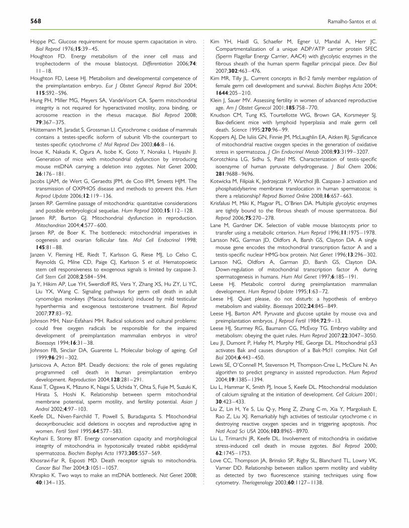
Hoppe PC. Glucose requirement for mouse sperm capacitation in vitro.Biol Reprod 1976;15:39–45.
Houghton FD. Energy metabolism of the inner cell mass andtrophectoderm of the mouse blastocyst. Differentiation 2006;74:11–18.
Houghton FD, Leese HJ. Metabolism and developmental competence ofthe preimplantation embryo. Eur J Obstet Gynecol Reprod Biol 2004;115:S92–S96.
Hung PH, Miller MG, Meyers SA, VandeVoort CA. Sperm mitochondrialintegrity is not required for hyperactivated motility, zona binding, oracrosome reaction in the rhesus macaque. Biol Reprod 2008;79:367–375.
Huttemann M, Jaradat S, Grossman LI. Cytochrome c oxidase of mammalscontains a testes-specific isoform of subunit VIb-the counterpart totestes-specific cytochrome c? Mol Reprod Dev 2003;66:8–16.
Inoue K, Nakada K, Ogura A, Isobe K, Goto Y, Nonaka I, Hayashi JI.Generation of mice with mitochondrial dysfunction by introducingmouse mtDNA carrying a deletion into zygotes. Nat Genet 2000;26:176–181.
Jacobs LJAM, de Wert G, Geraedts JPM, de Coo IFM, Smeets HJM. Thetransmission of OXPHOS disease and methods to prevent this. HumReprod Update 2006;12:119–136.
Jansen RP. Germline passage of mitochondria: quantitative considerationsand possible embryological sequelae. Hum Reprod 2000;15:112–128.
Jansen RP, Burton GJ. Mitochondrial dysfunction in reproduction.Mitochondrion 2004;4:577–600.
Jansen RP, de Boer K. The bottleneck: mitochondrial imperatives inoogenesis and ovarian follicular fate. Mol Cell Endocrinol 1998;145:81–88.
Janzen V, Fleming HE, Riedt T, Karlsson G, Riese MJ, Lo Celso C,Reynolds G, Milne CD, Paige CJ, Karlsson S et al. Hematopoieticstem cell responsiveness to exogenous signals is limited by caspase-3.Cell Stem Cell 2008;2:584–594.
Jia Y, Hikim AP, Lue YH, Swerdloff RS, Vera Y, Zhang XS, Hu ZY, Li YC,Liu YX, Wang C. Signaling pathways for germ cell death in adultcynomolgus monkeys (Macaca fascicularis) induced by mild testicularhyperthermia and exogenous testosterone treatment. Biol Reprod2007;77:83–92.
Johnson MH, Nasr-Esfahani MH. Radical solutions and cultural problems:could free oxygen radicals be responsible for the impaireddevelopment of preimplantation mammalian embryos in vitro?Bioessays 1994;16:31–38.
Johnson FB, Sinclair DA, Guarente L. Molecular biology of ageing. Cell1999;96:291–302.
Jurisicova A, Acton BM. Deadly decisions: the role of genes regulatingprogrammed cell death in human preimplantation embryodevelopment. Reproduction 2004;128:281–291.
Kasai T, Ogawa K, Mizuno K, Nagai S, Uchida Y, Ohta S, Fujie M, Suzuki K,Hirata S, Hoshi K. Relationship between sperm mitochondrialmembrane potential, sperm motility, and fertility potential. Asian JAndrol 2002;4:97–103.
Keefe DL, Niven-Fairchild T, Powell S, Buradagunta S. Mitochondrialdeoxyribonucleic acid deletions in oocytes and reproductive aging inwomen. Fertil Steril 1995;64:577–583.
Keyhani E, Storey BT. Energy conservation capacity and morphologicalintegrity of mitochondria in hypotonically treated rabbit epididymalspermatozoa. Biochim Biophys Acta 1973;305:557–569.
Khosravi-Far R, Esposti MD. Death receptor signals to mitochondria.Cancer Biol Ther 2004;3:1051–1057.
Khrapko K. Two ways to make an mtDNA bottleneck. Nat Genet 2008;40:134–135.
Kim YH, Haidl G, Schaefer M, Egner U, Mandal A, Herr JC.Compartmentalization of a unique ADP/ATP carrier protein SFEC(Sperm Flagellar Energy Carrier, AAC4) with glycolytic enzymes in thefibrous sheath of the human sperm flagellar principal piece. Dev Biol2007;302:463–476.
Kim MR, Tilly JL. Current concepts in Bcl-2 family member regulation offemale germ cell development and survival. Biochim Biophys Acta 2004;1644:205–210.
Klein J, Sauer MV. Assessing fertility in women of advanced reproductiveage. Am J Obstet Gynecol 2001;185:758–770.
Knudson CM, Tung KS, Tourtellotte WG, Brown GA, Korsmeyer SJ.Bax-deficient mice with lymphoid hyperplasia and male germ celldeath. Science 1995;270:96–99.
Koppers AJ, De Iuliis GN, Finnie JM, McLaughlin EA, Aitken RJ. Significanceof mitochondrial reactive oxygen species in the generation of oxidativestress in spermatozoa. J Clin Endocrinol Metab 2008;93:3199–3207.
Korotchkina LG, Sidhu S, Patel MS. Characterization of testis-specificisoenzyme of human pyruvate dehydrogenase. J Biol Chem 2006;281:9688–9696.
Kotwicka M, Filipiak K, Jedrzejczak P, Warchol JB. Caspase-3 activation andphosphatidylserine membrane translocation in human spermatozoa: isthere a relationship? Reprod Biomed Online 2008;16:657–663.
Krisfalusi M, Miki K, Magyar PL, O’Brien DA. Multiple glycolytic enzymesare tightly bound to the fibrous sheath of mouse spermatozoa. BiolReprod 2006;75:270–278.
Lane M, Gardner DK. Selection of viable mouse blastocysts prior totransfer using a metabolic criterion. Hum Reprod 1996;11:1975–1978.
Larsson NG, Garman JD, Oldfors A, Barsh GS, Clayton DA. A singlemouse gene encodes the mitochondrial transcription factor A and atestis-specific nuclear HMG-box protein. Nat Genet 1996;13:296–302.
Larsson NG, Oldfors A, Garman JD, Barsh GS, Clayton DA.Down-regulation of mitochondrial transcription factor A duringspermatogenesis in humans. Hum Mol Genet 1997;6:185–191.
Leese HJ. Metabolic control during preimplantation mammaliandevelopment. Hum Reprod Update 1995;1:63–72.
Leese HJ. Quiet please, do not disturb: a hypothesis of embryometabolism and viability. Bioessays 2002;24:845–849.
Leese HJ, Barton AM. Pyruvate and glucose uptake by mouse ova andpreimplantation embryos. J Reprod Fertil 1984;72:9–13.
Leese HJ, Sturmey RG, Baumann CG, McEvoy TG. Embryo viability andmetabolism: obeying the quiet rules. Hum Reprod 2007;22:3047–3050.
Leu JI, Dumont P, Hafey M, Murphy ME, George DL. Mitochondrial p53activates Bak and causes disruption of a Bak-Mcl1 complex. Nat CellBiol 2004;6:443–450.
Lewis SE, O’Connell M, Stevenson M, Thompson-Cree L, McClure N. Analgorithm to predict pregnancy in assisted reproduction. Hum Reprod2004;19:1385–1394.
Liu L, Hammar K, Smith PJ, Inoue S, Keefe DL. Mitochondrial modulationof calcium signaling at the initiation of development. Cell Calcium 2001;30:423–433.
Liu Z, Lin H, Ye S, Liu Q-y, Meng Z, Zhang C-m, Xia Y, Margoliash E,Rao Z, Liu XJ. Remarkably high activities of testicular cytochrome c indestroying reactive oxygen species and in triggering apoptosis. ProcNatl Acad Sci USA 2006;103:8965–8970.
Liu L, Trimarchi JR, Keefe DL. Involvement of mitochondria in oxidativestress-induced cell death in mouse zygotes. Biol Reprod 2000;62:1745–1753.
Love CC, Thompson JA, Brinsko SP, Rigby SL, Blanchard TL, Lowry VK,Varner DD. Relationship between stallion sperm motility and viabilityas detected by two fluorescence staining techniques using flowcytometry. Theriogenology 2003;60:1127–1138.
568 Ramalho-Santos et al.
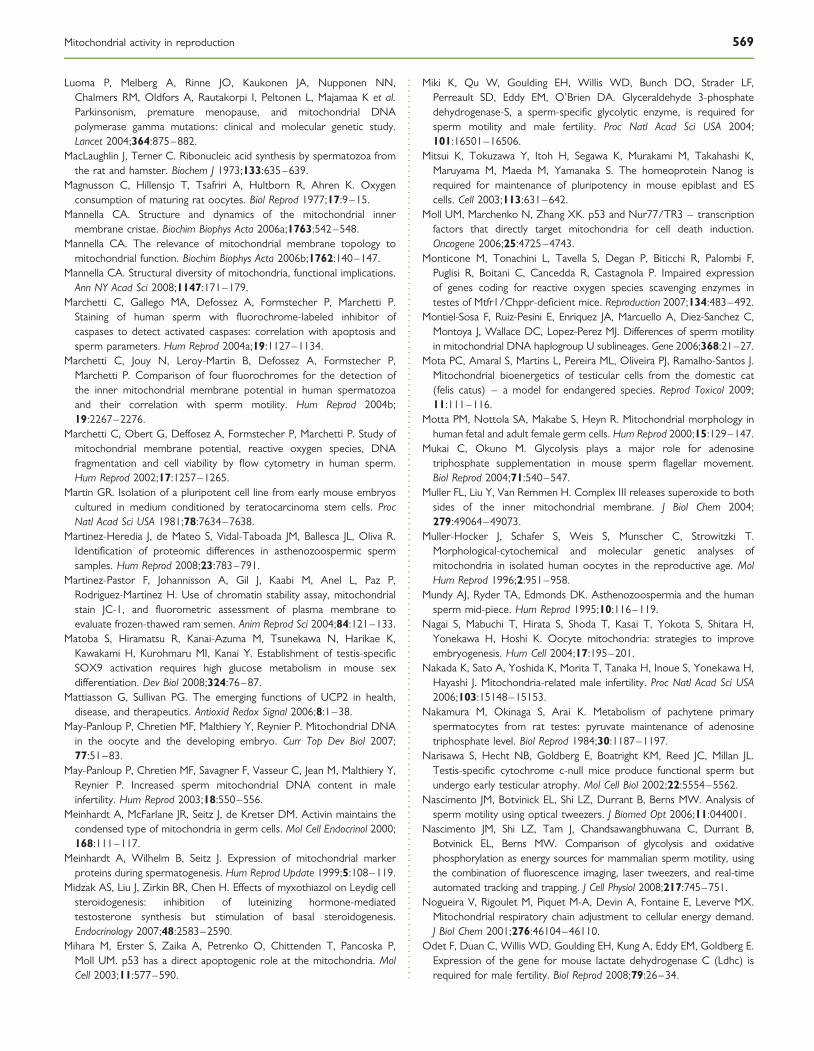
Luoma P, Melberg A, Rinne JO, Kaukonen JA, Nupponen NN,Chalmers RM, Oldfors A, Rautakorpi I, Peltonen L, Majamaa K et al.Parkinsonism, premature menopause, and mitochondrial DNApolymerase gamma mutations: clinical and molecular genetic study.Lancet 2004;364:875–882.
MacLaughlin J, Terner C. Ribonucleic acid synthesis by spermatozoa fromthe rat and hamster. Biochem J 1973;133:635–639.
Magnusson C, Hillensjo T, Tsafriri A, Hultborn R, Ahren K. Oxygenconsumption of maturing rat oocytes. Biol Reprod 1977;17:9–15.
Mannella CA. Structure and dynamics of the mitochondrial innermembrane cristae. Biochim Biophys Acta 2006a;1763:542–548.
Mannella CA. The relevance of mitochondrial membrane topology tomitochondrial function. Biochim Biophys Acta 2006b;1762:140–147.
Mannella CA. Structural diversity of mitochondria, functional implications.Ann NY Acad Sci 2008;1147:171–179.
Marchetti C, Gallego MA, Defossez A, Formstecher P, Marchetti P.Staining of human sperm with fluorochrome-labeled inhibitor ofcaspases to detect activated caspases: correlation with apoptosis andsperm parameters. Hum Reprod 2004a;19:1127–1134.
Marchetti C, Jouy N, Leroy-Martin B, Defossez A, Formstecher P,Marchetti P. Comparison of four fluorochromes for the detection ofthe inner mitochondrial membrane potential in human spermatozoaand their correlation with sperm motility. Hum Reprod 2004b;19:2267–2276.
Marchetti C, Obert G, Deffosez A, Formstecher P, Marchetti P. Study ofmitochondrial membrane potential, reactive oxygen species, DNAfragmentation and cell viability by flow cytometry in human sperm.Hum Reprod 2002;17:1257–1265.
Martin GR. Isolation of a pluripotent cell line from early mouse embryoscultured in medium conditioned by teratocarcinoma stem cells. ProcNatl Acad Sci USA 1981;78:7634–7638.
Martinez-Heredia J, de Mateo S, Vidal-Taboada JM, Ballesca JL, Oliva R.Identification of proteomic differences in asthenozoospermic spermsamples. Hum Reprod 2008;23:783–791.
Martinez-Pastor F, Johannisson A, Gil J, Kaabi M, Anel L, Paz P,Rodriguez-Martinez H. Use of chromatin stability assay, mitochondrialstain JC-1, and fluorometric assessment of plasma membrane toevaluate frozen-thawed ram semen. Anim Reprod Sci 2004;84:121–133.
Matoba S, Hiramatsu R, Kanai-Azuma M, Tsunekawa N, Harikae K,Kawakami H, Kurohmaru MI, Kanai Y. Establishment of testis-specificSOX9 activation requires high glucose metabolism in mouse sexdifferentiation. Dev Biol 2008;324:76–87.
Mattiasson G, Sullivan PG. The emerging functions of UCP2 in health,disease, and therapeutics. Antioxid Redox Signal 2006;8:1–38.
May-Panloup P, Chretien MF, Malthiery Y, Reynier P. Mitochondrial DNAin the oocyte and the developing embryo. Curr Top Dev Biol 2007;77:51–83.
May-Panloup P, Chretien MF, Savagner F, Vasseur C, Jean M, Malthiery Y,Reynier P. Increased sperm mitochondrial DNA content in maleinfertility. Hum Reprod 2003;18:550–556.
Meinhardt A, McFarlane JR, Seitz J, de Kretser DM. Activin maintains thecondensed type of mitochondria in germ cells. Mol Cell Endocrinol 2000;168:111–117.
Meinhardt A, Wilhelm B, Seitz J. Expression of mitochondrial markerproteins during spermatogenesis. Hum Reprod Update 1999;5:108–119.
Midzak AS, Liu J, Zirkin BR, Chen H. Effects of myxothiazol on Leydig cellsteroidogenesis: inhibition of luteinizing hormone-mediatedtestosterone synthesis but stimulation of basal steroidogenesis.Endocrinology 2007;48:2583–2590.
Mihara M, Erster S, Zaika A, Petrenko O, Chittenden T, Pancoska P,Moll UM. p53 has a direct apoptogenic role at the mitochondria. MolCell 2003;11:577–590.
Miki K, Qu W, Goulding EH, Willis WD, Bunch DO, Strader LF,Perreault SD, Eddy EM, O’Brien DA. Glyceraldehyde 3-phosphatedehydrogenase-S, a sperm-specific glycolytic enzyme, is required forsperm motility and male fertility. Proc Natl Acad Sci USA 2004;101:16501–16506.
Mitsui K, Tokuzawa Y, Itoh H, Segawa K, Murakami M, Takahashi K,Maruyama M, Maeda M, Yamanaka S. The homeoprotein Nanog isrequired for maintenance of pluripotency in mouse epiblast and EScells. Cell 2003;113:631–642.
Moll UM, Marchenko N, Zhang XK. p53 and Nur77/TR3 – transcriptionfactors that directly target mitochondria for cell death induction.Oncogene 2006;25:4725–4743.
Monticone M, Tonachini L, Tavella S, Degan P, Biticchi R, Palombi F,Puglisi R, Boitani C, Cancedda R, Castagnola P. Impaired expressionof genes coding for reactive oxygen species scavenging enzymes intestes of Mtfr1/Chppr-deficient mice. Reproduction 2007;134:483–492.
Montiel-Sosa F, Ruiz-Pesini E, Enriquez JA, Marcuello A, Diez-Sanchez C,Montoya J, Wallace DC, Lopez-Perez MJ. Differences of sperm motilityin mitochondrial DNA haplogroup U sublineages. Gene 2006;368:21–27.
Mota PC, Amaral S, Martins L, Pereira ML, Oliveira PJ, Ramalho-Santos J.Mitochondrial bioenergetics of testicular cells from the domestic cat(felis catus) – a model for endangered species. Reprod Toxicol 2009;11:111–116.
Motta PM, Nottola SA, Makabe S, Heyn R. Mitochondrial morphology inhuman fetal and adult female germ cells. Hum Reprod 2000;15:129–147.
Mukai C, Okuno M. Glycolysis plays a major role for adenosinetriphosphate supplementation in mouse sperm flagellar movement.Biol Reprod 2004;71:540–547.
Muller FL, Liu Y, Van Remmen H. Complex III releases superoxide to bothsides of the inner mitochondrial membrane. J Biol Chem 2004;279:49064–49073.
Muller-Hocker J, Schafer S, Weis S, Munscher C, Strowitzki T.Morphological-cytochemical and molecular genetic analyses ofmitochondria in isolated human oocytes in the reproductive age. MolHum Reprod 1996;2:951–958.
Mundy AJ, Ryder TA, Edmonds DK. Asthenozoospermia and the humansperm mid-piece. Hum Reprod 1995;10:116–119.
Nagai S, Mabuchi T, Hirata S, Shoda T, Kasai T, Yokota S, Shitara H,Yonekawa H, Hoshi K. Oocyte mitochondria: strategies to improveembryogenesis. Hum Cell 2004;17:195–201.
Nakada K, Sato A, Yoshida K, Morita T, Tanaka H, Inoue S, Yonekawa H,Hayashi J. Mitochondria-related male infertility. Proc Natl Acad Sci USA2006;103:15148–15153.
Nakamura M, Okinaga S, Arai K. Metabolism of pachytene primaryspermatocytes from rat testes: pyruvate maintenance of adenosinetriphosphate level. Biol Reprod 1984;30:1187–1197.
Narisawa S, Hecht NB, Goldberg E, Boatright KM, Reed JC, Millan JL.Testis-specific cytochrome c-null mice produce functional sperm butundergo early testicular atrophy. Mol Cell Biol 2002;22:5554–5562.
Nascimento JM, Botvinick EL, Shi LZ, Durrant B, Berns MW. Analysis ofsperm motility using optical tweezers. J Biomed Opt 2006;11:044001.
Nascimento JM, Shi LZ, Tam J, Chandsawangbhuwana C, Durrant B,Botvinick EL, Berns MW. Comparison of glycolysis and oxidativephosphorylation as energy sources for mammalian sperm motility, usingthe combination of fluorescence imaging, laser tweezers, and real-timeautomated tracking and trapping. J Cell Physiol 2008;217:745–751.
Nogueira V, Rigoulet M, Piquet M-A, Devin A, Fontaine E, Leverve MX.Mitochondrial respiratory chain adjustment to cellular energy demand.J Biol Chem 2001;276:46104–46110.
Odet F, Duan C, Willis WD, Goulding EH, Kung A, Eddy EM, Goldberg E.Expression of the gene for mouse lactate dehydrogenase C (Ldhc) isrequired for male fertility. Biol Reprod 2008;79:26–34.
Mitochondrial activity in reproduction 569

Oh SK, Kim HS, Ahn HJ, Seol HW, Kim YY, Park YB, Yoon CJ, Kim DW,Kim SH, Moon SY. Derivation and characterization of new humanembryonic stem cell lines: SNUhES1, SNUhES2, and SNUhES3. StemCells 2005;23:211–219.
Olson GE, Winfrey VP. Mitochondria-cytoskeleton interactions in thesperm midpiece. J Struct Biol 1990;103:13–22.
Orrenius S, Gogvadze V, Zhivotovsky B. Mitochondrial oxidative stress:implications for cell death. Annu Rev Pharmacol Toxicol 2007;47:143–183.
Otani H, Tanaka O, Kasai K, Yoshioka T. Development of mitochondrialhelical sheath in the middle piece of the mouse spermatid tail: regulardispositions and synchronized changes. Anat Rec 1988;222:26–33.
Ott M, Gogvadze V, Orrenius S, Zhivotovsky B. Mitochondria, oxidativestress and cell death. Apoptosis 2007;12:913–922.
Paasch U, Grunewald S, Agarwal A, Glandera HJ. Activation pattern ofcaspases in human spermatozoa. Fertil Steril 2004a;81:802–809.
Paasch U, Grunewald S, Dathe S, Glander HJ. Mitochondria of humanspermatozoa are preferentially susceptible to apoptosis. Ann N Y AcadSci 2004b;1030:403–409.
Pan G, Thomson JA. Nanog and transcriptional networks in embryonicstem cell pluripotency. Cell Res 2007;17:42–49.
Pareek TK, Joshi AR, Sanyal A, Dighe RR. Insights into male germ cellapoptosis due to depletion of gonadotropins caused by GnRHantagonists. Apoptosis 2007;12:1085–1100.
Parrish JJ, Susko-Parrish JL, First NL. Capacitation of bovine sperm byheparin: inhibitory effect of glucose and role of intracellular pH. BiolReprod 1989;41:683–699.
Pereira L, Goncalves J, Goios A, Rocha T, Amorim A. Human mtDNAhaplogroups and reduced male fertility: real association or hiddenpopulation substructuring. Int J Androl 2005;28:241–247.
Perez GI, Acton BM, Jurisicova A, Perkins GA, White A, Brown J,Trbovich AM, Kim MR, Fissore R, Xu J et al. Genetic variancemodifies apoptosis susceptibility in mature oocytes via alterations inDNA repair capacity and mitochondrial ultrastructure. Cell DeathDiffer 2007;14:524–533.
Perez GI, Tao XJ, Tilly JL. Fragmentation and death (a.k.a. apoptosis) ofovulated oocytes. Mol Hum Reprod 1999;5:414–420.
Perez GI, Trbovich AM, Gosden RG, Tilly JL. Mitochondria and the deathof oocytes. Nature 2000;403:500–501.
Peterson RN, Freund M. ATP synthesis and oxidative metabolism inhuman spermatozoa. Biol Reprod 1970;3:47–54.
Piccoli C, Ria R, Scrima R, Cela O, D’Aprile A, Boffoli D, Falzetti F,Tabilio A, Capitanio N. Characterization of mitochondrial andextra-mitochondrial oxygen consuming reactions in humanhematopoietic stem cells. Novel evidence of the occurrence ofNAD(P)H oxidase activity. J Biol Chem 2005;280:26467–26476.
Prince FP. Lamellar and tubular associations of the mitochondrial cristae:unique forms of the cristae present in steroid-producing cells.Mitochondrion 2002;1:381–389.
Publicover SJ, Giojalas LC, Teves ME, de Oliveira GS, Garcia AA,Barratt CL, Harper CV. Ca2þ signalling in the control of motility andguidance in mammalian sperm. Front Biosci 2008;13:5623–5637.
Publicover S, Harper CV, Barratt C. [Ca2þ]i signalling in sperm-making themost of what you’ve got. Nat Cell Biol 2007;9:235–242.
Puglisi R, Bevilacqua A, Carlomagno G, Lenzi A, Gandini L, Stefanini M,Mangia F, Boitani C. Mice overexpressing the mitochondrialphospholipid hydroperoxide glutathione peroxidase in male germ cellsshow abnormal spermatogenesis and reduced fertility. Endocrinology2007;148:4302–4309.
Qin H, Yu T, Qing T, Liu Y, Zhao Y, Cai J, Li J, Song Z, Qu X, Zhou P et al.Regulation of apoptosis and differentiation by p53 in human embryonicstem cells. J Biol Chem 2007;282:5842–5852.
Raikwar SP, Zavazava N. Insulin producing cells derived from embryonicstem cells: are we there yet? J Cell Physiol 2009;218:256–263.
Raineri I, Carlson EJ, Gacayan R, Carra S, Oberley TD, Huang TT,Epstein CJ. Strain-dependent high-level expression of a transgenefor manganese superoxide dismutase is associated with growthretardation and decreased fertility. Free Radic Biol Med 2001;31:1018–1030.
Ramalho-Santos J, Amaral A, Brito R, Freitas M, Almeida Santos T.Simultaneous analysis of cytoskeletal patterns and chromosomepositioning in human fertilization failures. Fertil Steril 2004;82:1654–1659.
Ramalho-Santos J, Amaral A, Sousa AP, Rodrigues AS, Martins L,Baptista M, Mota PC, Tavares R, Amaral S, Gamboa S. Probing thestructure and function of mammalian sperm using optical andfluorescence microscopy. In: Mendez-Villas A, Diaz J (eds). ModernResearch and Educational Topics in Microscopy. Badajoz, Spain:FORMATEX, 2007, 394–402.
Rardin MJ, Wiley SE, Murphy AN, Pagliarini DJ, Dixon JE. Dual specificityphosphatases 18 and 21 target to opposing sides of the mitochondrialinner membrane. J Biol Chem 2008;283:15440–15450.
Rebrin I, Kamzalov S, Sohal RS. Effects of age and caloric restriction onglutathione redox state in mice. Free Radic Biol Med 2003;35:626–635.
Reynaud K, Driancourt MA. Oocyte attrition. Mol Cell Endocrinol 2000;163:101–108.
Robinson R, Fritz IB. Metabolism of glucose by Sertoli cells in culture. BiolReprod 1981;24:1032–1041.
Rodriguez I, Ody C, Araki K, Garcia I, Vassalli P. An early and massivewave of germinal cell apoptosis is required for the development offunctional spermatogenesis. EMBO J 1997;16:2262–2270.
Rogers BJ, Chang L, Yanagimachi R. Glucose effect on respiration: possiblemechanism for capacitation in guinea pig spermatozoa. J Exp Zool 1979;207:107–112.
Rogers BJ, Perreault SD. Importance of glycolysable substrates for in vitrocapacitation of human spermatozoa. Biol Reprod 1990;43:1064–1069.
Rogers BJ, Yanagimachi R. Retardation of guinea pig sperm acrosomereaction by glucose: the possible importance of pyruvate and lactatemetabolism in capacitation and the acrosome reaction. Biol Reprod1975;13:568–575.
Rossato M. Endocannabinoids, sperm functions and energy metabolism.Mol Cell Endocrinol 2008;286:S31–S35.
Roveri A, Casasco A, Maiorino M, Dalan P, Calligaro A, Ursini F.Phospholipid hydroperoxide glutathione peroxidase of rat testis.Gonadotropin dependence and immunocytochemical identification.J Biol Chem 1992;267:6142–6146.
Ruiz-Pesini E, Diez C, Lapena AC, Perez-Martos A, Montoya J, Alvarez E,Arenas J, Lopez-Perez MJ. Correlation of sperm motility withmitochondrial enzymatic activities. Clin Chem 1998;44:1616–1620.
Ruiz-Pesini E, Diez-Sanchez C, Lopez-Perez MJ, Enriquez JA. The role ofthe mitochondrion in sperm function: is there a place for oxidativephosphorylation or is this a purely glycolytic process? Curr Top DevBiol 2007;77:3–19.
Ruiz-Pesini E, Lapena AC, Diez-Sanchez C, Perez-Martos A, Montoya J,Alvarez E, Diaz M, Urries A, Montoro L, Lopez-Perez MJ et al.Human mtDNA haplogroups associated with high or reducedspermatozoa motility. Am J Hum Genet 2000a;67:682–696.
Ruiz-Pesini E, Lapena AC, Diez C, Alvarez E, Enriquez JA, Lopez-Perez MJ.Seminal quality correlates with mitochondrial functionality. Clin ChimActa 2000b;300:97–105.
Russell LD, Chiarini-Garcia H, Korsmeyer SJ, Knudson CM.Bax-dependent spermatogonia apoptosis is required for testiculardevelopment and spermatogenesis. Biol Reprod 2002;66:950–958.
570 Ramalho-Santos et al.

Ryu BY, Orwig KE, Kubota H, Avarbock MR, Brinster RL. Phenotypic andfunctional characteristics of spermatogonial stem cells in rats. Dev Biol2004;274:158–170.
Santambrogio P, Biasiotto G, Sanvito F, Olivieri S, Arosio P, Levi S.Mitochondrial ferritin expression in adult mouse tissues. J HistochemCytochem 2007;55:1129–1137.
Saretzki G, Walter T, Atkinson S, Passos JF, Bareth B, Keith WN,Stewart R, Hoare S, Stojkovic M, Armstrong L et al. Downregulationof multiple stress defense mechanisms during differentiation of humanembryonic stem cells. Stem Cells 2008;26:455–464.
Sathananthan AH, Trounson AO. Mitochondrial morphology duringpreimplantational human embryogenesis. Hum Reprod 2000;15:148–159.
Saunders PT, Millar MR, West AP, Sharpe RM. Mitochondrial cytochromeC oxidase II messenger ribonucleic acid is expressed in pachytenespermatocytes at high levels and in a stage-dependent manner duringspermatogenesis in the rat. Biol Reprod 1993;48:57–67.
Schieke SM, Ma M, Cao L, McCoy JP Jr, Liu C, Hensel NF, Barrett AJ,Boehm M, Finkel T. Mitochondrial metabolism modulatesdifferentiation and teratoma formation capacity in mouse embryonicstem cells. J Biol Chem 2008;283:28606–28512.
Seitz J, Mobius J, Bergmann M, Meinhardt A. Mitochondrial differentiationduring meiosis of male germ cells. Int J Androl 1995;18:7–11.
Slotte H, Gustafson O, Nylund L, Pousette A. ATP and ADP in humanpre-embryos. Hum Reprod 1990;5:319–322.
Spinaci M, De Ambrogi M, Volpe S, Galeati G, Tamanini C, Seren E. Effectof staining and sorting on boar sperm membrane integrity,mitochondrial activity and in vitro blastocyst development.Theriogenology 2005;64:191–201.
Spitkovsky D, Sasse P, Kolossov E, Bottinger C, Fleischmann BK,Hescheler J, Wiesner RJ. Activity of complex III of the mitochondrialelectron transport chain is essential for early heart muscle celldifferentiation. FASEB J 2004;18:1300–1302.
Steger K, Wilhelm J, Konrad L, Stalf T, Greb R, Diemer T, Kliesch S,Bergmann M, Weidner W. Both protamine-1 to protamine-2 mRNAratio and Bcl2 mRNA content in testicular spermatids and ejaculatedspermatozoa discriminate between fertile and infertile men. HumReprod 2008;23:11–16.
Stern S, Biggers JD, Anderson E. Mitochondria and early development ofthe mouse. J Exp Zool 1971;176:179–191.
St John JC, Bowles EJ, Amaral A. Sperm mitochondria and fertilisation. SocReprod Fertil Suppl 2007;65:399–416.
St John JC, Jokhi RP, Barratt CL. Men with oligoasthenoteratozoospermiaharbour higher numbers of multiple mitochondrial DNA deletions intheir spermatozoa, but individual deletions are not indicative ofoverall aetiology. Mol Hum Reprod 2001;7:103–111.
St John JC, Jokhi RP, Barratt CL. The impact of mitochondrial genetics onmale infertility. Int J Androl 2005;28:65–73.
St John JC, Ramalho-Santos J, Gray HL, Petrosko P, Rawe VY, Navara CS,Simerly CR, Schatten GP. The expression of mitochondrial DNAtranscription factors during early cardiomyocyte in vitro differentiationfrom human embryonic stem cells. Cloning Stem Cells 2005;7:141–153.
Stocco DM, Wells J, Clark BJ. The effects of hydrogen peroxide onsteroidogenesis in mouse Leydig tumor cells. Endocrinology 1993;133:2827–2832.
Storey BT. Effect of ionophores and inhibitors and uncouplers ofoxidative phosphorylation on sperm respiration. Arch Androl 1978;1:169–177.
Storey BT. Strategy of oxidative metabolism in bull spermatozoa. J Exp Zool1980;212:61–67.
Storey BT. Mammalian sperm metabolism: oxygen and sugar, friend andfoe. Int J Dev Biol 2008;52:427–437.
Su D, Novoselov SV, Sun QA, Moustafa ME, Zhou Y, Oko R, Hatfield DL,Gladyshev VN. Mammalian selenoprotein thioredoxin-glutathionereductase. Roles in disulfide bond formation and sperm maturation.J Biol Chem 2005;280:26491–26498.
Sun QY, Wu GM, Lai L, Park KW, Cabot R, Cheong HT, Day BN,Prather RS, Schatten H. Translocation of active mitochondria duringpig oocyte maturation, fertilization and early embryo development invitro. Reproduction 2001;122:155–163.
Sutovsky P, Moreno RD, Ramalho-Santos J, Dominko T, Simerly C,Schatten G. Ubiquitin tag for sperm mitochondria. Nature 1999;402:371–372.
Takahashi K, Tanabe K, Ohnuki M, Narita M, Ichisaka T, Tomoda K,Yamanaka S. Induction of pluripotent stem cells from adult humanfibroblasts by defined factors. Cell 2007;131:861–872.
Tavares RS, Martins FC, Oliveira PJ, Ramalho-Santos J, Peixoto FP.Parabens in male infertility-Is there a mitochondrial connection?Reprod Toxicol 2009;27:1–7.
Terada K, Ueno S, Yomogida K, Imai T, Kiyonari H, Takeda N, Yano M,Abe S, Aizawa S, Mori M. Expression of Tom34 splicing isoforms inmouse testis and knockout of Tom34 in mice. J Biochem 2003;133:625–631.
Tesarik J, Sousa M. Mechanism of calcium oscillations in human oocytes: atwo-store model. Mol Hum Reprod 1996;2:383–386.
Thompson JG, Partridge RJ, Houghton FD, Cox CI, Leese HJ. Oxygenuptake and carbohydrate metabolism by in vitro derived bovineembryos. J Reprod Fertil 1996;106:299–306.
Thomson JA, Itskovitz-Eldor J, Shapiro SS, Waknitz MA, Swiergiel JJ,Marshall VS, Jones JM. Embryonic stem cell lines derived from humanblastocysts. Science 1998;282:1145–1147.
Thouas GA, Trounson AO, Wolvetang EJ, Jones GM. Mitochondrialdysfunction in mouse oocytes results in preimplantation embryoarrest in vitro. Biol Reprod 2004;71:1936–1942.
Tilly JL. Commuting the death sentence: how oocytes strive to survive. NatRev Mol Cell Biol 2001;2:838–848.
Traba J, Satrustegui J, del Arco A. Characterization of SCaMC-3-Like/slc25a41 a novel calcium independent mitochondrial ATP-Mg/Picarrier. Biochem J 2008; doi:10.1042/BJ20081262.
Tremellen K. Oxidative stress and male infertility–a clinical perspective.Hum Reprod Update 2008;14:243–258.
Trifunovic A, Wredenberg A, Falkenberg M, Spelbrink JN, Rovio AT,Bruder CE, Bohlooly YM, Gidlof S, Oldfors A, Wibom R et al.Premature ageing in mice expressing defective mitochondrial DNApolymerase. Nature 2004;429:417–423.
Troiano L, Granata AR, Cossarizza A, Kalashnikova G, Bianchi R, Pini G,Tropea F, Carani C, Franceschi C. Mitochondrial membrane potentialand DNA stainability in human sperm cells: a flow cytometry analysiswith implications for male infertility. Exp Cell Res 1998;241:384–393.
Twaina-Bechor E, Bartoov B. The relationship between ejaculated ramsperm mitochondrial protein synthesis and motility. Andrologia 1994;26:351–355.
Ursini F, Heim S, Kiess M, Maiorino M, Roveri A, Wissing J, Flohe L. Dualfunction of the selenoprotein PHGPx during sperm maturation. Science1999;285:1393–1396.
Van Blerkom J. Mitochondria as regulatory forces in oocytes,preimplantation embryos and stem cells. Reprod Biomed Online 2008;16:553–569.
Van Blerkom J, Cox H, Davis P. Regulatory roles for mitochondria in theperi-implantation mouse blastocyst: possible origins and developmentalsignificance of differential DeltaPsim. Reproduction 2006;131:961–976.
Van Blerkom J, Davis P. High-polarized (Delta Psi m(HIGH)) mitochondriaare spatially polarized in human oocytes and early embryos in stablesubplasmalemmal domains: developmental significance and the
Mitochondrial activity in reproduction 571

concept of vanguard mitochondria. Reprod Biomed Online 2006;13:246–254.
Van Blerkom J, Davis P. Mitochondrial signaling and fertilization. Mol HumReprod 2007;13:759–770.
Van Blerkom J, Davis P, Alexander S. Differential mitochondrialdistribution in human pronuclear embryos leads to disproportionateinheritance between blastomeres: relationship to microtubularorganization, ATP content and competence. Hum Reprod 2000;15:2621–2633.
Van Blerkom J, Davis P, Alexander S. Inner mitochondrial membranepotential (DeltaPsim), cytoplasmic ATP content and free Ca2þ levelsin metaphase II mouse oocytes. Hum Reprod 2003;18:2429–2440.
Van Blerkom J, Davis PW, Lee J. ATP content of human oocytes anddevelopmental potential and outcome after in-vitro fertilization andembryo transfer. Hum Reprod 1995;10:415–424.
Van Blerkom J, Davis P, Mathwig V, Alexander S. Domains ofhigh-polarized and low-polarized mitochondria may occur in mouseand human oocytes and early embryos. Hum Reprod 2002;17:393–406.
Varum S, Bento C, Sousa AP, Gomes-Santos CS, Henriques P,Almeida-Santos T, Teodosio C, Paiva A, Ramalho-Santos J.Characterization of human sperm populations using conventionalparameters, surface ubiquitination, and apoptotic markers. Fertil Steril2007;87:572–583.
Vaziri H, Dessain SK, Ng Eaton E, Imai SI, Frye RA, Pandita TK,Guarente L, Weinberg RA. hSIR2(SIRT1) functions as anNAD-dependent p53 deacetylase. Cell 2001;107:149–159.
Vazquez-Memije ME, Capin R, Tolosa A, El-Hafidi M. Analysis ofage-associated changes in mitochondrial free radical generation by rattestis. Mol Cell Biochem 2008;307:23–30.
Vazquez-Memije ME, Cardenas-Mendez MJ, Tolosa A, El-Hafidi ME.Respiratory chain complexes and membrane fatty acids compositionin rat testis mitochondria throughout development and ageing. ExpGerontol 2005;40:482–490.
Vera Y, Diaz-Romero M, Rodrigues S, Lue Y, Wang C, Swerdloff RS,Hikim APS. Mitochondria dependent pathway is involved inHeat-induced male germ cells death: lessons from mutant mice. BiolReprod 2004;70:1534–1540.
Vera Y, Erkkila K, Wang C, Nunez C, Kyttanen S, Lue Y, Dunkel L,Swerdloff RS, Hikim APS. Involvement of p38 mitogen-activatedprotein kinase and inducible nitric oxide synthase in apoptoticsignaling of murine and human male germ cells after hormonedeprivation. Mol Endocrinol 2006;20:1597–1609.
Wallace DC. Mitochondrial diseases in man and mouse. Science 1999;283:1482–1488.
Wang S, Lee DP, Gong N, Schwerbrock NM, Mashek DG,Gonzalez-Baro MR, Stapleton C, Li LO, Lewin TM, Coleman RA.Cloning and functional characterization of a novel mitochondrialN-ethylmaleimide-sensitive glycerol-3-phosphate acyltransferase(GPAT2). Arch Biochem Biophys 2007;465:347–358.
Wang X, Sharma RK, Gupta A, George V, Thomas AJ, Falcone T,Agarwal A. Alterations in mitochondria membrane potential andoxidative stress in infertile men: a prospective observational study.Fertil Steril 2003;80:844–850.
Wassarman PM, Josefowicz WJ. Oocyte development in the mouse: anultrastructural comparison of oocytes isolated at various stages ofgrowth and meiotic competence. J Morphol 1978;156:209–235.
Wegrzyn J, Potla R, Chwae YJ, Sepuri NBV, Zhang Q, Koeck T,Derecka M, Karol Szczepanek K, Szelag M, Gornicka A et al. Functionof mitochondrial Stat3 in cellular respiration. Science 2009;323:793–797.
Weng SL, Taylor SL, Morshedi M, Schuffner A, Duran EH, Beebe S,Oehninger S. Caspase activity and apoptotic markers in ejaculatedhuman sperm. Mol Hum Reprod 2002;8:984–991.
Wenger RH, Katschinski DM. The hypoxic testis and post-meioticexpression of PAS domain proteins. Semin Cell Dev Biol 2005;6:547–553.
Wilding M, Dale B, Marino M, di Matteo L, Alviggi C, Pisaturo ML,Lombardi L, De Placido G. Mitochondrial aggregation patterns andactivity in human oocytes and preimplantation embryos. Hum Reprod2001;16:909–917.
Williams AC, Ford WC. The role of glucose in supporting motilityand capacitation in human spermatozoa. J Androl 2001;22:680–695.
Wycherley G, Kane MT, Hynes AC. Oxidative phosphorylation and thetricarboxylic acid cycle are essential for normal development ofmouse ovarian follicles. Hum Reprod 2005;20:2757–2763.
Yamanaka S. Induction of pluripotent stem cells from mouse fibroblasts byfour transcription factors. Cell Prolif 2008;41:51–56.
Yamane T, Dylla SJ, Muijtjens M, Weissman IL. Enforced Bcl-2 expressionoverrides serum and feeder cell requirements for mouse embryonicstem cell self-renewal. Proc Natl Acad Sci USA 2005;102:3312–3317.
Yan W, Samson M, Jegou B, Toppari J. Bcl-w Forms Complexes with Baxand Bak, and Elevated Ratios of Bax/Bcl-w and Bak/Bcl-w Correspondto Spermatogonial and Spermatocyte Apoptosis in the Testis. MolEndocrinol 2000;14:682–699.
Yang HW, Hwang KJ, Kwon HC, Kim HS, Choi KW, Oh KS. Detection ofreactive oxygen species (ROS) and apoptosis in human fragmentedembryos. Hum Reprod 1998;13:998–1002.
Yesodi V, Yaron Y, Lessing JB, Amit A, Ben-Yosef D. The mitochondrialDNA mutation (deltamtDNA5286) in human oocytes: correlationwith age and IVF outcome. J Assist Reprod Genet 2002;19:60–66.
Yu J, Vodyanik MA, Smuga-Otto K, Antosiewicz-Bourget J, Frane JL,Tian S, Nie J, Jonsdottir GA, Ruotti V, Stewart R et al. Inducedpluripotent stem cell lines derived from human somatic cells. Science2007;318:1917–1920.
Zamzami N, Brenner C, Marzo I, Susin S, Kroemer G. Subcellular andsubmitochondrial model of action of Bcl-2-like oncoproteins.Oncogene 1998;16:2265–2282.
Zheng S, Turner TT, Lysiak JJ. Caspase 2 activity contributes to the initialwave of germ cell apoptosis during the first round of spermatogenesis.Biol Reprod 2006;74:1026–1033.
Zirkin BR, Chen H. Regulation of Leydig cell steroidogenic function duringaging. Biol Reprod 2000;63:977–981.
Submitted on January 18, 2009; resubmitted on March 22, 2009; accepted onApril 6, 2009
572 Ramalho-Santos et al.
Copyright © 2022 FDOKUMEN