Effect of Myofascial Therapy on Pain and Functionality ... - MDPI
26
International Journal of Environmental Research and Public Health Systematic Review Effect of Myofascial Therapy on Pain and Functionality of the Upper Extremities in Breast Cancer Survivors: A Systematic Review and Meta-Analysis Inmaculada Carmen Lara-Palomo 1, * , Adelaida María Castro-Sánchez 1 , Marta MaríaCórdoba-Peláez 1 , Manuel Albornoz-Cabello 2 and Lucía Ortiz-Comino 3 Citation: Lara-Palomo, I.C.; Castro-Sánchez, A.M.; Córdoba-Peláez, M.M.; Albornoz-Cabello, M.; Ortiz-Comino, L. Effect of Myofascial Therapy on Pain and Functionality of the Upper Extremities in Breast Cancer Survivors: A Systematic Review and Meta-Analysis. Int. J. Environ. Res. Public Health 2021, 18, 4420. https://doi.org/10.3390/ ijerph18094420 Academic Editor: María M. Morales Suárez-Varela Received: 9 March 2021 Accepted: 19 April 2021 Published: 21 April 2021 Publisher’s Note: MDPI stays neutral with regard to jurisdictional claims in published maps and institutional affil- iations. Copyright: © 2021 by the authors. Licensee MDPI, Basel, Switzerland. This article is an open access article distributed under the terms and conditions of the Creative Commons Attribution (CC BY) license (https:// creativecommons.org/licenses/by/ 4.0/). 1 Department of Nursing, Physical Therapy and Medicine, University of Almeria, Road Sacramento s/n, 04120 Almeria, Spain; [email protected] (A.M.C.-S.); [email protected] (M.M.C.-P.) 2 Department of Physiotherapy, University of Sevilla, Avicena Street s/n, 41009 Sevilla, Spain; [email protected] 3 Department of Physical Therapy, University of Granada, Technological Park of Health Sciences, Avenue of Illustration 60, 18071 Granada, Spain; [email protected] * Correspondence: [email protected]; Tel.: +34-655-388-324 Abstract: (1) Objective: The purpose was to analyze the effectiveness of myofascial therapy on mus- culoskeletal pain and functionality of the upper extremities in female breast cancer survivors, and to evaluate the changes in range of motion, quality of life, and mood state of these patients. (2) Methods: Systematic searches were performed on the MEDLINE/PubMed, Web of Science, Scopus, and Physio- therapy Evidence Databases for articles published until October 2020, in order to identify randomized controlled trials which analyzed the effectiveness of myofascial therapy as compared to a control group, passive treatment, placebo, or another intervention, and allowed co-interventions on female breast cancer survivors. Two reviewers examined the sources individually, calculated the risk of bias and extracted the data (PROSPERO number CRD42020215823). (3) Results: A total of eight RCTs were included. The results suggested that myofascial therapy does not have a greater statistically signifi- cant immediate effect on pain intensity (SMD: -0.15; 95% CI -0.48, 0.19), functionality (SMD: -0.17; 95% CI -0.43, 0.09) and range of motion in flexion (SMD: 0.30; 95% CI -0.13, 0.74) than an inactive, passive treatment or another intervention. However, a statistically significant result was observed for the abduction shoulder in favor of the experimental group (SMD: 0.46; 95% CI 0.05, 0.87; p = 0.03). (4) Conclusion: In general, although we found greater overall effects in support of the intervention with myofascial therapy than other control groups/types of interventions, the subgroup analysis revealed inconsistent results supporting myofascial therapy applied to breast cancer survivors. Keywords: breast cancer; myofascial release; functionality; pain; quality of life; range of motion 1. Introduction Of the 18.1 million cases of cancer globally diagnosed [1], breast cancer accounts for 11.6%, being the most common cancer among women [2,3]. In Spain, this incidence increases to 28.7%, and one in eight women is at risk of developing breast cancer [4]. The rising incidence of breast cancer in developed countries is mainly due to demographic factors, lifestyle, and reproduction rates [5]. Despite this high incidence, thanks to de- velopments in early detection techniques, as well as rapid implementation of treatment protocols [6], its survival rate is over 90% [7]. Treatment of breast cancer generally involves a combination of different methods, and may produce toxicities, which can be cumulative and difficult to separate clinically, includ- ing surgery, radiation, chemotherapy, hormonal therapy, and/or targeted therapy [8,9]. Surgical treatment for breast cancer includes breast-conserving surgery, combined with ra- diation, or mastectomy with or without radiation and with or without immediate/deferred reconstruction [9]. Breast-conserving therapy, consisting of breast-conserving surgery Int. J. Environ. Res. Public Health 2021, 18, 4420. https://doi.org/10.3390/ijerph18094420 https://www.mdpi.com/journal/ijerph
-
Upload
khangminh22 -
Category
Documents
-
view
2 -
download
0
Transcript of Effect of Myofascial Therapy on Pain and Functionality ... - MDPI
International Journal of
Environmental Research
and Public Health
Systematic Review
Effect of Myofascial Therapy on Pain and Functionality of theUpper Extremities in Breast Cancer Survivors: A SystematicReview and Meta-Analysis
Inmaculada Carmen Lara-Palomo 1,* , Adelaida María Castro-Sánchez 1 , Marta María Córdoba-Peláez 1,Manuel Albornoz-Cabello 2 and Lucía Ortiz-Comino 3
�����������������
Citation: Lara-Palomo, I.C.;
Castro-Sánchez, A.M.;
Córdoba-Peláez, M.M.;
Albornoz-Cabello, M.; Ortiz-Comino,
L. Effect of Myofascial Therapy on
Pain and Functionality of the Upper
Extremities in Breast Cancer
Survivors: A Systematic Review and
Meta-Analysis. Int. J. Environ. Res.
Public Health 2021, 18, 4420.
https://doi.org/10.3390/
ijerph18094420
Academic Editor:
María M. Morales Suárez-Varela
Received: 9 March 2021
Accepted: 19 April 2021
Published: 21 April 2021
Publisher’s Note: MDPI stays neutral
with regard to jurisdictional claims in
published maps and institutional affil-
iations.
Copyright: © 2021 by the authors.
Licensee MDPI, Basel, Switzerland.
This article is an open access article
distributed under the terms and
conditions of the Creative Commons
Attribution (CC BY) license (https://
creativecommons.org/licenses/by/
4.0/).
1 Department of Nursing, Physical Therapy and Medicine, University of Almeria, Road Sacramento s/n,04120 Almeria, Spain; [email protected] (A.M.C.-S.); [email protected] (M.M.C.-P.)
2 Department of Physiotherapy, University of Sevilla, Avicena Street s/n, 41009 Sevilla, Spain; [email protected] Department of Physical Therapy, University of Granada, Technological Park of Health Sciences,
Avenue of Illustration 60, 18071 Granada, Spain; [email protected]* Correspondence: [email protected]; Tel.: +34-655-388-324
Abstract: (1) Objective: The purpose was to analyze the effectiveness of myofascial therapy on mus-culoskeletal pain and functionality of the upper extremities in female breast cancer survivors, and toevaluate the changes in range of motion, quality of life, and mood state of these patients. (2) Methods:Systematic searches were performed on the MEDLINE/PubMed, Web of Science, Scopus, and Physio-therapy Evidence Databases for articles published until October 2020, in order to identify randomizedcontrolled trials which analyzed the effectiveness of myofascial therapy as compared to a control group,passive treatment, placebo, or another intervention, and allowed co-interventions on female breastcancer survivors. Two reviewers examined the sources individually, calculated the risk of bias andextracted the data (PROSPERO number CRD42020215823). (3) Results: A total of eight RCTs wereincluded. The results suggested that myofascial therapy does not have a greater statistically signifi-cant immediate effect on pain intensity (SMD: −0.15; 95% CI −0.48, 0.19), functionality (SMD: −0.17;95% CI −0.43, 0.09) and range of motion in flexion (SMD: 0.30; 95% CI −0.13, 0.74) than an inactive,passive treatment or another intervention. However, a statistically significant result was observed forthe abduction shoulder in favor of the experimental group (SMD: 0.46; 95% CI 0.05, 0.87; p = 0.03).(4) Conclusion: In general, although we found greater overall effects in support of the intervention withmyofascial therapy than other control groups/types of interventions, the subgroup analysis revealedinconsistent results supporting myofascial therapy applied to breast cancer survivors.
Keywords: breast cancer; myofascial release; functionality; pain; quality of life; range of motion
1. Introduction
Of the 18.1 million cases of cancer globally diagnosed [1], breast cancer accountsfor 11.6%, being the most common cancer among women [2,3]. In Spain, this incidenceincreases to 28.7%, and one in eight women is at risk of developing breast cancer [4]. Therising incidence of breast cancer in developed countries is mainly due to demographicfactors, lifestyle, and reproduction rates [5]. Despite this high incidence, thanks to de-velopments in early detection techniques, as well as rapid implementation of treatmentprotocols [6], its survival rate is over 90% [7].
Treatment of breast cancer generally involves a combination of different methods, andmay produce toxicities, which can be cumulative and difficult to separate clinically, includ-ing surgery, radiation, chemotherapy, hormonal therapy, and/or targeted therapy [8,9].Surgical treatment for breast cancer includes breast-conserving surgery, combined with ra-diation, or mastectomy with or without radiation and with or without immediate/deferredreconstruction [9]. Breast-conserving therapy, consisting of breast-conserving surgery
Int. J. Environ. Res. Public Health 2021, 18, 4420. https://doi.org/10.3390/ijerph18094420 https://www.mdpi.com/journal/ijerph

Int. J. Environ. Res. Public Health 2021, 18, 4420 2 of 26
(lumpectomy) plus radiation, is the standard treatment for most women with stage I andII breast carcinomas [10]. However, all survivors are at risk of suffering from side effectsof treatment in the short- or long-term, such as hemorrhage, infection at the surgical site,weakness in the arm or shoulder, restricted movement, swelling, numbness, pain, andlymphedema [11–15].
Radiation therapy is an adjuvant therapy that is used in 50% of patients, which maylead to fibrosis in the adjacent tissue [8]. Radiation-induced fibrosis is a form of damageto normal tissues after radiation therapy. It can affect the underlying fascia, muscles,organs, and bones on both the affected and unaffected sides, cause persistent symptomsand aesthetic disfigurement, thereby affecting quality of life [16–19].
Furthermore, the dissection of the axillary lymph nodes, the radiation on the regionallymph nodes and the patient’s preoperative body mass index, among others, are consideredto be factors that contribute to the development of one of the most frequent side effectsof breast cancer: lymphedema [20]. The risk of getting lymphedema after overcomingcancer is between 6% and 45% [21–24] and, of these cases, 90% arise between 18 and24 months after treatment [20,25]. Additionally, lymphedema is associated with othersymptoms, such as pain, bloating, pressure, fatigue, limited joint movement, mainly in theabduction of the shoulder and elbow bending, and the subsequent reduction in use of theaffected limb [22,26]. Conversely, postoperative pain is another side effect which occursin at least half of women who have undergone surgery between 6–15 months after theoperation [27,28]. The prevalence of neuropathic pain is 24% nine months after surgery [28].
While improvement in diagnostic processes and in the choices available for medicaltreatment to reduce possible long-term effects have led to a higher survival rate after breastcancer diagnosis, new challenges have arisen in addressing these effects in healthcaresystems [29]. There are several studies which suggest the use of physical therapy totreat side effects of breast cancer following medical treatment. Among the most highly-recommended therapies are mobilization, active exercises or active-assisted exercises, andmanual therapy [30–32]. Myofascial release is found under the scope of manual therapy,and is a low-impact, long-term treatment with the aim of restoring the length of the fascia,eliminating functional limitations and reducing the perception of pain to improve thefunction of the locomotor system [33]. Numerous clinical trials have demonstrated thebenefits of myofascial therapy on different populations, showing an improvement in rangeof motion and a decrease in perceived pain [34–37]. However, to date, no systematicreviews nor meta-analyses have been carried out about the effects of myofascial therapyon the treatment of the side effects derived from the medical treatment of breast cancer,whereas because of its characteristics as a manual therapy specialty as well as the absenceof secondary side-effects after its use [33], it seems an adequate technique to manage withbreast cancer survivors’ impairments.
Thus, the objective of this study is to perform a systematic review, together with ameta-analysis, to check the effects of myofascial therapy on female breast cancer survivors’pain, functionality, joint range of motion, and mood state. We hypothesize that myofascialrelease is an adequate approach to improve these factors.
2. Materials and Methods2.1. Protocol and Registry
A systematic review and meta-analysis was carried out, taking into account the itemson the recent declaration of ‘preferred reporting items for systematic reviews and meta-analyses’ (PRISMA) [38]. Systematic review registration: http://www.crd.york.ac.uk/PROSPERO (accessed on 21 April 2021). PROSPERO registration number: CRD42020215823(23 November 2020).
2.2. Search and Information Sources
The bibliographic search was performed during the months of March through Octo-ber 2020 on the Medline (through the platform PubMed), Scopus, Web of Science, and PEDro

Int. J. Environ. Res. Public Health 2021, 18, 4420 3 of 26
databases. The search strategy was based on terms registered on the MeSH list (“breast”,“cancer”, and “myofascial”) combined with the Boolean operator (AND), adapted to thecharacteristics of each database. Any duplicates that were identified in the multiple databasesearches were removed. Additionally, the reference lists for the included studies were also ex-amined and experts in the field were contacted (for example, authors of the included studies)to obtain additional information or information which was not implicit in the published trials.
2.3. Inclusion and Exclusion Criteria
The inclusion and exclusion criteria were defined using the PICO process [39] (Patient,Problem or Population, Intervention, Comparison, Control or Comparator, Outcome(s)).
2.3.1. Types of Studies
Randomized controlled clinical trials (RCTs) were included. Quasi-experimental con-trolled trials were excluded. In addition, the following inclusion criteria were taken intoaccount: RCTs published in the last 20 years (from 2000 onwards), written in English, avail-able in full-text version, focused on the ongoing effects of breast cancer intervention withmyofascial techniques. All studies that did not meet these characteristics were excluded.
2.3.2. Types of Participants
Studies with female subjects who had completed breast cancer treatment at least twomonths prior and had upper limb or neck pain. An exception was made with one of thestudies, which was related to palliative care, but was included, as the same aspects wereassessed as in the other studies. The age of participants and tumor location were notconsidered as criteria.
2.3.3. Types of Interventions
Studies were included in which side effects derived from breast cancer treatmentwere treated with myofascial therapy. Studies that treated these effects with myofascialtherapy accompanied by other manual or physical therapies were not excluded, nor werestudies restricted by the duration of treatment, frequency or type of techniques appliedin the treatment. Studies comparing myofascial therapy with any other intervention orno intervention were included. We also included any study which compared myofascialtherapy accompanied by other therapies, with only the application of those other therapies.All habitual medication was allowed to be taken during the studies.
2.3.4. Types of Outcome Measures
Studies that evaluated one or both of the following aspects as primary measureswere chosen: pain and functionality of the shoulder affected. For pain, the studies shouldhave used the Visual Analogue Scale (VAS), or a comparable numerical scale, and in theevaluation of functionality, the Disabilities of the Arm, Shoulder and Hand (DASH), orscale should have been used. As a secondary outcome measure, the following were alsotaken into consideration: the evaluation of shoulder mobility using goniometry; the moodstate of the participants evaluated, for example, with the Profile of Mood States (POMS)scale; and quality of life measured using the Health Questionnaire SF-36. The resultswere collected in three specific time periods: immediately following treatment, short-term(≤3 months), medium-term (between six and nine months), and long-term (≥12 months).
2.4. Study Selection
Two reviewers (I.C.L.-P., M.M.C.-P.) used the pre-specified criteria to select relevanttitles, abstracts and full articles. An article was deleted if it was determined that it didnot meet the inclusion criteria. If there were any hesitations about the selection decisions,a third reviewer (A.M.C.-S.) was consulted. Once the review was completed, the searchstrategy was repeated, in case any new studies had been published, and they were analyzedto assess their inclusion. The last search was carried out in November 2020.

Int. J. Environ. Res. Public Health 2021, 18, 4420 4 of 26
2.5. Data Extraction and Management
Firstly, the titles and abstracts of the references retrieved from the searches wereselected. The full text was obtained for references which the authors considered potentiallyrelevant. Full-text references were then independently evaluated for inclusion according tothe inclusion criteria for considering studies for this review.
To manage the data, a data summary sheet was created, based on the Cochranerecommendations. The data extracted were: author and year, type of study, number andtype of patients, type of intervention, number and duration of treatment sessions, outcomemeasures, and primary outcomes found among groups. If key information was missingfrom the study report, the authors of the report were contacted to obtain this information.
2.6. Risk of Bias Assessment
We used the recommendations from the Cochrane Collaboration to evaluate the riskof bias for all of the articles. Each item was evaluated with the objective of discerningwhether the trials eligible for inclusion in this review were valid enough for their resultsto be interpreted. The items evaluated were: specification of selection criteria, randomsequence generation, homogeneity among groups, allocation concealment, blinding ofparticipants and personnel, blinding of the outcome assessors, incomplete outcome data,selective reports, and other biases.
Two reviewers (I.C.L.-P., L.O.-C.) independently assessed the risk of bias for each ofthe articles selected for the current study. An arbitrator (A.M.C.-S.) was consulted to settleany disagreements.
Each item was classified as “high risk”, “low risk”, or “unclear risk” of bias. A sensitivityanalysis was performed on the primary results to explore the effects of including and excludingtrials with a high risk of bias (sensitivity analysis).
2.7. Statistical Analysis
A heterogeneity analysis of the selected studies was performed. In terms of continuousdata and dichotomous data, effect sizes were measured using standardized mean difference(SMD) and 95% confidence interval (CI), or risk ratio (RR) with 95% CI, respectively.Heterogeneity within RCTs was examined using the I2 test, considering I2 ≥ 50% as a sign ofsubstantial heterogeneity. Once there were >2 homogeneous studies, RevMan 5.4 (CochranCollaboration, London, UK) software was used to perform meta-analyses. Sensitivityanalyses were conducted for the robustness of the result of meta-analyses.
3. Results3.1. Selection of Studies
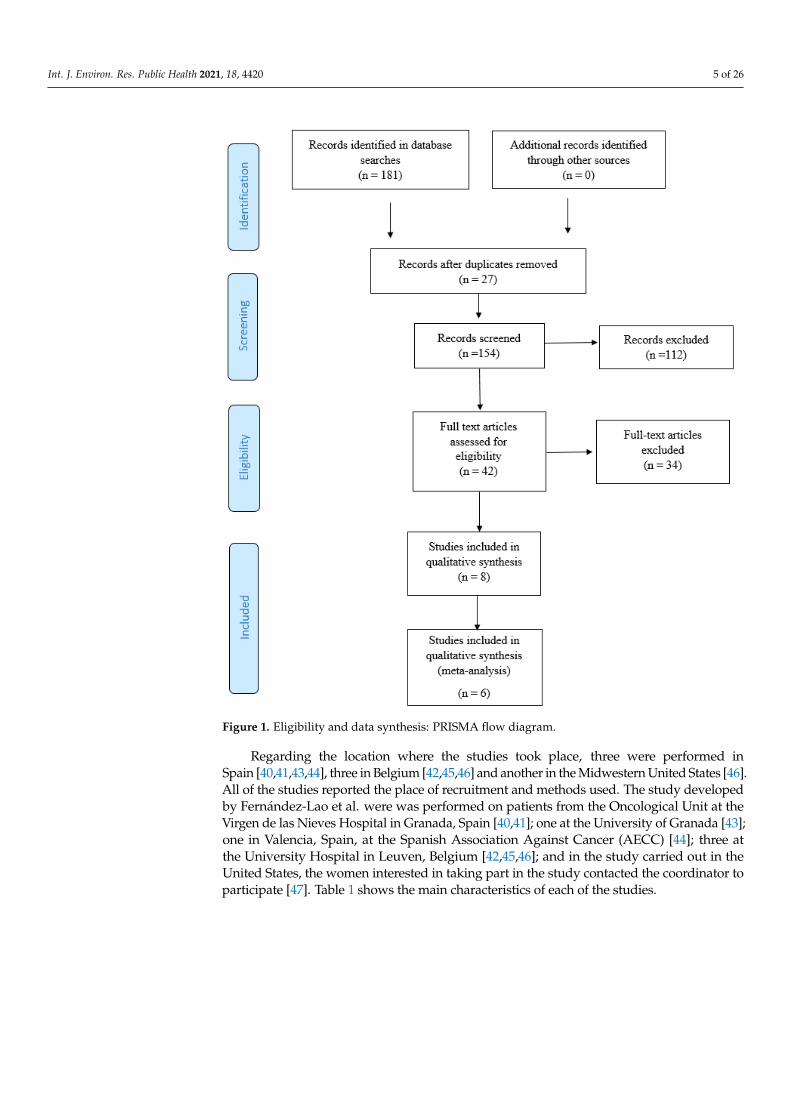
The initial search retrieved 181 potential articles, of which 112 were excluded afterreviewing their titles and abstracts, and 27 were duplicates, leaving 42 full-text articles to bereviewed. After the application of the inclusion and exclusion criteria, 34 of these articleswere eliminated, leaving eight studies for this systematic review and meta-analysis. Figure 1illustrates the different phases of the review, using the eligibility and data-synthesis PRISMAflow diagram.
3.2. Characteristics of Studies Included
Eight RCTs were included, with a total of 333 participants [40–47]. The study devel-oped by Fernández-Lao et al., divided into two publications [40,41] contained the smallestsample-size, with 20 participants; whereas the one with the largest sample size was thestudy by Groef et al. [42] with 147 participants.
Int. J. Environ. Res. Public Health 2021, 18, 4420 5 of 26
Int. J. Environ. Res. Public Health 2021, 18, x 5 of 23
Figure 1 illustrates the different phases of the review, using the eligibility and data-syn-thesis PRISMA flow diagram.
Figure 1. Eligibility and data synthesis: PRISMA flow diagram.
3.2. Characteristics of Studies Included Eight RCTs were included, with a total of 333 participants [40–47]. The study devel-
oped by Fernández-Lao et al., divided into two publications [40,41] contained the smallest sample-size, with 20 participants; whereas the one with the largest sample size was the study by Groef et al. [42] with 147 participants.
Regarding the location where the studies took place, three were performed in Spain [40,41,43,44], three in Belgium [42,45,46] and another in the Midwestern United States [46]. All of the studies reported the place of recruitment and methods used. The study devel-oped by Fernández-Lao et al. were was performed on patients from the Oncological Unit at the Virgen de las Nieves Hospital in Granada, Spain [40,41]; one at the University of Granada [43]; one in Valencia, Spain, at the Spanish Association Against Cancer (AECC) [44]; three at the University Hospital in Leuven, Belgium [42,45,46]; and in the study car-ried out in the United States, the women interested in taking part in the study contacted
Figure 1. Eligibility and data synthesis: PRISMA flow diagram.
Regarding the location where the studies took place, three were performed inSpain [40,41,43,44], three in Belgium [42,45,46] and another in the Midwestern United States [46].All of the studies reported the place of recruitment and methods used. The study developedby Fernández-Lao et al. were was performed on patients from the Oncological Unit at theVirgen de las Nieves Hospital in Granada, Spain [40,41]; one at the University of Granada [43];one in Valencia, Spain, at the Spanish Association Against Cancer (AECC) [44]; three atthe University Hospital in Leuven, Belgium [42,45,46]; and in the study carried out in theUnited States, the women interested in taking part in the study contacted the coordinator toparticipate [47]. Table 1 shows the main characteristics of each of the studies.
Int. J. Environ. Res. Public Health 2021, 18, 4420 6 of 26
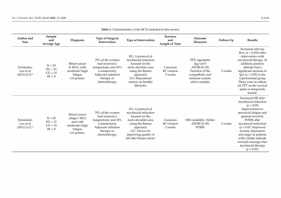
Table 1. Characteristics of the RCTs included in this review.
Author andYear
Sampleand
Average AgeDiagnosis Type of Surgical
Intervention Type of InterventionSessions
andLength of Time
OutcomeMeasures Follow-Up Results
Fernández-Lao et al.
(2012) [40] *
N = 20EG = 10CG = 1049 ± 8
Breast cancer(I–IIIA), with
moderate/highfatigue
(>6 points).
70% of the womenhad received a
lumpectomy and 30%a mastectomy.
Adjuvant radiationtherapy or
chemotherapy.
EG: A protocol ofmyofascial induction
focused on theneck–shoulder area
using the Barnesapproach.
CG: Educationalsession on healthy
lifestyles.
2 sessions40′/session
3 weeks
PPT: algometer(kg/cm2)
ATOM (0–45)Function of the
sympathetic andimmune system:saliva samples
3 weeks
Increased salivaryflow (p = 0.010) after
intervention withmyofascial therapy. In
addition, positiveattitude had a
significant increase inIgA (p < 0.05) in theexperimental group.
There were no effectsfor PPT on the cervical
spine or temporalismuscle.
Fernández-Lao et al.
(2011) [41] *
N = 20EG = 10CG = 1049 ± 8
Breast cancer(stage I–IIIA)
and withmoderate/high
fatigue(>6 points).
70% of the womenhad received a
lumpectomy and 30%a mastectomy.
Adjuvant radiationtherapy or
chemotherapy.
EG: A protocol ofmyofascial induction
focused on theneck–shoulder area
using the Barnesapproach.
CG: Advice forimproving quality of
life after breast cancer.
2 sessions40′/session
2 weeks
HR variability: HolterATOM (0–45)
POMS2 weeks
Increased HR aftermyofascial induction
(p < 0.05)Improvement in
perceived fatigue andgeneral mood by
POMS aftermyofascial induction(p < 0.05. Improvedanxiety, depression
and anger in patientswith a better attitudetowards massage after
myofascial therapy(p < 0.05).

Int. J. Environ. Res. Public Health 2021, 18, 4420 7 of 26
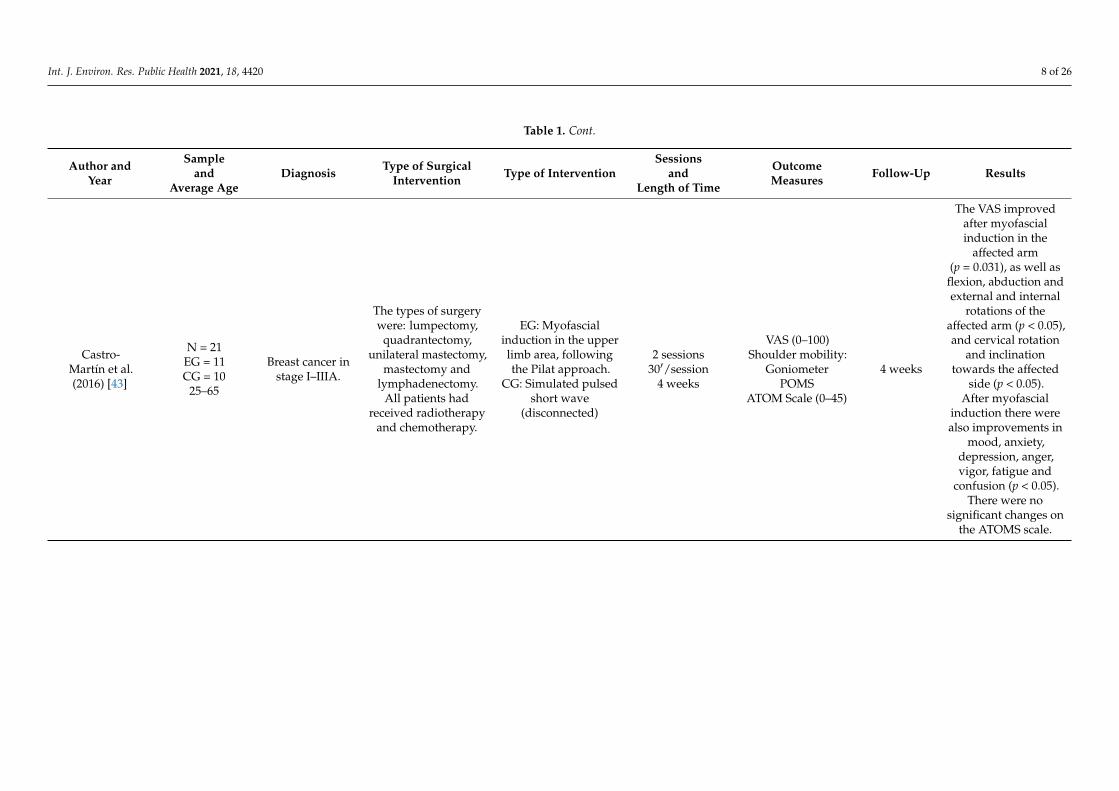
Table 1. Cont.
Author andYear
Sampleand
Average AgeDiagnosis Type of Surgical
Intervention Type of InterventionSessions
andLength of Time
OutcomeMeasures Follow-Up Results
Groef et al.(2017) [42]
N = 147EG = 72CG = 75
EG: 53.9 ± 11.5CG:
54.7 ± 11.9
Patients withunilateralaxillary
clearance forprimary breast
cancer aftersurgery.
Between 60–70%received a
mastectomy andbetween 30–40%
breast conservation.Adjuvant radiation
therapy orchemotherapy.
EG: Standard physicaltherapy program andmyofascial induction
(in active triggerpoints of the affectedlimb and in adhesions
of the pectoral,axillary and cervicalregion, diaphragm
and scar).CG: Standard physicaltherapy intervention
and placebointervention (statichand placement on
the upper bodyand arm).
8 sessions30′/session
8 weeks
VAS (0–100)DASH (0–100)SF-36 (0–100)
PPT: algometer(kg/cm2)
McGill PainQuestionnaire
8 weeks9 and
12 months
The PPT in the uppertrapezius (p = 0.012)
was significantlyhigher at 4 months in
the interventiongroup, and at 4 and
9 months insupraspinatus(p = 0.021) and
(p = 0.040),respectively.
Int. J. Environ. Res. Public Health 2021, 18, 4420 8 of 26
Table 1. Cont.
Author andYear
Sampleand
Average AgeDiagnosis Type of Surgical
Intervention Type of InterventionSessions
andLength of Time
OutcomeMeasures Follow-Up Results
Castro-Martín et al.(2016) [43]
N = 21EG = 11CG = 10
25–65
Breast cancer instage I–IIIA.
The types of surgerywere: lumpectomy,
quadrantectomy,unilateral mastectomy,
mastectomy andlymphadenectomy.
All patients hadreceived radiotherapy
and chemotherapy.
EG: Myofascialinduction in the upperlimb area, followingthe Pilat approach.
CG: Simulated pulsedshort wave
(disconnected)
2 sessions30′/session
4 weeks
VAS (0–100)Shoulder mobility:
GoniometerPOMS
ATOM Scale (0–45)
4 weeks
The VAS improvedafter myofascialinduction in the
affected arm(p = 0.031), as well as
flexion, abduction andexternal and internal
rotations of theaffected arm (p < 0.05),and cervical rotation
and inclinationtowards the affected
side (p < 0.05).After myofascial
induction there werealso improvements in
mood, anxiety,depression, anger,vigor, fatigue and
confusion (p < 0.05).There were no
significant changes onthe ATOMS scale.
Int. J. Environ. Res. Public Health 2021, 18, 4420 9 of 26
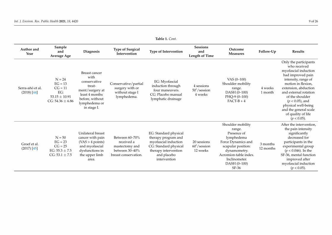
Table 1. Cont.
Author andYear
Sampleand
Average AgeDiagnosis Type of Surgical
Intervention Type of InterventionSessions
andLength of Time
OutcomeMeasures Follow-Up Results
Serra-añó et al.(2018) [44]
N = 24EG = 13CG = 11
EG:53.15 ± 10.91
CG: 54.36 ± 6.86
Breast cancerwith
conservativetreat-
ment/surgery atleast 4 monthsbefore, withoutlymphedema or
in stage I.
Conservative/partialsurgery with orwithout stage Ilymphedema.
EG: Myofascialinduction throughfour maneuvers.
CG: Placebo manuallymphatic drainage
4 sessions50′/session
4 weeks
VAS (0–100)Shoulder mobility
range.DASH (0–100)PHQ-9 (0–100)
FACT-B + 4
4 weeks1 month
Only the participantswho received
myofascial inductionhad improved painintensity, range ofmotion in flexion,
extension, abductionand external rotation
of the shoulder(p < 0.05), and
physical well-beingand the general scale
of quality of life(p < 0.05).
Groef et al.(2017) [45]
N = 50EG = 23CG = 25
EG: 55.3 ± 7.5CG: 53.1 ± 7.5
Unilateral breastcancer with pain(VAS > 4 points)and myofascialdysfunctions inthe upper limb
area.
Between 60–70%received a
mastectomy andbetween 30–40%
breast conservation.
EG: Standard physicaltherapy program andmyofascial induction
CG: Standard physicaltherapy intervention
and placebointervention
20 sessions60′/session
12 weeks
Shoulder mobilityrange.
Presence oflymphedema
Force Dynamics andscapular position:
dynamometry.Acromion-table index.
Inclinometer.DASH (0–100)
SF-36
3 months12 months
After the intervention,the pain intensity
significantlydecreased for
participants in theexperimental group
(p < 0.046). In theSF-36, mental function
improved aftermyofascial induction
(p < 0.05).

Int. J. Environ. Res. Public Health 2021, 18, 4420 10 of 26
Table 1. Cont.
Author andYear
Sampleand
Average AgeDiagnosis Type of Surgical
Intervention Type of InterventionSessions
andLength of Time
OutcomeMeasures Follow-Up Results
Groef et al.(2017) [46]
N = 50EG = 23CG = 25
EG: 55.36 ± 7.5CG: 53.1 ± 7.5
Unilateral breastcancer with pain(VAS > 4 points)and myofascialdysfunctions inthe upper limb
area.
Between 60–70%received a
mastectomy andbetween 30–40%
breast conservation.
EG: Standard physicaltherapy program andmyofascial induction
CG: Standard physicaltherapy intervention
and placebointervention
20 sessions60′/session
12 weeks
VAS (0–100)McGill
PPT: algometer(kg/cm2)Shoulder
functionality: DASH(0–100)
SF-36 (0–100)
3 months6 and 12months
Increase in theexternal scale of the
scapula inthe experimental
group (p < 0.05) andimprovement in
physical functionrelated to quality of
life (p = 0.018).
Massingill et al.(2018) [47]
N = 21EG = 11CG = 10EG/CG:
21 − 55 +
Breast cancerpatients who
have persistentpain andmobility
limitations afterbreast cancer
surgery.
The types of breastcancer surgery
included biopsy,lumpectomy,
mastectomy or certaintypes of
reconstruction.
EG: Myofascialmassage
CG: Relaxing massage
Two 30-minsessions a week
for 8 weeks
Pain (0–30, with 0being nothing and
30 being themaximum)
Mobility (0–40)Quality of life (0–100)
8 weeks.
The EG experiencedmore favorable
changes in pain thanthe CG (−10.7 vs.+0.4, p < 0.001),
mobility(−14.5 vs. −0.8,
p < 0.001) and overallhealth (+29.5 vs. −2.5,
p = 0.002) after8 weeks
Abbreviations: ATOM, Attitudes Toward Massage Scale; DASH, Disabilities of the Arm, Shoulder, and Hand Questionnaire; HR, Heart Rate; CG, Control Group; EG, Experimental Group; IgA, Immunoglobulin A;POMS, Profile of Mood States; PPT, Pressure Pain Thresholds; SF-36, Health Questionnaire SF-36; VAS, Visual Analog Scale; PHQ-9, Patient Health Questionnaire-9; FACT-B+4, The Functional Assessment ofCancer Therapy for breast cancer patients. * These publications belong to the same study, but evaluate different outcomes.

Int. J. Environ. Res. Public Health 2021, 18, 4420 11 of 26
3.3. Characteristics of the Participants
All of the studies contained participants diagnosed with breast cancer. No distinctionor mention of race is made in any study. The age of the patients was detailed in all of thestudies, ranging from 21–65 years old [40–47]. In addition, the stage of patients’ cancer wasconsidered as inclusion criteria by Fernández-Lao et al., and was their study was carriedout on subjects with cancer in stages I-IIIA [40,41]. Only the study by Serra-Añó et al. [44]considered the type of surgery the patients had undergone (only conservative/partial)when choosing study participants.
3.4. Characteristics of the Interventions
All of the studies had two comparison branches. The studies compared myofascialtherapy with: educational sessions on healthy lifestyles, focusing on nutrition, relaxationtechniques, and exercise, with advice on how to improve quality of life after cancer [40,41],with a standard physical therapy program and consistent placebo intervention with bilat-eral static hand placements on the upper body and arm [42,45,46], with pulsed shortwavetherapy [43], and with relaxing Swedish massage which avoided the affected area [47].
The types of myofascial interventions used, although all of them were manual inter-ventions, varied among the studies. A myofascial induction protocol centered on the neckand shoulder following the Barnes approach [40,41], trigger point treatments on upperlimbs and adhesions in the pectoral muscle, cervical region, diaphragm, and scars [42,45,46],myofascial massage specific to the chest, thorax, and shoulder of the affected side [46], andmyofascial release [43,44] were all used.
With regard to the number and length of sessions and the duration of the therapy, thestudies were very heterogeneous. Fernández-Lao et al. conducted two 40-min sessionsseparated by an interval of two and three weeks [40,41], two studies conducted two 30-minsessions per week for eight weeks [42,47] two conducted two 30-min sessions per weekfor 12 weeks, later reduced to one session per week [45,46], one a 50-min session per weekfor four weeks [44], and finally, one study conducted two 30-min sessions four weeksapart [43] (see Table 2).
3.5. Outcome Measures3.5.1. Primary Measures
Pain: All but one of the studies evaluated pain or pain-related outcomes [41]. In themajority of cases, it was measured using a visual analog scale (VAS) [42–45,47]; in two ofthe studies, quantitative and qualitative aspects of pain were assessed using the McGillPain Questionnaire [42,45]. Furthermore, three studies measured pain threshold withpressure from an algometer on different muscle points [40,42,46]. Pain was assessed usinga scale from 0 to 30 [47], a scale from 0 to 10 [43,44], and a scale from 0 to 100 [42,46]. Onestudy measured pain in the cervical spine, the affected limb and non-affected limb [43],and two measured pain in the shoulder, neck region, arm, armpit, trunk side and breastregion [42,46]. The unit of measurement differed between the studies, as one used kPa [40],and the other two used kg/cm2 [42,46].
Shoulder functionality: the majority of the studies measured functional status usingthe Disabilities of the Arm, Shoulder and Hand (DASH) scale [41,43–45]; for all of them,total scores were 0–100, with a higher score indicating a greater disability. One studymeasured shoulder functionality on an unvalidated scale ranging from 0 to 40, where 0 wasno difficulty and 40 was severe difficulty [46].

Int. J. Environ. Res. Public Health 2021, 18, 4420 12 of 26
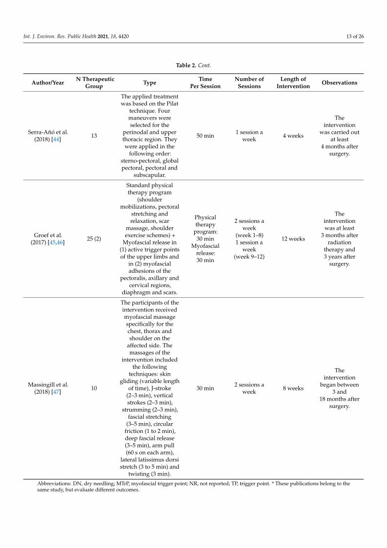
Table 2. Intervention characteristics of the Myofascial Therapy Group.
Author/Year N TherapeuticGroup Type Time
Per SessionNumber of
SessionsLength of
Intervention Observations
Fernández-Lao et al.
(2012) [40] *10
Myofascial release: thepatients received amyofascial release
protocol which focusedon the neck andshoulder area
following the Barnesapproach.
The protocol includedlongitudinal strokes,J-strokes, sustained
suboccipital pressure,frontal bone
decompression and theear traction technique.
40 min(length
adapted tothe tissue
response ofthe patient)
2 sessionsseparated by
a 3-weekinterval
5 weeks N/A
Fernández-Lao et al.
(2011) [41] *10
Myofascial release:protocol which focused
on the neck andshoulder area using the
Barnes approach.The protocol includedlongitudinal strokes,J-strokes, sustained
suboccipital pressure,frontal bone
decompression and theear traction technique.
40 min(length
adapted tothe tissue
response ofthe patient)
2 sessionsseparated by
2 weeks4 weeks
80% of thepatients
underwentsurgery at least
12 monthsbefore the
intervention.
Groef et al.(2017) [42] 72
Standard physicaltherapy program
(shouldermobilizations, pectoral
stretching andrelaxation, scar
massage, shoulderexercise schemes) +Myofascial therapy
consisting of manualmyofascial release
techniques on (1) activemyofascial triggerpoints in the upper
limb area and (2)myofascial adhesions
in the pectoral, axillaryand cervical regions,
diaphragm and scars.
Physicaltherapy
program:30 min
Myofascialrelease:30 min
2 sessions aweek 8 weeks
The patientswere asked to
performexercises twicea day at home.
Myofascialinterventions
were performedfrom 2 to
4 months aftersurgery.
Castro-Martín et al.(2016) [43]
21
The patients received afascial relaxation
intervention whichfocused on the upperlimb area, using the
Pilat approach.
30 min(length
adapted tothe tissue
response ofthe patient)
2 sessionsseparated by
a 4-weekinterval
4 weeks
60% of patientsreceived
myofascialintervention in
less than12 months after
surgery.
Int. J. Environ. Res. Public Health 2021, 18, 4420 13 of 26
Table 2. Cont.
Author/Year N TherapeuticGroup Type Time
Per SessionNumber of
SessionsLength of
Intervention Observations
Serra-Añó et al.(2018) [44] 13
The applied treatmentwas based on the Pilat
technique. Fourmaneuvers wereselected for the
perinodal and upperthoracic region. Theywere applied in the
following order:sterno-pectoral, globalpectoral, pectoral and
subscapular.
50 min 1 session aweek 4 weeks
Theintervention
was carried outat least
4 months aftersurgery.
Groef et al.(2017) [45,46] 25 (2)
Standard physicaltherapy program
(shouldermobilizations, pectoral
stretching andrelaxation, scar
massage, shoulderexercise schemes) +
Myofascial release in(1) active trigger pointsof the upper limbs and
in (2) myofascialadhesions of the
pectoralis, axillary andcervical regions,
diaphragm and scars.
Physicaltherapy
program:30 min
Myofascialrelease:30 min
2 sessions aweek
(week 1–8)1 session a
week(week 9–12)
12 weeks
Theinterventionwas at least
3 months afterradiation
therapy and3 years after
surgery.
Massingill et al.(2018) [47] 10
The participants of theintervention receivedmyofascial massagespecifically for thechest, thorax andshoulder on the
affected side. Themassages of the
intervention includedthe following
techniques: skingliding (variable length
of time), J-stroke(2–3 min), verticalstrokes (2–3 min),
strumming (2–3 min),fascial stretching
(3–5 min), circularfriction (1 to 2 min),deep fascial release(3–5 min), arm pull(60 s on each arm),
lateral latissimus dorsistretch (3 to 5 min) and
twisting (3 min).
30 min 2 sessions aweek 8 weeks
Theintervention
began between3 and
18 months aftersurgery.
Abbreviations: DN, dry needling; MTrP, myofascial trigger point; NR, not reported; TP, trigger point. * These publications belong to thesame study, but evaluate different outcomes.
Int. J. Environ. Res. Public Health 2021, 18, 4420 14 of 26
3.5.2. Secondary Measures
Shoulder mobility: Three studies evaluated shoulder mobility using manual goniom-etry in degrees [43–45]. One study measured flexion, abduction and active external andinternal rotation on the affected and non-affected side [43]. Another also took into accountextension and adduction but only of the affected side [44], and another measured theflexion and abduction, and the upward scapular rotation [45].
Mood state: Two studies evaluated the mood states of participants with the Profile ofMood States (POMS) scale [41,43]. Another study evaluated depression with the Patient-9Health Questionnaire (PHQ-9), and one study linked participants’ saliva flow rate andthe amount of immunoglobulin A and cortisol to stress levels and the patients’ attitudetowards the intervention.
Quality of life: Five of the eight chosen studies evaluated participants’ quality oflife [42,44–47], although the scales that were used varied among the studies. The mostcommonly-used scale was the SF-36 [42,45,46], although one study used the short version,SF-12 [47], and another one used the Functional Assessment of Cancer Therapy-Breast(FACT-B + 4) scale.
3.6. Follow-Up
More than half the studies limited their follow-up to immediately following theintervention [40,41,43,44,47]. Just two studies followed-up between ≥6 months and≤9 months [45,46] and three studies also followed-up long-term (>9 months) [42,45,46].
3.7. Risk of Bias in the Studies Included
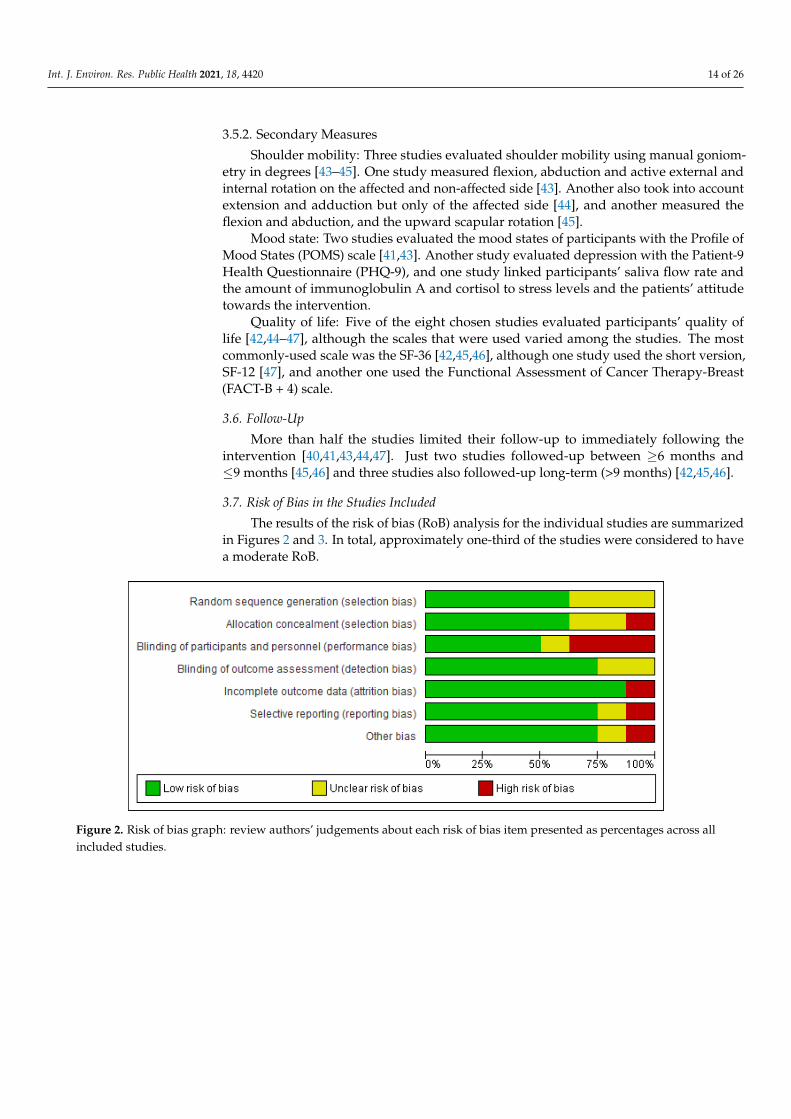
The results of the risk of bias (RoB) analysis for the individual studies are summarizedin Figures 2 and 3. In total, approximately one-third of the studies were considered to havea moderate RoB.
Int. J. Environ. Res. Public Health 2021, 18, x 12 of 23
Figure 2. Risk of bias graph: review authors’ judgements about each risk of bias item presented as percentages across all included studies.
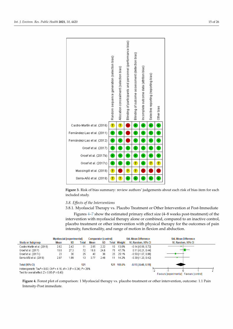
3.8. Effects of the Interventions 3.8.1. Myofascial Therapy vs. Placebo Treatment or Other Intervention at Post-Immedi-ate
Figures 4–7 show the estimated primary effect size (4–8 weeks post-treatment) of the intervention with myofascial therapy alone or combined, compared to an inactive control, placebo treatment or other intervention with physical therapy for the outcomes of pain intensity, functionality, and range of motion in flexion and abduction. 1. Pain Intensity
This sub-analysis included four trials [42–45] with 242 participants. No significant differences were observed between the effects of myofascial therapy alone or in combina-tion with a standard physical therapy program and placebo intervention or a standard physical therapy program (Figure 4. SMD −0.15, 95% CI −0.48 to 0.19, I2 = 28%). 2. Functionality
Meta-analyses of three studies [42,44,46] with 121 participants revealed that there were no statistically significant differences between the two groups on post-immediate functionality (Figure 5, SMD −0.17, 95% CI −0.43 to 0.09, I2 = 0%). However, the results were in favor of the group treated with myofascial therapy. 3. Range of motion
This subgroup involved 3 trials [43–45] covering 95 participants. Meta-analysis result demonstrated that myofascial therapy does not have a greater statistically significant im-mediate effect on range of motion in flexion (Figure 6, SMD 0.30, 95% CI −0.13 to 0.74, I2 = 11%) than a placebo treatment or other intervention. However, a statistically significant result was observed for shoulder range of motion in abduction in favor of the experi-mental group (Figure 7, SMD 0.46, 95% CI 0.05 to 0.87, I2 = 0%; p = 0.03).
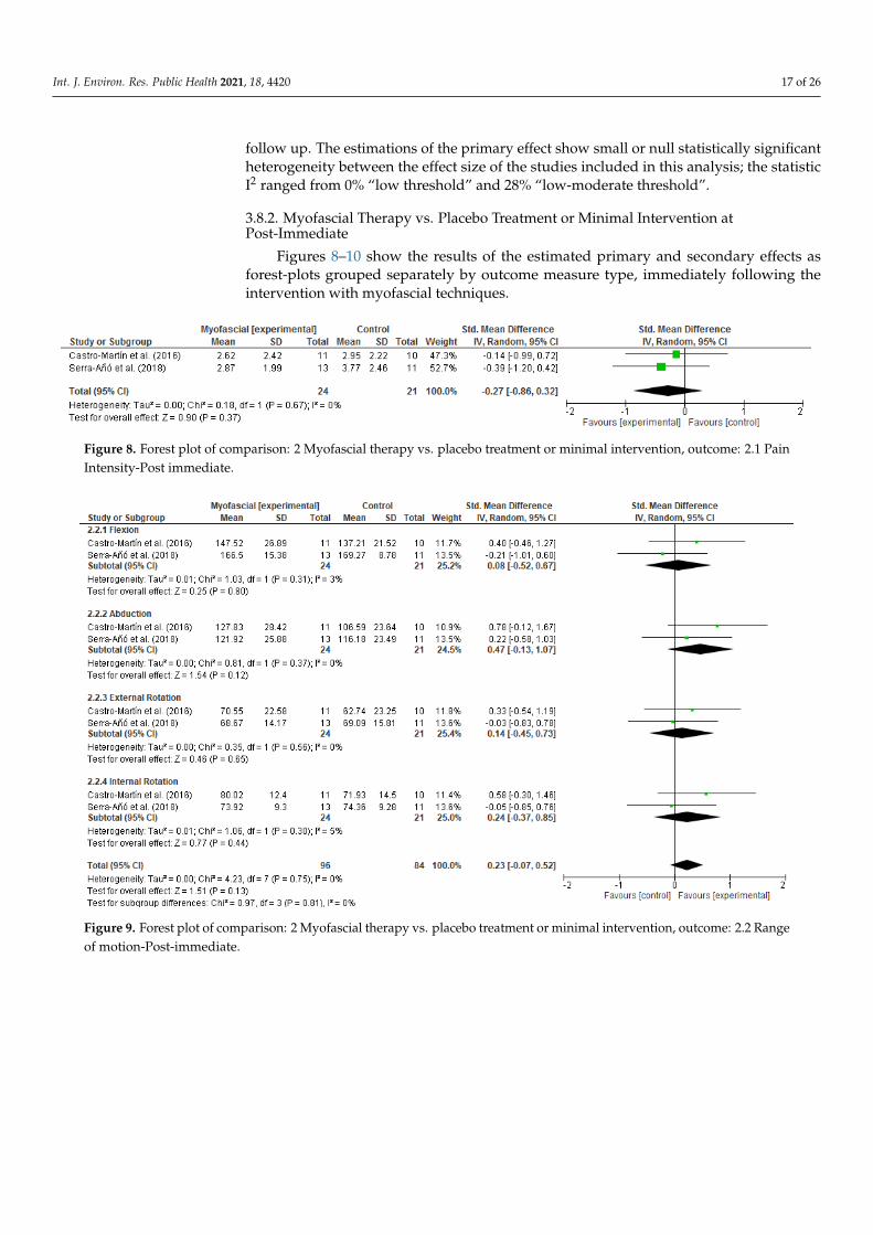
Low-moderate quality evidence indicates that myofascial therapy does not have a greater statistically significant immediate effect on pain intensity, functionality, and range of motion in flexion than a placebo treatment or other intervention at post-immediate fol-low up. The estimations of the primary effect show small or null statistically significant heterogeneity between the effect size of the studies included in this analysis; the statistic I2 ranged from 0% “low threshold” and 28% “low-moderate threshold”.
Figure 2. Risk of bias graph: review authors’ judgements about each risk of bias item presented as percentages across allincluded studies.
Int. J. Environ. Res. Public Health 2021, 18, 4420 15 of 26Int. J. Environ. Res. Public Health 2021, 18, x 13 of 23
Figure 3. Risk of bias summary: review authors’ judgements about each risk of bias item for each included study.
Figure 4. Forest plot of comparison: 1 Myofascial therapy vs. placebo treatment or other intervention, outcome: 1.1 Pain Intensity-Post immediate.
Figure 5. Forest plot of comparison: 1 Myofascial therapy vs. placebo treatment or other intervention, outcome: 1.2 Func-tionality-Post-immediate.
Figure 3. Risk of bias summary: review authors’ judgements about each risk of bias item for eachincluded study.
3.8. Effects of the Interventions3.8.1. Myofascial Therapy vs. Placebo Treatment or Other Intervention at Post-Immediate
Figures 4–7 show the estimated primary effect size (4–8 weeks post-treatment) of theintervention with myofascial therapy alone or combined, compared to an inactive control,placebo treatment or other intervention with physical therapy for the outcomes of painintensity, functionality, and range of motion in flexion and abduction.
Int. J. Environ. Res. Public Health 2021, 18, x 13 of 23
Figure 3. Risk of bias summary: review authors’ judgements about each risk of bias item for each included study.
Figure 4. Forest plot of comparison: 1 Myofascial therapy vs. placebo treatment or other intervention, outcome: 1.1 Pain Intensity-Post immediate.
Figure 5. Forest plot of comparison: 1 Myofascial therapy vs. placebo treatment or other intervention, outcome: 1.2 Func-tionality-Post-immediate.
Figure 4. Forest plot of comparison: 1 Myofascial therapy vs. placebo treatment or other intervention, outcome: 1.1 PainIntensity-Post immediate.

Int. J. Environ. Res. Public Health 2021, 18, 4420 16 of 26
Int. J. Environ. Res. Public Health 2021, 18, x 13 of 23
Figure 3. Risk of bias summary: review authors’ judgements about each risk of bias item for each included study.
Figure 4. Forest plot of comparison: 1 Myofascial therapy vs. placebo treatment or other intervention, outcome: 1.1 Pain Intensity-Post immediate.
Figure 5. Forest plot of comparison: 1 Myofascial therapy vs. placebo treatment or other intervention, outcome: 1.2 Func-tionality-Post-immediate. Figure 5. Forest plot of comparison: 1 Myofascial therapy vs. placebo treatment or other intervention, outcome:1.2 Functionality-Post-immediate.
Int. J. Environ. Res. Public Health 2021, 18, x 14 of 23
Figure 6. Forest plot of comparison: 1 Myofascial therapy vs. placebo treatment or other intervention, outcome: 1.3 Range of motion-Flexion-Post immediate.
Figure 7. Forest plot of comparison: 1 Myofascial therapy vs. placebo treatment or other intervention, outcome: 1.4 Range of motion-Abduction-Post-immediate.
3.8.2. Myofascial Therapy vs. Placebo Treatment or Minimal Intervention at Post-Imme-diate.
Figures 8–10 show the results of the estimated primary and secondary effects as for-est-plots grouped separately by outcome measure type, immediately following the inter-vention with myofascial techniques. 1. Pain intensity
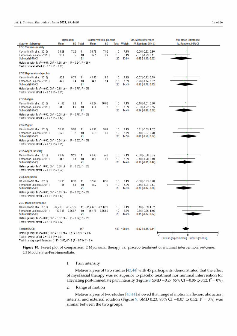
Meta-analyses of two studies [43,44] with 45 participants, demonstrated that the ef-fect of myofascial therapy was no superior to placebo treatment nor minimal intervention for alleviating post-immediate pain intensity (Figure 8, SMD −0.27, 95% CI −0.86 to 0.32, I2 = 0%). 2. Range of motion
Meta-analyses of two studies [43,44] showed that range of motion in flexion, abduc-tion, internal and external rotation (Figure 9, SMD 0.23, 95% CI −0.07 to 0.52, I2 = 0%) was similar between the two groups. 3. Mood state
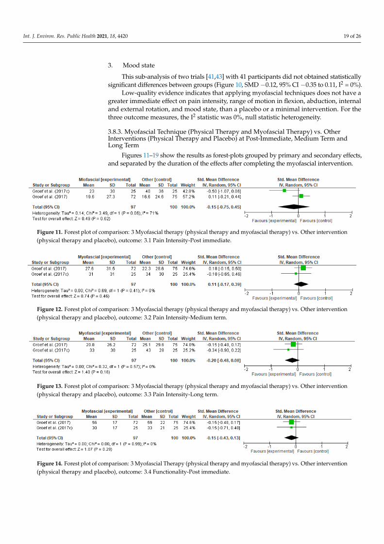
This sub-analysis of two trials [41,43] with 41 participants did not obtained statisti-cally significant differences between groups (Figure 10, SMD −0.12, 95% CI −0.35 to 0.11, I2 = 0%).
Low-quality evidence indicates that applying myofascial techniques does not have a greater immediate effect on pain intensity, range of motion in flexion, abduction, internal and external rotation, and mood state, than a placebo or a minimal intervention. For the three outcome measures, the I2 statistic was 0%, null statistic heterogeneity.
3.8.3. Myofascial Technique (Physical Therapy and Myofascial Therapy) vs. Other Inter-ventions (Physical Therapy and Placebo) at Post-Immediate, Medium Term and Long Term
Figures 11–19 show the results as forest-plots grouped by primary and secondary effects, and separated by the duration of the effects after completing the myofascial inter-vention. 1. Pain Intensity
Figure 6. Forest plot of comparison: 1 Myofascial therapy vs. placebo treatment or other intervention, outcome: 1.3 Rangeof motion-Flexion-Post immediate.
Int. J. Environ. Res. Public Health 2021, 18, x 14 of 23
Figure 6. Forest plot of comparison: 1 Myofascial therapy vs. placebo treatment or other intervention, outcome: 1.3 Range of motion-Flexion-Post immediate.
Figure 7. Forest plot of comparison: 1 Myofascial therapy vs. placebo treatment or other intervention, outcome: 1.4 Range of motion-Abduction-Post-immediate.
3.8.2. Myofascial Therapy vs. Placebo Treatment or Minimal Intervention at Post-Imme-diate.
Figures 8–10 show the results of the estimated primary and secondary effects as for-est-plots grouped separately by outcome measure type, immediately following the inter-vention with myofascial techniques. 1. Pain intensity
Meta-analyses of two studies [43,44] with 45 participants, demonstrated that the ef-fect of myofascial therapy was no superior to placebo treatment nor minimal intervention for alleviating post-immediate pain intensity (Figure 8, SMD −0.27, 95% CI −0.86 to 0.32, I2 = 0%). 2. Range of motion
Meta-analyses of two studies [43,44] showed that range of motion in flexion, abduc-tion, internal and external rotation (Figure 9, SMD 0.23, 95% CI −0.07 to 0.52, I2 = 0%) was similar between the two groups. 3. Mood state
This sub-analysis of two trials [41,43] with 41 participants did not obtained statisti-cally significant differences between groups (Figure 10, SMD −0.12, 95% CI −0.35 to 0.11, I2 = 0%).
Low-quality evidence indicates that applying myofascial techniques does not have a greater immediate effect on pain intensity, range of motion in flexion, abduction, internal and external rotation, and mood state, than a placebo or a minimal intervention. For the three outcome measures, the I2 statistic was 0%, null statistic heterogeneity.
3.8.3. Myofascial Technique (Physical Therapy and Myofascial Therapy) vs. Other Inter-ventions (Physical Therapy and Placebo) at Post-Immediate, Medium Term and Long Term
Figures 11–19 show the results as forest-plots grouped by primary and secondary effects, and separated by the duration of the effects after completing the myofascial inter-vention. 1. Pain Intensity
Figure 7. Forest plot of comparison: 1 Myofascial therapy vs. placebo treatment or other intervention, outcome: 1.4 Rangeof motion-Abduction-Post-immediate.
1. Pain Intensity
This sub-analysis included four trials [42–45] with 242 participants. No significantdifferences were observed between the effects of myofascial therapy alone or in combinationwith a standard physical therapy program and placebo intervention or a standard physicaltherapy program (Figure 4. SMD −0.15, 95% CI −0.48 to 0.19, I2 = 28%).
2. Functionality
Meta-analyses of three studies [42,44,46] with 121 participants revealed that therewere no statistically significant differences between the two groups on post-immediatefunctionality (Figure 5, SMD −0.17, 95% CI −0.43 to 0.09, I2 = 0%). However, the resultswere in favor of the group treated with myofascial therapy.
3. Range of motion
This subgroup involved 3 trials [43–45] covering 95 participants. Meta-analysis resultdemonstrated that myofascial therapy does not have a greater statistically significant im-mediate effect on range of motion in flexion (Figure 6, SMD 0.30, 95% CI −0.13 to 0.74,I2 = 11%) than a placebo treatment or other intervention. However, a statistically significantresult was observed for shoulder range of motion in abduction in favor of the experimentalgroup (Figure 7, SMD 0.46, 95% CI 0.05 to 0.87, I2 = 0%; p = 0.03).
Low-moderate quality evidence indicates that myofascial therapy does not have agreater statistically significant immediate effect on pain intensity, functionality, and rangeof motion in flexion than a placebo treatment or other intervention at post-immediate
Int. J. Environ. Res. Public Health 2021, 18, 4420 17 of 26
follow up. The estimations of the primary effect show small or null statistically significantheterogeneity between the effect size of the studies included in this analysis; the statisticI2 ranged from 0% “low threshold” and 28% “low-moderate threshold”.
3.8.2. Myofascial Therapy vs. Placebo Treatment or Minimal Intervention atPost-Immediate
Figures 8–10 show the results of the estimated primary and secondary effects asforest-plots grouped separately by outcome measure type, immediately following theintervention with myofascial techniques.
Int. J. Environ. Res. Public Health 2021, 18, x 15 of 23
This meta-analysis included two studies and 197 participants [42,46]. The point esti-mates for the SMD between the groups varied from 0.11 to 0.20 (Figures 11–13. 95% IC; I2 = 0% to 71%) in the short-, medium-, and long-term; in all cases. The results of this meta-analysis indicate that there is not a statistically significant effect for pain intensity in favor of myofascial therapy over other types of interventions. 2. Functionality
Two studies with 197 participants were meta-analyzed for shoulder functionality [42,46]. The estimates varied from 0.15 to 0.19 (SMD) (Figures 14–16. 95% IC; I2 = 0%) and although all of them favored the myofascial therapy, their differences were not statisti-cally significant. 3. Quality of life
The result of the meta-analysis of two studies [42,46] demonstrated there were no statistically significant differences between the two groups for quality of life at any time (Figures 17–19), grouped estimates ranged from 0.03 to 0.15 (SMD) (95% IC; I2 = 22% to 73%).
Moderate-high quality evidence indicates that myofascial therapy has no greater im-mediate, medium- and long-term effect on pain intensity, functionality, and quality of life than other forms of intervention.
The grouped estimates show little statistical heterogeneity among the studies’ effect sizes. Of the six comparisons of pain and disability, five showed a null I2 statistic, only immediate post-treatment pain was above the ‘high’ threshold of 75%.
Figure 8. Forest plot of comparison: 2 Myofascial therapy vs. placebo treatment or minimal intervention, outcome: 2.1 Pain Intensity-Post immediate.
Figure 8. Forest plot of comparison: 2 Myofascial therapy vs. placebo treatment or minimal intervention, outcome: 2.1 PainIntensity-Post immediate.
Int. J. Environ. Res. Public Health 2021, 18, x 15 of 23
This meta-analysis included two studies and 197 participants [42,46]. The point esti-mates for the SMD between the groups varied from 0.11 to 0.20 (Figures 11–13. 95% IC; I2 = 0% to 71%) in the short-, medium-, and long-term; in all cases. The results of this meta-analysis indicate that there is not a statistically significant effect for pain intensity in favor of myofascial therapy over other types of interventions. 2. Functionality
Two studies with 197 participants were meta-analyzed for shoulder functionality [42,46]. The estimates varied from 0.15 to 0.19 (SMD) (Figures 14–16. 95% IC; I2 = 0%) and although all of them favored the myofascial therapy, their differences were not statisti-cally significant. 3. Quality of life
The result of the meta-analysis of two studies [42,46] demonstrated there were no statistically significant differences between the two groups for quality of life at any time (Figures 17–19), grouped estimates ranged from 0.03 to 0.15 (SMD) (95% IC; I2 = 22% to 73%).
Moderate-high quality evidence indicates that myofascial therapy has no greater im-mediate, medium- and long-term effect on pain intensity, functionality, and quality of life than other forms of intervention.
The grouped estimates show little statistical heterogeneity among the studies’ effect sizes. Of the six comparisons of pain and disability, five showed a null I2 statistic, only immediate post-treatment pain was above the ‘high’ threshold of 75%.
Figure 8. Forest plot of comparison: 2 Myofascial therapy vs. placebo treatment or minimal intervention, outcome: 2.1 Pain Intensity-Post immediate.
Figure 9. Forest plot of comparison: 2 Myofascial therapy vs. placebo treatment or minimal intervention, outcome: 2.2 Rangeof motion-Post-immediate.
Int. J. Environ. Res. Public Health 2021, 18, 4420 18 of 26
Int. J. Environ. Res. Public Health 2021, 18, x 16 of 23
Figure 9. Forest plot of comparison: 2 Myofascial therapy vs. placebo treatment or minimal intervention, outcome: 2.2 Range of motion-Post-immediate.
Figure 10. Forest plot of comparison: 2 Myofascial therapy vs. placebo treatment or minimal intervention, outcome: 2.3 Mood States-Post-immediate.
Figure 11. Forest plot of comparison: 3 Myofascial therapy (physical therapy and myofascial therapy) vs. Other interven-tion (physical therapy and placebo), outcome: 3.1 Pain Intensity-Post immediate.
Figure 10. Forest plot of comparison: 2 Myofascial therapy vs. placebo treatment or minimal intervention, outcome:2.3 Mood States-Post-immediate.
1. Pain intensity
Meta-analyses of two studies [43,44] with 45 participants, demonstrated that the effectof myofascial therapy was no superior to placebo treatment nor minimal intervention foralleviating post-immediate pain intensity (Figure 8, SMD−0.27, 95% CI−0.86 to 0.32, I2 = 0%).
2. Range of motion
Meta-analyses of two studies [43,44] showed that range of motion in flexion, abduction,internal and external rotation (Figure 9, SMD 0.23, 95% CI −0.07 to 0.52, I2 = 0%) wassimilar between the two groups.
Int. J. Environ. Res. Public Health 2021, 18, 4420 19 of 26
3. Mood state
This sub-analysis of two trials [41,43] with 41 participants did not obtained statisticallysignificant differences between groups (Figure 10, SMD −0.12, 95% CI −0.35 to 0.11, I2 = 0%).
Low-quality evidence indicates that applying myofascial techniques does not have agreater immediate effect on pain intensity, range of motion in flexion, abduction, internaland external rotation, and mood state, than a placebo or a minimal intervention. For thethree outcome measures, the I2 statistic was 0%, null statistic heterogeneity.
3.8.3. Myofascial Technique (Physical Therapy and Myofascial Therapy) vs. OtherInterventions (Physical Therapy and Placebo) at Post-Immediate, Medium Term andLong Term
Figures 11–19 show the results as forest-plots grouped by primary and secondary effects,and separated by the duration of the effects after completing the myofascial intervention.
Int. J. Environ. Res. Public Health 2021, 18, x 16 of 23
Figure 9. Forest plot of comparison: 2 Myofascial therapy vs. placebo treatment or minimal intervention, outcome: 2.2 Range of motion-Post-immediate.
Figure 10. Forest plot of comparison: 2 Myofascial therapy vs. placebo treatment or minimal intervention, outcome: 2.3 Mood States-Post-immediate.
Figure 11. Forest plot of comparison: 3 Myofascial therapy (physical therapy and myofascial therapy) vs. Other interven-tion (physical therapy and placebo), outcome: 3.1 Pain Intensity-Post immediate. Figure 11. Forest plot of comparison: 3 Myofascial therapy (physical therapy and myofascial therapy) vs. Other intervention(physical therapy and placebo), outcome: 3.1 Pain Intensity-Post immediate.
Int. J. Environ. Res. Public Health 2021, 18, x 17 of 23
Figure 12. Forest plot of comparison: 3 Myofascial therapy (physical therapy and myofascial therapy) vs. Other interven-tion (physical therapy and placebo), outcome: 3.2 Pain Intensity-Medium term.
Figure 13. Forest plot of comparison: 3 Myofascial therapy (physical therapy and myofascial therapy) vs. Other interven-tion (physical therapy and placebo), outcome: 3.3 Pain Intensity-Long term.
Figure 14. Forest plot of comparison: 3 Myofascial Therapy (physical therapy and myofascial therapy) vs. Other interven-tion (physical therapy and placebo), outcome: 3.4 Functionality-Post immediate.
Figure 15. Forest plot of comparison: 3 Myofascial therapy (physical therapy and myofascial therapy) vs. Other interven-tion (physical therapy and placebo), outcome: 3.5 Functionality-Medium term.
Figure 16. Forest plot of comparison: 3 Myofascial therapy (physical therapy and myofascial therapy) vs. Other interven-tion (physical therapy and placebo), outcome: 3.6 Functionality-Long term.
Figure 12. Forest plot of comparison: 3 Myofascial therapy (physical therapy and myofascial therapy) vs. Other intervention(physical therapy and placebo), outcome: 3.2 Pain Intensity-Medium term.
Int. J. Environ. Res. Public Health 2021, 18, x 17 of 23
Figure 12. Forest plot of comparison: 3 Myofascial therapy (physical therapy and myofascial therapy) vs. Other interven-tion (physical therapy and placebo), outcome: 3.2 Pain Intensity-Medium term.
Figure 13. Forest plot of comparison: 3 Myofascial therapy (physical therapy and myofascial therapy) vs. Other interven-tion (physical therapy and placebo), outcome: 3.3 Pain Intensity-Long term.
Figure 14. Forest plot of comparison: 3 Myofascial Therapy (physical therapy and myofascial therapy) vs. Other interven-tion (physical therapy and placebo), outcome: 3.4 Functionality-Post immediate.
Figure 15. Forest plot of comparison: 3 Myofascial therapy (physical therapy and myofascial therapy) vs. Other interven-tion (physical therapy and placebo), outcome: 3.5 Functionality-Medium term.
Figure 16. Forest plot of comparison: 3 Myofascial therapy (physical therapy and myofascial therapy) vs. Other interven-tion (physical therapy and placebo), outcome: 3.6 Functionality-Long term.
Figure 13. Forest plot of comparison: 3 Myofascial therapy (physical therapy and myofascial therapy) vs. Other intervention(physical therapy and placebo), outcome: 3.3 Pain Intensity-Long term.
Int. J. Environ. Res. Public Health 2021, 18, x 17 of 23
Figure 12. Forest plot of comparison: 3 Myofascial therapy (physical therapy and myofascial therapy) vs. Other interven-tion (physical therapy and placebo), outcome: 3.2 Pain Intensity-Medium term.
Figure 13. Forest plot of comparison: 3 Myofascial therapy (physical therapy and myofascial therapy) vs. Other interven-tion (physical therapy and placebo), outcome: 3.3 Pain Intensity-Long term.
Figure 14. Forest plot of comparison: 3 Myofascial Therapy (physical therapy and myofascial therapy) vs. Other interven-tion (physical therapy and placebo), outcome: 3.4 Functionality-Post immediate.
Figure 15. Forest plot of comparison: 3 Myofascial therapy (physical therapy and myofascial therapy) vs. Other interven-tion (physical therapy and placebo), outcome: 3.5 Functionality-Medium term.
Figure 16. Forest plot of comparison: 3 Myofascial therapy (physical therapy and myofascial therapy) vs. Other interven-tion (physical therapy and placebo), outcome: 3.6 Functionality-Long term.
Figure 14. Forest plot of comparison: 3 Myofascial Therapy (physical therapy and myofascial therapy) vs. Other intervention(physical therapy and placebo), outcome: 3.4 Functionality-Post immediate.

Int. J. Environ. Res. Public Health 2021, 18, 4420 20 of 26
Int. J. Environ. Res. Public Health 2021, 18, x 17 of 23
Figure 12. Forest plot of comparison: 3 Myofascial therapy (physical therapy and myofascial therapy) vs. Other interven-tion (physical therapy and placebo), outcome: 3.2 Pain Intensity-Medium term.
Figure 13. Forest plot of comparison: 3 Myofascial therapy (physical therapy and myofascial therapy) vs. Other interven-tion (physical therapy and placebo), outcome: 3.3 Pain Intensity-Long term.
Figure 14. Forest plot of comparison: 3 Myofascial Therapy (physical therapy and myofascial therapy) vs. Other interven-tion (physical therapy and placebo), outcome: 3.4 Functionality-Post immediate.
Figure 15. Forest plot of comparison: 3 Myofascial therapy (physical therapy and myofascial therapy) vs. Other interven-tion (physical therapy and placebo), outcome: 3.5 Functionality-Medium term.
Figure 16. Forest plot of comparison: 3 Myofascial therapy (physical therapy and myofascial therapy) vs. Other interven-tion (physical therapy and placebo), outcome: 3.6 Functionality-Long term.
Figure 15. Forest plot of comparison: 3 Myofascial therapy (physical therapy and myofascial therapy) vs. Other intervention(physical therapy and placebo), outcome: 3.5 Functionality-Medium term.
Int. J. Environ. Res. Public Health 2021, 18, x 17 of 23
Figure 12. Forest plot of comparison: 3 Myofascial therapy (physical therapy and myofascial therapy) vs. Other interven-tion (physical therapy and placebo), outcome: 3.2 Pain Intensity-Medium term.
Figure 13. Forest plot of comparison: 3 Myofascial therapy (physical therapy and myofascial therapy) vs. Other interven-tion (physical therapy and placebo), outcome: 3.3 Pain Intensity-Long term.
Figure 14. Forest plot of comparison: 3 Myofascial Therapy (physical therapy and myofascial therapy) vs. Other interven-tion (physical therapy and placebo), outcome: 3.4 Functionality-Post immediate.
Figure 15. Forest plot of comparison: 3 Myofascial therapy (physical therapy and myofascial therapy) vs. Other interven-tion (physical therapy and placebo), outcome: 3.5 Functionality-Medium term.
Figure 16. Forest plot of comparison: 3 Myofascial therapy (physical therapy and myofascial therapy) vs. Other interven-tion (physical therapy and placebo), outcome: 3.6 Functionality-Long term. Figure 16. Forest plot of comparison: 3 Myofascial therapy (physical therapy and myofascial therapy) vs. Other intervention(physical therapy and placebo), outcome: 3.6 Functionality-Long term.
Int. J. Environ. Res. Public Health 2021, 18, x 18 of 23
Figure 17. Forest plot of comparison: 3 Myofascial therapy (physical therapy and myofascial therapy) vs. Other interven-tion (physical therapy and placebo), outcome: 3.7 Quality of life-Post immediate.
Figure 18. Forest plot of comparison: 3 Myofascial therapy (physical therapy and myofascial therapy) vs. Other interven-tion (Physical therapy and placebo), outcome: 3.8 Quality of life-Medium term.
Figure 19. Forest plot of comparison: 3 Myofascial therapy (physical therapy and myofascial therapy) vs. Other interven-tion (physical therapy and placebo), outcome: 3.9 Quality of life-Long term.
Figure 17. Forest plot of comparison: 3 Myofascial therapy (physical therapy and myofascial therapy) vs. Other intervention(physical therapy and placebo), outcome: 3.7 Quality of life-Post immediate.
Int. J. Environ. Res. Public Health 2021, 18, x 18 of 23
Figure 17. Forest plot of comparison: 3 Myofascial therapy (physical therapy and myofascial therapy) vs. Other interven-tion (physical therapy and placebo), outcome: 3.7 Quality of life-Post immediate.
Figure 18. Forest plot of comparison: 3 Myofascial therapy (physical therapy and myofascial therapy) vs. Other interven-tion (Physical therapy and placebo), outcome: 3.8 Quality of life-Medium term.
Figure 19. Forest plot of comparison: 3 Myofascial therapy (physical therapy and myofascial therapy) vs. Other interven-tion (physical therapy and placebo), outcome: 3.9 Quality of life-Long term.
Figure 18. Forest plot of comparison: 3 Myofascial therapy (physical therapy and myofascial therapy) vs. Other intervention(Physical therapy and placebo), outcome: 3.8 Quality of life-Medium term.

Int. J. Environ. Res. Public Health 2021, 18, 4420 21 of 26
Int. J. Environ. Res. Public Health 2021, 18, x 18 of 23
Figure 17. Forest plot of comparison: 3 Myofascial therapy (physical therapy and myofascial therapy) vs. Other interven-tion (physical therapy and placebo), outcome: 3.7 Quality of life-Post immediate.
Figure 18. Forest plot of comparison: 3 Myofascial therapy (physical therapy and myofascial therapy) vs. Other interven-tion (Physical therapy and placebo), outcome: 3.8 Quality of life-Medium term.
Figure 19. Forest plot of comparison: 3 Myofascial therapy (physical therapy and myofascial therapy) vs. Other interven-tion (physical therapy and placebo), outcome: 3.9 Quality of life-Long term. Figure 19. Forest plot of comparison: 3 Myofascial therapy (physical therapy and myofascial therapy) vs. Other intervention(physical therapy and placebo), outcome: 3.9 Quality of life-Long term.
1. Pain Intensity
This meta-analysis included two studies and 197 participants [42,46]. The pointestimates for the SMD between the groups varied from 0.11 to 0.20 (Figures 11–13. 95% IC;I2 = 0% to 71%) in the short-, medium-, and long-term; in all cases. The results of thismeta-analysis indicate that there is not a statistically significant effect for pain intensity infavor of myofascial therapy over other types of interventions.
2. Functionality
Two studies with 197 participants were meta-analyzed for shoulder functionality [42,46].The estimates varied from 0.15 to 0.19 (SMD) (Figures 14–16. 95% IC; I2 = 0%) and althoughall of them favored the myofascial therapy, their differences were not statistically significant.
3. Quality of life
The result of the meta-analysis of two studies [42,46] demonstrated there were nostatistically significant differences between the two groups for quality of life at any time(Figures 17–19), grouped estimates ranged from 0.03 to 0.15 (SMD) (95% IC; I2 = 22% to 73%).
Moderate-high quality evidence indicates that myofascial therapy has no greaterimmediate, medium- and long-term effect on pain intensity, functionality, and quality oflife than other forms of intervention.
The grouped estimates show little statistical heterogeneity among the studies’ effectsizes. Of the six comparisons of pain and disability, five showed a null I2 statistic, onlyimmediate post-treatment pain was above the ‘high’ threshold of 75%.
4. Discussion4.1. Summary of Evidence
We set out to conduct a unique, up-to-date review on the impact of myofascial therapyon breast cancer survivors. We found 8 published RCTs that evaluated the effects ofmyofascial therapy on these patients, although two studies could not be meta-analyzed.The study by Massingill et al. [47], could not be analyzed in the meta-analysis becauseof incomplete data in the results, and that of Groef et al. [45], presented duplicate resultsthat were published in another of the studies included. Ultimately, for the qualitativeanalysis, six publications were considered, including five studies (as those publicationsby Fernández-Lao et al. [40,41] shared the same participants). Thus, a reduced number ofpatients than previously mentioned, 262 participants total, were included.
Int. J. Environ. Res. Public Health 2021, 18, 4420 22 of 26
In general, we found that interventions with myofascial therapy as the sole interven-tion or combined with physical therapy, do not generate, with low or moderate-qualityproof, a significant improvement on pain intensity, functionality, range of motion and moodstate in female survivors of breast cancer compared to an inactive control group or placeboor in comparison with other interventions.
There were greater general effects in favor of myofascial therapy than other con-trols/interventions, but the subgroup analyses revealed inconsistent, insignificant results.Only the range of motion in abduction showed statistical significance in favor of the experi-mental group. Here, myofascial therapy is at least equivalent to other forms of intervention.
Several factors should be considered when interpreting our findings for clinical rec-ommendation and implementation. Although there was no statistical significance, all ofthe studies individually reported a clinically relevant improvement in favor of the exper-imental group with myofascial intervention. The effectiveness of myofascial therapy isunclear because the subgroup analyses were hampered by the small number of studiesthat included the same outcome measures and follow-up beyond immediately followingthe intervention. Very few studies reported comparable estimates regarding secondaryoutcome measures to calculate the effect of using myofascial therapy.
4.2. Agreements or Disagreements with Other Studies or Reviews
Myofascial therapy and its effect on certain populations has been briefly described inliterature. Other meta-analyses that have attempted to demonstrate the efficacy of myofas-cial therapy have also faced the challenge of only finding articles of low methodologicalquality, and as a result, a small number of articles are included in the meta-analysis. In2016, Webb and colleagues [48] did not find conclusive results after their meta-analysisabout the efficacy of myofascial therapy on joint range of motion and perceived pain. Arecently-published article also obtained outcomes that tend towards the statistical signif-icance of myofascial techniques for the improvement of joint range of motion, withoutthese generating a clinically relevant change [49]. Our article correlates with this article, asobtaining statistical significance in the improvement of the range of motion in shoulderabduction reaffirms myofascial therapy as an effective technique in the improvement ofthis specific variable. Additionally, regarding survivors of breast cancer, other systematicreviews have been carried out with meta-analyses using the generic term “manual ther-apy”, which encompasses myofascial therapy. In the review carried out by Pinheiro andcolleagues (2019), manual therapy does prove to be effective to decrease musculoskeletalpain [50]. However, this author could only include five articles in their meta-analysis,again, due to the limiting characteristics of the trials carried on manual therapy and cancer.These results are contradictory to those found in the review by Groef et al. (2015), thatwhen analyzing the effectiveness of various postoperative physical therapy modalities,including myofascial therapy for the treatment of pain and range of motion of the shoul-der in breast cancer, reported that to date, no RCT had reported on the effectiveness ofmyofascial therapy started in the postoperative phase after breast cancer treatment [30].Despite the differences found regarding pain, our findings showed no improvement usingmyofascial therapy as opposed to other therapies on quality of life, which is consistentwith the findings obtained in the review by Pinheiro and colleagues (2019) [50].
Nevertheless, despite not obtaining clear, insightful results in recent years on the clini-cal evidence of manual therapy and, thus, myofascial therapy, these techniques continue tobe prescribed as treatment for pain management in cancer patients [51], and also for ad-dressing other symptoms such as anxiety or altered mood states, due to their non-invasivenature and the absence of negative side effects. Myofascial therapy, described by Pilat in2003 [52], is a relatively recent technique, which, despite its frequent use in clinical physicaltherapy, has not been explored deeply or frequently enough to test its effects on differentpopulations. Thereby, as shown in this review, there are many different approaches ofmyofascial therapy. In order to understand whether myofascial therapy is a useful rehabil-
Int. J. Environ. Res. Public Health 2021, 18, 4420 23 of 26
itation approach for this population, these differences should also be objective of futurerandomized controlled trials, to check the superiority of specific methods among others.
5. Study Limitations
Our review was limited by the small number published trials to date, the impossi-bility of blinding participants and physical therapists, the small sample size, and non-homogeneous follow-up of the included studies.
The outcomes for pain intensity, functionality, and range of motion should be con-sidered key to evaluating the effects of myofascial therapy on the study population, asthese factors are the main causes of the emotional burden of the disease [53]. Many of theincluded studies did not report these results and, when reported, they were measuredat different time periods. The lack of standardization measurements makes quantitativesynthesis of the body of evidence problematic.
The inconsistent nature of data collection and reporting made it difficult to drawconclusions regarding medium- and long-term results, as half of the studies only collecteddata immediately following the intervention.
For example, in the comparison between myofascial therapy versus a placebo orminimal intervention, the studies reported on the outcome measures of pain, range ofmotion and mood state only immediately following treatment. The fact that this data wasnot reported in a comparable manner over time limits our ability to estimate the true effectof myofascial therapy on a critical outcome.
6. Conclusions
While our subgroup analyses show non-significance between groups, the results areinconclusive. The choice of myofascial therapy over other control groups with/withoutintervention for breast cancer survivors is likely to result in a positive effect on painintensity, functionality, and range of motion. Myofascial therapy is also likely to have abeneficial effect on quality of life and mood state outcomes. However, given the resultsobtained, it would seem that there is little to gain from referring these patients to myofascialtreatment. Despite this, the volume of evidence is small and additional similar studies arelikely to greatly change the estimation of the effectiveness of myofascial therapy versusinactive controls, placebo, or other physical therapy interventions. Future studies areneeded to confirm whether myofascial therapy is useful or not to man-age breast cancersurvivors’ sequelae.
Author Contributions: Conceptualization: I.C.L.-P., L.O.-C., M.M.C.-P.; data curation: I.C.L.-P.;formal analysis: I.C.L.-P. and A.M.C.-S.; methodology: M.A.-C., A.M.C.-S., L.O.-C., and M.M.C.-P.;supervision: I.C.L.-P.; writing—original draft preparation: I.C.L.-P. and L.O.-C.; writing—review andediting: I.C.L.-P., A.M.C.-S. and M.A.-C. All authors have read and agreed to the published versionof the manuscript.
Funding: This research received no external funding.
Institutional Review Board Statement: Not applicable.
Informed Consent Statement: Not applicable.
Data Availability Statement: All available data can be obtained by contacting the corresponding author.
Conflicts of Interest: The authors declare no conflict of interest.
References1. Aapro, M.; Piccart, M. Breast cancer. Crit. Rev. Oncol. Hematol. 1998, 27, 135–137. [CrossRef]2. Bray, F.; Ren, J.; Masuyer, E.; Ferlay, J. Global estimates of cancer prevalence for 27 sites in the adult population in 2008. Int. J.
Cancer 2013, 132, 1133–1145. [CrossRef]3. Naghavi, M.; Abajobir, A.; Abbafati, C.; Abbas, K.; Abd-Allah, F.; Abera, S.F.; Aboyans, V.; Adetokunboh, O.; Afshin, A.; Agrawal,
A.; et al. Global, regional, and national age-sex specific mortality for 264 causes of death, 1980–2016: A systematic analysis for theGlobal Burden of Disease Study 2016. Lancet 2017, 390, 1151–1210.
Int. J. Environ. Res. Public Health 2021, 18, 4420 24 of 26
4. Fitzmaurice, C.; Akinyemiju, T.; Al Lami, F.; Alam, T.; Alizadeh-Navaei, R.; Allen, C.; Alsharif, U.; Alvis-Guzman, N.; Amini, E.;Anderson, B.O.; et al. Global, Regional, and National Cancer Incidence, Mortality, Years of Life Lost, Years Lived with Disability,and Disability-Adjusted Life-Years for 29 Cancer Groups, 1990 to 2016. JAMA Oncol. 2018, 4, 1553.
5. Rojas, K.; Stuckey, A. Breast Cancer Epidemiology and Risk Factors. Clin. Obstet. Gynecol. 2016, 59, 651–672. [CrossRef]6. Tabar, L.; Dean, P.B.; Chen, T.H.; Yen, A.M.-F.; Chen, S.L.S.; Fann, J.C.Y.; Chiu, S.Y.-H.; Ku, M.M.-S.; Wu, W.Y.; Hsu, C.-Y.; et al.
The incidence of fatal breast cancer measures the increased effectiveness of therapy in women participating in mammographyscreening. Cancer 2019, 125, 515–523. [CrossRef]
7. Howlader, N.; Noone, A.M.; Krapcho, M.; Miller, D.; Bishop, K.; Altekruse, S.F.; Kosary, C.L.; Yu, M.; Ruhl, J.; Tatalovich, Z.; et al.SEER Cancer Statistics Review, 1975–2013; National Cancer Institute: Bethesda, MD, USA, 2016; Based on November 2015 SEERData Submission, Posted to the SEER Web Site, April 2016. Available online: https://seer.cancer.gov/archive/csr/1975_2013/(accessed on 1 December 2020).
8. Warpenburg, M.J. Deep Friction Massage in Treatment of Radiation-induced Fibrosis: Rehabilitative Care for Breast CancerSurvivors. Integr. Med. 2014, 13, 32–36.
9. Runowicz, C.D.; Leach, C.R.; Henry, N.L.; Henry, K.S.; Mackey, H.T.; Cowens-Alvarado, R.L.; Cannady, R.S.; Pratt-Chapman,M.L.; Edge, S.B.; Jacobs, L.A.; et al. American Cancer Society/American Society of Clinical Oncology Breast Cancer SurvivorshipCare Guideline. CA Cancer J. Clin. 2016, 66, 43–73. [CrossRef]
10. Lazovich, D.; Solomon, C.C.; Thomas, D.B.; Moe, R.E.; White, E. Breast conservation therapy in the United States following the1990 National Institutes of Health Consensus Development Conference on the treatment of patients with early stage invasivebreast carcinoma. Cancer 1999, 86, 628–637. [CrossRef]
11. Douay, N.; Akerman, G.; Clément, D.; Malartic, C.; Morel, O.; Barranger, E. Seroma after axillary lymph node dissection in breastcancer. Gynecol. Obstet. Fertil. 2008, 36, 130–135. [CrossRef]
12. Francis, W.; Abghari, P.; Du, W.; Rymal, C.; Suna, M.; Kosir, M. Improving surgical outcomes: Standardizing the reporting ofincidence and severity of acute lymphedema after sentinel lymph node biopsy and axillary lymph node dissection. Am. J. Surg.2006, 192, 636–639. [CrossRef] [PubMed]
13. Kitamura, Y.; Ohno, Y.; Kasahara, S.; Murata, K.; Sugiyama, H.; Oshima, A.; Tsukuma, H.; Ajiki, W.; Hasegawa, T. Statisticalestimation of the number of breast cancer patients with disabilities resulting from surgery. Breast Cancer 2005, 12, 130–134.[CrossRef] [PubMed]
14. Madsen, A.; Haugaard, K.; Soerensen, J.; Bokmand, S.; Friis, E.; Holtveg, H.; Garne, J.; Horby, J.; Christiansen, P. Arm morbidityfollowing sentinel lymph node biopsy or axillary lymph node dissection: A study from the danish breast cancer cooperative.Breast 2007, 10, 291–401.
15. Nesvold, I.; Dahl, A.; Løkkevik, E.; Marit Mengshoel, A.; Fosså, S. Arm and shoulder morbidity in breast cancer patients afterbreast-conserving therapy versus mastectomy. Acta Oncol. 2008, 47, 835–842. [CrossRef] [PubMed]
16. Moore, G.J.; Mendenhall, N.P.; Kamath, S.S.; Lind, S.S.; Haigh, L.S.; Copeland, E.M. Persistent symptomatology after breastconservation therapy: Prevalence and impact on quality of life. Int. J. Radiat. Oncol. Biol. Phys. 1998, 42, 2058. [CrossRef]
17. Bentzen, S.M.; Thames, H.D.; Overgaard, M. Latent-time estimation for late cutaneous and subcutaneous radiation reactions in asingle-follow-up clinical study. Radiother. Oncol. 1989, 15, 267–274. [CrossRef]
18. Dupilka, A.; Gramling, K. Radiation Side Effects and Syndromes. Available online: http://www.physio-pedia.com/Radiation_Side_Effects_and_Syndromes (accessed on 21 April 2014).
19. Stubblefield, M.D. Radiation fibrosis syndrome: Neuromuscular and musculoskeletal complications in cancer survivors. PM&R2011, 3, 1041–1054.
20. McDuff, S.G.R.; Mina, A.I.; Brunelle, C.L.; Salama, L.; Warren, L.E.; Abouegylah, M.; Swaroop, M.; Skolny, M.N.; Asdourian, M.;Gillespie, T.; et al. Timing of Lymphedema After Treatment for Breast Cancer: When Are Patients Most At Risk? Int. J. Radiat.Oncol. Biol. Phys. 2019, 103, 62–70. [CrossRef]
21. Warren, L.E.G.; Miller, C.L.; Horick, N.; Skolny, M.N.; Jammallo, L.S.; Sadek, B.T.; Shenouda, M.N.; O’Toole, J.A.; MacDonald,S.M.; Specht, M.C. The impact of radiation therapy on the risk of lymphedema after treatment for breast cancer: A prospectivecohort study. Int. J. Radiat. Oncol. Biol. Phys. 2014, 88, 565–571. [CrossRef]
22. Norman, S.A.; Localio, A.R.; Potashnik, S.L.; Torpey, H.A.S.; Kallan, M.J.; Weber, A.L.; Miller, L.T.; DeMichele, A.; Solin, L.J.Lymphedema in breast cancer survivors: Incidence, degree, time course, treatment, and symptoms. J. Clin. Oncol. 2009,27, 390–397. [CrossRef]
23. DiSipio, T.; Rye, S.; Newman, B.; Hayes, S. Incidence of unilateral arm lymphoedema after breast cancer: A systematic review andmeta-analysis. Lancet Oncol. 2013, 14, 500–515. [CrossRef]
24. Togawa, K.; Ma, H.; Sullivan-Halley, J.; Neuhouser, M.L.; Imayama, I.; Baumgartner, K.B.; Smith, A.W.; Alfano, C.M.; McTiernan,A.; Ballard-Barbash, R.; et al. Risk factors for self-reported arm lymphedema among female breast cancer survivors: A prospectivecohort study. Breast Cancer Res. 2014, 16, 414. [CrossRef]
25. Rupp, J.; Hadamitzky, C.; Henkenberens, C.; Christiansen, H.; Steinmann, D.; Bruns, F. Frequency and risk factors for arm lym-phedema after multimodal breast-conserving treatment of nodal positive breast Cancer—A long-term observation. Radiat. Oncol.2019, 14, 39. [CrossRef]
26. Ridner, S.H. Quality of life and symptom cluster associated with breast cancer treatment-related lymphedema. Support. CareCancer 2005, 13, 904–911. [CrossRef] [PubMed]
Int. J. Environ. Res. Public Health 2021, 18, 4420 25 of 26
27. Edmond, S.N.; Shelby, R.A.; Keefe, F.J.; Fisher, H.M.; Schmidt, J.; Soo, M.S.; Skinner, C.S.; Ahrendt, G.M.; Manculich, J.;Sumkin, J.H.; et al. Persistent Breast Pain Among Women with Histories of Breast-Conserving Surgery for Breast CancerCompared with Women without Histories of Breast Surgery or Cancer. Clin. J. Pain 2017, 33, 51–56. [CrossRef]
28. Bruce, J.; Thornton, A.; Powell, R.; Johnston, M.; Wells, M.; Heys, S.; Thompson, A.M.; Smith, W.C.; Chambers, W.A.; Scott, N.W.;et al. Psychological, surgical, and sociodemographic predictors of pain outcomes after breast cancer surgery: A population-basedcohort study. Pain 2014, 155, 232–243. [CrossRef] [PubMed]
29. Blumen, H.; Fitch, K.; Polkus, V. Comparison of Treatment Costs for Breast Cancer, by Tumor Stage and Type of Service. Am.Health Drug Benefits 2016, 9, 23–32. [PubMed]
30. De Groef, A.; Van Kampen, M.; Dieltjens, E.; Christiaens, M.; Neven, P.; Geraerts, I.; Devoogdt, N. Effectiveness of PostoperativePhysical Therapy for Upper-Limb Impairments After Breast Cancer Treatment: A Systematic Review. Arch. Phys. Med. Rehabil.2015, 96, 1140–1153. [CrossRef] [PubMed]
31. Borstad, J.; Ludewig, P. The Effect of Long Versus Short Pectoralis Minor Resting Length on Scapular Kinematics in HealthyIndividuals. J. Orthop. Sports Phys. Ther. 2005, 35, 227–238. [CrossRef]
32. McNeely, M.; Campbell, K.; Ospina, M.; Rowe, B.; Dabbs, K.; Klassen, T.; Mackey, J.; Courneya, K. Exercise interventions forupper-limb dysfunction due to breast cancer treatment. Cochrane Database Syst. Rev. 2010, 6, CD005211. [CrossRef]
33. Pilat, A. Myofascial induction approaches. In Fascia: The Tensional Network of the Human Body. The Science and Clinical Applicationsin Manual and Movement Therapy; Churchill Livingstone: London, UK, 2011; p. 455.
34. Rodríguez-Huguet, M.; Gil-Salú, J.L.; Rodríguez-Huguet, P.; Cabrera-Afonso, J.R.; Lomas-Vega, R. Effects of Myofascial Releaseon Pressure Pain Thresholds in Patients with Neck Pain: A Single-Blind Randomized Controlled Trial. Am. J. Phys. Med. Rehabil.2018, 97, 16–22. [CrossRef]
35. Chamorro Comesaña, A.; del Suárez Vicente, M.P.; Docampo Ferreira, T.; del Pérez-La Fuente Varela, M.M.; Porto Quintáns,M.M.; Pilat, A. Effect of myofascial induction therapy on post-c-section scars, more than one and a half years old. Pilot study.J. Bodyw. Mov. Ther. 2017, 21, 197–204. [CrossRef] [PubMed]
36. Ozsoy, G.; Ilcin, N.; Ozsoy, I.; Gurpinar, B.; Buyukturan, O.; Buyukturan, B.; Kararti, C.; Sas, S. The Effects Of Myofascial ReleaseTechnique Combined with Core Stabilization Exercise in Elderly with Non-Specific Low Back Pain: A Randomized Controlled,Single-Blind Study. Clin. Interv. Aging 2019, 14, 1729–1740. [CrossRef] [PubMed]
37. Ceca, D.; Elvira, L.; Guzmán, J.F.; Pablos, A. Benefits of a self-myofascial release program on health-related quality of life inpeople with fibromyalgia: A randomized controlled trial. J. Sports Med. Phys. Fit. 2017, 57, 993–1002.
38. Urrútia, G.; Bonfill, X. Declaración Prisma: Una propuesta para mejorar la publicación de revisiones sistemáticas y metaanálisis.Med. Clin. 2010, 135, 507–511. [CrossRef]
39. Da Costa Santos, C.M.; de Mattos Pimenta, C.A.; Nobre, M.R. The PICO strategy for the research question construction andevidence search. Rev. Lat. Am. Enferm. 2007, 15, 508–511. [CrossRef]
40. Fernández-Lao, C.; Cantarero-Villanueva, I.; Díaz-Rodríguez, L.; Fernández-de-las-Peñas, C.; Sánchez-Salado, C.; Arroyo-Morales,M. The Influence of Patient Attitude Toward Massage on Pressure Pain Sensitivity and Immune System after Application ofMyofascial Release in Breast Cancer Survivors: A Randomized, Controlled Crossover Study. J. Manip. Physiol. Ther. 2012,35, 94–100. [CrossRef]
41. Fernández-Lao, C.; Cantarero-Villanueva, I.; Díaz-Rodríguez, L.; Cuesta-Vargas, A.; Fernández-Delas-Peñas, C.; Arroyo-Morales,M. Attitudes towards massage modify effects of manual therapy in breast cancer survivors: A randomised clinical trial withcrossover design. Eur. J. Cancer Care 2011, 21, 233–241. [CrossRef] [PubMed]
42. De Groef, A.; Van Kampen, M.; Vervloesem, N.; De Geyter, S.; Christiaens, M.; Neven, P.; Vos, L.; de Vrieze, T.; Geraerts, I.;Devoogdt, N. Myofascial techniques have no additional beneficial effects to a standard physical therapy programme for upperlimb pain after breast cancer surgery: A randomized controlled trial. Clin. Rehabil. 2017, 31, 1625–1635. [CrossRef]
43. Castro-Martín, E.; Ortiz-Comino, L.; Gallart-Aragón, T.; Esteban-Moreno, B.; Arroyo-Morales, M.; Galiano-Castillo, N. MyofascialInduction Effects on Neck-Shoulder Pain in Breast Cancer Survivors: Randomized, Single-Blind, Placebo-Controlled CrossoverDesign. Arch. Phys. Med. Rehabil. 2017, 98, 832–840. [CrossRef]
44. Serra-Añó, P.; Inglés, M.; Bou-Catalá, C.; Iraola-Lliso, A.; Espí-López, G. Effectiveness of myofascial release after breast cancersurgery in women undergoing conservative surgery and radiotherapy: A randomized controlled trial. Support. Care Cancer 2018,27, 2633–2641. [CrossRef]
45. De Groef, A.; Van Kampen, M.; Verlvoesem, N.; Dieltjens, E.; Vos, L.; De Vrieze, T.; Christiaens, M.-R.; Neven, P.; Geraerts, I.;Devoogdt, N. Effect of myofascial techniques for treatment of upper limb dysfunctions in breast cancer survivors: Randomizedcontrolled trial. Support. Care Cancer 2017, 25, 2119–2127. [CrossRef]
46. De Groef, A.; Van Kampen, M.; Vervloesem, N.; Dieltjens, E.; Christiaens, M.; Neven, P.; Vos, L.; de Vrieze, T.; Geraerts, I.;Devoogdt, N. Effect of myofascial techniques for treatment of persistent arm pain after breast cancer treatment: Randomizedcontrolled trial. Clin. Rehabil. 2017, 32, 451–461. [CrossRef]
47. Massingill, J.; Jorgensen, C.; Dolata, J.; Sehgal, A. Myofascial Massage for Chronic Pain and Decreased Upper Extremity MobilityAfter Breast Cancer Surgery. Int. J. Ther. Massag. Bodyw. Res. Educ. Pract. 2018, 11, 4–9.
48. Webb, T.R.; Rajendran, D. Myofascial techniques: What are their effects on joint range of motion and pain? A systematic reviewand meta-analysis of randomised controlled trials. J. Bodyw. Mov. Ther. 2016, 20, 682–699. [CrossRef] [PubMed]

Int. J. Environ. Res. Public Health 2021, 18, 4420 26 of 26
49. Burk, C.; Perry, J.; Lis, S.; Dischiavi, S.; Bleakley, C. Can Myofascial Interventions have a remote effect on ROM? A systematicReview and Meta-analysis. J. Sport Rehabil. 2020, 29, 650–656. [PubMed]
50. Pinheiro Da Silva, F.; Moreira, G.M.; Zomkowski, K.; Amaral de Noronha, M.; Flores Sperandio, F. Manual therapy as treatmentfor chronic musculoskeletal pain in female breast cancer survivors: A systematic review and meta-analysis. J. Manip. Physiol. Ther.2019, 42, 504–513. [CrossRef] [PubMed]
51. Deng, G. Integrative Medicine Therapies for Pain Management in Cancer Patients. Cancer J. 2019, 25, 343–348. [CrossRef]52. Pilat, A. Myofascial Therapies: Myofascial Induction; McGraw-Hill Interamericana: Madrid, Spain, 2003.53. Bjerkeset, E.; Rhrl, K.; Schou-Bredal, I. Symptom cluster of pain, fatigue, and psychological distress in breast cancer survivors:
Prevalence and characteristics. Breast Cancer Res. Treat. 2020, 180, 63–71. [CrossRef] [PubMed]