Synthesis and Vibrational Circular Dichroism Analysis ... - MDPI

Molecular structure, vibrational spectra and photochemistry
of 5-mercapto-1-methyltetrazole
A. Gomez-Zavaglia a,b, I.D. Reva a, L. Frija c, M.L. Cristiano c, R. Fausto a,*
a Department of Chemistry, University of Coimbra, R. Larga, 300 4535, Coimbra, Portugalb Facultad de Farmacia y Bioquımica, Universidad de Buenos Aires, Buenos Aires, Argentina
c Department of Chemistry and Biochemistry, University of Algarve, Faro, Portugal
Received 3 August 2005; revised 12 August 2005; accepted 13 August 2005
Available online 28 September 2005
Abstract
In this work, 5-mercapto-1-methyltetrazole was studied by low temperature matrix-isolation and solid-state infrared spectroscopy,
DFT(B3LYP)/6-311CCG(d,p) calculations and photochemical methods. In the low temperature neat solid phase and isolated in an argon matrix,
the compound was found to exist in the 1-methyl-1,4-dihydro-5H-tetrazole-5-thione tautomeric form. The infrared spectra of the compound were
fully assigned and correlated with structural properties. In situ UV-irradiation (lO235 nm) of the matrix-isolated monomer is shown to induce
different photochemical processes, all of them involving cleavage of the tetrazole ring: e.g. (1) molecular nitrogen expulsion, with production of
1-methyl-1H-diazirene-3-thiol, which is produced in two different conformers; (2) ring cleavage leading to production of methyl isothiocyanate
and azide; (3) simultaneous elimination of nitrogen and sulphur with production of N-methylcarbodiimide. Following these photoprocesses,
subsequent reactions occur, leading to production of methyl diazene, carbon monosulphide and nitrogen hydride. Spectroscopic evidence of the
production of the above-mentioned chemical species is provided.
q 2005 Elsevier B.V. All rights reserved.
Keywords: 5-Mercapto-1-methyltetrazole; Matrix-isolation infrared spectroscopy; Photochemistry; N-methylcarbodiimide; 1-Methyl-1H-diazirene-3-thiol; Methyl
isothiocyanate; Azide; Methyl diazene; Carbon monosulphide; Nitrogen hydride
1. Introduction
Tetrazoles have received much attention due to their
important industrial and biological practical applications
[1,2]. In particular, 5-mercapto-1-methyltetrazole (1-methyl-
1H-tetrazole-5-thiol; MTT) has been used in the synthesis of
pharmacologically active cephalosporins [3] and, as cesium
salts, as part of the thiolate/disulfide redox couple [4]. It has
also been demonstrated that MTT increases prothrombin time
and decreases plasma factor VII and prothrombin levels in
vitamin K-deficient male rats [5].
From a more fundamental point of view, MTT is also a very
interesting compound because of two main factors: firstly, the
existence of different possible tautomers (Fig. 1), including
thiol and thione tautomeric forms; secondly, its expectable rich
photochemical reactivity, as it was previously observed for
other tetrazole derivatives [6–12].
0022-2860/$ - see front matter q 2005 Elsevier B.V. All rights reserved.
doi:10.1016/j.molstruc.2005.08.019
* Corresponding author. Tel.: C35 1239 852080; fax: C35 1239 827703.
E-mail address: [email protected] (R. Fausto).
The fact that tautomerism influences the reactivity of the
mercapto-azoles (including tetrazoles) has been demon-
strated for both polymerization processes and substitution
reactions in different moieties [13–15]. For example, 2,5-
dimercapto-1,3,4-thiadiazole exists in the thione–thiol form
in the solid state [16], whereas in solution a solvent-
dependent equilibrium is believed to exist between the
thione–thiol and thione–thione tautomers, the former being
the prevailing species in polar solvents [14]. On the other
hand, for 2-mercapto-5-methyl-1,3,4-thiadiazole, the thione
form exists predominantly in DMSO solution as well as in
the solid state [14,17]. The thiol–thione tautomerism in
mercapto-azoles has, in fact, been a point of controversy for
many authors, thus justifying further investigation on this
topic using complementary experimental and theoretical
methods that may provide a deeper insight on this problem.
In this regard, matrix isolation spectroscopy appears as a
very powerful technique. In a low temperature inert matrix
(typically 10 K), the studied guest molecules do not rotate
and also the effects of inhomogeneous broadening of the
vibrational bands are minimized. As a result, the vibrational
bands in the spectra of matrix isolated compounds become
Journal of Molecular Structure 786 (2006) 182–192
www.elsevier.com/locate/molstruc
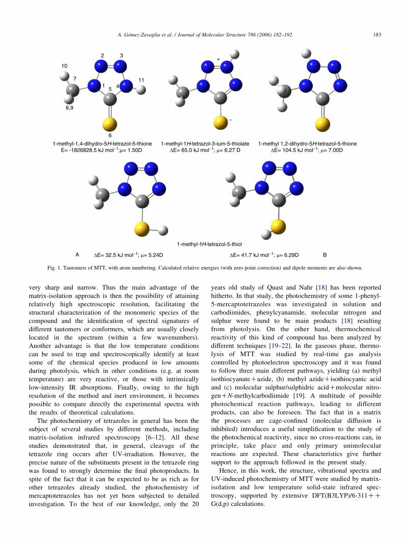
Fig. 1. Tautomers of MTT, with atom numbering. Calculated relative energies (with zero point correction) and dipole moments are also shown.
A. Gomez-Zavaglia et al. / Journal of Molecular Structure 786 (2006) 182–192 183
very sharp and narrow. Thus the main advantage of the
matrix-isolation approach is then the possibility of attaining
relatively high spectroscopic resolution, facilitating the
structural characterization of the monomeric species of the
compound and the identification of spectral signatures of
different tautomers or conformers, which are usually closely
located in the spectrum (within a few wavenumbers).
Another advantage is that the low temperature conditions
can be used to trap and spectroscopically identify at least
some of the chemical species produced in low amounts
during photolysis, which in other conditions (e.g. at room
temperature) are very reactive, or those with intrinsically
low-intensity IR absorptions. Finally, owing to the high
resolution of the method and inert environment, it becomes
possible to compare directly the experimental spectra with
the results of theoretical calculations.
The photochemistry of tetrazoles in general has been the
subject of several studies by different methods, including
matrix-isolation infrared spectroscopy [6–12]. All these
studies demonstrated that, in general, cleavage of the
tetrazole ring occurs after UV-irradiation. However, the
precise nature of the substituents present in the tetrazole ring
was found to strongly determine the final photoproducts. In
spite of the fact that it can be expected to be as rich as for
other tetrazoles already studied, the photochemistry of
mercaptotetrazoles has not yet been subjected to detailed
investigation. To the best of our knowledge, only the 20
years old study of Quast and Nahr [18] has been reported
hitherto. In that study, the photochemistry of some 1-phenyl-
5-mercaptotetrazoles was investigated in solution and
carbodiimides, phenylcyanamide, molecular nitrogen and
sulphur were found to be main products [18] resulting
from photolysis. On the other hand, thermochemical
reactivity of this kind of compound has been analyzed by
different techniques [19–22]. In the gaseous phase, thermo-
lysis of MTT was studied by real-time gas analysis
controlled by photoelectron spectroscopy and it was found
to follow three main different pathways, yielding (a) methyl
isothiocyanateCazide, (b) methyl azideCisothiocyanic acid
and (c) molecular sulphur/sulphidric acidCmolecular nitro-
genCN-methylcarbodiimide [19]. A multitude of possible
photochemical reaction pathways, leading to different
products, can also be foreseen. The fact that in a matrix
the processes are cage-confined (molecular diffusion is
inhibited) introduces a useful simplification to the study of
the photochemical reactivity, since no cross-reactions can, in
principle, take place and only primary unimolecular
reactions are expected. These characteristics give further
support to the approach followed in the present study.
Hence, in this work, the structure, vibrational spectra and
UV-induced photochemistry of MTT were studied by matrix-
isolation and low temperature solid-state infrared spec-
troscopy, supported by extensive DFT(B3LYP)/6-311CCG(d,p) calculations.
A. Gomez-Zavaglia et al. / Journal of Molecular Structure 786 (2006) 182–192184
2. Materials and methods
2.1. Infrared spectroscopy
MTT was obtained from Aldrich (purityO99%). The IR
spectra were obtained using a Mattson (Infinity 60AR Series)
Fourier Transform infrared spectrometer, equipped with a
deuterated triglycine sulphate (DTGS) detector and a Ge/KBr
beamsplitter, with 0.5 cmK1 spectral resolution. Necessary
modifications of the sample compartment of the spectrometer
were done in order to accommodate the cryostat head and allow
purging of the instrument by a stream of dry nitrogen to remove
water vapors and CO2. A solid sample of MTT was placed in a
specially designed doubly thermostattable Knudsen cell [23].
During deposition, both the sample container and valve nozzle
compartments of this cell were kept at 323 K. Matrices were
prepared by co-deposition of MTT vapors coming out of the
Knudsen cell together with large excess of the matrix gas
(argon N60, obtained from Air Liquide) onto the CsI substrate
of the cryostat cooled to 10 K. Care was taken to keep the
guest-to-host ratio in matrices low enough to avoid association.
All experiments were performed using an APD Cryogenics
closed-cycle helium refrigeration system with a DE-202A
expander.
Irradiation of the samples was carried out with a 150 W
xenon arc lamp (Osram XBO 150 W/CR OFR). No changes in
the spectrum of the matrix-isolated 2MTA were observed
during irradiation through a cut-off filter transmitting light
above 285 nm. However, irradiation of the matrices through
the outer KBr window of the cryostat (lO235 nm), resulted in
a series of photochemical transformations, as will be described
below.
2.2. Computational methodology
The quantum chemical calculations for MTT were
performed with GAUSSIAN 98 program package [24] at the
DFT level of theory, using the 6-311CCG(d,p) basis set and
the three-parameter density functional abbreviated as B3LYP,
which includes Becke’s gradient exchange correction [25] and
the Lee, Yang, Parr correlation functional [26]. The calcu-
lations on the possible photoproducts were carried out at the
same level of theory.
Geometrical parameters of the considered conformations
were optimized using the Geometry Direct Inversion of the
Invariant Subspace (GDIIS) method [27]. In order to assist
the analysis of the experimental spectra, vibrational
frequencies and IR intensities were also calculated with
the same basis set. The computed harmonic frequencies
were scaled down by a single factor (0.978) to correct them
for the effects of basis set limitations, neglected part of
electron correlation and anharmonicity effects. Normal
coordinate analysis was undertaken in the internal coordi-
nates space, as described by Schachtschneider [28], using
the program BALGA and the optimized geometries and
harmonic force constants resulting from the DFT(B3LYP)/6-
311CCG(d,p) calculations.
3. Results and discussion
The compound under study can exist in several
tautomeric forms, depicted in Fig. 1. The 5-mercapto
tautomer (1-methyl-1H-tetrazol-5-thiol) may exist in two
different conformers, A and B, with the A form, with the
methyl group and sulphidryl hydrogen atom oriented to
opposite sides, being predicted to be 9.2 kJ molK1
more stable than the B form. Because of the destabilizing
CH3/H(S) interaction in conformer B, this form has the
thiol hydrogen atom considerably out of the tetrazole-ring
plane (the calculated NZC–S–H dihedral angle is 136.98),
whereas in conformer A this atom is in the ring plane. There
are also two 5-thione tautomers, which differ in the position
of the tetrazole-ring hydrogen atom. The most stable thione
tautomer (1-methyl-1,4-dihydro-5H-tetrazol-5-thione) corre-
sponds to the global minimum of the molecule in the gas
phase and is considerably more stable than all other species
(the most stable thiol tautomer is the second most stable
tautomer and has a relative energy of 32.5 kJ molK1). In this
tautomer, all atoms are in the same plane, with exception of
two methyl hydrogen atoms, which occupy symmetrical
positions above and below the molecular plane; the in-plane
methyl hydrogen atom points toward N(2). The second
thione tautomer, 1-methyl-1,2-dihydro-5H-tetrazol-5-thione,
has an energy higher than that of the global minimum by
104.5 kJ molK1, which results mainly from the additional
repulsion between the methyl group and the tetrazole-ring
hydrogen atom. Indeed, this repulsion leads to a distortion of
the tetrazole-ring from planarity, with pyramidalization of
both N(1) and N(2) atoms (the C(7)–N(1)–C(5)–N(4) and
H–N(2)–N(1)–C(5) dihedral angles are K164.1 and 146.38,
respectively), and rotation of the methyl group relatively to
the tetrazole-ring (noteworthy this is the only MTT tautomer
where the methyl group has one of the hydrogen atoms
pointing to a perpendicular direction relatively to the
tetrazole ring, instead of pointing to N(2); in fact, as a
whole, the results show that the methyl hydrogen atoms tend
to stay as far as possible both from the sulphur atom and the
hydrogen atom linked to N(2)). Finally, a mesoionic
tautomer (1-methyl-1H-tetrazol-3-ium-5-thiolate) does also
exist, with relative energy 65.0 kJ molK1. The fully
optimized geometries for all tautomers are provided in
Appendix (Table S1).
3.1. IR spectrum of the matrix isolated compound (as-deposited
matrix)
Taking into account the relative stability of all the possible
tautomers, only the most stable form (1-methyl-1,4-dihydro-
5H-tetrazol-5-thione) should be significantly populated in the
saturated vapor of the compound used for matrix preparation
(TZ323 K). It could then be expected that this would also be
the single form present in the as-deposited matrices.
According to the expectations, only the most stable
tautomer of MTT was found in the as-deposited argon
matrices. This is clearly demonstrated in Fig. 2, by the fairly

Wavenumber/ cm−1
3600 3000 1400 1200 1000 800 6000
10
20
30
40
50
Rel
ativ
e In
tens
ity
3600 30000
0.1
0.2
0.3
0.4
0.5
1400 1200 1000 800 600
Abs
orba
nce
Argon10K
(a)
(b)
Fig. 2. (a) Infrared spectrum of MTT in argon matrix (substrate temperatureZ10 K; nozzle temperatureZ323 K), and (b) DFT(B3LYP)/6-311CCG(d,p)
calculated spectrum. The calculated spectrum of MTT (1-methyl-1,4-dihydro-
5H-tetrazol-5-thione tautomer) was simulated using GAUSSIAN functions
centered at the calculated (scaled) frequency and with bandwidth-at-half-
height equal to 5 cmK1.
A. Gomez-Zavaglia et al. / Journal of Molecular Structure 786 (2006) 182–192 185
good agreement between the observed infrared spectrum of
the as-deposited argon matrix of MTT and that calculated at the
DFT(B3LYP)/6-311CCG(d,p) level of theory for the
1-methyl-1,4-dihydro-5H-tetrazol-5-thione tautomer.1
The proposed band-assignments are given in Table 1. Four
intense bands dominate the calculated spectrum: 3578.5 (N–H
stretching; nN–H), 1498.2 (N–H in-plane bending; dN–H),
1382.2 (NaN stretching; nNaN) and 516.1 (N–H out-of-plane
rocking; gN–H) cmK1. Accordingly, intense features are
observed in the spectrum of the as-deposited matrix nearly at
these wavenumbers. The features ascribable to the nN–H, dN–
H and gN–H modes appear as triplets (at ca. 3485, 1490 and
540 cmK1, respectively), which indicate that the molecules of
MTT can occupy at least three different matrix sites. In fact, all
vibrations predicted to give rise to strong or medium intensity
bands are observed as triplets—see Fig. 2 and Table 1. On the
other hand, the band ascribed to nNaN shows six components
(between 1380.5 and 1362.6 cmK1), probably as a result of the
involvement of this vibration in a Fermi resonance interaction
with the nN1–N2CdN–C(H3) combination tone, whose
1 Besides the calculated infrared spectrum of 1-methyl-1,4-dihydro-5H-
tetrazol-5-thione shown in Fig. 2, the spectra of all the remaining tautomers
were also calculated at the same level of theory. The whole set of calculated
spectra and results of normal coordinate analyses are provided in Appendix
(Tables S2–S8).
fundamentals are observed at ca. 960 and 410 cmK1,
respectively.
The assignment of the remaining bands could also be made
easily, due to the good description of the experimental spectra
by the calculations. Three additional points should, however,
be noticed: (1) similarly to nNaN, the dCH3 (methyl
symmetric bending) mode does also give rise to a sextet of
bands (between 1422.5 and 1411.5 cmK1; see Fig. 2 and
Table 1) as a result of a Fermi resonance interaction (in this
case involving the 2nN-C(H3) overtone); (2) besides being the
dominant coordinate contributing to the triplet observed around
1490 cmK1 the dN–H coordinate also contributes significantly
to the normal mode which gives rise to the features observed
near 1272 cmK1, ascribed to the gCH03 methyl rocking
(according to the normal coordinate analysis results, provided
in Appendix (Table S4), the dN–H coordinate contributes ca.
30% to the potential energy of this vibration); and (3) the
nCaS stretching mode is predicted to be significantly coupled
with one of the tetrazole-ring deformational modes (dring1)
and contributing ca. 50% to the potential energy of the mode
observed at 550.8 cmK1 (see Table S4 in Appendix), which is a
considerably low wavenumber and indicates that the CaS bond
in MTT is weaker than a typical CaS double bond (in
agreement with this result, the CaS bond length in MTT,
166 pm—see Table S1 in Appendix, is predicted to be between
the observed bond lengths for the CaS and C–S bonds in
dithioesters, which have been found to be ca. 162.5 and 170–
164 pm long, respectively [29–31]).
3.2. UV-irradiation experiments (lO 235 nm)
As mentioned in Section 1, thermolysis of 5-mercapto-
tetrazole derivatives in solution has already been described
by Awadallah et al. [19]. According to these authors, three
main different reaction pathways were observed, yielding (a)
methyl isothiocyanateCazide, (b) methyl azideCisothiocya-
nic acid and (c) molecular sulphur/sulphidric acidCmolecular nitrogenCN-methylcarbodiimide. However,
because in a matrix the reactions are cage confined, and
also because of different properties of the media, e.g.
dielectric constant, the situation may be different for the
matrix-isolated compound.
In this study, the photochemical processes resulting from
in situ broadband UV irradiation (lO235 nm) of matrix-
isolated monomeric MTT were probed by FT-IR spectroscopy.
The interpretation of the experimental results was supported by
extensive DFT calculations of the possible photoproducts and,
whenever available, by taking into consideration previously
reported spectroscopic data obtained for putative
photoproducts.
Fig. 3 shows the spectra (2300–1500 cmK1 region) of the
irradiated matrix, obtained after 20 and 100 min of irradiation.
Taking into consideration the dependence of the band
intensities with time of irradiation, two groups of bands can
be easily distinguished, one of them increasing in intensity
mostly during the first 20 min of irradiation and decreasing
upon prolonged irradiation (group A bands), and the other one,
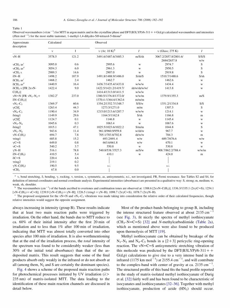
Table 1
Observed wavenumbers (n/cmK1) for MTT in argon matrix and in the crystalline phase and DFT(B3LYP)/6-311CCG(d,p) calculated wavenumbers and intensities
(I/km molK1) for the most stable tautomer, 1-methyl-1,4-dihydro-5H-tetrazol-5-thionea
Approximate
description
Calculated Observed
n I n (Ar; 10 K)b I n (Glass; 275 K) I
nN–H 3578.5 121.2 3491.6/3487.6/3485.3 m/S/sh 3067.2/2857.8/2801.6/
2684/2657.8
S/S/S
w/w
nCH3 as 0 3095.0 0.6 2995.6 w 2974.7 S
nCH3 as 00 3054.3 6.0 2961.3 w 2950.5 S
nCH3 s 2989.3 14.6 2907.0 w 2919.8 S
dN–H 1498.2 187.9 1491.8/1488.9/1486.0 S/sh/S 1510.7/1488.6 S/sh
dCH3 as 0 1468.2 2.4 1462.7 w 1462.6 w
dCH3 as 00 1440.9 16.4 1436.7/1435.4/1433.8 w/w/w 1434.4 w
dCH3 s [FR 2xnN-
C(H3)]
1422.4 9.0 1422.5/1421.2/1419.7/
1414.4/1313.0/1411.5
sh/w/sh/w/w/
w/w/w
1413.8 w
nNaN [FR nN1–N2C
dN–C(H3)]
1382.2c 237.0 1380.5/1376.8/1372.0/
1370.4 /1364.6/1362.6
w/w/m
m/m/m
1379.9/1355.3 m/S
nN1–C5 1369.3c 60.6 1354.2/1352.7/1348.7 S/S/w 1351.2/1334.8 S/S
gCH03 1263.4 44.3 1273.5/1271.0 m/m 1307.5 S
nN4–C5 1190.4 34.9 1215.0/1213.6/1207.7 w/w/w 1214.1 w
dring1 1149.9 29.6 1164.5/1162.8 S/sh 1166.8 m
gCH003 1124.7 0.1 1146.8 w 1145.4 w
nN3–N4 1045.6 0.3 1063.4 w 1067.6 w
dring2 1015.1 47.1 1025.5/1023.6/1022.2 S/m/m 1044.8 S
nN1–N2 943.6 11.4 961.8/960.9/959.8 w/sh/w 967.7 w
nN–C(H3) 694.7 7.8 705.1/703.8/702.8 sh/w/w 704.3 m
tring1 685.8 15.2 693.2/691.4 w/w 689.7/676.6 w/w
gCaS 649.0 0.8 663.6/661.8 w/w 670.1 w
nCaS 540.2 3.7 550.8 w 530.8 w
gN–H 516.1 78.8 540.8/538.7/527.3 m/S/w 908.7/862.2/788.4 w/w/m
dN–C(H3) 410.5 5.4 410.1 w 424.0 m
dCaS 220.4 4.6
n.i. n.i.
tring2 219.1 0.2
gN–C(H3) 190.5 9.5
tCH3 67.8 0.0
a n, bond stretching, d, bending, g, rocking, t, torsion, s, symmetric, as, antisymmetric, n.i., not investigated, FR, Fermi resonance. See Tables S2 and S4, for
definition of internal coordinates and normal coordinate analysis. Experimental intensities (absorbance) are presented in a qualitative way: S, strong; m, medium; w,
weak, sh, shoulder.b The wavenumbers (cmK1) of the bands ascribed to overtones and combination tones are observed at: 1388.6 [2xnN–C(H3)]; 1336.3/1335.1 (2xgCaS); 1254.1
[nN–C(H3)CnCaS]; 1239.9 [nN–C(H3)CgN–H]; 1226.5 (tring1CgN–H); 1098.7 (2xgCaS); 1078.7 (2xgN–H).c The proposed assignment for the nNaN and nN1–C5 vibrations was made taking into consideration the relative order of their calculated frequencies, though
relative intensities would suggest the opposite assignment.
A. Gomez-Zavaglia et al. / Journal of Molecular Structure 786 (2006) 182–192186
always increasing in intensity (group B). These results indicate
that at least two main reaction paths were triggered by
irradiation. On the other hand, the bands due to MTT reduce to
ca. 60% of their initial intensity after the first 20 min of
irradiation and to less than 1% after 100 min of irradiation,
indicating that MTT was almost totally converted into other
species after 100 min of irradiation. It is also worthmentioning
that at the end of the irradiation process, the total intensity of
the spectrum was found to be considerably weaker (less than
30% of the initial total absorbance) than that of the as-
deposited matrix. This result suggests that some of the final
products absorb only weakly in the infrared or do not absorb at
all (among them, N2 and S are certainly the dominant species).
Fig. 4 shows a scheme of the proposed main reaction paths
for photochemical processes initiated by UV irradiation (lO235 nm) of matrix-isolated MTT. The data leading to the
identification of these main reaction channels are discussed in
detail below.
Most of the product-bands belonging to group B, including
the intense structured feature observed at about 2135 cmK1
(see Fig. 3), fit nicely the spectra of methyl isothiocyanate
(CH3–NaCaS) [32] and N-methylcarbodiimide (Table 2a),
which as mentioned above were also found to be produced
upon thermolysis of MTT [19].
Methyl isothiocyanate can be obtained by breakage of the
N1–N2 and N4–C5 bonds in a [2C3] pericyclic ring-opening
reaction. The nNaCaS antisymmetric stretching vibration of
this molecule was predicted by the DFT(B3LYP)/6-311CCG(d,p) calculations to give rise to a very intense band in the
infrared (1175 km molK1) at 2155.4 cmK1, and will contribute
to the complex-band with center of gravity at ca. 2135 cmK1.
The structured profile of this band fits the band profile reported
in the study of matrix-isolated methyl isothiocyanate of Durig
et al. [32] fairly well and has been found to be characteristic of
isocyanates and isothiocyanates [32–36]. Together with methyl
isothiocyanate, production of azide (HN3) should occur.

0.05
0.00
0.10
0.15
0.20
1500
Cal
cula
ted
Inte
nsi
ty/
km m
ol–1
Ab
sorb
ance
1600170018002300 2200 2100 2000 1900
1400
1200
1000
800
600
400
200
0
Wavenumber/ cm–1
(a)
(b)
Fig. 3. (a) Infrared spectra (2300–1600 cmK1 region) of the irradiated matrix of
MTT, obtained after 20 min (dotted line) and 100 min (solid line) of irradiation.
The spectrum of the as-deposited matrix in the same spectral region is also
presented, the corresponding trace (solid line) being slightly shifted down
relatively to the spectra of the irradiated matrix. In the spectra, bands due to
traces of water, appearing in the 1550–1650 cmK1 range, were subtracted.
(b) Calculated spectra (2300–1500 cmK1 region) for MTT and most of the
observed photoproducts (among the identified photoproducts, only CS and HN
do not absorb in the spectral region shown in the figure). The calculated spectra
are presented as delta functions centered at the calculated (scaled) frequency.
The intensity of the calculated band of MTT was multiplied by 7, in order to
approximately preserve the relative band intensities in the experimental and
calculated spectra shown in the figure.
A. Gomez-Zavaglia et al. / Journal of Molecular Structure 786 (2006) 182–192 187
Identification of this species is complicated by the fact that its
most intense band (nNaNCaNK antisymmetric stretching) is
expected to lie nearly at the same position as the nNaCaS
antisymmetric stretching of methyl isothiocyanate [37] and to
have an intrinsic intensity considerably lower than this latter
(405 km molK1; DFT(B3LYP)/6-311CCG(d,p) calculated
value). However, the spectrum of matrix-isolated azide is
well known [37–41] and the analysis of other regions of the
spectra reveals that this species is not present in the irradiated
matrix. Indeed, matrix-isolated azide was found to be
photochemically labile when subjected to UV-irradiation
(lO254 nm [37]), giving rise to N2 and (3S) HN. The triplet
HN was reported to give rise to a weak band at ca. 3133 cmK1
in argon matrix [37]. In the spectra of the photolysed matrix,
two bands (type B) were observed not far from this frequency
(3100 and 3086 cmK1). The higher frequency band exhibits
the relatively broad profile expected for a small size species
like NH, which will be partially rotating in the matrix (on the
other hand, the lower frequency band is sharp and is ascribable
to a combination mode of N-methylcarbodiimide, as discussed
below). Hence, the results suggest that the azide initially
formed together with methyl isothiocyanate quickly reacts to
form N2 and triplet HN.
N-methylcarbodiimide production occurs simultaneously
with expulsion of both molecular nitrogen and sulphur, which
are not expected to give rise to any observable IR band. From
the 16 vibrations of N-methylcarbodiimide predicted to occur
in the studied spectral region, all but the very low intensity
gCH003 rocking mode were observed in the spectra of the
irradiated matrix (see Table 2b). The calculations predict the
most intense bands of N-methylcarbodiimide, corresponding to
the nNaCaN antisymmetric stretching and dN–H in plane
bending modes, at 2191.9 cmK1 (with intensity
953.1 km molK1) and 919.9 cmK1 (245.0 km molK1),
respectively. The dN–H vibration is assigned to a multiplet
of bands appearing in the 890–950 cmK1 spectral range. The
multiplet structure of the band clearly reveals that the molecule
is produced in a variety of sites whose specificity seems to
affect considerably the vibrations mostly localized in the NH
group. Indeed, the less intense gN–H out of plane mode was
also found to give rise to a structured band (with two main
components at 587.1 and 588.8 cmK1; see Table 2b) and the
nN–H stretching mode to a broadband around 3400 cmK1,
which can also result from unresolved superposition of a series
of bands with slightly different frequencies. On the other hand,
with all probability, the nNaCaN antisymmetric stretching
mode contributes to the experimentally observed complex
feature with gravity center at ca. 2135 cmK1, also due to the
nNaCaS antisymmetric stretching of methyl isothiocyanate.
The bands belonging to group A are, in general, more
structured than those belonging to group B, clearly indicating
that the species which originate them are produced in multiple
sites and, probably, interacting considerably with other species
present in the matrix cage. In order to identify the products that
give rise to the observed A-type bands, extensive calculations
of the IR spectrum of putative candidates were undertaken. The
series of molecules considered is listed in Table S9, provided in
Appendix. An essential criterion the putative photoproduced
species should obey was to give rise to significantly intense
bands in the 1720–1820 cmK1 spectral range, where the most
intense A-type bands are observed. Apart from diazomethyl
radical (HCNN), which also exhibits bands in this region [42]
but whose production looks very improbable, only 1-methyl-
1H-diazirene-3-thiol (MDT) was found to satisfy this criterion.
MDT can be obtained from MTT by nitrogen elimination
followed by H-1,3 migration from the nitrogen N(4) to the
sulphur atom and ring-closure, and may exist in two
conformers. One of the conformers has the SH group anti
relatively to the methyl group, whereas the second form has
these groups syn; see Fig. 4. The two conformers of MDT are
predicted by the calculations to have very close energies (the
anti form is 1.34 kJ molK1 more stable than the syn form, as
predicted by the DFT(B3LYP)/6-311CCG(d,p) calculations)

N2
+
+HN
–CS
–N2
–CS
–N2
–N2S+
MTT
Methyl diazene
Methyl isothiocyanate Azide
1-methyl–1,2–dihydrotetrazete
N-Methyl-carbodiimide1-Methyl-1H-diazirene-3-thiol
48.4
215.8
449.1
361.8
55.0 202.1
–184.8
Fig. 4. Proposed pathways for reactions resulting from irradiation (lO235 nm) of MTT monomer in argon matrix. Note that no bands due to 1-methyl-
1,2-dihydrotetrazete and azide were identified experimentally; the relevance of these two species is discussed in the text. Zero-point corrected energy differences
(kJ molK1) between products and reactants for each process are given in the figure. In cases where more than one conformer exist, energies relate to reactions
involving the most stable conformers.
A. Gomez-Zavaglia et al. / Journal of Molecular Structure 786 (2006) 182–192188
and, with all probability, both are present in the photolysed
matrix (see Table 2c for assignments).
Upon prolonged irradiation, MDT undergoes secondary
photoreactions, reducing its concentration in the matrix. Since
no new bands appear in the spectra together with disappearance
of the bands due to this species, it shall be converted in weakly
absorbing species. Indeed, MDT can further react to give rise to
methyl diazene (CH3NaNH) plus CS. Methyl diazene may
Table 2a
Observed and calculated IR bands of methyl isothiocyanate
Observed frequency (cmK1) Literature frequency (cmK1)
[32]
Calculated [B3L
Frequency (cmK
661.5 661 659.2
1062.0 1106 1094.2
n.o. 1109 1098.7
n.o. 1142 (gas) 1148.4
1422.3 1421 1420.8
1456.8 1452 1454.1
1468.1 1459 1462.9
2079.3 2085
–a 2118/2126 2155.4
2215.9 2211/2217
2233.4/2252.9 2236/2256
2801.5 2791
n.o. 2818
2898.3 2887
2953.2 2952 2957.8
2991.1 2998 3017.9
3029.0 2998 3040.2
a Very strong and complex multiplet between 2105 and 2193 cmK1 (superimpos
also exist in two conformers (Z and E isomers), and contribute
to the bands observed at 1656/1639, 1457, 1433, 1395, 1110
and 846/842 cmK1 (Z/E isomers, respectively), which are
predicted by the calculations to occur at 1642/1623, 1457 (in
both Z and E isomers), 1437, 1377/1376, 1119/1120 and
851/845 cmK1; Table 2d. The presence of CS in the photolysed
matrix manifests itself as the complex band lying in the 1257–
1243 cmK1 range (in the gaseous phase it was observed at
YP/6-311CCG(d,p)] Approximate description
1) Intensity (km molK1)
24.0 nCN
23.4 nNCS sym
!0.1 gCH003
5.3 gCH03
66.6 dCH3 sym
8.7 dCH3 asym 0
9.2 dCH3 asym 00
nCNCdCH3 sym
1175.3 nNCS asym
2x nNCS sym FR nNCS asym
dCNCCnNCS asym
nNCS asymCnCN
2x dCH3 sym
2x dCH3 asym0
55.9 nCH3 sym
15.2 nCH3 asym00
12.1 nCH3 asym0
ed with the nNCN asym band of N-methylcarbodiimide; see text).

Table 2b
Observed and calculated IR bands of N-methylcarbodiimide
Observed frequency (cmK1) Calculated [B3LYP/6-311CCG(d,p)] Approximate description
Frequency (cmK1) Intensity (km molK1)
587.1/588.8 558.9 72.8 gN–H
627.7 626.9 18.4 dNCN
853.4 870.0 24.0 nNC(H3)
891.5/903.0/934.1/938.8/940.9/951.3 919.9 245.0 dN–H
1110.4 1111.9 9.1 gCH03
n.o. 1113.7 1.0 gCH003
1391.7 1368.4 10.5 nNCN sym
1424.9 1425.3 22.8 dCH3 sym
1456.8 1457.9 7.2 dCH3 asym00
1470.6 1473.6 16.2 dCH3 asym0
–a 2191.9 953.1 nNCN asym
2953.2 2955.3 61.2 nCH3 sym
3029.0 3024.0 23.1 nCH3 asym0
3029.0 3030.4 17.4 nCH3 asym00
3085.7 nNCN asymCdN–H
w3400 3496.9 62.3 nN–H
a Very strong and complex multiplet between 2105 and 2193 cmK1 (superimposed with the nNCS asym band of methyl isothiocyanate; see text).
A. Gomez-Zavaglia et al. / Journal of Molecular Structure 786 (2006) 182–192 189
1272 cmK1 [43]). Note that methyl diazene and CS (plus N2)
are also the expected photoproducts resulting from direct CS
elimination from MTT (eventually implying formation of the
unstable 1-methyl-1,2-dihydrotetrazete intermediate, which
was not observed spectroscopically; see Fig. 4).
In summary, in situ UV-irradiation (lO235 nm) of the
matrix-isolated monomer of MTT is shown to trigger different
photochemical processes, all of them involving cleavage of the
tetrazole ring: e.g. (1) loss of nitrogen, with production of
1-methyl-1H-diazirene-3-thiol, which is produced in two
different conformers; (2) ring cleavage leading to production
Table 2c
Observed and calculated IR bands of 1-methyl-1H-diazirene-3-thiol (MDT)
Observed frequency (cmK1) Calculated [B3LYP/6-311CCG(d,p)]
Frequency (cmK1) Intensity (
w565 565.8, 560.2 5.1, 4.4
640.6 622.4 22.6
666.8/671.1/672.5/679.2 625.3 24.1
786.2 843.1 26.8
789.1 868.6 26.5
866.8 949.8 10.4
868.4 952.8 7.8
n.o. 1080.5, 1180.3 1.0, 1.3
1080.5 1118.2, 1119.8 3.2, 4.3
1101.4/1102.9/1104.4/1105.7/
1118.3/1121.3/1124.0/1131.3
1229.4 31.9
1153.6/1156.4/1171.2/1173.4/
1175.8
1241.6 48.2
1408.2 1399.2, 1398.3 4.5, 3.5
1442.1 1436.8, 1436.1 4.5, 4.6
1461.6 1464.4, 1464.9 12.3, 12.
1749.4/1752.5/1756.1/1757.2/
1770.1/1774.9/1779.1
1734.7 127.3
1792.1/1794.0/1817.9/1819.1 1746.9 142.3
n.o. 2603.8, 2591.1 0.2, 0.4
2928.4 2937.5, 2937.6 29.9, 29.
2971.4 3001.2, 3002.0 18.3, 16.
3029.0 3042.0, 3043.0 15.8, 17.
of methyl isothiocyanate and azide; (3) simultaneous elimin-
ation of nitrogen and sulphur with production of
N-methylcarbodiimide and, eventually, (4) CS elimination,
leading to methyl diazene, CS and N2 Following these
photoprocesses, subsequent reactions occur, with production
of methyl diazene, carbon monosulphide and triplet nitrogen
hydride. We have also investigated the possibility of
occurrence of the third previously observed thermal channel
[19], which leads to formation of methylazide and isothio-
cyanic acid. Our data, however, do not show any clear evidence
of presence of these two species in the irradiated matrices.
Approximate description Conformer
km molK1)
gC–S syn, anti
nC–S syn
nC–S anti
dCSH anti
dCSH syn
nN–C(H3) syn
nN–C(H3) anti
gCH003 syn, anti
gCH03 syn, anti
nCN syn
nCN anti
dCH3 sym syn, anti
dCH3 asym0 syn, anti
0 dCH3 asym00 syn, anti
nCaN syn
nCaN anti
nS–H syn, anti
8 nCH3 sym syn, anti
4 nCH3 asym 00 syn, anti
1 nCH3 asym 0 syn, anti

Table 2d
Observed and calculated IR bands of methyl diazene
Observed frequency (cmK1) Calculated [B3LYP/6-311CCG(d,p)] Approximate description Conformer
Frequency (cmK1) Intensity (km molK1)
583.5 544.0 16.9 dCNN E
n.o. 564.6 1.2 dCNN Z
842.0 845.4 34.1 gN–H E
845.9 851.0 43.7 gN–H Z
n.o. 885.0 6.7 nCN Z
903.0 909.6 13.6 nCN E
1110.4 1119.6, 1119.1 8.6, 3.4 gCH03 E, Z
n.o. 1130.3 8.2 gCH003 Z
1142.1 1145.7 20.3 gCH003 E
1395.5 1376.2, 1376.8 7.8, 11.8 dCH3 sym E, Z
n.o. 1422.7 8.0 dCH3 asym00 Z
n.o. 1428.4 4.4 dCH3 asym0 Z
1432.9 1436.6 16.4 dCH3 asym0 E
1432.9 1436.7 12.2 dCH3 asym00 E
1456.8 1456.7, 1456.8 22.1, 77.3 dNH E, Z
1639.2 1623.0 5.6 nNaN E
1656.4 1642.3 29.8 nNaN Z
2898.3 2899.6 37.5 nCH3 sym Z
2953.2 2948.9 16.2 nCH3 sym E
2953.2 2950.5 99.7 nN–H Z
3029.0 3027.6, 3015.4 8.8, 10.9 nCH3 asym0 E, Z
3029.0 3037.3, 3036.2 15.0, 5.6 nCH3 asym00 E, Z
n.o. 3206.8 12.3 nN–H E
A. Gomez-Zavaglia et al. / Journal of Molecular Structure 786 (2006) 182–192190
3.3. IR spectrum of MTT in the low temperature neat solid state
Fig. 5 shows the spectrum of the low temperature crystalline
phase obtained by deposition of the vapor of MTT onto the cold
substrate of the cryostat (at 10 K) followed by slow annealing
of the solid film. Crystallization was found to start at a
temperature of ca. 150 K.
2050230025502800305033003550Wavenumbe
νNH
νNH´´ νNH´
νNH
2xγCH3´
2xνN=N
νCH3 s
νCH3 as
A
B
Fig. 5. (A) Infrared spectrum of crystalline MTT (as film; TZ275 K). (B) DFT(B3LY
dihydro-5H-tetrazol-5-thione tautomer). The calculated spectrum was simulated us
bandwidth-at-half-height equal to 5 cmK1.
The first conclusion that can be extracted from the spectra
shown in Fig. 5 is that, as it was found in the gaseous phase and
for the matrix-isolated compound, in the neat crystalline phase
also MTT exists exclusively as the thione tautomer. Indeed, the
correlation between the observed spectrum for the crystal and
the calculated for the thione tautomer of MTT is evident from
the data shown in Fig. 5.
5508001050130015501800r/ cm–1
γNH´
γNH
γNH
γNH´´
P)/6-311CCG(d,p) calculated spectrum for the monomer MTT (1-methyl-1,4-
ing GAUSSIAN functions centered at the calculated (scaled) frequency and with

A. Gomez-Zavaglia et al. / Journal of Molecular Structure 786 (2006) 182–192 191
Naturally, as it could be anticipated, the main differences
between the calculated MTT spectrum (and that of the matrix-
isolated compound) and the observed spectrum of the crystal
can be easily identified as occurring for bands ascribable to
vibrations of the NH group, which in the crystal is involved in
hydrogen bonding interactions. According to Rozenberg et al.
[44,45], for nearly linear H-bonds, the red shift of the nNH
stretching mode, n1, and the blue shift of the corresponding
out-of-the-plane gNH rocking mode, n4, which occur upon
H-bond formation, may be empirically correlated, because both
parameters correlate with the H-bond enthalpy (DH)
Dn24 ðcmK2ÞZ 2:5ðDn1 ðcmK1Þ1=2K18 (1)
ðDHÞ2 Z 1:92½ðDn1ÞK40� (2)
KDH Z 0:67Dn24 (3)
where Dn24Z ð10K2nH
4 Þ2Kð10K2n0
4Þ2 and Dn1ZnH
1 Kn01;, and the
superscripts ‘H’ and ‘0’ pertain to H-bonded and free
molecules, respectively.
In general, for crystals with natural isotopic content, the
nNH stretching spectral region is considerably complex,
owing to extensive vibrational coupling between the nNH
modes and low frequency vibrations, presence of overtones
and combination tones and Fermi resonance interactions.
This fact makes the bands corresponding to the nNH
stretching mode difficult to assign with certainty. However,
using the correlation (1) it is possible to estimate the
position of these bands in the spectrum of the crystal from
the position of the bands ascribed to the nNH rocking mode,
which can be easily observed in the 1000–700 cmK1 region.
Indeed, a very important property exhibited by n4 is that, in
contrast to n1, it gives rise to bands that do not change their
molar integral intensity with the H-bond energy and thus
they remain narrow and well resolved independently of the
strength of the H-bond interaction [44,45]. Consequently, n4
can be used for a direct quantitative estimation of the
relative abundance of different H-bonds in a given crystal.
On the other hand, as mentioned above, the peak positions
of the bands due to both n1 and n4 are sensitive to the
H-bond energy and can be used to estimate this property on
the basis of the empirical quantitative relationships (2) and
(3) [44,45].
Three bands are observed in the 1000–700 cmK1 region of
the spectrum of crystalline MTT that do not have counterpart in
both the calculated spectrum and that of the matrix-isolated
monomer: 788.4, 862.2 and 908.7 cmK1 (see Fig. 5). Accord-
ing to Rozenberg et al. [44,45], this means that three different
kinds of H-bonds, with different strengths, are present in the
sample. Using the empirical correlation (1), the frequencies
corresponding to the nNH stretching modes associated with
each one of the three types of H-bonds in crystalline MTT can
be estimated as 3069, 2846 and 2675 cmK1. Accordingly,
bands at 3067.2, 2857.8/2801.6 (average frequency
2829.7 cmK1) and 2684/2657.8 (average frequency
2671.3 cmK1) are observed experimentally and then ascribed
to the nNH stretching vibration in the three differently
H-bonded NH groups (Fig. 5).
On the other hand, as mentioned above, the relative
intensities of the n4 bands correlate with the relative abundance
of a given type of H-bond [44,45], whose energies can be
calculated by using either relationship (2) or (3). Then, the
results indicate that 86.9% of the H-bonds in the MTT
crystalline sample have an enthalpy of K24.61 kJ molK1,
11.5% an enthalpy of K32.40 kJ molK1 and 1.6% an enthalpy
of K37.24 kJ molK1. The enthalpy values presented corre-
spond to mean values obtained using Eqs. (2) and (3) above,
and correlate with the 788.4/3067.2 cmK1, 862.2/2829.7 cmK1
and 908.7/2671.3 cmK1 n4/n1 bands, respectively. Without
knowledge of the crystal structure of MTT, an unequivocal
interpretation cannot be given for the presence of the three
different kinds of H-bonds in the crystal. Taking into
consideration the estimated percentages indicated above, the
less abundant type of H-bonds result with all probability from
crystal defects. However, it is important to point out that the
reliability of the empirical correlations here used has been
tested exhaustively for a large number of different crystals with
different types of H-bond networks, with excellent results [44–
46], without any doubt the characteristic ordered H-bonds in
the MTT crystal are of at least two different types.
The assignment of the remaining bands observed in the
spectrum of the crystal is straightforward and is presented in
Table 1.
4. Conclusions
The DFT(B3LYP)/6-311CCG(d,p) calculations predict
that in the gaseous phase MTT should exist almost exclusively
as the thione tautomer. Accordingly, this was the only tautomer
of MTT observed for the compound isolated in a low
temperature argon matrix, as well as in the case of the neat
crystalline phase. Rozenberg’s [44,45] empirical correlation
between the frequencies of the stretching and out-of-the-plane
rocking modes originated in H-bonded NH groups was used to
characterize the H-bonding scheme in crystalline MTT,
allowing the conclusion to be drawn that three different types
of H-bonds are present in the crystal, with energies of K24.61,
K32.40 and K37.24 kJ molK1.
Irradiation (lO235 nm) of the matrix-isolated monomer of
MTT leads to observation of several photochemical processes.
Three dominant reaction pathways were identified: (1) loss of
nitrogen, with production of 1-methyl-1H-diazirene-3-thiol
(two different conformers); (2) ring cleavage leading to
production of methyl isothiocyanate and azide; (3) simul-
taneous elimination of nitrogen and sulphur with production of
N-methylcarbodiimide. Following these photoprocesses, sub-
sequent reactions occur, with production of methyl diazene,
carbon monosulphide and nitrogen hydride. The possibility of
occurrence of direct elimination of CS from the MTT molecule
upon irradiation, leading to methyl diazene, CS and N2, cannot
also be excluded.

A. Gomez-Zavaglia et al. / Journal of Molecular Structure 786 (2006) 182–192192
Acknowledgements
The authors acknowledge the Portuguese Science
Foundation (FCT-POCTI/QUI/59019/2004) and FEDER for
financial support. A.G.-Z. thanks FCT (Grant
SFRH/BPD/11499/2002), CONICET and the Argentinian
Agencia Nacional de Promocion Cientıfica y Tecnologica
(PICT 13080). I.D.R. acknowledges FCT for the Grant
SFRH/BPD/1661/2000.
Supplementary data (Appendix)
Supplementary data associated with this article can be found,
in the online version, at doi:10.1016/j.molstruc.2005.08.019
References
[1] H. Singh, A. Chawla, V. Kapoor, D. Paul, R. Malhotra, Prog. Med. Chem.
17 (1980) 151.
[2] G. Sandmann, C. Schneider, P. Boger, Z. Naturforsch. C 51 (1996) 534.
[3] J. Buynak, V. Doppalapudi, A. Rao, S. Nidamarthy, G. Adam, Bioorg.
Med. Chem. Lett. 10 (2000) 847.
[4] I. Renard, H. Li, B. Marsan, Electrochim. Acta 48 (2003) 831.
[5] K. Uchida, T. Shike, H. Kakushi, H. Takase, Y. Nomura, T. Harauchi,
T. Yoshizaki, Thrombosis Res. 39 (1985) 741.
[6] I.R. Dunkin, C.J. Shields, H. Quast, Tetrahedron 45 (1989) 259.
[7] G. Maier, J. Eckwert, A. Bothur, P. Reisenauer, C. Schmidt, Liebigs Ann.
(1996) 1041.
[8] F. Billes, H. Endredi, G. Keresztury, J. Mol. Struct. (Theochem) 530
(2000) 183.
[9] S. Bugalho, E. Macoas, L. Cristiano, R. Fausto, Phys. Chem. Chem. Phys.
3 (2001) 3541.
[10] S. Bugalho, A. Serra, L. Lapinski, L. Cristiano, R. Fausto, Phys. Chem.
Chem. Phys. 4 (2002) 1725.
[11] S. Bugalho, L. Lapinski, L. Cristiano, L. Frija, R. Fausto, Vibrat.
Spectrosc. 30 (2002) 213.
[12] A. Gomez-Zavaglia, I.D. Reva, M.L. Cristiano, L. Frija, R. Fausto,
J. Phys. Chem. A 109 (2005) 7967.
[13] E. Shouji, D. Buttry, J. Phys. Chem. B. 102 (1998) 1444.
[14] F. Hipler, R. Fischer, Muller, J. J. Chem. Soc. Perkin Trans. 2 (2002) 1620.
[15] A.R. Katritzky, J. Borowiecka, W.-Q. Fan, L.H. Brannigan, J. Heterocycl.
Chem. 28 (1991) 1139.
[16] J.W. Bats, Acta Crystallogr, B 32 (1976) 2866.
[17] J.H. Looker, N.A. Khatri, R.B. Patterson, C.A. Kingsbury, J. Heterocycl.
Chem. 15 (1978) 1383.
[18] H. Quast, U. Nahr, Chem. Ber. 118 (1984) 526.
[19] A. Awadallah, K. Kowski, P. Rademacher, J. Heterocycl. Chem. 34
(1997) 113.
[20] J.B. Christensen, A. Holm, Acta Chem, Scand. 51 (1997) 527.
[21] R. Flammang, M. Barbieux-Flammang, P. Gerbaux, C.T. Pedersen,
J. Chem. Soc. Perkin Trans. 2 (1997) 1261.
[22] I. Alkorta, J. Elguero, Struct. Chem. 14 (2003) 377.
[23] I.D. Reva, S. Stepanian, L. Adamowicz, R. Fausto, J. Phys. Chem. A 105
(2001) 4773.
[24] M. Frisch, G. Trucks, H. Schlegel, G. Scuseria, M. Robb, J. Cheeseman,
V. Zakrzewski, J. Montgomery, R. Stratmann, K. Burant, S. Dapprich, J.
Millam, A. Daniels, K. Kudin, M. Strain, O. Farkas, J. Tomasi, V. Barone,
M. Cossi, R. Cammi, B. Mennucci, C. Pomelli, C. Adamo, S. Clifford, J.
Ochterski, G. Petersson, P. Ayala, Q. Cui, K. Morokuma, D. Malick, A.
Rabuck, K. Raghavachari, J. Foresman, J. Cioslowski, J. Ortiz, A. Baboul,
B. Stefanov, G. Liu, A. Liashenko, P. Piskorz, I. Komaromi, R. Gomperts,
R. Martin, D. Fox, T. Keith, M. Al-Laham, C. Peng, A. Nanayakkara, M.
Challacombe, P. Gill, B. Johnson, W. Chen, M. Wong, J. Andres, C.
Gonzalez, M. Head-Gordon, S. Replogle, J. Pople, (1998) GAUSSIAN 98,
Revision A.9; Gaussian Inc.: Pittsburgh, PA.
[25] A.D. Becke, Phys. Rev. A 38 (1988) 3098.
[26] C.T. Lee, W.T. Yang, R.G. Parr, Phys. Rev. B 37 (1988) 785.
[27] P. Csaszar, P. Pulay, J. Mol. Struct. (Theochem.) 114 (1984) 31.
[28] J.H. Schachtschneider, (1969) Technical Report; Shell Development Co.
Emeryville, CA.
[29] R. Fausto, J.J.C. Teixeira-Dias, P.J. Tonge, P.R. Carey, J. Mol. Struct. 324
(1994) 113.
[30] B. Bak, O.J. Nielsen, H. Svanholt, J.J. Christiansen, J. Mol. Spectrosc. 39
(1979) 134.
[31] R. Fausto, L.A.E. Batista de Carvalho, J.J.C. Teixeira-Dias, M.N. Ramos,
J. Chem. Soc. Faraday Trans. 2. 85 (1989) 1945.
[32] J. Durig, J. Sullivan, D. Durig, S. Cradock, Can. J. Chem. 63 (1985) 2000.
[33] R.G. Lett, W.H. Flygare, J. Chem. Phys. 47 (1967) 4730.
[34] R.F. Curl Jr., V.M. Rao, K.V.L.N. Sastry, J.A. Hodgeson, J. Chem. Phys.
39 (1963) 3335.
[35] J. Koput, Chem. Phys. Lett. 242 (1995) 514.
[36] R. Fausto, I.D. Reva, L. Lapinski, Abstracts of the XX IUPAC
Symposium on Photochemistry, Granada, 2004, pp. 280.
[37] H. Himmel, M. Junker, H. Schnockel, J. Chem. Phys. 117 (2002) 3321.
[38] G. Pimentel, S. Charles, K. Rosengren, J. Chem. Phys. 44 (1966) 3029.
[39] D. Milligan, M. Jacox, J. Chem. Phys. 41 (1964) 2838.
[40] C. Moore, K. Rosengren, J. Chem. Phys. 44 (1966) 4108.
[41] W. Zhizhong, J. Mol. Struct. (Theochem.). 434 (1998) 1.
[42] J.F. Ogilvie, Can. J. Chem. 46 (1968) 2472.
[43] J. Crovisier, BASEMOL—‘Constants for Molecules of Astrophysics
Interest in the Gas Phase: Photodissociation, Microwave and Infrared
Spectra’, Laboratoire d’Etudes Spatiales et d’Instrumentation en
Astrophysique, Observatoire de Paris-Meudon, France, 2002. http://
www.usr.obspm.fr/wcrovisie/basemole/
[44] M. Rozenberg, Spectrochim. Acta A 52 (1996) 1559.
[45] M. Rozenberg, G. Shoham, I. Reva, R. Fausto, Spectrochim. Acta A 60
(2004) 463.
[46] M. Rozenberg, G. Shoham, I. Reva, R. Fausto, Phys. Chem. Chem. Phys.
7 (2005) 2376.
Copyright © 2022 FDOKUMEN