Molecular investigation of the evolutionary history and ...
244
Faculteit der Diergeneeskunde Faculteit Geneeskunde Hoofdafdeling Infectieziekten Rega Instituut & Immunologie Afdeling Klinische & Yalelaan 1, 3584 CL Utrecht Epidemiologische Virologie Minderbroedersstraat 10, 3000 Leuven Molecular investigation of the evolutionary history and diversity of primate T-lymphotropic virus types 1 and 3. Moleculair onderzoek naar de evolutiegeschiedenis en divergentie van primaat T-lymfotroop virus types 1 en 3. (met een samenvatting in het Nederlands) Proefschrift ter verkrijging van de graad van doctor aan de Universiteit Utrecht op gezag van de Rector Magnificus Prof. Dr. W.H.Gispen ingevolge het besluit van het College voor Promoties in het openbaar te verdedigen op donderdag 27 oktober 2005 des middags te 14.30 uur door Sonia Jeanne Albertine Van Dooren geboren op 29 september 1969, te Brussel
-
Upload
khangminh22 -
Category
Documents
-
view
0 -
download
0
Transcript of Molecular investigation of the evolutionary history and ...

Faculteit der Diergeneeskunde Faculteit Geneeskunde Hoofdafdeling Infectieziekten Rega Instituut & Immunologie Afdeling Klinische & Yalelaan 1, 3584 CL Utrecht Epidemiologische Virologie
Minderbroedersstraat 10, 3000 Leuven
Molecular investigation of the evolutionary history and diversity of
primate T-lymphotropic virus types 1 and 3.
Moleculair onderzoek naar de evolutiegeschiedenis en divergentie van
primaat T-lymfotroop virus types 1 en 3.
(met een samenvatting in het Nederlands)
Proefschrift
ter verkrijging van de graad van doctor aan de Universiteit Utrecht
op gezag van de Rector Magnificus Prof. Dr. W.H.Gispen ingevolge het besluit van het College voor Promoties
in het openbaar te verdedigen op donderdag 27 oktober 2005 des middags te 14.30 uur
door
Sonia Jeanne Albertine Van Dooren geboren op 29 september 1969, te Brussel
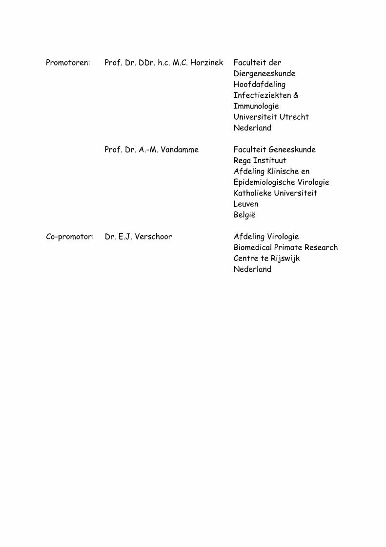
Promotoren: Prof. Dr. DDr. h.c. M.C. Horzinek Faculteit der
Diergeneeskunde Hoofdafdeling
Infectieziekten & Immunologie Universiteit Utrecht
Nederland Prof. Dr. A.-M. Vandamme Faculteit Geneeskunde Rega Instituut
Afdeling Klinische en Epidemiologische Virologie
Katholieke Universiteit Leuven
België Co-promotor: Dr. E.J. Verschoor Afdeling Virologie Biomedical Primate Research
Centre te Rijswijk Nederland

“Nothing in biology makes sense,
except in the light of evolution”
Theodosius Dobzhansky (1973)
To Wim Seppe
Reyn Roos

Van Dooren, Sonia J.A. Molecular investigation of the evolutionary history and diversity of primate T-lymphotropic virus types 1 and 3 / Sonia Jeanne Albertine Van Dooren Proefschrift Universiteit Utrecht- Met lit. opg. – Met samenvatting in het Nederlands ISBN 90-39340803
5
Contents Contents....................................................................................................................5
Abbreviations.............................................................................................................9
Chapter 1
Introduction ...........................................................................................................13
Introduction to Retroviruses ...................................................... 14
Primate T-lymphotropic viruses................................................... 16
Epidemiology of PTLV.............................................................. 19
Molecular biology of PTLV......................................................... 23
PTLV diagnostic and characterization techniques ............................... 28
Molecular epidemiology and evolution of PTLV................................... 30
Molecular evolution, phylogenetic inference and molecular clocks.............. 36
Scope of the thesis................................................................ 40
Chapter 2
Evidence for a post-Columbian introduction of human T-cell lymphotropic
virus type I in Latin America ..............................................................................43 Abstract ............................................................................ 45
Introduction ........................................................................ 46
Methods............................................................................. 48
Results .............................................................................. 54
Discussion........................................................................... 62
References.......................................................................... 65
Chapter 3
Dating the origin of the African human T-cell lymphotropic virus type-I
(HTLV-I) subtypes ...............................................................................................73 Abstract ............................................................................ 75
Introduction ........................................................................ 76
Materials and Methods ............................................................ 77
Results .............................................................................. 79
Discussion........................................................................... 90
Literature Cited.................................................................... 94
Contents
6
Chapter 4
The low evolutionary rate of human T-cell lymphotropic virus type-1
confirmed by analysis of vertical transmission chains......................................99 Abstract ........................................................................... 101
Introduction ....................................................................... 102
Materials and Methods ........................................................... 104
Results ............................................................................. 107
Discussion.......................................................................... 112
Literature Cited................................................................... 115
Chapter 5
Full-genome analysis of a highly divergent simian T-cell lymphotropic
virus type 1 strain in Macaca arctoides ..........................................................119 Abstract ........................................................................... 121
Short communication .............................................................. 122
References......................................................................... 130
Chapter 6
Phylogeny of novel primate T-lymphotropic virus type 1 strains in
various Asian and African non-human primate host species. ........................133 Abstract. .......................................................................... 135
Introduction ....................................................................... 136
Results and discussion ............................................................ 137
Materials and methods ........................................................... 146
Reference List .................................................................... 147
Chapter 7
Evidence for a second simian T-cell lymphotropic virus type 3 in
Cercopithecus nictitans from Cameroon ...........................................................151 Letter to the editor .............................................................. 153
References......................................................................... 156
Chapter 8
Identification in gelada baboons (Theropithecus gelada) of a distinct
simian T-cell lymphotropic virus type 3 with a broad range of
Western blot reactivity .....................................................................................159
Contents
7
Abstract ........................................................................... 161
Introduction ....................................................................... 162
Methods............................................................................ 164
Results and discussion ............................................................ 167
References......................................................................... 178
Chapter 9
General discussion ...............................................................................................183
HTLV-1 evolutionary rate estimates ............................................ 185 Tick and calibration of PTLV molecular clocks ....................................................................185 Overview of obtained HTLV-1 evolutionary rates ..............................................................186 Compatibility of different HTLV-1 evolutionary rate estimates ....................................188 Hypotheses on origin and dissemination of HTLV-1 in Latin America and Africa .......195
PTLV-1 molecular evolution and phylogeography................................ 197 African PTLV-1: geographic-dependent evolution...............................................................197 Asian PTLV-1: host-dependent evolution ..............................................................................199 African-Asian PTLV-1: incongruence in evolutionary patterns ........................................201 Resolving the PTLV-1 phylogeography? ................................................................................ 202
STLV-3 epidemiology and molecular epidemiology .............................. 204 Overview of STLV-3 infected host species ........................................................................ 204 Variability in STLV-3 seroreactivity .................................................................................... 205 STLV-3 epidemiology................................................................................................................ 206 Molecular epidemiology of STLV-3 ....................................................................................... 207 Time of origin and diversification of STLV-3 .................................................................... 209
Conclusions and perspectives ..................................................... 210
Reference list ........................................................................................................211
Summary...............................................................................................................231
Samenvatting ........................................................................................................233
List of publications .................................................................................................237
Curriculum vitae....................................................................................................241
Dankwoord............................................................................................................242

9
Abbreviations 3rd cp third codon position γ 32P gamma-labelled radioactive phosphate λ evolutionary rate (nucleotide substitutions per site per year) µ evolutionary rate (nucleotide substitutions per site per year) χ2-test chi-square test amb ambiguous mutation ATLL adult T-cell leukaemia/lymphoma ATLV adult T-cell leukaemia virus BC before Christ bl branch length BLV bovine leukaemia virus bp base pair bZIP basic-leucine zipper CA capsid CD4 cluster of differentiation antigen 4 CD8 cluster of differentiation antigen 8 CFSE carboxyfluorescein succinimidyl ester CPT cell preparation tube CTL cytotoxic T-lymphocyte DNA deoxyribonucleic acid dNTPs deoxyribonucleotidetriphosphates EDTA ethylenediaminetetraacetic acid EIA enzyme immunosorbent assay env envelope gene FITC fluorescein isothiocyanate gag group antigen gene gDNA genomic deoxyribonucleic acid GLUT-1 glucose transporter protein -1 gp glycoprotein HAM human associated myelopathy HBZ human T-lymphotropic virus basic-leucine zipper transcription
factor
Abbreviations
10
HCl hydrochloric acid HIV human immunodeficiency virus HTLV human T-lymphotropic virus IDU intravenous drug user IFA indirect immunofluorescence assay IgG immunoglobuline G IL-2 interleukine-2 IN integrase KA nonsynonymous substitution distance KS synonymous substitution distance kbp kilo base pair KCl potassium chloride LRT likelihood ratio test LTR long terminal repeat MA matrix MgCl2 magnesium chloride ML maximum likelihood MLK maximum likelihood enforcing a molecular clock mpars maximum parsimony mRNA messenger ribonucleic acid mtDNA mitochondrial deoxyribonucleic acid NA not available NC nucleocapsid ND not done NHP non-human primate NJ neighbour joining nt nucleotide NTs/s/y nucleotide substitutions/site/year ORF open reading frame p protein P probability (statistical significance level) Pars parsimony PBL peripheral blood lymphocytes PBMC peripheral blood mononuclear cells PCR polymerase chain reaction
Abbreviations
11
pol polymerase gene pro or PR protease PTLV primate T-lymphotropic virus pX 3’ region of PTLV genome between envelope gene and long
terminal repeat R repeat region of long terminal repeat r evolutionary rate (nucleotide substitutions per site per year) rex regulator of expression RNA ribonucleic acid rof regulator of expression -xI open reading frame RSV Rous sarcoma virus RT reverse transcriptase s transition sa splice acceptor sd splice donor SPR subtree-pruning-regrafting STLV simian T-lymphotropic virus SU surface protein T divergence time tax transactivator of expression TBR tree-bisection-reconnection ti/tv transition transversion ratio TM transmembrane protein tof transactivator of expression -xII open reading frame tRNApro transfer ribonucleic acid with proline TSP tropical spastic paraparesis U3 unique region at 3’end of long terminal repeat U5 unique region at 5’end of long terminal repeat v transversion vDNA viral deoxyribonucleic acid WB Western blot ya years ago


Chapter 1
Introduction
Primate T-lymphotropic
Viruses
Primate T-lymphotropic Viruses
14
Introduction to Retroviruses Retroviruses
Retroviruses were among the first known viruses, originally discovered in the beginning of the 20th century in animals with neoplastic diseases, especially leukaemias and lymphomas (Gross, L, 1983). The first discovered retrovirus, Rous sarcoma virus (RSV) later became the prototype RNA tumour virus.
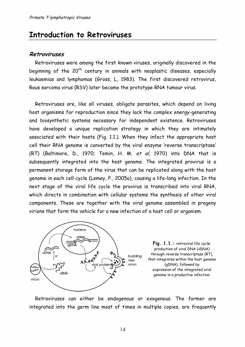
Retroviruses are, like all viruses, obligate parasites, which depend on living host organisms for reproduction since they lack the complex energy-generating and biosynthetic systems necessary for independent existence. Retroviruses have developed a unique replication strategy in which they are intimately associated with their hosts (Fig. 1.1.). When they infect the appropriate host cell their RNA genome is converted by the viral enzyme ‘reverse transcriptase’ (RT) (Baltimore, D., 1970; Temin, H. M. et al, 1970) into DNA that is subsequently integrated into the host genome. The integrated provirus is a permanent storage form of the virus that can be replicated along with the host genome in each cell cycle (Lemey, P., 2005a), causing a life-long infection. In the next stage of the viral life cycle the provirus is transcribed into viral RNA, which directs in combination with cellular systems the synthesis of other viral components. These are together with the viral genome assembled in progeny virions that form the vehicle for a new infection of a host cell or organism.
Fig. 1.1.: retroviral life cycle: production of viral DNA (vDNA)
through reverse transcriptase (RT), that integrates within the host genome
(gDNA), followed by expression of the integrated viral genome in a productive infection.
Retroviruses can either be endogenous or exogenous. The former are integrated into the germ line most of times in multiple copies, are frequently
Chapter 1 Introduction
15
defective, and are therefore usually not capable of producing mature, infectious virions. Some can produce complete provirus transcripts, but generally do not form infectious viral particles (Urnovitz, H. B. et al, 1996). Exogenous retroviruses are integrated into the host genome, though not in the germ line. They have an active replicating capacity, are always antigenic, and are often pathogenic. Classification of Retroviruses
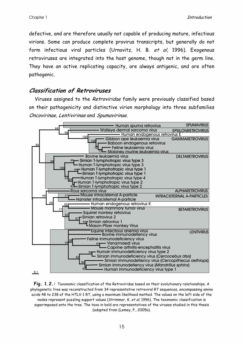
Viruses assigned to the Retroviridae family were previously classified based on their pathogenicity and distinctive virion morphology into three subfamilies Oncovirinae, Lentivirinae and Spumavirinae.
Fig. 1.2.: Taxonomic classification of the Retroviridae based on their evolutionary relationships. A
phylogenetic tree was reconstructed from 34 representative retroviral RT sequences, encompassing amino acids 48 to 238 of the HTLV-1 RT, using a maximum likelihood method. The values on the left side of the
nodes represent puzzling support values (Strimmer, K. et al, 1996). The taxonomic classification is superimposed onto the tree. The taxa in bold are representatives of the viruses studied in this thesis
(adapted from (Lemey, P., 2005a).
0.1
7376
100100
9899
99
99
82
8983
100
99
86
77
55
9687
62
77
64
99
8758
99
55
83
82
99
Bovine leukaemia virusSimian T-lymphotropic virus type 3
Human T-lymphotropic virus type 1Simian T-lymphotropic virus type 1
Human T-lymphotropic virus type 2Simian T-lymphotropic virus type 2
DELTARETROVIRUS
Human T-lymphotropic virus type 3
Human T-lymphotropic virus type 4
Human spuma retrovirus
Human endogenous retrovirus EGibbon ape leukaemia virusBaboon endogenous retrovirus
Feline leukaemia virusMoloney murine leukaemia virus
EPSILONRETROVIRUS
GAMMARETROVIRUS
Walleye dermal sarcoma virusSPUMAVIRUS
Rous sarcoma virusMouse intracisternal A-particle
Hamster intracisternal A-particleHuman endogenous retrovirus KMouse mammory tumor virus
Squirrel monkey retrovirusSimian retrovirus 2
Simian retrovirus 1Mason-Pfizer monkey Virus
ALPHARETROVIRUS
BETARETROVIRUS
INTRACISTERNAL A-PARTICLES
Bovine immunodefiency virusEquine infectious anemia virus
Feline immunodeficiency virusVisna/maedi virusCaprine arthritis-encephalitis virus
Human immunodeficiency virus type 2Simian immunodeficiency virus (Cercocebus atys)
Simian immunodefiency virus (Cercopithecus aethiops)
Human immunodeficiency virus type 1Simian immunodefiency virus (Mandrillus sphinx)
LENTIVIRUS
Primate T-lymphotropic Viruses
16
The newer taxonomy of the Retroviridae is based on genomic organisation and evolutionary relationships (=phylogeny). Their genomic structure is either simple or complex (Coffin, J. M., 1992): simple retroviruses usually carry only elementary information coded by structural, enzymatic and envelope genes, whereas complex retroviral genomes also code for additional regulatory proteins. The Retroviridae can be further divided into genera based on their evolutionary relationships as shown in Fig. 1.2. (Coffin, J. M., 1992).
Human T-lymphotropic virus types 1 to 4 (HTLV-1 to -4) belong to the genus of Deltaretroviruses, together with their non-human primate (NHP) counterparts named simian T-lymphotropic virus types 1 to 3 (STLV-1 to -3) (Fig. 1.2.). Both human and simian T-lymphotropic viruses are generally referred to as primate T-lymphotropic Viruses (PTLV). Together with bovine leukaemia virus (BLV) they comprise an oncogenic genus of retroviruses, which are complex of nature and exogenous.
Primate T-lymphotropic viruses HTLV: a human pathogenic retrovirus
Due to the tumour-inducing capacity of certain animal retroviruses, a major search for retroviruses in humans was initiated in the 1950s and 1960s. However, the first human retrovirus was only discovered in the early 1980s, when culturing primary human cells became possible with the help of growth factors -interleukine-2 (IL-2)- (Morgan, D. A. et al, 1976). Competitive immunological and sensitive, specific detection techniques, like RT-assays, also added to this success (Baltimore, D., 1970; Temin, H. M. et al, 1970).
The discovery of this first human retrovirus began with the observation of an RT-positive cell line derived from a patient with a cutaneous T-cell malignancy (Poiesz, B. J. et al, 1981). The existence of this virus, called human T-cell lymphotropic virus (HTLV), was further established after the obtained proof that the proviral DNA and RT were novel, re-isolation was possible, and the virus was infectious, replicating and present in other persons (Gallo, R. C., 2002).
In the same year, a team in Japan independently reported the finding of a retrovirus they called adult T-cell leukaemia virus (ATLV) (Hinuma, Y. et al,
Chapter 1 Introduction
17
1981). Collaborative efforts showed the viruses were of the same type, named HTLV, later refined as HTLV type 1.
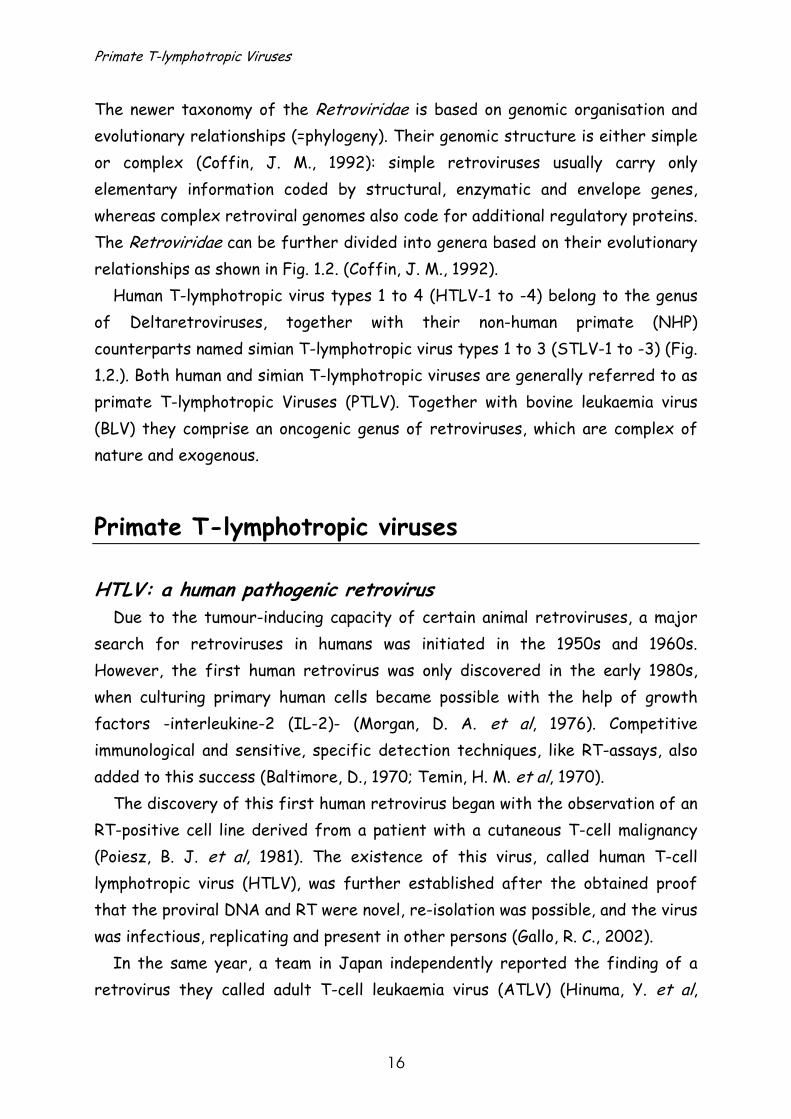
The aggressive form of leukaemia and lymphoma that is associated with HTLV-1, known as adult T-cell leukaemia/lymphoma (ATLL), generally appears a few decades after infection in 3-5% of the infected persons (Fig.1.3A&B.) (reviewed in (Blattner, W., 2005).
After the initial description of ATLL other diseases have been associated with this viral infection, including neurological disorders, resembling multiple sclerosis, though more progressive (Blattner, W., 2005). This human associated myelopathy (HAM) or tropical spastic paraparesis (TSP) (Gessain, A. et al, 1985; Osame, M. et al, 1987) occurs in less than 5% of HTLV-1 infected patients after a long latency period (Fig.1.3C.) (Levin, M. C. et al, 1997). Prior to occurrence of
(A) (B)
(C)
Fig. 1.3.: (A) patient with ATLL (B) patient with cutaneous lymphoma (C) patient diagnosed with HAM/TSP (photographs courtesy of Dr. E. Gotuzzo and Dr. T. Verdonck, photos belong to IMTA v H .-
Cayetano Heredia University, Lima, Peru [ published with permission])
Primate T-lymphotropic Viruses
18
these two types of malignancy, patients are asymptomatic carriers but are frequently burdened by opportunistic infections (Hanchard, B. et al, 1991; Gabet, A. S. et al, 2000; Edlich, R. F. et al, 2003).
In 1982 the discovery of a second, related human retrovirus, HTLV type 2, was reported (Kalyanaraman, V. S. et al, 1982). Although this virus was first identified in a patient with a T-cell variant of hairy cell leukaemia, it could not be further linked with leukaemia. It seems to be more associated with neurological syndromes (Hall, W. W. et al, 1994; Murphy, E. L. et al, 1997; Roucoux, D. F. et al, 2004).
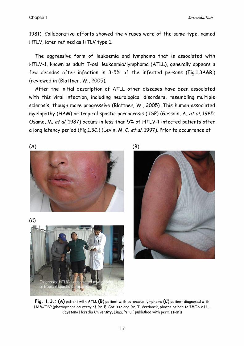
Pathogenicity of STLV
The NHP counterpart of HTLV, STLV (Fig.1.4.), has occasionally been associated with malignant lymphoma or leukaemia in macaques, baboons, African green monkeys and gorillas, though so far only for STLV type 1 infection (Miyoshi, I. et al, 1982; Homma, T. et al, 1984; Lee, R. V. et al, 1985; Blakeslee, J. R., Jr. et al, 1987; Sakakibara, I. et al, 1986; Tsujimoto, H. et al, 1987; McCarthy, T. J. et al, 1990; Voevodin, A. et al, 1996). A case of enhanced oncogenicity has been reported due to STLV-1 transmission between captive heterologous species (i.c. from rhesus macaques to baboons), while this phenomenon was not observed between homologous macaque species residing in the same centre (Voevodin, A. et al, 1996).
No cases of chronical neurological diseases comparable to HAM/TSP in humans have been described yet (Fultz, P. N., 1994).
Fig. 1.4.: Electron microscopic picture of STLV-3 viral particles obtained by cocultivation of purified
peripheral blood monocytes from healthy donors with cells from a STLV-3 infected Papio hamadryas. (photograph courtesy of Prof. Dr. Sobis and Michiels M.).
Chapter 1 Introduction
19
Epidemiology of PTLV
Transmission HTLV has not followed the explosive spread of Human Immunodeficiency
Virus (HIV), probably due to the fact that HTLV is not as transmissible as HIV. Both blood-born viruses have similar transmission routes: vertically via breastfeeding from mother-to-infant (Kinoshita, K. et al, 1984; Hino, S., 1989), horizontally via sexual contact (Kaplan, J. E. et al, 1996) or parenteral via injecting equipment. However, in contrast with HIV, HTLV is mainly infectious through cell-bound virus and not by free circulating virus. The need of cellular blood fractions explains the lower risk of HTLV-1 and -2 seropositivity after blood transfusion (Hjelle, B. et al, 1990; Sullivan, M. T. et al, 1991; Manns, A. et al, 1992). Another efficient parenteral route of HTLV transmission, intravenous drug usage, has mainly been documented for HTLV-2 (Lee, H. et al, 1989).
The risk of HTLV infection through infected breast milk has a higher incidence in endemic areas where prolonged breastfeeding is a common practice for health, hygiene, cultural and/or poverty reasons (Wiktor, S. Z. et al, 1997; Ishak, R. et al, 2001).
The HTLV seroprevalence increases with age, especially in women, due to the risk of horizontal transmission through sexual contact, with a higher risk for man-to-woman (60,8%) than woman-to-man (0.4%) transmission (Kajiyama, W. et al, 1986; Murphy, E. L. et al, 1989; Blattner, W. A. et al, 1990; Plancoulaine, S. et al, 1998).
STLV infection within the same species is mainly sustained through sexual
contact, fighting and breastfeeding. However, sexual transmission among STLV-1 infected non-human primate species in captivity has only been observed in very few cases (Fultz, P. N. et al, 1990; Georges-Courbot, M. C. et al, 1996; Niphuis, H. et al, 2003). Mother-to-child transmission is probably less efficient than in humans, based on studies showing that captive offspring of STLV-1 positive mothers were not infected (Niphuis, H. et al, 2003).

Primate T-lymphotropic Viruses
20
Fig. 1.5.: After hunting and killing a red colobus monkey (P. badius), chimpanzees consume almost all parts of their prey (photo courtesy of C. Boesch, reprinted with permission from American Society for
Microbiology, Journals Department and (Leendertz, F. H. et al, 2004a),
(A) (B)
(C)
Fig.1.6.: (A) hunter with vervet monkey (http://www.zoo.cam.ac.uk/ioz/projects/bushmeat.htm) (B)
non-human primate as house pet (http://bushmeat.net/about.html) (C) slaughter of gorilla bushmeat (http://www.solcomhouse.com/extermination.htm, courtesy of Karl Ammann)
Chapter 1 Introduction
21
Fighting between and hunting for sympatric STLV-infected species (Fig.1.5.) appears to be the main route of STLV cross-species transmission (Nerrienet, E. et al, 1998; Niphuis, H. et al, 2003; Leendertz, F. H. et al, 2004b).
The hunting and slaughter of STLV-infected NHPs by humans in Africa
(Fig.1.6A&C.) creates a potential risk for STLV cross-species transmissions to humans (Courgnaud, V. et al, 2004; Wolfe, N. D. et al, 2005), also shown for simian foamy virus (Wolfe, N. D. et al, 2004). In addition, STLV-infected monkeys that are kept as household pets (Fig.1.6B.) might be another potential source of cross-species transmission.
Epidemiology
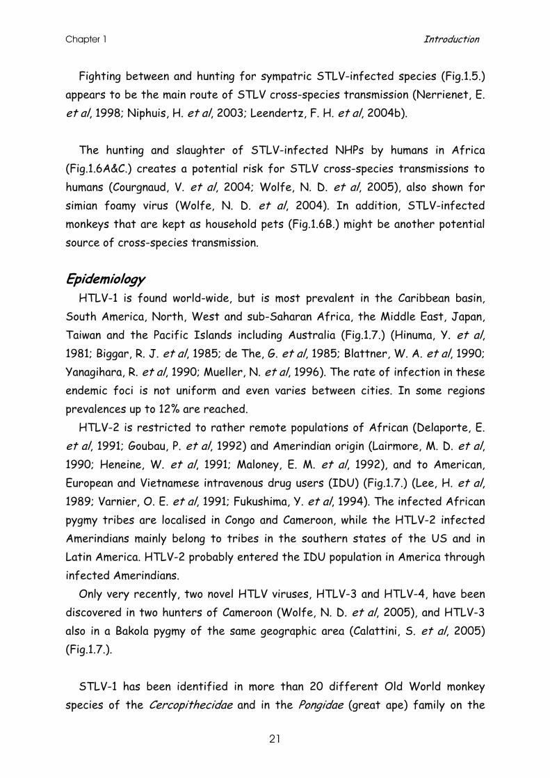
HTLV-1 is found world-wide, but is most prevalent in the Caribbean basin, South America, North, West and sub-Saharan Africa, the Middle East, Japan, Taiwan and the Pacific Islands including Australia (Fig.1.7.) (Hinuma, Y. et al, 1981; Biggar, R. J. et al, 1985; de The, G. et al, 1985; Blattner, W. A. et al, 1990; Yanagihara, R. et al, 1990; Mueller, N. et al, 1996). The rate of infection in these endemic foci is not uniform and even varies between cities. In some regions prevalences up to 12% are reached.
HTLV-2 is restricted to rather remote populations of African (Delaporte, E. et al, 1991; Goubau, P. et al, 1992) and Amerindian origin (Lairmore, M. D. et al, 1990; Heneine, W. et al, 1991; Maloney, E. M. et al, 1992), and to American, European and Vietnamese intravenous drug users (IDU) (Fig.1.7.) (Lee, H. et al, 1989; Varnier, O. E. et al, 1991; Fukushima, Y. et al, 1994). The infected African pygmy tribes are localised in Congo and Cameroon, while the HTLV-2 infected Amerindians mainly belong to tribes in the southern states of the US and in Latin America. HTLV-2 probably entered the IDU population in America through infected Amerindians.
Only very recently, two novel HTLV viruses, HTLV-3 and HTLV-4, have been discovered in two hunters of Cameroon (Wolfe, N. D. et al, 2005), and HTLV-3 also in a Bakola pygmy of the same geographic area (Calattini, S. et al, 2005) (Fig.1.7.).
STLV-1 has been identified in more than 20 different Old World monkey
species of the Cercopithecidae and in the Pongidae (great ape) family on the
Primate T-lymphotropic Viruses
22
African continent and in Southeast Asia (Fig.1.7.) (reviewed in (Slattery, J. P. et al, 1999). The prevalence of STLV-1 infection in the wild varies greatly between 0 and 80%, depending on the species examined. In general, infection rates in great apes seem to be lower (Niphuis, H. et al, 2003).
Fig.1.7.: Global epidemiology of PTLV. Normal versus large sized squares and triangles correspond to a seroprevalence of 0.3-3% and > 3% respectively. All different PTLV types can be found within Central
Africa. The only two known cases of STLV-2 infection were discovered in Congolese
Pan paniscus (bonobos) (Fig.1.7.) (Giri, A. et al, 1994; Liu, H. F. et al, 1994b; Vandamme, A. M. et al, 1996).
STLV-3 infection was first identified in 1994 in a Papio hamadryas (sacred baboon) of Eritrean origin (Goubau, P. et al, 1994). In the last four years this type of virus has been identified in other West and Central African NHP species (Fig.1.7.), a few of which will be described further into detail in chapters 7 and 8 of this manuscript (Van Dooren, S. et al, 2001; Van Dooren, S. et al, 2004; Meertens, L. et al, 2001a; Meertens, L. et al, 2003b; Meertens, L. et al, 2003a).
HTLV-1
HTLV-2 endemic
HTLV-3
HTLV-4
STLV-1
STLV-2
STLV-3
HTLV-2 IDUepidemic
Chapter 1 Introduction
23
Recently, the co-circulation of STLV-1/STLV-3 infection in the same wild living primate species, Cercocebus agilis from Cameroon, has been reported (Courgnaud, V. et al, 2004).
So far, no NHP counterpart for HTLV-4 has been discovered yet.
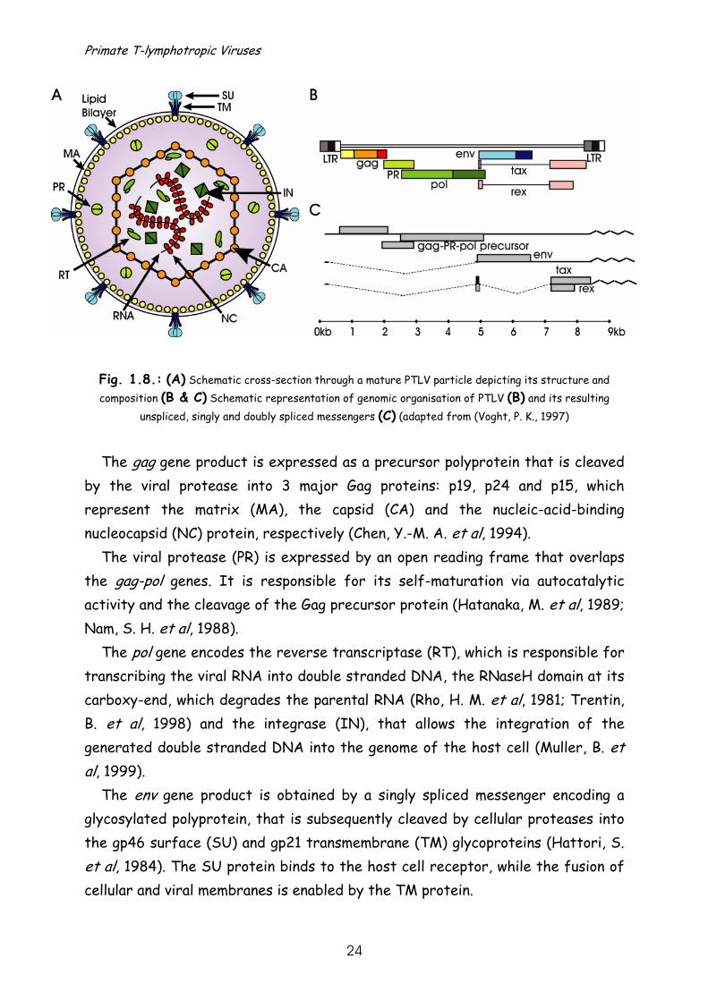
Molecular biology of PTLV Structure
PTLV is a round-shaped enveloped virus of approximately 100nm diameter (Fig.1.8A.) (Ohtouki, Y. et al, 1982). The virion is surrounded by a proteolipid envelope bilayer of host cell origin. It is equipped with transmembrane (TM) and surface (SU) viral proteins, protruding through the membrane. The inner envelope contains the matrix (MA) layer, which helps to organize the viral components at the inner cell membrane. The icosahedral capsid (CA) protects the diploid (+) single-stranded viral RNA and the functional protease (PR), reverse transcriptase (RT) and integrase (IN), which are organized into a ribonucleoprotein complex by the nucleocapsid (NC) (Poiesz, B. J. et al, 2003).
Genomic organisation
The 8-kb genome of PTLV is a positive, single-stranded RNA that encodes for structural (Gag=group antigens), functional (PR=protease and Pol=polymerase), envelope (=Env) and regulatory proteins (Tax and Rex) (Fig.1.8B.).
The genome is flanked by non-coding, short repeated sequences at both ends, termed ‘R’, and unique ‘U5’ and ‘U3’ sequences at the 5’ and 3’ ends, respectively. In the proviral form of the approximately 8.8-kbp DNA genome, these long terminal repeats (LTR), containing U3-R-U5 sequences, are present at both ends of the proviral genome (Josephs, S. F. et al, 1984). The LTRs are essential for the provirus transcription and gene expression (Kitado, H. et al, 1987).
The virus makes maximal use of this approximately 9-kbp genome by employing multiple RNA transcript splicing patterns, and differential and shifting start sites for protein translation (Fig.1.8B&C.) (Poiesz, B. J. et al, 2003). The primary full-length messenger RNA encodes a large gag-PR-pol precursor obtained by two ‘-1’ ribosomal frame shifts (Nam, S. H. et al, 1993).
Primate T-lymphotropic Viruses
24
Fig. 1.8.: (A) Schematic cross-section through a mature PTLV particle depicting its structure and composition (B & C) Schematic representation of genomic organisation of PTLV (B) and its resulting
unspliced, singly and doubly spliced messengers (C) (adapted from (Voght, P. K., 1997)
The gag gene product is expressed as a precursor polyprotein that is cleaved
by the viral protease into 3 major Gag proteins: p19, p24 and p15, which represent the matrix (MA), the capsid (CA) and the nucleic-acid-binding nucleocapsid (NC) protein, respectively (Chen, Y.-M. A. et al, 1994).
The viral protease (PR) is expressed by an open reading frame that overlaps the gag-pol genes. It is responsible for its self-maturation via autocatalytic activity and the cleavage of the Gag precursor protein (Hatanaka, M. et al, 1989; Nam, S. H. et al, 1988).
The pol gene encodes the reverse transcriptase (RT), which is responsible for transcribing the viral RNA into double stranded DNA, the RNaseH domain at its carboxy-end, which degrades the parental RNA (Rho, H. M. et al, 1981; Trentin, B. et al, 1998) and the integrase (IN), that allows the integration of the generated double stranded DNA into the genome of the host cell (Muller, B. et al, 1999).
The env gene product is obtained by a singly spliced messenger encoding a glycosylated polyprotein, that is subsequently cleaved by cellular proteases into the gp46 surface (SU) and gp21 transmembrane (TM) glycoproteins (Hattori, S. et al, 1984). The SU protein binds to the host cell receptor, while the fusion of cellular and viral membranes is enabled by the TM protein.
Chapter 1 Introduction
25
An additional coding region at the 3’ end (pX region) contains 2 regulatory genes designated tax and rex for transactivator and regulator of expression. Tax transactivates transcription initiation from the promotor in the 5’LTR (Cann, A. J. et al, 1985; Sodroski, J. et al, 1985) and it interacts with a number of transcriptional factors (Chen, Y.-M. A. et al, 1994). The Rex protein acts at post-transcriptional levels and regulates viral gene expression (Hidaka, M. et al, 1988; Seiki, M. et al, 1988; Nosaka, T. et al, 1989).
Within the proximal pX region additional mRNAs can be produced by alternative splicing, encoding proteins like p12I, p13II and p30II (Ciminale, V. et al, 1992; Ciminale, V. et al, 1995). p12I is believed to play a role in the viral replication and T-cell activation, while p30II seems to be a modulator of transcription and p13II targets mitochondria (Albrecht, B. et al, 2002).
Recently a novel viral transcription factor encoded by the complementary strand of the HTLV-I RNA genome located between the env and tax/rex genes has been characterized. The minus-strand gene protein, designated HBZ (for HTLV-1 bZIP factor), down-regulates viral transcription (Gaudray, G. et al, 2002).
Life cycle
The first step in the PTLV life cycle is the binding of the virion to a specific cell (Fig.1.9.). Although HTLV-1 and HTLV-2 can infect different haematopoietic, epithelial and mesenchymal cell types in vitro, in vivo analysis indicates a preferential tropism for CD4+ and CD8+ T-lymphocytes (Yoshida, M., 1994). Viral attachment to the host cell surface is mediated through a specific interaction between its envelope glycoproteins and a specific cell surface receptor. Recently, the ubiquitous glucose-transporter protein GLUT-1, has been identified as the receptor for HTLV-1 and HTLV-2 (Manel, N. et al, 2003), fulfilling predictions from previous studies (Sommerfelt, M. A. et al, 1990). However, cell-free HTLV is poorly infectious and is thought to spread primarily by cell-to-cell fusion (Fig1.9. and Fig. 1.10.).
After fusion of the envelope SU protein to the cell membrane and penetration of the virus into the cell, the virus will be uncoated. The viral core is delivered in the cytoplasm, where the viral RNA is reverse transcribed by RT using tRNApro as primer. This first DNA strand synthesis is initiated when still packaged in the

Primate T-lymphotropic Viruses
26
virion core, the parental RNA is then removed by RNaseH activity of the RT and the complementary DNA strand is synthesized. The linear double-stranded DNA copy, flanked by the LTRs, migrates into the nucleus and integrates randomly into host chromosomal DNA by the viral integrase. Once integrated, the provirus uses the cell machinery to transcribe primary genomic RNA. Part of these synthesized viral mRNAs is processed into singly- and doubly-spliced subgenomic transcripts. After being transported to the cytoplasm, the processed mRNA is translated into the corresponding viral proteins. Other RNA copies become full-length progeny virion RNA.
Fig.1.9.: Life cycle of PTLV. Infection of bystander cell either by binding and fusion of mature virion or
by cell-cell contact with PTLV infected cell (adapted from (Rambaut, A. et al, 2004).
Finally, the virion core is assembled at the plasma membrane and progeny
virus is released (Chen, Y.-M. A et al, 1994; Coffin, J. M., 1996). This may happen either by a process of budding and subsequent maturation into free circulating
Chapter 1 Introduction
27
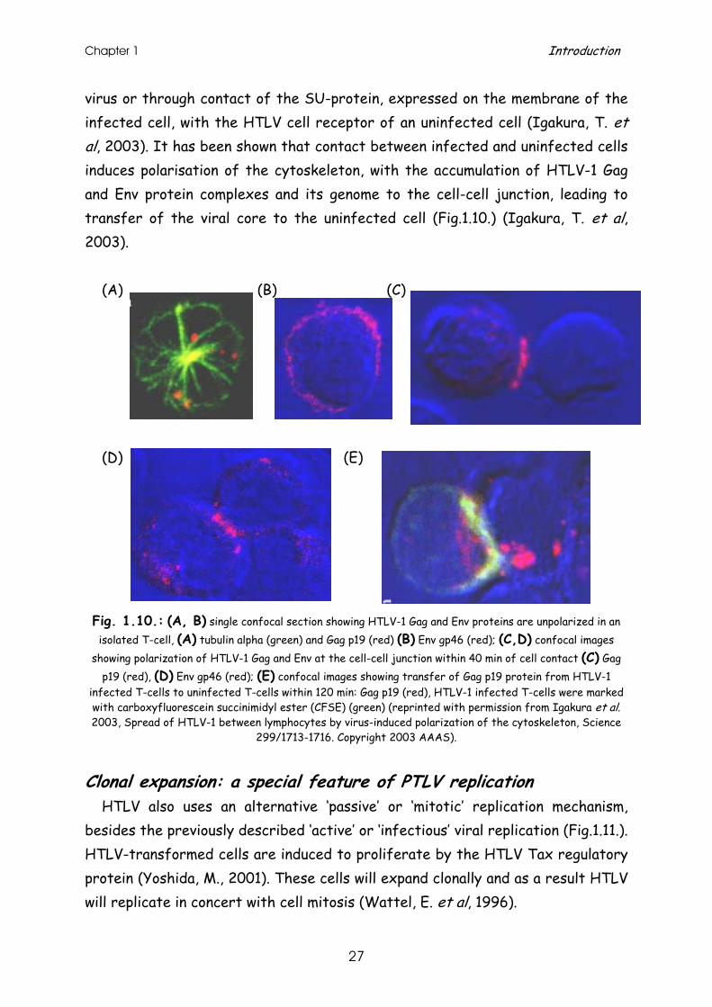
virus or through contact of the SU-protein, expressed on the membrane of the infected cell, with the HTLV cell receptor of an uninfected cell (Igakura, T. et al, 2003). It has been shown that contact between infected and uninfected cells induces polarisation of the cytoskeleton, with the accumulation of HTLV-1 Gag and Env protein complexes and its genome to the cell-cell junction, leading to transfer of the viral core to the uninfected cell (Fig.1.10.) (Igakura, T. et al, 2003).
(A) (B) (C) (D) (E)
Fig. 1.10.: (A, B) single confocal section showing HTLV-1 Gag and Env proteins are unpolarized in an isolated T-cell, (A) tubulin alpha (green) and Gag p19 (red) (B) Env gp46 (red); (C,D) confocal images
showing polarization of HTLV-1 Gag and Env at the cell-cell junction within 40 min of cell contact (C) Gag p19 (red), (D) Env gp46 (red); (E) confocal images showing transfer of Gag p19 protein from HTLV-1
infected T-cells to uninfected T-cells within 120 min: Gag p19 (red), HTLV-1 infected T-cells were marked with carboxyfluorescein succinimidyl ester (CFSE) (green) (reprinted with permission from Igakura et al. 2003, Spread of HTLV-1 between lymphocytes by virus-induced polarization of the cytoskeleton, Science
299/1713-1716. Copyright 2003 AAAS).
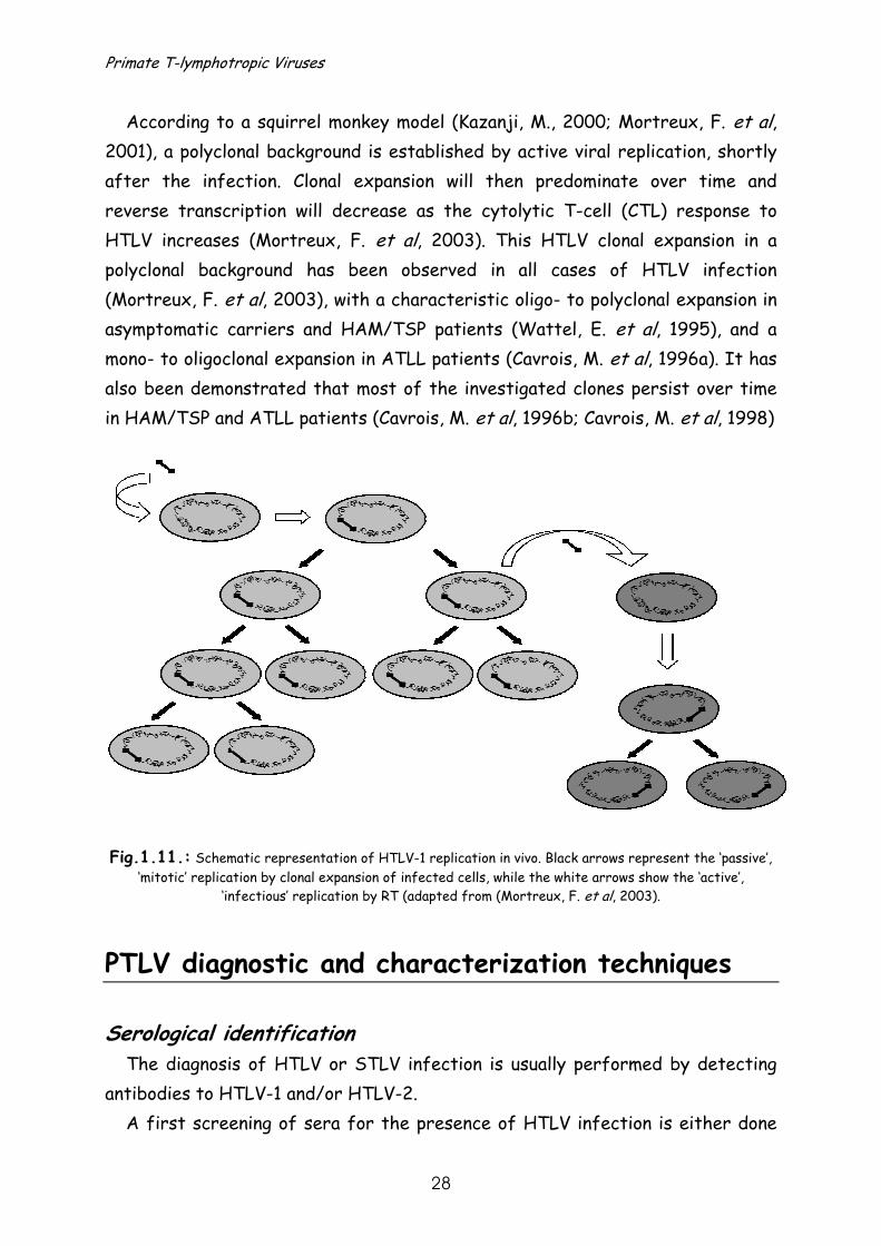
Clonal expansion: a special feature of PTLV replication
HTLV also uses an alternative ‘passive’ or ‘mitotic’ replication mechanism, besides the previously described ‘active’ or ‘infectious’ viral replication (Fig.1.11.). HTLV-transformed cells are induced to proliferate by the HTLV Tax regulatory protein (Yoshida, M., 2001). These cells will expand clonally and as a result HTLV will replicate in concert with cell mitosis (Wattel, E. et al, 1996).
Primate T-lymphotropic Viruses
28
According to a squirrel monkey model (Kazanji, M., 2000; Mortreux, F. et al, 2001), a polyclonal background is established by active viral replication, shortly after the infection. Clonal expansion will then predominate over time and reverse transcription will decrease as the cytolytic T-cell (CTL) response to HTLV increases (Mortreux, F. et al, 2003). This HTLV clonal expansion in a polyclonal background has been observed in all cases of HTLV infection (Mortreux, F. et al, 2003), with a characteristic oligo- to polyclonal expansion in asymptomatic carriers and HAM/TSP patients (Wattel, E. et al, 1995), and a mono- to oligoclonal expansion in ATLL patients (Cavrois, M. et al, 1996a). It has also been demonstrated that most of the investigated clones persist over time in HAM/TSP and ATLL patients (Cavrois, M. et al, 1996b; Cavrois, M. et al, 1998) Fig.1.11.: Schematic representation of HTLV-1 replication in vivo. Black arrows represent the ‘passive’,
‘mitotic’ replication by clonal expansion of infected cells, while the white arrows show the ‘active’, ‘infectious’ replication by RT (adapted from (Mortreux, F. et al, 2003).
PTLV diagnostic and characterization techniques
Serological identification The diagnosis of HTLV or STLV infection is usually performed by detecting
antibodies to HTLV-1 and/or HTLV-2. A first screening of sera for the presence of HTLV infection is either done
Chapter 1 Introduction
29
by an enzyme immunosorbent assay (EIA), using viral purified lysate and/or recombinant proteins or synthetic peptides, or by a particle agglutination test that consists of gelatin particles coated with viral lysate. As both tests, especially those based on viral lysate only, can give aspecific reactions and mainly contain HTLV-1 antigens or peptides, confirmatory testing is advised.
Confirmation tests often allow discrimination between HTLV-1 and -2. Indirect immunofluorescence assays (IFA), detecting antibodies cross-reacting with PTLV type specific cells (as antigens) applied on microscopic slides, have been frequently used in the past (Gallo, D. et al, 1988; Matsumoto, C. et al, 1990). Other confirmatory tests like Western blot (Wiktor, S. Z. et al, 1991) or Innolia (Goubau, P. et al, 1999) detect antibodies against HTLV-1/HTLV-2 and make use of specific and type-specific immunodominant proteins bound to a membrane or synthetic peptides sprayed in lines on a strip, respectively.
A significant minority of isolates has no typical HTLV-1 or HTLV-2 profile in Western blot or Innolia. They only react with one or a few antigens or synthetic peptides and are scored as HTLV-seroindeterminate. The majority of these reactions are aspecific and most patients are actually negative for HTLV-infection, which can be shown with additional, molecular biological based confirmatory tests on serial samples. Conversely, some HTLV-1 or HTLV-2 infected individuals remain seronegative or seroindeterminate, even after confirmatory serological testing. This might be due to several reasons, one of which is the predominant use of HTLV-1 antigens to screen for both types of infections (Poiesz, B. J. et al, 2000b). Another reason might be that PTLV seroconversion often takes up to 2 years, compared to 3-6 months for HIV (Poiesz, B. et al, 2000a).
A seroindeterminate result might also be an indication of a significant
diversity to the included antigens, especially for STLV strains and thus may suggest the presence of a divergent strain. Genetic identification and characterization
Currently, the most sensitive and specific techniques are DNA-dependent polymerase chain reactions (PCR) (Poiesz, B. J. et al, 2000b). The method amplifies specific DNA segments, i.c. a proviral genomic region, by cycles of template denaturation, primer annealing and strand extension using a

Primate T-lymphotropic Viruses
30
thermostable DNA polymerase. A diagnostic generic and type-specific PTLV PCR has been developed in our laboratory allowing detection and discrimination of PTLV types 1 to 3 (Vandamme, A. M. et al, 1997).
Molecular epidemiology and evolution of PTLV The relationship of the different PTLV strains, obtained from different
species and from diverse regions, can be investigated at the molecular level, through differences in their proviral sequences using phylogenetic inference methods. Like pedigrees, phylogenetic trees schematically represent the relation between strains but also allow us to analyse the evolutionary history. If a time scale can be superimposed on the tree, the rate of evolution of these viruses can be estimated and subsequently other events in the viral history can be dated (details see ‘molecular evolution, phylogenetic inference and molecular clocks’ p.36).
PTLV has an extra-ordinary slow rate of evolutionary change, estimated to be
1000 to 10000 times slower than the rate of other pathogenic viruses, including HIV (Suzuki, Y. et al, 1998). In vivo passage of HTLV in several epidemiologically linked patients demonstrates little to no genetic variation (Gessain, A. et al, 1992; Liu, H. F. et al, 1994a), which might be partially explained by its favoured replication through clonal expansion (Wattel, E. et al, 1996; Cavrois, M. et al, 1996a). This ‘mitotic’ replication provides HTLV with the feature to evolve in concert with cellular genes, and presumably in concert with its host. Indeed, the dissemination of PTLV seems to coincide with human and NHP migrations over the world. Thus anthropological documented migrations of (PTLV-infected) hosts can be used to trace back the origin and dissemination of the virus and viral types, and vice versa (Gessain, A. et al, 1992; Miura, T. et al, 1994).
Based on the common occurrence of all PTLV types on the African continent,
(HTLV types 1 to 4 and STLV types 1 to 3), an African origin for the common ancestor of all PTLVs has been proposed (Vandamme, A. M. et al, 1998a; Salemi, M. et al, 1999). Clearly, Central Africa seems to be the centre of PTLV infection
Chapter 1 Introduction
31
and the home of a plethora of PTLV strains, as has been reported before for SIV (Peeters, M. et al, 2002). All different PTLV types have been discovered there in a large variety of primate species. Salemi et al. (Salemi, M. et al, 2000) estimated the PTLV time of origin, using PTLV-1, PTLV-2, STLV-3 strains and BLV as outgroup, to be about 0.8 to 1.3 million years ago.
Molecular epidemiology and evolution of PTLV-1
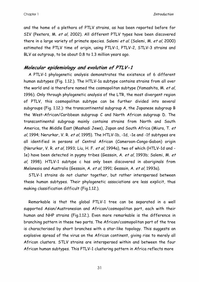
A PTLV-1 phylogenetic analysis demonstrates the existence of 6 different human subtypes (Fig. 1.12.). The HTLV-1a subtype contains strains from all over the world and is therefore named the cosmopolitan subtype (Yamashita, M. et al, 1996). Only through phylogenetic analysis of the LTR, the most divergent region of PTLV, this cosmopolitan subtype can be further divided into several subgroups (Fig. 1.12.): the transcontinental subgroup A, the Japanese subgroup B the West-African/Caribbean subgroup C and North African subgroup D. The transcontinental subgroup mainly contains strains from North and South America, the Middle East (Mashadi Jews), Japan and South Africa (Miura, T. et al, 1994; Nerurkar, V. R. et al, 1995). The HTLV-1b, -1d, -1e and -1f subtypes are all identified in persons of Central African (Cameroon-Congo-Gabon) origin (Nerurkar, V. R. et al, 1993; Liu, H. F. et al, 1994a), two of which (HTLV-1d and -1e) have been detected in pygmy tribes (Gessain, A. et al, 1993b; Salemi, M. et al, 1998). HTLV-1 subtype c has only been discovered in aboriginals from Melanesia and Australia (Gessain, A. et al, 1991; Gessain, A. et al, 1993a).
STLV-1 strains do not cluster together, but rather interspersed between these human subtypes. Their phylogenetic associations are less explicit, thus making classification difficult (Fig.1.12.).
Remarkable is that the global PTLV-1 tree can be separated in a well
supported Asian/Austronesian and African/cosmopolitan part, each with their human and NHP strains (Fig.1.12.). Even more remarkable is the difference in branching pattern in these two parts. The African/cosmopolitan part of the tree is characterised by short branches with a star-like topology. This suggests an explosive spread of the virus on the African continent, giving rise to merely all African clusters. STLV strains are interspersed within and between the four African human subtypes. This PTLV-1 clustering pattern in Africa reflects more
Primate T-lymphotropic Viruses
32
Fig.1.12.: Puzzle maximum likelihood tree of the LTR region of 79 representative PTLV-1 strains. The STLV-1 host species are represented by a symbol after the viral strain name. Strains with no symbols
are HTLV-1 strains. The values on the left side of the nodes represent puzzling support values (Strimmer, K. et al, 1996). The clear separation between Asian/Austronesian and African/cosmopolitan
strains is demonstrated. The cosmopolitan HTLV-1a subtype and Melanesian HTLV-1c subtype are indicated together with the Central African HTLV-1b, -1d, -1e and -1f subtype clades, interspersed with STLV-1
strains.
the common geographic origin of viral strains than a common host species origin.
Chapter 1 Introduction
33
This suggests that interspecies transmissions of the virus occurred frequently in Africa (Vandamme, A. M. et al, 1998a). The only exception to this observation is HTLV-1 subtype a, which is also the only subtype that is found world-wide. Its dissemination to the New World and Japan was originally thought to be ancient, supported by the detection of HTLV-1 infection in the Japanese Ainu and Ryukyans, both considered to be direct descendants of old Mongoloid populations (Ishida, T. et al, 1985). The same subtype was believed to have arisen in the New World, like HTLV-2 (see below), by spread through mongoloid migrations via Beringia in the Palaeolithic era (Miura, T. et al, 1994). However, HTLV-1a might have been introduced in these places much more recent as a result of Portuguese navigation adventures and African slave trade (Gessain, A. et al, 1992; Gessain, A. et al, 1994a; Song, K. J. et al, 1994; Yanagihara, R. et al, 1995; Van Dooren, S. et al, 1998). The increased human mobility in the last century might have been responsible for the further world-wide dissemination. The only human strains in the Asian/Austronesian part of the tree belong to the HTLV-1 subtype c, identified in Australian and Melanesian aboriginals (Gessain, A. et al, 1991; Nerurkar, V. R. et al, 1993; Song, K. J. et al, 1994). As no NHPs or fossils of NHPs have been found in this geographic area, the divergent character between Austronesian human and Asian/Indonesian STLV strains can be explained by geographic separation. However, this divergent character has also been observed between the STLV strains, discovered in different macaque species (Watanabe, T. et al, 1985; Koralnik, I. J. et al, 1994; Voevodin, A. et al, 1994; Ibrahim, F. et al, 1995; Mahieux, R. et al, 1997; Richards, A. L. et al, 1998) and in orang-utans (Ibuki, K. et al, 1997; Verschoor, E. J. et al, 1998). Their deeply branching monophyletic clades suggest that these viral strains have undergone a long independent evolution in their host species (Ibrahim, F. et al, 1995). It has been suggested that HTLV-1c arose through the first settlers of Melanesia and Australia who probably acquired the virus from STLV-1 infected NHPs along their migratory pathway (Gessain, A. et al, 1993a; Yanagihara, R. et al, 1995).
Intriguingly, when the global bifurcated Asian/Austronesian and African/cosmopolitan PTLV-1 tree is rooted, the root lies within the Asian part of the tree, among STLV-1 strains of Asian macaques. This observation does not fit with the general accepted hypothesis that the ancestor of all PTLVs and all PTLV types arose in Africa.
Primate T-lymphotropic Viruses
34
Molecular epidemiology and evolution of PTLV-2
HTLV-2 was originally thought to be a New World virus, based on its primary discovery in American IDUs and remote Amerindian populations (Heneine, W. et al, 1991; Maloney, E. M. et al, 1992; Hjelle, B. et al, 1994). Restriction enzyme and phylogenetic analyses showed that HTLV-2 strains could be subdivided into two main subtypes, HTLV-2a and -2b (Fig.1.13.) (Hall, W. W. et al, 1992; Hjelle, B. et al, 1993). Some Brazilian isolates that are phenotypically closely related to HTLV-2b, but phylogenetically belong to HTLV-2a, were designated HTLV-2c (Ishak, R. et al, 2001). The discovery of HTLV-2 on the African continent in Gabon and in Cameroonian and Congolese pygmy tribes gave rise to the hypothesis of an ancient, African origin of HTLV-2 (Delaporte, E. et al, 1991; Goubau, P. et al, 1992; Gessain, A. et al, 1994b; Gessain, A. et al, 1995; Goubau, P. et al, 1996). This hypothesis was supported by the identification of a divergent HTLV-2 strain in Bambuti pygmies living in a remote area of the Ituri forest in Congo, designated HTLV-2d (Goubau, P. et al, 1996; Vandamme, A. M. et al, 1998b). The fact that the virus, besides their occurrence in IDUs, is restricted to indigenous populations of African and Amerindian origin suggests that HTLV-2 has been introduced into the New World by an ancient Mongoloid migration over the Bering Strait. However, this hypothesis can not explain the striking similarity between HTLV-2 strains in Bakola pygmies of Cameroon and HTLV-2b in Colombian Wayuu Amerindians (Gessain, A. et al, 1995). It has been suggested that this might be due to an undocumented, more recent, and probably indirect contact between Amerindians and African pygmies. The two independent discoveries of the NHP variant of this virus, originally named STLV-PP, only identified in pygmy chimpanzees (Pan paniscus) (Giri, A. et al, 1994; Liu, H. F. et al, 1994b; Vandamme, A. M. et al, 1996), living in Africa and nowadays restricted to Congo, again strengthened the hypothesis of an ancient African origin of PTLV-2. Although HTLV-2 and STLV-PP are phylogenetically much more distinct than HTLV-1 and STLV-1 strains, they seem to share a common ancestor (Fig.1.13.). STLV-PP has therefore been renamed STLV-2. Molecular epidemiology and evolution of PTLV-3
STLV-3 (PH969) was originally discovered in 1994 in an Eritrean sacred baboon (Papio hamadryas), and was clearly divergent from PTLV-1 and PTLV-2
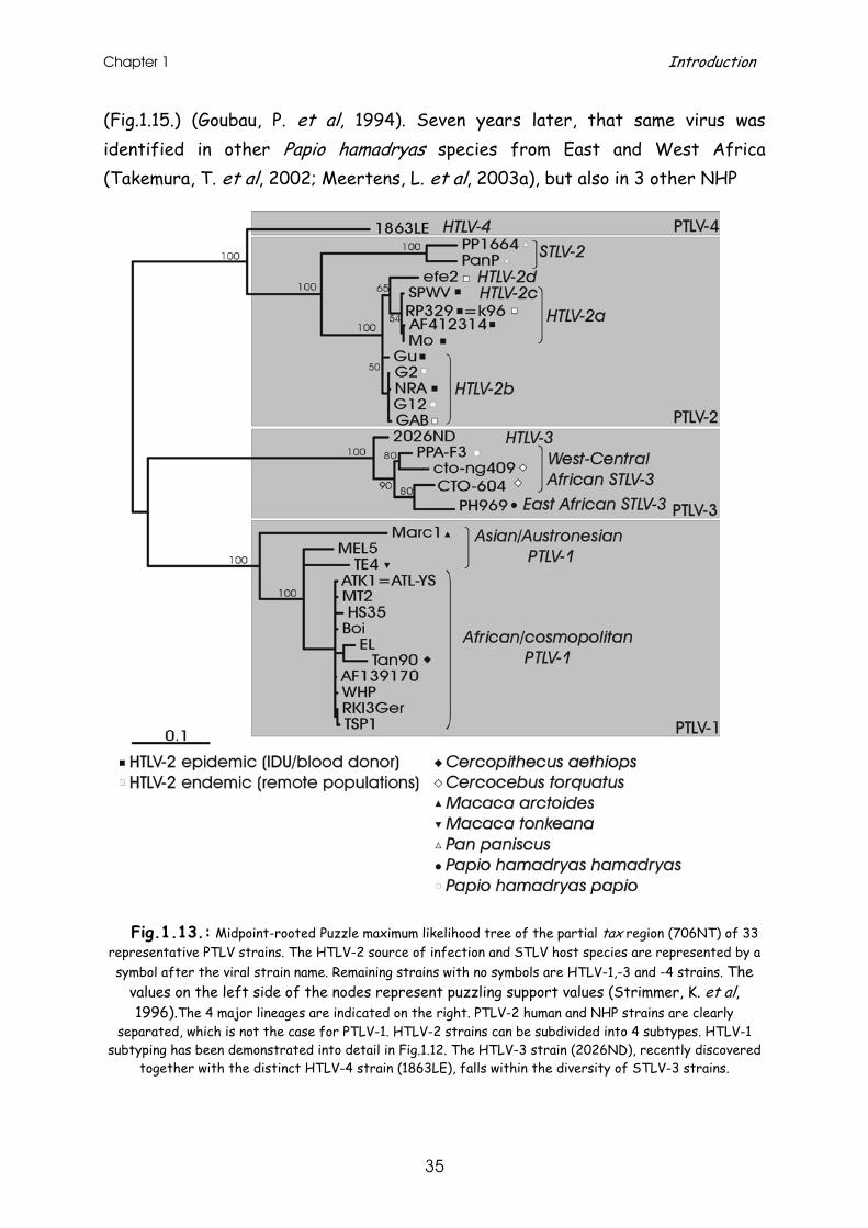
Chapter 1 Introduction
35
(Fig.1.15.) (Goubau, P. et al, 1994). Seven years later, that same virus was identified in other Papio hamadryas species from East and West Africa (Takemura, T. et al, 2002; Meertens, L. et al, 2003a), but also in 3 other NHP
Fig.1.13.: Midpoint-rooted Puzzle maximum likelihood tree of the partial tax region (706NT) of 33
representative PTLV strains. The HTLV-2 source of infection and STLV host species are represented by a symbol after the viral strain name. Remaining strains with no symbols are HTLV-1,-3 and -4 strains. The
values on the left side of the nodes represent puzzling support values (Strimmer, K. et al, 1996).The 4 major lineages are indicated on the right. PTLV-2 human and NHP strains are clearly
separated, which is not the case for PTLV-1. HTLV-2 strains can be subdivided into 4 subtypes. HTLV-1 subtyping has been demonstrated into detail in Fig.1.12. The HTLV-3 strain (2026ND), recently discovered
together with the distinct HTLV-4 strain (1863LE), falls within the diversity of STLV-3 strains.
Primate T-lymphotropic Viruses
36
species (Meertens, L. et al, 2001a; Meertens, L. et al, 2003b), 2 of which will be discussed into detail in chapter 7 and 8 (Van Dooren, S. et al, 2001; Van Dooren, S. et al, 2004).
The human variant, HTLV-3, has only very recently been discovered in a Cameroonian bushmeat hunter (Wolfe, N. D. et al, 2005) and Bakola pygmy (Calattini, S. et al, 2005), and seem to fall within the diversity of STLV-3 strains. Molecular epidemiology and evolution of HTLV-4
Together with HTLV-3, a novel viral strain, HTLV-4, was recently also identified in a Cameroonian bushmeat hunter. This virus is distinct from all known HTLV and STLV types, supported by the distinct clustering as a new phylogenetic lineage with high bootstrap support (Wolfe, N. D. et al, 2005).
Molecular evolution, phylogenetic inference and molecular clocks Concept of molecular evolution
Differences between organisms are merely the result of evolution. Biological evolution is a process of change in the properties of populations of organisms. Some of these changes can be inferred morphologically, but they all result from changes at the genetic level. Evolutionary changes in populations are inheritable via the genetic material, the DNA or RNA of an organism, from one generation to the next. Together with proteins and RNA this genetic information constitutes, through their interaction with the environment, the phenotype of a living organism. Since RNA is transcribed from DNA and proteins are translated from RNA, all necessary information for reconstitution is embedded in DNA sequences. Thus the genetic information within organisms is the "document" of their evolutionary history. Comparisons of the DNA sequences of various genes between different organisms can tell us a lot about their relationships and molecular evolution.
Chapter 1 Introduction
37
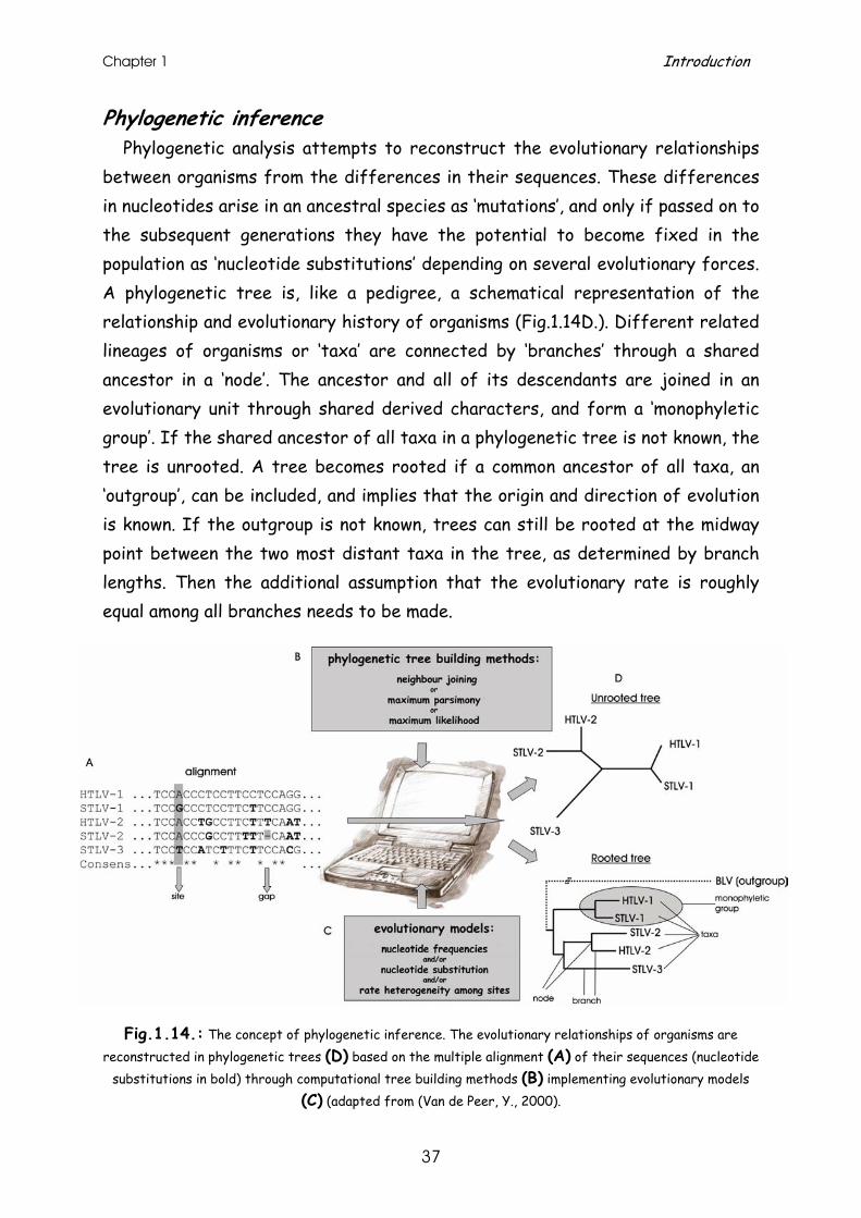
Phylogenetic inference Phylogenetic analysis attempts to reconstruct the evolutionary relationships
between organisms from the differences in their sequences. These differences in nucleotides arise in an ancestral species as ‘mutations’, and only if passed on to the subsequent generations they have the potential to become fixed in the population as ‘nucleotide substitutions’ depending on several evolutionary forces. A phylogenetic tree is, like a pedigree, a schematical representation of the relationship and evolutionary history of organisms (Fig.1.14D.). Different related lineages of organisms or ‘taxa’ are connected by ‘branches’ through a shared ancestor in a ‘node’. The ancestor and all of its descendants are joined in an evolutionary unit through shared derived characters, and form a ‘monophyletic group’. If the shared ancestor of all taxa in a phylogenetic tree is not known, the tree is unrooted. A tree becomes rooted if a common ancestor of all taxa, an ‘outgroup’, can be included, and implies that the origin and direction of evolution is known. If the outgroup is not known, trees can still be rooted at the midway point between the two most distant taxa in the tree, as determined by branch lengths. Then the additional assumption that the evolutionary rate is roughly equal among all branches needs to be made.
Fig.1.14.: The concept of phylogenetic inference. The evolutionary relationships of organisms are
reconstructed in phylogenetic trees (D) based on the multiple alignment (A) of their sequences (nucleotide substitutions in bold) through computational tree building methods (B) implementing evolutionary models
(C) (adapted from (Van de Peer, Y., 2000).
Primate T-lymphotropic Viruses
38
The first, crucial step in the construction of a phylogenetic tree, is the
alignment of the different sequences (Fig.1.14A.). Each of the sequences will be compared with each other and will be searched for homologous residues. The more divergent the investigated lineages are, the less similarity will be found in their sequences. Alignments will be generated by arranging the homologous residues in columns as much as possible and by correcting for differences in sequence length and for insertions and deletions by means of gaps (hyphens). Positions, also known as ‘sites’, or stretches that can not be aligned unambiguously are better removed from the alignment, as this reduces the quality of the global alignment and finally could result in an incorrect inference of the phylogenetic tree.
Phylogenetic tree building or inference methods (Fig.1.14B.) are computational
methods aimed at discovering which of the possible trees is according to a particular evolutionary model the most correct or optimal tree. When the assumptions of the model do not hold, this optimal tree can be different from the true biological tree, which accurately represents the evolutionary history of all taxa. All of the mathematical and/or statistical methods for inferring the branching pattern of taxa, as well as the branch lengths connecting them, can be classified based on the data types and the cluster algorithm used. Distance methods transform and reduce the sequence data into pairwise distances, a measure for dissimilarity, and then use them in a matrix during tree building. Character-based methods use the aligned characters, such as DNA or amino acid sequences, directly during tree inference.
Clustering methods use pairwise distances and cluster the different taxa stepwise resulting in only one ‘best’ tree. The most commonly used distance method using a sequential clustering procedure is ‘neighbour-joining’. The robustness of the obtained singular tree can be evaluated with a bootstrap analysis, in which the tree topologies of newly ad random generated alignments (from the original alignment) are compared.
Optimality approaches first define an optimality criterion (fewest number of events or highest likelihood) and then use a specific algorithm for finding trees with the best value for the objective function. Exact algorithms, like exhaustive and branch-and-bound searches ‘guarantee’ to find the optimal tree, while
Chapter 1 Introduction
39
heuristic algorithms are approximate methods that ‘attempt’ to find the optimal tree. ‘Maximum parsimony’ uses as optimality criterion that the ‘most-parsimonious’ tree is the one that requires the fewest number of evolutionary steps to explain all different mutations in the sequences. The ‘maximum likelihood’ algorithm calculates the probability for each sequence position of expecting each possible nucleotide or amino acid in the ancestral nodes and infers the likelihood of the tree structure for this probability. The likelihood of all reasonable tree topologies is searched and compared, and the tree with the highest likelihood is considered to be the preferred tree.
Modelling genetic change
All of these different methods use models of evolution (Fig.1.14C.), which are needed to perceive patterns in the stochastic and random processes of evolution and to generate, via a (statistical) description, synthetic data with properties that mimic those of real data. An important prerequisite for computing branch lengths is the prior specification of a model of nucleotide substitution. These models for DNA sequence evolution are nested, and either do or do not take unequal nucleotide frequencies and/or unequal probabilities of nucleotide substitutions into account. The rate at which nucleotide substitutions occur can vary substantially for different positions in a sequence. The models correcting for site-to-site rate variation account for invariable sites and/or rate heterogeneity over sites, modelled by a gamma distribution, further shaped by the α-parameter. Molecular clocks
Sequence variation is the result of accumulating nucleotide substitutions in the genome of an organism over time. The speed at which these accumulate determines the evolutionary rate. When nucleotide substitutions accumulate more or less at a constant rate in different lineages, the genes follow a molecular clock. However, lineages do not necessarily have a constant evolutionary rate. Different selective pressures and population bottlenecks can lead to dissimilar rates of evolution. If the molecular clock holds for a particular set of sequences and their outgroup is known, then measuring the evolutionary rate allows us to date coalescence and divergence times. Conversely, if the divergence time between 2 species is known, the rate of evolution of a gene can
Primate T-lymphotropic Viruses
40
be inferred and other nodes of the tree can be dated. If the molecular clock does not hold, such estimations are only approximate. A strict molecular clock implies that all lineages evolve at a constant rate during evolution and is known today to be untrue for most, if not all organisms. However, local molecular clocks do exist for some closely related species (e.g. chimp mtDNA and primate retroviruses).
The existence of a molecular clock can be tested with a mathematical and statistical technique, called a likelihood ratio test using the maximum likelihood algorithm. In this test the likelihood of the general hypothesis, assuming that the lineages do not evolve clock-like will be compared to the likelihood of the nested hypothesis i.c. clock restriction, implying that all branch lengths in a tree from root to tip are equal (= null hypothesis). If the likelihood of the clock-like tree is not significantly different from the non-clock like tree, a molecular clock can be assumed. This is tested by comparing the double of the difference in log likelihood of the non-clock-like and clock-like tree to the chi square value of the 95% confidence intervals for n-2 degrees of freedom, where n is the number of strains: if 2(lnLNCL-lnLCL)< χ2 (0.95) for n-2 degrees of freedom, the molecular clock cannot be rejected.
Specialised literature on molecular evolution, phylogenetic inference and
molecular clocks can be retrieved from (Page, R. D. et al, 1998; Salemi, M. et al, 2003; Semple, C. et al, 2003; Felsenstein, J., 2004).
Scope of the thesis The research area of the studies described in this thesis can be subdivided
into three parts. First, we focussed on the remarkable slow rate of evolution of PTLV-1. This
virus seems to evolve and migrate partially in concert with its host, either human or NHP. In studies that preceded our research, several hypotheses on the dissemination of the virus on different continents had been launched by imposing anthropologically documented host migrations on the phylogeny of the

Chapter 1 Introduction
41
virus. However, few attempts had been made to investigate this by means of molecular clock analysis. In chapter 2 and 3 we have tried to elucidate the introduction of HTLV-1 in the New World and to date the origin of the different HTLV-1 subtypes in Africa, respectively. Unfortunately, these evolutionary rate estimates and inferred dates rely on the underlying assumption that the anthropological dates used to calibrate the molecular clock are correct. In a third study, described in chapter 4, we estimated the HTLV-1 evolutionary rate independent of anthropological findings, using HTLV-1 vertical transmission chains and compared them with previously obtained rates.
In the second part of this thesis we aimed to shed light on the evolutionary
patterns of PTLV-1 in Asia. The goal of the study in chapter 5 was to gain insight in the PTLV-1 evolution in Asia by the full genome sequencing and analysis of the currently most divergent STLV-1 in Macaca arctoides. In chapter 6 we analysed the Asian PTLV-1 phylogeny further with the inclusion of various novel Asian STLV-1 strains. We also attempted to resolve the ambiguous (African or Asian?) phylogeographical origin of PTLV-1 by phylogenetic analysis of STLV-1 strains discovered in Macaca sylvanus, the only macaque species that remained on the African continent.
Finally, we explored the diversity of STLV type 3 strains. Until 2001 only one
STLV-3 strain was known, originally discovered by our research group in 1994 in an Eritrean Papio hamadryas. In chapter 7 we describe the discovery and the partial tax phylogenetic analysis of a second STLV-3 strain in Cercopithecus nictitans from Cameroon. Shortly after, several other viral strains, belonging to the same type, were described by different groups. In the final study presented in this thesis in chapter 8, we identified STLV-3 infection in Theropithecus gelada. By means of full genome sequencing, phylogenetic and molecular clock analysis we investigated the diversity among all known STLV-3 strains and we estimated the origin of STLV-3.

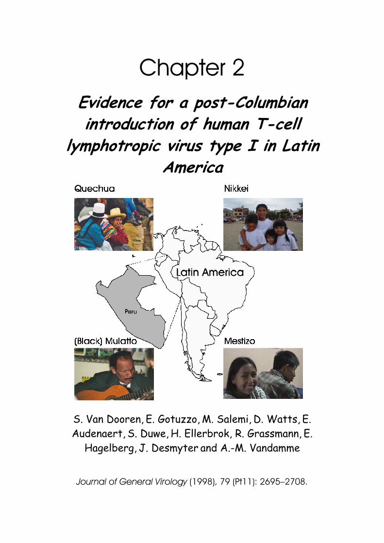
Chapter 2 Evidence for a post-Columbian introduction of human T-cell
lymphotropic virus type I in Latin America
S. Van Dooren, E. Gotuzzo,
M. Salemi,
D. Watts,
E.
Audenaert, S. Duwe,
H. Ellerbrok, R. Grassmann,
E.
Hagelberg, J. Desmyter
and A.-M. Vandamme
Journal of General Virology (1998), 79 (Pt11): 2695–2708.

Photographs previous page Quechua/(Black)Mulatto/Mestizo: courtesy of Dr.T. Verdonck Nikkei: http://www.prensanikkei.com/fotosaludos/index.html
45
Evidence for a post-Columbian introduction of human T-cell lymphotropic virus type I in Latin America
S. Van Dooren,
1 E. Gotuzzo,
2 M. Salemi,
1 D. Watts,
3 E. Audenaert,
1 S. Duwe,
4 H.
Ellerbrok,4
R. Grassmann,5
E. Hagelberg,6
J. Desmyter1 and A.-M. Vandamme
1
1 Rega Institute for Medical Research, KU Leuven, Minderbroedersstraat 10, B-3000 Leuven, Belgium 2Instituto de medicina tropical ‘ Alexander Von Humboldt ’, Universidad Peruana Cayetano Heredia, Lima, Peru 3
U.S. Naval Medical Research Institute Detachment, Lima, Peru 4
Robert Koch Institute, Berlin, Germany 5 Institute of Virology, University of Erlangen-Nürnberg, Germany 6 Department of Genetics, University of Cambridge, UK
Journal of General Virology (1998), 79 (Pt11): 2695–2708.
To investigate the origin and dissemination of human T-cell lymphotropic virus type I in Latin America, we performed phylogenetic analysis on the LTR and env sequences of 13 HTLV-I isolates from Peruvians of four different ethnic groups: blacks and some mulattos of African origin; Quechuas of Inca origin; Nikkei of Japanese descendance; and Mestizos, a mixed population of white and Indian origin. All Peruvian samples could be situated within the cosmopolitan subtype HTLV-Ia, yet one sample showed an indeterminate Western blot pattern, lacking reactivity towards the HTLV-I type specific MTA1 peptide. Within the LTR, we could confirm the previously reported subdivision into four subgroups – one big transcontinental clade A, a Japanese clade B, a West African/Caribbean clade C and a North African clade D – and we identified a new separate subgroup E of black Peruvian strains. The clustering of the Peruvian samples seemed to depend on the ethnic origin of the host. The largest heterogeneity was observed in the black Peruvian samples. The mitochondrial DNA type of one of these black Peruvian strains of subgroup E was identical to that of West African source populations of the slave trade. Both findings support the idea of multiple post-Columbian introductions of African HTLV-Ia strains into the black Latin American population. Additionally, a tight cluster of Nikkei and Japanese samples implied a separate and rather recent transmission of a Japanese lineage of HTLV-I into Peru. A well-supported cluster of Latin American strains (including Peruvian Quechuas and Colombian Amerindians) could be situated within the transcontinental group. Molecular clock analysis of the Latin American and Japanese clade resulted in an equal evolutionary rate for those strains. Along with the anthropologically documented peopling of the Americas, the analysis was more in favour of a recent (400 to 100 years ago) introduction of HTLV-Ia into the American continent rather than a Palaeolithic introduction.
J.Gen.Vir. (1998) 79: 2695-2708
46
Introduction Human T-cell lymphotropic virus type I (HTLV-I) is regarded as the
aetiological agent of adult T-cell leukaemia (ATL) (Yoshida et al., 1982) and the neurological disorder ‘tropical spastic paraparesis’ or ‘HTLV-I associated myelo-pathy’ (TSP/HAM) (Gessain et al., 1985; Osame et al., 1987). HTLV-I is distributed worldwide, but is endemic in Central and West Africa, the Caribbean basin, parts of Latin America, Japan and Melanesia/Australia. A second type of HTLV (HTLV-II), initially identified in a patient with atypical hairy cell leukaemia (Kalyanaraman et al., 1982), has recently been associated with chronic myelopathy (Murphy et al., 1997). HTLV-II infection has been reported in a number of native Amerindians (Maloney et al., 1992; Hjelle et al., 1993), in African pygmies (Delaporte et al., 1991;Goubau et al., 1992, 1993a, b; Gessain et al., 1995; Tuppin et al., 1996) and also among injecting drug users in the United States and Europe (Lee et al., 1989; Zella et al., 1990). Two other divergent types of simian T-cell lymphotropic virus have been described: STLV-L, isolated from an Ethiopian Papio hamadryas (Goubau et al., 1994; Van Brussel et al., 1996), and STLV-II, isolated from a Pan paniscus from the Democratic Republic of Congo (Vandamme et al., 1996). Since the biological and molecular aspects of HTLV and STLV are very similar, both types of viruses are conveniently catalogued into one group of primate T-cell lymphotropic viruses (PTLV).
HTLV is transmitted in a cell-associated manner, mainly by vertical infection
from mother to child but also by limited horizontal transmission via sexual intercourse, blood transfusion or intravenous drug abuse (Tachibana et al., 1988; Imai et al., 1983). The remarkable genetic stability of the PTLV genome can be partially explained by the observation that provirus replication via clonal expansion of the infected cells is preferred to virus replication by reverse transcription (Wattel et al., 1995). Taking the anthropological background of virus-carrying populations into consideration, phylogenetic analysis of PTLV could be an interesting tool for the study of the origin and dissemination of this virus, because of its limited horizontal transmission and low evolutionary rate.
Chapter 2 Introduction of HTLV-1 in Latin America
47
An early Asian origin of PTLV-I was suggested based upon phylogenetic analyses of the LTR and env region of very divergent HTLV-I and STLV-I strains from Asia, Australia and Africa (Gessain et al., 1993; Koralnik et al., 1994; Mahieux et al., 1997b ; Miura et al., 1994; Nerurkar et al., 1993; Song et al., 1994; A.-M. Vandamme and others, unpublished). The ancient lineage of STLV-I probably migrated within NHPs towards Japan, and other parts of Asia and Africa. The rise of different human HTLV-I subtypes in Africa (HTLV-Ia, Ib, Id, Ie and If) and one subtype in Melanesia/Australia (HTLV-Ic) was probably the result of several interspecies transmissions (at least one for each subtype) from NHPs to humans (Ibrahim et al., 1995; Koralnik et al., 1994; Liu et al., 1996; Mahieux et al., 1997a; Salemi et al., 1998b; Song et al., 1994; Vandamme et al., 1994). Within the cosmopolitan HTLV-Ia subtype, four other groups can be distinguished in the LTR analysis, as suggested by Miura et al. (1997): a transcontinental group (A) containing strains from South Africa, Japan, North and South America and the Middle East; a Japanese group (B); a West African/Caribbean group (C); and a North African group (D). Two hypotheses about the dissemination of HTLV-Ia in the New World have been formulated: either an ancient introduction of HTLV-Ia (subgroups A and B) by mongoloid migrations over the Bering Strait (Miura et al., 1994; Yamashita et al., 1998) or a post-Columbian introduction initially from Africa (as a result of the slave trade) (Gallo, 1986; Gessain et al., 1992; Vandamme et al., 1994).
The ancestors of the Amerindians are probably mongoloids who crossed the
Bering Strait during the last glacial period and spread to East and South America. Based on anthropological, linguistic and genetic information, three migration waves between 35000 and 10000 years ago are inferred (Greenberg et al., 1986) : the Palaeo-Indians from Siberia first populated the American continent (the Quechuas are descendants of these Palaeolithic Indians); the Na-Dene speaking people then moved to South Alaska and the north-west coast of North America; the Eskimos occupied the north coast of North America last with a late spread to Greenland, whereas the Aleuts occupied the Aleutian Islands (Cavalli-Sforza et al., 1994). The subsequent Spanish colonization of Latin America contributed to the African and later to the Asian slave trade towards the American continent (Sanchez-Albornoz et al., 1984), which contributed to race mixtures. In the last few decades, many Asian people
J.Gen.Vir. (1998) 79: 2695-2708
48
(especially Japanese) have migrated to Latin America. In Peru they are called Nikkei. Peru harbours four different ethnic groups infected with HTLV-I: the Quechuas as South American Inca Indians (50% of the population), the Mestizos as individuals of mixed white-Indian origin (45 % of the population), the blacks of African origin, including to some extent mulattos of mixed white-black origin (5% of the population) and the Nikkei as Japanese immigrants (< 1 % of the population) (Crowther et al., 1990).
We performed phylogenetic analyses on the LTR and env region and molecular
clock analyses on the LTR region of 13 Peruvian HTLV-I strains from the four infected ethnic groups and used them as molecular epidemiological tools to in-vestigate a possible correlation with the ethnic origin of the host. In addition, to obtain information on the possible ancestry of the Peruvian HTLV-I strains, we sequenced an informative fragment (the first segment of the hypervariable noncoding region) of mitochondrial DNA (mtDNA) of ten infected individuals. MtDNA is maternally inherited and can provide useful insights into the ultimate origin of the maternal lineages in human population groups (Wallace, 1995).
Methods Samples
Patient isolates were obtained directly from Peru (Qu1, 2, 3.Peru, Me1, 2, 3.Peru, Bl1, 2, 3.Peru and Ni1, 2, 3.Peru) and from a Peruvian immigrant in Germany (RKI4-Peru) (see Table 2.1.). The Peruvian samples were shipped in Vacutainer cell preparation tube (CPT) tubes (Becton Dickinson). PBMCs were separated in a Vacutainer CPT with sodium citrate as anticoagulant, a thixotropic polyester gel and a Ficoll–Hypaque solution as blood separation media. The cells were washed twice with RPMI 1640 medium and stored as cell pellets (106 cells) at -80°C. The blood of patient RKI4-Peru was taken on EDTA.
Serology
HTLV-I positive sera were identified with a particle agglutination test (Serodia-HTLV, Fujirebio, Japan) and an indirect immunofluorescence assay
Chapter 2 Introduction of HTLV-1 in Latin America
49
(IFA) on MT2 cells, a continuous HTLV-I producing cell line, on Cl19 cells, an HTLV-II producing cell line, and on PH969 cells, an STLV-L producing cell line. Positive sera were further screened with the Western blot HTLV Blot 2.4 (Genelabs Diagnostics, Singapore, Malaysia) (see Fig. 2.1.). Table 2.1. Geographical and ethnic origin of the Peruvian samples with the estimated HTLV-I infection rate among these populations
HTLV-I infection Samples Ethnic origin City rate (%)
Qu1, 2, 3.Peru Quechua Cusco 2.5 Me1, 2, 3.Peru Mestizo Lima 0.3 Bl1, 2, 3.Peru Black (including mulattos) Chincha 4.1 Ni1, 2, 3.Peru Nikkei Lima 5.7 RKI4-Peru Black (including mulattos) Peruvian immigrant in Germany –
PCR DNA was extracted from the PBMCs of the blood sample for most of the
patient samples (except for RKI4-Peru, for which DNA was extracted from a permanent cell line established from the Peruvian ATL patient) using the QiaAmp Blood kit (Qiagen). Amplification of the proviral DNA of 105 cells was performed by a nested hot-start PCR with AmpliTaq DNA polymerase and Ampliwax or with AmpliTaq Gold DNA polymerase (Perkin Elmer) in a Perkin Elmer 9600 thermal cycler using standard conditions: each PCR reaction mixture contained 10 mM Tris–HCl pH 8.3, 50 mM KCl, 200 µM dNTPs, 2 mM MgCl
2, 0.2
µM outer primers or 0.5 µM inner primers and 0.025U/µl AmpliTaq (Gold) DNA polymerase and was cycled 35 times for the outer PCR and 30 times for the inner PCR using 30 s denaturation at 95°C, 30 s annealing at 55°C and 45 s extension at 72°C followed by one cycle of 10 min extension at 72°C. The LTR region was amplified as two overlapping fragments: an LTR–gag PCR of 473 bp with AV117–AV118 as outer and AV119–AV120 as inner primers and a tax–LTR PCR of 458 bp with AV121–AV122 as outer and AV123–AV124 as inner primers (Salemi et al., 1998b). Standard conditions were used, except 50°C annealing temperature for the primer-set AV123–AV124 and 1.5 mM MgCl
2 for the AV117–
AV118, AV121–AV122 and AV123–AV124 PCRs. The DNA of RKI4-Peru was amplified with the primer pair S1 (5’ GGCCCAGACTAAGGCTCTGACGTCTC 3’) and R1 (5’ CGGGTTCTAGGCGATATGCCGCC 3’) resulting in a 0.8 kbp fragment covering the 5’LTR and extending in the gag region from nucleotide 140 to 909.
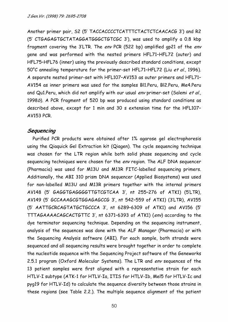
J.Gen.Vir. (1998) 79: 2695-2708
50
Another primer pair, S2 (5’ TACCACCCCTCATTTCTACTCTCAACACG 3’) and R2 (5’ CTGAGAGTGCTATAGGATGGGCTGTCGC 3’), was used to amplify a 0.8 kbp fragment covering the 3’LTR. The env PCR (522 bp) amplified gp21 of the env gene and was performed with the nested primers HFL71–HFL72 (outer) and HFL75–HFL76 (inner) using the previously described standard conditions, except 50°C annealing temperature for the primer-set HFL71–HFL72 (Liu et al., 1996). A separate nested primer-set with HFL107–AV153 as outer primers and HFL71–AV154 as inner primers was used for the samples Bl1.Peru, Bl2.Peru, Me4.Peru and Qu1.Peru, which did not amplify with our usual env primer-set (Salemi et al., 1998b). A PCR fragment of 520 bp was produced using standard conditions as described above, except for 1 min and 30 s extension time for the HFL107–AV153 PCR.
Sequencing
Purified PCR products were obtained after 1% agarose gel electrophoresis using the Qiaquick Gel Extraction kit (Qiagen). The cycle sequencing technique was chosen for the LTR region while both solid phase sequencing and cycle sequencing techniques were chosen for the env region. The ALF DNA sequencer (Pharmacia) was used for M13U and M13R FITC-labelled sequencing primers. Additionally, the ABI 310 prism DNA sequencer (Applied Biosystems) was used for non-labelled M13U and M13R primers together with the internal primers AV148 (5’ GAGGTGAGGGGTTGTCGTCAA 3’, nt 255–276 of ATK1) (5’LTR), AV149 (5’ GCCAAAGCGTGGAGAGCCG 3’, nt 542–559 of ATK1) (3’LTR), AV155 (5’ AATTGCRCAGTATGCTGCCCA 3’, nt 6289–6309 of ATK1) and AV156 (5’ TTTAGAAAACAGCACTGTTC 3’, nt 6371–6393 of ATK1) (env) according to the dye terminator sequencing technique. Depending on the sequencing instrument, analysis of the sequences was done with the ALF Manager (Pharmacia) or with the Sequencing Analysis software (ABI). For each sample, both strands were sequenced and all sequencing results were brought together in order to complete the nucleotide sequence with the Sequencing Project software of the Geneworks 2.5.1 program (Oxford Molecular Systems). The LTR and env sequences of the 13 patient samples were first aligned with a representative strain for each HTLV-I subtype (ATK-1 for HTLV-Ia, ITIS for HTLV-Ib, Mel5 for HTLV-Ic and pyg19 for HTLV-Id) to calculate the sequence diversity between those strains in these regions (see Table 2.2.). The multiple sequence alignment of the patient
Chapter 2 Introduction of HTLV-1 in Latin America
51
samples together with related sequences in the GenBank/EMBL database was done in Geneworks and further edited in MacClade 3.04 (Maddison & Maddison, 1992).
Phylogenetic analysis
The aligned sequences were used in the Phylip 3.56 software package (Felsenstein, 1989) to construct ‘Neighbour Joining’ (NJ) trees and ‘Maximum Likelihood’ (ML) trees (using the F84 substitution model). For the ‘Fitch and Wagner Parsimony’ tree construction method (Pars) the PAUP 4.0d59 software package (Swofford, 1991) was used. The reliability of the NJ and Pars trees was evaluated by analysing 1000 bootstrap replicates (Felsenstein, 1985; Zharkikh et al., 1992). The sequences of the 13 patient samples were aligned with all available HTLV-Ia LTR and env sequences and with prototypic strains for the other subtypes which were defined as outgroups. The strains used in these analyses are listed below. First, a phylogenetic analysis of the 13 patient samples and 47 published HTLV-I cosmopolitan strains was performed using the entire LTR region (734 bp). The second analysis of the LTR region was performed with 96 strains for which a shorter 514 bp LTR fragment was available, including additional Latin American strains from Colombia (SIB170, SIB190, SIB 220, COL001, COL013, COL084, COL443), strains from Taiwan (TA1, TA2, TA4, TA5, TA6, TA7, TA8, TAC1, TAC3), Ivory Coast (AKR, SIE), South Africa (RSA1, RSA6), the Caribbean (RD1) and the Middle East (GER1 and GER2 from Iran, HE from Israel and L001–L005, L113, L118, L189, L195 and L302 from Mashadi Jews). All available HTLV-Ia env sequences together with ITIS, Mel5 and pyg19 (55 strains) were aligned with the 11 samples from Peru (see Table 2.1., Qu3.Peru and RKI4-Peru excluded) and submitted to phylogenetic analysis in order to determine their position within the cosmopolitan subtype. The empirical transition/transversion bias, determined in the MacClade program, was 5 for the long LTR analysis, 4.5 for the short LTR analysis and 3.2 for the env analysis. The trees were drawn with the TreeView 1.4 program (Roderic D. M. Page, University of Glasgow, UK). Further drawing improvements were done in the Canvas 3.0.6 software package.
J.Gen.Vir. (1998) 79: 2695-2708
52
Strains used for LTR and env phylogenetic analysis The nucleotide sequences of the Peruvian isolates Qu1, 2, 3.Peru; Me1, 2,
3.Peru; Bl1, 2, 3.Peru and Ni1, 2, 3.Peru and RKI4-Peru have been deposited in the EMBL Nucleotide Sequence Database under accession numbers Y16475–Y16497 and AF054627.
The other HTLV-I strains were those described by Bazarbachi et al. (1995)
(BOI, L36905), Chou et al. (1995) (ATL-YS, U19949), Dasgupta et al. (1992) (73RM, M81248), Ellerbrok et al. (1997) (RKI1-Iran, AF3010; RKI2-Rum, AF3012), Engelbrecht et al. (1996) (TBH-1 to -7, L76025–L76030, L76034), Evangelista et al. (1990) (TSP-1, M86840), Gasmi et al. (1994) (Bo, OD, Pr52 and Pr144, U12804–U12807), Gessain et al. (1991) (Bellona1, M94195; Bellona2, M93098), Gessain et al. (1993) (MEL5, L02534), Gray et al. (1990) (MT2, M37747), Josephs et al. (1984) (CR1, K02722), Komurian et al. (1991) (AKR and SIE), Koralnik et al. (1994) (H5, H10, H14–16 U03133–U03136, U03138, U03153–U03155), Kilim et al. (1994) (HE, S76263), Lin et al. (1996) (TA1 and TA2, U53069–U53070; TA4–TA8, U53072–U53076; TAC1, U53078; TAC3, U53080), Liu et al. (1996) (AMA, CMC, FCR, HKN, ITIS, JCP, MAQS, MASU, X88871–X88877 and X88879–X88883), Mahieux et al. (1997a) (Eth.D, Abl.A, Mel.J, Vid.D, Abl.M, Z28963–Z28967; afs911, afs3, afs2, afs1, L46647–L46650; 482guinbis, L48560; 428maurit, L48561; pyg19, L76310 and L76414; PH120–PH123, PH153, PH154, PH157, PH238, PH239, PH71, L46595–L46604; PH333, PH184–PH186, PH179, PH111, PH52, L46606–L46612; PH63, L46617; PH198, L46638), Malik et al. (1988) (HS35, D00294), Miura et al. (1994) (AINU, D23694; GH78, D23693), Miura et al. (1997) (COL001, COL013, COL084, COL443, SIB190, SIB220, D82946–D82951; SIB770, D23691), Nerurkar et al. (1994) (Brazil-R-1, U11559), Picard et al. (1995) (BCI1-2, U32552; BCl2-1, U32554 and U32557), Ratner et al. (1991) (CH, M69044), Schulz et al. (1991) (H990, U81862; pt1ATL, U81864; pt3ATL, U81866; pt8ATL and pt9ATL, U81868–U81869), Seiki et al. (1983) (ATK1 and ATM, J02029–J02030), Shirabe et al. (1990) (HCT), Tsujimoto et al. (1988) (CH26, D23690; H5, M37299), Vandamme et al. (1994) (ITIS, Z32527; MT4-LB, Z31661), Voevodin et al. (1995) (KUW-1, L42253; KUW-2, L42225; KUW-3, L42255), Voevodin et al. (1997) (IRN-1, IRN-2, IRN-4, SAS and Abl.A, U87260–U87264), Yamashita et al. (1995) (GER2 and GER1, U21139–U25140; L001–L005, L113, L118, L189, L195,

Chapter 2 Introduction of HTLV-1 in Latin America
53
L195MW2 and L302, D38382–D38403; RSA6 and RSA1, U25062–U25063).
Molecular clock evaluation The molecular clock hypothesis was tested using the Likelihood Ratio Test
(LRT) (Felsenstein, 1989; Huelsenbeck & Rannala, 1997). ML trees (not enforcing the constancy of evolutionary rates along each branch of the tree) were obtained using the program PUZZLE 4.0 (Strimmer & von Haeseler, 1997); branch lengths and likelihoods were then calculated again with the Maximum Likelihood Method enforcing the clock hypothesis (MLK) and the likelihoods were compared with those obtained for the ML trees in an LRT as already described for HTLV-II (Salemi et al., 1998a). For both methods (ML and MLK) on each subtree, the same substitution model (Tamura–Nei) and rate heterogeneity model (gamma distributed) as well as the same input parameters such as transition/transversion score, nucleotide frequencies and alpha parameter for the gamma distribution were used. If the difference in likelihoods between the ML and MLK methods was not statistically significant (using the χ2 test), the molecular clock hypothesis was accepted. To estimate the evolutionary rate ‘r’ (= nucleotide substitutions/site/year) the following formula was used: r = b.l./T, where ‘b.l.’ is the branch length of the MLK tree (in PUZZLE 4.0) from a particular strain in that tree to the root node of the cluster of interest and ‘T’ is the divergence time used to calibrate the clock. Mitochondrial (mt)DNA analysis
DNA samples were PCR amplified in 20 µl reaction volumes, using primers HVR1 (5’ CTAACCTGAATCGGAGGACAAC 3’ ) and HVR4 (5’ GCATACCGCCAAAAGATAAAA 3’) specific for a 1239 bp fragment of human mtDNA, under the conditions described previously (Hagelberg & Clegg, 1991). A 1 µl aliquot of each PCR product was reamplified with internal primers and subjected to automated DNA sequencing on a LI-COR 4200 sequencing system (MWG-Biotech). We obtained 360 bp of sequence between nucleotide positions 16041 and 16400 on the mtDNA reference sequence (Anderson et al., 1981). Individuals exhibiting a T to C transition at position 16189 had a homopolymeric tract of C residues that prevented sequencing past this point. In these cases, two partial single sequences that joined the C tract were obtained. The DNA sequences were aligned manually and compared to published sequences.
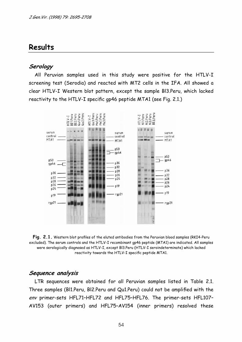
J.Gen.Vir. (1998) 79: 2695-2708
54
Results
Serology All Peruvian samples used in this study were positive for the HTLV-I
screening test (Serodia) and reacted with MT2 cells in the IFA. All showed a clear HTLV-I Western blot pattern, except the sample Bl3.Peru, which lacked reactivity to the HTLV-I specific gp46 peptide MTA1 (see Fig. 2.1.)
Fig. 2.1. Western blot profiles of the eluted antibodies from the Peruvian blood samples (RKI4-Peru
excluded). The serum controls and the HTLV-I recombinant gp46 peptide (MTA1) are indicated. All samples were serologically diagnosed as HTLV-I, except Bl3.Peru (HTLV-I seroindeterminate) which lacked
reactivity towards the HTLV-I specific peptide MTA1.
Sequence analysis LTR sequences were obtained for all Peruvian samples listed in Table 2.1.
Three samples (Bl1.Peru, Bl2.Peru and Qu1.Peru) could not be amplified with the env primer-sets HFL71–HFL72 and HFL75–HFL76. The primer-sets HFL107–AV153 (outer primers) and HFL75–AV154 (inner primers) resolved these

Chapter 2 Introduction of HTLV-1 in Latin America
55
amplification problems. For isolate Qu3.Peru, the env sequence could not be obtained due to sequencing problems with primer AV155. These LTR and env sequences were compared to prototypic strains of each subtype: ATK1 for HTLV-Ia, ITIS for HTLV-Ib, Mel5 for HTLV-Ic and pyg19 for HTLV-Id (see Table 2.2.). As expected the genetic divergence was larger for the LTR (especially the U3 region) than for the env region. Sequence comparison using the prototypic strains of each subtype demonstrated that all Peruvian samples showed the lowest divergence with the cosmopolitan subtype HTLV-Ia, ranging from 0.7 to 3.2% nucleotide divergence. Therefore all Peruvian strains belonged to the cosmopolitan subtype.
Table 2.2. LTR and env sequence diversity of the Peruvian strains compared to the prototypic strains of each HTLV-I subtype
LTR env
HTLV-Ia HTLV-Ib HTLV-Ic HTLV-Id HTLV-Ia HTLV-Ib HTLV-Ic HTLV-Id Strain ATK1 ITIS Mel5 pyg19 ATK1 ITIS Mel5 pyg19
ATK1 0.0 5.3 9.5 5.3 0.0 2.9 7.5 3.6 Qu1.Peru 2.9 5.7 9.2 6.2 1.3 2.3 6.5 2.9 Qu2.Peru 2.6 5.7 9.0 6.2 1.7 2.7 6.9 3.3 Qu3.Peru 2.6 5.7 9.0 5.9 – – – – Me1.Peru 2.9 5.9 8.9 6.2 1.5 2.5 6.7 3.1 Me2.Peru 3.2 6.4 9.3 6.4 1.5 2.5 6.7 3.1 Me3.Peru 2.4 5.1 9.4 5.4 1.3 2.3 6.5 2.9 Bl1.Peru 2.0 4.1 8.3 4.4 2.3 3.3 7.5 3.8 Bl2.Peru 2.6 5.6 9.2 5.7 1.7 2.7 6.9 3.3 Bl3.Peru 1.7 4.6 9.5 5.1 1.7 3.1 7.3 3.1 Ni1.Peru 0.8 4.8 9.4 5.0 1.5 2.9 7.1 3.1 Ni2.Peru 1.3 5.0 9.9 5.3 1.3 3.1 7.3 3.4 Ni3.Peru 0.7 4.6 9.2 4.9 1.3 3.1 7.3 3.1 RKI4-Peru 1.8 4.0 8.3 4.1 – – – –
Phylogenetic analysis of the LTR region
The phylogenetic analyses of the total LTR region using 63 strains (see Methods) resulted in three phylogenetic trees with a topology that was almost identical when using the three different methods, NJ, Pars and ML (for NJ tree see Fig. 2.2.). The cosmopolitan subtype HTLV-Ia was clearly separated from subtypes -Ib, -Ic and -Id with bootstrap values of 90.5% for NJ and 89% for Pars. Within the HTLV-Ia cosmopolitan subtype, four subgroups could be identified as suggested by Miura et al. (1997): a West African/Caribbean subgroup C; a North African subgroup D; a Japanese subgroup B also containing
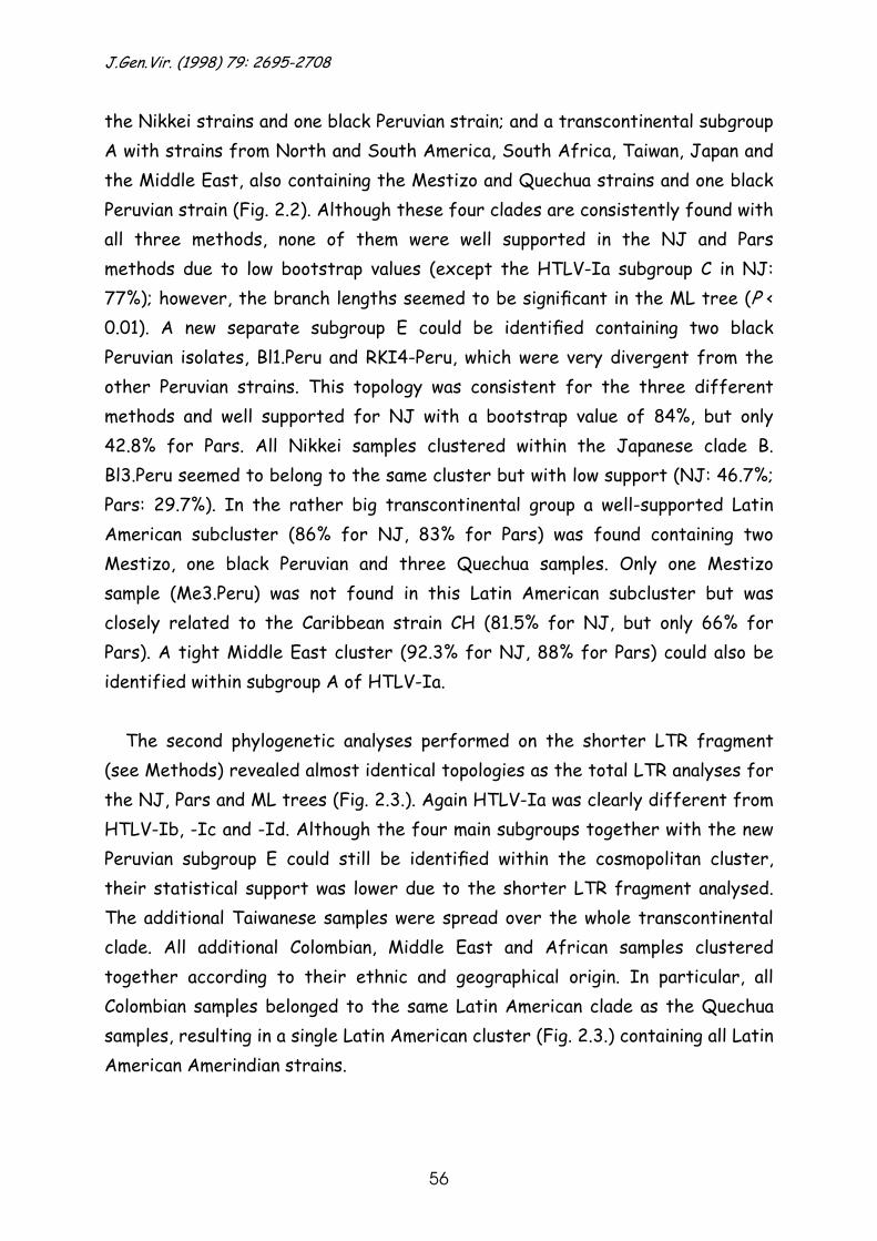
J.Gen.Vir. (1998) 79: 2695-2708
56
the Nikkei strains and one black Peruvian strain; and a transcontinental subgroup A with strains from North and South America, South Africa, Taiwan, Japan and the Middle East, also containing the Mestizo and Quechua strains and one black Peruvian strain (Fig. 2.2). Although these four clades are consistently found with all three methods, none of them were well supported in the NJ and Pars methods due to low bootstrap values (except the HTLV-Ia subgroup C in NJ: 77%); however, the branch lengths seemed to be significant in the ML tree (P < 0.01). A new separate subgroup E could be identified containing two black Peruvian isolates, Bl1.Peru and RKI4-Peru, which were very divergent from the other Peruvian strains. This topology was consistent for the three different methods and well supported for NJ with a bootstrap value of 84%, but only 42.8% for Pars. All Nikkei samples clustered within the Japanese clade B. Bl3.Peru seemed to belong to the same cluster but with low support (NJ: 46.7%; Pars: 29.7%). In the rather big transcontinental group a well-supported Latin American subcluster (86% for NJ, 83% for Pars) was found containing two Mestizo, one black Peruvian and three Quechua samples. Only one Mestizo sample (Me3.Peru) was not found in this Latin American subcluster but was closely related to the Caribbean strain CH (81.5% for NJ, but only 66% for Pars). A tight Middle East cluster (92.3% for NJ, 88% for Pars) could also be identified within subgroup A of HTLV-Ia.
The second phylogenetic analyses performed on the shorter LTR fragment
(see Methods) revealed almost identical topologies as the total LTR analyses for the NJ, Pars and ML trees (Fig. 2.3.). Again HTLV-Ia was clearly different from HTLV-Ib, -Ic and -Id. Although the four main subgroups together with the new Peruvian subgroup E could still be identified within the cosmopolitan cluster, their statistical support was lower due to the shorter LTR fragment analysed. The additional Taiwanese samples were spread over the whole transcontinental clade. All additional Colombian, Middle East and African samples clustered together according to their ethnic and geographical origin. In particular, all Colombian samples belonged to the same Latin American clade as the Quechua samples, resulting in a single Latin American cluster (Fig. 2.3.) containing all Latin American Amerindian strains.

Chapter 2 Introduction of HTLV-1 in Latin America
57
Fig. 2.2. Rooted NJ tree of 63 HTLV-I strains based upon a 722 bp fragment of the LTR region. The
bootstrap values (above 50 % and using 1000 bootstrap samples) on the branches represent the percentage of trees for which the sequences at one end of the branch form a monophyletic group. Mel5, ITIS and pyg19 are used as outgroups. Detailed information about the strains is given in the Methods section.
Strains within each subgroup not completely described in the NJ tree are the following, ranked from top to bottom: West African/Caribbean subgroup C: HS35 (Caribbean), GH78 (Ghana); North African subgroup D: Pr144 (Morocco), OD (Mauritania), Pr52 (Morocco), Bo (Algeria); Black Peruvian subgroup E : Bl1.Peru
(Peru, black), RKI4-Peru (Peru, black); Japanese subgroup B: Bl3.Peru (Peru, black), ATM (Japan), MT4-LB (Japan), HCT (Japan), ATK1 (Japan), Ni3.Peru (Peru, Nikkei), Ni2.Peru (Peru, Nikkei), HKN/ATL-YS/TBH-5 (Japan/USA, Japanese/South Africa), Ni1.Peru (Peru, Nikkei), BCI2-1 (British Colombia, Amerindian), H5 (Japan); Latin American cluster within the transcontinental subgroup A: Bl2.Peru (Peru, black), JCP
(Brazil, white), MAQS (Brazil, black), FCR (Brazil, black), MASU (Brazil, white), Qu1.Peru (Peru, Quechua), Qu3.Peru (Peru, Quechua), Qu2.Peru (Peru, Quechua), Me2.Peru (Peru, Mestizo), Me1.Peru (Peru,
Mestizo); Middle Eastern cluster within the transcontinental subgroup A: KUW-2/KUW-3 (Kuwait), KUW-1 (Kuwait), RKI1-Iran/IRN-1/IRN-4/SAS (Iran), IRN-2 (Iran), Abl.A (South Africa), BCI1-2 (British
Colombia, Amerindian), CH26 (Chile). Geographical origin and ethnic origin (if known) are given in italics between parentheses.
J.Gen.Vir. (1998) 79: 2695-2708
58
Fig. 2.3. Rooted NJ tree of 96 HTLV-I strains based upon a 514 bp fragment of the LTR region. The bootstrap values (above 50 % and using 1000 bootstrap samples) on the branches represent the percentage
of trees for which the sequences at one end of the branch form a monophyletic group. Mel5, ITIS and pyg19 are used as outgroups. Detailed information about the strains is given in the Methods section.
Strains within each subgroup not completely described in the NJ tree are the following, ranked from top to bottom: West African/Caribbean subgroup C: HS35 (Caribbean), SIE (Ivory Coast), GH78 (Ghana), AKR (Ivory Coast); North African subgroup D: Pr144 (Morocco), OD (Mauritania), Pr52 (Morocco), Bo (Algeria); Japanese subgroup B: ATM (Japan), MT4-LB (Japan), Bl3.Peru (Peru, black), HCT (Japan), ATK1 (Japan), TA7 (Taiwan), Ni3.Peru/BCI2-1/TA8 (Peru, Nikkei/British Colombia, Amerindian/Taiwan), TA6 (Taiwan),
HKN/ATL-YS/TBH-5 (Japan/USA, Japanese/South Africa), Ni2.Peru (Peru, Nikkei), Ni1.Peru (Peru, Nikkei), H5 (Japan); Black Peruvian subgroup E: Bl1.Peru (Peru, black), RKI4-Peru (Peru, black); Latin American cluster within the transcontinental subgroup A: SIB220 (Colombia, Kansa, Amerindian), JCP
(Brazil, white), MAQS (Brazil, black), FCR (Brazil, black), MASU (Brazil, white), SIB170 (Colombia, Kansa,
Chapter 2 Introduction of HTLV-1 in Latin America
59
Amerindian), Qu3.Peru (Peru, Quechua), Bl2.Peru (Peru, black), Qu2.Peru (Peru, Quechua), Qu1.Peru (Peru, Quechua), COL001 (Colombia, black), COL013 (Colombia, black), COL084 (Colombia, black), Me2.Peru
(Peru, Mestizo), Me1.Peru (Peru, Mestizo), COL443 (Colombia, Inga, Amerindian), SIB190 (Colombia, Kansa, Amerindian); Middle Eastern cluster within the transcontinental subgroup A: L004M (Mashadi Jew), L118U (Mashadi Jew), L002D (Mashadi Jew), RKI1-Iran/IRN-1/IRN-4/SAS/GER2 (Iran)/KUW-2/KUW-3 (Kuwait)/HE (Israel), L302S (Mashadi Jew), L002C (Mashadi Jew), L302T (Mashadi Jew), KUW-1 (Kuwait),
L001A (Mashadi Jew), L005G (Mashadi Jew), IRN-2(Iran), L004F (Mashadi Jew), L003E (Mashadi Jew), L189N (Mashadi Jew), L003V (Mashadi Jew), L001B (Mashadi Jew), L118K (Mashadi Jew), L189L (Mashadi
Jew), L005I (Mashadi Jew), L113J (Mashadi Jew), L113H (Mashadi Jew), GER1 (Iran), L195MW2Q (Mashadi Jew), L195 (Mashadi Jew), L195MW2R (Mashadi Jew). Geographical origin and ethnic origin (if
known) are given in italics between parentheses.
Molecular clock analysis
This was done for the short LTR fragment on the Latin American cluster separately (node A, Fig. 2.3.), on the Japanese subgroup B separately (node B, Fig.2.3.), on the Latin American cluster together with the Japanese subgroup B (node A+B, Fig. 2.3.), on the total transcontinental HTLV-Ia subgroup A (node C, Fig. 2.3.), and on the transcontinental subgroup A together with the Japanese subgroup B and the black Peruvian subgroup E (node D, Fig. 2.3.) (analysis see Table 2.3.). In each analysis, the transition/transversion bias was recalculated for the tree of that particular clade as implemented in the PUZZLE 4.0 software, since the molecular clock evaluation was performed on each of these clades separately. The transition/transversion score for the Japanese subgroup seemed extremely high: in the alignment only one transversion from G to T was observed compared to 17 transitions. When for these calculations the ti/tv bias in this Japanese subgroup was artificially changed to 2.96, which is the estimated ti/tv bias for the whole cosmopolitan HTLV-Ia, the molecular clock hypothesis was still accepted. The general results showed that the molecular clock was accepted in the first four analyses, but was rejected for the last analysis. These analyses revealed an equal evolutionary rate within and between the Latin American cluster and the Japanese subgroup B and within the transcontinental subgroup A. The branch lengths (calculated in the MLK tree) from a particular strain in the Japanese clade or the Latin American clade to the respective root nodes of these separate clades were almost identical (0.005). The branch length from a particular strain to the root node of the Japanese clade and Latin American clade together was twice as long (0.01). To calibrate the molecular clock, two different time-periods for the divergence time within the Latin American and the Japanese clade (root node A and B in Fig. 2.3.) and between these two clades (root node D in Fig. 2.3.) were used. The first
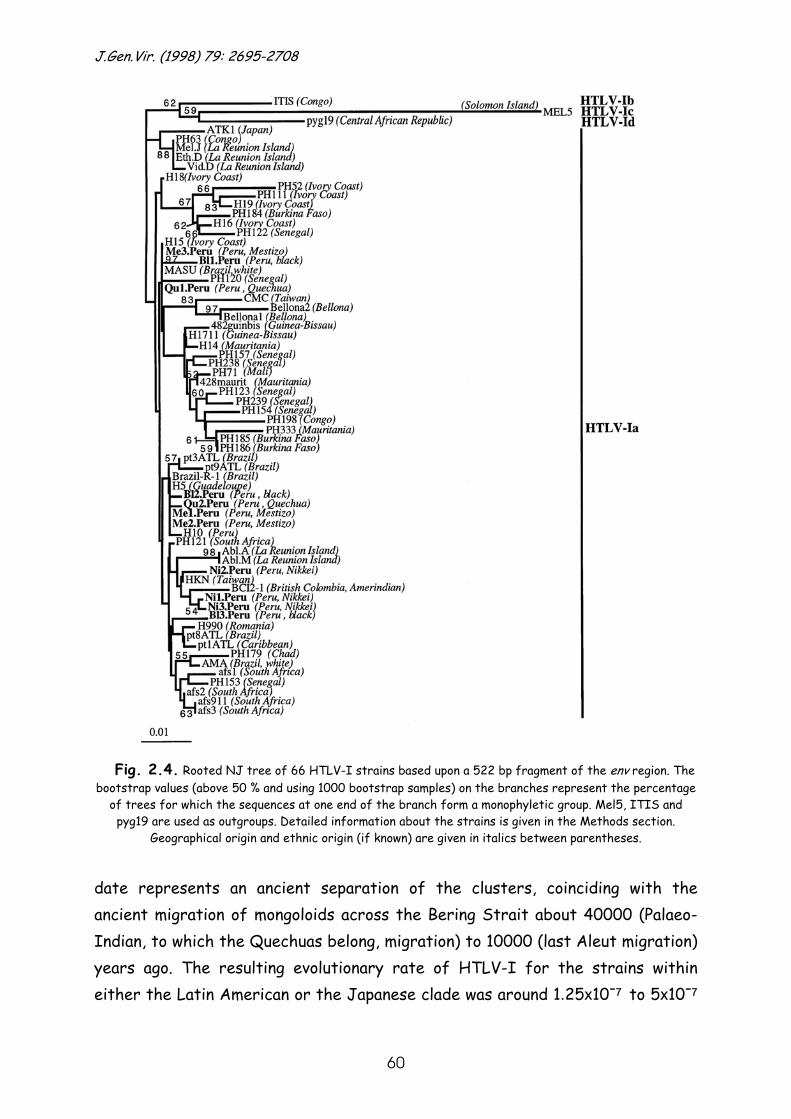
J.Gen.Vir. (1998) 79: 2695-2708
60
Fig. 2.4. Rooted NJ tree of 66 HTLV-I strains based upon a 522 bp fragment of the env region. The
bootstrap values (above 50 % and using 1000 bootstrap samples) on the branches represent the percentage of trees for which the sequences at one end of the branch form a monophyletic group. Mel5, ITIS and pyg19 are used as outgroups. Detailed information about the strains is given in the Methods section.
Geographical origin and ethnic origin (if known) are given in italics between parentheses.
date represents an ancient separation of the clusters, coinciding with the ancient migration of mongoloids across the Bering Strait about 40000 (Palaeo-Indian, to which the Quechuas belong, migration) to 10000 (last Aleut migration) years ago. The resulting evolutionary rate of HTLV-I for the strains within either the Latin American or the Japanese clade was around 1.25x10⁻⁷ to 5x10⁻⁷
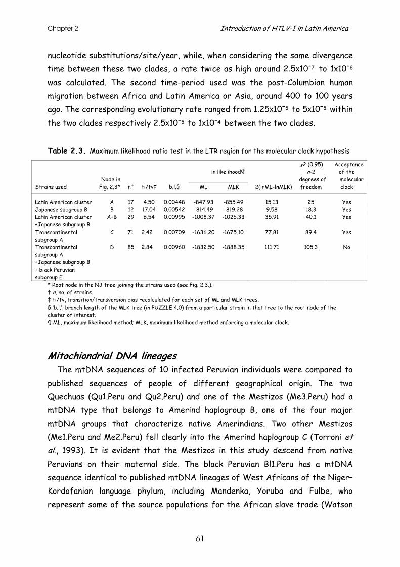
Chapter 2 Introduction of HTLV-1 in Latin America
61
nucleotide substitutions/site/year, while, when considering the same divergence time between these two clades, a rate twice as high around 2.5x10⁻⁷ to 1x10⁻⁶ was calculated. The second time-period used was the post-Columbian human migration between Africa and Latin America or Asia, around 400 to 100 years ago. The corresponding evolutionary rate ranged from 1.25x10⁻⁵ to 5x10⁻⁵ within the two clades respectively 2.5x10⁻⁵ to 1x10⁻⁴ between the two clades.
Table 2.3. Maximum likelihood ratio test in the LTR region for the molecular clock hypothesis
χ2 (0.95) Acceptance ln likelihood¶ n-2 of the
Node in degrees of molecular Strains used Fig. 2.3* n† ti/tv‡ b.l.§ ML MLK 2(lnML-lnMLK) freedom clock
Latin American cluster A 17 4.50 0.00448 -847.93 -855.49 15.13 25 Yes Japanese subgroup B B 12 17.04 0.00542 -814.49 -819.28 9.58 18.3 Yes Latin American cluster A+B 29 6.54 0.00995 -1008.37 -1026.33 35.91 40.1 Yes +Japanese subgroup B Transcontinental subgroup A
C 71 2.42 0.00709 -1636.20 -1675.10 77.81 89.4 Yes
Transcontinental subgroup A
D 85 2.84 0.00960 -1832.50 -1888.35 111.71 105.3 No
+Japanese subgroup B + black Peruvian subgroup E
* Root node in the NJ tree joining the strains used (see Fig. 2.3.). † n, no. of strains. ‡ ti/tv, transition/transversion bias recalculated for each set of ML and MLK trees. § ‘b.l.’, branch length of the MLK tree (in PUZZLE 4.0) from a particular strain in that tree to the root node of the cluster of interest. ¶ ML, maximum likelihood method; MLK, maximum likelihood method enforcing a molecular clock.
Mitochiondrial DNA lineages The mtDNA sequences of 10 infected Peruvian individuals were compared to
published sequences of people of different geographical origin. The two Quechuas (Qu1.Peru and Qu2.Peru) and one of the Mestizos (Me3.Peru) had a mtDNA type that belongs to Amerind haplogroup B, one of the four major mtDNA groups that characterize native Amerindians. Two other Mestizos (Me1.Peru and Me2.Peru) fell clearly into the Amerind haplogroup C (Torroni et al., 1993). It is evident that the Mestizos in this study descend from native Peruvians on their maternal side. The black Peruvian Bl1.Peru has a mtDNA sequence identical to published mtDNA lineages of West Africans of the Niger–Kordofanian language phylum, including Mandenka, Yoruba and Fulbe, who represent some of the source populations for the African slave trade (Watson
J.Gen.Vir. (1998) 79: 2695-2708
62
et al., 1997). The mtDNA type of Bl2.Peru is identical to some more widespread African mtDNA sequences, while the sequence of the third black individual in the Japanese clade (Bl3.Peru) could not be located exactly within a known African or Japanese haplogroup. The two Nikkei (Ni1.Peru and Ni3.Peru) have mtDNA sequences identical or closely related to several individuals of the Japan-II group (Horai & Hayasaka, 1990).
Discussion
The phylogenetic analysis on the LTR and env region of our 13 Peruvian cosmopolitan HTLV-Ia isolates was performed to get a clearer idea about the origin and dissemination of the cosmopolitan HTLV-Ia virus in Latin America. The West African/Caribbean subgroup C, the North African subgroup D and even the black Peruvian subgroup E (in the full LTR analysis) branch off before the Japanese subgroup B and transcontinental subgroup A. This suggests an African origin of these two latter subgroups. Additionally, the strains from black Peruvians were spread all over the HTLV-Ia clade, which is compatible with a greater virus variation in the black Peruvian population than in the Nikkei or Mestizo and Quechua population of Peru. Moreover, one of the affected black Peruvians (Bl1.Peru), in the distinct black Peruvian HTLV-Ia clade, has a mtDNA type identical to that of some West African source populations for the slave trade (Watson et al., 1997) and the black Peruvian strain clustering with the Quechua strains was also isolated from an individual with a typical African mtDNA lineage. These observations imply that the virus has been introduced into the American continent on a number of occasions from several African lineages, probably during and after the slave trade. The tight clustering of the Nikkei as well as the Quechua strains according to their ethnic origin in separate and well-defined clusters and the low divergence within these clades suggest a recent and separate spread of the HTLV-Ia virus in these Latin American populations. The Peruvian Japanese HTLV-Ia lineage is probably the result of recent Japanese immigrations, but has possibly indirectly an African origin. Genetic admixture of the black Latin Americans with other Latin American ethnic groups like Quechuas and Mestizos on the other hand probably
Chapter 2 Introduction of HTLV-1 in Latin America
63
gave rise to HTLV-Ia in these indigenous Latin American populations. The topology of the LTR trees together with the low divergence within the Japanese subgroup B and within the Latin American cluster argue for a ‘post-Columbian’ time-frame at root node B (Japanese subgroup B) and root node A (Latin American clade). In order to verify this hypothesis of a post-Columbian introduction of HTLV-Ia in Latin America, molecular clock analyses were performed to calculate the divergence time in the Japanese subgroup B or in the Latin American clade of the transcontinental subgroup A. The hypothesis of an ancient Palaeolithic introduction of HTLV-Ia in these populations (40000 to 10000 years ago) would result in an evolutionary rate of 1.25x10⁻⁷ to 5x10⁻⁷. This rate is only slightly faster than the evolutionary rate of cellular genes of around 10−8 to 10−9 (Britten, 1986; Weissmann & Weber, 1986). Such an extremely low evolutionary rate for the retrovirus HTLV-Ia seems highly unlikely. RNA viruses (including retroviruses) generally have an evolutionary rate of 10−4 to 10−2 nucleotide substitutions/site/year. However, the evolutionary rate of horizontally transmitted HTLV-II (in injecting drug users) is estimated to be around 2.7x10⁻⁵ to 1x10⁻⁴ nucleotide substitutions/site/year (Salemi et al., 1998a), which is the lowest evolutionary rate reported for a retrovirus so far. Assuming a post-Columbian time-frame of divergence (400 to 100 years ago) within these Japanese and Latin American HTLV-Ia clades, the evolutionary rate would be between 1.25x10⁻⁵ and 5x10⁻⁵, approximately in the same range as the rate of HTLV-II in intravenous drug users. This suggests that the strains within both clades probably diversified in a post-Columbian period. The separation between the Latin American and the Japanese clade could potentially be pre-Columbian, since they probably already diversified in Africa as they seem to be the result of the introduction of separate African lineages. However, their separation cannot date from the last glacial period (40000 to 10000 years ago), since the resulting evolutionary rate of 2.5x10⁻⁷ to 1x10⁻⁶ would be too low. The molecular clock analyses calculated an equal evolutionary rate within and between the Latin American clade and the Japanese clade and within the transcontinental HTLV-Ia clade, containing the AINU strain of one of the oldest populations of Japan. This suggests a post-Columbian spread of both the Japanese and transcontinental clade, possibly through separate introductions from Africa.
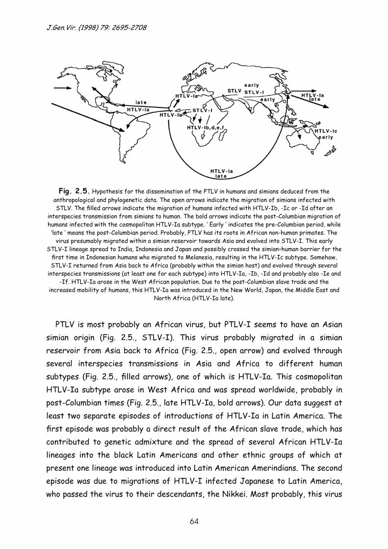
J.Gen.Vir. (1998) 79: 2695-2708
64
Fig. 2.5. Hypothesis for the dissemination of the PTLV in humans and simians deduced from the anthropological and phylogenetic data. The open arrows indicate the migration of simians infected with STLV. The filled arrows indicate the migration of humans infected with HTLV-Ib, -Ic or -Id after an
interspecies transmission from simians to human. The bold arrows indicate the post-Columbian migration of humans infected with the cosmopolitan HTLV-Ia subtype. ‘ Early ’ indicates the pre-Columbian period, while ‘late ’ means the post-Columbian period. Probably, PTLV has its roots in African non-human primates. The
virus presumably migrated within a simian reservoir towards Asia and evolved into STLV-I. This early STLV-I lineage spread to India, Indonesia and Japan and possibly crossed the simian–human barrier for the first time in Indonesian humans who migrated to Melanesia, resulting in the HTLV-Ic subtype. Somehow, STLV-I returned from Asia back to Africa (probably within the simian host) and evolved through several
interspecies transmissions (at least one for each subtype) into HTLV-Ia, -Ib, -Id and probably also -Ie and -If. HTLV-Ia arose in the West African population. Due to the post-Columbian slave trade and the
increased mobility of humans, this HTLV-Ia was introduced in the New World, Japan, the Middle East and North Africa (HTLV-Ia late).
PTLV is most probably an African virus, but PTLV-I seems to have an Asian simian origin (Fig. 2.5., STLV-I). This virus probably migrated in a simian reservoir from Asia back to Africa (Fig. 2.5., open arrow) and evolved through several interspecies transmissions in Asia and Africa to different human subtypes (Fig. 2.5., filled arrows), one of which is HTLV-Ia. This cosmopolitan HTLV-Ia subtype arose in West Africa and was spread worldwide, probably in post-Columbian times (Fig. 2.5., late HTLV-Ia, bold arrows). Our data suggest at least two separate episodes of introductions of HTLV-Ia in Latin America. The first episode was probably a direct result of the African slave trade, which has contributed to genetic admixture and the spread of several African HTLV-Ia lineages into the black Latin Americans and other ethnic groups of which at present one lineage was introduced into Latin American Amerindians. The second episode was due to migrations of HTLV-I infected Japanese to Latin America, who passed the virus to their descendants, the Nikkei. Most probably, this virus
Chapter 2 Introduction of HTLV-1 in Latin America
65
was also indirectly a post-Columbian introduction from Africa. To draw any definite conclusions on the introduction of HTLV-Ia in Latin America, the phylogeny of new and more American isolates should be investigated and more detailed studies on the rate of nucleotide substitutions for HTLV-I might be necessary to clarify this issue.
We thank Martine Michiels and Martin Reynders of the Virology Laboratory,
University Hospitals, Leuven, Belgium, and Claudia Koch of the Robert Koch Institute, Berlin, Germany for excellent technical assistance, Bernadett Brado and J. J. Bugert of the Medical Poliklinik, Heidelberg, Germany for providing the EDTA blood of patient RKI4-Peru and Ria Swinnen of the Virology Laboratory, University Hospitals, Leuven, Belgium for fine editorial help. This study was conducted in accordance with human subjects protocols approved by the Naval Medical Research Institute Committee for the Protection of Human Subjects of Peru. The opinions and assertions contained herein are the private ones of the writers and are not to be construed as official or as reflecting the views of the Navy Department or the Naval Service at large. Erika Hagelberg acknowledges the financial support of the Wellcome Trust and the Kon-Tiki Museum, Oslo. The Rega Institute is part of the HERN concerted action supported by the Biomed programme of the European Commission. This work was supported by the Belgian Fonds voor Geneeskundig Wetenschappelijk Onderzoek (Krediet no. 3.0098.94).
References
Anderson, S., Bankier, A. T., Barrell, B., de Bruijn, M. H. L., Coulson, A. R., Drouin, J., Eperon, I. C., Nierlich, D. P., Roe, B. A., Sanger, F., Schreier, P. H., Smith, A. J. H., Staden, R. & Young, I. G. (1981). Sequence organisation of the human mitochondrial genome. Nature 290, 457–465. Bazarbachi, A., Huang, M., Gessain, A., Saal, F., Saib, A., Peries, J., De Thé , H. & Galibert, F. (1995). Human T-cell-leukemia virus type I in post-transfusional spastic paraparesis: complete proviral sequence from uncultured blood cells. International Journal of Cancer 63, 494–499. Britten, R. J. (1986). Rates of DNA sequence evolution differ between taxonomic groups. Science 231, 1393–1398. Cavalli-Sforza, L. L., Menozzi, P. & Piazza, A. (1994). The History and Geography of Human Genes. Princeton, NJ: Princeton University Press. Chou, K. S., Okayama, A., Tachibana, N., Lee, T. H. & Essex, M. (1995). Nucleotide sequence analysis of a full-length human T-cell leukemia virus type I from adult T-cell leukemia cells: a prematurely terminated pX open reading frame II. International Journal of Cancer 60, 701–706. Crowther, G., Rachowiecki, R. & Dydynski, K. (1990). South America on a shoestring. In Lonely Planet, 4th edn, pp. 626–627. London: Lonely Planet Publications.

J.Gen.Vir. (1998) 79: 2695-2708
66
Dasgupta, P., Reddy, C. D., Saikumar, P. & Reddy, E. P. (1992). The cellular proto-oncogene product Myb acts as transcriptional activator of the long terminal repeat of human T-lymphotropic virus type I. Journal of Virology 66, 270–276. Delaporte, E., Monplaisir, N., Louwagie, J., Peeters, M., Martin-Prével, Y., Louis, J.-P., Trebucq, A., Bedjabaga, L., Ossari, S., Honoré , C., Larouzé , B., d’Auriol, L., Van der Groen, G. & Piot, P. (1991). Prevalence of HTLV-I and HTLV-II infection in Gabon, Africa: comparison of the serological and PCR results. International Journal of Cancer 49, 373–376. Ellerbrok, H., Fleischer, C., Vandamme, A.-M., Kücherer, C. & Pauli, G. (1997). Sequence analysis of two HTLV type I infections imported to Germany. AIDS Research and Human Retroviruses 13, 1255–1258. Engelbrecht, S., van Rensburg, E. J. & Robson, B. A. (1996). Sequence variation and subtyping of human and simian T-cell lymphotropic virus type I strains from South Africa. Journal of Acquired Immune Deficiency Syndromes and Human Retrovirology 12, 298–302. Evangelista, A., Maroushek, S., Minnigan, H., Larson, A., Retzel, E., Haase, A., Gonzalez-Dunia, D., McFarlin, D., Mingioli, E., Jacobson, S., Osame, M. & Sonoda, S. (1990). Nucleotide sequence analysis of a provirus derived from an individual with tropical spastic paraparesis. Microbial Pathogenesis 8, 259–278. Felsenstein, J. (1985). Confidence limits on phylogenies : an approach using the bootstrap. Evolution 39, 783–791. Felsenstein, J. (1989). PHYLIP – phylogeny inference package (version 3.2). Cladistics 5, 164–166. Gallo, R. C. (1986). The first human retrovirus. Scientific American 255, 88–98. Gasmi, M., Farouqi, B., d’Incan, M. & Desgranges, C. (1994). Long terminal repeat sequence analysis of HTLV type I molecular variants identified in four north African patients. AIDS Research and Human Retroviruses 10, 1313–1315. Gessain, A., Yanagihara, R., Franchini, G., Garruto, R. M., Jenkins, C. L., Ajdukiewicz, A. B., Gallo, R. C. & Gajdusek, D. C. (1991). Highly divergent molecular variants of human T-lymphotropic virus type I from isolated populations in Papua New Guinea and the Solomon Islands. Proceedings of the National Academy of Sciences, USA 88, 7694–7698. Gessain, A., Barin, F., Vernant, J., Gout, O., Maurs, L., Calender, A. & de Thé, G. (1985). Antibodies to human T-lymphotropic virus type-I in patients with tropical spastic paraparesis. Lancet ii, 407–410. Gessain, A., Gallo, R. C. & Franchini, G. (1992). The low degree of HTLV-1 genetic drift in vivo as a means to follow viral transmission and movement of ancient human population. Journal of Virology 66, 2288–2295. Gessain, A., Boeri, E., Yanagihara, R., Gallo, R. C. & Franchini, G. (1993). Complete nucleotide sequence of a highly divergent human T-cell leukemia (lymphotropic) virus type I (HTLV-I) variant from Melanesia: genetic and phylogenetic relationship to HTLV-I strains from other geographical regions. Journal of Virology 67, 1015–1023. Gessain, A., Mauclère, P., Froment, A., Biglione, M., Le Hasran, J. Y., Tekaia, Fr., Millan, J. & de Thé, G. (1995). Isolation and molecular characterization of a human T-cell lymphotropic virus type II (HTLV-II), subtype B, from a healthy Pygmy living in a remote area of Cameroon: an ancient origin for HTLV-II in Africa. Proceedings of the National Academy of Sciences, USA 92, 4041–4045. Goubau, P., Desmyter, J. & Ghesquiere, J. (1992). HTLV-II among pygmies [Letter]. Nature
Chapter 2 Introduction of HTLV-1 in Latin America
67
35, 201. Goubau, P., Liu, H.-F., De Lange, G. G., Vandamme, A.-M. & Desmyter, J. (1993 a). HTLV-II seroprevalence in pygmies across Africa since 1970. AIDS Research and Human Retroviruses 9, 709–713. Goubau, P., Desmyter, J., Swanson, P., Reynders, M., Shih, J., Surmont, I., Kazadi, K. & Lee, H. (1993 b). Detection of HTLV-I and HTLV-II infection in Africans using type-specific envelope peptides. Journal of Medical Virology 39, 28–32. Goubau, P., Van Brussel, M., Vandamme, A.-M., Liu, H.-F. & Desmyter, J. (1994). A primate T-lymphotropic virus, PTLV-L, different from human T-lymphotropic viruses types I and II, in a wild-caught baboon (Papio hamadryas). Proceedings of the National Academy of Sciences, USA 91, 2848–2852. Gray, G. S., White, M., Bartman, T. & Mann, D. (1990). Envelope gene sequence of HTLV-1 isolate MT-2 and its comparison with other HTLV1 isolates. Virology 177, 391–395. Greenberg, J., Turner, C. & Zegura, S. (1986). The settlement of the Americas: a comparison of the linguistic, dental, and genetic evidence. Current Anthropology 27, 477–497. Hagelberg, E. & Clegg, J. B. (1991). Isolation and characterisation of DNA from archaeological bone. Proceedings of the Royal Society of London, series B 244, 45–50. Hjelle, B., Zhu, S. W., Takahashi, H., Ijichi, S. & Hall, W. W. (1993). Endemic human T cell leukemia virus type II infection in southwestern US Indians involves two prototype variants of virus. Journal of Infectious Diseases 168, 737–740. Horai, S. & Hayasaka, K. (1990). Intraspecific nucleotide sequence differences in the major noncoding region of human mitochondrial DNA. American Journal of Human Genetics 46, 828–842. Huelsenbeck, J. P. & Rannata, B. (1997). Phylogenetic methods come of age: testing hypotheses in an evolutionary context. Science 276, 227–232. Ibrahim, F., De Thé, G. & Gessain, A. (1995). Isolation and characterization of a new simian T-cell leukemia virus type I from naturally infected Celebes macaques (Macaca tonkeana): complete nucleotide sequence and phylogenetic relationship with the Australo– Melanesian human T-cell leukemia virus type I. Journal of Virology 69, 6980–6993. Imai, J. & Jinuma, Y. (1983). Epstein–Barr virus-specific antibodies in patients with adult T-cell leukemia (ATL) and healthy ATL-virus-carriers. International Journal of Cancer 31, 197–200. Josephs, S. F., Wong-Staal, F., Manzari, V., Gallo, R. C., Sodroski, J. G., Trus, M. D., Perkins, D., Patarca, R. & Haseltine, W. A. (1984). Long terminal repeat structure of an American isolate of type I human T-cell leukemia virus. Virology 139, 340–345. Kalyanaraman, V. S., Sarngadharan, M. G., Robert-Guroff, M., Miyoshi, I., Blayney, D., Golde, D. & Gallo, R. C. (1982). A new subtype of human T-cell leukemia virus (HTLV-II) associated with a T-cell variant of hairy cell leukemia. Science 218, 571–573. Kilim, Y., Rosenblatt, J. D. & Danon, Y. L. (1994). Sequence analysis of the new human T cell leukaemia/lymphoma virus type I isolate (HTLV-I) in Israel. Israel Journal of Medical Sciences 30, 891–894. Komurian, F., Pelloquin, F. & de Thé, G. (1991). In vivo genomic variability of human T-cell leukemia virus type I depends more upon geography than upon pathologies. Journal of Virology 65, 3770–3778. Koralnik, I., Boeri, E., Saxinger, W. C., Monico, A. L., Fullen, J., Gessain, A., Guo, H. G., Gallo, R. C., Markham, P., Kalyanaraman, V., Hirsch, V., Allan, J., Murthy, K., Alford, P., Slattery, J. P., O’Brien, S. J. & Franchini, G. (1994). Phylogenetic associations
J.Gen.Vir. (1998) 79: 2695-2708
68
of human and simian T-cell leukemia�lymphotropic virus type I strains: evidence for interspecies transmission. Journal of Virology 68, 2693–2707. Lee, H., Swanson, P., Shorty, V. S., Zack, J. A., Rosenblatt, J. D. & Chen, I. S. Y. (1989). High rate of HTLV-II infection in seropositve IV drug abusers in New Orleans. Science 244, 471–475. Lin, M. T., Chen, Y. C., Chen, P. J., Yang, Y. C., Tang, J. L., Wu, J. M., Chuang, S. E. & Yang, C. S. (1996). Envelope gene sequences of human T-lymphotropic virus type 1 in Taiwan. Archives of Virology 141, 219–229. Liu, H. F., Goubau, P., Van Brussel, M., Van Laethem, K., Chen, Y. C., Desmyter, J. & Vandamme, A.-M. (1996). The three human Tlymphotropic virus type I subtypes arose from three geographically distinct simian reservoirs. Journal of General Virology 77, 359–368. Maddison, W. & Maddison, R. (1992). MacClade Version 3. Analysis of Phylogeny and Character Evolution. Sunderland, MA: Sinauer Associates. Mahieux, R., Ibrahim, F., Mauclere, P., Herve, V., Michel, P., Tekaia, F., Chappey, C., Garin, B., Van Der Ryst, E., Guillemain, B., Ledru, E., Delaporte, E., De The, G. & Gessain, A. (1997 a). Molecular epidemiology of 58 new African human T-cell leukemia virus type I (HTLV-I) strains: identification of a new and distinct HTLV-I molecular subtype in Central Africa and in pygmies. Journal of Virology 71, 1317–1333. Mahieux, R., Pecon-Slattery, J. & Gessain, A. (1997 b). Molecular characterization and phylogenetic analyses of a new, highly divergent simian T-cell lymphotropic virus (STLV-1marc1) in Macaca arctoides. Journal of Virology 71, 6253–6258. Malik, K. T. A., Even, J. & Karpas, A. (1988). Molecular cloning and complete nucleotide sequence of an adult T cell leukemia virus�human T-cell leukemia virus type I (ATLV�HTLV-I) isolate of Caribbean origin: relationship to other members of the ATLV�HTLV-I subgroup. Journal of General Virology 69, 1695–1710. Maloney, E. M., Biggar, R. J., Neel, J. V., Taylor, M., Hahn, B. H., Shaw, G. M. & Blattner, W. A. (1992). Endemic human T cell lymphotropic virus type II infection among isolated Brazilian Amerindians. Journal of Infectious Diseases 166, 100–107. Miura, T., Fukunaga, T., Igarashi, T., Yamashita, F., Ido, E., Funahashi, S. I., Ishida, T., Washio, K., Ueda, S., Hashimoto, K. I., Yoshida, M., Osame, M., Singhal, B. S., Zaninovic, V., Cartier, L., Sonoda, S., Tajima, K., Ina, Y., Gojobori, T. & Hayami, M. (1994). Phylogenetic subtypes of human T-lymphotropic virus type I and their relations to the anthropological background. Proceedings of the National Academy of Sciences, USA 91, 1124–1127. Miura, T., Yamashita, M., Zaninovic, V., Cartier, L., Takehisa, J., Igarashi, T., Ido, E., Fujiyoshi, T., Sonoda, S., Tajima, K. & Hayami, M. (1997). Molecular phylogeny of human T-cell leukemia virus type I and II of Amerindians in Colombia and Chile. Journal of Molecular Evolution 44, S76–S82. Murphy, E. L., Fridey, J., Smith, J. W., Engstrom, J., Sacher, R. A., Miller, K., Gibble, J., Stevens, J., Thomson, R., Hansma, D., Kaplan, J., Khabbaz, R. & Nemo, G. (1997). HTLV associated myelopathy in a cohort of HTLV-I and HTLV-II-infected blood donors. The REDS investigators. Neurology 48, 315–320. Nerurkar, V. R., Song, K.-J., Saitou, B., Melland, R. R. & Yanagihara, R. (1993). Interfamilial and intrafamilial genomic diversity and molecular phylogeny of human T-cell lymphotropic virus type I from Papua New Guinea and the Solomon Islands. Virology 196, 506–513. Nerurkar, V. R., Song, K.-J., Bastian, I. B., Garin, B., Franchini, G. & Yanagihara, R.
Chapter 2 Introduction of HTLV-1 in Latin America
69
(1994). Genotyping of human T cell lymphotropic virus type I using Australo–Melanesian topotype-specific oligonucleotide primer-based polymerase chain reaction: insights into viral evolution and dissemination. Journal of Infectious Diseases 170, 1353–1360. Osame, M., Matsumoto, M., Usuku, K., Izumo, S., Ijichi, N., Amitani, H., Tara, M. & Igata, A. (1987). Chronic progressive myelopathy associated with elevated antibodies to human T-lymphotropic virus type I and adult T-cell leukemialike cells. Annals of Neurology 21, 117–122. Picard, F. J., Coulthart, M. B., Oger, J., King, E. E., Kim, S., Arp, J., Rice, G. P. A. & Dekaban, G. A. (1995). Human T-lymphotropic virus type 1 in coastal natives of British Columbia: phylogenetic a�nities and possible origins. Journal of Virology 69, 7248–7256. Ratner, L., Philpott, T. & Trowbridge, D. B. (1991). Nucleotide sequence analysis of isolates of human T-lymphotropic virus type I of diverse geographical origins. AIDS Research and Human Retroviruses 7, 923–941. Salemi, M., Vandamme, A.-M., Gradozzi, C., Van Laethem, K., Ferrante, P., Taylor, G., Casoli, C., Goubau, P., Desmyter, J. & Bertazzoni, U. (1998 a). Evolutionary rate and genetic heterogeneity of human T-cell lymphotropic virus type II (HTLV-II) using new isolates from European injecting drug users. Journal of Molecular Evolution 46, 602–611. Salemi, M., Van Dooren, S., Audenaert, E., Delaporte, E., Goubau, P., Desmyter, J. & Vandamme, A.-M. (1998 b). Two new human Tlymphotropic virus type I phylogenetic subtypes in seroindeterminates, a Mbuti pygmy and a Gabonese, have closest relatives among African SLTV-I strains. Virology (in press). Sanchez-Albornoz, N., Morse, R., Bakewell, P., Florescano, E., Morner, M., MacLeod, M. J., Lockhart, J. & Lavrin, A. (1984). Colonial Latin America. In The Cambridge History of Latin America, vol. 2, pp. 15–355. Cambridge: Cambridge University Press. Schulz, T. F., Calabro, M. L., Hoad, J. G., Carrington, C. V., Matutes, E., Catovsky, D. & Weiss, R. A. (1991). HTLV-1 envelope sequences from Brazil, the Caribbean, and Romania: clustering of sequences according to geographic origin and variability in an antibody epitope. Virology 184, 483–491. Seiki, M., Hattori, S., Hirayama, Y. & Yoshida, M. (1983). Human adult T-cell leukemia virus: completed nucleotide sequence of the provirus genome integrated in leukemia cell DNA. Proceedings of the National Academy of Sciences, USA 80, 3618–3622. Shirabe, S., Nakamura, T., Tsujihata, M., Nagataki, S., Seiki, M. & Yoshida, M. (1990). Retrovirus from human T-cell leukemia virus type I-associated myelopathy is the same strain as a prototype human T-cell leukemia virus type I. Archives of Neurology 47, 1258–1260. Song, K. J., Nerurkar, V. R., Saitou, N., Lazo, A., Blakeslee, J. R., Miyoshi, I. & Yanagihara, R. (1994). Genetic analysis and molecular phylogeny of simian T-cell lymphotropic virus type I: evidence for independent virus evolution in Asia and Africa. Virology 199, 56–66. Strimmer, K. & von Haeseler, A. (1997). Likelihood-mapping: a simple method to visualize phylogenetic content of a sequence alignment. Proceedings of the National Academy of Sciences, USA 94, 6815–6819. Swofford, D. L. (1991). PAUP: phylogenetic analysis using parsimony, version 3.1 [computer program]. Illinois Natural History Survey, Champaign, Illinois. Tachibana, N., Okayama, A. & Ishizaki, J. (1988). Suppression of tuberculin skin reaction in healthy HTLV-I carriers from Japan. International Journal of Cancer 42, 829–831. Torroni, A., Schurr, T. G., Cabell, M. F., Brown, M. D., Nell, J. V., Larsen, M., Smith, D. G. and others (1993). Asian a�nities and continental radiation of the four founding native American mtDNAs. American Journal of Human Genetics 53, 563–590. Tsujimoto, A., Teruuchi, T., Imamura, J., Shimotohno, K., Miyoshi, I. & Miwa, M.

J.Gen.Vir. (1998) 79: 2695-2708
70
(1988). Nucleotide sequence analysis of a provirus derived from HTLV-1-associated myelopathy (HAM). Molecular Biology & Medicine 5, 29–42. Tuppin, P., Gessain, A., Kazanji, M., Mahieux, R., Cosnefroy, J.-F., Tekaia, F., Georges-Courbot, M.-C., Georges, A. & De The� , G. (1996). Evidence in Gabon for an intrafamilial clustering with mother-to-child and sexual transmission of new molecular variant of human Tlymphotropic virus type-II subtype b. Journal of Medical Virology 48, 22–32. Van Brussel, M., Goubau, P., Desmyter, J. & Vandamme, A.-M. (1996). The genomic structure of a new simian T-lymphotropic virus, STLVPH969, di�ers from that of human T-lymphotropic virus types I and II. Journal of General Virology 77, 347–358. Vandamme, A.-M., Liu, H.-F., Goubau, P. & Desmyter, J. (1994). Primate T-lymphotropic virus type I LTR sequence variation and its phylogenetic analysis: compatibility with an African origin of PTLV-I. Virology 202, 212–223. Vandamme, A.-M., Liu, H.-F., Van Brussel, M., De Meurichy, W., Desmyter, J. & Goubau, P. (1996). The presence of a divergent Tlymphotropic virus in a wild-caught pygmy chimpanzee (Pan paniscus) supports and African origin for the human T-lymphotropic�simian T-lymphotropic group of viruses. Journal of General Virology 77, 1089–1099. Voevodin, A. & Gessain, A. (1997). Common origin of human Tlymphotropic virus type I from Iran, Kuwait, Israel and La Reunion Island. Journal of Medical Virology 52, 77–82. Voevodin, A., Al-Mufti, S., Farah, S., Khan, R. & Miura, T. (1995). Molecular characterization of human T-lymphotropic virus, type 1 (HTLV-1) found in Kuwait: close similarity with HTLV-1 isolates originating from Mashhad, Iran. AIDS Research and Human Retroviruses 11, 1255–1259. Wallace, D. E. (1995). Mitochondrial DNA variation in human evolution, degenerative disease, and aging. American Journal of Human Genetics 57, 201–223. Watson, E., Forster, P., Richards, M. & Bandelt, H. J. (1997). Mitochondrial footprints of human expansions in Africa. American Journal of Human Genetics 61, 691–704. Wattel, E., Vartanian, J.-P., Pannetier, C. & Wain-Hobson, S. (1995). Clonal expansion of human T-cell leukemia virus type I infected cells in asymptomatic and symptomatic carriers without malignancy. Journal of Virology 69, 2863–2868. Weissmann, C. & Weber, H. (1986). The interferon genes. Progress in Nucleic Acid Research and Molecular Biology 33, 251–301. Yamashita, M., Kitze, B., Miura, T., Weber, T., Fujiyoshi, T., Takehisa, J., Chen, J. L., Sonoda, S. & Hayami, M. (1995). The phylogenetic relationship of HTLV type I from non-Mashhadi Iranians to that from Mashhadi Jews. AIDS Research and Human Retroviruses 11, 1533–1535. Yamashita, M., Picchio, G., Veronesi, R., Ohkura, S., Bare, P. & Hayami, M. (1998). HTLV-Is in Argentina are phylogenetically similar to those of other South American countries, but di�erent from HTLV-Is in Africa. Journal of Medical Virology 55, 152–160. Yoshida, M., Miyoshi, I. & Hinuma, Y. (1982). Isolation and characterization of retrovirus from cell lines of human adult T-cell leukemia and its implication in the disease. Proceedings of the National Academy of Sciences, USA 79, 2031–2035. Zella, D., Mori, L., Sala, M., Ferrante, P., Casoli, C., Magnani, G., Achilli, G., Cattaneo, E., Lori, F. & Bertazzoni, U. (1990). HTLV-II infection in Italian drug abusers. Lancet ii, 575–576. Zharkikh, A. & Li, W.-H. (1992). Statistical properties of bootstrap estimation of

Chapter 2 Introduction of HTLV-1 in Latin America
71
phylogenetic variability from nucleotide sequences. I. Four taxa with a molecular clock. Molecular Biology and Evolution 9, 1119–1147.
THOMAS SCHULZ, editor
Received April 15,1998; Accepted July 3, 1998

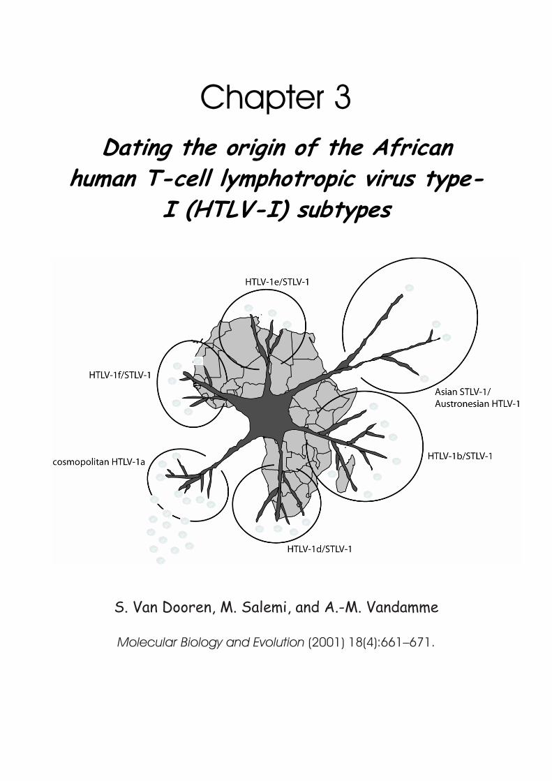
Chapter 3 Dating the origin of the African
human T-cell lymphotropic virus type-I (HTLV-I) subtypes
S. Van Dooren, M. Salemi, and A.-M. Vandamme
Molecular Biology and Evolution (2001) 18(4):661–671.

75
Dating the origin of the African human T-cell lymphotropic virus type-I (HTLV-I) subtypes
S. Van Dooren, M. Salemi, and A.-M. Vandamme
Rega Institute for Medical Research, Katholieke Universiteit Leuven, Leuven, Belgium
Molecular Biology and Evolution (2001) 18(4):661–671.
To investigate the origin of the African PTLV-I virus, we phylogenetically
analyzed the available HTLV-I and STLV-I strains. We also attempted to date the presumed interspecies transmissions that resulted in the African HTLV-I subtypes. Molecular-clock analysis was performed using the Tamura-Nei substitution model and gamma distributed rate heterogeneity based on the maximum-likelihood topology of the combined long-terminal-repeat and env third-codon-position sequences. Since the molecular clock was not rejected and no evidence for saturation was found, a constant rate of evolution at these positions for all 33 HTLV-I and STLV-I strains was reasonably assumed. The spread of PTLV-I in Africa is estimated to have occurred at least 27,300 ± 8,200 years ago. Using the available strains, the HTLV-If subtype appears to have emerged within the last 3,000 years, and the HTLV-Ia, HTLV-Ib, HTLV-Id, and HTLV-Ie subtypes appear to have diverged between 21,100 and 5,300 years ago. Interspecies transmissions, most probably simian to human, must have occurred around that time and probably continued later. When the synonymous and nonsynonymous substitution ratios were compared, it was clear that purifying selection was the driving force for PTLV-I evolution in the env gene, irrespective of the host species. Due to the small number of strains in some of the investigated groups, these data on selective pressure should be taken with caution.
Mol.Biol.Evol. (2001) 18: 661-671
76
Introduction The human and simian T-cell lymphotropic viruses type I (HTLV-I and STLV-I,
respectively) share numerous epidemiological, molecular, phylogenetic, and geographical features and are therefore referred to as primate T-cell lymphotropic viruses type I (PTLV-I). Virtually all Old World monkey species from the Cercopithecinae subfamily (Macaca, Cercopithecus, Papio, Mandrillus, Cercocebus and Erythrocebus) and from the Pongidae family (Pongo pygmaeus, Pan troglodytes, and Gorilla gorilla) harbour STLV-I (Lee et al. 1985; Koralnik et al. 1994; Saksena et al. 1994; Fultz et al. 1997; Mahieux et al. 1998; Verschoor et al. 1998). PTLV-I has been associated with both malignant lymphoma and leukaemia in humans (Yoshida, Miyoshi, and Hinuma 1982) and nonhuman primates (Miyoshi et al. 1982; Lee et al. 1985; Sakakibara et al. 1986; Tsujimoto et al. 1987; Voevodin et al. 1996). Chronic neurological pathologies (Gessain et al. 1985; Osame et al. 1987) have been described only in humans to date.
According to previously published phylogenetic data, the HTLV-I strains can
be classified into six different subtypes: the cosmopolitan HTLV-Ia subtype (Miura et al. 1994, 1997); the Central African subtypes HTLV-Ib (Hahn et al. 1984; Vandamme et al. 1994), HTLV-Id (Mahieux et al. 1997), HTLV-Ie, and HTLV-If (Salemi et al. 1998); and the Australo-Melanesian subtype HTLV-Ic (Gessain et al. 1991; Bastian et al. 1993). Most of these HTLV-I strains are phylogenetically indistinguishable from STLV-I strains. In every genetic region studied, STLV-I and HTLV-I strains from the same geographic origin show a closer phylogenetic relationship than do PTLV-I strains from the same primate species. The only clear separation within the trees inferred from these sequences is found between the African (including the cosmopolitan subtype HTLV-Ia) PTLV-I strains and the Asian-Austronesian strains. The separate and deep-branching pattern of the Australo-Melanesian HTLV-Ic strains and the Asian STLV-I strains more or less according to host genus probably indicates that these strains have undergone a long, independent evolution in their host over a long period. In contrast, there appears to have been a more recent introduction of the PTLV-I virus on the African continent, followed by a spread
Chapter 3 Dating the origin of African HTLV-I subtypes
77
of this virus, leading to different subtypes. HTLV-I clusters are interspersed with STLV-I strains from different species, suggesting that species and genus barriers have been repeatedly crossed, at least once for each human subtype (Vandamme, Salemi, and Desmyter 1998). Considering the evolutionary inferences, based on these phylogenetic studies, several transmission events between primate genera (Macaca and Papio, Cercopithecus and Papio) and primate ‘‘families’’ (human and simian, Cercopithecus and Pan troglodytes) (Koralnik et al. 1994; Voevodin et al. 1996; Mahieux et al. 1998; Vandamme, Salemi, and Desmyter 1998) must have occurred in the past and are likely still ongoing.
In this study, we succeeded in estimating a time frame for the origin of the African HTLV-I subtypes and of the presumed interspecies transmissions that most probably occurred at the origin of these subtypes based on the currently known HTLV-I and STLV-I sequences. Since African STLV-I strains have been found to cluster tightly with African HTLV-I subtypes, except for HTLV-Ia, we were able to investigate the possible selective pressure due to intra- and interspecies transmission using synonymous versus nonsynonymous substitution ratios.
Materials and Methods
Phylogenetic Analysis Phylogenetic trees were generated from the multiple alignments (made in
Geneworks 2.5.1, Oxford Molecular Systems, United Kingdom) of the long-terminal-repeat (LTR) and env regions separately, using neighbour-joining (NJ), maximum-parsimony (mpars), and maximum-likelihood (ML) (under the Tamura-Nei substitution model) methods implemented in the software package PAUP*, version 4.0b4a (Swofford 1998). The transition/transversion ratios used were scored using Puzzle, version 4.0 (Strimmer and von Haeseler 1997): 4.44 for the LTR alignment and 5.57 for the env analysis. To test the robustness of the NJ and mpars tree topologies, 1,000 bootstrap replicates were performed.

Mol.Biol.Evol. (2001) 18: 661-671
78
The GenBank accession numbers for the LTR phylogenetic analysis were AF012728–AF012730, AF035538–AF035541, AF045929, AF045931– AF045933, AF054627, AF061438, AF061441, AF061837, AF061838, AF061840, AF061847–AF061849, D00294, D23693, D23694, J02029, L02534, L36905, L47128, L58023, L60024, L60026, L75787, L76032, L76033, L76306, L76307, L76309, L76310, L76312, M33063, M33064, M92845, U12806, U12807, U86376, Y13347, Y16475, Y16481, Y17014, Y17016, Y17017, Z32527, and Z46900. The GenBank accession numbers for the env phylogenetic analysis were AF035542–AF035545, AF045928, D00294, J02029, L02534, L36905, L42250, L46624, L46627, L46628, L46630, L46641, L46645, L76414, M94195, U03122, U03124, U03126–U03132, U03134, U03142, U03146– U03152, U03154, U03157–U03160, U56855, U94516, X88882, Y13348, Y16486, Y16492, Y17021–Y17023, Y19058–Y19061, Z28966, and Z46900.
The program SplitsTree, version 2.3f (Huson 1998), was used to generate
splits graphs for the LTR-env (third-codon-position) data set composed for the molecular-clock analysis. The split decomposition method is a transformation-based approach. Evolutionary data are transformed, or, more precisely, ‘‘canonically decomposed,’’ into a sum of ‘‘weakly compatible splits’’ and then represented by a so-called splits graph. For ideal data, this is a tree, whereas less ideal data will give rise to a treelike network that can be interpreted as possible evidence for different and conflicting phylogenies.
Molecular-Clock Analysis
The molecular-clock hypothesis, assuming a constant rate of evolution, was tested on the LTR-env data set in Puzzle, version 4.0 (Strimmer and von Haeseler 1997), as previously described (Van Dooren et al. 1998). The most appropriate substitution model for HTLV-I, the Tamura-Nei substitution model with a gamma distributed rate heterogeneity (Salemi, Desmyter, and Vandamme 2000), was used. Test for Purifying Versus Positive Selection
ML trees generated in PAUP*, version 4.0b4a (Swofford 1998), were used in PAML, version 3.0 (Yang 1997), to reconstruct the ancestral sequences at the internal nodes of the tree with BaseML. The synonymous (silent) versus
Chapter 3 Dating the origin of African HTLV-I subtypes
79
nonsynonymous (amino acid change) substitution distances (KS and KA, respectively) were estimated in Dambe according to the Li93 method (Xia 2000) between neighbouring internal nodes and between each tip of the tree and the node of its most recent common ancestor. In the Li93 method, sequences are pairwise-compared codon by codon and divided into three categories of sites; zerofold-, twofold-, and four-fold-degenerate sites. Transitions and transversions are then scored after applying Kimura’s two-parameter method to correct for multiple hits. The presence of purifying selection is assumed when the KA/KS ratio is less than 1, whereas KA/KS ratios greater than 1 are evidence for positive selection. The statistical significance of the difference between KA
and KS is calculated using a paired t-test. The strains used were as follows: for HTLV-Ia— D00294, J02029, L03561,
L33265, L36905, L46596, L46597, L46600–L46603, L46608, L46610, L46615, L48560, L48561, M67490, M86840, M93098, U03134, U03136, U03153, U03154, U81865, U81866, X88879–X88881, Y16488, Y16490–Y16497, Z28964; for HTLV-Ib + STLV-I—L26586, L46613, L46614, L46616, L46618–L46623, L46626, L46627, L46630, L46631, L46637, L46642, L46643, L46646, L48558, L48559, L76415, M67514, U03124, U03139, U03141, U03142, U03147, UO3148, X88882, X88884, X88885, Y17023. for HTLV-Id + STLV-I—AF04931, L46624, L46644, L46645, L76414, Y19060, Y19061.
Results
LTR and env Phylogenetic Analyses Phylogenetic analysis of the LTR region was performed on a 510-nt fragment
and separately on a 522-nt fragment of the gp21 of the env region using all available known EMBL/GenBank database STLV-I strain sequences (33 strains for LTR and 45 strains for env). Since the cosmopolitan HTLV-Ia and the Central African HTLV-Ib are well-established subtypes with good phylogenetic support (Liu et al. 1996; Mahieux et al. 1997; Miura et al. 1997), only a few strains of each subtype, representing the highest divergence within these subtypes, were chosen to illustrate their relationship to the simian strains. All
Mol.Biol.Evol. (2001) 18: 661-671
80
available strains of the other African HTLV-I subtypes (21 HTLV-I strains in total) were also included. The African segment of the tree was rooted using two Asian PTLV-I strains, the HTLV-Ic strain Mel5, and the STLV-I strain TE4.
The topologies of the phylogenetic trees generated by three different
methods (NJ, mpars, and ML) were very similar, although the internal branching pattern within some well-defined clusters remained ambiguous. Figures 3.1. and 3.2. show the NJ tree of the LTR and env regions, respectively, with bootstrap support for the NJ and mpars trees noted on the branches. The separation between the Asian-Austronesian and the African strains was well supported in all analyses (NJ and mpars bootstrap values of >92% and ML P < 0.01), implying that the Asian strains are an appropriate outgroup for analyzing the topology of the African part of the tree.
One cluster that was well supported in the LTR region (>94% for NJ and
mpars; P < 0.01 for ML) but only moderately in the env region (>56% for NJ and mpars; P < 0.01 for ML) contained exclusively human strains: the cosmopolitan HTLV-Ia. All other Central African human strains clustered between and together with African simian strains in four different human subtypes. In addition, several simian clades had no human representative. HTLV-Ib sequences from Central Africa clustered with STLV-I strains from African P. troglodytes, (P < 0.01 for LTR and env ML; LTR NJ 82%). In a second well-supported clade, the Cameroonian HTLV-Id strains clustered with Gabonese and Cameroonian STLV-I strains from the Mandrillus genus (NJ: 81% for LTR and 64% for env; mpars: >53%; P < 0.01 for ML). Another Gabonese Mandrillus sphinx strain, mnd9, seemed to be almost identical to the described Gabonese Lib2, belonging to the newly defined HTLV-If subtype (with very high bootstrap values for NJ and mpars [>99%]; P < 0.01 for ML). Both strains were closely related to a cluster of STLV-I strains from the Papio genus (NJ: 82%; mpars: 79%; P < 0.01 in ML for LTR). The African subtype HTLV-Ie, so far containing only one human strain, Efe1, clustered with STLV-I strains from the Cercopithecus genus in LTR and from Papio spp. in env. This clade further clusters within large group of STLV-I strains from different simian species and genera originating from Congo, Kenya, Tanzania, and South Africa. Although clustering was not well supported by bootstrap analyses, quartet puzzling analysis has shown that clustering with
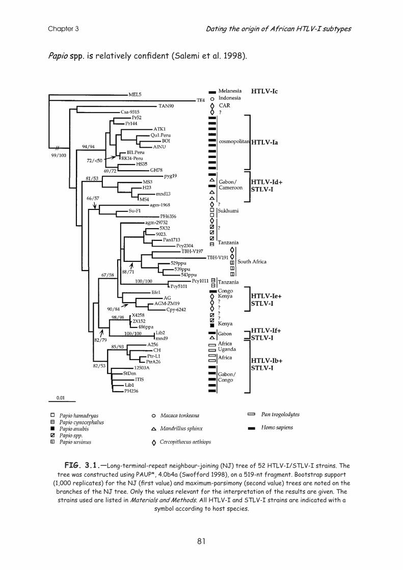
Chapter 3 Dating the origin of African HTLV-I subtypes
81
Papio spp. is relatively confident (Salemi et al. 1998).
FIG. 3.1.—Long-terminal-repeat neighbour-joining (NJ) tree of 52 HTLV-I/STLV-I strains. The tree was constructed using PAUP*, 4.0b4a (Swofford 1998), on a 519-nt fragment. Bootstrap support
(1,000 replicates) for the NJ (first value) and maximum-parsimony (second value) trees are noted on the branches of the NJ tree. Only the values relevant for the interpretation of the results are given. The strains used are listed in Materials and Methods. All HTLV-I and STLV-I strains are indicated with a
symbol according to host species.
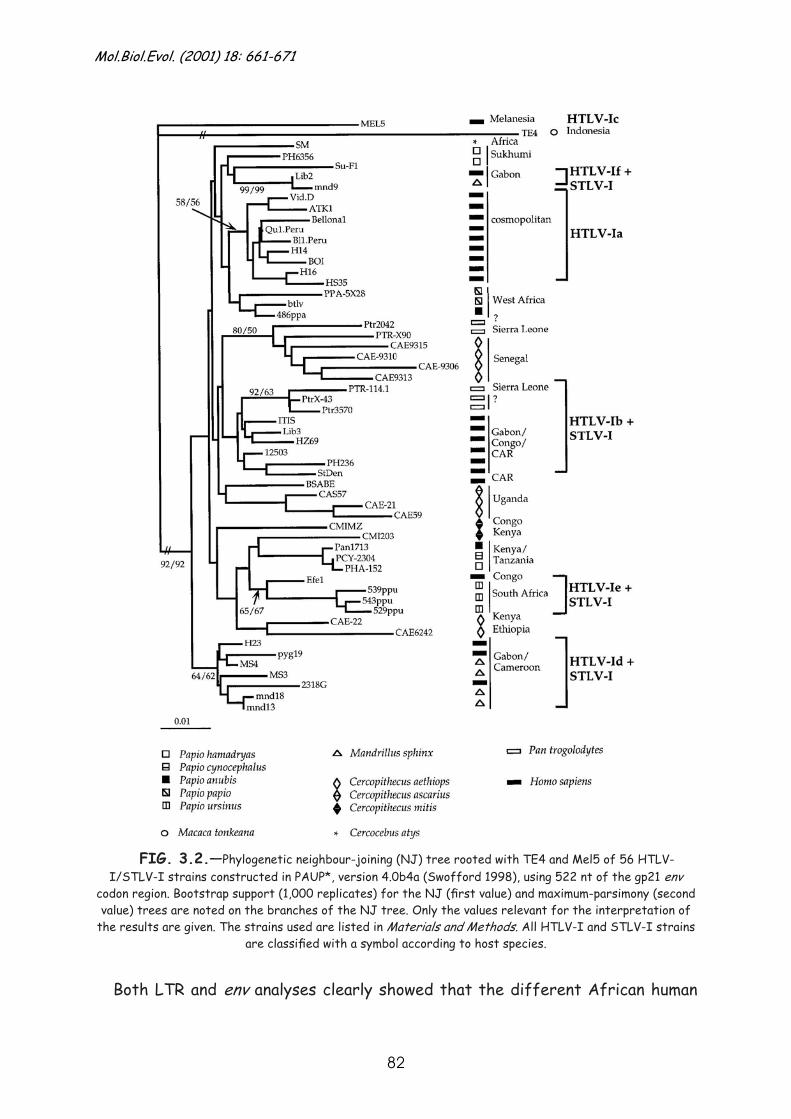
Mol.Biol.Evol. (2001) 18: 661-671
82
FIG. 3.2.—Phylogenetic neighbour-joining (NJ) tree rooted with TE4 and Mel5 of 56 HTLV-I/STLV-I strains constructed in PAUP*, version 4.0b4a (Swofford 1998), using 522 nt of the gp21 env
codon region. Bootstrap support (1,000 replicates) for the NJ (first value) and maximum-parsimony (second value) trees are noted on the branches of the NJ tree. Only the values relevant for the interpretation of
the results are given. The strains used are listed in Materials and Methods. All HTLV-I and STLV-I strains are classified with a symbol according to host species.
Both LTR and env analyses clearly showed that the different African human
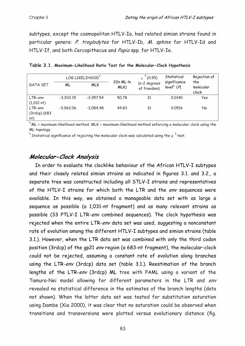
Chapter 3 Dating the origin of African HTLV-I subtypes
83
subtypes, except the cosmopolitan HTLV-Ia, had related simian strains found in particular genera: P. troglodytes for HTLV-Ib, M. sphinx for HTLV-Id and HTLV-If, and both Cercopithecus and Papio spp. for HTLV-Ie. Table 3.1. Maximum-Likelihood Ratio Test for the Molecular-Clock Hypothesis LOG LIKELIHOOD
a
DATA SET ML MLK
2(ln ML-ln
MLK)
χ 2
(0.95) (n-2 degrees of freedom)
Statistical significance levelb (P)
Rejection of the molecular clock
LTR-env (1,031 nt)
-3,932.15 -3,957.54 50.78 31 0.0140 Yes
LTR-env (3rdcp) (683 nt)
-3,062.06 -3,084.48 44.83 31 0.0516 No
a ML = maximum-likelihood method; MLK = maximum-likelihood method enforcing a molecular clock using the ML topology. b Statistical significance of rejecting the molecular clock was calculated using the χ 2 test.
Molecular-Clock Analysis In order to evaluate the clocklike behaviour of the African HTLV-I subtypes
and their closely related simian strains as indicated in figures 3.1. and 3.2., a separate tree was constructed including all STLV-I strains and representatives of the HTLV-I strains for which both the LTR and the env sequences were available. In this way, we obtained a manageable data set with as large a sequence as possible (a 1,031-nt fragment) and as many relevant strains as possible (33 PTLV-I LTR-env combined sequences). The clock hypothesis was rejected when the entire LTR-env data set was used, suggesting a nonconstant rate of evolution among the different HTLV-I subtypes and simian strains (table 3.1.). However, when the LTR data set was combined with only the third codon position (3rdcp) of the gp21 env region (a 683-nt fragment), the molecular-clock could not be rejected, assuming a constant rate of evolution along branches using the LTR-env (3rdcp) data set (table 3.1.). Reestimation of the branch lengths of the LTR-env (3rdcp) ML tree with PAML using a variant of the Tamura-Nei model allowing for different parameters in the LTR and env revealed no statistical difference in the estimates of the branch lengths (data not shown). When the latter data set was tested for substitution saturation using Dambe (Xia 2000), it was clear that no saturation could be observed when transitions and transversions were plotted versus evolutionary distance (fig.
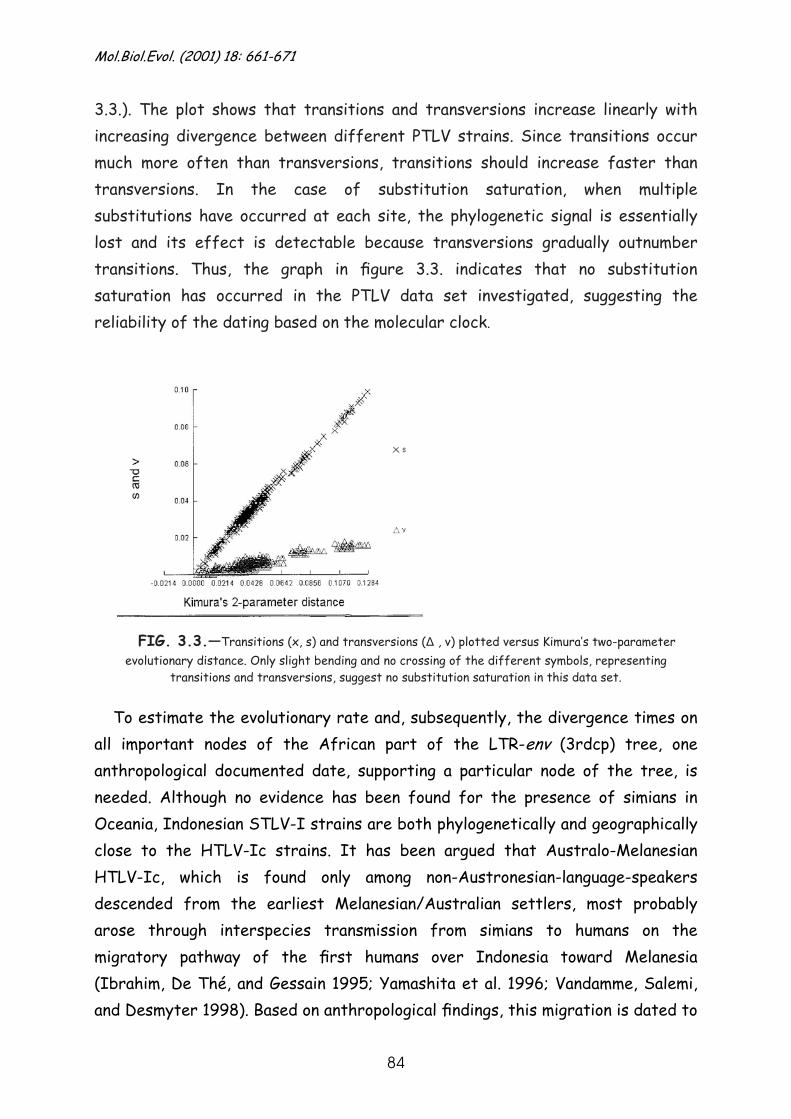
Mol.Biol.Evol. (2001) 18: 661-671
84
3.3.). The plot shows that transitions and transversions increase linearly with increasing divergence between different PTLV strains. Since transitions occur much more often than transversions, transitions should increase faster than transversions. In the case of substitution saturation, when multiple substitutions have occurred at each site, the phylogenetic signal is essentially lost and its effect is detectable because transversions gradually outnumber transitions. Thus, the graph in figure 3.3. indicates that no substitution saturation has occurred in the PTLV data set investigated, suggesting the reliability of the dating based on the molecular clock.
FIG. 3.3.—Transitions (x, s) and transversions (Δ , v) plotted versus Kimura’s two-parameter evolutionary distance. Only slight bending and no crossing of the different symbols, representing
transitions and transversions, suggest no substitution saturation in this data set.
To estimate the evolutionary rate and, subsequently, the divergence times on all important nodes of the African part of the LTR-env (3rdcp) tree, one anthropological documented date, supporting a particular node of the tree, is needed. Although no evidence has been found for the presence of simians in Oceania, Indonesian STLV-I strains are both phylogenetically and geographically close to the HTLV-Ic strains. It has been argued that Australo-Melanesian HTLV-Ic, which is found only among non-Austronesian-language-speakers descended from the earliest Melanesian/Australian settlers, most probably arose through interspecies transmission from simians to humans on the migratory pathway of the first humans over Indonesia toward Melanesia (Ibrahim, De Thé, and Gessain 1995; Yamashita et al. 1996; Vandamme, Salemi, and Desmyter 1998). Based on anthropological findings, this migration is dated to
Chapter 3 Dating the origin of African HTLV-I subtypes
85
around 50,000 ± 10,000 years ago. There is direct archaeological evidence of an early Australian settlement around 40,000 years ago (Cavalli-Sforza, Menozzi, and Piazza 1994) whereas thermoluminescence dating and mitochondrial DNA analysis (Roberts, Jones, and Smith 1990; E. Hagelberg, personal communication) suggest migrations as early as 60,000 years ago. Therefore, we assume that HTLV-Ic now found in Australo-Melanesia separated from the rest of the PTLV-I strains around 50,000 years ago, with a 95% confidence interval of 60,000–40,000 years ago.
The accuracy of the estimated evolutionary rate depends not only on the
accuracy of the time frame but also on the accurate estimation of genetic distances among taxa and the accuracy of the tree topology. To investigate the robustness of the LTR-env trees, two distance-based tree construction methods (NJ and FITCH) and one character-based method (ML) were used with an experimentally determined transition/transversion bias of 7.02. Although the major African clades themselves remained supported as described above, their branching order was dependent on the method used. This could result either from a true multifurcation, from recombination events in the past, or from a lack of data to resolve the multifurcating tree. It is unlikely that recent recombination events took place, since previous analyses showed no evidence of recombination between different subtypes (Salemi, Desmyter, and Vandamme 2000). However, it cannot be excluded that recombination took place very early, before the establishment of the subtypes. Using the splits decomposition method implemented in the software package of SplitsTree (Huson 1998), networks, indicating conflicting topologies, were especially situated close to the central node of the African part of the tree from which all of the African lineages arise (fig. 3.4.). However, overall, SplitsTree tends to support a multifurcation, and this multifurcation at the origin of PTLV-I in Africa as presented in figure 3.5. was used in order to deal with these nonconsistent tree topologies. For the same reason, considering the low bootstrap support for the topology within the subtypes in figures 3.1. and 3.2., we also represented the divergence within the HTLV-I subtypes as multifurcations in the schematic representation of figure 3.5.
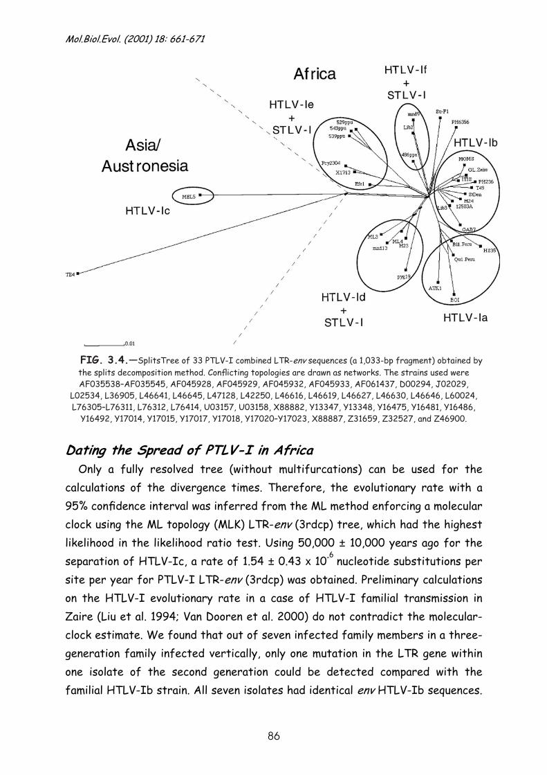
Mol.Biol.Evol. (2001) 18: 661-671
86
FIG. 3.4.—SplitsTree of 33 PTLV-I combined LTR-env sequences (a 1,033-bp fragment) obtained by the splits decomposition method. Conflicting topologies are drawn as networks. The strains used were AF035538–AF035545, AF045928, AF045929, AF045932, AF045933, AF061437, D00294, J02029,
L02534, L36905, L46641, L46645, L47128, L42250, L46616, L46619, L46627, L46630, L46646, L60024, L76305–L76311, L76312, L76414, U03157, U03158, X88882, Y13347, Y13348, Y16475, Y16481, Y16486,
Y16492, Y17014, Y17015, Y17017, Y17018, Y17020–Y17023, X88887, Z31659, Z32527, and Z46900.
Dating the Spread of PTLV-I in Africa Only a fully resolved tree (without multifurcations) can be used for the
calculations of the divergence times. Therefore, the evolutionary rate with a 95% confidence interval was inferred from the ML method enforcing a molecular clock using the ML topology (MLK) LTR-env (3rdcp) tree, which had the highest likelihood in the likelihood ratio test. Using 50,000 ± 10,000 years ago for the separation of HTLV-Ic, a rate of 1.54 ± 0.43 x 10⁻⁶
nucleotide substitutions per site per year for PTLV-I LTR-env (3rdcp) was obtained. Preliminary calculations on the HTLV-I evolutionary rate in a case of HTLV-I familial transmission in Zaire (Liu et al. 1994; Van Dooren et al. 2000) do not contradict the molecular-clock estimate. We found that out of seven infected family members in a three-generation family infected vertically, only one mutation in the LTR gene within one isolate of the second generation could be detected compared with the familial HTLV-Ib strain. All seven isolates had identical env HTLV-Ib sequences.
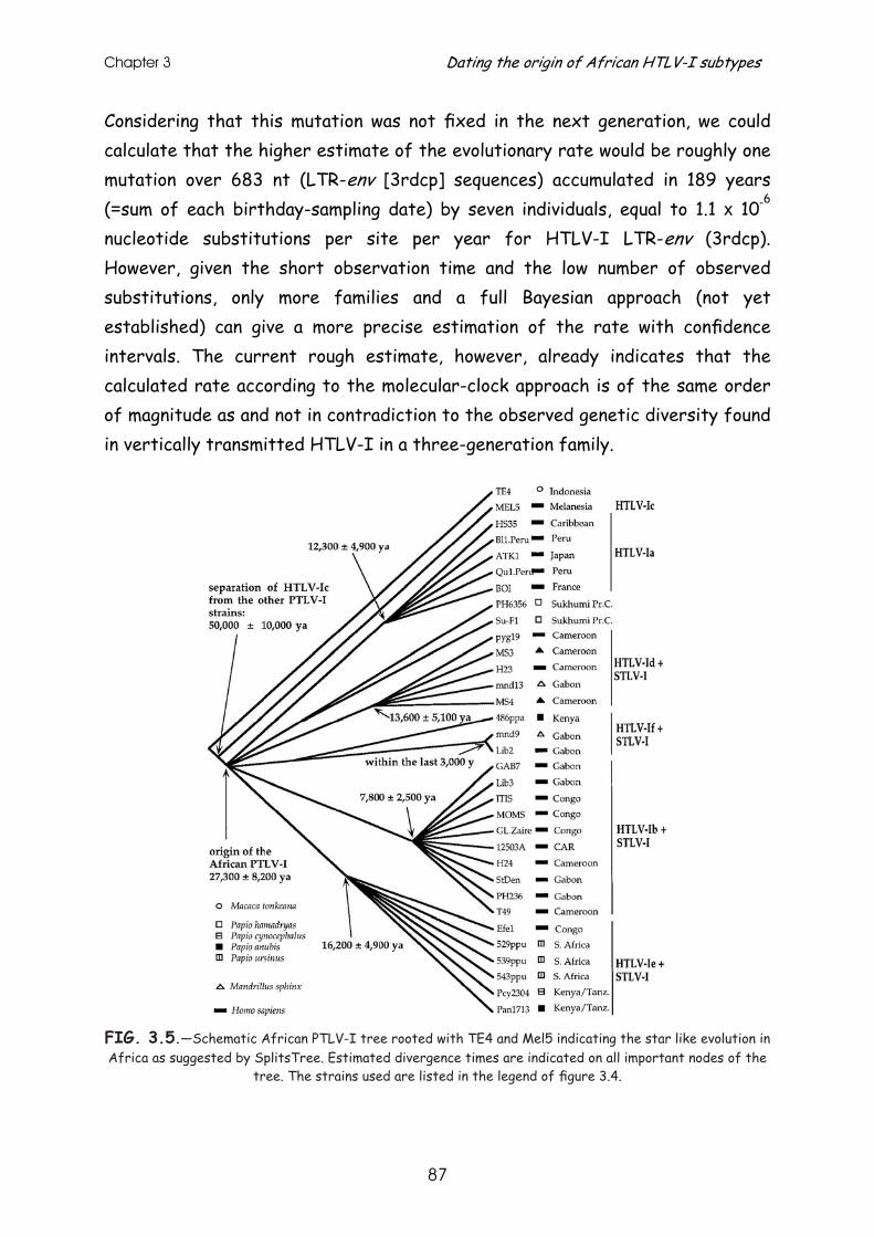
Chapter 3 Dating the origin of African HTLV-I subtypes
87
Considering that this mutation was not fixed in the next generation, we could calculate that the higher estimate of the evolutionary rate would be roughly one mutation over 683 nt (LTR-env [3rdcp] sequences) accumulated in 189 years (=sum of each birthday-sampling date) by seven individuals, equal to 1.1 x 10⁻⁶
nucleotide substitutions per site per year for HTLV-I LTR-env (3rdcp). However, given the short observation time and the low number of observed substitutions, only more families and a full Bayesian approach (not yet established) can give a more precise estimation of the rate with confidence intervals. The current rough estimate, however, already indicates that the calculated rate according to the molecular-clock approach is of the same order of magnitude as and not in contradiction to the observed genetic diversity found in vertically transmitted HTLV-I in a three-generation family.
FIG. 3.5.—Schematic African PTLV-I tree rooted with TE4 and Mel5 indicating the star like evolution in Africa as suggested by SplitsTree. Estimated divergence times are indicated on all important nodes of the
tree. The strains used are listed in the legend of figure 3.4.
Mol.Biol.Evol. (2001) 18: 661-671
88
The branch lengths of the MLK tree and their standard errors were then
further used to deduce the divergence times (including 95% confidence intervals) at the nodes of the African part of the tree using the estimated evolutionary rate and its standard error. The calculated dates at the origin of the HTLV-I subtypes are based on the currently known sequences and should be seen as lower limits (meaning true dates may be older). This is because current available strains are subject to sampling bias, and the true divergence within the clades may be higher. The African PTLV-I probably originated at least 27,300 ± 8,200 years ago. Considering the strains that we have, all major African HTLV-I subtypes except one (HTLV-If) probably arose within a range of no more than 10,000 years (see schematic multifurcated tree in fig. 3.5.). The Central African HTLV-Ie virus probably arose first, at least 16,200 ± 4,900 years ago. Later, the other subtypes arose: HTLV-Id 13,600 ± 5,100 years ago, the HTLV-Ia subtype 12,300 ± 4,900 years ago, and the HTLV-Ib subtype 7,800 ± 2,500 years ago. One exception seems to be the HTLV-If subtype, which has a very recent origin compared with the other African subtypes. Since Lib2 and mnd9 had identical sequences in the LTR-env (3rdcp), only a lower estimate for the HTLV-If origin could be calculated based on these two strains, and true dates of origin for this clade may be older, considering the possible sampling bias. Given an evolutionary rate of 1.54 x 10⁻⁶
nucleotide substitutions per site per year for an LTR-env (3rdcp) fragment of 683 nt, theoretically only one mutation would occur every 1,000 years in that region (evolutionary rate µ= 1.54 x 10-6
x 683 = 1.05 x 10⁻³
substitutions per year). Under a molecular clock following a Poisson distribution, Pn(t) = e-µt
.x (µt)n/n!, where Pn(t) is the probability of n
substitutions in time t with an evolutionary rate µ, the probability of observing no substitutions in t years between Lib2 and mnd9 in the LTR-env (3rdcp) data set is given by P0(t) = e-µt
. Therefore, we can be 95% confident that the two identical sequences in the LTR-env (3rdcp) data set diverged within the last 3,000 years (t =-ln(0.05)/1.54 x 10-6
x 683 nt). Further dating within these human HTLV-I subtypes to calculate certain
interspecies transmission events could not be performed due to the ambiguous internal topology of the HTLV-I/STLV-I strains within the clades indicated in figure 3.5. Several separate STLV-I clades, clearly seen in figures 3.1. and 3.2.,
Chapter 3 Dating the origin of African HTLV-I subtypes
89
are not represented in figures 3.4. and 3.5., since for none of these strains are both the LTR and env region available.
Test for the Selective Pressure Exerted on the env Region
Synonymous and nonsynonymous substitution distances were investigated for all available strains in the env region of HTLV-Ib, HTLV-Id, and their closely related STLV-I strains and for 39 strains representing the HTLV-Ia cluster.
For HTLV-Id, a branch-and-bound search was performed in order to
determine the ML tree with PAUP*, version 4.0b4a (Swofford 1998). For HTLV-Ia and HTLV-Ib/STLV-I, a heuristic search was performed because the number of strains was too large for a branch-and-bound search. ML trees were generated in PAUP*, version 4.0b4a, with the subtree-pruning-regrafting (SPR) branch-swapping algorithm on an NJ starting tree. The ancestral sequence topology, reconstructed on these ML trees in PAML, version 3.0 (Yang 1997), was used to calculate the synonymous versus nonsynonymous substitution distances in Dambe (Xia 2000). An interesting observation is that some of the HTLV-I and STLV-I sequences are identical to those of their reconstructed ancestors. The average KA/KS ratios, shown in column 6 of table 3.2., were always less than 1. Moreover, the difference between KA and KS was significant (with P < 0.01) for the HTLV-Ib/STLV-I and HTLV-Id/STLV-I groups using a paired t-test. Within the investigated HTLV-Ia clade, the difference between KA and KS
was not statistically significant. The divergence between these HTLV-Ia strains is probably too small given the amount of KA and/or KS distances of 0 and considering the large standard error for KA and KS values differing from 0. Although sporadic KA values were larger than KS values between specific nodes, no particular selective pressure was observed along human and simian strains and their ancestral sequences. Thus, there is evidence of strong purifying selection within the investigated HTLV-Ib/STLV-I and HTLV-Id/STLV-I groups. In addition, this purifying selection was equally obvious when human and simian strains were compared. Although the KA/KS ratio was less than 1 for HTLV-Ia, t-statistics indicated no evidence for negative selective pressure within this group of human strains. However, these results should still be carefully interpreted, since only the HTLV-Ia and HTLV-Ib subtypes had a reasonable amount of strains present in the clades investigated here.
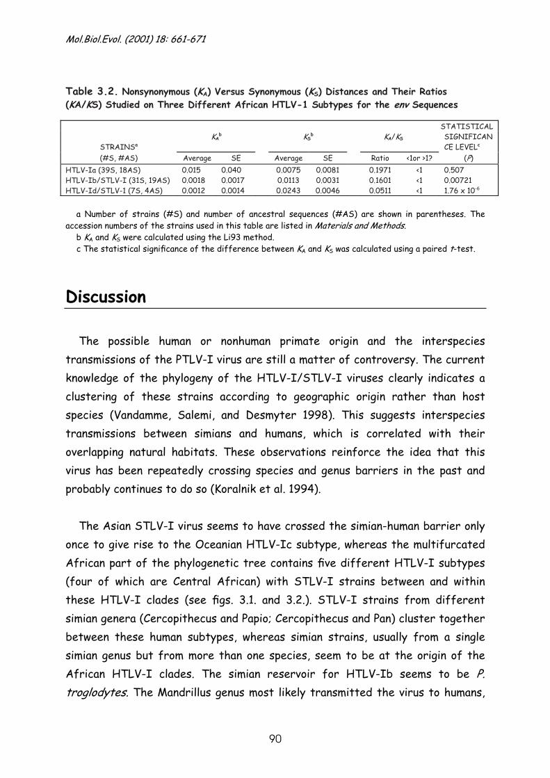
Mol.Biol.Evol. (2001) 18: 661-671
90
Table 3.2. Nonsynonymous (KA) Versus Synonymous (KS) Distances and Their Ratios (KA/KS) Studied on Three Different African HTLV-1 Subtypes for the env Sequences
STATISTICAL
STRAINSa KA
b KSb KA/KS SIGNIFICAN
CE LEVELc (#S, #AS) Average SE Average SE Ratio <1or >1? (P)
HTLV-Ia (39S, 18AS) HTLV-Ib/STLV-I (31S, 19AS) HTLV-Id/STLV-1 (7S, 4AS)
0.015 0.0018 0.0012
0.040 0.0017 0.0014
0.0075 0.0113 0.0243
0.0081 0.0031 0.0046
0.1971 0.1601 0.0511
<1 <1 <1
0.507 0.00721 1.76 x 10⁻⁶
a Number of strains (#S) and number of ancestral sequences (#AS) are shown in parentheses. The
accession numbers of the strains used in this table are listed in Materials and Methods. b KA and KS were calculated using the Li93 method. c The statistical significance of the difference between KA and KS was calculated using a paired t-test.
Discussion The possible human or nonhuman primate origin and the interspecies
transmissions of the PTLV-I virus are still a matter of controversy. The current knowledge of the phylogeny of the HTLV-I/STLV-I viruses clearly indicates a clustering of these strains according to geographic origin rather than host species (Vandamme, Salemi, and Desmyter 1998). This suggests interspecies transmissions between simians and humans, which is correlated with their overlapping natural habitats. These observations reinforce the idea that this virus has been repeatedly crossing species and genus barriers in the past and probably continues to do so (Koralnik et al. 1994).
The Asian STLV-I virus seems to have crossed the simian-human barrier only
once to give rise to the Oceanian HTLV-Ic subtype, whereas the multifurcated African part of the phylogenetic tree contains five different HTLV-I subtypes (four of which are Central African) with STLV-I strains between and within these HTLV-I clades (see figs. 3.1. and 3.2.). STLV-I strains from different simian genera (Cercopithecus and Papio; Cercopithecus and Pan) cluster together between these human subtypes, whereas simian strains, usually from a single simian genus but from more than one species, seem to be at the origin of the African HTLV-I clades. The simian reservoir for HTLV-Ib seems to be P. troglodytes. The Mandrillus genus most likely transmitted the virus to humans,
Chapter 3 Dating the origin of African HTLV-I subtypes
91
giving rise to the HTLV-Id and HTLV-If subtypes (or vice versa?). The HTLV-Ie subtype Efe1 clusters in the LTR region with low bootstrap support together with Cercopithecus spp., whereas in the env region Efe1 clusters with a higher bootstrap support with Papio spp. This clustering with STLV-I strains from different genera does not reflect conflicting topologies between the LTR and env tree, but, rather, that for none of these strains are both LTR and env known.
It is now clearly established that PTLV-I originated in Asia (Vandamme,
Salemi, and Desmyter 1998), implying a migration of the host species from Asia to Africa. One could speculate on whether this host species that introduced this virus in Africa is of human or non-human primate origin. Two factors could point to African PTLV-I having human origins: (1) the Oceanian HTLV-Ic subtype is the closest ancestral clade of the African PTLV-I strains (Ibrahim et al. 1995; Mahieux et al. 1998; Salemi, Desmyter, and Vandamme 2000), and (2) most HTLV-I subtypes have a simian counterpart, but the point at which the clade branches is exactly as likely among HTLV-I strains as it is among STLV-I strains, suggesting that human-to-simian transmission cannot be excluded. However, it would be more logical to assume a simian origin of the African PTLV-I virus based on primate behaviour and phylogenetic data. First, many different simian species are infected with STLV-I. The virus could have been easily transmitted to different simian species and genera through fighting and to humans through hunting, slaughtering, and consuming raw infected simian meat or keeping simians as house pets. Second, although sampling in humans has been more intensive than sampling in simians, there are several simian STLV-I clades with no human representative and only one human clade (HTLV-Ia) with no simian representative (figs. 3.1. and 3.2.). From this point of view, the simian origin of the HTLV-Ia subtype remains a mystery. Perhaps the simian strains related to HTLV-Ia have not yet been discovered because of the limited sampling in simians thus far.
We tried to elucidate the time frame for the origin of the HTLV-I subtypes
and the presumed interspecies transmissions at the origin of these subtypes. Based on a starting date of 50,000 ± 10,000 years ago, representing the first human migrations from Indonesia toward Melanesia and Australia and most
Mol.Biol.Evol. (2001) 18: 661-671
92
probably coinciding with the separate evolution of HTLV-Ic, a time frame of 27,300 ± 8,200 years ago was inferred for the origin of the African PTLV-I. The migration of the PTLV-I host species from Asia to Africa must therefore have occurred roughly between 60,000 and 20,000 years ago. No evidence for simian migrations from Asia to Africa has yet been found for this time period. It has been suggested that human retrograde flows from West Asia to Africa might have occurred between 60,000 and 40,000 years ago (Cavalli-Sforza, Menozzi, and Piazza 1994). Anthropological findings even suggest that the ancestors of the Khoisan originated in East Africa or even earlier in Arabia and are probably related to West Asians. The southward expansion of the Khoisan must have happened during the last Stone Age, between 20,000 and 8,000 years ago. Another possible introduction of HTLV-I in Africa could be due to the anthropologically documented migration of Indo-European populations from Asia to Europe and North Africa that were at the origin of a late Paleolithic culture called Iberomarusian, flourishing in Spain and North Africa from 20,000 to 7,500 B.C. (Cavalli-Sforza, Menozzi, and Piazza 1994). If one of these migrations coincided with the origin of the African PTLV-I strains, one would have to assume a human origin for the African PTLV-I strains, possibly via an early human-to-simian transmission that then spread among simian species and possibly later was again transmitted to humans. Phylogenetically, it is more plausible that African PTLV-I is of simian origin; however, this would imply a simian migration from Asia to Africa, possibly coinciding with human migrations. Alternatively, perhaps PTLV-I could have been introduced into Africa by another domestic animal genus migrating along with these humans.
Dating the African primate interspecies transmissions at the origin of the
HTLV-I subtypes remains difficult. The calculated dates reflect only the origin of the HTLV-I/STLV-I clades based on the common ancestor of the currently known HTLV-I and STLV-I sequences, subject to sampling bias. True divergences within these clades might possibly be higher than the currently observed ones. From our calculations, all different African human subtypes but one (HTLV-If) seem to have arisen at least 16,200 ± 4,900 to 7,800 ± 2,500 years ago. The HTLV-If subtype seems to have had its origin more recently, less than 3,000 years ago, based on the Lib2 and mnd9 sequences. However, the simian strains clustering between the human HTLV-I subtypes do not form clear
Chapter 3 Dating the origin of African HTLV-I subtypes
93
clades, which makes it practically impossible to date transmissions between these different simian genera. The STLV-I strains clustering within one human subtype display ambiguous internal branching order with low bootstrap support, making it impossible to calculate dates of single interspecies transmission events. The only clear conclusion to be drawn from these computations is that simian-to-human (or vice versa?) transmissions at the origin of each HTLV-I subtype happened somewhere between 16,200 ± 4,900 to 7,800 ± 2,500 years ago for HTLV-Ia, HTLV-Ib, HTLV-Id, and HTLV-Ie, with probably still ongoing simian-to-human transmissions within these subtypes. The interspecies transmissions for HTLV-If occurred more recently, within the last 3,000 years.
Accurate dating is not only dependent on molecular-clock determination, but
also on an accurate estimate of the evolutionary rate and on representative sampling. Preliminary investigations on the HTLV-I sequence variation within families in endemic regions are in complete agreement with the rate used here to calibrate the clock (Van Dooren et al. 2000). This suggests that the estimated rate used is reasonable, although the true divergence dates could still be more ancient than the calculated dates as a result of sampling bias.
Synonymous versus nonsynonymous substitution calculations provided evidence
for a strong negative selective pressure, thus supporting purifying selection as a driving force for PTLV-I evolution, irrespective of whether the virus is transmitted within its own host species or whether species barriers are crossed. This implies that the third codon position of the env gene has a different influence on the evolution of PTLV-I than the first and second codon position of env, which have a stronger tendency to stay invariable. It can be said that the evolution of the third codon position is much more relaxed. This is completely compatible with the observations of the molecular-clock analyses, in which all investigated strains were evolving at the same rate when considering sequences of the LTR region combined with only the third codon position of the env region, especially since no significant substitution saturation was observed. Although here we only investigated the env region, we have described that PTLV-I strains in endemic populations evolve at equal rates when only the third codon positions of their total genome (tax and rex excluded) are considered (Salemi, Desmyter, and Vandamme 2000). Positive selection would generally be expected when the

Mol.Biol.Evol. (2001) 18: 661-671
94
virus has to cross species or genus barriers. However, this is not what we observe; on the contrary. The observation of a strong purifying selection could in part explain the stability of the PTLV-I genome in general, independent of the host species. The meaning of this phenomenon deserves further research.
Whether the African PTLV-I virus has a human or a simian origin is far from
being resolved. Molecular-clock analyses and selective pressure calculations give us only a rough idea about divergence times and selective pressure exerted on PTLV-I. To find the missing link between Asia and Africa, it would be interesting to analyze the phylogeny, evolutionary rate, and selective pressure aspects of more HTLV-I and STLV-I strains from all over the world, including anthropological specimens, and try to correlate them with the anthropological data.
We are grateful to Ria Swinnen for fine editorial help. The Rega Institute is part of the HERN concerted action supported by the Biomed program of the European Commission. This study was supported in part by grant 3009894N of the Belgian Foundation for Scientific Research. M.S. was supported by the Research Council of the Katholieke Universiteit Leuven.
Literature Cited BASTIAN, I., J. GARDNER, D.WEBB, and I. GARDNER. 1993. Isolation of a human T-lymphotropic virus type I strain from Australian aboriginals. J. Virol. 67:843–851. CAVALLI-SFORZA, L. L., P. MENOZZI, and A. PIAZZA. 1994. The history and geography of human genes. Princeton University Press, Princeton, N.J. FULTZ, P.N., L. SU, P. MAY, and J.T. WEST. 1997. Isolation of sooty mangabey simian T-cell leukemia virus type I (STLV-I(sm)) and characterization of a mangabey T-cell line coinfected with STLV-I (sm) and simian immunodeficiency virus SIVsmmPBj14. Virology 235:271–285. GESSAIN, A., F. BARIN, J.C. VERNANT, O. GOUT, L.MAURS, A. CALENDER, and G. DE THE. 1985. Antibodies to human T. lymphotropic virus type-I in patients with tropical spastic paraparesis. Lancet 2:407–410. GESSAIN, A., R. YANAGIHARA, G. FRANCHINI, R.M. GARRUTO, C.L. JENKINS, A.B. AJDUKIEWICZ, R.C. GALLO, and D.C. GAJDUSEK. 1991. Highly divergent molecular variants of human T-lymphotropic virus type I from isolated populations in Papua New Guinea and the Solomon Islands. Proc. Natl. Acad. Sci. USA 88:7694–7698. HAHN, B.H., G.M. SHAW, M. POPOVIC, A. LO-MONICO, R.C. GALLO, and F. WONG-
Chapter 3 Dating the origin of African HTLV-I subtypes
95
STAAL. 1984. Molecular cloning and analysis of a new variant of human T-cell leukemia virus (HTLV-Ib) from an African patient with adult T-cell leukemia-lymphoma. Int. J. Cancer 34:613–618. HUSON, D. H. 1998. SplitsTree: analyzing and visualizing evolutionary data. Bioinformatics 14:68–73. IBRAHIM, F., G. DE THE, and A. GESSAIN. 1995. Isolation and characterization of a new simian T-cell leukemia virus type 1 from naturally infected Celebes macaques (Macaca tonkeana): complete nucleotide sequence and phylogenetic relationship with the Australo-Melanesian human T-cell leukemia virus type 1. J. Virol. 69:6980–6993. KORALNIK, I.J., E. BOERI, W.C. SAXINGER et al. (17 co-authors). 1994. Phylogenetic associations of human and simian T-cell leukemia/lymphotropic virus type I strains: evidence for interspecies transmission. J. Virol. 68:2693–2707. LEE R.V., A.W. PROWTEN, S.K.SATCHIDANAND, and B.I.S. SRIVASTAVA. 1985. Non-Hodgkin’s lymphoma and HTLV1 antibodies in a gorilla. N. Engl. J. Med. 312:118–119. LIU, H.-F., P. GOUBAU, M. VAN BRUSSEL, K. VAN LAETHEM, Y.-C. CHEN,J. DESMYTER, and A.-M. VANDAMME. 1996. The three human T-lymphotropic virus type I subtypes arose from three geographically distinct simian reservoirs. J. Gen. Virol. 77:359–368. LIU, H.-F., A.-M. VANDAMME, K. KAZADI, H. CARTON,J. DESMYTER, and P. GOUBAU. 1994. Familial transmission and minimal sequence variability of human T-lymphotropic virus type I (HTLV-I) in Zaire. AIDS Res. Hum. Retroviruses 10:1135–1142. MAHIEUX, R., C. CHAPPEY, M.-C. GEORGES-COURBOT, G. DUBREUIL, P.MAUCLERE, A. GEORGES, and A. GESSAIN. 1998. Simian T-cell lymphotropic virus type 1 from Man-drillus sphinx as a simian counterpart of human T-cell lymphotropic virus type 1 subtype D. J. Virol. 72:10316–10322. MAHIEUX, R., F. IBRAHIM, P.MAUCLERE et al. (14 co-authors). 1997. Molecular epidemiology of 58 new African human T-cell leukemia virus type I (HTLV-1) strains: identification of a new and distinct HTLV-1 molecular subtype in Central Africa and in pygmies. J. Virol. 71:1317–1333. MIURA, T., T. FUKUNAGA, T. IGARASHI et al. (20 co-authors). 1994. Phylogenetic subtypes of human T-lymphotropic virus type I and their relations to the anthropological back-ground. Proc. Natl. Acad. Sci. USA 91:1124–1127. MIURA, T., M. YAMASHITA, V. ZANINOVIC et al. (11 co-au-thors). 1997. Molecular phylogeny of human T-cell leukemia virus type I and II of Amerindians in Colombia and Chile. J. Mol. Evol. 44:S76–S82. MIYOSHI, I., S. YOSHIMOTO, M. FUJISHITA, H. TAGUCHI, I. KUBONISHI, K. NIIYA, and M. MINEZAWA. 1982. Natural adult T-cell leukaemia virus infection in Japanese monkeys. Lancet 2:658. OSAME, M., M. MATSUMOTO, K. USUKU, S. IZUMO, N. IJICHI, H. AMITANI, M. TARA, and A. IGATA. 1987. Chronic progressive myelopathy associated with elevated antibodies to human T-lymphotropic virus type I and adult T-cell leukemia like cells. Ann. Neurol. 21:117–122. ROBERTS, R.G., R. JONES, and M.A. SMITH. 1990. Report of thermoluminescence dates supporting the arrival of people between 50 and 60 kya in southern Australia. Nature 345: 153. SAKAKIBARA, I., Y. SUGIMOTO, A. SASAGAWA, S. HONJO, H. TSUJIMOTO, H. NAKAMURA, and M. HAYAMI. 1986. Spontaneous malignant lymphoma in an African green monkey naturally infected with simian T-cell lymphotropic virus (STLV). J. Med. Primatol. 15:311–318.
Mol.Biol.Evol. (2001) 18: 661-671
96
SAKSENA, N.K., V. HERVE, J.P.DURAND et al. (19 co-authors). 1994. Seroepidemiologic, molecular, and phylogenetic analyses of simian T-cell leukemia viruses (STLV-I) from various naturally infected monkey species from central and western Africa. Virology 198:297–310. SALEMI, M., J. DESMYTER, and A.-M. VANDAMME. 2000. Tempo and mode of human and simian T-lymphotropic viruses (HTLV/STLV) evolution revealed by analyses of full genome sequences. Mol. Biol. Evol. 17:374–386. SALEMI, M., S. VAN DOOREN, E. AUDENAERT, E. DELAPORTE, P. GOUBAU, J. DESMYTER, and A.-M. VANDAMME. 1998. Two new human T-lymphotropic virus type I phylogenetic subtypes in seroindeterminates, a Mbuti pygmy and a Gabonese, have closest relatives among African SLTV-I strains. Virology 246:277–287. STRIMMER, K., and A. VON HAESELER. 1997. Likelihood-map-ping: a simple method to visualize phylogenetic content of a sequence alignment. Proc. Natl. Acad. Sci. USA 94:6815– 6819. SWOFFORD, D. L. 1998. PAUP*: phylogenetic analysis using parsimony (*and other methods). Version 4.0beta4a. Sin-auer, Sunderland, Mass. TSUJIMOTO, H., Y. NODA, K.-I. ISHIKAWA, H. NAKAMURA, M. FUKASAWA, I. SAKAKIBARA, A. SASAGAWA, S. HONJO, and M. HAYAMI. 1987. Development of adult T-cell leukemia-like disease in African green monkey associated with clonal integration of simian T-cell leukemia virus type I. Cancer Res. 47:269–274. VAN DOOREN, S., E. GOTUZZO, M.SALEMI et al. (11 co-au-thors). 1998. Evidence for a post-Columbian introduction of human T-cell lymphotropic virus type I in Latin America. J. Gen. Virol. 79:2695–2708. VAN DOOREN, S., M. SALEMI, M., H-F. LIU, D.VANCUYCK , C. REMONDEGUI, M.B.BOUZAS, A.TALARMIN, J. DESMYTER, P. GOUBAU, and A.-M. VANDAMME. 2000. Intrafamilial HTLV-I sequence divergence: a tool for estimating the HTLV-I evolutionary rate? HERN Meeting, Potsdam, Germany, May 19–21, 2000. VANDAMME, A.-M., H.-F. LIU, P. GOUBAU, and J. DESMYTER. 1994. Primate T-lymphotropic virus type I LTR sequence variation and its phylogenetic analysis: compatibility with an African origin of PTLV-I. Virology 202:212–223. VANDAMME, A.-M., M. SALEMI, and J. DESMYTER. 1998. The simian origins of the pathogenic human T-cell lymphotropic virus type I. Trends Microbiol. 6:477–483. VERSCHOOR, E.J., K.S. WARREN, H. NIPHUIS,HERIYANTO, R.A. SWAN, and L.L. HEENEY. 1998. Characterization of a simian T-lymphotropic virus from a wild-caught orangutan (Pongo pygmaeus) from Kalimantan, Indonesia. J. Gen. Virol. 79:51–55. VOEVODIN, A., E. SAMILCHUK, H. SCHATZL, E. BOERI, and G. FRANCHINI. 1996. Interspecies transmission of macaque simian T-cell leukemia/lymphoma virus type 1 in baboons resulted in an outbreak of malignant lymphoma. J. Virol. 70:1633–1639. XIA, X. 2000. Data analysis in molecular biology and evolution (DAMBE). Kluwer Academic Publishers, Boston. YAMASHITA, M., E. IDO, T. MIURA, and M. HAYAMI. 1996. Molecular epidemiology of HTLV-I in the world. 13:S124– S131. YANG, Z. 1997. PAML: a program package for phylogenetic analysis by maximum likelihood. CABIOS 13:555–556. YOSHIDA, M., I. MIYOSHI, and Y. HINUMA. 1982. Isolation and characterization of retrovirus from cell lines of human adult T-cell leukemia and its implication in the disease. Proc. Natl. Acad. Sci. USA 79:2031–2035.

Chapter 3 Dating the origin of African HTLV-I subtypes
97
PEKKA PAMILO, reviewing editor
Accepted December 19, 2000

Chapter 4 The low evolutionary rate of human T-cell lymphotropic virus type-1 confirmed by analysis of vertical
transmission chains
S. Van Dooren, O.G. Pybus, M. Salemi, H.-F. Liu, P. Goubau, C.
Remondegui, A. Talarmin, E. Gotuzzo, L.C.J. Alcantara, B.
Galvão Castro, and A.-M. Vandamme
Molecular. Biology and Evolution (2004) 21(3):603–611.

Photograph previous page: courtesy of Dr. Tine Verdonck
101
The low evolutionary rate of human T-cell lymphotropic virus type-1 confirmed by analysis of vertical transmission chains S. Van Dooren,* O.G. Pybus,† M. Salemi,* H.-F. Liu, ‡ P. Goubau,§ C. Remondegui,‼
A.Talarmin,¶
E. Gotuzzo,# L.C.J. Alcantara,** B. Galvão
Castro,**and A.-M. Vandamme* * Rega Institute for Medical Research, Katholieke Universiteit Leuven, Leuven, Belgium; † Department of Zoology, University of Oxford, Oxford, U.K.; ‡ Department of Medical Research, Mackay Memorial Hospital, Taipei, Taiwan; § Université Catholique de Louvain, Unité de Virologie, Bruxelles, Belgium;
‼ Hospital San Roque, San Salvador de Jujuy, Argentina; ¶ Laboratoire de Rétrovirologie, Institut Pasteur de la Guyane, Cayenne, French Guiana;
# Instituto de Medicina Tropical ‘Alexander Von Humboldt’, Universidad Peruana Cayetano Heredia, Lima, Peru; ** Gonçalo Moniz Research Center, Oswaldo Cruz Foundation, Bahia School of Medicine and Public Health, Salvador, Bahia, Brazil
Molecular Biology and Evolution (2004) 21(3):603–611.
The evolutionary rate of the human T-cell lymphotropic virus type-1 (HTLV-1) is considered to be very low, in strong contrast to the related human retrovirus HIV. However, current estimates of the HTLV-1 rate rely on the anthropological calibration of phylogenies using assumed dates of human migration events. To obtain an independent rate estimate, we analyzed two variable regions of the HTLV-1 genome (LTR and env) from eight infected families. Remarkable genetic stability was observed, as only two mutations in LTR (756 bp) and three mutations in env (522 bp) occurred within the 16 vertical transmission chains, including one ambiguous position in each region. The evolutionary rate in HTLV-1 was then calculated using a maximum-likelihood approach that used the highest and lowest possible times of HTLV-1 shared ancestry, given the known transmission histories. The rates for the LTR and env regions were 9.58 Х 10
-8–1.25 Х 10
-5 and 7.84
Х 10-7
–2.33 Х 10-5nucleotide substitutions per site per year, respectively.
A more precise estimate was obtained for the combined LTR-env data set, which was 7.06 Х 10
-7–1.38 Х 10
-5substitutions per site per year. We also
note an interesting correlation between the occurrence of mutations in HTLV-1 and the age of the individual infected.

Mol.Biol.Evol. (2004) 21: 603-611
102
Introduction The two types of human T-cell lymphotropic virus (HTLV), type 1 (HTLV-1) and
type 2 (HTLV-2), belong to the family Retroviridae, together with the human immunodeficiency viruses (HIV-1 and HIV-2). Although HTLV and HIV have a comparable morphology, life cycle, and genetic structure, their replication strategies seem to be quite different, resulting in different evolutionary behaviours. In particular, the HTLV genome is remarkably stable compared with that of HIV. Additionally, HTLV is not as transmissible as HIV, even though both viruses are blood borne and use similar transmission routes: (1) vertical, from mother-to-infant (especially via breastfeeding, for HTLV) (Wiktor et al. 1997; Bittencourt 1998; Ando et al. 2003), (2) horizontal, via sexual contact (Murphy et al. 1989; Kaplan et al. 1996; Plancoulaine et al. 1998), and (3) parenteral, via injection equipment. (Lee et al. 1989; Hjelle et al. 1990; Sullivan et al. 1991; Manns et al. 1992; Take et al. 1993). HTLV is mainly infectious through cell-bound virus and not through freely circulating virus. Initially, a short period of active viral replication is needed to establish infection of the lymphocytes. After transformation and immortalization of the lymphocytes, HTLV replication is largely maintained through clonal expansion of the infected cells (Wattel et al. 1995, 1996), rather than by the replication and propagation of new virions by reverse transcription of integrated proviral DNA. Thus, the sequence divergence observed among HTLV strains results from mutations incorporated during active viral replication and from somatic mutations acquired by the provirus during cellular mitosis.
The genetic stability of HTLV has been indicated by several estimates of the
evolutionary rate of the virus. The investigation of intrafamilial HTLV infections has demonstrated the presence of almost identical HTLV-1 sequences in several family members sampled over several generations (Nerurkar et al. 1993; Liu et al. 1994). In a more precise manner, the evolutionary rate of HTLV-1, measured as the number of nucleotide substitutions per site per year, has been estimated by phylogenetic analysis using two different methods. The conventional approach, which involves dividing the difference in branch lengths in a
Chapter 4 HTLV-1 evolutionary rate in vertical transmission chains
103
phylogenetic tree by the difference in isolation time (Li, Tanimura, and Sharp 1988), although straightforward, is not ideally suited for HTLV strains, as the observed sequence divergence is usually too small for reliable estimates to be obtained. This method has only been applied to HTLV-2 strains from intravenous drug users (Salemi et al. 1999). The second approach assumes a known time for a particular node in the HTLV phylogeny. All current calculations of the HTLV-1 evolutionary rate are based on an assumed time point in the migration history of the human host population (Yanagihara et al. 1995; Salemi et al. 1998; Van Dooren et al. 1998; Salemi, Desmyter, and Vandamme 2000; Van Dooren, Salemi, and Vandamme 2001). This time point is obtained from anthropological studies, so the estimated rates are dependent on the accuracy of the anthropological date and rely heavily on the assumption that the phylogenetic node in question coincides with the anthropological event.
Herein, we report an alternative approach that uses HTLV-1 familial
transmission data that is independent of anthropological dates. Familial HTLV-1 is predominantly transmitted vertically from mother to offspring or horizontally through sexual contact. However, only HTLV-1 sequences from vertically infected family members can contribute to the estimation of evolutionary rates, as data on the timing of horizontal transmissions are almost impossible to collect, forcing us to exclude these events from our calculations.
To obtain an estimate of the molecular evolutionary rate of HTLV-1 that is
independent of anthropological data, we have sequenced the complete LTR region and a 522 bp fragment of the gp21 env region from HTLV-1 strains in eight families. First, the phylogenetic relationships of the strains were studied. Subsequently, we estimated the evolutionary rate of the sequenced genome regions from the total number of mutations accumulated during the combined period of vertical infection that was represented by the pedigrees of the infected families.
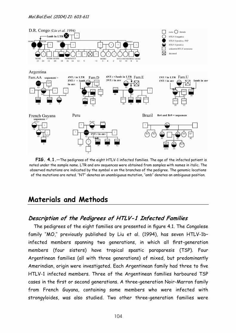
Mol.Biol.Evol. (2004) 21: 603-611
104
FIG. 4.1.—The pedigrees of the eight HTLV-1 infected families. The age of the infected patient is noted under the sample name. LTR and env sequences were obtained from samples with names in italic. The observed mutations are indicated by the symbol x on the branches of the pedigree. The genomic locations of the mutations are noted. ‘‘NT’’ denotes an unambiguous mutation, ‘‘amb’’ denotes an ambiguous position.
Materials and Methods
Description of the Pedigrees of HTLV-1 Infected Families The pedigrees of the eight families are presented in figure 4.1. The Congolese
family ‘‘MO,’’ previously published by Liu et al. (1994), has seven HTLV-1b– infected members spanning two generations, in which all first-generation members (four sisters) have tropical spastic paraparesis (TSP). Four Argentinean families (all with three generations) of mixed, but predominantly Amerindian, origin were investigated. Each Argentinean family had three to five HTLV-1 infected members. Three of the Argentinean families harboured TSP cases in the first or second generations. A three-generation Noir-Marron family from French Guyana, containing some members who were infected with strongyloides, was also studied. Two other three-generation families were
Chapter 4 HTLV-1 evolutionary rate in vertical transmission chains
105
included: one from Peru of mixed origin (mestizo) and one from Brazil of black origin. Both families have five to six HTLV-1–infected family members, with a TSP case in the second generation of the Peruvian family. Blood samples from all members of the same family were collected in the same year.
DNA Isolation, PCR, and Sequencing
For all non-Argentinean families, DNA was extracted from lymphocytes isolated from blood that was collected on EDTA tubes or Vacutainer Cell Preparation tubes (Becton Dickinson). For the Argentinean families, DNA was extracted from dried whole-blood spots collected on filter paper. DNA was extracted using the classical proteinase K digestion and phenol-chloroform extraction, followed by ethanol precipitation or using the QiaAmp Mini Blood kit (Qiagen). The LTR and env regions were PCR amplified using previously described primers and PCR conditions (Van Dooren et al. 1998). The PCR products were agarose gel purified using the Qiaquick Gel extraction kit (Qiagen). Population sequencing was done by direct sequencing of the purified PCR products using the ABI Prism Big Dye Terminator Cycle Sequencing Ready Reaction Version 3.0 mixture (Applied Biosystems), and sequences were generated on an ABI Prism 310 according to the manufacturers’ instructions (Applied Biosystems). Sequence assembly was done using the ABI Sequence Navigator software (Applied Biosystems).
The accession numbers of the new Latin American sequences are AY324777 to AY324788 (LTR) and AY324789 to AY324800 (env). For the African family members, the accession numbers are Z31659 (MOMJ LTR) and X88884 (MOMS env).
Phylogeny
Only the phylogeny of the LTR region was studied, as closely related strains are best investigated using a highly variable genome region. A Blast search (www.ncbi.nml. gov/blast) was conducted on the Latin American isolates as an aid to HTLV-1 subtype identification. A detailed phylogenetic analysis was then performed. ClustalX (Jeanmougin et al. 1998) was used to align the nine nonidentical familial sequences with 59 HTLV-1 reference sequences from GenBank that belonged to the same subtype. Reference strains from the same geographic area were preferentially chosen, and three strains from other
Mol.Biol.Evol. (2004) 21: 603-611
106
subtypes were included as outgroups. Minor editing of the alignment was performed manually in MacClade version 3.04 (Maddison and Maddison 1992). Phylogenetic trees were estimated with PAUP* version 4.0b10 (Swofford 1998). The Tamura-Nei substitution model with gamma-distributed rate heterogeneity among sites was chosen as best model for PTLV-1 strains (Salemi, Desmyter, and Vandamme 2000) and was thus used to construct neighbour-joining (NJ) and maximum-likelihood (ML) trees. Using empirical base frequencies, the NJ tree was constructed by optimizing the substitution rate matrix and gamma shape parameter three times; this was followed by a bootstrap analysis (1,000 replicates). The ML tree was estimated using the substitution model and parameters obtained above. A heuristic ML search was performed using the subtree-pruning-regrafting branch-swapping algorithm and an NJ starting tree. Statistical support for the ML tree branches was calculated in PAUP* using a likelihood ratio test that compared the likelihood of the estimated branch length with that of a zero branch length.
HTLV-1 Evolutionary Rate Estimation
The sequences from each family were aligned and the position and number of observed mutations in the LTR and env regions were scored (see fig. 4.1.).
The HTLV evolutionary rate was calculated using a homogenous Poisson
process model. Taken together, the pedigrees represent a total amount of time (t years), during which n mutations were observed in the combined LTR-env region. Therefore, the likelihood of the evolutionary rate (λ) in this region is
( )n
t
tn
entL λλλ
!),|(
−
= (1)
The pedigree data can be used to calculate the highest (y) and lowest (x) possible values of t. For each pedigree, the lowest possible value of t is obtained by summing the ages of the different offspring at the time of sampling, and then adding the age of the oldest child to account for evolution that occurred within the mother. This procedure assumes a maximum amount of shared ancestry. Summing this value across all pedigrees gives a lowest possible value of x = 419 years. For the highest possible t, the age of the mother at the time of sampling was summed for each offspring and also for the mother. This
Chapter 4 HTLV-1 evolutionary rate in vertical transmission chains
107
procedure assumes no shared ancestry within the pedigree of each family. When summed across all pedigrees, this yields a maximum value of y = 1,101 years. Therefore, the pedigrees represent a total of 419 to 1,101 years of HTLV-1 evolution. When this range is incorporated uniformly into the Poisson model we obtain the marginal likelihood
( )∫=
−
−=
y
xt
nt
dtxy
tn
enL .)(!
)|( λλλ
(2)
which has the solution
( ) ( )( )∑=
−−
−
−=
n
i
iyix yexeixy
nL0 !
1)(
1)|( λλλ
λ λλ (3)
Equation 3 was used to obtain maximum-likelihood estimates of the evolutionary rate λ. Because two of the proposed positions are ambiguous, separate estimates of λ were obtained for n=3, n=4, and n=5. Approximate 95% confidence intervals were obtained using the likelihood ratio statistic. The value λ represents the number of mutations per year in the investigated LTR and/or env region. Thus, the standard rate of molecular evolution (mutations per nucleotide site per year) is obtained by dividing λ by the length of the investigated region (1,278 nucleotides for LTR-env).
Results
PCR and Sequencing PCR amplification of HTLV DNA was successful for all blood samples
(indicated in italic in fig. 4.1.), except for a few members of the Brazilian family (Br1, Br5, and Br8). Different combinations of primers and extra inner PCR cycles were tried out, and alternative primer sets for LTR (Alcantara et al. 2003) and env (Yang et al. 1997) were used, resulting in negative or very faint PCR bands. These three samples were therefore not included in the study.
Phylogeny
Figure 4.2. shows the results of the phylogenetic analysis. Both NJ and ML
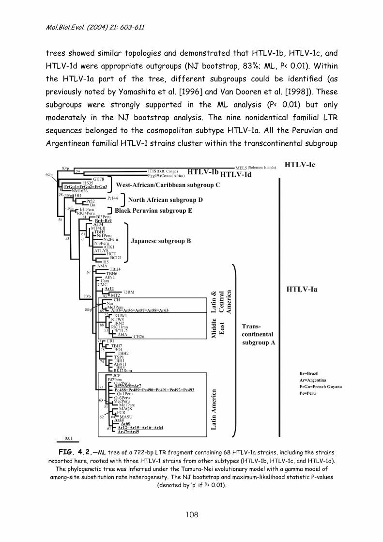
Mol.Biol.Evol. (2004) 21: 603-611
108
trees showed similar topologies and demonstrated that HTLV-1b, HTLV-1c, and HTLV-1d were appropriate outgroups (NJ bootstrap, 83%; ML, P< 0.01). Within the HTLV-1a part of the tree, different subgroups could be identified (as previously noted by Yamashita et al. [1996] and Van Dooren et al. [1998]). These subgroups were strongly supported in the ML analysis (P< 0.01) but only moderately in the NJ bootstrap analysis. The nine nonidentical familial LTR sequences belonged to the cosmopolitan subtype HTLV-1a. All the Peruvian and Argentinean familial HTLV-1 strains cluster within the transcontinental subgroup
FIG. 4.2.—ML tree of a 722-bp LTR fragment containing 68 HTLV-1a strains, including the strains reported here, rooted with three HTLV-1 strains from other subtypes (HTLV-1b, HTLV-1c, and HTLV-1d).
The phylogenetic tree was inferred under the Tamura-Nei evolutionary model with a gamma model of among-site substitution rate heterogeneity. The NJ bootstrap and maximum-likelihood statistic P-values
(denoted by ‘p’ if P< 0.01).
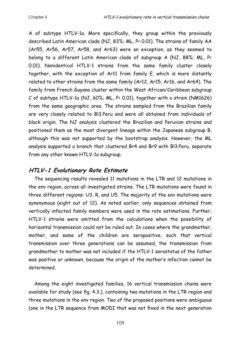
Chapter 4 HTLV-1 evolutionary rate in vertical transmission chains
109
A of subtype HTLV-1a. More specifically, they group within the previously described Latin American clade (NJ, 83%; ML, P< 0.01). The strains of family AA (Ar55, Ar56, Ar57, Ar58, and Ar63) were an exception, as they seemed to belong to a different Latin American clade of subgroup A (NJ, 88%; ML, P< 0.01). Nonidentical HTLV-1 strains from the same family cluster closely together, with the exception of Ar11 from family E, which is more distantly related to other strains from the same family (Ar12, Ar15, Ar16, and Ar64). The family from French Guyana cluster within the West African/Caribbean subgroup C of subtype HTLV-1a (NJ, 60%; ML, P< 0.01), together with a strain (NM1626) from the same geographic area. The strains sampled from the Brazilian family are very closely related to Bl3.Peru and were all obtained from individuals of black origin. The NJ analysis clustered the Brazilian and Peruvian strains and positioned them as the most divergent lineage within the Japanese subgroup B, although this was not supported by the bootstrap analysis. However, the ML analysis supported a branch that clustered Br4 and Br9 with Bl3.Peru, separate from any other known HTLV-1a subgroup.
HTLV-1 Evolutionary Rate Estimate
The sequencing results revealed 11 mutations in the LTR and 12 mutations in the env region, across all investigated strains. The LTR mutations were found in three different regions: U3, R, and U5. The majority of the env mutations were synonymous (eight out of 12). As noted earlier, only sequences obtained from vertically infected family members were used in the rate estimations. Further, HTLV-1 strains were omitted from the calculations when the possibility of horizontal transmission could not be ruled out. In cases where the grandmother, mother, and some of the children are seropositive, such that vertical transmission over three generations can be assumed, the transmission from grandmother to mother was not included if the HTLV-1 serostatus of the father was positive or unknown, because the origin of the mother’s infection cannot be determined.
Among the eight investigated families, 16 vertical transmission chains were
available for study (see fig. 4.3.), containing two mutations in the LTR region and three mutations in the env region. Two of the proposed positions were ambiguous (one in the LTR sequence from MODI that was not fixed in the next generation
Mol.Biol.Evol. (2004) 21: 603-611
110
and one in the env sequence from Ar47), making it impossible to draw conclusions regarding mutation fixation. Mutations in the LTR sequences occurred only in the R and U5 regions, and mutations in the env region occurred only at third codon positions (two synonymous mutations in family U and one nonsynonymous mutation in the Peruvian family).
ArgentinaFam.AA
Ar5616
Ar585
Ar5713
Ar6316
Fam.E
Ar1214
Ar167
Fam.D
Ar1514
French Guyana
Peru D.R. Congo (Liu et al. 1994) Brazil
Ar5634
00
Ar5834
00
Ar5734
00
Ar6334
nt≠ LTRnt≠env
lowest t = 419 years
Ar715
Ar615
Ar741
00
Ar641
Ar1241
00
Ar1641
00
Ar1541
Fam.U
Ar4935
Ar4730
Ar4435
Ar4970
11
Ar4770
0(1)
Ar4470
FrGu35
FrGu25
FrGu340
00
FrGu240
Pe49119
Pe49216
Pe49312
Pe49019
Pe49242
00
Pe49342
00
Pe49042
Pe49142
01
MOBE23
MODI23
MOBE41
(1)0
MODI41
MOBO16
MOGA16
MOBO39
00
MOGA39
MOMJ6
MOMA6
MOMJ29
00
MOMA29
Br419
Br919
Br442
00
Br942
nt≠ LTRnt≠env
highest t = 1101 years
lowest t = 419 years
highest t = 1101 years
FIG. 4.3.—The HTLV-1 vertical transmission chains, demonstrating the calculation of the lowest (t=419years) and highest (t=1,101years) possible times during which the mutations within the vertically
infected family members occurred. The lowest time is the result of the sum of the ages of the different offspring at the time of sampling, plus the age of the oldest child for the mother to correct for shared
ancestry. For the highest time, the age of the mother at the time of sampling was counted for each offspring and also for the mother (thus assuming no shared ancestry). The number of mutations observed in the LTR and env regions are shown below the transmission chains. The observed ambiguities are noted in
parentheses.
It is noteworthy that all mutations, including ambiguous ones, occurred in the
oldest individuals. The HTLV-1 strains of the family members between 5 and 19 years of age had no mutations, whereas sequences from individuals older than 18 years (19, 23, 30, and 35 years of age, respectively) showed at least one mutation in the LTR and/or env. To assess the statistical uncertainty arising from the presence of ambiguous mutations, separate evolutionary rate estimates were calculated for (1) no ambiguous changes, (2) one ambiguous change, and (3)
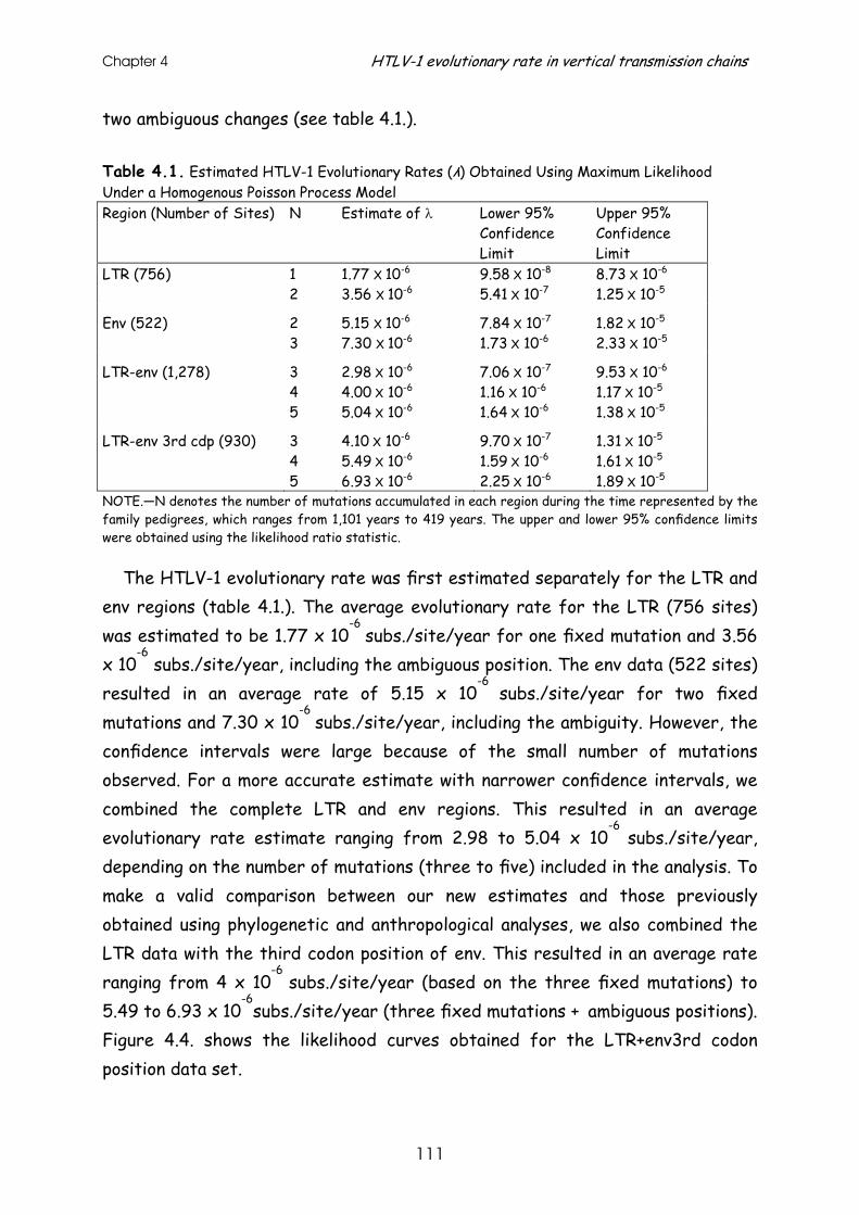
Chapter 4 HTLV-1 evolutionary rate in vertical transmission chains
111
two ambiguous changes (see table 4.1.).
Table 4.1. Estimated HTLV-1 Evolutionary Rates (λ) Obtained Using Maximum Likelihood Under a Homogenous Poisson Process Model Region (Number of Sites) N Estimate of λ Lower 95%
Confidence Limit
Upper 95% Confidence Limit
LTR (756) 1 2
1.77 Х 10-6 3.56 Х 10-6
9.58 Х 10-8 5.41 Х 10-7
8.73 Х 10-6 1.25 Х 10-5
Env (522) 2 3
5.15 Х 10-6 7.30 Х 10-6
7.84 Х 10-7 1.73 Х 10-6
1.82 Х 10-5 2.33 Х 10-5
LTR-env (1,278) 3 4 5
2.98 Х 10-6 4.00 Х 10-6 5.04 Х 10-6
7.06 Х 10-7 1.16 Х 10-6 1.64 Х 10-6
9.53 Х 10-6 1.17 Х 10-5 1.38 Х 10-5
LTR-env 3rd cdp (930) 3 4 5
4.10 Х 10-6 5.49 Х 10-6 6.93 Х 10-6
9.70 Х 10-7 1.59 Х 10-6 2.25 Х 10-6
1.31 Х 10-5 1.61 Х 10-5 1.89 Х 10-5
NOTE.—N denotes the number of mutations accumulated in each region during the time represented by the family pedigrees, which ranges from 1,101 years to 419 years. The upper and lower 95% confidence limits were obtained using the likelihood ratio statistic.
The HTLV-1 evolutionary rate was first estimated separately for the LTR and env regions (table 4.1.). The average evolutionary rate for the LTR (756 sites) was estimated to be 1.77 x 10
-6 subs./site/year for one fixed mutation and 3.56
x 10-6
subs./site/year, including the ambiguous position. The env data (522 sites) resulted in an average rate of 5.15 x 10
-6 subs./site/year for two fixed
mutations and 7.30 x 10⁻⁶
subs./site/year, including the ambiguity. However, the confidence intervals were large because of the small number of mutations observed. For a more accurate estimate with narrower confidence intervals, we combined the complete LTR and env regions. This resulted in an average evolutionary rate estimate ranging from 2.98 to 5.04 x 10
-6 subs./site/year,
depending on the number of mutations (three to five) included in the analysis. To make a valid comparison between our new estimates and those previously obtained using phylogenetic and anthropological analyses, we also combined the LTR data with the third codon position of env. This resulted in an average rate ranging from 4 x 10
-6 subs./site/year (based on the three fixed mutations) to
5.49 to 6.93 x 10⁻⁶
subs./site/year (three fixed mutations + ambiguous positions).
Figure 4.4. shows the likelihood curves obtained for the LTR+env3rd codon position data set.
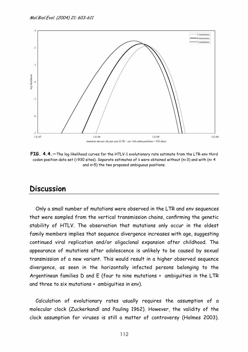
Mol.Biol.Evol. (2004) 21: 603-611
112
FIG. 4.4.—The log likelihood curves for the HTLV-1 evolutionary rate estimate from the LTR-env third codon position data set (=930 sites). Separate estimates of λ were obtained without (n=3) and with (n=
4
and n=5) the two proposed ambiguous positions.
Discussion Only a small number of mutations were observed in the LTR and env sequences
that were sampled from the vertical transmission chains, confirming the genetic stability of HTLV. The observation that mutations only occur in the oldest family members implies that sequence divergence increases with age, suggesting continued viral replication and/or oligoclonal expansion after childhood. The appearance of mutations after adolescence is unlikely to be caused by sexual transmission of a new variant. This would result in a higher observed sequence divergence, as seen in the horizontally infected persons belonging to the Argentinean families D and E (four to nine mutations +
ambiguities in the LTR
and three to six mutations + ambiguities in env).
Calculation of evolutionary rates usually requires the assumption of a
molecular clock (Zuckerkandl and Pauling 1962). However, the validity of the clock assumption for viruses is still a matter of controversy (Holmes 2003).
Chapter 4 HTLV-1 evolutionary rate in vertical transmission chains
113
Although the hypothesis of a strict molecular clock is rejected for many viral data sets, most likely because evolutionary rates vary among lineages, the resulting effect on estimated dates may be small if the rate variation is not large (Jenkins et al. 2002). Previous studies of PTLV have shown that the clock assumption may be applied but only under certain conditions. Either lineages (or complete clades) deviating from the clock must be removed from the analysis (Van Dooren et al. 1998), or sites affecting the molecular clock must be eliminated (Salemi et al. 2000; Van Dooren et al. 2001), resulting in the loss of molecular information. Alternatively, statistical methods that explicitly incorporate rate variation could be used (e.g., Thorne et al. 1998). HTLV-1 sequences from vertical transmission chains might be expected to evolve in a more clocklike manner, as the heterogeneity in the evolutionary rate is probably smaller when closely related sequences are considered.
The estimation of the HTLV-1 evolutionary rate using a molecular clock
requires an accurate calibration date for at least one node in the phylogeny. Previous analyses (Yanagihara et al. 1995; Salemi et al. 1998; Salemi, Desmyter, and Vandamme 2000; Van Dooren et al. 1998; Van Dooren, Salemi, and Vandamme 2001) have used time points based on anthropological events in the history of the viral host. In the analysis presented here, the calibration dates represent a well-known time frame, corresponding to the total number of years during which mutations in the vertical transmission chains occurred. More specifically, this time frame was expressed as the highest and lowest possible times for the accumulation of mutations among the different pedigrees. The reasoning behind the highest possible time is that, theoretically, one cannot know whether a mutation observed in the HTLV strain of the offspring was already present in the HTLV population of the mother before she gave birth (e.g., as a minor variant among the major HTLV clones). The lowest possible time assumes that the mutation really arose within the offspring. Thus for each mother-offspring pair, the highest or lowest possible times represent the minimum and maximum amount of shared common ancestry.
The inclusion of ambiguous positions in the calculations led to several
different rate estimates. The estimates presented in table 4.1. revealed that the LTR and env regions evolve at approximately equivalent rates, with a slightly
Mol.Biol.Evol. (2004) 21: 603-611
114
higher rate for the envelope gene compared with the LTR noncoding region. The combined LTR-env region resulted in estimates with smaller confidence intervals, thanks to an increased total amount of mutations.
It is important to note that the rate estimates provided in table 4.1. reflect
the evolution of HTLV-1 within vertically infected family members. As vertical HTLV-1 transmission is one of the main transmission routes in endemically infected areas, our estimate should reflect the true HTLV-1 evolutionary rate in such populations. All previously published estimates lie within the confidence limits of our new estimates. Our estimates for the LTR correspond to those of a previous study that investigated the cosmopolitan HTLV-1a subtype and its introduction into Latin America. (Van Dooren et al. 1998). The hypothesis concerning the dissemination of HTLV-1a in the New World is still a matter of controversy. Some analyses suggest the virus was first introduced on the American continent by an ancient migration of mongoloids across the Bering Strait 40,000 to 10,000 years ago (Miura et al. 1994, 1997; Yamashita et al. 1998; Ohkura et al. 1999; Li et al. 1999, Ramirez et al. 2002). Other molecular epidemiological studies indicate that HTLV was first introduced along with the post-Columbian African migration, which started approximately 400 years ago (Gessain et al. 1992, Gessain, Gallo, and Franchini 2000; Vandamme et al. 1994, 2000). Assuming either an ancient introduction or a post-Columbian introduction, the HTLV-1 LTR rate was estimated to be around 1.25 to 5 x 10
-7or 1.25 x 10
-5
subs./site/year, respectively. These estimates correspond to the upper and lower 95% confidence limits of the estimates for the LTR provided here. However, this comparison is complicated by the worldwide distribution of HTLV-1a strains included in the previous study; these strains are not necessarily restricted to the endemically infected populations. If horizontal transmission is greater among these nonendemic HTLV-1a isolates, then a rate that only takes into account vertical transmission will be an underestimate. In that case, the recent introduction theory would be more plausible. We have previously published an estimated rate for the combined LTR+env third codon position (Van Dooren, Salemi, and Vandamme 2001), using a data set containing African HTLV-1 subtypes and some STLV-1 strains that cluster with the human strains. In that study, the earliest human migrations to Melanesia and Australia 60,000 to 40,000 years ago were used as an anthropological calibration point, as these
Chapter 4 HTLV-1 evolutionary rate in vertical transmission chains
115
dates could be correlated with the isolated presence of HTLV-1 subtype c in Australia and Melanesia. This study reported a rate of 1.56 ± 0.43 x 10
-6
subs./site/year, similar to the range calculated here for four mutations. The analysis of Van Dooren, Salemi, and Vandamme (2001) contained simian strains, and the presence of this cross-species transmission event could have led to a difference between the estimated rates. Other published estimates of the evolutionary rate in PTLV, based on larger concatenated gene regions, resulted in rate estimates of 2.5 to 6.8 x 10
-7 subs./site/year (Yanagihara et al. 1995) and
≤ 1.67 ± 0.17 x 10⁻⁶ subs./site/year at the third codon position, using the
Melanesian migration calibration (Salemi, Desmyter, and Vandamme 2000). These data cannot be compared directly with the vertical transmission chain estimates presented here, because different coding regions and/or different PTLV types were investigated.
Using 16 HTLV-1 vertical transmission chains from eight HTLV-1 infected families, we have confirmed the genetic stability of the virus, even in genomic regions such as LTR and env that are expected to be relatively variable. Our estimates are based on a new approach that is independent of anthropological calibrations. In the future, more precise rate estimates could be calculated if samples and medical histories from patients were gathered over more than three generations, illustrating the importance of long-term sample collection, curation, and storage.
We thank Eddie Holmes and Philippe Lemey for the helpful discussions and for the
editorial advice. This work was supported by the Fonds voor Wetenschappelijk Onderzoek (Grant 0288.01). O.G.P. was funded by The Wellcome Trust.
Literature Cited Ando, Y., Y. Matsumoto, S. Nakano, K. Saito, K. Kakimoto, T. Tanigawa, Y. Ekuni, M. Kawa, and T. Toyama. 2003. Long-term follow up study of vertical HTLV-I infection in children breast-fed by seropositive mothers. J. Infect. 46:177–179. Alcantara, L. C., S. Van Dooren, M. S. Gonc¸alves et al. (12 coauthors). 2003. Globin haplotypes of human T-cell lymphotropic virus type I (HTLV-I) infected individuals in Salvador, Bahia, Brazil suggest a postcolumbian African origin of this virus. J. Acquir. Immune Defic. Syndr. 33:536– 542.
Mol.Biol.Evol. (2004) 21: 603-611
116
Bittencourt, A. L. 1998. Vertical transmission of HTLV-I/II: a review. Rev. Inst. Med. Trop. Sao Paulo 40:245–251. Gessain, A., R. C. Gallo, and G. Franchini. 1992. Low degree of human T-cell leukemia/lymphoma virus type I genetic drift in vivo as a means of monitoring viral transmission and movement of ancient human populations. J. Virol. 66:2288–2295. Gessain, A., J. Pecon-Slattery, L. Meertens, and R. Mahieux. 2000. Origins of HTLV-1 in South America. Nat. Med. 6:232. Hjelle, B, R. Mills, G. Mertz, and S. Swenson. 1990. Transmission of HTLV-II via blood transfusion. Vox. Sang. 59:119–122. Holmes, E. C. 2003. Molecular clocks and the puzzle of RNA virus origins. J. Virol. 77:3893–
3897. Jeanmougin, F., J. D. Thompson, M. Gouy, D. G. Higgins, and T. J. Gibson. 1998. Multiple sequence alignment with Clustal X. Trends Biochem. Sci. 23:403–405. Jenkins, G. M., A. Rambaut, O. G. Pybus, and E. C. Holmes. 2002. Rates of molecular evolution in RNA viruses: a quantitative phylogenetic analysis. J. Mol. Evol. 54:156– 165. Kaplan, J. E., R. F. Khabbaz, E. L. Murphy et al. (15 co-authors). 1996. Male-to-female transmission of human T-cell lymphotropic virus types I and II: association with viral load. The Retrovirus Epidemiology Donor Study Group. J. Acquir. Immune Defic. Syndr. Hum. Retrovirol. 1:193–201. Lee, H., P. Swanson, V. S. Shorty, J. A. Zack, J. D. Rosenblatt, and I. S. Chen. 1989. High rate of HTLV-II infection in seropositive i.v. drug abusers in New Orleans. Science. 28:471–475. Li, H.C., T. Fujiyoshi, H. Lou, S. Yashiki, S. Sonoda, L. Cartier, L. Nunez, I. Munoz, S. Horai, and K. Tajima. 1999. The presence of ancient human T-cell lymphotropic virus type I provirus DNA in an Andean mummy. Nat. Med. 5:1428– 1432. Li, W. H., M. Tanimura, and P. M. Sharp. 1988. Rates and dates of divergence between AIDS virus nucleotide sequences. Mol. Biol. Evol. 5:313–330. Liu H.F., A. M. Vandamme, K. Kazadi, H. Carton, J. Desmyter, and P. Goubau. 1994. Familial transmission and minimal sequence variability of human T-lymphotropic virus type I (HTLV-I) in Zaire. AIDS Res. Hum. Retrovir. 10:1135–1142. Maddison W., and R. Maddison. 1992. MacClade: analysis of phylogeny and character evolution. Version 3. Sinauer Associates, Sunderland, Mass. Manns, A., R. J. Wilks, E. L. Murphy, G. Haynes, J. P. Figueroa, M. Barnett, B. Hanchard, and W. A. Blattner. 1992. A prospective study of transmission by transfusion of HTLV-I and risk factors associated with seroconversion. Int. J. Cancer 30:886–891. Miura, T., T. Fukunaga, T. Igarashi et al. (20 co-authors) 1994. Phylogenetic subtypes of human T-lymphotropic virus type I and their relations to the anthropological background. Proc. Natl. Acad. Sci. USA 1994 91:1124–1127. Miura, T., M. Yamashita, V. Zaninovic et al. (11 co-authors). 1997. Molecular phylogeny of human T-cell leukemia virus type I and II of Amerindians in Colombia and Chile. J Mol Evol. 44:S76–82. Murphy, E. L., J. P. Figueroa, W. N. Gibbs, A. Brathwaite, M. Holding-Cobham, D. Waters, B. Cranston, B. Hanchard, and W. A. Blattner. 1989. Sexual transmission of human T-lymphotropic virus type I (HTLV-I). Ann. Intern. Med. 1:555–560. Nerurkar, V. R., K. J. Song, N. Saitou, R. R. Melland, and R. Yanagihara. 1993. Interfamilial and intrafamilial genomic diversity and molecular phylogeny of human T-cell lympho-tropic virus type I from Papua New Guinea and the Solomon Islands. Virology 196:506–513.
Chapter 4 HTLV-1 evolutionary rate in vertical transmission chains
117
Ohkura, S., M. Yamashita, L. Cartier, D. G. Tanabe, M. Hayami, S. Sonoda, and K. Tajima. 1999. Identification and phylogenetic characterization of a human T-cell leukaemia virus type I isolate from a native inhabitant (Rapa Nui) of Easter Island. J. Gen. Virol. 80:1995–2001. Plancoulaine, S., R. P. Buigues, E. L. Murphy et al. (12 coauthors). 1998. Demographic and familial characteristics of HTLV-1 infection among an isolated, highly endemic population of African origin in French Guiana. Int J Cancer 4:331–336. Ramirez, E., L. Cartier, C. Villota, and J. Fernandez. 2002. Genetic characterization and phylogeny of human T-cell lymphotropic virus type I from Chile. Virus Res. 20:135–149. Salemi, M., J. Desmyter, and A. M. Vandamme. 2000. Tempo and mode of human and simian T-lymphotropic virus (HTLV/ STLV) evolution revealed by analyses of full-genome sequences. Mol. Biol. Evol. 17:374–386. Salemi, M., A. M. Vandamme, J. Desmyter, C. Casoli, and U. Bertazzoni. 1999. The origin and evolution of human T-cell lymphotropic virus type II (HTLV-II) and the relationship with its replication strategy. Gene 24:11–21. Salemi, M., A. M. Vandamme, C. Gradozzi, K. Van Laethem, E. Cattaneo, G. Taylor, C. Casoli, P. Goubau, J. Desmyter, and U. Bertazzoni. 1998. Evolutionary rate and genetic heterogeneity of human T-cell lymphotropic virus type II (HTLV-II) using isolates from European injecting drug users. J. Mol. Evol. 46:602–611. Sullivan, M. T., A. E. Williams, C. T. Fang, T. Grandinetti, B. J. Poiesz, and G. D. Ehrlich. 1991. Transmission of human T-lymphotropic virus types I and II by blood transfusion: a retrospective study of recipients of blood components (1983 through 1988). The American Red Cross HTLV-I/II Collaborative Study Group. Arch. Intern. Med. 151:2043–2048. Swofford, D. L. 1998. PAUP*: phylogenetic analysis using parsimony (*and other methods). Version 4.0b5. Sinauer Associates, Sunderland, Mass. Take, H, M. Umemoto, K. Kusuhara, and K. Kuraya. 1993. Transmission routes of HTLV-I: an analysis of 66 families. Jpn. J. Cancer Res. 84:1265–1267. Thorne, J. L., H. Kishino, and I. S. Painter I. S. 1998. Estimating the rate of evolution of the rate of molecular evolution. Mol. Biol. Evol. 15:1647–1657. Vandamme, A. M., W. W. Hall, M. Lewis, P. Goubau, and M. Salemi. 2000. Origins of HTLV-1 in South America. Nat. Med. 6:232–233. Vandamme, A. M., H. F. Liu, P. Goubau, and J. Desmyter. 1994. Primate T-lymphotropic virus type I LTR sequence variation and its phylogenetic analysis: compatibility with an African origin of PTLV-I. Virology. 202:212–223. Van Dooren, S, E. Gotuzzo, M. Salemi et al. (11 co-authors) 1998. Evidence for a post-Columbian introduction of human T-cell lymphotropic virus type I in Latin America. J. Gen. Virol. 79:2695–2708. Van Dooren, S., M. Salemi, and A. M. Vandamme. 2001. Dating the origin of the African human T-cell lymphotropic virus type-I (HTLV-I) subtypes. Mol. Biol. Evol. 18:661–671. Wattel, E., M. Cavrois, A. Gessain, and S. Wain-Hobson. 1996. Clonal expansion of infected cells: a way of life for HTLV-I. J. Acquir. Immune Defic. Syndr. Hum. Retrovirol. 13:S92– S99. Wattel, E., J. P. Vartanian, C. Pannetier, and S. Wain-Hobson. 1995. Clonal expansion of human T-cell leukemia virus type I-infected cells in asymptomatic and symptomatic carriers without malignancy. J. Virol. 69:2863–2868. Wiktor, S. Z., E. J. Pate, P. S. Rosenberg, M. Barnett, P. Palmer, D. Medeiros, E. M. Maloney, and W. A. Blattner. 1997. Mother-to-child transmission of human T-cell lymphotropic virus type I associated with prolonged breast-feeding. J. Hum. Virol. 1:37–44.
Mol.Biol.Evol. (2004) 21: 603-611
118
Yamashita, M., E. Ido, T. Miura, and M. Hayami. 1996. Molecular epidemiology of HTLV-I in the world. J. Acquir. Immune Defic. Syndr. Hum. Retrovirol. 1(Suppl):S124–S131. Yamashita, M., G. Picchio, R. Veronesi, S. Ohkura, P. Bare, and M. Hayami. 1998. HTLV-Is in Argentina are phylogenetically similar to those of other South American countries, but different from HTLV-Is in Africa. J. Med. Virol. 55:152–160. Yanagihara, R., N. Saitou, V. R. Nerurkar, K. J. Song, I. Bastian, G. Franchini, and D. C. Gajdusek. 1995. Molecular phylogeny and dissemination of human T-cell lymphotropic virus type I viewed within the context of primate evolution and human migration. Cell Mol. Biol. 41:S145–161. Yang, Y. C., T. Y. Hsu, M. Y. Liu, M. T. Lin, J. Y. Chen, and C. S. Yang. 1997. Molecular subtyping of human T-lympho-tropic virus type I (HTLV-I) by a nested polymerase chain reaction-restriction fragment length polymorphism analysis of the envelope gene: two distinct lineages of HTLV-I in Taiwan. J. Med. Virol. 51:25–31. Zuckerkandl, E., and L. Pauling. 1962. Molecular disease, evolution, and genetic heterogeneity. Pp 189–225 inM. Kasha and B. Pullman, eds. Horizons in Biochemistry Academic Academic Press, New York.
DAN GRAUR, Associate Editor
Accepted November 4, 2003
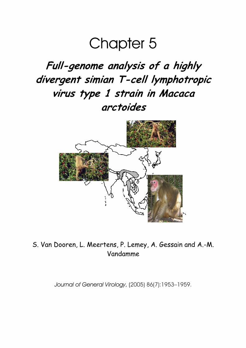
Chapter 5 Full-genome analysis of a highly
divergent simian T-cell lymphotropic virus type 1 strain in Macaca
arctoides
S. Van Dooren, L. Meertens, P. Lemey, A. Gessain and A.-M. Vandamme
Journal of General Virology, (2005) 86(7):1953–1959.

Photographs previous page: (upper) http://www.bbc.co.uk/nature/wildfacts/factfiles/225.shtml (lower) http://www.wared.gibbonproject.org/pdf.%20files/animal%20description/Stump-tailed%20Macaque_completed.pdf
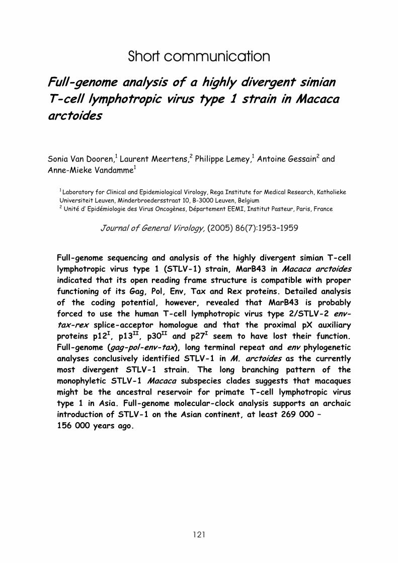
121
Short communication
Full-genome analysis of a highly divergent simian T-cell lymphotropic virus type 1 strain in Macaca arctoides Sonia Van Dooren,1 Laurent Meertens,2 Philippe Lemey,1 Antoine Gessain2 and Anne-Mieke Vandamme1
1 Laboratory for Clinical and Epidemiological Virology, Rega Institute for Medical Research, Katholieke Universiteit Leuven, Minderbroedersstraat 10, B-3000 Leuven, Belgium 2 Unité d’ Epidémiologie des Virus Oncogènes, Département EEMI, Institut Pasteur, Paris, France
Journal of General Virology, (2005) 86(7):1953–1959
Full-genome sequencing and analysis of the highly divergent simian T-cell lymphotropic virus type 1 (STLV-1) strain, MarB43 in Macaca arctoides indicated that its open reading frame structure is compatible with proper functioning of its Gag, Pol, Env, Tax and Rex proteins. Detailed analysis of the coding potential, however, revealed that MarB43 is probably forced to use the human T-cell lymphotropic virus type 2/STLV-2 env-tax-rex splice-acceptor homologue and that the proximal pX auxiliary proteins p12I, p13II, p30II and p27I seem to have lost their function. Full-genome (gag-pol-env-tax), long terminal repeat and env phylogenetic analyses conclusively identified STLV-1 in M. arctoides as the currently most divergent STLV-1 strain. The long branching pattern of the monophyletic STLV-1 Macaca subspecies clades suggests that macaques might be the ancestral reservoir for primate T-cell lymphotropic virus type 1 in Asia. Full-genome molecular-clock analysis supports an archaic introduction of STLV-1 on the Asian continent, at least 269 000 – 156 000 years ago.
J.Gen.Vir. (2005) 86:1953–1959
122
Simian T-cell lymphotropic virus (STLV) has been discovered in a broad spectrum of Old World monkey species and great apes and has been classified into STLV types 1-3. The human variant, human T-cell lymphotropic virus (HTLV) has been discovered in endemic populations, sometimes living in remote areas, but has also become disseminated worldwide. Both simian and human viral strains are generally referred to as primate T-cell lymphotropic virus (PTLV). STLV type 1 (STLV-1) has been discovered in more than 23 different species of the families Cercopithecidae (Pecon Slattery et al., 1999; Courgnaud et al., 2004) and Pongidae (Ibuki et al., 1997; Voevodin et al., 1997; Meertens et al., 2001; Nerrienet et al., 2004) and seems to be scattered not only over the African continent, but also among macaque and great ape species living in Asia. HTLV-1, which is classified into six different subtypes, has one cosmopolitan subtype (HTLV-1a; Yamashita et al., 1996) four subtypes restricted to Africa (HTLV-1b, -d, -e and –f; Nerurkar et al., 1993; Liu et al., 1994; Mahieux et al., 1997a; Salemi et al., 1998) and one subtype isolated only in descendants of the first settlers of Melanesia and Australia (HTLV-1c; Gessain et al., 1991). PTLV-1 phylogeny indicates a clear separation between the Asian-Melanesian and African STLV-1/HTLV-1 strains (Gessain et al., 1993; Nerurkar et al., 1993; Song et al., 1994). This distant, continental, phylogenetic relationship of PTLV-1 strains is reflected in a comparative difference in evolutionary patterns, although this might be partially biased by the fragmentary sampling in Asia. Asian STLV-1 strains cluster according to host species and form deep branches, indicating that they have undergone a long and independent species and subspecies evolution (Ibrahim et al., 1995). The same phenomenon has been observed to a lesser extent in its Melanesian/Australian human counterpart, HTLV-1c. On the African continent, the pattern of PTLV-1 infection has been a star-like burst followed by successful spread, characterised by a broad range of interspecies transmissions (Vandamme et al., 1998). The relationship between the different African PTLV-1 strains seems to be dependent more on geography than on the primate host species.
Overall, PTLV-1 phylogeny is peculiar in that it suggests an Asian origin for
PTLV-1 when phylogenetic trees are rooted with strains of other PTLV types. This observation questions the generally accepted hypothesis that the ancestor of all PTLVs originated in Africa (Salemi et al., 1999), based on the common

Chapter 5 Full genome analysis of STLV-1 in Macaca arctoides
123
occurrence of all different PTLV types on the African continent. Taken together, these facts create doubts about the possible origin of PTLV and the dissemination of PTLV-1 to Asia and Africa.
Full-genome sequencing and analysis of one of the most divergent STLV-1
strains of Asian origin would allow us to unravel a piece of this Asian PTLV-1 dissemination. Mahieux et al. (1997b) demonstrated that six Macaca arctoides of Southeast Asian origin housed at the Strasbourg Primatology Center showed a peculiar Western blot profile with an HTLV-2-like reactivity. Sequencing of part of the tax (406bp) region and part of the env (306bp) region of one M. arctoides strain, Marc1, showed that this strain was the most divergent among Asian STLV-1 strains. In the present study we sequenced the STLV-1 gp21env and long terminal repeat (LTR) regions from four other M. arctoides strains, identified by Mahieux et al. (1997b) [MAC912, MAC757, MAC897 and MAC11 (LTRregion only)] according to the methodology described previously (Mahieux et al., 1997a). The STLV-1 full genome sequence was obtained from (sufficiently available) original lymphocyte DNA of M. arctoides animal B43 (MarB43), as a virus-producing cell line could not be established for Marc1. The primers and PCR conditions for amplification are available at http://www.kuleuven.be/rega/cev/TableMarB43sequencingproject.pdf. The full genome of STLV MarB43 was obtained as shown in the sequencing project represented schematically in Fig. 5.1(a)., after agarose-gel purification of the PCR products with a Qiaquick gel extraction kit (Qiagen) and direct sequencing of both strands using an Big Dye Terminator technology on the ABI 310 Genetic Analyser (Applied Biosystems). The different sequenced fragments were analysed with Sequencing Analysis software (Applied Biosystems) and assembled using the software package Geneworks 2.5.1 (Oxford Molecular Systems). The Genbank accession number of the MarB43 complete genome is AY590142, and those of the M. arctoides env and LTR sequences AY141153-AY141155 and AY141171-AY141174, respectively.
All potential open reading frames (ORFs) of the MarB43 complete sequence
were analysed in Geneworks 2.5.1 (Oxford Molecular Systems), verified manually and compared with that of the ATK1 HTLV-1a reference strain.
J.Gen.Vir. (2005) 86:1953–1959
124
Fig.5.1: (a) MarB43 proviral full-genome sequencing strategy. Four proviral fragments were PCR-
amplified and sequenced directly. Three of the four fragments were amplified by using nested PCRs, with one outer (closed arrows) and two to three inner (open arrows) PCRs. (b,c) Schematic representation of the STLV-1 MarB43 proviral genome (b) and the putative resulting mRNAs (c). Nucleotide numbering is
according to the MarB43 proviral genome. Putative splice-donor (sd) and splice-acceptor (sa) sites and their positions within the proviral genome are indicated with vertical downward and upward arrows, respectively.
The positions of the primers designed to analyse the putative singly and doubly spliced messengers are indicated with small horizontal arrows.
The theoretical splicing pattern was analysed by using different software packages (http://www.biogenio.com/splice/index.html, http://www.lecb.ncifcrf. gov/~toms/spliceanalysis.html, http://l25.itba.mi.cnr.it/~webgene/wwwsplice view.html). The genomic organisation of MarB43 resembles that of other STLV-1 and HTLV-1 strains (Fig. 5.1b.). The major ORFs were found to be conserved. The 3’end of the tax gene was comparable with that of HTLV-1 and STLV-1 strains. Based on sequence comparison and theoretical splicing analysis, a few
RT INT ENV
REX
PRO
gp46 gp21 TAXPOL
U3 R U5U3 R U5 GAG
p24 p15p19
LTR LTR
0kb 1 2 3 4 5 6 7 8 9kb
5M4461 3M63655M4509 3M5312
5M5210
AV46
5M7711 MVB3PP84215M219 3M45885M219 3M1440
5M1343 3M31155M3007 3M4588
5M5210
3M6296
5M6230 3M70985M6725 3M7648
sd-LTR470
sa-env5088
sd-tax-rex5227
sa-tax-rex7367
gag-pol-env precursor
rextax
env
5M386 3M5422 3M74833M7324
*
**
*
AV46AV45
(a)
(b)
(c)
Chapter 5 Full genome analysis of STLV-1 in Macaca arctoides
125
differences in splicing pattern compared with HTLV-1 could be observed. MarB43 probably uses the HTLV-2/STLV-2 env-tax-rex splice-acceptor homologue (GCCCTCCAG↑5088GAGG; Fig. 5.1c.) due to a critical point mutation (A→T) in the conventional HTLV-1 splice-acceptor sequence (TATCCGTTG↑4897CTCT). A sequence-comparison analysis of the translation products within the proximal pX region, the pX ORF-I, -II and –V, normally obtained by alternative splicing, revealed that they probably cannot be synthesized. The three potential splice-acceptor sites of these ORFs were conserved enough to maintain their function (Fig. 5.1c.). However, identical point mutations within these ORFs, resulting in the absence of initiation codons and/or in early stop codons, were observed in the MarB43 and Marc1 sequences, two variants of this STLV-1 clade sequenced in different laboratories. The only exception would be p21 Rex, where the initiation codon remained conserved. Extra stop codons in tof/p30 (pX-Tax-ORFII), rof/p27 (pX-Rex-ORFI) and pX-Rex-ORFV shortly after the splice-acceptor sites (after amino acid positions 10, 8 and 31, respectively) would result in truncated proteins. The pX ORF-I and –II contained neither the p12 nor the p13 initiation codons, due to point mutations. On the one hand, the abolishment of the function of these proteins could explain the difficulties encountered during the PCR amplifications and the failure to establish a virus-producing cell line, respectively, as p13II and p30II are possibly involved in the maintenance of high proviral loads and p12I in the infectivity of primary lymphocytes and virus replication (Johnson et al., 2001; Albrecht & Lairmore, 2002). The transmission efficiency of this viral strain, however, has been proven in vivo, where this particular virus has been found circulating among at least six M. arctoides of the same colony (Mahieux et al., 1997b). On the other hand, the latter observation clearly demonstrates the viability of this circulating virus and thus questions the role of p12I (only potentially functional in HTLV-1a strains) in the infectivity and virus replication, both essential for the establishment of infection. These possible singly and doubly spliced mRNAs and the alternative splicing in the pX region were further experimentally analysed by RT-PCR on total RNA as described previously (Meertens et al., 2002). MarB43-specific primers were developed to analyse the LTR splice-donor and env and tax-rex splice-acceptor sites, respectively (sense primer 5M386, 5’-TACCTGAGGCCGCCATCCGTGC-3’; antisense primers 3M5422, 5’-AAGGGG-GTTGTAGGGCCTGATC-3’, and 3M7483, 5’-GAAGGCGGGCTGAGCATAGTCC-3’,
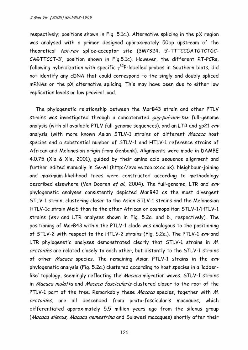
J.Gen.Vir. (2005) 86:1953–1959
126
respectively; positions shown in Fig. 5.1c.). Alternative splicing in the pX region was analysed with a primer designed approximately 50bp upstream of the theoretical tax-rex splice-acceptor site (3M7324, 5’-TTTCCGATGTCTGC- CAGTTCCT-3’, position shown in Fig.5.1c). However, the different RT-PCRs, following hybridization with specific γ32P-labelled probes in Southern blots, did not identify any cDNA that could correspond to the singly and doubly spliced mRNAs or the pX alternative splicing. This may have been due to either low replication levels or low proviral load.
The phylogenetic relationship between the MarB43 strain and other PTLV
strains was investigated through a concatenated gag-pol-env-tax full-genome analysis (with all available PTLV full-genome sequences), and an LTR and gp21 env analysis (with more known Asian STLV-1 strains of different Macaca host species and a substantial number of STLV-1 and HTLV-1 reference strains of African and Melanesian origin from Genbank). Alignments were made in DAMBE 4.0.75 (Xia & Xie, 2001), guided by their amino acid sequence alignment and further edited manually in Se-Al (http://evolve.zoo.ox.ac.uk). Neighbour-joining and maximum-likelihood trees were constructed according to methodology described elsewhere (Van Dooren et al., 2004). The full-genome, LTR and env phylogenetic analyses consistently depicted MarB43 as the most divergent STLV-1 strain, clustering closer to the Asian STLV-1 strains and the Melanesian HTLV-1c strain Mel5 than to the other African or cosmopolitan STLV-1/HTLV-1 strains (env and LTR analyses shown in Fig. 5.2a. and b., respectively). The positioning of MarB43 within the PTLV-1 clade was analogous to the positioning of STLV-2 with respect to the HTLV-2 strains (Fig. 5.2a.). The PTLV-1 env and LTR phylogenetic analyses demonstrated clearly that STLV-1 strains in M. arctoides are related closely to each other, but distantly to the STLV-1 strains of other Macaca species. The remaining Asian PTLV-1 strains in the env phylogenetic analysis (Fig. 5.2a.) clustered according to host species in a ‘ladder-like’ topology, seemingly reflecting the Macaca migration waves. STLV-1 strains in Macaca mulatta and Macaca fascicularis clustered closer to the root of the PTLV-1 part of the tree. Remarkably these Macaca species, together with M. arctoides, are all descended from proto-fascicularis macaques, which differentiated approximately 5.5 million years ago from the silenus group (Macaca silenus, Macaca nemestrina and Sulawesi macaques) shortly after their
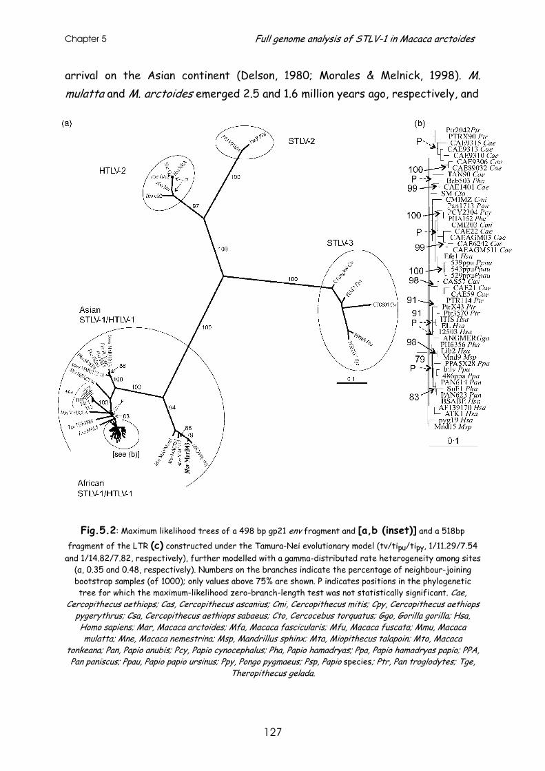
Chapter 5 Full genome analysis of STLV-1 in Macaca arctoides
127
arrival on the Asian continent (Delson, 1980; Morales & Melnick, 1998). M. mulatta and M. arctoides emerged 2.5 and 1.6 million years ago, respectively, and
Fig.5.2: Maximum likelihood trees of a 498 bp gp21 env fragment and [a,b (inset)] and a 518bp
fragment of the LTR (c) constructed under the Tamura-Nei evolutionary model (tv/tipu/tipy, 1/11.29/7.54 and 1/14.82/7.82, respectively), further modelled with a gamma-distributed rate heterogeneity among sites
(α, 0.35 and 0.48, respectively). Numbers on the branches indicate the percentage of neighbour-joining bootstrap samples (of 1000); only values above 75% are shown. P indicates positions in the phylogenetic tree for which the maximum-likelihood zero-branch-length test was not statistically significant. Cae,
Cercopithecus aethiops; Cas, Cercopithecus ascanius; Cmi, Cercopithecus mitis; Cpy, Cercopithecus aethiops pygerythrus; Csa, Cercopithecus aethiops sabaeus; Cto, Cercocebus torquatus; Ggo, Gorilla gorilla; Hsa, Homo sapiens; Mar, Macaca arctoides; Mfa, Macaca fascicularis; Mfu, Macaca fuscata; Mmu, Macaca mulatta; Mne, Macaca nemestrina; Msp, Mandrillus sphinx; Mta, Miopithecus talapoin; Mto, Macaca
tonkeana; Pan, Papio anubis; Pcy, Papio cynocephalus; Pha, Papio hamadryas; Ppa, Papio hamadryas papio; PPA, Pan paniscus; Ppau, Papio papio ursinus; Ppy, Pongo pygmaeus; Psp, Papio species; Ptr, Pan troglodytes; Tge,
Theropithecus gelada.
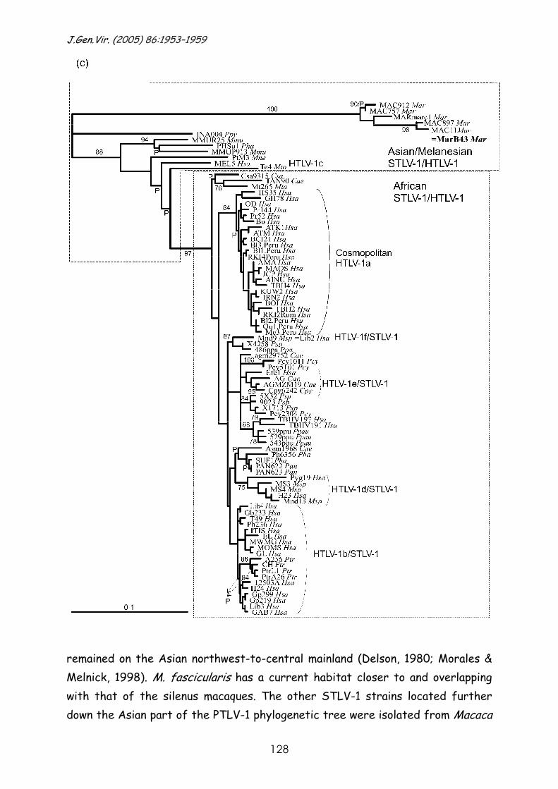
J.Gen.Vir. (2005) 86:1953–1959
128
remained on the Asian northwest-to-central mainland (Delson, 1980; Morales & Melnick, 1998). M. fascicularis has a current habitat closer to and overlapping with that of the silenus macaques. The other STLV-1 strains located further down the Asian part of the PTLV-1 phylogenetic tree were isolated from Macaca
Chapter 5 Full genome analysis of STLV-1 in Macaca arctoides
129
tonkeana and M. nemestrina (in the LTR analysis only; Fig. 5.2b.) of the Indonesian archipelago and in the Japanese Macaca fuscata. The first two Macaca species are descended from the silenus group, which migrated soon after their arrival in Asia across the Sundaland from southern China to what is now known as the Indonesian archipelago (Morales & Melnick, 1998). The Japanese M. fuscata probably differentiated from eastern rhesus macaques in more recent times (0.5 million years ago) (Delson, 1980; Morales & Melnick, 1998). The PTLV-1 topology from root to tip is thus indicative for an Asian host species migration-dependent evolution of the virus, suggestive of virus-host co-evolution, known as vicariance (Holmes, 2004), at least in the Macaca species. However, the observed degree of incongruence between the virus phylogeny and the host phylogeny creates doubts about the possibility of vicariance. The closer relationship of STLV-1 in M. mulatta and M. fascicularis to STLV-1 strains in silenus macaques (M. tonkeana and M. nemestrina) compared with STLV-1 in M. arctoides is in conflict with the host-species tree, where M. mulatta, M. fascicularis and M. arctoides all belong to the proto-fascicularis group.
Moreover, this apparent virus-host co-divergence is inconsistent with
molecular timings. The full-genome phylogenetic tree was recalibrated including the new, highly divergent MarB43 strain under a relaxed molecular clock, which accommodates rate variation among lineages, according to methodology described previously (Lemey et al., 2005) One calibration node, used frequently to estimate a timescale for PTLV evolution, appeared to coincide with the earliest human migrations to Melanesia and Australia 60 000-40 000 years ago (Gessain et al., 1993; Nerurkar et al., 1993; Yanagihara et al., 1995; Salemi et al., 2000; Van Dooren et al., 2001; Meertens & Gessain, 2003). Based on this time point, the mean evolutionary rate per gene was estimated to be 7x10⁻⁷ (4x10⁻⁷ –1.1x10⁻⁶) nucleotide substitutions per site per year. The molecular-clock calculations suggest that STLV-1 originated in Asia at least 269 000-156 000 years ago, using MarB43 as the earliest divergence within the PTLV-1 lineage, whereas PTLV-1 started to diverge in Africa only 23 000-18 000 years ago. The archaic presence of STLV-1 on the Asian continent is much more recent than the Macaca host invasion of Asia. The assumption of virus-host co-evolution is further violated by the occurrence of STLV-1 infection in orang-utans (Fig. 5.2b.) (Ibuki et al., 1997; Verschoor et al., 1998), suggesting STLV-1 cross-
J.Gen.Vir. (2005) 86:1953–1959
130
species transmissions between distantly related primate species. This apparent vicariance and virus-host phylogenetic incongruence has also been observed for a related primate retrovirus, simian immunodefiency virus. Charleston & Robertson (2002) demonstrated by simulation analysis that this might be explained by differential success in viral host-switching related to host (sub)species similarity and geographical proximity. A non-negligible difference, however, is the long branching pattern of the Asian host subspecies-specific PTLV-1 monophyletic clusters, suggesting that this preferential host subspecies switching mainly occurred early in the STLV-1 evolution and that the virus then further evolved within its specific host.
The current PTLV env phylogeny data certainly corroborate the enormous
diversity of PTLV-1 observed in Asia. The STLV-1 biodiversity found to date within macaques suggests that they could have formed the reservoir species for the founding PTLV-1 in Asia. To unravel the PTLV evolution further, in particular in Asia, and its relation to Africa, more PTLV strains from primates in the wild should be identified and analysed genetically, especially from Asia, to encompass a possible bias of an incomplete STLV-1 sampling pattern.
The genbank/EMBL/DDBJ accession numbers for the M. arctoides MarB43 complete
genome and the env and LTR sequences determined in this work are AY590142, AY141153-AY141155 and AY141171-AY141174, respectively.
This work was supported by the Fonds voor Wetenschappelijk Onderzoek (Grant
0288.01). L.M. is supported by the CANAM. We are grateful to N. Herrenschmidt and E. André from the Strasbourg Primatology Center for the generous gift of the biological materials of the M. arctoides troop.
References Albrecht, B. & Lairmore, M. D. (2002). Critical role of human Tlymphotropic virus type 1 accessory proteins in viral replication and pathogenesis. Microbiol Mol Biol Rev 66, 396Ð406. Charleston, M. A. & Robertson, D. L. (2002). Preferential host switching by primate lentiviruses can account for phylogenetic similarity with the primate phylogeny. Syst Biol 51, 528Ð535. Courgnaud, V., Van Dooren, S., Liegeois, F. & 7 other authors (2004). Simian T-cell leukemia virus (STLV) infection in wild primate populations in Cameroon: evidence for dual STLV type 1 and type 3 infection in agile mangabeys (Cercocebus agilis). J Virol 78, 4700Ð4709.
Chapter 5 Full genome analysis of STLV-1 in Macaca arctoides
131
Delson, E. (1980). Fossil macaques, phyletic relationships and a scenario of deployment. In The Macaques: Studies in Ecology, Behavior, and Evolution, pp. 10Ð30. Edited by D. G. Lindburg. New York: Van Nostrand Reinhold. Gessain, A., Yanagihara, R., Franchini, G., Garruto, R. M., Jenkins, C. L., Ajdukiewicz, A. B., Gallo, R. C. & Gajdusek, D. C. (1991). Highly divergent molecular variants of human T-lymphotropic virus type I from isolated populations in Papua New Guinea and the Solomon Islands. Proc Natl Acad Sci U S A 88, 7694Ð7698. Gessain, A., Boeri, E., Yanagihara, R., Gallo, R. C. & Franchini, G. (1993). Complete nucleotide sequence of a highly divergent human T-cell leukemia (lymphotropic) virus type I (HTLV-I) variant from Melanesia: genetic and phylogenetic relationship to HTLV-I strains from other geographical regions. J Virol 67, 1015Ð1023. Holmes, E. C. (2004). The phylogeography of human viruses. Mol Ecol 13, 745Ð756. Ibrahim, F., de Thé, G. & Gessain, A. (1995). Isolation and characterization of a new simian T-cell leukemia virus type 1 from naturally infected Celebes macaques (Macaca tonkeana): complete nucleotide sequence and phylogenetic relationship with the Australo-Melanesian human T-cell leukemia virus type 1. J Virol 69, 6980Ð6993. Ibuki, K., Ido, E., Setiyaningsih, S., Yamashita, M., Agus, L. R., Takehisa, J., Miura, T., Dondin, S. & Hayami, M. (1997). Isolation of STLV-I from orangutan, a great ape species in Southeast Asia, and its relation to other HTLV-Is/STLV-Is. Jpn J Cancer Res 88, 1Ð4. Johnson, J. M., Harrod, R. & Franchini, G. (2001). Molecular biology and pathogenesis of the human T-cell leukaemia/lymphotropic virus type-1 (HTLV-1). Int J Exp Pathol 82, 135Ð147. Lemey, P., Pybus, O. G., Van Dooren, S. & Vandamme, A.-M. (2005). A Bayesian statistical analysis of human T-cell lymphotropic virus evolutionary rates. Infect Genet Evol 5, 291Ð298. Liu, H. F., Vandamme, A.-M., Kazadi, K., Carton, H., Desmyter, J. & Goubau, P. (1994). Familial transmission and minimal sequence variability of human T-lymphotropic virus type I (HTLV-I) in Zaire. AIDS Res Hum Retroviruses 10, 1135Ð1142. Mahieux, R., Ibrahim, F., Mauclère P. & 11 other authors (1997a). Molecular epidemiology of 58 new African human T-cell leukemia virus type 1 (HTLV-1) strains: identification of a new and distinct HTLV-1 molecular subtype in Central Africa and in Pygmies. J Virol 71, 1317Ð1333. Mahieux, R., Pecon-Slattery, J. & Gessain, A. (1997b). Molecular characterization and phylogenetic analyses of a new, highly divergent simian T-cell lymphotropic virus type 1 (STLV-1marc1) in Macaca arctoides. J Virol 71, 6253Ð6258. Meertens, L. & Gessain, A. (2003). Divergent simian T-cell lymphotropic virus type 3 (STLV-3) in wild-caught Papio hamadryas papio from Senegal: widespread distribution of STLV-3 in Africa. J Virol 77, 782Ð789. Meertens, L., Rigoulet, J., Mauclère P. & 8 other authors (2001). Molecular and phylogenetic analyses of 16 novel simian T cell leukemia virus type 1 fromAfrica: close relationship of STLV-1 fromAllenopithecus nigroviridis to HTLV-1 subtype B strains. Virology 287, 275Ð285. Meertens, L., Mahieux, R., Mauclère P., Lewis, J. & Gessain, A. (2002). Complete sequence of a novel highly divergent simian T-cell lymphotropic virus from wild-caught red-capped mangabeys (Cercocebus torquatus) from Cameroon: a new primate T-lymphotropic virus type 3 subtype. J Virol 76, 259Ð268. Morales, J. C. & Melnick, D. J. (1998). Phylogenetic relationships of the macaques (Cercopithecidae: Macaca), as revealed by high resolution restriction site mapping of mitochondrial ribosomal genes. J Hum Evol 34, 1Ð23. Nerrienet, E., Meertens, L., Kfutwah, A., Foupouapouognigni, Y., Ayouba, A. & Gessain,
J.Gen.Vir. (2005) 86:1953–1959
132
A. (2004). Simian T cell leukaemia virus type I subtype B in a wild-caught gorilla (Gorilla gorilla gorilla) and chimpanzee (Pan troglodytes vellerosus) fromCameroon. J Gen Virol 85, 25Ð29. Nerurkar, V. R., Song, K.-J., Saitou, N., Melland, R. R. & Yanagihara, R. (1993). Interfamilial and intrafamilial genomic diversity and molecular phylogeny of human T-cell lymphotropic virus type I from Papua New Guinea and the Solomon Islands. Virology 196, 506Ð513. Pecon Slattery, J., Franchini, G. & Gessain, A. (1999). Genomic evolution, patterns of global dissemination, and interspecies transmission of human and simian T-cell leukemia/lymphotropic viruses. Genome Res 9, 525Ð540. Salemi, M., Van Dooren, S., Audenaert, E., Delaporte, E., Goubau, P., Desmyter, J. & Vandamme, A.-M. (1998). Two new human Tlymphotropic virus type I phylogenetic subtypes in seroindeterminates, a Mbuti pygmy and a Gabonese, have closest relatives among African STLV-I strains. Virology 246, 277Ð287. Salemi, M., Van Dooren, S. & Vandamme, A.-M. (1999). Origin and evolution of human and simian T-cell lymphotropic viruses. AIDS Rev 1, 131Ð139. Salemi, M., Desmyter, J. & Vandamme, A.-M. (2000). Tempo and mode of human and simian T-lymphotropic virus (HTLV/STLV) evolution revealed by analyses of full-genome sequences. Mol Biol Evol 17, 374Ð386. Song, K.-J., Nerurkar, V. R., Saitou, N., Lazo, A., Blakeslee, J. R., Miyoshi, I. & Yanagihara, R. (1994). Genetic analysis and molecular phylogeny of simian T-cell lymphotropic virus type I: evidence for independent virus evolution in Asia and Africa. Virology 199, 56Ð66. Vandamme, A.-M., Salemi, M. & Desmyter, J. (1998). The simian origins of the pathogenic human T-cell lymphotropic virus type I. Trends Microbiol 6, 477Ð483. Van Dooren, S., Salemi, M. & Vandamme, A.-M. (2001). Dating the origin of the African human T-cell lymphotropic virus type-I (HTLV-I) subtypes. Mol Biol Evol 18, 661Ð671. Van Dooren, S., Shanmugam, V., Bhullar, V., Parekh, B., Vandamme, A.-M., Heneine, W. & Switzer, W. M. (2004). Identification in gelada baboons (Theropithecus gelada) of a distinct simian T-cell lymphotropic virus type 3 with a broad range of Western blot reactivity. J Gen Virol 85, 507Ð519. Verschoor, E. J., Warren, K. S., Niphuis, H., Heriyanto, Swan, R. A. & Heeney, J. L. (1998). Characterization of a simian T-lymphotropic virus from a wild-caught orang-utan (Pongo pygmaeus) from Kalimantan, Indonesia. J Gen Virol 79, 51Ð55. Voevodin, A. F., Johnson, B. K., Samilchuk, E. I., Stone, G. A., Druilhet, R., Greer, W. J. & Gibbs, C. J., Jr (1997). Phylogenetic analysis of simian T-lymphotropic virus type I (STLV-I) in common chimpanzees (Pan troglodytes): evidence for interspecies transmission of the virus between chimpanzees and humans in Central Africa. Virology 238, 212Ð220. Xia, X. & Xie, Z. (2001). DAMBE: software package for data analysis in molecular biology and evolution. J Hered 92, 371Ð373. Yamashita, M., Ido, E., Miura, T. & Hayami, M. (1996). Molecular epidemiology of HTLV-I in the world. J Acquir Immune Defic Syndr Hum Retrovirol 13, S124ÐS131. Yanagihara, R., Saitou, N., Nerurkar, V. R., Song, K. J., Bastian, I., Franchini, G. & Gajdusek, D. C. (1995). Molecular phylogeny and dissemination of human T-cell lymphotropic virus type I viewed within the context of primate evolution and human migration. Cell Mol Biol 41 (Suppl. 1), S145ÐS161.
SIMON WAINE-HOBSON, editor
Received August 12, 2004; Accepted March 21, 2005
Chapter 6 Phylogeny of novel primate T-
lymphotropic virus type 1 strains in various Asian and African non-human
primate host species.
S. Van Dooren, E. J. Verschoor, Z. Fagrouch and A.-M. Vandamme
submitted to Virology

Photographs previous page: orang-utan: http://www.orangutans-sos.org/picgallery/source/24.html, photo by Elke Lies crested black macaque: http://www.tamansafari.com/mambo/index.php?option=content&task=view&id=160&Itemid= Moor macaque: http://members.tripod.com/uakari/macaca_maura.html siamang: http://members.tripod.com/uakari/hylobates_syndactylus.html Western lowland gorilla: http://www.congogorillaforest.com/congo-meet-gorilla red-tailed guenon: http://www.pri.kyoto-u.ac.jp/hope/reports/16-014.html West African chimpanzee: http://www.quantum-conservation.org/EEP/PAN%20TROGLODYTES%20VERUS.html (photo by Mr. Frands Carlsen) Barbary macaque: http://www.givskudzoo.dk/showlistrecord.asp?id=721&sortfield721=24&direction721=desc&doshow721=1&searchword721=&groupfield721=0&recordid721=3
135
Phylogeny of novel Primate T-Lymphotropic Virus type 1 strains in various Asian and African non-
human primate host species.
Sonia Van Dooren1, Ernst J. Verschoor2 Zahra Fagrouch2 and Anne-Mieke Vandamme1
1. Rega Institute for Medical Research, Katholieke Universiteit Leuven, Minderbroedersstraat 10, B-3000 Leuven, Belgium
2. Department of Virology, Biomedical Primate Research Center, PO Box 3306, 2280 GH Rijswijk, The Netherlands
submitted to Virology
To further unravel intra- and interspecies PTLV-1 evolution in Asia and
Africa, we phylogenetically analysed fifteen new STLV-1 strains discovered in eight different Asian and African non-human primate species. We show that orang-utan STLV-1s form a tight, deeply branching monophyletic cluster between Asian STLV-1 macaque species clades, suggesting natural cross-species transmission. Novel viruses of Macaca maura, Macaca nigra and siamang cluster with other Sulawesian STLV-1s, demonstrating close relatedness among the STLV-1s in these insular species and suggesting cross-species transmission to a siamang in captivity. Viruses from Western chimpanzees and a gorilla cluster within the HTLV-1b/STLV-1 clade, the latter close to a human strain, indicative of direct zoonosis. A new STLV-1 from Cercopithecus ascanius differs from the published STLV-Cas57, explainable by the existence of five geographically-separated subspecies. Barbary macaques carry a relatively recently acquired, typical African STLV-1, giving us no clue on the phylogeographical origin of PTLV-1.

submitted to Virology
136
Introduction
More than 25 different non-human primate species (NHP) belonging to the Old World monkeys and apes are infected with the Simian T-Lymphotropic Virus type 1 (STLV-1) (Koralnik et al., 1994; Saksena et al., 1994; Meertens et al., 2001; Gessain & Mahieux, 2000; Ibrahim et al., 1995; Ibuki et al., 1997; Liu et al., 1997; Mahieux et al., 1997; Mahieux et al., 1998; Mahieux et al., 2000; Nerrienet et al., 2001; Richards et al., 1998; Slattery et al., 1999; Voevodin et al., 1996; Voevodin et al., 1997). Their natural habitats are confined to sub-Saharan Africa and Asia, ranging from India to the Indonesian archipelago and to Japan. Humans harbour a similar virus, the Human T-Lymphotropic Virus type 1, identified in populations living in close geographic proximity to these STLV-1 infected NHPs. However, the increased human mobility facilitated the world-wide dissemination of HTLV-1. Because of their close molecular and phylogenetic relatedness, HTLV-1 and STLV-1 are together referred to as Primate T-Lymphotropic Viruses type 1 (PTLV-1), (Salemi et al., 1999).
Currently, HTLV-1 is divided in 6 different subtypes: one
Melanesian/Australian subtype c (HTLV-1c) (Gessain et al., 1991), 4 different African HTLV-1 subtypes (HTLV-1b, -1d, -1e and -1f), each of them resulting from separate simian-to-human transmissions in their respective geographic locations, and a cosmopolitan subtype, presumably of African origin (HTLV-1a) (Liu et al., 1996; Salemi et al., 1998). Because the STLV-1 phylogenetic associations are less explicit, classification in subtypes or subgroups remains difficult. The global PTLV-1 phylogeny shows a basal bifurcation between Asian/Melanesian and African PTLV-1s, characterised by a continental-specific branching pattern. Long branches for the host species-specific monophyletic clusters in Asia versus short branching clades correlated with host geographic proximity in Africa, denote the difference in evolution of the virus on the continents. Intriguingly, the root of the PTLV-1 tree lies among Asian/Melanesian PTLV-1 strains, while an African origin has been hypothesized based on the common occurrence of the four PTLV types on that continent (Vandamme et al., 1998; Salemi et al., 1999). It can be speculated that the progenitor African PTLV-1, most probably in simian hosts, was either an African

Chapter 6 Phylogeny of novel Asian and African PTLV-1 strains
137
remnant or was re-introduced in Africa from Asia (Salemi et al., 1999). The latter suggests a host back migration to Africa 20,000 to 60,000 years ago, which has been documented for humans but not for NHPs (Cavalli-Sforza et al., 1994; Van Dooren et al., 2001).
Clearly, the current PTLV-1 phylogeny is far from being complete and a better
understanding requires characterization of additional viral strains. STLV-1 infections in Asia have only been described in a minority of Asian primates, especially of the genus Macaca, in stark contrast with the STLV-1s identified in a plethora of African NHP species. In a recent study on the full genome analysis of the currently most divergent STLV-1 strain in Macaca arctoides we observed among the scarce Asian STLV-1 strains a host species-specific branching pattern seemingly reflecting macaque migration waves. To further unravel the Asian PTLV-1 phylogeny, and to check whether the PTLV-host migration coherence hypothesis would remain valid, we phylogenetically analysed novel Asian STLV-1 sequences from other macaque species and from Asian hominoids like siamang and orang-utan. In addition we analysed the phylogeny of new STLV-1 strains in a few African primate species known to be STLV-1 infected, like chimpanzees, gorilla and red-tailed guenon, to investigate their intra- and interspecies evolutionary relationships more into detail. Finally, we hoped that STLV-1 in barbary macaques, the only (North) African remnant macaque species (Fa, 1989), would form the missing link between Asian and African PTLV-1, thereby solving the enigma of the phylogeographical origin of PTLV-1.
Results and discussion
STLV-1 infection, seroprevalence and sequences STLV-1 infections were discovered through an in-house ELISA (Verschoor et
al., 1998) in 15 non-human primates belonging to 8 different species, 4 of Asian origin and 4 of African origin (Table 6.1.). STLV-1 LTR and env sequences were obtained from all seropositive animals. STLV-1 in orang-utans (Pongo pygmaeus) has been partially characterised before in two independent studies (in LTR and tax, respectively) (Ibuki et al., 1997;Verschoor et al., 1998). With the additional
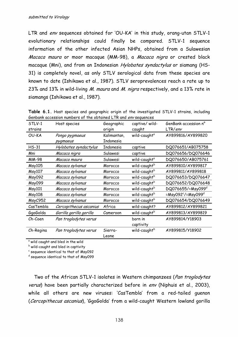
submitted to Virology
138
LTR and env sequences obtained for ‘OU-KA’ in this study, orang-utan STLV-1 evolutionary relationships could finally be compared. STLV-1 sequence information of the other infected Asian NHPs, obtained from a Sulawesian Macaca maura or moor macaque (MM-98), a Macaca nigra or crested black macaque (Mni), and from an Indonesian Hylobates syndactylus or siamang (HS-31) is completely novel, as only STLV serological data from these species are known to date (Ishikawa et al., 1987). STLV seroprevalences reach a rate up to 23% and 13% in wild-living M. maura and M. nigra respectively, and a 13% rate in siamangs (Ishikawa et al., 1987).
Table 6.1. Host species and geographic origin of the investigated STLV-1 strains, including Genbank accession numbers of the obtained LTR and env sequences STLV-1 strains
Host species Geographic origin
captive/ wild-caught
GenBank accession n° LTR/env
OU-KA Pongo pygmaeus pygmaeus
Kalimantan, Indonesia
wild-caughta AY899816/AY899820
HS-31 Hylobates syndactylus Indonesia captive DQ076651/AB075758 Mni Macaca nigra Sulawesi captive DQ076656/DQ076646 MM-98 Macaca maura Sulawesi wild-caughta DQ076650/AB075761 Msy105 Macaca sylvanus Morocco wild-caughtb AY899810/AY899817 Msy107 Macaca sylvanus Morocco wild-caughtb AY899811/AY899818 Msy092 Macaca sylvanus Morocco wild-caughtb DQ076653/DQ076647 Msy099 Macaca sylvanus Morocco wild-caughtb DQ076652/DQ076648 Msy101 Macaca sylvanus Morocco wild-caughtb DQ076655/=Msy099d
Msy108 Macaca sylvanus Morocco wild-caughtb =Msy092c/=Msy099d
MsyC952 Macaca sylvanus Morocco wild-caughtb DQ076654/DQ076649 CasTembla Cercopithecus ascanius Africa wild-caught? AY899812/AY899821 GgoGolda Gorilla gorilla gorilla Cameroon wild-caughtb AY899813/AY899819 Ch-Coen Pan troglodytes verus born in
captivity AY899814/Y18903
Ch-Regina Pan troglodytes verus Sierra-Leone
wild-caughtb AY899815/Y18902
a wild caught and bled in the wild b wild-caught and bled in captivity c sequence identical to that of Msy092 d sequence identical to that of Msy099
Two of the African STLV-1 isolates in Western chimpanzees (Pan troglodytes verus) have been partially characterized before in env (Niphuis et al., 2003), while all others are new viruses: ‘CasTembla’ from a red-tailed guenon (Cercopithecus ascanius), ‘GgoGolda’ from a wild-caught Western lowland gorilla
Chapter 6 Phylogeny of novel Asian and African PTLV-1 strains
139
(Gorilla gorilla gorilla) and STLV-1 in a troop of wild-caught barbary macaques (Macaca sylvanus) from Morocco (Msy092, Msy099, Msy101, Msy105, Msy107, Msy108, MsyC962). Only one other STLV-1 strain in Cercopithecus ascanius, known as 5 subspecies, could be retrieved from the Genbank database. Reports on STLV-infection in gorillas are scarce (Nerrienet et al., 2004; Courgnaud et al., 2004). In one study 2 out of 3 bushmeat animals were STLV seropositive, indicating a high rate of natural STLV infection among gorillas (Courgnaud et al., 2004). STLV-infection in chimpanzees is well known, however seroprevalences in the wild seem to be very low (0.03%) (Courgnaud et al., 2004). STLV-1 infection in Macaca sylvanus has never been reported before. A STLV serological survey of the barbary macaques of Gilbraltar has shown that none of these individuals were STLV-infected (E.J.Verschoor unpublished results). We now detected STLV-infection in 24 out of 54 animals of a troop of wild-caught, captive, free-ranging Macaca sylvanus of Moroccan origin, which have no documented history of contact with other African primate species, prior to and in captivity.
PTLV-1 phylogenetic analyses
The obtained PTLV-1 LTR (Figure 6.1.) and env neighbour joining (NJ) and maximum likelihood (ML) phylogenetic trees corroborate the basal separation between Asian and African PTLV-1. The rooted env tree shows that the Asian STLV-1s are the first to branch off (Figure 6.2.).
The human strains typically cluster in the 6 well defined HTLV-1 subtype
clades, which could be identified both in the LTR and env phylogenetic trees. Although only the Austronesian HTLV-1c and Central African HTLV-1f/STLV-1 subtype clades were significantly supported by bootstrap analyses as presented in Figures 6.1. and 6.2., a concise phylogenetic analysis of the complete LTR demonstrated that all HTLV-1 clusters were highly supported (data not shown). However, investigation of the evolutionary relationships between the novel STLV-1 strains and their most similar sequences according to BLAST forced us to analyse only part of the LTR, thereby reducing the sequence information and consequently also the bootstrap values. The Austronesian HTLV-1c strains are clustering tightly together, distinct from all Asian STLV-1 strains and closest to African PTLV-1. The cosmopolitan HTLV-1a monophyletic clade within the
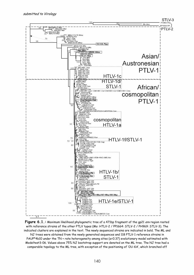
submitted to Virology
140
Figure 6.1.: Maximum likelihood phylogenetic tree of a 471bp fragment of the gp21 env region rooted with reference strains of the other PTLV types (Mo: HTLV-2 / PP1664: STLV-2 / PH969: STLV-3). The
indicated clusters are explained in the text. The newly sequenced strains are indicated in bold. The ML and NJ trees were obtained from the newly generated sequences and 118 PTLV-1 reference strains in
PAUP*4b10 under the TN + rate heterogeneity among sites (α=0.37) evolutionary model estimated with Modeltest3-06. Values above 75% NJ bootstrap support are denoted on the ML tree. The NJ tree had a
comparable topology to the ML tree, with exception of the positioning of ‘OU-KA’, which branched off

Chapter 6 Phylogeny of novel Asian and African PTLV-1 strains
141
immediately after the Macaca arctoides STLV-1 monophyletic clade in the NJ analysis with a bootstrap support of 76%. The actual positioning and bootstrap support of ‘OU-KA’ in the NJ tree is noted by a
dashed arrow. Abbreviations of host species names are as follows: Ani=Allenopithecus nigroviridis, Cae=Chlorocebus aethiops, Cag=Cercocebus agilis, Cas=Cercopithecus ascanius, Cmi=Cercopithecus mitis, Cmo=Cercopithecus mona, Cni=Cercopithecus nictitans, Cpo= Cercopithecus pogonias, Cto= Cercocebus torquatus, Ggo=Gorilla gorilla, Hsa=Homo sapiens, Hsy=Hylobates syndactylus, Mar=Macaca arctoides,
Mfa=Macaca fascicularis, Mfu=Macaca fuscata, Mma=Macaca maura, Mmu=Macaca mulatta, Mni=Macaca nigra, Mog=Miopithecus ogouensis, Msp=Mandrillus sphinx, Msy=Macaca sylvanus, Mto=Macaca tonkeana,
Pan=Papio hamadryas anubis, Pcb= Piliocolobus badius, Pcy=Papio hamadryas cynocephalus, Pha=Papio hamadryas, PP=Pan paniscus, Ppa=Papio hamadryas papio, Ppy=Pongo pygmaeus, Ptr=Pan troglodytes,
Pur=Papio hamadryas ursinus, Tge=Theropithecus gelada.
African part of the trees only joins human strains in contrast with all other African HTLV-1 subtypes. The HTLV-1b, -1d, -1e and -1f subtype clades all comprise human strains of Central African origin and STLV-1 strains of the same geographic area, of which some extend to West Africa (e.g. STLV-1 strains in HTLV-1b cluster).
The Asian part of the trees (Figures 6.1. and 6.2.) consists of deeply branching monophyletic clades, uniting STLV-1 strains of a single macaque species, with exception of a few STLV-1s discovered in Papioni (Pha, Pcy and Tge in Figures 6.1. and 6.2.), resulting from interspecies transmissions from macaques to baboons in captivity (Voevodin et al., 1996; Van Dooren et al., 2004). The orang-utan STLV-1 strains have independently been demonstrated to cluster between these STLV-1 macaque species clades (Ibuki et al., 1997; Verschoor et al., 1998). We show for the first time the joint, tight clustering of both ‘INA-004’ and ‘OU-KA’ orang-utan strains in the LTR phylogeny, interleaved between the macaque STLV-1 monophyletic clusters. Their positioning closest to the most divergent STLV-1s of Macaca arctoides is in env confirmed in the NJ phylogenetic analysis (76% bootstrap support, see arrow in Figure 6.1.). The Asian part of the PTLV-1 phylogenetic trees have a comparable topology regardless the genetic region analysed and the phylogenetic inference methods used, with exception of the inconsistent ML and NJ clustering pattern of ‘OU-KA’ in orang-utan in env. The tight clustering of STLV-1 in M. maura and M. nigra with other Sulawesian macaque strains from Macaca tonkeana in the env analysis demonstrates the close relationship between STLV-1s in these insular species. In the LTR analysis ‘Mni’ lies on a separate, though not supported branch closest
submitted to Virology
142
Figure 6.2.: Neighbour joining phylogenetic tree of a 502bp (381bp after gap-stripping) fragment of the LTR region of the newly generated sequences and 108 PTLV-1 reference strains obtained in PAUP*4b10 under the GTR + rate heterogeneity among sites (α=0.56) evolutionary model estimated with Modeltest3-
06. The indicated clusters are explained in the text. The newly sequenced strains are indicated in bold. The numbers on the branches of the NJ tree denote the NJ bootstrap support above 60% for that specific
cluster. Abbreviations of host species names are as follows: Ani=Allenopithecus nigroviridis, Cae=Chlorocebus aethiops, Cag=Cercocebus agilis, Cas=Cercopithecus ascanius, Cce=Cercopithecus cephus,
Chapter 6 Phylogeny of novel Asian and African PTLV-1 strains
143
Csa=Chlorocebus aethiops sabaeus, Cmi=Cercopithecus mitis, Cpo=Cercopithecus pogonias, Cpy=Cercopithecus pygerythrus, Cto=Cercocebus torquatus, Ggo=Gorilla gorilla, Hsa=Homo sapiens,
Hsy=Hylobates syndactylus, Mar=Macaca arctoides, Mma=Macaca maura, Mmu=Macaca mulatta, Mne=Macaca nemestrina, Mni=Macaca nigra, Mog=Miopithecus ogouensis, Msy=Macaca sylvanus,
Msp=Mandrillus sphinx, Mto=Macaca tonkeana, Pan=Papio hamadryas anubis, Pcb=Piliocolobus badius, Pcy=Papio hamadryas cynocephalus, Pha=Papio hamadryas, Ppa=Papio hamadryas papio, Ppy=Pongo pygmaeus,
Psp=Papio species, Ptr=Pan troglodytes, Pur=Papio hamadryas ursinus.
to the Sulawesian STLV-1 clade. The well supported monophyly of the ‘Mni’ M. nigra strain and ‘TG1884’ STLV-1 in Theropithecus gelada in env can be explained by interspecies transmissions in captivity. In a previous study on STLV-infection in T. gelada (Van Dooren et al., 2004), this latter species was known to be held in captivity with M. arctoides and M. nigra. Since no M. nigra STLV-1 strain was known at that time point and STLV-1 in M. arctoides was only distantly related, speculations on a possible close relatedness to STLV-1 in M. nigra were made, which has now shown to be true in the present study. The fact that our STLV-1 infected M. nigra had no relation with the macaques, whom were co-caged with the STLV-1 seropositive T. gelada together with its close relation to other Sulawesian STLV-1s, indicates that ‘Mni’ is a representative of naturally occurring STLV-1 within this species. Conversely, the clustering of STLV-1 in siamang (HS-31) within the same Sulawesian STLV-1 monophyletic clade cannot be easily reconciled based on the current geographic host species separation (Sulawesi for macaques versus the Malay Peninsula and Sumatra for siamangs). As Sulawesian macaques descend from a nemestrina-like macaque ancestor in the Pliocene their natural habitats might have overlapped with siamangs in a distant past (Groves, 1980). However, such an ancient cross-species transmission event does not fit with the molecular timings of Asian PTLV-1, previously estimated to have originated only 269,000 to 156,000 ya (Van Dooren et al., 2005). Given the captive nature of the STLV-1 infected siamang, a resulting STLV-1 cross-species transmission in captivity would be a more logical explanation. Important to note is that with the inclusion of more macaque strains of other species in this study, we still observe long-branched species-specific clades following a ladder-like topology, which reflect macaque migration waves and geographic distribution. This clustering pattern seems to be the result of interspecies transmissions between macaques in a distant past followed by a host-dependent evolution in each species. The monophyly and deep-branching pattern of orang-utan STLV-1s, of which ‘OU-KA’ was sampled in a

submitted to Virology
144
wild-caught animal, points to at least one natural cross-species transmission from macaques to orang-utans in a distant past, probably along their shared migratory pathways, to then further evolve within their genus.
The African part of the tree is characterised by a short-branched, complex
assemblage of PTLV-1 strains, rather indicative for a star-like burst of the virus after its introduction on the African continent (Salemi et al., 1999). Some well defined human subtype clades can be distinguished, with STLV-1 strains interspersed between or within these HTLV-1 subtypes (except for HTLV-1a). The high variety of Western chimpanzee STLV-1 strains are dispersed over the African PTLV-1 tree (Leendertz et al., 2004). Our strains ‘Ch-Coen’ and ‘Ch-Regina’ cluster closely to other chimpanzee STLV-1 strains of the same area within the HTLV-1b/STLV-1 clade. The newly identified gorilla virus (GgoGolda) also clusters within the HTLV-1b/STLV-1 clade. It is noteworthy that ‘GgoGolda’ has no particular close relation to the other known gorilla strains, (Cam12 and 02CM3157), but rather forms a reasonably supported monophyletic clade with a human strain ‘H24’ in LTR (68% bootstrap support). Although these bootstrap values seem to be rather low, they are among the highest within the African part of the PTLV-1 tree, suggesting that this clustering is significant. These observations corroborate that, given geographic proximity of hosts, PTLV-1 interspecies transmissions seem to occur frequently in Africa, even between distantly related species (Salemi et al., 1998; Leendertz et al., 2004). The close relation between human and great ape PTLV-1 strains in Africa is indicative for a direct zoonosis event, which might result from hunting and slaughter activities, as has been shown for simian foamy viruses (Wolfe et al., 2004). The Cercopithecus ascanius strain ‘CasTembla’ is not particularly closely related to ‘CAS57’ of the same host species. It is plausible that red-tailed guenons harbour different STLV-1s as this species consists of 5 geographically-separated subspecies.
Finally, we characterized STLV-1 in seven wild-caught barbary macaques from
Morocco. Barbary macaques are the North African remnants of macaque migrations from Africa to Asia more than 5.5 million years ago (Delson, 1980; Delson, 1996). No other macaques remained in Africa. This resulted in speculations that barbary macaques could be infected with the missing STLV-1
Chapter 6 Phylogeny of novel Asian and African PTLV-1 strains
145
link, reflecting the Africa to Asia migration of ancestral STLV-1. In that case one would expect them to cluster basal, close to the origin of either Asian or African PTLV-1s or close to the root of the PTLV-1 tree, thereby resolving the phylogeographical origin of PTLV-1. In our phylogenetic analyses the Macaca sylvanus (Msy in Figures 6.1. and 6.2.) STLV-1 strains cluster extremely closely together with a high bootstrap support of 98% in LTR and env, indicating a single or a limited number of STLV-1 introductions within this troop. However, their topology within the African part of the tree shows they are infected with a typical African strain. Their well-supported, extreme close positioning in env to STLV-1 in the sooty mangabeys (sm) and chimpanzees (Ptr-Leo and Ptr-Loukoum) from West Africa (86% bootstrap support) could also be observed to a lesser extent (low bootstrap support) in the LTR-NJ analysis, where Msy forms a sister-taxon of the mangabey and chimpanzee STLV-1 strains. Their non-basal and internal clustering in the African part of the tree further refutes the hypothesis of STLV-1 virus-host co-evolution, since it indicates that barbary macaques became STLV-1 infected after their split from the other Macaca species that migrated to Asia. As these STLV-1 infected barbary macaques have no documented history of contact with other primate species, they are likely naturally infected with this type of virus. Their close relationship to Western STLV species, together with their allopatric location towards sooty mangabeys and Western chimpanzees, indicate that interspecies transmissions between these naturally infected NHPs must have occurred in the past, before the desertification of the Sahara, which is at least 5000 years ago (ya) (http://members.cox.net/quaternary/nercAFRICA.html), when their natural habitats may have overlapped. This fits within the previously estimated time frame for the origin of PTLV-1 in Africa 35,000 to 19,000 ya and for the African HTLV-1 subtypes 21,000 to 3,000 ya (Van Dooren et al., 2001). Alternatively, the M. sylvanus STLV-1 infection might be, although highly unlikely, the result of a cross-species transmission in captivity.
From our analyses it is clear that additional sampling, identification and characterisation of STLV-1 strains is needed to further study the epidemiology and evolution of the virus, especially since the current PTLV-1 phylogeny might be biased by a greater sampling in Africa than in Asia and in humans than in NHPs.
submitted to Virology
146
Materials and methods STLV-1-infection was discovered in the 15 NHPs described in Table 6.1. using
an in-house ELISA (Verschoor et al., 1998). To partially characterise the viruses present in these species a 522bp gp21env fragment and total LTR sequences were obtained for all 15 STLV-infected animals using PCRs and sequencing methodology as previously described (Van Dooren et al., 1998). The Genbank accession numbers of all newly obtained LTR and env sequences are implemented in Table 6.1. A BLAST search was performed on the new LTR and env sequences to retrieve most similar sequences for inclusion in the alignment together with a substantial number of PTLV-1 reference strains. To be able to root the PTLV-1 tree in env, the STLV-3 ‘PH969’, HTLV-2 ‘Mo’ and STLV-2 ‘PP1664’ strains were included in the gp21env alignment (471 sites). Alignments were made in Dambe (Xia & Xie, 2001) and minor manual editing was done in the same program. The initial LTR alignment of 502 sites was gap-stripped to delete unresolved nucleotides, retaining 381 sites. The evolutionary model that best fits the data set was determined with Modeltest3-06 (Posada & Crandall, 1998). Neighbour joining and maximum likelihood trees were reconstructed using the PAUP*4b10 software package (Swofford, 1998) following the steps as described elsewhere (Van Dooren et al., 2005). Briefly, a heuristic search under the tree-bisection-reconnection algorithm using the NJ tree as starting tree was used to generate the ML tree. The NJ tree was evaluated by a bootstrap analysis of 1000 bootstrap replicates. The env and LTR trees were obtained under the Tamura Nei evolutionary model and General Time Reversible model respectively, further modelled with a gamma-distributed rate heterogeneity among sites (α-parameter=0.37 and 0.56 for env and LTR respectively).
This work was supported by the Fonds voor Wetenschappelijk Onderzoek (Grant
0288.01). We wish to thank Dr. Ishikawa for providing us with samples of STLV-
infected M. maura and H. syndactylus.
Chapter 6 Phylogeny of novel Asian and African PTLV-1 strains
147
Reference List Cavalli-Sforza, L. L., Menozzi, P., and Piazza, A. (1994). "The history and geography of human genes." Princeton University Press, Princeton, N.J.. Courgnaud, V., Van Dooren, S., Liegeois, F., Pourrut, X., Abela, B., Loul, S., Mpoudi-Ngole, E., Vandamme, A., Delaporte, E., and Peeters, M. (2004). Simian T-cell leukemia virus (STLV) infection in wild primate populations in Cameroon: Evidence for dual STLV type 1 and type 3 infection in agile mangabeys (Cercocebus agilis). Journal of Virology 78, 4700-4709. Delson, E. (1980). Fossil macaques, phyletic relationships and a scenario of deployment. In "The macaques: studies in ecology, bahaviour, and evolution" (D. G. Lindburg, Ed.), pp. 10-30. New York:Van Nostrand Reinhold Co.. Delson, E. The oldest monkeys in Asia. International symposium: Evolution of Asian primates.Freude & Kyoto University Primate Research Institute, Inuyama, Aichi, Japan , 40. 1996. Ref Type: Abstract Fa, J. E. (1989). The Genus Macaca - A Review of Taxonomy and Evolution. Mammal Review 19, 45-81. Gessain, A. and Mahieux, R. (2000). Epidemiology, origins and genetic diversity of HTLV-1 retrovirus and STLV-1 simian affiliated retroviruses. Bulletin de la Societe de Pathologie Exotique 93, 163-171. Gessain, A., Yanagihara, R., Franchini, G., Garruto, R. M., Jenkins, C. L., Ajdukiewicz, A. B., Gallo, R. C., and Gajdusek, D. C. (1991). Highly Divergent Molecular Variants of Human T-Lymphotropic Virus Type-I from Isolated Populations in Papua-New-Guinea and the Solomon-Islands. Proceedings of the National Academy of Sciences of the United States of America 88, 7694-7698. Groves, C. P. (1980). Speciation in Macaca: the view from Sulawesi. In "The macacques: studies in ecology, behavior and evolution" (Lindburg D.G., Ed.), pp. 84-124. Van Nostrand Reinhold Co., New York. Ibrahim, F., Dethe, G., and Gessain, A. (1995). Isolation and Characterization of A New Simian T-Cell Leukemia-Virus Type-1 from Naturally Infected Celebes Macaques (Macaca-Tonkeana) - Complete Nucleotide-Sequence and Phylogenetic Relationship with the Australo-Melanesian Human T-Cell Leukemia-Virus Type-1. Journal of Virology 69, 6980-6993. Ibuki, K., Ido, E., Setiyaningsih, S., Yamashita, M., Agus, L. R. P., Takehisa, J., Miura, T., Dondin, S., and Hayami, M. (1997). Isolation of STLV-I from orangutan, a great ape species in southeast Asia, and its relation to other HTLV-Is/STLV-Is. Japanese Journal of Cancer Research 88, 1-4. Ishikawa, K., Fukasawa, M., Tsujimoto, H., Else, J. G., Isahakia, M., Ubhi, N. K., Ishida, T., Takenaka, O., Kawamoto, Y., Shotake, T., and . (1987). Serological survey and virus isolation of simian T-cell leukemia/T-lymphotropic virus type I (STLV-I) in non-human primates in their native countries. Int.J.Cancer 40, 233-239. Koralnik, I. J., Boeri, E., Saxinger, W. C., Lomonico, A., Fullen, J., Gessain, A., Guo, H. G., Gallo, R. C., Markham, P., Kalyanaraman, V., Hirsch, V., Allan, J., Murthy, K., Alford, P., Slattery, J. P., Obrien, S. J., and Franchini, G. (1994). Phylogenetic Associations of Human and Simian T-Cell Leukemia/Lymphotropic Virus Type-I Strains - Evidence for Interspecies Transmission. Journal of Virology 68, 2693-2707.
submitted to Virology
148
Leendertz, F. H., Junglen, S., Boesch, C., Formenty, P., Couacy-Hymann, E., Courgnaud, V., Pauli, G., and Ellerbrok, H. (2004). High variety of different simian T-Cell leukemia virus type 1 strains in chimpanzees (Pan troglodytes verus) of the Tai National Park, Cote d'Ivoire. Journal of Virology 78, 4352-4356. Liu, H. F., Goubau, P., Van Brussel, M., Desmyter, J., and Vandamme, A. M. (1997). Phylogenetic analysis of a simian T lymphotropic virus type I from a hamadryas baboon. Aids Research and Human Retroviruses 13, 1545-1548. Liu, H. F., Goubau, P., Van Brussel, M., Van Laethem, K., Chen, Y. C., Desmyter, J., and Vandamme, A. M. (1996). The three human T-lymphotropic virus type I subtypes arose from three geographically distinct simian reservoirs. J.Gen.Virol. 77 ( Pt 2 ), 359-368. Mahieux, R., Chappey, C., Georges-Courbot, M. C., Dubreuil, G., Mauclere, P., Georges, A., and Gessain, A. (1998). Simian T-cell lymphotropic virus type 1 from Mandrillus sphinx as a simian counterpart of human T-cell lymphotropic virus type 1 subtype D. Journal of Virology 72, 10316-10322. Mahieux, R., Chappey, C., Meertens, L., Mauclere, P., Lewis, J., and Gessain, A. (2000). Molecular characterization and phylogenetic analyses of a new simian T cell lymphotropic virus type 1 in a wild-caught African baboon (Papio anubis) with an indeterminate STLV type 2-like serology. Aids Research and Human Retroviruses 16, 2043-2048. Mahieux, R., PeconSlattery, J., and Gessain, A. (1997). Molecular characterization and phylogenetic analyses of a new, highly divergent simian T-cell lymphotropic virus type 1 (STLV-1marc1) in Macaca arctoides. Journal of Virology 71, 6253-6258. Meertens, L., Rigoulet, J., Mauclere, P., Van Beveren, M., Chen, G. M., Diop, O., Dubreuil, G., Georges-Goubot, M. C., Berthier, J. L., Lewis, J., and Gessain, A. (2001). Molecular and phylogenetic analyses of 16 novel simian T cell leukemia virus type 1 from Africa: Close relationship of STLV-1 from Allenopithecus nigroviridis to HTLV-1 subtype B strains. Virology 287, 275-285. Nerrienet, E., Meertens, L., Kfutwah, A., Foupouapouognigni, Y., Ayouba, A., and Gessain, A. (2004). Simian T cell leukaemia virus type I subtype B in a wild-caught gorilla (Gorilla gorilla gorilla) and chimpanzee (Pan troglodytes vellerosus) from Cameroon. Journal of General Virology 85, 25-29. Nerrienet, E., Meertens, L., Kfutwah, A., Foupouapouognigni, Y., and Gessain, A. (2001). Molecular epidemiology of simian T-lymphotropic virus (STLV) in wild-caught monkeys and apes from Cameroon: a new STLV-1, related to human T-lymphotropic virus subtype F, in a Cercocebus agilis. Journal of General Virology 82, 2973-2977. Niphuis,H., Verschoor,E.J., Bontjer,I., Peeters,M., & Heeney,J.L. (2003) Reduced transmission and prevalence of simian T-cell lymphotropic virus in a closed breeding colony of chimpanzees (Pan troglodytes verus). Journal of General Virology 84, 615-620. Posada, D. and Crandall, K. A. (1998). MODELTEST: testing the model of DNA substitution. Bioinformatics 14, 817-818. Richards, A. L., Giri, A., Iskandriati, D., Pamungkas, J., Sie, A., Rosen, L., Anthony, R. L., and Franchini, G. (1998). Simian T-lymphotropic virus type I infection among wild-caught Indonesian pig-tailed macaques (Macaca nemestrina). Journal of Acquired Immune Deficiency Syndromes and Human Retrovirology 19, 542-545. Saksena, N. K., Herve, V., Durand, J. P., Leguenno, B., Diop, O. M., Digoutte, J. P., Mathiot, C., Muller, M. C., Love, J. L., Dube, S., Sherman, M. P., Benz, P. M., Erensoy, S., Galatluong, A., Galat, G., Paul, B., Dube, D. K., Barresinoussi, F., and Poiesz, B. J. (1994). Seroepidemiologic, Molecular, and Phylogenetic Analyses of Simian T-Cell Leukemia
Chapter 6 Phylogeny of novel Asian and African PTLV-1 strains
149
Viruses (Stlv-I) from Various Naturally Infected Monkey Species from Central and Western Africa. Virology 198, 297-310. Salemi, M., Van Dooren, S., Audenaert, E., Delaporte, E., Goubau, P., Desmyter, J., and Vandamme, A. M. (1998). Two new human T-lymphotropic virus type I phylogenetic subtypes in seroindeterminates, a Mbuti pygmy and a Gabonese, have closest relatives among African STLV-I strains. Virology 246, 277-287. Salemi, M., Van Dooren, S., and Vandamme, A.-M. (1999). Origin and evolution of human and simian T-cell lymphotropic viruses. Aids Reviews 1, 131-139. Slattery, J. P., Franchini, G., and Gessain, A. (1999). Genomic evolution, patterns of global dissemination, and interspecies transmission of human and simian T-cell leukemia lymphotropic viruses. Genome Research 9, 525-540. Swofford, D. L. PAUP*:phylogentic analysis using parsimony (*and other methods). (4.0beta10). 1998. Sunderland, Massachusetts, Sinauer. Ref Type: Computer Program Van Dooren, S., Meertens, L., Lemey, P., Gessain, A., and Vandamme, A (2005). Full genome analysis of a highly divergent Simian T-cell Lymphotropic Virus type-1 (STLV-1) strain in Macaca arctoides. Journal of General Virology 86, 1953-1959. Van Dooren, S., Salemi, M., and Vandamme, A. M. (2001). Dating the origin of the African human T-cell lymphotropic virus type-i (HTLV-I) subtypes. Molecular Biology and Evolution 18, 661-671. Van Dooren, S., Shanmugam, V., Bhullar, V., Parekh, B., Vandamme, A. M., Heneine, W., and Switzer, W. M. (2004). Identification in gelada baboons (Theropithecus gelada) of a distinct simian T-cell lymphotropic virus type 3 with a broad range of Western blot reactivity. Journal of General Virology 85, 507-519. Vandamme, A. M., Salemi, M., and Desmyter, J. (1998). The simian origins of the pathogenic human T-cell lymphotropic virus type I. Trends Microbiol. 6, 477-483. Verschoor, E. J., Warren, K. S., Niphuis, H., Heriyanto, Swan, R. A., and Heeney, J. L. (1998). Characterization of a simian T-lymphotropic virus from a wild-caught orangutan (Pongo pygmaeus) from Kalimantan, Indonesia. Journal of General Virology 79, 51-55. Voevodin, A., Samilchuk, E., Schatzl, H., Boeri, E., and Franchini, G. (1996). Interspecies transmission of macaque simian T-cell leukemia/lymphoma virus type 1 in baboons resulted in an outbreak of malignant lymphoma. Journal of Virology 70, 1633-1639. Voevodin, A. F., Johnson, B. K., Samilchuk, E. I., Stone, G. A., Druilhet, R., Greer, W. J., and Gibbs, C. J., Jr. (1997). Phylogenetic analysis of simian T-lymphotropic virus Type I (STLV-I) in common chimpanzees (Pan troglodytes): evidence for interspecies transmission of the virus between chimpanzees and humans in Central Africa. Virology 238, 212-220. Wolfe, N. D., Switzer, W. M., Carr, J. K., Bhullar, V. B., Shanmugam, V., Tamoufe, U., Prosser, A. T., Torimiro, J. N., Wright, A., Mpoudi-Ngole, E., McCutchan, F. E., Birx, D. L., Folks, T. M., Burke, D. S., and Heneine, W. (2004). Naturally acquired simian retrovirus infections in central African hunters. Lancet 363, 932-937. Xia, X. and Xie, Z. (2001). DAMBE: Data analysis in molecular biology and evolution. Journal of Heredity 92, 371-373.
MICHAEL EMERMAN, editor
Received June 16 2005

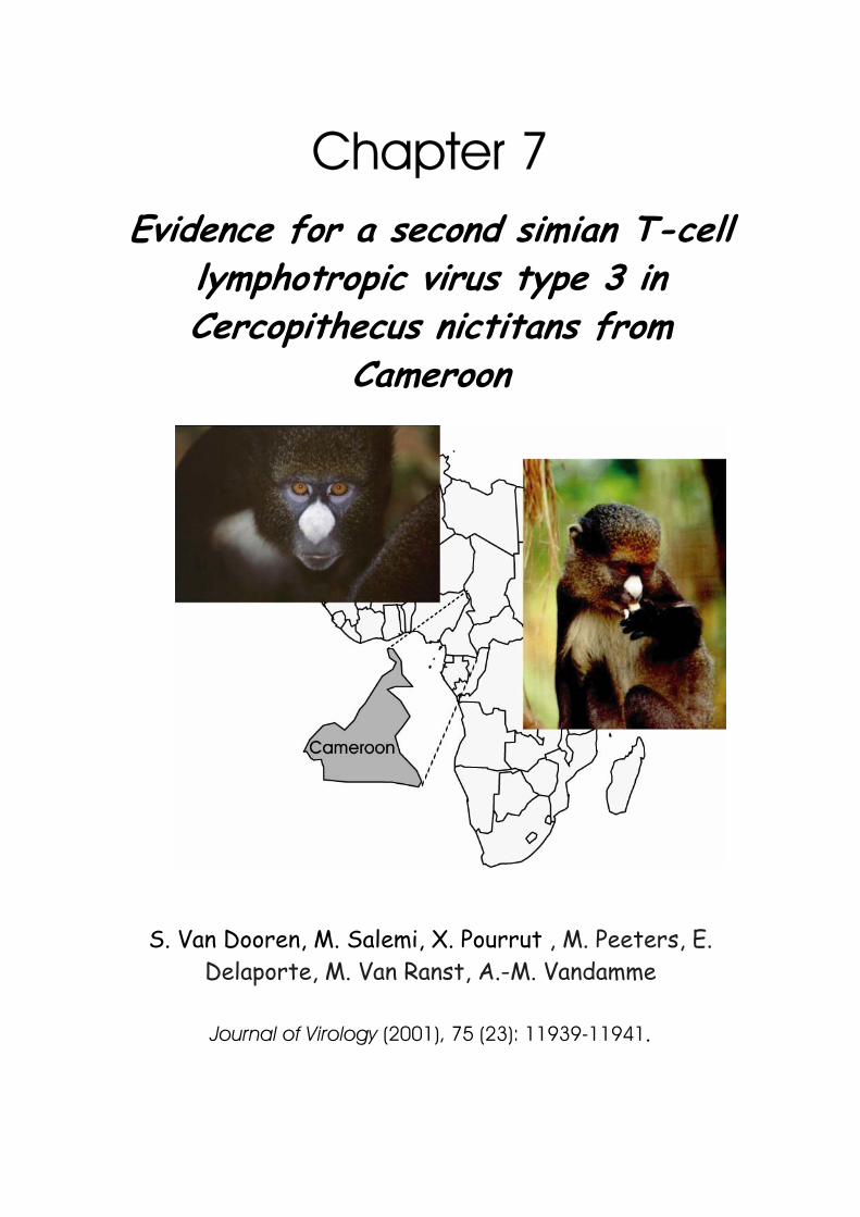
Chapter 7 Evidence for a second simian T-cell
lymphotropic virus type 3 in Cercopithecus nictitans from
Cameroon
S. Van Dooren, M. Salemi, X. Pourrut , M. Peeters, E. Delaporte, M. Van Ranst, A.-M. Vandamme
Journal of Virology (2001), 75 (23): 11939-11941.

Photographs previous page: http://www.cercopan.org/puttynose_guenons.htm
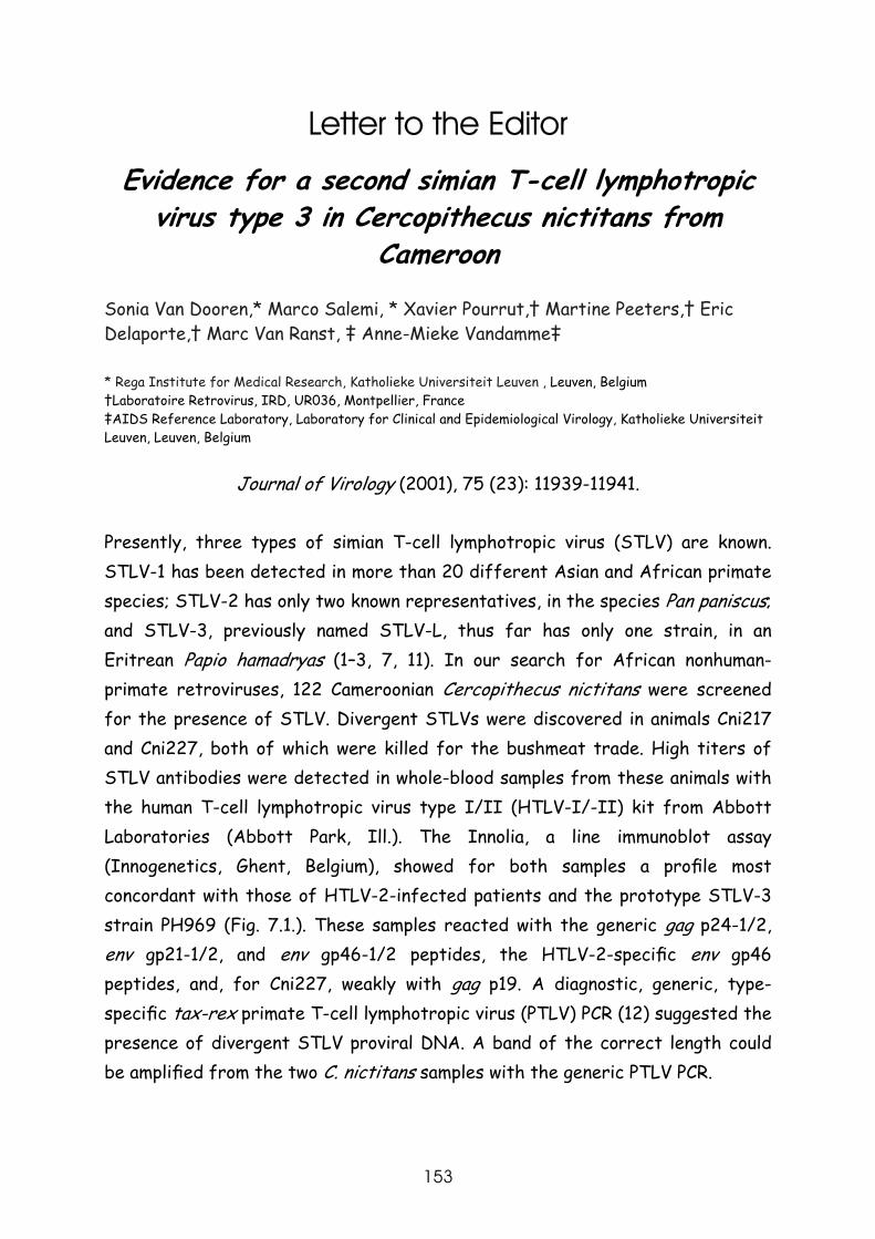
153
Letter to the Editor
Evidence for a second simian T-cell lymphotropic virus type 3 in Cercopithecus nictitans from
Cameroon Sonia Van Dooren,* Marco Salemi, * Xavier Pourrut,† Martine Peeters,† Eric Delaporte,† Marc Van Ranst, ‡ Anne-Mieke Vandamme‡
* Rega Institute for Medical Research, Katholieke Universiteit Leuven , Leuven, Belgium †Laboratoire Retrovirus, IRD, UR036, Montpellier, France ‡AIDS Reference Laboratory, Laboratory for Clinical and Epidemiological Virology, Katholieke Universiteit Leuven, Leuven, Belgium
Journal of Virology (2001), 75 (23): 11939-11941.
Presently, three types of simian T-cell lymphotropic virus (STLV) are known. STLV-1 has been detected in more than 20 different Asian and African primate species; STLV-2 has only two known representatives, in the species Pan paniscus; and STLV-3, previously named STLV-L, thus far has only one strain, in an Eritrean Papio hamadryas (1–3, 7, 11). In our search for African nonhuman-primate retroviruses, 122 Cameroonian Cercopithecus nictitans were screened for the presence of STLV. Divergent STLVs were discovered in animals Cni217 and Cni227, both of which were killed for the bushmeat trade. High titers of STLV antibodies were detected in whole-blood samples from these animals with the human T-cell lymphotropic virus type I/II (HTLV-I/-II) kit from Abbott Laboratories (Abbott Park, Ill.). The Innolia, a line immunoblot assay (Innogenetics, Ghent, Belgium), showed for both samples a profile most concordant with those of HTLV-2-infected patients and the prototype STLV-3 strain PH969 (Fig. 7.1.). These samples reacted with the generic gag p24-1/2, env gp21-1/2, and env gp46-1/2 peptides, the HTLV-2-specific env gp46 peptides, and, for Cni227, weakly with gag p19. A diagnostic, generic, type-specific tax-rex primate T-cell lymphotropic virus (PTLV) PCR (12) suggested the presence of divergent STLV proviral DNA. A band of the correct length could be amplified from the two C. nictitans samples with the generic PTLV PCR.
J. Vir. (2001) 75: 11939-11941
154
FIG. 7.1. Innolia results generated from HTLV-1, HTLV-2-infected-patient plasma, PH969 serum, Cni217 and Cni227 whole blood. Lanes 1 and 2 (from the right) contain the positive control (PC) and negative
control (NC) of the kit, respectively. Lanes 3 and 4 show the results for an HTLV-1-and an HTLV-2-infected-patient sample, respectively. Lane 4 contains the STLV-3 PH969 sample. Lanes 5 and 6 contain the Cni217 and Cni227 samples, respectively. The first three control lines contain human immunoglobulin G (IgG)
in different concentrations; they are followed by four confirmation lines (2 gag and 2 env HTLV-1/-2 antigen lines) and then three discriminatory lines (2 gag HTLV-1 and 1 env HTLV-2 peptide) at the bottom
of the strip.
The four different type-specific inner PCRs, however, discriminating HTLV-1-, HTLV-2-, STLV-2-, and STLV-3-like viruses, were not able to amplify the STLV proviral DNAs of Cni217 and Cni227. The sequenced inner generic tax-rex Cni217 and Cni227 PCR products of 219 bp exhibited sequence similarities of 88.6 and 89.0%, respectively, to STLV-3 strain PH969, detected in an Eritrean Papio hamadryas (3), while nucleotide divergences of 20 to 29% were evident for the prototypic HTLV-1/STLV-1 and HTLV-2/STLV-2 strains. The 219-bp tax-rex fragments of Cni217 and Cni227 were phylogenetically analyzed using PAUP*4.0b5 (9). Neighbour-joining (NJ) and maximum-likelihood (ML) trees were calculated for 22 different taxa by using the Tamura Nei substitution
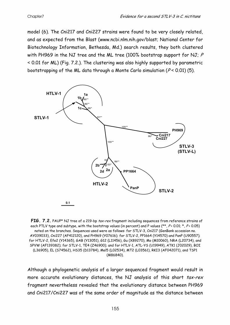
Chapter7 Evidence for a second STLV-3 in C. nictitans
155
model (6). The Cni217 and Cni227 strains were found to be very closely related, and as expected from the Blast (www.ncbi.nlm.nih.gov/blast; National Center for Biotechnology Information, Bethesda, Md.) search results, they both clustered with PH969 in the NJ tree and the ML tree (100% bootstrap support for NJ; P < 0.01 for ML) (Fig. 7.2.). The clustering was also highly supported by parametric bootstrapping of the ML data through a Monte Carlo simulation (P < 0.01) (5).
FIG. 7.2. PAUP* NJ tree of a 219-bp tax-rex fragment including sequences from reference strains of each PTLV type and subtype, with the bootstrap values (in percent) and P values (**, P < 0.01; *, P < 0.05)
noted on the branches. Sequences used were as follows: for STLV-3, Cni217 (GenBank accession no. AY039033), Cni227 (AF412120), and PH969 (Y07616); for STLV-2, PP1664 (Y14570) and PanP (U90557); for HTLV-2, Efe2 (Y14365), GAB (Y13051), G12 (L11456), Gu (X89270), Mo (M10060), NRA (L20734), and SPVW (AF139382); for STLV-1, TE4 (Z46900); and for HTLV-1, ATL-YS (U19949), ATK1 (J02029), BOI
(L36905), EL (S74562), HS35 (D13784), Mel5 (L02534), MT2 (L03561), RKI3 (AF042071), and TSP1 (M86840).
Although a phylogenetic analysis of a larger sequenced fragment would result in more accurate evolutionary distances, the NJ analysis of this short tax-rex fragment nevertheless revealed that the evolutionary distance between PH969 and Cni217/Cni227 was of the same order of magnitude as the distance between
0.1
PH969Cni217
PP1664
PanP
2a2d2b
1c
1b1a
STLV-3(STLV-L)
STLV-2HTLV-2
HTLV-1
STLV-1
Cni227
99/**83/*
83
100/**
100/**
97/**
88/*
96/**
86/**
96/*
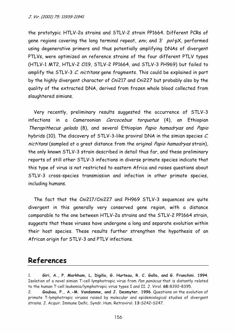
J. Vir. (2001) 75: 11939-11941
156
the prototypic HTLV-2a strains and STLV-2 strain PP1664. Different PCRs of gene regions covering the long terminal repeat, env, and 3’ pol-pX, performed using degenerative primers and thus potentially amplifying DNAs of divergent PTLVs, were optimized on reference strains of the four different PTLV types (HTLV-1 MT2, HTLV-2 Cl19, STLV-2 PP1664, and STLV-3 PH969) but failed to amplify the STLV-3 C. nictitans gene fragments. This could be explained in part by the highly divergent character of Cni217 and Cni227 but probably also by the quality of the extracted DNA, derived from frozen whole blood collected from slaughtered simians.
Very recently, preliminary results suggested the occurrence of STLV-3
infections in a Cameroonian Cercocebus torquatus (4), an Ethiopian Theropithecus gelada (8), and several Ethiopian Papio hamadryas and Papio hybrids (10). The discovery of STLV-3-like proviral DNA in the simian species C. nictitans (sampled at a great distance from the original Papio hamadryas strain), the only known STLV-3 strain described in detail thus far, and these preliminary reports of still other STLV-3 infections in diverse primate species indicate that this type of virus is not restricted to eastern Africa and raises questions about STLV-3 cross-species transmission and infection in other primate species, including humans.
The fact that the Cni217/Cni227 and PH969 STLV-3 sequences are quite
divergent in this generally very conserved gene region, with a distance comparable to the one between HTLV-2a strains and the STLV-2 PP1664 strain, suggests that these viruses have undergone a long and separate evolution within their host species. These results further strengthen the hypothesis of an African origin for STLV-3 and PTLV infections.
References 1. Giri, A., P. Markham, L. Digilio, G. Hurteau, R. C. Gallo, and G. Franchini. 1994. Isolation of a novel simian T-cell lymphotropic virus from Pan paniscus that is distantly related to the human T-cell leukemia/lymphotropic virus types I and II. J. Virol. 68:8392–8395. 2. Goubau, P., A.-M. Vandamme, and J. Desmyter. 1996. Questions on the evolution of primate T-lymphotropic viruses raised by molecular and epidemiological studies of divergent strains. J. Acquir. Immune Defic. Syndr. Hum. Retrovirol. 13:S242–S247.
Chapter7 Evidence for a second STLV-3 in C. nictitans
157
3. Liu, H. F., A.-M. Vandamme, M. Van Brussel, J. Desmyter, and P. Goubau. 1994. New retroviruses in human and simian T-lymphotropic viruses. Lancet 344:265–266. 4. Meertens, L., R. Mahieux, P. Mauclere, J. Lewis, and A. Gessain. 2001. Complete nucleotide sequence and phylogenetic analysis of a novel and highly divergent STLV (CT604) from red cap mangabeys (Cercocebus torquatus) from Cameroon: relationship with PTLV-L and emergence of a new viral type. Tenth International Conference on Human Retrovirology: HTLV and Related Viruses, Dublin, Ireland. AIDS Res. Hum. Retrovir. 17:S11.
5. Rambaut, A., and N. C. Grassly. 1997. Seq-Gen: an application for the Monte Carlo simulation of DNA sequence evolution along phylogenetic trees. Comp. Appl. Biosci. 13:235–238.
6. Salemi, M., J. Desmyter, and A.-M. Vandamme. 2000. Tempo and mode of human and simian T-lymphotropic virus (HTLV/STLV) evolution revealed by analyses of full-genome sequences. Mol. Biol. Evol. 17:374–386. 7. Slattery, J. P., G. Franchini, and A. Gessain. 1999. Genomic evolution, patterns of global dissemination, and interspecies transmission of human and simian T-cell leukemia/lymphotropic viruses. Genome Res. 9:525–540. 8. Switzer, W. M., V. Shanmugam, S. Van Dooren, A.-M. Vandamme, V. Bhullar, B. Parekh, and W. Heneine. 2001. Identification of a distinct STLV in an Ethiopian gelada baboon (Theropithicus gelada). Tenth International Conference on Human Retrovirology: HTLV and Related Viruses, Dublin, Ireland. AIDS Res. Hum. Retrovir. 17:S11. 9. Swofford, D. L. 1998. PAUP*: phylogenetic analysis using parsimony (* and other methods), version 4.0b5. Sinauer Associates, Sunderland, Mass. 10. Takemura, T., M. Yamashita, M. K. Shimada, T. Shotake, T. Miura, and M. Hayami. 2001. Natural infection with the third type of STLV among wild baboons in Ethiopia. Tenth International Conference on Human Retrovirology: HTLV and Related Viruses, Dublin, Ireland, P-Add-10. 11. Vandamme, A.-M., H.-F. Liu, M. Van Brussel, W. De Meurichy, J. Desmyter, and P. Goubau. 1996. The presence of a divergent T-lymphotropic virus in a wild-caught pygmy chimpanzee (Pan paniscus) supports an African origin for the human T-lymphotropic/simian T-lymphotropic group of viruses. J. Gen. Virol. 77:1089–1099. 12. Vandamme, A.-M., K. Van Laethem, H. F. Liu, M. Van Brussel, E. Delaporte, C. M. de Castro Costa, C. Fleischer, G. Taylor, U. Bertazzoni, J. Desmyter, and P. Goubau. 1997. Use of a generic polymerase chain reaction assay detecting human T-lymphotropic virus (HTLV) types I, II and divergent simian strains in the evaluation of individuals with indeterminate HTLV serology. J. Med.Virol.52:1–7.
RONALD DESROSIERS, editor
Received June 22 2001, Accepted September 6, 2001

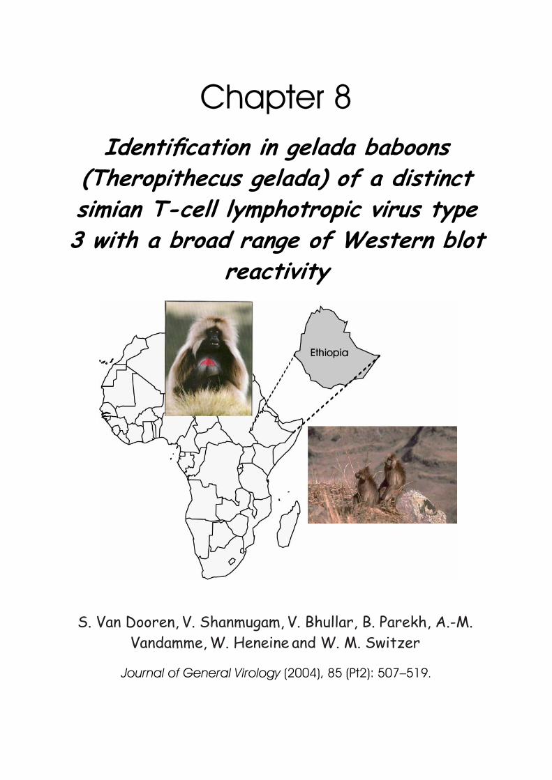
Chapter 8 Identification in gelada baboons
(Theropithecus gelada) of a distinct simian T-cell lymphotropic virus type 3 with a broad range of Western blot
reactivity
S. Van Dooren, V. Shanmugam,
V. Bhullar, B. Parekh, A.-M.
Vandamme, W. Heneine
and W. M. Switzer
Journal of General Virology (2004), 85 (Pt2): 507–519.

Photo previous page (right): J.S. Gartlan (taken at Semyen Mountains, Ethiopia) http://www.cites.org/gallery/species/mammal/mammals.html: (left) http://www.ethemb.se/Tourism/NationalParks/body_nationalparks.htm
161
Identification in gelada baboons (Theropithecus gelada) of a distinct simian T-cell lymphotropic virus type 3 with a broad range of Western blot reactivity
Sonia Van Dooren,1 Vedapuri Shanmugam,
2 Vinod Bhullar,
2Bharat Parekh,
3Anne-
Mieke Vandamme,1Walid Heneine
2 and William M. Switzer
2
1Rega Institute for Medical Research, Katholieke Universiteit Leuven, Leuven, Belgium
2,3HIV and Retrovirology Branch
2and HIV Immunology and Diagnostic Branch
3, Division of AIDS, STD and
TB Laboratory Research, National Center for HIV, STD and TB Prevention, Centers for Disease Control and Prevention, 1600 Clifton Rd, MS G-19, Atlanta, GA 30333, USA
Journal of General Virology (2004), 85 (Pt2): 507–519.
Antibodies to simian T-cell lymphotropic virus (STLV) were found in serum or plasma from 12 of 23 (52.2%) gelada baboons (Theropithecus gelada) captive in US zoos. A variety of Western blot (WB) profiles was seen in the 12 seroreactive samples, including human T-cell lymphotropic virus (HTLV)-1-like (n=5, 41.7 %), HTLV-2-like (n=1, 8.3 %), HTLV-untypable (n=4, 33.3%) and indeterminate (n=2, 16.6%) profiles. Phylogenetic analysis of tax or env sequences that had been PCR amplified from peripheral blood lymphocyte DNA available from nine seropositive geladas showed that four were infected with identical STLV-1s; these sequences clustered with STLV-1 from Celebes macaques and probably represent recent cross-species infections. The tax sequences from the five remaining geladas were also identical and clustered with STLV-3. Analysis of the complete STLV-3 genome (8917 bp) from one gelada, TGE-2117, revealed that it is unique, sharing only 62% similarity with HTLV-1/ATK and HTLV-2/Mo. STLV-3/TGE-2117 was closest genetically to STLV-3 from an Eritrean baboon (STLV-3/PH969, 95.6%) but more distant from STLV-3s from red-capped mangabeys from Cameroon and Nigeria (STLV-3/CTO-604, 87.7%, and STLV-3/CTO-NG409, 87.2%, respectively) and Senegalese baboons (STLV-3/PPA-F3, 88.4 %). The genetic relatedness of STLV-3/TGE-2117 to STLV-3 was confirmed by phylogenetic analysis of a concatenated gag-pol-env-tax sequence (6795 bp). An ancient origin of 73 628-109 809 years ago for STLV-3 was estimated by molecular clock analysis of third-codon positions of gag-pol-env-tax sequences. LTR sequences from five STLV-3-positive geladas were >99% identical and clustered with that from a Papio anubisxP. hamadryas hybrid Ethiopian baboon, suggesting a common source of STLV-3 in these sympatric animals. LTR sequences obtained 20 years apart from a mother-infant pair were identical, providing evidence of both mother-to-offspring transmission and a high genetic stability of STLV-3. Since STLV-3-infected primates show a range of HTLV-like WB profiles and have an ancient origin, further studies using STLV-3-specific testing are required to determine whether STLV-3 infects humans, especially in regions of Africa where STLV-3 is endemic.

J.Gen.Vir. (2004) 85: 507-519
162
Introduction Simian T-cell lymphotropic viruses (STLVs) are a diverse group of
deltaretroviruses that naturally infect many species of Old World primates (Gessain et al., 2002; Slattery et al., 1999). STLVs are related to human T-cell lymphotropic virus types 1 and 2 (HTLV-1 and -2) and collectively HTLVs and STLVs are commonly referred to as primate T-cell lymphotropic viruses (PTLVs) (Slattery et al., 1999). While STLV-1 has been found in over 20 species of African and Asian primates, STLV-2 has been detected in only two captive troops of bonobos (Pan paniscus; strains STLV-2/PP1664 and STLV-2/panp) (Giri et al., 1994; Goubau et al., 1994; Liu et al., 1994; Meertens et al., 2001; Meertens & Gessain, 2003; Miyoshi et al., 1982; Nerrienet et al., 2001; Van Brussel et al., 1997, 1999; Vandamme et al., 1996). STLV-3 was first identified in 1994 in only a single captive Eritrean sacred baboon (Papio hamadryas; strain STLV-3/PH969) (Goubau et al., 1994). More recently, STLV-3 has been found in wild-caught monkeys originating from different African regions including several Ethiopian sacred (P. hamadryas; strain PHA-7550) and hybrid baboons (P. hamadryas x P. anubis; strains HYB-2210 and -2220, respectively) (Takemura et al., 2002), Cameroonian and Nigerian red-capped mangabeys (Cercocebus torquatus torquatus; strains CTO-604 and CTO-602 and strain CTO-NG409, respectively) (Meertens et al., 2002, 2003), Cameroonian spot-nosed guenons (Cercopithecus nictitans; strains CNI-217 and CNI-227) (Van Dooren et al., 2001) and Senegalese olive baboons (P. papio; strains PPA-F3, -F4, -F5 and -F9) (Meertens & Gessain, 2003).
Despite these recent reports, the virological and immunological
characteristics of STLV-3 infection of non-human primates (NHPs) remain poorly understood. STLV-3 has the highest amino acid sequence identity to the HTLV-1 and HTLV-2 p24 Gag (85 %) and envelope transmembrane proteins (72-88%, respectively) (Meertens et al., 2002; Van Brussel et al., 1997). Thus, sera from STLV-3-infected primates show cross-reactivity to the p24 Gag and transmembrane (GD21) proteins in Western blot (WB) assays containing HTLV-1 antigens and conserved HTLV-1 and HTLV-2 recombinant GD21 proteins (Goubau et al., 1994; Meertens et al., 2001, 2002; Meertens & Gessain 2003; Takemura
Chapter 8 STLV-3 in geladas with variable WB reactivity
163
et al., 2002; Van Dooren et al., 2001). Sera from STLV-3-infected animals may also react in WB assays with the HTLV-2-type specific recombinant envelope (Env) protein, K55; thus, STLV-3 seroreactivity has been referred to as HTLV-2-like, though phylogenetic analysis clearly demonstrates the uniqueness of STLV-3 (Goubau et al., 1994; Meertens et al., 2001, 2002; Meertens & Gessain 2003; Takemura et al., 2002; Van Dooren et al., 2001). Therefore, the use of HTLV-1-based serological assays cannot reliably distinguish between STLV-3 and STLV-1/2 infections.
The recognition from recent data that STLV-3 is not a rare infection also
highlights the importance of defining its prevalence among different primate species, understanding its genetic diversity and evolution and determining whether humans exposed to NHPs are infected with STLV-3-like variants. However, little is known about the epidemiology and genetic diversity of STLV-3. Full-length STLV-3 genomes have been obtained only for strains PH969, CTO-604, CTO-NG409 and PPA-F3. For all other STLV-3 strains, only short sequences in the pX and LTR regions are available (Takemura et al., 2002; Van Dooren et al., 2001). Unlike STLV-1 and STLV-2, which are believed to be the simian equivalents of HTLV-1 and HTLV-2, it is not known whether STLV-3-like variants naturally infect humans. Limited testing of people with indeterminate HTLV serological results has found no evidence of STLV-3-like infections (Busch et al., 2000; Vandamme et al., 1997). However, phylogenetic studies of STLV-1 and HTLV-1 suggesting the occurrence of multiple interspecies transmissions of STLV (Gessain et al., 2002; Mahieux et al., 1998; Meertens et al., 2001; Slattery et al., 1999), combined with the ability of STLV-3 to grow in human cells in vitro (Goubau et al., 1994), both suggest that humans may be at risk for cross-species infections with STLV-3. Zoonotic transmission of simian retroviruses to humans is not uncommon, since studies of workers occupationally exposed to NHPs have documented infection with a number of simian retroviruses, including simian immunodeficiency virus, simian foamy virus and simian type D retrovirus (Heneine et al., 1998; Khabbaz et al., 1994; Lerche et al., 2001). Information on STLV-3 infection among captive primates may also help to assess the types of simian retroviruses that people may be occupationally exposed to. Thus, a better understanding of the genetic diversity of STLV-3 and its distribution among NHPs will help define the risks of transmitting these viruses to humans.
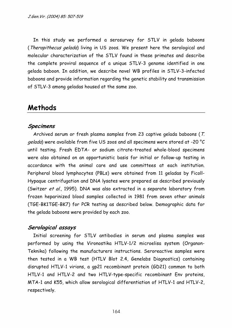
J.Gen.Vir. (2004) 85: 507-519
164
In this study we performed a serosurvey for STLV in gelada baboons
(Theropithecus gelada) living in US zoos. We present here the serological and molecular characterization of the STLV found in these primates and describe the complete proviral sequence of a unique STLV-3 genome identified in one gelada baboon. In addition, we describe novel WB profiles in STLV-3-infected baboons and provide information regarding the genetic stability and transmission of STLV-3 among geladas housed at the same zoo.
Methods Specimens
Archived serum or fresh plasma samples from 23 captive gelada baboons (T. gelada) were available from five US zoos and all specimens were stored at -20 °C until testing. Fresh EDTA- or sodium citrate-treated whole-blood specimens were also obtained on an opportunistic basis for initial or follow-up testing in accordance with the animal care and use committees at each institution. Peripheral blood lymphocytes (PBLs) were obtained from 11 geladas by Ficoll-Hypaque centrifugation and DNA lysates were prepared as described previously (Switzer et al., 1995). DNA was also extracted in a separate laboratory from frozen heparinized blood samples collected in 1981 from seven other animals (TGE-BK1TGE-BK7) for PCR testing as described below. Demographic data for the gelada baboons were provided by each zoo.
Serological assays
Initial screening for STLV antibodies in serum and plasma samples was performed by using the Vironostika HTLV-1/2 microelisa system (Organon-Teknika) following the manufacturers instructions. Seroreactive samples were then tested in a WB test (HTLV Blot 2.4, Genelabs Diagnostics) containing disrupted HTLV-1 virions, a gp21 recombinant protein (GD21) common to both HTLV-1 and HTLV-2 and two HTLV-type-specific recombinant Env proteins, MTA-1 and K55, which allow serological differentiation of HTLV-1 and HTLV-2, respectively.
Chapter 8 STLV-3 in geladas with variable WB reactivity
165
PCR analysis
Generic nested PTLV tax and STLV-1 env or STLV-3-specific LTR PCR was performed on DNA lysates prepared from uncultured PBLs. The PTLV tax and STLV-3-specific LTR nested PCR was also performed on DNA extracted from frozen blood samples from seven other geladas. The primer sequences, expected product sizes and amplification conditions are given in Table 8.1. The generic PTLV tax primers used in the primary PCR amplification have been shown previously to amplify HTLV-1, HTLV-2 and divergent PTLVs, including STLV-2/Panp and STLV-3/PHA-PH969 (Busch et al., 2000). In the current study, we also performed a semi-nested tax PCR using the primers PH2F and PH2R (Table 8.1.) to increase the amount of specific amplified product necessary for direct sequence analysis. The STLV-1 env and STLV-3 LTR primers were based on the HTLV-1/ATK and STLV-3/PHA-PH969 sequences with GenBank accession numbers J02029 and Y07616, respectively. To obtain the complete sequence of the unique gelada STLV-3, we PCR amplified sequential overlapping regions of the genome as previously described (Meertens et al., 2003).
Table 8.1.Sequences of primers used for amplifying partial tax, env and LTR regions of primate T-cell lymphotropic viruses Name Primersequence(5’→3’) Location* Expected
PCR Annea
ling No.of cycles
productsize(bp)
temp. (°C)
PH1F TTGTCATCAGCCCACTTCCCAGG tax,7243–7262,outer PH2R AAGGAGGGGAGTCGAGGGATAAGG tax,7478–7455,outer 236 50 40 PH2F CCCAGGTTTCGGGCAAAGCCTTCT tax,7257–7280,inner PH2R† AAGGAGGGGAGTCGAGGGATAAGG tax,7478–7455,inner 222 50 40 PLTRF1 CCCCAAGACGAACCACAACCACCAACT LTR,17–43,outer PLTRR1 CAACTGTTTGCTTTCTTCCCTAGGGCT LTR,699–673,outer 683 50 35 PLTRF2 TCATCCGTCTGAGAGCCGTCTCGC LTR,62–86,inner 602 50 35 PLTRR2 AGCCCAGACTCTTTAGGACCCAACTCC LTR,663–637,inner P21EF1 GGACCCACTGCTTTGACCCCC env,6019–6039,outer P21ER1 ATTGCGTGCTTGGTTTACAGG env,6682–6662,outer 664 45 35 P21EF2 CATAACTCCCTCATCCTGCCC env,6069–6089,inner P21ER2 AGTAATGGGGGTATCTGACGC env,6639–6619,inner 571 45 35 *Positions of the tax and LTR primers are given according to the STLV-3 (strain PH969) genome; the env primer positions are given according to HTLV-1 (strainATK). † Primer PH2R was used with PH2F in a semi-nested PCR.
J.Gen.Vir. (2004) 85: 507-519
166
Nested PCR products were electrophoresed in 1.8% agarose gels and visualized by ethidium bromide staining.
Sequence and phylogenetic analysis
PCR products were purified using the Qiaquick PCR purification kit (Qiagen) and sequenced in both directions using a BigDye terminator cycle kit (PE Biosystems) and an ABI 373 automated sequencer. BLAST searches at the National Center for Biotechnology Information website (http:// www.ncbi.nlm.nih.gov/BLAST/) were done on the env and tax nucleotide sequences to identify genetically similar sequences in the GenBank database. Percentage nucleotide identity was determined with the GAP program in the Wisconsin sequence analysis package on a UNIX workstation (Womble, 2000). Comparison of full-length STLV-3 genomes was done using STLV-3/TGE-2117 as the query sequence and the Kimura two-parameter model and a transition/ transversion ratio of 2.0 implemented in the program SimPlot (Lole et al., 1999). The coding regions of the gag, polymerase (pol), env and tax genes were concatenated prior to phylogenetic analysis using the DAMBE program version 4.0.75 (Xia & Xie, 2001). Phylogenetic analysis of the tax (180 bp) and concatenated gag-pol-env-tax (6795 bp) sequences was performed with the software package PAUP* version 4.0b10 (Swofford, 1998) on nucleotide alignments made using ClustalX (Thompson et al., 1994), which were manually edited in Se-Alv2.0.11 (http://evolve.zoo.ox.ac.uk/) according to the codon reading frames.
Neighbour-joining (NJ) and maximum-likelihood (ML) trees were constructed
under the most appropriate evolutionary model determined with Modeltest 3-06 (Posada & Crandall, 1998). For the gag-pol-env-tax sequences, the general time-reversible model, allowing six different substitution rate categories, with gamma distributed rate heterogeneity and an estimated proportion of invariable sites, provided the best fit to the data. For the tax analysis, the transitional model allowing different rates among transitions including a gamma distributed rate heterogeneity seemed to be the best-fitting model. For the env sequences, the Tamura Nei evolutionary model was used with gamma distributed rate heterogeneity, taking into account a different substitution rate for transversions and purine and pyrimidine transitions. The NJ trees were

Chapter 8 STLV-3 in geladas with variable WB reactivity
167
constructed by iteratively optimizing the model parameters, followed by a bootstrap analysis of 1000 replicates. The ML trees were constructed starting from the NJ tree with optimized parameters using a heuristic search with the nearest neighbour interchange and the subtree pruning-regrafting branch-swapping algorithm (Rogers & Swofford, 1999). LTR sequences were aligned using the CLUSTAL W program (Thompson et al., 1994) and distance-based LTR trees were generated using the Kimura two-parameter model in conjunction with the NJ method in the MEGA program (version 2.1) (Kumar et al., 2001). Bootstrap replicates (1000) were used to test the reliability of the final topology of the LTR tree.
Molecular clock analysis and dating
The molecular clock hypothesis was tested using the likelihood ratio test with the likelihoods for the ML and clock-like ML trees obtained in PAUP* (Swofford, 1998). The clock was tested with the best-fitting evolutionary model, estimated in Modeltest and with the NJ optimized parameters as described above. The evolutionary rate and subsequently the divergence times were estimated based on a known divergence time point and on the branch lengths of the ML clock tree according to the formula: evolutionary rate (r)=branch length (bl)/divergence time (t).
Results and discussion STLV-like antibodies were found in the sera or plasma of 12 of 23 (52.2%)
gelada baboons. Five of the 12 seroreactive baboon samples (41.7%) showed an STLV-1/HTLV-1-like WB profile (MTA-1 positive), one sample (8.3%) showed an STLV-2/HTLV-2-like WB pattern (K55 positive) and four samples (33.3%) showed reactivity to all HTLV proteins (GD21, gp21, p24, p32, p36, gp46 and p53) except the type-specific peptides and were considered positive but untypable (Table 8.2., Fig. 8.1.). The two other samples (16.6%) reacted weakly with p19 and/or GD21 and thus were classified as indeterminate (Table 8.2., Fig. 8.1.).
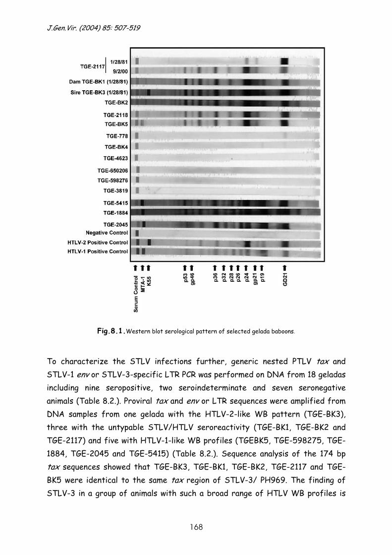
J.Gen.Vir. (2004) 85: 507-519
168
Fig.8.1.Western blot serological pattern of selected gelada baboons.
To characterize the STLV infections further, generic nested PTLV tax and STLV-1 env or STLV-3-specific LTR PCR was performed on DNA from 18 geladas including nine seropositive, two seroindeterminate and seven seronegative animals (Table 8.2.). Proviral tax and env or LTR sequences were amplified from DNA samples from one gelada with the HTLV-2-like WB pattern (TGE-BK3), three with the untypable STLV/HTLV seroreactivity (TGE-BK1, TGE-BK2 and TGE-2117) and five with HTLV-1-like WB profiles (TGEBK5, TGE-598275, TGE-1884, TGE-2045 and TGE-5415) (Table 8.2.). Sequence analysis of the 174 bp tax sequences showed that TGE-BK3, TGE-BK1, TGE-BK2, TGE-2117 and TGE-BK5 were identical to the same tax region of STLV-3/ PH969. The finding of STLV-3 in a group of animals with such a broad range of HTLV WB profiles is
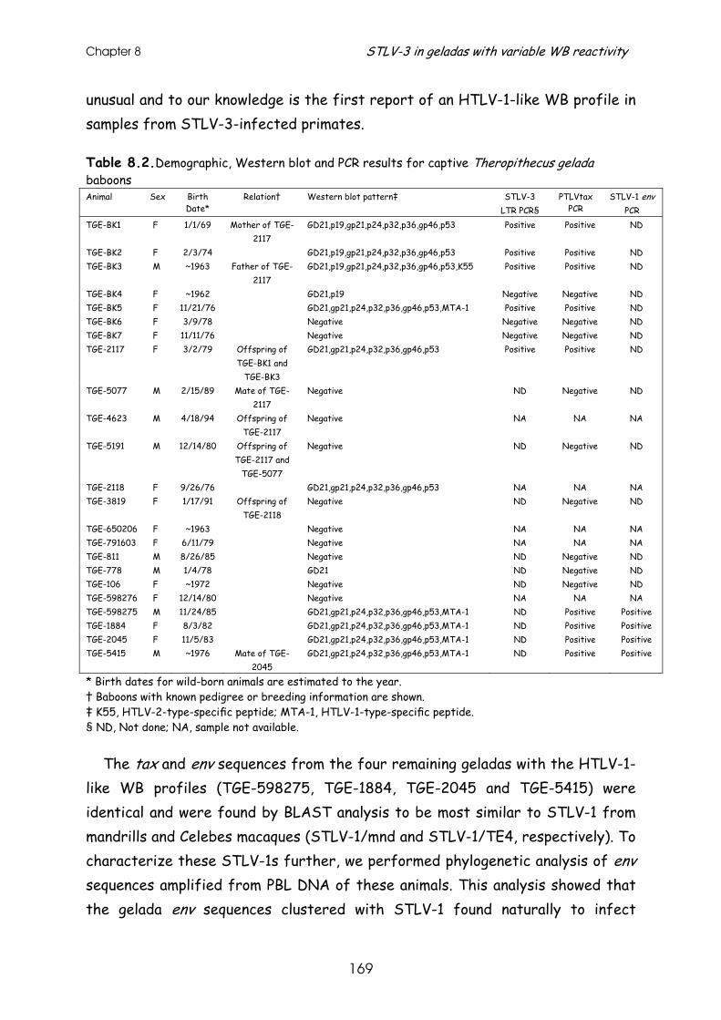
Chapter 8 STLV-3 in geladas with variable WB reactivity
169
unusual and to our knowledge is the first report of an HTLV-1-like WB profile in samples from STLV-3-infected primates.
Table 8.2.Demographic, Western blot and PCR results for captive Theropithecus gelada baboons Animal Sex Birth
Date* Relation† Western blot pattern‡ STLV-3
LTR PCR§ PTLVtax
PCR STLV-1 env
PCR TGE-BK1 F 1/1/69 Mother of TGE-
2117 GD21,p19,gp21,p24,p32,p36,gp46,p53 Positive Positive ND
TGE-BK2 F 2/3/74 GD21,p19,gp21,p24,p32,p36,gp46,p53 Positive Positive ND TGE-BK3 M ~1963 Father of TGE-
2117 GD21,p19,gp21,p24,p32,p36,gp46,p53,K55 Positive Positive ND
TGE-BK4 F ~1962 GD21,p19 Negative Negative ND TGE-BK5 F 11/21/76 GD21,gp21,p24,p32,p36,gp46,p53,MTA-1 Positive Positive ND TGE-BK6 F 3/9/78 Negative Negative Negative ND TGE-BK7 F 11/11/76 Negative Negative Negative ND TGE-2117 F 3/2/79 Offspring of
TGE-BK1 and TGE-BK3
GD21,gp21,p24,p32,p36,gp46,p53 Positive Positive ND
TGE-5077 M 2/15/89 Mate of TGE-2117
Negative ND Negative ND
TGE-4623 M 4/18/94 Offspring of TGE-2117
Negative NA NA NA
TGE-5191 M 12/14/80 Offspring of TGE-2117 and
TGE-5077
Negative ND Negative ND
TGE-2118 F 9/26/76 GD21,gp21,p24,p32,p36,gp46,p53 NA NA NA TGE-3819 F 1/17/91 Offspring of
TGE-2118 Negative ND Negative ND
TGE-650206 F ~1963 Negative NA NA NA TGE-791603 F 6/11/79 Negative NA NA NA TGE-811 M 8/26/85 Negative ND Negative ND TGE-778 M 1/4/78 GD21 ND Negative ND TGE-106 F ~1972 Negative ND Negative ND TGE-598276 F 12/14/80 Negative NA NA NA TGE-598275 M 11/24/85 GD21,gp21,p24,p32,p36,gp46,p53,MTA-1 ND Positive Positive TGE-1884 F 8/3/82 GD21,gp21,p24,p32,p36,gp46,p53,MTA-1 ND Positive Positive TGE-2045 F 11/5/83 GD21,gp21,p24,p32,p36,gp46,p53,MTA-1 ND Positive Positive TGE-5415 M ~1976 Mate of TGE-
2045 GD21,gp21,p24,p32,p36,gp46,p53,MTA-1 ND Positive Positive
* Birth dates for wild-born animals are estimated to the year. † Baboons with known pedigree or breeding information are shown. ‡ K55, HTLV-2-type-specific peptide; MTA-1, HTLV-1-type-specific peptide. § ND, Not done; NA, sample not available.
The tax and env sequences from the four remaining geladas with the HTLV-1-like WB profiles (TGE-598275, TGE-1884, TGE-2045 and TGE-5415) were identical and were found by BLAST analysis to be most similar to STLV-1 from mandrills and Celebes macaques (STLV-1/mnd and STLV-1/TE4, respectively). To characterize these STLV-1s further, we performed phylogenetic analysis of env sequences amplified from PBL DNA of these animals. This analysis showed that the gelada env sequences clustered with STLV-1 found naturally to infect
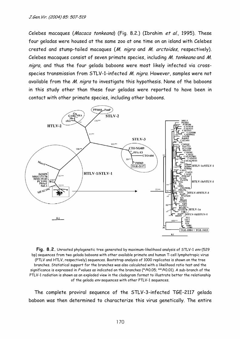
J.Gen.Vir. (2004) 85: 507-519
170
Celebes macaques (Macaca tonkeana) (Fig. 8.2.) (Ibrahim et al., 1995). These four geladas were housed at the same zoo at one time on an island with Celebes crested and stump-tailed macaques (M. nigra and M. arctoides, respectively). Celebes macaques consist of seven primate species, including M. tonkeana and M. nigra, and thus the four gelada baboons were most likely infected via cross-species transmission from STLV-1-infected M. nigra. However, samples were not available from the M. nigra to investigate this hypothesis. None of the baboons in this study other than these four geladas were reported to have been in contact with other primate species, including other baboons.
Fig. 8.2. Unrooted phylogenetic tree generated by maximum-likelihood analysis of STLV-1 env (529
bp) sequences from two gelada baboons with other available primate and human T-cell lymphotropic virus (PTLV and HTLV, respectively) sequences. Bootstrap analysis of 1000 replicates is shown on the tree branches. Statistical support for the branches was also calculated with a likelihood ratio test and the
significance is expressed in P values as indicated on the branches (*P<0.05; **P<0.01). A sub-branch of the PTLV-1 radiation is shown as an exploded view in the cladogram format to illustrate better the relationship
of the gelada env sequences with other PTLV-1 sequences.
The complete proviral sequence of the STLV-3-infected TGE-2117 gelada baboon was then determined to characterize this virus genetically. The entire
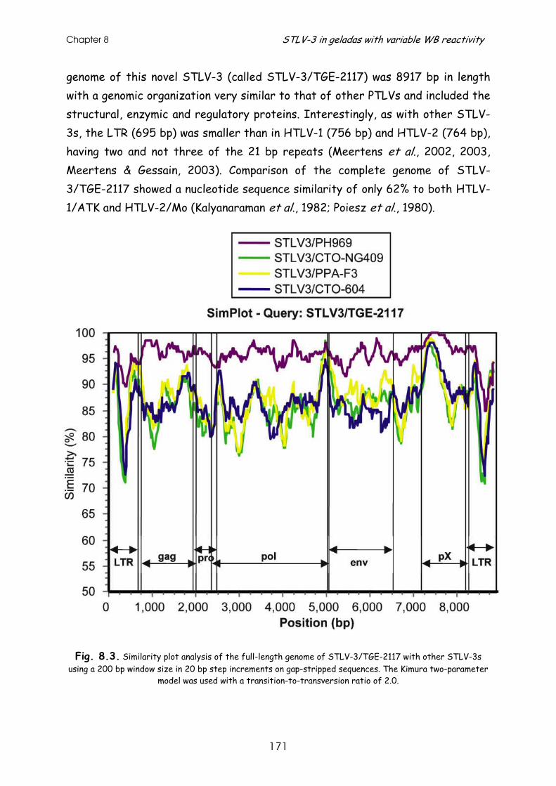
Chapter 8 STLV-3 in geladas with variable WB reactivity
171
genome of this novel STLV-3 (called STLV-3/TGE-2117) was 8917 bp in length with a genomic organization very similar to that of other PTLVs and included the structural, enzymic and regulatory proteins. Interestingly, as with other STLV-3s, the LTR (695 bp) was smaller than in HTLV-1 (756 bp) and HTLV-2 (764 bp), having two and not three of the 21 bp repeats (Meertens et al., 2002, 2003, Meertens & Gessain, 2003). Comparison of the complete genome of STLV-3/TGE-2117 showed a nucleotide sequence similarity of only 62% to both HTLV-1/ATK and HTLV-2/Mo (Kalyanaraman et al., 1982; Poiesz et al., 1980).
Fig. 8.3. Similarity plot analysis of the full-length genome of STLV-3/TGE-2117 with other STLV-3s using a 200 bp window size in 20 bp step increments on gap-stripped sequences. The Kimura two-parameter
model was used with a transition-to-transversion ratio of 2.0.
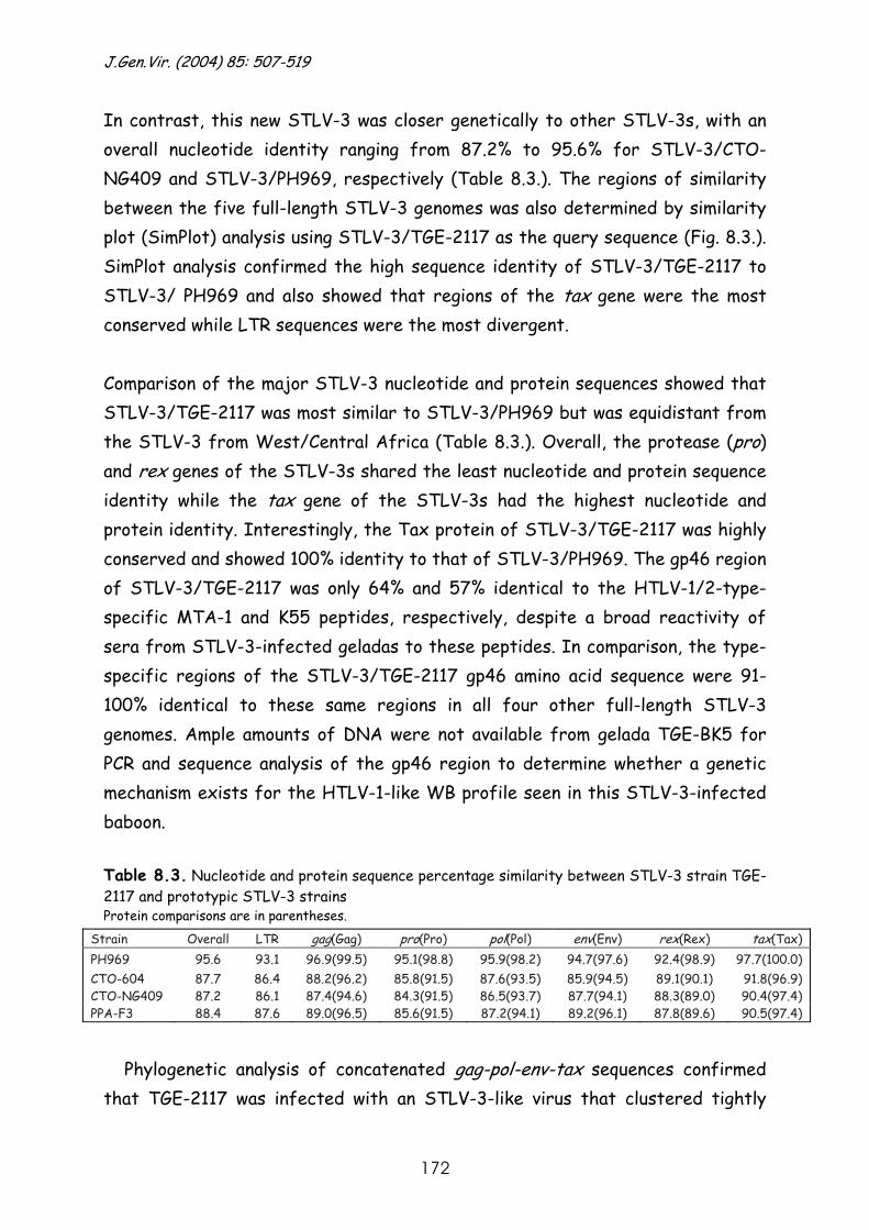
J.Gen.Vir. (2004) 85: 507-519
172
In contrast, this new STLV-3 was closer genetically to other STLV-3s, with an overall nucleotide identity ranging from 87.2% to 95.6% for STLV-3/CTO-NG409 and STLV-3/PH969, respectively (Table 8.3.). The regions of similarity between the five full-length STLV-3 genomes was also determined by similarity plot (SimPlot) analysis using STLV-3/TGE-2117 as the query sequence (Fig. 8.3.). SimPlot analysis confirmed the high sequence identity of STLV-3/TGE-2117 to STLV-3/ PH969 and also showed that regions of the tax gene were the most conserved while LTR sequences were the most divergent.
Comparison of the major STLV-3 nucleotide and protein sequences showed that STLV-3/TGE-2117 was most similar to STLV-3/PH969 but was equidistant from the STLV-3 from West/Central Africa (Table 8.3.). Overall, the protease (pro) and rex genes of the STLV-3s shared the least nucleotide and protein sequence identity while the tax gene of the STLV-3s had the highest nucleotide and protein identity. Interestingly, the Tax protein of STLV-3/TGE-2117 was highly conserved and showed 100% identity to that of STLV-3/PH969. The gp46 region of STLV-3/TGE-2117 was only 64% and 57% identical to the HTLV-1/2-type-specific MTA-1 and K55 peptides, respectively, despite a broad reactivity of sera from STLV-3-infected geladas to these peptides. In comparison, the type-specific regions of the STLV-3/TGE-2117 gp46 amino acid sequence were 91-100% identical to these same regions in all four other full-length STLV-3 genomes. Ample amounts of DNA were not available from gelada TGE-BK5 for PCR and sequence analysis of the gp46 region to determine whether a genetic mechanism exists for the HTLV-1-like WB profile seen in this STLV-3-infected baboon. Table 8.3. Nucleotide and protein sequence percentage similarity between STLV-3 strain TGE-2117 and prototypic STLV-3 strains Protein comparisons are in parentheses.
Strain Overall LTR gag(Gag) pro(Pro) pol(Pol) env(Env) rex(Rex) tax(Tax) PH969 95.6 93.1 96.9(99.5) 95.1(98.8) 95.9(98.2) 94.7(97.6) 92.4(98.9) 97.7(100.0) CTO-604 87.7 86.4 88.2(96.2) 85.8(91.5) 87.6(93.5) 85.9(94.5) 89.1(90.1) 91.8(96.9) CTO-NG409 87.2 86.1 87.4(94.6) 84.3(91.5) 86.5(93.7) 87.7(94.1) 88.3(89.0) 90.4(97.4) PPA-F3 88.4 87.6 89.0(96.5) 85.6(91.5) 87.2(94.1) 89.2(96.1) 87.8(89.6) 90.5(97.4)
Phylogenetic analysis of concatenated gag-pol-env-tax sequences confirmed
that TGE-2117 was infected with an STLV-3-like virus that clustered tightly
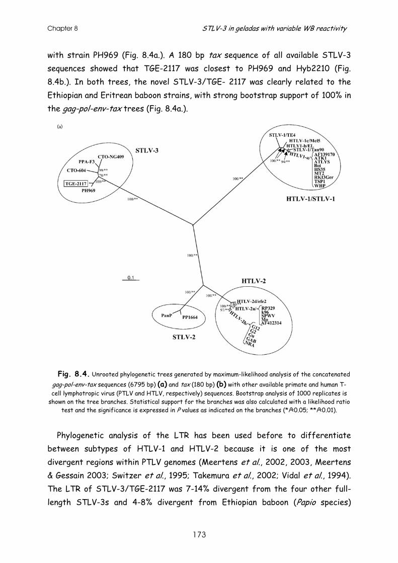
Chapter 8 STLV-3 in geladas with variable WB reactivity
173
with strain PH969 (Fig. 8.4a.). A 180 bp tax sequence of all available STLV-3 sequences showed that TGE-2117 was closest to PH969 and Hyb2210 (Fig. 8.4b.). In both trees, the novel STLV-3/TGE- 2117 was clearly related to the Ethiopian and Eritrean baboon strains, with strong bootstrap support of 100% in the gag-pol-env-tax trees (Fig. 8.4a.).
Fig. 8.4. Unrooted phylogenetic trees generated by maximum-likelihood analysis of the concatenated
gag-pol-env-tax sequences (6795 bp) (a) and tax (180 bp) (b) with other available primate and human T-cell lymphotropic virus (PTLV and HTLV, respectively) sequences. Bootstrap analysis of 1000 replicates is
shown on the tree branches. Statistical support for the branches was also calculated with a likelihood ratio test and the significance is expressed in P values as indicated on the branches (*P<0.05; **P<0.01).
Phylogenetic analysis of the LTR has been used before to differentiate
between subtypes of HTLV-1 and HTLV-2 because it is one of the most divergent regions within PTLV genomes (Meertens et al., 2002, 2003, Meertens & Gessain 2003; Switzer et al., 1995; Takemura et al., 2002; Vidal et al., 1994). The LTR of STLV-3/TGE-2117 was 7-14% divergent from the four other full-length STLV-3s and 4-8% divergent from Ethiopian baboon (Papio species)
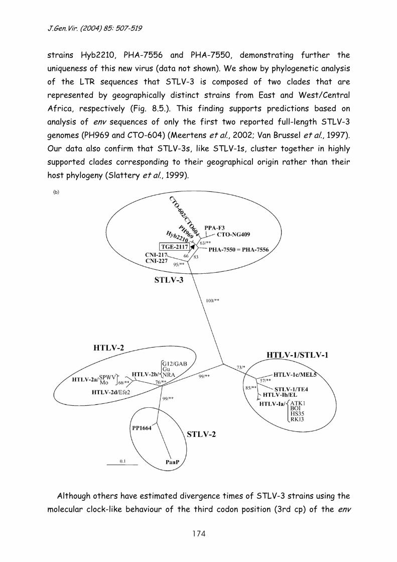
J.Gen.Vir. (2004) 85: 507-519
174
strains Hyb2210, PHA-7556 and PHA-7550, demonstrating further the uniqueness of this new virus (data not shown). We show by phylogenetic analysis of the LTR sequences that STLV-3 is composed of two clades that are represented by geographically distinct strains from East and West/Central Africa, respectively (Fig. 8.5.). This finding supports predictions based on analysis of env sequences of only the first two reported full-length STLV-3 genomes (PH969 and CTO-604) (Meertens et al., 2002; Van Brussel et al., 1997). Our data also confirm that STLV-3s, like STLV-1s, cluster together in highly supported clades corresponding to their geographical origin rather than their host phylogeny (Slattery et al., 1999).
Although others have estimated divergence times of STLV-3 strains using the
molecular clock-like behaviour of the third codon position (3rd cp) of the env
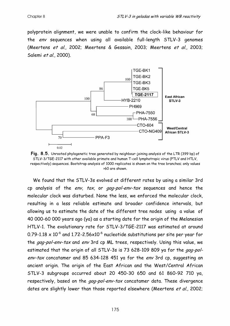
Chapter 8 STLV-3 in geladas with variable WB reactivity
175
polyprotein alignment, we were unable to confirm the clock-like behaviour for the env sequences when using all available full-length STLV-3 genomes (Meertens et al., 2002; Meertens & Gessain, 2003; Meertens et al., 2003; Salemi et al., 2000).
Fig. 8.5. Unrooted phylogenetic tree generated by neighbour-joining analysis of the LTR (399 bp) of STLV-3/TGE-2117 with other available primate and human T-cell lymphotropic virus (PTLV and HTLV,
respectively) sequences. Bootstrap analysis of 1000 replicates is shown on the tree branches; only values >60 are shown.
We found that the STLV-3s evolved at different rates by using a similar 3rd cp analysis of the env, tax, or gag-pol-env-tax sequences and hence the molecular clock was disturbed. None the less, we enforced the molecular clock, resulting in a less reliable estimate and broader confidence intervals, but allowing us to estimate the date of the different tree nodes using a value of 40 000-60 000 years ago (ya) as a starting date for the origin of the Melanesian HTLV-1. The evolutionary rate for STLV-3/TGE-2117 was estimated at around 0.79-1.18 x 10⁻⁶ and 1.72-2.56x10⁻⁶ nucleotide substitutions per site per year for the gag-pol-env-tax and env 3rd cp ML trees, respectively. Using this value, we estimated that the origin of all STLV-3s is 73 628-109 809 ya for the gag-pol-env-tax concatamer and 85 634-128 451 ya for the env 3rd cp, suggesting an ancient origin. The origin of the East African and the West/Central African STLV-3 subgroups occurred about 20 450-30 650 and 61 860-92 710 ya, respectively, based on the gag-pol-env-tax concatamer data. These divergence dates are slightly lower than those reported elsewhere (Meertens et al., 2002;
J.Gen.Vir. (2004) 85: 507-519
176
Meertens & Gessain, 2003) and may be due to the use of a constant-rate model in previous studies, which has been shown to overestimate divergence times (Yang, 1996).
Since STLV-3 is the most recently identified PTLV, very little is known about
its transmission or genomic stability in infected animals. Sera archived in 1981 from five seropositive geladas from the same zoo included a captive-born mother (TGE-BK1), a wild-born father (TGE-BK3), their female offspring (TGE-2117) who was born in 1979 and two other females (TGE-BK2 and TGE-BK5). LTR sequences from the mother and TGE-2117 were identical and clustered together with 100% bootstrap support by phylogenetic analysis (Fig. 8.5.), suggesting a mother-to-offspring transmission of STLV-3 infection in this pair. The LTR sequences from two other STLV-3-infected female geladas (TGE-BK2 and TGE-BK5) were essentially identical to male gelada TGE-BK3, having >99% nucleotide identity, suggesting a horizontal transmission. We also examined the genetic stability of STLV-3 over time by analysis of LTR sequences obtained from blood specimens from TGE-2117 and her parents that were collected 20 years apart. Interestingly, the LTR sequences for both parental baboons and the descendant TGE-2117 were identical, suggesting a high genetic stability of the STLV-3 genomes similar to that seen for other PTLVs (Slattery et al., 1999). Viral persistence by oligoclonal expansion of infected lymphocytes within animals is believed to confer the high genetic stability of the PTLVs (Gabet et al., 2003). This finding contrasts with the genetic mutability of other exogenous retroviruses, such as the human immunodeficiency virus, which has mutation rates 3-4 logs higher (Sharp et al., 1994).
This study reports the serological and molecular characterization of a novel STLV-3 in Ethiopian T. gelada baboons and the full-length sequence analysis of this distinct STLV-3 found in animal TGE-2117. This report is the first to find and characterize STLV-3 and STLV-1 infection in this baboon species. The absence of STLV infection of geladas observed by others (Takemura et al., 2002) may be explained by differences in the sensitivities of the antibody screening assays used in each study and/or the sampling of gelada specimens originating from different Ethiopian locations (Takemura et al., 2002). Interestingly, the STLV-3 LTR sequences obtained from five gelada baboons in
Chapter 8 STLV-3 in geladas with variable WB reactivity
177
the current study were more genetically related to an STLV-3 LTR sequence found in a wild-born Ethiopian P. hamadryas and P. anubis hybrid (Hyb2210) than to those found in either Eritrean or Ethiopian hamadryas baboons. These results suggest that the STLV-3s in the gelada and hybrid baboons are distinct from those found in hamadryas baboons and that the origin of the STLV-3s in the hybrid baboons may be from the gelada and not from infected hamadryas baboons as previously proposed (Takemura et al., 2002).
Although fossil evidence suggests that the extant Theropithecus baboons
once radiated across the African continent and exceeded the combined ranges of the Papio species, the gelada’s current habitat is restricted to the high plateaus of Ethiopia (Pickford, 1993). In this region, gelada baboons are commonly sympatric with anubis and hamadryas baboons but occupy different ecological niches. In contrast, anubis and hamadryas baboons commonly interbreed, as reported elsewhere (Takemura et al., 2002). None the less, these overlapping habitats may have provided opportunities for cross-species transmission of STLV-3 infections to or from each of these baboon species.
Questions about whether humans are infected with STLV-3- like viruses are
raised by the widespread distribution of STLV-3 across Africa and its probable ancient existence (Goubau et al., 1994; Meertens et al., 2001, 2002; Meertens & Gessain, 2003; Takemura et al., 2002; Van Dooren et al., 2001), the ability of STLV-3 to grow in human cells in vitro (Goubau et al., 1994) and evidence for multiple interspecies transmissions that have occurred among the PTLV-1s (Gessain et al., 2002; Slattery et al., 1999). The presence of STLV-3-like viruses in primates such as geladas that are commonly hunted for their coats or shot while raiding crops may support the possibility of such infection in humans (Pickford, 1993). In addition, the human population density on the Ethiopian plateau is among the highest in sub-Saharan Africa, thus placing the gelada in coexistence with humans living in this region and putting humans at risk for cross-species infections with STLV and other simian retroviruses. The finding by us and others of STLV-1 and STLV-3 infections in captive baboons also suggests that people working with these primates may be at risk for occupational exposure to these viruses (Goubau et al., 1994, Meertens & Gessain, 2003; Takemura et al., 2002).
J.Gen.Vir. (2004) 85: 507-519
178
HTLV-1, HTLV-2 and HTLV-indeterminate WB profiles have all been observed
in Ethiopian populations (Buckner et al., 1992; Vrielink et al., 1995), but it is not known if any of these reactive samples are associated with STLV-3-like infections. While limited studies of 24 African and 325 non-African human specimens with HTLV-seroindeterminate WB results have detected no STLV-3-like infection (Busch et al., 2000; Vandamme et al., 1997), additional studies of larger numbers of human samples with not only indeterminate but also HTLV-1- and -2-like WB profiles will be necessary to assess fully the rate of infection with STLV-3-like viruses. Our finding of one STLV-3-infected gelada baboon with an HTLV-1-like WB profile and a previous report of an STLV-1-infected baboon with an HTLV-2-like serotype (Mahieux et al., 2000) also raises questions about the accuracy of viral typing of PTLV infections by the currently used HTLV-1- and HTLV-2-type-specific Env peptides spiked onto HTLV-1 WB strips. Our results also highlight the need for better STLV-3-specific diagnostic assays to facilitate screening for STLV-3-like infections in both humans and NHPs.
The complete genome of STLV-3/TGE2117 has been assigned the GenBank accession number AY217650. The GenBank accession numbers for the STLV-3 tax and LTR and STLV-1 tax and env sequences are AY241678–AY241693.
We thank the Zoological Society of San Diego’s Center for Reproduction of
Endangered Species for providing DNA and serum samples from gelada baboons for this study and also the veterinary and clinical laboratory staff at the Los Angeles, Oakland, Gladys Porter and Folsom Children’s Zoos for providing us with additional gelada specimens.
Use of trade names is for identification only and does not imply endorsement by the
US Department of Health and Human Services, the Public Health Service, or the Centers for Disease Control and Prevention.
References
Buckner, C., Roberts, C. R., Foung, S. K. H. & 7 other authors (1992). Immune responsiveness to the recombinant envelope epitopes of human T lymphotropic virus types I and II in diverse geographic populations. J Infect Dis 166, 1160-1163.
Chapter 8 STLV-3 in geladas with variable WB reactivity
179
Busch, M., Switzer, W. M., Khabbaz, R. F., Kaplan, J. E., Heneine, W. & the Retrovirus Epidemiology Donor Study Group. (2000). Absence of evidence of infection with divergent simian T-cell lymphotropic viruses in blood donors with seroindeterminate human T-cell lymphotropic type 1 results. Transfusion 40, 443-449. Gabet, A. S., Gessain, A. & Wattel, E. (2003). High simian T-cell leukemia virus type 1 proviral loads combined with genetic stability as a result of cell-associated provirus replication in naturally infected, asymptomatic monkeys. Int J Cancer 107, 74-83. Gessain, A., Meertens, L. & Mahieux, R. (2002). Molecular epidemiology of human T cell leukemia/lymphoma viruses type 1 and type 2 (HTLV-1/2) and related simian retroviruses (STLV-1, STLV-2 and STLV-L/3). In The Molecular Epidemiology of Human Viruses, 1st edn, pp. 121165. Edited by T. Leitner. Boston: Kluwer Academic Publishers. Giri, A., Markham, P., Digilio, L., Hurteau, G., Gallo, R. C. & Franchini, G. (1994). Isolation of a novel simian T-cell lymphotropic virus from Pan paniscus that is distantly related to the human T-cell leukemia/lymphotropic virus types I and II. J Virol 68, 8392-8395. Goubau, P., Van Brussel, M., Vandamme, A. M., Liu, H. F. & Desmyter, J. (1994). A primate T-lymphotropic virus, PTLV-L, different from human T-lymphotropic viruses types I and II, in a wild-caught baboon (Papio hamadryas). Proc Natl Acad Sci U S A 91, 2848-2852. Heneine, W., Switzer, W. M., Sandstrom, P. & 9 other authors (1998). Identification of a human population infected with simian foamy viruses. Nat Med 4, 403407. Ibrahim, F., De The , G. & Gessain, A. (1995). Isolation and characterization of a new simian T-cell leukemia virus type 1 from naturally infected Celebes macaques (Macaca tonkeana): complete nucleotide sequence and phylogenetic relationship with the Australo- Melanesian human T-cell leukemia virus type 1. J Virol 69, 6980-6993. Kalyanaraman, V. S., Sarngadharan, M. G., Robert-Guroff, M., Miyoshi, I., Golde, D. & Gallo, R. C. (1982). A new subtype of human T-cell leukemia virus (HTLV-II) associated with a T-cell variant of hairy cell leukemia. Science 218, 571-573. Khabbaz, R. F., Heneine, W., George, J. R. & 7 other authors (1994). Brief report: infection of a laboratory worker with simian immunodeficiency virus. N Eng J Med 330, 172-177. Kumar, S., Tamura, K., Jakobsen, I. B. & Nei, M. (2001). MEGA2: molecular evolutionary genetics analysis software. Bioinformatics 17, 1244-1245. Lerche, N. W., Switzer, W. M., Yee, J. L., Shanmugam, V., Rosenthal, A. N., Chapman, L. E., Folks, T. M. & Heneine, W. (2001). Evidence of infection with simian type D retrovirus in persons occupationally exposed to nonhuman primates. J Virol 75, 1783-1789. Liu, H.-F., Vandamme, A. M., Van Brussel, M., Desmyter, J. & Goubau, P. (1994). New retroviruses in human and simian T-lymphotropic viruses. Lancet 344, 265-266. Lole, K. S., Bollinger, R. C., Paranjape, R. S., Gadkari, D., Kulkarni, S. S., Novak, N. G., Ingersoll, R., Sheppard, H. W. & Ray, S. C. (1999). Full-length human immunodeficiency virus type 1 genomes from subtype C-infected seroconverters in India, with evidence of intersubtype recombination. J Virol 73, 152-160. Mahieux, R., Chappey, C., Georges-Courbot, M. C., Dubreuil, G., Mauclere, P., Georges, A. & Gessain, A. (1998). Simian T-cell lymphotropic virus type 1 from Mandrillus sphinx as a simian counterpart of human T-cell lymphotropic virus type 1 subtype D. J Virol 72, 10316-10322. Mahieux, R., Chappey, C., Meertens, L., Mauclère, P., Lewis, J. & Gessain, A. (2000). Molecular characterization and phylogenetic analyses of a new simian T cell lymphotropic virus type 1 in a wildcaught African baboon (Papio anubis) with an indeterminate STLV type 2-like serology. AIDS Res Hum Retroviruses 16, 2043-2048.
J.Gen.Vir. (2004) 85: 507-519
180
Meertens, L. & Gessain, A. (2003). Divergent simian T cell lymphotropic virus type 3 in wild-caught Papio hamadryas papio from Senegal: widespread distribution of STLV-3 in Africa. J Virol 77, 782-789. Meertens, L., Rigoulet, J., Mauclere, P. & 8 other authors (2001). Molecular and phylogenetic analyses of 16 novel simian T cell leukemia virus type 1 from Africa: close relationship of STLV-1 from Allenopithecus nigroviridis to HTLV-1 subtype B strains. Virology 287, 275-285. Meertens, L., Mahieux, R., Mauclère, P., Lewis, J. & Gessain, A. (2002). Complete sequence of a novel highly divergent simian T-cell lymphotropic virus from wild-caught red-capped mangabeys (Cercocebus torquatus) from Cameroon: a new primate T-lymphotropic virus type 3 subtype. J Virol 76, 259-268. Meertens, L., Shanmugam, V., Gessain, A., Beer, B. E., Tooze, Z., Heneine, W. & Switzer, W. M. (2003). A novel, divergent simian T-cell lymphotropic virus type 3 in a wild-caught red-capped mangabey (Cercocebus torquatus torquatus) from Nigeria. J Gen Virol 84, 2723-2727. Miyoshi, I., Yoshimoto, S., Fujishita, M., Taguchi, H., Kubonishi, I., Niiya, K. & Minezawa, M. (1982). Natural adult T-cell leukemia virus infection in Japanese monkeys [letter]. Lancet ii, 658. Nerrienet, E., Meertens, L., Kfutwah, A., Foupouapouognigni, Y. & Gessain, A. (2001). Molecular epidemiology of simian T-lymphotropic virus (STLV) in wild-caught monkeys and apes from Cameroon: a new STLV-1, related to human T-lymphotropic virus subtype F, in a Cercocebus agilis. J Gen Virol 82, 2973-2977. Pickford, M. (1993). Climatic change, biogeography and Theropithecus. In Theropithecus: the Rise and Fall of a Primate Genus,1st edn, pp.227243. Edited by N. G. Jablonski. Cambridge: Cambridge University Press. Poiesz, B. J. W., Ruscetti, F., Gazdar, A. F., Bunn, P. A., Minna, J. D. & Gallo, R. C. (1980). Detection and isolation of type C retrovirus particles from fresh and cultured lymphocytes of a patient with cutaneous T-cell lymphoma. Proc Natl Acad Sci U S A 77, 7415-7419. Posada, D. & Crandall, K. A. (1998). MODELTEST: testing the model of DNA substitution. Bioinformatics 14, 817818. Rogers, J. S. & Swofford, D. L. (1999). Multiple local maxima for likelihoods of phylogenetic trees: a simulation study. Mol Biol Evol 16, 1079-1085. Salemi, M., Desmyter, J. & Vandamme, A. M. (2000). Tempo and mode of human and simian T-lymphotropic virus (HTLV/STLV) evolution revealed by analyses of full-genome sequences. Mol Biol Evol 17, 374-386. Sharp, P. M., Robertson, D. L., Gao, F. & Hahn, B. H. (1994). Origins and diversity of human immunodeficiency viruses. AIDS 8, S27-S42. Slattery, J. P., Franchini, G. & Gessain, A. (1999). Genomic evolution, patterns of global dissemination, and interspecies transmission of human and simian T-cell leukemia/lymphotropic viruses. Genome Res 9, 525-540. Switzer, W. M., Pieniazek, D., Swanson, P., Samdal, H. H., Soriano, V., Khabbaz, R., Kaplan, J. E., Lal, R. B. & Heneine, W. (1995). Phylogenetic relationship and geographic distribution of multiple human T-cell lymphotropic virus type II subtypes. J Virol 69, 621-632. Swofford, D. L. (1998). PAUP*. Phylogenetic analysis using parsimony (* and other methods), version 4. Sunderland, MA: Sinauer Associates.
Chapter 8 STLV-3 in geladas with variable WB reactivity
181
Takemura, T., Yamashita, M., Shimada, M. K., Ohkura, S., Shotake, T., Ikeda, M., Miura, T. & Hayami, M. (2002). High prevalence of simian T-lymphotropic virus type L in wild Ethiopian baboons. J Virol 76, 1642-1648. Thompson, J. D., Higgins, D. G. & Gibson, T. J. (1994). CLUSTAL W: improving the sensitivity of progressive multiple sequence alignment through sequence weighting, position-specific gap penalties and weight matrix choice. Nucleic Acids Res 22, 4673-4680. Van Brussel, M., Goubau, P., Rousseau, R., Desmyter, J. & Vandamme, A. M. (1997). Complete nucleotide sequence of the new simian T-lymphotropic virus, STLV-PH969 from a Hamadryas baboon, and unusual features of its long terminal repeat. J Virol 71, 5464-5472. Van Brussel, M., Salemi, M., Liu, H. F., Goubau, P., Desmyter, J. & Vandamme, A. M. (1999). The discovery of two new divergent STLVs has implications for the evolution and epidemiology of HTLVs. Rev Med Virol 9, 155-170. Vandamme, A. M., Liu, H. F., Van Brussel, M., De Meurichy, W., Desmyter, J. & Goubau, P. (1996). The presence of a divergent T-lymphotropic virus in a wild-caught pygmy chimpanzee (Pan paniscus) supports an African origin for the human Tlymphotropic/simian T-lymphotropic group of viruses. J Gen Virol 77, 1089-1099. Vandamme, A. M., Van Laethem, K., Liu, H. F. & 7 other authors (1997). Use of a generic polymerase chain reaction assay detecting human T-lymphotropic virus (HTLV) types I, II and divergent simian strains in the evaluation of individuals with indeterminate HTLV serology. J Med Virol 52, 17. Van Dooren, S., Salemi, M., Pourrut, X., Peeters, M., Delaporte, E., Van Ranst, M. & Vandamme, A. M. (2001). Evidence for a second simian T-cell lymphotropic virus type 3 in Cercopithecus nictitans from Cameroon. J Virol 75, 11939-11941. Vidal, A. U., Gessain, A., Yoshida, M., Tekaia, F., Garin, B., Guillemain, B., Schulz, T., Farid, R. & De The , G. (1994). Phylogenetic classification of human T cell leukaemia/lymphoma virus type I genotypes in five major molecular and geographical subtypes. J Gen Virol 75, 3655-3666. Vrielink, H., Sisay, Y., Reesnink, H. W., Woerdeman, M., Winkel, C., de Leeuw, S. J., Lelie, P. N. & van der Poel, C. L. (1995). Evaluation of a combined lysate/recombinant antigen anti-HTLV-I/II ELISA in high and low endemic areas of HTLV-I/II infection. Transfusion Med 5, 135137. Womble, D. D. (2000). GCG: the Wisconsin Package of sequence analysis programs. Methods Mol Biol 132, 322. Xia, X. & Xie, Z. (2001). DAMBE: data analysis in molecular biology and evolution. J Heredity 92, 371-373. Yang, Z. (1996). Among-site rate variation and its impact on phylogenetic analyses. Tree 11, 367-372.
SIMON WAINE-HOBSON, editor
Received 4 September 2003; accepted 16 October 2003

Chapter 9 General discussion

Chapter 9 General discussion
185
The rate of PTLV-1 and -3 evolutionary change has been the major focus throughout this thesis. This parameter has been estimated in different ways to confirm the pattern of observed viral genetic stability and/or to understand the time-scale of past viral evolutionary events. However, such rate estimations are only possible through accurate phylogenetic inference. This has also been a key tool in this work to investigate the evolutionary relationships of PTLV-1 and -3 strains, and to analyse the genetic diversity present within these viruses.
HTLV-1 evolutionary rate estimates Central to the research in chapters 2 to 4 is the estimation of the PTLV
evolutionary rate, in particular that of HTLV-1.
Tick and calibration of PTLV molecular clocks All of the published rate estimates have been obtained with different data sets, using different methods or time (frame) calibrations. All of the approaches vary in performance, accuracy and precision, and they all have their relative weaknesses and strengths.
A first shortcoming is that until recently accurate evolutionary rate estimates could only be obtained if the tick of the molecular clock was the same among the investigated lineages. However, as we have shown in chapters 2 to 4, molecular clocks are far from perfect for PTLV, as for most organisms, since clocks are local rather than universal. To satisfy the molecular clock hypothesis we either had to prune taxa (chapter 2), or to remove nucleotide substitution sites (chapter 3) from the alignment. This greatly reduces the original data set and thus might influence the accuracy of the rate estimates. Recently developed Bayesian methods account for this by relaxing the rate-constancy assumption, allowing rate heterogeneity among lineages.
Rate estimates of evolutionary change are also influenced by the type of time frame used. Known anthropological dates in the evolutionary history of the host can be applied to an equivalent event in history of the virus, thereby providing a calibration for the evolutionary rate (Lemey, P. et al, 2005b). This method requires phylogenetic concordance between pathogen and host (Holmes, E. C., 2003). For PTLV there seems to be a relationship, at least partially, between the
HTLV-1 evolutionary rate estimates
186
phylogeny of the virus and the geographic location of the host (chapter 2 and 3). Alternatively, temporal information can be used if the investigated viral population accumulates a statistically significant number of genetic differences in time, as shown for fast evolving RNA viruses and HIV (Korber, B. et al, 2000; Pybus, O. G. et al, 2001; Pybus, O. G. et al, 2003; Jenkins, G. M. et al, 2002; Lemey, P. et al, 2003). However, for PTLV this methodology is hampered by low levels of genetic diversity and the lack of viral fossil records of a substantial sequence length. Hence, pedigree data of HTLV-1 vertically infected family members together with demographic information introduce temporal information useful for evolutionary rate estimations as an alternative to anthropological events (chapter 4).
Overview of obtained HTLV-1 evolutionary rates There are two different anthropologically documented human migrations to
the New World that might explain the introduction of HTLV-1 in the New World. In chapter 2 we used both dates to calibrate the HTLV-1 evolutionary rate. Since the molecular clock was rejected for the total HTLV-1 LTR tree, we had to prune monophyletic clades out of the tree and checked for local clocks. The clocks tick at the same rate in a cluster comprising only strains from Latin America within the transcontinental subgroup A and in a cluster containing strains of the Japanese subgroup B. A first estimate of the evolutionary rate was 1.25-5 x 10⁻⁷ nucleotide substitutions/site/year (NTs/s/y) when using the ancient Paleolithic mongoloid Bering Strait migration 40,000 to 10,000 years ago (ya) as anthropologically documented human migration. The second estimate was 1.25-5 x 10⁻⁵ NTs/s/y based on post-Columbian human migrations between Africa and Latin America (400 to 100 ya). These results favour a recent introduction of HTLV-1 into the American continent, since the Palaeolithic rate estimate is close to the rate of cellular genes, which would be extremely low for a retrovirus. Together with the global HTLV-1 phylogeny, including novel strains from 4 different ethnic groups of Peru dispersed all over the HTLV-1 tree, we concluded that the introduction of HTLV-1 in the New World was the result of post-Columbian maritime explorations which started approximately 400 ya.
In chapter 3 we investigated in more detail the origin of PTLV-1 and its human subtypes in Africa, and the presumed interspecies transmissions at the origin of
Chapter 9 General discussion
187
these subtypes. Based on the same methodology, using as large a data set as possible to obtain more accurate estimates, we combined the LTR and env region of African PTLV-1 strains. Although rate constancy could not be observed between LTR and env, the LTR combined with the env 3rd codon position seemed to follow a molecular clock. With the assumption that HTLV-1c arose through cross-species transmission from Indonesian non-human primates (NHPs) to the first settlers of Melanesia 60,000 to 40,000 ya, we used this anthropologically documented date to calibrate the PTLV-1 phylogeny. We obtained a rate of 1.54 ± 0.43 x 10⁻⁶ NTs/s/y. Based on this rate we estimated that PTLV-1 originated in Africa no later than 27,300 ± 8,200 ya. HTLV-1f, almost identical to an STLV-1 in mandrill, appears to have emerged no later than 3,000 ya, while HTLV-1a, -1b, -1d and -1e started to diverge in a range of 21,100 to 5,300 ya.
The above described estimates have the disadvantage that the molecular clock is calibrated using a time scale for which the accuracy is greatly influenced by the accuracy of the anthropological date. In most analyses we partially took this into account by including an uncertainty in the anthropological calibrations. Despite this, we still assumed that the evolutionary history of the host really coincided with a concordant event in the evolutionary history of PTLV. To overcome this problem, we estimated the HTLV-1 evolutionary rate in chapter 4 without recourse to anthropological events by investigating the genetic diversity within the LTR and env region of 8 HTLV-1 infected families. In total only 5 substitutions could be observed in the LTR (756bp) and the gp21env (522bp) region in 16 vertical transmission chains: 2 mutations in the LTR and 3 mutations in env, one of which was polymorph in each investigated region. Given the known transmission histories and taking into account the highest (1109 years when assuming the mother was infected during her birth) and lowest (419 years when assuming the mother was infected at the time of giving birth to the first seropositive child) possible times of HTLV-1 shared ancestry, a maximum likelihood approach was used to calculate the different evolutionary rates. The average estimates of the evolutionary rate in the different regions, or combining the different regions, ranged from 1.77 to 7.30 x 10⁻⁶ NTs/s/y. However, including the lower and upper confidence limits rates ranged between 9.58 x 10⁻⁸ to 1.25 x 10⁻⁵ NTs/s/y for LTR and 7.84 x 10⁻⁷ to 2.33 x 10⁻⁵ NTs/s/y for env. A more precise estimate of 7.06 x 10⁻⁷ to 1.38 x 10⁻⁵ NTs/s/y was obtained combining the LTR-env data set. Although the low number of

HTLV-1 evolutionary rate estimates
188
observed mutations in the 16 vertical transmission chains corroborates the extreme genomic stability of HTLV-1 and indicates that HTLV-1 is a slowly evolving virus, they create a large variance in the maximum likelihood obtained rates.
Important to note is that these pedigree-based rates do cover the previously obtained anthropology-based rate estimates, thereby providing evidence that the two types of rate estimations do not contradict each other. Thus the post-Columbian host migration into the New World as calibration for the introduction of HTLV-1a on this continent and the anthropological documented first settlement of Melanesia as calibration point for the origin of HTLV-1c seem to be valid for the PTLV phylogeny and the virus indeed seems to evolve in concert with the migrations of its host.
Compatibility of different HTLV-1 evolutionary rate estimates A crucial question is to what extent the different rate estimates described
here, or published elsewhere, are compatible with each other (Table 9.1.). These estimates were obtained either under a strict or a relaxed molecular clock, using host anthropological time frames or pedigree data on genome fragments (LTR and env), 3rd codon positions (env or gag-pol-env-pX) or full genomes. At first glance they roughly are.
Few estimates have been obtained for all codon positions of the total or
selected parts of the PTLV full genome because of the lack of a molecular clock. A first attempt by Yanagihara (Yanagihara, R. et al, 1995) on a 579bp fragment of gag-pol-env-pX resulted in a rate of 0.4 and 6.8 x 10⁻⁷ NTs/s/y (Table 9.1.). The former estimate was based on the Japanese and rhesus macaque divergence of 0.3 to 1.8 million ya, deduced from palaeontological and mtDNA data. For the latter estimate, the Melanesian HTLV-1c calibration point of 50,000 ya was used, which might be more accurate since the macaque time frame is much older and thus subject to more variation. Although this first attempt was only approximate, given the inconsistency of the different anthropological calibrations and the lack of a molecular clock test, it clearly corroborates the conclusion of Ina et al. (Ina, Y. et al, 1990) that the mutation rate of HTLV-1 tends towards that of nuclear genes, but is much slower than other RNA viruses.
Chapter 9 General discussion
189
Table 9.1. Overview of different estimates of the HTLV-1 and PTLV evolutionary rate (NTs/s/y)
Gene region [calibration]
LTR env LTR-env Full genome
Yanagihara (1995) [macaque/Melanesia]
PTLV-1° 0.4-6.8 X 10⁻⁷
Van Dooren (1998) [BeringStrait/post-Columbian] CHAPTER 2
HTLV-1a° 1.25-5x10⁻⁷
(Palaeolithic) 1.25-5x10⁻⁵
(post-Columbian)
Salemi (2000) [Melanesia]
PTLV° 1.67±0.17x10⁻⁶ (3rdcp)
Van Dooren (2001) [Melanesia] CHAPTER 3
PTLV-1° 1.54±0.43x10⁻⁶ (LTR-env3rdcp)
Van Dooren (2004) [pedigree data] CHAPTER 4
HTLV-1° 0.096-12.5x10⁻⁶
HTLV-1° 0.78-23.3x10⁻⁶
HTLV-1° 0.71-13.8x10⁻⁶
0.97-18.9x10⁻⁶ (LTR-env3rdcp)
Lemey (2005b) [LTR-env: Melanesia/pedigree] [full genome: Melanesia/BeringStrait]
PTLV-1° 0.12-1.1x10⁻⁶
(prior) 0.22-1.3x10⁻⁶
(posterior)
PTLV-1° 0.21-4.5x10⁻⁷
(prior) 1.1-7.1x10⁻⁷
(posterior)
PTLV° 8.8 (1.4-11.9)x10-7(gag)* 7.8 (1.8-11.7)x10-7(pro)* 8.2 (1.7-11.9)x10-7(pol)* 8.0 (1.4-11.1)x10-7(env)* 6.17 (1.1-8.7)x10-7(tax)*
° different data sets used: HTLV-1a, HTLV-1, PTLV-1 or PTLV * Rates for each gene partition on the branches of the full genome PTLV tree (Lemey P., personal communication)
Strikingly, this approximate upper estimate of Yanagihara et al. is in complete agreement with recently published full genome estimates (Lemey, P. et al, 2005). They used improved methods, which allowed rate variation among genes and allowed the molecular clock to be relaxed among taxa. They examined the consistency of the HTLV-2 Bering Strait and HTLV-1c Melanesian anthropological calibrations, and obtained with a Bayesian approach rates for each gene partition that on average ranged from 6.2 to 8.9 x 10⁻⁷ NTs/s/y (Table 9.1.). The slightly slower evolving tax can be explained by the fact that
HTLV-1 evolutionary rate estimates
190
this coding region also encodes rex in another overlapping open reading frame, which implies that 3rd codon position in tax corresponds with the more conserved 2nd codon position in rex, thus allowing less variation.
We have shown in chapter 3 that evolution at the 3rd codon position in env is much more relaxed. Due to the possible 2- to 4-fold degenerative codon usage env sequences allow more nucleotide changes, especially at the 3rd codon position, without a change in amino acid. In chapter 3 the obtained synonymous substitution distances outweigh the non-synonymous ones (KA/KS ranging between 0.05 and 0.2), and in chapter 4, eight out of twelve observed mutations at the 3rd codon position of env were synonymous. Both values are suggestive for strong purifying selection acting on PTLV-1, showing that the 3rd codon position has a different influence on PTLV-evolution than the 1st and 2nd codon position. However, since 3rd codon positions evolve faster and thus mutations occur more frequently, these sites can only reliably reconstruct the evolutionary history if they are not subject to substitution saturation. Salemi et al. (Salemi, M. et al, 2000) demonstrated that the 3rd codon position sites of the PTLV full genome are not saturated at the intra-PTLV type level and follow a molecular clock. Using a higher upper Melanesian calibration time point of 60,000 ya, they obtained an evolutionary rate approximately half an order of magnitude higher (1.67±0.17x10⁻⁶ NTs/s/y, see Table 9.1.) than the rate at all codon positions.
A comparable reasoning can be followed for the LTR. With the exception of some conserved and functional important nucleotide stretches, which are essential for the viral replication cycle, this non-coding region is also much more inclined to evolve faster. A nice example of its variability is that different PTLV types can not be reliably aligned within this region. The question can be raised to what extent this diversity also holds for PTLV intratype analysis. In chapter 2 we clearly demonstrated that the different subgroups within the cosmopolitan subtype HTLV-1a can only be identified using the LTR and not the env phylogeny. This again shows that the LTR is more diverse and evolves faster than the rest of the genome. Thus it can be expected that this region is evolving in the same order of magnitude as the 3rd codon position in coding regions.
In chapter 3 we confirmed that the molecular clock of the LTR region ticks at the same speed as the 3rd codon position of env, at least for PTLV-1. The PTLV-1 rate of evolutionary change in the combined LTR and env 3rd codon regions, using an enlarged Melanesian calibration time frame including more uncertainty, was
Chapter 9 General discussion
191
estimated to be 1.54±0.43x10⁻⁶ NTs/s/y (Table 9.1.). This is comparable with that of the full genome 3rd codon position estimate (Salemi, M. et al, 2000).
The extreme genomic stability of HTLV-1 was demonstrated with the pedigree data set described in chapter 4, in which only 3 mutations and 2 polymorphisms in LTR and env could be observed in 16 vertical transmission chains. The low number of observed mutations in combination with the uncertainty in time of shared ancestry (taking the highest and lowest possible times as uncertainty interval) created a large variance in the maximum likelihood obtained rates, ranging from those of cellular genes to almost those of the slowest evolving RNA viruses (Suzuki, Y. et al, 1998; Jenkins, G. M. et al, 2002). The resulting lower limit of the LTR-env 3rd codon position rate (0.97x10⁻⁶
NTs/s/y) was in the range of previous estimates covering the same genetic regions, but the upper limit rate was one order of magnitude higher (1.9x10⁻⁵ NTs/s/y). The same could be said for the LTR combined with the total env, since all mutations within the env region occurred at the 3rd codon position, though the 95% confidence intervals were slightly smaller (0.71-13.8x10⁻⁷ NTs/s/y) due to the longer fragment analysed.
Major discrepancies were found between the different LTR rate estimates. They were investigated in 3 different studies. The first study, described in chapter 2, claims a post-Columbian introduction of HTLV-1a in Latin America based on the obtained rates of 1.25-5x10⁻⁵ NTs/s/y (Table 9.1.). As a Palaeolithic calibration of 40,000 to 10,000 ya resulted in rates of 1.25-5x10⁻⁷
NTs/s/y (Table 9.1.), which are in the range of those of cellular genes and of PTLV coding regions, and LTR is believed to evolve faster, this calibration seemed impossible. However, later rate estimates created doubt on this theory. As explained before, the pedigree-based rate estimates suffer from large variance, thereby accepting both an ancient and more recent introduction of HTLV-1 in Latin America. In the Bayesian statistical model (Lemey, P. et al, 2005), that was developed to compare and combine new improved anthropological-based LTR and env results with our previously obtained pedigree-based data, the prior Bayesian estimate for LTR resulted in narrower confidence intervals, ranging from 10-7 to 10-6 NTs/s/y (Table 9.1.). These estimates are more in favour of a Palaeolithic introduction and at first sight even exclude the possibility of a more recent origin of HTLV-1 in Latin-America. However, we should keep in mind a possible bias inherent to the use of only

HTLV-1 evolutionary rate estimates
192
HTLV-1a strains in the study of chapter 2, while all PTLV-1 subtypes were included in the Bayesian method. If we suppose that HTLV-1a spread world-wide the last four centuries, then this cosmopolitan type of virus might have a slightly increased rate of evolution due to the increased human mobility, possibly in combination of an increased (transcontinental) horizontal spread.
Finally, I would like to discuss the env evolutionary rate estimates that were obtained when using all codon positions (Table 9.1.). Again, the pedigree-based data resulted in rates with a large variance ranging in almost 1.5 orders of magnitude (0.78-23.3x10⁻⁶ NTs/s/y). The confidence limits obtained for the env gene partition in the Bayesian full genome analysis fitted smoothly within those of the pedigree-based rate estimate for env. However, the improved Bayesian ‘prior’ env anthropological-based rate estimate (0.21-4.5x10⁻⁷ NTs/s/y) was weakly discordant with the full genome Bayesian estimate for the env gene partition (1.4-11.1x10⁻⁷ NTs/s/y): the former upper limit range only fitted within the latter lower limit range. This shows that the same techniques applied on different data sets (i.c. PTLV versus PTLV-1 strains only) can result in different rate estimates. Interestingly, the ‘prior’ anthropological-based rate estimate changed weakly with the observation of the pedigree-based data, and slightly shifted the PTLV-1 evolution in env in the direction of the estimate for the env gene partition of the full genome estimate. Remarkably, the Bayesian posterior LTR estimate practically did not shift, while the env estimate shifted almost one order of magnitude in the lower limit.
On comparison, many of the average rate estimates differ, but when their
confidence intervals were included they all seem to overlap. Consideration of confidence intervals is important, as point estimates without inclusion of the measure of error associated with the estimate are meaningless, especially in hypothesis testing. All of the rate estimations discussed here were obtained under implicit assumptions (viral history coinciding with host history) and/or include uncertainties in the measure.
A first uncertainty arises in our estimations of the true tree. Phylogenetic inference methods include models of evolution that try to reconstruct the evolutionary history of taxa by perceiving patterns in data that are rather chaotic to understand. However, the obtained ‘optimal’ tree might differ from the ‘true’ tree. In chapter 2 and 3 we have seen that different phylogenetic
Chapter 9 General discussion
193
inference methods (neighbour joining, parsimony and maximum likelihood) globally result in the same topology, although within monophyletic clades and close to the tips of the tree, the branching pattern differs according to the method used. We always corrected for this uncertainty, as shown in chapter 3, where we were not able to date certain interspecies transmission events due to topological ambiguities. But even if this ‘optimal’ tree strongly resembles, or is the ‘true’ tree in terms of topology, it also needs to be accurate in the estimation of branch lengths, especially in molecular clock testing. Branch lengths estimated under a strict or relaxed molecular clock serve as calibrator and thus need to be optimal. We accounted for this in two ways. First the rate heterogeneity among sites, modelled with a gamma distribution further shaped by the α-parameter, has a great impact on branch lengths. This among-site rate heterogeneity was estimated and included in each analysis. In addition, the PTLV-1 rates of evolutionary change were all estimated taking into account the standard error on the estimated branch lengths.
A third assumption included in strict and relaxed molecular clock testing is the accuracy of the anthropological dates used to calibrate the clock. In all of the analyses, uncertainties around these calibration dates have been included. But even within the different analyses there has been some variance in the lower and upper limits of the anthropological dates used, which have minor influence on the estimated rates and subsequently the inferred dates. However, the question remains if these included uncertainties reflect the ‘true’ divergence between the investigated hosts and, more importantly, whether PTLV evolution is indeed shaped by their host migrations. As stated in the beginning of this discussion they partially seem to be, but it is largely unknown to what extent.
The uncertainty in anthropological calibrations and the uncertainty of them coinciding with equivalent events in the viral history stress the importance of determining evolutionary rate estimates with recourse to anthropological events. A first attempt has been described in chapter 4 by analysing the genetic diversity within HTLV-1 vertical transmission chains. However, this study lacks statistical power due to the low number of observed mutations in LTR and env, resulting in estimates that suffer from an enormous variance. Lemey et al. (Lemey, P. et al, 2005b) showed by simulation analysis for env, that the number of investigated HTLV-1 families should increase 40-fold for a statistical robust
HTLV-1 evolutionary rate estimates
194
analysis. Approximately 75 extra mutations in about 300 vertical transmission chains are needed for the pedigree data to be as informative as molecular clock calibrations: an extreme challenge for the future if we want to obtain anthropological-independent rate estimates!
Thus with the inclusion of uncertainties in topology, branch lengths and calibration points, the inconsistency between the different PTLV-1 rate estimates could be greatly reduced. More importantly, none of the estimated rates, ranging from approximate to highly modelled, upon discussion contradict each other. This suggests that the different estimates in PTLV evolutionary change are acceptable and that certainly none of them is completely wrong. Most of the estimated rates of PTLV evolution range within the order of magnitude of 10-6 to 10-7 NTs/s/y in the coding regions and are slightly higher at 3rd codon positions and in the LTR. The rate of PTLV type 1 does not differ significantly from the global PTLV rate; only the cosmopolitan HTLV-1a might be evolving slightly faster due to its world-wide dissemination and a possible increased horizontal (transcontinental) spread.
A final issue is to what extent this global PTLV evolutionary rate is consistent
with rates of other retro- and RNA viruses. The rate of HTLV-1 and PTLV evolution ranging between 10-6 to 10-7 NTs/s/y is extremely low compared to retroviruses, like HIV and to RNA viruses, since they all use an error-prone replication enzyme, either reverse transcriptase or RNA-polymerase. All of the latter are fast-evolving viruses, together spanning two orders of magnitude in rate variation, ranging from 5x10⁻⁴ to 4x10⁻³ NTs/s/y (Jenkins, 2002). PTLV and HTLV-1 evolve much slower, which is inherent to their virus-specific replication strategy of clonal expansion. However, PTLV seems to evolve slightly faster than simian foamy virus (SFV), recently shown to have mainly co-evolved with its host at a rate of 1.7 x 10⁻⁸ NTs/s/y (Switzer, W. M. et al, 2005). The numerous interspecies transmissions observed for PTLV-1 and the molecular clock estimations suggest that vicariance (=virus-host co-evolution) is highly unlikely for PTLV. Even our lowest rate estimates of the pedigree data, suffering from large variance, still do not reach the SFV estimate, which is almost one log higher. But why would SFV, an exogenous, persistent and non-pathogenic retrovirus evolve slower than PTLV? Can only PTLV horizontal transmission and pathogenicity, though at low incidence, account for this difference? Or should
Chapter 9 General discussion
195
we revise our theories on PTLV evolution? It might be that a distant PTLV evolution can not be mapped due to the fact that the currently sampled PTLV-1 strains represent the successful spread of the last 30,000 years in Africa and overweigh a minority of distantly related strains that might not have been sampled yet or even went extinct.
It is important to note that the comparison of these rate estimates might be biased due to the use of different types of time frames. All evolutionary rates of RNA viruses and HIV have been estimates using temporal information, while most of the PTLV estimates, including the one for SFV, are anthropological-based estimates.
Clearly, we have to keep in mind that all of these calculations remain
estimations and might be different from the truth. All of the phylogenetic inference and molecular clock methods, either strict or relaxed, remain approaches with several parameters that need to be estimated. Are the estimates under a relaxed molecular clock more accurate and/or precise than those under a strict molecular clock? At this time point I would not favour one approach above the other, but I would evoke researchers to apply different methods if possible and compare the obtained results as we did in this study.
All of the available methods and knowledge thus far should not be seen as tools to precisely infer rates or dates in the evolutionary history of such a slowly evolving virus, but more as an aid to unravel PTLV evolution. Whether they are close to the truth remains to be resolved.
Hypotheses on origin and dissemination of HTLV-1 in Latin America and Africa
The inconsistent average evolutionary rate estimates for HTLV-1 with large, overlapping confidence intervals could mean that we have to revise our tested hypotheses on the introduction of HTLV-1a in Latin America and the origin of African PTLV-1 and its human subtypes.
Regarding the introduction of HTLV-1a in Latin America, the phylogeny itself is already supportive of a non-Palaeolithic introduction of the virus on this continent. Others that favoured the mongoloid introduction theory (Ishida, T. et al, 1985; Miura, T. et al, 1994) agreed that HTLV-1, discovered in the Caribbean
HTLV-1 evolutionary rate estimates
196
basin, is of recent origin as a consequence of slave trade during maritime explorations. Irrespective the method used, a HTLV-1a LTR tree always clusters the Caribbean isolates with West-African isolates. This West-African/Caribbean cluster, dubbed subgroup C, together with the North African subgroup D always branches off first in a HTLV-1a tree, indicative of a post-Columbian origin. Moreover, isolates of African origin are present in all different subgroups. With the analysis of HTLV-1 strains of 4 different Peruvian ethnic groups in chapter 2, we have even shown the existence of an additional subgroup E, so far only containing strains of black Peruvian origin. In that same analysis, a black Peruvian strain is at the origin of the Japanese subgroup B. However, the dispersal of African strains all over the HTLV-1a tree on their own is no prove of a recent origin; they just suggest a common African origin for all these strains. As explained in chapter 2, the Latin American subcluster and Japanese subgroup B, probably group strains that originate of multiple introductions from Africa. Consequently, their most recent common ancestor diverged in Africa more than 400 ya and not in Latin-America or Japan. The question can then be raised how much longer than 400 ya? For a rate in the range of 10-7 to 10-6
NTs/s/y, covering the LTR rates obtained in all other studies, the corresponding divergence would be 4,500 to 45,000 ya. These estimates still allow a Palaeolithic introduction. However, from an anthropological point of view this Palaeolithic introduction can only be the result of the most ancient migration wave 35,000 ya, as almost all of the HTLV-1 infected Amerindians are descendent of Palaeo-Indians who first populated the American continent. The subsequent migration waves of Na-Dene speaking people and Eskimos reached no further than the north-west coast of North America. The separation of Palaeo-Indians from mongoloid populations that migrated out of Africa thus must even be more ancient. Taking this into account, together with the estimated origin of PTLV-1 in Africa, 27,300 ± 8,200 ya, and especially with the fact that the origin of HTLV-1a in Africa was dated back 12,300 ± 4,900 ya (chapter 3), an introduction of HTLV-1 in the New World by Palaeo-Indians seems impossible. As the estimated rates in chapter 2 were only obtained from small data sets, both in sequence length and number of strains, and were based on extremely closely related sequences without the inclusion of the error measured on the branch lengths, our estimates probably are very approximate. A rate in the order of magnitude of 10-6 NTs/s/y might be more correct for the LTR, as
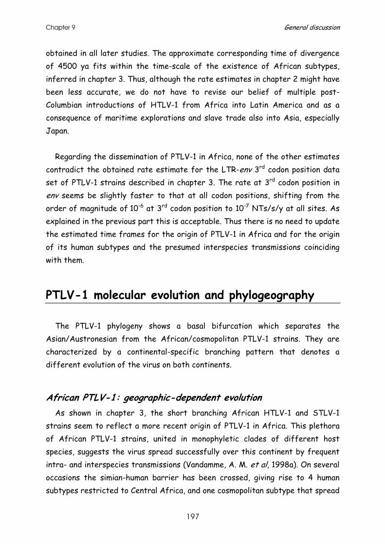
Chapter 9 General discussion
197
obtained in all later studies. The approximate corresponding time of divergence of 4500 ya fits within the time-scale of the existence of African subtypes, inferred in chapter 3. Thus, although the rate estimates in chapter 2 might have been less accurate, we do not have to revise our belief of multiple post-Columbian introductions of HTLV-1 from Africa into Latin America and as a consequence of maritime explorations and slave trade also into Asia, especially Japan.
Regarding the dissemination of PTLV-1 in Africa, none of the other estimates
contradict the obtained rate estimate for the LTR-env 3rd codon position data set of PTLV-1 strains described in chapter 3. The rate at 3rd codon position in env seems be slightly faster to that at all codon positions, shifting from the order of magnitude of 10-6 at 3rd codon position to 10-7 NTs/s/y at all sites. As explained in the previous part this is acceptable. Thus there is no need to update the estimated time frames for the origin of PTLV-1 in Africa and for the origin of its human subtypes and the presumed interspecies transmissions coinciding with them.
PTLV-1 molecular evolution and phylogeography The PTLV-1 phylogeny shows a basal bifurcation which separates the
Asian/Austronesian from the African/cosmopolitan PTLV-1 strains. They are characterized by a continental-specific branching pattern that denotes a different evolution of the virus on both continents.
African PTLV-1: geographic-dependent evolution As shown in chapter 3, the short branching African HTLV-1 and STLV-1
strains seem to reflect a more recent origin of PTLV-1 in Africa. This plethora of African PTLV-1 strains, united in monophyletic clades of different host species, suggests the virus spread successfully over this continent by frequent intra- and interspecies transmissions (Vandamme, A. M. et al, 1998a). On several occasions the simian-human barrier has been crossed, giving rise to 4 human subtypes restricted to Central Africa, and one cosmopolitan subtype that spread
PTLV-1 molecular evolution and phylogeography
198
world-wide (Nerurkar, V. R. et al, 1993; Liu, H. F. et al, 1994a; Gessain, A. et al, 1993b; Salemi, M. et al, 1998). Human strains are clearly joined in monophyletic clades that facilitate subtyping. STLV strains, however, are interspersed between and within these human subtype clades and are characterised by a geographic-dependent clustering rather than by host species clustering. However, their phylogenetic associations are less explicit, thus classification remains difficult.
In chapter 3 we demonstrated that each of the African subtypes, except the cosmopolitan HTLV-1a, seem to have a NHP counterpart. HTLV-1d and HTLV-1f strains were associated with mandrill STLV-1 strains from the same geographic region, Gabon and Cameroon. The HTLV-1e pygmy strain clustered in a clade joining strains of the Papio and/or Cercopithecus genus of sub-Saharan African origin, while HTLV-1b was only associated with chimpanzee STLV-1 strains from West and Central Africa. In chapter 6 we also demonstrated that novel West-African chimpanzee strains, Ch-Coen and Ch-Regina, clustered within this HTLV-1b/STLV-1 group, while other, more divergent strains of the same subspecies were not constrained to this clade (Leendertz, F. H. et al, 2004b). In the same study we showed with the discovery of new STLV-1 strains that HTLV-1b associations are no longer restricted to chimpanzee strains, but expanded to strains of Allenopithecus nigroviridis and gorillas, all of Central African origin (Meertens, L. et al, 2001b; Nerrienet, E. et al, 2004; Courgnaud, V. et al, 2004). The inclusion of our novel gorilla strain, ‘GgoGolda’, demonstrated no explicit clustering with other known gorilla strains, but rather a reasonably-supported clustering with a human strain also of Cameroonian origin. This close relatedness of HTLV and STLV strains has also been shown in chapter 3, with identical LTR sequences of mandrill STLV-1 and HTLV-1f from Cameroon. The HTLV-1f/STLV-1 and extended HTLV-1b/STLV-1 clade both confirm that, given geographic proximity of hosts, interspecies transmissions seem to occur frequently in Africa, even between distantly related species. The close relatedness of human-mandrill and human-gorilla PTLV-1 strains is even indicative of zoonoses. This is plausible and not surprising with the hunt for bushmeat, and has recently been shown for simian foamy viruses (Wolfe, N. D. et al, 2004). Finally, the clustering pattern of the novel Cercopithecus ascanius STLV-1 strain is a clear example of a host species-independent clustering (chapter 6). This novel strain is not particularly closely related to another strain of that same species, which can be
Chapter 9 General discussion
199
explained by the existence of 5 geographically-separated subspecies. Thus, the two studies presented in this thesis show that the current African
PTLV-1 tree is shaped by geographic relatedness as a consequence of frequent and successful interspecies transmissions between sympatric primates.
Asian PTLV-1: host-dependent evolution The shape of the Asian/Austronesian PTLV-1 tree is quite different from the
African PTLV-1 tree, and is characterised by long branched monophyletic clades uniting strains of the same host species. The Melanesian/Austronesian HTLV-1c strains cluster separately from the Asian STLV-1 strains closest to African PTLV-1. In chapter 5 and 6 we have shown that all macaque STLV-1 strains cluster according to the host species in different well supported clades. Some strains of African Papio spp. seem to cluster with these macaque STLV-1 strains, but they are the result of interspecies transmissions in captivity. We have shown in chapter 5 and 6 that these macaque species-specific clades follow a ladder-like topology in env, reflecting macaque migration waves. STLV-1 strains of Macaca arctoides (chapter 5) and M. mulatta, cluster closest to the root of the Asian PTLV-1 tree, followed by a strain of M. fascicularis.
Fig.9.1.: Current natural habitats of Asian STLV-1 infected non-human primates
PTLV-1 molecular evolution and phylogeography
200
All these macaque hosts belong to the fascicularis group, species that remained on the Asian mainland (Fig.9.1.), with the latter extending to Sumatra and Borneo. The PTLV-1 strains that cluster further down the tree belong to Japanese M. fuscata, who emerged more recently from the fascicularis group, to M. nemestrina of the silenus group, whose current habitat is overlapping with M. fascicularis, and to Sulawesi macaques, including M. nigra, and M. maura (both chapter 6) and M. tonkeana (Fig.9.1.). Incongruence between macaque virus- and host-phylogeny excludes the possibility of vicariance, as observed for SFV (Switzer, W. M. et al, 2005), but favours the idea of preferential host-switching between these sym- and parapratically speciated macaques, as shown by simulation analysis for SIV in Africa (Charleston, M. A. et al, 2002). Given the distal monophyletic species-specific clades, host-switching must have happened in a more distant past, and was followed by an independent evolution of the virus within its own species. Moreover, cross-species transmissions to other non-human primates must have occurred in the past based on STLV-1 strains discovered in orang-utans and siamang. The close relationship of two independently discovered and analysed orang-utan STLV-1 strains (chapter 6) in a long-branched monophyletic clade, clustering between macaque strains of the fascicularis group, suggests that the orang-utans became naturally infected with this STLV-1 in the past in their habitats overlapping with those of fascicularis-like macaques (Fig.9.1.). The single STLV-1 isolate of a captive Indonesian siamang, which clusters between Sulawesian macaques (chapter 6), does not exclude natural infection, but favours the possibility of cross-species transmission in captivity. The natural habitat of Sulawesi macaques might have overlapped with siamangs, as they have diverged from a nemestrina-like ancestor in the Pliocene (Groves, C. P., 1980; Melnick, D. J., 1990). However, such an ancient cross-species transmission event does not fit with the molecular clock analysis in chapter 5, suggesting an Asian STLV-1 origin of only 269,000 to 156,000 ya. Finally, the current Asian PTLV-1 topology, with orang-utan and siamang strains clustering in between the macaque STLV-1 monophyletic groups, suggests that macaques are the ancestral reservoir species for STLV-1 in Asia. In conclusion, the studies described in chapter 5 and 6 show the current Asian PTLV-1 phylogeny is shaped by intraclade host-species relatedness and interclade host migration and geographic relatedness. This clustering pattern
Chapter 9 General discussion
201
reflects ancient interspecies transmissions with a subsequent species-dependent evolution in macaques, including transmissions that occasionally cross the simian-ape boundary.
African-Asian PTLV-1: incongruence in evolutionary patterns Can the difference in evolutionary patterns between the two continents only
be explained by a sampling bias? Indeed, until now sampling in Asia has not been as extensive as in Africa, and may give us only a fragmentary view of the real Asian PTLV-1 biodiversity. Moreover, several isolates were derived from captive animals, but the same can be said for the African strains, though to a lesser extent. Despite this, the large STLV-1 diversity observed among the different macaque species today must be a reflection of the situation in the wild. As shown for M. arctoides, the STLV-1 diversity in this captive troop is substantial compared to that in a captive troop of M. tonkeana, indicating that at least some of these M. arctoides were probably naturally infected with the virus. The orangutan STLV-1 clade, that comprises two independently sampled and analyzed strains, also suggests that this species is naturally infected with this STLV-1.
Is there another driving force responsible for the observed degree of incongruence in evolutionary patterns? As viruses are obligate parasites, the success of their viral spread greatly depends on the availability of susceptible hosts. It is true that sub-Saharan Africa, and especially Central Africa, with its tropical rainforests, has a large density of different NHP host species. In South-East Asia, species are confined to the genus Macaca for the Cercopithecinae, to langurs and proboscis monkeys for the Colobinae and to gibbons and orangutans for the Hominoidae superfamily. In Africa the STLV-1 dissemination is successful for sym- or parapatric-living, though not necessarily sym- or parapatric-speciated NHPs. Why would the situation in Asia be different? Macaques not only live, but also speciated sym- and parapratically, suggesting that interspecies preferential viral host-switching should be very successful. STLV-1 transmission should thus be obvious between M. fascicularis, M. arctoides and M. nemestrina, between M. arctoides and M. mulatta and between Sulawesi macaques, since their natural habitats still overlap. Then, why do we not observe a mixed STLV-1 species phylogenetic clustering? And secondly, why are the branch lengths separating these monophyletic STLV-1
PTLV-1 molecular evolution and phylogeography
202
species clades so long, suggestive of interspecies transmission in a distant past, with a subsequent evolution within its species? This is especially true for M. arctoides versus M. mulatta and to a lesser extent for Sulawesi macaques. Even among these latter genetically and geographically closely related macaques, we observe a substantial diversity, which is higher than that observed between African STLV-1 strains originating from distant geographic regions.
Only an intensive sampling in Asian NHPs and analysis of the STLV-1 strains infecting these NHPs can shed light on the real STLV-1 biodiversity in Asia and might answer these questions. If a host-dependent clustering would then still largely hold, PTLV-1 evolution in Asia would clearly be different from that in Africa and even from that of PTLV in general. Those differences in phylogeography and phylodynamics would suggest that host species barriers are more easily crossed in Africa than in Asia, which deserves further attention. When the host-species specific clustering in macaques would no longer hold, but the long branching pattern in Asia versus the short branching pattern in Africa remains valid, our attention needs to be re-focused on African PTLV-1. Although species boundaries are frequently crossed for PTLV-1 in Africa, their diversity is still significantly less than observed among Asian PTLV-1 strains and within other PTLV types. The more recent explosive, successful PTLV-1 spread in Africa might be responsible for the observed difference in diversity as it may hamper the identification of the few, maybe even extinct, distantly related African PTLV-1 strains. Thus the search and identification of more STLV-1 strains of African origin are also needed to unravel this mystery.
Resolving the PTLV-1 phylogeography? Also the origin of PTLV-1 remains a matter of controversy. The common
occurrence of all different PTLV types on the African continent points to a PTLV common ancestor of African origin. Conversely, the current PTLV-1 phylogeny suggests an Asian origin, since Asian macaque strains are the first to branch off in a rooted PTLV-1 tree. If we would be able to find STLV-1 strains clustering basal close to the African/Asian bifurcation, or African STLV-1s close to the PTLV-1 root of the tree, then we might be able to resolve this discrepancy. As barbary macaques (M. sylvanus) are the only macaque species that remained in Africa along their migration pathway to Asia, we hoped STLV-1
Chapter 9 General discussion
203
in this species might form the missing link between African and Asian STLV-1. In chapter 6 we describe STLV-1 infection in a troop of wild-caught M. sylvanus, originating from Morocco, who had no documented history of contact with other African primate species, prior to and certainly not during their captivity. The STLV-1 infection in this species was shown to be a typical African STLV-1. They do not cluster basal, close to the bifurcation but rather between other African STLV-1 strains, in the LTR, and especially in the env region particularly close to strains of Western chimpanzees and sooty mangabeys. The fact that these wild-caught macaques presumably never were in contact with other primates, possibly reflects STLV-1 natural infection within this species. The almost identical sequences among these barbary macaques indicate that probably only one or a few individuals were naturally infected and passed on the virus within their troop. If M. sylvanus is naturally infected with STLV-1, than its close relationship with STLV-1s from allopatric West African naturally infected primates suggests interspecies transmissions in a more distant past, when their habitats might have overlapped. This must have been before the final desertification of the Sahara, at least 5000 ya, which is consistent with the timing for the origin of African HTLV-1 subtypes and the presumed interspecies transmissions. Alternatively, the M. sylvanus STLV-1 infection might be, although highly unlikely, the result of a cross-species transmission in captivity.
M. sylvanus clearly is STLV-1 infected; unfortunately its typical African STLV-1 gave us no clue on the origin of PTLV-1. As a consequence, the hypothesis of a reintroduction of Asian/Austronesian PTLV-1 in Africa, as postulated in chapter 3, still holds. Since there are no anthropological records of NHP back migrations in the postulated time frame of 60,000 to 20,000 ya and HTLV-1c seems to be closest related to African PTLV-1, a reintroduction by human hosts can be suggested. Human retrograde flows 22,000 to 8,000 ya by Khoisan from West Asia to Africa and by Indo-European from Asia to Europe and North Africa have been documented (Cavalli-Sforza, L. L et al, 1994) and might thus be directly or indirectly responsible for the introduction of Asian PTLV-1 in Africa. However a direct introduction implies a human-to-simian transmission, which is highly unlikely, given the transmission possibilities of such events and the wide-spread PTLV-1 biodiversity in a substantial number of different African NHP species known to date. Moreover, their interspersed clustering pattern between the human counterparts suggests these human
PTLV-1 molecular evolution and phylogeography
204
subtypes arose, via simian-to-human transmissions. An indirect transmission can be postulated through NHPs kept as pets by the back-migrating human populations. Alternatively, the origin of PTLV-1 might be more ancient than we reconstruct it today, as suggested earlier. Unfortunately, the latest non-human primate back-to–Africa migrations, presented as an alternative hypothesis to primate speciation and migration (Stewart, C. B. et al, 1998), date back to 10 million ya, which do not fit within the global PTLV molecular time scale.
Ultimately, if the discovery of novel diverse Asian and African PTLV-1 strains would be able to resolve the PTLV-1 phylogeographical origin and re-introduction, then the virus phylogeny might aid anthropology in reconstructing past host migrations, there where fossil records lack.
STLV-3 epidemiology and molecular epidemiology
Overview of STLV-3 infected host species In 1994 the first STLV-3 strain (PH969), originally designated STLV-L
(L=Leuven), was discovered in a wild-born, captive, though isolated Papio hamadryas of Eritrean origin (Goubau, P. et al, 1994).
It took us more than seven years to find a second STLV-3 infection in another species, Cercopithecus nictitans from Cameroon, which has been described in this thesis in chapter 7. Two out of 121 tested C. nictitans showed an HTLV-2-like reactivity on a line immunoblot assay, and our PTLV discriminatory PCR (Vandamme, A. M. et al, 1997) indicated STLV-3 infection. The subsequent sequencing and phylogenetic analysis of the tax PCR fragment corroborated the latter finding. Unfortunately, the quality of the stored whole blood, in combination with the divergent character of the virus hindered the accumulation of additional sequence data. Nevertheless, this was the first report and prove of natural STLV-3 infection in the wild, in a geographic region distant from the original discovered PH969.
Shortly after, other researchers detected STLV-3 infection in other species. A second report on STLV-3 infection in the wild with partial LTR and pX sequences (Takemura, T. et al, 2002), demonstrates a seroprevalence of 40 to
Chapter 9 General discussion
205
50% among Ethiopian hamadryas and hybrid (anubis x hamadryas) baboons. Ground breaking work on STLV-3 infection was done by Meertens et al. (Meertens, L. et al, 2001a; Meertens, L. et al, 2003b), who actually discovered STLV-3 infection in wild-caught, captive animals: in two Cercocebus torquatus from Cameroon, in one out of 12 C. torquatus from Nigeria, and in 6 out of 8 Papio hamadryas papio from Senegal. From each of these infected species of different geographic origin, one STLV-3 full genome was obtained and further analysed. Finally, we discovered a STLV-3 strain infecting 5 of 23 Ethiopian Theropithecus gelada in captivity, of which the serology, epidemiology and full genome analysis has been described in detail in chapter 8 of this thesis. It is noteworthy that Takemura et al. (Takemura, T. et al, 2002) also investigated the STLV-seroprevalence among 156 wild-living T. gelada. None of them were found to be STLV-infected, which can be explained by a difference in sensitivity of antibody screening assays and/or by the sampling in different Ethiopian locations.
Variability in STLV-3 seroreactivity The presumption of a divergent character of a virus frequently starts with
the observation of a weak reactivity in HTLV antibody screening assays and/or a peculiar profile in type-specific serological confirmatory tests. Confirmatory testing of the STLV-3 infected animals sometimes results in an HTLV-2-like profile (chapter 7), which has never been found in NHPs before, not even in the STLV-2 infected Pan paniscus (Liu, H. F. et al, 1994b). However, a large variety of seroreactivity has now been observed for STLV-3 infection. Either a combined or no reaction (chapter 8) with the specific recombinant or synthetic peptides for HTLV-1 and HTLV-2 has been detected, or, in a single case, only reaction with the HTLV-1-specific peptide (chapter 8). This HTLV-1-specific reaction can indicate that these animals are dually infected with STLV-1 and -3, as we described for Cameroonian Cercocebus agilis (Courgnaud, V. et al, 2004). Both T. gelada and P. hamadryas with HTLV-1-like seroreactivity are known to be infected with STLV-1 or STLV-3 (chapter 8; (Meertens, L. et al, 2003a), but the STLV-3 infected species with the HTLV-1-like seroreactivity have not been further PCR-tested for STLV-1 infection.
This variability in STLV seroreactivity, ranging from aspecific and
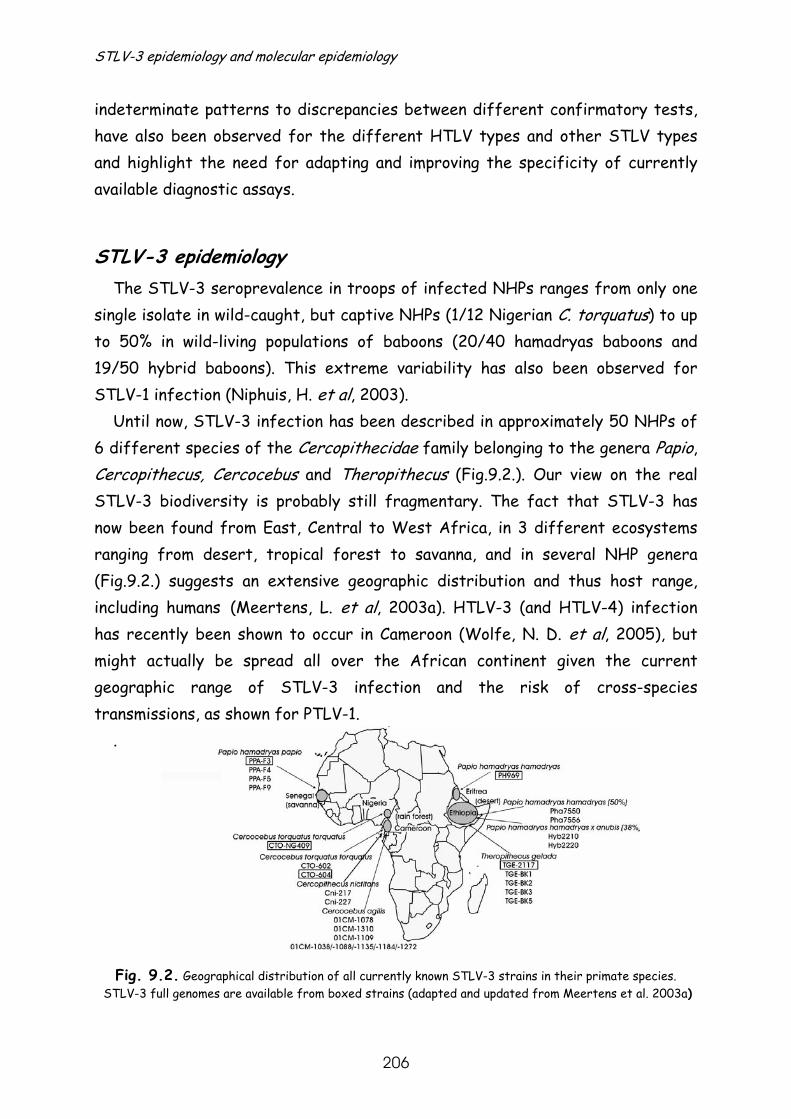
STLV-3 epidemiology and molecular epidemiology
206
indeterminate patterns to discrepancies between different confirmatory tests, have also been observed for the different HTLV types and other STLV types and highlight the need for adapting and improving the specificity of currently available diagnostic assays.
STLV-3 epidemiology The STLV-3 seroprevalence in troops of infected NHPs ranges from only one
single isolate in wild-caught, but captive NHPs (1/12 Nigerian C. torquatus) to up to 50% in wild-living populations of baboons (20/40 hamadryas baboons and 19/50 hybrid baboons). This extreme variability has also been observed for STLV-1 infection (Niphuis, H. et al, 2003).
Until now, STLV-3 infection has been described in approximately 50 NHPs of 6 different species of the Cercopithecidae family belonging to the genera Papio, Cercopithecus, Cercocebus and Theropithecus (Fig.9.2.). Our view on the real STLV-3 biodiversity is probably still fragmentary. The fact that STLV-3 has now been found from East, Central to West Africa, in 3 different ecosystems ranging from desert, tropical forest to savanna, and in several NHP genera (Fig.9.2.) suggests an extensive geographic distribution and thus host range, including humans (Meertens, L. et al, 2003a). HTLV-3 (and HTLV-4) infection has recently been shown to occur in Cameroon (Wolfe, N. D. et al, 2005), but might actually be spread all over the African continent given the current geographic range of STLV-3 infection and the risk of cross-species transmissions, as shown for PTLV-1.
.
Fig. 9.2. Geographical distribution of all currently known STLV-3 strains in their primate species. STLV-3 full genomes are available from boxed strains (adapted and updated from Meertens et al. 2003a)
Chapter 9 General discussion
207
Due to the low number of STLV-3 infected NHPs it remains difficult to study the possible STLV-3 transmission routes. However, based on demographic data in combination with STLV-3 sequence similarity of the infected captive T. gelada cohort (chapter 8) horizontal transmission between one male and 3 female could be suggested and evidence for mother-to-offspring transmission was found. Remarkably, isolates sampled 20 years apart from the offspring and parent pair showed identical sequences, demonstrating the genetic stability of this type of virus.
Molecular epidemiology of STLV-3 It is noteworthy that this extreme intraspecies genetic stability is also
observed among the other STLV-3 infected NHPs, on condition of shared habitat. As most of the investigated animals live in captivity, this observation is certainly biased by the increased risk of unnatural infection. Still, a comparable scenario may be true for the investigated STLV-3 strains from wild-living animals: a) only one mutation was observed in the 219bp tax fragment between the 2 STLV-3 strains in C. nictitans (chapter 7), and b) identical pX sequences and 3 mutations in a 492bp LTR fragment between 2 STLV-3 strains in P. hamadryas (Takemura, T. et al, 2002).
On the other hand, phylogenetic analyses of the LTR, env, pX and/or full genome show a substantial diversity between the different investigated species and even between the same species of different geographic areas. Conversely, minor divergence was observed in some Ethiopian and Cameroonian cases that involved different species of the same geographic area. The phylogenetic analyses of the most recently discovered STLV-3 in T. gelada, in chapter 8, including all other known STLV-3 strains, give us a nice overview of the currently known molecular epidemiology of STLV-3. Based on the LTR analysis (Fig.9.3.) the STLV-3 strains can be divided in a West/Central African and East African STLV-3 group. The former comprises strains of Cameroonian (CTO-604) and Nigerian (CTO-NG409) C. torquatus and the Senegalese P. hamadryas (PPA-F3). Remarkably, the Nigerian C. torquatus has a closer relationship with the geographical and host genus more distal baboon strain from Senegal, as compared to the strain in the same species from a neighbouring country. The
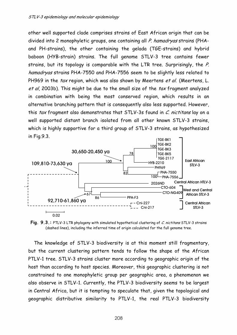
STLV-3 epidemiology and molecular epidemiology
208
TGE-BK1TGE-BK2TGE-BK3TGE-BK5
86
10082
100
78
100
0.02
PHA-7550PHA-7556
PH969
TGE-2117HYB-2210
CTO-604CTO-NG409
PPA-F3
East AfricanSTLV-3
West and CentralAfrican STLV-3
Cni-227Cni-217
Central African STLV-3
30,650-20,450 ya
92,710-61,860 ya
109,810-73,630 ya
2026ND Central African HTLV-3
67
other well supported clade comprises strains of East African origin that can be divided into 2 monophyletic groups, one containing all P. hamadryas strains (PHA- and PH-strains), the other containing the gelada (TGE-strains) and hybrid baboon (HYB-strain) strains. The full genome STLV-3 tree contains fewer strains, but its topology is comparable with the LTR tree. Surprisingly, the P. hamadryas strains PHA-7550 and PHA-7556 seem to be slightly less related to PH969 in the tax region, which was also shown by Meertens et al. (Meertens, L. et al, 2003b). This might be due to the small size of the tax fragment analyzed in combination with being the most conserved region, which results in an alternative branching pattern that is consequently also less supported. However, this tax fragment also demonstrates that STLV-3s found in C. nictitans lay on a well supported distant branch isolated from all other known STLV-3 strains, which is highly supportive for a third group of STLV-3 strains, as hypothesized in Fig.9.3.
Fig. 9.3.: PTLV-3 LTR phylogeny with simulated hypothetical clustering of C. nictitans STLV-3 strains
(dashed lines), including the inferred time of origin calculated for the full genome tree. The knowledge of STLV-3 biodiversity is at this moment still fragmentary,
but the current clustering pattern tends to follow the shape of the African PTLV-1 tree. STLV-3 strains cluster more according to geographic origin of the host than according to host species. Moreover, this geographic clustering is not constrained to one monophyletic group per geographic area, a phenomenon we also observe in STLV-1. Currently, the PTLV-3 biodiversity seems to be largest in Central Africa, but it is tempting to speculate that, given the topological and geographic distributive similarity to PTLV-1, the real PTLV-3 biodiversity
Chapter 9 General discussion
209
reaches that of PTLV-1 in Africa. We speculate that the HTLV-3 diversity still remains mainly undiscovered as a specific search for this type of virus has been hampered by an HTLV-1- and -2-like seroreactivity. If the HTLV-3s are no dead-end infections and human-to-human transmission can be shown, than its diversity might be in the same order of magnitude as the one of HTLV-1.
All of the above observations suggest that STLV-3 evolution, as we can
reconstruct it today, reflects the STLV-1 situation in Africa, with the important difference that the observed STLV-3 diversity is much larger than in African STLV-1 and more similar to that of Asian STLV-1 for the full genome analysis. When the divergent STLV-3 C. nictitans strains are included in the tax analysis, STLV-3 diversity reaches that of HTLV-2 strains versus STLV-2 PP1664.
Time of origin and diversification of STLV-3 When we estimate the separation between, and the origin of the currently
known West/Central versus East African STLV-3s, based on the estimated evolutionary rates of 0.79-1.18 x 10⁻⁶ to 1.72-2.56 x 10⁻⁶ NTs/s/y for gag-pol-env-pX and env3rd codon position respectively, we obtained approximate dates ranging from 128,500 to 73,630 ya depending on the genetic region(s) investigated (Fig.9.3. for full genome estimates). The origin of all PTLV-3s is certainly more ancient, given the divergence of the C. nictitans strains. Unfortunately, a molecular clock analysis on the short tax fragment is impossible, thus the origin of PTLV-3 remains to be determined. As the East African strains cluster closely together, their origin seems to be much more recent 30,650 to 20,450 ya compared that of the West/Central African strains. The latter strains cluster more basal, closer to the node of origin of STLV-3 and are estimated to have originated 92,710-61,860 ya. These estimates are slightly more recent than those calculated by Meertens et al. (Meertens, L. et al, 2003b; Meertens, L. et al, 2003a; Meertens, L. et al, 2002). This can be explained by the use of another methodology in combination with the inclusion of only the lower limit of 60.000 ya for the origin of HTLV-1c in their analysis, resulting in a rate of PTLV-3 evolution corresponding with our lower limit estimate.
Thus, when integrating the STLV-3 diversity in C. nictitans in PTLV-3 molecular timings, its origin will probably reach the time frame estimated for
STLV-3 epidemiology and molecular epidemiology
210
the origin of Asian PTLV-1 (chapter 5) and even that for the STLV-2/HTLV-2 separation (400,000 ± 20,000ya(Salemi, M. et al, 2000). Consistent molecular timings for the rise of different PTLV types, though incompatible with that of African PTLV-1, again resurrects the focus on PTLV-1 in Africa and shows that, regardless the number of PTLV-1 strains analyzed, questions on PTLV-1 evolution in Africa remain unanswered.
Conclusions and perspectives The hypothesis of an African common ancestor for PTLV has been based on
the observation that all different PTLV types 1 to 4 are found in Africa. The fact that the largest intratype PTLV diversity is observed in Central Africa, e.g. the Congolese HTLV-2d/STLV-2 diversity and the Cameroonian HTLV-3 and C. nictitans/C. torquatus STLV-3 diversity, together with the known Central African HTLV-1(b,d to f) and STLV-1 biodiversity, strongly suggests that this region is the cradle of PTLV infection and thus corroborates the African origin of PTLV. However, the observation that Asian, and not African STLV-1 strains, remain at the root of the PTLV-1 tree weakens this hypothesis.
The major challenge for molecular epidemiologists in the future will thus be
the resolution of the evolutionary differences and phylogeographical origin of PTLV-1 and possibly even PTLV. We have made a first step in that direction and have tried to contribute to the solution of this question in several ways. We have studied the tick of the PTLV molecular clock in order to be able to date the origin of PTLV-1 in Africa and Asia and of PTLV-3 in Africa. The phylogenetic analyses of divergent or novel Asian strains contributed to the understanding of PTLV-1 evolution in Asia and ultimately we hoped that African M. sylvanus STLV-1 strains would help us to resolve the phylogeographical origin of PTLV-1. By analyzing the currently known STLV-3 biodiversity, we hypothesized a Central African origin of PTLV-3, time–scaled in the order of magnitude of PTLV-2 and Asian PTLV-1. To be able to resolve this controversy about the PTLV origin, additional sampling and analysis of African and Asian PTLV isolates will be needed, preferentially within the hosts’ natural habitat.
211
Reference list
1. Albrecht,B. & Lairmore,M.D. (2002) Critical role of human T-lymphotropic virus type 1 accessory proteins in viral replication and pathogenesis. Microbiology and Molecular Biology Reviews 66, 396-+.
2. Baltimore,D. (1970) RNA-dependent DNA polymerase in virions of RNA tumour viruses. Nature 226, 1209-1211.
3. Biggar,R.J., Gigase,P.L., Melbye,M.& other authors (1985) ELISA HTLV retrovirus antibody reactivity associated with malaria and immune complexes in healthy Africans. Lancet 2, 520-523.
4. Blakeslee,J.R., Jr., McClure,H.M., Anderson,D.C., Bauer,R.M., Huff,L.Y., & Olsen,R.G. (1987) Chronic fatal disease in gorillas seropositive for simian T-lymphotropic virus I antibodies. Cancer Lett 37, 1-6.
5. Blattner, W. (2005) Retroviruses. In Viral infections of human epidemiology and control, Edited by A. S. Evans, New York: Plenum.
6. Blattner,W.A., Saxinger,C., Riedel,D., Hull,B., Taylor,G., Cleghorn,F., Gallo,R., Blumberg,B., & Bartholomew,C. (1990) A study of HTLV-I and its associated risk factors in Trinidad and Tobago. J Acquir Immune Defic Syndr 3, 1102-1108.
7. Calattini,S., Chevalier,S.A., Duprez,R., Bassot,S., Froment,A., Mahieux,R., & Gessain,A. (2005) Discovery of a new human T-cell lymphotropic virus (HTLV-3) in Central Africa. Retrovirology 2, 30.
8. Cann,A.J., Rosenblatt,J.D., Wachsman,W., Shah,N.P., & Chen,I.S. (1985) Identification of the gene responsible for human T-cell leukaemia virus transcriptional regulation. Nature 318, 571-574.
9. Cavalli-Sforza, L. L., Menozzi, P. & Piazza, A. (1994) The history and geography of human genes. Princeton University Press, Princeton, N.J.
10. Cavrois,M., Gessain,A., Wain-Hobson,S., & Wattel,E. (1996a) Proliferation of HTLV-1 infected circulating cells in vivo in all asymptomatic carriers and patients with TSP/HAM. Oncogene 12, 2419-2423.
11. Cavrois,M., Leclercq,I., Gout,O., Gessain,A., Wain-Hobson,S., &
Reference list
212
Wattel,E. (1998) Persistent oligoclonal expansion of human T-cell leukemia virus type 1-infected circulating cells in patients with Tropical spastic paraparesis/HTLV-1 associated myelopathy. Oncogene 17, 77-82.
12. Cavrois,M., Wain-Hobson,S., Gessain,A., Plumelle,Y., & Wattel,E. (1996b) Adult T-cell leukemia/lymphoma on a background of clonally expanding human T-cell leukemia virus type-1-positive cells. Blood 88, 4646-4650.
13. Charleston,M.A. & Robertson,D.L. (2002) Preferential host switching by primate lentiviruses can account for phylogenetic similarity with the primate phylogeny. Systematic Biology 51, 528-535.
14. Chen, Y.-M. A., Sankalé, J.-L. (1994) Other human retroviruses. In Aids in Africa, Edited by M. Essex, S. Mboup, P. J. Kanki & M. R. Kalengayi, New York: Raven Press,Ltd.
15. Ciminale,V., D'Agostino,D.M., Zotti,L., Franchini,G., Felber,B.K., & Chieco-Bianchi,L. (1995) Expression and characterization of proteins produced by mRNAs spliced into the X region of the human T-cell leukemia/lymphotropic virus type II. Virology 209, 445-456.
16. Ciminale,V., Pavlakis,G.N., Derse,D., Cunningham,C.P., & Felber,B.K. (1992) Complex splicing in the human T-cell leukemia virus (HTLV) family of retroviruses: novel mRNAs and proteins produced by HTLV type I. J Virol 66, 1737-1745.
17. Coffin, J. M. (1992) Structure and classification of retroviruses. In The Retroviridae, Edited by J. A. Levy, New York: Plenum Press.
18. Coffin, J. M. (1996) Retroviridae: the viruses and their replication. In Fields Virology, Edited by B. N. Fields, D. M. Knipe & P. M. Howley, Philadelphia: Lippencott-Raven Publishers.
19. Courgnaud,V., Van Dooren,S., Liegeois,F.& other authors (2004) Simian T-cell leukemia virus (STLV) infection in wild primate populations in Cameroon: Evidence for dual STLV type 1 and type 3 infection in agile mangabeys (Cercocebus agilis). Journal of Virology 78, 4700-4709.
20. de The,G., Gessain,A., Gazzolo,L.& other authors (1985) Comparative seroepidemiology of HTLV-I and HTLV-III in the French West Indies and some African countries. Cancer Res 45, 4633s-4636s.
21. Delaporte,E., Louwagie,J., Peeters,M.& other authors (1991) Evidence

Reference list
213
of HTLV-II infection in central Africa. AIDS 5, 771-772.
22. Edlich,R.F., Hill,L.G., & Williams,F.M. (2003) Global epidemic of human T-cell lymphotrophic virus type-I (HTLV-I): an update. J Long Term Eff Med Implants 13, 127-140.
23. Felsenstein, J. (2004) Inferring phylogenies. Sunderland, Massachusetts: Sinauer Associates, Inc.
24. Fukushima,Y., Nakasone,T., Nakata,S., Pham,S., Dao,D.D., Komuro,K., & Honda,M. (1994) Human T-lymphotropic virus type II in Vietnam. Lancet 344, 485.
25. Fultz, P. N. (1994) Simian T-lymphotropic Virus Type I. In The Retroviridae, Edited by J. A. Levy, New York: Plenum Press.
26. Fultz,P.N., Gordon,T.P., Anderson,D.C., & McClure,H.M. (1990) Prevalence of natural infection with simian immunodeficiency virus and simian T-cell leukemia virus type I in a breeding colony of sooty mangabey monkeys. AIDS 4, 619-625.
27. Gabet,A.S., Mortreux,F., Talarmin,A.& other authors (2000) High circulating proviral load with oligoclonal expansion of HTLV-1 bearing T cells in HTLV-1 carriers with strongyloidiasis. Oncogene 19, 4954-4960.
28. Gallo,D., Hoffman,M.N., Cossen,C.K., Diggs,J.L., Hurst,J.W., & Penning,L.M. (1988) Comparison of immunofluorescence, enzyme immunoassay, and Western blot (immunoblot) methods for detection of antibody to human T-cell leukemia virus type I. J Clin Microbiol 26, 1487-1491.
29. Gallo,R.C. (2002) Human retroviruses after 20 years: a perspective from the past and prospects for their future control. Immunological Reviews 185, 236-265.
30. Gaudray,G., Gachon,F., Basbous,J., Biard-Piechaczyk,M., Devaux,C., & Mesnard,J.M. (2002) The complementary strand of the human T-cell leukemia virus type 1 RNA genome encodes a bZIP transcription factor that down-regulates viral transcription. J Virol 76, 12813-12822.
31. Georges-Courbot,M.C., Moisson,P., Leroy,E.& other authors (1996) Occurrence and frequency of transmission of naturally occurring simian retroviral infections (SIV, STLV, and SRV) at the CIRMF Primate Center, Gabon. J Med Primatol 25, 313-326.
Reference list
214
32. Gessain,A., Boeri,E., Yanagihara,R., Gallo,R.C., & Franchini,G. (1993a) Complete nucleotide sequence of a highly divergent human T-cell leukemia (lymphotropic) virus type I (HTLV-I) variant from melanesia: genetic and phylogenetic relationship to HTLV-I strains from other geographical regions. Journal of Virology 67, 1015-1023.
33. Gessain,A., Gallo,R.C., & Franchini,G. (1992) Low degree of human T-cell leukemia/lymphoma virus type I genetic drift in vivo as a means of monitoring viral transmission and movement of ancient human populations. J Virol 66, 2288-2295.
34. Gessain,A., Herve,V., Jeannel,D., Garin,B., Mathiot,C., & de The,G. (1993b) HTLV-1 but not HTLV-2 found in pygmies from Central African Republic. J Acquir Immune Defic Syndr 6, 1373-1374.
35. Gessain,A., Koralnik,I.J., Fullen,J.& other authors (1994a) Phylogenetic study of ten new HTLV-I strains from the Americas. AIDS Res Hum Retroviruses 10, 103-106.
36. Gessain,A., Mauclere,P., Froment,A., Biglione,M., Le Hesran,J.Y., Tekaia,F., Millan,J., & de The,G. (1995) Isolation and molecular characterization of a human T-cell lymphotropic virus type II (HTLV-II), subtype B, from a healthy Pygmy living in a remote area of Cameroon: an ancient origin for HTLV-II in Africa. Proc Natl Acad Sci U S A 92, 4041-4045.
37. Gessain,A., Tuppin,P., Kazanji,M., Cosnefroy,J.Y., Georges-Courbot,M.C., Georges,A.J., & de The,G. (1994b) A distinct molecular variant of HTLV-IIB in Gabon, Central Africa. AIDS Res Hum Retroviruses 10, 753-755.
38. Gessain,A., Vernant,J.C., Maurs,L., Barin,F., Gout,O., Calender,A., & Dethe,G. (1985) Antibodies to Human T-Lymphotropic Virus Type-I in Patients with Tropical Spastic Paraparesis. Lancet 2, 407-410.
39. Gessain,A., Yanagihara,R., Franchini,G., Garruto,R.M., Jenkins,C.L., Ajdukiewicz,A.B., Gallo,R.C., & Gajdusek,D.C. (1991) Highly Divergent Molecular Variants of Human T-Lymphotropic Virus Type-I from Isolated Populations in Papua-New-Guinea and the Solomon-Islands. Proceedings of the National Academy of Sciences of the United States of America 88, 7694-7698.
40. Giri,A., Markham,P., Digilio,L., Hurteau,G., Gallo,R.C., & Franchini,G. (1994) Isolation of A Novel Simian T-Cell Lymphotropic
Reference list
215
Virus from Pan-Paniscus That Is Distantly Related to the Human T-Cell Leukemia Lymphotropic Virus Type-I and Type-Ii. Journal of Virology 68, 8392-8395.
41. Goubau,P., Desmyter,J., Ghesquiere,J., & Kasereka,B. (1992) HTLV-II among pygmies. Nature 359, 201.
42. Goubau, P., Desmyter, J., Michiels, M., Vandamme, A.-M., Hulstaert, F., Govers, L., and Zrein, M. Validation of the Innolia HTLVI/II in the confirmation and discrimination of HTLV infections. ? 1999.
Ref Type: Abstract
43. Goubau,P., Van Brussel,M., Vandamme,A.M., Liu,H.F., & Desmyter,J. (1994) A primate T-lymphotropic virus, PTLV-L, different from human T-lymphotropic viruses types I and II, in a wild-caught baboon (Papio hamadryas). Proc Natl Acad Sci U S A 91, 2848-2852.
44. Goubau,P., Vandamme,A., Beuselinck,K., & Desmyter,J. (1996) Proviral HTLV-I and HTLV-II in the Efe pygmies of northeastern Zaire. J Acquir Immune Defic Syndr Hum Retrovirol 12, 208-209.
45. Gross, L. (1983) Oncogenic viruses. Oxford: Pergamon.
46. Groves, C. P. (1980) Speciation in Macaca: the view from Sulawesi. In The macacques: studies in ecology, behavior and evolution, Edited by Lindburg D.G., New York: Van Nostrand Reinhold Co.
47. Hall,W.W., Kubo,T., Ijichi,S., Takahashi,H., & Zhu,S.W. (1994) Human T-Cell Leukemia-Lymphoma Virus, Type-Ii (Htlv-Ii) - Emergence of An Important Newly Recognized Pathogen. Seminars in Virology 5, 165-178.
48. Hall,W.W., Takahashi,H., Liu,C., Kaplan,M.H., Scheewind,O., Ijichi,S., Nagashima,K., & Gallo,R.C. (1992) Multiple isolates and characteristics of human T-cell leukemia virus type II. J Virol 66, 2456-2463.
49. Hanchard,B., LaGrenade,L., Carberry,C., Fletcher,V., Williams,E., Cranston,B., Blattner,W.A., & Manns,A. (1991) Childhood infective dermatitis evolving into adult T-cell leukaemia after 17 years. Lancet 338, 1593-1594.
50. Hatanaka,M. & Nam,S.H. (1989) Identification of HTLV-I gag protease and its sequential processing of the gag gene product. J Cell Biochem 40,
Reference list
216
15-30.
51. Hattori,S., Kiyokawa,T., Imagawa,K., Shimizu,F., Hashimura,E., Seiki,M., & Yoshida,M. (1984) Identification of gag and env gene products of human T-cell leukemia virus (HTLV). Virology 136, 338-347.
52. Heneine,W., Kaplan,J.E., Gracia,F., Lal,R., Roberts,B., Levine,P.H., & Reeves,W.C. (1991) HTLV-II endemicity among Guaymi Indians in Panama. N Engl J Med 324, 565.
53. Hidaka,M., Inoue,J., Yoshida,M., & Seiki,M. (1988) Post-transcriptional regulator (rex) of HTLV-1 initiates expression of viral structural proteins but suppresses expression of regulatory proteins. EMBO J 7, 519-523.
54. Hino,S. (1989) Milk-borne transmission of HTLV-I as a major route in the endemic cycle. Acta Paediatr Jpn 31, 428-435.
55. Hinuma,Y., Nagata,K., Hanaoka,M., Nakai,M., Matsumoto,T., Kinoshita,K.I., Shirakawa,S., & Miyoshi,I. (1981) Adult T-cell leukemia: antigen in an ATL cell line and detection of antibodies to the antigen in human sera. Proc Natl Acad Sci U S A 78, 6476-6480.
56. Hjelle,B., Khabbaz,R.F., Conway,G.A., North,C., Green,D., & Kaplan,J.E. (1994) Prevalence of human T cell lymphotropic virus type II in American Indian populations of the southwestern United States. Am J Trop Med Hyg 51, 11-15.
57. Hjelle,B., Mills,R., Mertz,G., & Swenson,S. (1990) Transmission of HTLV-II via blood transfusion. Vox Sang 59, 119-122.
58. Hjelle,B., Zhu,S.W., Takahashi,H., Ijichi,S., & Hall,W.W. (1993) Endemic human T cell leukemia virus type II infection in southwestern US Indians involves two prototype variants of virus. J Infect Dis 168, 737-740.
59. Holmes,E.C. (2003) Molecular clocks and the puzzle of RNA virus origins. J Virol 77, 3893-3897.
60. Homma,T., Kanki,P.J., King,N.W., Jr.& other authors (1984) Lymphoma in macaques: association with virus of human T lymphotrophic family. Science 225, 716-718.
61. Ibrahim,F., Dethe,G., & Gessain,A. (1995) Isolation and
Reference list
217
Characterization of A New Simian T-Cell Leukemia-Virus Type-1 from Naturally Infected Celebes Macaques (Macaca-Tonkeana) - Complete Nucleotide-Sequence and Phylogenetic Relationship with the Australo-Melanesian Human T-Cell Leukemia-Virus Type-1. Journal of Virology 69, 6980-6993.
62. Ibuki,K., Ido,E., Setiyaningsih,S., Yamashita,M., Agus,L.R.P., Takehisa,J., Miura,T., Dondin,S., & Hayami,M. (1997) Isolation of STLV-I from orangutan, a great ape species in southeast Asia, and its relation to other HTLV-Is/STLV-Is. Japanese Journal of Cancer Research 88, 1-4.
63. Igakura,T., Stinchcombe,J.C., Goon,P.K., Taylor,G.P., Weber,J.N., Griffiths,G.M., Tanaka,Y., Osame,M., & Bangham,C.R. (2003) Spread of HTLV-I between lymphocytes by virus-induced polarization of the cytoskeleton. Science 299, 1713-1716.
64. Ina,Y. & Gojobori,T. (1990) Molecular evolution of human T-cell leukemia virus. J Mol Evol 31, 493-499.
65. Ishak,R., Vallinoto,A.C., Azevedo,V.N., Lewis,M., Hall,W.W., & Guimaraes Ishak,M.O. (2001) Molecular evidence of mother-to-child transmission of HTLV-IIc in the Kararao Village (Kayapo) in the Amazon region of Brazil. Rev Soc Bras Med Trop 34, 519-525.
66. Ishida,T., Yamamoto,K., Omoto,K., Iwanaga,M., Osato,T., & Hinuma,Y. (1985) Prevalence of a human retrovirus in native Japanese: evidence for a possible ancient origin. J Infect 11, 153-157.
67. Jenkins,G.M., Rambaut,A., Pybus,O.G., & Holmes,E.C. (2002) Rates of molecular evolution in RNA viruses: a quantitative phylogenetic analysis. J Mol Evol 54, 156-165.
68. Josephs,S.F., Wong-Staal,F., Manzari,V., Gallo,R.C., Sodroski,J.G., Trus,M.D., Perkins,D., Patarca,R., & Haseltine,W.A. (1984) Long terminal repeat structure of an American isolate of type I human T-cell leukemia virus. Virology 139, 340-345.
69. Kajiyama,W., Kashiwagi,S., Nomura,H., Ikematsu,H., Hayashi,J., & Ikematsu,W. (1986) Seroepidemiologic study of antibody to adult T-cell leukemia virus in Okinawa, Japan. Am J Epidemiol 123, 41-47.
70. Kalyanaraman,V.S., Sarngadharan,M.G., Robert-Guroff,M., Miyoshi,I., Golde,D., & Gallo,R.C. (1982) A new subtype of human T-

Reference list
218
cell leukemia virus (HTLV-II) associated with a T-cell variant of hairy cell leukemia. Science 218, 571-573.
71. Kaplan,J.E., Khabbaz,R.F., Murphy,E.L.& other authors (1996) Male-to-female transmission of human T-cell lymphotropic virus types I and II: association with viral load. The Retrovirus Epidemiology Donor Study Group. J Acquir Immune Defic Syndr Hum Retrovirol 12, 193-201.
72. Kazanji,M. (2000) HTLV type 1 infection in squirrel monkeys (Saimiri sciureus): a promising animal model for HTLV type 1 human infection. AIDS Res Hum Retroviruses 16, 1741-1746.
73. Kinoshita,K., Hino,S., Amagaski,T.& other authors (1984) Demonstration of adult T-cell leukemia virus antigen in milk from three sero-positive mothers. Gann 75, 103-105.
74. Kitado,H., Chen,I.S., Shah,N.P., Cann,A.J., Shimotohno,K., & Fan,H. (1987) U3 sequences from HTLV-I and -II LTRs confer pX protein response to a murine leukemia virus LTR. Science 235, 901-904.
75. Koralnik,I.J., Boeri,E., Saxinger,W.C.& other authors (1994) Phylogenetic Associations of Human and Simian T-Cell Leukemia/Lymphotropic Virus Type-I Strains - Evidence for Interspecies Transmission. Journal of Virology 68, 2693-2707.
76. Korber,B., Muldoon,M., Theiler,J., Gao,F., Gupta,R., Lapedes,A., Hahn,B.H., Wolinsky,S., & Bhattacharya,T. (2000) Timing the ancestor of the HIV-1 pandemic strains. Science 288, 1789-1796.
77. Lairmore,M.D., Jacobson,S., Gracia,F.& other authors (1990) Isolation of human T-cell lymphotropic virus type 2 from Guaymi Indians in Panama. Proc Natl Acad Sci U S A 87, 8840-8844.
78. Lee,H., Swanson,P., Shorty,V.S., Zack,J.A., Rosenblatt,J.D., & Chen,I.S. (1989) High rate of HTLV-II infection in seropositive i.v. drug abusers in New Orleans. Science 244, 471-475.
79. Lee,R.V., Prowten,A.W., Satchidanand,S.K., & Srivastava,B.I. (1985) Non-Hodgkin's lymphoma and HTLV-1 antibodies in a gorilla. N Engl J Med 312, 118-119.
80. Leendertz,F.H., Boesch,C., Ellerbrok,H., Rietschel,W., Couacy-Hymann,E., & Pauli,G. (2004a) Non-invasive testing reveals a high prevalence of simian T-lymphotropic virus type 1 antibodies in wild adult

Reference list
219
chimpanzees of the Tai National Park, Cote d'Ivoire. Journal of General Virology 85, 3305-3312.
81. Leendertz,F.H., Junglen,S., Boesch,C., Formenty,P., Couacy-Hymann,E., Courgnaud,V., Pauli,G., & Ellerbrok,H. (2004b) High variety of different simian T-Cell leukemia virus type 1 strains in chimpanzees (Pan troglodytes verus) of the Tai National Park, Cote d'Ivoire. Journal of Virology 78, 4352-4356.
82. Lemey, P. Molecular investigation of evolutionary and population genetic processes in human retroviruses. 2005a. Rega Institute for Medical Research, KULeuven, Belgium.
Ref Type: Thesis/Dissertation
83. Lemey,P., Pybus,O.G., Van Dooren,S., & Vandamme,A.-M. (2005b) A Bayesian statistical analysis of Human T-Cell Lymphotropic Virus evolutionary rates. Infection, Genetics and Evolution 5, 291-298.
84. Lemey,P., Pybus,O.G., Wang,B., Saksena,N.K., Salemi,M., & Vandamme,A.M. (2003) Tracing the origin and history of the HIV-2 epidemic. Proc Natl Acad Sci U S A 100, 6588-6592.
85. Levin,M.C. & Jacobson,S. (1997) HTLV-I associated myelopathy tropical spastic paraparesis (HAM/TSP): A chronic progressive neurologic disease associated with immunologically mediated damage to the central nervous system. Journal of Neurovirology 3, 126-140.
86. Liu,H.F., Vandamme,A.M., Kazadi,K., Carton,H., Desmyter,J., & Goubau,P. (1994a) Familial transmission and minimal sequence variability of human T-lymphotropic virus type I (HTLV-I) in Zaire. Aids Research and Human Retroviruses 10, 1135-1142.
87. Liu,H.F., Vandamme,A.M., Van Brussel,M., Desmyter,J., & Goubau,P. (1994b) New retroviruses in human and simian T-lymphotropic viruses. Lancet 344, 265-266.
88. Mahieux,R., PeconSlattery,J., & Gessain,A. (1997) Molecular characterization and phylogenetic analyses of a new, highly divergent simian T-cell lymphotropic virus type 1 (STLV-1marc1) in Macaca arctoides. Journal of Virology 71, 6253-6258.
89. Maloney,E.M., Biggar,R.J., Neel,J.V., Taylor,M.E., Hahn,B.H., Shaw,G.M., & Blattner,W.A. (1992) Endemic human T cell lymphotropic virus type II infection among isolated Brazilian Amerindians. J Infect Dis
Reference list
220
166, 100-107.
90. Manel,N., Kim,F.J., Kinet,S., Taylor,N., Sitbon,M., & Battini,J.L. (2003) The ubiquitous glucose transporter GLUT-1 is a receptor for HTLV. Cell 115, 449-459.
91. Manns,A., Wilks,R.J., Murphy,E.L., Haynes,G., Figueroa,J.P., Barnett,M., Hanchard,B., & Blattner,W.A. (1992) A Prospective-Study of Transmission by Transfusion of Htlv-I and Risk-Factors Associated with Seroconversion. International Journal of Cancer 51, 886-891.
92. Matsumoto,C., Mitsunaga,S., Oguchi,T., Mitomi,Y., Shimada,T., Ichikawa,A., Watanabe,J., & Nishioka,K. (1990) Detection of human T-cell leukemia virus type I (HTLV-I) provirus in an infected cell line and in peripheral mononuclear cells of blood donors by the nested double polymerase chain reaction method: comparison with HTLV-I antibody tests. J Virol 64, 5290-5294.
93. McCarthy,T.J., Kennedy,J.L., Blakeslee,J.R., & Bennett,B.T. (1990) Spontaneous malignant lymphoma and leukemia in a simian T-lymphotropic virus type I (STLV-I) antibody positive olive baboon. Lab Anim Sci 40, 79-81.
94. Meertens,L. & Gessain,A. (2003a) Divergent simian T-cell lymphotropic virus type 3 (STLV-3) in wild-caught Papio hamadryas papio from Senegal: Widespread distribution of STLV-3 in Africa. Journal of Virology 77, 782-789.
95. Meertens,L., Mahieux,R., Mauclere,P., Lewis,J., & Gessain,A. (2002) Complete sequence of a novel highly divergent simian T-cell lymphotropic virus from wild-caught red-capped mangabeys (Cercocebus torquatus) from Cameroon: A new primate T-lymphotropic virus type 3 subtype. Journal of Virology 76, 259-268.
96. Meertens,L., Mahieux,R., MauclErel,P., Lewis,J., & Gessain,A. (2001a) Complete nucleotide sequence and phylogenetic analysis of a novel and highly divergent STLV (CT604) from red cap mangabeys (cercocebus torquatus) from Cameroon: Relationship with PTLV-L and emergence of a new viral type. Aids Research and Human Retroviruses 17, S11.
97. Meertens,L., Rigoulet,J., Mauclere,P.& other authors (2001b) Molecular and phylogenetic analyses of 16 novel simian T cell leukemia virus type 1 from Africa: Close relationship of STLV-1 from Allenopithecus nigroviridis to HTLV-1 subtype B strains. Virology 287,
Reference list
221
275-285.
98. Meertens,L., Shanmugam,V., Gessain,A., Beer,B.E., Tooze,Z., Heneine,W., & Switzer,W.M. (2003b) A novel, divergent simian T-cell lymphotropic virus type 3 in a wild-caught red-capped mangabey (Cercocebus torquatus torquatus) from Nigeria. Journal of General Virology 84, 2723-2727.
99. Melnick,D.J. (1990) Molecules, Evolution and Time. Trends in Ecology & Evolution 5, 172-173.
100. Miura,T., Fukunaga,T., Igarashi,T.& other authors (1994) Phylogenetic subtypes of human T-lymphotropic virus type I and their relations to the anthropological background. Proc Natl Acad Sci U S A 91, 1124-1127.
101. Miyoshi,I., Yoshimoto,S., Fujishita,M., Taguchi,H., Kubonishi,I., Niiya,K., & Minezawa,M. (1982) Natural adult T-cell leukemia virus infection in Japanese monkeys. Lancet 2, 658.
102. Morgan,D.A., Ruscetti,F.W., & Gallo,R. (1976) Selective Invitro Growth of T-Lymphocytes from Normal Human Bone Marrows. Science 193, 1007-1008.
103. Mortreux,F., Gabet,A.S., & Wattel,E. (2003) Molecular and cellular aspects of HTLV-1 associated leukemogenesis in vivo. Leukemia 17, 26-38.
104. Mortreux,F., Kazanji,M., Gabet,A.S., de Thoisy,B., & Wattel,E. (2001) Two-step nature of human T-cell leukemia virus type 1 replication in experimentally infected squirrel monkeys (Saimiri sciureus). J Virol 75, 1083-1089.
105. Mueller,N., Okayama,A., Stuver,S., & Tachibana,N. (1996) Findings from the Miyazaki Cohort Study. J Acquir Immune Defic Syndr Hum Retrovirol 13 Suppl 1, S2-S7.
106. Muller,B. & Krausslich,H.G. (1999) Characterization of human T-cell leukemia virus type I integrase expressed in Escherichia coli. Eur J Biochem 259, 79-87.
107. Murphy,E.L., Figueroa,J.P., Gibbs,W.N., Brathwaite,A., Holding-Cobham,M., Waters,D., Cranston,B., Hanchard,B., & Blattner,W.A. (1989) Sexual transmission of human T-lymphotropic virus type I (HTLV-I). Ann Intern Med 111, 555-560.
Reference list
222
108. Murphy,E.L., Fridey,J., Smith,J.W.& other authors (1997) HTLV-associated myelopathy in a cohort of HTLV-I and HTLV-II-infected blood donors. The REDS investigators. Neurology 48, 315-320.
109. Nam,S.H., Copeland,T.D., Hatanaka,M., & Oroszlan,S. (1993) Characterization of ribosomal frameshifting for expression of pol gene products of human T-cell leukemia virus type I. J Virol 67, 196-203.
110. Nam,S.H., Kidokoro,M., Shida,H., & Hatanaka,M. (1988) Processing of gag precursor polyprotein of human T-cell leukemia virus type I by virus-encoded protease. J Virol 62, 3718-3728.
111. Nerrienet,E., Amouretti,X., Muller-Trutwin,M.C.& other authors (1998) Phylogenetic analysis of SIV and STLV type I in mandrills (Mandrillus sphinx): indications that intracolony transmissions are predominantly the result of male-to-male aggressive contacts. AIDS Res Hum Retroviruses 14, 785-796.
112. Nerrienet,E., Meertens,L., Kfutwah,A., Foupouapouognigni,Y., Ayouba,A., & Gessain,A. (2004) Simian T cell leukaemia virus type I subtype B in a wild-caught gorilla (Gorilla gorilla gorilla) and chimpanzee (Pan troglodytes vellerosus) from Cameroon. Journal of General Virology 85, 25-29.
113. Nerurkar,V.R., Achiron,A., Song,K.J., Melland,R.R., Pinhashamiel,O., Melamed,E., Shohat,B., & Yanagihara,R. (1995) Human T-Cell Lymphotropic Virus Type-I in Iranian-Born Mashhadi Jews - Genetic and Phylogenetic Evidence for Common Source of Infection. Journal of Medical Virology 45, 361-366.
114. Nerurkar,V.R., Song,K.J., Saitou,N., Melland,R.R., & Yanagihara,R. (1993) Interfamilial and intrafamilial genomic diversity and molecular phylogeny of human T-cell lymphotropic virus type I from Papua New Guinea and the Solomon Islands. Virology 196, 506-513.
115. Niphuis,H., Verschoor,E.J., Bontjer,I., Peeters,M., & Heeney,J.L. (2003) Reduced transmission and prevalence of simian T-cell lymphotropic virus in a closed breeding colony of chimpanzees (Pan troglodytes verus). Journal of General Virology 84, 615-620.
116. Nosaka,T., Siomi,H., Adachi,Y., Ishibashi,M., Kubota,S., Maki,M., & Hatanaka,M. (1989) Nucleolar targeting signal of human T-cell leukemia virus type I rex-encoded protein is essential for cytoplasmic accumulation of unspliced viral mRNA. Proc Natl Acad Sci U S A 86, 9798-
Reference list
223
9802.
117. Ohtouki,Y., Akagi,T., Takahashi,H., & Miyoshi,I. (1982) Ultrastructural study of type C virus particles in a human cord T-cell line established by co-cultivation with adult T-cell leukemia cells. Arch Virol 73, 69-73.
118. Osame,M., Matsumoto,M., Usuku,K., Izumo,S., Ijichi,N., Amitani,H., Tara,M., & Igata,A. (1987) Chronic Progressive Myelopathy Associated with Elevated Antibodies to Human T-Lymphotropic Virus Type-I and Adult T-Cell Leukemia-Like Cells. Annals of Neurology 21, 117-122.
119. Page, R. D. & Holmes, E. C. (1998) Molecular evolution. A phylogenetic approach. Oxford: Blackwell Science Ltd?
120. Peeters,M., Courgnaud,V., Abela,B.& other authors (2002) Risk to human health from a plethora of simian immunodeficiency viruses in primate bushmeat. Emerg Infect Dis 8, 451-457.
121. Plancoulaine,S., Buigues,R.P., Murphy,E.L.& other authors (1998) Demographic and familial characteristics of HTLV-1 infection among an isolated, highly endemic population of African origin in French Guiana. Int J Cancer 76, 331-336.
122. Poiesz,B., Dube,D., Dube,S., Love,J., Papsidero,L., Uner,A., & Hutchinson,R. (2000a) HTLV-II-associated cutaneous T-cell lymphoma in a patient with HIV-1 infection. N Engl J Med 342, 930-936.
123. Poiesz,B.J., Dube,S., Choi,D.& other authors (2000b) Comparative performances of an HTLV-I/II EIA and other serologic and PCR assays on samples from persons at risk for HTLV-II infection. Transfusion 40, 924-930.
124. Poiesz,B.J., Poiesz,M.J., & Choi,D. (2003) The human T-cell lymphoma/leukemia viruses. Cancer Invest 21, 253-277.
125. Poiesz,B.J., Ruscetti,F.W., Reitz,M.S., Kalyanaraman,V.S., & Gallo,R.C. (1981) Isolation of A New Type-C Retrovirus (Htlv) in Primary Uncultured Cells of A Patient with Sezary T-Cell Leukemia. Nature 294, 268-271.
126. Pybus,O.G., Charleston,M.A., Gupta,S., Rambaut,A., Holmes,E.C., & Harvey,P.H. (2001) The epidemic behavior of the hepatitis C virus.

Reference list
224
Science 292, 2323-2325.
127. Pybus,O.G., Drummond,A.J., Nakano,T., Robertson,B.H., & Rambaut,A. (2003) The epidemiology and iatrogenic transmission of hepatitis C virus in Egypt: a Bayesian coalescent approach. Mol Biol Evol 20, 381-387.
128. Rambaut,A., Posada,D., Crandall,K.A., & Holmes,E.C. (2004) The causes and consequences of HIV evolution. Nature Reviews Genetics 5, 52-61.
129. Rho,H.M., Poiesz,B., Ruscetti,F.W., & Gallo,R.C. (1981) Characterization of the reverse transcriptase from a new retrovirus (HTLV) produced by a human cutaneous T-cell lymphoma cell line. Virology 112, 355-360.
130. Richards,A.L., Giri,A., Iskandriati,D., Pamungkas,J., Sie,A., Rosen,L., Anthony,R.L., & Franchini,G. (1998) Simian T-lymphotropic virus type I infection among wild-caught Indonesian pig-tailed macaques (Macaca nemestrina). Journal of Acquired Immune Deficiency Syndromes and Human Retrovirology 19, 542-545.
131. Roucoux,D.F. & Murphy,E.L. (2004) The epidemiology and disease outcomes of human T-lymphotropic virus type II. AIDS Rev 6, 144-154.
132. Sakakibara,I., Sugimoto,Y., Sasagawa,A., Honjo,S., Tsujimoto,H., Nakamura,H., & Hayami,M. (1986) Spontaneous malignant lymphoma in an African green monkey naturally infected with simian T-lymphotropic virus (STLV). J Med Primatol 15, 311-318.
133. Salemi,M., Desmyter,J., & Vandamme,A.M. (2000) Tempo and mode of human and simian T-lymphotropic virus (HTLV/STLV) evolution revealed by analyses of full-genome sequences. Molecular Biology and Evolution 17, 374-386.
134. Salemi,M., Van Dooren,S., Audenaert,E., Delaporte,E., Goubau,P., Desmyter,J., & Vandamme,A.M. (1998) Two new human T-lymphotropic virus type I phylogenetic subtypes in seroindeterminates, a Mbuti pygmy and a Gabonese, have closest relatives among African STLV-I strains. Virology 246, 277-287.
135. Salemi,M., Van Dooren,S., & Vandamme,A.-M. (1999) Origin and evolution of human and simian T-cell lymphotropic viruses. Aids Reviews 1, 131-139.
Reference list
225
136. Salemi, M. & Vandamme, A.-M. (2003) The phylogenetic handbook. Cambridge: Cambridge University Press.
137. Seiki,M., Inoue,J., Hidaka,M., & Yoshida,M. (1988) Two cis-acting elements responsible for posttranscriptional trans-regulation of gene expression of human T-cell leukemia virus type I. Proc Natl Acad Sci U S A 85, 7124-7128.
138. Semple, C. & Steel, M. (2003) Phylogenetics. Oxford: Oxford University Press.
139. Slattery,J.P., Franchini,G., & Gessain,A. (1999) Genomic evolution, patterns of global dissemination, and interspecies transmission of human and simian T-cell leukemia lymphotropic viruses. Genome Research 9, 525-540.
140. Sodroski,J., Rosen,C., Goh,W.C., & Haseltine,W. (1985) A transcriptional activator protein encoded by the x-lor region of the human T-cell leukemia virus. Science 228, 1430-1434.
141. Sommerfelt,M.A. & Weiss,R.A. (1990) Receptor interference groups of 20 retroviruses plating on human cells. Virology 176, 58-69.
142. Song,K.J., Nerurkar,V.R., Saitou,N., Lazo,A., Blakeslee,J.R., Miyoshi,I., & Yanagihara,R. (1994) Genetic-Analysis and Molecular Phylogeny of Simian T-Cell Lymphotropic Virus Type-I - Evidence for Independent Virus Evolution in Asia and Africa. Virology 199, 56-66.
143. Stewart,C.B. & Disotell,T.R. (1998) Primate evolution - in and out of Africa. Curr Biol 8, R582-R588.
144. Strimmer,K. & von Haeseler,A. (1996) Quartet puzzling: A quartet maximum-likelihood method for reconstructing tree topologies. Molecular Biology and Evolution 13, 964-969.
145. Sullivan,M.T., Williams,A.E., Fang,C.T., Grandinetti,T., Poiesz,B.J., & Ehrlich,G.D. (1991) Transmission of human T-lymphotropic virus types I and II by blood transfusion. A retrospective study of recipients of blood components (1983 through 1988). The American Red Cross HTLV-I/II Collaborative Study Group. Arch Intern Med 151, 2043-2048.
146. Suzuki,Y. & Gojobori,T. (1998) The origin and evolution of human T-cell lymphotropic virus types I and II. Virus Genes 16, 69-84.

Reference list
226
147. Switzer,W.M., Salemi,M., Shanmugam,V.& other authors (2005) Ancient co-speciation of simian foamy viruses and primates. Nature 434, 376-380.
148. Takemura,T., Yamashita,M., Shimada,M.K., Ohkura,S., Shotake,T., Ikeda,M., Miura,T., & Hayami,M. (2002) High prevalence of simian T-lymphotropic virus type L in wild Ethiopian baboons. Journal of Virology 76, 1642-1648.
149. Temin,H.M. & Mizutani,S. (1970) RNA-dependent DNA polymerase in virions of Rous sarcoma virus. Nature 226, 1211-1213.
150. Trentin,B., Rebeyrotte,N., & Mamoun,R.Z. (1998) Human T-cell leukemia virus type 1 reverse transcriptase (RT) originates from the pro and pol open reading frames and requires the presence of RT-RNase H (RH) and RT-RH-integrase proteins for its activity. J Virol 72, 6504-6510.
151. Tsujimoto,H., Noda,Y., Ishikawa,K., Nakamura,H., Fukasawa,M., Sakakibara,I., Sasagawa,A., Honjo,S., & Hayami,M. (1987) Development of adult T-cell leukemia-like disease in African green monkey associated with clonal integration of simian T-cell leukemia virus type I. Cancer Res 47, 269-274.
152. Urnovitz,H.B. & Murphy,W.H. (1996) Human endogenous retroviruses: Nature, occurrence, and clinical implications in human disease. Clinical Microbiology Reviews 9, 72-&.
153. Van de Peer, Y. Powers and pitfalls of phylogenetic analyses. 6th European workshop on virus evolution and molecular epidemiology . 2000.
154. Van Dooren,S., Gotuzzo,E., Salemi,M.& other authors (1998) Evidence
for a post-Columbian introduction of human T-cell lymphotropic virus [type I] [corrected] in Latin America. J Gen Virol 79 ( Pt 11), 2695-2708.
155. Van Dooren,S., Salemi,M., Pourrut,X., Peeters,M., Delaporte,E., Van Ranst,M., & Vandamme,A.M. (2001) Evidence for a second simian T-cell lymphotropic virus type 3 in Cercopithecus nictitans from Cameroon. Journal of Virology 75, 11939-11941.
156. Van Dooren,S., Shanmugam,V., Bhullar,V., Parekh,B., Vandamme,A.M., Heneine,W., & Switzer,W.M. (2004) Identification in gelada baboons (Theropithecus gelada) of a distinct simian T-cell

Reference list
227
lymphotropic virus type 3 with a broad range of Western blot reactivity. Journal of General Virology 85, 507-519.
157. Vandamme,A.M., Liu,H.F., Van Brussel,M., De Meurichy,W., Desmyter,J., & Goubau,P. (1996) The presence of a divergent T-lymphotropic virus in a wild-caught pygmy chimpanzee (Pan paniscus) supports an African origin for the human T-lymphotropic/simian T-lymphotropic group of viruses. J Gen Virol 77 ( Pt 5), 1089-1099.
158. Vandamme,A.M., Salemi,M., & Desmyter,J. (1998a) The simian origins of the pathogenic human T-cell lymphotropic virus type I. Trends Microbiol 6, 477-483.
159. Vandamme,A.M., Salemi,M., Van Brussel,M., Liu,H.F., Van Laethem,K., Van Ranst,M., Michels,L., Desmyter,J., & Goubau,P. (1998b) African origin of human T-lymphotropic virus type 2 (HTLV-2) supported by a potential new HTLV-2d subtype in Congolese Bambuti Efe Pygmies. J Virol 72, 4327-4340.
160. Vandamme,A.M., Van Laethem,K., Liu,H.F.& other authors (1997) Use of a generic polymerase chain reaction assay detecting human T-lymphotropic virus (HTLV) types I, II and divergent simian strains in the evaluation of individuals with indeterminate HTLV serology. J Med Virol 52, 1-7.
161. Varnier,O.E., Lillo,F., Alexander,S.S., Forbis,R.M., Present,W., & Lazzarin,A. (1991) HTLV seroreactivity in Italian intravenous drug addicts is primarily due to HTLV-II infection. JAMA 265, 597.
162. Verschoor,E.J., Warren,K.S., Niphuis,H., Heriyanto, Swan,R.A., & Heeney,J.L. (1998) Characterization of a simian T-lymphotropic virus from a wild-caught orangutan (Pongo pygmaeus) from Kalimantan, Indonesia. Journal of General Virology 79, 51-55.
163. Voevodin,A., Franchini,G., Koralnik,I., Schatzl,H., Chikobava,M., Samilchuk,E., & Boeri,E. (1994) Interspecies Transmission of Stlv-1 Resulted in the Outbreak of Malignant-Lymphoma. Aids Research and Human Retroviruses 10, S121.
164. Voevodin,A., Samilchuk,E., Schatzl,H., Boeri,E., & Franchini,G. (1996) Interspecies transmission of macaque simian T-cell leukemia/lymphoma virus type 1 in baboons resulted in an outbreak of malignant lymphoma. Journal of Virology 70, 1633-1639.
Reference list
228
165. Voght, P. K. (1997) Historical introduction to the general properties of retroviruses. In Retroviruses, Edited by J. M. Coffin, S. H. Hughes & H. E. and Varmus, New York: Cold Spring Harbor Press.
166. Watanabe,T., Seiki,M., Tsujimoto,H., Miyoshi,I., Hayami,M., & Yoshida,M. (1985) Sequence homology of the simian retrovirus genome with human T-cell leukemia virus type I. Virology 144, 59-65.
167. Wattel,E., Cavrois,M., Gessain,A., & Wain-Hobson,S. (1996) Clonal expansion of infected cells: a way of life for HTLV-I. J Acquir Immune Defic Syndr Hum Retrovirol 13 Suppl 1, S92-S99.
168. Wattel,E., Vartanian,J.P., Pannetier,C., & Wain-Hobson,S. (1995) Clonal expansion of human T-cell leukemia virus type I-infected cells in asymptomatic and symptomatic carriers without malignancy. J Virol 69, 2863-2868.
169. Wiktor,S.Z., Pate,E.J., Rosenberg,P.S., Barnett,M., Palmer,P., Medeiros,D., Maloney,E.M., & Blattner,W.A. (1997) Mother-to-child transmission of human T-cell lymphotropic virus type I associated with prolonged breast-feeding. J Hum Virol 1, 37-44.
170. Wiktor,S.Z., Pate,E.J., Weiss,S.H., Gohd,R.S., Correa,P., Fontham,E.T., Hanchard,B., Biggar,R.J., & Blattner,W.A. (1991) Sensitivity of HTLV-I antibody assays for HTLV-II. Lancet 338, 512-513.
171. Wolfe,N.D., Heneine,W., Carr,J.K.& other authors (2005) Emergence of unique primate T-lymphotropic viruses among central African bushmeat hunters. Proc Natl Acad Sci U S A.
172. Wolfe,N.D., Switzer,W.M., Carr,J.K.& other authors (2004) Naturally acquired simian retrovirus infections in central African hunters. Lancet 363, 932-937.
173. Yamashita,M., Ido,E., Miura,T., & Hayami,M. (1996) Molecular epidemiology of HTLV-I in the world. J Acquir Immune Defic Syndr Hum Retrovirol 13 Suppl 1, S124-S131.
174. Yanagihara,R., Jenkins,C.L., Alexander,S.S., Mora,C.A., & Garruto,R.M. (1990) Human T lymphotropic virus type I infection in Papua New Guinea: high prevalence among the Hagahai confirmed by western analysis. J Infect Dis 162, 649-654.
175. Yanagihara,R., Saitou,N., Nerurkar,V.R., Song,K.J., Bastian,I.,
Reference list
229
Franchini,G., & Gajdusek,D.C. (1995) Molecular Phylogeny and Dissemination of Human T-Cell Lymphotropic Virus Type-I Viewed Within the Context of Primate Evolution and Human Migration. Cellular and Molecular Biology 41, S145-S161.
176. Yoshida,M. (1994) Tenth anniversary perspectives on AIDS. Host-HTLV type I interaction at the molecular level. AIDS Res Hum Retroviruses 10, 1193-1197.
177. Yoshida,M. (2001) Multiple viral strategies of HTLV-1 for dysregulation of cell growth control. Annu Rev Immunol 19, 475-496.

231
Summary The Primate T-lymphotropic viruses (PTLV) comprise a group of complex
retroviruses that infect both humans (HTLV) and simians (STLV) and have been associated with leukaemia or lymphoma and with neurological disorders. PTLVs have a peculiar replication strategy: their way of life is mainly determined by clonal expansion of the infected cells, rather than by the error-prone reverse transcription process. As a consequence PTLVs are, unlike HIV and RNA viruses, characterised by an extra-ordinary genomic stability. Due to this mitotic spread, thereby using the cell replication machinery, this virus seems to have evolved partially in concert with its host, in particular with its hosts’ migrations.
In the first part of this study we have tried to quantify this low rate of PTLV
type 1 evolutionary change and subsequently tried to date events in the PTLV-1 history. By superimposing an anthropological-documented host migration event on an equivalent event in the reconstructed viral phylogeny, we provided a calibration for the PTLV-1 evolutionary rate.
In chapter 2 we reconstructed the cosmopolitan HTLV-1a phylogeny including new strains of 4 different Peruvian ethnic groups. We demonstrated through different estimated HTLV-1 rates that HTLV-1 disseminated in Latin America as a consequence of a post-Columbian introduction of the virus from Africa and probably not due an ancient Palaeolithic Bering Strait migration of mongoloids to the New World.
In chapter 3 we wanted to have a closer look at PTLV-1 in Africa. PTLV-1 is widespread in Africa and known in a large variety of non-human primates (NHPs) and in humans living in close geographic proximity of these STLV-1 infected monkeys and apes. However, the possible African origin of PTLV-1 remains ambiguous, since Asian PTLV-1 strains seem to be ancestral to African PTLV-1. To answer how PTLV-1 might have been (re)introduced in Africa, we attempted to date the origin of African PTLV-1 and the presumed interspecies transmissions that resulted in the African HTLV-1 subtypes. We estimated the spread of PTLV-1 in Africa occurred at least 27,300 ± 8,200 ya and the different African HTLV-1 subtypes appeared to have emerged in a range of 21,100 to 3,000 ya. Host retrograde flows from Asia to Africa have only been
232
documented for humans in that time frame. The accuracy of the estimated evolutionary rates and the inferred dates
greatly rely on the assumption that the anthropological dates are correct and really coincide with a specific event in the viral history. We therefore estimated the HTLV-1 evolutionary rate with recourse to anthropological events using pedigree data of HTLV-1 vertically infected family members (chapter 4). Although the low number of observed mutations corroborated the genetic stability of the virus, they also created a large variance in the estimated rates. They actually covered all previously estimated rates, but ranged almost from rates of cellular genes to those of slowly evolving RNA viruses.
In a second part of this study we wanted to unravel the Asian PTLV-1
evolution. The full genome sequencing and analysis of the currently most divergent Macaca arctoides STLV-1, described in chapter 5, revealed a host species-dependent clustering in a ladder-like topology, reflecting macaque migration waves. Molecular clock analysis showed vicariance could not explain this phenomenon, but rather preferential viral host-switching between closely related macaques in a more distant past.
In chapter 6 we showed that this clustering pattern was confirmed by the analysis of novel strains in orang-utans, siamang and Sulawesi macaques. We argued that orang-utans are probably naturally infected with this type of virus. We hoped that STLV-1 discovered in Macaca sylvanus, the only macaque species that remained in Africa, would help us to resolve the African or Asian phylogeographical origin of PTLV-1. However, this species is infected with a typical African STLV-1, closely related to that of western African NHPs, giving us no clue on the PTLV-1 origin.
In a third and last part of this thesis we focussed on the identification of
novel STLV-3 strains. In chapter 7 and 8 we described the discovery of STLV-3 infection in 2 out of 121 Cercopithecus nictitans from Cameroon and in 5 of 23 Ethiopian Theropithecus gelada, respectively. We analysed through phylogenetic inference the genetic diversity between all known STLV-3 strains and inferred by molecular clock analysis a time frame for the origin of STLV-3 in Africa and for the emergence of the West/Central African and East African STLV-3 strains.
233
Samenvatting De primaat T-lymfotrope virussen (PTLV) omvatten een groep van complexe
retrovirussen die zowel mens (HTLV) als aap (STLV) infecteren en geassocieerd zijn met leukemieën of lymfomas en neurologische aandoeningen. Eigen aan PTLVs is hun bijzondere replicatiestrategie: hun levenswijze wordt grotendeels bepaald door clonale expansie van geïnfecteerde cellen en niet zozeer door replicatie via het minder betrouwbare reverse transcriptie proces. Bijgevolg worden PTLVs, in tegenstelling tot HIV en RNA virussen, gekenmerkt door een buitengewone genetische stabiliteit. Daar het virus zich op mitotische wijze verspreid en daarbij gebruik maakt van het replicatie mechanisme van de cel, lijkt de evolutie van het virus athans gedeeltelijk in harmonie te verlopen met zijn gastheer, daarbij vooral de gastheermigraties volgend.
In een eerste deel van dit werk hebben we getracht deze lage
evolutiesnelheid van PTLV type 1 te kwantificeren en daaropvolgend gebeurtenissen in de PTLV-1 geschiedenis te dateren. De calibratie van de PTLV-1 evolutiesnelheid gebeurde met behulp van anthropologisch gedocumenteerde gastheermigraties voor equivalente gebeurtenissen in de fylogenie van het virus.
In hoofdstuk 2 hebben we de cosmopolitische HTLV-1a fylogenie gereconstrueerd inclusief de karakterisatie van een aantal nieuwe stammen van 4 verschillende Peruviaanse ethnische groepen. Op basis hiervan en van verschillende berekende HTLV-1 evolutiesnelheden hebben we aangetoond dat HTLV-1 zich in Latijns-Amerika heeft verspreid door een post-Columbiaanse introductie van het virus vanuit Afrika en waarschijnlijk niet via een oude paleolitische migratie van mongoloiden naar de Nieuwe Wereld.
In hoofdstuk 3 werd de aandacht verlegd naar PTLV-1 in Afrika. PTLV-1 is wijd verspreid in Afrika in een groot aantal apen en in mensen die, geografisch, in nauw contact met deze STLV-1 geïnfecteerde apen leven. De origine van PTLV-1 in Afrika blijft dubieus, aangezien Aziatische PTLV-1s hun voorouder lijken te zijn. Om te traceren hoe PTLV-1 ge(her)introduceerd werd in Afrika, hebben we getracht de Afrikaanse origine van PTLV-1 te dateren, evenals de veronderstelde interspecies transmissies die leidden tot het ontstaan van de Afrikaanse HTLV-1 subtypes. We berekenden dat PTLV-1 zich in Afrika ten minste 27,300 ± 8,200 jaar geleden heeft verspreid, en dat de verschillende
234
Afrikaanse HTLV-1 subtypes 21,100 tot 3,000 jaar geleden zijn opgedoken. Migraties van Azië terug naar Afrika zijn in die tijdspanne enkel voor de menselijke gastheer gedocumenteerd.
De accuraatheid van de berekende evolutiesnelheden en de afgeleide datums hangen sterk af van de accuraatheid van de anthropologische data, en of deze samenvallen met een specifiek voorval in de virale geschiedenis. Daarom hebben we de evolutiesnelheid van HTLV-1 bepaald onafhankelijk van deze anthropologische gegevens, door gebruik te maken van vertikale transmissieketens in HTLV-1 geïnfecteerde families (hoofdstuk 4). Het klein aantal waargenomen mutaties bekrachtigen enerzijds de genetische stabiliteit van het virus, maar creeëren anderzijds een enorme variantie in de berekeningen. De verkregen evolutiesnelheden overlappen deze van vorige berekeningen, maar variëren tussen de evolutiesnelheden van cellulaire genen tot die van traag evoluerende RNA virussen.
In een 2e deel van de studie analyseerden we de evolutie van Aziatische PTLV-
1s. Het sequeneren en de fylogenetische analyse van het volledige genoom van de meest divergente STLV-1 in Macaca arctoides, beschreven in hoofdstuk 5, onthult een gastheer-afhankelijk clusteringspatroon met een trapsgewijze topologie die blijkbaar makaak migraties reflecteert. Moleculaire klok analyse toonde aan dat dit fenomeen niet kan verklaard worden door virus-gastheer co-evolutie, maar eerder doordat het virus de voorkeur zou geven aan gastheer-wissels tussen nauwe verwante makaak soorten, weliswaar in een tamelijk ver verleden.
In hoofdstuk 6 bleef dit clusteringspatroon behouden met de analyse van nieuwe stammen in een orangoetan, siamang en in Sulawesi makaken. We argumenteerden dat orangoetans waarschijnlijk natuurlijk geïnfecteerd zijn met dit type virus. Daarnaast hoopten we met de ontdekking van STLV-1 infectie in berbermaken, de enige nog bestaande Afrikaanse makaak species, de Afrikaanse of Aziatische fylogeografische orgine van PTLV-1 te achterhalen. Deze apesoort lijkt geïnfecteerd met een typisch Afrikaanse STLV-1, nauw verwant aan dat van West-Afrikaanse apen, en helpt ons niet direct verder in het ontrafelen van de origine van PTLV-1.
Het derde en laatste deel van deze thesis richt zich op de identificatie van
235
nieuwe PTLV-3 stammen. In hoofdstuk 7 en 8 beschrijven we respectievelijk de ontdekking van STLV-3 infectie in 2 van 121 geteste Cercopithecus nictitans van Kameroen en in 5 op 23 Ethiopische Theropithecus gelada. Via fylogenetische analyse onderzochten we de genetische diversiteit tussen alle gekende STLV-3s en via molecularie klok analyse dateerden we de origine van STLV-3 in Afrika en het opduiken van West/Centraal-Afrikaanse STLV-3s enerzijds en Oost-Afrikaanse STLV-3 stammen anderzijds.

237
List of publications
1. Vandamme A.-M., Van Dooren S., Kok W., Goubau P., Fransen K., Kievits T., Schmit J.-C., De Clercq E., & Desmyter J. Detection of HIV-1 RNA in plasma and serum samples using the NASBA amplification system compared to RNA-PCR. Journal of Virological Methods 52, 121-132, 1995.
2. Vandamme A.-M., Schmit J.-C., Van Dooren S., Van Laethem K., Gobbers E., Kok W.,
Goubau P., Witvrouw M., Peetermans W., De Clercq E. & Desmyter J. Quantification of HIV-1 RNA in plasma : comparable results with the NASBA® HIV-1 RNA QT and with the AMPLICOR® HIV MONITOR test. Journal of AIDS and Human Retrovirology 13, 127-139, 1996.
3. Van Laethem K., Beuselinck K., Van Dooren S., De Clercq E., Desmyter J. & Vandamme A.-
M. Diagnosis of human immunodeficiency virus infection by a polymerase chain reaction assay evaluated in patients harbouring strains of diverse geographical origin. Journal of Virological Methods 70, 153-166, 1998.
4. Salemi M., Van Dooren S., Audenaert E., Delaporte E., Goubau P., Desmyter J. & Vandamme
A.-M. Two new human T-lymphotropic virus type I phylogenetic subtypes in seroindeterminates, a Mbuti Pygmy and a Gabonese, have closest relatives among African STLV-I strains. Virology 246, 277-287, 1998.
5. Van Dooren S., Gotuzzo E., Salemi M., Watts D., Audenaert E., Duwe S., Ellerbrok H.,
Grassmann R., Hagelberg E., Desmyter J. & Vandamme A.-M. Evidence for a post-Columbian introduction of human T-cell lymphotropic virus type I in Latin-America. Journal of General Virology 79, 2695-2708, 1998.
6. Salemi M., Van Dooren S. & Vandamme A.-M. Origin and evolution of human and simian T-
cell lymphotropic viruses. Aids reviews 1, 131-139, 1999.
7. Daelemans D., Schols D., Witvrouw M., Pannecouque C., Hatse S., Van Dooren S., Hamy F., Klimkait T., De Clercq E. & Vandamme A.-M. A second target for the peptoid Tat/transactivation response element inhibitor CGP64222: inhibition of Human Immunodeficiency Virus replication by blocking CXC-chemokine receptor 4-mediated virus entry. Molecular Pharmacology, 57, 118-124, 2000.
8. Van Dooren S., Salemi M. & Vandamme A.-M. Dating the origin of the African Human T-
Cell Lymphotropic Virus type-I (HTLV-I) subtypes. Molecular Biology and Evolution, 18, 661-671, 2001.
9. Balotta C, Facchi G., Violin M., Van Dooren S., Cozzi-Lepri A., Forbici F., Bertoli A., Riva C.,
Senese D., Caramello P., Carnevale G., Rizzardini G., Cremonini L., Monno L., Rezza G., Perno C.F., Ippolito G., d’Arminio-Monforte A., Vandamme A.-M. & Moroni M, for the ICONA study group. Increasing prevalence of non-clade B HIV-1 strains in heterosexual men and women, as monitored by the analysis of the reverse transcriptase and protease sequences. Journal of Acquired Immune Deficiency Syndromes, 27, 499-505, 2001.
238
10. Van Dooren S., Salemi M., Pourrut X., Peeters M., Delaporte E., Van Ranst M. & Vandamme
A.-M. Evidence for a second Simian T-Cell Lymphotropic Virus type 3 in Cercopithecus nicitans from Cameroon. Journal of Virology, 11939-11941, 2001.
11. Van Dooren S., Switzer W.M., Heneine W., Goubau P., Verschoor E., Parekh B., De
Meurichy W., Furley C., Van Ranst M. & Vandamme A.-M. Lack of evidence for infection with simian immunodeficiency virus in bonobos. AIDS Research and Human Retroviruses, 18, 213-216, 2002.
12. Snoeck J., Van Dooren S., Van Laethem K., Derdelinckx I., Van Wijngaerden E., De Clercq
E. & Vandamme A.-M. Prevalence and origin of HIV-1 group M subtypes among patients attending a Belgian hospital in 1999. Virus Research. 85, 95-107, 2002.
13. Shindo N., Alcantara L.C., Van Dooren S., Salemi M., Costa M.C.R., Kashima S., Covas D.T.,
Teva A., Pellegrini M., Brito I., Vandamme A.M. & Galvao-Castro B. Human retroviruses (HIV and HTLV) in Brazilian Indians: seroepidemiological study and molecular epidemiology of HTLV type 2 isolates. AIDS Research and Human Retroviruses, 18, 71-77, 2002.
14. Alcantara L.C., Shindo N., Van Dooren S., Salemi M., Costa M.C.R., Kashima S., Covas D.T.,
Vandamme A.-M. & Galvao-Castro B. Brazilian HTLV type 2a strains from intravenous drug users (IDUs) appear to have originated from two sources: Brazilian Amerindians and European/North American IDUs. AIDS Research Human Retroviruses, 19, 519-523, 2003.
15. Alcantara L.C., Van Dooren S., Gonçalves M.S., Kashima S., Ramos Costa M.C., Neves
Santos F.L., Bittencourt A.L., Dourado I., Filho A.A., Covas D.T., Vandamme A.-M., & Galvão-Castro B.. Globin haplotypes of Human T-Cell Lymphotropic Virus Type I-infected individuals in Salvador, Bahia, Brazil, suggest a post-Columbian African origin of this virus. Journal of Acquired Immune Deficiency Syndrome, 33, 536-542, 2003.
16. de Jesus Teixeira Santos T., de Castro Costa C.M., Goubau P., Vandamme A.-M., Desmyter
J., Van Dooren S., Mota R.M.S., Bovy de Castor Costa F., Oliveira A.C.S., Gomes V.B.A.F., Carneiro-Proietti A.B., Sales de Bruin V.M., de Sousa F.C.F. & Oria R.B. Western blot seroindeterminate individuals for Human T-lymphotropic Virus I/II (HTLV-I/II) in Fortaleza (Brazil): a serological and molecular diagnostic and epidemiological approach. The Brazilian Journal of Infectious Diseases, 7, 203-211, 2003.
17. Van Dooren S., Shanmugam V., Bhullar V., Parekh B., Vandamme A-M., Heneine W. &
Switzer W.M. Identification in gelada baboons (Theropithecus gelada) of a distinct simian T-cell lymphotropic virus type 3 with a broad range of Western blot reactivity. Journal of General Virology, 85, 507-519, 2004.
18. Van Dooren S., Pybus O.G., Salemi M., Liu H. F., Goubau P., Remondegui C., Talarmin A.,
Gotuzzo E., Alcantara L.C.J., Galvao-Castro B. & Vandamme A.-M. The low evolutionary rate of human T-cell lymphotropic virus type-1 confirmed by analysis of vertical transmission chains. Molecular Biology and Evolution, 21, 603-611, 2004.
19. Courgnaud V., Van Dooren S., Liegeois F., Pourrut X., Abela B., Loul S., Mpoudi-Ngole E.,
Vandamme A.-M., Delaporte E. & Peeters M. Simian T-Cell Leukemia Virus (STLV) infection in wild primate populations in Cameroon: evidence for Dual STLV type 1 and type 3
239
Infection in agile mangabeys (Cercocebus agilis). Journal of Virology, 78, 4700-4709, 2004.
20. Lemey P., Pybus O.G., Van Dooren S. & Vandamme A.-M. A Bayesian statistical analysis of
human T-cell lymphotropic virus evolutionary rates. Infection, Genetics and Evolution, 5, 291-298, 2005.
21. Lemey P., Van Dooren S. & Vandamme A.-M. Evolutionary dynamics of human retroviruses
investigated through full-genome scanning. Molecular Biology and Evolution, 22, 942-951, 2005.
22. Van Dooren S., Meertens L., Lemey P., Gessain A. & Vandamme A.-M. Full genome analysis
of a highly divergent simian T-cell lymphotropic virus type 1 strain in Macaca arctoides. Journal of General Virology, 86, 1953–1959, 2005.
23. Van Dooren S. Central Africa: cradle of divergent PTLV types. Aids Reviews, 7, 126,
2005. Hot news (editorial).
24. Lemey P., Van Dooren S., Van Laethem K., Schrooten Y., Derdelinckx I., Goubau P., Vaira D., Brun-Vézinet F., & Vandamme A.-M. Molecular testing of multiple HIV-1 transmissions in a criminal case. In press in ‘AIDS’.

241
Curriculum vitae
SONIA VAN DOOREN was born in Brussels, Belgium on September 29, 1969. In 1987 she graduated at the Sint-Pieterscollege, Brussels, Belgium. She
started to study at the Rega School linked to the Katholieke Universiteit Leuven, Belgium. She finished successfully in 1991 in the specialisation ‘clinical chemistry’ and graduated with greatest distinction.
In 1991 she started her professional career at the Rega Institute for Medical
Research of the Katholieke Universiteit Leuven under the guidance of Prof. Anne-Mieke Vandamme.
At first she undertook research on the Human Immunodeficiency Virus (HIV), in particular on the development and optimisation of qualitative and quantitative HIV molecular biological techniques and on the genotypic evaluation of emerging HIV antiviral drug resistance.
Intrigued by the extra-ordinary genetic stability and peculiar replication strategy of the primate T-lymphotropic viruses, she shifted her research area and she became specialised in the investigation of the molecular evolution of human and simian T-lymphotropic viruses.
In November 2004 she applied for admittance to the PhD program under the
guidance of Prof. Dr. DDr. h.c. M.C. Horzinek at the University Utrecht in the Netherlands at the faculty of Veterinary Medicine, department Infectious Diseases and Immunology.
242
Dankwoord Dit was het dan... mijn aandeel in het onderzoek naar de evolutiegeschiedenis van de primaat T-lymfotrope virussen. Maar voor ik de laatste hand leg aan dit werk , wil ik nog graag even, in het meest gelezen stukje van dit proefschrift, de andere kant van bovenvermelde evolutiegeschiedenis belichten, namelijk deze die uiteindelijk geleid heeft tot dit proefschrift. Iedereen mag weten dat ik best trots ben op dit boekje, maar iedereen moet ook weten dat dit enkel mogelijk was door de bijdrage van talrijke personen. Velen vormden een belangrijke en zeer gewaardeerde steun in de constructie van deze onderzoeksboom, zij het dankzij hun wetenschappelijk inzicht of passie, hun tijd, hulp of energie, hun aanwezigheid, vertrouwen, vriendschap of liefde... Hier reserveer ik een plaatsje om eenieder te bedanken, ook zij die ik in de spanning van het laatste moment zou zijn vergeten te incorporeren.
243
De eigenlijke wortel van dit werk situeert zich in het ‘AIDSlabo’ van het Rega Instituut aan de KULeuven, bij mijn supervisor en promotor Prof. Anne-Mieke Vandamme. Als eerste tak binnen haar onderzoeksgroep ben ik zo’n 14 jaar geleden in het labo gestart. Vooral dankzij haar vertrouwen in dit werk en haar steun in mijn ambitie ben ik in die tijdspanne kunnen uitgroeien van laborante tot promovendus. Annemie, bedankt voor de kansen die je me geboden hebt en de vrijheid die je me gegeven hebt in dit onderzoek. Mijn interesse en passie voor wetenschappelijk onderzoek werden dankzij jouw gedrevenheid en kritische ingesteldheid steeds meer aangewakkerd. Maar de fylogeografie van dit werk reikt duidelijk verder dan Belgische grenzen. Deze evolutie naar promotie was enkel mogelijk door extra steun aan Nederlandse zijde van enerzijds mijn promotor, Prof. Marian C. Horzinek, professor aan de Universiteit Utrecht, en anderzijds mijn copromotor, Dr. Ernst Verschoor van het Biomedical Primate Research Centre te Rijswijk. Professor, hartelijk dank om dit promotietraject voor mij te behartigen. Uw interesse in mijn werk en uw steun tijdens het schrijven van dit proefschrift hebben me duidelijk gemotiveerd om dit avontuur tot een goed einde te brengen. Ernst, ik zal jouw rol in dit promotietraject nooit vergeten. Promoveren: wat eens begon als een broze droom nam sinds eind vorig jaar steeds meer reële vormen aan. Jij vormde de link tussen mij en Prof. Horzinek, jij zorgde voor een belangrijke schakel in de verwezelijking van één van mijn dromen. All collaborators who contributed to the work described in this thesis form an important clade within this evolutionary research tree. Their help resulted in several publications, which provide the basis of this manuscript. I want to express my appreciation to all of my co-authors. Daarnaast wil ik ook mijn dank uiten ten opzichte van de overige leden van de promotiecommissie, Prof. Peter Rottier, Prof. Albert Osterhaus, Prof. Ben Berkhout, Prof. Jonathan Heeney, Prof. Guido Vanham en Prof. Martine Peeters, voor de kritische evaluatie van dit proefschrift. Echter, calibratie van deze onderzoeksboom toont aan dat de dienst Virologie aan de Universitaire Ziekenhuizen Leuven hier als outgroep fungeert. Inderdaad, deze duidelijke gerelateerde tak, in 1990 onder leiding van Prof. Dr. Jan Desmyter en Prof. Dr. Patrick Goubau, opende voor mij de boeiende wereld van de virussen. Het aanstekelijke enthousiasme van Josette Costermans, Martin Reynders, Katleen Maris, Martine Michiels en Nicole Pieters alsmede het overige labopersoneel stimuleerden mijn interesse voor wetenschappelijk onderzoek op virussen. Prof. Desmyter en Prof. Goubau alsook Prof. Dr. Erik De Clercq wil ik graag danken voor de kans die ze me boden van start te gaan aan de Universitaire Ziekenhuizen Leuven en later mijn werk verder te zetten aan het Rega Instituut. De ‘AV’-onderzoeksgroep is in die 14 jaar uitgegroeid en geëvolueerd tot een goed ondersteunde cluster. Collega’s, jullie zijn stuk voor stuk geweldige persoonlijkheden! Ana Abecasis, Kris Covens, Koen Deforche, Cathy De Meyer, Philippe Lemey, Roeland Rens, Yoeri Schrooten, Joke Snoeck, Ria Swinnen, Kristof Theys, Kristel Van Laethem, Johan Van Weyenbergh, Jurgen Vercauteren... jullie zorgen dag in dag uit voor een warme sfeer op het labo. Tussen de soms hectische experimenten is er steeds tijd voor een praatje, een grapje of een glaasje... ook buiten het labo. Maar ook met ex-collega’s als Inge Derdelinckx, Hsin-Fu Liu, Bart Maes, Dimitris Paraskevis, Chiara Riva, Marco Salemi, Jean-Claude Schmit, Marjan Van Brussel en Kristien Van Vaerenbergh beleefde ik fijne tijden. Kristel en Philippe, niet voor niets heb ik jullie als paranimfen gekozen. Ik bewonder jullie wetenschappelijke inzet en gedrevenheid, jullie behulpzaamheid en tijd voor wetenschappelijke en andere discussies, maar evenzeer jullie humor... kortom voor mij zijn jullie beiden prachtige voorbeelden van fijne wetenschappers... Ria,

244
jij neemt een bijzondere plaats in binnen onze cluster. Ik apprecieer je administratieve hulp, maar ik bewonder vooral je sociale engagement binnen onze groep. Roeland, Koen, Joeri, Annabel en Cathy, bedankt voor jullie hulp bij grafische, software en/of praktische problemen. Graag wil ik ook een woordje van dank richten tot Prof. Dr. Christophe Pannecouque en Dr. Dirk Daelemans en hun team voor hun hulp en expertise in het celcultuurwerk, tot Prof. Marc Van Ranst en zijn team voor de fijne samenwerkingen, tot Prof. Jan Balzarini en Prof. Johan Neyts en hun team voor hun samenwerking en aanstekelijke wetenschappelijke gedrevenheid. Gezamelijk begonnen op de ‘minus one’ zijn jullie, Anje Claeys, Valery Fikkert, Wim Pluymers, Benedicte Van Maele en Barbara Van Remoortel, naast (ex)collega’s ook vrienden geworden... het is een privilege jullie te mogen kennen. Een niet onbelangrijke tak binnen dit onderzoek vormen de verschillende studenten die ik heb begeleid. Elke Audenaert, Inge van Goidsenhoven, Marie Prims, Loes Claes en Jo Martens, ik vond het fijn jullie te mogen begeleiden. Jullie experimentele resultaten zitten verwerkt in dit boekje, waarvoor dank. De tijd die ik voor het ‘HTLV-RT’ project gespendeerd heb bij Innogenetics zal ik ook nooit vergeten, ook al was dit onderzoek geen makkie. Erwin Sablon, Fons Bosmans, dankzij jullie expertise heb ik heel wat opgestoken over clonerings- en proteine opzuiveringstechnieken. Wouter Mijs, Geert Jannes, jullie waren fantastische ‘Innogenetics’-collega’s. Jullie grapjes mis ik nog steeds… Vrienden en familie, ik hoop dat het jullie nu wat duidelijker is geworden wat ik daar allemaal uitspook in het ‘AIDSlabo’. Ik waardeer jullie belangstelling en begrip, telkens ik het omwille van tijdsnood liet afweten of op jullie rekende. Oma Tinneke en opa Frans, oma Lea en opa Noël, dank u voor jullie steun en zorg voor onze kindjes op de momenten dat Wim en ik het beiden te druk hadden. Wim, jij bent er een uit duizenden! Seppe, Reyn en Roos, ... kids jullie zijn werkelijk uniek! Zonder jullie onvoorwaardelijke liefde, steun en begrip zou ik de eindstreep in dit promotietraject niet gehaald hebben. Wim, de manier waarop je mijn verloren tijd met de kids dit jaar hebt ingevuld vind ik bijzonder. Seppe, Reyn en Roos, het ontladen gevoel dat jullie me bezorgen na een zware dag is onbeschrijfelijk. En ik, ... ik ben een ongelofelijke geluksvogel met jullie! Nog even, dan ben ik er weer helemaal voor jullie!!! Bedankt iedereen! Eén van mijn dromen is verwezenlijkt en jullie hebben hier allen toe bijgedragen! Thanks to everybody! One of my dreams came true and all of you contributed to that!


![Investigation of evolutionary determinants of ethnic discrimination tendencies [Ispitivanje evolucijskih odrednica sklonosti etničkoj diskriminaciji]](https://static.fdokumen.com/doc/165x107/6320bcd7a3cd9cf8960697f6/investigation-of-evolutionary-determinants-of-ethnic-discrimination-tendencies-ispitivanje.jpg)