Molecular epidemiology and evolution of avian infectious bronchitis virus in Spain over a...
10
Molecular epidemiology and evolution of avian infectious bronchitis virus in Spain over a fourteen-year period Roser Dolz a, ⁎ , Joan Pujols a , German Ordóñez c , Ramon Porta c , Natàlia Majó a,b a Centre de Recerca en Sanitat Animal (CReSA), Esfera UAB, Universitat Autònoma de Barcelona, 08193 Bellaterra, Barcelona, Spain b Departament de Sanitat i d'Anatomia Animals, Universitat Autònoma de Barcelona, 08193 Bellaterra, Barcelona, Spain c Centre de Sanitat Avícola de Catalunya i Aragó (CESAC), 43206 Reus, Tarragona, Spain Received 26 July 2007; returned to author for revision 20 August 2007; accepted 16 December 2007 Available online 24 January 2008 Abstract An in-depth molecular study of infectious bronchitis viruses (IBV) with particular interest in evolutionary aspects of IBV in Spain was carried out in the present study based on the S1 gene molecular characterization of twenty-six Spanish strains isolated over a fourteen-year period. Four genotypes were identified based on S1 gene sequence analyses and phylogenetic studies. A drastic virus population shift was demonstrated along time and the novel Italy 02 serotype was shown to have displaced the previous predominant serotype 4/91 in the field. Detailed analyses of synonymous to non-synonymous ratio of the S1 gene sequences of this new serotype Italy 02 suggested positive selection pressures might have contributed to the successful establishment of Italy 02 serotype in our country. In addition, differences on the fitness abilities of new emergent genotypes were indicated. Furthermore, intergenic sequences (IGs)-like motifs within S1 gene sequences of IBV isolates were suggested to enhance the recombination abilities of certain serotypes. © 2007 Elsevier Inc. All rights reserved. Keywords: Infectious bronchitis virus; Genotyping; S1 gene; Selection pressures; Phylogeny; Coronavirus; Recombination; Virus neutralization; Evolution; Sequencing Introduction Avian infectious bronchitis virus (IBV) is considered one of the major poultry pathogens, being probably endemic in all regions with intensive poultry production (Office, 2004). It is primarily a respiratory pathogen of domestic fowl, although strains can vary in pathogenicity and tissue tropism (Jones and Ambali, 1987; Jones and Jordan, 1970; Winterfield and Hitchner, 1962). IBV is a Group 3 member in the genus Coronavirus (Gonzalez et al., 2003) and its genome consists of a 27 Kb single- stranded positive-sense RNA molecule that encodes for four structural proteins. The nucleoprotein (N) is associated with viral genome to form the nucleocapsid, whereas the remaining struc- tural proteins, the spike (S), small membrane (M), and envelope proteins (E), are inserted in the envelope surrounding the nucleo- capsid. In addition, the genome also encodes for the replicase complex responsible for carrying out the unique discontinuous transcription process that leads to the generation of a nested set of six 3′ and 5′ coterminal subgenomic mRNAs (Stern and Kennedy, 1980; Stern and Sefton, 1982). The continuous emergence of new IBV serotypes has complicated the design of appropriate control programs due to the antigenic variation and the low degree of cross protection observed among IBV serotypes and has pointed out the necessity for accurate techniques to diagnose and classify this viral agent (Cowen and Hitchner, 1975). Nucleotide sequencing and subsequent genetic analysis of the S1 protein gene sequences provides a fast and accurate method to classify and predict IBV serotype, but also a powerful instrument to monitor phylogenetic and epidemiological evolution of IBV subtypes (Adzhar et al., 1997; Cavanagh and Davis, 1986; Cavanagh et al., 1992b, 1986; Kant et al., 1992; Koch et al., 1990; Lee and Available online at www.sciencedirect.com Virology 374 (2008) 50 – 59 www.elsevier.com/locate/yviro ⁎ Corresponding author. Centre de Recerca en Sanitat Animal (CReSA), Edifici CReSA, Campus de la Universitat Autònoma de Barcelona, 08193 Bellaterra (Barcelona), Spain. Fax: +34 93 5814490. E-mail address: [email protected] (R. Dolz). 0042-6822/$ - see front matter © 2007 Elsevier Inc. All rights reserved. doi:10.1016/j.virol.2007.12.020
-
Upload
independent -
Category
Documents
-
view
3 -
download
0
Transcript of Molecular epidemiology and evolution of avian infectious bronchitis virus in Spain over a...
Available online at www.sciencedirect.com
08) 50–59www.elsevier.com/locate/yviro
Virology 374 (20
Molecular epidemiology and evolution of avian infectious bronchitis virus inSpain over a fourteen-year period
Roser Dolz a,⁎, Joan Pujols a, German Ordóñez c, Ramon Porta c, Natàlia Majó a,b
a Centre de Recerca en Sanitat Animal (CReSA), Esfera UAB, Universitat Autònoma de Barcelona, 08193 Bellaterra, Barcelona, Spainb Departament de Sanitat i d'Anatomia Animals, Universitat Autònoma de Barcelona, 08193 Bellaterra, Barcelona, Spain
c Centre de Sanitat Avícola de Catalunya i Aragó (CESAC), 43206 Reus, Tarragona, Spain
Received 26 July 2007; returned to author for revision 20 August 2007; accepted 16 December 2007Available online 24 January 2008
Abstract
An in-depth molecular study of infectious bronchitis viruses (IBV) with particular interest in evolutionary aspects of IBV in Spain was carriedout in the present study based on the S1 gene molecular characterization of twenty-six Spanish strains isolated over a fourteen-year period. Fourgenotypes were identified based on S1 gene sequence analyses and phylogenetic studies. A drastic virus population shift was demonstrated alongtime and the novel Italy 02 serotype was shown to have displaced the previous predominant serotype 4/91 in the field. Detailed analyses ofsynonymous to non-synonymous ratio of the S1 gene sequences of this new serotype Italy 02 suggested positive selection pressures might havecontributed to the successful establishment of Italy 02 serotype in our country. In addition, differences on the fitness abilities of new emergentgenotypes were indicated. Furthermore, intergenic sequences (IGs)-like motifs within S1 gene sequences of IBV isolates were suggested toenhance the recombination abilities of certain serotypes.© 2007 Elsevier Inc. All rights reserved.
Keywords: Infectious bronchitis virus; Genotyping; S1 gene; Selection pressures; Phylogeny; Coronavirus; Recombination; Virus neutralization; Evolution;Sequencing
Introduction
Avian infectious bronchitis virus (IBV) is considered one ofthe major poultry pathogens, being probably endemic in allregions with intensive poultry production (Office, 2004). It isprimarily a respiratory pathogen of domestic fowl, althoughstrains can vary in pathogenicity and tissue tropism (Jones andAmbali, 1987; Jones and Jordan, 1970; Winterfield and Hitchner,1962). IBV is a Group 3 member in the genus Coronavirus(Gonzalez et al., 2003) and its genome consists of a 27 Kb single-stranded positive-sense RNA molecule that encodes for fourstructural proteins. The nucleoprotein (N) is associated with viralgenome to form the nucleocapsid, whereas the remaining struc-
⁎ Corresponding author. Centre de Recerca en Sanitat Animal (CReSA),Edifici CReSA, Campus de la Universitat Autònoma de Barcelona, 08193Bellaterra (Barcelona), Spain. Fax: +34 93 5814490.
E-mail address: [email protected] (R. Dolz).
0042-6822/$ - see front matter © 2007 Elsevier Inc. All rights reserved.doi:10.1016/j.virol.2007.12.020
tural proteins, the spike (S), small membrane (M), and envelopeproteins (E), are inserted in the envelope surrounding the nucleo-capsid. In addition, the genome also encodes for the replicasecomplex responsible for carrying out the unique discontinuoustranscription process that leads to the generation of a nested setof six 3′ and 5′ coterminal subgenomic mRNAs (Stern andKennedy, 1980; Stern and Sefton, 1982).
The continuous emergence of new IBV serotypes hascomplicated the design of appropriate control programs due tothe antigenic variation and the low degree of cross protectionobserved among IBV serotypes and has pointed out thenecessity for accurate techniques to diagnose and classify thisviral agent (Cowen and Hitchner, 1975). Nucleotide sequencingand subsequent genetic analysis of the S1 protein genesequences provides a fast and accurate method to classify andpredict IBV serotype, but also a powerful instrument to monitorphylogenetic and epidemiological evolution of IBV subtypes(Adzhar et al., 1997; Cavanagh and Davis, 1986; Cavanaghet al., 1992b, 1986; Kant et al., 1992; Koch et al., 1990; Lee and
51R. Dolz et al. / Virology 374 (2008) 50–59
Jackwood, 2001; Liu et al., 2006; Mase et al., 2004; Mooreet al., 1998; Wang and Huang, 2000). Mutation and recom-bination processes have been demonstrated to be involved in thegenetic variation, and therefore in the evolution of IBV, leadingto the emergence of new variant strains and giving rise to viruspopulation diversity (Cavanagh et al., 1992a; Kottier et al.,1995; Lee and Jackwood, 2000; Wang et al., 1997). However,very distinct spatiotemporal and epidemiological behaviours areshown among these new strains. Whereas some emergentviruses rapidly spread to other geographic areas and becomeestablished, as happened with the 4/91 serotype in Europe or theGeorgia serotype in North America, others remain restricted tothe region of origin. More interestingly, hardly ever genotypesare exchanged among different continents (Kusters et al., 1987;Zanella et al., 2003). Nevertheless, factors that determinedistinct spreading and fitness abilities of the new emergentstrains are poorly understood. Hence, monitoring IBV sub-population dynamics in a specific region over time may revealmolecular features that contributed to the different fitting andspreading abilities of IBV strains.
IBV has been diagnosed in Spain since the early seventies byvirus isolation and serological techniques. In 2002, an epide-miological survey among the most important Spanish poultrycompanies pointed out IB as one of the main infectious diseasesaffecting farms and revealed the necessity of an in-depth studyto determine the epidemiological situation and to improve theefficacy of vaccination and control programs against the dis-ease. Furthermore, the availability of IBV strains isolated inSpain during the past 14 years provided a unique opportunityfor a detailed genetic study to determine not only the prevalentIBV genotypes in our country but also the dynamics of viralsubpopulation which is necessary for a deeply understanding ofIBV evolution and epidemiology.
In this study, twenty-six IB viruses isolated in Spain fromclinical outbreaks occurring between 1992 and 2005 weremolecularly characterized by sequencing the whole S1 gene. Toincrease our insight into factors involved in IBV evolutionaryprocess, specific tests to determine the presence of positiveselection forces were carried out. Furthermore, several methodswere implemented to identify putative recombination eventsamong these isolates and establish the extent of recombination intoIBV evolution. Finally, the antigenic relationships of the majorgenotypes identified in our country with reference strains wereassessed by cross VN assays to ascertain the practical implicationsof molecular changes observed into virus immunogenicity.
Results
S1 gene phylogenetic and sequence analyses revealed that fourdifferent genotypes have been present in Spain over the last14 years
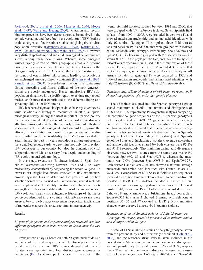
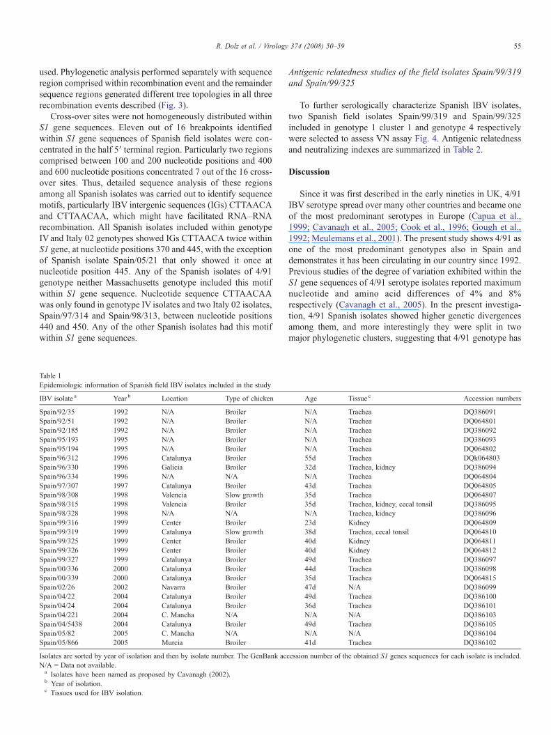
Phylogenetic analysis based on both S1 gene nucleotide andamino acid deduced sequences of the twenty-six Spanishisolates and the reference IBV strains showed that Spanishisolates were separated into four distinct genetic groups orgenotypes (Fig. 1). Genotype I included thirteen out of the
twenty-six field isolates, isolated between 1992 and 2000, thatwere grouped with 4/91 reference isolates. Seven Spanish fieldisolates, from 1997 to 2005, were included in genotype II, andshowed maximum nucleotide and amino acid identities withItaly 02 strains. Genotype III comprised three field virusesisolated between 1996 and 2000 that were grouped with isolatesof the Massachusetts serotype. Particularly, Spain/98/308 andSpain/00/339 isolates were grouped with Massachusetts vaccinestrains (H120) in the phylogenetic tree, and they are likely to bereisolations of vaccine strains used in the immunization of thoseflocks. Finally, Spanish genotype IV included 3 isolates thatsplit in a unique genetic group in the phylogenetic analysis. Allviruses included in genotype IV were isolated in 1999 andshowed maximum nucleotide and amino acid identities withItaly 02 isolates (90.6–92% and 89–91.1% respectively).
Genetic studies of Spanish isolates of 4/91 genotype (genotype I)showed the presence of two distinct genetic clusters
The 13 isolates assigned into the Spanish genotype I groupshared maximum nucleotide and amino acid divergences of7.3% and 10.3% respectively. Phylogenetic analyses comparingthe complete S1 gene sequences of the 13 Spanish genotype Ifield isolates and all 4/91 S1 gene sequences previouslypublished in the GenBank database, including British, Frenchand Iranian isolates, revealed that Spanish isolates were clearlygrouped in two separated genetic clusters identified as Spanishgenotype I cluster 1 (including 11 isolates) and Spanishgenotype I cluster 2 (including 2 isolates). Average nucleotideand amino acid identities shared by both clusters were 93.1%and 91.3% respectively. The minimum amino acid divergenceobserved between two isolates from the same year was 0.4%(between Spain/92/185 and Spain/92/51), whereas the max-imum was 9.9% (between Spain/99/319 and Spain/99/327).Both cluster 1 and cluster 2 isolates showed maximum averagenucleotide and amino acid identities with French isolate FR-94047-94. Comparison of 4/91 Spanish field isolates sequencesrevealed a common unique deletion at amino acid position 58(located in HVR1) in 6 isolates included in cluster 1. Fourisolates within this same group shared an amino acid deletion atposition 340, located in HVR3. Both isolates included in cluster2 showed 8 unique amino acid substitutions. In addition, isolateSpain/99/327 in cluster 2 showed 3 amino acid deletions atpositions 55, 56 and 57 (located in HVR1). No cumulativechanges were observed among 4/91 Spanish isolates.
Sequence analysis of Spanish isolates of Italy 02 genotype(Genotype II) clearly revealed presence of cumulative aminoacid changes within S1 gene
A total of 11 Spanish field strains of Italy 02 genotype, sevenfrom the present study and 4 previously described (Dolz et al.,2006), and the reference strain Italy 02 were included in thepresent study. Maximum nucleotide and amino acid divergencewithin Spanish Italy 02 isolates was 5.7% and 9.9%, respec-tively. The maximum amino acid distance between two virusesisolated the same year was 3.6% (Spain/04/5438 and Spain/04/
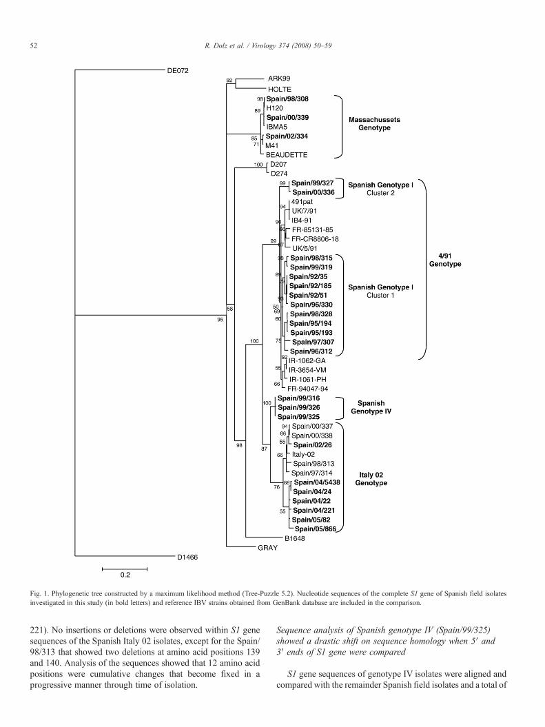
Fig. 1. Phylogenetic tree constructed by a maximum likelihood method (Tree-Puzzle 5.2). Nucleotide sequences of the complete S1 gene of Spanish field isolatesinvestigated in this study (in bold letters) and reference IBV strains obtained from GenBank database are included in the comparison.
52 R. Dolz et al. / Virology 374 (2008) 50–59
221). No insertions or deletions were observed within S1 genesequences of the Spanish Italy 02 isolates, except for the Spain/98/313 that showed two deletions at amino acid positions 139and 140. Analysis of the sequences showed that 12 amino acidpositions were cumulative changes that become fixed in aprogressive manner through time of isolation.
Sequence analysis of Spanish genotype IV (Spain/99/325)showed a drastic shift on sequence homology when 5′ and3′ ends of S1 gene were compared
S1 gene sequences of genotype IV isolates were aligned andcompared with the remainder Spanish field isolates and a total of
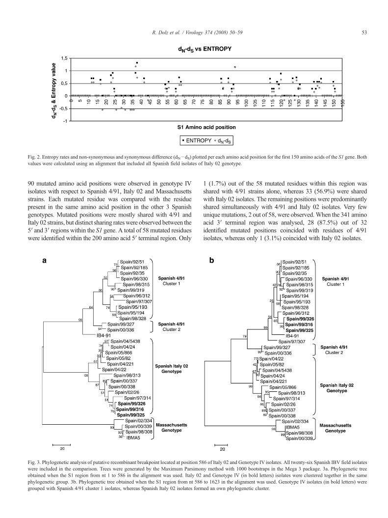
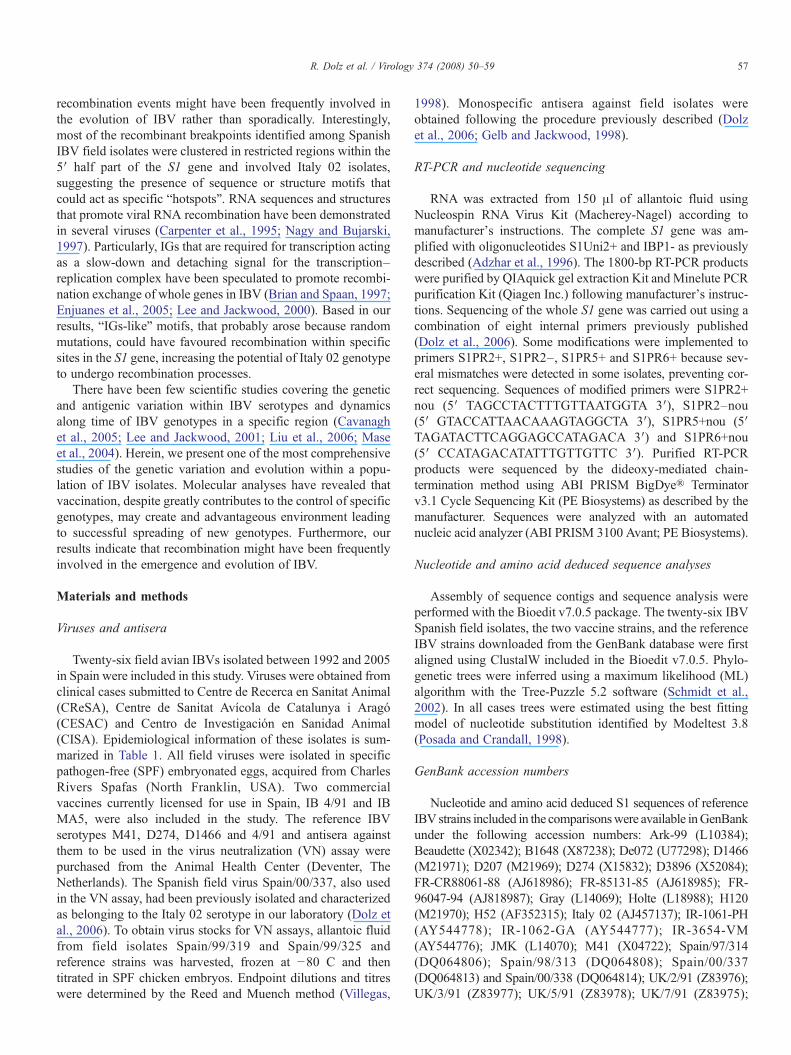
Fig. 2. Entropy rates and non-synonymous and synonymous difference (dN−dS) plotted per each amino acid position for the first 150 amino acids of the S1 gene. Bothvalues were calculated using an alignment that included all Spanish field isolates of Italy 02 genotype.
53R. Dolz et al. / Virology 374 (2008) 50–59
90 mutated amino acid positions were observed in genotype IVisolates with respect to Spanish 4/91, Italy 02 and Massachusettsstrains. Each mutated residue was compared with the residuepresent in the same amino acid position in the other 3 Spanishgenotypes. Mutated positions were mostly shared with 4/91 andItaly 02 strains, but distinct sharing rateswere observed between the5′ and 3′ regions within the S1 gene. A total of 58 mutated residueswere identified within the 200 amino acid 5′ terminal region. Only
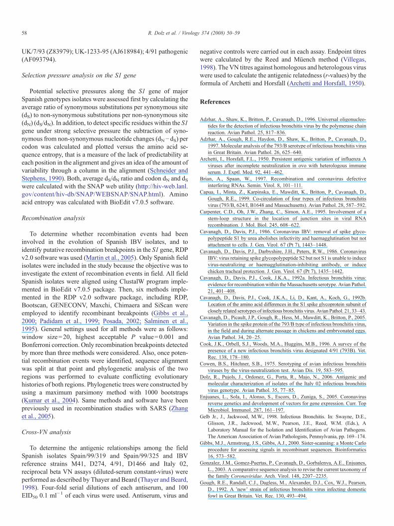
Fig. 3. Phylogenetic analysis of putative recombinant breakpoint located at position 58were included in the comparison. Trees were generated by the Maximum Parsimonobtained when the S1 region from nt 1 to 586 in the alignment was used. Italy 02phylogenetic group. 3b. Phylogenetic tree obtained when the S1 region from nt 586grouped with Spanish 4/91 cluster 1 isolates, whereas Spanish Italy 02 isolates form
1 (1.7%) out of the 58 mutated residues within this region wasshared with 4/91 strains alone, whereas 33 (56.9%) were sharedwith Italy 02 isolates. The remaining positions were predominantlyshared simultaneously with 4/91 and Italy 02 isolates. Very fewuniquemutations, 2 out of 58, were observed.When the 341 aminoacid 3′ terminal region was analysed, 28 (87.5%) out of 32identified mutated positions coincided with residues of 4/91isolates, whereas only 1 (3.1%) coincided with Italy 02 isolates.
6 of Italy 02 and Genotype IV isolates. All twenty-six Spanish IBV field isolatesy method with 1000 bootstraps in the Mega 3 package. 3a. Phylogenetic treeand Genotype IV (in bold letters) isolates were clustered together in the sameto 1623 in the alignment was used. Genotype IV isolates (in bold letters) wereed an own phylogenetic cluster.
54 R. Dolz et al. / Virology 374 (2008) 50–59
These observations correlated with distinct amino acid iden-tities observed when both regions were compared separatelyamong the three genotypes. Genotype IV had maximum aminoacid identities with Italy 02 Spanish genotype (87.3%–95.4%)when the S1 200 Aa 5′ terminal region was compared, whereasamino acid identities in the same region with 4/91 Spanishgenotype was much lower (68.8%–75.8%). On the contrary,when the 341 Aa 3′ end of the S1 gene was compared, genotypeIV showed maximum amino acid identities with 4/91 Spanishisolates (92.9%–97.9%), and lower similarities with Italy 02Spanish isolates (88.8%–91.2%).
Positive selection pressure is detected in several amino acidpositions within Italy 02 isolates, particularly within epitoperegion 2
The average dS/dN ratio within the 13 Spanish isolates of the4/91 genotype was estimated to be 3.43, whereas within theItaly 02 Spanish isolates was 1.97. To identify regions orspecific positions within the S1 gene under positive selection,the dN−dS difference per codon was calculated for the SpanishItaly 02 isolates. The resulting graphic of the dN−dS differenceplotted against entropy values for the initial 150 amino acidpositions of Spanish Italy 02 isolates is shown in Fig. 2. Clearly,amino acid positions with positive dN−dS values also showed
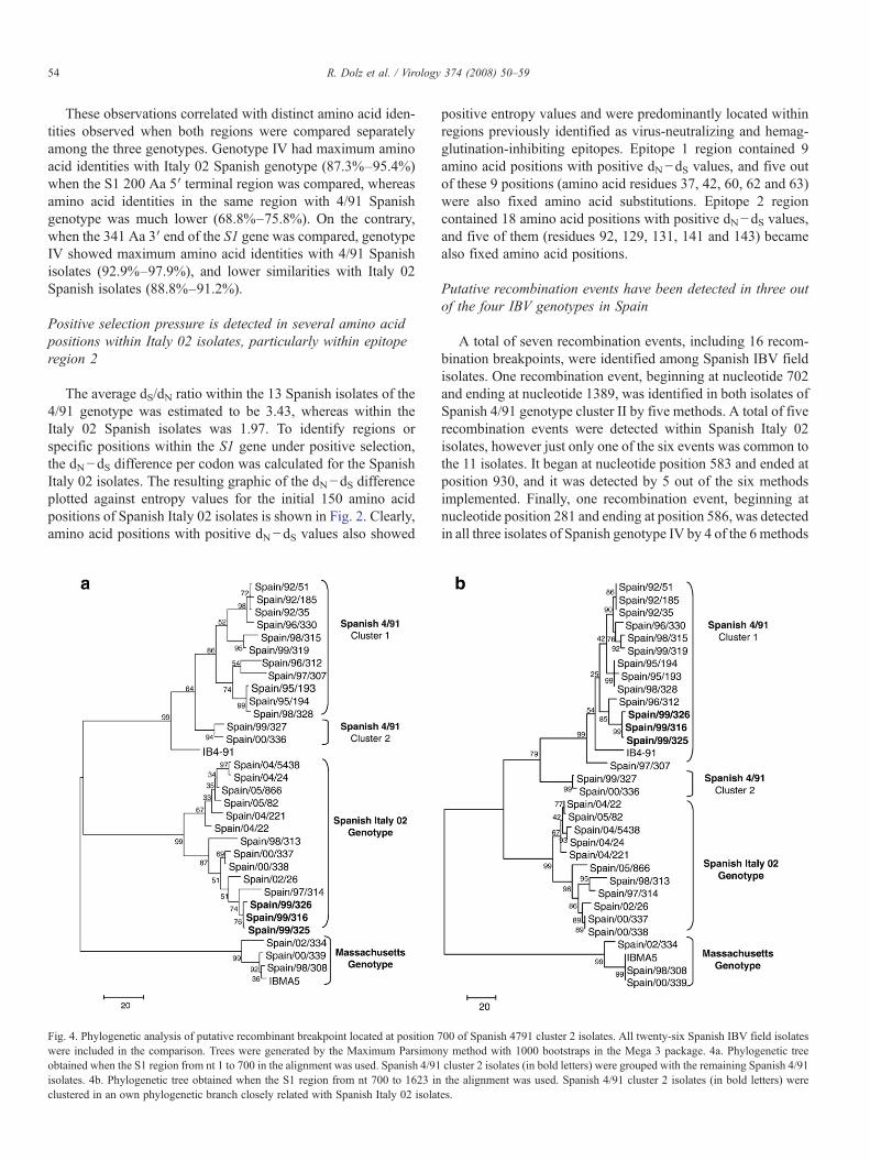
Fig. 4. Phylogenetic analysis of putative recombinant breakpoint located at position 7were included in the comparison. Trees were generated by the Maximum Parsimonobtained when the S1 region from nt 1 to 700 in the alignment was used. Spanish 4/91isolates. 4b. Phylogenetic tree obtained when the S1 region from nt 700 to 1623 inclustered in an own phylogenetic branch closely related with Spanish Italy 02 isolat
positive entropy values and were predominantly located withinregions previously identified as virus-neutralizing and hemag-glutination-inhibiting epitopes. Epitope 1 region contained 9amino acid positions with positive dN−dS values, and five outof these 9 positions (amino acid residues 37, 42, 60, 62 and 63)were also fixed amino acid substitutions. Epitope 2 regioncontained 18 amino acid positions with positive dN−dS values,and five of them (residues 92, 129, 131, 141 and 143) becamealso fixed amino acid positions.
Putative recombination events have been detected in three outof the four IBV genotypes in Spain
A total of seven recombination events, including 16 recom-bination breakpoints, were identified among Spanish IBV fieldisolates. One recombination event, beginning at nucleotide 702and ending at nucleotide 1389, was identified in both isolates ofSpanish 4/91 genotype cluster II by five methods. A total of fiverecombination events were detected within Spanish Italy 02isolates, however just only one of the six events was common tothe 11 isolates. It began at nucleotide position 583 and ended atposition 930, and it was detected by 5 out of the six methodsimplemented. Finally, one recombination event, beginning atnucleotide position 281 and ending at position 586, was detectedin all three isolates of Spanish genotype IV by 4 of the 6 methods
00 of Spanish 4791 cluster 2 isolates. All twenty-six Spanish IBV field isolatesy method with 1000 bootstraps in the Mega 3 package. 4a. Phylogenetic treecluster 2 isolates (in bold letters) were grouped with the remaining Spanish 4/91the alignment was used. Spanish 4/91 cluster 2 isolates (in bold letters) werees.
55R. Dolz et al. / Virology 374 (2008) 50–59
used. Phylogenetic analysis performed separately with sequenceregion comprised within recombination event and the remaindersequence regions generated different tree topologies in all threerecombination events described (Fig. 3).
Cross-over sites were not homogeneously distributed withinS1 gene sequences. Eleven out of 16 breakpoints identifiedwithin S1 gene sequences of Spanish field isolates were con-centrated in the half 5′ terminal region. Particularly two regionscomprised between 100 and 200 nucleotide positions and 400and 600 nucleotide positions concentrated 7 out of the 16 cross-over sites. Thus, detailed sequence analysis of these regionsamong all Spanish isolates was carried out to identify sequencemotifs, particularly IBV intergenic sequences (IGs) CTTAACAand CTTAACAA, which might have facilitated RNA–RNArecombination. All Spanish isolates included within genotypeIVand Italy 02 genotypes showed IGs CTTAACA twice withinS1 gene, at nucleotide positions 370 and 445, with the exceptionof Spanish isolate Spain/05/21 that only showed it once atnucleotide position 445. Any of the Spanish isolates of 4/91genotype neither Massachusetts genotype included this motifwithin S1 gene sequence. Nucleotide sequence CTTAACAAwas only found in genotype IV isolates and two Italy 02 isolates,Spain/97/314 and Spain/98/313, between nucleotide positions440 and 450. Any of the other Spanish isolates had this motifwithin S1 gene sequences.
Table 1Epidemiologic information of Spanish field IBV isolates included in the study
IBV isolate a Year b Location Type of chicken
Spain/92/35 1992 N/A BroilerSpain/92/51 1992 N/A BroilerSpain/92/185 1992 N/A BroilerSpain/95/193 1995 N/A BroilerSpain/95/194 1995 N/A BroilerSpain/96/312 1996 Catalunya BroilerSpain/96/330 1996 Galicia BroilerSpain/96/334 1996 N/A N/ASpain/97/307 1997 Catalunya BroilerSpain/98/308 1998 Valencia Slow growthSpain/98/315 1998 Valencia BroilerSpain/98/328 1998 N/A N/ASpain/99/316 1999 Center BroilerSpain/99/319 1999 Catalunya Slow growthSpain/99/325 1999 Center BroilerSpain/99/326 1999 Center BroilerSpain/99/327 1999 Catalunya BroilerSpain/00/336 2000 Catalunya BroilerSpain/00/339 2000 Catalunya BroilerSpain/02/26 2002 Navarra BroilerSpain/04/22 2004 Catalunya BroilerSpain/04/24 2004 Catalunya BroilerSpain/04/221 2004 C. Mancha N/ASpain/04/5438 2004 Catalunya BroilerSpain/05/82 2005 C. Mancha N/ASpain/05/866 2005 Murcia Broiler
Isolates are sorted by year of isolation and then by isolate number. The GenBank acN/A = Data not available.a Isolates have been named as proposed by Cavanagh (2002).b Year of isolation.c Tissues used for IBV isolation.
Antigenic relatedness studies of the field isolates Spain/99/319and Spain/99/325
To further serologically characterize Spanish IBV isolates,two Spanish field isolates Spain/99/319 and Spain/99/325included in genotype 1 cluster 1 and genotype 4 respectivelywere selected to assess VN assay Fig. 4. Antigenic relatednessand neutralizing indexes are summarized in Table 2.
Discussion
Since it was first described in the early nineties in UK, 4/91IBV serotype spread over many other countries and became oneof the most predominant serotypes in Europe (Capua et al.,1999; Cavanagh et al., 2005; Cook et al., 1996; Gough et al.,1992; Meulemans et al., 2001). The present study shows 4/91 asone of the most predominant genotypes also in Spain anddemonstrates it has been circulating in our country since 1992.Previous studies of the degree of variation exhibited within theS1 gene sequences of 4/91 serotype isolates reported maximumnucleotide and amino acid differences of 4% and 8%respectively (Cavanagh et al., 2005). In the present investiga-tion, 4/91 Spanish isolates showed higher genetic divergencesamong them, and more interestingly they were split in twomajor phylogenetic clusters, suggesting that 4/91 genotype has
Age Tissue c Accession numbers
N/A Trachea DQ386091N/A Trachea DQ064801N/A Trachea DQ386092N/A Trachea DQ386093N/A Trachea DQ06480255d Trachea DQk06480332d Trachea, kidney DQ386094N/A Trachea DQ06480443d Trachea DQ06480535d Trachea DQ06480735d Trachea, kidney, cecal tonsil DQ386095N/A Trachea, kidney DQ38609623d Kidney DQ06480938d Trachea, cecal tonsil DQ06481040d Kidney DQ06481140d Kidney DQ06481249d Trachea DQ38609744d Trachea DQ38609835d Trachea DQ06481547d N/A DQ38609949d Trachea DQ38610036d Trachea DQ386101N/A N/A DQ38610349d Trachea DQ386105N/A N/A DQ38610441d Trachea DQ386102
cession number of the obtained S1 genes sequences for each isolate is included.

Table 2Neutralizing index and antigenic relatedness of Spanish field isolates Spain/99/319 (Genotype I) and Spain/99/325 (Genotype IV) field isolates and the mainreference IBV European serotypes
Virus 1 Virus 2 Neutralizingindex virus 1 a
Neutralizingindex virus 2 b
Antigenicrelatedness (%)
Spain/99/325 c
M41 0.6 0.6 23.2D274 0.84 0.6 33.0D1466 0.6 0.6 24.34/91 0.8 0.6 26.3Spain/00/337 e 2.8 1.98 67.5Spain/99/319 0.8 1.7 47.3Spain/99/325 2.9 2.9 100.0
Spain/99/319 d
M41 0.6 0.6 27.3D274 1.2 0.6 46.3D1466 0.84 0.6 33.84/91 3 0.6 59.8Spain/00/337 1.4 1.2 43.6Spain/99/325 1.7 0.8 47.3Spain/99/319 2.1 2.1 100.0
Maximumantigenic relatedness obtained for each isolate is indicated in bold letters.a Neutralizing index obtained from antisera of virus 1 against virus 2.b Neatruazing index obtained from antisera of virus 2 against virus 1.c Spanish isolate representative from Genotype IV.d Spanish isolate representative from Genotype I (4/91 Spanish isolate).e Spanish field isolate previously demonstrated to belong to Italy 02 serotype
(Dolz et al., 2006).
56 R. Dolz et al. / Virology 374 (2008) 50–59
gone through evident evolutive changes during these years inour country. Furthermore, cross VN results carried out with afield 4/91 Spanish isolate demonstrates that these unique evo-lutive processes contributed to greatly decrease the antigenicrelatedness between the 4/91 Spanish field strains and thereference 4/91 serotype. Hence, these findings suggest thatantigenic subtypes may be present in field and in the brink ofarising as a new serotype, regardless of the level of geneticvariation displayed.
It is particularly remarkable that two Spanish 4/91 strainsisolated the same year practically showed the maximumdivergence exhibited within one IBV genotype and furthermore,they were grouped in separate phylogenetic clusters. Although avery rapid evolutive rate from a common ancestor might havelead to a similar situation, no cumulative changes were observedamong Spanish 4/91 isolates and unique amino acidic changesobserved in one of these two groups were not shared by theother. Moreover, the average dS/dN ratio for the whole S1 genewas not suggestive of a high evolutive rate. Therefore, onepossible explanation for this scenario would be that 4/91 strainsisolated in Spain evolved from at least two distinct evolutivelineages probably originated in two different ancestors. Takinginto account that both isolates assigned to Spanish 4/91 cluster 2are the more recently isolated, a new reintroduction of 4/91isolates in our country could be hypothesized as has beenpreviously suspected in other countries (Adzhar et al., 1997).On the other hand, based on the results of recombinationanalyses carried out in this study, the possibility that recom-bination processes might have been involved in the rapidevolution of the Spanish 4/91 cluster 2 giving rise to a newlineage cannot be excluded.
Interestingly, the present study also shows an importantshift in virus subpopulation dynamics along time with a clearreplacement of predominant genotypes in our country. Whereas4/91 was the predominant genotype in Spain in the nineties, thesituation took a turn at the end of that decade when the new Italy02 genotype appeared and displaced the previous predominantgenotype. High sequence divergences and the presence ofcumulative changes observed within Spanish Italy 02 isolatesfurther support a fast evolutionary rate of this serotype in ourcountry. A wide variety of causes could have been driving theobserved virus subpopulation replacement. However, we foundstrong evidences of the positive selection of mutations withinthe S1 epitope regions of Italy 02 isolates which suggest aselection pressure-driven evolution of this genotype in Spain.Evidently, some cautionary remarks should be considered as ouranalyses were only focused on the S1 protein gene, but selectionpressures could have occurred in other regions within IBVgenome. Therefore, to accurately discern the evolutionary his-tory of this genotypic shift, whole genome studies should becarried out.
At this point an intriguing question comes out regardingfactors contributing to create a positive selection pressure. Ourdata suggests positive selected mutations of Italy 02 might havelead these new isolates escaping the immune system. Evidencesof positive selection pressures acting over specific residueswithin antigenic sites have been previously reported for otherviruses against which partial cross protection is induced afterinfection or vaccination, similarly to what happens with IBV(Grenfell et al., 2004; Haydon et al., 2001; Hurst, 2002). In thissense, it is noteworthy that at the late nineties, commercialvaccines against the 4/91 became available in Spain. Therefore,it is likely that vaccination implemented against 4/91 serotypewas very successful in order to achieve an effective control ofinfections caused by this serotype in our country, based on thedrastic reduction of 4/91 isolations from clinical cases. But onthe other hand, it is tempting to speculate that the high immunepressure against 4/91 isolates might have provided an environ-ment in which the novel variant genotype Italy 02 had higherfitness advantage contributing to a faster establishment andspreading of this serotype in Spain.
Another challenging aspect of IBVepidemiology in Spain isthe emergence of the Genotype IV isolates and its relationshipwith Italy 02 genotype. Genotype IV was established as a newgenotype based on the mosaic nature of its S1 gene. VN studiesrevealed Italy 02 and Genotype IV as being subtypes within acommon serotype. However, the restricted epidemiologicalrelevance of genotype IV, all three isolates included within thisgenotype were isolated in 1999 and in the same geographicregion, clearly indicates distinct fitness abilities among IBVisolates. Therefore, this finding demonstrates that new unknownIBV isolates are emerging constantly, but only those withadvantageous features within a particular environment becomeestablished, whereas others disappear and remain unidentified.
The extent of recombination events among IBV field isolateshas been poorly investigated, even though recombination is awell-known feature of IBV (Cavanagh et al., 1992a; Kottieret al., 1995; Lee and Jackwood, 2000). Our data suggests that
57R. Dolz et al. / Virology 374 (2008) 50–59
recombination events might have been frequently involved inthe evolution of IBV rather than sporadically. Interestingly,most of the recombinant breakpoints identified among SpanishIBV field isolates were clustered in restricted regions within the5′ half part of the S1 gene and involved Italy 02 isolates,suggesting the presence of sequence or structure motifs thatcould act as specific “hotspots”. RNA sequences and structuresthat promote viral RNA recombination have been demonstratedin several viruses (Carpenter et al., 1995; Nagy and Bujarski,1997). Particularly, IGs that are required for transcription actingas a slow-down and detaching signal for the transcription–replication complex have been speculated to promote recombi-nation exchange of whole genes in IBV (Brian and Spaan, 1997;Enjuanes et al., 2005; Lee and Jackwood, 2000). Based in ourresults, “IGs-like” motifs, that probably arose because randommutations, could have favoured recombination within specificsites in the S1 gene, increasing the potential of Italy 02 genotypeto undergo recombination processes.
There have been few scientific studies covering the geneticand antigenic variation within IBV serotypes and dynamicsalong time of IBV genotypes in a specific region (Cavanaghet al., 2005; Lee and Jackwood, 2001; Liu et al., 2006; Maseet al., 2004). Herein, we present one of the most comprehensivestudies of the genetic variation and evolution within a popu-lation of IBV isolates. Molecular analyses have revealed thatvaccination, despite greatly contributes to the control of specificgenotypes, may create and advantageous environment leadingto successful spreading of new genotypes. Furthermore, ourresults indicate that recombination might have been frequentlyinvolved in the emergence and evolution of IBV.
Materials and methods
Viruses and antisera
Twenty-six field avian IBVs isolated between 1992 and 2005in Spain were included in this study. Viruses were obtained fromclinical cases submitted to Centre de Recerca en Sanitat Animal(CReSA), Centre de Sanitat Avícola de Catalunya i Aragó(CESAC) and Centro de Investigación en Sanidad Animal(CISA). Epidemiological information of these isolates is sum-marized in Table 1. All field viruses were isolated in specificpathogen-free (SPF) embryonated eggs, acquired from CharlesRivers Spafas (North Franklin, USA). Two commercialvaccines currently licensed for use in Spain, IB 4/91 and IBMA5, were also included in the study. The reference IBVserotypes M41, D274, D1466 and 4/91 and antisera againstthem to be used in the virus neutralization (VN) assay werepurchased from the Animal Health Center (Deventer, TheNetherlands). The Spanish field virus Spain/00/337, also usedin the VN assay, had been previously isolated and characterizedas belonging to the Italy 02 serotype in our laboratory (Dolz etal., 2006). To obtain virus stocks for VN assays, allantoic fluidfrom field isolates Spain/99/319 and Spain/99/325 andreference strains was harvested, frozen at −80 C and thentitrated in SPF chicken embryos. Endpoint dilutions and titreswere determined by the Reed and Muench method (Villegas,
1998). Monospecific antisera against field isolates wereobtained following the procedure previously described (Dolzet al., 2006; Gelb and Jackwood, 1998).
RT-PCR and nucleotide sequencing
RNA was extracted from 150 μl of allantoic fluid usingNucleospin RNA Virus Kit (Macherey-Nagel) according tomanufacturer's instructions. The complete S1 gene was am-plified with oligonucleotides S1Uni2+ and IBP1- as previouslydescribed (Adzhar et al., 1996). The 1800-bp RT-PCR productswere purified by QIAquick gel extraction Kit and Minelute PCRpurification Kit (Qiagen Inc.) following manufacturer's instruc-tions. Sequencing of the whole S1 gene was carried out using acombination of eight internal primers previously published(Dolz et al., 2006). Some modifications were implemented toprimers S1PR2+, S1PR2–, S1PR5+ and S1PR6+ because sev-eral mismatches were detected in some isolates, preventing cor-rect sequencing. Sequences of modified primers were S1PR2+nou (5′ TAGCCTACTTTGTTAATGGTA 3′), S1PR2–nou(5′ GTACCATTAACAAAGTAGGCTA 3′), S1PR5+nou (5′TAGATACTTCAGGAGCCATAGACA 3′) and S1PR6+nou(5′ CCATAGACATATTTGTTGTTC 3′). Purified RT-PCRproducts were sequenced by the dideoxy-mediated chain-termination method using ABI PRISM BigDye® Terminatorv3.1 Cycle Sequencing Kit (PE Biosystems) as described by themanufacturer. Sequences were analyzed with an automatednucleic acid analyzer (ABI PRISM 3100 Avant; PE Biosystems).
Nucleotide and amino acid deduced sequence analyses
Assembly of sequence contigs and sequence analysis wereperformed with the Bioedit v7.0.5 package. The twenty-six IBVSpanish field isolates, the two vaccine strains, and the referenceIBV strains downloaded from the GenBank database were firstaligned using ClustalW included in the Bioedit v7.0.5. Phylo-genetic trees were inferred using a maximum likelihood (ML)algorithm with the Tree-Puzzle 5.2 software (Schmidt et al.,2002). In all cases trees were estimated using the best fittingmodel of nucleotide substitution identified by Modeltest 3.8(Posada and Crandall, 1998).
GenBank accession numbers
Nucleotide and amino acid deduced S1 sequences of referenceIBV strains included in the comparisonswere available inGenBankunder the following accession numbers: Ark-99 (L10384);Beaudette (X02342); B1648 (X87238); De072 (U77298); D1466(M21971); D207 (M21969); D274 (X15832); D3896 (X52084);FR-CR88061-88 (AJ618986); FR-85131-85 (AJ618985); FR-96047-94 (AJ818987); Gray (L14069); Holte (L18988); H120(M21970); H52 (AF352315); Italy 02 (AJ457137); IR-1061-PH(AY544778); IR-1062-GA (AY544777); IR-3654-VM(AY544776); JMK (L14070); M41 (X04722); Spain/97/314(DQ064806); Spain/98/313 (DQ064808); Spain/00/337(DQ064813) and Spain/00/338 (DQ064814); UK/2/91 (Z83976);UK/3/91 (Z83977); UK/5/91 (Z83978); UK/7/91 (Z83975);
58 R. Dolz et al. / Virology 374 (2008) 50–59
UK/7/93 (Z83979); UK-1233-95 (AJ618984); 4/91 pathogenic(AF093794).
Selection pressure analysis on the S1 gene
Potential selective pressures along the S1 gene of majorSpanish genotypes isolates were assessed first by calculating theaverage ratio of synonymous substitutions per synonymous site(dS) to non-synonymous substitutions per non-synonymous site(dN) (dS/dN). In addition, to detect specific residues within the S1gene under strong selective pressure the subtraction of syno-nymous from non-synonymous nucleotide changes (dN−dS) percodon was calculated and plotted versus the amino acid se-quence entropy, that is a measure of the lack of predictability ateach position in the alignment and gives an idea of the amount ofvariability through a column in the alignment (Schneider andStephens, 1990). Both, average dS/dN ratio and codon dN and dSwere calculated with the SNAP web utility (http://hiv-web.lanl.gov/content/hiv-db/SNAP/WEBSNAP/SNAP.html). Aminoacid entropy was calculated with BioEdit v7.0.5 software.
Recombination analysis
To determine whether recombination events had beeninvolved in the evolution of Spanish IBV isolates, and toidentify putative recombination breakpoints in the S1 gene, RDPv2.0 software was used (Martin et al., 2005). Only Spanish fieldisolates were included in the study because the objective was toinvestigate the extent of recombination events in field. All fieldSpanish isolates were aligned using ClustalW program imple-mented in BioEdit v7.0.5 package. Then, six methods imple-mented in the RDP v2.0 software package, including RDP,Bootscan, GENECONV, Maxchi, Chimaera and SiScan wereemployed to identify recombinant breakpoints (Gibbs et al.,2000; Padidam et al., 1999; Posada, 2002; Salminen et al.,1995). General settings used for all methods were as follows:window size=20, highest acceptable P value=0.001 andBonferroni correction. Only recombination breakpoints detectedby more than three methods were considered. Also, once poten-tial recombination events were identified, sequence alignmentwas split at that point and phylogenetic analysis of the tworegions was performed to evaluate conflicting evolutionaryhistories of both regions. Phylogenetic trees were constructed byusing a maximum parsimony method with 1000 bootstraps(Kumar et al., 2004). Same methods and software have beenpreviously used in recombination studies with SARS (Zhanget al., 2005).
Cross-VN analysis
To determine the antigenic relationships among the fieldSpanish isolates Spain/99/319 and Spain/99/325 and IBVreference strains M41, D274, 4/91, D1466 and Italy 02,reciprocal beta VN assays (diluted-serum constant-virus) wereperformed as described by Thayer and Beard (Thayer and Beard,1998). Four-fold serial dilutions of each antiserum, and 100EID50 0.1 ml−1 of each virus were used. Antiserum, virus and
negative controls were carried out in each assay. Endpoint titreswere calculated by the Reed and Müench method (Villegas,1998). TheVN titres against homologous and heterologous viruswere used to calculate the antigenic relatedness (r-values) by theformula of Archetti and Horsfall (Archetti and Horsfall, 1950).
References
Adzhar, A., Shaw, K., Britton, P., Cavanagh, D., 1996. Universal oligonucleo-tides for the detection of infectious bronchitis virus by the polymerase chainreaction. Avian Pathol. 25, 817–836.
Adzhar, A., Gough, R.E., Haydon, D., Shaw, K., Britton, P., Cavanagh, D.,1997. Molecular analysis of the 793/B serotype of infectious bronchitis virusin Great Britain. Avian Pathol. 26, 625–640.
Archetti, I., Horsfall, F.L., 1950. Persistent antigenic variation of influenza Aviruses after incomplete neutralization in ovo with heterologous immuneserum. J. Exptl. Med. 92, 441–462.
Brian, A., Spaan, W., 1997. Recombination and coronavirus defectiveinterfering RNAs. Semin. Virol. 8, 101–111.
Capua, I., Minta, Z., Karpinska, E., Mawditt, K., Britton, P., Cavanagh, D.,Gough, R.E., 1999. Co-circulation of four types of infectious bronchitisvirus (793/B, 624/I, B1648 and Massachusetts). Avian Pathol. 28, 587–592.
Carpenter, C.D., Oh, J.W., Zhang, C., Simon, A.E., 1995. Involvement of astem-loop structure in the location of junction sites in viral RNArecombination. J. Mol. Biol. 245, 608–622.
Cavanagh, D., Davis, P.J., 1986. Coronavirus IBV: removal of spike glyco-polypeptide S1 by urea abolishes infectivity and haemagglutination but notattachment to cells. J. Gen. Virol. 67 (Pt 7), 1443–1448.
Cavanagh, D., Davis, P.J., Darbyshire, J.H., Peters, R.W., 1986. CoronavirusIBV: virus retaining spike glycopolypeptide S2 but not S1 is unable to inducevirus-neutralizing or haemagglutination-inhibiting antibody, or inducechicken tracheal protection. J. Gen. Virol. 67 (Pt 7), 1435–1442.
Cavanagh, D., Davis, P.J., Cook, J.K.A., 1992a. Infectious bronchitis virus:evidence for recombination within theMassachusetts serotype. Avian Pathol.21, 401–408.
Cavanagh, D., Davis, P.J., Cook, J.K.A., Li, D., Kant, A., Koch, G., 1992b.Location of the amino acid differences in the S1 spike glycoprotein subunit ofclosely related serotypes of infectious bronchitis virus. Avian Pathol. 21, 33–43.
Cavanagh, D., Picault, J.P., Gough, R., Hess, M., Mawditt, K., Britton, P., 2005.Variation in the spike protein of the 793/B type of infectious bronchitis virus,in the field and during alternate passage in chickens and embryonated eggs.Avian Pathol. 34, 20–25.
Cook, J.K., Orbell, S.J., Woods, M.A., Huggins, M.B., 1996. A survey of thepresence of a new infectious bronchitis virus designated 4/91 (793B). Vet.Rec. 138, 178–180.
Cowen, B.S., Hitchner, S.B., 1975. Serotyping of avian infectious bronchitisviruses by the virus-neutralization test. Avian Dis. 19, 583–595.
Dolz, R., Pujols, J., Ordonez, G., Porta, R., Majo, N., 2006. Antigenic andmolecular characterization of isolates of the Italy 02 infectious bronchitisvirus genotype. Avian Pathol. 35, 77–85.
Enjuanes, L., Sola, I., Alonso, S., Escors, D., Zuniga, S., 2005. Coronavirusreverse genetics and development of vectors for gene expression. Curr. TopMicrobiol. Immunol. 287, 161–197.
Gelb Jr., J., Jackwood, M.W., 1998. Infectious Bronchitis. In: Swayne, D.E.,Glisson, J.R., Jackwood, M.W., Pearson, J.E., Reed, W.M. (Eds.), ALaboratory Manual for the Isolation and Identification of Avian Pathogens.The American Association of Avian Pathologists, Pennsylvania, pp. 169–174.
Gibbs, M.J., Armstrong, J.S., Gibbs, A.J., 2000. Sister-scanning: a Monte Carloprocedure for assessing signals in recombinant sequences. Bioinformatics16, 573–582.
Gonzalez, J.M., Gomez-Puertas, P., Cavanagh, D., Gorbalenya, A.E., Enjuanes,L., 2003. A comparative sequence analysis to revise the current taxonomy ofthe family Coronaviridae. Arch. Virol. 148, 2207–2235.
Gough, R.E., Randall, C.J., Dagless, M., Alexander, D.J., Cox, W.J., Pearson,D., 1992. A 'new' strain of infectious bronchitis virus infecting domesticfowl in Great Britain. Vet. Rec. 130, 493–494.

59R. Dolz et al. / Virology 374 (2008) 50–59
Grenfell, B.T., Pybus, O.G., Gog, J.R., Wood, J.L., Daly, J.M., Mumford, J.A.,Holmes, E.C., 2004. Unifying the epidemiological and evolutionarydynamics of pathogens. Science 303, 327–332.
Haydon, D.T., Bastos, A.D., Knowles, N.J., Samuel, A.R., 2001. Evidence forpositive selection in foot-and-mouth disease virus capsid genes from fieldisolates. Genetics 157, 7–15.
Hurst, L.D., 2002. The Ka/Ks ratio: diagnosing the form of sequence evolution.Trends Genet. 18, 486.
Jones, R.C., Ambali, A.G., 1987. Re-excretion of an enterotropic infectiousbronchitis virus by hens at point of lay after experimental infection at dayold. Vet. Rec. 120, 617–618.
Jones, R.C., Jordan, F.T.W., 1970. The exposure of day-old chicks to infectiousbronchitis and the subsequent development of the oviduct. Vet. Rec. 87,504–505.
Kant, A., Koch, G., van Roozelaar, D.J., Kusters, J.G., Poelwijk, F.A., van derZeijst, B.A., 1992. Location of antigenic sites defined by neutralizing mono-clonal antibodies on the S1 avian infectious bronchitis virus glycopolypeptide.J. Gen. Virol. 73 (Pt 3), 591–596.
Koch, G., Hartog, L., Kant, A., van Roozelaar, D.J., 1990. Antigenic domains onthe peplomer protein of avian infectious bronchitis virus: correlation withbiological functions. J. Gen. Virol. 71 (Pt 9), 1929–1935.
Kottier, S.A., Cavanagh, D., Britton, P., 1995. Experimental evidence of recom-bination in coronavirus infectious bronchitis virus. Virology 213, 569–580.
Kumar, S., Tamura, K., Nei, M., 2004. Integrated software for MolecularEvolutionaryGeneticsAnalysis and sequence alignment. BriefBioinform. 5, 2.
Kusters, J.G., Niesters, H.G., Bleumink-Pluym, N.M., Davelaar, F.G., Horzinek,M.C., Van der Zeijst, B.A., 1987. Molecular epidemiology of infectiousbronchitis virus in The Netherlands. J. Gen. Virol. 68 (Pt 2), 343–352.
Lee, C.W., Jackwood, M.W., 2000. Evidence of genetic diversity generated byrecombination among avian coronavirus IBV. Arch. Virol. 145, 2135–2148.
Lee, C.W., Jackwood, M.W., 2001. Origin and evolution of Georgia 98 (GA98),a new serotype of avian infectious bronchitis virus. Virus Res. 80, 33–39.
Liu, S.W., Zhang, Q.X., Chen, J.D., Han, Z.X., Liu, X., Feng, L., Shao, Y.H.,Rong, J.G., Kong, X.G., Tong, G.Z., 2006. Genetic diversity of avianinfectious bronchitis coronavirus strains isolated in China between 1995 and2004. Arch Virol. 151, 1133–1148.
Martin, D.P., Williamson, C., Posada, D., 2005. RDP2: recombination detectionand analysis from sequence alignments. Bioinformatics 21, 260–262.
Mase, M., Tsukamoto, K., Imai, K., Yamaguchi, S., 2004. Phylogenetic analysisof avian infectious bronchitis virus strains isolated in Japan. Arch. Virol.149, 2069–2078.
Meulemans, G., Boschmans, M., Decaesstecker, M., Berg, T.P.v.d., Denis, P.,Cavanagh, D., van den Berg, T.P., 2001. Epidemiology of infectiousbronchitis virus in Belgian broilers: a retrospective study, 1986 to 1995.Avian Pathol. 30, 411–421.
Moore, K.M., Bennett, J.D., Seal, B.S., Jackwood, M.W., 1998. Sequencecomparison of avian infectious bronchitis virus S1 glycoproteins of the
Florida serotype and five variant isolates from Georgia and California. VirusGenes 17, 63–83.
Nagy, P.D., Bujarski, J.J., 1997. Engineering of homologous recombination hot-spots with AU-rich sequences in brome mosaic virus. J. Virol. 71, 3799–3810.
Office, e.d.i., 2004. Avian infectious bronchitis. In: OIE (Eds.), Manual ofDiagnostic Tests and Vaccines for Terrestrial Animals. Paris. Chapter 2.7.6.
Padidam, M., Sawyer, S., Fauquet, C.M., 1999. Possible emergence of newgeminiviruses by frequent recombination. Virology 265, 218–225.
Posada, D., 2002. Evaluation of methods for detecting recombination from DNAsequences: empirical data. Mol. Biol. Evol. 19, 708–717.
Posada, D., Crandall, K.A., 1998. MODELTEST: testing the model of DNAsubstitution. Bioinformatics 14, 817–818.
Salminen, M.O., Carr, J.K., Burke, D.S., McCutchan, F.E., 1995. Identificationof breakpoints in intergenotypic recombinants of HIV type 1 by boot-scanning. AIDS Res. Hum. Retroviruses 11, 1423–1425.
Schmidt, H.A., Strimmer, K., Vingron, M., von Haeseler, A., 2002. TREE-PUZZLE: maximum likelihood phylogenetic analysis using quartets andparallel computing. Bioinformatics 18, 502–504.
Schneider, T.D., Stephens, R.M., 1990. Sequence logos: a new way to displayconsensus sequences. Nucleic Acids Res. 18, 6097–6100.
Stern, D.F., Kennedy, S.I., 1980. Coronavirus multiplication strategy. II.Mapping the avian infectious bronchitis virus intracellular RNA species tothe genome. J. Virol. 36, 440–449.
Stern, D.F., Sefton, B.M., 1982. Coronavirus proteins: biogenesis of avianinfectious bronchitis virus virion proteins. J. Virol. 44, 794–803.
Thayer, S.G., Beard, C.W., 1998. Serologic procedures. In: Swayne, D.E., Glisson,J.R., Jackwood,M.W., Pearson, J.E., Reed,W.M. (Eds.), A LaboratoryManualfor the Isolation and Identification of Avian Pathogens. The AmericanAssociation of Avian Pathologists, Pennsylvania, pp. 255–266.
Villegas, P., 1998. Titration of biological suspensions. In: Swayne, D.E., Glisson,J.R., Jackwood, M.W., Pearson, J.E., Reed, W.M. (Eds.), A LaboratoryManual for the Isolation and Identification of Avian Pathogens. TheAmerican Association of Avian Pathologists, Pennsylvania, pp. 248–254.
Wang, C.H., Huang, Y.C., 2000. Relationship between serotypes and genotypesbased on the hypervariable region of the S1 gene of infectious bronchitisvirus. Arch. Virol. 145, 291–300.
Wang, L., Xu, Y., Collisson, E.W., 1997. Experimental confirmation ofrecombination upstream of the S1 hypervariable region of infectiousbronchitis virus. Virus Res. 49, 139–145.
Winterfield, R.W., Hitchner, S.B., 1962. Etiology of an infectious nephritis-nephrosis syndrome of chickens. Am. J. Vet. Res. 23, 1273–1279.
Zanella, A., Lavazza, A., Marchi, R., Moreno Martin, A., Paganelli, F., 2003.Avian infectious bronchitis: characterization of new isolates from Italy.Avian Dis. 47, 180–185.
Zhang, X.W., Yap, Y.L., Danchin, A., 2005. Testing the hypothesis of arecombinant origin of the SARS-associated coronavirus. Arch. Virol. 150,1–20.