Molecular characterization of a novel simian immunodeficiency virus lineage (SIVtal) from northern...
11
Molecular characterization of a novel simian immunodeficiency virus lineage (SIVtal) from northern talapoins (Miopithecus ogouensis) Florian Liegeois a , Valerie Courgnaud a , William M. Switzer b , Hayley W. Murphy c , Severin Loul d , Avelin Aghokeng a , Xavier Pourrut a , Eitel Mpoudi-Ngole d , Eric Delaporte a , Martine Peeters a, ⁎ a UMR145, Institut de Recherche pour le Developpement (IRD), and University of Montpellier I, Montpellier, France b Laboratory Branch, Division of HIV/AIDS Prevention, National Center for HIV, STD, and TB Prevention, Centers for Disease Control and Prevention, Atlanta, GA 30333, USA c Zoo New England, 1 Franklin Park Road, Boston, MA 02121, USA d Projet PRESICA, Hôpital Militaire, Yaounde, Cameroon Received 26 September 2005; returned to author for revision 9 December 2005; accepted 6 January 2006 Available online 8 February 2006 Abstract Simian immunodeficiency viruses (SIVs) are found in an extensive number of African primates, and humans continue to be exposed to these viruses by hunting and handling of primate bushmeat and following occupational exposures to captive nonhuman primates. Here, we report the molecular characterization of a new SIV lineage, SIVtal, from wild-caught and captive talapoin monkeys (Miopithecus ogouensis) from Cameroon and U.S. zoos, respectively. Phylogenetic tree analyses of a small fragment in the pol gene indicated that all SIVtal strains clustered together forming a single species-specific lineage. Full-length sequence analysis for two strains, SIVtal-00CM266 and SIVtal-01CM8023, from wild-caught animals in Cameroon confirmed that SIVtal was distinct from all primate lentiviruses isolated so far and represents a new SIV lineage. Phylogenetic analyses in different viral genes showed a significant clustering of the SIVtal lineage with the Cercopithecus-specific SIVs. In addition, SIVtal and Cercopithecus-specific SIVs share functional motifs in Gag and Env that distinguish them from other primate lentiviruses. Like SIVsyk and SIVdeb, a vpu gene homologue was also absent in SIVtal. Although northern talapoins belong to the Miopithecus genus, their SIVs belong to the Cercopithecus SIV lineage, suggesting evolution from a common ancestor or cross-species transmission between both primate genera. © 2006 Elsevier Inc. All rights reserved. Keywords: SIV; Non-human primate; Talapoin; HIV; Evolution; Cameroon Introduction SIVs are a large group of viruses that are found naturally in an extensive number of African primate species and serological and/or molecular evidence for SIV infection have been reported in at least 36 African nonhuman primates (NHPs) (Bailes, 2002; Peeters and Courgnaud, 2002; Reed et al., 2004; Takemura et al., 2005; Verschoor et al., 2004). Both HIV-1 and HIV-2, the etiologic agents for AIDS in humans, are the result of cross-species transmissions from SIVs from primates in Africa. Their closest relatives are respectively, SIVcpz in chimpanzees (Pan troglodytes troglodytes) from West central Africa and SIVsm in sooty mangabeys (Cercocebus atys) from West Africa (Corbet et al., 2000; Gao et al., 1992, 1999). Although SIVs are called immunodeficiency viruses, these viruses do not typically induce an AIDS-like disease in their natural hosts, suggesting that they have been associated and evolved with their hosts over an extended period of time (Kestens et al., 1995; Muller-Trutwin et al., 1996; Pandrea et al., 2003; Silvestri et al., 2003). A high genetic diversity is observed among the known SIVs but, generally each primate species is infected with a species-specific virus which form monophyletic lineages in phylogenetic trees. In addition, several “major SIV lineages” have been identified which represent groups of SIVs from different primate species that are more closely related to one another than they are to other Virology 349 (2006) 55 – 65 www.elsevier.com/locate/yviro ⁎ Corresponding author. Fax: +33 4 67 41 61 46. E-mail address: [email protected] (M. Peeters). 0042-6822/$ - see front matter © 2006 Elsevier Inc. All rights reserved. doi:10.1016/j.virol.2006.01.011
-
Upload
independent -
Category
Documents
-
view
2 -
download
0
Transcript of Molecular characterization of a novel simian immunodeficiency virus lineage (SIVtal) from northern...
06) 55–65www.elsevier.com/locate/yviro
Virology 349 (20
Molecular characterization of a novel simian immunodeficiency virus lineage(SIVtal) from northern talapoins (Miopithecus ogouensis)
Florian Liegeois a, Valerie Courgnaud a, William M. Switzer b, Hayley W. Murphy c,Severin Loul d, Avelin Aghokeng a, Xavier Pourrut a, Eitel Mpoudi-Ngole d,
Eric Delaporte a, Martine Peeters a,⁎
a UMR145, Institut de Recherche pour le Developpement (IRD), and University of Montpellier I, Montpellier, Franceb Laboratory Branch, Division of HIV/AIDS Prevention, National Center for HIV, STD, and TB Prevention, Centers for Disease Control
and Prevention, Atlanta, GA 30333, USAc Zoo New England, 1 Franklin Park Road, Boston, MA 02121, USA
d Projet PRESICA, Hôpital Militaire, Yaounde, Cameroon
Received 26 September 2005; returned to author for revision 9 December 2005; accepted 6 January 2006Available online 8 February 2006
Abstract
Simian immunodeficiency viruses (SIVs) are found in an extensive number of African primates, and humans continue to be exposed to theseviruses by hunting and handling of primate bushmeat and following occupational exposures to captive nonhuman primates. Here, we report themolecular characterization of a new SIV lineage, SIVtal, from wild-caught and captive talapoin monkeys (Miopithecus ogouensis) from Cameroonand U.S. zoos, respectively. Phylogenetic tree analyses of a small fragment in the pol gene indicated that all SIVtal strains clustered togetherforming a single species-specific lineage. Full-length sequence analysis for two strains, SIVtal-00CM266 and SIVtal-01CM8023, from wild-caughtanimals in Cameroon confirmed that SIVtal was distinct from all primate lentiviruses isolated so far and represents a new SIV lineage. Phylogeneticanalyses in different viral genes showed a significant clustering of the SIVtal lineage with the Cercopithecus-specific SIVs. In addition, SIVtal andCercopithecus-specific SIVs share functional motifs in Gag and Env that distinguish them from other primate lentiviruses. Like SIVsyk andSIVdeb, a vpu gene homologue was also absent in SIVtal. Although northern talapoins belong to the Miopithecus genus, their SIVs belong to theCercopithecus SIV lineage, suggesting evolution from a common ancestor or cross-species transmission between both primate genera.© 2006 Elsevier Inc. All rights reserved.
Keywords: SIV; Non-human primate; Talapoin; HIV; Evolution; Cameroon
Introduction
SIVs are a large group of viruses that are found naturally inan extensive number of African primate species and serologicaland/or molecular evidence for SIV infection have been reportedin at least 36 African nonhuman primates (NHPs) (Bailes,2002; Peeters and Courgnaud, 2002; Reed et al., 2004;Takemura et al., 2005; Verschoor et al., 2004). Both HIV-1and HIV-2, the etiologic agents for AIDS in humans, are theresult of cross-species transmissions from SIVs from primatesin Africa. Their closest relatives are respectively, SIVcpz in
⁎ Corresponding author. Fax: +33 4 67 41 61 46.E-mail address: [email protected] (M. Peeters).
0042-6822/$ - see front matter © 2006 Elsevier Inc. All rights reserved.doi:10.1016/j.virol.2006.01.011
chimpanzees (Pan troglodytes troglodytes) from West centralAfrica and SIVsm in sooty mangabeys (Cercocebus atys) fromWest Africa (Corbet et al., 2000; Gao et al., 1992, 1999).
Although SIVs are called immunodeficiency viruses, theseviruses do not typically induce an AIDS-like disease in theirnatural hosts, suggesting that they have been associated andevolved with their hosts over an extended period of time(Kestens et al., 1995; Muller-Trutwin et al., 1996; Pandrea etal., 2003; Silvestri et al., 2003). A high genetic diversity isobserved among the known SIVs but, generally each primatespecies is infected with a species-specific virus which formmonophyletic lineages in phylogenetic trees. In addition,several “major SIV lineages” have been identified whichrepresent groups of SIVs from different primate species thatare more closely related to one another than they are to other
56 F. Liegeois et al. / Virology 349 (2006) 55–65
SIVs (Bibollet-Ruche et al., 2004). For some of these groups,virus and host phylogenies seem to match, suggesting virus/host co-speciation, but there are also numerous examples ofcross-species transmission and recombination (Allan et al.,1991; Bailes et al., 2003; Beer et al., 1999, 2001; Clewley etal., 1998; Courgnaud et al., 2001, 2003; Jin et al., 1994;Souquiere et al., 2001). Among the African primates theCercopithecus genus harbors the largest number of speciesknown to be infected with related SIVs and recently aCercopithecus-specific SIV lineage has been described(Bibollet-Ruche et al., 2004). Despite examples of co-evolution between viruses and hosts, and cross-speciestransmission within this genus, the SIVsyk, SIVdeb, SIVgsn,SIVmus, SIVmon, and SIVden viruses which are all derivedfrom different Cercopithecus species, consistently form onehighly supported group in phylogenetic trees and sharefunctional motifs in gag and env that distinguish them fromother primate lentiviruses (Bibollet-Ruche et al., 2004; Dazzaet al., 2005). These results suggest that these SIVs evolvedfrom a common ancestor that most likely infected aCercopithecus host in the distant past. Interestingly, it hasalso been shown that one primate species can be infected withtwo different SIVs. For example, mandrills from central andsouthern Gabon are infected with SIVmnd-1, whereas thoseliving in northern Gabon and the south of Cameroon areinfected with SIVmnd-2 (Souquiere et al., 2001). The geneticdiversity among NHP lentiviruses is thus extremely complex.The current knowledge of SIV/HIV phylogeny indicates thatboth co-evolution and cross-species transmissions withconcomitant genetic recombination have driven lentiviralevolution.
Given that viruses from chimpanzees and sooty manga-beys have both crossed the species barrier on multipleoccasions resulting in HIV-1 and HIV-2, it is thus importantto identify and characterize SIVs that circulate in Africanprimates species to estimate which lentiviruses couldpotentially infect the human population. In a recent surveyof bushmeat markets in Cameroon, we found evidencesuggesting that a substantial proportion of wild-livingmonkeys are SIV infected and PCR analysis on a subsetof samples led to the discovery of new SIV strains notpreviously known to infect primates (Peeters et al., 2002).This study thus documented for the first time that humanswho hunt and butcher primates are routinely exposed to aplethora of genetically divergent SIV strains. In addition,persons who work with NHPs in captivity are also at riskfor SIV infection (Khabbaz et al., 1994; Sotir et al., 1997).Therefore, to gain further insights into the diversity of SIVsto which humans are exposed and to SIV evolution overall,full-length SIVtal sequences were derived from two wild-caught northern talapoin monkeys (Miopithecus ogouensis)which were identified as SIV-infected during a survey ofprimate bushmeat in Cameroon. In addition, to characterizeSIVs recently identified in captive talapoins (Ndongmo etal., 2004), partial DNA sequences were also amplified fromfour captive seropositive talapoins housed in different U.S.zoos. The talapoin monkey, the smallest species of Old
World monkeys, belongs to the Miopithecus genus in whichtwo species have been identified (Groves, 2001). Thenorthern talapoin (M. ogouensis) are found in southernCameroon south of the Sanaga River, and in EquatorialGuinea and Gabon, while the southern talapoin (M.talapoin) occurs south of the Congo River in Angola andnorthwest of the Democratic Republic of Congo (DRC,formerly Zaire) (Gautier-Hion et al., 1999; Groves, 2001).Our results indicate that the SIVtal strains obtained from wildand captive talapoins, all belonging to the M. ogouensisspecies, form a separate species-specific lineage whichconsistently falls in the recently described CercopithecusSIV cluster.
Results and discussion
Species identification of the SIV-positive talapoins
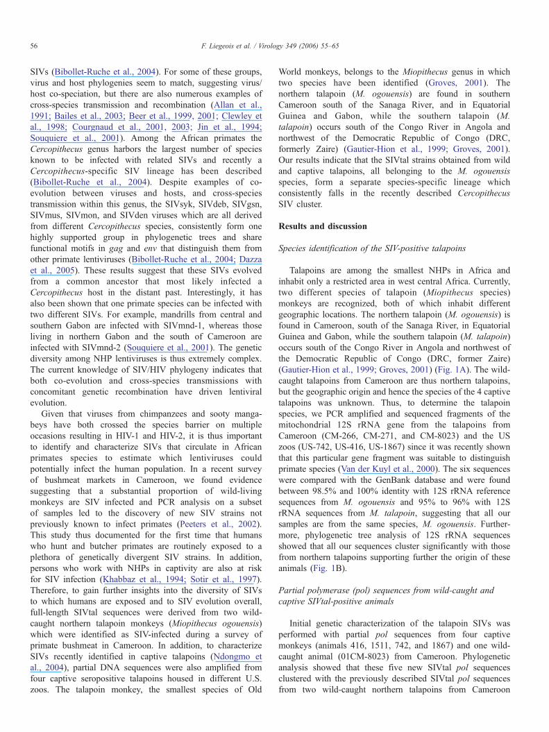
Talapoins are among the smallest NHPs in Africa andinhabit only a restricted area in west central Africa. Currently,two different species of talapoin (Miopithecus species)monkeys are recognized, both of which inhabit differentgeographic locations. The northern talapoin (M. ogouensis) isfound in Cameroon, south of the Sanaga River, in EquatorialGuinea and Gabon, while the southern talapoin (M. talapoin)occurs south of the Congo River in Angola and northwest ofthe Democratic Republic of Congo (DRC, former Zaire)(Gautier-Hion et al., 1999; Groves, 2001) (Fig. 1A). The wild-caught talapoins from Cameroon are thus northern talapoins,but the geographic origin and hence the species of the 4 captivetalapoins was unknown. Thus, to determine the talapoinspecies, we PCR amplified and sequenced fragments of themitochondrial 12S rRNA gene from the talapoins fromCameroon (CM-266, CM-271, and CM-8023) and the USzoos (US-742, US-416, US-1867) since it was recently shownthat this particular gene fragment was suitable to distinguishprimate species (Van der Kuyl et al., 2000). The six sequenceswere compared with the GenBank database and were foundbetween 98.5% and 100% identity with 12S rRNA referencesequences from M. ogouensis and 95% to 96% with 12SrRNA sequences from M. talapoin, suggesting that all oursamples are from the same species, M. ogouensis. Further-more, phylogenetic tree analysis of 12S rRNA sequencesshowed that all our sequences cluster significantly with thosefrom northern talapoins supporting further the origin of theseanimals (Fig. 1B).
Partial polymerase (pol) sequences from wild-caught andcaptive SIVtal-positive animals
Initial genetic characterization of the talapoin SIVs wasperformed with partial pol sequences from four captivemonkeys (animals 416, 1511, 742, and 1867) and one wild-caught animal (01CM-8023) from Cameroon. Phylogeneticanalysis showed that these five new SIVtal pol sequencesclustered with the previously described SIVtal pol sequencesfrom two wild-caught northern talapoins from Cameroon
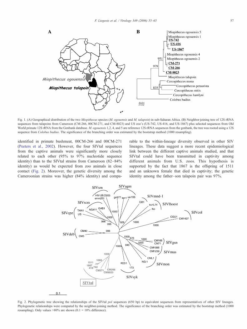
Fig. 1. (A) Geographical distribution of the two Miopithecus species (M. ogouensis and M. talapoin) in sub-Saharan Africa. (B) Neighbor-joining tree of 12S rRNAsequences from talapoins from Cameroon (CM-266, 00CM-271, and CM-8023) and US zoo's (US-742, US-416, and US-1867) plus selected sequences from OldWorld primate 12S rRNA from the Genbank database.M. ogouensis 1,2, 4, and 5 are reference 12S rRNA sequences from the genbank, the tree was rooted using a 12Ssequence from Colobus badius. The significance of the branching order was estimated by the bootstrap method (1000 resampling).
57F. Liegeois et al. / Virology 349 (2006) 55–65
identified in primate bushmeat, 00CM-266 and 00CM-271(Peeters et al., 2002). However, the four SIVtal sequencesfrom the captive animals were significantly more closelyrelated to each other (95% to 97% nucleotide sequenceidentity) than to the SIVtal strains from Cameroon (82–84%identity) as would be expected from zoo animals in closecontact (Fig. 2). Moreover, the genetic diversity among theCameroonian strains was higher (84% identity) and compa-
Fig. 2. Phylogenetic tree showing the relationships of the SIVtal pol sequences (Phylogenetic relationships were computed by the neighbor-joining method. The sigresampling). Only values N80% are shown (0.1 = 10% difference).
rable to the within-lineage diversity observed in other SIVlineages. These data suggest a more recent epidemiologicallink between the different captive animals studied, and thatSIVtal could have been transmitted in captivity amongdifferent animals from U.S. zoos. This hypothesis issupported by the fact that 1867 is the offspring of 1511and an unknown female that died in captivity; the geneticidentity among the father–son talapoin pair was 97%.
650 bp) to equivalent sequences from representatives of other SIV lineages.nificance of the branching order was estimated by the bootstrap method (1000
58 F. Liegeois et al. / Virology 349 (2006) 55–65
Full-length genome sequence and genomic organization ofSIVtal
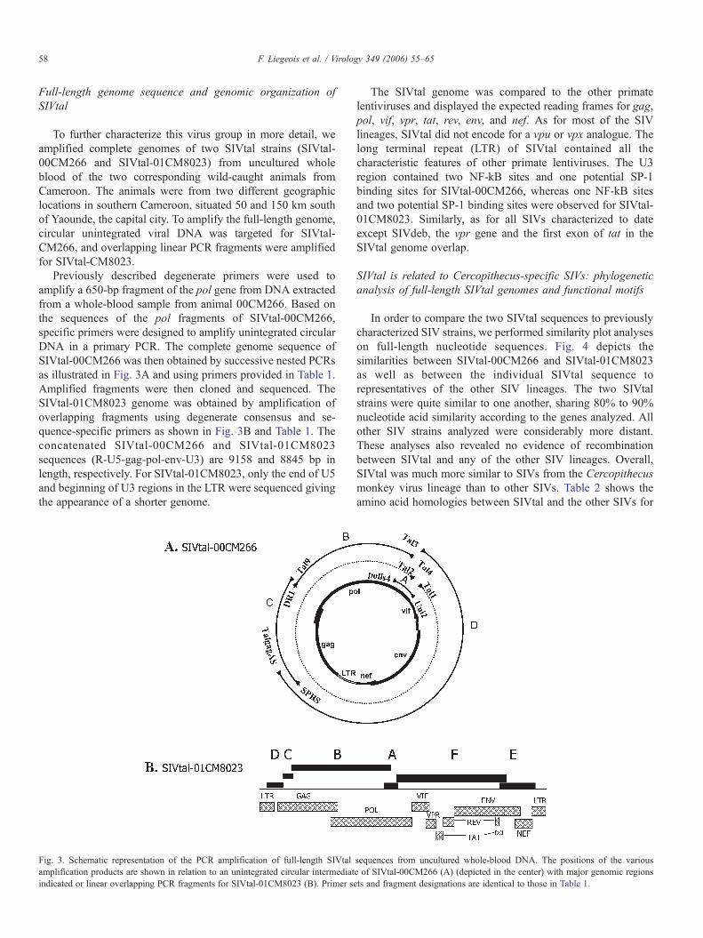
To further characterize this virus group in more detail, weamplified complete genomes of two SIVtal strains (SIVtal-00CM266 and SIVtal-01CM8023) from uncultured wholeblood of the two corresponding wild-caught animals fromCameroon. The animals were from two different geographiclocations in southern Cameroon, situated 50 and 150 km southof Yaounde, the capital city. To amplify the full-length genome,circular unintegrated viral DNA was targeted for SIVtal-CM266, and overlapping linear PCR fragments were amplifiedfor SIVtal-CM8023.
Previously described degenerate primers were used toamplify a 650-bp fragment of the pol gene from DNA extractedfrom a whole-blood sample from animal 00CM266. Based onthe sequences of the pol fragments of SIVtal-00CM266,specific primers were designed to amplify unintegrated circularDNA in a primary PCR. The complete genome sequence ofSIVtal-00CM266 was then obtained by successive nested PCRsas illustrated in Fig. 3A and using primers provided in Table 1.Amplified fragments were then cloned and sequenced. TheSIVtal-01CM8023 genome was obtained by amplification ofoverlapping fragments using degenerate consensus and se-quence-specific primers as shown in Fig. 3B and Table 1. Theconcatenated SIVtal-00CM266 and SIVtal-01CM8023sequences (R-U5-gag-pol-env-U3) are 9158 and 8845 bp inlength, respectively. For SIVtal-01CM8023, only the end of U5and beginning of U3 regions in the LTR were sequenced givingthe appearance of a shorter genome.
Fig. 3. Schematic representation of the PCR amplification of full-length SIVtalamplification products are shown in relation to an unintegrated circular intermediatindicated or linear overlapping PCR fragments for SIVtal-01CM8023 (B). Primer se
The SIVtal genome was compared to the other primatelentiviruses and displayed the expected reading frames for gag,pol, vif, vpr, tat, rev, env, and nef. As for most of the SIVlineages, SIVtal did not encode for a vpu or vpx analogue. Thelong terminal repeat (LTR) of SIVtal contained all thecharacteristic features of other primate lentiviruses. The U3region contained two NF-kB sites and one potential SP-1binding sites for SIVtal-00CM266, whereas one NF-kB sitesand two potential SP-1 binding sites were observed for SIVtal-01CM8023. Similarly, as for all SIVs characterized to dateexcept SIVdeb, the vpr gene and the first exon of tat in theSIVtal genome overlap.
SIVtal is related to Cercopithecus-specific SIVs: phylogeneticanalysis of full-length SIVtal genomes and functional motifs
In order to compare the two SIVtal sequences to previouslycharacterized SIV strains, we performed similarity plot analyseson full-length nucleotide sequences. Fig. 4 depicts thesimilarities between SIVtal-00CM266 and SIVtal-01CM8023as well as between the individual SIVtal sequence torepresentatives of the other SIV lineages. The two SIVtalstrains were quite similar to one another, sharing 80% to 90%nucleotide acid similarity according to the genes analyzed. Allother SIV strains analyzed were considerably more distant.These analyses also revealed no evidence of recombinationbetween SIVtal and any of the other SIV lineages. Overall,SIVtal was much more similar to SIVs from the Cercopithecusmonkey virus lineage than to other SIVs. Table 2 shows theamino acid homologies between SIVtal and the other SIVs for
sequences from uncultured whole-blood DNA. The positions of the variouse of SIVtal-00CM266 (A) (depicted in the center) with major genomic regionsts and fragment designations are identical to those in Table 1.
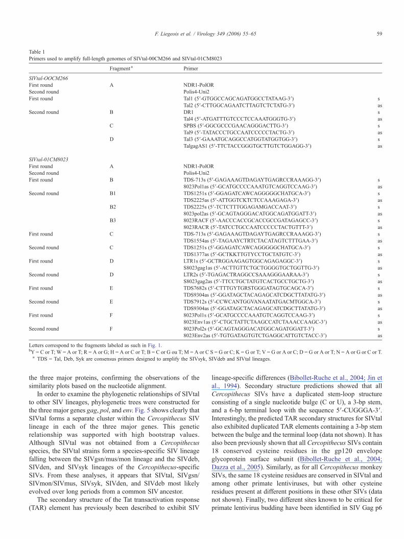
Table 1Primers used to amplify full-length genomes of SIVtal-00CM266 and SIVtal-01CM8023
Fragment a Primer
SIVtal-OOCM266First round A NDR1-PolORSecond round Polis4-Uni2First round Tal1 (5′-GTGGCCAGCAGATGGCCTATAAG-3′) s
Tal2 (5′-CTTGGCAGAATCTTAGTCTCTATG-3′) asSecond round B DR1 s
Tal4 (5′-ATGATTTGTCCCTCCAAATGGGTG-3′) asC SPBS (5′-GGCGCCCGAACAGGGACTTG-3′) s
Tal9 (5′-TATACCCTGCCAATCCCCCTACTG-3′) asD Tal3 (5′-GAAATGCAGGCCATGGTATGGTGG-3′) s
TalgagAS1 (5′-TTCTACCGGGTGCTTGTCTGGAGG-3′) as
SIVtal-01CM8023First round A NDR1-PolORSecond round Polis4-Uni2First round B TDS-713s (5′-GAGAAAGTDAGAYTGAGRCCRAAAGG-3′) s
8023Pol1as (5′-GCATGCCCCAAATGTCAGGTCCAAG-3′) asSecond round B1 TDS1251s (5′-GGAGATCAWCAGGGGGCHATGCA-3′) s
TDS2225as (5′-ATTGGTCKTCTCCAAAGAGA-3′) asB2 TDS2225s (5′-TCTCTTTGGAGAMGACCAAT-3′) s
8023pol2as (5′-GCAGTAGGGACATGGCAGATGGATT-3′) asB3 8023RACF (5′-AACCCACCGCACCGCCGATAGAGCC-3′) s
8023RACR (5′-TATCCTGCCAATCCCCCTACTGTTT-3′) asFirst round C TDS-713s (5′-GAGAAAGTDAGAYTGAGRCCRAAAGG-3′) s
TDS1554as (5′-TAGAAYCTRTCTACATAGTCTTTGAA-3′) asSecond round C TDS1251s (5′-GGAGATCAWCAGGGGGCHATGCA-3′) s
TDS1377as (5′-GCTKKTTGTYCCTGCTATGTC-3′) asFirst round D LTR1s (5′-GCTRGGAAGAGTGGCAGAGAGGC-3′) s
S8023gag1as (5′-ACTTGTTCTGCTGGGGTGCTGGTTG-3′) asSecond round D LTR2s (5′-TGAGACTRAGGCCSAAAGGGAARAA-3′) s
S8023gag2as (5′-TTCCTGCTATGTCACTGCCTGCTG-3′) asFirst round E TDS7682s (5′-CTTTGYTGRSTGGGATAGTGCAGCA-3′) s
TDS9304as (5′-GGATAGCTACAGAGCATCDGCTTATATG-3′) asSecond round E TDS7912s (5′-CCWCANTGGVANAATATGACMTGGCA-3′) s
TDS9304as (5′-GGATAGCTACAGAGCATCDGCTTATATG-3′) asFirst round F 8023Pol1s (5′-GCATGCCCCAAATGTCAGGTCCAAG-3′) s
8023Env1as (5′-CTGCTATTCTAAGCCATCTAAACCAAGC-3′) asSecond round F 8023Pol2s (5′-GCAGTAGGGACATGGCAGATGGATT-3′) s
8023Env2as (5′-TGTGATAGTGTCTGAGGCATTGTCTACC-3′) as
Letters correspond to the fragments labeled as such in Fig. 1.bY = C or T; W = A or T; R = A or G; H = A or C or T; B = C or G ou T; M = A or C S = G or C; K = G or T; V = G or A or C; D = G or A or T; N = A or G or C or T.a TDS = Tal, Deb, Syk are consensus primers designed to amplify the SIVsyk, SIVdeb and SIVtal lineages.
59F. Liegeois et al. / Virology 349 (2006) 55–65
the three major proteins, confirming the observations of thesimilarity plots based on the nucleotide alignment.
In order to examine the phylogenetic relationships of SIVtalto other SIV lineages, phylogenetic trees were constructed forthe three major genes gag, pol, and env. Fig. 5 shows clearly thatSIVtal forms a separate cluster within the Cercopithecus SIVlineage in each of the three major genes. This geneticrelationship was supported with high bootstrap values.Although SIVtal was not obtained from a Cercopithecusspecies, the SIVtal strains form a species-specific SIV lineagefalling between the SIVgsn/mus/mon lineage and the SIVdeb,SIVden, and SIVsyk lineages of the Cercopithecus-specificSIVs. From these analyses, it appears that SIVtal, SIVgsn/SIVmon/SIVmus, SIVsyk, SIVden, and SIVdeb most likelyevolved over long periods from a common SIV ancestor.
The secondary structure of the Tat transactivation response(TAR) element has previously been described to exhibit SIV
lineage-specific differences (Bibollet-Ruche et al., 2004; Jin etal., 1994). Secondary structure predictions showed that allCercopithecus SIVs have a duplicated stem-loop structureconsisting of a single nucleotide bulge (C or U), a 3-bp stem,and a 6-bp terminal loop with the sequence 5′-CUGGGA-3′.Interestingly, the predicted TAR secondary structures for SIVtalalso exhibited duplicated TAR elements containing a 3-bp stembetween the bulge and the terminal loop (data not shown). It hasalso been previously shown that all Cercopithecus SIVs contain18 conserved cysteine residues in the gp120 envelopeglycoprotein surface subunit (Bibollet-Ruche et al., 2004;Dazza et al., 2005). Similarly, as for all Cercopithecus monkeySIVs, the same 18 cysteine residues are conserved in SIVtal andamong other primate lentiviruses, but with other cysteineresidues present at different positions in these other SIVs (datanot shown). Finally, two different sites known to be critical forprimate lentivirus budding have been identified in SIV Gag p6
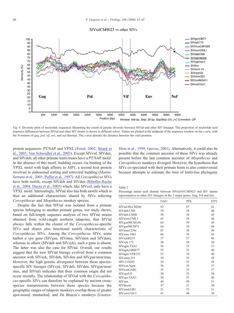
Fig. 4. Diversity plots of nucleotide sequences illustrating the extent of genetic diversity between SIVtal and other SIV lineages. The proportion of nucleotide acidsequence differences between SIVtal and other SIV strains is shown in different colors. Values are plotted at the midpoint of the sequence window on the x-axis, withthe N-termini of gag, pol, vif, env, and nef denoted. The y-axis denotes the distance between the viral proteins.
Table 2Percentage amino acid identity between SIVtal-01CM8023 and SIV strainsrepresentative to other SIV lineages in the 3 major genes, Gag, Pol and Env
GAG POL ENV
SIVtal-00-CM266 83 87 81SIVdeb.CM5 59 58 40SIVdeb.CM40 58 58 42SIVmon.CML1 65 58 46SIVgsn99CM166 64 59 45SIVgsn99CM71 64 59 44SIVmus1239s 65 58 46SIVmus.1085 66 58 45SIVsykKE51 57 57 46SIVsyk.173 58 58 48SIVagm.TAN1 56 55 37SIVagm.GRI677 55 53 40SIVagm.VER155 53 54 40SIVsmm.251 54 53 38HIV-2-D205 54 53 38SIVrcm.Ngm 56 55 37SIVrcmGAB1 55 55 37SIVcpzUS 50 52 38SIVcpz.TAN1 50 53 40SIVsun 46 49 31SIVlhoest 47 51 30SIVmnd.GB1 51 53 31SIVcol-CGU1 41 48 26
60 F. Liegeois et al. / Virology 349 (2006) 55–65
protein sequences: PT/SAP and YPXL (Freed, 2002; Strack etal., 2003; Von Schwedler et al., 2003). Except SIVcol, SIVden,and SIVdeb, all other primate lentiviruses have a PT/SAP motif.In the absence of this motif, budding occurs via binding of theYPXL motif with high affinity to AIP1, a second host proteininvolved in endosomal sorting and retroviral budding (Martin-Serrano et al., 2001; Puffer et al., 1997). All Cercopithecus SIVshave both motifs, except SIVdeb and SIVden (Bibollet-Rucheet al., 2004; Dazza et al., 2005) which, like SIVcol, only have aYPXL motif. Interestingly, SIVtal also has both motifs which isthus an additional characteristic shared by SIVs infectingCercopithecus and Miopithecus monkey species.
Despite the fact that SIVtal was isolated from a primatespecies belonging to another primate genus, our study shows,based on full-length sequence analysis of two SIVtal strainsobtained from wild-caught northern talapoins, that SIVtalalways falls within the cluster of the Cercopithecus-specificSIVs and shares also functional motifs characteristic ofCercopithecus SIVs. Among the Cercopithecus SIVs, someharbor a vpu gene (SIVgsn, SIVmus, SIVmon and SIVden),whereas in others (SIVdeb and SIVsyk), such a gene is absent.The latter was also the case for SIVtal. Overall, our resultssuggest that the new SIVtal lineage evolved from a commonancestor with SIVsyk, SIVdeb, SIVden and SIVgsn/mon/mus.However, the high genetic divergence between these species-specific SIV lineages (SIVsyk, SIVdeb, SIVden, SIVgsn/mon/mus, and SIVtal) indicates that their common origin did notoccur recently. The relationship of SIVtal with the Cercopithe-cus-specific SIVs can therefore be explained by ancient cross-species transmissions between these species because thegeographic ranges of talapoin monkeys overlap those of greaterspot-nosed, mustached, and De Brazza's monkeys (Gautier-
Hion et al., 1999; Groves, 2001). Alternatively, it could also bepossible that the common ancestor of these SIVs was alreadypresent before the last common ancestor of Miopithecus andCercopithecus monkeys diverged. However, the hypothesis thatSIVs co-speciated with their primate hosts is also controversialbecause attempts to estimate the time of lentivirus phylogeny
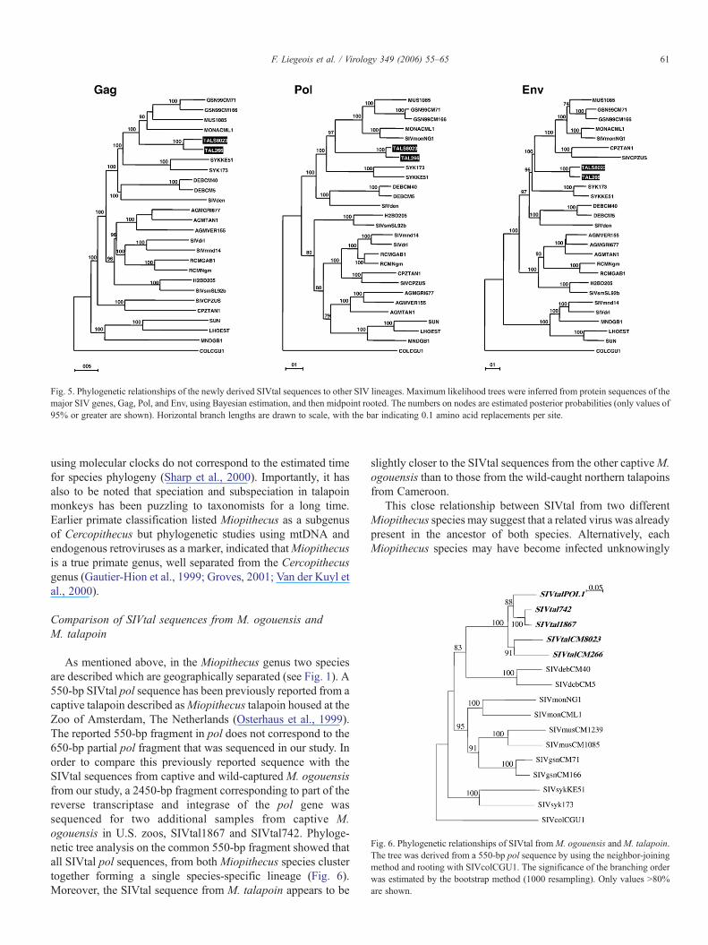
Fig. 5. Phylogenetic relationships of the newly derived SIVtal sequences to other SIV lineages. Maximum likelihood trees were inferred from protein sequences of themajor SIV genes, Gag, Pol, and Env, using Bayesian estimation, and then midpoint rooted. The numbers on nodes are estimated posterior probabilities (only values of95% or greater are shown). Horizontal branch lengths are drawn to scale, with the bar indicating 0.1 amino acid replacements per site.
Fig. 6. Phylogenetic relationships of SIVtal fromM. ogouensis andM. talapoin.The tree was derived from a 550-bp pol sequence by using the neighbor-joiningmethod and rooting with SIVcolCGU1. The significance of the branching orderwas estimated by the bootstrap method (1000 resampling). Only values N80%are shown.
61F. Liegeois et al. / Virology 349 (2006) 55–65
using molecular clocks do not correspond to the estimated timefor species phylogeny (Sharp et al., 2000). Importantly, it hasalso to be noted that speciation and subspeciation in talapoinmonkeys has been puzzling to taxonomists for a long time.Earlier primate classification listed Miopithecus as a subgenusof Cercopithecus but phylogenetic studies using mtDNA andendogenous retroviruses as a marker, indicated thatMiopithecusis a true primate genus, well separated from the Cercopithecusgenus (Gautier-Hion et al., 1999; Groves, 2001; Van der Kuyl etal., 2000).
Comparison of SIVtal sequences from M. ogouensis andM. talapoin
As mentioned above, in the Miopithecus genus two speciesare described which are geographically separated (see Fig. 1). A550-bp SIVtal pol sequence has been previously reported from acaptive talapoin described asMiopithecus talapoin housed at theZoo of Amsterdam, The Netherlands (Osterhaus et al., 1999).The reported 550-bp fragment in pol does not correspond to the650-bp partial pol fragment that was sequenced in our study. Inorder to compare this previously reported sequence with theSIVtal sequences from captive and wild-captured M. ogouensisfrom our study, a 2450-bp fragment corresponding to part of thereverse transcriptase and integrase of the pol gene wassequenced for two additional samples from captive M.ogouensis in U.S. zoos, SIVtal1867 and SIVtal742. Phyloge-netic tree analysis on the common 550-bp fragment showed thatall SIVtal pol sequences, from both Miopithecus species clustertogether forming a single species-specific lineage (Fig. 6).Moreover, the SIVtal sequence from M. talapoin appears to be
slightly closer to the SIVtal sequences from the other captiveM.ogouensis than to those from the wild-caught northern talapoinsfrom Cameroon.
This close relationship between SIVtal from two differentMiopithecus species may suggest that a related virus was alreadypresent in the ancestor of both species. Alternatively, eachMiopithecus species may have become infected unknowingly
62 F. Liegeois et al. / Virology 349 (2006) 55–65
with the same virus by close contact in a zoo exhibit shared byboth species. The close genetic relationship of these virusessupports the latter hypothesis. Nevertheless, identification ofSIV-infected, wild-captured southern talapoins and full molec-ular characterization of their corresponding viral genomes areneeded to fully elucidate the diversity and evolution of SIVs inthis genus.
In this paper, we showed that northern talapoins arenaturally infected with a new species-specific primatelentivirus, termed SIVtal. Phylogenetic tree analysis shows asignificant clustering of the SIVtal viruses with the Cerco-pithecus-specific SIVs. Overall, our data add further evidenceof the complex evolutionary history of primate lentiviruses.Full-length sequences of SIVs from additional Cercopithecusspecies and from other nonhuman primate genera are neededfor a better understanding of the overall evolution of primatelentiviruses. More sequences from the same species butsampled at distant geographic locations are also necessary tounderstand whether the identified viruses are the real species-specific SIVs or whether they represent a strain naturallyinfecting sympatric primate species that were introduced bycross-species transmission. Further studies of the lentivirusesin NHP species are not only required to understand the originand evolution of primate lentiviruses but may also yieldimportant insights into the risks for human exposure to SIV-infected primates and the potential for the introduction of newretroviral zoonoses into the general population. African NHPspecies represent a large reservoir for simian lentiviruses, andwe recently showed that humans are exposed to a largediversity of SIV through hunting and handling of primatebushmeat (Peeters et al., 2002). Although talapoins are lessfrequently hunted due to their small size, humans may also beexposed to SIVs from these monkeys.
Materials and methods
Animals and serologic testing
Whole-blood samples from two wild-caught and fourcaptive talapoin monkeys, previously identified as SIV-positive by the presence of cross-reacting antibodies withHIV antigens using INNOLIA-LIA HIV confirmation test(Innogenetics, Ghent, Belgium) (Peeters et al., 2002) orpeptide EIA and Western Blots (Ndongmo et al., 2004), wereavailable for study. The two talapoins from Cameroon(00CM-266 and 02CM-8023) were obtained from wild-caughtanimals sampled as bushmeat with government approval fromthe Cameroonian Ministry of Environment and Forestry aspreviously described (Peeters et al., 2002). Blood specimensfrom four talapoins (416 (female), 1867 (male), 1511 (male),and 742 (female) housed in North American zoos wereobtained on an opportunistic basis in accordance with theanimal care and use committees at each institution. Of note,1867 is the offspring of 1511 and a female (1009) that died incaptivity and 416 and 742 both had wild-born sires.Peripheral blood mononuclear cells (PBMC) and plasmawere obtained by Ficoll-Hypaque centrifugation.
PCR amplification and sequencing of pol
Total DNAwas isolated from whole blood or PBMCs usingthe QIAamp blood kit (Qiagen) according to the manufacturer'sinstructions. A small fragment of pol (650 bp) was amplified forall samples with degenerate consensus primers (DR1-PolOR forthe first round and Polis4-UNIPOL2 for the second round) aspreviously described (Clewley et al., 1998; Courgnaud et al.,2001; Peeters et al., 2002). For a subset of samples, we alsoamplified a 2450-bp pol fragment, using DR1-PolOR primersfor the first round and DR4-UNIPOL2 for the second round andby using PCR conditions as previously reported (Courgnaud etal., 2003). After purification, the PCR products were thendirectly sequenced on both strands.
Amplification of complete SIVtal genomes
For two animals, 00CM-266 and 01CM-8023, the full-lengthSIVtal sequence was obtained either by amplification ofunintegrated circular DNA or by overlapping PCR fragments,respectively. The amplification strategies and primers used toobtain the different fragments are summarized in Fig. 3 andTable 1. For SIVtal-00CM266, a 650-bp pol fragment wasobtained as described above. To obtain the full-length genomicsequence of SIVtal-00CM266, two specific primers weregenerated based on the known pol sequence to amplify in thefirst round unintegrated circular viral forms: Tal1 and Tal2.Several nested PCRs were then performed to generate over-lapping fragments spanning the entire genome, using combina-tions of SIVtal-266-specific and/or SIV consensus primers:DR1 and Tal4, SPBS positions 248–267 and Tal9; positions2358–2381, and Tal3 positions 4568–4591, and TalgagAS1positions 576–600. PCRs were performed using a Long Expandor Expand High Fidelity PCR kit (Roche Molecular Biochem-icals), including a hot start (92 °C for 3 min) with the followingcycle conditions: 10 cycles of denaturation at 92 °C for 10 s,annealing at 56 °C for 30-s extension at 68 °C for 7 min,followed by 20 cycles with extension at 68 °C for 7 min with anincrement of 20 s per cycle. Amplification was completed by afinal extension at 68 °C for 10 min. Cycling conditions for innerPCRs were 10 cycles of denaturation at 94 °C for 15 s,annealing at 55 °C (with primers SPBS and Tal9 or at 58 °Cwith primers Tal3 and TalgagAS1) for 30-s extension at 72 °Cfor 2.5 min or 3.5 min, followed by 20 cycles with an incrementof 5 s per cycle of the extension time at 72 °C, followed by 1cycle of 7-min extension at 72 °C. PCR products were purifiedand cloned into pGEM-TEasy vector (Promega). DNA wassequenced using cycle sequencing and dye terminator method-ologies (ABI PRISM Big Dye Terminator Cycle SequencingReady Reaction kit with AmpliTaq FS DNA polymerase (PEBiosystems, Warrington, England) on an automated sequencer(ABI 373, model Stretch, Applied Biosystems) using theGenome Priming System GPS™-1 (New England BioLabs,Beverly, MA, USA). To reconstitute the full-length genomesequence, overlapping sequences were assembled into contig-uous sequences by using the Sequencher software (Gene CodesCorp., USA).
63F. Liegeois et al. / Virology 349 (2006) 55–65
For SIVtal-CM8023, unintegrated circular DNA could notbe amplified, and the full-length sequence was obtained byamplification of overlapping PCR fragments with consensusprimers designed on the newly obtained SIVtal-00CM266sequence and other SIVs available from the database andGenBank. The primers used are shown in Table 1, and thecorresponding amplification strategy is depicted in Fig. 3B. Allamplifications were performed using the Expand High FidelityPCR kit (Roche Applied Science, Indianapolis, IN) according tothe manufacturer's instructions. Each amplification reactionincluded a manual hot-start and 35 cycles. Annealingtemperatures were 50 °C except for the amplification offragments B and D for which 53 °C was used. Extensiontimes varied depending on the size of the expected fragment andwere typically set at 1 min/kb. Amplified fragments wereagarose gel purified and directly sequenced using Big-DyeChemistry (Applied Biosystems, France) according to themanufacturer's instructions. Electrophoresis and data collectionwere done on an Applied Biosystems 3100 Genetic Analyzer.To reconstitute the full-length genome sequence, overlappingsequences were assembled into contiguous sequences by usingSEQMAN DNASTAR software (Lasergene, DNASTAR, Inc.,Madison, WI).
Talapoin species identification
To determine the species of talapoin monkeys, we amplifiedin a single round PCR, a 390-bp segment of the mitochondrial12S rRNA gene using primers 12S-L01091/12S-H01478 asdescribed previously (Van der Kuyl et al., 2000). PCR productswere then directly sequenced and compared to the GenBankdatabase by using the BLAST program. Phylogenetic relation-ships of the primate mitochondrial 12S rRNA sequences wereestimated using the neighbor-joining method with the Kimura's2 parameter model implemented in the Clustal X program(Thompson et al., 1997). The reliability of branching orders wastested using the bootstrap approach (1000 replicates).
Genetic analyses
Nucleotide and protein sequences were aligned usingClustalX, with minor manual adjustments. Sites that could notbe unambiguously aligned and sites with a gap in any sequencewere excluded. Proteome sequences were generated by joiningdeduced Gag, Pol, Vif, Env, and Nef amino acid sequences; thecarboxy terminal Gag, Pol, and Env amino acid sequences thatoverlapped with Pol, Vif, and Nef amino acid sequences,respectively, were excluded. The nucleotidic and predictedprotein sequences encoded by SIVtal were compared torepresentatives of known SIV/HIV lineages. In order to studywhether the newly characterized SIVtal sequences wererecombinant with any of the other SIV lineages, similarityplot analysis was performed on the nucleotide alignment withthe new SIVtal sequences and known SIV strains with theSIMPLOT package version 2.5 (Ray, 1999; http://www.med.jhu.edu/deptmed/sray) using a sliding window of 400 nucleo-tides (nt) moved in steps of 20 nt.
Phylogenetic tree analysis was done on amino acid sequences.Maximum likelihood trees were inferred by Bayesian estima-tion of phylogeny, based upon the posterior probabilitydistribution of trees. The method was implemented inMrBayesv3.1 (Huelsenbeck et al., 2001) using the Jones–Taylor and Thornton model of protein evolution (Jones et al.,1992) with gamma distributed rates at sites (Ziheng, 1993). Theprogram was run for 500,000 generations, including a “burn in”of 500 generations. The trees shown are majority rule consensustrees.
Secondary structure predictions
The TAR RNA secondary structure was predicted and drawnusing the GENQUEST DNASTAR package (Lasergene,DNASTAR, Inc., Madison, WI).
Nucleotide sequence accession number
The complete sequences (SIVtal-00CM266 and SIVtal-01CM8023) and partial pol sequences (SIVtal742, SIVtal1867,SIVtal416, and SIVtal1511) are available GenBank (accessionnumbers AY655740-AY655744 and AM182197).
Acknowledgments
This work was supported in part by grants from the NationalInstitute of Health (RO1 AI 50529) and the Agence National deRecherche sur le SIDA (ANRS). We thank the CameroonianMinistries of Health, Research, and Environment and Forestryand Wildlife, for the permission to perform this study, and thestaff from project PRESICA for the logistical support andassistance in the field. We also thank the veterinary andadministrative staff at the Audubon Zoo for providing additionaltalapoin specimens.
References
Allan, J.S., Short, M., Taylor, M.E., Su, S., Hirsch, V.M., Johnson, P.R., Shaw,G.M., Hahn, B.H., 1991. Species-specific diversity among simianimmunodeficiency viruses from African green monkeys. J. Virol. 65 (6),2816–2828.
Bailes, E., Chaudhuri, R.R., Santiago, M.L., Bibollet-Ruche, F., Hahn, B.H.,Sharp, P.M., 2002. The evolution of primate lentiviruses and the origin ofAIDS. In: Leitner, T.A. (Ed.), The Molecular Epidemiology of HumanViruses. Kluwer Academic Publishers, Boston, MA, pp. 65–96.
Bailes, E., Gao, F., Bibollet-Ruche, F., Courgnaud, V., Peeters, M., Marx, P.A.,Hahn, B.H., Sharp, P.M., 2003. Hybrid origin of SIV in chimpanzees.Science 300 (5626), 1713.
Beer, B.E., Bailes, E., Goeken, R., Dapolito, G., Coulibaly, C.,Norley, S.G., Kurth, R., Gautier, J.P., Gautier-Hion, A., Vallet, D., Sharp,P.M., Hirsch, V.M., 1999. Simian immunodeficiency virus (SIV) from sun-tailed monkeys (Cercopithecus solatus): evidence for host-dependentevolution of SIV within the C. lhoesti superspecies. J. Virol. 73 (9),7734–7744.
Beer, B.E., Foley, B.T., Kuiken, C.L., Tooze, Z., Goeken, R.M., Brown, C.R.,Hu, J., St Claire, M., Korber, B.T., Hirsch, V.M., 2001. Characterizationof novel simian immunodeficiency viruses from red-capped manga-beys from Nigeria (SIVrcmNG409 and -NG411). J. Virol. 75 (24),12014–12027.
64 F. Liegeois et al. / Virology 349 (2006) 55–65
Bibollet-Ruche, F., Bailes, E., Gao, F., Pourrut, X., Barlow, K.L.,Clewley, J.P., Mwenda, J.M., Langat, D.K., Chege, G.K., McClure,H.M., Mpoudi-Ngole, E., Delaporte, E., Peeters, M., Shaw, G.M.,Sharp, P.M., Hahn, B.H., 2004. New simian immunodeficiency virusinfecting De Brazza's monkeys (Cercopithecus neglectus): evidencefor a Cercopithecus monkey virus clade. J. Virol. 78 (14),7748–7762.
Clewley, J.P., Lewis, J.C., Brown, D.W., Gadsby, E.L., 1998. A novel simianimmunodeficiency virus (SIVdrl) pol sequence from the drill monkey,Mandrillus leucophaeus. J. Virol. 72 (12), 10305–10309.
Corbet, S., Muller-Trutwin, M.C., Versmisse, P., Delarue, S., Ayouba, A., Lewis,J., Brunak, S., Martin, P., Brun-Vezinet, F., Simon, F., Barre-Sinoussi, F.,Mauclere, P., 2000. Env sequences of simian immunodeficiency virusesfrom chimpanzees in Cameroon are strongly related to those of humanimmunodeficiency virus group N from the same geographic area. J. Virol. 74(1), 529–534.
Courgnaud, V., Pourrut, X., Bibollet-Ruche, F., Mpoudi-Ngole, E., Bourgeois,A., Delaporte, E., Peeters, M., 2001. Characterization of a novel simianimmunodeficiency virus from guereza colobus monkeys (Colobus guereza)in Cameroon: a new lineage in the nonhuman primate lentivirus family.J. Virol. 75 (2), 857–866.
Courgnaud, V., Formenty, P., Akoua-Koffi, C., Noe, R., Boesch, C., Delaporte,E., Peeters, M., 2003. Partial molecular characterization of two simianimmunodeficiency viruses (SIV) from African colobids: SIVwrc fromWestern red colobus (Piliocolobus badius) and SIVolc from olive colobus(Procolobus verus). J. Virol. 77 (1), 744–748.
Dazza, M.C., Ekwalanga, M., Nende, M., Shamamba, K.B., Bitshi, P.,Paraskevis, D., Saragosti, S., 2005. Characterization of a novel vpu-harboring simian immunodeficiency virus from a Dent's Mona monkey(Cercopithecus mona denti). J. Virol. 79 (13), 8560–8571.
Freed, E.O., 2002. Viral late domains. J. Virol. 76 (10), 4679–4687.Gao, F., Yue, L., White, A.T., Pappas, P.G., Barchue, J., Hanson, A.P., Greene,
B.M., Sharp, P.M., Shaw, G.M., Hahn, B.H., 1992. Human infection bygenetically diverse SIVSM-related HIV-2 in West Africa. Nature 358(6386), 495–499.
Gao, F., Bailes, E., Robertson, D.L., Chen, Y., Rodenburg, C.M., Michael, S.F.,Cummins, L.B., Arthur, L.O., Peeters, M., Shaw, G.M., Sharp, P.M., Hahn,B.H., 1999. Origin of HIV-1 in the chimpanzee Pan troglodytes troglodytes.Nature 397 (6718), 436–441.
Gautier-Hion, A., Colyn, M., Gautier, JP., 1999. Histoire naturelle des primatesd'Afrique centrale. Ecofac Multipress, Gabon.
Groves, C., 2001. Primate Taxonomy. Smithsonian Series in ComparativeEvolutionary Biology. Smithsonian Institution Press.
Huelsenbeck, J.P., Ronquist, F., Nielsen, R., Bollback, J.P., 2001. Bayesianinference of phylogeny and its impact on evolutionary biology. Science 294(5550), 2310–2314.
Jin, M.J., Hui, H., Robertson, D.L., Muller, M.C., Barre-Sinoussi, F., Hirsch,V.M., Allan, J.S., Shaw, G.M., Sharp, P.M., Hahn, B.H., 1994. Mosaicgenome structure of simian immunodeficiency virus from West Africangreen monkeys. EMBO J. 13 (12), 2935–2947.
Jones, D.T., Taylor, W.R., Thornton, J.M., 1992. The rapid generation ofmutation data matrices from protein sequences. Comput. Appl. Biosci. 8 (3),275–282.
Kestens, L., Vingerhoets, J., Peeters, M., Vanham, G., Vereecken, C., Penne, G.,Niphuis, H., van Eerd, P., van der Groen, G., Gigase, P.a., Heeney, J., 1995.Phenotypic and functional parameters of cellular immunity in a chimpanzeewith a naturally acquired simian immunodeficiency virus infection. J. Infect.Dis. 172 (4), 957–963.
Khabbaz, R.F., Heneine, W., George, J.R., Parekh, B., Rowe, T., Woods, T.,Switzer, W.M., McClure, H.M., Murphey-Corb, M., Folks, T.M., 1994.Brief report: infection of a laboratory worker with simian immunodeficiencyvirus. N. Engl. J. Med. 330 (3), 172–177.
Martin-Serrano, J., Zang, T., Bieniasz, P.D., 2001. HIV-1 and Ebola virusencode small peptide motifs that recruit Tsg101 to sites of particle assemblyto facilitate egress. Nat. Med. 7 (12), 1313–1319.
Muller-Trutwin, M.C., Corbet, S., Tavares, M.D., Herve, V.M., Nerrienet, E.,Georges-Courbot, M.C., Saurin, W., Sonigo, P., Barre-Sinoussi, F., 1996.The evolutionary rate of nonpathogenic simian immunodeficiency virus
(SIVagm) is in agreement with a rapid and continuous replication in vivo.Virology 223 (1), 89–102.
Ndongmo, C.B., Switzer, W.M., Pau, C.P., Zeh, C., Schaefer, A., Pieniazek, D.,Folks, T.M., Kalish, M.L., 2004. New multiple antigenic peptide-basedenzyme immunoassay for detection of simian immunodeficiency virusinfection in nonhuman primates and humans. J. Clin. Microbiol. 42 (11),5161–5169.
Osterhaus, A.D., Pedersen, N., van Amerongen, G., Frankenhuis, M.T.,Marthas, M., Reay, E., Rose, T.M., Pamungkas, J., Bosch, M.L., 1999.Isolation and partial characterization of a lentivirus from talapoin monkeys(Myopithecus talapoin). Virology 260 (1), 116–124.
Pandrea, I., Onanga, R., Kornfeld, C., Rouquet, P., Bourry, O., Clifford, S.,Telfer, P.T., Abernethy, K., White, L.T., Ngari, P., Muller-Trutwin, M.,Roques, P., Marx, P.A., Simon, F., Apetrei, C., 2003. High levels ofSIVmnd-1 replication in chronically infected Mandrillus sphinx. Virology317 (1), 119–127.
Peeters, M., Courgnaud, V., 2002. Overview of primate lentiviruses and theirevolution of non-human primates in Africa. In: KuikenC., Foley B., Freed E.,Hahn B., Korber B., Marx P.A., McCutchan F., Mellors J.W., Wollinsky S.(Eds), HIV Sequence Compendium 2002. Theoretical Biology andBiophysics Group, Los Alamos National Laboratory, Los Alamos, N.M.2–23.
Peeters, M., Courgnaud, V., Abela, B., Auzel, P., Pourrut, X., Bibollet-Ruche, F.,Loul, S., Liegeois, F., Butel, C., Koulagna, D., Mpoudi-Ngole, E., Shaw,G.M., Hahn, B.H., Delaporte, E., 2002. Risk to human health from aplethora of simian immunodeficiency viruses in primate bushmeat. Emerg.Infect. Dis. 8 (5), 451–457.
Puffer, B.A., Parent, L.J., Wills, J.W., Montelaro, R.C., 1997. Equine infectiousanemia virus utilizes a YXXL motif within the late assembly domain of theGag p9 protein. J. Virol. 71 (9), 6541–6546.
Ray, S., 1999. SImPLot version 2.5 software; http://www.med.jhu.edu/deptmed/sray/.
Reed, P., Souquiere, S., Telfer, P., Makuwa, M., Metzger, M., Ela-Mba, MA.,Roques, P., Marx, P., 2004. Molecular characterization of a new primatelentivirus (SIVery) from Cercopithecus erythroitis tissue samples fromBioko, Equatorial Guinea. 11th International Workshop, HIV Dynamics andEvolution. Stockholm.
Sharp, P.M., Bailes, E., Gao, F., Beer, B.E., Hirsch, V.M., Hahn, B.H., 2000.Origins and evolution of AIDS viruses: estimating the time-scale. Biochem.Soc. Trans. 28 (2), 275–282.
Silvestri, G., Sodora, D.L., Koup, R.A., Paiardini, M., O'Neil, S.P.,McClure, H.M., Staprans, S.I., Feinberg, M.B., 2003. NonpathogenicSIV infection of sooty mangabeys is characterized by limited bystanderimmunopathology despite chronic high-level viremia. Immunity 18 (3),441–452.
Sotir, M., Switzer, W., Schable, C., Schmitt, J., Vitek, C., Khabbaz, R.F., 1997.Risk of occupational exposure to potentially infectious nonhuman primatematerials and to simian immunodeficiency virus. J. Med. Primatol. 26 (5),233–240.
Souquiere, S., Bibollet-Ruche, F., Robertson, D.L., Makuwa, M., Apetrei, C.,Onanga, R., Kornfeld, C., Plantier, J.C., Gao, F., Abernethy, K., White, L.J.,Karesh, W., Telfer, P., Wickings, E.J., Mauclere, P., Marx, P.A., Barre-Sinoussi, F., Hahn, B.H., Muller-Trutwin, M.C., Simon, F., 2001. WildMandrillus sphinx are carriers of two types of lentivirus. J. Virol. 75 (15),7086–7096.
Strack, B., Calistri, A., Craig, S., Popova, E., Gottlinger, H.G., 2003. AIP1/ALIX is a binding partner for HIV-1 p6 and EIAV p9 functioning in virusbudding. Cell 114 (6), 689–699.
Takemura, T., Ekwalanga, M., Bikandou, B., Ido, E., Yamaguchi-Kabata, Y.,Ohkura, S., Harada, H., Takehisa, J., Ichimura, H., Parra, H.J., Nende,M., Mubwo, E., Sepole, M., Hayami, M., Miura, T., 2005. A novelsimian immunodeficiency virus from black mangabey (Lophocebusaterrimus) in the Democratic Republic of Congo. J. Gen. Virol. 86(Pt. 7), 1967–1971.
Thompson, J.D., Gibson, T.J., Plewniak, F., Jeanmougin, F., Higgins, D.G.,1997. The CLUSTAL_X windows interface: flexible strategies for multiplesequence alignment aided by quality analysis tools. Nucleic Acids Res. 25(24), 4876–4882.

65F. Liegeois et al. / Virology 349 (2006) 55–65
Van der Kuyl, A.C., Dekker, J.T., Goudsmit, J., 2000. Primate genusMiopithecus: evidence for the existence of species and subspecies ofdwarf guenons based on cellular and endogenous viral sequences. Mol.Phylogenet. Evol. 14 (3), 403–413.
Verschoor, E.J., Fagrouch, Z., Bontjer, I., Niphuis, H., Heeney, J.L., 2004. Anovel simian immunodeficiency virus isolated from a Schmidt's guenon(Cercopithecus ascanius schmidti). J. Gen. Virol. 85 (Pt. 1), 21–24.
Von Schwedler, U.K., Stuchell, M., Muller, B., Ward, D.M., Chung, H.Y.,Morita, E., Wang, H.E., Davis, T., He, G.P., Cimbora, D.M., Scott, A.,Krausslich, H.G., Kaplan, J., Morham, S.G., Sundquist, W.I., 2003. Theprotein network of HIV budding. Cell 114 (6), 701–713.
Ziheng, Y., 1993. Maximum-likelihood estimation of phylogeny from DNAsequences when substitution rates differ over sites. Mol. Biol. Evol. 10 (6),1396–1401.