Neural activity at the human olfactory epithelium reflects olfactory perception
Upload
khangminh22Category
view
6download
0
International Journal of
Molecular Sciences
Review
Molecular and Genetic Factors Involved in Olfactory andGustatory Deficits and Associations with Microbiota inParkinson’s Disease
Melania Melis 1 , Antje Haehner 2, Mariano Mastinu 1, Thomas Hummel 2 and Iole Tomassini Barbarossa 1,*
�����������������
Citation: Melis, M.; Haehner, A.;
Mastinu, M.; Hummel, T.; Tomassini
Barbarossa, I. Molecular and Genetic
Factors Involved in Olfactory and
Gustatory Deficits and Associations
with Microbiota in Parkinson’s
Disease. Int. J. Mol. Sci. 2021, 22, 4286.
https://doi.org/10.3390/ijms22084286
Academic Editor: Yuzuru Imai
Received: 26 March 2021
Accepted: 17 April 2021
Published: 20 April 2021
Publisher’s Note: MDPI stays neutral
with regard to jurisdictional claims in
published maps and institutional affil-
iations.
Copyright: © 2021 by the authors.
Licensee MDPI, Basel, Switzerland.
This article is an open access article
distributed under the terms and
conditions of the Creative Commons
Attribution (CC BY) license (https://
creativecommons.org/licenses/by/
4.0/).
1 Department of Biomedical Sciences, University of Cagliari, Monserrato, 09042 Cagliari, Italy;[email protected] (M.M.); [email protected] (M.M.)
2 Smell and Taste Clinic, Department of Otorhinolaryngology, Technical University of Dresden, 01307 Dresden,Germany; [email protected] (A.H.); [email protected] (T.H.)
* Correspondence: [email protected]; Tel.: +39-070-675-4144
Abstract: Deficits in olfaction and taste are among the most frequent non-motor manifestations inParkinson’s disease (PD) that start very early and frequently precede the PD motor symptoms. Thelimited data available suggest that the basis of the olfactory and gustatory dysfunction related to PDare likely multifactorial and may include the same determinants responsible for other non-motorsymptoms of PD. This review describes the most relevant molecular and genetic factors involved inthe PD-related smell and taste impairments, and their associations with the microbiota, which alsomay represent risk factors associated with the disease.
Keywords: Parkinson’s disease; smell; taste
1. Introduction
Parkinson’s disease (PD) is a chronic neurodegenerative disorder with a prevalence of1% in subjects aged 60–69, increasing to 3% in those over 80 years of age [1]. Pathologically,the disease is characterized by dopaminergic neuronal loss in the substantia nigra, and itis associated with intracellular inclusions, called Lewy bodies, in the neurons of affectedbrain regions. The Lewy bodies are intra-cytoplasmic eosinophilic deposits of a misfoldedprotein, α-synuclein which spreads to different regions of the brain in a prion-like fashion,giving rise to the successive non-motor and motor symptoms [2–4]. Clinically, PD is charac-terized by the presence of motor symptoms such as bradykinesia (slow movement), rigidity,tremor, postural instability (balance problems), difficulty with walking, and coordination.In addition, PD is also characterized by the occurrence of several non-motor symptoms suchas sleep disturbances, apathy, anxiety, autonomic dysfunction, gastrointestinal dysfunction(such as nausea, dysphagia, abnormal salivation, constipation and defecatory dysfunc-tion [5,6]), cognitive impairment, olfactory and gustatory dysfunctions [7,8]. Specifically,olfactory dysfunction is accepted to be an early biomarker of the disease since precedesthe occurrence of clinical motor symptoms [9], with an incidence raging between 50% and96% [10–12]. Although the occurrence of taste dysfunctions in PD is less clear, they havebeen described as non-motor manifestations of the early stages of PD [13–15], highlightingtheir role in the diagnosis of the disease.
Taste and olfaction play a key role in individuals’ behaviors, their interactions with theenvironment and memory processes [16], furthermore, they represent the most importantfactors influencing food preferences and therefore eating behavior and diet [17,18]. Infact, they cooperate and enables organisms to distinguish nutrient-rich food from noxioussubstances, and acts as a final checkpoint for food acceptance or rejection behavior [19,20];therefore, it is not surprising that disorders in these two sensory modalities can havesignificant effects on the life’s quality [21].
Int. J. Mol. Sci. 2021, 22, 4286. https://doi.org/10.3390/ijms22084286 https://www.mdpi.com/journal/ijms
Int. J. Mol. Sci. 2021, 22, 4286 2 of 19
The purpose of this review is to summarize the knowledge about smell and tastedisorders in idiopathic PD, describing the most relevant molecular and genetic factorsinvolved in the smell and taste impairments related with PD. We also describe the principalassociations with microbiota. Accounting for these factors, could lead to a more preciseassessment which would greatly help clinicians in the early diagnosis of PD.
2. Functions and Dysfunctions of Taste2.1. Taste System
In the common language, the word “taste” is often used to describe sensations arisingfrom the oral cavity. However, in biology the sense of taste includes all sensations mediatedby a chemosensory gustatory system specialized anatomically and physiologically [22].The molecular mechanisms underlying the perception of taste include the reception andsignal transduction mechanisms, which play important roles in the oral cavity and also ina diversity of tissues including the respiratory and gastrointestinal tracts, kidney and evenbrain [23].
Taste sensation begins with the activation of taste receptor cells (TRCs), which areorganized in taste buds located mostly on the superior surface of the tongue. The receptionand transduction mechanisms of taste stimuli are located at the chemosensory apical tip ofthe TRCs. The generated signals are transmitted, via three cranial nerves (CN) (facial, VII;glossopharyngeal, IX and vagus, X), to the rostral part of the solitary tract nucleus (NST)of the medulla. The projections from the NST include parabrachial nucleus, thalamus(ventral posteromedial nucleus), gustatory areas of the cortex in the insula, amygdala,hypothalamus and basal ganglia [24].
It is generally assumed that human taste sensations can be divided into five qualities:bitter, sour, salty, sweet, and umami. Recently, the fat taste has been acknowledged as asixth primary sensory quality [25–28].
2.2. Chemoreceptors, Receptor Genes and Taste
Chemoreceptors of the plasma membrane of TRCs interact with specific chemicalstimuli to initiate an afferent signal to the brain, which results in taste perception. Special-ized chemoreceptors mediate specific coding mechanisms for different taste stimuli andprovide the basis for discrimination across taste qualities [29]. In humans, the chemorecep-tion of sweet, umami, and bitter taste involves membrane proteins from the TAS1R andTAS2R families, which belong to a superfamily of G protein–coupled receptors (GPCRs).Various receptors for detection of long chain fatty acids have been proposed [30,31], includ-ing CD36 [27,32,33]. Candidate chemoreceptors have been suggested for salty and sourtaste qualities [34–38]. The existence of several chemoreceptors reflect the importance ofdistinguishing beneficial from harmful chemicals of the environment [39].
Progress with understanding of the interaction between taste stimuli and chemorecep-tors and the identifying of patterns of their expression in taste cells sheds light on codingof taste information by the nervous system [22]. Variations in taste receptor genes affectexpression and function of taste receptors, and therefore influence taste function [40]. Itis well known that variation in taste receptor genes can result in differences of the sweet,umami and bitter perception, while less is known about the genetics of sour and saltytaste [41]. The TAS2R38 gene, codifying for receptor binding the bitter-tasting thioureacompounds such as PROP and PTC, is one of the most studied in this field [42]. Variationsin TAS2R38 gene greatly contribute to the thiourea taster groups: super-tasters, mediumtasters and non-tasters [43,44].
2.3. Extra-Gustatory Taste Receptors
As stated above, taste receptors and signal transduction molecules for sweet, bitter,and umami tastes are expressed not only in TRCs of oral cavity, but also in cells of a varietyof extra-oral tissues throughout the body, including the brain [23]. The functions of thisinternal chemoreceptors have only been partially elucidated. However, it possible to state

Int. J. Mol. Sci. 2021, 22, 4286 3 of 19
that they detect chemical compounds of internal environment and that modifications ofthis internal chemo-sensation can affect physiological functions [45]. Specifically, TAS2Rs,which detect bitter compounds, mediate several non-tasting functions and their geneticvariants are associated with diverse disorders [46]. Brain neurons have been shown torespond to different chemicals [47]. Bitter TAS2Rs are expressed in multiple regions of therat brain, TAS2R4, TAS2R107 and TAS2R38 are expressed in the brain stem, cerebellum,cortex, and nucleus accumbens and calcium signaling showed the functionality of T2R4expressed in these cells [48].
2.4. Taste Dysfunction in Neurogenerative Disease (Overview)
Taste dysfunctions are described as ageusia (complete loss of taste), hypogeusia (par-tial loss of taste), parageusia (inadequate or wrong taste perception) and phantogeusia(presence of a persistent and unpleasant taste) [49]. Taste disorders are generally associatedto medical conditions, pharmacologic or surgical interventions, exposure to toxic chemicals,head injury, advanced age or neurodegenerative diseases [14,50–58]. Over recent years,the link between taste dysfunctions and neurodegenerative disorders has increasinglybeen recognized. Some authors showed patients with Alzheimer’s disease (AD) to reportesignificant reduction of taste function, by showing an increase of the detection threshold ofthe four basic tastes (sweet, salty, sour, and bitter) [53,54]. However, others reported nodifference in detection threshold of sucrose [59,60] and sour [59] or total absence of tastealterations [61]. In a case study, Petzold et al. [62] indicated that patients with amyotrophiclateral sclerosis (ALS) reported a persistent bitter or metallic taste (phantogeusia), althoughno hypogeusia for taste qualities were observed. Tarlarini and colleagues showed reductionof taste and its negative consequences on psychological status and quality of life in ALSpatients [63]. Taste disorders also have been described as a prominent early feature inCreutzfeldt–Jakob disease (CJD) which is one of prion diseases, a group of neurodegener-ative disorders, characterized by accumulation of abnormal prion proteins in the centralnervous system. In 2001, Reuber and colleagues describe for the first time a patient withCJD whose first symptoms included deficits of taste and smell [64].
2.5. Taste Impairments in PD
In recent years several studies evaluated gustatory function in PD patients [14,50,51,65–68],but reporting inconsistent results. This may because they were carried out by using smallsample size or different assessment methods: whole mouth test (WMT), supra-threshold tastesolutions sprayed into the oral cavity [69]; taste strip test, (TST), in which patients had to identifya taste from a taste strip [70,71] and electrogustometry (EGM), rapid measure of taste thresholdby using electric current as stimulus) [72,73].
Despite the different tests adopted by the research groups, it is generally reportedthat taste can be affected in PD patients by showing persistent, but slight and stable tasteimpairments [74]. In particular, most of the studies identified a reduced taste sensitivitywith an estimated frequency between 9% and 27% [14,50–52]. Shah et al. [51], using EGM,found that about 27% of PD patients had an impaired taste function. Taste thresholdsmeasured in the front and back of the tongue were higher in PD patients, than in healthycontrols (HC), suggesting significant deficits in CN VII and CN IX. Deeb et al. [50] by usingEGM showed that about 22% of PD patients had impaired taste function. Kim et al. [14] byusing TSTs reported a decrease in the ability to identify tastants in female but not in malePD patients when compared to HC. Cecchini et al. [68] reported difference between PDpatients and HC in taste performance assessed by the TST, but not by WMT. In fact, onlythe TST score was significantly lower in PD patients than HC. The reason of the fact thatWMT do not show reduction of taste could be due to the use of stimuli at supra-thresholdconcentration, which are not able to capture slight impairment of taste function.
Doty et al. [13] studied whole-mouth (WMT) and regional taste perception of early-stage PD patients and HC matched on the basis of age, sex, and race. They reported that theWMT scores were lower in the PD patients than in controls (for all four taste stimuli), and
Int. J. Mol. Sci. 2021, 22, 4286 4 of 19
the intensity ratings for the weaker concentrations of all stimuli, except caffeine, tended tobe higher in the PD patients than in HC. This last finding is consistent with the findings ofSienkiewicz- Jarosz and co-workers who demonstrated that, in the WMT test, PD patientsrated quinine [65] and sucrose as more intense than HC [66]. Moreover, Doty et al. [13]using regional tests showed that subjects tended to better identify and rate the stimuli asmore intense on the front than in the back of the tongue with respect to controls. Thesefindings suggest that the suprathreshold measures of taste function are influenced by PDwhich differentially influences taste function on CN VII and CN IX. These results are notobserved if the taste techniques are limited to WM. In addition, in the same study [13]EGM was not able to observe differences between the PD patients and controls. In addition,a reduced identification of sweet [75], salty or bitter stimuli was found [13]. Despite theslightly controversial results, it appears that taste is affected in PD, although less frequentlythan smell. However, future investigations are necessary to explore the causes of tasteimpairments related to PD.
It is interesting to note that the taste loss has been related mostly to the advanced stagesof the disease [50], whereas reports on prodromal presentation are rare. Pont-Sunyer andcolleagues [76] observed that the time of the taste loss onset varied between 2 and 10 yearsbefore diagnosis. Taste loss was present before the onset of motor symptoms in morethan 70% of PD patients, providing evidence for a very-early onset of taste loss, which iscomparable to that of olfactory impairments. Therefore, the evaluation of the taste functionmay be used in combination with that olfactory as a potential marker of PD. However, it isknown that anosmics are more poorly able to taste than normal persons [77–79].
2.6. Role of Taste Receptors in PD
The role of taste and smell receptors in PD has been investigated showing that thecortical olfactory receptors (ORs) and the TAS2Rs are altered in PD patients [80]. Olfactoryreceptors OR2L13, OR1E1, OR2J3, OR52L1, and OR11H1 and taste receptors TAS2R5and TAS2R50 were downregulated, whereas TAS2R10 and TAS2R13 were upregulated,at premotor and parkinsonian stages, in the frontal cortex area 8 of the brains in PDpatients [80]. These findings support the idea that ORs and TA2SRs in the cerebral cortexmay have physiologic functions that are affected in PD patients. The identification ofaltered regulation of OR and TAS2R in PD patients, suggests the study of the chemicalsignaling system of the brain to understand the mechanisms involved in the occurrence ofthe neurodegenerative diseases. Future studies will have to point out whether the alteredTAS2R may play a role in the inflammatory mechanisms associated with the initiation ofmisfolding of the α-synuclein cascade.
2.7. Relationships between TAS2R38 and Taste Dysfunction in PD
TAS2R38 has been associated with a variety of non-tasting physiological mecha-nisms [17,42,46,81–85]. The allelic diversity of the gene codifying for TAS2R38 resultsin three non-synonymous coding single nucleotide polymorphisms (SNPs), which giverise to two major variants: the functional form containing proline, alanine and valine(haplotype named PAV) and the non-functional variant containing alanine, valine andisoleucine (haplotype named AVI) [44,86]. TAS2R38 SNPs dictate individual differences inPTC/PROP tasting [44,87,88], food linking patterns [82,89] and also in TAS2R38-mediatedpathophysiology [46], such as susceptibility, severity, and prognosis of upper respiratoryinfection, rhinosinusitis and biofilm formation in chronic rhinosinusitis patients [90–97],development of colonic neoplasm [98–100], taste disorders [101], and neurodegenerativediseases [102].
In the following paragraphs, we focus on TAS2R38 polymorphisms, the relative abilityto perceive the bitter taste of thiourea compounds and its association with microbiota, as agenetic risk factors for development of PD.
Moberg and colleagues were the first that examine PTC sensitivity in PD patients andHC to determine whether taster status can be a marker for PD. They showed significant

Int. J. Mol. Sci. 2021, 22, 4286 5 of 19
differences in the distribution of taster and non-taster subjects between the PD patients HC.They showed that only 44% of PD patients could detect the bitterness of PTC, as comparedto 75% of HC [103]. Cossu et al. [102] confirmed the result showing a reduced of PROPtaste sensitivity in PD patients compared to HC. Specifically, a decreased perceived tasteintensity and reduced ability to recognize bitter-taste quality was found. They also showedan increase in the frequency of the PD patients classified as PROP non-tasters (54.13%) anda decrease in frequency of PD patients classified as PROP super-tasters (8.25%) comparedto HC. Furthermore, the results showed that the homozygous genotype for the tastingvariant of TAS2R38 (PAV) was uncommon in PD patients, only 5% of them carried thisgenotype, whereas most of them carried the non-taster form (AVI). These results seem toindicate that individuals who have a couple of tasting haplotypes (PAV/PAV) at TAS2R38may be at lower risk of developing PD, with respect to those with the haplotype (AVI).Therefore, the latter might represent a prodromal genetic marker for the identification ofearly pre-degenerative changes that could be instrumental to understand the origin of thisdisorder. Thus, studying the PROP phenotype and genotype may represent a new, simpleway to identify increased predisposition for PD.
2.8. Role of Microbiota on Relationships between TAS2R38 and Taste Dysfunction in PD
PD has been associated with the dysbiosis of gut microbiota [104] and imbalance in gutmicrobiota plays an important role in worsening of disease [105–108]. Specific taste receptors,expressed in the lower gastrointestinal tract (GI), respond to change of the composition of gutmicrobiota and regulate immune responses against pathogens [46,109–111]. In particular, it isknown that when TAS2R38 expressed in the enteroendocrine cells of the gut is activated bybacterial molecules, it increases the release of β-defensin (an anti-microbial compound) [46] anda peptide hormone termed cholecystokinin (CCK). This hormone can limit the absorption ofdietary toxins [112,113], inhibit feeding behavior and gastric function [114–116] and it also canplay a key role in regulating the immune response to antigens and bacterial toxins [117]. Thus,the response of TAS2R38 represents an important defense of the organism in contrasting thenoxious effects in the gut lumen.
Vascellari et al. [118] showed that the composition of the gut microbiota was differentacross genotypes of TAS2R38 in PD patients. Specifically, a decrease in bacteria alpha-diversity with a predominant reduction of Clostridium genus was associated with AVI/AVIgenotype, compared to the PAV/PAV genotype. It is important to mention that somemembers of Clostridium genus produce toxin [119], while other members confer beneficialeffects which has a multitude of metabolic function in the GI tract, such as modulation ofgastrointestinal motility, barrier integrity and immune response [119–121]. Therefore, adecrease in the abundance of helpful-Clostridium molecules associated to a high frequencyof the form of TAS2R38 receptor at a low affinity for the ligands might determine, in PD,a decrease in the activation of protective signaling-molecules involved in the regulationof the immune response. This factor could affect different cellular processes which areimpaired in PD, thereby contributing to the development of gut dysbiosis [118].
3. Functions and Dysfunctions of Smell3.1. Olfactory System
Olfactory perception starts at the level of the olfactory epithelium in the roof of thenasal cavity. Olfactory receptor neurons (ORN) send their axons towards the olfactorybulbs. ORN carry olfactory receptors (OR; approximately 400 different OR can be expressedby humans). In the olfactory bulb ORN axons synapse with second order neurons, themitral cells. All ORN carrying the same OR converge in the same glomerulus in thebulb. Axons from the mitral cells form the olfactory tract which directly connects to theprimary olfactory cortex (including piriform and entorhinal cortices or the amygdalae).The secondary olfactory cortex includes structures such as the hippocampus, the anteriorinsula or the orbitofrontal cortex [122].

Int. J. Mol. Sci. 2021, 22, 4286 6 of 19
The olfactory system has 3 major functions [18]: (1) food intake control (e.g., lo-calization, appetite), (2) social communication (e.g., reproductive behavior, detection offear-related cues, recognition of kin, recognition of disease), and (3) detection of danger(e.g., toxicity, fires, prevention of food poisoning).
3.2. Smell Dysfunction in Neurogenerative Disease (Overview)
Impaired olfaction has been associated with a variety of age-related neurodegenerativeconditions that impair cognitive and motor function, including PD [123,124], AD [125],and Huntington’s disease [126]. Smell loss may therefore be considered an importantcontribution to the diagnosis of neurodegenerative diseases. In PD, olfactory loss hasbeen extensively studied and is now widely acknowledged as one of the major non-motorsymptoms of the disease which precedes the occurrence of clinical motor symptoms [127].A variety of psychophysical methods were used to evaluate orthonasal olfactory functionin PD, mainly based on the identification of suprathreshold odors (UPSIT [128]) or on ancomprehensive approach divided into a threshold, a discrimination, and an identificationpart, with the last two being suprathreshold odors (Sniffin’ Sticks [129]). Olfactory dis-turbances are found in around 90% of patients with PD [11] and have been consideredas a supportive criterion in clinical PD diagnosis according to the International Parkin-son’s Disease and Movement Disorder Society diagnostic criteria [130]. The majority ofPD patients with smell loss are already functionally anosmic or severely hyposmic at thetime of testing regardless of the type of olfactory test being used for diagnosis. Wenninget al. [131] presented data suggesting that olfactory function is differentially impaired indistinct Parkinsonian syndromes. They reported a preserved or mildly impaired olfactoryfunction to be more likely for atypical parkinsonism such as multiple system atrophy, pro-gressive supranuclear palsy, or corticobasal degeneration, whereas markedly pronouncedolfactory loss appeared to suggest PD. Similar results were reported by Müller et al. [132]and Krismer et al. [133]. In dementias, the loss of smell is usually very severe. This appliesto Lewy body disease (LBD), in which significant olfactory deficits were found [134,135]which does not allow differentiation from PD. Similar olfactory deficits have been shown inAD. In a meta-analysis by Mesholam et al. [125], olfactory deficits in patients with AD andPD were relatively uniform although there was a trend toward better performance in ADpatients on threshold tests compared to odor identification tests. Smell loss can be observedin patients with mild cognitive impairment [136] and is associated with the progressionfrom MCI to AD [137]. Huntington´s disease patients present with moderate hyposmiaaffecting olfactory detection threshold, odor discrimination and odor identification [126].Deficits in odor identification are prevalent prior to diagnosis of HD [138]. In patientswith cerebellar ataxia, olfactory impairment was found in Friedreich’s ataxia [139] andspinocerebellar ataxias [140–142]. Mild olfactory impairment has also been demonstratedin motor neuron disease [143,144].
3.3. Smell Impairments as a Biomarker for Early Onset, Progression, Cognitive Decline andDifferential Diagnosis in PD
Support for the existence of a prodromal phase of PD, including a long pre-motorphase, comes from imaging, neuropathology, and various clinical or epidemiologicalsurveys. Loss of smell is recognized as a very early non-motor symptoms of PD and hasbeen suggested as a possible biomarker [145]. Several population-based studies alreadypointed out the association between unexplained smell loss and later development ofPD. Our data of a large, thoroughly diagnosed patient cohort study of a Smell and TasteClinic suggest a 10% rate of PD development among patients with diagnosed idiopathicolfactory loss [146]. The duration of the hyposmic phase prior to PD diagnosis is still amatter of debate. In many previous studies investigating the prospective risk for PD inrelation to baseline [147–149] follow-up periods ranged from 2 to a maximum of 8 years.We could demonstrate that the olfactory dysfunction frequently precedes the PD motorsymptoms by more than 10 years [146]. Accordingly, other authors indicated an incidentof PD beyond 5 years of follow-up [150,151]. Additionally, studies that used retrospective
Int. J. Mol. Sci. 2021, 22, 4286 7 of 19
patients’ self-reports reported the onset of olfactory dysfunction on average more than10 years before PD diagnosis [76,152]. Other authors assumed that this period may lastup to decades [153]. In a follow-up study of patients with idiopathic REM sleep behaviordisorder who phenoconverted to PD or dementia, olfactory loss was the first marker todevelop, with predicted onset >20 years before phenoconversion [154].
Furthermore, current studies indicate a correlation between olfactory function andprogression of the disease as measured by motor and other non-motor symptoms. An asso-ciation between disease severity and smell loss [50,155–158] and a disease duration-relatedprogression of olfactory loss [157] might suggest the use of olfactory function as potentialmarker of PD progression. This was confirmed by an imaging study using Dat-SPECT,indicating that a more pronounced olfactory dysfunction was associated with greater lossof nigrostriatal dopamine neurons [159]. Additionally, non-motor symptoms such as cogni-tive impairment, depression, anxiety and sleep disturbances which are typically relatedto PD severity are associated with the degree of olfactory loss [155,159,160]. The closecorrelation between smell function and cognitive impairment is reflected by the results ofthe Parkinson’s Progression Markers Initiative study [161] which indicated that olfactoryloss is one of the strongest clinical predictor of cognitive impairment in the first 2 yearsafter PD diagnosis. Decline in cognition seems to be linked to progressive cholinergicdenervation in PD as described by Bohnen et al. [162] who found a positive correlationbetween odor identification performance and forebrain cholinergic pathway integrity inPD patients.
3.4. Neuropathology of Smell Dysfunction in PD
The olfactory system could be one of the peripheral sites where PD first develops [4].However, there is little and inconsistent information on changes at the olfactory periph-ery. While α-synuclein aggregates (Lewy bodies and neurites) have been described in theolfactory bulb (OB) at early neuropathological stages of the disease, α-synuclein was notdetected in olfactory epithelium biopsies of PD patients [163], it was found however, inolfactory cells in PD autopsy cases [164]. Further, in-vivo examinations of the olfactoryepithelium revealed histological changes comparable to other causes of smell loss [163]which suggest non-specific peripheral changes in the olfactory system in PD. On the OBlevel, PD seems to differ from other causes of olfactory loss. In etiologies involving pe-ripheral olfactory loss, such as postinfectious or sinonasal smell disorders [165,166], butalso in more central pathologies such as depression [167], schizophrenia [168], and tem-poral lobe epilepsy [169], a clear and consistent correlation between olfactory functionand OB volume can be observed. These data suggest that smell loss is associated with ameasurable OB volume loss in these pathologies. In PD, however, despite of the severityof olfactory impairment it remains a matter of debate whether PD patients present withdecreased OB volumes compared to age-matched controls. So far, a number of recentstudies have reported conflicting results: while some studies [170,171] reported an overallreduction of the OB volume in PD, the vast majority of studies [172–175] question anyOB volume differences between PD and HC. This is in line with findings of an increasednumber of olfactory dopaminergic periglomerular cells in PD patients [176,177] whichmight underlie hyposmia in PD patients. However, in central regions related to bothprimary (piriform cortex, amygdala) and secondary integrative (orbitofrontal cortex) ol-factory processing, a significant atrophy was found in PD which correlated with olfactoryperformance [178–180]. This might suggest that central olfactory areas in PD seem to repre-sent the degree of disease progression, whereas this correlation is not seen in peripheralolfactory structures.
3.5. Molecular and Genetic Mechanisms Involved in Olfactory Deficit in PD
The causes of olfactory dysfunction in PD are poorly understood, but it is supposedthat they are related with both peripheral and central olfactory impairments [181]. Themechanisms implicated in the smell impairments in PD may involve neuropathological

Int. J. Mol. Sci. 2021, 22, 4286 8 of 19
alterations and/or dysfunctions caused by alteration in the neurotransmitter levels [182].The importance of these mechanisms is addressed in the following paragraphs.
The olfactory system is one of the earliest brain regions involved in PD before in-volvement of the nigrostriatal pathway [4,183]. The α-synuclein deposition, predominantcomponent of Lewy bodies [184,185], have been identified before in the olfactory bulb,anterior olfactory nucleus, and several areas of olfactory cortices of PD patients [186,187],than in the substantia nigra [4,188]. The α-synuclein pathology appears before in theolfactory nerve layer and then it spread to the central olfactory structures [189]. However,the involvement of the olfactory epithelium on olfactory loss in PD have not been welldefined. In fact, no significant difference was found by immunohistochemical markersfor α-synuclein between PD patients and HC [163,190]. These findings suggested that thechanges in the olfactory function in PD may be due to processes associated with formationof Lewy bodies in the central olfactory areas and not in the peripheral ones [124,191]. Theα-synuclein pathology has been revealed across the central olfactory system, includingthe anterior olfactory nucleus, cortical nucleus of the amygdala, piriform cortex, olfactorytubercle, entorhinal cortex, and orbitofrontal cortex [191,192]. In particular, the Lewypathology in the anterior olfactory nucleus of the olfactory bulb is correlated with neuronalloss [193]. Furthermore, the olfactory nerves were grossly atrophic in all PD patients [193].The cortical nucleus of the amygdala, which has major olfactory connections, have moreα-synuclein pathology and neuronal loss than other nuclei in the amygdala [194], conse-quently its volume is reduced by 20% [194]. Moreover, the reduced volume in the amygdalaand piriform cortex inversely correlates with olfactory deficits, suggesting that neural lossin these regions could play a role on the olfactory impairments of PD [178,179].
Several neurotransmitter systems are altered in PD and most of them have been asso-ciated with olfactory loss, including dopaminergic, cholinergic and serotoninergic systems.Dopamine has long been known to play a key role in the pathogenesis of PD. Some studiessuggested that the olfactory dysfunction of PD patients could reflect damage to dopamin-ergic cells [195]. As a matter of fact, correlations between odor identification tests (UPSITscores) and a decrease in dopamine transporter activity in the striatum, substantia nigra andhippocampus in PD patients have been found [50,196,197]. However, the use of dopamin-ergic replacement therapy has no effect on olfactory test scores [198,199]. Nevertheless, it isstill not known whether changes in dopamine activity are directly associated with olfactoryloss or whether there is an unknown common underlying mechanism. Acetylcholine levelsare also altered in PD. It is known that acetylcholine release and activation of its receptorsfacilitate olfactory learning, memory, and odor discrimination [200–203]. Thus, cholinergicdeficits may be responsible, at least in part, for the olfactory dysfunction in PD. It has beenfound that in PD the Lewy bodies and neuronal loss in the substantia nigra occur simultane-ously with accumulation of the α-synuclein deposition in cholinergic neurons of the basalforebrain [4,204–206]. Furthermore, the nucleus basalis, a main cholinergic nucleus withprojection to olfaction-related brain regions, is significantly damaged in PD [207–209]. Inaddition, Bohnen and colleagues found a positive association between odor identificationperformance and acetylcholinesterase activity in PD patients [162]. Serotonin is anotherneurotransmitter with a possible role in the pathogenesis of olfactory dysfunction in PD. Itarises from the raphe nuclei, which send projections to the olfactory bulb [210–212]. In PDpatients, Lewy pathology is found in the raphe nuclei [213], parallel to marked depletionof serotonin in the olfactory bulb and other areas of the olfactory system [214–216], whilea relative protection of serotonin was found in other diseases without important olfactoryimpairments [217]. Although the evidence is not conclusive so far, these studies suggest thatchanges in levels of some neurotransmitters may be implicated in the olfactory loss in PD.
Polymorphisms of specific genes coding for membrane receptors or odorant bindingproteins (OBPs) (carrier proteins that vehicle the molecules toward receptor sites [218]),have been reported as mechanisms that result in the functional variations of olfactoryfunction [219–222]. Recently, the polymorphism rs2590498 (A/G) of the OBPIIa gene hasbeen shown to affect retronasal [219] and olfactory [223] perception. Subjects with the A

Int. J. Mol. Sci. 2021, 22, 4286 9 of 19
allele were generally more sensitive than those with the G allele. Moreover, bioinformaticsdata suggested that the presence of the mutation in this locus decreases the expressionof OBPIIa protein in the olfactory epithelium [224]. The same polymorphism affected theolfactory performance of woman with PD [224]. Specifically, the olfactory performanceof women with PD carrying two sensitive alleles (AA) was higher than that of womenwith PD with at least one insensitive allele (G) and of all men with PD. Interestingly, theolfactory scores of the AA genotype women with PD were not different from those ofHC participants. These findings indicate that the AA homozygous condition in this locuspreserves the olfactory function of women with PD, but not that of men. Furthermore,these results indicate that the smell dysfunction related to PD may occur, at least in part, ata peripheral level. Therefore, OBPIIa locus may provide a mechanism to determine the riskfactor for olfactory deficits in woman with PD at the molecular level.
3.6. Microbiota and Olfactory Deficit in PD
The associations between PD and the olfactory dysfunction and the composition of gutmicrobiota are unequivocal [225,226]. However, results on a role of the nasal microbiomein olfactory dysfunction in PD are not conclusive. A recent study has shown that thereare no significant differences in the nasal microbiome composition between PD patientsand HC [227]. However, the incapacity to collect samples in the olfactory cleft did notrule out the existence of differences in the microbial composition around the olfactoryneuroepithelium. In addition, high spatial variability of microbial communities in thenasal cavity can exist [228]. Additionally, another study did not find consistent differencein the nasal microbiota composition between PD patients and HC, even though a highinterindividual variability was observed, with sex as the strongest factor [229]. Futurestudies in which samples of nasal microbiome are collected in the olfactory cleft are neededto understand its role in olfactory dysfunction of PD patients.
4. Conclusions
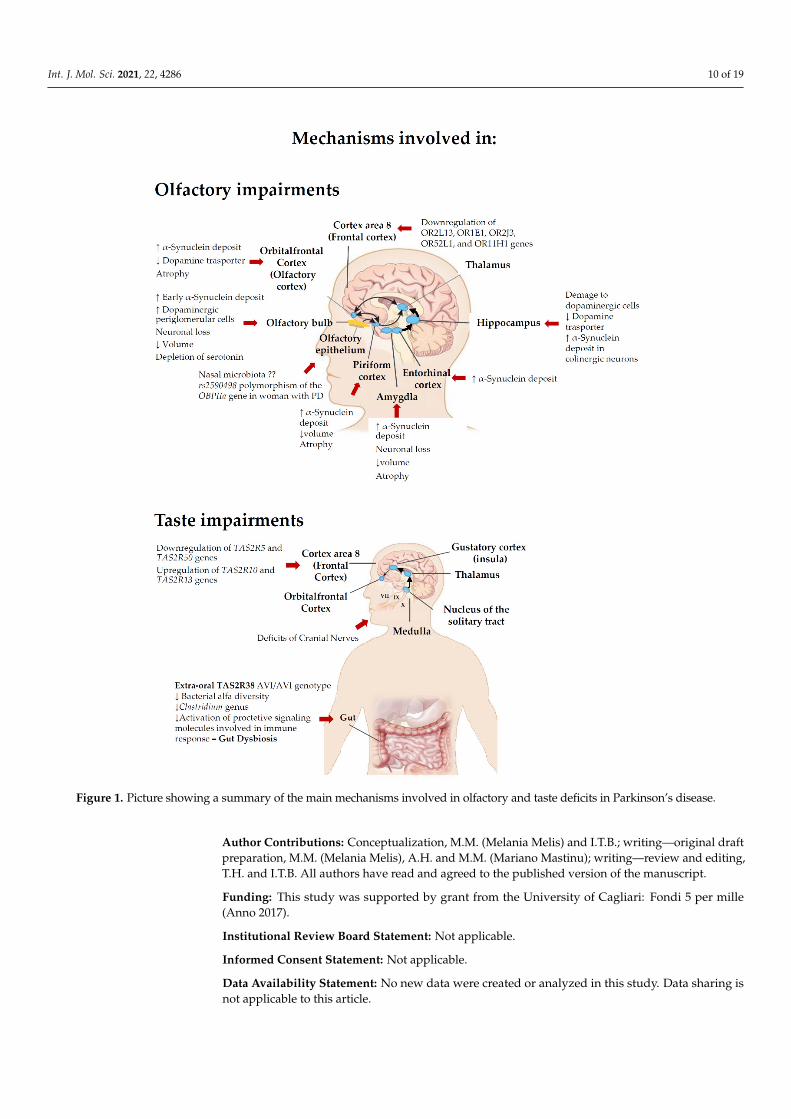
In conclusion, this review examined the influence of smell and taste disorders inPD, describing the most relevant factors such as genetic factors, microbiome and someneuropathological mechanisms which appear to be involved in the PD-related smell andtaste impairments (Figure 1). The underlying causes or their relative degree of impact arestill not conclusive, thus further pre-clinical and clinical research on cellular and molecularmechanisms underlying these dysfunctions in PD are required.
Our review confirms the relevance of smell and taste in the PD patients showingthat smell and taste dysfunction are non-motor manifestations of PD that may impairthe quality of life. Albeit the symptom “olfactory loss” has been approved as a helpfulmeasure in clinical PD diagnosis according to the new International Parkinson’s Diseaseand Movement Disorder Society diagnostic criteria [130], the use of taste as a biomarkeris not yet included in the diagnosis of PD. Nevertheless, it is known that in many of thepatients, taste loss accompanies smell dysfunction [146], thus testing these two sensoryfunctions together would help clinicians in early diagnosis of PD enhancing the predictivevalue for diagnosis of disease.
Smell and taste impairment in PD are connected to different anatomical pathways.As mentioned above, the α-synuclein pathology involves the central olfactory systemand smell impairment is often detectable years before the onset of motor symptoms ofPD. Differently the pathology at the basis of taste dysfunction is less understood andthe nucleus tractus solitarius is usually not involved by Lewy body pathology even inthe latest stages of the disease [4]. However, such pathology does occur in the anteriorinsular/operculum region, which is an important relay station for axons connected to theorbitofrontal cortex [230,231]. Consequently, taste impairment in PD probably depends onthe involvement of the cortex in the neurodegenerative process.
Int. J. Mol. Sci. 2021, 22, 4286 10 of 19Int. J. Mol. Sci. 2021, 22, 4286 10 of 19
Figure 1. Picture showing a summary of the main mechanisms involved in olfactory and taste deficits in Parkinson’s disease.
Our review confirms the relevance of smell and taste in the PD patients showing that smell and taste dysfunction are non-motor manifestations of PD that may impair the qual-ity of life. Albeit the symptom “olfactory loss” has been approved as a helpful measure in clinical PD diagnosis according to the new International Parkinson’s Disease and Move-ment Disorder Society diagnostic criteria [130], the use of taste as a biomarker is not yet included in the diagnosis of PD. Nevertheless, it is known that in many of the patients, taste loss accompanies smell dysfunction [146], thus testing these two sensory functions together would help clinicians in early diagnosis of PD enhancing the predictive value for diagnosis of disease.
Smell and taste impairment in PD are connected to different anatomical pathways. As mentioned above, the α-synuclein pathology involves the central olfactory system and
Figure 1. Picture showing a summary of the main mechanisms involved in olfactory and taste deficits in Parkinson’s disease.
Author Contributions: Conceptualization, M.M. (Melania Melis) and I.T.B.; writing—original draftpreparation, M.M. (Melania Melis), A.H. and M.M. (Mariano Mastinu); writing—review and editing,T.H. and I.T.B. All authors have read and agreed to the published version of the manuscript.
Funding: This study was supported by grant from the University of Cagliari: Fondi 5 per mille(Anno 2017).
Institutional Review Board Statement: Not applicable.
Informed Consent Statement: Not applicable.
Data Availability Statement: No new data were created or analyzed in this study. Data sharing isnot applicable to this article.

Int. J. Mol. Sci. 2021, 22, 4286 11 of 19
Acknowledgments: Melania Melis gratefully acknowledges the Sardinia Regional Government forthe financial support: P.O.R. SARDEGNA F.S.E. 2014–2020—Asse III “Istruzione e Formazione”,Obiettivo Tematico: 10, Obiettivo Specifico: 10.5, Azione dell’accordo fi Partenariato:10.5.12 “Avvisodi chiamata per il finanziamento di Progetti di ricercar—Anno 2017”.
Conflicts of Interest: The authors declare no conflict of interest.
References1. Dorsey, E.R.; Elbaz, A.; Nichols, E.; Abd-Allah, F.; Abdelalim, A.; Adsuar, J.C.; Ansha, M.G.; Brayne, C.; Choi, J.-Y.J.; Collado-
Mateo, D. Global, regional, and national burden of Parkinson’s disease, 1990–2016: A systematic analysis for the Global Burdenof Disease Study 2016. Lancet Neurol. 2018, 17, 939–953. [CrossRef]
2. Pang, S.Y.-Y.; Ho, P.W.-L.; Liu, H.-F.; Leung, C.-T.; Li, L.; Chang, E.E.S.; Ramsden, D.B.; Ho, S.-L. The interplay of aging, geneticsand environmental factors in the pathogenesis of Parkinson’s disease. Transl. Neurodegener. 2019, 8, 23. [CrossRef]
3. Kalia, L.V.; Lang, A.E. Parkinson’s disease. Lancet 2015, 386, 896–912. [CrossRef]4. Braak, H.; Del Tredici, K.; Rub, U.; de Vos, R.A.; Jansen Steur, E.N.; Braak, E. Staging of brain pathology related to sporadic
Parkinson’s disease. Neurobiol. Aging 2003, 24, 197–211. [CrossRef]5. Edwards, L.L.; Quigley, E.M.; Pfeiffer, R.F. Gastrointestinal dysfunction in Parkinson’s disease: Frequency and pathophysiology.
Neurology 1992, 42, 726–732. [CrossRef] [PubMed]6. Mulak, A.; Bonaz, B. Brain-gut-microbiota axis in Parkinson’s disease. World J. Gastroenterol. 2015, 21, 10609–10620. [CrossRef]7. Halliday, G.; Barker, R.A.; Rowe, D.B. Non-Dopamine Lesions in Parkinson’s Disease; Oxford University Press: Oxford, UK, 2010.8. Schapira, A.H.V.; Chaudhuri, K.R.; Jenner, P. Non-motor features of Parkinson disease. Nat. Rev. Neurosci. 2017, 18, 435–450.
[CrossRef]9. Hawkes, C.H.; Del Tredici, K.; Braak, H. A timeline for Parkinson’s disease. Parkinsonism Relat. Disord. 2010, 16, 79–84. [CrossRef]10. Doty, R.L. Olfactory dysfunction in Parkinson disease. Nat. Rev. Neurol. 2012, 8, 329–339. [CrossRef] [PubMed]11. Haehner, A.; Boesveldt, S.; Berendse, H.W.; Mackay-Sim, A.; Fleischmann, J.; Silburn, P.A.; Johnston, A.N.; Mellick, G.D.;
Herting, B.; Reichmann, H.; et al. Prevalence of smell loss in Parkinson’s disease–a multicenter study. Parkinsonism Relat. Disord.2009, 15, 490–494. [CrossRef]
12. Haugen, J.; Müller, M.L.; Kotagal, V.; Albin, R.L.; Koeppe, R.A.; Scott, P.J.; Frey, K.A.; Bohnen, N.I. Prevalence of impaired odoridentification in Parkinson disease with imaging evidence of nigrostriatal denervation. J. Neural. Transm. 2016, 123, 421–424.[CrossRef]
13. Doty, R.L.; Nsoesie, M.T.; Chung, I.; Osman, A.; Pawasarat, I.; Caulfield, J.; Hurtig, H.; Silas, J.; Dubroff, J.; Duda, J.E.; et al. Tastefunction in early stage treated and untreated Parkinson’s disease. J. Neurol. 2015, 262, 547–557. [CrossRef] [PubMed]
14. Kim, H.J.; Jeon, B.S.; Lee, J.Y.; Cho, Y.J.; Hong, K.S.; Cho, J.Y. Taste function in patients with Parkinson disease. J. Neurol. 2011, 258,1076–1079. [CrossRef] [PubMed]
15. Cecchini, M.P.; Fasano, A.; Boschi, F.; Osculati, F.; Tinazzi, M. Taste in Parkinson’s disease. J. Neurol. 2015, 262, 806–813. [CrossRef][PubMed]
16. Huart, C.; Collet, S.; Rombaux, P. Chemosensory pathways: From periphery to cortex. B-ENT 2009, 5 (Suppl. S13), 3–9.17. Tepper, B.J.; Banni, S.; Melis, M.; Crnjar, R.; Tomassini Barbarossa, I. Genetic sensitivity to the bitter taste of 6-n-propylthiouracil
(PROP) and its association with physiological mechanisms controlling body mass index (BMI). Nutrients 2014, 6, 3363–3381.[CrossRef]
18. Stevenson, R.J. An initial evaluation of the functions of human olfaction. Chem. Senses 2010, 35, 3–20. [CrossRef] [PubMed]19. Scott, K. Taste recognition: Food for thought. Neuron 2005, 48, 455–464. [CrossRef]20. Chaudhari, N.; Roper, S.D. The cell biology of taste. J. Cell Biol. 2010, 190, 285–296. [CrossRef]21. Hummel, T.; Landis, B.N.; Hüttenbrink, K.-B. Smell and taste disorders. GMS Curr. Top. Otorhinolaryngol. Head Neck Surg. 2011,
10, Doc04.22. Bachmanov, A.A.; Beauchamp, G.K. Taste receptor genes. Annu. Rev. Nutr. 2007, 27, 389–414. [CrossRef]23. Yamamoto, K.; Ishimaru, Y. Oral and extra-oral taste perception. Semin. Cell Dev. Biol. 2013, 24, 240–246. [CrossRef]24. Rolls, E.; Scott, T. Central taste anatomy and neurophysiology. In Handbook of Olfaction and Gustation; Doty, R., Ed.; Marcel Dekker:
New York, NY, USA, 2003; pp. 679–705.25. Ebba, S.; Abarintos, R.A.; Kim, D.G.; Tiyouh, M.; Stull, J.C.; Movalia, A.; Smutzer, G. The examination of fatty acid taste with
edible strips. Physiol. Behav. 2012, 106, 579–586. [CrossRef] [PubMed]26. Mattes, R.D. Oral fatty acid signaling and intestinal lipid processing: Support and supposition. Physiol. Behav. 2011, 105, 27–35.
[CrossRef] [PubMed]27. Pepino, M.Y.; Love-Gregory, L.; Klein, S.; Abumrad, N.A. The fatty acid translocase gene CD36 and lingual lipase influence oral
sensitivity to fat in obese subjects. J. Lipid Res. 2012, 53, 561–566. [CrossRef] [PubMed]28. Mattes, R.D. Fat Taste in Humans: Is It a Primary? In Fat Detection: Taste, Texture, and Post Ingestive Effects; Montmayeur, J.P., le
Coutre, J., Eds.; CRC Press: Boca Raton, FL, USA, 2010; pp. 167–193.29. Ohla, K.; Yoshida, R.; Roper, S.D.; Di Lorenzo, P.M.; Victor, J.D.; Boughter, J.D.; Fletcher, M.; Katz, D.B.; Chaudhari, N. Recognizing
Taste: Coding Patterns Along the Neural Axis in Mammals. Chem. Senses 2019, 44, 237–247. [CrossRef] [PubMed]

Int. J. Mol. Sci. 2021, 22, 4286 12 of 19
30. Cartoni, C.; Yasumatsu, K.; Ohkuri, T.; Shigemura, N.; Yoshida, R.; Godinot, N.; le Coutre, J.; Ninomiya, Y.; Damak, S. TastePreference for Fatty Acids Is Mediated by GPR40 and GPR120. J. Neurosci. 2010, 30, 8376–8382. [CrossRef] [PubMed]
31. Khan, N.A.; Besnard, P. Oro-sensory perception of dietary lipids: New insights into the fat taste transduction. Biochim. Biophys.Acta 2009, 1791, 149–155. [CrossRef] [PubMed]
32. Ozdener, M.H.; Subramaniam, S.; Sundaresan, S.; Sery, O.; Hashimoto, T.; Asakawa, Y.; Besnard, P.; Abumrad, N.A.; Khan, N.A.CD36- and GPR120-mediated Ca(2)(+) signaling in human taste bud cells mediates differential responses to fatty acids and isaltered in obese mice. Gastroenterology 2014, 146, 995–1005. [CrossRef]
33. Reed, D.R.; Xia, M.B. Recent advances in fatty acid perception and genetics. Adv. Nutr. 2015, 6, 353S–360S. [CrossRef]34. Heck, G.L.; Mierson, S.; DeSimone, J.A. Salt taste transduction occurs through an amiloride-sensitive sodium transport pathway.
Science 1984, 223, 403–405. [CrossRef] [PubMed]35. Lin, W.; Finger, T.E.; Rossier, B.C.; Kinnamon, S.C. Epithelial Na+ channel subunits in rat taste cells: Localization and regulation
by aldosterone. J. Comp. Neurol. 1999, 405, 406–420. [CrossRef]36. Huang, A.L.; Chen, X.; Hoon, M.A.; Chandrashekar, J.; Guo, W.; Trankner, D.; Ryba, N.J.; Zuker, C.S. The cells and logic for
mammalian sour taste detection. Nature 2006, 442, 934–938. [CrossRef]37. Ishimaru, Y.; Inada, H.; Kubota, M.; Zhuang, H.; Tominaga, M.; Matsunami, H. Transient receptor potential family members
PKD1L3 and PKD2L1 form a candidate sour taste receptor. Proc. Natl. Acad. Sci. USA 2006, 103, 12569–12574. [CrossRef]38. LopezJimenez, N.D.; Cavenagh, M.M.; Sainz, E.; Cruz-Ithier, M.A.; Battey, J.F.; Sullivan, S.L. Two members of the TRPP family of
ion channels, Pkd1l3 and Pkd2l1, are co-expressed in a subset of taste receptor cells. J. Neurochem. 2006, 98, 68–77. [CrossRef][PubMed]
39. Roper, S.D.; Chaudhari, N. Taste buds: Cells, signals and synapses. Nat. Rev. Neurosci. 2017, 18, 485–497. [CrossRef] [PubMed]40. Bachmanov, A.A.; Bosak, N.P.; Lin, C.; Matsumoto, I.; Ohmoto, M.; Reed, D.R.; Nelson, T.M. Genetics of taste receptors. Curr.
Pharm. Des. 2014, 20, 2669–2683. [CrossRef] [PubMed]41. Feeney, E.; O’Brien, S.; Scannell, A.; Markey, A.; Gibney, E.R. Genetic variation in taste perception: Does it have a role in healthy
eating? Proc. Nutr. Soc. 2011, 70, 135–143. [CrossRef]42. Tepper, B.J.; White, E.A.; Koelliker, Y.; Lanzara, C.; d’Adamo, P.; Gasparini, P. Genetic variation in taste sensitivity to 6-n-
propylthiouracil and its relationship to taste perception and food selection. Ann. N. Y. Acad. Sci. 2009, 1170, 126–139. [CrossRef]43. Kim, U.K.; Drayna, D. Genetics of individual differences in bitter taste perception: Lessons from the PTC gene. Clin. Genet. 2005,
67, 275–280. [CrossRef] [PubMed]44. Bufe, B.; Breslin, P.A.; Kuhn, C.; Reed, D.R.; Tharp, C.D.; Slack, J.P.; Kim, U.K.; Drayna, D.; Meyerhof, W. The molecular basis
of individual differences in phenylthiocarbamide and propylthiouracil bitterness perception. Curr. Biol. 2005, 15, 322–327.[CrossRef]
45. Depoortere, I. Taste receptors of the gut: Emerging roles in health and disease. Gut 2014, 63, 179–190. [CrossRef]46. Lu, P.; Zhang, C.H.; Lifshitz, L.M.; ZhuGe, R. Extraoral bitter taste receptors in health and disease. J. Gen. Physiol. 2017, 149,
181–197. [CrossRef] [PubMed]47. Roitman, M.F.; Wheeler, R.A.; Carelli, R.M. Nucleus accumbens neurons are innately tuned for rewarding and aversive taste
stimuli, encode their predictors, and are linked to motor output. Neuron 2005, 45, 587–597. [CrossRef]48. Singh, N.; Vrontakis, M.; Parkinson, F.; Chelikani, P. Functional bitter taste receptors are expressed in brain cells. Biochem. Biophys.
Res. Commun. 2011, 406, 146–151. [CrossRef]49. Fark, T.; Hummel, C.; Hahner, A.; Nin, T.; Hummel, T. Characteristics of taste disorders. Eur. Arch. Otorhinolaryngol. 2013, 270,
1855–1860. [CrossRef]50. Deeb, J.; Shah, M.; Muhammed, N.; Gunasekera, R.; Gannon, K.; Findley, L.J.; Hawkes, C.H. A basic smell test is as sensitive as a
dopamine transporter scan: Comparison of olfaction, taste and DaTSCAN in the diagnosis of Parkinson’s disease. QJM Int. J.Med. 2010, 103, 941–952. [CrossRef] [PubMed]
51. Shah, M.; Deeb, J.; Fernando, M.; Noyce, A.; Visentin, E.; Findley, L.J.; Hawkes, C.H. Abnormality of taste and smell in Parkinson’sdisease. Parkinsonism Relat. Disord. 2009, 15, 232–237. [CrossRef]
52. Kashihara, K.; Hanaoka, A.; Imamura, T. Frequency and characteristics of taste impairment in patients with Parkinson’s disease:Results of a clinical interview. Intern. Med. 2011, 50, 2311–2315. [CrossRef] [PubMed]
53. Ogawa, T.; Irikawa, N.; Yanagisawa, D.; Shiino, A.; Tooyama, I.; Shimizu, T. Taste detection and recognition thresholds in Japanesepatients with Alzheimer-type dementia. Auris Nasus Larynx 2017, 44, 168–173. [CrossRef]
54. Sakai, M.; Ikeda, M.; Kazui, H.; Shigenobu, K.; Nishikawa, T. Decline of gustatory sensitivity with the progression of Alzheimer’sdisease. Int. Psychogeriatr. 2016, 28, 511–517. [CrossRef] [PubMed]
55. Doty, R.L.; Chen, J.H.; Overend, J. Taste Quality Confusions: Influences of Age, Smoking, PTC Taster Status, and other SubjectCharacteristics. Perception 2017, 46, 257–267. [CrossRef]
56. Whissell-Buechy, D. Effects of age and sex on taste sensitivity to phenylthiocarbamide (PTC) in the Berkeley Guidance sample.Chem. Senses 1990, 15, 39–57. [CrossRef]
57. Schiffman, S.S.; Zervakis, J. Taste and smell perception in the elderly: Effect of medications and disease. Adv. Food Nutr. Res. 2002,44, 247–346.
58. Reiter, E.R.; DiNardo, L.J.; Costanzo, R.M. Effects of head injury on olfaction and taste. Otolaryngol. Clin. N. Am. 2004, 37,1167–1184. [CrossRef] [PubMed]

Int. J. Mol. Sci. 2021, 22, 4286 13 of 19
59. Koss, E.; Weiffenbach, J.M.; Haxby, J.V.; Friedland, R.P. Olfactory detection and identification performance are dissociated in earlyAlzheimer’s disease. Neurology 1988, 38, 1228–1232. [CrossRef] [PubMed]
60. Murphy, C.; Gilmore, M.M.; Seery, C.S.; Salmon, D.P.; Lasker, B.R. Olfactory thresholds are associated with degree of dementia inAlzheimer’s disease. Neurobiol. Aging 1990, 11, 465–469. [CrossRef]
61. Suto, T.; Meguro, K.; Nakatsuka, M.; Kato, Y.; Tezuka, K.; Yamaguchi, S.; Tashiro, M. Disorders of “taste cognition” are associatedwith insular involvement in patients with Alzheimer’s disease and vascular dementia: “memory of food is impaired in dementiaand responsible for poor diet”. Int. Psychogeriatr. 2014, 26, 1127–1138. [CrossRef]
62. Petzold, G.C.; Einhäupl, K.M.; Valdueza, J.M. Persistent bitter taste as an initial symptom of amyotrophic lateral sclerosis. J.Neurol. Neurosurg. Psychiatry 2003, 74, 687–688. [CrossRef] [PubMed]
63. Tarlarini, C.; Greco, L.C.; Lizio, A.; Gerardi, F.; Sansone, V.A.; Lunetta, C. Taste changes in amyotrophic lateral sclerosis andeffects on quality of life. Neurol. Sci. 2019, 40, 399–404. [CrossRef]
64. Reuber, M.; Al-Din, A.S.; Baborie, A.; Chakrabarty, A. New variant Creutzfeldt-Jakob disease presenting with loss of taste andsmell. J. Neurol. Neurosurg. Psychiatry 2001, 71, 412–413. [CrossRef] [PubMed]
65. Sienkiewicz-Jarosz, H.; Scinska, A.; Kuran, W.; Ryglewicz, D.; Rogowski, A.; Wrobel, E.; Korkosz, A.; Kukwa, A.; Kostowski, W.;Bienkowski, P. Taste responses in patients with Parkinson’s disease. J. Neurol. Neurosurg. Psychiatry 2005, 76, 40–46. [CrossRef]
66. Sienkiewicz-Jarosz, H.; Scinska, A.; Swiecicki, L.; Lipczynska-Lojkowska, W.; Kuran, W.; Ryglewicz, D.; Kolaczkowski, M.;Samochowiec, J.; Bienkowski, P. Sweet liking in patients with Parkinson’s disease. J. Neurol. Sci. 2013, 329, 17–22. [CrossRef][PubMed]
67. Lang, C.J.; Leuschner, T.; Ulrich, K.; Stossel, C.; Heckmann, J.G.; Hummel, T. Taste in dementing diseases and parkinsonism. J.Neurol. Sci. 2006, 248, 177–184. [CrossRef] [PubMed]
68. Cecchini, M.P.; Osculati, F.; Ottaviani, S.; Boschi, F.; Fasano, A.; Tinazzi, M. Taste performance in Parkinson’s disease. J. Neural.Transm. 2014, 121, 119–122. [CrossRef]
69. Hummel, T.; Hummel, C.; Welge-Luessen, A. Assessment of Olfaction and Gustation. In Management of Smell and Taste Disorders:A Pratical Guide for Clinicians; Welge-Luessen, A., Hummel, T., Eds.; Thieme: Stuttgart, Germany, 2013; pp. 58–75.
70. Mueller, C.; Kallert, S.; Renner, B.; Stiassny, K.; Temmel, A.F.; Hummel, T.; Kobal, G. Quantitative assessment of gustatory functionin a clinical context using impregnated “taste strips”. Rhinology 2003, 41, 2–6. [PubMed]
71. Landis, B.N.; Welge-Luessen, A.; Bramerson, A.; Bende, M.; Mueller, C.A.; Nordin, S.; Hummel, T. “Taste Strips”-a rapid,lateralized, gustatory bedside identification test based on impregnated filter papers. J. Neurol. 2009, 256, 242–248. [CrossRef][PubMed]
72. Berling, K.; Knutsson, J.; Rosenblad, A.; von Unge, M. Evaluation of electrogustometry and the filter paper disc method for tasteassessment. Acta Otolaryngol. 2011, 131, 488–493. [CrossRef]
73. Tomita, H.; Ikeda, M. Clinical use of electrogustometry: Strengths and limitations. Acta Otolaryngol. Suppl. 2002, 546, 27–38.[CrossRef] [PubMed]
74. Ricatti, M.J.; Ottaviani, S.; Boschi, F.; Fasano, A.; Tinazzi, M.; Cecchini, M.P. A prospective evaluation of taste in Parkinson’sdisease. J. Neural. Transm. 2017, 124, 347–352. [CrossRef]
75. Cecchini, M.P.; Federico, A.; Zanini, A.; Mantovani, E.; Masala, C.; Tinazzi, M.; Tamburin, S. Olfaction and taste in Parkinson’sdisease: The association with mild cognitive impairment and the single cognitive domain dysfunction. J. Neural. Transm. 2019,126, 585–595. [CrossRef] [PubMed]
76. Pont-Sunyer, C.; Hotter, A.; Gaig, C.; Seppi, K.; Compta, Y.; Katzenschlager, R.; Mas, N.; Hofeneder, D.; Brücke, T.; Bayés, A.; et al.The onset of nonmotor symptoms in Parkinson’s disease (the ONSET PD study). Mov. Disord. 2015, 30, 229–237. [CrossRef]
77. Migneault-Bouchard, C.; Hsieh, J.W.; Hugentobler, M.; Frasnelli, J.; Landis, B.N. Chemosensory decrease in different forms ofolfactory dysfunction. J. Neurol. 2020, 267, 138–143. [CrossRef] [PubMed]
78. Landis, B.N.; Scheibe, M.; Weber, C.; Berger, R.; Brämerson, A.; Bende, M.; Nordin, S.; Hummel, T. Chemosensory interaction:Acquired olfactory impairment is associated with decreased taste function. J. Neurol. 2010, 257, 1303–1308. [CrossRef]
79. Gudziol, H.; Rahneberg, K.; Burkert, S. Anosmics are more poorly able to taste than normal persons. Laryngo-rhino-otologie 2007,86, 640–643. [CrossRef] [PubMed]
80. Garcia-Esparcia, P.; Schlüter, A.; Carmona, M.; Moreno, J.; Ansoleaga, B.; Torrejón-Escribano, B.; Gustincich, S.; Pujol, A.; Ferrer, I.Functional genomics reveals dysregulation of cortical olfactory receptors in Parkinson disease: Novel putative chemoreceptors inthe human brain. J. Neuropathol. Exp. Neurol. 2013, 72, 524–539. [CrossRef] [PubMed]
81. Hayes, J.E.; Duffy, V.B. Revisiting sugar-fat mixtures: Sweetness and creaminess vary with phenotypic markers of oral sensation.Chem. Senses 2007, 32, 225–236. [CrossRef]
82. Tepper, B.J. Nutritional implications of genetic taste variation: The role of PROP sensitivity and other taste phenotypes. Annu.Rev. Nutr. 2008, 28, 367–388. [CrossRef] [PubMed]
83. Tepper, B.J.; Melis, M.; Koelliker, Y.; Gasparini, P.; Ahijevych, K.L.; Tomassini Barbarossa, I. Factors Influencing the PhenotypicCharacterization of the Oral Marker, PROP. Nutrients 2017, 9, 1275. [CrossRef]
84. Duffy, V.B.; Bartoshuk, L.M. Food acceptance and genetic variation in taste. J. Am. Diet Assoc. 2000, 100, 647–655. [CrossRef]85. Tepper, B.J.; Nurse, R.J. PROP taster status is related to fat perception and preference. Ann. N. Y. Acad. Sci. 1998, 855, 802–804.
[CrossRef] [PubMed]

Int. J. Mol. Sci. 2021, 22, 4286 14 of 19
86. Duffy, V.B.; Davidson, A.C.; Kidd, J.R.; Kidd, K.K.; Speed, W.C.; Pakstis, A.J.; Reed, D.R.; Snyder, D.J.; Bartoshuk, L.M. BitterReceptor Gene (TAS2R38), 6-n-Propylthiouracil (PROP) Bitterness and Alcohol Intake. Alcohol. Clin. Exp. Res. 2004, 28, 1629–1637.[CrossRef]
87. Kim, U.K.; Jorgenson, E.; Coon, H.; Leppert, M.; Risch, N.; Drayna, D. Positional cloning of the human quantitative trait locusunderlying taste sensitivity to phenylthiocarbamide. Science 2003, 299, 1221–1225. [CrossRef] [PubMed]
88. Calò, C.; Padiglia, A.; Zonza, A.; Corrias, L.; Contu, P.; Tepper, B.J.; Barbarossa, I.T. Polymorphisms in TAS2R38 and the taste budtrophic factor, gustin gene co-operate in modulating PROP taste phenotype. Physiol. Behav. 2011, 104, 1065–1071. [CrossRef]
89. Keller, K.L.; Adise, S. Variation in the Ability to Taste Bitter Thiourea Compounds: Implications for Food Acceptance, DietaryIntake, and Obesity Risk in Children. Annu. Rev. Nutr. 2016, 36, 157–182. [CrossRef] [PubMed]
90. Lee, R.J.; Xiong, G.; Kofonow, J.M.; Chen, B.; Lysenko, A.; Jiang, P.; Abraham, V.; Doghramji, L.; Adappa, N.D.; Palmer, J.N.; et al.T2R38 taste receptor polymorphisms underlie susceptibility to upper respiratory infection. J. Clin. Investig. 2012, 122, 4145–4159.[CrossRef]
91. Lee, R.J.; Cohen, N.A. Role of the bitter taste receptor T2R38 in upper respiratory infection and chronic rhinosinusitis. Curr. Opin.Allergy Clin. Immunol. 2015, 15, 14–20. [CrossRef]
92. Adappa, N.D.; Zhang, Z.; Palmer, J.N.; Kennedy, D.W.; Doghramji, L.; Lysenko, A.; Reed, D.R.; Scott, T.; Zhao, N.W.;Owens, D.; et al. The bitter taste receptor T2R38 is an independent risk factor for chronic rhinosinusitis requiring sinussurgery. Int. Forum Allergy Rhinol. 2014, 4, 3–7. [CrossRef]
93. Lee, R.J.; Cohen, N.A. The emerging role of the bitter taste receptor T2R38 in upper respiratory infection and chronic rhinosinusitis.Am. J. Rhinol. Allergy 2013, 27, 283–286. [CrossRef]
94. Adappa, N.D.; Farquhar, D.; Palmer, J.N.; Kennedy, D.W.; Doghramji, L.; Morris, S.A.; Owens, D.; Mansfield, C.; Lysenko, A.;Lee, R.J.; et al. TAS2R38 genotype predicts surgical outcome in nonpolypoid chronic rhinosinusitis. Int. Forum Allergy Rhinol.2016, 6, 25–33. [CrossRef]
95. Adappa, N.D.; Truesdale, C.M.; Workman, A.D.; Doghramji, L.; Mansfield, C.; Kennedy, D.W.; Palmer, J.N.; Cowart, B.J.; Cohen,N.A. Correlation of T2R38 taste phenotype and in vitro biofilm formation from nonpolypoid chronic rhinosinusitis patients. Int.Forum Allergy Rhinol. 2016, 6, 783–791. [CrossRef] [PubMed]
96. Adappa, N.D.; Workman, A.D.; Hadjiliadis, D.; Dorgan, D.J.; Frame, D.; Brooks, S.; Doghramji, L.; Palmer, J.N.; Mansfield, C.;Reed, D.R.; et al. T2R38 genotype is correlated with sinonasal quality of life in homozygous DeltaF508 cystic fibrosis patients. Int.Forum Allergy Rhinol. 2016, 6, 356–361. [CrossRef] [PubMed]
97. Workman, A.D.; Cohen, N.A. Bitter taste receptors in innate immunity: T2R38 and chronic rhinosinusitis. J. Rhinol. Otol. 2017, 5,12–18.
98. Carrai, M.; Steinke, V.; Vodicka, P.; Pardini, B.; Rahner, N.; Holinski-Feder, E.; Morak, M.; Schackert, H.K.; Gorgens, H.;Stemmler, S.; et al. Association between TAS2R38 gene polymorphisms and colorectal cancer risk: A case-control study in twoindependent populations of Caucasian origin. PLoS ONE 2011, 6, e20464. [CrossRef]
99. Choi, J.H.; Lee, J.; Choi, I.J.; Kim, Y.W.; Ryu, K.W.; Kim, J. Genetic Variation in the TAS2R38 Bitter Taste Receptor and GastricCancer Risk in Koreans. Sci. Rep. 2016, 6, 26904. [CrossRef] [PubMed]
100. Basson, M.D.; Bartoshuk, L.M.; Dichello, S.Z.; Panzini, L.; Weiffenbach, J.M.; Duffy, V.B. Association between 6-n-propylthiouracil(PROP) bitterness and colonic neoplasms. Dig. Dis. Sci. 2005, 50, 483–489. [CrossRef] [PubMed]
101. Melis, M.; Grzeschuchna, L.; Sollai, G.; Hummel, T.; Tomassini Barbarossa, I. Taste disorders are partly genetically determined:Role of the TAS2R38 gene, a pilot study. Laryngoscope 2019, 129, E307–E312. [CrossRef] [PubMed]
102. Cossu, G.; Melis, M.; Sarchioto, M.; Melis, M.; Melis, M.; Morelli, M.; Tomassini Barbarossa, I. 6-n-propylthiouracil taste disruptionand TAS2R38 nontasting form in Parkinson’s disease. Mov. Disord. 2018, 33, 1331–1339. [CrossRef]
103. Moberg, P.J.; Balderston, C.C.; Rick, J.H.; Roalf, D.R.; Weintraub, D.; Kleiner-Fisman, G.; Stern, M.B.; Duda, J.E. Phenylthiocar-bamide (PTC) perception in Parkinson disease. Cogn. Behav. Neurol. 2007, 20, 145–148. [CrossRef]
104. Sun, M.-F.; Shen, Y.-Q. Dysbiosis of gut microbiota and microbial metabolites in Parkinson’s Disease. Ageing Res. Rev. 2018, 45,53–61. [CrossRef] [PubMed]
105. Braak, H.; de Vos, R.A.; Bohl, J.; Del Tredici, K. Gastric alpha-synuclein immunoreactive inclusions in Meissner’s and Auerbach’splexuses in cases staged for Parkinson’s disease-related brain pathology. Neurosci. Lett. 2006, 396, 67–72. [CrossRef] [PubMed]
106. Kieburtz, K.; Wunderle, K.B. Parkinson’s disease: Evidence for environmental risk factors. Mov. Disord. 2013, 28, 8–13. [CrossRef]107. Savica, R.; Carlin, J.M.; Grossardt, B.R.; Bower, J.H.; Ahlskog, J.E.; Maraganore, D.M.; Bharucha, A.E.; Rocca, W.A. Medical records
documentation of constipation preceding Parkinson disease: A case-control study. Neurology 2009, 73, 1752–1758. [CrossRef]108. Shannon, K.M.; Keshavarzian, A.; Dodiya, H.B.; Jakate, S.; Kordower, J.H. Is alpha-synuclein in the colon a biomarker for
premotor Parkinson’s Disease? Evidence from 3 cases. Mov. Disord. 2012, 27, 716–719. [CrossRef]109. Feng, P.; Chai, J.; Yi, H.; Redding, K.; Margolskee, R.F.; Huang, L.; Wang, H. Aggravated gut inflammation in mice lacking the
taste signaling protein alpha-gustducin. Brain Behav. Immun. 2018, 71, 23–27. [CrossRef]110. Worthington, J.J. The intestinal immunoendocrine axis: Novel cross-talk between enteroendocrine cells and the immune system
during infection and inflammatory disease. Biochem. Soc. Trans. 2015, 43, 727–733. [CrossRef]111. Zhang, Y.; Hoon, M.A.; Chandrashekar, J.; Mueller, K.L.; Cook, B.; Wu, D.; Zuker, C.S.; Ryba, N.J. Coding of sweet, bitter, and
umami tastes: Different receptor cells sharing similar signaling pathways. Cell 2003, 112, 293–301. [CrossRef]
Int. J. Mol. Sci. 2021, 22, 4286 15 of 19
112. Emson, P.C.; Lee, C.M.; Rehfeld, J.F. Cholecystokinin octapeptide: Vesicular localization and calcium dependent release from ratbrain in vitro. Life Sci. 1980, 26, 2157–2163. [CrossRef]
113. Jeon, T.I.; Seo, Y.K.; Osborne, T.F. Gut bitter taste receptor signalling induces ABCB1 through a mechanism involving CCK.Biochem. J. 2011, 438, 33–37. [CrossRef] [PubMed]
114. Li, Y.; Hao, Y.; Owyang, C. High-affinity CCK-A receptors on the vagus nerve mediate CCK-stimulated pancreatic secretion inrats. Am. J. Physiol. 1997, 273, G679–G685. [CrossRef] [PubMed]
115. Moran, T.H.; Kornbluh, R.; Moore, K.; Schwartz, G.J. Cholecystokinin inhibits gastric emptying and contracts the pyloric sphincterin rats by interacting with low affinity CCK receptor sites. Regul. Pept. 1994, 52, 165–172. [CrossRef]
116. Simasko, S.M.; Wiens, J.; Karpiel, A.; Covasa, M.; Ritter, R.C. Cholecystokinin increases cytosolic calcium in a subpopulation ofcultured vagal afferent neurons. Am. J. Physiol. Regul. Integr. Comp. Physiol. 2002, 283, R1303–R1313. [CrossRef] [PubMed]
117. Luyer, M.D.; Greve, J.W.; Hadfoune, M.; Jacobs, J.A.; Dejong, C.H.; Buurman, W.A. Nutritional stimulation of cholecystokininreceptors inhibits inflammation via the vagus nerve. J. Exp. Med. 2005, 202, 1023–1029. [CrossRef]
118. Vascellari, S.; Melis, M.; Cossu, G.; Melis, M.; Serra, A.; Palmas, V.; Perra, D.; Oppo, V.; Fiorini, M.; Cusano, R.; et al. Geneticvariants of TAS2R38 bitter taste receptor associate with distinct gut microbiota traits in Parkinson’s disease: A pilot study. Int. J.Biol. Macromol. 2020, 165, 665–674. [CrossRef] [PubMed]
119. Guo, P.; Zhang, K.; Ma, X.; He, P. Clostridium species as probiotics: Potentials and challenges. J. Anim. Sci. Biotechnol. 2020, 11, 24.[CrossRef] [PubMed]
120. Agus, A.; Planchais, J.; Sokol, H. Gut Microbiota Regulation of Tryptophan Metabolism in Health and Disease. Cell. Host Microbe2018, 23, 716–724. [CrossRef]
121. Roager, H.M.; Licht, T.R. Microbial tryptophan catabolites in health and disease. Nat. Commun. 2018, 9, 3294. [CrossRef]122. Gottfried, J.A. Smell: Central nervous processing. Adv. Otorhinolaryngol. 2006, 63, 44–69. [PubMed]123. Doty, R.L.; Deems, D.A.; Stellar, S. Olfactory dysfunction in parkinsonism: A general deficit unrelated to neurologic signs, disease
stage, or disease duration. Neurology 1988, 38, 1237–1244. [CrossRef]124. Hawkes, C.H.; Shephard, B.C.; Daniel, S.E. Olfactory dysfunction in Parkinson’s disease. J. Neurol. Neurosurg. Psychiatry 1997, 62,
436–446. [CrossRef]125. Mesholam, R.I.; Moberg, P.J.; Mahr, R.N.; Doty, R.L. Olfaction in neurodegenerative disease: A meta-analysis of olfactory
functioning in Alzheimer’s and Parkinson’s diseases. Arch. Neurol. 1998, 55, 84–90. [CrossRef]126. Nordin, S.; Paulsen, J.S.; Murphy, C. Sensory- and memory-mediated olfactory dysfunction in Huntington’s disease. J. Int.
Neuropsychol. Soc. 1995, 1, 281–290. [CrossRef]127. Berendse, H.W.; Roos, D.S.; Raijmakers, P.; Doty, R.L. Motor and non-motor correlates of olfactory dysfunction in Parkinson’s
disease. J. Neurol. Sci. 2011, 310, 21–24. [CrossRef]128. Doty, R.L.; Shaman, P.; Dann, M. Development of the University of Pennsylvania Smell Identification Test: A standardized
microencapsulated test of olfactory function. Physiol. Behav. 1984, 32, 489–502. [CrossRef]129. Hummel, T.; Kobal, G.; Gudziol, H.; Mackay-Sim, A. Normative data for the “Sniffin’ Sticks” including tests of odor identi-
fication, odor discrimination, and olfactory thresholds: An upgrade based on a group of more than 3000 subjects. Eur. Arch.Otorhinolaryngol. 2007, 264, 237–243. [CrossRef]
130. Postuma, R.B.; Berg, D.; Stern, M.; Poewe, W.; Olanow, C.W.; Oertel, W.; Obeso, J.; Marek, K.; Litvan, I.; Lang, A.E.; et al. MDSclinical diagnostic criteria for Parkinson’s disease. Mov. Disord. 2015, 30, 1591–1601. [CrossRef]
131. Wenning, G.K.; Shephard, B.; Hawkes, C.; Petruckevitch, A.; Lees, A.; Quinn, N. Olfactory function in atypical parkinsoniansyndromes. Acta Neurol. Scand. 1995, 91, 247–250. [CrossRef]
132. Müller, A.; Reichmann, H.; Livermore, A.; Hummel, T. Olfactory function in idiopathic Parkinson’s disease (IPD): Results fromcross-sectional studies in IPD patients and long-term follow-up of de-novo IPD patients. J. Neural. Transm. 2002, 109, 805–811.[CrossRef] [PubMed]
133. Krismer, F.; Pinter, B.; Mueller, C.; Mahlknecht, P.; Nocker, M.; Reiter, E.; Djamshidian-Tehrani, A.; Boesch, S.M.; Wenning, G.K.;Scherfler, C.; et al. Sniffing the diagnosis: Olfactory testing in neurodegenerative parkinsonism. Parkinsonism Relat. Disord. 2017,35, 36–41. [CrossRef] [PubMed]
134. Liberini, P.; Parola, S.; Spano, P.; Antonini, L. Olfactory dysfunction in dementia associated with Lewy bodies. Parkinsonism Relat.Disord. 1999, 5, 30.
135. Driver-Dunckley, E.; Adler, C.H.; Hentz, J.G.; Dugger, B.N.; Shill, H.A.; Caviness, J.N.; Sabbagh, M.N.; Beach, T.G. Olfactorydysfunction in incidental Lewy body disease and Parkinson’s disease. Parkinsonism Relat. Disord. 2014, 20, 1260–1262. [CrossRef][PubMed]
136. Eibenstein, A.; Fioretti, A.B.; Simaskou, M.N.; Sucapane, P.; Mearelli, S.; Mina, C.; Amabile, G.; Fusetti, M. Olfactory screeningtest in mild cognitive impairment. Neurol. Sci. 2005, 26, 156–160. [CrossRef]
137. Roberts, R.O.; Christianson, T.J.; Kremers, W.K.; Mielke, M.M.; Machulda, M.M.; Vassilaki, M.; Alhurani, R.E.; Geda, Y.E.;Knopman, D.S.; Petersen, R.C. Association Between Olfactory Dysfunction and Amnestic Mild Cognitive Impairment andAlzheimer Disease Dementia. JAMA Neurol. 2016, 73, 93–101. [CrossRef]
138. Paulsen, J.S.; Langbehn, D.R.; Stout, J.C.; Aylward, E.; Ross, C.A.; Nance, M.; Guttman, M.; Johnson, S.; MacDonald, M.;Beglinger, L.J.; et al. Detection of Huntington’s disease decades before diagnosis: The Predict-HD study. J. Neurol. Neurosurg.Psychiatry 2008, 79, 874–880. [CrossRef] [PubMed]

Int. J. Mol. Sci. 2021, 22, 4286 16 of 19
139. Connelly, T.; Farmer, J.M.; Lynch, D.R.; Doty, R.L. Olfactory dysfunction in degenerative ataxias. J. Neurol. Neurosurg. Psychiatry2003, 74, 1435–1437. [CrossRef] [PubMed]
140. Velázquez-Pérez, L.; Fernandez-Ruiz, J.; Díaz, R.; González, R.P.; Ochoa, N.C.; Cruz, G.S.; Mederos, L.E.; Góngora, E.M.; Hudson,R.; Drucker-Colin, R. Spinocerebellar ataxia type 2 olfactory impairment shows a pattern similar to other major neurodegenerativediseases. J. Neurol. 2006, 253, 1165–1169. [CrossRef]
141. Moscovich, M.; Munhoz, R.P.; Moro, A.; Raskin, S.; McFarland, K.; Ashizawa, T.; Teive, H.A.G.; Silveira-Moriyama, L. OlfactoryFunction in SCA10. Cerebellum 2019, 18, 85–90. [CrossRef] [PubMed]
142. Galvez, V.; Diaz, R.; Hernandez-Castillo, C.R.; Campos-Romo, A.; Fernandez-Ruiz, J. Olfactory performance in spinocerebellarataxia type 7 patients. Parkinsonism Relat. Disord. 2014, 20, 499–502. [CrossRef] [PubMed]
143. Sajjadian, A.; Doty, R.; Gutnick, D.; Chirurgi, R.; Sivak, M.; Perl, D. Olfactory dysfunction in amyotrophic lateral sclerosis.Neurodegeneration 1994, 3, 153–157.
144. Viguera, C.; Wang, J.; Mosmiller, E.; Cerezo, A.; Maragakis, N.J. Olfactory dysfunction in amyotrophic lateral sclerosis. Ann. Clin.Transl. Neurol. 2018, 5, 976–981. [CrossRef]
145. Miller, D.B.; O’Callaghan, J.P. Biomarkers of Parkinson’s disease: Present and future. Metabolism 2015, 64, S40–S46. [CrossRef][PubMed]
146. Haehner, A.; Masala, C.; Walter, S.; Reichmann, H.; Hummel, T. Incidence of Parkinson’s disease in a large patient cohort withidiopathic smell and taste loss. J. Neurol. 2019, 266, 339–345. [CrossRef]
147. Ross, G.W.; Petrovitch, H.; Abbott, R.D.; Tanner, C.M.; Popper, J.; Masaki, K.; Launer, L.; White, L.R. Association of olfactorydysfunction with risk for future Parkinson’s disease. Ann. Neurol. 2008, 63, 167–173. [CrossRef]
148. Ponsen, M.M.; Stoffers, D.; Twisk, J.W.; Wolters, E.; Berendse, H.W. Hyposmia and executive dysfunction as predictors of futureParkinson’s disease: A prospective study. Mov. Disord. 2009, 24, 1060–1065. [CrossRef] [PubMed]
149. Berg, D.; Godau, J.; Seppi, K.; Behnke, S.; Liepelt-Scarfone, I.; Lerche, S.; Stockner, H.; Gaenslen, A.; Mahlknecht, P.; Huber, H.; et al.The PRIPS study: Screening battery for subjects at risk for Parkinson’s disease. Eur. J. Neurol. 2013, 20, 102–108. [CrossRef][PubMed]
150. Chen, H.; Shrestha, S.; Huang, X.; Jain, S.; Guo, X.; Tranah, G.J.; Garcia, M.E.; Satterfield, S.; Phillips, C.; Harris, T.B. Olfaction andincident Parkinson disease in US white and black older adults. Neurology 2017, 89, 1441–1447. [CrossRef]
151. Mahlknecht, P.; Kiechl, S.; Willeit, J.; Poewe, W.; Seppi, K. Reader response: Olfaction and incident Parkinson disease in US whiteand black older adults. Neurology 2018, 90, 940. [CrossRef]
152. Gaenslen, A.; Swid, I.; Liepelt-Scarfone, I.; Godau, J.; Berg, D. The patients’ perception of prodromal symptoms before the initialdiagnosis of Parkinson’s disease. Mov. Disord. 2011, 26, 653–658. [CrossRef]
153. Marek, K.; Jennings, D. Can we image premotor Parkinson disease? Neurology 2009, 72, S21–S26. [CrossRef]154. Fereshtehnejad, S.M.; Yao, C.; Pelletier, A.; Montplaisir, J.Y.; Gagnon, J.F.; Postuma, R.B. Evolution of prodromal Parkinson’s
disease and dementia with Lewy bodies: A prospective study. Brain 2019, 142, 2051–2067. [CrossRef] [PubMed]155. Masala, C.; Solla, P.; Liscia, A.; Defazio, G.; Saba, L.; Cannas, A.; Cavazzana, A.; Hummel, T.; Haehner, A. Correlation among
olfactory function, motors’ symptoms, cognitive impairment, apathy, and fatigue in patients with Parkinson’s disease. J. Neurol.2018, 265, 1764–1771. [CrossRef] [PubMed]
156. Cavaco, S.; Gonçalves, A.; Mendes, A.; Vila-Chã, N.; Moreira, I.; Fernandes, J.; Damásio, J.; Teixeira-Pinto, A.; Bastos Lima, A.Abnormal Olfaction in Parkinson’s Disease Is Related to Faster Disease Progression. Behav. Neurol. 2015, 2015, 976589. [CrossRef][PubMed]
157. Sasaki, S.; Horie, Y. Association Between Olfactory Impairment and Disease Severity and Duration in Parkinson’s Disease. Mov.Disord. Clin. Pract. 2020, 7, 820–826. [CrossRef] [PubMed]
158. He, R.; Zhao, Y.; He, Y.; Zhou, Y.; Yang, J.; Zhou, X.; Zhu, L.; Zhou, X.; Liu, Z.; Xu, Q.; et al. Olfactory Dysfunction Predicts DiseaseProgression in Parkinson’s Disease: A Longitudinal Study. Front. Neurosci. 2020, 14, 569777. [CrossRef]
159. Roos, D.S.; Twisk, J.W.R.; Raijmakers, P.; Doty, R.L.; Berendse, H.W. Hyposmia as a marker of (non-)motor disease severity inParkinson’s disease. J. Neural. Transm. 2019, 126, 1471–1478. [CrossRef] [PubMed]
160. Hu, M.T.; Szewczyk-Królikowski, K.; Tomlinson, P.; Nithi, K.; Rolinski, M.; Murray, C.; Talbot, K.; Ebmeier, K.P.; Mackay, C.E.;Ben-Shlomo, Y. Predictors of cognitive impairment in an early stage Parkinson’s disease cohort. Mov. Disord. 2014, 29, 351–359.[CrossRef] [PubMed]
161. Schrag, A.; Siddiqui, U.F.; Anastasiou, Z.; Weintraub, D.; Schott, J.M. Clinical variables and biomarkers in prediction of cognitiveimpairment in patients with newly diagnosed Parkinson’s disease: A cohort study. Lancet Neurol. 2017, 16, 66–75. [CrossRef]
162. Bohnen, N.I.; Müller, M.L.; Kotagal, V.; Koeppe, R.A.; Kilbourn, M.A.; Albin, R.L.; Frey, K.A. Olfactory dysfunction, centralcholinergic integrity and cognitive impairment in Parkinson’s disease. Brain 2010, 133, 1747–1754. [CrossRef]
163. Witt, M.; Bormann, K.; Gudziol, V.; Pehlke, K.; Barth, K.; Minovi, A.; Hähner, A.; Reichmann, H.; Hummel, T. Biopsies of olfactoryepithelium in patients with Parkinson’s disease. Mov. Disord. 2009, 24, 906–914. [CrossRef] [PubMed]
164. Saito, Y.; Shioya, A.; Sano, T.; Sumikura, H.; Murata, M.; Murayama, S. Lewy body pathology involves the olfactory cells inParkinson’s disease and related disorders. Mov. Disord. 2016, 31, 135–138. [CrossRef] [PubMed]
165. Rombaux, P.; Mouraux, A.; Bertrand, B.; Nicolas, G.; Duprez, T.; Hummel, T. Olfactory function and olfactory bulb volume inpatients with postinfectious olfactory loss. Laryngoscope 2006, 116, 436–439. [CrossRef]

Int. J. Mol. Sci. 2021, 22, 4286 17 of 19
166. Rombaux, P.; Potier, H.; Bertrand, B.; Duprez, T.; Hummel, T. Olfactory bulb volume in patients with sinonasal disease. Am. J.Rhinol. 2008, 22, 598–601. [CrossRef] [PubMed]
167. Negoias, S.; Croy, I.; Gerber, J.; Puschmann, S.; Petrowski, K.; Joraschky, P.; Hummel, T. Reduced olfactory bulb volume andolfactory sensitivity in patients with acute major depression. Neuroscience 2010, 169, 415–421. [CrossRef] [PubMed]
168. Turetsky, B.I.; Moberg, P.J.; Yousem, D.M.; Doty, R.L.; Arnold, S.E.; Gur, R.E. Reduced olfactory bulb volume in patients withschizophrenia. Am. J. Psychiatry 2000, 157, 828–830. [CrossRef] [PubMed]
169. Hummel, T.; Henkel, S.; Negoias, S.; Galván, J.R.; Bogdanov, V.; Hopp, P.; Hallmeyer-Elgner, S.; Gerber, J.; Reuner, U.; Haehner, A.Olfactory bulb volume in patients with temporal lobe epilepsy. J. Neurol. 2013, 260, 1004–1008. [CrossRef] [PubMed]
170. Brodoehl, S.; Klingner, C.; Volk, G.F.; Bitter, T.; Witte, O.W.; Redecker, C. Decreased olfactory bulb volume in idiopathic Parkinson’sdisease detected by 3.0-tesla magnetic resonance imaging. Mov. Disord. 2012, 27, 1019–1025. [CrossRef]
171. Chen, S.; Tan, H.Y.; Wu, Z.H.; Sun, C.P.; He, J.X.; Li, X.C.; Shao, M. Imaging of olfactory bulb and gray matter volumes in brainareas associated with olfactory function in patients with Parkinson’s disease and multiple system atrophy. Eur. J. Radiol. 2014, 83,564–570. [CrossRef] [PubMed]
172. Mueller, A.; Rodewald, A.; Reden, J.; Gerber, J.; von Kummer, R.; Hummel, T. Reduced olfactory bulb volume in post-traumaticand post-infectious olfactory dysfunction. Neuroreport 2005, 16, 475–478. [CrossRef] [PubMed]
173. Hakyemez, H.A.; Veyseller, B.; Ozer, F.; Ozben, S.; Bayraktar, G.I.; Gurbuz, D.; Cetin, S.; Yildirim, Y.S. Relationship of olfactoryfunction with olfactory bulbus volume, disease duration and Unified Parkinson’s disease rating scale scores in patients with earlystage of idiopathic Parkinson’s disease. J. Clin. Neurosci. 2013, 20, 1469–1470. [CrossRef]
174. Altinayar, S.; Oner, S.; Can, S.; Kizilay, A.; Kamisli, S.; Sarac, K. Olfactory disfunction and its relation olfactory bulb volume inParkinson’s disease. Eur. Rev. Med. Pharmacol. Sci. 2014, 18, 3659–3664. [PubMed]
175. Flores-Cuadrado, A.; Saiz-Sanchez, D.; Mohedano-Moriano, A.; Lamas-Cenjor, E.; Leon-Olmo, V.; Martinez-Marcos, A.; Ubeda-Bañon, I. Astrogliosis and sexually dimorphic neurodegeneration and microgliosis in the olfactory bulb in Parkinson’s disease.NPJ Parkinsons Dis. 2021, 7, 11. [CrossRef] [PubMed]
176. Huisman, E.; Uylings, H.B.; Hoogland, P.V. A 100% increase of dopaminergic cells in the olfactory bulb may explain hyposmia inParkinson’s disease. Mov. Disord. 2004, 19, 687–692. [CrossRef] [PubMed]
177. Mundiñano, I.C.; Caballero, M.C.; Ordóñez, C.; Hernandez, M.; DiCaudo, C.; Marcilla, I.; Erro, M.E.; Tuñon, M.T.; Luquin, M.R.Increased dopaminergic cells and protein aggregates in the olfactory bulb of patients with neurodegenerative disorders. ActaNeuropathol. 2011, 122, 61–74. [CrossRef]
178. Lee, E.Y.; Eslinger, P.J.; Du, G.; Kong, L.; Lewis, M.M.; Huang, X. Olfactory-related cortical atrophy is associated with olfactorydysfunction in Parkinson’s disease. Mov. Disord. 2014, 29, 1205–1208. [CrossRef] [PubMed]
179. Wattendorf, E.; Welge-Lüssen, A.; Fiedler, K.; Bilecen, D.; Wolfensberger, M.; Fuhr, P.; Hummel, T.; Westermann, B. Olfactoryimpairment predicts brain atrophy in Parkinson’s disease. J. Neurosci. 2009, 29, 15410–15413. [CrossRef] [PubMed]
180. Wu, X.; Yu, C.; Fan, F.; Zhang, K.; Zhu, C.; Wu, T.; Li, K.; Chan, P. Correlation between progressive changes in piriform cortex andolfactory performance in early Parkinson’s disease. Eur. Neurol. 2011, 66, 98–105. [CrossRef]
181. Rey, N.L.; Wesson, D.W.; Brundin, P. The olfactory bulb as the entry site for prion-like propagation in neurodegenerative diseases.Neurobiol. Dis. 2018, 109, 226–248. [CrossRef]
182. Fullard, M.E.; Morley, J.F.; Duda, J.E. Olfactory Dysfunction as an Early Biomarker in Parkinson’s Disease. Neurosci. Bull. 2017, 33,515–525. [CrossRef]
183. Ponsen, M.M.; Stoffers, D.; Booij, J.; van Eck-Smit, B.L.; Wolters, E.; Berendse, H.W. Idiopathic hyposmia as a preclinical sign ofParkinson’s disease. Ann. Neurol. 2004, 56, 173–181. [CrossRef]
184. Spillantini, M.G.; Schmidt, M.L.; Lee, V.M.; Trojanowski, J.Q.; Jakes, R.; Goedert, M. Alpha-synuclein in Lewy bodies. Nature 1997,388, 839–840. [CrossRef] [PubMed]
185. Spillantini, M.G.; Crowther, R.A.; Jakes, R.; Hasegawa, M.; Goedert, M. alpha-Synuclein in filamentous inclusions of Lewy bodiesfrom Parkinson’s disease and dementia with lewy bodies. Proc. Natl. Acad. Sci. USA 1998, 95, 6469–6473. [CrossRef] [PubMed]
186. Ross, G.W.; Abbott, R.D.; Petrovitch, H.; Tanner, C.M.; Davis, D.G.; Nelson, J.; Markesbery, W.R.; Hardman, J.; Masaki, K.;Launer, L.; et al. Association of olfactory dysfunction with incidental Lewy bodies. Mov. Disord. 2006, 21, 2062–2067. [CrossRef][PubMed]
187. Beach, T.G.; White, C.L., 3rd; Hladik, C.L.; Sabbagh, M.N.; Connor, D.J.; Shill, H.A.; Sue, L.I.; Sasse, J.; Bachalakuri, J.; Henry-Watson, J.; et al. Olfactory bulb alpha-synucleinopathy has high specificity and sensitivity for Lewy body disorders. ActaNeuropathol. 2009, 117, 169–174. [CrossRef]
188. Halliday, G.M.; McCann, H. The progression of pathology in Parkinson’s disease. Ann. N. Y. Acad. Sci. 2010, 1184, 188–195.[CrossRef]
189. Doty, R.L. The olfactory vector hypothesis of neurodegenerative disease: Is it viable? Ann. Neurol. 2008, 63, 7–15. [CrossRef][PubMed]
190. Duda, J.E.; Shah, U.; Arnold, S.E.; Lee, V.M.; Trojanowski, J.Q. The expression of alpha-, beta-, and gamma-synucleins in olfactorymucosa from patients with and without neurodegenerative diseases. Exp. Neurol. 1999, 160, 515–522. [CrossRef]
191. Silveira-Moriyama, L.; Holton, J.L.; Kingsbury, A.; Ayling, H.; Petrie, A.; Sterlacci, W.; Poewe, W.; Maier, H.; Lees, A.J.; Revesz, T.Regional differences in the severity of Lewy body pathology across the olfactory cortex. Neurosci. Lett. 2009, 453, 77–80. [CrossRef]

Int. J. Mol. Sci. 2021, 22, 4286 18 of 19
192. Ubeda-Bañon, I.; Flores-Cuadrado, A.; Saiz-Sanchez, D.; Martinez-Marcos, A. Differential Effects of Parkinson’s Disease onInterneuron Subtypes within the Human Anterior Olfactory Nucleus. Front. Neuroanat. 2017, 11, 113. [CrossRef]
193. Pearce, R.K.; Hawkes, C.H.; Daniel, S.E. The anterior olfactory nucleus in Parkinson’s disease. Mov. Disord. 1995, 10, 283–287.[CrossRef]
194. Harding, A.J.; Stimson, E.; Henderson, J.M.; Halliday, G.M. Clinical correlates of selective pathology in the amygdala of patientswith Parkinson’s disease. Brain 2002, 125, 2431–2445. [CrossRef]
195. Swanson, R.L.; Newberg, A.B.; Acton, P.D.; Siderowf, A.; Wintering, N.; Alavi, A.; Mozley, P.D.; Plossl, K.; Udeshi, M.; Hurtig, H.Differences in [99mTc]TRODAT-1 SPECT binding to dopamine transporters in patients with multiple system atrophy andParkinson’s disease. Eur. J. Nucl. Med. Mol. Imaging 2005, 32, 302–307. [CrossRef] [PubMed]
196. Siderowf, A.; Newberg, A.; Chou, K.L.; Lloyd, M.; Colcher, A.; Hurtig, H.I.; Stern, M.B.; Doty, R.L.; Mozley, P.D.; Wintering, N.; et al.[99mTc]TRODAT-1 SPECT imaging correlates with odor identification in early Parkinson disease. Neurology 2005, 64, 1716–1720.[CrossRef]
197. Bohnen, N.I.; Gedela, S.; Kuwabara, H.; Constantine, G.M.; Mathis, C.A.; Studenski, S.A.; Moore, R.Y. Selective hyposmia andnigrostriatal dopaminergic denervation in Parkinson’s disease. J. Neurol. 2007, 254, 84–90. [CrossRef]
198. Doty, R.L.; Stern, M.B.; Pfeiffer, C.; Gollomp, S.M.; Hurtig, H.I. Bilateral olfactory dysfunction in early stage treated and untreatedidiopathic Parkinson’s disease. J. Neurol. Neurosurg. Psychiatry 1992, 55, 138–142. [CrossRef]
199. Quinn, N.P.; Rossor, M.N.; Marsden, C.D. Olfactory threshold in Parkinson’s disease. J. Neurol. Neurosurg. Psychiatry 1987, 50,88–89. [CrossRef]
200. Doty, R.L.; Bagla, R.; Kim, N. Physostigmine enhances performance on an odor mixture discrimination test. Physiol. Behav. 1999,65, 801–804. [CrossRef]
201. Chaudhury, D.; Escanilla, O.; Linster, C. Bulbar acetylcholine enhances neural and perceptual odor discrimination. J. Neurosci.2009, 29, 52–60. [CrossRef]
202. Mandairon, N.; Peace, S.T.; Boudadi, K.; Boxhorn, C.E.; Narla, V.A.; Suffis, S.D.; Cleland, T.A. Compensatory responses toage-related decline in odor quality acuity: Cholinergic neuromodulation and olfactory enrichment. Neurobiol. Aging 2011, 32,2254–2265. [CrossRef]
203. Devore, S.; Manella, L.C.; Linster, C. Blocking muscarinic receptors in the olfactory bulb impairs performance on an olfactoryshort-term memory task. Front. Behav. Neurosci. 2012, 6, 59. [CrossRef]
204. Doty, R.L. Olfaction in Parkinson’s disease and related disorders. Neurobiol. Dis. 2012, 46, 527–552. [CrossRef]205. Oh, E.; Park, J.; Youn, J.; Kim, J.S.; Park, S.; Jang, W. Olfactory dysfunction in early Parkinson’s disease is associated with short
latency afferent inhibition reflecting central cholinergic dysfunction. Clin. Neurophysiol. 2017, 128, 1061–1068. [CrossRef]206. Versace, V.; Langthaler, P.B.; Sebastianelli, L.; Höller, Y.; Brigo, F.; Orioli, A.; Saltuari, L.; Nardone, R. Impaired cholinergic
transmission in patients with Parkinson’s disease and olfactory dysfunction. J. Neurol. Sci. 2017, 377, 55–61. [CrossRef]207. Arendt, T.; Bigl, V.; Arendt, A.; Tennstedt, A. Loss of neurons in the nucleus basalis of Meynert in Alzheimer’s disease, paralysis
agitans and Korsakoff’s Disease. Acta Neuropathol. 1983, 61, 101–108. [CrossRef]208. Nakano, I.; Hirano, A. Parkinson’s disease: Neuron loss in the nucleus basalis without concomitant Alzheimer’s disease. Ann.
Neurol. 1984, 15, 415–418. [CrossRef]209. Rogers, J.D.; Brogan, D.; Mirra, S.S. The nucleus basalis of Meynert in neurological disease: A quantitative morphological study.
Ann. Neurol. 1985, 17, 163–170. [CrossRef] [PubMed]210. Petzold, G.C.; Hagiwara, A.; Murthy, V.N. Serotonergic modulation of odor input to the mammalian olfactory bulb. Nat. Neurosci.
2009, 12, 784–791. [CrossRef]211. D’Souza, R.D.; Vijayaraghavan, S. Nicotinic receptor-mediated filtering of mitral cell responses to olfactory nerve inputs involves
the α3β4 subtype. J. Neurosci. 2012, 32, 3261–3266. [CrossRef] [PubMed]212. Huang, Z.; Thiebaud, N.; Fadool, D.A. Differential serotonergic modulation across the main and accessory olfactory bulbs. J.
Physiol. 2017, 595, 3515–3533. [CrossRef]213. Braak, E.; Sandmann-Keil, D.; Rüb, U.; Gai, W.P.; de Vos, R.A.; Steur, E.N.; Arai, K.; Braak, H. alpha-synuclein immunopositive
Parkinson’s disease-related inclusion bodies in lower brain stem nuclei. Acta Neuropathol. 2001, 101, 195–201. [CrossRef]214. Scatton, B.; Javoy-Agid, F.; Rouquier, L.; Dubois, B.; Agid, Y. Reduction of cortical dopamine, noradrenaline, serotonin and their
metabolites in Parkinson’s disease. Brain Res. 1983, 275, 321–328. [CrossRef]215. Qamhawi, Z.; Towey, D.; Shah, B.; Pagano, G.; Seibyl, J.; Marek, K.; Borghammer, P.; Brooks, D.J.; Pavese, N. Clinical correlates of
raphe serotonergic dysfunction in early Parkinson’s disease. Brain 2015, 138, 2964–2973. [CrossRef]216. Vermeiren, Y.; Janssens, J.; Van Dam, D.; De Deyn, P.P. Serotonergic Dysfunction in Amyotrophic Lateral Sclerosis and Parkinson’s
Disease: Similar Mechanisms, Dissimilar Outcomes. Front. Neurosci. 2018, 12, 185. [CrossRef]217. Kovacs, G.G.; Klöppel, S.; Fischer, I.; Dorner, S.; Lindeck-Pozza, E.; Birner, P.; Bötefür, I.C.; Pilz, P.; Volk, B.; Budka, H. Nucleus-
specific alteration of raphe neurons in human neurodegenerative disorders. Neuroreport 2003, 14, 73–76. [CrossRef]218. Briand, L.; Eloit, C.; Nespoulous, C.; Bezirard, V.; Huet, J.C.; Henry, C.; Blon, F.; Trotier, D.; Pernollet, J.C. Evidence of an
odorant-binding protein in the human olfactory mucus: Location, structural characterization, and odorant-binding properties.Biochemistry 2002, 41, 7241–7252. [CrossRef] [PubMed]

Int. J. Mol. Sci. 2021, 22, 4286 19 of 19
219. Tomassini Barbarossa, I.; Ozdener, M.H.; Melis, M.; Love-Gregory, L.; Mitreva, M.; Abumrad, N.A.; Pepino, M.Y. Variant ina common odorant-binding protein gene is associated with bitter sensitivity in people. Behav. Brain Res. 2017, 329, 200–204.[CrossRef] [PubMed]
220. Keller, A.; Zhuang, H.; Chi, Q.; Vosshall, L.B.; Matsunami, H. Genetic variation in a human odorant receptor alters odourperception. Nature 2007, 449, 468–472. [CrossRef]
221. Mainland, J.D.; Keller, A.; Li, Y.R.; Zhou, T.; Trimmer, C.; Snyder, L.L.; Moberly, A.H.; Adipietro, K.A.; Liu, W.L.; Zhuang, H.; et al.The missense of smell: Functional variability in the human odorant receptor repertoire. Nat. Neurosci. 2014, 17, 114–120.[CrossRef]
222. Barbarossa, I.T.; Melis, M.; Mattes, M.Z.; Calò, C.; Muroni, P.; Crnjar, R.; Tepper, B.J. The gustin (CA6) gene polymorphism,rs2274333 (A/G), is associated with fungiform papilla density, whereas PROP bitterness is mostly due to TAS2R38 in anethnically-mixed population. Physiol. Behav. 2015, 138, 6–12. [CrossRef]
223. Sollai, G.; Melis, M.; Magri, S.; Usai, P.; Hummel, T.; Tomassini Barbarossa, I.; Crnjar, R. Association between the rs2590498polymorphism of Odorant Binding Protein (OBPIIa) gene and olfactory performance in healthy subjects. Behav. Brain Res. 2019,372, 112030. [CrossRef]
224. Melis, M.; Sollai, G.; Masala, C.; Pisanu, C.; Cossu, G.; Melis, M.; Sarchioto, M.; Oppo, V.; Morelli, M.; Crnjar, R.; et al. Odoridentification performance in idiopathic Parkinson’s disease is associated with gender and the genetic variability of the olfactorybinding protein. Chem. Senses 2019, 44, 311–318. [CrossRef] [PubMed]
225. Scheperjans, F.; Aho, V.; Pereira, P.A.; Koskinen, K.; Paulin, L.; Pekkonen, E.; Haapaniemi, E.; Kaakkola, S.; Eerola-Rautio, J.;Pohja, M.; et al. Gut microbiota are related to Parkinson’s disease and clinical phenotype. Mov. Disord. 2015, 30, 350–358.[CrossRef]
226. Keshavarzian, A.; Green, S.J.; Engen, P.A.; Voigt, R.M.; Naqib, A.; Forsyth, C.B.; Mutlu, E.; Shannon, K.M. Colonic bacterialcomposition in Parkinson’s disease. Mov. Disord. 2015, 30, 1351–1360. [CrossRef]
227. Pereira, P.A.B.; Aho, V.T.E.; Paulin, L.; Pekkonen, E.; Auvinen, P.; Scheperjans, F. Oral and nasal microbiota in Parkinson’s disease.Parkinsonism Relat. Disord. 2017, 38, 61–67. [CrossRef] [PubMed]
228. Yan, M.; Pamp, S.J.; Fukuyama, J.; Hwang, P.H.; Cho, D.Y.; Holmes, S.; Relman, D.A. Nasal microenvironments and interspecificinteractions influence nasal microbiota complexity and S. aureus carriage. Cell Host Microbe 2013, 14, 631–640. [CrossRef]
229. Heintz-Buschart, A.; Pandey, U.; Wicke, T.; Sixel-Döring, F.; Janzen, A.; Sittig-Wiegand, E.; Trenkwalder, C.; Oertel, W.H.;Mollenhauer, B.; Wilmes, P. The nasal and gut microbiome in Parkinson’s disease and idiopathic rapid eye movement sleepbehavior disorder. Mov. Disord. 2018, 33, 88–98. [CrossRef] [PubMed]
230. Braak, H.; Del Tredici, K. Neuroanatomy and pathology of sporadic Parkinson’s disease. Adv. Anat. Embryol. Cell. Biol. 2009, 201,1–119. [PubMed]
231. Doty, R.L.; Hawkes, C.H. Chemosensory dysfunction in neurodegenerative diseases. Handb. Clin. Neurol. 2019, 164, 325–360.
Copyright © 2022 FDOKUMEN