
Genetic message
28
361 18 Altering the Genetic Message Concept Outline 18.1 Mutations are changes in the genetic message. Mutations Are Rare But Important. Changes in genes provide the raw material for evolution. Kinds of Mutation. Some mutations alter genes themselves, others alter the positions of genes. Point Mutations. Radiation damage or chemical modification can change one or a few nucleotides. Changes in Gene Position. Chromosomal rearrangement and insertional inactivation reflect changes in gene position. 18.2 Cancer results from mutation of growth- regulating genes. What Is Cancer? Cancer is a growth disorder of cells. Kinds of Cancer. Cancer occurs in almost all tissues, but more in some than others. Some Tumors Are Caused by Chemicals. Chemicals that mutate DNA cause cancer. Other Tumors Result from Viral Infection. Viruses carrying growth-regulating genes can cause cancer. Cancer and the Cell Cycle. Cancer results from mutation of genes regulating cell proliferation Smoking and Cancer. Smoking causes lung cancer. Curing Cancer. New approaches offer promise of a cure. 18.3 Recombination alters gene location. An Overview of Recombination. Recombination is produced by gene transfer and by reciprocal recombination. Gene Transfer. Many genes move within small circles of DNA called plasmids. Plasmids can move between bacterial cells and carry bacterial genes. Some gene sequences move from one location to another on a chromosome. Reciprocal Recombination. Reciprocal recombination can alter genes in several ways. Trinucleotide Repeats. Increases in the number of repeated triplets can produce gene disorders. 18.4 Genomes are continually evolving. Classes of Eukaryotic DNA. Unequal crossing over expands eukaryotic genomes. I n general, the genetic message can be altered in two broad ways: mutation and recombination. A change in the content of the genetic message—the base sequence of one or more genes—is referred to as a mutation. Some mu- tations alter the identity of a particular nucleotide, while others remove or add nucleotides to a gene. A change in the position of a portion of the genetic message is referred to as recombination. Some recombination events move a gene to a different chromosome; others alter the location of only part of a gene. In this chapter, we will first consider gene mutation, using cancer as a focus for our inquiry (fig- ure 18.1). Then we will turn to recombination, focusing on how it has affected the organization of the eukaryotic genome. FIGURE 18.1 Cancer. A scanning electron micrograph of deadly cancer cells (8000×).
Transcript of Genetic message

361
18Altering the Genetic
Message
Concept Outline
18.1 Mutations are changes in the genetic message.
Mutations Are Rare But Important. Changes in genesprovide the raw material for evolution.Kinds of Mutation. Some mutations alter genesthemselves, others alter the positions of genes.Point Mutations. Radiation damage or chemicalmodification can change one or a few nucleotides.Changes in Gene Position. Chromosomalrearrangement and insertional inactivation reflect changesin gene position.
18.2 Cancer results from mutation of growth-regulating genes.
What Is Cancer? Cancer is a growth disorder of cells.Kinds of Cancer. Cancer occurs in almost all tissues, butmore in some than others.Some Tumors Are Caused by Chemicals. Chemicalsthat mutate DNA cause cancer.Other Tumors Result from Viral Infection. Virusescarrying growth-regulating genes can cause cancer.Cancer and the Cell Cycle. Cancer results frommutation of genes regulating cell proliferation Smoking and Cancer. Smoking causes lung cancer.Curing Cancer. New approaches offer promise of a cure.
18.3 Recombination alters gene location.
An Overview of Recombination. Recombination isproduced by gene transfer and by reciprocal recombination.Gene Transfer. Many genes move within small circles ofDNA called plasmids. Plasmids can move between bacterialcells and carry bacterial genes. Some gene sequences movefrom one location to another on a chromosome.Reciprocal Recombination. Reciprocal recombinationcan alter genes in several ways.Trinucleotide Repeats. Increases in the number ofrepeated triplets can produce gene disorders.
18.4 Genomes are continually evolving.
Classes of Eukaryotic DNA. Unequal crossing overexpands eukaryotic genomes.
In general, the genetic message can be altered in twobroad ways: mutation and recombination. A change in
the content of the genetic message—the base sequence ofone or more genes—is referred to as a mutation. Some mu-tations alter the identity of a particular nucleotide, whileothers remove or add nucleotides to a gene. A change inthe position of a portion of the genetic message is referredto as recombination. Some recombination events move agene to a different chromosome; others alter the locationof only part of a gene. In this chapter, we will first considergene mutation, using cancer as a focus for our inquiry (fig-ure 18.1). Then we will turn to recombination, focusing onhow it has affected the organization of the eukaryoticgenome.
FIGURE 18.1Cancer. A scanning electron micrograph of deadly cancer cells(8000×).

Evolution can be viewed as the selection of particularcombinations of alleles from a pool of alternatives. Therate of evolution is ultimately limited by the rate at whichthese alternatives are generated. Genetic change throughmutation and recombination provides the raw material forevolution.
Genetic changes in somatic cells do not pass on to off-spring, and so have less evolutionary consequence thangerm-line change. However, changes in the genes of so-matic cells can have an important immediate impact, par-ticularly if the gene affects development or is involved withregulation of cell proliferation.
Rare changes in genes, called mutations, can havesignificant effects on the individual when they occur insomatic tissue, but are only inherited if they occur ingerm-line tissue. Inherited changes provide the rawmaterial for evolution.
362 Part V Molecular Genetics
Mutations Are RareBut ImportantThe cells of eukaryotes contain an enor-mous amount of DNA. If the DNA in all ofthe cells of an adult human were lined upend-to-end, it would stretch nearly 100 bil-lion kilometers—60 times the distance fromEarth to Jupiter! The DNA in any multicel-lular organism is the final result of a longseries of replications, starting with theDNA of a single cell, the fertilized egg. Or-ganisms have evolved many different mech-anisms to avoid errors during DNA replica-tion and to preserve the DNA fromdamage. Some of these mechanisms “proof-read” the replicated DNA strands for accu-racy and correct any mistakes. The proof-reading is not perfect, however. If it were,no variation in the nucleotide sequences ofgenes would be generated.
Mistakes Happen
In fact, cells do make mistakes during repli-cation, and damage to the genetic messagealso occurs, causing mutation (figure 18.2).However, change is rare. Typically, a par-ticular gene is altered in only one of a mil-lion gametes. If changes were common, thegenetic instructions encoded in DNAwould soon degrade into meaningless gib-berish. Limited as it might seem, the steadytrickle of change that does occur is the very stuff of evo-lution. Every difference in the genetic messages thatspecify different organisms arose as the result of geneticchange.
The Importance of Genetic Change
All evolution begins with alterations in the genetic mes-sage: mutation creates new alleles, gene transfer and trans-position alter gene location, reciprocal recombination shuf-fles and sorts these changes, and chromosomalrearrangement alters the organization of entire chromo-somes. Some changes in germ-line tissue produce alter-ations that enable an organism to leave more offspring, andthose changes tend to be preserved as the genetic endow-ment of future generations. Other changes reduce the abil-ity of an organism to leave offspring. Those changes tendto be lost, as the organisms that carry them contributefewer members to future generations.
18.1 Mutations are changes in the genetic message.
FIGURE 18.2Mutation. Normal fruit flies have one pair of wings extending from the thorax. Thisfly is a mutant because of changes in bithorax, a gene regulating a critical stage ofdevelopment; it possesses two thoracic segments and thus two sets of wings.

Kinds of MutationBecause mutations can occur randomly anywhere in a cell’sDNA, mutations can be detrimental, just as making a ran-dom change in a computer program or a musical score usu-ally worsens performance. The consequences of a detri-mental mutation may be minor or catastrophic, dependingon the function of the altered gene.
Mutations in Germ-Line Tissues
The effect of a mutation depends critically on the identityof the cell in which the mutation occurs. During the em-bryonic development of all multicellular organisms, therecomes a point when cells destined to form gametes (germ-line cells) are segregated from those that will form theother cells of the body (somatic cells). Only when a muta-tion occurs within a germ-line cell is it passed to subse-quent generations as part of the hereditary endowment ofthe gametes derived from that cell.
Mutations in Somatic Tissues
Mutations in germ-line tissue are of enormous biologicalimportance because they provide the raw material fromwhich natural selection produces evolutionary change.Change can occur only if there are new, different allelecombinations available to replace the old. Mutation pro-duces new alleles, and recombination puts the alleles to-gether in different combinations. In animals, it is the oc-currence of these two processes in germ-line tissue that isimportant to evolution, as mutations in somatic cells (so-matic mutations) are not passed from one generation tothe next. However, a somatic mutation may have drastic ef-fects on the individual organism in which it occurs, as it ispassed on to all of the cells that are descended from theoriginal mutant cell. Thus, if a mutant lung cell divides, allcells derived from it will carry the mutation. Somatic muta-tions of lung cells are, as we shall see, the principal cause oflung cancer in humans.
Point Mutations
One category of mutational changes affects the message it-self, producing alterations in the sequence of DNA nu-cleotides (table 18.1 summarizes the sources and types ofmutations). If alterations involve only one or a few base-pairs in the coding sequence, they are called point muta-tions. While some point mutations arise due to sponta-neous pairing errors that occur during DNA replication,others result from damage to the DNA caused by muta-gens, usually radiation or chemicals. The latter class ofmutations is of particular practical importance becausemodern industrial societies often release many chemicalmutagens into the environment.
Changes in Gene Position
Another category of mutations affects the way the geneticmessage is organized. In both bacteria and eukaryotes, indi-vidual genes may move from one place in the genome toanother by transposition. When a particular gene movesto a different location, its expression or the expression ofneighboring genes may be altered. In addition, large seg-ments of chromosomes in eukaryotes may change their rel-ative locations or undergo duplication. Such chromosomalrearrangements often have drastic effects on the expres-sion of the genetic message.
Point mutations are changes in the hereditary messageof an organism. They may result from spontaneouserrors during DNA replication or from damage to theDNA due to radiation or chemicals.
Chapter 18 Altering the Genetic Message 363
Table 18.1 Types of Mutation
Mutation Example result
NO MUTATION
Normal B protein is produced by the B gene.
POINT MUTATION
Base substitution B protein is inactive because changed amino acid disrupts function.
Insertion B protein is inactive because inserted material disrupts proper shape.
Deletion B protein is inactive because portion of protein is missing.
CHANGES IN GENE POSITION
Transposition B gene or B protein may be regulated differently because of change in gene position.
Chromosomal rearrangement B gene may be inactivated or regulated differently in its new location on chromosome.
Substitution of oneor a few bases
Addition ofone or afew bases
A B C
A C
A C
A C B
A C
Loss of one or afew bases
A C
B
B

Point MutationsPhysical Damage to DNA
Ionizing Radiation. High-energyforms of radiation, such as X rays andgamma rays, are highly mutagenic.When such radiation reaches a cell, it isabsorbed by the atoms it encounters,imparting energy to the electrons intheir outer shells. These energizedelectrons are ejected from the atoms,leaving behind free radicals, ionizedatoms with unpaired electrons. Freeradicals react violently with other mol-ecules, including DNA.
When a free radical breaks bothphosphodiester bonds of a DNA helix,causing a double-strand break, thecell’s usual mutational repair enzymescannot fix the damage. The two frag-ments created by the break must be aligned while the phos-phodiester bonds between them form again. Bacteria haveno mechanism to achieve this alignment, and double-strandbreaks are lethal to their descendants. In eukaryotes, whichalmost all possess multiple copies of their chromosomes,the synaptonemal complex assembled in meiosis is used topair the fragmented chromosome with its homologue. Infact, it is speculated that meiosis may have evolved initiallyas a mechanism to repair double-strand breaks in DNA (seechapter 12).
Ultraviolet Radiation. Ultraviolet (UV) radiation, thecomponent of sunlight that tans (and burns), contains muchless energy than ionizing radiation. It does not induce atomsto eject electrons, and thus it does not produce free radicals.The only molecules capable of absorbing UV radiation arecertain organic ring compounds, whose outer-shell elec-trons become reactive when they absorb UV energy.
DNA strongly absorbs UV radiation in the pyrimidinebases, thymine and cytosine. If one of the nucleotides oneither side of the absorbing pyrimidine is also a pyrimidine,a double covalent bond forms between them. The resultingcross-link between adjacent pyrimidines is called a pyrimi-dine dimer (figure 18.3). In most cases, cellular UV repairsystems either cleave the bonds that link the adjacentpyrimidines or excise the entire pyrimidine dimer from thestrand and fill in the gap, using the other strand as a tem-plate (figure 18.4). In those rare instances in which apyrimidine dimer goes unrepaired, DNA polymerase mayfail to replicate the portion of the strand that includes thedimer, skipping ahead and leaving the problem area to befilled in later. This filling-in process is often error-prone,however, and it may create mutational changes in the basesequence of the gap region. Some unrepaired pyrimidinedimers block DNA replication altogether, which is lethal tothe cell.
Sunlight can wreak havoc on the cells of the skin be-cause its UV light causes mutations. Indeed, a strong anddirect association exists between exposure to bright sun-light, UV-induced DNA damage, and skin cancer. A deeptan is not healthy! A rare hereditary disorder among hu-mans called xeroderma pigmentosum causes these prob-lems after a lesser exposure to UV. Individuals with thisdisorder develop extensive skin tumors after exposure tosunlight because they lack a mechanism for repairing theDNA damage UV radiation causes. Because of the manydifferent proteins involved in excision and repair of pyrimi-dine dimers, mutations in as many as eight different genescause the disease.
364 Part V Molecular Genetics
T
T
T
T
T
T A
A
Thyminedimer
Ultravioletlight
Kink
FIGURE 18.3Making a pyrimidine dimer. When two pyrimidines, such as two thymines, are adjacent toeach other in a DNA strand, the absorption of UV radiation can cause covalent bonds toform between them—creating a pyrimidine dimer. The dimer introduces a “kink” into thedouble helix that prevents replication of the duplex by DNA polymerase.
T T
C A T A A C A G
T T
C A T A A C A G
G T A G T C
1
G T A G T C
2
C A T A A C A G
G T A T T G T C
4
C A T A A C A G
G T A T C
3
T
FIGURE 18.4Repair of apyrimidine dimer.Some pyrimidinedimers are repairedby excising thedimer, as well as ashort run ofnucleotides on eitherside of it, and thenfilling in the gapusing the otherstrand as a template.

Chemical Modification of DNA
Many mutations result from direct chemical modificationof the DNA. The chemicals that act on DNA fall intothree classes: (1) chemicals that resemble DNA nu-cleotides but pair incorrectly when they are incorporatedinto DNA (figure 18.5). Some of the new AIDSchemotherapeutic drugs are analogues of nitrogenousbases that are inserted into the viral or infected cell DNA.This DNA cannot be properly transcribed, so viralgrowth slows; (2) chemicals that remove the amino groupfrom adenine or cytosine, causing them to mispair; and (3)chemicals that add hydrocarbon groups to nucleotidebases, also causing them to mispair. This last group in-cludes many particularly potent mutagens commonly usedin laboratories, as well as compounds sometimes releasedinto the environment, such as mustard gas.
Spontaneous Mutations
Many point mutations occur spontaneously, without ex-posure to radiation or mutagenic chemicals. Sometimesnucleotide bases spontaneously shift to alternative confor-mations, or isomers, which form different kinds of hydro-gen bonds than the normal conformations. During repli-cation, DNA polymerase pairs a different nucleotide withthe isomer than it would have otherwise selected. Unre-paired spontaneous errors occur in fewer than one in abillion nucleotides per generation, but they are still animportant source of mutation.
Sequences sometimes misalign when homologouschromosomes pair, causing a portion of one strand toloop out. These misalignments, called slipped mispair-ing, are usually only transitory, and the chromosomesquickly revert to the normal arrangement (figure 18.6). Ifthe error-correcting system of the cell encounters aslipped mispairing before it reverts, however, the systemwill attempt to “correct” it, usually by excising the loop.This may result in a deletion of several hundred nu-cleotides from one of the chromosomes. Many of thesedeletions start or end in the middle of a codon, therebyshifting the reading frame by one or two bases. These so-called frame-shift mutations cause the gene to be readin the wrong three-base groupings, distorting the geneticmessage, just as the deletion of the letter F from the sen-tence, THE FAT CAT ATE THE RAT shifts the read-ing frame of the sentence, producing the meaninglessmessage, THE ATC ATA TET HER AT. Some chemi-cals specifically promote deletions and frame-shift muta-tions by stabilizing the loops produced during slippedmispairing, thus increasing the time the loops are vulner-able to excision.
Chapter 18 Altering the Genetic Message 365
H
Cytosine
NH
O
NH2
N
H
Thymine
NCH3H
O
O
N
H
5-Bromouracil
NBrH
O
O
N
FIGURE 18.5Chemicals that resemble DNA bases can cause mutations. Forexample, DNA polymerase cannot distinguish between thymineand 5-bromouracil, which are similar in shape. Once incorporatedinto a DNA molecule, however, 5-bromouracil tends to rearrangeto a form that resembles cytosine and pairs with guanine. Whenthis happens, what was originally an A-T base-pair becomes a G-C base-pair.
Resumption ofcorrect pairing
Correctpairing
Slippedmispairing
Excision of loop
ResultResult
FIGURE 18.6Slipped mispairing. Slipped mispairing occurs when a sequenceis present in more than one copy on a chromosome and the copieson homologous chromosomes pair out of register, like a shirtbuttoned wrong. The loop this mistake produces is sometimesexcised by the cell’s repair enzymes, producing a short deletionand often altering the reading frame. Any chemical that stabilizesthe loop increases the chance it will be excised.
The major sources of physical damage to DNA areionizing radiation, which breaks the DNA strands;ultraviolet radiation, which creates nucleotide cross-links whose removal often leads to errors in baseselection; and chemicals that modify DNA bases andalter their base-pairing behavior. Unrepairedspontaneous errors in DNA replication occur rarely.

Changes in Gene Position Chromosome location is an important factor in determin-ing whether genes are transcribed. Some genes cannot betranscribed if they are adjacent to a tightly coiled region ofthe chromosome, even though the same gene can be tran-scribed normally in any other location. Transcription ofmany chromosomal regions appears to be regulated in thismanner; the binding of specific proteins regulates the de-gree of coiling in local regions of the chromosome, deter-mining the accessibility RNA polymerase has to genes lo-cated within those regions.
Chromosomal Rearrangements
Chromosomes undergo several different kinds of grossphysical alterations that have significant effects on the loca-tions of their genes. The two most important are translo-cations, in which a segment of one chromosome becomespart of another chromosome, and inversions, in which theorientation of a portion of a chromosome is reversed.Translocations often have significant effects on gene ex-pression. Inversions, on the other hand, usually do not altergene expression, but they are nonetheless important. Re-combination within a region that is inverted on one homo-logue but not the other (figure 18.7) leads to serious prob-lems: none of the gametes that contain chromatidsproduced following such a crossover event will have a com-plete set of genes.
Other chromosomal alterations change the number ofgene copies an individual possesses. Particular genes or seg-ments of chromosomes may be deleted or duplicated,whole chromosomes may be lost or gained (aneuploidy), andentire sets of chromosomes may be added (polyploidy). Mostdeletions are harmful because they halve the number ofgene copies within a diploid genome and thus seriously af-fect the level of transcription. Duplications cause gene im-balance and are also usually harmful.
Insertional Inactivation
Many small segments of DNA are capable of movingfrom one location to another in the genome, using an en-zyme to cut and paste themselves into new genetic neigh-borhoods. We call these mobile bits of DNA transpos-able elements, or transposons. Transposons select theirnew locations at random, and are as likely to enter onesegment of a chromosome as another. Inevitably, sometransposons end up inserted into genes, and this almostalways inactivates the gene. The encoded protein nowhas a large meaningless chunk inserted within it, disrupt-ing its structure. This form of mutation, called inser-tional inactivation, is common in nature. Indeed, itseems to be one of the most significant causes of muta-tion. The original white-eye mutant of Drosophila discov-ered by Morgan (see chapter 13) is the result of a trans-position event, a transposon nested within a geneencoding a pigment-producing enzyme.
As you might expect, a variety of human gene disordersare the result of transposition. The human transposoncalled Alu, for example, is responsible for an X-linked he-mophilia, inserting into clotting factor IX and placing apremature stop codon there. It also causes inherited highlevels of cholesterol (hypercholesterolemia), Alu elementsinserting into the gene encoding the low density lipopro-tein (LDL) receptor. In one very interesting case, aDrosophila transposon called Mariner proves responsible fora rare human neurological disorder called Charcot-Marie-Tooth disease, in which the muscles and nerves of the legsand feet gradually wither away. The Mariner transposon isinserted into a key gene called CMT on chromosome 17,creating a weak site where the chromosome can break. Noone knows how the Drosophila transposon got into thehuman genome.
Many mutations result from changes in gene location orfrom insertional inactivation.
366 Part V Molecular Genetics
c
f
g
h
i
b
d
e
a
E
F
G
H
I
B
D
C
A
1
c
f
g
h
i
b
d
ea E
F
G
H
I
B
D
C
A
Invertedsegment
2
c
f
g
h
i
b
d
ea
F
G
H
I
B
C
A E
D
3
c
f
g
h
i
be
F
H
I
B
C
A
4
G
c
f
g
h
i
b
d
e
aF
G HI
B
C
A
E
D
5
d
a
D
E
FIGURE 18.7The consequence of inversion. (1) When a segment of a chromosome is inverted, (2) it can pair in meiosis only by forming an internalloop. (3) Any crossing over that occurs within the inverted segment during meiosis will result in nonviable gametes; some genes are lostfrom each chromosome, while others are duplicated (4 and 5). For clarity, only two strands are shown, although crossing over occurs in thefour-strand stage. The pairing that occurs between inverted segments is sometimes visible under the microscope as a characteristic loop(inset).

What Is Cancer?Cancer is a growth disorder of cells. It starts when an ap-parently normal cell begins to grow in an uncontrolled andinvasive way (figure 18.8). The result is a cluster of cells,called a tumor, that constantly expands in size. Cells thatleave the tumor and spread throughout the body, formingnew tumors at distant sites, are called metastases (figure18.9). Cancer is perhaps the most pernicious disease. Ofthe children born in 1999, one-third will contract cancer atsome time during their lives; one-fourth of the male chil-dren and one-third of the female children will someday dieof cancer. Most of us have had family or friends affected bythe disease. In 1997, 560,000 Americans died of cancer.
Not surprisingly, researchers are expending a great dealof effort to learn the cause of this disease. Scientists havemade a great deal of progress in the last 20 years usingmolecular biological techniques, and the rough outlines ofunderstanding are now emerging. We now know that can-cer is a gene disorder of somatic tissue, in which damagedgenes fail to properly control cell proliferation. The cell di-vision cycle is regulated by a sophisticated group of pro-teins described in chapter 11. Cancer results from the mu-tation of the genes encoding these proteins.
Cancer can be caused by chemicals that mutate DNA orin some instances by viruses that circumvent the cell’s nor-mal proliferation controls. Whatever the immediate cause,however, all cancers are characterized by unrestrainedgrowth and division. Cell division never stops in a cancer-ous line of cells. Cancer cells are virtually immortal—untilthe body in which they reside dies.
Cancer is unrestrained cell proliferation caused bydamage to genes regulating the cell division cycle.
Chapter 18 Altering the Genetic Message 367
18.2 Cancer results from mutation of growth-regulating genes.
FIGURE 18.8Lung cancer cells (530×). These cells are from a tumor locatedin the alveolus (air sac) of a lung.
FIGURE 18.9Portrait of a cancer. This ball of cells is acarcinoma (cancer tumor) developing fromepithelial cells that line the interior surfaceof a human lung. As the mass of cellsgrows, it invades surrounding tissues,eventually penetrating lymphatic andblood vessels, both plentiful within thelung. These vessels carry metastatic cancercells throughout the body, where theylodge and grow, forming new masses ofcancerous tissue.
Carcinoma of the lung
Connective tissue
Lymphatic vessel
Smooth muscle
Metastatic cells Blood vessel
Blood vessel

Kinds of CancerCancer can occur in almost any tissue,so a bewildering number of differentcancers occur. Tumors arising fromcells in connective tissue, bone, ormuscle are known as sarcomas, whilethose that originate in epithelial tissuesuch as skin are called carcinomas. Inthe United States, the three deadliesthuman cancers are lung cancer, cancerof the colon and rectum, and breastcancer (table 18.2). Lung cancer, re-sponsible for the most cancer deaths,is largely preventable; most cases re-sult from smoking cigarettes. Col-orectal cancers appear to be fosteredby the high-meat diets so favored inthe United States. The cause of breastcancer is still a mystery, although in1994 and 1995 researchers isolatedtwo genes responsible for hereditarysusceptibility to breast cancer, BRCA1and BRCA2 (Breast Cancer genes #1and #2 located on human chromo-somes 17 and 13); their discovery of-fers hope that researchers will soon beable to unravel the fundamental mechanism leading tohereditary breast cancer, about one-third of all breastcancers.
The association of particular chemicals with cancer,particularly chemicals that are potent mutagens, led re-searchers early on to the suspicion that cancer might becaused, at least in part, by chemicals, the so-called chem-ical carcinogenesis theory. Agents thought to causecancer are called carcinogens. A simple and effectiveway to test if a chemical is mutagenic is the Ames test(figure 18.10), named for its developer, Bruce Ames. Thetest uses a strain of Salmonella bacteria that has a defec-tive histidine-synthesizing gene. Because these bacteriacannot make histidine, they cannot grow on media withoutit. Only a back-mutation that restores the ability to manu-facture histidine will permit growth. Thus the number ofcolonies of these bacteria that grow on histidine-freemedium is a measure of the frequency of back-mutation. Amajority of chemicals that cause back-mutations in thistest are carcinogenic, and vice versa. To increase the sen-sitivity of the test, the strains of bacteria are altered todisable their DNA repair machinery. The search for thecause of cancer has focused in part on chemical carcino-gens and other environmental factors, including ionizingradiation such as X rays (figure 18.11).
Cancers occur in all tissues, but are more common insome than others.
368 Part V Molecular Genetics
Table 18.2 Incidence of Cancer in the United States in 1999
Type of Cancer New Cases Deaths % of Cancer Deaths
Lung 171,600 158,900 28Colon and rectum 129,400 56,600 10Leukemia/lymphoma 94,200 49,100 9Breast 176,300 43,700 8Prostate 179,300 37,000 7Pancreas 28,600 28,600 5Ovary 25,200 14,500 3Stomach 21,900 13,500 2Liver 14,500 13,600 2Nervous system/eye 19,000 13,300 2Bladder 54,200 12,100 2Oral cavity 29,800 8,100 2Kidney 30,000 11,900 2Cervix/uterus 50,200 11,200 2Malignant melanoma 44,200 7,300 1Sarcoma (connective tissue) 10,400 5,800 1All other cancers 143,000 77,900 14
In the United States in 1999 there were 1,221,800 reported cases of new cancers and 563,100 cancerdeaths, indicating that roughly half the people who develop cancer die from it.
Source: Data from the American Cancer Society, Inc., 1999.
Suspectedcarcinogen
Histidine-dependentbacteria
Rat liver extract
Mix
Pour into petri dishand incubate on histidine-lackingmedium
Count the number ofbacterialcolonies that grow
FIGURE 18.10The Ames test. This test is uses a strain of Salmonella bacteriathat requires histidine in the growth medium due to a mutatedgene. If a suspected carcinogen is mutagenic, it can reverse thismutation. Rat liver extract is added because it contains enzymesthat can convert carcinogens into mutagens. The mutagenicity ofthe carcinogen can be quantified by counting the number ofbacterial colonies that grow on a medium lacking histidine.
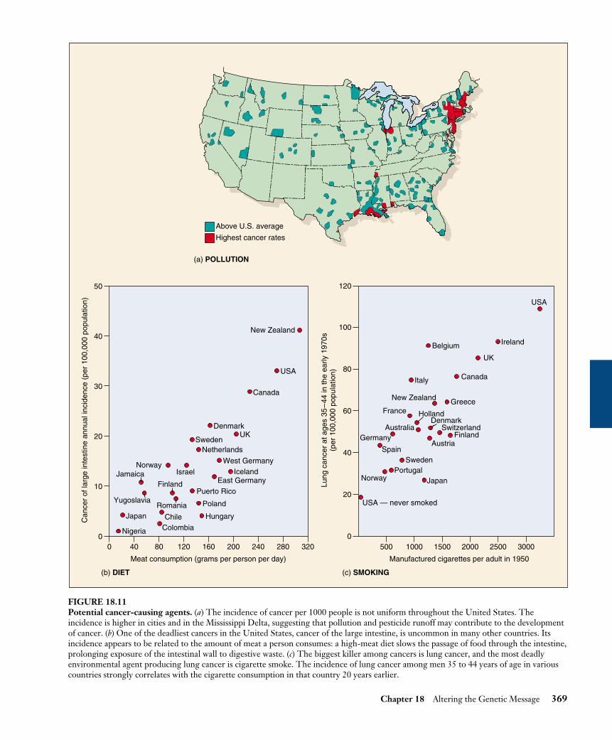
Chapter 18 Altering the Genetic Message 369
Nigeria
Japan
ColombiaChile Hungary
Poland
Puerto RicoFinland
Yugoslavia
JamaicaNorway
Israel
SwedenNetherlands
UKDenmark
Canada
USA
New Zealand
West GermanyIceland
East Germany
Romania
0
10
20
30
40
50
Meat consumption (grams per person per day)
Can
cer
of la
rge
inte
stin
e an
nual
inci
denc
e (p
er 1
00,0
00 p
opul
atio
n)
0 40 80 120 160 200 240 280 3200
20
40
60
80
100
120
Manufactured cigarettes per adult in 1950
Lung
can
cer
at a
ges
35–
44 in
the
early
197
0s(p
er 1
00,0
00 p
opul
atio
n)
500 1000 1500 2000 2500 3000
Spain
GermanyAustralia
France
Sweden
FinlandSwitzerland
DenmarkHolland
GreeceNew Zealand
Austria
Norway
USA
Ireland
UK
CanadaItaly
Belgium
JapanPortugal
USA — never smoked
Above U.S. average
Highest cancer rates
(a) POLLUTION
(b) DIET (c) SMOKING
FIGURE 18.11Potential cancer-causing agents. (a) The incidence of cancer per 1000 people is not uniform throughout the United States. Theincidence is higher in cities and in the Mississippi Delta, suggesting that pollution and pesticide runoff may contribute to the developmentof cancer. (b) One of the deadliest cancers in the United States, cancer of the large intestine, is uncommon in many other countries. Itsincidence appears to be related to the amount of meat a person consumes: a high-meat diet slows the passage of food through the intestine,prolonging exposure of the intestinal wall to digestive waste. (c) The biggest killer among cancers is lung cancer, and the most deadlyenvironmental agent producing lung cancer is cigarette smoke. The incidence of lung cancer among men 35 to 44 years of age in variouscountries strongly correlates with the cigarette consumption in that country 20 years earlier.

Some Tumors Are Caused byChemicalsEarly Ideas
The chemical carcinogenesis theory was first advanced over200 years ago in 1761 by Dr. John Hill, an English physi-cian, who noted unusual tumors of the nose in heavy snuffusers and suggested tobacco had produced these cancers. In1775, a London surgeon, Sir Percivall Pott, made a similarobservation, noting that men who had been chimneysweeps exhibited frequent cancer of the scrotum, and sug-gesting that soot and tars might be responsible. Britishsweeps washed themselves infrequently and always seemedcovered with soot. Chimney sweeps on the continent, whowashed daily, had much less of this scrotal cancer. Theseand many other observations led to the hypothesis thatcancer results from the action of chemicals on the body.
Demonstrating That Chemicals Can CauseCancer
It was over a century before this hypothesis was directlytested. In 1915, Japanese doctor Katsusaburo Yamagiwa ap-plied extracts of coal tar to the skin of 137 rabbits every 2 or3 days for 3 months. Then he waited to see what wouldhappen. After a year, cancers appeared at the site of applica-tion in seven of the rabbits. Yamagiwa had induced cancerwith the coal tar, the first direct demonstration of chemicalcarcinogenesis. In the decades that followed, this approachdemonstrated that many chemicals were capable of causingcancer. Importantly, most of them were potent mutagens.
Because these were lab studies, many people did not ac-cept that the results applied to real people. Do tars in fact in-duce cancer in humans? In 1949, the American physicianErnst Winder and the British epidemiologist Richard Dollindependently reported that lung cancer showed a stronglink to the smoking of cigarettes, which introduces tars intothe lungs. Winder interviewed 684 lung cancer patients and600 normal controls, asking whether each had ever smoked.Cancer rates were 40 times higher in heavy smokers than innonsmokers. Doll’s study was even more convincing. He in-terviewed a large number of British physicians, noting whichones smoked, then waited to see which would develop lungcancer. Many did. Overwhelmingly, those who did weresmokers. From these studies, it seemed likely as long as 50years ago that tars and other chemicals in cigarette smoke in-duce cancer in the lungs of persistent smokers. While thissuggestion was (and is) resisted by the tobacco industry, theevidence that has accumulated since these pioneering studiesmakes a clear case, and there is no longer any real doubt.Chemicals in cigarette smoke cause cancer.
Carcinogens Are Common
In ongoing investigations over the last 50 years, manyhundreds of synthetic chemicals have been shown capable
of causing cancer in laboratory animals. Among them aretrichloroethylene, asbestos, benzene, vinyl chloride, arsenic, arylamide, and a host of complex petroleumproducts with chemical structures resembling chickenwire. People in the workplace encounter chemicals daily(table 18.3).
In addition to identifying potentially dangerous sub-stances, what have the studies of potential carcinogenstold us about the nature of cancer? What do these cancer-causing chemicals have in common? They are all mutagens,each capable of inducing changes in DNA.
Chemicals that produce mutations in DNA are oftenpotent carcinogens. Tars in cigarette smoke, forexample, are the direct cause of most lung cancers.
370 Part V Molecular Genetics
Table 18.3 Chemical Carcinogens in the Workplace
Workers at Risk Chemical Cancer for Exposure
COMMON EXPOSURE
Benzene Myelogenous Painters; dye users; leukemia furniture finishers
Diesel exhaust Lung Railroad and bus-garage workers; truckers; miners
Mineral oils Skin Metal machinistsPesticides Lung SprayersCigarette tar Lung Smokers
UNCOMMON EXPOSURE
Asbestos Mesothelioma, Brake-lining, lung insulation workers
Synthetic mineral Lung Wall and pipe fibers insulation and duct
wrapping usersHair dyes Bladder Hairdressers and
barbersPaint Lung PaintersPolychlorinated Liver, skin Users of hydraulic biphenyls fluids and
lubricants, inks, adhesives, insecticides
Soot Skin Chimney sweeps; bricklayers; firefighters; heating-unit service workers
RARE EXPOSURE
Arsenic Lung, skin Insecticide/herbicide sprayers; tanners; oil refiners
Formaldehyde Nose Wood product,paper, textiles, andmetal product workers

Other Tumors Result from ViralInfectionChemical mutagens are not the only carcinogens, however.Some tumors seem almost certainly to result from viral in-fection. Viruses can be isolated from certain tumors, andthese viruses cause virus-containing tumors to develop inother individuals. About 15% of human cancers are associ-ated with viruses.
A Virus That Causes Cancer
In 1911, American medical researcher Peyton Rous re-ported that a virus, subsequently named Rous avian sar-coma virus (RSV), was associated with chicken sarcomas.He found that RSV could infect and initiate cancer inchicken fibroblast (connective tissue) cells growing in cul-ture; from those cancerous cells, more viruses could be iso-lated. Rous was awarded the 1966 Nobel Prize in Physiol-ogy or Medicine for this discovery. RSV proved to be akind of RNA virus called a retrovirus. When retrovirusesinfect a cell, they make a DNA copy of their RNA genomeand insert that copy into the host cell’s DNA.
How RSV Causes Cancer
How does RSV initiate cancer? When RSV was comparedto a closely related virus, RAV-O, which is unable to trans-form normal chicken cells into cancerous cells, the twoviruses proved to be identical except for one gene that waspresent in RSV but absent from RAV-O. That gene wascalled the src gene, short for sarcoma.
How do viral genes cause cancer? An essential clue camein 1970, when temperature-sensitive RSV mutants wereisolated. These mutants would transform tissue culturecells into cancer cells at 35°C, but not at 41°C. Tempera-ture sensitivity of this kind is almost always associated withproteins. It seemed likely, therefore, that the src gene wasactively transcribed by the cell, rather than serving as arecognition site for some sort of regulatory protein. Thiswas an exciting result, suggesting that the protein specifiedby this cancer-causing gene, or oncogene, could be iso-lated and its properties studied.
The src protein was first isolated in 1977 by J. MichaelBishop and Harold Varmus, who won the Nobel Prize fortheir efforts. It turned out to be an enzyme of moderatesize that phosphorylates (adds a phosphate group to) the ty-rosine amino acids of proteins. Such enzymes, called tyro-sine kinases, are quite common in animal cells. One exam-ple is an enzyme that also serves as a plasma membranereceptor for epidermal growth factor, a protein that sig-nals the initiation of cell division. This finding raised theexciting possibility that RSV may cause cancer by introduc-ing into cells an active form of a normally quiescentgrowth-promoting enzyme. Later experiments showed thisis indeed the case.
Origin of the src Gene
Does the src gene actually integrate into the host cell’schromosome along with the rest of the RSV genome? Oneway to answer this question is to prepare a radioactive ver-sion of the gene, allow it to bind to complementary se-quences on the chicken chromosomes, and examine wherethe chromosomes become radioactive. The result of thisexperiment is that radioactive src DNA does in fact bind tothe site where RSV DNA is inserted into the chickengenome—but it also binds to a second site where there isno part of the RSV genome!
The explanation for the second binding site is that thesrc gene is not exclusively a viral gene. It is also a growth-promoting gene that evolved in and occurs normally inchickens. This normal chicken gene is the second sitewhere src binds to chicken DNA. Somehow, an ancestor ofRSV picked up a copy of the normal chicken gene in somepast infection. Now part of the virus, the gene is tran-scribed under the control of viral promoters rather thanunder the regulatory system of the chicken genome (figure18.12).
Studies of RSV reveal that cancer results from theinappropriate activity of growth-promoting genes thatare less active or completely inactive in normal cells.
Chapter 18 Altering the Genetic Message 371
Tyrosine kinase gene of chickenchromosome with 6 introns
Retrovirus genomewithout oncogene (RAV-0)
Envelope proteins
Genome of Rous avian sarcoma virus (RSV)
RNA transcript
Reverse transcriptase
DNA copy
1 2 3 4 5 6
gag
src
pol env
gag pol env src
FIGURE 18.12How a chicken gene got into the RSV genome. RSV containsonly a few genes: gag and env, which encode the viral protein coatand envelope proteins, and pol, which encodes reversetranscriptase. It also contains the src gene that causes sarcomas,which the RAV-O virus lacks. RSV originally obtained its src genefrom chickens, where a copy of the gene occurs normally and iscontrolled by the chicken’s regulatory genes.

Cancer and the Cell CycleAn important technique used to study tumors is calledtransfection. In this procedure, the nuclear DNA fromtumor cells is isolated and cleaved into random fragments.Each fragment is then tested individually for its ability toinduce cancer in the cells that assimilate it.
Using transfection, researchers have discovered thatmost human tumors appear to result from the mutation ofgenes that regulate the cell cycle. Sometimes the mutationof only one or two gene is all that is needed to transformnormally dividing cells into cancerous cells in tissue culture(table 18.4).
Point Mutations Can Lead to Cancer
The difference between a normal gene encoding a proteinthat regulates the cell cycle and a cancer-inducing versioncan be a single point mutation in the DNA. In one case ofras-induced bladder cancer, for example, a single DNAbase change from guanine to thymine converts a glycinein the normal ras protein into a valine in the cancer-caus-ing version. Several other ras-induced human carcinomashave been shown to also involve single nucleotide substi-tutions.
Telomerase and Cancer
Telomeres are short sequences of nucleotides repeatedthousands of times on the ends of chromosomes. BecauseDNA polymerase is unable to copy chromosomes all theway to the tip (there is no place for the primer necessary tocopy the last Okazaki fragment), telomeric segments arelost every time a cell divides.
In healthy cells a tumor suppressor inhibits productionof a special enzyme called telomerase that adds the losttelomere material back to the tip. Without this enzyme, acell’s chromosomes lose material from their telomeres witheach replication. Every time a chromosome is copied as thecell prepares to divide, more of the tip is lost. After some30 divisions, so much is lost that copying is no longer pos-sible. Cells in the tissues of an adult human have typicallyundergone 25 or more divisions. Cancer can’t get very farwith only the 5 remaining cell divisions. Were cancer tostart, it would grind to a halt after only a few divisions forlack of telomere.
Thus, we see that the cell’s inhibition of telomerase insomatic cells is a very effective natural brake on the cancerprocess. Any mutation that destroys the telomerase in-hibitor releases that brake, making cancer possible. Thus,when researchers looked for telomerase in human ovariantumor cells, they found it. These cells contained muta-tions that had inactivated the cell control that blocks thetranscription of the telomerase gene. Telomerase pro-duced in these cells reversed normal telomere shortening,allowing the cells to proliferate and gain the immortalityof cancer cells.
Mutations in Proto-Oncogenes: Accelerating theCell Cycle
Most cancers are the direct result of mutations in growth-regulating genes. There are two general classes of cancer-inducing mutations: mutations of proto-oncogenes andmutations of tumor-suppressor genes.
Genes known as proto-oncogenes encode proteinsthat stimulate cell division. Mutations that overactivatethese stimulatory proteins cause the cells that containthem to proliferate excessively. Mutated proto-oncogenesbecome cancer-causing genes called oncogenes (Greekonco-, “tumor”) (figure 18.13). Often the induction ofthese cancers involves changes in the activity of intracel-lular signalling molecules associated with receptors onthe surface of the plasma membrane. In a normal cell, thesignalling pathways activated by these receptors triggerpassage of the G1 checkpoint of cell proliferation (see fig-ure 11.17).
The mutated alleles of these oncogenes are geneticallydominant. Among the most widely studied are myc and ras.Expression of myc stimulates the production of cyclins andcyclin-dependent protein kinases (Cdks), key elements inregulating the checkpoints of cell division.
372 Part V Molecular Genetics
Growth-factor receptors:PDGF receptorerbB
Growth-factors:PDGF
Cytoplasmic steroid-typegrowth-factor receptors:RET
Cytoplasmicserine/threonine-specificprotein kinases:raf
Membrane/cytoskeleton-protein kinases:src
Nuclearproteins:mycbclMDM
Cytoplasmic tyrosine-specific protein kinases:N-ras
G proteins:K-ras
FIGURE 18.13The main classes of oncogenes. Before they are altered bymutation to their cancer-causing condition, oncogenes are calledproto-oncogenes (that is, genes able to become oncogenes).Illustrated here are the principal classes of proto-oncogenes, withsome typical representatives indicated.

The ras gene product is involved in the cellular responseto a variety of growth factors such as EGF, an intercellularsignal that normally initiates cell proliferation. When EGFbinds to a specific receptor protein on the plasma mem-brane of epithelial cells, the portion of the receptor thatprotrudes into the cytoplasm stimulates the ras protein tobind to GTP. The ras protein/GTP complex in turn re-
cruits and activates a protein called Raf to the inner surfaceof the plasma membrane, which in turn activates cytoplas-mic kinases and so triggers an intracellular signaling system(see chapter 7). The final step in the pathway is the activa-tion of transcription factors that trigger cell proliferation.Cancer-causing mutations in ras greatly reduce the amountof EGF necessary to initiate cell proliferation.
Chapter 18 Altering the Genetic Message 373
Table 18.4 Some Genes Implicated in Human Cancers
Gene Product Cancer
ONCOGENES
Genes Encoding Growth Factors or Their Receptorserb-B Receptor for epidermal growth factor Glioblastoma (a brain cancer); breast cancererb-B2 A growth factor receptor (gene also called neu) Breast cancer; ovarian cancer; salivary gland cancerPDGF Platelet-derived growth factor Glioma (a brain cancer)RET A growth factor receptor Thyroid cancer
Genes Encoding Cytoplasmic Relays in Intracellular Signaling PathwaysK-ras Protein kinase Lung cancer; colon cancer; ovarian cancer;
pancreatic cancerN-ras Protein kinase Leukemias
Genes Encoding Transcription Factors That Activate Transcription of Growth-Promoting Genesc-myc Transcription factor Lung cancer; breast cancer; stomach cancer;
leukemiasL-myc Transcription factor Lung cancerN-myc Transcription factor Neuroblastoma (a nerve cell cancer)
Genes Encoding Other Kinds of Proteinsbcl-2 Protein that blocks cell suicide Follicular B cell lymphomabcl-1 Cyclin D1, which stimulates the cell Breast cancer; head and neck cancers
cycle clock (gene also called PRAD1)MDM2 Protein antagonist of p53 tumor-supressor protein Wide variety of sarcomas (connective tissue
cancers)
TUMOR-SUPRESSOR GENES
Genes Encoding Cytoplasmic ProteinsAPC Step in a signaling pathway Colon cancer; stomach cancerDPC4 A relay in signaling pathway that inhibits cell division Pancreatic cancerNF-1 Inhibitor of ras, a protein that stimulates cell division Neurofibroma; myeloid leukemiaNF-2 Inhibitor of ras Meningioma (brain cancer); schwannoma (cancer
of cells supporting peripheral nerves)Genes Encoding Nuclear Proteins
MTS1 p16 protein, which slows the cell cycle clock A wide range of cancersp53 p53 protein, which halts cell division at the G1 checkpoint A wide range of cancersRb Rb protein, which acts as a master brake of the cell cycle Retinoblastoma; breast cancer; bone cancer;
bladder cancerGenes Encoding Proteins of Unknown Cellular Locations
BRCA1 ? Breast cancer; ovarian cancerBRCA2 ? Breast cancerVHL ? Renal cell cancer

Mutations in Tumor-Suppressor Genes:Inactivating the Cell’s Inhibitors of Proliferation
If the first class of cancer-inducing mutations “steps on theaccelerator” of cell division, the second class of cancer-inducing mutations “removes the brakes.” Cell division isnormally turned off in healthy cells by proteins that pre-vent cyclins from binding to Cdks. The genes that encodethese proteins are called tumor-suppressor genes. Theirmutant alleles are genetically recessive.
Among the most widely studied tumor-suppressorgenes are Rb, p16, p21, and p53. The unphosphorylatedproduct of the Rb gene ties up transcription factor E2F,which transcribes several genes required for passage
through the G1 checkpoint into S phase of the cell cycle(figure 18.14). The proteins encoded by p16 and p21 re-inforce the tumor-suppressing role of the Rb protein,preventing its phosphorylation by binding to the appro-priate Cdk/cyclin complex and inhibiting its kinase activ-ity. The p53 protein senses the integrity of the DNA andis activated if the DNA is damaged (figure 18.15). It ap-pears to act by inducing the transcription of p21, whichbinds to cyclins and Cdk and prevents them from inter-acting. One of the reasons repeated smoking leads inex-orably to lung cancer is that it induces p53 mutations. In-deed, almost half of all cancers involve mutations of thep53 gene.
374 Part V Molecular Genetics
RbRetinoblastoma protein p16 Tumor suppressor
Cell division No cell divisionx
Rb
Cyclins
Cdk
Growthblockedat G1
Cellnucleus
E2F
E2F
E2F
Rb
Rb
ATP
P
Mitosisinitiated
Mitosisinhibited
Cellnucleus
p16 binds to Cdk, preventingphosphorylation of Rb
p16
Cdk
Growthblockedat G1
E2F
E2F
Rb
Rb
p16
Cyclins
FIGURE 18.14How the tumor-suppressor genes Rb and p16 interact toblock cell division. The retinoblastoma protein (Rb) binds to thetranscription factor (E2F) that activates genes in the nucleus,preventing this factor from initiating mitosis. The G1 checkpoint ispassed when Cdk interacts with cyclins to phosphorylate Rb,releasing E2F. The p16 tumor-suppressor protein reinforces Rb’sinhibitory action by binding to Cdk so that Cdk is not available tophosphorylate Rb.
1. Halts cell cycle at G1 checkpoint
2. Activates DNA repair system
Cdk
Damage toDNA
p53
Initiatestranscriptionof repairenzymes
Initiatestranscriptionof p21
DNArepair
p21
Cyclins
p21
Blocks cell cycleat G1 checkpoint
Prevents DNAreplication
FIGURE 18.15The role of tumor-suppressor p53 in regulating the cellcycle. The p53 protein works at the G1 checkpoint to check forDNA damage. If the DNA is damaged, p53 activates the DNArepair system and stops the cell cycle at the G1 checkpoint (beforeDNA replication). This allows time for the damage to be repaired.p53 stops the cell cycle by inducing the transcription of p21. Thep21 protein then binds to cyclins and prevents them fromcomplexing with Cdk.

Cancer-Causing Mutations Accumulate overTime
Cells control proliferation at several checkpoints, and allof these controls must be inactivated for cancer to be ini-tiated. Therefore, the induction of most cancers involvesthe mutation of multiple genes; four is a typical number(figure 18.16). In many of the tissue culture cell lines usedto study cancer, most of the controls are already inacti-vated, so that mutations in only one or a few genes trans-form the line into cancerous growth. The need to inacti-vate several regulatory genes almost certainly explainswhy most cancers occur in people over 40 years old (fig-ure 18.17); in older persons, there has been more time forindividual cells to accumulate multiple mutations. It isnow clear that mutations, including those in potentiallycancer-causing genes, do accumulate over time. Using thepolymerase chain reaction (PCR), researchers in 1994searched for a certain cancer-associated gene mutation inthe blood cells of 63 cancer-free people. They found thatthe mutation occurred 13 times more often in people over60 years old than in people under 20.
Cancer is a disease in which the controls that normallyrestrict cell proliferation do not operate. In some cases,cancerous growth is initiated by the inappropriateactivation of proteins that regulate the cell cycle; inother cases, it is initiated by the inactivation of proteinsthat normally suppress cell division.
Chapter 18 Altering the Genetic Message 375
MUTATEDGENE
Tumorsuppressor
Normalepithelium
Hyperproliferativeepithelium
Early benignpolyp
Intermediatebenign polyp
Late benignpolyp
Carcinoma Metastasis
APC OncogeneK-rasTumorsuppressorDCC
Tumorsuppressorp53
OthermutationsLoss of APC Mutation of K-ras and DCC Mutation of p53
FIGURE 18.16The progression of mutations that commonly lead to colorectal cancer. The fatal metastasis is the last of six serial changes that theepithelial cells lining the rectum undergo. One of these changes is brought about by mutation of a proto-oncogene, and three of theminvolve mutations that inactivate tumor-suppressor genes.
0 10 20 30 40 50 60 70 800
25
5075
100
200
300
400
500
Age at death (years)
Ann
ual d
eath
rat
e
150
250
350
450
FIGURE 18.17The annual death rate from cancer climbs with age. The rateof cancer deaths increases steeply after age 40 and even moresteeply after age 60, suggesting that several independent mutationsmust accumulate to give rise to cancer.

Smoking and CancerHow can we prevent cancer? The most obvious strategy isto minimize mutational insult. Anything that decreases ex-posure to mutagens can decrease the incidence of cancerbecause exposure has the potential to mutate a normal geneinto an oncogene. It is no accident that the most reliabletests for the carcinogenicity of a substance are tests thatmeasure the substance’s mutagenicity.
The Association between Smoking and Cancer
About a third of all cases of cancer in the United States aredirectly attributable to cigarette smoking. The associationbetween smoking and cancer is particularly striking forlung cancer (figure 18.18). Studies of male smokers show ahighly positive correlation between the number of ciga-rettes smoked per day and the incidence of lung cancer(figure 18.19). For individuals who smoke two or morepacks a day, the risk of contracting lung cancer is at least 40times greater than it is for nonsmokers, whose risk level ap-proaches zero. Clearly, an effective way to avoid lung can-cer is not to smoke. Other studies have shown a clear rela-tionship between cigarette smoking and reduced lifeexpectancy (figure 18.20). Life insurance companies havecalculated that smoking a single cigarette lowers one’s lifeexpectancy by 10.7 minutes (longer than it takes to smokethe cigarette)! Every pack of 20 cigarettes bears an unwrit-ten label:
“The price of smoking this pack of cigarettes is 31⁄2 hoursof your life.”
Smoking Introduces Mutagens to the Lungs
Over half a million people died of cancer in the UnitedStates in 1999; about 28% of them died of lung cancer.About 140,000 persons were diagnosed with lung cancereach year in the 1980s. Around 90% of them died withinthree years after diagnosis; 96% of them were cigarettesmokers.
Smoking is a popular pastime. In the United States, 24%of the population smokes, and U.S. smokers consumed over450 billion cigarettes in 1999. The smoke emitted fromthese cigarettes contains some 3000 chemical components,including vinyl chloride, benzo[a]pyrenes, and nitroso-nor-nicotine, all potent mutagens. Smoking places these muta-gens into direct contact with the tissues of the lungs.
Mutagens in the Lung Cause Cancer
Introducing powerful mutagens to the lungs causes consid-erable damage to the genes of the epithelial cells that linethe lungs and are directly exposed to the chemicals. Amongthe genes that are mutated as a result are some whose nor-mal function is to regulate cell proliferation. When thesegenes are damaged, lung cancer results.
376 Part V Molecular Genetics
FIGURE 18.18Photo of a cancerous human lung. The bottom half of the lungis normal, while a cancerous tumor has completely taken over thetop half. The cancer cells will eventually break through into thelymph and blood vessels and spread through the body.
Cigarettes smoked per day
Inci
denc
e of
can
cer
per
100,
000
men
10
0
100
200
300
400
500
20 30 40
FIGURE 18.19Smoking causes cancer. The annual incidence of lung cancer per100,000 men clearly increases with the number of cigarettessmoked per day.

This process has been clearly demonstrated forbenzo[a]pyrene (BP), one of the potent mutagens releasedinto cigarette smoke from tars in the tobacco. The epithe-lial cells of the lung absorb BP from tobacco smoke andchemically alter it to a derivative form. This derivativeform, benzo[a]pyrene-diolepoxide (BPDE), binds directlyto the tumor-suppressor gene p53 and mutates it to an in-active form. The protein encoded by p53 oversees the G1cell cycle checkpoint described in chapter 11 and is one ofthe body’s key mechanisms for preventing uncontrolled cellproliferation. The destruction of p53 in lung epithelial cellsgreatly hastens the onset of lung cancer—p53 is mutated toan inactive form in over 70% of lung cancers. When exam-ined, the p53 mutations in cancer cells almost all occur atone of three “hot spots.” The key evidence linking smokingand cancer is that when the mutations of p53 caused byBPDE from cigarettes are examined, they occur at thesame three specific “hot spots!”
The Incidence of Cancer Reflects Smoking
Cigarette manufacturers argue that the causal connectionbetween smoking and cancer has not been proved, andthat somehow the relationship is coincidental. Look care-fully at the data presented in figure 18.21 and see if youagree. The upper graph, compiled from data on Americanmen, shows the incidence of smoking from 1900 to 1990and the incidence of lung cancer over the same period.Note that as late as 1920, lung cancer was a rare disease.About 20 years after the incidence of smoking began toincrease among men, lung cancer also started to becomemore common.
Now look at the lower graph, which presents data onAmerican women. Because of social mores, significantnumbers of American women did not smoke until afterWorld War II, when many social conventions changed. Aslate as 1963, when lung cancer among males was near cur-rent levels, this disease was still rare in women. In theUnited States that year, only 6588 women died of lung can-cer. But as more women smoked, more developed lungcancer, again with a lag of about 20 years. Americanwomen today have achieved equality with men in the num-bers of cigarettes they smoke, and their lung cancer deathrates are today approaching those for men. In 1990, morethan 49,000 women died of lung cancer in the UnitedStates. The current annual rate of deaths from lung cancerin male and female smokers is 180 per 100,000, or about 2out of every 1000 smokers each year.
The easiest way to avoid cancer is to avoid exposure tomutagens. The single greatest contribution one canmake to a longer life is not to smoke.
Chapter 18 Altering the Genetic Message 377
400
20
40
60
80
100
Age
Per
cent
age
aliv
e
Never smoked regularly
1–14 cigarettes a day
15–24 cigarettes a day
25 or more a day
55 70 85
FIGURE 18.20Tobacco reduces life expectancy. The world’s longest-runningsurvey of smoking, begun in 1951 in Britain, revealed that by 1994the death rate for smokers had climbed to three times the rate fornonsmokers among men 35 to 69 years of age.Source: Data from New Scientist, October 15, 1994.
0
1000
2000
3000
4000
5000
0
20
40
60
80
100
0
1000
2000
3000
4000
5000
0
20
40
60
80
100
Cig
aret
tes
smok
ed p
er c
apita
per
yea
r
Inci
denc
e of
lung
can
cer
(per
100
,000
per
yea
r)
1900 1920 1940 1960 1980 1990
1900 1920 1940 1960 1980 1990
Men
Women
Lungcancer
Lungcancer
Smoking
Smoking
FIGURE 18.21The incidence of lung cancer in men and women. What dothese graphs indicate about the connection between smoking andlung cancer?

Curing CancerPotential cancer therapies are being developed on manyfronts (figure 18.22). Some act to prevent the start of can-cer within cells. Others act outside cancer cells, preventingtumors from growing and spreading.
Preventing the Start of Cancer
Many promising cancer therapies act within potential can-cer cells, focusing on different stages of the cell’s “Shall Idivide?” decision-making process.
1. Receiving the Signal to Divide. The first step in thedecision process is the reception of a “divide” signal, usu-ally a small protein called a growth factor released from aneighboring cell. The growth factor is received by a pro-tein receptor on the cell surface. Mutations that increasethe number of receptors on the cell surface amplify the di-vision signal and so lead to cancer. Over 20% of breast can-cer tumors prove to overproduce a protein called HER2 as-sociated with the receptor for epidermal growth factor.
Therapies directed at this stage of the decision processutilize the human immune system to attack cancer cells.Special protein molecules called “monoclonal antibodies,”created by genetic engineering, are the therapeutic agents.These monoclonal antibodies are designed to seek out andstick to HER2. Like waving a red flag, the presence of themonoclonal antibody calls down attack by the immune sys-tem on the HER2 cell. Because breast cancer cells overpro-duce HER2, they are killed preferentially. Genentech’s re-cently approved monoclonal antibody, called “herceptin,”has given promising results in clinical tests. In other tests,the monoclonal antibody C225, directed against epidermalgrowth factor receptors, has succeeded in curing advancedcolon cancer. Clinical trials of C225 have begun.
2. The Relay Switch. The second step in the decisionprocess is the passage of the signal into the cell’s interior,the cytoplasm. This is carried out in normal cells by a pro-tein called Ras that acts as a relay switch. When growthfactor binds to a receptor like EGF, the adjacent Ras pro-tein acts like it has been “goosed,” contorting into a newshape. This new shape is chemically active, and initiates achain of reactions that passes the “divide” signal inward to-ward the nucleus. Mutated forms of the Ras protein behavelike a relay switch stuck in the “ON” position, continuallyinstructing the cell to divide when it should not. 30% of allcancers have a mutant form of Ras.
Therapies directed at this stage of the decision processtake advantage of the fact that normal Ras proteins are in-active when made. Only after it has been modified by thespecial enzyme farnesyl transferase does Ras protein becomeable to function as a relay switch. In tests on animals, farne-syl transferase inhibitors induce the regression of tumorsand prevent the formation of new ones.
3. Amplifying the Signal. The third step in the decisionprocess is the amplification of the signal within the cyto-plasm. Just as a TV signal needs to be amplified in order tobe received at a distance, so a “divide” signal must be am-plified if it is to reach the nucleus at the interior of the cell,a very long journey at a molecular scale. Cells use an inge-nious trick to amplify the signal. Ras, when “ON,” activatesan enzyme, a protein kinase. This protein kinase activatesother protein kinases that in their turn activate still others.The trick is that once a protein kinase enzyme is activated,it goes to work like a demon, activating hoards of othersevery second! And each and every one it activates behavesthe same way too, activating still more, in a cascade of ever-widening effect. At each stage of the relay, the signal is am-plified a thousand-fold. Mutations stimulating any of theprotein kinases can dangerously increase the already ampli-fied signal and lead to cancer. Five percent of all cancers,for example, have a mutant hyperactive form of the proteinkinase Src.
Therapies directed at this stage of the decision processemploy so-called “anti-sense RNA” directed specificallyagainst Src or other cancer-inducing kinase mutations. Theidea is that the src gene uses a complementary copy of itselfto manufacture the Src protein (the “sense” RNA or mes-senger RNA), and a mirror image complementary copy ofthe sense RNA (“anti-sense RNA”) will stick to it, gum-ming it up so it can’t be used to make Src protein. The ap-proach appears promising. In tissue culture, anti-senseRNAs inhibit the growth of cancer cells, and some also ap-pear to block the growth of human tumors implanted inlaboratory animals. Human clinical trials are underway.
4. Releasing the Brake. The fourth step in the decisionprocess is the removal of the “brake” the cell uses to re-strain cell division. In healthy cells this brake, a tumor sup-pressor protein called Rb, blocks the activity of a transcrip-tion factor protein called E2F. When free, E2F enables thecell to copy its DNA. Normal cell division is triggered tobegin when Rb is inhibited, unleashing E2F. Mutationswhich destroy Rb release E2F from its control completely,leading to ceaseless cell division. Forty percent of all can-cers have a defective form of Rb.
Therapies directed at this stage of the decision processare only now being attempted. They focus on drugs able toinhibit E2F, which should halt the growth of tumors aris-ing from inactive Rb. Experiments in mice in which theE2F genes have been destroyed provide a model system tostudy such drugs, which are being actively investigated.
5. Checking That Everything Is Ready. The fifth step inthe decision process is the mechanism used by the cell to en-sure that its DNA is undamaged and ready to divide. This jobis carried out in healthy cells by the tumor-suppressor proteinp53, which inspects the integrity of the DNA. When it de-tects damaged or foreign DNA, p53 stops cell division andactivates the cell’s DNA repair systems. If the damage doesn’t
378 Part V Molecular Genetics

get repaired in a reasonable time, p53 pulls the plug, trigger-ing events that kill the cell. In this way, mutations such asthose that cause cancer are either repaired or the cells con-taining them eliminated. If p53 is itself destroyed by muta-tion, future damage accumulates unrepaired. Among thisdamage are mutations that lead to cancer. Fifty percent of allcancers have a disabled p53. Fully 70 to 80% of lung cancershave a mutant inactive p53—the chemical benzo[a]pyrene incigarette smoke is a potent mutagen of p53.
A promising new therapy using adenovirus (responsiblefor mild colds) is being targeted at cancers with a mutantp53. To grow in a host cell, adenovirus must use the prod-uct of its gene E1B to block the host cell’s p53, thereby en-abling replication of the adenovirus DNA. This means thatwhile mutant adenovirus without E1B cannot grow inhealthy cells, the mutants should be able to grow in, anddestroy, cancer cells with defective p53. When humancolon and lung cancer cells are introduced into mice lack-ing an immune system and allowed to produce substantialtumors, 60% of the tumors simply disappear when treatedwith E1B-deficient adenovirus, and do not reappear later.Initial clinical trials are less encouraging, as many peoplepossess antibodies to adenovirus.
6. Stepping on the Gas. Cell division starts with replica-tion of the DNA. In healthy cells, another tumor suppressor“keeps the gas tank nearly empty” for the DNA replicationprocess by inhibiting production of an enzyme called telom-erase. Without this enzyme, a cell’s chromosomes lose ma-terial from their tips, called telomeres. Every time a chro-mosome is copied, more tip material is lost. After somethirty divisions, so much is lost that copying is no longerpossible. Cells in the tissues of an adult human have typi-cally undergone twenty five or more divisions. Cancer can’tget very far with only the five remaining cell divisions, soinhibiting telomerase is a very effective natural break on thecancer process. It is thought that almost all cancers involve amutation that destroys the telomerase inhibitor, releasingthis break and making cancer possible. It should be possibleto block cancer by reapplying this inhibition. Cancer thera-pies that inhibit telomerase are just beginning clinical trials.
Preventing the Spread of Cancer
7. Tumor Growth. Once a cell begins cancerous growth,it forms an expanding tumor. As the tumor grows ever-larger, it requires an increasing supply of food and nutri-ents, obtained from the body’s blood supply. To facilitatethis necessary grocery shopping, tumors leak out sub-stances into the surrounding tissues that encourage angio-genesis, the formation of small blood vessels. Chemicalsthat inhibit this process are called angiogenesis inhibitors.In mice, two such angiogenesis inhibitors, angiostatin andendostatin, caused tumors to regress to microscopic size.This very exciting result has proven controversial, but ini-tial human trials seem promising.
8. Metastasis. If cancerous tumors simply continued togrow where they form, many could be surgically removed,and far fewer would prove fatal. Unfortunately, many can-cerous tumors eventually metastasize, individual cancercells breaking their moorings to the extracellular matrixand spreading to other locations in the body where they ini-tiate formation of secondary tumors. This process involvesmetal-requiring protease enzymes that cleave the cell-ma-trix linkage, components of the extracellular matrix such asfibronectin that also promote the migration of several non-cancerous cell types, and RhoC, a GTP-hydrolyzing en-zyme that promotes cell migration by providing neededGTP. All of these components offer promising targets forfuture anti-cancer therapy.
Therapies such as those described here are only part of awave of potential treatments under development and clini-cal trial. The clinical trials will take years to complete, butin the coming decade we can expect cancer to become acurable disease.
Understanding of how mutations produce cancer hasprogressed to the point where promising potentialtherapies can be tested.
Chapter 18 Altering the Genetic Message 379
Cellsurfaceprotein
Nucleus
Cell
Amplifyingenzyme
Extracellular matrix
Capillarynetwork
Angiogenesis
Divisionoccurs
Cell migration
1
2
34 65
7
8
FIGURE 18.22New molecular therapies for cancer target eight differentstages in the cancer process. (1) On the cell surface, a growthfactor signals the cell to divide. (2) Just inside the cell, a proteinrelay switch passes on the divide signal. (3) In the cytoplasm,enzymes amplify the signal. In the nucleus, (4) a “brake”preventing DNA replication is released, (5) proteins check thatthe replicated DNA is not damaged, and (6) other proteins rebuildchromosome tips so DNA can replicate. (7) The new tumorpromotes angiogenesis, the formation of growth-promoting bloodvessels. (8) Some cancer cells break away from the extracellularmatrix and invade other parts of the body.

An Overview of RecombinationMutation is a change in the content of an organism’s geneticmessage, but it is not the only source of genetic diversity.Diversity is also generated when existing elements of thegenetic message move around within the genome. As ananalogy, consider the pages of this book. A point mutationwould correspond to a change in one or more of the letterson the pages. For example, “ . . . in one or more of the let-ters of the pages” is a mutation of the previous sentence, inwhich an “n” is changed to an “f.” A significant alteration isalso achieved, however, when we move the position ofwords, as in “ . . . in one or more of the pages on the let-ters.” The change alters (and destroys) the meaning of thesentence by exchanging the position of the words “letters”and “pages.” This second kind of change, which representsan alteration in the genomic location of a gene or a fragmentof a gene, demonstrates genetic recombination.
Gene Transfer
Viewed broadly, genetic recombination can occur by twomechanisms (table 18.5). In gene transfer, one chromo-some or genome donates a segment to another chromo-some or genome. The transfer of genes from the humanimmunodeficiency virus (HIV) to a human chromosome isan example of gene transfer. Because gene transfer occursin both prokaryotes and eukaryotes, it is thought to be themore primitive of the two mechanisms.
Reciprocal Recombination
Reciprocal recombination is whentwo chromosomes trade segments. It isexemplified by the crossing over thatoccurs between homologous chromo-somes during meiosis. Independent as-sortment during meiosis is anotherform of reciprocal recombination. Dis-cussed in chapters 12 and 13, it is re-sponsible for the 9:3:3:1 ratio of pheno-types in a dihybrid cross and occursonly in eukaryotes.
Genetic recombination is a changein the genomic association amonggenes. It often involves a change inthe position of a gene or portion ofa gene. Recombination of this sortmay result from one-way genetransfer or reciprocal geneexchange.
380 Part V Molecular Genetics
Table 18.5 Classes of Genetic Recombination
Class Occurrence
GENE TRANSFERS
Conjugation Occurs predominantly but not exclusively in bacteria and is targeted to specific locations in the genome
Transposition Common in both bacteria and eukaryotes; genes move to new genomic locations, apparently at random
RECIPROCAL RECOMBINATIONS
Crossing over Requires the pairing of homologous chromosomes and may occur anywhere along their length
Unequal crossing over The result of crossing over between mismatched segments; leads to gene duplication and deletion
Gene conversion Occurs when homologous chromosomes pair and one is “corrected” to resemble the other
Independent assortment Haploid cells produced by meiosis contain only one randomly selected member of each pair of homologous chromosomes
18.3 Recombination alters gene location.
FIGURE 18.23A Nobel Prize for discovering gene transfer by transposition.Barbara McClintock receiving her Nobel Prize in 1983.

Gene TransferGenes are not fixed in their locations onchromosomes or the circular DNA mole-cules of bacteria; they can move around.Some genes move because they are part ofsmall, circular, extrachromosomal DNAsegments called plasmids. Plasmids enterand leave the main genome at specific placeswhere a nucleotide sequence matches onepresent on the plasmid. Plasmids occur pri-marily in bacteria, in which the main ge-nomic DNA can interact readily with otherDNA fragments. About 5% of the DNAthat occurs in a bacterium is plasmid DNA.Some plasmids are very small, containingonly one or a few genes, while others arequite complex and contain many genes.Other genes move within transposons,which jump from one genomic position toanother at random in both bacteria and eu-karyotes.
Gene transfer by plasmid movement wasdiscovered by Joshua Lederberg and EdwardTatum in 1947. Three years later, trans-posons were discovered by Barbara McClin-tock. However, her work implied that theposition of genes in a genome need not beconstant. Researchers accustomed to viewinggenes as fixed entities, like beads on a string,did not readily accept the idea of trans-posons. Therefore, while Lederberg andTatum were awarded a Nobel Prize for theirdiscovery in 1958, McClintock did not re-ceive the same recognition for hers until1983 (figure 18.23).
Plasmid Creation
To understand how plasmids arise, consider a hypotheti-cal stretch of bacterial DNA that contains two copies ofthe same nucleotide sequence. It is possible for the twocopies to base-pair with each other and create a transient“loop,” or double duplex. All cells have recombinationenzymes that can cause such double duplexes to undergoa reciprocal exchange, in which they exchange strands.As a result of the exchange, the loop is freed from therest of the DNA molecule and becomes a plasmid (figure18.24, steps 1–3). Any genes between the duplicated se-quences (such as gene A in figure 18.24) are transferredto the plasmid.
Once a plasmid has been created by reciprocal exchange,DNA polymerase will replicate it if it contains a replicationorigin, often without the controls that restrict the maingenome to one replication per cell division. Consequently,some plasmids may be present in multiple copies, others injust a few copies, in a given cell.
Integration
A plasmid created by recombination can reenter the maingenome the same way it left. Sometimes the region of theplasmid DNA that was involved in the original exchange,called the recognition site, aligns with a matching se-quence on the main genome. If a recombination event oc-curs anywhere in the region of alignment, the plasmid willintegrate into the genome (figure 18.24, steps 4–6). Inte-gration can occur wherever any shared sequences exist, soplasmids may be integrated into the main genome at posi-tions other than the one from which they arose. If a plas-mid is integrated at a new position, it transfers its genes tothat new position.
Transposons and plasmids transfer genes to newlocations on chromosomes. Plasmids can arise from andintegrate back into a genome wherever DNA sequencesin the genome and in the plasmid match.
Chapter 18 Altering the Genetic Message 381
4
Integration
Plasmid
D B� C�
CD� B
A
B� D C�
CD� B
B
3
1
2
Homologouspairing
Bacterialchromosome
D� C� CB� DA
A
Homologouspairing
Excision
Excision Integration
6
5
FIGURE 18.24Integration and excision of a plasmid. Because the ends of the two sequences inthe bacterial genome are the same (D′, C′, B′, and D, C, B), it is possible for the twoends to pair. Steps 1–3 show the sequence of events if the strands exchange duringthe pairing. The result is excision of the loop and a free circle of DNA—a plasmid.Steps 4–6 show the sequence when a plasmid integrates itself into a bacterialgenome.

Gene Transfer by Conjugation
One of the startling discoveries Lederberg and Tatummade was that plasmids can pass from one bacterium to an-other. The plasmid they studied was part of the genome ofEscherichia coli. It was given the name F for fertility factorbecause only cells which had that plasmid integrated intotheir DNA could act as plasmid donors. These cells arecalled Hfr cells (for “high-frequency recombination”). TheF plasmid contains a DNA replication origin and severalgenes that promote its transfer to other cells. These genesencode protein subunits that assemble on the surface of thebacterial cell, forming a hollow tube called a pilus.
When the pilus of one cell (F+) contacts the surface ofanother cell that lacks a pilus, and therefore does not con-tain an F plasmid (F–), the pilus draws the two cells closetogether so that DNA can be exchanged (figure 18.25).First, the F plasmid binds to a site on the interior of the F+
cell just beneath the pilus. Then, by a process calledrolling-circle replication, the F plasmid begins to copy itsDNA at the binding point. As it is replicated, the single-stranded copy of the plasmid passes into the other cell.There a complementary strand is added, creating a new,stable F plasmid (figure 18.26). In this way, genes arepassed from one bacterium to another. This transfer ofgenes between bacteria is called conjugation.
In an Hfr cell, with the F plasmid integrated into themain bacterial genome rather than free in the cytoplasm,the F plasmid can still organize the transfer of genes. Inthis case, the integrated F region binds beneath the pilusand initiates the replication of the bacterial genome, transfer-ring the newly replicated portion to the recipient cell.Transfer proceeds as if the bacterial genome were simply apart of the F plasmid. By studying this phenomenon, re-searchers have been able to locate the positions of differentgenes in bacterial genomes (figure 18.27).
Gene Transfer by Transposition
Like plasmids, transposons (figure 18.28) move from onegenomic location to another. After spending many genera-tions in one position, a transposon may abruptly move to anew position in the genome, carrying various genes alongwith it. Transposons encode an enzyme called trans-posase, that inserts the transposon into the genome (figure18.29). Because this enzyme usually does not recognize anyparticular sequence on the genome, transposons appear tomove to random destinations.
The movement of any given transposon is relativelyrare: it may occur perhaps once in 100,000 cell generations.Although low, this rate is still about 10 times as frequent as
382 Part V Molecular Genetics
FIGURE 18.25Contact by a pilus. The pilus of an F+ cell connects to an F- celland draws the two cells close together so that DNA transfer canoccur.
F+
(donor cell)F-
(recipient cell)
F Plasmid Bacterialchromosome
Conjugationbridge
FIGURE 18.26Gene transfer between bacteria. Donor cells (F+) contain an F plasmid that recipient cells (F–) lack. The F plasmid replicates itself andtransfers the copy across a conjugation bridge. The remaining strand of the plasmid serves as a template to build a replacement. When thesingle strand enters the recipient cell, it serves as a template to assemble a double-stranded plasmid. When the process is complete, bothcells contain a complete copy of the plasmid.

the rate at which random mutational changes occur. Fur-thermore, there are many transposons in most cells. Hence,over long periods of time, transposition can have an enor-mous evolutionary impact.
One way this impact can be felt is through mutation.The insertion of a transposon into a gene often destroysthe gene’s function, resulting in what is termed insertionalinactivation. This phenomenon is thought to be the causeof a significant number of the spontaneous mutations ob-served in nature.
Transposition can also facilitate gene mobilization,the bringing together in one place of genes that are usu-ally located at different positions in the genome. In bacte-ria, for example, a number of genes encode enzymes thatmake the bacteria resistant to antibiotics such as peni-cillin, and many of these genes are located on plasmids.The simultaneous exposure of bacteria to multiple antibi-otics, a common medical practice some years ago, favorsthe persistence of plasmids that have managed to acquireseveral resistance genes. Transposition can rapidly gener-ate such composite plasmids, called resistance transferfactors (RTFs), by moving antibiotic resistance genesfrom several plasmids to one. Bacteria possessing RTFsare thus able to survive treatment with a wide variety ofantibiotics. RTFs are thought to be responsible for muchof the recent difficulty in treating hospital-engenderedStaphylococcus aureus infections and the new drug-resistantstrains of tuberculosis.
Plasmids transfer copies of bacterial genes (and evenentire genomes) from one bacterium to another.Transposition is the one-way transfer of genes to arandomly selected location in the genome. The genesmove because they are associated with mobile geneticelements called transposons.
Chapter 18 Altering the Genetic Message 383
Plasmid
Transposon
FIGURE 18.28Transposon. Transposonsform characteristic stem-and-loop structures called“lollipops” because their twoends have the samenucleotide sequence asinverted repeats. These endspair together to form thestem of the lollipop.
Transposon
Transposase
FIGURE 18.29Transposition. Transposase does not recognize any particularDNA sequence; rather, it selects one at random, moving thetransposon to a random location. Some transposons leave a copyof themselves behind when they move.
Map ofE. coli
genome
leu azi tonlac
gal
tyr-cys
hisade
ser-gly
xyl
met-B12
thiDirectionof
transfer
azi
0 min 10 min 20 min 25 min
ton
Time elapsed from beginning ofconjugation until interruption
(a)
(b)
lac gal thr
RargFIGURE 18.27A conjugation map of the E. coli chromosome. Scientists havebeen able to break the Escherichia coli conjugation bridges byagitating the cell suspension rapidly in a blender. By agitating atdifferent intervals after the start of conjugation, investigators canlocate the positions of various genes along the bacterial genome. (a)The closer the genes are to the origin of replication, the sooner onehas to turn on the blender to block their transfer. (b) Map of the E. coli genome developed using this method.

Reciprocal RecombinationIn the second major mechanism for producing genetic re-combination, reciprocal recombination, two homologouschromosomes exchange all or part of themselves during theprocess of meiosis.
Crossing Over
As we saw in chapter 12, crossing over occurs in the firstprophase of meiosis, when two homologous chromosomesline up side by side within the synaptonemal complex. Atthis point, the homologues exchange DNA strands at oneor more locations. This exchange of strands can producechromosomes with new combinations of alleles.
Imagine, for example, that a giraffe has genes encodingneck length and leg length at two different loci on one ofits chromosomes. Imagine further that a recessive mutationoccurs at the neck length locus, leading after several roundsof independent assortment to some individuals that are ho-mozygous for a variant “long-neck” allele. Similarly, a re-cessive mutation at the leg length locus leads to homozy-gous “long-leg” individuals.
It is very unlikely that these two mutations would ariseat the same time in the same individual because the prob-ability of two independent events occurring together isthe product of their individual probabilities. If the spon-taneous occurrence of both mutations in a single individ-ual were the only way to produce a giraffe with both along neck and long legs, it would be extremely unlikelythat such an individual would ever occur. Because of re-combination, however, a crossover in the interval be-tween the two genes could in one meiosis produce achromosome bearing both variant alleles. This ability toreshuffle gene combinations rapidly is what makes re-combination so important to the production of naturalvariation.
Unequal Crossing Over
Reciprocal recombination can occur in any region alongtwo homologous chromosomes with sequences similarenough to permit close pairing. Mistakes in pairing occa-sionally happen when several copies of a sequence exist indifferent locations on a chromosome. In such cases, onecopy of a sequence may line up with one of the duplicatecopies instead of with its homologous copy. Such misalign-ment causes slipped mispairing, which, as we discussed ear-lier, can lead to small deletions and frame-shift mutations.If a crossover occurs in the pairing region, it will result inunequal crossing over because the two homologues will ex-change segments of unequal length.
In unequal crossing over, one chromosome gains extracopies of the multicopy sequences, while the other chro-mosome loses them (figure 18.30). This process can gener-
ate a chromosome with hundreds of copies of a particulargene, lined up side by side in tandem array.
Because the genomes of most eukaryotes possess mul-tiple copies of transposons scattered throughout thechromosomes, unequal crossing over between copies oftransposons located in different positions has had a pro-found influence on gene organization in eukaryotes. Aswe shall see later, most of the genes of eukaryotes appearto have been duplicated one or more times during theirevolution.
Gene Conversion
Because the two homologues that pair within a synaptone-mal complex are not identical, some nucleotides in one ho-mologue are not complementary to their counterpart in theother homologue with which it is paired. These occasionalnonmatching pairs of nucleotides are called mismatchpairs.
As you might expect, the cell’s error-correcting machin-ery is able to detect mismatch pairs. If a mismatch is de-tected during meiosis, the enzymes that “proofread” newDNA strands during DNA replication correct it. The mis-matched nucleotide in one of the homologues is excisedand replaced with a nucleotide complementary to the onein the other homologue. Its base-pairing partner in the firsthomologue is then replaced, producing two chromosomeswith the same sequence. This error correction causes oneof the mismatched sequences to convert into the other, aprocess called gene conversion.
Unequal crossing over is a crossover betweenchromosomal regions that are similar in nucleotidesequence but are not homologous. Gene conversion isthe alteration of one homologue by the cell’s error-detection and repair system to make it resemble theother homologue.
384 Part V Molecular Genetics
16 Gene copies
16 Gene copies
27 Gene copies
5 Gene copies
FIGURE 18.30Unequal crossing over. When a repeated sequence pairs out ofregister, a crossover within the region will produce onechromosome with fewer gene copies and one with more. Much ofthe gene duplication that has occurred in eukaryotic evolutionmay well be the result of unequal crossing over.

Trinucleotide RepeatsIn 1991, a new kind of change in the genetic material wasreported, one that involved neither changes in the identityof nucleotides (mutation) nor changes in the position ofnucleotide sequences (recombination), but rather an in-crease in the number of copies of repeated trinucleotide se-quences. Called trinucleotide repeats, these changes ap-pear to be the root cause of a surprisingly large number ofinherited human disorders.
The first examples of disorders resulting from the ex-pansion of trinucleotide repeat sequences were reported inindividuals with fragile X syndrome (the most common formof developmental disorder) and spinal muscular atrophy. Inboth disorders, genes containing runs of repeated nu-cleotide triplets (CGG in fragile X syndrome and CAG inspinal muscular atrophy) exhibit large increases in copynumber. In individuals with fragile X syndrome, for exam-ple, the CGG sequence is repeated hundreds of times (fig-ure 18.31), whereas in normal individuals it repeats onlyabout 30 times.
Ten additional human genes are now known to have al-leles with expanded trinucleotide repeats (figure 18.32).Many (but not all) of these alleles are GC-rich. A few of thealleles appear benign, but most are associated with herita-ble disorders, including Huntington’s disease, myotonicdystrophy, and a variety of neurological ataxias. In eachcase, the expansion transmits as a dominant trait. Often therepeats are found within the exons of their genes, butsometimes, as in the case of fragile X syndrome, they arelocated outside the coding segment. Furthermore, althoughthe repeat number is stably transmitted in normal families,it shows marked instability once it has abnormally ex-panded. Siblings often exhibit unique repeat lengths.
As the repeat number increases, disease severity tends toincrease in step. In fragile X syndrome, the CGG tripletnumber first increases from the normal stable range of 5 to55 times (the most common allele has 29 repeats) to an un-stable number of repeats ranging from 50 to 200, with no
detectable effect. In offspring, the number increasesmarkedly, with copy numbers ranging from 200 to 1300,with significant mental retardation (see figure 18.31). Simi-larly, the normal allele for myotonic dystrophy has 5 GTCrepeats. Mildly affected individuals have about 50, and se-verely affected individuals have up to 1000.
Trinucleotide repeats appear common in human genes,but their function is unknown. Nor do we know the mech-anism behind trinucleotide repeat expansion. It may in-volve unequal crossing over, which can readily producecopy-number expansion, or perhaps some sort of stutter inthe DNA polymerase when it encounters a run of triplets.The fact that di- and tetranucleotide repeat expansions arenot found seems an important clue. Undoubtedly, furtherexamples of this remarkable class of genetic change will bereported in the future. Considerable research is currentlyfocused on this extremely interesting area.
Many human genes contain runs of a trinucleotidesequence. Their function is unknown, but if the copynumber expands, hereditary disorders often result.
Chapter 18 Altering the Genetic Message 385
Normalallele
CGG
5–55CGG repeats
CGGCGG
Pre-fragile Xallele
50–200CGG repeats
Fragile Xallele
200–1300CGG repeats
FIGURE 18.31CGG repeats in fragile X alleles. The CGG triplet is repeatedapproximately 30 times in normal alleles. Individuals with pre-fragile X alleles show no detectable signs of the syndrome but dohave increased numbers of CGG repeats. In fragile X alleles, theCGG triplet repeats hundreds of times.
CGG CAG CTGGAA
Fragile X syndromeFragile site 11BFragile XE syndrome
Spinal and bulbar muscular atrophySpinocerebellar ataxia type 1Huntington's diseaseDentatorubral-pallidoluysian atrophyMachado-Joseph disease
Myotonic dystrophyFriedreich's ataxia
Exon 1 Exon 2 Exon 3Intron 1 Intron 2Repeatedtrinucleotide
Condition
FIGURE 18.32A hypothetical gene showing the locations and types of trinucleotide repeats associated with various human diseases. The CGGrepeats of fragile X syndrome, fragile XE syndrome, and fragile site 11B occur in the first exon of their respective genes. GAA repeatscharacteristic of Friedreich’s ataxia exist in the first intron of its gene. The genes for five different diseases, including Huntington’s disease,have CAG repeats within their second exons. Lastly, the myotonic dystrophy gene contains CTG repeats within the third exon.

Classes of Eukaryotic DNAThe two main mechanisms of genetic recombination, genetransfer and reciprocal recombination, are directly respon-sible for the architecture of the eukaryotic chromosome.They determine where genes are located and how manycopies of each exist. To understand how recombinationshapes the genome, it is instructive to compare the effectsof recombination in bacteria and eukaryotes.
Comparing Bacterial and Eukaryotic DNASequences
Bacterial genomes are relatively simple, containing genesthat almost always occur as single copies. Unequal crossingover between repeated transposition elements in their cir-cular DNA molecules tends to delete material, fostering themaintenance of a minimum genome size (figure 18.33a).For this reason, these genomes are very tightly packed,with few or no noncoding nucleotides. Recall the efficientuse of space in the organization of the lac genes describedin chapter 16.
In eukaryotes, by contrast, the introduction of pairs ofhomologous chromosomes (presumably because of theirimportance in repairing breaks in double-stranded DNA)has led to a radically different situation. Unequal crossingover between homologous chromosomes tends to promotethe duplication of material rather than its reduction (figure18.33b). Consequently, eukaryotic genomes have been in aconstant state of flux during the course of their evolution.Multiple copies of genes have evolved, some of them subse-quently diverging in sequence to become different genes,which in turn have duplicated and diverged.
Six different classes of eukaryotic DNA sequences arecommonly recognized, based on the number of copies ofeach (table 18.6).
Transposons
Transposons exist in multiple copies scattered about thegenome. In Drosophila, for example, more than 30 differenttransposons are known, most of them present at 20 to 40different sites throughout the genome. In all, the knowntransposons of Drosophila account for perhaps 5% of itsDNA. Mammalian genomes contain fewer kinds of trans-posons than the genomes of many other organisms, al-though the transposons in mammals are repeated moreoften. The family of human transposons called ALU ele-ments, for example, typically occurs about 300,000 times ineach cell. Transposons are transcribed but appear to playno functional role in the life of the cell. As noted earlier inthis chapter, many transposition events carry transposonsinto the exon portions of genes, disrupting the function ofthe protein specified by the gene transcript. These inser-tional inactivations are thought to be responsible for manynaturally occurring mutations.
Tandem Clusters
A second class consists of DNA sequences that are repeatedmany times, one copy following another in tandem array.By transcribing all of the copies in these tandem clusterssimultaneously, a cell can rapidly obtain large amounts ofthe product they encode. For example, the genes encodingrRNA are present in several hundred copies in most eu-karyotic cells. Because these clusters are active sites ofrRNA synthesis, they are readily visible in cytologicalpreparations, where they are called nucleolar organizerregions. When transcription of the rRNA gene clustersceases during cell division, the nucleolus disappears fromview under the microscope, but it reappears when tran-scription begins again.
The genes present in a tandem cluster are very similar insequence but not always identical; some may differ by one
386 Part V Molecular Genetics
18.4 Genomes are continually evolving.
Unequal crossing over withina bacterial genome deletes material
Lost
(a)
Unequal crossing over between chromosomes adds material to one and subtracts it from the other
(b)
X X
FIGURE 18.33Unequal crossing over has different consequences in bacteriaand eukaryotes. (a) Bacteria have a circular DNA molecule, and acrossover between duplicate regions within the molecule deletesthe intervening material. (b) In eukaryotes, with two versions ofeach chromosome, crossing over adds material to onechromosome; thus, gene amplification occurs in thatchromosome.

or a few nucleotides. Each gene in the cluster is separatedfrom its neighbors by a short “spacer” sequence that is nottranscribed. Unlike the genes, the spacers in a cluster varyconsiderably in sequence and in length.
Multigene Families
As we have learned more about the nucleotide sequences ofeukaryotic genomes, it has become apparent that manygenes exist as parts of multigene families, groups of re-lated but distinctly different genes that often occur to-gether in a cluster. Multigene families differ from tandemclusters in that they contain far fewer genes (from three toseveral hundred), and those genes differ much more fromone another than the genes in tandem clusters. Despitetheir differences, the genes in a multigene family are clearlyrelated in their sequences, making it likely that they arosefrom a single ancestral sequence through a series of un-equal crossing over events. For example, studies of the evo-lution of the hemoglobin multigene family indicate that theancestral globin gene is at least 800 million years old. Bythe time modern fishes evolved, this ancestral gene had al-ready duplicated, forming the α and β forms. Later, afterthe evolutionary divergence of amphibians and reptiles,these two globin gene forms moved apart on the chromo-some; the mechanism of this movement is not known, butit may have involved transposition. In mammals, two morewaves of duplication occurred to produce the array of 11globin genes found in the human genome. Three of thesegenes are silent, encoding nonfunctional proteins. Othergenes are expressed only during embryonic (ζ and ε) orfetal (γ) development. Only four (δ, β, α1, and α2) encodethe polypeptides that make up adult human hemoglobin.
Satellite DNA
Some short nucleotide sequences are repeated several mil-lion times in eukaryotic genomes. These sequences are col-lectively called satellite DNA and occur outside the mainbody of DNA. Almost all satellite DNA is either clusteredaround the centromere or located near the ends of thechromosomes, at the telomeres. These regions of the chro-mosomes remain highly condensed, tightly coiled, and un-
transcribed throughout the cell cycle; this suggests thatsatellite DNA may serve some sort of structural function,such as initiating the pairing of homologous chromosomesin meiosis. About 4% of the human genome consists ofsatellite DNA.
Dispersed Pseudogenes
Silent copies of a gene, inactivated by mutation, are calledpseudogenes. Such mutations may affect the gene’s pro-moter (see chapter 16), shift the reading frame of the gene,or produce a small deletion. While some pseudogenesoccur within a multigene family cluster, others are widelyseparated. The latter are called dispersed pseudogenesbecause they are believed to have been dispersed from theiroriginal position within a multigene family cluster. No onesuspected the existence of dispersed pseudogenes until afew years ago, but they are now thought to be of majorevolutionary significance in eukaryotes.
Single-Copy Genes
Ever since eukaryotes appeared, processes such as unequalcrossing over between different copies of transposons haverepeatedly caused segments of chromosomes to duplicate,and it appears that no portion of the genome has escapedthis phenomenon. The duplication of genes, followed bythe conversion of some of the copies into pseudogenes, hasprobably been the major source of “new” genes during theevolution of eukaryotes. As pseudogenes accumulate muta-tional changes, a fortuitous combination of changes mayeventually result in an active gene encoding a protein withdifferent properties. When that new gene first arises, it is asingle-copy gene, but in time it, too, will be duplicated.Thus, a single-copy gene is but one stage in the cycle ofduplication and divergence that has characterized the evo-lution of the eukaryotic genome.
Gene sequences in eukaryotes vary greatly in copynumber, some occurring many thousands of times,others only once. Many protein-encoding eukaryoticgenes occur in several nonidentical copies, some ofthem not transcribed.
Chapter 18 Altering the Genetic Message 387
Table 18.6 Classes of DNA Sequences Found in Eukaryotes
Class Description
Transposons Thousands of copies scattered around the genomeTandem clusters Clusters containing hundreds of nearly identical copies of a geneMultigene families Clusters of a few to several hundred copies of related but distinctly different genesSatellite DNA Short sequences present in millions of copies per genomeDispersed pseudogenes Inactive members of a multigene family separated from other members of the familySingle-copy genes Genes that exist in only one copy in the genome

388 Part V Molecular Genetics
Chapter 18 Summary Questions Media Resources
18.1 Mutations are changes in the genetic message.
• A mutation is any change in the hereditary message.• Mutations that change one or a few nucleotides are
called point mutations. They may arise as a result ofdamage from ionizing or ultraviolet radiation,chemical mutagens, or errors in pairing during DNAreplication.
1. What are pyrimidine dimers?How do they form? How arethey repaired? What mayhappen if they are not repaired?2. Explain how slippedmispairing can cause deletionsand frame-shift mutations.
• Cancer is a disease in which the regulatory controlsthat normally restrain cell division are disrupted.
• A variety of environmental factors, including ionizingradiation, chemical mutagens, and viruses, have beenimplicated in causing cancer.
• The best way to avoid getting cancer is to avoidexposure to mutagens, especially those in cigarettesmoke.
3. What is transfection? Whathas it revealed about the geneticbasis of cancer?4. About how many genes can bemutated to cause cancer? Whydo most cancers requiremutations in multiple genes?
18.2 Cancer results from mutation of growth-regulating genes.
• Recombination is the creation of new genecombinations. It includes changes in the position ofgenes or fragments of genes as well as the exchange ofentire chromosomes during meiosis.
• Genes may be transferred between bacteria whenthey are included within small circles of DNA calledplasmids.
• Transposition is the random movement of geneswithin transposons to new locations in the genome. Itis responsible for many naturally occurringmutations, as the insertion of a transposon into agene often inactivates the gene.
• Crossing over involves a physical exchange of geneticmaterial between homologous chromosomes duringthe close pairing that occurs in meiosis. It mayproduce chromosomes that have differentcombinations of alleles.
5. What is geneticrecombination? Whatmechanisms produce it? Whichof these mechanisms occurs inprokaryotes, and which occurs ineukaryotes?6. What is a plasmid? What is atransposon? How are plasmidsand transposons similar, andhow are they different?7. What are mismatched pairs?How are they corrected? Whateffect does this correction haveon the genetic message?
18.3 Recombination alters gene location.
• Satellite sequences are short sequences of nucleotidesrepeated millions of times.
• Tandem clusters are genes that occur in thousands ofcopies grouped together at one or a few sites on achromosome. These genes encode products that arerequired by the cell in large amounts.
• Multigene families consist of copies of genesclustered at one site on a chromosome that diverge insequence more than the genes in a tandem cluster.
8. What kinds of genes exist inmultigene families? How arethese families thought to haveevolved?9. What are pseudogenes? Howmight they have been involved inthe evolution of single-copygenes?
18.4 Genomes are continually evolving.
www.mhhe.com www.biocourse.com
• Mutations• DNA repair
• Experiment:Luria/Delbrück-Mutations Occur inRandom
• Polymerase ChainReaction
• Student Research: Ageand Breast Cancer
On Science Articles:• Understanding Cancer• Evidence Links
Cigarette Smoking toLung Cancer
• Deadly Cancer isBecoming MoreCommon
• RecombinantDNA/Technology
• Experiments:McClintock/Stern
• Student research:DNA repair in fish