Message - World Academy of Hydrocephalus
73
http://ifne2010.umin.ne.jp
-
Upload
khangminh22 -
Category
Documents
-
view
0 -
download
0
Transcript of Message - World Academy of Hydrocephalus

http://ifne2010.umin.ne.jp


�J. Neuroendoscopy, Vol. �, No. 2 20�0
Welcome Message
Congress Presidential Address
Masakazu Miyajima, M.D., Ph.D.Congress President of IFNE
Dear Colleagues and Friends,
It gives us great pleasure to invite you to the International Federation of Neuroendoscopy (IFNE) Interim Meeting “The Neuroendocopy Masters 20�0” which will be held in Tokyo from December �2 to �3, 20�0. We will share our respective experiences and mainly debate about the actual questions and developments of the use of an endoscope in our fields:
�. The long term outcome after third ventriculostomy in children under one year of age
2. The role of third ventriculostomy in adult hydrocephalus (NPH, LOVA)3. New techniques and instruments for the neuroendoscopy4. The advantages of the use of an endoscope in pituitary and skull base
surgery5. The advantage of the use of an endoscope in spinal and peripheral nerve
surgery6. The co-operation among its members, education, teaching and training to
neuroendoscopic surgeons world-wide
Your ideas matter and we are looking forward to hearing what you have to say.
Best Regards,
Masakazu Miyajima, M.D., Ph.D.President of the IFNE Interim Meeting: The Neuroendoscopy Masters, 20�0 Tokyo
Department of Neurosurgery, Juntendo University, Tokyo, Japan

2 J. Neuroendoscopy, Vol. �, No. 2 20�0
Welcome Message
In the year of bright lights shinning on the earth starting the 2�st century, 200�, the International Study Group of Neuroendoscopy [ISGNE] was established. The �5 pioneering neurosurgeons from the world in this research field of the rapidly and widely expanding new neurosurgical treatment modalities, “Neuroendoscopic Surgery”, gathered in Awaji International Park, Kobe, Japan on September 25th, 200� against the violence power of the terrorism on September ��th.
The objective of the Study Group was to contribute to the advancement of neuroendoscopy by serving:・ as the International Study Group to promote worldwide collaborative
research in the field of neuroendoscopy. ・ as a forum for effective communication among neurosurgeons actively
practicing neuroendoscopic operations and with basic scientists, engineers and other specialists in related fields.
・ as a medium for development and promotion of scientific knowledge, surgical technique, instrumentation and minimally-invasive neurosurgery within the area of neuroendoscopy
The scientific activities of ISGNE have promoted the cooperative studies on the controversial unsolved clinical problems and created new aspects on instrumentation, surgical techniques and treatment strategies of neuroendoscopic surgery, as shown in the series of publications on its academic achievements. The ISGNE has been managed as a purely scientific study group ever since.
“The World Congress of Neuroendoscopy” by ISGNE has convened every two years with great success:・ 200�: The �st World Congress of Neuroendoscopy of ISGNE, Kobe, Japan,
Congress President : Shizuo Oi ・ 2003: The 2nd World Congress of Neuroendoscopy of ISGNE, Naples, Italy,
Congress President : Giuseppe Cinalli ・ 2005: The 3rd World Congress of Neuroendoscopy of ISGNE, Marburg,
Germany, Congress President : Dieter Hellwig ・ 2007: The 4th World Congress of Neuroendoscopy of ISGNE, Versailles,
France, Congress Presidents : Phillipe Decq & Paulo Cappabianca ・ 2009: The 5th World Congress of Neuroendoscopy of IFNE, Athens, Greece,
Congress President: Spyridon Sgouros
Along with those scientific activities by ISGNE, there have been several Study Groups on Neuroendoscopy created in the different regions/countries in the world. Based on this historical background, it was strongly supported to create a world-wide association in the spirit of ISGNE by those who have contributed to the academic achievements of ISGNE and rapidly growing various regional Study Groups or Societies of Neuroendoscopy.
The IFNE, “THE INTERNATIONAL FEDERATION OF NEUROENDOSCOPY” was established as it transformed from ISGNE based on this historical trend and current status of the world-wide support. The objective of the Federation shall be to
IFNE Presidential Address
History of International Federation of Neuroendoscopy [IFNE]Shizuo Oi, M.D., Ph.D.President of IFNE

3J. Neuroendoscopy, Vol. �, No. 2 20�0
contribute to the advancement of neuroendoscopy by serving:・ as an association of societies and/or individuals interested in the field of
neuroendoscopy, locally, nationally, and internationally. ・ as a forum for effective communication among neurosurgeons actively
practicing neuroendoscopic operations and with basic scientists, engineers and other specialists in related fields.
・ as a medium for development and promotion of scientific knowledge, surgical technique, instrumentation and minimally-invasive neurosurgery within the area of neuroendoscopy.
・ as a parent organization to the International Study Group of Neuroendoscopy [ISGNE], which will continue to exist and function as a Committee of IFNE, with main aim to promote worldwide collaborative research in the field of neuroendoscopy.
・ as a workshop to promote active co-operation among its members, education, teaching and training to neuroendoscopic surgeons world-wide.
・ as a body to promote high standards of training in the specialty at an international level for present or prospective neuroendoscopic surgeons and develop guidelines and indications for neuroendoscopic procedures.
Such purpose shall be served by meetings, publications, committee activities, and any other action which may be deemed appropriate.
I, as the President of IFNE, would like to invite all of you to our academic activities in the field of “Neuroendoscopy” to promote the advanced minimally-invasive neurosurgery in the world.
The spirit of ISGNE shall contribute to the academic and scientific collaboration of world-wide activities of “neuroendoscopy” among the individual and society/study group members of IFNE in 2�st century.
Shizuo Oi, M.D., Ph.D.President of IFNE
Professor of Neurosurgery, The Jikei University School of Medicine, Tokyo, Japan & International Neuroscience Institute, Hannover, Germany
The foundieng membrs of ISGNE in Awaji, 25th September 200�

4 J. Neuroendoscopy, Vol. �, No. 2 20�0
Welcome Message
Scientific Committee Chairman Address
Kenichi Nishiyama, M.D., Ph.D.Scientific Committee Chairman
Dear Colleagues and Friends,
Welcome to the IFNE Interim Meeting, The Neuroendocopy Masters 20�0 in Tokyo. The main topics of the 20�0 meeting will include hydrocephalus, intra-ventricular lesions, skull base surgery and cerebrovascular diseases. Among them, two topics will be highlighted during the meeting: “The role of endoscopic third ventriculostomy for normal pressure hydrocephalus” and “Extended endoscopic skull base surgery”. The presenters will endeavor to elucidate and educate on therapeutic challenges of each area. This meeting will also include unique sessions on new horizons in advanced neuroendoscopy and co-operation, education, teaching and training to neuroendoscopic surgeons world-wide. For speakers of flash presentation and poster presentation, best presentations will be judged and recognized during the meeting.
You all, with your work, are making a contribution to the progress of neuro-endoscopic surgery.
Kenichi Nishiyama, M.D., Ph.D.Scientific Committee Chairman of the IFNE Interim Meeting, 20�0 Tokyo
Department of Neurosurgery, Brain Research Institute, University of Niigata, Niigata, Japan

5J. Neuroendoscopy, Vol. �, No. 2 20�0
Committee
Faculty
Congress President Masakazu Miyajima (Tokyo, Japan)Congress Honorary President Hajime Arai (Tokyo, Japan)IFNE President Shizuo Oi (Tokyo, Japan)IFNE Vice President Shlomi Constantini (Tel Aviv, Israel)Scientific Committee Chairman Kenichi Nishiyama (Niigata, Japan)
IFNE Executive Board
IFNE President Shizuo Oi (Tokyo, Japan)IFNE Vice President Shlomi Constantini (Tel Aviv, Israel)Secretary Spyros Sgouros (Athens, Greece)Treasure Takayuki Inagaki (Osaka, Japan)Education Andre Grötenhuis (Nijmegen, the Netherland)Liaison Rick Abbott (New York, USA)Audit Henry Schroeder (Greisfwald, Germany)Scientific Giuseppe Cinalli (Napoli, Italy)Guidelines Chandrashekhar Deopujari (Mumbai, India)Regional Representative Mahmood Qureshi (Nairobi, Kenya) Jie Ma (Shanghai, China) Carlos Gagliardi (La Plata, Argentina)WFNS NE Liaison Paolo Cappabianca (Napoli, Italy)ISPN Liaison Ezzio Di Rocco (Rome, Italy)GLEN Liaison Luiz Carlos de Alencastro (Porto Alegre, Brazil)Special Advisors Benjamin C. Warf (Boston, USA)20�� Congress President Marco A. Barajas Romeo (Guadalajara, Mexico)

6 J. Neuroendoscopy, Vol. �, No. 2 20�0
General Information
Date December 12 (Sun) and 13 (Mon)
Venue Hotel Nikko Tokyo �-9-� Daiba, Minato-ku, Tokyo �35-8625, Japan Tel: +8�-3-5500-5500 / Fax: +8�-3-5500-2525
President Masakazu Miyajima, M.D., Ph.D. Department of Neurosurgery, Juntendo University 3-�-3, Hongo Bunkyo-ku, Tokyo ��3-842� Japan TEL: 8�-3-38�3-3��� / FAX: +8�-3-5689-8343 URL: http://ifne2010.umin.ne.jp/ E-mail: [email protected]
Official language English
Social Program
Welcome Reception December 12 (Sunday) �9:00 – 2�:00 Orion (�st Floor at Hotel Nikko Tokyo) * Attire: Casual * No reservation is required. Free entrance for all registered attendees and accompanying persons.
Gala Dinner December �3 (Monday) �9:00 – 2�:00 Pegasus (B) (�st Floor at Hotel Nikko Tokyo) * Attire: casual * Reserved seats. Please register in advance.
Congress Information

7J. Neuroendoscopy, Vol. �, No. 2 20�0
Hotel Nikko Tokyo
�-9-� Daiba, Minato-ku, Tokyo �35-8625, JapanTEL: (8�) 3-5500-5500 FAX: (8�) 3-5500-2525http://www.hnt.co.jp/en/index.html
From Narita Airport / Haneda Airport
There are direct airport shuttle buses available from Narita / Haneda Airport. Please check the limousine bus companies for the schedule.
Narita Airport Limousine Bus
http://www.limousinebus.co.jp/en/platform_searches/index/ 2/49
Haneda Airport Limousine Bus
http://www.limousinebus.co.jp/en/platform_searches/index/ 4/49
From Train Stations
DAIBA station on the Yurikamome line is facing the hotel entrance. It is �5 minutes from Shimbashi station linked with JR lines. Tokyo Teleport station of the Rinkai line is �5minutes by foot. The Rinkai line has connections for Shinjuku area. Free shuttle bus services are also available from the hotel.
Access
Tours
Congress Tour ■ Congress Tour - Kamakura December �3 (Mon), 09:30 - �7:00 (scheduled to be changed) Tour fee (per person): JPY �0,000 Tour goes to the historic city of Kamakura, visits the famous statue of Buddha, Hase Temple with Hase Kannon (the statue of goddess of mercy), and Tsurugaoka Hachimangu Shrine, etc. Tour fee includes the entrance fees to the venues and lunch. *Please see the information posted at the registration for more details.
City Tours (Public) ■ Hato Bus Tour http://www.hatobus.com/en/index.html
■ Sun Rise Tour http://www.jtb-sunrisetours.jp/JTB.SunriseTours/frontend/category.aspx?SubCategoryNo=�4 * Please contact the tour companies directly for your reservation.

8 J. Neuroendoscopy, Vol. �, No. 2 20�0
Floor Plan Hotel Nikko Tokyo (1st Floor)
Gal
a D
inn
er(D
ec. 1
3)(P
egas
us
C)
Ora
l Ro
om
(Peg
asu
s A
)
Han
ds-
on
Wo
rksh
op
/Po
ster
Exh
ibit
ion
(Peg
asu
s B
)
PC P
revi
ew
Exec
uti
ve B
oar
d M
eeti
ng
(D
ec. 1
3)(J
up
iter
)
Resistration
Wel
com
e R
ecep
tio
n(D
ec. 1
2)(O
rio
n)

9J. Neuroendoscopy, Vol. �, No. 2 20�0
Registration Hours
December �� (Saturday) �5:00 – �7:00December �2 (Sunday) 07:00 – �7:00December �3 (Monday) 07:00 – �4:00
Registration Fee
Pre-registration(before Nov. 12, 2010)
On-site registration(after Nov. 13, 2010)
Attendee JPY 30,000 JPY 40,000
Resident JPY �5,000 JPY 20,000
Nurse / Co-Medical JPY �5,000 JPY 20,000
Accompanying Person JPY �0,000 JPY �0,000
Gala Dinner JPY �0,000
Note:Registration fee includes the following: • Participation in all scientific sessions • Entry to exhibition area • Participation in the welcome reception • Printed materials of the congress
Accompanying person’s fee only applies to direct family members of the attendees. No colleagues or friends of attendees can take advantage of this fee.
Terms of Payment
All registration should be followed by payment of registration fee. Payment of registration fee should be made by either by cash (Japanese yen only)
or credit card (Visa or Master card only). Please note that other foreign currencies or other types of credit card are accepted.
Registration

�0 J. Neuroendoscopy, Vol. �, No. 2 20�0
Hands-on Workshop (1)─ Spine Supported by Medtronic Sofamor Danek
December �2 (Sun) �2:30 – �3:00
Dr. Yasuhiko Nishimura
Hands-on Workshop (2)─ Skull base and spine Supported by Karl Storz Endoscopy Japan
December �3 (Mon) �2:30 – �3:00
Skull base: Dr. Paolo CastelnuovoSpine: Dr. Junichi Mizuno
* Free admission. No reservation is required. Place: ”Pegasus B”
Exhibition Hours
December �2 (Sun) 08:00 – �8:00December �3 (Mon) 08:00 – �5:00
Hands-on Workshop
Exhibition

��J. Neuroendoscopy, Vol. �, No. 2 20�0
Hands-on Workshop (1)─ Spine Supported by Medtronic Sofamor Danek
December �2 (Sun) �2:30 – �3:00
Dr. Yasuhiko Nishimura
Hands-on Workshop (2)─ Skull base and spine Supported by Karl Storz Endoscopy Japan
December �3 (Mon) �2:30 – �3:00
Skull base: Dr. Paolo CastelnuovoSpine: Dr. Junichi Mizuno
* Free admission. No reservation is required. Place: ”Pegasus B”
Oral Presentation・Presentation Time: Keynote Presentation: 20 min (includes Q&A) Abstract Presentation: �0 min (includes Q&A) Flash Presentation: 4 min (Q&A: � min at the end of each session) * Please check the program to see your designated session. * All presentations should be made on a computer, using MS PowerPoint. Please use Windows PowerPoint 2003 or 2007.
* You can bring data by USB or CD; no ZIP or Floppy discs are accepted. You can also bring your own laptop if you wish. Please make sure that your laptop is equipped with mini D-sub (�5 pins)
* If you are using MAC, please bring your own laptop. Please make sure to bring AC adaptor and connector to mini D-sub (�5 pins).
* If your presentation includes the movies, you are advised to bring your own laptop. * Please deactivate your screensaver and power saving settings. * Please bring your own power cable. Presentations using batteries may result in
problems. * To avoid losing important data saved on the PC used for your presentation, be sure
to backup such data beforehand. ・ Each presenter is asked to check in his/her presentation material at least 30minutes prior
to his/her session at the PC Preview area.
Hours of PC Preview Area
December �2 (Sunday) 07:00 – �7:00
December �3 (Monday) 07:00 – �6:00
・* For those whose presentations include movies must bring your own laptop. Please make sure that your laptop is equipped with mini D-sub (�5 pins). Also please make sure to bring your own AC adaptor.
Poster Presentation** Each poster will be provided with �80cm (height) x 90 cm
(width) poster board with leaving a 20 cm x 20 cm space for poster number on the upper left side. (Please see below)
* Presenters are responsible for bringing their poster to the poster room and posting them to their assigned boards. The congress secretariat will place the poster number on each board in advance.
* Please displayed your posters between 7:�5-08:�5 on December �2 (Sun)
* All posters should be taken down from the boards by �6:00 on December �3 (Mon). Please note that the conference secretariat will not assume responsibility for possible loss of posters left on the board any posters left after the last day of the congress.
* Posters can be displayed on the board using only pushpins. No tape or glue is allowed. Pins will be available at the poster exhibition area.
Presentation Guidelines

�2 J. Neuroendoscopy, Vol. �, No. 2 20�0
7:55 Opening Remarks
8:00-9:20
OS� Hydrocephalus (1)
9:20-10:20
OS2 Hydrocephalus (2)
10:20-10:35 Coffee Break
10:35-12:00
Symposium � The role of ETV for NPH
12:00-12:30
IFNE Presidential Address
12:30-13:00
Hands-on Workshop (1)─ Spine
12:30-13:30 Lunch and Poster viewing
13:30-14:50
OS3 Intra-ventricular lesions (1)
14:50-15:45
OS4 Intra-ventricular lesions (2)
15:45-16:00 Coffee Break
16:00-16:55
OS5 Intra-ventricular lesions (3)
16:55-18:40
OS6 �New horizons in advanced neuroen-doscopy
Welcome Party Orion :�1st Floor at Hotel Nikko Tokyo
8:00-9:15
OS7 Skull base (1)
9:15-10:15
OS8 Skull base (2)
10:15-10:30 Coffee Break
10:30-12:30
Symposium 2 Extended endoscopic skull base surgery
12:30-13:00Hands-on Workshop (2) ─ Skull base and spine
12:30-13:30 Lunch and Poster viewing
13:30-14:10
OS9 Cerebrovascular disease
14:10-15:30
Panel discussion:
Neuroendoscopy in the world (1)
15:30-15:45 Coffee Break
15:45-17:30
Panel discussion:
Neuroendoscopy in the world (2)
17:30 Closing Remarks
Gala Dinner Pegasus (B) :�1st Floor at Hotel Nikko Tokyo
7:55
8:00
9:00
10:00
11:00
12:00
13:00
14:00
15:00
16:00
17:00
18:00
19:00
December 12
Time Table
December 13

Opening Remarks Congress President: Masakazu Miyajima
13J. Neuroendoscopy, Vol. 1, No. 2 2010
IFNEInterimMeeting: TheNeuroendoscopyMasters,2010
7:55-8:00
■Hydrocephalus (1)Session Chairs: Kenichi Nishiyama, Ulrich. W. Thomale
8:00-9:20 O1-1 [Keynote] Neuro-endoscopy in infants and the IIHS progress report (20 min)
Shlomi Constantini (Tel Aviv, Israel)
O1-2 [Keynote] Long-term outcome of endoscopic third ventriculostomy (ETV) compared to CSF shunt in children with hydrocephalus (20 min)Abhaya V. Kulkarni (Toronto, Canada)
O1-3 [Keynote] Radiological features of hydrocephalus due to isolated aqueduct stenosis in children in the first 2 years of life. Qualitative analysis of neuroradiological material of the International Infant Hydrocephalus Study (20 min) Spyridon Sgouros (Athene, Greece)
O1-4 [Keynote] Infant hydrocephalus in Africa: prevalence, causes, endoscopic treatment, and strategies for the future (20 min) Benjamin C. Warf (Boston, USA)
■Hydrocephalus (2) Session Chairs: Nakamasa Hayashi, Abhaya V. Kulkarni
9:20-10:20 O2-1 Development of Arachnoid Villus (10 min)
Yusuf Ersahin (Izmir, Turkey)
O2-2 Endoscopic aqueductoplasty with or without stent in the treatment of aqueductal obstructive hydrocephalus (10 min) Chen Guoqiang (Beijing, China)
O2-3 Endoscopic Third ventriculostomy for Hydrocephalus associated to Posterior Fossa Tumors (10 min) Federico Di Rocco (Paris, France)
O2-4 The analysis of Five cases of aqueduct stenosis treated successfully by Endoscopic Third Ventriclestomy after shunt malfunction (4 min) Jun Muto (Tokyo, Japan)
O2-5 The Endscopic Third Ventriculostomy can improve higher brain function in the patient of Long-standing Overto Ventriculomegaly in Adult? (4 min) Shizuka Majima (Osaka, Japan)
O2-6 Dyanamic changes of the anatomical components surrounding the third ventricle play a key role in successful neuroendoscopic third ventriculostomy for chronic hydrocephalus
(4 min)Namiko Nishida (Osaka, Japan)
O2-7 Obstruction stoma after endoscopic third ventriculostomy (4 min) Atsuko Harada (Niigata, Japan)
December 12, Sunday 2010

14 J. Neuroendoscopy, Vol. 1, No. 2 2010
O2-8 Utilization of Infant feeding catheter in Transcranial Endoscopy: An Improvised Technique (4 min) Azmir Alias (Kuala Lumpur, Malaysia)
O2-9 Shunt related porencephalic cyst treated by neuroendoscopic technique (4 min) Masaki Shinoda (Tokyo, Japan)
▶Q&A
10:20-10:35 CoffeeBreak
■ Simposium (1) : The role of ETV for NPH Session Chairs: Dieter Hellwig, Shoichiro Ishihara, Uwe Kehler
10:35-12:00 S1-1 Idiopathic normal pressure hydrocephalus: comparison between Japanese guidelines and
International guidelines (10 min)Masatsune Ishikawa (Kyoto, Japan)
S1-2 Reported results on endoscopic third ventriculostomy in normal pressure hydrocephalus (10 min)Mikhail Chernov (Tokyo, Japan)
S1-3 ETV in selected cases of communicating hydrocephalus in adults (10 min) Shoichiro Ishihara (Saitama, Japan)
S1-4 Infratentorial intracisternal obstructive hydrocephalus (InfinOH): how often is this subtype, which can be treated endoscopically, among idiopathic normal pressure hydrocephalus (iNPH)? (10 min)Uwe Kehler (Hamburg, Germany)
S1-5 Clinical outcome of neuroendoscopic third ventriculostomy (ETV) in elderly patients (10 min)Tamotsu Miki (Tokyo, Japan)
S1-6 [Keynote] A prospective, Randomised, Controlled Trial to the Neuroendoscopic Treatment of Idiopathic Normal Pressure Hydrocephalus (20 min) Richard J. Edwards (Bristol, UK)
▶Discussion
■ IFNE Presidential Adress Session Chair: Spyridon Sgouros
12:00-12:30 Historical and Academic Footsteps of Advanced Neuro-endoscopy in 21st Century: The International Study Group of Neuroendoscopy [ISGNE ] and International Federation of Neuroendoscopy [ IFNE ] with Worldwide FriendshipShizuo Oi (Tokyo, Japan)
Hands-on Workshop (1)─ Spine MedtronicSofamorDanek
12:30-13:00
12:30-13:15 Lunch
13:15-13:30 Posterviewingtime

15J. Neuroendoscopy, Vol. 1, No. 2 2010
■ Intra-ventricular lesions (1) Session Chairs: Giuseppe Cinalli, Takayuki Inagaki
13:30-14:50 O3-1 [Keynote] Infantile/toddler hydrocephalus (20 min)
Conor Mallucci (Liverpool, UK)
O3-2 [Keynote] Navigated endoscopic treatment of paraventricular cysts in children (20 min) Ulrich-W. Thomale (Berlin, Germany)
O3-3 Endoscopic Approach to Temporal Horn: Anatomical Study (10 min) Enrique Ferrer (Barcelona, Spain)
O3-4 Combined intraoperative-MRI and navigated endoscopy in children with multiloculated hydrocephalus and complex cysts: a series-based feasibility study (4 min) Liana Beni-Adani (Tel-Aviv, Israel)
O3-5 Neuroendoscopic approach to quadrigeminal cistern arachnoid cysts (4 min) Goksin Sengul (Erzurum, Turkey)
O3-6 Supraorbital Endoscopic Approach to Colloid Cysts (4 min) Alberto Delitala (Roma, Italy)
O3-7 Endoscopic Management of Large Cystic Craniopharyngiomas with Intraventricular Extension Using the Image Guided Expandable Brain Port (4 min) Vickneswaran Mathaneswaran (Kuala Lumpur, Malaysia)
O3-8 The Use of an Expandable Endoscopic Port with Electromagnetic Navigation for treating Intraventricular Lesions in Neonates and Infants (4 min) Vickneswaran Mathaneswaran (Kuala Lumpur, Malaysia)
O3-9 Multidisciplinary usage and efficacy of neuroendoscpy in surgery for intra- and para-ventricular tumors (4 min) Mayu Takahashi (Kitakyushu, Japan)
▶Q&A
■ Intra-ventricular lesions (2) Session Chairs: Takayuki Kitamura, Tai-Tong Wong
14:50-15:45 O4-1 [Keynote] Endoscopic approaches for pediatric brain tumors (20 min)
Tai-Tong Wong (Taipei, Taiwan)
O4-2 Pineal region tumor and QOL (10 min) Wuttipong Tirakotai (Thailand)
O4-3 Nationwide Investigation on the Current status of Therapeutic Neuroendoscopy for Ventricular and Paraventricular Tumoristic Lesions (10 min) Nakamasa Hayashi (Toyama, Japan)
O4-4 Efficiency and complications of neuroendoscopic biopsies of ventricular tumors (4 min)Hisayuki Murai (Chiba, Japan)
O4-5 The role of endoscopic biopsy and third ventriculostomy in the management of pineal region tumors (4 min) Junichi Yoshimura (Niigta, Japan)
O4-6 The role of neuroendoscopy for intracranial germ cell tumors (4 min) Shingo Takano (Tsukuba, Japan)
▶Q&A
15:45-16:00 CoffeeBreak

16 J. Neuroendoscopy, Vol. 1, No. 2 2010
■ Intra-ventricular lesions (3) Session Chairs: Tamotsu Miki, Marco Barajas Romero
16:00-16:55
O5-1 [Keynote] (20 min) Carlos Gagliardi (La Plata, Argentina)
O5-2 Endoscopic tumor removal for intraparenchymal / intraventricular tumors using transparent sheath (10 min) Tadashi Watanabe (Fukushima, Japan)
O5-3 Role of endoscopy in pediatric intraventricular tumours (10 min) Chandrashekhar Deopujari (Mumbai, India)
O5-4 An approach for safe trans-cylinder neurosurgery and current issues to expand the indication (4 min) Yugo Kishida (Nagoya, Japan)
O5-5 Endoscopic transaqueductal removal of fourth ventricular neurocysticercosis: 3 cases report (4 min) Xiao Qing (Beijing, China)
O5-6 Burr-hole Endoscopic Supracerebellar Infratentorial Resection of Pineal Tumor: Technical Case Report (4 min) Chi-Tun Tang (Taipei, Taiwan)
▶Q&A
■ New horizons in advanced neuroendoscopy Session Chairs: Alvaro Cordova, Tetsuya Nagatani, Henry W. S. Schroeder
16:55-18:40
O6-1 [Keynote] (20 min) Luiz Carlos de Alencastro (Porto Alegre, Brazil)
O6-2 [Keynote] Experience with the LOTTA ventriculoscope (20 min) Henry W. S. Schroeder (Greifswald, Germany)
O6-3 [Keynote] Novel Neuroendoscope (20 min) Kazunari Oka (Saitama, Japan)
O6-4 [Keynote] Telecontrolled micromanipulator system (NeuRobot) for the minimally invasive intraventricular surgery (20 min) Kazuhiro Hongo (Matsumoto, Japan)
O6-5 (10 min) Takamoto Suzuki (Saitama, Japan)
O6-6 Sonography Attending or Leading to Endocopy (10 min) Mark A. Barajas (Guadalajara, Mexico)
19:00- Welcomeparty
17J. Neuroendoscopy, Vol. 1, No. 2 2010
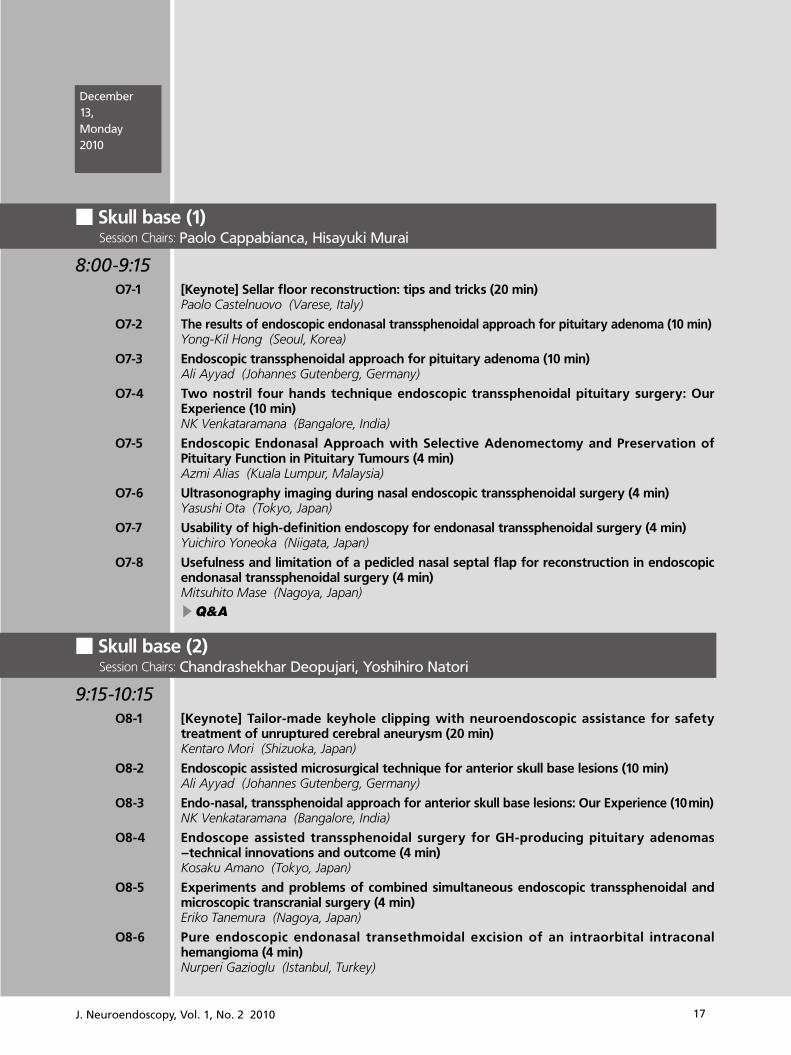
■ Skull base (1)Session Chairs: Paolo Cappabianca, Hisayuki Murai
8:00-9:15 O7-1 [Keynote] Sellar floor reconstruction: tips and tricks (20 min)
Paolo Castelnuovo (Varese, Italy)
O7-2 The results of endoscopic endonasal transsphenoidal approach for pituitary adenoma (10 min)Yong-Kil Hong (Seoul, Korea)
O7-3 Endoscopic transsphenoidal approach for pituitary adenoma (10 min) Ali Ayyad (Johannes Gutenberg, Germany)
O7-4 Two nostril four hands technique endoscopic transsphenoidal pituitary surgery: Our Experience (10 min) NK Venkataramana (Bangalore, India)
O7-5 Endoscopic Endonasal Approach with Selective Adenomectomy and Preservation of Pituitary Function in Pituitary Tumours (4 min) Azmi Alias (Kuala Lumpur, Malaysia)
O7-6 Ultrasonography imaging during nasal endoscopic transsphenoidal surgery (4 min) Yasushi Ota (Tokyo, Japan)
O7-7 Usability of high-definition endoscopy for endonasal transsphenoidal surgery (4 min) Yuichiro Yoneoka (Niigata, Japan)
O7-8 Usefulness and limitation of a pedicled nasal septal flap for reconstruction in endoscopic endonasal transsphenoidal surgery (4 min) Mitsuhito Mase (Nagoya, Japan)
▶Q&A
■ Skull base (2) Session Chairs: Chandrashekhar Deopujari, Yoshihiro Natori
9:15-10:15 O8-1 [Keynote] Tailor-made keyhole clipping with neuroendoscopic assistance for safety
treatment of unruptured cerebral aneurysm (20 min) Kentaro Mori (Shizuoka, Japan)
O8-2 Endoscopic assisted microsurgical technique for anterior skull base lesions (10 min) Ali Ayyad (Johannes Gutenberg, Germany)
O8-3 Endo-nasal, transsphenoidal approach for anterior skull base lesions: Our Experience (10 min) NK Venkataramana (Bangalore, India)
O8-4 Endoscope assisted transsphenoidal surgery for GH-producing pituitary adenomas -technical innovations and outcome (4 min) Kosaku Amano (Tokyo, Japan)
O8-5 Experiments and problems of combined simultaneous endoscopic transsphenoidal and microscopic transcranial surgery (4 min) Eriko Tanemura (Nagoya, Japan)
O8-6 Pure endoscopic endonasal transethmoidal excision of an intraorbital intraconal hemangioma (4 min) Nurperi Gazioglu (Istanbul, Turkey)
December 13, Monday 2010

18 J. Neuroendoscopy, Vol. 1, No. 2 2010
O8-7 The advantages of binostril endoscopic endonasal surgery for pituitary and skull base tumors (4 min) Hiroyoshi Akutsu (Tsukuba, Japan)
▶Q&A
10:15-10:30 CoffeeBreak
■ Simposium (2) : Extended endoscopic skull base surgery Session Chairs: Carlos Gagliardi, Federico Di Rocco, Naokatsu Saeki
10:30-12:30 S2-1 [Keynote] Endoscopic Endonasal Skull Base Surgery (20 min)
Paolo Cappabianca (Napoli, Italy)
S2-2 [Keynote] Evolution of Endoscopy in Skullbase Surgery (20 min) Chandrashekhar Deopujari (Bombay, India)
S2-3 [Keynote] Endoscopic endonasal skullbase surgery (20 min) Henry W. S. Schroeder (Ernst Moritz Arndt, Germany)
S2-4 [Keynote] Endoscopic endonasal skull base surgery (20 min) Naokatsu Saeki (Chiba, Japan)
S2-5 Dural reconstruction with a vascularized mucosal flap after endonasal endoscopic skull base surgery (10 min) Masahiro Toda (Tokyo, Japan)
S2-6 Endoscopic endonasal approach for skull base chordoma and chondrosarcoma - a follow up of rwenty cases (10 min) Yen Yu-Shu (Taipei, Taiwan)
▶Discussion
Hands-on Workshop (2)─ Skull base and spine KarlStorz
12:30-13:00
12:30-13:15 Lunch
13:15-13:30 Posterviewingtime
■ Cerebrovascular disease Session Chairs: Yoko Kato, NK Venkataramana
13:30-14:10 O9-1 What has changed after introduction of neuroendoscopy to treatment of intracerebral
hemorrhage (10 min) Takeya Watabe (Aichi, Japan)
O9-2 Endoscopic evacuation of various types of intracerebral hematomas (4 min) Shigeo Yamashiro (Kumamoto, Japan)
O9-3 Efficient endoscopic evacuation method for hypertensive intracerebral hematoma (4 min)Kentaro Fujii (Kariya, Japan)
O9-4 Neuroendoscopic evacuation of intraventricular hematoma associated with thalamic hemorrhage to shorten the duration of external ventricular drainage (4 min) Sadahiro Nomura (Yamaguchi, Japan)

19J. Neuroendoscopy, Vol. 1, No. 2 2010
O9-5 The comparative study of the outcome after neuroendoscopic surgery for intracerebral hemorrhage (4 min) Kazuo Tokushige (Komoro, Japan)
O9-6 Significance of endoscope-assisted microsurgery for clipping of unruptured cerebral aneurysms: Prevention of perforating artery infarction (4 min) Yoshiaki Kumon (Ehime, Japan)
O9-7 Hemostatic agent delivery system for neurosurgical endoscopy (4 min)Vickneswaran Mathaneswaran (Kuala Lumpur, Malaysia)
▶Q&A
■ Panel discussion: Neuroendoscopy in the world (1) Session Chairs: Giuseppe Cinalli, Shlomi Constantini
14:10-15:30 PD-1 From Japan (15 min)
Takayuki Ohira (Tokyo, Japan)
PD-2 Endoscopic transsphenoidal surgery initial experience from Pakistan (15 min)Salman Sharif (Karachi, Pakistan)
PD-3 From Thailand (15 min)Wuttipong Tirakotai (Thailand)
PD-4 Changingthe Paradigm in Neuroendoscopic training (15 min)Mahmood (Moody) Qureshi (Nairobi, Kenya)
PD-5 Russian neuroendoscopy: the past, present, future (15 min)Sufianov Albert A (Tumen, Russia)
15:30-15:45 CoffeeBreak
■ Panel discussion: Neuroendoscopy in the world (2) Session Chairs: Shizuo Oi, Mahmood (Moody) Qureshi, Benjamin Walf
15:45-17:30 PD-6 The Evolution of Neuroendoscopy (15 min)
Dieter Hellwig (Hannover, Germany)
PD-7 From Italy (15 min)Giuseppe Cinalli (Napoles, Italy)
PD-8 Report of 500 neuroendoscopic procedures: results, analysis and discussion (15 min)Alvaro Cordova (Montevideo, Urguay)
PD-9 From Algentina (15 min)Carlos Gagliardi (La plata, Algentina)
PD-10 Educate One to save a Few, Educate a Few to Save Many (15 min) Benjamin C. Warf (Boston, USA)
▶Discussion
■ IFNE 2010 Congress presidential adress and closing remarks
17:30 Masakazu Miyajima (Tokyo, Japan)
19:00 Galadinner

20 J. Neuroendoscopy, Vol. 1, No. 2 2010
■ Poster session
8:00-18:00
P-1 A case of neonatal huge arachnoid cyst in the posterior fossa treated by endoscopic fenestrationYoung-Soo Park (Nara, Japan)
P-2 New endoscopic treatment for sylvian fissure arachnoid cyst Hisaaki Uchikado (Kurume, Japan)
P-3 Complete anterograde amnesia following spontaneous haemorrhage in a colloid cyst resolving after neuroendoscopic excision Ramanan Sivakumaran (Frenchay, Bristol)
P-4 The thirdventricle pressure monitoring after endoscopic third ventriculostomy: an effective method to manage the 'adaptation period'. Yasushi Shin (Nara, Japan)
P-5 Neuroradiological and neuroendoscopic findings in two patients with sylvian aqueduct syndrome induced by shunt malfunction Ryo Doi (Kurume, Japan)
P-6 Treatment strategy for intraventricular and paraventricular tumors in the neuroendo-scopic eraKunikazu Kurosaki (Toyama, Japan)
P-7 Navigation-guided neuroendoscopic biopsy for intraparechymal brain tumors Yasunori Fujimoto (Osaka, Japan)
P-8 Efficacy of endoscopic tumor biopsy for management of central nervous system lymphoma associating with ventricular system Nobuyuki Nakajima (Tokyo, Japan)
P-9 Results of surgical treatment for Cushing’s disease Grigoriev AJ (Russia)
P-10 Significance of sequestration of the lateral recess for endoscopic sphenoid sinus surgery in the management of mucormycosis Ryusuke Ogawa (Tane, Japan)
P-11 Using of silicone balloon tube for prevent of cerebllospinal fluid leakage after endoscopic transnasal pituitary surgery Nobuyuki Kobayashi (Ibaraki, Japan)
P-12 Usefulness and limitations of endoscope in the microsurgery of petrous apex cholesteatoma Adriana Tahara (Hiroshima, Japan)
P-13 A case of visual deterioration after pituitary surgery Shinya Jinguuji (Niigata, Japan)
P-14 The role of neuroendoscopy for pineal region tumor Tomonori Suzuki (Saitama, Japan)
December 12, Sunday13, Monday 2010
21J. Neuroendoscopy, Vol. 1, No. 2 2010
O1-1 Neuro-endoscopy in infants and the IIHS progress report
Shlomi Constantini1, Abhaya Kulkarni2, Spyros Sgouros3
1Tel Aviv, Israel, 2Toronto, Canada, 3Athene, Greece
During recent meetings of the International Study Group on Neuroendoscopy (ISGNE) (within the IFNE) and the International Society for Pediatric Neurosurgery (ISPN), the consensus view emerged that there is a need to assess scientifically the value and efficacy of neuroendoscopic procedures against shunting in a scientific manner to resolve long-lasting debates on the subject. A prospective randomized, controlled trial of endoscopic third ventriculostomy Vs shunting in children presenting under the age of 2 years with pure aqueduct stenosis is been proposed and organized (the International Infant Hydrocephalus Study, IIHS)
The participating surgeons must adhere to the philosophy of randomization and be suitably experienced in endoscopic techniques in infants. The primary outcome of the trial is the overall health related quality of life of these children at 5 years of age. Hence, the study is focusing on the effect of surgery on neurodevelopment, rather than the less important issue of shunt or stoma survival, that has been debated extensively with no conclusion so far. Intention-to-treat 1analysis is performed according to the first surgery. Secondary outcomes such as complication and reoperation rate, total hospitalization time, and cost, need for repeat imaging, and others will be analyzed as well.
Pure aqueduct stenosis is relatively rare, making recruitment problematic, but has been chosen to avoid other confounding factors that could influence outcome. More than 40 centers worldwide have committed already to patient recruitment to the study. We have already more than 150 patients enrolled. It is anticipated that recruitment will last for 3 more years, aiming for 100 ”clean” patients per arm.
In this talk we will provide more details on the progress of the IIHS and also overview the current situation of neuroendoscopic procedures for other pathologies.
O1-2Long-term outcome of endoscopic third ventriculostomy (ETV) compared to CSF shunt in children with hydrocephalus
Abhaya V. KulkarniHospital for Sick Children, Toronto, Canada
With ETV continuing to gain popularity as a treatment for pediatric hydrocephalus, it is important to critically consider how the long-term outcome of this treatment compares to traditional CSF shunting. In the absence of randomized data, however, comparison of outcome between ETV and CSF shunt is very difficult because of the vast differences in the patient populations, i.e., there is strong treatment selection bias.
In comparing the rate of treatment failures, our group has used propensity-score adjustment techniques and analysis stratified-by-prognosis to partially overcome this treatment selection bias. Using these methods in a sample of 1209 patients, we identified the characteristics of patients that predict a better treatment outcome from ETV compared to shunt in both the short- and long-term. Conversely, we identified which patients actually fare better with CSF shunt compared to ETV. We have discovered as well that, across all patient groups, the early rate of ETV failure is higher than that of shunt failure, but the delayed failure rate of ETV is consistently lower than that of CSF shunt. This suggests that there could be some consideration for ETV in even less-than-ideal candidates in the hopes of achieving long term benefit in at least some.
In comparing quality of life outcome, our group has used subgroup analysis and multivariate regression analysis to partially overcome the problem of treatment selection bias. Using these methods in both small (N = 47) and large (N = 603) samples of patients, we have found very little difference in the long-term quality of life outcome of ETV compared to shunt.
In conclusion, there is much that still needs to be learned about the long-term outcome of ETV compared to CSF shunt. While we await more definitive randomized data, our current analyses are beginning to shed some light on this complex issue.
■ Hydrocephalus (1)
Day 1 December 12 Sunday
21
ABSTRACTS

22 J. Neuroendoscopy, Vol. 1, No. 2 2010
O1-3 Radiological features of hydrocephalus due to isolated aqueduct stenosis in children in the first 2 years of life. Qualitative analysis of neuroradiological material of the International Infant Hydrocephalus Study
Spyridon SgourosAthene, Greece
O1-4 Infant hydrocephalus in Africa: prevalence, causes, endoscopic treatment, and strategies for the future
Benjamin C. Warf Director of Neonatal and Congenital Anomaly Neurosurgery Department of Neurosurgery Children's Hospital Boston, USA
We estimate that up to 250,000 infants may develop hydrocephalus in Sub-Saharan Africa each year. The majority is caused by neonatal infection, and should be preventable. To this end, we are currently working to identify the responsible pathogens. With an average of 1 neurosurgeon per 10,000,000 people in Sub-Saharan Africa, initial treatment for hydrocephalus is often inaccessible. This also renders shunt-dependence more dangerous in Africa than in the developed world. Endoscopic third ventriculostomy combined with choroid plexus cauterization (ETV/CPC) has proven effective in avoiding shunt dependence in the majority of infants. Unlike shunts, most failures of endoscopic treatment are evident in the early months after surgery when responding to treatment failure is less urgent, while later failures are rare. We have identified easily accessible clinical parameters that predict the likelihood of success in a given patient. There appears to be no developmental advantage to shunt-dependence compared to treatment by ETV/CPC. We have trained and equipped surgeons in 10 developing countries to perform the technique. We hope to broaden the scope of this program in the future to provide the training, mentoring, patient follow up, and research needed to significantly reduce the morbidity and mortality of this disease.
22

23J. Neuroendoscopy, Vol. 1, No. 2 2010
O2-1 Development of Arachnoid Villus
Yusuf Ersahin1, Eylem Burcu Akgül1, Yusuf Ersahin1, Yesim Ertan2, Taner Akalıdn2, Cafer Uysal31Division of Pediatric Neurosurgery, Ege Univerity Faculty of Medicine, Izmir, Turkey2Department of Pathology, Ege Univerity Faculty of Medicine, Bornova, Izmir, Turkey3Izmir Branch Chairmanship, The Council of Forensic Medicine, Izmir, Turkey
Arachnoid villi are considered as the major site of cerebrospinal fluid absorption. We aimed to study the development of arachnoid villus in fetuses and small infants.
Using random sampling method, postmortem fetuses greater than 26 weeks of gestation and the children under the age of 1 were chosen from the autopsy materials. 2 male and 2 female intrauterine dead fetuses; 3 male and 3 female, totally 6 children under the age of one and one 3 years old male were included in this study. The autopsy cases which have central nervous system disease or pathology were excluded from this study.
In cases of intrauterine fetus greater than 26 weeks of gestation and children under the age of 1; complete invagination of arachnoid villi into the superior sagittal sinus were examined histologically. In intrauterine period and in the first six months of life arachnoid villi structures were not found in histological preparations although in preparations taken after the six months of life samples showed similarities to arachnoid granulations. These structures were considered as arachnoid villi drafts after immunohistochemical analysis. In the control case who were 3 years old, maturation of arachnoid villi were complete and the arachnoid villi were invaginated into the superior sagittal sinus as fingerlike extensions.
In conclusion, the absence of arachnoid villi around the superior sagittal sinus can account for the high failure rate of endoscopic third ventriculostomy before the age of six months.
O2-2Endoscopic aqueductoplasty with or without stent in the treatment of aqueductal obstructive hydrocephalus
Chen Guoqiang, Xiao Qing, Zheng Jiaping, Wu Jinting, Huang Yiyang, Zuo HuancongYuquan Hospital Tsinghua University, China
Objective: To summarize the operative indication and effect of endoscopic aqueductoplasty (EAP) with or without stent for the treatment of aqueductal obstructive hydrocephalus.
Methods: 76 cases of aqueductal obstructive hydrocephalus due to different etiology were treated with EAP with or without stent under flexible endoscope via trans-frontal or suboccipital approach respectively from February, 2007 to May, 2010.
Results: Of 76 patients, aqueductal membranous obstruction in 20 cases, aqueductal stenosis in 24 cases, posthemorrhagic hydrocephalus in 19 cases and postinfective hydrocephalus in 13 cases. Sucessful single EAP with no corresponding complications achieved in 61 cases apart from 3 cases who suffered a second EAP with stent due to restenosis 2 to 3 months after primary EAP. Primary EAP with stent in 10 cases. Failure of EAP appeared in 2 cases because of long-segment tightly adherence and zigzag shape of the aqueduct. A satisfactory cerebrospinal fluid flow through aqueduct were demonstrated by using cine-MRI during a follow-up period of 6-36 months (mean 18.6 months) in 65 cases.
Conclusion: Endoscopic aqueductoplasty with or without stent by means of flexible endoscope is a safe and effective method for the treatment of aqueductal obstructive hydrocephalus. Aqueductal membranous obstruction is the optimal indication and the stent is usually necessary for those aqueductal stenosis due to posthemorrhagic or postinfective hydrocephalus.
23
■ Hydrocephalus (2)

O2-3Endoscopic Third ventriculostomy for Hydrocephalus associated to Posterior Fossa Tumors
Federico Di Rocco, Carlos Eduardo Jucá, Thomas Roujeau, Stephanie Puget, Michel Zerah, Christian Sainte-RosePediatric Neurosurgery Hopital Necker Enfants Malades, France
Background: ETV is nowadays the best option for treating the hydrocephalus associated to posterior fossa tumors persisting after the lesion removal. Management of hydrocephalus prior to the removal of the posterior fossa tumor is, on the other hand, still debated. Some authors emphasize the advantages of an immediate tumor removal which may cure the associate hydrocephalus in a relevant number of cases. In the clinical practice, however, such a policy is not ever achievable. Furthermore, the mere excision of the lesion has been demonstrated to be accompanied by a persisting hydrocephalus in about a third of the cases.
Preoperative ETV allows controlling the ICP, avoiding an emergency procedure, scheduling the operation for tumor removal appropriately, eliminating the risks of an external drainage. It also reduces the incidence of postoperative hydrocephalus. A final advantage, more difficult to weight is the possibility to perform the removal of the lesion on a relaxed brain.
In the last years we performed an ETV in all patients with hydrocephalus associated to a posterior fossa tumor and carried out the lesion removal 48 hours later. Aim of this study is to review the results of such an approach.
Patients and Methods: Between January 1995 and December 2004, 157 ETVs were performed for hydrocephalus secondary to a posterior fossa tumor. There were 87 boys (mean age : 6 years ).
Results: ETV was successful in 127 cases. Six children necessitated a second ETV, whereas 21 were shunted. Three patients died during the post-operative period. Among the shunted patients, 7 underwent one or several shunt revisions. Mean hospitalization was 7 days. No permanent morbidity due to the ETV was recorded. Mean follow up was 44.73 months.
Discussion: Out of the 154 survivors, ETV treated the hydrocephalus in 86% of the cases successfully. The rate of persistent postoperative hydrocephalus was reduced to 17% compared to 30% of literature. Out of the 27 children with persistent postoperative hydrocephalus, 6 responded to a redone ETV. The placement of an extrathecal CSF device was required only in 21 children (14%). Minor rates of extrathecal -shunted patients are reported by authors who utilize ETV postoperatively. The assessment of the persistent hydrocephalus in these children is based on prolonged ICIP monitoring through an external CSF drainage, which may result in infective complications and longer hospital stay.
O2-4 The analysis of Five cases of aqueduct stenosis treated successfully by Endoscopic Third Ventriclestomy after shunt malfunction
Jun Muto1, Takayuki Ohira1, Takahito Yazaki2, Kazunari Yoshida1
1Department of Neurosurgery, International University of Health and Welfare Mita Hospital, Japan2Department of Neurosurgery Keio University, School of Medicine, Japan
Purpose: We have introduced five cases of aqueduct stenosis treated successfully by Endoscopic Third Ventriclestomy(ETV) after shunt malfunction, and analyzed the timings of operation, strategies, intraoperative findings of ETV operations in the cases of shunt malfunction.
Cases and Results: We have experienced the five cases for 32 years. The each patients’ age occurring the aqueduct stenosis are 5, 6, 7, 9, 19 years old. The average distance between the first shunt operation and ETV are 16.0 years (1〜31 years). The average frequency of operations before ETV are 2.8 times (1〜10 effective for aqueduct stenosis and there is no sudden occlusion.
Discussion: The effectiveness of ETV after long term shunt malfunction are reported.The ETV could be effective for the primary and secondary aqueduct stenosis.We should think the therapeutic options of the ETV whether it is pre- or post- shunt operation.
24 J. Neuroendoscopy, Vol. 1, No. 2 2010

O2-5 The Endscopic Third Ventriculostomy can improve higher brain function in the patient of Long-standing Overto Ventriculomegaly in Adult?
Shizuka Majima, Syogo Fukuya, Tomohiko Ozaki, Koshi Ninomiya, Akihiro Tateishi, Masami Nishio, Takuyu TakiKansai Rosai Hospital, Japan
Object: Generally, headache and NPH-like symptoms are the main presenting features of Long-standing Overt Ventriculomegaly in adults (LOVA) patients. Endscopic third ventriculostomy (ETV) has been accepted as a procedure of choice for the treatment of LOVA. Although the outcome of higher brain function remains controversial in LOVA. The purpose of this study is to evaluate the role of ETV in the treatment of LOVA.
Methods: Date collected in four patients with LOVA (three females and one males) who had undergone ETV between 2009 and 2010 were reviewed outcome. The patients ranged from 52 to 71 years old on first presentation. All patients presented with headache, gait disturbance, urinary incontinence and disturbance of cognitive functions. All patients underwent magnetic resonance imaging. In all four patients, the magnetic resonance imaging revealed severe triventriculomegaly, macrochephalus, and aqueductal stenosis. In addition, we evaluated the effect of ETV as the higher brain function evaluation using Mini-Mental State Examination (MMSE) and initiative ability.
Results: All patients had undergone successful ETV and improved not only headache, gait disturbance and urinary incontinence, but also slow movement and MMSE. They had no complications of hemorrhages, infections, stoma closure.
Conclusion: In LOVA patients, ETV lead to improvement in higher brain functions. MMSE provides useful for evaluating cognitive functions anytime. ETV may be indicated for more comfortable and independence life support of LOVA patients.
O2-6 Dyanamic changes of the anatomical components surrounding the third ventricle play a key role in successful neuroendoscopic third ventriculostomy for chronic hydrocephalus
Namiko Nishida1, Yoo Kang2, Jun A Takahashi1, Masatsune Ishikawa3
1Department of Neurosurgery, Tazuke Kofukai medical research institute and Kitano Hospital, Japan2Department of Neurosurgery, Jurakukai Ohno Memorial Hospitall, Japan3Department of Neurosurgery and normal pressure hydrocephalus center, Rakuwakai Otowa Hospitall, Japan
The aim of this study was to analyze the relation between morphological variables of adult-onset chronic hydrocephalus and clinical outcome after neuroendoscopic third ventriculostomy (nETV). We assessed consecutive 9 patients (age 63 ± 6.9 years, disease duration 5.1±5.8 years, follow up 16.6±11.6 months). Clinical evaluation was based on idiopathic normal pressure hydrocephalus grading scale (iNPHGS) and 3M up and go test. Following values were obtained from pre- and postoperative MRI images: maximum bifrontal width (A), bicaudate width (B), third ventricular width (C), and cella media width (I), distance between anterior and posterior commissures (J), altitude of anterior commissure (K) and posterior commissure (L) from planum sphenoidale. Aqueductal obstruction and prepontine arachnoid scarring were examined intraoperatively. Eight out of nine patients recognized the alleviation of symptoms. Locomotive improvement were represented by up and go test (20.1 ± 5.5 to 15.9 ± 2.0 sec., p = 0.05). Hydrocephalic triad represented by iNPHGS improved from 6.6 ± 2.6 to 3.3 ± 1.6 (p = 0.01). Radiologically, C, J, and altitude distance between commissures (K-L) reduced significantly. The change of up and go test and that of C correlated significantly (Spearman r = 0.82, p = 0.02). The change of iNPHGS and that of K-L correlated moderately (Spearman r = 0.59). Intraoperative findings of aqueduct and prepontine space did not affect the outcome significantly. We propose that dyanamic changes of the anatomical components surrounding the third ventricle may play a key role in successful nETV for chronic hydrocephalus.
25J. Neuroendoscopy, Vol. 1, No. 2 2010

26 J. Neuroendoscopy, Vol. 1, No. 2 2010
O2-7 Obstruction stoma after endoscopic third ventriculostomy
Atsuko Harada, Kenichi Nishiyama, Junichi Yoshimura, Yukihiko FujiiDepartment of Neurosurgery, Brain research intsitute, Niigata University, Japan
Object: The goal of this study was to clarify the endoscopic and histopathological features of the obstruction of stomas after endoscopic third ventriculostomy (ETV) and to analyze their mechanism.
Methods and results: Between 1997 and 2010, 143 ETV procedures were undertaken in our department. In 9 patients with ventriculostoma obstruction, ETV procedures were repeated. At the second ETV procedures, the ventriculostomas of the first procedures were endoscopically inspected. In two of the patients, histopathological examinations were carried out. The primary etiologies of hydrocephalus were as follows: midbrain tumor (4 cases), primary aqueductal stenosis (1), Chiari malformation type 1 (1), Chiari malformation type 2 (1), obstructive hydrocephalus prior intracranial hemorrhage (1), and Blake's pouch cyst (1). The endoscopic findings at the second procedures were as follows: complete obstruction of the ventriculostoma by postoperative granulation tissue in 7 cases (group 1) and patent ventriculostoma with arachnoid membranes newly formed in the basal cisterns just below the floor of the third ventricle in 2 (group 2). Two of the nine patients suffered postoperative infection in group 1. In the patient having midbrain tumor associated with neurofibromatosis type 1, the pathological examination of the granulation of the obstructed ventriculostoma revealed gliosis with reactive astrocytes. All of the two patients in group 2 suffered complicated infection. Our histopathological examination of one patient of group 2 disclosed the proliferation of macrophage. The interval between the first and second ETV procedures in group 1 and group 2 ranged from 14 to 644 days and from 9 to 26 days, respectively.
Conclusions: The risk of ventriculostoma obstruction seems to increase with infection, neurofibromatosis, and insufficient ventriculostoma. It also seems plausible that the obstruction of ventriculostomas is partly attributable to newly formed arachnoid membranes after postoperative infection.
O2-8 Utilization of infant feeding catheter in Transcranial Endoscopy: An Improvised Technique
Azmi Alias, Mohammed Saffari MH.Department of Neurosurgery, Hospital Kuala Lumpur, Malaysia
Objectives: We present our experience on utilizing Infant feeding catheter as an adjunct tools in various Transcranial Intraventricular Neuroendoscopy procedures performed at Department of Neurosurgery, Hospital Kuala Lumpur, Malaysia from January 2005 to October 2010.
Methods: A modification on 5 Fr infant feeding catheter is made by cutting it’s distal end and used as a tools for aspiration, fenestration, irrigation and suction biopsy. Depending on type of procedures, the proximal end of the catheter is connected either to 20ml syrinx or suction tip and inserted through the endoscope working channel. Intraventricular debris, clots or pus is simultaneously aspirated and irrigated with Hartmann’s solution through the catheter under endoscopic visualization till clear return. Aesculap (MINOP) Cranial endoscope was used in most of the cases with free hand technique.
The procedures include Endoscopic Excision of Colloid Cysts , Endoscopic Intraventricular lavage for Pyogenic Ventriculitis, Compartmentalized Hydrocephalus, fenestration of Intraventricular Arachnoid Cyst, Intraventricular tumours and haemorrhage. All cases had radiological evidence of obstructive hydrocephalus and dilated ventricle on CTscan / MRI findings.
Results: Complete decompression of Colloid Cysts was achieved in all cases with 90% complete excision of the cyst wall. None of the patient with colloid cyst required any shunts after the procedure. Patients with Pyogenic Ventriculitis and haemorrhage had a significant removal of pus and debris with reduced size of ventricle.
Conclusion: Simple modification using the infant feeding catheter proved to be helpful in our experience on various Transcranial Neuroendoscopy procedures. It can be used as an effective adjunct tools at a very low cost.

O2-9 Shunt related porencephalic cyst treated by neuroendoscopic technique
Masaki Shinoda, Motoharu Fujii, Hidetoshi Matsukawa, Daisuke Yamamoto, Atsushi Murakata, Ryoichi IshikawaSt. Luke’s International Hospital, Japan
There are various things for a complication of VP shunt, but there are few reports of intracerebral cyst formation. We experience the case that showed an intracerebral cyst along catheter to a VP shunt postoperative patient this time and report it because we experienced the case that we perform shunt restoration with rigid neuroendoscopy and got excellent results.
A case: 11 m/o girl. She had Arnold-Chiari malformation. Closure of myelomeningocele which level was S1 was performed at her first day of life. VP shunt was performed on her third day of age because of her developing hydrocephalus. The postoperative course was good. Extrusion of the ventricular catheter was complicated on her fourth month of age, and reinsertion of the ventricular catheter was performed. The postoperative course was good, but MRI showed intracerebral cyst formation in the frontal lobe of 3.8 x 3.0 x 3.6 cm along cerebral ventricle side catheter. Second shunt revision with neuroendoscopy was performed. The previous catheter was present without any damages. The gliosis was present as adhered and unified surround the catheter. The catheter trace was carefully enlarged with neuroendoscopic technique and re-inserted ventricular catheter with transversal holes. The cyst was disappeared after the operation.
S1-1 Idiopathic normal pressure hydrocephalus: comparison between Japanese guidelines and International guidelines
Masatsune Ishikawa Normal Pressure Hydrocephalus Center, Rakuwakai Otowa Hospital, Kyoto, Japan
27J. Neuroendoscopy, Vol. 1, No. 2 2010
■ Simposium (1) : The role of ETV for NPH

S1-2 Reported results on endoscopic third ventriculostomy in normal pressure hydrocephalus
Mikhail Chernov1, Yoshihiro Muragaki1, 2, Yoshikazu Okada2, Hiroshi Iseki1, 2
1Faculty of Advanced Techno-Surgery; Tokyo Women’s Medical University, Japan2Department of Neurosurgery; Tokyo Women’s Medical University, Japan
Introduction: Normal pressure hydrocephalus (NPH) with a typical clinical syndrome (gait disturbances, dementia, urinary inconsistency) is a well recognized clinical entity. Ventriculoperitoneal shunting represents the standard treatment, which, however, is accompanied by high rate of complications. During the last decade there is a growing interest in endoscopic third ventriculostomy (ETV) for treatment of this condition.
Objective: Analysis of the published clinical series, which included patients with NPH treated with ETV. Results: Twelve published series were identified in the literature. The number of patients varied from 4 to 110. In the
majority of studies the effectiveness of ETV varied from 65% to 85%. It seems that underlying pathophysiological mechanism of NPH has profound influence on the results of the procedure. Possible favorable prognostic factors include short duration of symptoms, preponderance of gait disturbances, deformation of the third ventricle floor, and mismatch of the resistance to CSF outflow during lumbar and ventricular infusion tests. Attainment of the so-called “complete third ventriculostomy” with perforation of the third ventricle floor, Liliequist`s membrane, and basal arachnoid adhesions, is important.
Conclusion: According to published results ETV may be effective in the majority of patients with NPH. Nevertheless, favorable prognostic factors and criteria of patients’ selection for the procedure require further clarification.
S1-3ETV in selected cases of communicating hydrocephalus in adults
Shoichiro IshiharaSaitama, Japan
28 J. Neuroendoscopy, Vol. 1, No. 2 2010

29J. Neuroendoscopy, Vol. 1, No. 2 2010
S1-4 Infratentorial intracisternal obstructive hydrocephalus (InfinOH): how often is this subtype, which can be treated endoscopically, among idiopathic normal pressure hydrocephalus (iNPH)?
Uwe Kehler, Herzog, JuleNeurosurgical Department, Asklepios Clinic Altona, Hamburg, Germany
Objective: InfinOH is a communicating hydrocephalus with infratentorial intracisternal CSF-pathway obstruction, which can be treated by endoscopic third ventriculostomy (ETV). We have found earlier that InfinOH with clinical presentation of an iNPH can be treated successfully with ETV, showing a success rate of around 70%. The incidence of InfinOH in iNPH patients is unknown. But knowing the incidence could help us to focus our attention on this subtype, assisting to detect iNPH patients which could be treated with ETV instead of shunting.
Methods: Between August 2005 and June 2010 iNPH patients, who were treated in our department were evaluated. In all cases thin mid-line sagittal T2 weighted MRI slices were analyzed for InfinOH: Diagnosis was confirmed, when the aqueduct as well as the outlet of the fourth ventricle were patent but the floor of the 3rd ventricle was bulged downward and eventually the lamina terminalis pushed forward as well.
Results: We treated surgically 165 patients with iNPH between 2005 and June 2010. Twenty-one patients with clinical signs of iNPH were detected after MRI-analyzis as InfinOHs. This results in a frequency of 12,7% of iNPH. The mean age of InfinOH group was 56,7 years (range: 35 -76y) , in the rest of iNPH it was 72,2 years (range: 25-87 years). All InfinOH patients were treated with ETV, all other pat. were treated with vp-shunt.
Conclusions: InfinOH is not frequently (12,7%) found in iNPH patients. But if these patients would be neglected, almost 13% of patients would get unnecessarily a shunt, although ETV would be very promising alternative to shunt surgery. Focusing our attention on InfinOH could help improving patients’ selection for ETV or shunt.
S1-5 Clinical outcome of neuroendoscopic third ventriculostomy (ETV) in elderly patients
Tamotsu MIKI1, Takehiro Tomita1, Tomoo Ohashi1, Tatsuya Nakamura1, Shinjiro Fukami2, Jun Wada2, Noubuyuki Nakajima2
1The Department of Neurosurgery, Tokyo Medical University, Ibaraki Medical Center, Japan2The Department of Neurosurgery, Tokyo Medical University, Japan
Neuroendoscopic third ventriculostomy (ETV) is an established surgical procedure for the treatment of non-communicating hydrocephalus. We performed an investigation of the clinical outcome of ETV in geriatric patients.
The subjects were 24 patients aged from 70 to 84 years old (mean age: 75.6 years). They were compared with non-geriatric patients in terms of their preoperative condition, shunt history, underlying cause of hydrocephalus, operative results, complications, and other factors. The postoperative follow-up period ranged from 4 to 114 months, with a mean of 56.5 months. Fourteen patients had symptoms associated with increased intracranial pressure, while 10 patients showed NPH-like symptoms. Seventeen patients (71%) showed improvement after initial ETV, as compared with 87% in non-geriatric patients.
Pathologic evaluation of hydrocephalus based on neuroendoscopic intraventricular observation and/or biopsy is very useful even in geriatric patients because it provides data that cannot be obtained by diagnostic imaging. However, the results of ETV were slightly worse in geriatric patients than in non-geriatric patients. The possible reasons include (1) problems with the indication for ETV, (2) problems with the surgical technique due to anatomical changes related to aging such as arteriosclerosis of the basilar artery, and (3) age-related cerebral atrophy and loss of elasticity. Very careful preoperative assessment is necessary before performing ETV in geriatric patients.

■ Intra-ventricular lesions (1)
S1-6 A Prospective, Randomised, Controlled Trial of the Neuroendoscopic Treatment of Idiopathic Normal Pressure Hydrocephalus (ISRCTN29863839)
Richard J. Edwards, Kristian Aquilina, Martin Bunnage, Ian K PopleDept of Neurosurgery, Frenchay Hospital, Bristol, UK
Introduction: Previous retrospective studies have suggested that endoscopic treatment of iNPH may be as effective as CSF shunting. We report a blinded PRCT comparing iNPH patients treated with neuroendoscopy and patients treated with a programmable VP shunt.
Methods: Patients were randomized to neuorendoscopy (treatment) or VP shunting (control). Patients in the treatment arm were offered crossover to VP shunting in the event of treatment failure. The primary outcome measure (intention to treat analysis) was the 3-month Raftopoulos gait score assessed on gait video by a blinded assessor. Secondary outcome measures included modified Rankin Disablity Score; Barthel index; Dutch NPH score; Folstein MMSE Score and the Mattis II Dementia Rating Scale measured at 3 & 6 months and 2-years. The neuropsychologist was blinded to the treatment allocation.
Results: The trial was stopped early after randomization of 21 patients due to concerns over a lack of efficacy. There were significant improvements in both gait (p = 0.04) and mRS (p = 0.001) at 3 months in the shunted group but not the neuroendoscopy group. Cognition, by a number of outcome measures, was significantly better in control patients at 3 & 6 months and 2 years. All patients in the neuroendoscopy arm eventually “crossed over” to CSF shunting. Comparison of pre- and post-crossover gait and functional scores showed a significant improvement after placement of a shunt, but cognitive scores did not improve. There were no stoma blockages.
Conclusions: CSF shunting with a programmable valve is superior to treatment with endoscopic third ventriculostomy with choroid plexus coagulation in idiopathic NPH.
O3-1Infantile/toddler hydrocephalus
Conor MallucciDepartment of Neurosurgery, Royal Liverpool Children’s Hospital, Liverpool, UK
30 J. Neuroendoscopy, Vol. 1, No. 2 2010

31J. Neuroendoscopy, Vol. 1, No. 2 2010
O3-2Navigated endoscopic treatment of paraventricular cysts in children
Ulrich-W ThomalePediatric Neurosurgery, Campus Virchow Klinikum, Charité Universitátsmedizin Berlin, Germany
Paraventricular cysts may occur in congenital and in acquired conditions. The clinical symptoms may range from unspecific signs to progressive impairment, which may be caused by mass effect or CSF pathway obstruction.
For surgical treatment endoscopic fenestration is often chosen with navigated guidance to define the optimal entry point, the trajectory and the target for fenestration. In rare cases, placement of stents is indicated to enable sustainable communication of fluid. In congenital arachnoid cysts decision making for invasive treatment is often difficult when clinical symptoms are unspecific. Endoscopic treatment is reasonable to use in cases with significant mass effect or obvious signs of clinical impairment. In pineal cysts and suprasellar cysts, fenestration and third ventriculostomy is performed in parallel. For temporal cysts either fenestration towards the lateral ventricle or towards the basal cisterns is performed. In cases of acquired infectious conditions, when multi-cystic ventricles develop, early and repetitive endoscopic treatments are necessary, with the goal to establish ventricular cystic communication and to simplify the CSF diversion system with a minimal amount of ventricular catheters. For paraventricular tumor cysts an endoscopic approach can be chosen, if progression is limited to cystic components or if radical surgical treatment is of unreasonable risk. Thereby, guided endoscopy can facilitate the internal drainage of cyst fluid towards the CSF spaces, if a possible protein load is justifiable.
Endoscopic treatment is a helpful technique for paraventricular cysts as minimal invasive approach. Navigated guidance facilitates its optimal application strategy to secure the success of treatment.
O3-3 Endoscopic Approach to Temporal Horn: Anatomical Study
Enrique Ferrer1, Gonzalez Josep1, Benet Arnau2, Prats Albert2, Cordero Esteban1, Rumiá Jordi11Dept. Neurosurgery, Hospital Clinic Barcelona, Spain2Dept. Anatomy University of Barcelona, Spain
Introduction: Intraventricular endoscopic surgery is considered as a minimally invasive technique useful in the neurosurgical treatment of ventricular system diseases.
This technique has been developed mainly to treat pathology placed on frontal horn and third ventricle area.Authors make an anatomical study of endoscopic approach to temporal horn. Material and methods: In neuroanatomy lab, authors perform a macroscopic preliminary study on a formaline
anatomical specimen, of the temporal horn extending from uncal recess since to ventricular atrium. Helped by CT Scan and neuronavigator we studied 12 different temporal lobes specially prepared using the distensible fixation technique from Cambridge® and after injected with intravascular latex. Measures data collection and space reference of approach entry point and trajectory, has been done. Following this data, cortical area and optimal trajectory to get temporal horn in a more efficient way is defined in order to minimize eloquent structures surgical lesion.
Results: Optimal skin and bone entry point is defined in 2.76 cm. +/- 0.35 lateral to sagittal suture and 5.00 cm. +/- 0.47 superior to inion. Entry angle is 41.81º +/- 3.59 respect orbito-meatal axis and 5.85º +/-1.07 respect sagittal axis.
Distance from entry to ventricular atrium is 3.97cm. +/- 0.51. Tracking distance into the ventricle is 5.18 cm +/- 0.40. Finally subcortical anatomy in relation to the trajectory and temporal horn endoscopic anatomy is described using telescopes of 0º and 30º.
Conclusion: Endoscopic approach to temporal horn as has been described in the study is affordable and safe enough. Authors would like to develop this surgical corridor in order to treat endoscopically some pathological conditions and probably disconnection techniques used for epilepsy treatment under minimally invasive philosophy.

32 J. Neuroendoscopy, Vol. 1, No. 2 2010
O3-4 Combined intraoperative-MRI and navigated endoscopy in children with multiloculated hydrocephalus and complex cysts: a series-based feasibility study
Liana Beni-Adani, Dimitrios Paraskevopoulos, Naresh Byani, Shlomi ConstantiniDana Children's Hospital, Tel-Aviv Souraski Medical Center, Israel
Objective: The rationale of endoscopy for complex cysts and multiloculated hydrocephalus is minimize the number of isolated CSF spaces and decrease shunt dependency, number of proximal catheters, and if possible even to avoid a shunt. However, in cases of distorted anatomy, endoscopy alone might be problematic for orientation and re-evaluation of brain-shift. The aim of this project was to assess in infants the feasibility and efficacy of intraoperative-MRI (ioMRI) navigated-neuroendoscopy.
Patients and methods: We report our experience with five infants (ages 6-14 months), who were operated for multicystic hydrocephalus presenting with shunt malfunction (4 patients) and a quadrigeminal fetal arachnoid cyst (1 patient). IoMR (0.12/ 0.15 Tesla PoleStar) was combined with navigated endoscopy.
Results: The ioMRI provided clear images that correlated with the endoscopic appearance of the cystic membranes in all patients, was helpful in determining trajectories and redefining targets. The ioMR-images documented brain-shift and changes of CSF spaces during surgery. There were no intraoperative complications or technical difficulties of visualization. Patients were followed up for 6 months -7 years. The infant with the quadrigeminal cyst is still shunt free, and the patients with multicystic HCP have 1-2 shunts each.
Conclusions: The advantages of navigated-endoscopy and ioMRI may complement each other in specific indications and complicated procedures, where intraoperative changes alter the spatial distribution and targets and trajectories need to be redefined. IoMRI enabled transforming a blind conventional stereotactic procedure into a visually controlled procedure, allowed accurate navigation of the endoscope, and minimized number of compartments in all patients.
O3-5Neuroendoscopic approach to quadrigeminal cistern arachnoid cysts
Goksin Sengul, Yusuf Tuzun, Murteza Cakir, Sencer Duman, Abdullah Colak, Hakan Hadi Kadioglu, Ismail Hakki AydinAtaturk University, Department of Neurosurgery, Erzurum, Turkey
Objective: The introduction of neuroendoscopy has provided a minimally invasive modality for the surgical treatment of quadrigeminal arachnoid cysts. Three pediatric patients with arachnoid cyst of the quadrigeminal cistern treated by endoscopic fenestration are reported.
Method: The hospital records of patients were retrospectively rewieved. All patients had hydrocephalus. A lateral ventricle-ystostomy and endoscopic third ventriculostomy were performed by using rigid neuroendoscopes.
Results: There were one boy and two girls with ages 7 months, 9 months and 14 years, respectively. One patient had undergone shunting prior to neuroendoscopic surgery. The postoperative course was uneventful in all cases, with no complications. They showed disappearance of intracranial hypertension symptoms and significant reduction of the cyst size.
Conclusion: Neuroendoscopic technique is an effective and suitable method for the treatment of quadrigeminal cistern arachnoid cysts and accompanying hydrocephalus.

O3-6Supraorbital Endoscopic Approach to Colloid Cysts
Alberto Delitala, Andrea BrunoriDepartment of Neurosurgery, Ospedale San Camillo, Roma, Italy
In the last decade the endoscopic technique has developed as a 杜inimally invasive? approach for the resection of colloid cyst into the third ventricle. It is controversial whether a complete resection of the cyst is necessary to obtain a long term recovery. An anatomical limitation to a complete removal of the colloid cyst is the attachment of the cyst to the velum interpositum, that is difficult to visualize with conventional pre-coronal burr-hole approach, even if the burr-hole is placed some centimetres anterior to the coronal suture.
Matherial & Method: In order to better control the attachment of the cyst to the roof of the third ventricle, we performed a supraorbital endoscopic approach guided by the neuronavigation station (Medtronic). We applied this technique to remove a colloid cyst in four patients with slight enlargement of the ventricular system. A right supraorbital key-hole was done, and with the aid of the navigation system a rigid 12- endoscope was driven through the frontal horn of the ventricular system, medially to the caudatus nucleus, along the direction of the foramen of Monro. With free-hand technique, the cystic wall was coagulated and punctured, and the colloid was evacuated with grasping forceps and with continuous irrigation into the cystic space. This manoeuvre allows gentle retraction of the cystic wall and the visualization of the cyst attachment to the roof of the third ventricle.
Results: In order to avoid traction on the fornix, a subtotal resection of the cyst wall was accomplished in one case, where a small remnant of the cystic wall was left intact adhering to the velum interpositum. Despite of the fact that in one case a small piece of cystic wall was left adhering on the roof of the third ventricle, post-operative radiological images did not reveal any residual cyst in all cases. No patient manifested a neurological impairment after the procedure and all were discharged from hospital on fourth post-operative day. At mean radiological follow-up of 12 months, in no patient the relapse of the cyst was documented.
Conclusions: the supraorbital approach allows a good visualization of the nervous structures and of the cystic wall with its attachment, that is usually not visible with conventional approaches. With this approach a near complete resection of the cystic wall is possible, with no retraction of the adjacent structures and satisfying clinical results. A longer follow-up is needed to assess the efficacy of this technique.
O3-7Endoscopic Management of Large Cystic Craniopharyngiomas with Intraventricular Extension Using the Image Guided Expandable Brain Port
Vickneswaran Mathaneswaran1, Dharmendra Ganesan1, Nor Faisal Ahmad Bahuri1, Basri Johan Jeet Abdullah2
1Division of Neurosurgery, Department of Surgery, University Malaya Medical Centre, Kuala Lumpur, Malaysia2Department of Biomedical Imaging, University of Malaya Medical Centre, Kuala Lumpur, Malaysia
Introduction: Craniopharyngiomas frequently have multiple cystic components that grow superiorly and obstructs the foramen of Monro causing unilateral or bilateral hydrocephalus. These patient’s often present acutely with obstructive hydrocephalus requiring CSF diversion prior definitive management.
We present our experience using the expandable image guided neuroendoscopic port together with and endoscope in managing 4 patients (2 children and 2 adults) over a 18 month period.
Methodology and Results: In all four patients we were able to decompress the cyst and parts of the tumor avoiding any need for CSF diversion. In 3 patients this was followed by definitive excision at a later date of the residue using standard microsurgical techniques. In one patient the residue was judged small enough to avoid further surgery and the patient underwent radiotherapy.
Conclusion: In conclusion, this techniques avoids the need for a VP shunt (uni or bilateral) and reduces risk of CSF contamination of cyst fluid which can be a problem with conventional endoscopic techniques.
33J. Neuroendoscopy, Vol. 1, No. 2 2010
O3-8The Use of an Expandable Endoscopic Port with Electromagnetic Navigation for treating Intraventricular Lesions in Neonates and Infants
Vickneswaran Mathaneswaran1, Nor Faisal Ahmad Bahuri1, Vairavan Narayanan1, Jaweed1, Basri Johan Jeet Abdullah2, Dharmendra Ganesan1
1Division of Neurosurgery, Department of Surgery,University Malaya Medical Centre, Kuala Lumpur, Malaysia2Department of Biomedical Imaging, University of Malaya Medical Centre, Kuala Lumpur, Malaysia
Introduction: Operating within the ventricles of neonates and infants using an endoscope throws up major challenges due to the poorly myelinated parenchyma that can be easily damaged by excessive manipulation of the endoscope traversing the brain parenchyma.
We have successfully combined the use of electromagnetic (EM) navigation-Axiem Medtronic ® together with an expandable port to enable us to operate on a number of infants and neonates for a variety of intra-ventricular pathologies.
Methodology and Description of Port: The port in its closed position (during introduction into the brain) consisted of two flat titanium blades surrounded by a stretchable plastic sheath. In this closed position the leading edge measured 3mm by 6mm. The titanium blade construct allowed the port to be able to carry an electro-magnetic probe at its tip.
The port is introduced along a pre-planned trajectory guided by the EM probe that is housed within the port.Once the port is its optimal position as confirmed by the EM probe the blades can be spread open that in turn stretches
the plastic sheath creating a rectangular tunnel through which an endoscope and a variety of micro-instruments can be introduced independent of each other.
Results: Over the last 2 years we have utilised this technique in 6 children for a variety of pathologies that consisted of in 4 patients to re-establish CSF flow and 2 patients to excise or decompress intra-ventricular lesions. In all patients we were able to achieve our set target without any untoward incidents.
Discussion: The use of EM navigation together with the expandable port allows more complex procedures within the ventricles to be carried out in neonates and very young infants in a minimally invasive fashion.
O3-9Multidisciplinary usage and efficacy of neuroendoscpy in surgery for intra- and para-ventricular tumors
Mayu Takahashi, Junkoh Yamamoto, Takeshi Saito, Shigeru NishizawaDepartment of Neurosurgery, University of Occupational and Environmental Health, Japan
Neuroendoscopy is becoming one of the essential tools in neurosurgery. More and more newly usage of neuroendoscopy will be investigated. We, here, present various usage of neuroendoscopy for intra- and para- ventricular tumors and its efficacy will be discussed. From 2005 to 2010, we operated 17 patients suffered from intra- or para-ventricular tumor using neuroendoscopy.
Representative case 1: Fourteen year-old-boy suffered from a cystic tumor in the anterior horn of left lateral ventricle. The tumor was totally removed microscopically, but MR imaging taken 6 months after showed recurrence in the body of left lateral ventricle and around the foramen Monro. The patient underwent the second surgery through the same approach with assistant of neuroendoscopy. Using endoscopy, the posterior horn of the lateral ventricle, third ventricle were observed searching the residual tumor.
Case 2: Three year-old-girl suffered from atypical choroid plexus papilloma. After removal of the tumor several times, an intra-ventricular large cyst was formed. The cyst was opened endoscopically assisted by neuronavigation. Neuronavigation leads endoscopy to the exact area. During the procedure, confirmation of the recurrence was also possible. Biopsy with or without endoscopic third ventriculostomy is the most frequent and effective surgery. In addition, the advantage of the neuroendoscopy is to make blind corner visible. Combination of neuroendoscopy and other modalities such as microscope or neuronavigation can achieve more successful result especially in surgery for intra- and para-ventricular tumors, since the anatomical structure of ventricles are complicated, and adjacent to delicate structures such as fornix or brain stem.
34 J. Neuroendoscopy, Vol. 1, No. 2 2010

■ Intra-ventricular lesions (2)
O4-1Endoscopic approaches for pediatric brain tumors
Tai-Tong WongNeurological Institute, Taiwan
Endoscopy has been applied for the management of pediatric brain tumors and the associated obstructive hydrocephalus. The techniques include endoscopic third ventriculostomy (ETV), endoscopic biopsy, and endoscopic assisted open microsurgery of tumor in selected cases. Sometimes, frameless neuronavigation is also applied for image guided endoscopy. Neuroendoscopy has been developed for the management of intraventricular – paraventrciular tumors, intra-axial tumors, sellar region tumors, and clival tumors. In Taipei Veterans General Hospital, we stepwise developed neruoendoscopic surgery for pediatric brain tumors since 1995. For intraventricular/paraventricular tumors, we apply ETV, endoscopic tranventricular biopsy, and endoscopic assisted microsurgery. Transventricular endoscopic management of craniopharyngiomas is used for Ommaya reservoir catheter insertion or opening of tiny recurrent cyst. Image guided endoscopic biopsy or radical resection of selective intra-axial tumors is applied. Endoscopic endonasal approache for biopsy/resection of sellar region tumor and clival chordoma is performed by a devoted neurosurgeon who is experienced with this specific approach.
O4-2Pineal region tumor and QOL
Wuttipong TirakotaiThailand
35J. Neuroendoscopy, Vol. 1, No. 2 2010

O4-3Nationwide Investigation on the Current Status of Therapeutic Neuroendoscopy for Ventricular and Paraventricular Tumoristic Lesions
Nakamasa Hayashi1, Naokatsu Saeki2, Shinichiro Ishihara3, Takayuki Kitamura3, Tamotsu Miki3, Tomoru Miwa3, Masakazu Miyajima3, Naoyuki Murai3, Kenichi Nishiyama3, Takayuki Ohira3, Shigeki Ono3, Tomonari Suzuki3, Shingo Takano3, Shunro Endo3
1Department of Neurosurgery, University of Toyama, Japan2Chiba University Graduate School of Medicine, Japan3co-researchers of “endoscopic tumor biopsy” study in Japan, Japan
Purpose: We report investigation on the current status of neuroendoscopic biopsy for ventricular and paraventricular tumor as well as treatment for coexisting hydrocephalus in Japan.
Method: Patients who had underwent therapeutic neuroendoscopy between 2005 and 2009 were included. Main items included as follows: age; sex; localization of tumor; pathological diagnosis using biopsy; the presence, treatment and efficacy of coexisting hydrocephalus; perioperative complications; activities of daily living (ADL) before and after therapeutic neuroendoscopy; and the presence of subarachnoid dissemination during postoperative course.
Results: Seven hundred and fourteen cases from 123 sites (462 in male and 252 in female, average age: 33.3 years old) were enrolled. Localization of tumor was mainly classified into lateral ventricle (91), third ventricle (339), fourth ventricle (18), suprasellar region (75) and other areas of periventricle (191). Most commonly observed tumors were 177 cases (39%) of germ cell tumors in the third ventricle, 56 cases (75%) of cystic tumors in the suprasellar region, and 71 cases (38%) of astrocytic tumors in the thalamus-basal ganglia. Although 638 of 691 cases (92.3%) could receive neuroendoscopic diagnosis using biopsy, 21 (astrocytic tumors 7, pineal tumors 4, germ cell tumors 6, others 4) were wrong last diagnosis cases. The proper diagnosis rate was 89.3%. Coexisting hydrocephalus was observed in 517 (72.4%), of 316 and 39 underwent fenestration of the third ventricle and fenestration of the septum, respectively. The response rate was 95.3% and 89.7%, respectively. Perioperative complications, such as hydrocephalus, infection due to cerebrospinal fluid leakage, and bleeding in the ventricle or tumor, other than fever were found in 82 (11.5%), by which ADL decreased postoperatively in 16 (2.2%). [Conclusion] We considered that neuroendoscopic diagnosis using biopsy for ventricular and paraventricular tumor is highly safe. In addition, it is demonstrated that it is highly useful to treat coexisting hydrocephalus.
O4-4Efficiency and complications of neuroendoscopic biopsies of ventricular tumors
Hisayuki Murai1, Seiichiro Hirono1, Kentaro Horiguchi1, Yuzo Hasegawa3, Youji Okahara11, Kenichi Nishiyama2, Jyunichi Yoshimura2, Naokatu Saeki11Dept. of Neurosurgery, Chiba University, Japan2Dept. of Neurosurgery, Niigata University, Japan3Dept. of Neurosurgery, Chiba Cancer Center, Japan
Efficiency and complications of neuroendoscopic biopsies or removal of ventricular tumors were studied. Methods: Patients who had underwent neuroendoscopic tumor biopsy or removal between 1999 and July of 2010 were
included. Endoscopic biopsies were made on 55 cases. Partial removal was made on 31 cases and total removal was made on 1 case. Nine re-operated cases were excluded and 78 cases were analyzed. Need of additional craniotomy or shunt surgery, accuracy of histological diagnosis and complications were examined.
Results: Hydrocephalus was associated in 56 cases (72%). Third ventriculostomy was made on 30 cases, and long term response rate was 83%. Septostomy was made on 15 cases, and long term response rate was 93%. Temporary external drainage was placed in 6 cases. Shunt surgeries were made on 5 cases primarily and additional shunt surgery was made on 10 cases. Proper histological diagnosis rate was 94%. Craniotomy was not performed on 82 % of cases. Causes of histological misdiagnosis were technical failure in one and tumor inhomogeneity in 4 cases. As to complications, intra-operative bleedings in 2 cases, late onset tumor bleeding in 2cases, and early leptomeningeal dissemination in 2 cases were noted.
Conclusion: Neuroendoscopic biopsies of ventricular tumors were effective and less invasive in most cases. Efficiency of neuroendoscopic biopsy was limited in inhomogeneous tumors such as mixed germ cell tumors. Vascularity and bleeding nature of the tumors must be considered before biopsy, and proper indication must be made to minimize complications.
36 J. Neuroendoscopy, Vol. 1, No. 2 2010

O4-5The role of endoscopic biopsy and third ventriculostomy in the management of pineal region tumors
Junichi Yoshimura, Nobuya Jinguji, Atsuko Harada, Yuichiro Yoneoka, Kenichi Nishiyama, Yukihiko FujiiDepartment of Neurosurgery, Brain Research Institute, University of Niigata, Japan
Objective: The goal of this study is to determine the role of endoscopic surgery for pineal region tumors, especially to evaluate safety and efficiency of biopsy.
Patients and methods: We have used transventricular endoscopic biopsy and third ventriculostomy in 10 patients who were suspected to have pineal region tumors. If markers for non-germinomatous germ cell tumors were obviously elevated, craniotomy was performed. The patient age ranged 11 to 74 years old. There were 7 male and 3 female patients. We used a high-definition flexible endoscope via single precoronal burr hole, and performed 10 tumor biopsies and 9 third ventriculostomy procedures. Diagnostic accuracy and complications were evaluated.
Results: There were 6 pure germinomas, one pineal parencymal tumor, one astrocytoma, one glioblastoma and one hemangiopericytoma. Among 6 pure germinomas, the diagnosis changed to mixed germ cell tumor by second look surgery in one case.
Discrepancies between preoperative clinical diagnosis and postoperative histological diagnosis were observed in 5 patients (germinoma → pineal parenchymal tumor, glioma → germinoma, metastasis → glioblastoma, metastasis →hemangiopericyroma, non-germinoma→ germinoma). There was no major complication with permanent neurological deficit. But one procedure resulted in remarkable tumor bleeding.
Discussion and Conclusion: The histologies of pineal region tumors without elevation of germ cell tumor markers were variety. We could perform postoperative definitive adjuvant therapy according to histological diagnoses obtained by endoscopic biopsies. There was no major complication in our series. Thus neuroendoscopic procedures are safe and efficient in dealing with obstructive hydrocephalus and diagnostic biopsy of pineal region tumors.
O4-6The role of neuroendoscopy for intracranial germ cell tumors
Shingo Takano, Tetsuya Yamamoto, Satoshi Ihara, Hiroyoshi Akutsu, Eiichi Ishikawa, Akira MatsumuraDepartment of Neurosurgery, Institute of Clinical Medicine, University of Tsukuba, Japan
Purposes: Intracranial germ cell tumors are chemo-radiosensitive. Therefore the role of neuroendoscopy for them is quite valuable. Treatment results for germ cell tumor were evaluated before and after the neuroendoscopy introduction from the single institution experience.
Materials and Methods: 56 cases of intracranial germ cell tumors (germinoma 40, non-germinoma 16) that were treated at University of Tsukuba between 1980 and 2010. Among them, neuroendoscopy was used for diagnosis and treatment at 13 cases (Endo group) since introduction of neuroendoscopy 2003. Neuroendoscopy was not used for other 43 cases (non-E group). Treatment results (progression free survival, overall survival, frequency of shunt operation) were evaluated between Endo and non-E group.
Results: For Endo group (9 germinomas and 4 non-germinomas), there was no progression or dead case during 2.6 ~ 88.4 (average 47) months follow up period. For Non-E group (31 germinomas and 12 non-germinomas), there were 14 progression and 8 dead cases during 3.9 ~ 332 (average 100) months follow up period. Progression was significantly frequent in Non-E group (p < 0.03). For Non-E group, progression was observed at median 64 months with germinoma and 16 months with non germinoma. The VP shunt was needed for 0 % and 25.6 % of Endo group and non-E group respectively (p < 0.05).
Conclusion: With the introduction of neuroendoscopy, 1) Progression was less frequent, resulting good prognosis especially for non-germinomas, 2) VP shunt operation is no more needed for germ cell tumors. Because suprasellar germinoma has been diagnosed through endoscopic transsphenoidal approach and basal ganglia germinoma has been biopsied through rigid endoscope. In future, all of germ cell tumor will be diagnosed and treated by endoscopy alone expecting favorable results.
37J. Neuroendoscopy, Vol. 1, No. 2 2010

■ Intra-ventricular lesions (3)
O5-1
Carlos GagliardiiLa Plata, Argentina
O5-2Endoscopic tumor removal for intraparenchymal / intraventricular tumors using transparent sheath
Tadashi Watanabe1, Taku Sato1, Keiko Oda1, Eiji Ito1, Masahiro Ichikawa1, Jun Sakuma1, Kiyoshi Saito1, Toshihiko Wakabayashi21Department of Neurosurgery, Fukushima Medical University, Japan2Department of Neurosurgery, Nagoya University, Japan
The most biggest advantage of the endoscopic surgery is wide surgical view thorough a minimal corridor. Transparent sheath makes a safe surgical area in the brain tissue in endoscopic surgery for deep brain lesions with clear surgical view obtained by high definition endoscope.
We experienced pure endoscopic tumor removal in 9 cases of intraventricular tumors (one cavernous hemangioma, one colloid cyst, two gliomas, one central neurocytoma, one intra-3rdventricle pituitary adenoma, one intra-lateral-ventricle metastatic tumor, two teratomas), and 5 cases of intraparenchymal tumors (one metastatic tumor, one glioblastoma, 3 cavernous hemangiomas).
After making 1.5cm burrhole, the first tap is done by transparent test tap needle observed by 2.7mm endoscope with navigation system. 10mm transparent sheath was inserted along the trajectory. Tumor is removed mostly by suction. Hemostasis is performed by mono-polar coagulator through the suction tip. Thin flexible bipolar coagulator and mono-shaft scissors are available. The assistant hold the sheath to keep the depth. Sheath itself act as a brain retractors, which is controlled by surgeon to keep surgical fields. Total removal was achieved in 5cases, subtotal removal in 4 cases, intentional partial removal in 4 cases.
The problem of this procedure is limited working space by 1cm sheath. Tools are also limited. Development of instruments will expand the surgical indications and will make the procedures comfortable and safe.
38 J. Neuroendoscopy, Vol. 1, No. 2 2010

39J. Neuroendoscopy, Vol. 1, No. 2 2010
O5-3Role of endoscopy in pediatric intraventricular tumours
C.E. DeopujariBombay Hospital Institute of Medical Sciences and B J Wadia Childrens Hospital, Mumbai, India
Lateral and third ventricular regions are affected by various kind of benign and malignant tumors frequently in younger age groups. To deal with them is challenging, as anatomically these regions are quite deep seated and need to be approached in a way to minimize trauma to eloquent cortical areas as well as major neurovascular structures. Functionally important nearby structures also need to be taken care of, while approaching and excising these lesions.
We have operated on 179 cases of intraventricular tumors, out of which 59 were in pediatric age group. This data has been analyzed for role of endoscopic biopsy and possibility of resection.
In most of the cases treated by microsurgical resection, we have used endoscope assisted technique in selected cases and found it a very useful adjunct.
O5-4An approach for safe trans-cylinder neurosurgery and current issues to expand the indication
Yugo Kishida1, Tetsuya Nagatani1, Tadashi Watanabe2, Eriko Tanemura1, Kazuhito Takeuchi1, Toshihiko Wakabayashi11Department of Neurosurgery, Nagoya University, Japan2Department of Neurosurgery, Fukushima Medical University, Japan
Objective: Endonasal transsphenoidal surgery (eTSS) is in widespread use and its procedure is already established as endoscope-controlled surgery. In contrast, application range of trans-cylinder surgery is limited because of technical difficulty. The major problems of this surgery are restriction of instrument mobility, difficulty of correct insertion of sheath, disorientation, and insufficient hemostatic devices. We present a clinical case and discuss the current issues of trans-cylinder neurosurgery.
Case presentation: 25-year-old male who suffered from recurrent pineal teratoma with large epidermoid compornent. Intervention: Trans-cylinder debulking operation was performed to reduce tumor volume. We prepare the 3D virtual
imaging for preoperative planning, neuronavigation system, 3 mm rigid scope, flexible scope, and one shaft bipolar. 3 mm rigid scope allowed us to do two-handed operation involving detachment and hemostasis with high-definition image. Subtotal removal of epidermoid compornent was achieved.
Conclusion: Several techniques enabled us to do safe trans-cylinder operation. But operative indication is still limited in case of soft, hypovascular lesions at present. Improvement of instruments for hemostasis, sharp dissection, and debulking of hard compornent is urgent to apply this surgery for various tumors.

O5-5Endoscopic transaqueductal removal of fourth ventricular neurocysticercosis: 3 cases report
Xiao Qing, Chen Guoqiang, Zheng Jiaping, Wu Jinting, Huang Yiyang, Zuo HuancongYuquan Hospital Tsinghua University, China
Objective: To present our experience of transaqueductal removal of fourth ventricular neurocysticercosis (FVNCC) with an electrical flexible endoscope.
Methods: 3 cases of fourth ventricular cysticercosis were transaqueductal removed with FUJINON EB-270P flexible endoscope through a frontal precoronal burr hole. Diagnosis was established on imaging and confirmed on histology in all of the cases. Of 2 cases whose preoperative MRI showed a cystic lesion in fourth ventricle, one was combined with string-of-beads multiple cysts in basal cistern and suffered shunt obstruction 3 months postoperation. The preoperative MRI of the other case showed the cystic lesion in third ventricle, but during the course of exploration of the ventricles under the endoscope, the lesion was seen to have migrated into fourth ventricle.
Results: Complete excision of cysts in fourth ventricle was performed in all cases with no significant complications. The string-of-beads multiple cysts in basal cistern were removed simultaneously through the orificium fistulae of the third ventricle floor with the flexible endoscope. Satisfactory postoperative CSF flow around foramen magnum was detected by cine phase-contrast magnetic resonance imaging. All of the patients were asymptomatic, with an average follow-up of 6 months.
Conclusions: The whole ventricular system can be explored easily with electrical flexible endoscope. Electrical flexible endoscopic management of fourth ventricular cysticercosis should be the optimal choice of the disease due to its minimal invasion, less complications, shorter length of stay and cheaper treatment costs.
O5-6Burr-hole Endoscopic Supracerebellar Infratentorial Resection of Pineal Tumor: Technical Case Report
Chi-Tun Tang, Kuan-Yin Tseng, Wei-Hsu Liu, Hsin-I Ma, Ming-Ying LiuNational defense medical center/Tri-service general hospital, Taiwan
Objective: Pineal tumors are long considered as a critical dilemma for the surgical approach.The standard supracerebellar infratentorial corridor provides a clean and wild exposure of the deep area but poses greater risk of air-embolism, pneumocephalus and forniceal injury. Pure endosopic resection of pineal cyst has been reported in the literature. We report the first novel application of the endoscope via suboccipital burr-holes to completely excise the lesion with excellent recovery.
Clinical Presentation: A 22-year-old man experienced intractable headache and gaze problem before his visit to our Neuro Clinic. A thorough examination demonstrated a pineal tumor exerting mass effect on the tectum and hypothalamus with no dominant hydrocephalus. The patient was referred for surgery due to the poor progress after a course of medical management.
Technique: After the smooth induction and general anesthesia, the patient was positioned in the sitting position. Transesophageal echocardiography (TEE) was set to detect potential air-embolism. The supracerebellar infratentorial corridor was accessed through a paired burr-holes immediately beneath the superior nuchal line of the occipital bone. The tumor was resected completely under the endoscopic visualization. The down-gaze palsy improved and his headache resolved completely after the operation.
Conclusion: The endoscopic technique warrants minimal bony work, less air-related complication and no violation to periventricular structures. It allows the delicate inspection of the deep veins and can be performed independent of the ventricle size and tentorial inclination. This innovative less invasive method offers an alternative choice for resection of pineal tumors.
40 J. Neuroendoscopy, Vol. 1, No. 2 2010

■ New horizons in advanced neuroendoscopy
O6-1
Luiz Carlos de AlencastroPorto Alegre, Brazil
O6-2Experience with the LOTTA ventriculoscope
Henry W. S. SchroederDepartment of Neurosurgery, Ernst Moritz Arndt University, Germany
Objective: To describe the experience with a new multipurpose ventriculoscope. Material and Method: The endoscopic system includes an endoscope, an endoscopic sheath, and a trocar. The outer
diameter of the sheath is 6.8 mm. The working length is 14 cm. The endoscope has an outer diameter of 6.0 mm. It contains a 2.7 mm rod lens, light fibers, a 2.9 mm main working channel, and two 1.6 mm side channels. The ventriculoscope combines the advantages of several neuroendoscopic systems which are already available and provides some new features.
Results: To date, 68 procedures including 32 ETV, 8 septostomies, 6 tumor biopsies, 8 aqueductal stentings, 4 colloid cyst extirpations, 3 aqueductoplasties, 2 cystocisternostomies, 1 foraminoplasty, 3 ventriculocystostomies, and 1 ventriculocystocisternostomy have been performed with the ventriculoscope. The age of the patients ranged from 6 months to 72 years. All procedures were successfully completed. No procedure had to be abandoned because of technical problems. However, in two patients, the diameter of the foramen of Monro was too small to allow insertion of the scope into the third ventricle. There was no mortality. One salt wasting syndrome occurred after ETV in a 1.5 year old girl, but resolved spontaneously.
Conclusion: The ventriculoscope is a universal neuroendoscopic system with which all endoscopic intraventricular procedures can be performed in most of the patients. Because of its diameter, it is not designed for the endoscopic treatment of newborns.
41J. Neuroendoscopy, Vol. 1, No. 2 2010

O6-3 Novel Neuroendoscope
Kazunari OkaDepartment of Neurosurgery, JA Satte General Hospital, Japan
A number of new modalities of endoscopic imaging have been developed or are under development. Development of image technology yields clear images of fine architectures and its findings reflect abnormalities in the spatial area. Image technology is making exciting progress, mainly with high definition television, miniaturization, image processing, and 3D imaging.
1. HV TV: High vision videoscopeHigh vision zoom videoscopeHigh-pixel zoom videoscope
2. Image processing:Narrow band image videoscopeFluorescence videoscopeSpecific light spectra endoscopeInfrared Videoscope
3. 3D imaging:3D rigid endoscope
4. High magnifying imaging:EndocytoscopeConfocal endomicroscope
Now, high vision videoscope, narrow band image videoscope, fluorescence videoscope, infrared videoscope and 3D endoscope begin to introduce in neurosurgery. On other hand, high magnifying videoscopes are utilizing for the gastro-intestinal tract. Our neurosurgeon should know how to handle new digital instruments of any sort.
Hereupon, on looking back on our flexible neuroendoscope, allow me to introduce new multi-functional endoscopes to you.
O6-4Telecontrolled micromanipulator system (NeuRobot) for the minimally invasive intraventricular surgery
Kazuhiro Hongo1, Tetsuya Goto1, Yukinari Kakizawa1, Jun-ichi Koyama2, Keiichi Sakai11Department of Neurosurgery, Shinshu University School of Medicine, Japan2Ina Central Hospital, Japan
The NeuRobot, telecontrolled micromanipulator system, has been developed and its preliminary results was reported (Neurosurgery 51: 985-988, 2002). It is a master-slave manipulator having three micromanipulators of 1 mm in diameter, stereoscopic endoscope of 4 mm in diameter, and five small holes for irrigation and suction in a 10-mm rigid cylinder. Each micromanipulator has 3 degrees of freedom, and is controlled with a master lever. This robot system has a good indication for the intraventricular surgery. With this system, we have conducted third ventriculostomy for the patient with obstructive hydrocephalus and tumor removal for the patient having an intraventricular tumor. These procedures were adequately performed without any complications.
In this presentation, introduction of the NeuRobot system, experience of the clinical usage and also future perspective of the robot assisted neurosurgery will be described.
42 J. Neuroendoscopy, Vol. 1, No. 2 2010

43J. Neuroendoscopy, Vol. 1, No. 2 2010
O6-5
Takamoto SuzukiSaitama, Japan
O6-6Sonography Attending or Leading to Endoscopy
Marco Antonio Barajas Romero1, Héctor Velázquez Santana2, Ramiro López Elizalde2, Adrian Santana Ramírez1Hospital San Javier, Guadalajara, Mexico2Hospital Juan I Menchaca, Guadalajara, Mexico
Objectives: Demonstrate the support of sonography as a tool in the armamentarium of the neurosurgeon when we use Neuroendoscopy with the philosophical base, of minimally invasive techniques in neurosurgery.
Methods: From January 2005 to October 2010 we used the sonography in 100 hundred neuroendoscopy procedures in patients from 3 to 70 years, in purely intra-ventricular pathology , multi-cyst hydrocephalus, placement of intracranial pressure monitoring catheter in small ventricles, intra- ventricular, or extra-axial tumors.
Results: Sonography appears to be a very versatile and can be used in different ways, to planning, before open duramadre, leading, or attending. In neurosurgical procedures, gives us certainty, for avoiding damage, to brain structures, reducing morbidity, time in surgery and cost.
Conclusion: Sonography should be used in combination with all minimally invasive techniques in neurosurgery, especialy in neuroendoscopy, is a tool simple, fast, to help us navigate in real time even after opening the duramdre.

O7-1Sellar floor reconstruction: tips and tricks
Paolo Castelnuovo, Ferreli F, Battaglia PDept. of Otorhinolaryngology University of Insubria, Varese, Italy
At the beginning of the 1990s endoscopic transnasal technique was introduced in the otolaryngological and neurosurgeon fields for the treatment of sellar and parasellar lesions. Endoscopic endonasal transsphenoidal surgery become a relatively common surgical procedure to treat sellar and parasellar tumors. In our Institute all operation were performed by a team including both ENT and NCH surgeons and the two nostril technique were routinely utilized. The more frequent surgical route to approach the sellar region was the “ direct paraseptal-transsphenoidal”, in selected cases we performed the “ transethmoid-transsphenoidal” or “ transethmoid-transpterygoid-transsphenoidal” approach. These different approaches were chosen according to the extension of the lesion. When the sellar floor rapair was required, at the end of the surgery, a multi-layered free graft reconstruction with autogenic materials (fascia lata, septal or turbinate mucoperiosteum, septal cartilage, turbinate bone) was performed. In selected cases the recontruction was achieved by fascia lata intraduraly and naso-septal flap overlay. Early experiences with eterologus material failed, in few cases, because of its extrusion or infections. Lumbar drainage is not used at the end of the procedure. Dural defects are rare but must be closed because of the risk of developing meningitis, encephalitis, or a cerebral abscess. Our experience confirms that endoscopic skull base reconstruction with autologus material is an effective and safe method to repair sella floor defects.
O7-2The results of endoscopic endonasal transsphenoidal approach for pituitary adenoma
Yong-Kil Hong, Jung-Sik Bae, Seok-gu Kang, Sin-Soo JeunSeoul St. Mary's Hospital, The Catholic University of Korea, Seoul, Korea
Objective: Endoscopic endonasal transsphenoidal approach (EETSA) has been increasingly used for pituitary adenomas. Its surgical method and results were reported variable. We present our EETSA experience for pituitary adenomas.
Methods: We performed consecutive EETSA in --- patients between November, 2002 and September, 2010 and analyzed 167 patients with pituitary adenomas who were followed up more than 3 months. EETSA was done via one nostril in 88 patients in earlier period and via bi-nostril in 79 patients since December, 2008. Thirty six patients underwent reconstructive procedure of skull base using the pedicled septal mucosal flap between February, 2009 and March, 2010. Pre- and post-operative MRI, the change of neurologic examination including visual acuity and visual field, and hormonal laboratory profiles were checked in all the patients.
Results: There were 20 (12%) microadenomas, 126 (75.4%) macroadenomas, and 21 (12.6%) giant adenomas (≥ 4cm). Cavernous sinus involvement was observed in 27 (16.2%) patients. Gross total tumor removal was achieved in 88 (52.7%) patients, subtotal resection (>80%) in 47 (28.1%) patients, and partial resection in 32 (19.2%) patients. Eighty-five (50.9%) patients displayed the defect of visual field initially. The visual symptoms improved immediately after EETSA in 70 (82.4%) patients. Thirty-five (21%) patients had functioning pituitary adenoma (20 GH, 11 PRL, 3 ACTH, 1 TSH). Twenty-five (71.4%) patients showed a hormonal remission after EETSA. Post-operative complications occurred in 14 (8.4%) patients mostly in earlier period, which includes 8 CSF leakages (including 2 meningitis and 1 nasal bleeding), 3 subarachnoid hemorrhages, 3 visual acuity deteriorations (one case was combined with CSF leakage) and 1 transient 3rd nerve palsy. Of 36 patients underwent reconstructive procedure using the pedicled septal mucosal flap, there was no CSF leakage, but developed transient hyposmia in 3 patients.
Conclusion: EETSA via bi-nostril technique is an effective and safe method for the treatment of pituitary adenomas. And the pedicled septal mucosal flap (PSMF) reconstruction is a reliable surgical technique for repairing of anterior skull base when intraoperative CSF leakage is anticipated.
44 J. Neuroendoscopy, Vol. 1, No. 2 2010
■ Skull base (1)
Day 2December 13 Monday

O7-3Endoscopic transsphenoidal approach for pituitary adenoma
Ali AyyadDepartment of Neurosurgery, Johannes Gutenberg University, Germany
Introduction: Microsurgical transsphenoidal surgery for pituitary tumours has been the standard therapy for decades established by Harvey Cushing in the early twentieth century. Although the microsurgical transsphenoidal approach via transseptal or sublabial incision has until today been the operative technique of choice in many neurosurgical centres, endoscopy wins more and more significance and acceptance in the therapy of pituitary tumors.
The endoscope is the latest innovation in the field of optical instrumentation; it allows the Surgeon eye? to penetrate the depth and width of the access route.
Methods: During 6 years between Dec.2003 till June.2009 we have operated 356 patients with pituitary adenoma using the endoscopic transsphenoidal approach.185 males,171 females. 102 had hormone active tumors , most patients presented with visual disturbances.
Results: Toatal excision was achieved in 88% of patients. 72% of hormone active adenomas were cured. 76% patients with visual disturbances improved.
Conclusions: The minimum traumatization of the nasal cavitiy without nasal retractor, the optical advantages of the endoscopic visualization in anatomical orientation and tumor removal and the early postoperative improve of the patients without nasal packing are obvious advantages of the endoscopic binostril technique.
O7-4Two nostril four hands technique endoscopic transsphenoidal pituitary surgery: Our Experience
NK Venkataramana, NB PrahladaAdvanced Neuro Science Institute, Division of Endo skull base surgery, BGS Global Hospital, Bangalore, India
Surgical approaches to the pituitary have undergone numerous refinements over the last 100 years. The Trans sphenoidal surgery has taken a major leap from microscopic surgery to endoscopic surgery. The advancements in the optics and instruments have revolutionized the techniques. To begin with we used mononostril endoscopic technique, later we switched over to a team approach wherein use of two nostrils, four hands has increased the efficacy and safety of this approach. The aim of this study is to report the results of a consecutive series of patients underwent pituitary surgery in our service.
In this retro-prospective study, 33 patients who underwent endonasal transsphenoidal pituitary surgery. The initial part of the surgery is done by the ENT surgeon creating a surgical corridor and also raising nasal septal or turbinate flaps for reconstruction. Then the Neurosurgeon will join from the floor of the sella and remove the pathology. Though there is a learning curve once the co-ordination is achieved the surgery is faster, safer, ensuring total removal of the adenoma. It is often possible to preserve the normal gland. 32 were pituitary adenomas and 1 was a cavernous hemangioma occupying the sella. We have no mortality in the series, one patient had temporary CSF leak. We have achieved good functional reconstruction in all. The technical details and challenges faced in the recurrent adenomas will be presented.
The endoscopic endonasal approach for resection of pituitary adenomas provides acceptable results representing a safe alternative procedure to the microscopic approach. This less invasive method, associated with a small number of complications, provides excellent tumor removal rates and represents an important tool for the achievement of good results in the pituitary surgery, mainly for the complete removal of large adenomas. Use of team approach with two nostril and four hands technique augments the efficacy and safety of the procedure.
45J. Neuroendoscopy, Vol. 1, No. 2 2010

O7-5Endoscopic Endonasal Approach with Selective Adenomectomy and Preservation of Pituitary Function in Pituitary Tumours
Azmi Alias1, Narizan Ariffin2, Mohammed Saffari MH1
1Department of Neurosurgery, Hospital Kuala Lumpur, Malaysia2Department of Otorhinolaringology & HNS, Hospital Kuala Lumpur, Malaysia
Introduction: Endonasal endoscopic technique offers significant advantage as its allows direct visualization and assessment of the critical anatomy of the intrasellar area with the possibility to differentiate the normal pituitary gland from abnormal pathology. This provide an opportunity to preserve the functioning tissues especially in patient with preoperative normal pituitary functions.
Although the hormonal deficiency can always be replaced after surgery, life long therapy would not be convenient to a patient. The problem is the lesion but not the normal gland. Therefore, attempt should be made to preserve the potentially functioning pituitary gland while removing the pathological lesions aggressively.
Methods: From 2006 to 2010, a total number of 25 patients with Pituitary Tumors who had a preoperative normal pituitary hormonal level (except in functioning adenomas) were included in this prospective analysis. All underwent endoscopic transsphenoidal surgery at Department of Neurosurgery, Hospital Kuala Lumpur, Malaysia with the aim to preserve the function of normal pituitary glands and avoiding long term postoperative hormonal replacement therapy. The pathology includes Nonfunctioning Pituitary Macroadenoma, , GH-secreting adenoma, Cushing Disease and Prolactinoma. A combined Neurosurgeon-Otorhinolaryngologist with binostrils -4 hands techniques using Karl Storz endoscopes were performed in all cases.
For small tumors, the position of enhancing normal posterior pituitary gland were predicted from preoperative MRI study based on few characteristics on the Saggital plane : isointense on T1 weighted, usually cresenteric in shape as it is compressed posterior-superiorly by the tumour and enhanced following gadolinium contrast injection. The direction of the pituitary stalk which lead to the possible normal pituitary gland could be predicted by tracing the position of mamillary body and infundibular recess as it continue downwards to pituitary stalk.
As most of the pituitary tumors are soft, we performed intracapsular dissection, with the initial step to curette the tumour inferiorly towards clivus first and preserving the superior capsule as it may represent the compressed normal pituitary gland. A plane between potentially normal gland and tumour were created. Image Guided system was used especially in lateralized tumour and recurrence.
Results: A potentially normal pituitary glands were identified and preserved in 80% of cases while the post operative normal pituitary function were preserved in 70% (at minimum of 6 months hormonal assessment).
Conclusion: Preservation of normal pituitary gland functions in endoscopic transsphenoidal surgery is possible in significant number of the patients with pituitary tumours ,therefore eliminate the life-long drugs dependency.
O7-6Ultrasonography imaging during nasal endoscopic transsphenoidal surgery
Yasushi Ota1, Mami Ishikawa2, Ichiro Suzuki11Japanese Red Cross Medical Center, Japan2Saitama Medical Center, Jichi Medical University, Japan
Objectives: Pituitary tumors are commonly treated by nasal endoscopic transsphenoidal surgery (TSS). However, the navigation systems used during surgery do not provide real-time information. The objective was to obtain clear, real-time images of a pituitary tumor and carotid arteries during nasal endoscopic TSS by using ultrasonography (US).
Methods: A 59-year-old Japanese woman presented with right visual impairment and tunnel vision. Pituitary gland tumor was diagnosed and nasal endoscopic TSS was performed. Bilateral upper turbinates were incised and both sides of the sphenoid sinus opened widely. After creating a nasal-septum membrane flap, the nasal septal cartilage and vomer were partially removed. Following partial removal of the posterior wall of the sphenoid sinus and extension of the US probe to contact the dura mater, the bilateral carotid arteries and the pituitary gland tumor were clearly visualized. The US image became clearer when the sphenoid sinus was filled with physiological saline. Histopathological analysis of the resected specimen revealed a pituitary adenoma.
Results: US conducted at the time of nasal endoscopic TSS was useful in avoiding damage to the carotid arteries and for showing the extent of the tumor. Doppler US imaging was particularly useful in depicting the arteries.
Conclusion: Clear, real-time US images of a pituitary tumor and the carotid arteries can be obtained during nasal endoscopic TSS. US imaging promises to be useful for base-of-skull tumor resections performed via a nasal endoscopic approach.
46 J. Neuroendoscopy, Vol. 1, No. 2 2010

O7-7Usability of high-definition endoscopy for endonasal transsphenoidal surgery
Yuichiro Yoneoka, Shinya Jinguji, Yukihiko FujiiDepartment of Neurosurgery, Brain Research Institute, University of Niigata, Japan
Background: The transnasal endoscopic approach to the sellar and parasellar areas is an alternative to the microscopic approach and over the last decade has come into prominence as a new technique. In addition, the use of high-definition endoscopes facilitates extended approaches safer and more effective than ever before, which permits visualization beyond the abilities of the microscope in many aspects.
Purpose: We present cases of high-definition endoscopic surgery and discuss the usability of high-definition endoscope for transsphenoidal surgery.
Case presentation: Case 1: A 75-year-old woman, an actively working local obstetric nurse, was found to have acromegaly when she sought orthopedic attention because of hip pain. Pituitary MR imaging showed a thin adenoma layer beneath the flattened pituitary in the empty sellae. To minimize endocrinological complications, the surgeon removed adenoma gently and totally. She had a endocrinological remission and left the hospital without complications. High-definition endoscopy allowed to differentiate adenoma tissue from the pituitary.
Case 2: A 59-year-old man was found to have a growth hormone secreting pituitary adenoma by a local physician and referred to us. MR imaging showed a pituitary adenoma in the narrow space between the internal carotid arteries (ICAs). The narrowest distance between ICAs was 8 mm. Under high-definition endoscopic view, the adenoma tissue was totally sucked and removed. He had a endocrinological remission and left the hospital without sequels. High-definition endoscopy gave a three-dimensional appearance to flat-panel display, which was helpful not to injury the ICAs and the cavernous sinuses.
Case 3: A 50-year-old woman, complaining of memory disturbance and hypersomnia, was revealed to have bilateral hypothalamic enhanced lesions and the swollen pituitary stalk using MR imaging. Malignant lymphoma, inflammation, granulomatous lesions were considered in the differential diagnosis. To confirm the diagnosis, a biopsy of the hypothalamic lesion was performed through a endonasal approach. High-definition endoscopy gave a three-dimensional (3D) appearance to flat-panel display, which was helpful to distinguish vascularity of the target lesion by appearance.
Discussion: High-definition endoscopy allowed 1) to differentiate pathological tissues from the normal structures, 2) to distinguish vascularity of the target lesion by appearance, and 3) to give a three-dimensional appearance to flat-panel display.For further progress of safe and effective endoscopic surgery, more improved visualization including 3D display was highly expected.
O7-8Usefulness and limitation of a pedicled nasal septal flap for reconstruction in endoscopic endonasal transsphenoidal surgery
Mitsuhito Mase1, Motoki Tanikawa1, Noritaka Aihara1, Tomoshi Osawa1, Yusuke Nishikawa1, Yoshihisa Nakamura2, Motohiko Suzuki2, Kazuo Yamada1
1Department of Neurosurgery, Nagoya City University Graduate School of Medical Sciences, Japan2Department of Otolaryngology, Nagoya City University Graduate School of Medical Sciences, Japan
Objectives: Usefulness of pedicled nasal septal flap (vascularized tissue) is reported for the reconstruction in endonasal cranial base surgery. We have also introduced this procedure for one year. In this study, we analyzed our clinical results and discuss the usefulness and limitation of this repair method.
Materials and methods: The patients who underwent endoscopic endonasal transshenoidal surgery received repair with a pedicled nasal septal flap.at Nagoya City University and its related hospitals from December 1, 2009 to September 30, 2010 were retrospectively reviewed. We analyzed intraoperative findings, the outcome and complications of these cases.
Results: 13 patients (10 pituitary adenomas, 3 craniopharyngiomas) were included this study. There were 5 cases with opening of arachnoid membrane during the operation, who received a postoperative lumbar drainage for one week without sinus balloon packing. We encountered one postoperative CSF leakage (7.7%) with meningitis (7.7%), which required re-operation for stopping CSF leakage. In this case, artificial dura with loose dural sutures were performed before repair with the pedicled flap in the first operation. We performed dural palsty using fascia with a pedicled flap in the second operation.
Conclusion: A pedicled nasal septal flap is very useful for reconstruction and repair in endonasal transsphenoidal surgery avoiding complications caused by permanent communication between the cranial and nasal cavities, however, CSF leakage still occurs using this methods. Dural plasty using autograft would bring less incidence of this complication.
47J. Neuroendoscopy, Vol. 1, No. 2 2010

O8-1Tailor-made keyhole clipping with neuroendoscopic assistance for safety treatment of unruptured cerebral aneurysm
Kentaro MoriDepartment of Neurosurgery, Juntendo University, Shizuoka Hospital, Japan
To perform safely aneurysmal clipping surgery via keyhole mini-craniotomy, we introduced tailor-made method based on surgical simulation using three-dimensional (3D) imaging technique in each patient. We reconstructed 3D image of skin, skull, cerebral arteries and veins, and aneurysm. The size, shape, and location of the scheduled keyhole and the patient’s head position were determined by virtual osteostomy technique. The site of opening of the sylvian fissure was also determined according to the spatial relation between the aneurysm and sylvian veins. We performed 170 tailor-made clipping surgeries in 160 patients. Neuroendoscopy was used for observation of aneurysmal complex before and after the clipping. Supraorbital keyholes were used for clipping of internal carotid artery aneurysms. Pterional keyholes were used for clipping of middle cerebral artery aneurysms. Lateral supraorbital keyholes were used for clipping of anterior communicating artery aneurysms. The mean sizes of supraorbital keyhole, pterional keyhole, and lateral supraorbital keyhole were 29.0±3.1 mm, 24.8±2.3 mm, and 30.8±3.4 mm, respectively. 98.8% of the patients was good recovery and the morbidity rate was low (1.6%). Pre-operative careful simulation makes it possible to perform safety keyhole clipping surgery. Neuroendoscopic assistance is an invaluable adjunct in keyhole clipping surgery, allowing inspection of anatomical features not accessible by operating microscope. It was possible to identify important vessels such as perforator artery without retraction of brain or aneurysm and assess completeness of aneurysm occlusion by clip in the case of keyhole surgery.
O8-2Endoscopic assisted microsurgical technique for anterior skull base lesions
Ali AyyadDepartment of neurosurgery, Johannes Gutenberg University, Germany
The priority in contemporary neurosurgery is to achieve the greatest therapeutic effect while causing the least iatrogenic injury.
The evolution of microsurgical techniques with refined instrumentation and illumination and the enormous development of preoperative and intraoperative diagnostic tools enable neurosurgeons to treat different lesions through limited and specific keyhole approaches. The concept of keyhole surgery is based on the careful preoperative study of diagnostic images (MRI, CT, Angiography) to determine the anatomic windows that provide access to the pathological processes, taking into consideration the individual pathoanatomic situation of the patient.
The special architecture of the anterior cranial fossa offers several anatomic windows to reach deep-seated lesions. However, when the approach is made from an anterior subfrontal direction, the suprasellar anatomic structures are free for surgical dissection and are not hidden by any brain structures. Here we describe the technique of a limited subfrontal approach with a supraorbital craniotomy through an eyebrow skin incision, which is based on our surgical experience with patients who had a meningiomas within the anterior cranial fossa.
Using different agled endoscopes tumor removal could be successfully controlled. Methods: During a 7 year period between Oct.2000 till Oct.2009 we have performed endoscope assisted microsurgical
procedures for ant.skull base lesions including: Aneurysm (ICA, A.C.A, A.com.A, P.com.A) 232, Ant. Cranial fossa meningioma107, Craniopharyngiomas 78, Arachnoid cysts
42, Astrocytoma 24, Epidermoids/Dermoid 32, Pituitary adenoma 64, Germinoma 8, Teratoma 11, Hamartoma 5The postoperative complications associated with approach were: Supraorbital hypesthesia 17 patients, Permanent palsy of the frontal muscle 12 cases, Permanent hyposmia in 24 patients,
Wound healing disturbances 3 cases, Subcutaneous CSF collection & leak in 11 patients, Postop. bleeding 8 cases Conclusion: In our experience, the supraorbital craniotomy allows a wide, intracranial exposure for extended, bilaterally
situated, or even deep-seated intracranial areas, according to the strategy of keyhole craniotomies. The supraorbital craniotomy offers equal surgical possibilities with less approach-related morbidity owing to limited exposure of the cerebral surface and minimal brain retraction. the optical advantages of the endoscopic visualization in anatomical orientation and tumor removalimproves the surgical outcome.
All these factors contribute to improve the postoperative due to reduction in the complications. In addition, the short skin incision within the eyebrow and careful soft tissue dissection result in a pleasing cosmetic outcome.
48 J. Neuroendoscopy, Vol. 1, No. 2 2010
■ Skull base (2)

O8-3Endo-nasal, transsphenoidal approach for anterior skull base lesions: Our Experience
NK Venkataramana, NB PrahladaAdvanced Neuro Science Institute, Division of Endo skull base surgery, BGS Global Hospital, Bangalore, India
Objective: The introduction of endoscopy to transsphenoidal surgery, with its improved illumination and wider field of view, has added significant potential for the resection and repair of a variety of cranial base lesions. We review our experience with the expanded endoscopic endonasal approach in a series of 18 patients with anterior cranial base pathology.
Methods: In the last 3 years, the expanded endoscopic endonasal approach was used in 18 patients with the following pathologies: 3 giant pituitary macroadenomas,
1 craniopharyngiomas; 1 esthesioneuroblastomas; 2 Sphenoid CSF Rhinorrhoea, 3 Posterior ethmoids CSF leak, 2 suprasellar Rathke's pouch cysts, 3 Clival Chordomas, 1 Cavernous haemangioma, 1 suprasellar meningioma and 1 suprasellar arachnoid cyst. This study specifically focused on the surgical indications and approaches to these lesions and the surgical results, complications, and limitations associated with this technique.
Results: Gross total tumor removal, as assessed by postoperative magnetic resonance imaging, was possible in the majority.
Complications: There was no operative mortality. One patient had temporary quadriparesis and other had major CSF leak requiring repair.
Conclusion: The expanded endoscopic endonasal approach is a promising minimally invasive alternative to open transcranial approaches for selective lesions of the midline anterior cranial base. The avoidance of craniotomy and brain retraction and reduced neurovascular manipulation with less morbidity are potential advantages. Major complications have been few, but there are also limitations with this technique. This Multi-disciplinary approach should be included in the armamentarium of cranial base surgeons and considered as an option in the management of selected patients with these complex pathologies. Good surgical training, anatomical orientation and proper pre operative planning and selection can avoid major complications.
O8-4Endoscope assisted transsphenoidal surgery for GH-producing pituitary adenomas -technical innovations and outcome
Kosaku Amano1, Tatakakazu Kawamata2, Tomokatsu Hori3, Yoshikazu Okada1
1Department of Neurosurgery, Tokyo Women's Medical University, Tokyo, Japan2Department of Neurosurgery, Tokyo Women's Medical University Yachiyo Medical Center, Chiba, Japan3Department of Neurosurgery, Moriyama Memorial Hospital, Tokyo, Japan
Background: Endoscope-assisted transsphenoidal surgery (TSS) is safe and effective in patients with GH-producing pituitary adenomas(GHomas). We report the endocrinological follow-up results of a large cohort of such patients who underwent endoscopic TSS after the introduction of some technical innovations.
Case Description & Methods: The study includes one hundred fifty patients with GHomas operated on consecutively by TSS between September 1998 and Jun 2010. Man : Woman = 58 : 92. Age : 10-73 years (average 47.1). These cases were classified into three periods, early fifty cases (I:September/1998-), intermediate fifty cases(II:September/2002-), and last fifty cases (III: August/2006-). We removed the main bulk and杜the microsurgical pseudocapsule of the tumor under microscope. A rigid 30 and 70 degrees angled endoscopes were consecutively introduced to remove residual tumor at the far lateral side using curved suction (with extension tube), angled irrigation suction, and flexible forceps.
Results: The remission rates based on Cortina consensus after TSS showed improvement comparing series of I to III, indicating 44%, 62%, and 84% as a whole for series I, II and III respectively, and 56.8%, 76.7%, and 100% in Knosp grade 0-1, and 20%, 53.3%, and 76.9% in Knosp grade 2-3.
Conclusions: Our data confirm that technical innovations and proficiency in endoscopic manipulation improve the remission rate of GH-producing pituitary adenomas, and it can be improved further with the development of new devices.
49J. Neuroendoscopy, Vol. 1, No. 2 2010
O8-5Experiments and problems of combined simultaneous endoscopic transsphenoidal and microscopic transcranial surgery
Eriko Tanemura, Tetsuya Nagatani, Yugo Kishida, Kazuhito Takeuchi, Toshihiko WakabayashiDepartment of Neurosurgery, Nagoya Graduate University, Japan
In our institution the endoscopic transsphenoidal surgery was innovated for pituitary adenoma since 2003. We have chosen the staged surgery to the giant adenoma cases with invasion to supra-sellar region or fibrous-rich adenoma. However, there is concern that postoperative hemorrhage in the remnant tumor. Alternatively, there is strain to the patient multiple course surgery with general anesthesia. Meanwhile, the only transcranial surgery might make incomplete removal behind the region of the optic nerves or intra-sella.
From these experiences, we tried recent three cases to perform the combined simultaneous endoscopic transsphenoidal and microscopic transcranial surgery. Firstly, we discussed each case in detail and decided the transcranial approach course having the prediction of accessible lesions preoperatively. During surgery, cooperation between transsphenoidal team and transcranial team made easy to identify the anatomical structure and increase the efficiency of removal. In addition, there is advantage the reparation of sellar floor on both sides against CSF leakage.
However, the problems are remained. It is necessary to experience. We will report these cases.
O8-6Pure endoscopic endonasal transethmoidal excision of an intraorbital intraconal hemangioma
Nurperi Gazioglu, Bashar Abuzayed, Necmettin TanrioverDepartment of Neurosurgery, Cerrahpasa Medical Faculty, Istanbul University, Turkey
Objective: To report a successful minimal invasive endoscopic endonasal approach to the orbital intraconal benign lesion. Patient And Method: A 50-year-old man complaining from gradual decrease of visual acuity in his left eye since two
years was investigated with cranial MRI which revealed a left intraorbital intraconal round shaped lesion consistent with hemangioma. The patient was operated under general anesthesia; Binostril endonasal transethmoidal endoscopic approach was performed with the use of neuronavigation.
Results: The surgical approach was easy with the help of neuronavigation and endoscopic anatomic knowledge. The lesion was benign and not adherent to the muscles. It was easily dissected and resected totaly without complication. Early postoperative course was uneventfull. Third month postoperative control revealed that his visual acuity and visual fields were normalised and MRI demonstrated total excision of the lesion.
Conclusion: Endoscopic endonasal approach to the medial orbital wall is well known. But even intraconal benign lesions, if they are not adherent to the surrounding anatomical structures can be resected by this minimal invasive technique, without craniotomy.
50 J. Neuroendoscopy, Vol. 1, No. 2 2010

O8-7The advantages of binostril endoscopic endonasal surgery for pituitary and skull base tumors
Hiroyoshi Akutsu1, Shingo Takano1, Hidekazu Murashita2, Tomofumi Hoshino2, Akira Matsumura1
1Department of Neurosurgery, University of Tsukuba, Japan2Department of Otolaryngology, University of Tsukuba, Japan
Objective: We have performed 185 endoscope assisted microscopic transsphenoidal surgery (endoscope assisted mTSS) for pituitary and skull base tumors. On the other hand, since the end of 2009, we have performed 24 binostril endoscopic transsphenoidal surgeries (binostril eTSS) for these tumors. We assessed the advantages of binostril eTSS compared with endoscope assisted mTSS.
Methods: Twenty four patients underwent eTSS using four hands technique. In 5 of 24 surgeries were extended eTSS. Fifteen patients were operated on with ENT surgeon. We used HD CCD camera, Endoscrub, and XPS microdebridder.
Results: In all cases, the extent of the tumor removal was acceptable and no major complication occurred. The advantages of eTSS compared with endoscope assisted mTSS are a better manipulation of instrument due to absence of a nasal speculum and a flexible movement and focusing of endoscope, those are especially advantageous in a extended transsphenoidal approach. Using a high-definition endoscope, the visualization is excellent and the quality of image is not inferior to a microscope. The invasiveness of eTSS is low if performed with ENT surgeon, and the postoperative care of the nasal cavity by a ENT surgeon improves the comfortableness of patients.
Conclusion: Binostril eTSS is an excellent method for the treatment of pituitary and skull base tumors, especially for a extended transsohenoidal surgery.
51J. Neuroendoscopy, Vol. 1, No. 2 2010

52 J. Neuroendoscopy, Vol. 1, No. 2 2010
S2-1Endoscopic Endonasal Skull Base Surgery
Paolo CappabiancaNapoli, Italy
The base of the skull is amongst the most fascinating and complex anatomical areas, either from the anatomical and surgical perspectives. It can be involved in a variety of lesions, either neoplastic or not and the successful treatment of such pathologies may be extremely difficult to achieve, without paying an high cost in terms of invasiveness, morbidity and mortality, specially for those lesions located in the midline. For most of the lesions of the skull base area, a variety of innovative skull base cranio-facial approaches including anterior, antero-lateral and and postero-lateral routes, have been developed. Most large tumors often require combinations of multiple approaches or staged operations, with extensive bone and tissue disruption, which can be aesthetically disfiguring, and a certain degree of neurovascular manipulation, with obvious repercussions on the perioperative morbidity and/or mortality rates, is a prerequisite step of the surgical action.
The evolution of surgical techniques has lead in the last decades to a progressive reduction of the invasiveness of these approaches, namely through the transcranial routes, but the possibility to access the skull base from the nose was initially reserved only for sellar lesions through the sphenoid sinus cavity. It was Weiss in 1987 that termed and originally described the extended transsphenoidal approach, intending a transsphenoidal approach with removal of additional bone along the tuberculum sellae and the posterior planum sphenoidale between the optic canals, with subsequent opening of the dura mater above the diaphragma sellae. This route allows midline access and visibility to the suprasellar space while obviating brain retraction, and makes possible to treat transsphenoidally small midline suprasellar lesions traditionally approached transcranially, namely tuberculum sellae meningiomas and craniopharyngiomas. Initially, such operations were done with microsurgical technique.
Perhaps, it has been the fundamental contribute brought by the endoscope in transsphenoidal surgery, together with the progress in diagnostic imaging techniques and the intraoperative neuronavigation systems, that have boosted the development of the extension of the transsphenoidal approach to the entire midline skull base. Furthermore, because of the increased visualization offered by the endoscope, a variety of modifications of the standard transsphenoidal approach have been described, which have created new surgical routes targeted for the extrasellar compartment from the anterior cranial base to the craniocervical junction. As a matter of facts, endoscopy has caused a renewal of the interest for anatomic studies, which are essential to the comprehension of the approach itself, and has contributed to the more contemporary knowledge of the possibilities of the transsphenoidal approach also on clinical settings .
Such considerations give an idea of the extended endoscopic endonasal route as a versatile approach that offers the possibility to expose the entire midline skull base from below, with the possibility to pass through a less noble structure (nasal cavity) in order to reach a more noble one (the brain with its neurovascular structures). Indeed, cases of suprasellar, retroclival, and intracavernous lesions treated by means of the transsphenoidal technique, either fully endoscopic or endo-microscopically assisted procedures are now routinely done in Centers dedicated to such type of surgery.
S2-2Evolution of Endoscopy in Skullbase Surgery
Chandrashekhar Deopujari, Nishit ShahBombay Hospital, India
Endoscopy has added a new dimention to brain surgery in the last decade. Over 400 transnasal surgeries have been performed by us during this period for pituitary and other skullbase lesions using the endoscope.Evolution of endoscopic assisted techniques earlier for pituitary tumors have now allowed us to use this technique for radical excision of craniopharyngiomas, chordomas and other skullbase tumors by purely endoscopic methid. Our experience of 350 pituitary tumors and 60 other lesions is presented to describe technical changes over the years, management of CSF leaks and other outcome parameters
■ Simposium (2) : Extended endoscopic skull base surgery

53J. Neuroendoscopy, Vol. 1, No. 2 2010
S2-3Endoscopic endonasal skullbase surgery
Henry W. S. Schroeder, Jörg Baldauf, Michael FritschDepartment of Neurosurgery, Ernst Moritz Arndt University, Germany
Endoscopic endonasal skullbase surgery has been increasingly used to approach a variety of skullbase tumors. The endonasal approach has several advantages compared to the transcranial approach, but also some limitations. For extradural lesions and pituitary tumors it is clearly the prefered approach. Additionally, most craniopharyngeomas can be treated nicely via an endonasal approach. Most meningeomas of the anterior skullbase, however, are better treated via a transcranial approach. Only small tuberculum sellae or planum sphenoidale meningeomas should be approached via an endonasal route. The trauma to the skullbase is much larger, the risk of CSF leak high, and last but not least the discomfort for the patient seems to be higher and prolonged after the endonasal approach.
S2-4Endoscopic endonasal skull base surgery
Naokatsu Saeki1, Hisayuki Murai1, Kentaro Horiguchi1, Yuzo Hasegawa1, Toyoyuki Hanazawa2, Yositaka Okamoto2
1Neurosurgery Chiba Univ., Japan2Rhinology Chiba Univ., Japan
Here we report endoscopic pituitary and skull base surgery in our institute. Endonasal approach via sphenoid ostium was carried out without nasal specula. Postoperative nasal packing was basically
not needed in such cases. In cases with meningiomas, craniopharyngiomas and giant pituitary adenomas, which needed intra-dural procedure, nasal procedures such as middle nasal conchotomy, posterior ethmoidectomy and skull base techniques such as optic canal decompression and removal of planum sphenoidale were carried out to gain the wider operative field. Navigation and ultrasonic doppler were essential. Angled endoscope realized more successful removal of tumor under direct visualization extending into cavernous sinus and lower clivus.
In case with CSF leakage during operation, dural opening was covered by vascularized mucoseptal flap obtained from the nasal septum.
Lumbar drainage system to prevent postoperative CSF rhinorrhea became needless in many cases. Angled suction tips, single-shaft coagulation tools and slim and longer holding forceps, all of which were newly designed for endoscopic surgery, were essential for smoother procedure.
Endonasal endoscopic pituitary surgery has realized less invasive transsphenoidal surgery since no postoperative nasal packing and less dependency on lumbar drainage. The endoscopic pituitary surgery will be more common and become a standard procedure. The endoscopic skull base surgery has enabled more aggressive removal of extrasellar tumors with the aid of nasal and skull base techniques. The postoperative CSF leakage is now under control due to novel methods which have been proposed to close the dural defect in a water-tight manner. This endoscopic skull base surgery is more highly specialized, needs special techniques and surgical training. Patients selection is also important. This also needs collaboration with ENT doctors. To be acknowledged as a safe and successful procedure in skull base surgery, this complex procedure may be preferably carried out only in center hospitals, which deal with many patients with a skull base lesion.

S2-5Dural reconstruction with a vascularized mucosal flap after endonasal endoscopic skull base surgery
Masahiro Toda1, Toshiki Tomita2, Kazunari Yoshida1
1Department of Neurosurgery, Keio University School of Medicine, Japan2Department of Otolaryngology, Keio University School of Medicine, Japan
Dural reconstruction is one of the most important procedures in endonasal skull base surgery. In this presentation, we report our recent experience with using a vascularized flap of nasoseptal mucosa for endonasal dural reconstruction. In patients considered to be at risk of intraoperative cerebrospinal fluid (CSF) leakage, a nasoseptal flap was harvested in the first step of the operation. Although it was difficult to isolate the nasoseptal flap in patients who were undergoing reoperation, we succeeded in preparing a flap in every case. However, since postoperative necrosis of the flap occurred in 3 of the 43 patients in our series, the nasoseptal flap must be handled with great care. In the cases which CSF leakage developed following sellar surgery, the dural defect was closed with a fat graft and covered with a flap. When the leak followed subdural skull base surgery, the dural defect was closed with a subdural fascial graft inlay and overlaid with a nasoseptal flap. Postoperative CSF leakage developed in 3 of the 43 patients; one of the patients required endoscopic closure and the other two responded to conservative treatment. Postoperative crust formation and smell disorders associated with the nasoseptal flap developed in some patients, but they resolved in a few months except in every patient except one. Although further advances in surgical technology and the introduction of new biomaterials are required, dural reconstruction with vascularized flaps after endonasal skull base surgery helps to prevent postoperative CSF leakage.
S2-6Endoscopic endonasal approach for skull base chordomas and chondrosarcomas - a follow-up of twenty cases
Yen Yu-Shu, Wong Tai-Tong, Pan Hung-Chi, Shih Yang-ShinNeurosurgery, Neurological Institute, Taipei Veterans General Hospital, Taipei, Taiwan
Surgical approaches to skull base chordomas and chondrosarcomas usually need multidisciplinary, multi-staged, or combined operations for radical evacuation and removal. High mortality and morbidity rates are likely to be created by extensive surgeries for these tumors. The anterior endoscopic endonasal surgery offers an ideal operative trajectory to the frequent epidural and midline chordomas and chondrosarcomas. From Jul 2002 to Oct 2009, 20 cases with skull base chordomas and chondrosarcomas were operated with endoscopic endonasal approach. 94% (16/17)cases with pre-operative cranial neuropathy got improved at post-operative 3 months' follow-up, while complications with 2 (10%) post-operative CSF leakage and 1 (5%) permanent CN8 injury were encountered with no mortality. With an average follow up of 50 (11-98) months, 4 cases (20%) died from disease progression though further craniotomy, radiosurgery or radiotherapy were added. From our preliminary results, we believe the endoscopic endonasal approach is a versatile minimally-invasive surgery for skull base chordomas and chondrosarcomas with comparable survival to other treatment modalities.
54 J. Neuroendoscopy, Vol. 1, No. 2 2010

55J. Neuroendoscopy, Vol. 1, No. 2 2010
O9-1What has changed after introduction of neuroendoscopy to treatment of intracerebral hemorrhage
Takeya Watabe, Daikichi Oguri, Natsuki Hattori, Yoko Kato, Yuichi HiroseDepartment of Neurosugery, Fujita Health University, Japan
Introduction: Neuroendoscopic treatment has become one of popular options, and it enabled new treatment methods for lesions which had been difficult to manage effectively. Current status of treatment of intracerebral hemorrhage after introduction of neuroendoscopy in our institute is reported.
Materials and methods: We analyzed 83 patients with intracerebral hemorrhage which was treated by endoscopic surgery from July 2006 to June 2010. Difference was assessed by comparison with the previous treatment status without endoscopic surgery.
Results: Intraventricular hemorrhages (29 cases) and thalamic hemorrhages (25 cases) were effectively treated by endoscopic surgery. Intraventricular hemorrhage can be cleaned up and the endoscopic third ventriculostomy could be performed simultaneously if needed, resulting in shorter drainage period (mean 4.9 days) and less possibility of shunt surgery (0%). Thalamic hemorrhages can be removed together with treatment of accompanied intraventricular hemorrhage through transventricular approach. Early recovery of motor function was occasionally observed when the hematoma size was not large. Because of less invasiveness, endoscopic treatment was performed in elderly patients. Among 83 patients, 24 patients (28.9%) were elderly of 75 years or more.
Discussion: Most advantages of neuroendoscopy are less invasiveness and accessibility to the deeply seated lesions such as ventricles. Intraventricular hemorrhages and thalamic hemorrhages had been less surgically indicated, however they could be effectively treated. In the aged society such as Japan, one must face problems how to treat elderly patients. Although surgical indication is controversial, less invasive endoscopic treatment may provide earlier recovery and shorter intensive care period.
O9-2Endoscopic evacuation of various types of intracerebral hematomas
Shigeo Yamashiro1, Yasuyuki Hitoshi1, Akimasa Yoshida1, Jun-ichi Kuratsu2
1Department of Neurosurgery, Kumamoto Rosai Hospital, Japan2Department of Neurosurgery, Kumamoto University Graduate School of Medicine, Japan
Use of a neuroendoscope in evacuation of intracerebral hematoma is becoming increasingly widespread in recent years. We describe our extensive experience using this method in 55 cases, including various types of non-hypertensive hemorrhages caused by amyloid angiopathy (5 cases), vascular malformation (1 cases), and head injury (3 cases). A rigid endoscope and a suction device with electrocoagulator are introduced through a transparent sheath into the hematoma cavity. Guidance with a stereotactic system facilitates access to deep-seated hematomas. Patients with putaminal and cerebellar hematomas had favorable outcomes by endoscopic evacuation. Although bilateral massive intraventricular hemorrhage could be sufficiently evacuated by approaching to the bleeding origin via contralateral anterior horn and septum pellucidum, patients survived but the outcomes were unsatisfactory. Subcortical hematomas related to amyloid angiopathy are usually treated by small craniotomy, and endoscope facilitated removal for the deep-seated part of hematomas. Cerebral contusion with massive subcortical hematomas causes continuous intracerebral hypertension, and sometimes requires large craniectomy, however, endoscopic removal of the contusional hematomas also decreased and stabilized intracerebral pressure postoperatively, resulting in a more favorable outcome for these patients. In view of its rapid, efficient and less-invasive facility, endoscopic surgery is applicable not only for hypertensive but also various non-hypertensive types of hematomas that we encounter during emergency treatment.
■ Cerebrovascular disease

56 J. Neuroendoscopy, Vol. 1, No. 2 2010
O9-3Efficient endoscopic evacuation method for hypertensive intracerebral hematoma
Kentaro Fujii1, Kyozo Kato1, Hayato Tajima1, Tomotaka Oshima1, Toshihiko Wakabayashi21Kariya Toyota General Hospital, Japan2Nagoya University Graduate School of Medicine, Japan
Introduction: We described our efficient endoscopic evacuation method for hypertensive intracerebral hematoma (HICH). Method: All endoscopic procedures were performed under generalized anesthesia. The position of the burr hole was
determined by the image guidance of the neuro-navigation. A rigid rod endoscope was fixed with Endoarm(Olympus. Co. Ltd) which could easily held the endoscope at any suitable position and any angle. Then the endoscope and the suction device with irrigation channel were introduced through a transparent sheath into the hematoma cavity. Twenty three HICH, including 14 putaminal, 6 subcortical, and 3 cerebeller hematoma were evacuated under the endoscope.
Result: In all cases, arterial bleedings encountered during the evacuation were well controlled with monopolar coagulation through suction device. Repeated irrigation and suction technique was useful to see the hematoma under the evacuation. The hematoma evacuation ratio was more than 90 %.
Conclusion: Our endoscopic evacuation method using Endoarm and the suction device with irrigation channel is efficient and safe procedure for HICH.
O9-4Neuroendoscopic evacuation of intraventricular hematoma associated with thalamic hemorrhage to shorten the duration of external ventricular drainage
Sadahiro Nomura, Hideyuki Ishihara, Hiroyasu Koizumi, Hiroshi Yoneda, Hisaharu Goto, Fumiaki Oka, Michiyasu SuzukiDepartment of Neurosurgery, Yamaguchi University School of Medicine, Japan
Background: We report neuroendoscopic evacuation of an intraventricular hematoma (IVH) in 13 patients with thalamic hemorrhage. We discuss strategies to improve the outcome and to shorten the management period by using external ventricular drainage (EVD).
Methods: Patients were classified into fair (modified Rankin scale [mRS] grade 4 or less) and poor (mRS grade 5) outcome groups, and depending on the duration of EVD, into short (7 days or shorter) and long EVD (8 days or longer) groups.
Results: The postoperative residual IVH, graded using the Graeb score, was better for the fair outcome group than for the poor outcome group (3.9 [1.2] vs. 5.7 [1.0], p < 0.05). The postoperative Graeb score was significantly better for the short EVD group than for the long EVD group (3.6 [0.8] vs. 6.0 [0.6], p < 0.01). The duration of EVD was not correlated with the IVH at the fourth ventricle, but it was correlated with the IVH at the foramen of Monro (p < 0.05) and the third ventricle (p < 0.01). Reduction in the volume of thalamic hemorrhage had no effect on the neurological outcome or duration of EVD.
Conclusion: Neuroendoscopic evacuation of the IVH at the foramen of Monro and the third ventricle shortened the duration of EVD for hydrocephalus caused by thalamic hemorrhage with IVH involvement. Removal of the thalamic hemorrhage and IVH at the fourth ventricle was not necessary.

O9-5The Comparative Study of the Outcome after Neuroendoscopic Surgery for Intracerebral Hemorrhage
Kazuo Tokushige1, Takayuki Kuroyanagi1, Takashi Unoki1, Jun Tsuyuzaki2, Michitaka Nakagawa2, Toshiki Takemae3, Masanobu Hokama4, Keiichi Sakai5, Kazuhiro Hongo5
1Komoro Kosei General Hospital, Neurosurgical Dept., Japan2Komoro Kosei General Hospital, Neurological Dept., Japan3Nagano Municipal Hospital, Neurosurgical Dept., Japan4Shinonoi General Hospital, Neurosurgical Dept., Japan5Shinshu University, Neurosurgical Dept., Japan
The neuroendoscopic surgery for the intracerebral hematoma is known as one of the less invasive treatments. However it is not easy to prove how it can be reflected on the outcome. Fifty-five cases of the intracerebral hematoma were treated endoscopically. Forty-nine cases are discussed except the cases associated with arteriovenous malformation, moyamoya disease, malignant tumor (lung cancer) and severe liver cirrhosis. The following factors and subsequent outcomes were retrospectively investigated; hematoma location (supratentorial right / left, infratentorial), hematoma volume (ml), consciousness level (GCS), motor function (NIHSS-motor factor; sum of upper / lower extremities) and surgical method (microscope versus neuroendoscope). Analyzed data showed the cases of significant poor prognosis were caused by following factors; large hematoma more than 51ml in volume, poor consciousness level less than 8 in total GCS with or without herniation signs on arrival, and severe hemiparesis. In the outcome after surgical treatments for cerebral hematoma, neuroendoscopic surgery is thought to be available and to be almost equal to microsurgery. And neuroendoscopic surgery tended to have less blood loss and the operation time is shorter compared with microscopic surgery. It is necessary to furthermore analyze the elements which influence the outcome to verify the availability and indication of the neuroendoscopic surgery for intracerebral hematoma in near future.
O9-6Significance of endoscope-assisted microsurgery for clipping of unruptured cerebral aneurysms: Prevention of perforating artery infarction
Yoshiaki Kumon, Hideaki Watanabe, Shinji Iwata, Shirou OhueDepartment of Neurosurgery, Ehime University Graduate School of Medicine, Japan
Introduction: We evaluated the significance of endoscope-assisted microsurgery for clipping of unruptured cerebral aneurysms.
Methods: One hundred and thirty four patients with unruptured cerebral aneurysms were operated on from January 2002. Among them, 100 patients (with 112 aneurysms) who were operated with usage of endoscope were studied. Rigid type endoscope (Olympus, Japan) with diameter of 2.7 mm and tip angle of 30 or 70 degree was used mainly by free hand. Endo Arm (Olympus) was used for fixation of endoscope in 40 cases.
Results: 1) Observation with the endoscope was done only before clipping in 7 patients, only after clipping in 25 patients, before and after clipping in 36 patients, and during clipping in 32 patients. Purpose of endoscope usage was to recognize the location of clip tip in 82 patients, neck remnant or parent artery stenosis in 63 patients, and occlusion of perforating artery in 53 patients. 2) Among 24 patients in whom re-clipping was done, the reason was insufficient location of clip tip in 10 patients, suspicion of perforating artery occlusion in 8, neck remnant in 6, and stenosis of parent artery in 3. 3) Infarctions of perforating artery on postoperative MR images were observed in 4 patients with MCA aneurysms. Although one patient showed mild hemiparesis transiently, other 3 patients were asymptomatic.
Conclusion: It may become possible to perform safe and perfect clipping of unruptured cerebral aneurysms by usage of endoscope, although it remains difficult to preserve perforating artery perfectly.
57J. Neuroendoscopy, Vol. 1, No. 2 2010

58 J. Neuroendoscopy, Vol. 1, No. 2 2010
O9-7Hemostatic Agent Delivery System for Neurosurgical Endoscopy
Vickneswaran Mathaneswaran, Kevin Sek Weng Yew, Hari ChandranDivision of Neurosurgery, Department of Surgery, University Malaya Medical Centre, Kuala Lumpur, Malaysia
Introduction: The advent of endoscopic neurosurgery has presented problems with hemostasis due to limited access. The delivery of haemostatic agents such as Surgicel®, FloSeal® or Avitene® is usually difficult as it involved multiple steps.
We have developed a system which allows the delivery of a variety of haemostatic agents more ergonomically when performing endoscopic procedures both in base of skull procedures and intra-parenchymal free hand procedures.
Methodology and Description: Our system comprises an external tube which houses a neurosurgical pattie and hemostatic material and another internal tube which deliver suction and a piston to advance the hemostatic material into place.
Results: This system was especially efficacious in controlling venous and sinus bleeding in endoscopic pituitary surgery, base of skull surgery and control of arterial bleeds in endoscopic excision of intraparenchymal lesions.
We have used this system for over one year and have found that it allows accurate and rapid application of haemostatic agents to better control of bleeding and this is especially evident when bleeding is brisk and control is imperative.
Conclusion: It is ergonomically efficient as it can be deployed with one hand. It also prevents the haemostatic agent from being washed away and consequently a smaller amount of the material is needed.
PD-1
Takayuki OhiraTokyo, Japan
■ Panel discussion: Neuroendoscopy in the world (1)
59J. Neuroendoscopy, Vol. 1, No. 2 2010
PD-2Endoscopic Transphenoidal Surgery Initial experience from Pakistan
Salman SharifLiaquat National Hospital & Medical College Karachi, Pakistan
The use of endoscopy is fast making its mark in Neurosurgical practice. It allows the Neurosurgeon to handle a variety of lesions with less invasiveness than would be required using the traditional
PD-3
Wuttipong TirakotaiThailand

PD-4Changing the Paradigm in Neuroendoscopic training
Mahmood (Moody) QureshiAga Khan University Hospital Nairobi, Kenya
Sub-Saharan Africa has amongst the highest incidence and prevalence rates of hydrocephalus compared to rates seen anywhere in the world. In Kenya, on the Eastern Coast of Africa, and a population of 38 million, over 2000 cases of neonatal hydrocephalus are estimated to occur annually. In the ten countries of the East , Central and Southern Africa, with a population of nearly 300 million over 14,000 new cases are estimated. Of these only a quarter receive shunting procedures. On the other hand, complications from shunting in the developing world environment result occur in upto 25 % of these shunted cases, and are a cause of significant morbidity, mortality and resource wastage.
The presentation describes the approach developed by neurosurgeons in East Africa that has promoted the use of an outreach model of Neuroendoscopy for treatment of Hydrocephlus. The use of a single portable endoscopic equipment system has enabled training to be conducted in over 22 institutions in 9 countries of Sub-Saharan Africa and also acrossthe continent in Peru, South America. Over 50 clinicians (neurourgeons and residents in training), and close to 100 nurses have been trained using this single system.
The presentation highlights the impact this novel approach has had and the value of utilizing neuroendoscopic ventriculostomy for the vast majority of patients who would otherwise have no access to treatment for their condition.
PD-5Russian neuroendoscopy: the past, present, future
Sufianov Albert AFederal Center of Neurosurgery, Russia
One of the first Russian papers in using endoscopy in neurosurgery was published in 1970th. One of the earliest neuroendoscopic technologies were ventriculoscopy used at internal hydrocephaly with endoscopic third ventriculostomy. In this period of time Russian neurosurgeons used for the hydrocephaly treatment also such operations as aqueductoplasty and cysts fenestration. Now endoscopy in Russia is presented by wide range of neuroendoscopic operations for hydrocephaly treatments, cysts and tumor treatments, intravascular endoscopies, brain injuries, the spine and peripheral nerves endoscopy. Unfortunately, neuroendoscopy is used only in several central clinics of our country and has no wide distribution. With introduction of the state national project “Zdorovie” (Health) new modern neurosurgical centers will be created. One of them was already built in Tyumen. There wide introduction of neuroendoscopy for Russian neurosurgeons will be realized: the using modern endoscopic techniques, training on their base of young neurosurgeons.
60 J. Neuroendoscopy, Vol. 1, No. 2 2010

■ Panel discussion: Neuroendoscopy in the world (2)
61J. Neuroendoscopy, Vol. 1, No. 2 2010
PD-6The Evolution of Neuroendoscopy
Dieter HellwigInternational Neuroscience Institute Hannover, Germany
Introduction: With the aim of minimizing surgical trauma, so-called minimally-invasive neurosurgical procedures have gained in popularity over the last decades. In this context neuroendoscopy has undergone a renaissance following the development of suitable flexible, steerable fiberscopes, as well as highly-developed rigid endoscopes. The refinement of supplementary instruments, such as forceps, scissors, dissectors, balloon catheters, laser technology and electrosurgery has opened a wide indication spectrum for neuroendoscopic interventions, both in intracranial and intraspinal spaces.
Indications: The main indications for neuroendoscopic interventions are procedures in preformed intracranial or intraspinal spaces, such as the ventricular, subarachnoid or subdural compartments, and in pathological cavities as brain cysts, intracerebral haematoma and brain abscesses. In the last years also endoscopy through the nasal cavities for pituitary processes and skull base lesions have been standardized.
Material and Methods: The evolution of neuroendoscopy based on the specific description of the topographical anatomy of preformed intracranial spaces, and the development of useful endoscopes as well as supplementary working instruments. The design and application of electrosurgical devices has been another milestone to establish this technique in daily operative routine. Furthermore three-dimensional approach planning using digital fluoroscopy, CCT-or MRI stereotactic guidance as well as neuronavigation offers a high grade of safety and precision. All these components have contributed to make neuroendoscopic interventions acceptable as an alternative treatment option in well-defined indications.
Conclusion: In conclusion, today after a long developmental period, endoscopy is now integrated in modern neurosurgery. Neuronavigation and microsurgery as well as intraoperative MRI or ultrasound are compatible techniques in special indications. Furthermore virtual neuroendoscopy is a powerful tool for preoperative approach planning and postoperative follow-up. However, these technologies are cost-intensive, very sensitive and tend to break. Therefore the knowledge of the endoscopic topographical anatomy and the availability of basic and useful instruments are still indispensable prerequisites.
PD-7
Giuseppe CinalliNaples, Italy

62 J. Neuroendoscopy, Vol. 1, No. 2 2010
PD-8Report of 500 neuroendoscopic procedures: results, analysis and discussion
Alvalo CordovaMontevideo, Urguay
Introduction: We started to perform neuroendoscopy in 2002.The senior author introduced this technique in our country, Uruguay.
Material and methods: Since 2002 500 neuroendoscopic procedures have been performed: 205 third ventriculostomies, 40 cranial arachnoid cysts, 7 spinal arachnoid cysts, 15 colloid cysts, 24 pituitary surgery, 65 aneurysm assisted clipping and bypasses, 20 cerebral tumours 30 stem cells implants, 10 spinaloscopies and 14 syringomyelia with flexible techniques.
Discussion: Advantages and disadvantages of this technique are discussed, comparative with the results of microsurgery alone. We added progressively clinical practice joined with teaching activities in the Latin American Group of Neuroendoscopy around all Latin America.
PD-9
Carlos GagliardiLa plata, Algentina

PD-10Educate One to Save a Few, Educate a Few to Save Many
Benjamin C. Warf Associate Professor of Surgery, Harvard Medical School / Department of Neurosurgery, Children's Hospital Boston, USA
Roughly 1/3 of the world's nearly 7 billion people are covered by around 1/20 of its neurosurgeons. Neurosurgeons in the more developed countries have a moral obligation to increase access to neurosurgical care for the rest of the world. This can be done most effectively through neurosurgical education. Many neurosurgeons have already contributed greatly in this way. Because of insufficient access to neurosurgical care most children with hydrocephalus in Africa go untreated. Possibly as many as 2000 infants per neurosurgeon per year will develop hydrocephalus in Sub-Saharan Africa. We have adopted a disease-specific strategy for training and equipping centers to provide evidence-based endoscopic treatment of hydrocephalus in order to save lives while avoiding the danger of shunt dependence, which is magnified in this context. In Uganda, we have successfully educated one to save a relative few. Our aim is to educate a few to save many. Such a disease-specific approach may provide a useful strategy for increasing access to care for other common, treatable neurosurgical conditions in resource-poor settings.
P-1A case of neonatal huge arachnoid cyst in the posterior fossa treated by endoscopic fenestration
Young-Soo Park, Fumihiko Nishimura, Yasuo Hironaka, Yasushi Motoyama, Hiroyuki NakaseDepartment of Neurosurgery, Nara Medical University, Japan
Endoscopic treatment of arachnoid cysts via a minimally invasive burr hole approach is an effective and safe technique, but results are still controversial especially in infantile cases. We present a case of a neonate with a huge posterior fossa arachnoid cyst who was successfully treated with endoscopic fenestration. This neonate was detected abnormal cystic lesion in the posterior fossa during fetus period by US. Just after birth, his anterior fontanelle was mildly tense. He suffered from not only huge retro cerebellar arachnoid cyst but also tetralogy of Fallot. The initial MRI did not reveal ventricular dilatation, so it might be impossible to make an anterior approach via foramen of Monro. We decided cautious follow up because of unsteady cardiopulmonary conditions due to tetralogy of Fallot. In the CT finding day 17, triventriculomegaly was rapidly progressed caused by aqueductal stenosis, his anterior fontanelle had simultaneously gotten bulging. After stabilizing of his respiratory state, we performed endoscopic fenestration of the posterior fossa arachnoid cyst through the posterior wall of the third ventricle (ventriculocystostomy) and third ventriculostomy (ETV) via two burr holes placed at the frontal in day 35. The postoperative course was uneventful and MRI image demonstrated a decrease in the size of cyst. There were three possible access routes (anterior, lateral and posterior) to approach to the cyst. At the beginning, we abandoned the anterior route because of the narrow foramen of Monro. The smallest-diameter rigid-rod endoscope, Oi-Handy pro®, and our wait for several weeks contributed to the successful result.
63J. Neuroendoscopy, Vol. 1, No. 2 2010
■ Poster session

64 J. Neuroendoscopy, Vol. 1, No. 2 2010
P-2New endoscopic treatment for sylvian fissure arachnoid cyst
Hisaaki Uchikado, Ryo Doi, Mitsuhide Maeda, Kei Noguchi, Minoru ShigemoriDepartment of Neurosurgery, Kurume University School of Medicine, Japan
Backgroun: Sylvian fissure cysts are a relatively common, benign pathology, but endoscopic treatment is still controversial. We developed new endoscopic treatment to fenestrate the basal cistern and distal sylvian cistern in two symptomatic patient.
Case 1: A 6-years-old boy with Galassi grade 3 cyst in the middle fossa who presented with intermittent headache and macrocrania.
Case 2: A 57-years-old female with Galassi grade 3 cyst of the sylvian fissure arachnoid cyst who presented with chronic headache and speech arrest atacks. The new treatment was perfomed in 2 patients.
Results: There were no perioperative complications. Follow-up MRI revealed reduction of the cyst volume. The benefit of this new treatment will be discussed.
P-3Complete anterograde amnesia following spontaneous haemorrhage in a colloid cyst resolving after neuroendoscopic excision
Ramanan Sivakumaran, Richard EdwardsDepartment of Neurosurgery, Frenchay Hospital, Bristol, United Kingdom
Introduction: Amnesia is a recognised complication of surgery for third ventricular colloid cyst excision, usually occurring as a result of forniceal injury. We present a case of acute onset complete anterograde amnesia, secondary to spontaneous haemorrhage into a colloid cyst, in which cognitive function returned to normal immediately after surgical excision.
Case history: A 48 year old man was referred to our department following an abrupt episode of complete anterograde amnesia, which had begun one day prior to this. This was preceded by 4 day history of increasing headache. No other focal neurological features were present and the GCS was 14 (E4 V4 M6). MRI pre and post contrast demonstrated a 3rd ventricular cystic lesion with evidence of acute haemorrhage within it. The lesion was obstructing the foramina of Monro resulting in hydrocephalus.
An emergency neuroendoscopic complete excision of the lesion was successfully performed. Histology confirmed the diagnosis of a colloid cyst with evidence of haemorrhage within it. Post-operatively, this patient’s anterograde amnesia completely resolved and he was discharged two days post-operatively. At his 2 month follow-up, he had returned to full time employment.
Discussion: The abrupt onset of this patient’s anterograde amnesia is likely to be due to haemorrhage occurring within the cyst, causing a neuropraxic injury to the fornix. Immediate, complete resolution of anterograde amnesia following surgical excision of colloid cysts has not previously been described. This case illustrates that timely surgical excision of colloid cysts may rapidly improve memory disturbance in addition to treating acute hydrocephalus.

65J. Neuroendoscopy, Vol. 1, No. 2 2010
P-4The thirdventricle pressure monitoring after endoscopic third ventriculostomy: an effective method to manage the ‘adaptation period’
Yasushi Shin, Takeshi Matsuyama, Hiroaki Tanaka, Hideaki Mishima, Shoichiro KawaguchiNara prefectual Nara hospital, Japan
Endoscopic third ventriculostomy (ETV) has become the treatment of choice for non-communicating hydrocephalus. Nevertheless, which technique should be considered of choice to identify features correlating with the failure of an endoscopic procedure and which is the optimal postoperative period care standard are still a matter of debate. Traditional neuroimaging techniques have several limitations in assessing the success of the procedure mostly in the early postoperative period.
ETV needs the time period of the change of the CSF pathways and the CSFcirculation.It is assumed that the pressure gradient between the thirdventricle and the basal cistern has the relevance to the change of the CSF circulation.
The purpose of the study was to clarify the change of the thirdventricle pressure during the adaptation period and evaluate the effectiveness of the ETV.
At our institution 12 consecutive patient affected by non communicating hydrocephalous under went ETV.and at the end of procedure, a ventricular transducer is left in the thirdventricle the patient returns to the ward and the intraventricle pressure values are recorded continuously.
In our experience, the continuous measuring of the intraventricle pressure at the third ventricle has been of great help in verifying the correct functioning of the communication between the third ventricle and the subarachnoid spaces during the adaptation period . elevated intraventricle pressure in two-four days was found in about 90% of our operated on patients. continuous monitoring shows the tendency of the intraventricle pressure and enables us assume the change of CSF circulation.
The authors encourage postoperative intraventricle pressure monitoring in order to define CSF circulation patterns following ETV.
P-5Neuroradiological and neuroendoscopic findings in two patients with sylvian aqueduct syndrome induced by shunt malfunction
Ryo Doi, Hisaaki Uchikado, Nobuyuki Takeshige, Minoru ShigemoriDepartment of Neurosurgery, Kurume University School of Medicine, Japan
We report two cases (40-year-old women and 24-year-old men) of sylvian aqueduct syndrome(SAS) complicated with shunt malfunction. These were successfully treated by endoscopic third ventriculostomy (ETV). Both patients had received ventriculo-peritoneal shunts for hydrocephalus due to aqueduct stenosis long time ago. They presented with many clinical symptoms suggestive of global rostral midbrain dysfunction, including the SAS, parkinsonism, pyramidal signs and others. MRI showed slit like ventricles, but slight dilatation of 3rd ventricles and compression of the rostral midbrain. At ETV, interpeduncular recess were stretched and thinly transparent. These findings might suggest important anatomical change in patients with SAS by shunt malfunction.Their symptoms quickly improved after ETV.
P-6Treatment strategy for intraventricular and paraventricular tumors in the neuroendoscopic era
Kunikazu Kurosaki, Nakamasa Hayashi, Hideo Hamada, Shoichi Nagai, Masanori Kurimoto, Shunro EndoDepartment of Neurosurgery, Faculty of Medicine, University of Toyama, Japan
Introduction: Neuroendoscopic procedures for intraventricular and paraventricular tumors are various. We present here our treatment strategy and results for such tumors.
Objectives: Twenty-nine cases of neuroendoscopic surgery for intraventricular and paraventricular tumors were performed from 2005 to the present.
Results: In 17 cases in which identification of tumor pathology was considered necessary, such as germ cell tumor, malignant lymphoma, and glioma, biopsy was performed. In patients with hydrocephalus, surgery for it was performed at the same time.杜 In 6 patients with hydrocephalus due to 3 gliomas and cystic tumors including 2 craniopharyngiomas and 1 pituitary adenoma, the combined neuroendoscopic procedures for hydrocephalus were performed.
The rate of diagnosis on endoscopic tumor biopsy was 82%. Cases in which specimens were not obtained at biopsy included 3 of 17 (2 with intratumoral hemorrhage and 1 with hemorrhagic tumor). Complications due to tumor biopsy included 1 case of transient hemiparesis and 1 of secondary hydrocephalus after operation. Thirteen patients (81%) obtained improvement of ADL of 16 treated for hydrocephalus. There was one complication of surgery for hydrocephalus, a chronic subdural hematoma. Neuroendoscope-assisted microneurosurgery was performed for 6 intraventricular tumors for which surgical resection was considered necessary, and excellent results were obtained.
Conclusion: In cases in which identification of tumor pathology is considered necessary, biopsy is performed, while in patients with hydrocephalus, surgery for it was performed at the same time, and then adjuvant therapy based on pathology is performed.
P-7Navigation-guided neuroendoscopic biopsy for intraparechymal brain tumors
Yasunori Fujimoto, Naoki Kagawa, Hideyuki Arita, Noriyuki Kijima, Fukuko Yamamoto, Manabu Kinoshita, Naoya Hashimoto, Toshiki YoshimineDepartment of Neurosurgery, Osaka University Graduate School of Medicine, Japan
Objectives: Benefits of neuroendoscopic biopsy are being established for the management of intra/para-ventricular tumors. Otherwise for biopsy of intraparenchymal tumors, stereotactic or open biopsy would be the procedure of choice. Here, we present our techniques for biopsyies of intraparenchymal tumors using neuroendoscopy under the guidance of a navigation system.
Materials and Methods: We have performed nine procedures of neuroendoscopic biopsies of the intraparenchymal brain tumors; five gliomas, three primary central nervous system lymphomas, and one dysembryoplastic neuroepithelial tumor. Fluorescence-guidance was used in all cases with the assistance of a neuronavigation system in eight cases and a stereotaxy in one case. PET images were also referred to decide the targets in some cases.
Results: Pathological diagnosis was accomplished in all cases. Furthermore, direct observation of the tumors was possible, and intraoperative confirmation of the aimed target was possible, especially in the cases in which the tumor yielded fluorescence after administration of fluorescent agents. Larger specimens were obtained by this method than by stereotactic biopsy, and in situ hemostasis was achieved using a monopolar coagulator and absorbable hemostats.
Conclusions: We believe that neuroendoscopy combined with navigation guidance contributes to safe and sure biopsies of intraparenchymal tumors. Risk assessment, however, is necessary on a case-by-case basis, and superiority of endoscopic biopsy compared to stereotactic or open biopsy should be further evaluated.
66 J. Neuroendoscopy, Vol. 1, No. 2 2010

P-8Efficacy of endoscopic tumor biopsy for management of central nervous system lymphoma associating with ventricular system
Nobuyuki Nakajima1, Jiro Akimoto1, Kunitoshi Otsuka2, Hiroaki Namatame1, Ryo Hashimoto1, Shinjiro Fukami1, Jo Haraoka1, Tamotsu Miki11Department of Neurosurgery, Tokyo Medical University, Japan2Department of Neurosurgery, Tokyo Medical University Hachioji Medical Center, Japan
Purpose: The aim of this study is to retrospectively evaluate the efficacy of the trans-ventricular endoscopic tumor biopsy (ETB) for central nervous system lymphoma (CNSL) associating with the ventricular system.
Methods: We assessed 5 patients (median age, 75 years) of CNSL associating with cerebral ventricular system who were performed ETB between 2001 and 2009. Tumor surface and ventricular cerebrospinal fluid (CSF) dissemination were observed under the endoscopic views.
Results: Median follow-up period of all patients was 23 months. Tumors were located in lateral ventricle in two cases, corpus callosum in two cases, and third ventricle in one case. ETB provided a definitive pathological diagnosis, diffuse large B-cell lymphoma, in all cases. There was no permanent morbidity or mortality attributed to the procedure. We observed the surface of CNSL, which appeared as white-colored, sake lees-like tumor under the endoscopic views. MR images with Gd-DTPA showed the ventricular CSF dissemination in only one case, but endoscopic views demonstrated it in all cases.
Conclusions: Transventricular ETB of CNSL associated with cerebral ventricular system provided the accurate pathological diagnosis without morbidity and mortality. These results suggested that ETB could be a suitable candidate for management of CNSL.
P-9Results of surgical treatment for Cushing’s disease
Grigoriev AJ, Azizyan VN, Ivashenko OV, Kolesnikova GS, Marova EI, Arapova SDNational Endocrinology Research Center, Russia
ACTH-dependent Cushing’s syndrome is a heterogeneous disorder requiring a multidisciplinaryand individualized approach to patient management. Generally, the treatment of choice for Cush-ing’s disease is curative surgery with selective pituitary. Second-line treatments include more radical surgery, radiation therapy, medical therapy, and bilateral adrenalectomy. Because of thesignificant morbidity of Cushing’s disease, early diagnosis and prompt therapy are warranted. In the neurosurgical department of Endocrinology Research Center for the past five years have been operated 163 patients with pituitary tumors secreting ACTH-hormone. Of those initially been operated 153 patients, re-10. Age of patients ranged from 15 to 62 years, the median - 37 years. Male - 13, female - 140, ratio 1:10,5. Patients with microadenoma - 104 (68%), macroadenoma - 49 (32%). 31 patients were operated using a microscope, 122 with en-doscopic technique.
Results of treatment: The early postoperative remission rates are 60%-93%. Remission at 6 months was 79%, and remission after 12-48 months was about 77%. (Relapse subsequently de-veloped in 4 patients). Comparative results between transsphenoidal microsurgery and endoscopic operations was not obtained.
67J. Neuroendoscopy, Vol. 1, No. 2 2010

P-10Significance of sequestration of the lateral recess for endoscopic sphenoid sinus surgery in the management of mucormycosis
Rysuke Ogawa1, Toru Ikenaga2, Shinya Sumioka1, Nobuko Yanagawa1, Kazuyuki Tane1
1Division of Neurosurgery, Center for Neurological Disorders and Stroke, Tane General Hospital, Japan2Department of Neurosurgery, Tesseikai Neurosurgical Hospital, Japan
Objective: We report a rare case of mucormycosis of the sphenoid sinus where the well-developed lateral recess constituted a separate compartment, necessitating the second surgery after an initial endoscopic attempt of curettage in the main sinus cavity.
Clinical Presentation: A 61-year-old female presented with symptoms of severe headache, double vision and ptosis of the left eye. Her initial CT scans and MR images showed a huge, expansile mass in the sphenoid sinus destroying the clivus, but results were not conclusive for the diagnosis.
Intervention: She underwent endoscopic endonasal surgery for exploration into the sphenoid sinus. The sinus was filled with creamy caseous necrotic materials. Curettage of contents was done. Hisotopathological examination led to diagnosis of mycetoma of the mucor. Although the main sinus became clear postoperatively, the right lateral recess turned out a separate cavity, still containing infected materials. We therefore conducted the second surgery two weeks later. The lateral recess was accessed via the main sinus cavity by removing a septum, and thoroughly irrigated with the mucosa remained intact. Following surgery, we administered liposomal amphotericin B (3 mg/kg/day) intravenously for four weeks.
Conclusion: In mucormycosis involving the sphenoid sinus, diagnostic evaluation for possible sequestration of the lateral recess should be considered. Treatment of these lesions includes extensive surgical curettage, irrigation and subsequent intravenous high-dose amphotericin B. Modes of surgery should be based on several factors including risk factors, condition of the host, and extent of the fungal invasion. Purely endoscopic approaches may be sufficient in selected cases.
P-11Using of silicone balloon tube for prevent of cerebllospinal fluid leakage after endoscopic transnasal pituitary surgery
Nobuyuki Kobayashi1, Takayuki Annou2, Eiju Watanabe3, Keiichi Ichimura4, Naokatsu Saeki51Department of Neurosurgery, Yuuai Memorial Hospital, Japan2Department of Neurosurgery, Sano General Hospital, Japan3Depatment of Neurosurgery, Jichi Medical University, Japan4Department of Otolaryngology, Jichi Medical University, Japan5Department of Neurosurgery, Chiba University Graduate School of Medicine, Japan
Object: In transsphenoidal pituitary surgery, leakage of cerebrospinal fluid (CSF) is one of main complications. To prevent CSF leakage, several methods have been adopted to repair sellar floor. Most techniques utilize autologous tissue grafts of adipose tissue, fascia lata or several types of artificial materials with or without the use of postoperative lumbar CSF drainage. However, neither of these methods is highly effective for completely preventing CSF leakage. We have been trying to develop easy and secure method to repair sellar floor with packing materials (Surgicel Gummi) and newly designed balloon made from silicone tube (Sinus Balloon). We have named this procedure FBB (Fibrin-Bone-Balloon) method.
Methods: One hundred-five patients who underwent transnasal pituitary surgery during 5 years were performed retrospective review. In non-FBB group, 57 cases were sealed with bone fragment for sellar floor and were tightly packed with subcutaneous adipose tissue within sphenoid sinus. In FBB group, 48 cases was used to reconstruct sellar floor with Surgicel Gummi, and packed within sphenoid sinus by Sinus Balloon.
Result: In patients treated using FBB method for repair of sellar floor, we encountered 8 cases with intraoperative CSF outflow. Two of eight (25%) required lumbar drainage and only 1 patient (12.5%) received surgery to repair sellar floor. In another population using adipose tissue packing, we encountered 5 intraoperative CSF outflow. All patients required lumbar drainage. And three of five (60%) received surgery to repair sellar floor.
Conclusion: The improvement in postoperative CSF leakage control demonstrates that FBB method is effective reconstructive technique.
68 J. Neuroendoscopy, Vol. 1, No. 2 2010

P-12Usefulness and limitations of endoscope in the microsurgery of petrous apex cholesteatoma
Adriana Tahara, Kuniki Eguchi, Satoshi Yamaguchi, Masaaki Takeda, Takafumi Mitsuhara, Toshinori Matsushige, Kaoru Kurisu, Katsuhiro Hirakawa, Takaharu TatsukawaHiroshima University Hospital, Neurosurgery Department, Japan
Petrous bone cholesteatomas are benign tumors located medial to the tympanic cavity and account for 4 to 9% of all petrosal lesions. We report 6 cases of petrous bone cholesteatoma presenting hearing impairment and facial palsy treated with endoscopic assistance. The tumors were located in the supralabyrinthine area extending to the apex in 3 cases, in the other 3 cases they were bigger than 40 mm and eroded all the petrous bone, mean size was 40.5 mm. They were removed through the middle fossa anterior petrosal approach mainly under the microscopic view. After gross total removal, the endoscope was introduced aiming a more detailed evaluation beyond the visible corners. Using the 0° and 30° angle lenses it was possible to remove tumor located in the labyrinth anterolateral area, inside the internal acoustic meatus and its capsule attached to bone was identified and removed. Assisted by the 70° lens additional but not all rests could be removed. The patients didn’t develop other cranial nerves deficits after surgery.
The main purpose of this surgery is preservation of hearing when it is not very compromised and avoidance of facial nerve injury. The endoscope was very useful to amplify the vision field, allowing a more detailed resection with less potential damage in this kind of pathology.
P-13A case of visual deterioration after pituitary surgery
Shinya JinguujiNiigata University, Japan
A 66-year-old man had a 6-month history of visual disturbance. Magnetic resonance imaging (MRI) revealed a 37-mm pituitary tumor with a cystic portion, which was strongly pressed against the optic tract above. Blood examination revealed anterior pituitary insufficiency, but prolactin levels were normal. Corrected visual acuities of the right and left eyes were 0.8 and 0.1, respectively. Visual field examination indicated bitemporal hemianopsia. Funduscopy revealed bilateral optic atrophy. The pituitary tumor was diagnosed as a nonfunctioning pituitary adenoma, and endoscopic endonasal transsphenoidal surgery was performed. Cerebrospinal fluid (CSF) leakage, which occurred after gross total tumor removal, was stopped by packing abdominal fat into the tumor cavity. Visual deterioration developed on postoperative day 1. Computed tomography (CT) revealed no intratumoral hemorrhage, and compression due to the abdominal fat was minimal. Hence, vascular impairment of the optic nerve might have caused visual deterioration. Visual acuity did not improve after steroid treatment; corrected visual acuities of the right and left eyes at discharge were 0.01 and 0.2, respectively, which indicated postoperative visual deterioration. MRI at 6 postoperative months revealed herniation of the right optic nerve into the intrasellar region. The tumor capsule was strongly attached to the optic tract, resulting in the traction of the tumor capsule in the intraoperative phase, causing vascular impairment of the optic nerve. Therefore, intraoperative procedures may cause visual deterioration when the tumor capsule is strongly attached to the optic tract. Hence, patients with severe visual disturbance before operation or those with previous pituitary apoplexy should be operated with caution.
69J. Neuroendoscopy, Vol. 1, No. 2 2010

P-14The role of neuroendoscopy for pineal region tumors
Tomonari Suzuki, Tatsuya Sugiyama, Kohei Fukuoka, Takaaki Yanagisawa, Masao Matsutani, Ryo Nishikawa, Shoichiro IshiharaSaitama Medical University International Medical Center, Japan
Various tumors occur in pineal region and many of them are germ cell tumors and pineal parenchymal tumors. Germ cell tumors are sensitive to radiotherapy and chemotherapy. On the other hand, pineal parenchymal tumors are not sensitive as germ cell tumors and extensive resection is needed.
We present our treatment strategy for pineal region tumors and the role of neuroendoscopy.We classify germ cell tumors into three groups (good, intermediate and poor prognosis group) according to the histology
and treatment method is determined by the groups, the level of tumor markers (AFP, b HCG in serum and/or CSF), the tumor size and extence of hydrocephalus.
If obstructive hydrocephalus exist, we perform endoscopic third ventriculostomy (ETV) .When AFP and/or b HCG are elevated, the tumor is thought to be germ cell tumor with moderate or poor prognosis. Germ
cell tumors except for germinoma needs extensive resection since it improves the prognosis. therefor we perform open surgery and neuroensocopic biopsy is not necessary for them. If the markers are extensively high (b HCG > 2000 mIU/ml, AFP> 2000 ng/ml), the tumor thought to be poor prognosis tumor and early treatment is recommended. So we sometimes perform neuroendoscopic biopsy to start radiochemotherapy quickly.
If negative for the markers, neuroendoscopy is useful to establish the histological diagnosis. In case of small germinoma (< 1 cm) or malignant lymphoma, open surgery is not needed because chemotherapy and/or radiotherapy will achieve complete remission.
Neuroendoscopy is usefull fo r minimal invasive diagnosis as well as management of hydrocephalus.
70 J. Neuroendoscopy, Vol. 1, No. 2 2010

71J. Neuroendoscopy, Vol. 1, No. 2 2010
Medical U and A Inc.
Tanaka Neurosurgical Hospital
Sponsors
Advertisers
Bayer Yakuhin Ltd.
Eisai Co., Ltd.
Glaxo SmithKline K.K.
Johnson & Johnson K.K.
Medical U and A Inc.
MSD K.K.
NAKANISHI INC
Nihon Medi-Physics Co., Ltd.
Otsuka Pharmaceutical Co., Ltd.
sanofi-aventis K.K.
Taisho Toyama Pharmaceutical Co., Ltd
Takeda Pharmaceutical Company Limited
Teijin Pharma Limited
Exhibitors
Karl Storz Endoscopy Japan
Medicaline Co., Ltd.
Medtronic Sofamor Danek
Olympus Corporation
Otsuka Pharmaceutical Factory, Inc.
Renishaw K.K.
Teijin Pharma Limited